Microvascular Obstruction Complicating Acute Right Ventricular
Myocardial Infarction
Biventricular MO
1Daniele Andreini, MD,
1Gianluca Pontone, MD,
1Saima Mushtaq, MD,
2Jan Bogaert, MD
1Mauro Pepi, MD,
1Erika Bertella, and
3Pier Giorgio Masci, MD.
1Centro Cardiologico Monzino, IRCCS, Milan, Italy
2 Radiology Department - University Hospitals Leuven, Leuven, Belgium
3 Fondazione C.N.R/Regione Toscana ‘G. Monasterio’, Pisa, Italy.
Address for correspondence: Daniele Andreini, MD
Via C. Parea 4
20138 Milan
Tel: +39-02-58002577
Tel: +39-02-58002231
E-mail: [email protected]
ABSTRACT
We presented a case of a 53-year-old hypertensive man, admitted to the emergency department of
our Institute with a diagnosis of inferior ST-segment elevation myocardial infarction (MI) with right
ventricular (RV) involvement, treated by percutaneous coronary intervention (PCI) with bare metal
stent deployment in the proximally occluded right coronary artery. Post-procedural flow was sub-
optimal and an ECG at 1-hour after PCI showed incomplete ST-segment elevation resolution. Four
days after the acute event, the patient underwent contrast-enhanced cardiovascular magnetic
resonance that showed diffuse pericardial effusion, akinetic left ventricular (LV) inferior and
inferolateral walls and akinetic right ventricular inferior free-wall with an impairment of global
systolic function. Late gadolinium enhancement (LGE) imaging showed transmural LGE of LV
inferior and inferolateral walls with extensive microvascular obstruction. Additionally LGE of RV
inferior free-wall was also seen with evident hypointense core comprised within hyperenhanced
myocardium, consistent with microvascular obstruction of the infarcted RV myocardium. At four-
month follow-up cine imaging showed persistent akinesia of LV inferior and inferolateral walls and
persistent RV global systolic dysfunction. Moreover, LGE of LV inferior and inferolateral walls
and RV inferior free-wall was still evident with resolution of microvascular obstruction. This is a
peculiar case of a patient with high ischemic and ischemia/reperfusion burden causing extensive
damage of coronary microvasculature complicating not only the left but also right ventricular
infarcted myocardium.
KEYWORDS
Acute myocardial infarction; right ventricular infarction; late gadolinium enhancement;
microvascular obstruction.

BACKGROUND
The clinical and prognostic role of myocardial infarction location and size evaluated by cardiac
magnetic resonance (CMR) in patients with acute myocardial infarction (MI) has been recently
described (1). The presence of microvascular obstruction (MO) within infarcted region may
adversely influence left ventricular remodelling after MI (2).
CASE PRESENTATION
A 53-year-old man, with history of hypertension and smoking habit, was admitted to the
emergency department of our Institute because of acute chest pain. At admission, 7 hours after
symptoms onset, the patient was still symptomatic, the physical examination was unremarkable
(Killip class I) and blood pressure was 100/70 mmHg. Twelve-lead ECG findings were consistent
with acute inferior ST-segment elevation MI with right ventricular (RV) involvement (Figure 1,
Panel A). Invasive coronary angiography showed proximally occluded right coronary artery and the
patient underwent percutaneous coronary intervention (PCI) with bare metal stent deployment
though post-procedural flow was sub-optimal (TIMI flow 2) (Figure 1, Panel C and D). After vessel
recanalization he experienced an episode of ventricular fibrillation effectively treated with a single
DC shock. Twelve-lead ECG at 1-hour after PCI showed incomplete ST-segment elevation
resolution (<50%) (Figure 1, panel B). Four days after the acute event, the patient underwent
contrast-enhanced CMR (Discovery 450, GE Healthcare, Milwaukee, WI, USA, 1,5 T unit). Cine
imaging by steady-state free-precession sequence showed diffuse pericardial effusion and akinetic
left ventricular (LV) inferior and inferolateral walls. Right ventricular inferior free-wall was also
akinetic yielding an impairment of global systolic function (ejection-fraction=40%) (Movie A). Ten
minutes after the administration of 0.1 mmol/Kg of Gadolinium-BOPTA (Multihance, Bracco,
Milan, Italy), late gadolinium enhancement (LGE) imaging by segmented T1-weighted gradient-
echo inversion-recovery sequence showed transmural LGE of LV inferior and inferolateral walls
with extensive MO. Additionally LGE of RV inferior free-wall was also seen with evident
hypointense core comprised within hyperenhanced myocardium (Figure 2, Panel A and B). This
finding was consistent with MO of the infarcted RV myocardium. At four-month follow-up cine
imaging showed resolution of pericardial effusion, persistent akinesia of LV inferior and
inferolateral walls and slight functional improvement limited to the basal portion of RV inferior
free-wall without any consistent recovery of RV global systolic function (ejection-fraction=43%)
(Movie B). On post-contrast imaging, LGE of LV inferior and inferolateral walls and RV inferior
free-wall was still evident with resolution of MO (Figure 2, panel C and D).
To the best of our knowledge this is the first case showing MO complicating infarcted RV free-
wall. Experimental studies indicate that the right ventricle is more resistant to ischemia than the left
ventricle because of its more favorable oxygen demand/supply profile (3). Accordingly it is
conceivable that the right ventricle is less predispose to develop ischemia/reperfusion injury at the
moment of flow restoration. Microvascular obstruction is a hallmark of ischemia/reperfusion injury,
and impedes adequate reperfusion of the previously ischemic myocardium even so the effective
recanalization of the infarct-related artery by thrombolysis or primary PCI. Using contrast-enhanced
CMR, MO of infarcted LV myocardium has been reported in almost half of the patients with acute
ST-segment elevation MI treated by primary PCI (4). On the contrary, MO of infarcted RV free-
wall has never been detected previously. In a recent study investigating the RV ischemic pattern in
a large cohort of acute ST-segment elevation MI patients treated by primary PCI we did not detect
RV MO in any of the patients showing LGE of RV free-wall on early post-infarction contrast-
enhanced CMR (5). However, in the current manuscript we describe a peculiar case of a patient
with high ischemic and ischemia/reperfusion burden causing extensive damage of coronary
microvasculature complicating not only the left but also right ventricular infarcted myocardium.
CONCLUSIONS
CMR allowed to detect a very unusual case of left and right ventricular infarction complicated by an
extensive damage of coronary microvascular circulation.
CONSENT
Written informed consent was obtained from the patient for publication of this Case report and any
accompanying images and movies. A copy of the written consent is available for review by the
Editor-in-Chief of this Journal.
LIST OF ABBREVIATIONS
CMR=cardiac magnetic resonance.
LGE=late gadolinium enhancement.
LV=left ventricular.
MI=myocardial infarction.
MO=microvascular obstruction.
PCI=percutaneous coronary intervention.
RV=right ventricular.
COMPETING INTERESTS
The authors declare that they have no competing interests.
AUTHORS’ CONTRIBUTIONS
Daniele Andreini, Saima Mushtaq and Erika Bertella made analysis and interpretation of CMR data.
Daniele Andreini and Pier Giorgio Masci have been involved in drafting the manuscript.
Gianluca Pontone, Mauro Pepi and Jan Bogaert have revised the manuscript and given the final
approval.
REFERENCES
1) Masci PG, Ganame J, Francone M, Desmet W, Lorenzoni V, Iacucci I, Barison A, Carbone
I, Lombardi M, Agati L, Janssens S, Bogaert. Relationship between location and size of
myocardial infarction and their reciprocal influences on post-infarction left ventricular
remodelling. J. Eur Heart J. 2011, 32:1640-8.
2) Bernhard L. Gerber, Carlos E. Rochitte, Jacques A. Melin, Elliot R. McVeigh, David A.
Bluemke, Katherine C. Wu, Lewis C. Becker, Joa˜o A.C. Lima. Microvascular
obstruction and left ventricular remodeling early after acute myocardial infarction.
Circulation 2000, 101:2734-2741.
3) Bowers TR, O'Neill WW, Grines C, Pica MC, Safian RD, Goldstein JA. Effect of
reperfusion on biventricular function and survival after right ventricular infarction. N
Engl J Med. 1998, 338:933-940.
4) Hombach V, Grebe O, Merkle N, Waldenmaier S, Höher M, Kochs M, Wöhrle J, Kestler
HA. Sequelae of acute myocardial infarction regarding cardiac structure and function
and their prognostic significance as assessed by magnetic resonance imaging. Eur Heart
J. 2005, 26:549-57.
5) Masci PG, Francone M, Desmet W, Ganame J, Todiere G, Donato R, Siciliano V, Carbone
I, Mangia M, Strata E, Catalano C, Lombardi M, Agati L, Janssens S, Bogaert J. Right
ventricular ischemic injury in patients with acute ST-segment elevation myocardial
infarction: characterization with cardiovascular magnetic resonance. Circulation. 2010,
122:1405-12.
FIGURE LEGENDS
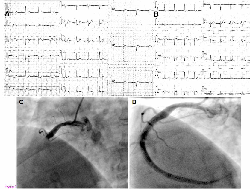
Figure 1. ECG and coronary angiography before and after PCI.
Panel A. Twelve and right ventricular precordial leads before primary PCI showing ST-segment
elevation, Q-wave and diphasic T-wave in the inferior leads. In addition, ST-segment elevation is
also evident in the right precordial leads (from V3R to V5R)
Panel B. Twelve-lead ECG 1-hour after primary PCI shows incomplete (<50%) ST-segment
elevation resolution in the inferior leads.
Panel C. Right coronary angiogram before primary PCI showing the occlusion of the proximal
portion of the vessel.
Panel D. Right coronary angiogram after primary PCI shows sub-optimal reflow in the vessel
(TIMI flow 2).
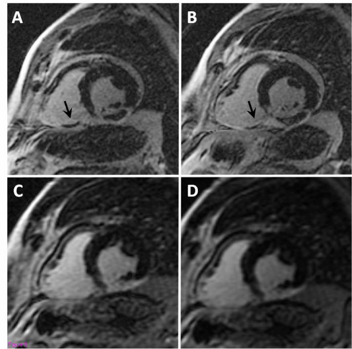
Figure 2. Cardiac magnetic resonance at 4 days and 4 months after myocardial infarction.
Mid short-axis LGE images at 4 days (panels A and B) and 4 months (panels C and D) after acute
MI. Early post- infarction LGE images (panels A and B) show transmural LGE of LV inferior and
inferolateral walls with extensive MO. In addition, LGE of RV inferior free-wall is also seen with
hypointense core (arrows) comprised within hyperenhanced myocardium. This finding is consistent
with MO complicating the infarcted RV myocardium. At 4 months follow-up (panels C and D),
LGE of LV inferior and inferolateral walls and RV inferior free-wall is still evident with resolution
of MO.
MI= myocardial infarction; LGE=late-gadolinium enhancement; MO=microvascular obstruction;
PCI: percutaneous coronary intervention
MOVIE LEGENDS
Movie A. Cardiac magnetic resonance, cine short axis imaging at 4 days after myocardial
infarction.
Cine short-axis imaging at 4 days after acute MI showing diffuse pericardial effusion, akinesia of
LV inferior and inferolateral walls and of RV inferior free-wall.
Movie B. Cardiac magnetic resonance, cine short axis imaging at 4 months after myocardial
infarction.
Cine short-axis imaging 4 months after acute MI showing resolution of pericardial effusion but
persistent akinesia of LV inferior and inferolateral wall and slight functional improvement limited
to the basal portion of RV inferior free-wall.

Additional files provided with this submission:
Additional file 1: Movie A.mpg, 10566Khttp://jcmr-online.com/imedia/1549214609683475/supp1.mpegAdditional file 2: Movie B.mpg, 10333Khttp://jcmr-online.com/imedia/1745971042683475/supp2.mpeg