Cardiovascular MR T2-STIR imaging does not discriminate between intramyocardial haemorrhage and microvascular obstruction during the subacute phase of a reperfused myocardial infarction Esben Søvsø Szocska Hansen, 1,2 Steen Fjord Pedersen, 3 Steen Bønløkke Pedersen, 4 Uffe Kjærgaard, 1 Nikolaj Hjort Schmidt, 5 Hans Erik Bøtker, 6 Won Yong Kim 1,6 To cite: Hansen ESS, Pedersen SF, Pedersen SB, et al. Cardiovascular MR T2- STIR imaging does not discriminate between intramyocardial haemorrhage and microvascular obstruction during the subacute phase of a reperfused myocardial infarction. Open Heart 2016;3:e000346. doi:10.1136/openhrt-2015- 000346 ESSH and SFP contributed equally. Received 6 October 2015 Revised 24 February 2016 Accepted 29 March 2016 For numbered affiliations see end of article. Correspondence to Esben Søvsø Szocska Hansen; [email protected] ABSTRACT Objective: Microvascular obstruction (MVO) and intramyocardial haemorrhage (IMH) are known complications of myocardial ischaemia-reperfusion injury. Whereas MVO is an established marker for a poor clinical outcome, the clinical significance of IMH remains less well defined. Cardiovascular MR (CMR) and T2 weighted short tau inversion recovery (T2-STIR) imaging have been used to detect IMH and to explore its clinical importance. IMH is typically identified within the area-at- risk as a hypointense signal core on T2-STIR images. Because MVO will also appear as a hypointense signal core, T2-STIR imaging may not be an optimal method for assessing IMH. In this study, we sought to investigate the ability of T2-STIR to discriminate between MVO with IMH in a porcine myocardial ischaemia-reperfusion model that expressed MVO with and without IMH. Method: MVO with and without IMH (defined from both macroscopic evaluation and T1 weighted CMR) was produced in 13 pigs by a 65-min balloon occlusion of the mid left anterior descending artery, followed by reperfusion. Eight days after injury, all pigs underwent CMR imaging and subsequently the hearts were assessed by gross pathology. Results: CMR identified MVO in all hearts. CMR and pathology showed that IMH was present in 6 of 13 (46%) infarcts. The sensitivity and specificity of T2-STIR hypointense signal core for identification of IMH was 100% and 29%, respectively. T2-values between hypointense signal core in the pigs with and without IMH were similar (60.4±3 ms vs 63.0±4 ms). Conclusions: T2-STIR did not allow identification of IMH in areas with MVO in a porcine model of myocardial ischaemic/reperfusion injury in the subacute phase of a reperfused myocardial infarction. INTRODUCTION Timely reperfusion is mandatory to salvage ischaemic myocardium and to reduce mortality in acute myocardial infarction. 1 Reperfusion following prolonged ischaemia may, however, injure the myocardial micro- vasculature and cause microvascular obstruc- tion (MVO). 2 This is a serious condition because it prevents adequate myocardial blood perfusion despite patent coronary arteries. Severe myocardial microvascular injury may also trigger intramyocardial haem- orrhage (IMH) due to extravasation of ery- throcytes through the damaged endothelial wall. 3–8 Whereas MVO is recognised as a strong predictor of ventricular remodelling and poor clinical outcome, it remains KEY QUESTIONS What is already known about this subject? ▸ Cardiovascular MR (CMR) using T2 weighted imaging with short tau inversion recovery (T2-STIR) is being used to detect microvascular obstruction (MVO) with intramyocardial haemor- rhage (IMH) as markers of myocardial ischaemia-reperfusion. However, the validity of T2 weighted CMR in assessment of IMH has not been tested. What does this study add? ▸ In this study, we find that T2-STIR does not discriminate between MVO and IMH in an experimental model of myocardial ischaemia- reperfusion that expresses MVO with and without IMH during the subacute phase of a reperfused myocardial infarction. How might this impact on clinical practice? ▸ A comprehensive multicontrast CMR protocol is needed to evaluate the presence of MVO and IMH in acute myocardial infarction. Hansen ESS, Pedersen SF, Pedersen SB, et al. Open Heart 2016;3:e000346. doi:10.1136/openhrt-2015-000346 1 Basic and translational research on February 24, 2020 by guest. Protected by copyright. http://openheart.bmj.com/ Open Heart: first published as 10.1136/openhrt-2015-000346 on 20 April 2016. Downloaded from

Welcome message from author
This document is posted to help you gain knowledge. Please leave a comment to let me know what you think about it! Share it to your friends and learn new things together.
Transcript

Cardiovascular MR T2-STIR imagingdoes not discriminate betweenintramyocardial haemorrhageand microvascular obstruction duringthe subacute phase of a reperfusedmyocardial infarction
Esben Søvsø Szocska Hansen,1,2 Steen Fjord Pedersen,3
Steen Bønløkke Pedersen,4 Uffe Kjærgaard,1 Nikolaj Hjort Schmidt,5
Hans Erik Bøtker,6 Won Yong Kim1,6
To cite: Hansen ESS,Pedersen SF, Pedersen SB,et al. Cardiovascular MR T2-STIR imaging does notdiscriminate betweenintramyocardial haemorrhageand microvascularobstruction during thesubacute phase of areperfused myocardialinfarction. Open Heart2016;3:e000346.doi:10.1136/openhrt-2015-000346
ESSH and SFP contributedequally.
Received 6 October 2015Revised 24 February 2016Accepted 29 March 2016
For numbered affiliations seeend of article.
Correspondence toEsben Søvsø SzocskaHansen;[email protected]
ABSTRACTObjective: Microvascular obstruction (MVO) andintramyocardial haemorrhage (IMH) are knowncomplications of myocardial ischaemia-reperfusioninjury. Whereas MVO is an established marker for a poorclinical outcome, the clinical significance of IMH remainsless well defined. Cardiovascular MR (CMR) and T2weighted short tau inversion recovery (T2-STIR) imaginghave been used to detect IMH and to explore its clinicalimportance. IMH is typically identified within the area-at-risk as a hypointense signal core on T2-STIR images.Because MVO will also appear as a hypointense signalcore, T2-STIR imaging may not be an optimal method forassessing IMH. In this study, we sought to investigatethe ability of T2-STIR to discriminate between MVO withIMH in a porcine myocardial ischaemia-reperfusionmodel that expressed MVO with and without IMH.Method: MVO with and without IMH (defined from bothmacroscopic evaluation and T1 weighted CMR) wasproduced in 13 pigs by a 65-min balloon occlusion of themid left anterior descending artery, followed byreperfusion. Eight days after injury, all pigs underwentCMR imaging and subsequently the hearts wereassessed by gross pathology.Results: CMR identified MVO in all hearts. CMR andpathology showed that IMH was present in 6 of 13 (46%)infarcts. The sensitivity and specificity of T2-STIRhypointense signal core for identification of IMH was100% and 29%, respectively. T2-values betweenhypointense signal core in the pigs with and without IMHwere similar (60.4±3 ms vs 63.0±4 ms).Conclusions: T2-STIR did not allow identification ofIMH in areas with MVO in a porcine model of myocardialischaemic/reperfusion injury in the subacute phase of areperfused myocardial infarction.
INTRODUCTIONTimely reperfusion is mandatory to salvageischaemic myocardium and to reduce
mortality in acute myocardial infarction.1
Reperfusion following prolonged ischaemiamay, however, injure the myocardial micro-vasculature and cause microvascular obstruc-tion (MVO).2 This is a serious conditionbecause it prevents adequate myocardialblood perfusion despite patent coronaryarteries. Severe myocardial microvascularinjury may also trigger intramyocardial haem-orrhage (IMH) due to extravasation of ery-throcytes through the damaged endothelialwall.3–8 Whereas MVO is recognised as astrong predictor of ventricular remodellingand poor clinical outcome, it remains
KEY QUESTIONS
What is already known about this subject?▸ Cardiovascular MR (CMR) using T2 weighted
imaging with short tau inversion recovery(T2-STIR) is being used to detect microvascularobstruction (MVO) with intramyocardial haemor-rhage (IMH) as markers of myocardialischaemia-reperfusion. However, the validity ofT2 weighted CMR in assessment of IMH has notbeen tested.
What does this study add?▸ In this study, we find that T2-STIR does not
discriminate between MVO and IMH in anexperimental model of myocardial ischaemia-reperfusion that expresses MVO with andwithout IMH during the subacute phase of areperfused myocardial infarction.
How might this impact on clinical practice?▸ A comprehensive multicontrast CMR protocol is
needed to evaluate the presence of MVO andIMH in acute myocardial infarction.
Hansen ESS, Pedersen SF, Pedersen SB, et al. Open Heart 2016;3:e000346. doi:10.1136/openhrt-2015-000346 1
Basic and translational research
on February 24, 2020 by guest. P
rotected by copyright.http://openheart.bm
j.com/
Open H
eart: first published as 10.1136/openhrt-2015-000346 on 20 April 2016. D
ownloaded from

unknown whether IMH further aggravates the clinicaloutcome.9 To determine the clinical consequences ofIMH, it is essential to develop a reliable, non-invasiveimaging technique capable of differentiating betweenMVO, and MVO with IMH.10 11 Cardiovascular MR(CMR) utilises first-pass perfusion imaging andT2-weighted images to detect MVO and IMH.Specifically, T2 weighted short tau inversion recovery(T2-STIR) imaging was applied to detect IMH6 7 and toinvestigate the prognostic value of IMH defined fromT2-STIR.6 7 12 13 Within the area-at-risk (AAR), usuallyidentified by T2-weighted imaging as oedema, a hypoin-tense core on T2-STIR images has been ascribed to para-magnetic effects of haemoglobin breakdown products,and has consequently been considered indicative ofIMH.14 15 However, because MVO consists of non-oedematous tissue, a hypointense signal core within theAAR may not be specific for IMH.16 MVO and IMH rep-resent two separate pathologies but often appear simul-taneously since IMH is highly associated with MVO.16
We investigated the ability of T2-STIR imaging to detectIMH and to discriminate between IMH and MVOusing a unique porcine model of myocardialischaemia-reperfusion that expresses MVO and MVOwith IMH.17
METHODSAnimal modelFifteen female Danish Landrace pigs each weighing∼40 kg were used for the experiments. The pigs werepresedated with an intramuscular injection of Stressnil(1 mL/kg) and midazolam (1 mL/kg). After inductionof anaesthesia with intravenous propofol (3.33 mg/kg)and endotracheal intubation, anaesthesia was main-tained with sevoflurane (2.5%) and continuous rateinfusion of fentanyl (3 mg/kg/h). The pigs were mech-anically ventilated with a tidal volume of 425 mL (12ventilations/min). By ultrasound guidance, an 8 F intro-ducer sheath was inserted into the right commonfemoral artery. This was followed by an intravenousbolus injection of heparin (100 IU/kg). Coronary occlu-sion was induced by placing a 2.5 mm angioplastyballoon in the left anterior descending coronary artery(LAD) distal to the second diagonal branch artery andinflating it to 10 atm. The balloon occluded the LAD for65 min, and was then deflated and removed. A coronaryangiogram was performed following balloon inflation, toconfirm occlusion, and also following balloon deflation,to confirm reperfusion. The animals were randomisedto receive either intracoronary infusion of saline ordimethyl sulfoxide during reperfusion as a potentialadjunctive therapy to reduce reperfusion injury. Thedetails of the experimental protocol are describedelsewhere.17
During the procedure, heart rate, ECG, blood pres-sure, temperature and oxygen saturation were continu-ously monitored. Ampicillin (2 mg) was administrated
intravenously before and after the procedure, and acetyl-salicylic acid (100 mg/day) was given orally after the pro-cedure and continued until euthanasia. To preventventricular fibrillation, 150 mg of amiodarone wasadministered intravenously prior to the induction ofmyocardial infarction. If ventricular fibrillation wasencountered, non-synchronised direct current defibrilla-tion (150 J) was performed. The paddles were pressedagainst the anterior chest wall above the sternum on theright side and below the sternum on the left side. At theend of the experiment, the pigs were awakened andreturned to their stables, where they stayed for 7–10 days(average 8.5 days) prior to CMR imaging, harvesting andevaluation by gross pathology.
CMR imagingAll the pigs underwent CMR imaging before euthanasia.The sedation protocol was as described above, except thatcontinuous propofol infusion (10 mg/mL, 12 mL/h) wasused instead of sevoflurane. CMR was performed on a1.5 T MR system (Intera, Philips Medical Systems, Best, theNetherlands) with a five-element cardiac synergy coil. Allimages of the heart were obtained with the pigs in thesupine position. First, a survey scan was performed to local-ise the heart and diaphragm, and then left ventricular(LV) function was assessed using a retrospective,ECG-triggered Balanced-Steady-State-Free-Precession(B-SSFP) breath-hold cine sequence in the cardiac short-axis, vertical long axis and horizontal long axis planes. Inthe cardiac short-axis, the LV volume was completelyencompassed by contiguous 8 mm slices with a spatialresolution of 1.22×1.22 mm and a field of view (FOV) of288×288 mm. The following imaging parameters wereused: repetition time (TR) 3.0 ms; echo time (TE) 1.5 ms;flip angle 60°; 30 heart phases. To ensure a strongT2-weighting, a T2-STIR fast spin echo sequence with along TE was obtained in the previously mentioned short-axis orientation to assess AAR. The sequence wasnavigator-gated and cardiac-triggered. The followingimaging parameters were used: TR 2400 ms; TE 100 ms;echo train length 20; fat inversion time 180 ms; flip angle90°; spatial resolution 0.54 mm×0.54 mm in-plane;number of averages 2; slice thickness 8 mm; FOV320×320 mm; 14 slices.We obtained T2-mapping in six animals by a multislice
spin echo sequence. The sequence was navigator-gatedand cardiac-triggered. The following imaging parameterswere used: TR 1100 ms; TE (8, 16, 24, 32, 40, 48, 56, 64)ms; flip angle 90°; spatial resolution 1.33×1.33 mmin-plane; slice thickness 8 mm; FOV 320×320 mm; 14slices.T1-weighted inversion recovery (T1W-IR) gradient
echo (GRE) imaging was obtained in the same short-axisslices for the purpose of identifying IMH, as previouslydescribed.18 The sequence was navigator-gated, free-breathing and cardiac-triggered. The following imagingparameters were used: TR 3.5 ms; TE 1.13 ms; flip angle30°; spatial resolution 1×1 mm in-plane; slice thickness
2 Hansen ESS, Pedersen SF, Pedersen SB, et al. Open Heart 2016;3:e000346. doi:10.1136/openhrt-2015-000346
Open Heart
on February 24, 2020 by guest. P
rotected by copyright.http://openheart.bm
j.com/
Open H
eart: first published as 10.1136/openhrt-2015-000346 on 20 April 2016. D
ownloaded from
8 mm (over contiguous slices); FOV 320×320 mm and 14slices. Before the acquisition of the T1W IR sequence, aTI scout (Look Locker sequence) was performed forthe purpose of obtaining the most appropriated TI tonull the signal intensity from blood. Typically, the TI wasfound to be optimal at approximately 500 ms.Subsequently, gadolinium enhanced first-pass myocar-
dial perfusion and late gadolinium enhancement (LGE)was performed for the purpose of identifying areasof MVO and myocardial infarction, respectively. Anintravenous bolus dose of 0.2 mmol/kg Gd-DTPA(Gadobutrol, Gadovist, Bayer Schering Pharma, Berlin,Germany) was administered manually. First-pass perfu-sion imaging was performed using a fast gradient echosequence with the following parameters: TR 2.5 ms; TE1.3 ms; flip angle 18°; spatial resolution 2.8×3.0 mmin-plane; slice thickness 10 mm; FOV 256×256 mm; threeslices acquired in the LV short-axis using a 5 mm inter-slice gap.LGE was acquired 15 min after gadolinium injection,
using a three-dimensional phase sensitive inversionrecovery-prepared T1-weighted gradient echo sequencewith the following parameters: TR 5.78 ms; TE 2.78 ms;echo train length 20; inversion time ∼320 ms; flip angle25°; spatial resolution 1.5×1.5 mm; slice thickness 8 mm;FOV 350×350 mm; 14 slices acquired in the LV short-axiswith no interslice gap. Following CMR, the pigs werekept under anaesthesia and moved to the operatingroom for organ harvesting.
Harvesting and pathologyMidline sternotomy was performed and a snare placedaround the LAD distal to the second diagonal branchat the same site as the previous balloon occlusion.Twenty-five millilitres of 10% Evans blue dye was theninjected into the left auricle and allowed to circulate forapproximately 10 s, to delineate the AAR.18–20 The animalwas subsequently euthanised and the heart excised. Theheart was then cut into consecutive 8 mm-thick slices inshort-axis planes. Each slice was photographed with adigital camera (Nikon, Tokyo, Japan) for the purpose ofregistering myocardial infarction, IMH and AAR.
DATA ANALYSISCMR imagesOne observer (WYK), blinded to the distribution of thegroups, analysed all the CMR images, using the semi-automatic, freely available software Segment V.1.9 R3697.First, LV volumes and function were calculated on theend-diastolic and end-systolic phases of the short-axiscine images. Second, myocardial infarct size was deter-mined in the LGE images by a semiautomated algorithmaccounting for partial volume effects.21 The infarct sizewas expressed as a percentage of the LV myocardium(infarction volume/LV myocardium volume×100%).The AAR was quantified from T2-STIR images by a semi-automated algorithm (AAR/LV myocardium
volume×100%).22 The salvaged myocardial percentagewas defined as the difference between the infarct sizeand AAR. Finally, the hypointense signal core within theAAR was defined by ROI analysis as a core with signalintensity measuring two SDs lower than the signal inten-sity of the AAR.23 24 The ROI defined by the hypoin-tense signal core was used on the T2-mapping images toextract T2 values. On the T1W IR images, myocardiumwith mean signal intensity more than two SDs above themean signal intensity from remote myocardium wasdefined as IMH (18). MVO was defined and quantifiedon the LGE images from visual inspection of no contrastenhancement within the infarct core. All CMR imageswere matched and aligned.
PathologyTwo observers by consensus (SFP and ESSH) analysedall photographed gross pathological images of the myo-cardial slices using the Adobe Photoshop software(Adobe Systems Inc, San Jose, California, USA). TheIMH volume was identified and measured as a distinctred to reddish black area within the AAR of the LV myo-cardium. MVO was not assessed in the pathology.
StatisticsThe significance of group differences was evaluatedusing the Mann-Whitney U test and the Wilcoxonmatched-pairs signed rank test was used to evaluate theamount of IMH in percentage of the left ventricle onT1W IR images compared to gross pathology. Data arepresented as mean and 95% CIs. A value of p<0.05 wasconsidered statistically significant. The associationbetween IMH and MVO, and presence of hypointensesignal core on T2-STIR images, was tested with a 2-tailedFisher’s exact test. T2-mapping values were tested usinga one-way analysis of variance with Holm-Sidak’s multiplecomparisons correction. Statistical analysis was per-formed with GraphPad Prism (6.05, GraphPad Software,La Jolla, California, USA).
RESULTSAll CMR examinations and experimental procedureswere successfully completed. Figure 1 shows representa-tive pathological photographs and CMR images of MVO(−IMH) and MVO (+IMH). Examples from gross path-ology assessment are shown in figure 2. The CMR andgross pathology results are summarised in table 1. Twopigs displayed neither MVO nor IMH. Global CMR para-meters did not show any significant difference in ejectionfraction or LV volumes between the two groups. AAR,infarct size and myocardial salvage also did not show anydifference between the groups. The MVO size measuredas a percentage of LV on LGE images showed no differ-ence in the MVO (+IMH) group compared with theMVO (−IMH) group, 2.1% vs 1.7%, respectively. Thesensitivity and specificity of T2-STIR hypointense signalcore to detect IMH was 100% and 29%, respectively, as
Hansen ESS, Pedersen SF, Pedersen SB, et al. Open Heart 2016;3:e000346. doi:10.1136/openhrt-2015-000346 3
Basic and translational research
on February 24, 2020 by guest. P
rotected by copyright.http://openheart.bm
j.com/
Open H
eart: first published as 10.1136/openhrt-2015-000346 on 20 April 2016. D
ownloaded from

shown in table 2. T1W IR showed that 0 out of 7 (0%)had IMH in the MVO (−IMH) group compared with 6of 6 (100%) pigs in MVO (+IMH) group (p<0.01).Wilcoxon matched-pairs signed rank test showed no dif-ference between the IMH % LV measured on T1W IR
(range 12–39% LV) compared with gross pathology(range 5–27% LV) (p=0.09, difference 6.4% LV,CI (−2.86 to 15.76), median 5.1%, Q1=2.1, Q3=10.6).The measured T2-values are visualised in figure 3. The
T2-values (n=7) from hypointense signal core in MVO
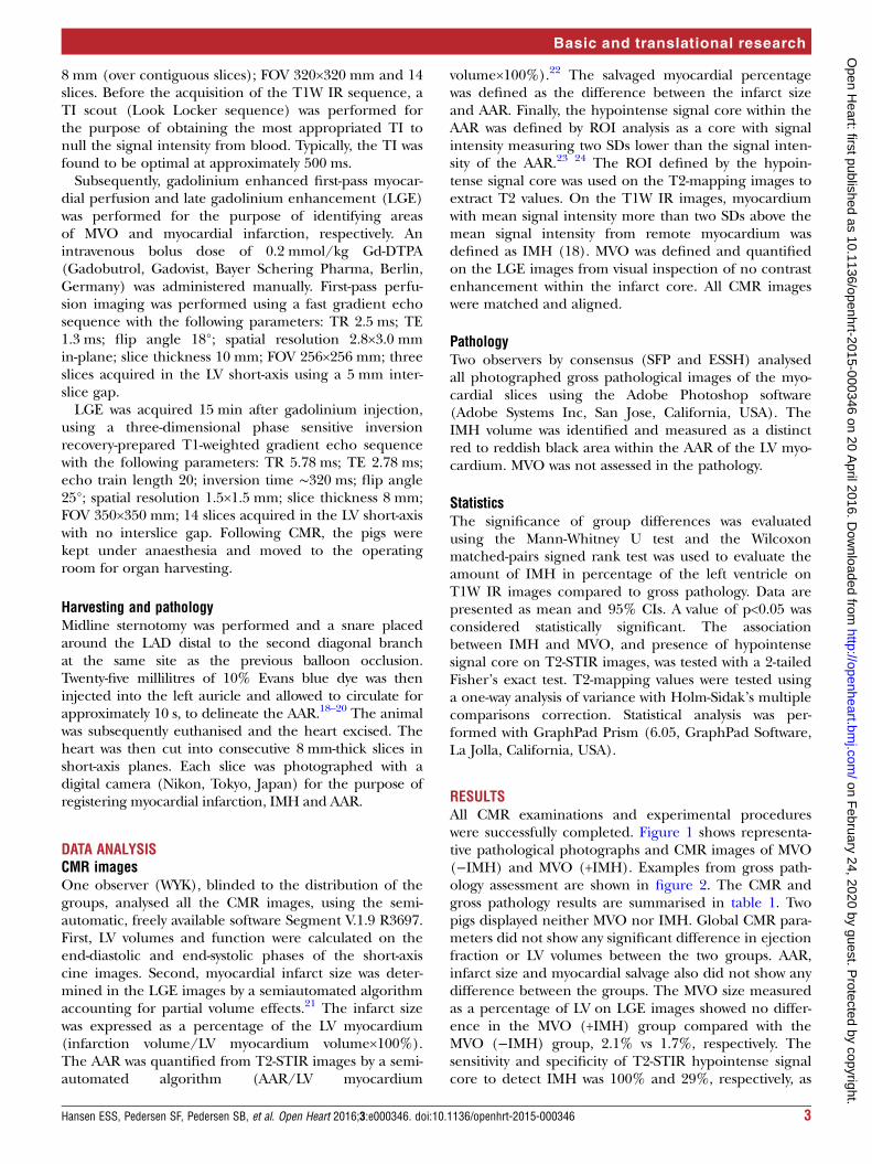
Figure 1 Upper row shows myocardial infarction with MVO (−IMH) (indicated by arrows) on pathology (A), T2-STIR (B), late
gadolinium enhancement (LGE) images (C) and T1W IR (D). Lower row shows myocardial infarction with MVO (+IMH) (indicated
by arrows) on pathology (E), T2-STIR (F) and LGE images (G), and T1W IR (H). IMH, intramyocardial haemorrhage; MVO,
microvascular obstruction; T2-STIR, T2 weighted short tau inversion recovery.
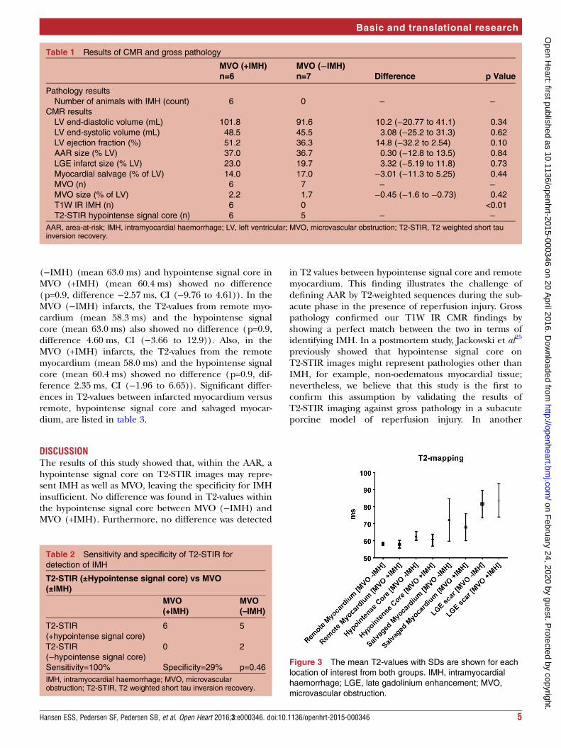
Figure 2 Examples from gross anatomical pathology. A (1–3) and B (1–3) show three slices from two pigs in the MVO (+IMH)
group, where the arrows indicated areas defined as IMH. C (1–3) and D (1–3) show three slices from two pigs in the MVO
(−IMH) group, where arrows points to the AAR where there is no indication of IMH. AAR, area-at-risk; IMH, intramyocardial
haemorrhage; MVO, microvascular obstruction.
4 Hansen ESS, Pedersen SF, Pedersen SB, et al. Open Heart 2016;3:e000346. doi:10.1136/openhrt-2015-000346
Open Heart
on February 24, 2020 by guest. P
rotected by copyright.http://openheart.bm
j.com/
Open H
eart: first published as 10.1136/openhrt-2015-000346 on 20 April 2016. D
ownloaded from
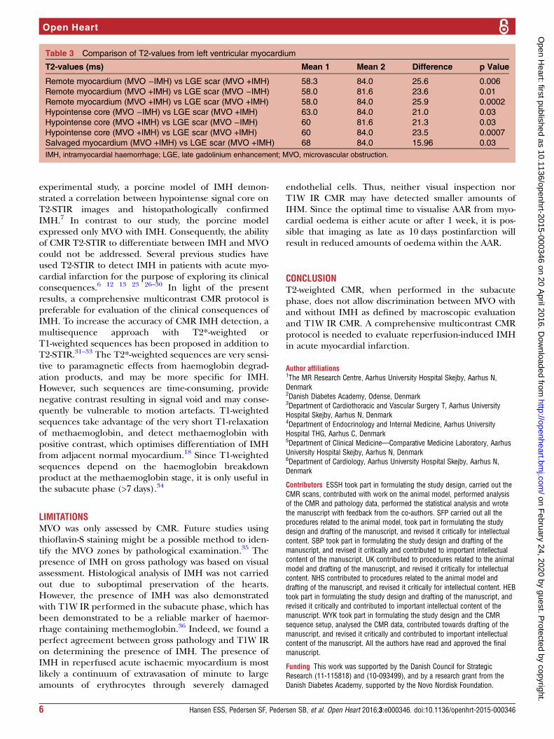
(−IMH) (mean 63.0 ms) and hypointense signal core inMVO (+IMH) (mean 60.4 ms) showed no difference(p=0.9, difference −2.57 ms, CI (−9.76 to 4.61)). In theMVO (−IMH) infarcts, the T2-values from remote myo-cardium (mean 58.3 ms) and the hypointense signalcore (mean 63.0 ms) also showed no difference (p=0.9,difference 4.60 ms, CI (−3.66 to 12.9)). Also, in theMVO (+IMH) infarcts, the T2-values from the remotemyocardium (mean 58.0 ms) and the hypointense signalcore (mean 60.4 ms) showed no difference (p=0.9, dif-ference 2.35 ms, CI (−1.96 to 6.65)). Significant differ-ences in T2-values between infarcted myocardium versusremote, hypointense signal core and salvaged myocar-dium, are listed in table 3.
DISCUSSIONThe results of this study showed that, within the AAR, ahypointense signal core on T2-STIR images may repre-sent IMH as well as MVO, leaving the specificity for IMHinsufficient. No difference was found in T2-values withinthe hypointense signal core between MVO (−IMH) andMVO (+IMH). Furthermore, no difference was detected
in T2 values between hypointense signal core and remotemyocardium. This finding illustrates the challenge ofdefining AAR by T2-weighted sequences during the sub-acute phase in the presence of reperfusion injury. Grosspathology confirmed our T1W IR CMR findings byshowing a perfect match between the two in terms ofidentifying IMH. In a postmortem study, Jackowski et al25
previously showed that hypointense signal core onT2-STIR images might represent pathologies other thanIMH, for example, non-oedematous myocardial tissue;nevertheless, we believe that this study is the first toconfirm this assumption by validating the results ofT2-STIR imaging against gross pathology in a subacuteporcine model of reperfusion injury. In another
Table 1 Results of CMR and gross pathology
MVO (+IMH)
n=6
MVO (−IMH)
n=7 Difference p Value
Pathology results
Number of animals with IMH (count) 6 0 − −CMR results
LV end-diastolic volume (mL) 101.8 91.6 10.2 (−20.77 to 41.1) 0.34
LV end-systolic volume (mL) 48.5 45.5 3.08 (−25.2 to 31.3) 0.62
LV ejection fraction (%) 51.2 36.3 14.8 (−32.2 to 2.54) 0.10
AAR size (% LV) 37.0 36.7 0.30 (−12.8 to 13.5) 0.84
LGE infarct size (% LV) 23.0 19.7 3.32 (−5.19 to 11.8) 0.73
Myocardial salvage (% of LV) 14.0 17.0 −3.01 (−11.3 to 5.25) 0.44
MVO (n) 6 7 − −MVO size (% of LV) 2.2 1.7 −0.45 (−1.6 to −0.73) 0.42
T1W IR IMH (n) 6 0 <0.01
T2-STIR hypointense signal core (n) 6 5 − −AAR, area-at-risk; IMH, intramyocardial haemorrhage; LV, left ventricular; MVO, microvascular obstruction; T2-STIR, T2 weighted short tauinversion recovery.
Table 2 Sensitivity and specificity of T2-STIR for
detection of IMH
T2-STIR (±Hypointense signal core) vs MVO
(±IMH)
MVO
(+IMH)
MVO
(–IMH)
T2-STIR
(+hypointense signal core)
6 5
T2-STIR
(−hypointense signal core)
0 2
Sensitivity=100% Specificity=29% p=0.46
IMH, intramyocardial haemorrhage; MVO, microvascularobstruction; T2-STIR, T2 weighted short tau inversion recovery.
Figure 3 The mean T2-values with SDs are shown for each
location of interest from both groups. IMH, intramyocardial
haemorrhage; LGE, late gadolinium enhancement; MVO,
microvascular obstruction.
Hansen ESS, Pedersen SF, Pedersen SB, et al. Open Heart 2016;3:e000346. doi:10.1136/openhrt-2015-000346 5
Basic and translational research
on February 24, 2020 by guest. P
rotected by copyright.http://openheart.bm
j.com/
Open H
eart: first published as 10.1136/openhrt-2015-000346 on 20 April 2016. D
ownloaded from
experimental study, a porcine model of IMH demon-strated a correlation between hypointense signal core onT2-STIR images and histopathologically confirmedIMH.7 In contrast to our study, the porcine modelexpressed only MVO with IMH. Consequently, the abilityof CMR T2-STIR to differentiate between IMH and MVOcould not be addressed. Several previous studies haveused T2-STIR to detect IMH in patients with acute myo-cardial infarction for the purpose of exploring its clinicalconsequences.6 12 13 23 26–30 In light of the presentresults, a comprehensive multicontrast CMR protocol ispreferable for evaluation of the clinical consequences ofIMH. To increase the accuracy of CMR IMH detection, amultisequence approach with T2*-weighted orT1-weighted sequences has been proposed in addition toT2-STIR.31–33 The T2*-weighted sequences are very sensi-tive to paramagnetic effects from haemoglobin degrad-ation products, and may be more specific for IMH.However, such sequences are time-consuming, providenegative contrast resulting in signal void and may conse-quently be vulnerable to motion artefacts. T1-weightedsequences take advantage of the very short T1-relaxationof methaemoglobin, and detect methaemoglobin withpositive contrast, which optimises differentiation of IMHfrom adjacent normal myocardium.18 Since T1-weightedsequences depend on the haemoglobin breakdownproduct at the methaemoglobin stage, it is only useful inthe subacute phase (>7 days).34
LIMITATIONSMVO was only assessed by CMR. Future studies usingthioflavin-S staining might be a possible method to iden-tify the MVO zones by pathological examination.35 Thepresence of IMH on gross pathology was based on visualassessment. Histological analysis of IMH was not carriedout due to suboptimal preservation of the hearts.However, the presence of IMH was also demonstratedwith T1W IR performed in the subacute phase, which hasbeen demonstrated to be a reliable marker of haemor-rhage containing methemoglobin.36 Indeed, we found aperfect agreement between gross pathology and T1W IRon determining the presence of IMH. The presence ofIMH in reperfused acute ischaemic myocardium is mostlikely a continuum of extravasation of minute to largeamounts of erythrocytes through severely damaged
endothelial cells. Thus, neither visual inspection norT1W IR CMR may have detected smaller amounts ofIHM. Since the optimal time to visualise AAR from myo-cardial oedema is either acute or after 1 week, it is pos-sible that imaging as late as 10 days postinfarction willresult in reduced amounts of oedema within the AAR.
CONCLUSIONT2-weighted CMR, when performed in the subacutephase, does not allow discrimination between MVO withand without IMH as defined by macroscopic evaluationand T1W IR CMR. A comprehensive multicontrast CMRprotocol is needed to evaluate reperfusion-induced IMHin acute myocardial infarction.
Author affiliations1The MR Research Centre, Aarhus University Hospital Skejby, Aarhus N,Denmark2Danish Diabetes Academy, Odense, Denmark3Department of Cardiothoracic and Vascular Surgery T, Aarhus UniversityHospital Skejby, Aarhus N, Denmark4Department of Endocrinology and Internal Medicine, Aarhus UniversityHospital THG, Aarhus C, Denmark5Department of Clinical Medicine—Comparative Medicine Laboratory, AarhusUniversity Hospital Skejby, Aarhus N, Denmark6Department of Cardiology, Aarhus University Hospital Skejby, Aarhus N,Denmark
Contributors ESSH took part in formulating the study design, carried out theCMR scans, contributed with work on the animal model, performed analysisof the CMR and pathology data, performed the statistical analysis and wrotethe manuscript with feedback from the co-authors. SFP carried out all theprocedures related to the animal model, took part in formulating the studydesign and drafting of the manuscript, and revised it critically for intellectualcontent. SBP took part in formulating the study design and drafting of themanuscript, and revised it critically and contributed to important intellectualcontent of the manuscript. UK contributed to procedures related to the animalmodel and drafting of the manuscript, and revised it critically for intellectualcontent. NHS contributed to procedures related to the animal model anddrafting of the manuscript, and revised it critically for intellectual content. HEBtook part in formulating the study design and drafting of the manuscript, andrevised it critically and contributed to important intellectual content of themanuscript. WYK took part in formulating the study design and the CMRsequence setup, analysed the CMR data, contributed towards drafting of themanuscript, and revised it critically and contributed to important intellectualcontent of the manuscript. All the authors have read and approved the finalmanuscript.
Funding This work was supported by the Danish Council for StrategicResearch (11-115818) and (10-093499), and by a research grant from theDanish Diabetes Academy, supported by the Novo Nordisk Foundation.
Table 3 Comparison of T2-values from left ventricular myocardium
T2-values (ms) Mean 1 Mean 2 Difference p Value
Remote myocardium (MVO −IMH) vs LGE scar (MVO +IMH) 58.3 84.0 25.6 0.006
Remote myocardium (MVO +IMH) vs LGE scar (MVO −IMH) 58.0 81.6 23.6 0.01
Remote myocardium (MVO +IMH) vs LGE scar (MVO +IMH) 58.0 84.0 25.9 0.0002
Hypointense core (MVO −IMH) vs LGE scar (MVO +IMH) 63.0 84.0 21.0 0.03
Hypointense core (MVO +IMH) vs LGE scar (MVO −IMH) 60 81.6 21.3 0.03
Hypointense core (MVO +IMH) vs LGE scar (MVO +IMH) 60 84.0 23.5 0.0007
Salvaged myocardium (MVO +IMH) vs LGE scar (MVO +IMH) 68 84.0 15.96 0.03
IMH, intramyocardial haemorrhage; LGE, late gadolinium enhancement; MVO, microvascular obstruction.
6 Hansen ESS, Pedersen SF, Pedersen SB, et al. Open Heart 2016;3:e000346. doi:10.1136/openhrt-2015-000346
Open Heart
on February 24, 2020 by guest. P
rotected by copyright.http://openheart.bm
j.com/
Open H
eart: first published as 10.1136/openhrt-2015-000346 on 20 April 2016. D
ownloaded from
Competing interests None declared.
Provenance and peer review Not commissioned; externally peer reviewed.
Data sharing statement No additional data are available.
Open Access This is an Open Access article distributed in accordance withthe Creative Commons Attribution Non Commercial (CC BY-NC 4.0) license,which permits others to distribute, remix, adapt, build upon this work non-commercially, and license their derivative works on different terms, providedthe original work is properly cited and the use is non-commercial. See: http://creativecommons.org/licenses/by-nc/4.0/
REFERENCES1. Maroko PR, Libby P, Ginks WR, et al. Coronary artery
reperfusion. I. Early effects on local myocardial function and theextent of myocardial necrosis. J Clin Invest 1972;51:2710–16.
2. Matsumura K, Jeremy RW, Schaper J, et al. Progression ofmyocardial necrosis during reperfusion of ischemic myocardium.Circulation 1998;97:795–804.
3. Basso C, Thiene G. The pathophysiology of myocardial reperfusion:a pathologist’s perspective. Heart 2006;92:1559–62.
4. Reffelmann T, Kloner RA. The no-reflow phenomenon: a basicmechanism of myocardial ischemia and reperfusion. Basic ResCardiol 2006;101:359–72.
5. Kloner RA, Ganote CE, Jennings RB. The “no-reflow” phenomenonafter temporary coronary occlusion in the dog. J Clin Invest1974;54:1496–508.
6. Beek AM, Nijveldt R, van Rossum AC. Intramyocardial hemorrhageand microvascular obstruction after primary percutaneous coronaryintervention. Int J Cardiovasc Imaging 2010;26:49–55.
7. Robbers LF, Eerenberg ES, Teunissen PF, et al. Magneticresonance imaging-defined areas of microvascular obstruction afteracute myocardial infarction represent microvascular destruction andhaemorrhage. Eur Heart J 2013;34:2346–53.
8. Payne AR, Berry C, Kellman P, et al. Bright-blood T(2)-weightedMRI has high diagnostic accuracy for myocardial hemorrhage inmyocardial infarction: a preclinical validation study in swine. CircCardiovasc Imaging 2011;4:738–45.
9. Betgem RP, de Waard GA, Nijveldt R, et al. Intramyocardialhaemorrhage after acute myocardial infarction. Nat Rev Cardiol2015;12:156–67.
10. Calvieri C, Masselli G, Monti R, et al. Intramyocardial hemorrhage:an enigma for cardiac MRI? BioMed Res Int 2015;2015:859073.
11. Hamirani YS, Wong A, Kramer CM, et al. Effect of microvascularobstruction and intramyocardial hemorrhage by CMR on LVremodeling and outcomes after myocardial infarction: a systematicreview and meta-analysis. JACC Cardiovasc Imaging2014;7:940–52.
12. Eitel I, Kubusch K, Strohm O, et al. Prognostic value anddeterminants of a hypointense infarct core in T2-weighted cardiacmagnetic resonance in acute reperfused ST-elevation-myocardialinfarction. Circ Cardiovasc Imaging 2011;4:354–62.
13. Husser O, Monmeneu JV, Sanchis J, et al. Cardiovascular magneticresonance-derived intramyocardial hemorrhage after STEMI:Influence on long-term prognosis, adverse left ventricular remodelingand relationship with microvascular obstruction. Int J Cardiol2013;167:2047–54.
14. Lotan CS, Bouchard A, Cranney GB, et al. Assessment ofpostreperfusion myocardial hemorrhage using proton NMR imagingat 1.5 T. Circulation 1992;86:1018–25.
15. Lotan CS, Miller SK, Bouchard A, et al. Detection of intramyocardialhemorrhage using high-field proton (1H) nuclear magneticresonance imaging. Cathet Cardiovasc Diagn 1990;20:205–11.
16. Ye YX, Basse-Lüsebrink TC, Arias-Loza PA, et al. Monitoring ofmonocyte recruitment in reperfused myocardial infarction withintramyocardial hemorrhage and microvascular obstruction bycombined fluorine 19 and proton cardiac magnetic resonanceimaging. Circulation 2013;128:1878–88.
17. Pedersen SF, Grøndal AK, Andersen NP, et al. Dimethyl sulfoxidereduces microvascular obstruction and intramyocardialhemorrhagein a Porcine ischemia-reperfusion model. Heart ResOpen J 2015;2:85–91.
18. Pedersen SF, Thrysøe SA, Robich MP, et al. Assessment ofintramyocardial hemorrhage by T1-weighted cardiovascularmagnetic resonance in reperfused acute myocardial infarction.J Cardiovasc Magn Reson 2012;14:59.
19. Nakamura K, Al-Ruzzeh S, Gray C, et al. Effect of myocardialreperfusion on the release of nitric oxide after regional ischemia: anexperimental model of beating-heart surgery. Tex Heart Inst J2006;33:35–9.
20. Redfors B, Shao Y, Omerovic E. Myocardial infarct size and area at riskassessment in mice. Exp Clin Cardiol 2012;17:268–72.
21. Heiberg E, Ugander M, Engblom H, et al. Automated quantificationof myocardial infarction from MR images by accounting for partialvolume effects: animal, phantom, and human study. Radiology2008;246:581–8.
22. Sjögren J, Ubachs JF, Engblom H, et al. Semi-automaticsegmentation of myocardium at risk in T2-weighted cardiovascularmagnetic resonance. J Cardiovasc Magn Reson 2012;14:10.
23. O h-Ici D, Ridgway JP, Kuehne T, et al. Cardiovascular magneticresonance of myocardial edema using a short inversion timeinversion recovery (STIR) black-blood technique: Diagnosticaccuracy of visual and semi-quantitative assessment. J CardiovascMagn Reson 2012;14:22.
24. Croisille P, Kim HW, Kim RJ. Controversies in cardiovascular MRimaging: T2-weighted imaging should not be used to delineate thearea at risk in ischemic myocardial injury. Radiology 2012;265:12–22.
25. Jackowski C, Christe A, Sonnenschein M, et al. Postmortemunenhanced magnetic resonance imaging of myocardial infarction incorrelation to histological infarction age characterization. Eur Heart J2006;27:2459–67.
26. Kali A, Tang RL, Kumar A, et al. Detection of acute reperfusionmyocardial hemorrhage with cardiac MR imaging: T2 versus T2.Radiology 2013;269:387–95.
27. Mather AN, Fairbairn TA, Ball SG, et al. Reperfusion haemorrhageas determined by cardiovascular MRI is a predictor of adverse leftventricular remodelling and markers of late arrhythmic risk. Heart2011;97:453–9.
28. Weaver JC, Ramsay DD, Rees D, et al. Dynamic changes in STsegment resolution after myocardial infarction and the associationwith microvascular injury on cardiac Magnetic Resonance Imaging.Heart Lung Circ 2011;20:111–18.
29. Amabile N, Jacquier A, Shuhab A, et al. Incidence, predictors, andprognostic value of intramyocardial hemorrhage lesions in STelevation myocardial infarction. Catheter Cardiovasc Interv2012;79:1101–8.
30. Wright J, Adriaenssens T, Dymarkowski S, et al. Quantification ofmyocardial area at risk with T2-weighted CMR: comparison withcontrast-enhanced CMR and coronary angiography. JACCCardiovasc Imaging 2009;2:825–31.
31. Kidambi A, Biglands JD, Higgins DM, et al. Susceptibility-weightedcardiovascular magnetic resonance in comparison to T2 and T2 starimaging for detection of intramyocardial hemorrhage following acutemyocardial infarction at 3 Tesla. J Cardiovasc Magn Reson2014;16:86.
32. Kidambi A, Mather AN, Motwani M, et al. The effect of microvascularobstruction and intramyocardial hemorrhage on contractile recoveryin reperfused myocardial infarction: insights from cardiovascularmagnetic resonance. J Cardiovasc Magn Reson 2013;15:58.
33. Kandler D, Lücke C, Grothoff M, et al. The relation betweenhypointense core, microvascular obstruction and intramyocardialhaemorrhage in acute reperfused myocardial infarctionassessed by cardiac magnetic resonance imaging. Eur Radiol2014;24:3277–88.
34. Habs M, Pfefferkorn T, Cyran CC, et al. Age determination of vesselwall hematoma in spontaneous cervical artery dissection: amulti-sequence 3T cardiovascular magnetic resonance study.J Cardiovasc Magn Reson 2011;13:76.
35. Wu KC, Kim RJ, Bluemke DA, et al. Quantification and time courseof microvascular obstruction by contrast-enhancedechocardiography and magnetic resonance imaging following acutemyocardial infarction and reperfusion. J Am Coll Cardiol1998;32:1756–64.
36. Moody AR, Murphy RE, Morgan PS, et al. Characterization ofcomplicated carotid plaque with magnetic resonance direct thrombusimaging in patients with cerebral ischemia. Circulation2003;107:3047–52.
Hansen ESS, Pedersen SF, Pedersen SB, et al. Open Heart 2016;3:e000346. doi:10.1136/openhrt-2015-000346 7
Basic and translational research
on February 24, 2020 by guest. P
rotected by copyright.http://openheart.bm
j.com/
Open H
eart: first published as 10.1136/openhrt-2015-000346 on 20 April 2016. D
ownloaded from
Related Documents