Cerebral Microvascular Obstruction by Fibrin is Associated with Upregulation of PAI-1 Acutely after Onset of Focal Embolic Ischemia in Rats Zheng Gang Zhang, 1 Michael Chopp, 1,5 Anton Goussev, 1 Dunyue Lu, 2 Daniel Morris, 3 Wayne Tsang, 1 Cecylia Powers, 1 and Khang-Loon Ho 4 Departments of 1 Neurology, 2 Neurosurgery, 3 Emergency Medicine, and 4 Pathology, Henry Ford Health Sciences Center, Detroit, Michigan 48202, and 5 Department of Physics, Oakland University, Rochester, Michigan 48309 The mechanisms underlying cerebral microvascular perfusion deficit resulting from occlusion of the middle cerebral artery (MCA) require elucidation. We, therefore, tested the hypothesis that intravascular fibrin deposition in situ directly obstructs cerebral microcirculation and that local changes in type 1 plas- minogen activator inhibitor (PAI-1) gene expression contribute to intravascular fibrin deposition after embolic MCA occlusion. Using laser-scanning confocal microscopy (LSCM) in combina- tion with immunofluorescent staining, we simultaneously mea- sured in three dimensions the distribution of microvascular plasma perfusion deficit and fibrin(ogen) immunoreactivity in a rat model of focal cerebral embolic ischemia (n 5 12). In addition, using in situ hybridization and immunostaining, we analyzed expression of PAI-1 in ischemic brain (n 5 13). A significant ( p , 0.05) reduction of cerebral microvascular plasma perfusion accompanied a significant ( p , 0.05) in- crease of intravascular and extravascular fibrin deposition in the ischemic lesion. Microvascular plasma perfusion deficit and fibrin deposition expanded concomitantly from the subcortex to the cortex during 1 and 4 hr of embolic MCA occlusion. Three- dimensional analysis revealed that intravascular fibrin deposi- tion directly blocks microvascular plasma perfusion. Vascular plugs contained erythrocytes, polymorphonuclear leukocytes, and platelets enmeshed in fibrin. In situ hybridization demon- strated induction of PAI-1 mRNA in vascular endothelial cells in the ischemic region at 1 hr of ischemia. PAI-1 mRNA signifi- cantly increased at 4 hr of ischemia. Immunohistochemical staining showed the same pattern of increased PAI-1 antigen in the endothelial cells. These data demonstrate, for the first time, that progressive intravascular fibrin deposition directly blocks cerebral microvascular plasma perfusion in the ischemic region during acute focal cerebral embolic ischemia, and upregulation of the PAI-1 gene in the ischemic lesion may foster fibrin deposition through suppression of fibrinolysis. Key words: stroke; plasminogen activator inhibitor; rat; fibrin; microvascular; perfusion; confocal microscopy Occlusion of the middle cerebral artery (MCA) results in pro- gressive impairment of downstream cerebral microvascular plasma perfusion (Crowell and Olsson, 1972; Little et al., 1975; Buchweitz-Milton and Weiss, 1988; Ennis et al., 1990). Using intravascular fluorescent tracer molecules or fluorescent tracers in combination with laser-scanning confocal microscopy (L SC M), we and others have shown a significant reduction of cerebral microvascular plasma perfusion and concomitant cerebral injury in the ischemic core at 1 and 4 hr after MCA occlusion (Dawson et al., 1997; Zhang et al., 1999a). Data are emerging to suggest that intravascular fibrin deposition contributes to microvascular obstruction (Okada et al., 1994; Siebler et al., 1994; Heyes and Cervos-Navarro, 1996). For example, microvascular fibrin depo- sition accumulates during early focal cerebral ischemia, and reperfusion in the nonhuman primate and fibrin-containing mi- crothromboemboli are found in acute human ischemic brain (Siebler et al., 1994; Heyes and Cervos-Navarro, 1996). A fibrin thrombus is formed from fibrinogen by activation of thrombin (Collen and Lijnen, 1991; L oscalzo and Schafer, 1992). Endogenous fibrinolysis is mediated by plasminogen activators that convert the zymogen plasminogen into the active serine protease plasmin. Plasmin is the primary enzyme responsible for removal of fibrin deposits (Collen and Lijnen, 1991; Vassalii et al., 1991; Plow et al., 1995). Type 1 plasminogen activator inhibitor (PAI-1) inhibits plasminogen activators in vivo (Loskutoff et al., 1989). PAI-1 is secreted by a variety of cells, including endothelial cells and platelets (Loskutoff et al., 1989; Braaten et al., 1993; Kollros et al., 1994; Stringer et al., 1994; Handt et al., 1996). Elevation of PAI-1 activity is associated with fibrin deposition after ischemia (Hamsten et al., 1987; Margaglione et al., 1994). Therefore, intravascular deposition of fibrin in ischemic brain suggests a perturbation of the procoagulant and fibrinolytic acti- vation cascades. Despite the increasing number of reports about the effects of fibrin deposition on microcirculatory impairment, information is lacking whether intravascular fibrin deposition in situ directly obstructs cerebral microcirculation and how local changes in PAI-1 gene expression contribute to intravascular fibrin deposi- tion. In this report, we used three-dimensional LSCM in combi- nation with immunofluorescent staining to investigate the effects of intravascular fibrin deposition on cerebral microvascular plasma perfusion deficits in a rat model of focal cerebral embolic ischemia (Zhang et al., 1997). In addition, using in situ hybrid- Received July 29, 1999; revised Sept. 10, 1999; accepted Sept. 29, 1999. This work was supported by National Institute of Neurological Disorders and Stroke Grants PO1 NS23393 and RO1 NS33627. We gratefully acknowledge Drs. D. J. L oskutoff and D. Belin for providing us anti-PAI-1 antibody and PAI-1 cDNA. We thank Denice Bliesath for manuscript preparation and Cynthia Roberts and Xiuli Zhang for technical assistance. Correspondence should be addressed to Dr. Michael Chopp, Henry Ford Hospi- tal, Neurology Department, 2799 West Grand Boulevard, Detroit, M I 48202. E-mail: [email protected] h.edu. Copyright © 1999 Society for Neuroscience 0270-6474/99/1910898-10$05.00/0 The Journal of Neuroscience, December 15, 1999, 19(24):10898–10907

Welcome message from author
This document is posted to help you gain knowledge. Please leave a comment to let me know what you think about it! Share it to your friends and learn new things together.
Transcript

Cerebral Microvascular Obstruction by Fibrin is Associated withUpregulation of PAI-1 Acutely after Onset of Focal EmbolicIschemia in Rats
Zheng Gang Zhang,1 Michael Chopp,1,5 Anton Goussev,1 Dunyue Lu,2 Daniel Morris,3 Wayne Tsang,1Cecylia Powers,1 and Khang-Loon Ho4
Departments of 1Neurology, 2Neurosurgery, 3Emergency Medicine, and 4Pathology, Henry Ford Health Sciences Center,Detroit, Michigan 48202, and 5Department of Physics, Oakland University, Rochester, Michigan 48309
The mechanisms underlying cerebral microvascular perfusiondeficit resulting from occlusion of the middle cerebral artery(MCA) require elucidation. We, therefore, tested the hypothesisthat intravascular fibrin deposition in situ directly obstructscerebral microcirculation and that local changes in type 1 plas-minogen activator inhibitor (PAI-1) gene expression contributeto intravascular fibrin deposition after embolic MCA occlusion.Using laser-scanning confocal microscopy (LSCM) in combina-tion with immunofluorescent staining, we simultaneously mea-sured in three dimensions the distribution of microvascularplasma perfusion deficit and fibrin(ogen) immunoreactivity in arat model of focal cerebral embolic ischemia (n 5 12). Inaddition, using in situ hybridization and immunostaining, weanalyzed expression of PAI-1 in ischemic brain (n 5 13). Asignificant ( p , 0.05) reduction of cerebral microvascularplasma perfusion accompanied a significant ( p , 0.05) in-crease of intravascular and extravascular fibrin deposition inthe ischemic lesion. Microvascular plasma perfusion deficit and
fibrin deposition expanded concomitantly from the subcortex tothe cortex during 1 and 4 hr of embolic MCA occlusion. Three-dimensional analysis revealed that intravascular fibrin deposi-tion directly blocks microvascular plasma perfusion. Vascularplugs contained erythrocytes, polymorphonuclear leukocytes,and platelets enmeshed in fibrin. In situ hybridization demon-strated induction of PAI-1 mRNA in vascular endothelial cells inthe ischemic region at 1 hr of ischemia. PAI-1 mRNA signifi-cantly increased at 4 hr of ischemia. Immunohistochemicalstaining showed the same pattern of increased PAI-1 antigen inthe endothelial cells. These data demonstrate, for the first time,that progressive intravascular fibrin deposition directly blockscerebral microvascular plasma perfusion in the ischemic regionduring acute focal cerebral embolic ischemia, and upregulationof the PAI-1 gene in the ischemic lesion may foster fibrindeposition through suppression of fibrinolysis.
Key words: stroke; plasminogen activator inhibitor; rat; fibrin;microvascular; perfusion; confocal microscopy
Occlusion of the middle cerebral artery (MCA) results in pro-gressive impairment of downstream cerebral microvascularplasma perfusion (Crowell and Olsson, 1972; Little et al., 1975;Buchweitz-Milton and Weiss, 1988; Ennis et al., 1990). Usingintravascular fluorescent tracer molecules or fluorescent tracersin combination with laser-scanning confocal microscopy (LSCM),we and others have shown a significant reduction of cerebralmicrovascular plasma perfusion and concomitant cerebral injuryin the ischemic core at 1 and 4 hr after MCA occlusion (Dawsonet al., 1997; Zhang et al., 1999a). Data are emerging to suggestthat intravascular fibrin deposition contributes to microvascularobstruction (Okada et al., 1994; Siebler et al., 1994; Heyes andCervos-Navarro, 1996). For example, microvascular fibrin depo-sition accumulates during early focal cerebral ischemia, andreperfusion in the nonhuman primate and fibrin-containing mi-crothromboemboli are found in acute human ischemic brain(Siebler et al., 1994; Heyes and Cervos-Navarro, 1996).
A fibrin thrombus is formed from fibrinogen by activation ofthrombin (Collen and Lijnen, 1991; Loscalzo and Schafer, 1992).Endogenous fibrinolysis is mediated by plasminogen activatorsthat convert the zymogen plasminogen into the active serineprotease plasmin. Plasmin is the primary enzyme responsible forremoval of fibrin deposits (Collen and Lijnen, 1991; Vassalii et al.,1991; Plow et al., 1995). Type 1 plasminogen activator inhibitor(PAI-1) inhibits plasminogen activators in vivo (Loskutoff et al.,1989). PAI-1 is secreted by a variety of cells, including endothelialcells and platelets (Loskutoff et al., 1989; Braaten et al., 1993;Kollros et al., 1994; Stringer et al., 1994; Handt et al., 1996).Elevation of PAI-1 activity is associated with fibrin depositionafter ischemia (Hamsten et al., 1987; Margaglione et al., 1994).Therefore, intravascular deposition of fibrin in ischemic brainsuggests a perturbation of the procoagulant and fibrinolytic acti-vation cascades.
Despite the increasing number of reports about the effects offibrin deposition on microcirculatory impairment, information islacking whether intravascular fibrin deposition in situ directlyobstructs cerebral microcirculation and how local changes inPAI-1 gene expression contribute to intravascular fibrin deposi-tion. In this report, we used three-dimensional LSCM in combi-nation with immunofluorescent staining to investigate the effectsof intravascular fibrin deposition on cerebral microvascularplasma perfusion deficits in a rat model of focal cerebral embolicischemia (Zhang et al., 1997). In addition, using in situ hybrid-
Received July 29, 1999; revised Sept. 10, 1999; accepted Sept. 29, 1999.This work was supported by National Institute of Neurological Disorders and
Stroke Grants PO1 NS23393 and RO1 NS33627. We gratefully acknowledge Drs.D. J. Loskutoff and D. Belin for providing us anti-PAI-1 antibody and PAI-1 cDNA.We thank Denice Bliesath for manuscript preparation and Cynthia Roberts andXiuli Zhang for technical assistance.
Correspondence should be addressed to Dr. Michael Chopp, Henry Ford Hospi-tal, Neurology Department, 2799 West Grand Boulevard, Detroit, MI 48202. E-mail:[email protected] © 1999 Society for Neuroscience 0270-6474/99/1910898-10$05.00/0
The Journal of Neuroscience, December 15, 1999, 19(24):10898–10907

ization and immunostaining, we analyzed expression of PAI-1 inischemic brain. Our data indicate that microvascular perfusiondeficits after embolic stroke may be facilitated by increases inPAI-1 levels, leading to intravascular fibrin deposition.
MATERIALS AND METHODSAll experimental procedures have been approved by the Care of Exper-imental Animals Committee of Henry Ford Hospital.Animal model. Male Wistar rats (n 5 25) weighing 300–350 gm wereanesthetized with halothane (1–3.5% in a mixture of 70% N2O and 30%O2) using a face mask. The rectal temperature was maintained at 37 61°C throughout the surgical procedure using a feedback-regulated waterheating system. The MCA was occluded by placement of an embolus atthe origin of the MCA (Zhang et al., 1997). Briefly, a single intactfibrin-rich 24-hr-old homologous clot (;1 ml) was placed at the origin ofthe MCA via a 15 mm length of modified PE-50 catheter. All ischemicrats exhibited neurological deficits after MCA occlusion. Rats were killedat 1 and 4 hr after MCA occlusion. Sham-operated rats were subjected tothe same procedure without injecting a clot.
Tissue preparation. (1) Vibratome sections: 1 (n 5 4) or 4 hr (n 5 4)after MCA occlusion, fluorescein isothiocyanate (FITC) dextran (2 310 6 molecular weight; Sigma, St. Louis, MO; 0.1 ml of 50 mg/ml) wasadministered intravenously. In addition, two sham-operated rats and twononoperated rats received FITC–dextran as control groups. Sham-operated rats were killed at 4 hr after sham operation. FITC–dextranremains dissolved and free in plasma (Morris et al., 1999; Zhang et al.,1999a). This dye circulated for 1 min, after which the anesthetizedanimals were killed by decapitation. The brains were rapidly removedfrom the severed heads and placed in 4% of paraformaldehyde at 4°C for48 hr. Coronal sections (100 mm) were cut on a vibratome. (2) Paraffinsections: 1 (n 5 6) or 4 (n 5 5) hr after placement of the embolus,animals were anesthetized (intramuscularly) with ketamine (44 mg/kg)and xylazine (13 mg/kg). Rats were transcardially perfused with hepa-rinized saline and 10% buffered formalin, and brains were removed. Twosham-operated rats were killed at 4 hr after sham operation and used asa control group. Using a rat brain matrix, each brain was cut into2-mm-thick coronal blocks, for a total of seven blocks per animal. Thebrain tissue was processed, embedded, and 6-mm-thick paraffin coronalsections from each block were cut and stained with hematoxylin andeosin for histopathological evaluation. A 6-mm-thick paraffin coronalsection from the center of ischemic core (section D; bregma, 20.8 mm)(Paxinos and Watson, 1986) was used for immunohistochemical stainingand for in situ hybridization.
Immunohistochemistry. A goat anti-mouse fibrinogen/fibrin antibodywas used at a titer of 1:1000 to assess the deposition of fibrin andfibrinogen-related antigen in brain (Accurate Chemical & Scientific,Westbury, NY). Although this antibody detects both fibrin and fibrino-gen, the titer of the antibody used in the present study primarily reactedwith fibrin (Ploplis et al., 1995; Kitching et al., 1997). A rabbit anti-mousePAI-1 antibody was used at a titer of 1:500 to assess the PAI-1 antigen (agift from Dr. D. J. Loskutoff, The Scripps Research Institute, La Jolla,CA). A mouse monoclonal antibody to microtubule-associated protein-2(MAP-2; clone AP20; Boehringer Mannheim. Indianapolis, IN) wasused at a titer 1:50 for evaluation of early ischemic neuronal injury(Dawson and Hallenbeck, 1996; Zhang et al., 1999a). A rabbit polyclonalantibody against cow glial fibrillary acidic protein (GFAP) (1:400; Dako,Carpinteria, CA) was used for evaluation of astrocytes. The immuno-specificities of MAP-2 and GFAP antibodies have been well demon-strated in rats (Garcia et al., 1994; Dawson and Hallenbeck, 1996).
Single immunofluorescence labeling was performed to measure fibrindeposition. Vibratome sections were incubated with the anti-fibrinogenantibody for 3 d at 4°C, and sections were then incubated with theCy5-conjugated anti-goat Ig antibody (Vector Laboratories, Burlingame,CA). Double immunofluorescence labeling for fibrin(ogen) and GFAP orfibrin(ogen) and MAP-2 was performed to simultaneously evaluate fibrindeposition and astrocytic reactivity or fibrin deposition and neuronalinjury, respectively. Vibratome sections were incubated with the antibodyagainst fibrinogen for 3 d at 4°C, and sections were then incubated withthe secondary antibody conjugated to Cy5. These vibratome sectionswere incubated with the antibody against GFAP or MAP-2 for 3 d at 4°Cand then with the secondary antibody conjugated to Cy3. Because vi-bratome coronal sections were perfused with FITC dextran, red Cy3 andfar red Cy5 fluorochromes were used for immunofluorescence double-labeling. Control experiments consisted of staining brain coronal tissue
sections as outlined above, but omitted the primary antibodies. Singleimmunofluorescence (GFAP or MAP-2)-stained sections were used tocompare the staining patterns to those obtained in the double-stainedsections.
Although the titer of the antibody against fibrinogen used in thepresent study primarily reacted with fibrin, contaminating fibrinogen maybe a problem in nonperfused tissue. To further confirm the specificity ofthis antibody for fibrin, immunostaining for fibrin(ogen) was performedon an additional set of rats that were extensively perfused as indicatedabove. Coronal sections from paraffin-embedded tissue (6 mm) wereincubated with the anti-fibrinogen antibody for 1 hr at room temperature.The immunoreactivity was visualized with diaminobenzidine.
Immunohistochemical staining for PAI-1 was performed on coronalsections (6-mm-thick) from paraffin-embedded tissue. The coronal sec-tions were incubated with the anti-PAI-1 antibody for 1 hr at roomtemperature. The sections were then incubated with biotinylated rabbitanti-goat IgG (Vector Laboratories). The immunoreactivity was visual-ized with diaminobenzidine.
In situ hybridization. The 562 bp mouse cDNA of PAI-1 was used as aprobe for in situ hybridization (a gift from Dr. D. Belin, University ofGeneva, Geneva, Switzerland) (Sappino et al., 1993). In situ hybridiza-tion was performed using a digoxigenin DNA labeling and detection kit(Boehringer Mannheim) according to manufacturer’s protocol. Briefly,after deparaffinizing, coronal sections (6 mm) were digested by protein-ase K (100 mg/ml) for 15 min at 37°C and were fixed by 10% formalde-hyde for 5 min at 4°C. Prehybridization solution containing 43 SSC, 50%deionized formamide, 13 Denhardt’s solution, 0.5 mg/ml of salmonsperm DNA, 10% dextran sulfate, and 0.25 mg/ml yeast tRNA wasapplied for 1 hr at room temperature. Denatured digoxigenin-labeledcDNA probe concentration for hybridization was 750 ng/ml. Coronalsections were incubated under coverslips overnight at 42°C with hybrid-ization solution in a humidified chamber. Posthybridization stringencywashes at room temperature included 23 SSC for 1 hr, 13 SSC for 1 hr,and 0.53 SSC for 1 hr. After treatment with 2% of normal sheep serum,hybridization probe was detected by anti-digoxigen antibody conjugatedto alkaline phosphatase at 1:500 dilution for 3 hr at room temperature.Nitroblue tetrazolium/5-bromo-4-chloro-3-indolyl phosphate solutionwas used as a color substrate. Color reaction time was 20 hr. Sectionswere dehydrated in a graded series of ethanol and mounted.
Three-dimensional image acquisition. The vibratome sections were an-alyzed with a Bio-Rad (Cambridge, MA) MRC 1024 (argon and kryp-ton) laser-scanning confocal imaging system mounted onto a Zeiss mi-croscope (Bio-Rad). With the FITC-perfused tissue samples from eachrat, 10 vibratome sections from interaural 6.38 mm to interaural 1.00 mm(Paxinos and Watson, 1986) at 2 mm interval were screened under a 103objective lens, and the ones that showed the greatest contrast in theplasma marker distribution, presumably the result of the placement ofthe embolic clot, were selected from each animal. Immunofluorescentstaining and analysis were performed on the adjacent sections from theselected section. For sections stained with the anti-fibrinogen antibody(Cy5), green (FITC–dextran perfused microvessels) and far red [Cy5,fibrin(ogen) immunoreactivity] fluorochromes on the sections were ex-cited by a laser beam at 488 and 647 nm; emissions were simultaneouslyacquired with two separate photomultiplier tubes through 522 and 680nm emission filters, respectively. For sections stained with both GFAP(Cy3) and fibrinogen (Cy5) antibodies, or MAP-2 (Cy3) and fibrinogen(Cy5), green (FITC–dextran perfused microvessels), red (Cy3, GFAP-immunoreactive astrocytes or MAP-immunoreactive neurons) and farred [Cy5, fibrin(ogen) immunoreactivity] fluorochromes on the sectionswere excited by a laser beam at 488, 568, and 647 nm, respectively.Optical emissions were sequentially detected with a photomultiplier tubethrough 522, 585, and 680 nm emission filters, respectively. Because thesize of the fluorescent spots in a two-dimensional image depends on thelaser power, iris, gain, and duration of sampling time, these parameterswere fixed within the same section during the acquisition of data. Areasof interest on the ipsilateral and homologous areas on the contralateralside were scanned with a 403 oil-immersion objective lens with a nu-merical aperture of 1.3 in 512 3 512 pixel (260.6 3 260.6 mm) format inthe x-y direction using a 43 frame-scan average. Twenty thin opticalsections along the z-axis with 1 mm step size were acquired. The tissuevolume or image size was 260.6 3 260.6 3 20 mm 3. Four fields of viewfrom low FITC–dextran-perfused regions in the subcortex and threefields from the cortex in the reference section were randomly selected. Atotal of 112 images were acquired from nonoverlapping fields.
Three-dimensional image analysis and reconstruction. To quantify
Zhang et al. • Cerebral Microvascular Obstruction by Fibrin J. Neurosci., December 15, 1999, 19(24):10898–10907 10899

FITC–dextran and fibrin(ogen) immunoreactivity in tissue samples, allFITC–dextran and fibrin(ogen)-immunoreactive images acquired fromthe LSCM were analyzed with The Microcomputer Imaging Device(Imaging Research, St. Catherines, Ontario, Canada) image analysissystem, as previously reported (Morris et al., 1999; Zhang et al., 1999a).Briefly, a single composite three-dimensional image (260.6 3 260.6 3 20mm 3) was reconstructed from the distribution of FITC–dextran or fi-brin(ogen) immunoreactivity. Because the z-step position was kept in-tact, the resulting reconstructions covered identical tissue volumes andcould be overlaid to produce composite images. A fixed grayscale displaycutoff of 60 for FITC–dextran or 150 for fibrin(ogen) immunoreactivitywas then applied to the model to ensure that the three-dimensionalreconstruction was an accurate rendering of the original tissue-stainingpattern. The choice of a cutoff value of 60 for FITC–dextran was basedon our previous studies (Morris et al., 1999; Zhang et al., 1999a). A cutoffvalue of 150 for fibrin(ogen) immunoreactivity was based on preliminaryimage analysis data. We used a series of cutoff values (20–200) forfibrin(ogen) immunoreactivity in our preliminary analysis and found thata cutoff value of 150 most faithfully reflects the original images. The totalvolume of staining present in the rendered cube of tissue was thencalculated in cubic micrometers and divided by the total tissue volume todetermine the percentage of tissue volume that was fluorescentlymarked.
To eliminate low-frequency variations in gray scale value in twodimensions and small size noise in three dimensions, all GFAP-immunoreactive images acquired from the LSCM were analyzed usingEigentool image analysis software on a SUN UltraSPARC2 workstation(SUNvision). Eigentool software, developed by the Image Analysis Lab-oratory at Henry Ford Hospital, has a comprehensive set of functions foranalyzing images in two and three dimensions (Windham et al., 1988;Soltanian-Zadeh and Windham, 1994). After the volume was thresh-olded by a gray level of 120, three-dimensional objects were determinedfrom the remaining voxels. A voxel was included in an object if a face,side, edge, or corner touched any voxel already in that object. The size ofa three-dimensional object was the number of voxels contained in theobject. All three-dimensional objects with fewer than two voxels wereeliminated from further analysis (Zhang et al., 1999b). The measure-ment of GFAP immunostaining present in tissue was simply the numberof voxels remaining in the volume. Data are presented as a percentage ofvolume, in which the number of GFAP-immunostained voxels was di-vided by the total number of voxels in the volume. These thresholdseliminate noise and do not alter the original signals (Zhang et al., 1999b).Binary images were generated from subimaging of GFAP-immunoreactive volume. These binary images were imported to theMCID image analysis system for constructing three-dimensional images.
Quantitation of PAI-1 mRNA and PAI-1-immunoreactive vessels. EachPAI-1-immunostained and PAI-1-hybridized coronal section was digi-tized under a 20 or 403 objective (BX40; Olympus Optical, Tokyo,Japan) for measurement of the number of PAI-1 mRNA and PAI-1immunoreactive vessels and diameters of the vessels using a three-CCDcolor video camera (DXC-970MD; Sony, Tokyo, Japan) interfaced withan MCID image analysis system. Numbers of vessels exhibiting PAI-1mRNA and PAI-1 immunoreactivity were counted throughout the brain,and the maximum diameter (the maximum internal distance perpendic-ular to the maximum curved chord) of these vessels was measured usingthe MCID system. Vessels were categorized by their diameters as:capillary (,7.5 mm), precapillary arterioles and postcapillary venules(.7.6–30 mm), and small arterioles and connecting vessels (.31–50 mm)(del Zoppo, 1994).
Statistical analysis. ANOVA followed by t tests with Bonferroni cor-rection were used to compare control, 1, and 4 hr groups. All data arepresented as mean 6 SE, and p , 0.05 was considered statisticallysignificant.
RESULTSDistribution of fibrin(ogen) immunoreactivity andcerebral microvascular plasma perfusionTo examine whether deposition of fibrin directly obstructs cere-bral microvascular plasma perfusion, distribution of FITC–dextran-filled cerebral microvessels and Cy5-labeled fibrin(ogen)immunoreactivity was measured in three dimensions in the con-trol and the embolic ischemic animals. FITC—dextran-filledmicrovessels in x-y projections exhibited an irregular and tortuous
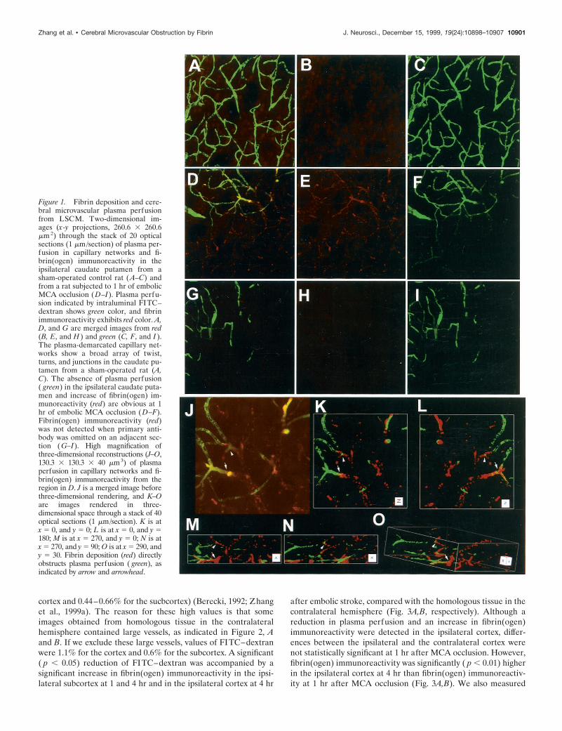
pattern in the sham-operated and nonsurgical brains (Fig. 1A,green). Intravascular blood cells were visible as dark oval figuresfilling the microvascular lumina between the intraluminal FITC–dextran (Fig. 1A). Fibrin(ogen) immunoreactivity was not de-tected in the control rat brains (Fig. 1B). In contrast, 1 hr afterMCA occlusion, large areas of little or no FITC–dextran wereprimarily detected in the ipsilateral subcortex (Fig. 1D, green) andoccasionally in the piriform cortex, suggesting nonperfused andunderperfused tissue. An irregular tubular pattern of fibrin(o-gen) immunoreactivity was observed in the areas with little or noFITC–dextran and extended to the areas with intraluminalFITC–dextran (Fig. 1E, red). Fibrin(ogen) immunoreactivity wasnot detected in the ischemic lesion when the primary antibodywas omitted (Fig. 1G). Intraluminal FITC–dextran terminatedabruptly within cerebral microvessels (Fig. 1D, green). Examina-tion of this region under high-power magnification revealed in-tense fibrin(ogen) immunoreactivity (Fig. 1J–O, red) proximal tointraluminal FITC–dextran (Fig. 1J–O, green) in x-y, x-z, and y-zprojections, indicating that intravascular deposition of fibrin lo-cally blocked perfusion of FITC–dextran. In addition, three-dimensional reconstructions revealed that intravascular fibrin(o-gen) immunoreactivity partially blocked intraluminal FITC–dextran perfusion upstream within relatively large vessels and ledto complete obstruction of FITC–dextran perfusion downstreamvessels (Fig. 2A,B). To further confirm the possible intravasculardeposition of fibrin observed on three-dimensional images, fibrin-(ogen) immunohistochemistry was performed on coronal sectionsfrom the extensively perfused brain tissue. Intravascularfibrin(ogen)-immunoreactive meshwork was also observed onveins (Fig. 2C,D) and capillaries (Fig. 2E) in extensively perfusedbrain tissue after 1 hr of MCA occlusion. Erythrocytes, polymor-phonuclear (PMN) leukocytes, and platelets were attached tofibrin by multiple connections, and aggregated platelets wereenmeshed in fibrin (Fig. 2C–E).
At 4 hr of MCA occlusion, the areas with little and no FITC–dextran in subcortex expanded to the cortex supplied by theMCA, and expansion of underperfused FITC–dextran areas wasaccompanied by an increase in fibrin(ogen) immunoreactivity. Inaddition to intravascular fibrin(ogen) immunoreactivity as seen at1 hr of embolic stroke, three-dimensional reconstruction revealedmassive irregular shapes of fibrin(ogen) immunoreactivity in theareas with no FITC–dextran in the subcortex (Fig. 2F, red),suggesting the presence of fibrin deposition in the parenchyma.To further confirm fibrin deposition, immunohistochemistry offibrin(ogen) was performed on extensively perfused brain tissue.The fibrin(ogen)-immunoreactive meshwork in the subcorticalparenchymal tissue colocalized with shrunken neurons and acti-vated astrocytes in perfused brain tissue (Fig. 2G). This stainingpattern was comparable to that seen in three-dimensional images,confirming deposition of fibrin in the parenchyma. The parenchy-mal fibrin(ogen) immunoreactivity was primarily detected in thesubcortex. The intravascular fibrin(ogen) immunoreactivity waspresent in areas of the piriform and parietal cortex supplied bythe MCA (Fig. 2H). These areas exhibited mixtures of nonFITC–dextran, FITC–dextran perfusion, and fibrin(ogen) im-munoreactivity (Fig. 2H).
To obtain quantitative data on levels of FITC–dextran andfibrin(ogen) immunoreactivity, we measured FITC–dextran andCy5–fibrin(ogen) immunoreactivity in three-dimensional imagesobtained from LSCM. Values of FITC–dextran were 1.7 and0.7% for the control cortex and subcortex (Fig. 2A,B), respec-tively, which are above previously published data (0.74–0.86% for
10900 J. Neurosci., December 15, 1999, 19(24):10898–10907 Zhang et al. • Cerebral Microvascular Obstruction by Fibrin
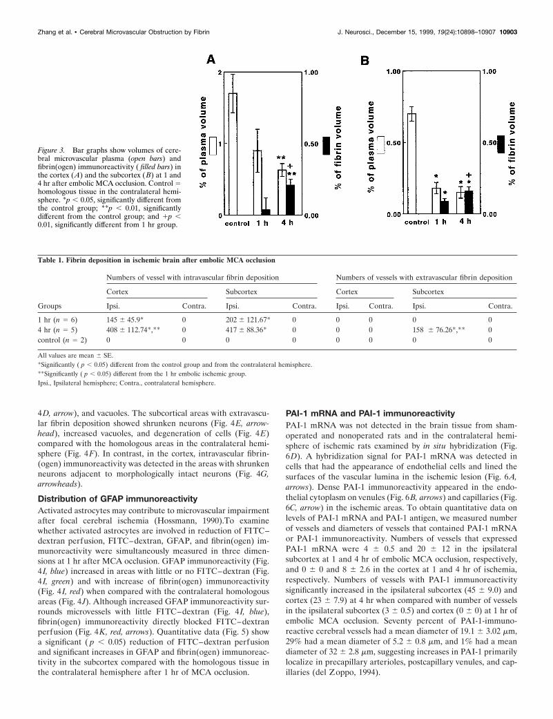
cortex and 0.44–0.66% for the sucbcortex) (Berecki, 1992; Zhanget al., 1999a). The reason for these high values is that someimages obtained from homologous tissue in the contralateralhemisphere contained large vessels, as indicated in Figure 2, Aand B. If we exclude these large vessels, values of FITC–dextranwere 1.1% for the cortex and 0.6% for the subcortex. A significant( p , 0.05) reduction of FITC–dextran was accompanied by asignificant increase in fibrin(ogen) immunoreactivity in the ipsi-lateral subcortex at 1 and 4 hr and in the ipsilateral cortex at 4 hr
after embolic stroke, compared with the homologous tissue in thecontralateral hemisphere (Fig. 3A,B, respectively). Although areduction in plasma perfusion and an increase in fibrin(ogen)immunoreactivity were detected in the ipsilateral cortex, differ-ences between the ipsilateral and the contralateral cortex werenot statistically significant at 1 hr after MCA occlusion. However,fibrin(ogen) immunoreactivity was significantly ( p , 0.01) higherin the ipsilateral cortex at 4 hr than fibrin(ogen) immunoreactiv-ity at 1 hr after MCA occlusion (Fig. 3A,B). We also measured
Figure 1. Fibrin deposition and cere-bral microvascular plasma perfusionfrom LSCM. Two-dimensional im-ages (x-y projections, 260.6 3 260.6mm 2) through the stack of 20 opticalsections (1 mm/section) of plasma per-fusion in capillary networks and fi-brin(ogen) immunoreactivity in theipsilateral caudate putamen from asham-operated control rat ( A–C) andfrom a rat subjected to 1 hr of embolicMCA occlusion (D–I). Plasma perfu-sion indicated by intraluminal FITC–dextran shows green color, and fibrinimmunoreactivity exhibits red color. A,D, and G are merged images from red(B, E, and H ) and green (C, F, and I ).The plasma-demarcated capillary net-works show a broad array of twist,turns, and junctions in the caudate pu-tamen from a sham-operated rat (A,C). The absence of plasma perfusion( green) in the ipsilateral caudate puta-men and increase of fibrin(ogen) im-munoreactivity (red) are obvious at 1hr of embolic MCA occlusion (D–F).Fibrin(ogen) immunoreactivity (red)was not detected when primary anti-body was omitted on an adjacent sec-tion ( G–I). High magnification ofthree-dimensional reconstructions (J–O,130.3 3 130.3 3 40 mm3) of plasmaperfusion in capillary networks and fi-brin(ogen) immunoreactivity from theregion in D. J is a merged image beforethree-dimensional rendering, and K–Oare images rendered in three-dimensional space through a stack of 40optical sections (1 mm/section). K is atx 5 0, and y 5 0; L is at x 5 0, and y 5180; M is at x 5 270, and y 5 0; N is atx 5 270, and y 5 90; O is at x 5 290, andy 5 30. Fibrin deposition (red) directlyobstructs plasma perfusion ( green), asindicated by arrow and arrowhead.
Zhang et al. • Cerebral Microvascular Obstruction by Fibrin J. Neurosci., December 15, 1999, 19(24):10898–10907 10901

numbers of vessels that were fibrin(ogen)-immunoreactive underlight microscopy (Table 1). A significant ( p , 0.05) increase ofintravascular fibrin(ogen) immunoreactivity was also detected inthe cortex and the subcortex at 1 and 4 hr after stroke comparedwith the contralateral hemisphere. Numbers of intravascularfibrin(ogen)-immunoreactive vessels were significantly ( p , 0.05)increased in the ipsilateral cortex at 4 hr after ischemia comparedwith the 1 hr group. Numbers of vessels with extravascular fibrindeposition were significantly ( p , 0.05) higher in the subcortex at4 hr after ischemia than at 1 hr and the control group (Table 1).
To examine neuronal response to plasma perfusion deficits andfibrin deposition, MAP-2 immunoreactivity was examined along
with FITC–dextran and fibrin(ogen) immunoreactivity. Triplefluorescence in the x-y projections revealed that intense fibrin(o-gen) immunoreactivity (Fig. 4A, red) was present in areas of lowMAP-2 immunoreactivity (Fig. 4A, blue) and little or no FITC–dextran (Fig. 4A, green) compared with the homologous tissue inthe contralateral hemisphere (Fig. 4B). Analysis of extensivelyperfused brain tissue under light microscopy revealed that acuteischemic neuronal injury, dark neurons, were present adjacent tovessels with fibrin deposition at 1 hr after embolic stroke (Fig. 4C,arrowhead). At 4 hr after stroke, the subcortical areas with intra-vascular fibrin deposition exhibited increased numbers ofshrunken neurons (Fig. 4D, arrowheads), swollen astrocytes (Fig.
Figure 2. Intravascular fibrin deposi-tion, erythrocytes, PMN leukocytes,and platelets from rats subjected to 1hr of embolic MCA occlusion. A is athree-dimensional reconstructed im-age (260.6 3 260.6 3 20 mm 3) througha stack of 20 optical sections (1 mm/section) of plasma perfusion and fi-brin(ogen) immunoreactivity from B,which is a merged image. Intravascularfibrin deposition in a relative largevessel causes the vessel to narrow anddecreases plasma perfusion in capil-laries (A, B). Erythrocytes (arrows),PMN leukocytes (curved arrow), andplatelets (arrowheads) were connectedwith fibrin within venules (C, D) andcapillaries (E) in the ipsilateral cau-date putamen from extensively per-fused brain tissue. F, H, I, and K areimages (x-y projections, 260.6 3 260.6mm 2) through the stack of 20 opticalsections (1 mm/section) of plasma per-fusion in capillary networks ( green)and fibrin(ogen) immunoreactivity(red) in the ipsilateral caudate puta-men (F) and in the ipsilateral cortex(H ) from a rat subjected to 4 hr ofembolic MCA occlusion. Fibrin(ogen)immunoreactivity (red) was present inextravascular space with little plasmaperfusion (F, green), and fibrin(ogen)immunoreactivity was not present inthe homologous tissue of the con-tralateral hemisphere ( I ). Parenchy-mal fibrin deposition was also ob-served in the ipsilateral caudateputamen ( G) but not the homologousarea of the contralateral hemisphere( J) from extensively perfused braintissue at 4 hr of MCA occlusion. Mix-ture (H, yellow) of microvascularplasma perfusion (H, green) with in-travascular fibrin deposition (H, red)was detected in the ipsilateral parietalcortex ( H ) compared with the con-tralateral homologous tissue (K, greenonly). Scale bars: D, 10 mm; C, E, 20mm; G, J, 100 mm.
10902 J. Neurosci., December 15, 1999, 19(24):10898–10907 Zhang et al. • Cerebral Microvascular Obstruction by Fibrin
4D, arrow), and vacuoles. The subcortical areas with extravascu-lar fibrin deposition showed shrunken neurons (Fig. 4E, arrow-head), increased vacuoles, and degeneration of cells (Fig. 4E)compared with the homologous areas in the contralateral hemi-sphere (Fig. 4F). In contrast, in the cortex, intravascular fibrin-(ogen) immunoreactivity was detected in the areas with shrunkenneurons adjacent to morphologically intact neurons (Fig. 4G,arrowheads).
Distribution of GFAP immunoreactivityActivated astrocytes may contribute to microvascular impairmentafter focal cerebral ischemia (Hossmann, 1990).To examinewhether activated astrocytes are involved in reduction of FITC–dextran perfusion, FITC–dextran, GFAP, and fibrin(ogen) im-munoreactivity were simultaneously measured in three dimen-sions at 1 hr after MCA occlusion. GFAP immunoreactivity (Fig.4 I, blue) increased in areas with little or no FITC–dextran (Fig.4 I, green) and with increase of fibrin(ogen) immunoreactivity(Fig. 4 I, red) when compared with the contralateral homologousareas (Fig. 4J). Although increased GFAP immunoreactivity sur-rounds microvessels with little FITC–dextran (Fig. 4I, blue),fibrin(ogen) immunoreactivity directly blocked FITC–dextranperfusion (Fig. 4K, red, arrows). Quantitative data (Fig. 5) showa significant ( p , 0.05) reduction of FITC–dextran perfusionand significant increases in GFAP and fibrin(ogen) immunoreac-tivity in the subcortex compared with the homologous tissue inthe contralateral hemisphere after 1 hr of MCA occlusion.
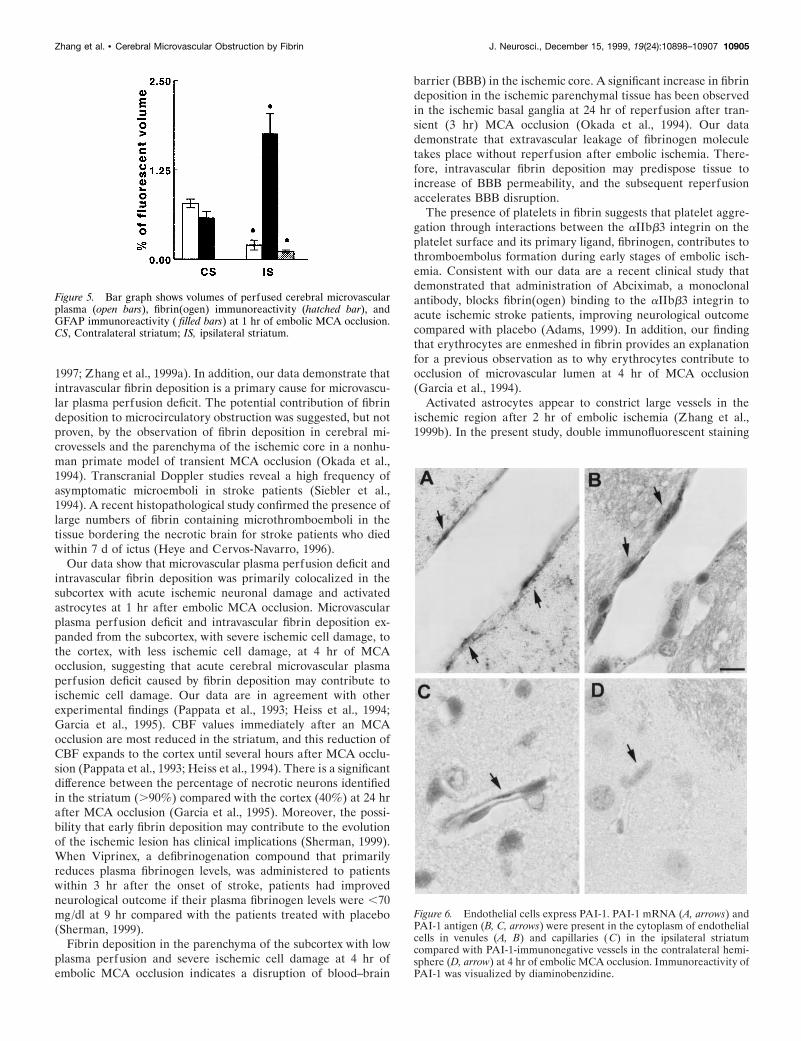
PAI-1 mRNA and PAI-1 immunoreactivityPAI-1 mRNA was not detected in the brain tissue from sham-operated and nonoperated rats and in the contralateral hemi-sphere of ischemic rats examined by in situ hybridization (Fig.6D). A hybridization signal for PAI-1 mRNA was detected incells that had the appearance of endothelial cells and lined thesurfaces of the vascular lumina in the ischemic lesion (Fig. 6A,arrows). Dense PAI-1 immunoreactivity appeared in the endo-thelial cytoplasm on venules (Fig. 6B, arrows) and capillaries (Fig.6C, arrow) in the ischemic areas. To obtain quantitative data onlevels of PAI-1 mRNA and PAI-1 antigen, we measured numberof vessels and diameters of vessels that contained PAI-1 mRNAor PAI-1 immunoreactivity. Numbers of vessels that expressedPAI-1 mRNA were 4 6 0.5 and 20 6 12 in the ipsilateralsubcortex at 1 and 4 hr of embolic MCA occlusion, respectively,and 0 6 0 and 8 6 2.6 in the cortex at 1 and 4 hr of ischemia,respectively. Numbers of vessels with PAI-1 immunoreactivitysignificantly increased in the ipsilateral subcortex (45 6 9.0) andcortex (23 6 7.9) at 4 hr when compared with number of vesselsin the ipsilateral subcortex (3 6 0.5) and cortex (0 6 0) at 1 hr ofembolic MCA occlusion. Seventy percent of PAI-1-immuno-reactive cerebral vessels had a mean diameter of 19.1 6 3.02 mm,29% had a mean diameter of 5.2 6 0.8 mm, and 1% had a meandiameter of 32 6 2.8 mm, suggesting increases in PAI-1 primarilylocalize in precapillary arterioles, postcapillary venules, and cap-illaries (del Zoppo, 1994).
Table 1. Fibrin deposition in ischemic brain after embolic MCA occlusion
Groups
Numbers of vessel with intravascular fibrin deposition Numbers of vessels with extravascular fibrin deposition
Cortex Subcortex Cortex Subcortex
Ipsi. Contra. Ipsi. Contra. Ipsi. Contra. Ipsi. Contra.
1 hr (n 5 6) 145 6 45.9* 0 202 6 121.67* 0 0 0 0 04 hr (n 5 5) 408 6 112.74*,** 0 417 6 88.36* 0 0 0 158 6 76.26*,** 0control (n 5 2) 0 0 0 0 0 0 0 0
All values are mean 6 SE.*Significantly ( p , 0.05) different from the control group and from the contralateral hemisphere.**Significantly ( p , 0.05) different from the 1 hr embolic ischemic group.Ipsi., Ipsilateral hemisphere; Contra., contralateral hemisphere.
Figure 3. Bar graphs show volumes of cere-bral microvascular plasma (open bars) andfibrin(ogen) immunoreactivity ( filled bars) inthe cortex (A) and the subcortex (B) at 1 and4 hr after embolic MCA occlusion. Control 5homologous tissue in the contralateral hemi-sphere. *p , 0.05, significantly different fromthe control group; **p , 0.01, significantlydifferent from the control group; and 1p ,0.01, significantly different from 1 hr group.
Zhang et al. • Cerebral Microvascular Obstruction by Fibrin J. Neurosci., December 15, 1999, 19(24):10898–10907 10903

DISCUSSIONTo directly address whether fibrin deposition obstructs cerebralmicrovascular plasma perfusion, we simultaneously measured inthree dimensions microvascular plasma perfusion and fibrin dep-osition in ischemic brain at 1 and 4 hr of embolic MCA occlusionusing intravascular fluorescent tracer molecules and immunoflu-orescent staining in combination with LSCM. The time points of1 and 4 hr chosen in the present study were based on our previousstudies in this model that a progressive cerebral microcirculatoryimpairment is present during this period (Zhang et al., 1997,1999a; Morris et al., 1999). Three-dimensional reconstructionsdemonstrate that fibrin deposition not only directly obstructedmicrovascular plasma perfusion, primarily within capillaries ofthe ischemic core, but that fibrin was also present within plasma-perfused vessels bordering the ischemic core at 1 hr after MCAocclusion. The marked increase of intravascular fibrin deposition
in the cortex and appearance of extravascular fibrin deposition inthe striatum were associated with reduction of cerebral microvas-cular plasma perfusion at 4 hr of ischemia. This three-dimensional assessment was confirmed by measuring numbers offibrin(ogen)-immunoreactive vessels on immunohistochemicallystained sections. Numbers of vessels with intravascular fibrindeposition in the cortex and parenchymal fibrin deposition in thestriatum are significantly higher at 4 hr of MCA occlusion than at1 hr of ischemia. These data suggest that cerebral microvascularplasma deficit secondary to MCA occlusion is an ongoing processthat expands from the subcortex to the cortex over time ofischemia, and intravascular fibrin deposition directly causes thisprogressive cerebral microcirculatory impairment. Our finding ofa progressive microvascular plasma perfusion deficit after isch-emia is consistent with previous studies of embolic ischemia inmice and of permanent MCA occlusion in rats (Dawson et al.,
Figure 4. Fibrin deposition and ischemic cell damage. A and B are images (x-y projections, 260.6 3 260.6 mm 2) through the stack of 20 optical sections(1 mm/section) of plasma perfusion in capillary networks ( green), fibrin(ogen) immunoreactivity (red), and MAP-2 immunoreactivity (blue) from a ratsubjected to 1 hr of MCA occlusion. Increase in fibrin(ogen) immunoreactivity (A, red) and loss of plasma perfusion (A, green) and MAP-2immunoreactivity (A, blue) on the ipsilateral hemisphere are evident (A) compared with the contralateral hemisphere (B). Dark neurons (C, arrowhead),shrunken neurons (D, arrowheads), and swollen astrocytes (D, arrow) were present in the striatum with intravascular fibrin deposition from extensivelyperfused brains at 1 ( C) and 4 ( D) hr of embolic MCA occlusion. Shrunken neurons (arrowhead) with vacuoles were present in the striatum withextravascular fibrin deposition (E) compared with intact neurons (arrowhead) in the contralateral striatum with patent vessels (curved arrow) at 4 hr ofembolic MCA occlusion (F). Shrunken neurons (arrowheads), intact neurons (curved arrow), and swollen astrocytes (arrow) were present in the cortexwith intravascular fibrin deposition (G) compared with intact neurons (arrowhead) in the contralateral cortex with patent vessels (curved arrow) at 4 hrof ischemia (H ). I–L are three-dimensional reconstructions of microvascular plasma perfusion ( green), fibrin(ogen) immunoreactivity (red), and GFAPimmunoreactivity (blue) in the caudate putamen from a rat subjected to 1 hr of embolic stroke. Enlargement of GFAP-immunoreactive cell bodies andprocesses (blue) surrounded vessels ( green) in the ischemic region ( I ) compared with the homologous tissue in the contralateral hemisphere ( J).Microvascular plasma perfusion (K, green, arrows) was directly blocked by fibrin deposition (K, red, arrows) when GFAP immunoreactivity was removed(K, L). The image size is 260.1 3 260.1 3 20 mm 3 for I–K. Scale bar: C–H, 10 mm.
10904 J. Neurosci., December 15, 1999, 19(24):10898–10907 Zhang et al. • Cerebral Microvascular Obstruction by Fibrin
1997; Zhang et al., 1999a). In addition, our data demonstrate thatintravascular fibrin deposition is a primary cause for microvascu-lar plasma perfusion deficit. The potential contribution of fibrindeposition to microcirculatory obstruction was suggested, but notproven, by the observation of fibrin deposition in cerebral mi-crovessels and the parenchyma of the ischemic core in a nonhu-man primate model of transient MCA occlusion (Okada et al.,1994). Transcranial Doppler studies reveal a high frequency ofasymptomatic microemboli in stroke patients (Siebler et al.,1994). A recent histopathological study confirmed the presence oflarge numbers of fibrin containing microthromboemboli in thetissue bordering the necrotic brain for stroke patients who diedwithin 7 d of ictus (Heye and Cervos-Navarro, 1996).
Our data show that microvascular plasma perfusion deficit andintravascular fibrin deposition was primarily colocalized in thesubcortex with acute ischemic neuronal damage and activatedastrocytes at 1 hr after embolic MCA occlusion. Microvascularplasma perfusion deficit and intravascular fibrin deposition ex-panded from the subcortex, with severe ischemic cell damage, tothe cortex, with less ischemic cell damage, at 4 hr of MCAocclusion, suggesting that acute cerebral microvascular plasmaperfusion deficit caused by fibrin deposition may contribute toischemic cell damage. Our data are in agreement with otherexperimental findings (Pappata et al., 1993; Heiss et al., 1994;Garcia et al., 1995). CBF values immediately after an MCAocclusion are most reduced in the striatum, and this reduction ofCBF expands to the cortex until several hours after MCA occlu-sion (Pappata et al., 1993; Heiss et al., 1994). There is a significantdifference between the percentage of necrotic neurons identifiedin the striatum (.90%) compared with the cortex (40%) at 24 hrafter MCA occlusion (Garcia et al., 1995). Moreover, the possi-bility that early fibrin deposition may contribute to the evolutionof the ischemic lesion has clinical implications (Sherman, 1999).When Viprinex, a defibrinogenation compound that primarilyreduces plasma fibrinogen levels, was administered to patientswithin 3 hr after the onset of stroke, patients had improvedneurological outcome if their plasma fibrinogen levels were ,70mg/dl at 9 hr compared with the patients treated with placebo(Sherman, 1999).
Fibrin deposition in the parenchyma of the subcortex with lowplasma perfusion and severe ischemic cell damage at 4 hr ofembolic MCA occlusion indicates a disruption of blood–brain
barrier (BBB) in the ischemic core. A significant increase in fibrindeposition in the ischemic parenchymal tissue has been observedin the ischemic basal ganglia at 24 hr of reperfusion after tran-sient (3 hr) MCA occlusion (Okada et al., 1994). Our datademonstrate that extravascular leakage of fibrinogen moleculetakes place without reperfusion after embolic ischemia. There-fore, intravascular fibrin deposition may predispose tissue toincrease of BBB permeability, and the subsequent reperfusionaccelerates BBB disruption.
The presence of platelets in fibrin suggests that platelet aggre-gation through interactions between the aIIbb3 integrin on theplatelet surface and its primary ligand, fibrinogen, contributes tothromboembolus formation during early stages of embolic isch-emia. Consistent with our data are a recent clinical study thatdemonstrated that administration of Abciximab, a monoclonalantibody, blocks fibrin(ogen) binding to the aIIbb3 integrin toacute ischemic stroke patients, improving neurological outcomecompared with placebo (Adams, 1999). In addition, our findingthat erythrocytes are enmeshed in fibrin provides an explanationfor a previous observation as to why erythrocytes contribute toocclusion of microvascular lumen at 4 hr of MCA occlusion(Garcia et al., 1994).
Activated astrocytes appear to constrict large vessels in theischemic region after 2 hr of embolic ischemia (Zhang et al.,1999b). In the present study, double immunofluorescent staining
Figure 6. Endothelial cells express PAI-1. PAI-1 mRNA (A, arrows) andPAI-1 antigen (B, C, arrows) were present in the cytoplasm of endothelialcells in venules (A, B) and capillaries ( C) in the ipsilateral striatumcompared with PAI-1-immunonegative vessels in the contralateral hemi-sphere (D, arrow) at 4 hr of embolic MCA occlusion. Immunoreactivity ofPAI-1 was visualized by diaminobenzidine.
Figure 5. Bar graph shows volumes of perfused cerebral microvascularplasma (open bars), fibrin(ogen) immunoreactivity (hatched bar), andGFAP immunoreactivity ( filled bars) at 1 hr of embolic MCA occlusion.CS, Contralateral striatum; IS, ipsilateral striatum.
Zhang et al. • Cerebral Microvascular Obstruction by Fibrin J. Neurosci., December 15, 1999, 19(24):10898–10907 10905

for fibrinogen and GFAP revealed that astrocytes are activated at1 hr of ischemia, as measured by a significant increase of GFAPimmunoreactivity. Activated astrocytes surround microvesselsthat are fibrin-immunoreactive, suggesting that reactive astro-cytes may also contribute to cerebral microcirculatory impair-ments during the early stage of embolic stroke by constrictingcerebral vessels.
The specificity of the anti-fibrinogen antibody that we used forfibrin detection is consistent with previous reports on this anti-body in a mouse model of thrombosis (Farrehi et al., 1998). In thepresent study, we found that fibrin(ogen) immunoreactivity wasonly detected in the ischemic region in both nonperfused andextensively perfused brain tissue but not in tissue from controlrats or when the primary antibody was omitted. Furthermore, thefibrin(ogen)-immunoreactive meshwork was found within vesselsand extravascular parenchymal tissue in the ischemic region.
PAI-1 is a rapid and specific inhibitor of t-PA and u-PA and isthe primary regulator of plasminogen activation in vivo (Loskut-off et al., 1989). However, recent studies demonstrate that neu-rons in the brain express neuroserpin, which efficiently inhibitsactivity of t-PA (Osterwalder et al., 1996; Hastings et al., 1997).Elevations in PAI-1 activity have been associated with fibrindeposition after ischemia, and increased plasma PAI-1 levels arecorrelated with the occurrence of previous ischemic episodes(Hamsten et al., 1987; Margaglione et al., 1994). However, little isknown about in situ PAI-1 localization in ischemic brain. Ourobservations of the induction of PAI-1 mRNA and PAI-1 antigenin the ischemic region after embolic MCA occlusion suggest thatlocal upregulation of PAI-1 may contribute to fibrin depositionduring early embolic stroke. In situ hybridization demonstratedinduction of PAI-1 mRNA in vascular endothelial cells in theischemic region at 1 hr of ischemia and a significant increase ofPAI-1 mRNA at 4 hr of ischemia.
Immunohistochemical staining showed the same pattern ofincreased PAI-1 antigen in the endothelial cells. These dataindicate that upregulation of PAI-1 is transcriptionally regulatedin the ischemic lesion. The endothelial cells synthesize and se-crete PAI-1 (Kollros et al., 1994). Platelets are the major reser-voir of PAI-1 in blood (Loskutoff et al., 1989; Braaten et al., 1993;Stringer et al., 1994). Inability to detect PAI-1 on platelets in thepresent study does not rule out the possibility that plateletscontribute to increase PAI-1. With fibrin strands of a fresh clotnear the endothelial surface, active PAI-1 is bound to the fibrinstrands, and is thus protected from fibrinolysis by t-PA (Braatenet al., 1993). In the present study, the time course of upregulationof PAI-1 gene expression in the endothelial cells and presence ofplatelets are related to the increase of fibrin deposition, suggest-ing that increases in PAI-1 levels may contribute to stabilizationof fibrin deposition within the cerebral microvasculature by atime-dependent increase in fibrinolytic resistance. This view issupported by fibrinolytic therapy with t-PA in this model in whichadministration of t-PA at 1 hr after MCA occlusion increasesCBF and reduces ischemic lesion volume, but not when t-PA isadministered at 4 hr after MCA occlusion (Jiang et al., 1998,1999). Local increase in PAI-1 expression may be a reason forfailure of fibrinolytic therapy at 4 hr of ischemia in this model.Our results are consistent with the recent finding that PAI-12/2mice exhibit less residual thrombus when compared with PAI-11/1 mice in a murine arterial thrombotic model (Farrehi et al.,1998).
In summary, intravascular fibrin deposition, composed oferythrocytes, PMN leukocytes, and platelets, directly obstructs
cerebral microvascular plasma perfusion. Upregulation of PAI-1gene in the endothelial cells may foster fibrin deposition throughsuppression of fibrinolysis. These data suggest that local pertur-bation of procoagulant and fibrinolytic genes in the brain may beimportant for cerebral microcirculatory impairment during earlyfocal embolic cerebral ischemia.
REFERENCESAdams H (1999) Preliminary safety report of an ongoing dose-escalation
trial Abciximab in acute ischemic stroke. Stroke 30:244.Bereczki D, Wei L, Otsuka T, Acuff V, Pettigrew K, Patlak C, Fenster-
macher J (1992) Hypoxia increases velocity of blood flow throughparenchymal microvascular systems in rat brain. J Cereb Blood FlowMetab 13:475–486.
Braaten JV, Handt S, Jerome WG, Kirkpatrick J, Lewis JC, Hantgan RR(1993) Regulation of fibrinolysis by platelet-released plasminogen ac-tivator inhibitor 1: light scattering and ultrastructural examination oflysis of a model platelet-fibrin thrombus. Blood 81:1290–1299.
Buchweitz-Milton E, Weiss HR (1988) Perfused microvascular mor-phometry during middle cerebral artery occlusion. Am J Physiol255:H623–H628.
Collen D, Lijnen HR (1991) Basic and clinical aspects of fibrinolysis andthrombolysis. Blood 78:3114–3124.
Crowell RM, Olsson Y (1972) Impaired microvascular filling after focalcerebral ischemia in the monkey. Modification by treatment. Neurology22:500–504.
Dawson DA, Hallenbeck JM (1996) Acute focal ischemia-induced alter-ations in MAP2 immunostaining: description of temporal changes andutilization as a marker for volumetric assessment of acute brain injury.J Cereb Blood Flow Metab 16:170–174.
Dawson DA, Ruetzler CA, Hallenbeck JM (1997) Temporal impair-ment of microcirculatory perfusion following focal cerebral ischemia inthe spontaneously hypertensive rat. Brain Res 749:200–208.
del Zoppo GJ (1994) Microvascular changes during cerebral ischemiaand reperfusion. Cerebrovasc Brain Metab Rev 6:47–96.
Ennis SR, Keep RF, Schielke GP, Betz AL (1990) Decrease in perfusionof cerebral capillaries during incomplete ischemia and reperfusion.J Cereb Blood Flow Metab 10:213–220.
Farrehi PM, Ozaki CK, Carmeliet P, Fay WP1 (1998) Regulation ofarterial thrombolysis by plasminogen activator inhibitor-1 in mice.Circulation 97:1002–1008.
Garcia JH, Liu KF, Yoshida Y, Chen S, Lian J (1994) Brain microves-sels: factors altering their patency after the occlusion of a middlecerebral artery (Wistar rat). Am J Pathol 145:728–740.
Garcia JH, Liu KF, Ho KL (1995) Neuronal necrosis after middle ce-rebral artery occlusion in Wistar rats progresses at different timeintervals in the caudoputamen and the cortex. Stroke 26:636–642.
Hamsten A, de Faire U, Walldius G, Dahlen G, Szamosi A, Landou C,Blomback M, Wiman B (1987) Plasminogen activator inhibitor in plas-ma: risk factor for recurrent myocardial infarction. Lancet 2:3–9.
Handt S, Jerome WG, Tietze L, Hantgan RR (1996) Plasminogen acti-vator inhibitor-1 secretion of endothelial cells increases fibrinolyticresistance of an in vitro fibrin clot: evidence for a key role of endothe-lial cells in thrombolytic resistance. Blood 87:4204–4213.
Hastings GA, Coleman TA, Haudenschild CC, Stefansson S, Smith EP,Barthlow R, Cherry S, Sandkvist M, Lawrence DA (1997) Neuroser-pin, a brain-associated inhibitor of tissue plasminogen activator islocalized primarily in neurons. Implications for the regulation of motorlearning and neuronal survival. J Biol Chem 272:33062–33067.
Heiss WD, Graf R, Wienhard K, Lottgen J, Saito R, Fujita T, Rosner G,Wagner R (1994) Dynamic penumbra demonstrated by sequentialmultitracer PET after middle cerebral artery occlusion in cats. J CerebBlood Flow Metab 14:892–902.
Heye N, Cervos-Navarro J (1996) Microthromboemboli in acute in-farcts: analysis of 40 autopsy cases. Stroke 27:431–444.
Hossmann KA (1990) Hemodynamics of post ischemic reperfusion ofthe brain. In: Protection of the brain from ischemia (Weistein PR,Faden AL, eds), pp 21–36. Baltimore: William and Wilkins.
Jiang Q, Zhang RL, Zhang ZG, Ewing JR, Divine GW, Chopp M (1998)Diffusion, T2, and perfusion weighted NMR imaging of middle cere-bral artery embolic stroke and rt-PA intervention in rat. J Cereb BloodFlow Metab 18:758–767.
Jiang Q, Zhang RL, Zhang ZG, Ewing JR, Jiang P, Divine GW, KnightRA, Chopp M (1999) MRI indices of therapeutic efficacy of rtPA
10906 J. Neurosci., December 15, 1999, 19(24):10898–10907 Zhang et al. • Cerebral Microvascular Obstruction by Fibrin

treatment of rat at 1 H and 4 H after embolic stroke. J Cereb BloodFlow Metab, in press.
Kitching AR, Holdsworth SR, Ploplis VA, Plow EF, Collen D, CarmelietP, Tipping PG (1997) Plasminogen and plasminogen activators protectagainst renal injury in crescentic glomerulonephritis. J Exp Med185:963–968.
Kollros PR, Konkle BA, Ambarian AP, Henrikson P (1994) Plasmino-gen activator inhibitor-1 expression by brain microvessel endothelialcells is inhibited by elevated glucose. J Neurochem 63:903–909.
Little JR, Kerr FW, Sundt TM Jr (1975) Microcirculatory obstruction infocal cerebral ischemia. Relationship to neuronal alterations. MayoClin Proc 50:264–270.
Loscalzo J, Schafer AI (1992) Anticoagulants, antiplatelet agents, andfibrinolysis. In: Vascular Medicine, a textbook for vascular biology anddisease. (Loscalzo J, Creager MA, Dzau VJ, eds), pp 659–682. Boston:Little, Brown.
Loskutoff DJ, Sawdey M, Mimuro J (1989) Type 1 plasminogen activa-tor inhibitor. Prog Hemost Thromb 9:87–115.
Margaglione M, Di Minno G, Grandone E, Vecchione G, Celentano E,Cappucci G, Grilli M, Simone P, Panico S, Mancini M (1994) Abnor-mally high circulation levels of tissue plasminogen activator and plas-minogen activator inhibitor-1 in patients with a history of ischemicstroke. Arterioscler Thromb 14:1741–1745.
Morris DC, Zhang ZG, Davies K, Fenstermacher J, Chopp M (1999)High resolution quantitation of microvascular plasma perfusion innon-ischemic and ischemic rat brain by laser-scanning confocal micros-copy. Brain Res Brain Res Protocols 4:185–191.
Okada Y, Copeland BR, Fitridge R, Koziol JA, del Zoppo GJ (1994)Fibrin contributes to microvascular obstructions and parenchymalchanges during early focal cerebral ischemia and reperfusion. Stroke25:1847–1853.
Osterwalder T, Contartese J, Stoeckli ET, Kuhn TB, Sonderegger P(1996) Neuroserpin, an axonally secreted serine protease inhibitor.EMBO J 15:2944–2953.
Pappata S, Fiorelli M, Rommel T, Hartmann A, Dettmers C, YamaguchiT, Chabriat H, Poline JB, Crouzel C, Di Giamberardino L, Baron JC(1993) PET study of changes in local brain hemodynamics and oxygenmetabolism after unilateral middle cerebral artery occlusion in ba-boons. J Cereb Blood Flow Metab 13:416–424.
Paxinos G, Watson C (1986) The rat brain in stereotaxic coordinates. Ed2. New York: Academic.
Ploplis VA, Carmeliet P, Vazirzadeh S, Van Vlaenderen I, Moons L,Plow EF, Collen D (1995) Effects of disruption of the plasminogengene on thrombosis, growth, and health in mice. Circulation 92:2585–2593.
Plow EF, Herren T, Redlitz A, Miles LA, Hoover Plow JL (1995) Thecell biology of the plasminogen system. FASEB J 9:939–945.
Sappino A-P, Madani R, Huarte J, Belin D, Kiss JZ, Wohlwend A,Vassalli J-D (1993) Extracellular proteolysis in the adult murinebrain. J Clin Invest 92:679–685.
Sherman DG (1999) Defibrinogenation with Viprinex (Ancord) fortreatment of acute ischemic stroke. Stroke 30:234.
Siebler M, Nachtmann A, Sitzer M, Steinmetz H (1994) Anticoagulationmonitoring and cerebral microemboli detection. Lancet 344:555.
Soltanian-Zadeh H, Windham JP (1994) Mathematical basis of eigen-image filtering. Magn Reson Med 31:465–467.
Stringer HA, van Swieten P, Heijnen HF, Sixma JJ, Pannekoek H (1994)Plasminogen activator inhibitor-1 released from activated plateletsplays a key role in thrombolysis resistance. Studies with thrombi gen-erated in the Chandler loop. Arterioscler Thromb 14:1452–1458.
Vassalii J-D, Sappino A-P, Belin D (1991) The plasminogen activator/plasmin system. J Clin Invest 88:1067–1072.
Windham JP, Abd-Allah MA, Reimann DA, Froelich JW, Haggar AM(1988) Eigenimage filtering in MR imaging. J Comput Assist Tomogr12:1–9.
Zhang RL, Chopp M, Zhang ZG, Jiang Q (1997) A rat model of focalembolic cerebral ischemia. Brain Res 766:83–92.
Zhang ZG, Davies K, Prostak J, Fenstermacher J, Chopp M (1999a)Quantitation of microvascular plasma reperfusion and neuronalmicrotubulin-associated protein in ischemic mouse brain by laser-scanning confocal microscopy. J Cereb Blood Flow Metab 19:68–78.
Zhang ZG, Bower L, Zhang RL, Chen S, Windham JP, Chopp M(1999b) Three dimensional measurement of cerebral microvascularplasma perfusion, glial fibrillary acidic protein and microtubule asso-ciated protein-2 immunoreactivity after embolic stroke in rats: a doublefluorescent labeled laser-scanning confocal microscopic study. BrainRes 844:55–66.
Zhang et al. • Cerebral Microvascular Obstruction by Fibrin J. Neurosci., December 15, 1999, 19(24):10898–10907 10907
Related Documents