CARDIAC The relation between hypointense core, microvascular obstruction and intramyocardial haemorrhage in acute reperfused myocardial infarction assessed by cardiac magnetic resonance imaging Diana Kandler & Christian Lücke & Matthias Grothoff & Claudia Andres & Lukas Lehmkuhl & Stefan Nitzsche & Franziska Riese & Meinhard Mende & Suzanne de Waha & Steffen Desch & Philipp Lurz & Ingo Eitel & Matthias Gutberlet Received: 11 October 2013 /Revised: 19 June 2014 /Accepted: 4 July 2014 # The Author(s) 2014. This article is published with open access at Springerlink.com Abstract Background Intramyocardial haemorrhage (IMH) and micro- vascular obstruction (MVO) represent reperfusion injury after reperfused ST-elevation myocardial infarction (STEMI) with prognostic impact and “hypointense core” (HIC) appearance in T 2 -weighted images. We aimed to distinguish between IMH and MVO by using T 2 * -weighted cardiovascular mag- netic resonance imaging (CMR) and analysed influencing factors for IMH development. Methods and results A total of 151 patients with acute STEMI underwent CMR after primary angioplasty. T 2 -STIR se- quences were used to identify HIC, late gadolinium enhance- ment to visualise MVO and T 2 * -weighted sequences to detect IMH. IMH + /IMH − patients were compared considering in- farct size, myocardial salvage, thrombolysis in myocardial infarction (TIMI) flow, reperfusion time, ventricular volumes, function and pre-interventional medication. Seventy-six pa- tients (50 %) were IMH + , 82 (54 %) demonstrated HIC and 100 (66 %) MVO. IMH was detectable without HIC in 16 %, without MVO in 5 % and HIC without MVO in 6 %. Multi- variable analyses revealed that IMH was associated with significant lower left ventricular ejection fraction and myocar- dial salvage index, larger left ventricular volume and infarct size. Patients with TIMI flow grade ≤1 before angioplasty demonstrated IMH significantly more often. Conclusions IMH is associated with impaired left ventricular function and higher infarct size. T 2 and HIC imaging showed moderate agreement for IMH detection. T 2 * imaging might be the preferred CMR imaging method for comprehensive IMH assessment. Key Points • Intramyocardial haemorrhage is a common finding in patients with acute reperfused myocardial-infarction. • T 2 * imaging should be the preferred CMR method for assessment of intramyocardial haemorrhage. • Intramyocardial haemorrhage can be considered as an important influencing factor on patient’ s outcome. Keywords Cardiac magnetic resonance imaging . Acute myocardial infarction . Haemorrhage . Microvascular obstruction . Hypointense core Abbreviations 3D-IR-GRE 3D inversion recovery gradient echo sequence AAR area at risk CMR cardiac magnetic resonance EDV end-diastolic volume ESV end-systolic volume Gd-DTPA gadolinium diethylenetriaminepentaacetic acid HIC hypointense core IMH intramyocardial haemorrhage LGE late gadolinium enhancement D. Kandler : C. Lücke : M. Grothoff : C. Andres : L. Lehmkuhl : S. Nitzsche : F. Riese : M. Gutberlet (*) Department of Diagnostic and Interventional Radiology, University Leipzig – Heart Centre, Strümpellstraße 39, 04289 Leipzig, Germany e-mail: [email protected] M. Mende Coordination Centre for Clinical Trials, University Leipzig, Leipzig, Germany S. de Waha : S. Desch : P. Lurz : I. Eitel Department of Internal Medicine/ Cardiology, University Leipzig – Heart Centre, Leipzig, Germany Eur Radiol DOI 10.1007/s00330-014-3318-3

Welcome message from author
This document is posted to help you gain knowledge. Please leave a comment to let me know what you think about it! Share it to your friends and learn new things together.
Transcript

CARDIAC
The relation between hypointense core, microvascularobstruction and intramyocardial haemorrhage in acutereperfused myocardial infarction assessed by cardiacmagnetic resonance imaging
Diana Kandler & Christian Lücke & Matthias Grothoff & Claudia Andres &Lukas Lehmkuhl & Stefan Nitzsche & Franziska Riese & Meinhard Mende &
Suzanne de Waha & Steffen Desch & Philipp Lurz & Ingo Eitel & Matthias Gutberlet
Received: 11 October 2013 /Revised: 19 June 2014 /Accepted: 4 July 2014# The Author(s) 2014. This article is published with open access at Springerlink.com
AbstractBackground Intramyocardial haemorrhage (IMH) and micro-vascular obstruction (MVO) represent reperfusion injury afterreperfused ST-elevation myocardial infarction (STEMI) withprognostic impact and “hypointense core” (HIC) appearancein T2-weighted images. We aimed to distinguish betweenIMH and MVO by using T2
*-weighted cardiovascular mag-netic resonance imaging (CMR) and analysed influencingfactors for IMH development.Methods and results A total of 151 patients with acute STEMIunderwent CMR after primary angioplasty. T2-STIR se-quences were used to identify HIC, late gadolinium enhance-ment to visualise MVO and T2
*-weighted sequences to detectIMH. IMH+/IMH− patients were compared considering in-farct size, myocardial salvage, thrombolysis in myocardialinfarction (TIMI) flow, reperfusion time, ventricular volumes,function and pre-interventional medication. Seventy-six pa-tients (50 %) were IMH+, 82 (54 %) demonstrated HIC and100 (66 %) MVO. IMH was detectable without HIC in 16 %,without MVO in 5 % and HIC without MVO in 6 %. Multi-variable analyses revealed that IMH was associated with
significant lower left ventricular ejection fraction and myocar-dial salvage index, larger left ventricular volume and infarctsize. Patients with TIMI flow grade ≤1 before angioplastydemonstrated IMH significantly more often.Conclusions IMH is associated with impaired left ventricularfunction and higher infarct size. T2 and HIC imaging showedmoderate agreement for IMH detection. T2
* imaging might bethe preferred CMR imaging method for comprehensive IMHassessment.Key Points• Intramyocardial haemorrhage is a common finding inpatients with acute reperfused myocardial-infarction.
• T2* imaging should be the preferred CMR method for
assessment of intramyocardial haemorrhage.• Intramyocardial haemorrhage can be considered as animportant influencing factor on patient’s outcome.
Keywords Cardiacmagnetic resonance imaging . Acutemyocardial infarction . Haemorrhage .Microvascularobstruction . Hypointense core
Abbreviations3D-IR-GRE 3D inversion recovery gradient echo
sequenceAAR area at riskCMR cardiac magnetic resonanceEDV end-diastolic volumeESV end-systolic volumeGd-DTPA gadolinium
diethylenetriaminepentaacetic acidHIC hypointense coreIMH intramyocardial haemorrhageLGE late gadolinium enhancement
D. Kandler :C. Lücke :M. Grothoff :C. Andres : L. Lehmkuhl :S. Nitzsche : F. Riese :M. Gutberlet (*)Department of Diagnostic and Interventional Radiology, UniversityLeipzig –Heart Centre, Strümpellstraße 39, 04289 Leipzig, Germanye-mail: [email protected]
M. MendeCoordination Centre for Clinical Trials, University Leipzig, Leipzig,Germany
S. de Waha : S. Desch : P. Lurz : I. EitelDepartment of Internal Medicine/ Cardiology, UniversityLeipzig – Heart Centre, Leipzig, Germany
Eur RadiolDOI 10.1007/s00330-014-3318-3

LV left ventricularMSI myocardial salvage indexMVO microvascular obstructionOR odds ratioPPCI primary percutaneous coronary interventionSA short-axisSSFP steady state free precessionSTEMI ST-elevation myocardial infarctionT2-STIR T2 short tau inversion recovery sequenceTIMI thrombolysis in myocardial infarctionTE echo timeTR repetition time
Introduction
Microvascular obstruction (MVO) and intramyocardial haem-orrhage (IMH) are both frequent phenomena after reperfusedST-elevation myocardial infarction (STEMI) [1, 2] and partlyrepresent reperfusion injury after primary percutaneous coro-nary intervention (PPCI) [3]. MVO or the so-called no-reflowphenomenon is caused by (1) embolization of particulatedebris, (2) release of vasoconstrictor, thrombogenic and in-flammatory substances, and (3) structural collapse of thecapillary bed. MVO is associated with adverse ventricularremodelling and worse prognosis [4]. Equally, IMH is con-sidered as severe damage after revascularization by PPCI orfibrinolysis which portends adverse prognosis [5]. Structuraland functional degradation of the microcirculation causesextravasation of erythrocytes in the reperfused myocardium[6, 7]. The pathophysiology of IMH is not fully understoodyet and it remains unresolved if IMH is a consequence ofMVO or vice versa and CMR and other imaging modalitiesmight help to better understand that phenomenon [8–14].However, previous studies demonstrated that IMH presenceis an independent predictor of adverse left ventricular (LV)remodelling and associated with larger infarct size [15].O’Regan et al. demonstrated a positive correlation betweenIMH and MVO and also IMH and infarct size in one of thefirst human “in vivo” cardiac magnetic resonance (CMR)studies using T2
*-weighted images [16]. T2*-weighted CMR
is a sensitive and potentially more specific technique fordetecting IMH [8, 9]. The T2
* sequence was originally usedto illustrate cerebral haemorrhage [17, 18]. T2-weighted CMRwas utilized for quantifying oedema [3, 5], detecting the areaat risk (AAR) and calculating the myocardial salvage[5,19–21]. In some cases a so-called hypointense core (HIC)could additionally be visualised within the increased signalintensity representing oedema. These regions of hypointensesignal have been demonstrated to correspond to histologicalevidence of myocardial haemorrhage [8, 9]. However, studieshave also demonstrated that MVO may also cause
hypointense infarct cores [22]. Consequently, it does not seempossible to differentiate if HIC illustrates MVO, IMH or bothby using exclusively T2-weighted sequences. To examine theexact extent and prognostic impact of both findings as well asinfluencing factors, it is crucial to precisely distinguish be-tween IMH and MVO.
The aim of this study was therefore to evaluate the perfor-mance of T2
* and HIC for detection of IMH and to assess therelationship between IMH and clinical as well as CMRmarkers of myocardial damage.
Material and methods
Patient population
A total of 151 consecutive patients (114 men, 37 women)undergoing PPCI for STEMI at a tertiary care centre wereincluded between August 2009 and March 2010. All patientspresented with typical chest pain within 12 h (Table 1) and hadST-segment elevation of ≥0.1 mV in ≥2 extremity leads or≥0.2 mV in ≥2 precordial leads.
All patients gave written informed consent and the studywas approved by the local ethics committee. This analysis is asubstudy from the Abciximab Intracoronary versus intrave-nously Drug Application in STEMI (AIDA STEMI) trial(ClinicalTrial.gov Identifier NCT00712101) [23–25]. Exclu-sion criteria for this CMR substudy were claustrophobia,pregnancy, clinical instability, metallic implants, implantedpacemakers or defibrillators.
The patients were categorized into two groups defined bythe presence or absence of IMH by using T2
* imaging. Fur-thermore, cardiovascular risk factors, reperfusion times, med-ication, culprit vessel, thrombolysis in myocardial infarction(TIMI) flow grade before and after PPCI as well as LVfunction and volumes were assessed to define possibleinfluencing factors of IMH (Table 1). For culprit lesion iden-tification, the three main vessels—left anterior descendingartery, right coronary artery or left circumflex artery—werealso assessed for proximal, mid or distal lesion location. Theblood flow in the culprit vessel was graded according to theTIMI flow grade before and after PPCI: grade 0, no reperfu-sion; grade 1, low reperfusion; grade 2, partial reperfusion;and grade 3, regular perfusion [26].
CMR imaging protocol
CMR was performed on a 1.5-Tesla scanner (Intera, PhilipsMedical Systems, Best, the Netherlands) using a five-channelphased-array surface coil within 8 days after PPCI.
– First, for functional analysis standard steady state freeprecession (SSFP) sequences in breath-hold were
Eur Radiol

obtained in four- and two-chamber view and a stack ofshort-axis slices to cover the whole LV as describedpreviously [27–29]. The scan parameters were as follows:repetition time (TR)=3.6 ms, echo time (TE)=1.8 ms, flipangle=60°, slice thickness=8 mm, maximum field ofview (FOV)=400 mm, matrix 256×256, in-plane resolu-tion <1.56×1.56×8 mm.
– Second, a stack of short-axis slices covering the wholeLV using a black blood T2-weighted short tau inversionrecovery (T2-STIR) sequence (TR=2 RR intervals,TE=80 ms, flip angle=90°, slice thickness=10 mm,
maximum field of view (FOV)=370 mm, matrix512×512, in-plane resolution <0.72×0.72×10 mm)were acquired for detecting HIC within myocardialoedema (Figs. 1a, 2a) and the area at risk (AAR)by a free-breathing navigator technique.
– Third, the same slice orientation as for the T2-STIR imagewas chosen for IMH detection in the middle of the myo-cardial oedema, centrally located within the “area at risk”of myocardial infarction (Figs. 1a, 2a). The T2
* sequencewith the following parameters was used: TR=17 ms, TE=1, 3, 6, 8, 10, 12, 15 ms, flip angle=
Table 1 Patient characteristics
All patients Haemorrhage presentin T2
* mappingHaemorrhage absentin T2
* mappingP value univariateanalysis
Χ2 univariateanalysis
OR univariateanalysis
95 % CI
(n=151) (n=76) (n=75)
Variable
Age, years 61 (29–88) 60 (42–82) 62 (29–88) 0.36 0.85 0.988 0.962–1.014
Male, n (%) 114 (75) 61 (80) 53 (71) 0.17 1.86 1.69 0.800–3.580
Female, n (%) 37 (25) 15 (20) 22 (29)
Cardiovascular risk factors, n (%)
Current smoking 81 (54) 44 (59) 37 (49) 0.29 1.11 1.41 0.743–2.680
Arterial hypertension 103 (68) 52 (69) 51 (67) 0.96 0.00 1.02 0.514–2.020
Hypercholesterolaemia 52 (34) 20 (27) 32 (43) 0.03 4.40 0.480 0.242–0.953
Diabetes mellitus 32 (21) 18 (24) 14 (19) 0.45 0.57 1.35 0.620–2.970
Obesity 100 (66) 47 (62) 53 (71) 0.28 1.17 0.674 0.330–1.380
Positive medical familyhistory of CAD
52 (34) 30 (40) 22 (29) 0.19 1.71 1.57 0.800–3.090
Stroke 1 (0.7) 0 (0) 1 (1.3) 0.49 0.00 0.00
BMI, kg/m2 28.3±4 27.9±5 28.7±4 0.28 1.33 0.955 0.883–1.030
Reperfusion times, min
Pain-to-balloon time 263±196 281±194 245±198 0.26 0.27 1.01 0.982–1.030
Door-to-balloon time 30±13 31±13 30±14 0.61 1.23 1.00 1.000–1.000
Culprit vessel
LAD (proximal/mid/distal) 64 (29/31/4) 38 (19/17/2) 26 (10/14/2) 0.06 4.03 2.450 1.020–5.880
RCA (proximal/mid/distal) 59 (28/13/18) 22 (8/7/7) 37 (20/6/11) 0.01 7.31 0.281 0.112–0.706
LCX (proximal/mid/distal) 28 (5/20/3) 16 (3/11/2) 12 (2/9/1) 0.42 0.24 1.570 0.254–9.760
TIMI flow grade before PPCI≤1, n (%)
92 (61) 54 (71) 38 (51) 0.02 5.26 2.23 1.120–4.440
TIMI flow grade after PPCI<3, n (%)
16 (11) 6 (8) 10 (13) 0.55 1.28 0.540 0.186–1.570
Medication, n (%)
Beta-blocker 38 (25) 26 (34) 12 (16) 0.01 6.40 2.730 1.250–5.950
ACE inhibitor 25 (17) 11 (15) 14 (19) 0.49 0.48 0.737 0.311–1.750
AT-1 antagonist 22 (15) 9 (12) 13 (17) 0.34 0.91 0.641 0.256–1.600
Acetylsalicylic acid (ASA) 32 (21) 18 (24) 14 (19) 0.45 0.57 1.350 0.616–2.970
Clopidogrel 10 (7) 6 (8) 4 (5) 0.75 0.40 1.520 0.412–5.620
Statin 24 (16) 11 (15) 13 (17) 0.63 0.23 0.810 0.336–1.940
Hours between PPCI and CMR, n (%)
12–72 h 110 (73) 51 (67) 59 (79) 0.11 3.33 1.25 0.984–1.590
>72 h 41 (27) 25 (33) 16 (21)
Data are reported as mean ± SD or mean (minimum–maximum) if continuous and number (percentage) if categorical
OR odds ratio, CI confidence interval
Eur Radiol
20°, slice thickness=10 mm, maximum field of view(FOV)=240 mm, matrix 256×256, in-plane resolution
<0.94×0.94×10 mm. Images at seven different TE(Figs. 1c, 2c) to calculate relaxation curves, a parameter
a b
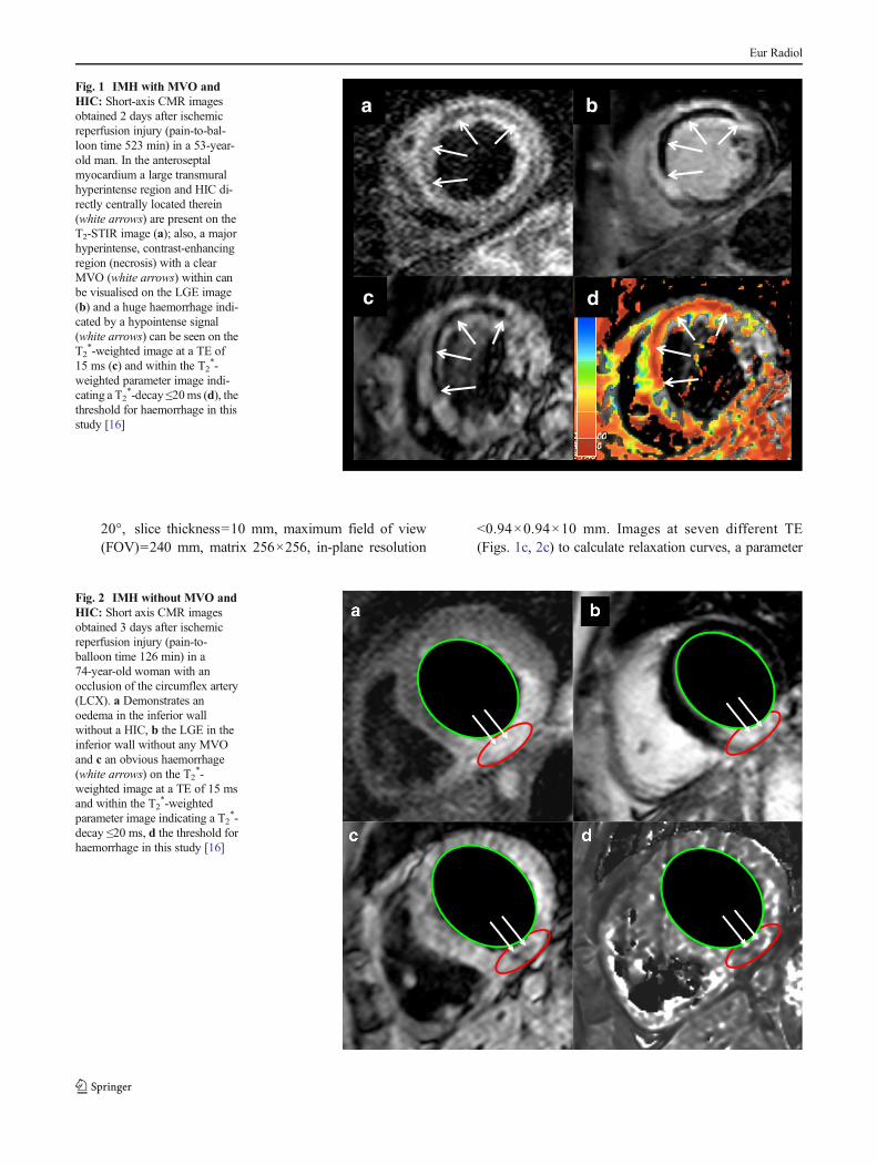
c d
Fig. 1 IMH with MVO andHIC: Short-axis CMR imagesobtained 2 days after ischemicreperfusion injury (pain-to-bal-loon time 523 min) in a 53-year-old man. In the anteroseptalmyocardium a large transmuralhyperintense region and HIC di-rectly centrally located therein(white arrows) are present on theT2-STIR image (a); also, a majorhyperintense, contrast-enhancingregion (necrosis) with a clearMVO (white arrows) within canbe visualised on the LGE image(b) and a huge haemorrhage indi-cated by a hypointense signal(white arrows) can be seen on theT2
*-weighted image at a TE of15 ms (c) and within the T2
*-weighted parameter image indi-cating a T2
*-decay ≤20ms (d), thethreshold for haemorrhage in thisstudy [16]
Fig. 2 IMH without MVO andHIC: Short axis CMR imagesobtained 3 days after ischemicreperfusion injury (pain-to-balloon time 126 min) in a74-year-old woman with anocclusion of the circumflex artery(LCX). a Demonstrates anoedema in the inferior wallwithout a HIC, b the LGE in theinferior wall without any MVOand c an obvious haemorrhage(white arrows) on the T2
*-weighted image at a TE of 15 msand within the T2
*-weightedparameter image indicating a T2
*-decay ≤20 ms, d the threshold forhaemorrhage in this study [16]
Eur Radiol

image (Figs. 1d, 2d) and the T2* decay [16] were obtained
from the parameter images. The occurrence or absence ofHIC with or without IMH was documented.
– Fourth and finally, late gadolinium enhancement (LGE)images (Figs. 1b, 2b), sensitive for necrosis and MVO,were acquired 10–15 min after intravenous administra-tion of 0.15 mmol/kg Gd-DTPA by using a 3D-inversionrecovery gradient echo sequence (IR-GRE) with the fol-lowing parameters: TR=2.9 ms, TE=1.5 ms, flip angle=15°, slice thickness=10 mm (5 mm increment), maxi-mum field of view (FOV)=370 mm, matrix 280×280,in-plane resolution <1.32×1.32×10 mm.
CMR image analysis
All images were analysed using a dedicated workstation(ViewForum R4.2V1C2 SP1, Software Release 2005-11-04,Philips Medical Systems, Best, the Netherlands). LV volumesand function for every patient were assessed by manuallyoutlining the endocardial contours of the LV from the SSFPshort axes sequences [5]. Short-axis T2-STIR sequences wereused to detect and to assess oedema and HIC (Figs. 1a, 2a). Inthe LGE images the size of myocardial infarction was mea-sured by using a threshold of two standard deviations of theremote myocardium [5, 27]. In addition, the presence orabsence of MVO was documented (Figs. 1b, 2b). The sal-vaged myocardium after PPCI was expressed by the myocar-dial salvage index (MSI) calculated from the difference be-tween oedema and necrosis divided by oedema [21]. IMHwasconsidered as the region with decreased signal intensity in theT2
*-weighted images based on the paramagnetic effects ofdeoxyhaemoglobin and the reduction of the T2 time [30].The used sequence provided T2
*-weighted images with thedescribed different TE additionally to a parameter image,which was used to calculate the T2
* decay. According toprevious publications a decay at the T2
* parameter image≤20 ms was considered as positive haemorrhage [16](Figs. 1d, 2d).
Statistical analysis
The study cohort was characterized by mean ± standard devi-ation (SD) for continuous variables and by absolute andrelative frequencies for categorical variables. Continuousvariables were compared within the group of patientswith and without IMH by Student’s t test. Proportionswere compared by Pearson’s χ2 test (Tables 1, 3) andFisher’s exact test where appropriate.
Furthermore, a stepwise logistic regression analysiswas applied to identify possible predictors of the occur-rence of IMH. All statistically significant clinical andCMR variables of the univariate analyses (Tables 1, 3)
were included in the final multivariable model. All var-iables selected by this algorithm were included in a finalmodel in order to get odds ratio estimates with thecorresponding 95 % confidence intervals (Table 4). Alltests performed were two-sided with a significance levelof 5 %. All statistical analyses were done by means ofSPSS software, version 16.0 for Windows.
Results
CMR was performed at a mean of 2.8 days (range 0–8 days)after PPCI. The mean patient age of the 76 patients with IMHwas 60±11 years. There were no significant differences be-tween patients with or without IMH concerning commonpatient characteristics except for hypercholesterolaemia(Table 1).
Functional and volumetric assessment
LV ejection fraction was significantly lower in patients withIMH compared to those without (43.5±9.7 % vs. 51.1±8.6 %,P<0.001) (Fig. 3). Patients with IMH also had a higher LV-EDV (163±40 ml vs. 145±30 ml, P<0.01) and LV-ESV(94±36 ml vs. 72±23 ml, P<0.001).
Comparison of T2* mapping and T2-weighted imaging
for IMH detection
By using T2* mapping IMH was detected in 50 % of all patients
(76/151). T2-weighted STIR images demonstrated HIC in 54 %(82/151) and LGE images demonstratedMVO in 66% (100/151)of all patients (Fig. 4).
p < 0.001
IMH Absent IMH Present
70
50
30
40
60
80
20
LV e
ject
ion
frac
tion
[%]
Fig. 3 Relationship between LV ejection fraction and haemorrhage.Patients with IMH had a significantly lower LV-EF than those withoutIMH (43.5±9.7 % vs. 51.1±8.6 %, P<0.001)
Eur Radiol

IMH occurred in the majority of patients (95 %; 72/76) concomitantly with MVO (Fig. 1) and in only 5 %(4/76) without (Fig. 2). When IMH and MVO werepresent at the same time IMH occurred in centre areasof the MVO. This phenomenon was observed especiallyin large infarcts with large MVO areas. Additionally,two of these four patients showed neither HIC nor aconcomitant MVO. HIC occurred in 6 % (5/82) ofpatients without MVO.
Eighty-four percent (64/76) of patients with IMHshowed HIC in T2-STIR images (Table 2), whereas in16 % (12/76) of patients IMH was not accompanied byHIC.
Influencing factors for IMH occurrence
Differences could be demonstrated related to potentialinfluencing factors for the occurrence of IMH. The followingfactors were analysed:
Location of occlusion
In 42 % (64/151) of patients the LAD, in 39 % (59/151) theRCA and in 18.5 % (28/151) the LCX was occluded. Theculprit vessel in the majority of patients with IMH was the
LAD (50 % vs. 35 %, P=0.06) and without IMH was theRCA (29 % vs. 49 %, P=0.01) (Table 1).
TIMI flow before PPCI
In IMH+ patients a TIMI flow≤1 before PPCI was signifi-cantly more frequent than in IMH− patients (72 % vs. 54 %,P<0.001) (Fig. 5).
Medication
A statistically significant difference in premedication of theIMH+ vs. IMH− group could only be demonstrated withregard to the intake of beta-blockers (34 % vs. 16 %;P= 0.01) (Table 1). All other premedication, likeangiotensin-1 antagonists, ACE inhibitors or the intake ofacetylsalicylic acid (ASA), clopidogrel or statins were notsignificantly different in both groups.
Infarct size/myocardial salvage index (MSI)
IMH+ patients had significantly larger infarcts (32.6±20.3 mlvs. 12.9±9.5 ml, P<0.001) (Fig. 6) and a lower MSI (36 %±21 % vs. 59 %±25 %, P<0.001) (Fig. 7).
Predictors of IMH
A stepwise logistic regression model was applied to identifypossible predictors of the occurrence of IMH (Tables 1, 3).After the use of a univariate analysis several significant pa-rameters were left for the multivariate analysis, like TIMI flowgrade before PPCI≤1 or the MSI. Nevertheless, the finalmultivariate analysis revealed that patients with a HIC, oreven more likely with the presence of MVO, demonstrated ahigh OR for the occurrence of IMH (Table 4). These resultsagain demonstrate the close relation between HIC and MVOand the frequent appearance of both phenomena togetherwith IMH. Surprisingly, an intake of beta-blockers demon-strated the highest OR for the occurrence of IMH. However,there was neither a significant difference between IMH+
and IMH− patients according to the presence of arterialhypertension, nor a tendency toward higher systolic ordiastolic blood pressure in the IMH+ group. However, inthe IMH+ group more patients were on a combined antihy-pertensive drug therapy with beta-blockers as compared tothe IMH− group.
Discussion
In our large study (n=151) we could demonstrate the impor-tance of using dedicated T2
*-weighted CMR sequences to
HIC(n=82, 54%)
MVO
(n=100, 66%)
Total
(n=151, 100%)
IMH +
(n=76, 50%)
Fig. 4 Venn diagram of the frequency of IMH, MVO and HIC out of thebasic population. It demonstrates the occurrence of IMH without MVOand/or HIC. CMR cardiovascular magnetic resonance, IMHintramyocardial haemorrhage, MVO microvascular obstruction, HIChypointense core in T2-STIR
Eur Radiol

detect IMH after acute reperfused STEMI to definitely distin-guish IMH fromMVO. In some cases IMH could be detectedeven without a visible MVO (Fig. 2), which is in contrast toseveral other studies [5, 6, 11, 31, 32]. Our data indicate thatT2-STIR sequences only, which are generally used to visualiseAAR and MSI, are not able to reliably detect IMH as a HICwithin the oedema. Furthermore, potential influencing factorsfor the occurrence of IMH could be evaluated.
Our most important findings can be summarized asfollows:
1. IMH is a common finding in patients with acute STEMIafter PPCI and was present in approximately 50 % of ourpatients.
2. Our data demonstrated that the majority of T2* defects
were also defects on T2 imaging (HIC), but some T2
defects had had no defect on T2* imaging. Although not
directly proven by our works, experimental studies sug-gest [33–36] that T2
* imaging is more accurate for IMHthan T2 imaging and therefore should be the preferredtechnique for detection of IMH.
3. However, according to the multivariate analysis HIC on T2imaging and MVO on LGE images are highly correlatedwith occurrence of defect on T2
* images indicating IMH.
CMR after reperfused STEMI and the role of the usedsequences
CMR is considered as the gold standard for the assessment ofventricular volumes and mass [28, 37] as well as in patientswith acute reperfused myocardial infarction as the preferredmethod for the noninvasive evaluation of viability, infarct sizeand myocardial salvage with a high prognostic value [5, 38,39]. The prognostic value of MVO and IMH after acutereperfused myocardial infarction has previously been de-scribed [5, 11, 16]. However, there is disagreement in theliterature about the best CMR sequences to detect IMH[5, 7, 10, 11, 15, 30, 33–36, 38, 40, 41] and how todefinitely differentiate it from MVO.
Pedersen et al. demonstrated in an animal study that T1-weighted sequences are more accurate than T2- or T2
*-weight-ed sequences for the detection of IMH [40]. In contrast to thisstudy Bradley and Kali et al. [17, 34] showed the highestsensitivity for IMH detection by using T2
*-weighted CMRsequences, which is in line with previous data in neuroradiol-ogy [17, 18, 42] and animal models using T2
* mapping in theheart [10]. Lotan et al. declared difficulties in the detection of
Table 2 Differences in thedetection of HIC and MVO inIMH+ versus IMH− patients
Data are reported as number(percentage)
+ present, − absent
Haemorrhage presentin T2
* mappingHaemorrhage absentin T2
* mappingP value
(n=76) (n=75)
HIC+ (STIR), n (%) 64 (84) 18 (24) <0.001
HIC− (STIR), n (%) 12 (16) 57 (76) <0.001
MVO+ (3D-IR-GRE), n (%) 72 (95) 28 (37) <0.001
MVO− (3D-IR-GRE), n (%) 4 (5) 47 (63) <0.001
Fig. 5 Relationship between TIMI flow before PPCI ≤1 and haemorrhage:In IMH+ patients a TIMI flow ≤1 before PPCI was significantly morefrequent than in IMH− patients (71 % vs. 51 %, P<0.001), which could beconsidered as a potential influencing factor for the development of IMH
p < 0.001p < 0.001
IMH Absent IMH Present
125
75
25
50
100
0
*
51
25
70
Infa
rct siz
e [m
l]
Fig. 6 Relationship between infarct size and haemorrhage: the IMHgroup had significantly larger mean infarct sizes (32.6±20.3 ml vs.12.9±9.5 ml, P<0.001) at the LGE sequences
Eur Radiol

haemorrhage by using T2-weighted images only. That studygroup stated that the reciprocal influencing effects of thehyperintense oedema and HIC may mask IMH [31]. Further-more, a postmortem study by Jackowski et al. [35] demon-strated that MVO without haemorrhage and HIC on T2-weighted images exist.
Nevertheless, there is a strong relationship between theoccurrence of IMH and MVO [41]. Also this study demon-strated MVO in 95 % of patients with IMH.
But it has also been described before that some patientsmay exclusively present IMH or MVO [38]. In the currentstudy 5 % of patients with IMH did not show any MVO.Therefore, we suppose that T2
*-weighted sequences coulddetect small amounts of haemorrhage with a very high
sensitivity, despite a slightly lower spatial resolution as com-pared to the T2-STIR, but also only slightly higher than theLGE sequences.
However, in previous studies and also in our study it couldnot definitely be clarified if IMH can be present without MVOand further studies are still needed to examine this finding.Prior analysis had demonstrated that T2
*-weighted sequencesin particular can reliably detect even small amounts of cerebralhaemorrhage [18, 42, 43]. But the spatial resolution of thepresently used LGE sequences may not be high enough todepict every MVO, while the corresponding T2
*-weightedsequences do for the detection of IMH. The spatial resolutionof the T2
* sequence in our study was slightly higher than thespatial resolution of the LGE sequence.
Nevertheless, artefacts are common on T2*-weighted im-
ages and could mimic IMH. To reduce this potential source ofmisinterpretation a hypointense signal on the T2
* images wasonly defined as IMH if the decay on the parameter image wasat most 20 ms as previously described [16]. In another studyan even more sensitive threshold with a T2
* decay of less than30mswas proposed [32], which may additionally increase thenumber of IMH+ patients without any visible MVO or HIC.
The occurrence of IMH without MVO on the LGE imagesmay also be caused by passive diffusion of contrast agent into
IMH Absent IMH Present
0.80
0.40
0.20
0.60
1.00
0.00
Myocardia
l S
alv
age Index [M
SI]
p < 0.001
Fig. 7 Relationship between myocardial salvage index and haemor-rhage. MSI in the IMH group was significantly lower than in the non-IMH group (0.36±0.21 vs. 0.59±0.25, P<0.001)
Table 3 CMR results
All patients Haemorrhage presentin T2
* mappingHaemorrhage absentin T2
* mappingP valueunivariateanalysis
Χ2 univariateanalysis
OR univariateanalysis
95 % CI
(n=151) (n=76) (n=75)
Variable
LVejection fraction (%) 47±10 43±10 51±9 <0.001 19.6 0.912 0.876–0.950
LVend-diastolic volume, ml 154±37 163±40 145±30 0.003 8.2 1.01 1.000–1.020
LVend-systolic volume, ml 83±32 94±36 72±23 <0.001 15.5 1.03 1.010–1.040
LV stroke volume, ml 71±16 69±15 73±15 0.07 3.2 0.981 0.960–1.002
Infarct size absolute, ml 23±19 33±20 13±9 <0.001 33.5 1.13 1.080–1.170
Myocardial salvage index (MSI) 0.47±0.26 0.36±0.21 0.59±0.25 <0.001 25.2 0.014 0.003–0.073
Volume of HIC at STIR, ml 2.3±4.3 4.2±5.4 0.3±0.8 <0.01 9.18 4.67 1.730–12.90
HIC at STIR, n (%) 82 (54) 64 (84) 18 (24) <0.001
Volume of MVO at 3D-IR-GRE, ml 3.1±6.5 5.8±8.3 0.4±0.9 <0.001 27.7 3.62 2.240–5.850
MVO present at 3D-IR-GRE, n (%) 100 (66) 72 (95) 28 (37) <0.001
Data are reported as mean±SD or mean (minimum–maximum) if continuous and number (percentage) if categorical
OR odds ratio, CI confidence interval
Table 4 Characteristics associated with IMH/predictors of IMH
Variable Odds ratio (95 % CI) P value multivariate
Volume of MVO (ml) 4.1 (1.4–12.3) 0.01
Volume of HIC (ml) 2.9 (1.8–4.8) <0.001
Beta-blocker at admission 4.6 (1.6–13.1) 0.004
Eur Radiol

the small infarct core, obscuring the presence of MVO on theLGE images [44].
Furthermore, it is not yet understood what a HIC in T2-weighted STIR sequences represents when IMH and MVOcould not be demonstrated (2 % (3/151) of all patients in thisstudy). In one of only a few histologically confirmed post-mortem analyses [35] HIC always represented MVO with orwithout IMH.
Relationship between IMH and potential influencing factors
Previous publications and this study confirmed that IMH isnot always detectable in patients after acute reperfusedSTEMI. In about half of our patients IMH was detectable,which is consistent with findings from Beek et al. with 49 %of patients with IMH [3] or Robbers et al. with 60 % [44], butmuch more frequent as described in other studies [5, 45].Therefore, potentially influencing factors for the occurrenceof IMH should be evaluated. In our study most patients withIMH showed an occluded LAD (50 %) and patients withoutIMH presented more often an occluded RCA, which hadalready been described by Amabile et al. [45]. The majorityof our patients (42 %) had their culprit lesion in the LADterritory (Table 1) and the IMH+ mostly in the proximalsegment (50 % vs. 38 %), whereas in the LCX it was mainlylocated at the middle segment, with an equal distributionwithin both groups. The second most involved territory wasthe RCA region (39 %), in which the IMH+ demonstrated analmost equal distribution of proximally and distally locatedlesions. But in the IMH− group the culprit lesion was mainlylocated proximally in more than 50 % of patients. Infarct sizeroughly correlates with the location of the culprit lesion and aproximal lesion in the LAD territory causes usually largerinfarcts than a proximal lesion in the RCA. Furthermore,IMH correlated significantly with infarct size.
Patients with a low TIMI flow before PPCI (≤1) seem tohave a higher risk for the development of IMH (Fig. 5).Seventy-two per cent of our patients with a TIMI flow grade≤1 demonstrated an IMH, which was significantly different tothose patients without IMH. Similar results could be demon-strated in prior studies, e.g. Eitel et al. reported on 87 %patients with a TIMI flow grade≤1 before PPCI [5] and aHIC, which was used as an indicator for IMH in this study.Nevertheless, in the final multivariate analysis (Table 4) onlyHIC and especially MVO were significant predictors of IMH.Furthermore, the beta-blocker intake at admission was astrong predictor of IMH in patients. In the IMH+ group morepatients were on a combined antihypertensive therapy indicat-ing a potentially more severe arterial hypertension, but this israther speculative and has to be proven using another studydesign. However, several large trials on the use of beta-blockers in myocardial infarction demonstrated that the useof beta-blockers reduced the risks of reinfarction and
ventricular fibrillation, but increased the risk of cardiogenicshock [46, 47]. These findings changed the clinical practice ofbeta-blocker use in patients presenting with acute coronarysyndromes, emphasizing the potential harm of beta-blockersin patients at increased risk of cardiogenic shock. Neverthe-less, this unexpected finding may also be an epiphenomenon.
Relationship between IMH and ventricular function
Other studies have already demonstrated the relationship be-tween the presence of IMH and significantly larger infarctsizes, decreased left ventricular function, less myocardial sal-vage and an adverse prognosis [3, 5, 11]. IMH after STEMI isseen as a reperfusion injury and closely related to larger infarctsizes [5]. In this study we confirmed a significantly lower LV-EF, larger LV-EDV and LV-ESV and larger infarct sizes inpatients with IMH. These findings were also seen in studies byEitel et al. and Beek et al. [3, 5, 44]. Beek et al. did not findany prognostic influence of MVO [3]. O’Regan et al. sup-posed that the occurrence of IMH is related to a reducedmyocardial salvage [16]. In our patients a significant impactof IMH on ventricular function and volumes could be dem-onstrated, therefore it might be considered to be a potentiallyimportant prognostic factor.
Limitations
This is a retrospective study on limited patients with acutereperfused ST-elevation myocardial infarction. A prospectivestudy on a bigger study cohort would be desirable to confirmour results and even more important to confirm the prognosticsignificance of IMH in this group of patients.
As a result of time constraints in this study, like in others[16], we used only one single slice T2
* mapping sequence inthe most representative slice in the middle of the myocardialoedema. An additional retrospective analysis of our data re-vealed that that was always in agreement with the centre of theLGE area. More slices could not be acquired and thereforepossible haemorrhage in other areas might be missed. There-fore, it can be considered that the frequency of IMH is prob-ably even higher than described in the present study. Furtherstudies should therefore use a whole heart T2
* mapping ap-proach.We also could not compare the total size of IMH to thesize of HIC or MVO because of the one single slice T2
*
mapping.The period of time after infarction at which CMR was
performed could also have been an important influencingfactor for the IMH detection rate, because of the differentdegradation stages of haemoglobin, which have an effect onthe signal intensity [17, 48, 49]. In this study the CMRanalysis was performed within 8 days after infarction (mean2.8 days). It is known that the oxyhaemoglobin of freshbleeding does not have any paramagnetic effect and therefore
Eur Radiol

can not be detected by T2*-weighted MRI. As a result of
oxidative denaturation, deoxyhaemoglobin with its detectableparamagnetic effects occurs usually after 12–72 h, which isknown from cerebral MRI studies [48]. Therefore, it can behypothesised that in these patients additional haemorrhagemay have been detected if MR imaging had been performedlater. In addition to that, Kali et al. described the goodcommitment of T2
*-weighted CMR for the assessment ofchanges in iron deposition in postinfarcted myocardium [33,34]. The paramagnetic effects of iron are still visible on T2
*-weighted images after months.
Conclusions
IMH is a frequent finding in reperfused STEMI associatedwith reduced ventricular function and myocardial damage,which can be assessed by CMR. Our data indicates—in con-cordance with other recently published data [33–36]—that T2
*
mapping seems to be more suitable than T2-weighted imagesalone for the detection of IMH in reperfused acute myocardialinfarction. Despite the fact that it is rather closely correlatedwith a HIC in T2-weighted images and MVO in LGE images,it can also occur alone according to our results.
In a larger trial with additional follow-up examinations andespecially the use of a full left ventricular coverage of T2
*
mapping, it has to be proven if this is really a separatephenomenon to MVO with a clinical and prognostic impactor just another possibility to detect MVO with a higher sensi-tivity as compared to T2-weighted STIR and LGE sequences.However, this may allow for a better risk stratification inpatients after acute reperfused myocardial infarction.
Acknowledgements The scientific guarantor of this publication is Prof.Dr. Matthias Gutberlet. The authors of this manuscript declare no rela-tionships with any companies whose products or services may be relatedto the subject matter of the article. The authors state that this work has notreceived any funding. Dr. Martin Mende kindly provided statisticaladvice for this manuscript. Institutional review board approval was ob-tained.Written informed consent was obtained from all subjects (patients)in this study. Some study subjects or cohorts have been previouslyreported [25]. Methodology: retrospective, observational, performed atone institution.
Open Access This article is distributed under the terms of the CreativeCommons Attribution Noncommercial License which permits any non-commercial use, distribution, and reproduction in any medium, providedthe original author(s) and the source are credited.
References
1. Jaffe R, Charron T, Puley G, Dick A, Strauss BH (2008)Microvascular obstruction and the no-reflow phenomenon after per-cutaneous coronary intervention. Circulation 117:3152–3156
2. Higginson LA, White F, Heggtveit HA, Sanders TM, Bloor CM,Covell JW (1982) Determinants of myocardial hemorrhage aftercoronary reperfusion in the anesthetized dog. Circulation 65:62–69
3. Beek AM, Nijveldt R, van Rossum AC (2010) Intramyocardialhemorrhage and microvascular obstruction after primary percutane-ous coronary intervention. Int J Cardiovasc Imaging 26:49–55
4. Bekkers SC, Yazdani SK, Virmani R, Waltenberger J (2010)Microvascular obstruction: underlying pathophysiology and clinicaldiagnosis. J Am Coll Cardiol 55:1649–1660
5. Eitel I, Kubusch K, Strohm O, Desch S, Mikami Y, de Waha S,Gutberlet M, Schuler G, Friedrich MG, Thiele H (2011) Prognosticvalue and determinants of a hypointense infarct core in T2-weightedcardiac magnetic resonance in acute reperfused ST-elevation-myocardial infarction. Circ Cardiovasc Imaging 4:354–362
6. Fishbein MC, Y-Rit J, Lando U, Kanmatsuse K,Mercier JC, GanzW(1980) The relationship of vascular injury and myocardial hemor-rhage to necrosis after reperfusion. Circulation 62:1274–1279
7. Ochiai K, Shimada T, Murakami Y, Ishibashi Y, Sano K, Kitamura J,Inoue S, Murakami R, Kawamitsu H, Sugimura K (1999)Hemorrhagic myocardial infarction after coronary reperfusion detect-ed in vivo bymagnetic resonance imaging in humans: prevalence andclinical implications. J Cardiovasc Mag Reson 1:247–256
8. Basso C, Corbetti F, Silva C, Abudureheman A, Lacognata C,Cacciavillani L, Tarantini G, Marra MP, Ramondo A, Thiene G,Iliceto S (2007) Morphologic validation of reperfused hemorrhagicmyocardial infarction by cardiovascular magnetic resonance. Am JCardiol 100:1322–1327
9. Van den Bos EJ, Baks T, Moelker AD, Kerver W, van Geuns RJ, vander Giessen WJ, Duncker DJ, Wielopolski PA (2006) Magneticresonance imaging of haemorrhage within reperfused myocardialinfarcts: possible interference with iron oxide-labelled cell tracking?Eur Heart J 27:1620–1626
10. Ghugre NR, Ramanan V, Pop M, Yang Y, Barry J, Qiang B, ConnellyKA, Dick AJ, Wright GA (2011) Quantitative tracking of edema,hemorrhage, and microvascular obstruction in subacute myocardialinfarction in a porcinemodel byMRI.MagnResonMed 66:1129–1141
11. Kumar A, Green JD, Sykes JM, Ephrat P, Carson JJ, Mitchell AJ,Wisenberg G, Friedrich MG (2011) Detection and quantification ofmyocardial reperfusion hemorrhage using T2*-weighted CMR.JACC Cardiovasc Imaging 4:1274–1283
12. Yellon DM, Hausenloy DJ (2007) Myocardial reperfusion injury. NEngl J Med 357:1121–1135
13. Dirksen MT, Laarman GJ, Simoons ML, Duncker DJ (2007)Reperfusion injury in humans: a review of clinical trials on reperfu-sion injury inhibitory strategies. Cardiovasc Res 74:343–355
14. Asanuma T, Tanabe K, Ochiai K, Yoshitomi H, Nakamura K,Murakami Y, Sano K, Shimada T, Murakami R, Morioka S, BeppuS (1997) Relationship between progressive microvascular damageand intramyocardial hemorrhage in patients with reperfused anteriormyocardial infarction: myocardial contrast echocardiographic study.Circulation 96:448–453
15. Ganame J, Messalli G, Dymarkowski S, Rademakers FE, Desmet W,Van de Werf F, Bogaert J (2009) Impact of myocardial haemorrhageon left ventricular function and remodelling in patients with reper-fused acute myocardial infarction. Eur Heart J 30:1440–9
16. O’Regan DP, Ahmed R, Karunanithy N, Neuwirth C, Tan Y,Durighel G, Hajnal JV, Nadra I, Corbett SJ, Cook SA (2009)Reperfusion hemorrhage following acute myocardial infarction: as-sessment with T2* mapping and effect on measuring the area at risk.Radiology 250:916–922
17. Bradley WG Jr (1993) MR appearance of hemorrhage in the brain.Radiology 189:15–26
18. Parizel PM, Makkat S, Van Miert E, Van Goethem JW, van denHauwe L, De Schepper AM (2001) Intracranial hemorrhage: princi-ples of CT and MRI interpretation. Eur Radiol 11:1770–1783
Eur Radiol

19. Friedrich MG, Abdel-Aty H, Taylor A, Schulz-Menger J, MessroghliD, Dietz R (2008) The salvaged area at risk in reperfused acutemyocardial infarction as visualized by cardiovascular magnetic reso-nance. J Am Coll Cardiol 51:1581–1587
20. O´Regan DP, Ahmed R, Neuwirth C, Tan Y, Durighel G, HajnalJV, Nadra I, Corbett SJ, Cook SA (2009) Cardiac MRI ofmyocardial salvage at the peri-infarct border zones after primarycoronary intervention. Am J Physiol Heart Circ Physiol 297:H340–346
21. Eitel I, Desch S, Fuernau G, Hildebrand L, Gutberlet M, Schuler G,Thiele H (2010) Prognostic significance and determinants of myo-cardial salvage assessed by cardiovascular magnetic resonance inacute reperfused myocardial infarction. J Am Coll Cardiol 55:2470–2479
22. Cannan C, Eitel I, Hare J, Kumar A, Friedrich M (2010) Hemorrhagein the myocardium following infarction. JACC Cardiovasc Imaging3:665–668
23. Thiele H, Wöhrle J, Neuhaus P, Brosteanu O, Sick P, Prondzinsky R,Birkemeyer R, Wiemer M, Kerber S, Schuehlen H, Kleinertz K,Axthelm C, Zimmermann R, Rittger H, Braun-Dullaeus RC, LauerB, Burckhardt W, Ferrari M, Bergmann MW, Hambrecht R, SchulerG, Abciximab Intracoronary versus intravenously Drug Applicationin ST-Elevation Myocardial Infarction (AIDA STEMI) Investigators(2010) Intracoronary compared with intravenous bolus abciximabapplication during primary percutaneous coronary intervention: de-sign and rationale of the Abciximab Intracoronary versus intrave-nously Drug Application in ST-Elevation Myocardial Infarction(AIDA STEMI) trial. Am Heart J 159:547–554
24. Thiele H,Wöhrle J, Hambrecht R, Rittger H, Birkemeyer R, Lauer B,Neuhaus P, Brosteanu O, Sick P, Wiemer M, Kerber S, Kleinertz K,Eitel I, Desch S, Schuler G (2012) Intracoronary versus intravenousbolus abciximab during primary percutaneous coronary interventionin patients with acute ST-elevation myocardial infarction: arandomised trial. Lancet 379:923–931
25. Eitel I, Wöhrle J, Suenkel H, Meissner J, Kerber S, Lauer B,Pauschinger M, Birkemeyer R, Axthelm C, Zimmermann R,Neuhaus P, Brosteanu O, de Waha S, Desch S, Gutberlet M,Schuler G, Thiele H (2013) Intracoronary comparedwith intravenousbolus abciximab application during primary percutaneous coronaryintervention in ST-segment elevation myocardial infarction: cardiacmagnetic resonance substudy of the AIDA STEMI trial. J Am CollCardiol 61:1447–1454
26. Chesebro JH, Knatterud G, Roberts R, Borer J, Cohen LS, Dalen J,Dodge HT, Francis CK, Hillis D, Ludbrook P (1987) Thrombolysisin myocardial infarction (TIMI) trial, phase I: a comparison betweenintravenous tissue plasminogen activator and intravenous streptoki-nase. Clinical findings through hospital discharge. Circulation 76:142–154
27. Grothoff M, Elpert C, Hoffmann J, Zachrau J, Lehmkuhl L, de WahaS, Desch S, Eitel I, Mende M, Thiele H, Gutberlet M (2012) Rightventricular injury in ST-elevation myocardial infarction: risk stratifi-cation by visualization of wall motion, edema, and delayed-enhancement cardiac magnetic resonance. Circ Cardiovasc Imaging5:60–68
28. Gutberlet M, Abdul-Khaliq H, Grothoff M, Schröter J, Schmitt B,Röttgen R, Lange P, Vogel M, Felix R (2003) Evaluation of leftventricular volumes in patients with congenital heart disease andabnormal left ventricular geometry. Comparison of MRI and trans-thoracic 3-dimensional echocardiography. RöFo 175:942–951
29. Shapiro EP, RogersWJ, Beyar R, Soulen RL, Zerhouni EA, Lima JA,Weiss JL (1989) Determination of left ventricular mass by magneticresonance imaging in hearts deformed by acute infarction.Circulation 79:706–711
30. Lotan CS, Bouchard A, Cranney GB, Bishop SP, Pohost GM (1992)Assessment of postreperfusion myocardial hemorrhage using protonNMR imaging at 1.5 T. Circulation 86:1018–1025
31. Lotan CS, Miller SK, Cranney GB, Pohost GM, Elgavish GA (1992)The effect of postinfarction intramyocardial hemorrhage on trans-verse relaxation time. Magn Reson Med 23:346–355
32. ZiaMI, Ghugre NR, Connelly KA, Strauss BH, Sparkes JD, Dick AJ,Wright GA (2012) Characterizing myocardial edema and hemor-rhage using quantitative T2 and T2* mapping at multiple time inter-vals post ST-segment elevation myocardial infarction. CircCardiovasc Imaging 5:566–572
33. Kali A, Kumar A, Cokic I, Tang RL, Tsaftaris SA, Friedrich MG,Dharmakumar R (2013) Chronic manifestation of postreperfusionintramyocardial hemorrhage as regional iron deposition: a cardiovas-cular magnetic resonance study with ex vivo validation. CircCardiovasc Imaging 6:218–228
34. Kali A, Tang RL, Kumar A, Min JK, Dharmakumar R (2013)Detection of acute reperfusion myocardial hemorrhage with cardiacMR imaging: T2 versus T2. Radiology 269:387–395
35. Jackowski C, Christe A, Sonnenschein M, Aghayev E, Thali MJ(2006) Postmortem unenhanced magnetic resonance imaging ofmyocardial infarction in correlation to histological infarction agecharacterization. Eur Heart J 27:2459–2467
36. O'Regan DP, Ariff B, Neuwirth C, Tan Y, Durighel G, Cook SA(2010) Assessment of severe reperfusion injury with T2* cardiacMRI in patients with acute myocardial infarction. Heart 96:1885–1891
37. Gutberlet M, Fröhlich M,Mehl S, Amthauer H, Hausmann H,MeyerR, Siniawski H, Ruf J, Plotkin M, Denecke T, Schnackenburg B,Hetzer R, Felix R (2005) Myocardial viability assessment in patientswith highly impaired left ventricular function: comparison of delayedenhancement, dobutamine stress MRI, end-diastolic wall thickness,and TI201-SPECT with functional recovery after revascularization.Eur Radiol 15:872–880
38. Husser O, Monmeneu JV, Sanchis J, Nunez J, Lopez-Lereu MP,Bonanad C, Chaustre F, Gomez C, Bosch MJ, Hinarejos R, ChorroFJ, Riegger GA, Llacer A, Bodi V (2012) Cardiovascular magneticresonance-derived intramyocardial hemorrhage after STEMI:Influence on long-term prognosis, adverse left ventricular remodelingand relationship with microvascular obstruction. Int J Cardiol. doi:10.1016/j.ijcard.2012.05.055
39. Ahmed N, Carrick D, Layland J, Oldroyd KG, Berry C (2012) Therole of cardiac magnetic resonance imaging (MRI) in acute myocar-dial infarction (AMI). Heart Lung Circ 22:243–255
40. Pedersen SF, Thrysøe SA, Robich MP, Paaske WP, Ringgaard S,Bøtker HE, Hansen ES, Kim WY (2012) Assessment ofintramyocardial hemorrhage by T1-weighted cardiovascular magnet-ic resonance in reperfused acute myocardial infarction. J CardiovascMagn Reson 14:59–67
41. Bekkers SC, Smulders MW, Passos VL, Leiner T, Waltenberger J,Gorgels AP, Schalla S (2010) Clinical implications of microvascularobstruction and intramyocardial haemorrhage in acute myocardialinfarction using cardiovascular magnetic resonance imaging. EurRadiol 20:2572–2578
42. Wiesmann M, Mayer TE, Yousry I, Hamann GF, Bruckmann H(2001) Detection of hyperacute parenchymal hemorrhage of the brainusing echo-planar T2*-weighted and diffusion-weighted MRI. EurRadiol 11:849–853
43. Linfante I, Llinas RH, Caplan LR, Warach S (1999) MRI features ofintracerebral hemorrhage within 2 hours from symptom onset. Stroke30:2263–2267
44. Robbers LF, Eerenberg ES, Teunissen PF, JansenMF, HollanderMR,Horrevoets AJ, Knaapen P, Nijveldt R, HeymansMW, LeviMM, vanRossum AC, Niessen HW, Marcu CB, Beek AM, van Royen N(2013) Magnetic resonance imaging-defined areas of microvascularobstruction after acute myocardial infarction represent microvasculardestruction and haemorrhage. Eur Heart J 34:2346–2353
45. Amabile N, Jacquier A, Shuhab A, Gaudart J, Bartoli JM, PaganelliF, Moulin G (2012) Incidence, predictors, and prognostic value of
Eur Radiol

intramyocardial hemorrhage lesions in ST elevation myocardial in-farction. Catheter Cardiovasc Interv 79:1101–1108
46. Chen ZM, Pan HC, Chen YP et al (2005) Early intravenous then oralmetoprolol in 45,852 patients with acute myocardial infarction:randomised placebo-controlled trial. Lancet 366:1622–1632
47. Antman EM, Hand M, Armstrong PW et al (2008) 2007 focusedupdate of the ACC/AHA 2004 guidelines for the management ofpatients with ST-elevation myocardial infarction: a report of the
American College of Cardiology/American Heart AssociationTask Force on Practice Guidelines. J Am Coll Cardiol 51:210–247
48. Anzalone N, Scotti R, Riva R (2004) Neuroradiologic differentialdiagnosis of cerebral intraparenchymal hemorrhage. Neurol Sci. doi:10.1007/s10072-004-0205-8
49. Wu KC (2012) CMR of microvascular obstruction and hemorrhagein myocardial infarction. J Cardiovasc Magn Reson 14:68
Eur Radiol
Related Documents

![Chorea Hyperglycemia Basal Ganglia 02 Syndrome in a Young ... · brain lesions; however, hypointense basal ganglia lesions have been most commonly reported [1,6]. Compared to acute](https://static.cupdf.com/doc/110x72/5eb4cc9f5df56b18411b11a5/chorea-hyperglycemia-basal-ganglia-02-syndrome-in-a-young-brain-lesions-however.jpg)


