Circulating Endothelial Progenitor Cells in KidneyTransplant PatientsGiovana S. Di Marco1., Peter Rustemeyer2., Marcus Brand1, Raphael Koch3, Dominik Kentrup1,
Alexander Grabner1, Burkhard Greve4, Werner Wittkowski2, Hermann Pavenstadt1, Martin Hausberg1,
Stefan Reuter1, Detlef Lang1*
1 Medizinische Klinik und Poliklinik D, Universitatsklinikum Munster, Munster, Germany, 2 Institut fur Anatomie, Universitatsklinikum Munster, Munster, Germany, 3 Institut
fur Medizinische Informatik und Biomathematik, Universitatsklinikum Munster, Munster, Germany, 4 Institut fur Strahlenbiologie, Universitatsklinikum Munster, Munster,
Germany
Abstract
Background: Kidney transplantation (RTx) leads to amelioration of endothelial function in patients with advanced renalfailure. Endothelial progenitor cells (EPCs) may play a key role in this repair process. The aim of this study was to determinethe impact of RTx and immunosuppressive therapy on the number of circulating EPCs.
Methods: We analyzed 52 RTx patients (58613 years; 33 males, mean 6 SD) and 16 age- and gender-matched subjects withnormal kidney function (57617; 10 males). RTx patients received a calcineurin inhibitor (CNI)-based (65%) or a CNI-freetherapy (35%) and steroids. EPC number was determined by double positive staining for CD133/VEGFR2 and CD34/VEGFR2by flow cytometry. Stromal cell-derived factor 1 alpha (SDF-1) levels were assessed by ELISA. Experimentally, to dissociatethe impact of RTx from the impact of immunosuppressants, we used the 5/6 nephrectomy model. The animals were treatedwith a CNI-based or a CNI-free therapy, and EPCs (Sca+cKit+) and CD26+ cells were determined by flow cytometry.
Results: Compared to controls, circulating number of CD34+/VEGFR2+ and CD133+/VEGFR2+ EPCs increased in RTxpatients. There were no correlations between EPC levels and statin, erythropoietin or use of renin angiotensin systemblockers in our study. Indeed, multivariate analysis showed that SDF-1 – a cytokine responsible for EPC mobilization – isindependently associated with the EPC number. 5/6 rats presented decreased EPC counts in comparison to control animals.Immunosuppressive therapy was able to restore normal EPC values in 5/6 rats. These effects on EPC number wereassociated with reduced number of CD26+ cells, which might be related to consequent accumulation of SDF-1.
Conclusions: We conclude that kidney transplantation and its associated use of immunosuppressive drugs increases thenumber of circulating EPCs via the manipulation of the CD26/SDF-1 axis. Increased EPC count may be associated toendothelial repair and function in these patients.
Citation: Di Marco GS, Rustemeyer P, Brand M, Koch R, Kentrup D, et al. (2011) Circulating Endothelial Progenitor Cells in Kidney Transplant Patients. PLoSONE 6(9): e24046. doi:10.1371/journal.pone.0024046
Editor: Aric Gregson, University of California Los Angeles, United States of America
Received December 23, 2010; Accepted August 3, 2011; Published September 8, 2011
Copyright: � 2011 Di Marco et al. This is an open-access article distributed under the terms of the Creative Commons Attribution License, which permitsunrestricted use, distribution, and reproduction in any medium, provided the original author and source are credited.
Funding: This study was supported by an unrestricted grant from the Else-Kroener Fresenius Foundation (P37/2004), Germany. The authors acknowledgesupport by Deutsche Forschungsgemeinschaft (DFG) and Open Access Publication Fund of University of Muenster. The funders had no role in study design, datacollection and analysis, decision to publish, or preparation of the manuscript.
Competing Interests: The authors have declared that no competing interests exist.
* E-mail: [email protected]
. These authors contributed equally to this work.
Introduction
Endothelial dysfunction is a typical finding in chronic kidney
disease (CKD). It contributes to accelerated arteriosclerosis
and impaired angiogenesis and, therefore, to high cardiovascular
morbidity and mortality in these patients. However, after renal
transplantation (RTx) endothelial function improves, even though
substantial dysfunction is still observed in these patients [1–3].
Thus, it is not surprising that endothelial damage, as a process of
the whole vasculature, is an important feature of chronic allograft
nephropathy [3].
Interestingly, these vascular lesions can be repaired by i)
migration and proliferation of endothelial cells contiguous to the
lesions or by ii) the so-called endothelial progenitor cells (EPCs)
[4]. These cells reside in the bone marrow and are mobilized to
the peripheral blood upon stimulation. Stimuli include tissue
ischemia and local release of cytokines and growth factors [5]. The
stromal cell-derived factor 1 alpha (SDF-1) is one of these che-
mokines that serve as chemoattractant for stem/progenitor cell
populations [6].
Patients with advanced renal failure were shown to have not
only significant lower EPC numbers compared to healthy controls
but, in addition, impaired EPC function [7]. EPC number and
function can be restored by initiation of dialysis or kidney
transplantation, procedures at least partially restoring or imitating
renal function [8–10].
During the transformation process of EPCs into mature
endothelial cells, human EPCs express different surface markers
PLoS ONE | www.plosone.org 1 September 2011 | Volume 6 | Issue 9 | e24046

at distinct stages including CD133, CD34 and vascular endothelial
growth factor receptor 2 (VEGF R2) [11]. Circulating EPCs seem
to prefer to locate at the sites of vascular lesions, thereby,
contributing essentially to both reendothelialization and revascu-
larization [12]. Thus, EPCs are critically involved in maintaining
the integrity of the endothelium and repairing vascular damage [13].
Immunosuppressive treatments of patients after RTx may
directly affect the endothelial function [14,15]. However, the exact
role of EPC and the EPC count in recipients of renal allografts is still
controversial. Therefore, the aim of the present study was to
determine i) the number of EPCs in stable renal allograft recipients
and ii) the EPC count association with different immunosuppressive
agents especially the comparison of calcineurin inhibitor (CNI)-
based and CNI-free therapies. Moreover, we provided a current
literature review on studies regarding EPC in RTx.
Results
Human studyClinical data of the study subjects are summarized in Table S1.
All patients received medication, including immunosuppressive
drugs, statins, antihypertensive drugs, and/or erythropoietin. We
studied a total of 52 stable kidney transplant patients and 16
gender- and age-matched subjects. 68% (38/56) of the patient
cases were on CNI (cyclosporine, 90.663.2 ng/ml, or FK506,
8.763.1 ug/ml), and 32% (18/56) were mostly on mycophenolate
mofetil (MMF, 3.661.7 ug/ml) and sirolimus (CNI-free therapy).
At the time of blood collection, most of the patients given a CNI-
based immunosuppression used a FK506 regimen (19/38) fol-
lowed by FK506+MMF (11/38); most recipients treated with a
CNI-free regimen received MMF and steroids (16/18). The
glomerular filtration rate (eGFR) estimated by the MDRD
(Modification of Diet in Renal Disease) formula was in all graft
recipients above 40 ml/min/1.73 m2 and in controls above 60 ml/
min/1.73 m2, respectively. The average time period between RTx
and blood collection was 59653 months (Mean 6 SD). A possible
interrelation between waiting time since surgery and EPC count was
ruled out by univariate regression analysis (data not shown).
Blood samples were obtained as part of a routine diagnostic or
screening procedure. They were analyzed within 1 hour. Figure 1
shows a representative density plot of the flow cytometric analysis
of a patient’s sample. CD133+/VEGFR+ cells were further cha-
Figure 1. Representative flow cytometry analysis of an EDTA-blood sample from a patient. Circulating EPCs were identified by theexpression of cell surface antigens, such as CD34+, CD133+, and VEGF-R2+. A) Density plot with forward (FSC) and side light scatter (SSC). P1-gate wasselected for further analysis. B) Density plot of PE-conjugated anti-VEGF-R2 antibody versus FITC-conjugated anti-CD133 antibody. Cells doublepositively stained for VEGF-R2 and CD133 (quadrant Q2) represent CD133+ endothelial progenitor cells (CD133+/VEGFR2+ EPCs). C) Mouse-IgG1-FITCnegative control and D) Mouse-IgG2a-PE negative control.doi:10.1371/journal.pone.0024046.g001
EPC Number in RTx
PLoS ONE | www.plosone.org 2 September 2011 | Volume 6 | Issue 9 | e24046

racterized immunohistochemically by the expression of von Will-
ebrand Factor (vWF) and their phenotypic definition as endothelial
precursors was confirmed by EPC outgrow in culture (Figure 2A
and B).
Circulating EPC – both, CD133+/VEGFR2+ and CD34+/
VEGFR2+ EPCs – number is increased in RTx recipients when
compared to controls (Figure 3). To elucidate the effect of
immunosuppressive therapy on EPC count, CNI-based and CNI-
free regimens were compared (Figure 4). Compared to controls, the
number of circulating CD133+/VEGFR2+ cells increased in RTx
patients independently of the immunosuppressive regimen used
(Table S1 and Figure 4), while CD34+/VEGFR2+ EPCs increased
only in CNI-treated patients only.
Besides the immunosuppressive therapy, we analyzed if renal
function (eGFR), diabetes mellitus and statin use interfere with the
EPC count. In multivariate analysis we could not show any
relation between eGFR or diabetes mellitus with circulating EPC
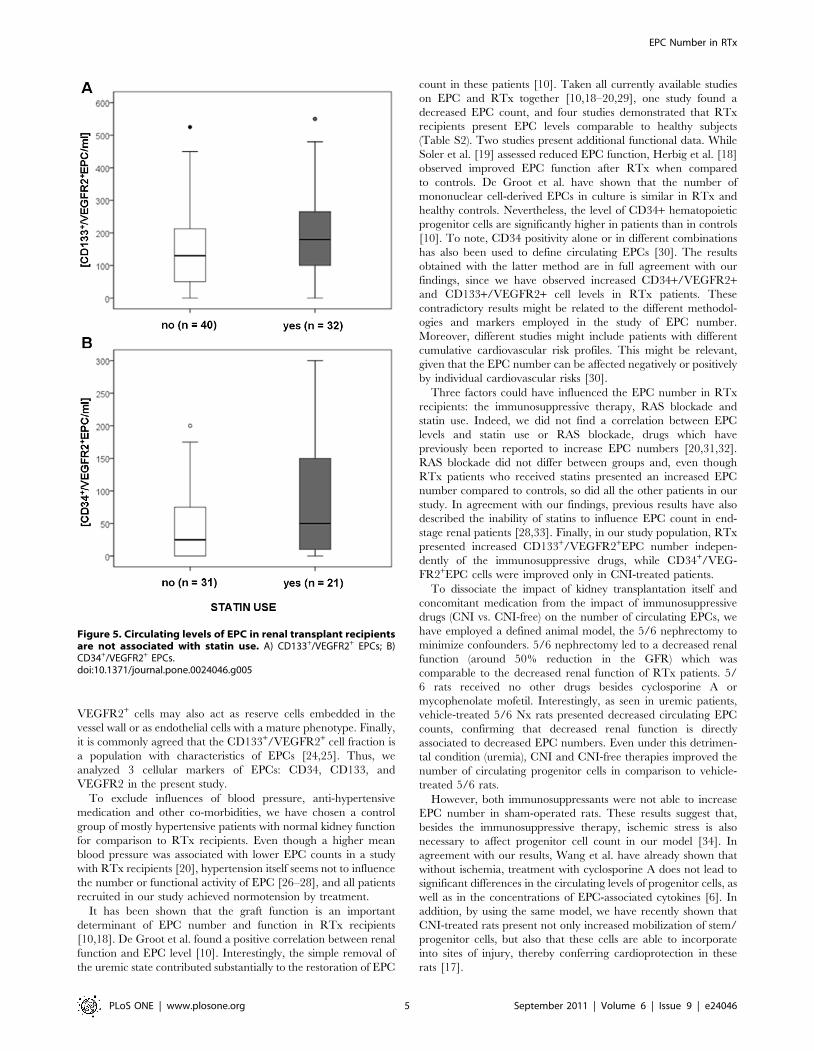
number (Table 1). RTx recipients receiving statins presented 50.0
(5.0–150.0; n = 21) CD34+/VEGFR2+EPCs/ml and 179.5
(100.0–272.5; n = 32) CD133+/VEGFR2+EPC/ml (results are
median and interquartile range), respectively; while RTx recipi-
ents without statin therapy tended to lower EPC counts/ml: 25.0
(0.0–75.0; n = 31) CD34+/VEGFR2+EPCs/ml and 130.0 (50.0–
218.75; n = 40) CD133+/VEGFR2+EPC/ml, respectively (Figure 5).
However, these differences did not reach statistical significance.
To investigate putative mechanisms in EPC mobilization, we
measured plasma levels of SDF-1 (Table S1). In RTx, elevated
EPC number was accompanied by increased SDF-1 levels.
Notably, multivariate regression analysis confirmed that plasma
SDF-1 levels were independently associated with circulating EPC
number (Table 1).
Animal studyTo distinguish the impact of RTx from the impact of
immunosuppressive drugs on the number of circulating EPCs - as
well as to avoid potential confounders, such as concomitant diseases
and medications present in human patients - we decided to use an
additional experimental model. Since our RTx patients presented a
59 to 62% reduction in the GFR in comparison to controls (Table
Figure 2. Isolation and characterization of CD133+/VEGFR2+cells. Cells sorted by FACS were further characterized by the expressionof a specific endothelial cell marker or cultured in a humanmethylcellulose base media (A and B, respectively). A) CD133+/VEGFR2+
cells were immunohistochemically stained with an antibody against vonWillebrand Factor (vWF). Negative control: omission of the primaryantibody. B) Phenotypically, colonies formed by these cells inmethylcellulose base media show the typical shape of early EPC-colonies with round immature cells in the center and dendritic orspindle cell-shaped peripheral cells (see magnification).doi:10.1371/journal.pone.0024046.g002
Figure 3. Circulating levels of endothelial progenitor cells(EPC) in renal transplant recipients. EPC levels were directlyquantified from whole blood taken from control subjects and patients(RTx) by flow cytometry, which identifies EPCs according to theexpression of cell surface antigens, such as (A) CD133+ and VEGF-R2+;and (B) CD34+ and VEGF-R2+. P value compared to control group isindicated (Mann-Whitney test).doi:10.1371/journal.pone.0024046.g003
EPC Number in RTx
PLoS ONE | www.plosone.org 3 September 2011 | Volume 6 | Issue 9 | e24046

S1), we have chosen the 5/6 nephrectomy (Nx) model that presents
a similar impairment of the renal function (50%-reduction of the
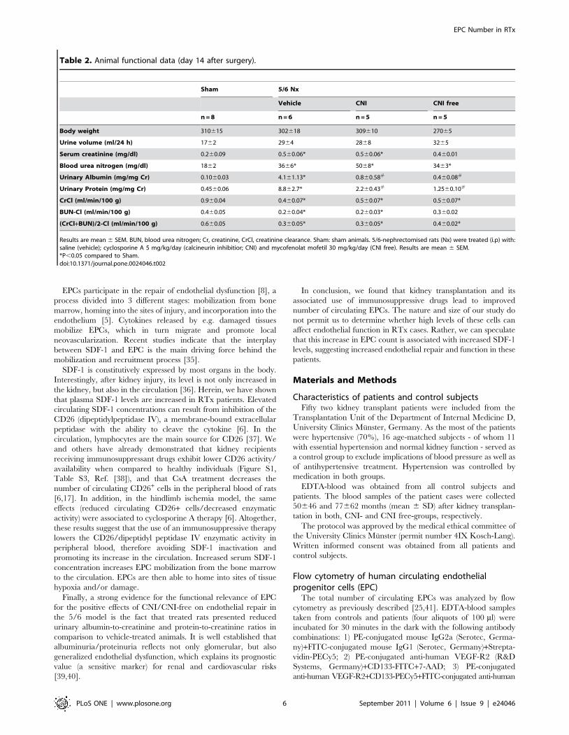
creatinine clearance). Based on the rat functional data assessed 14
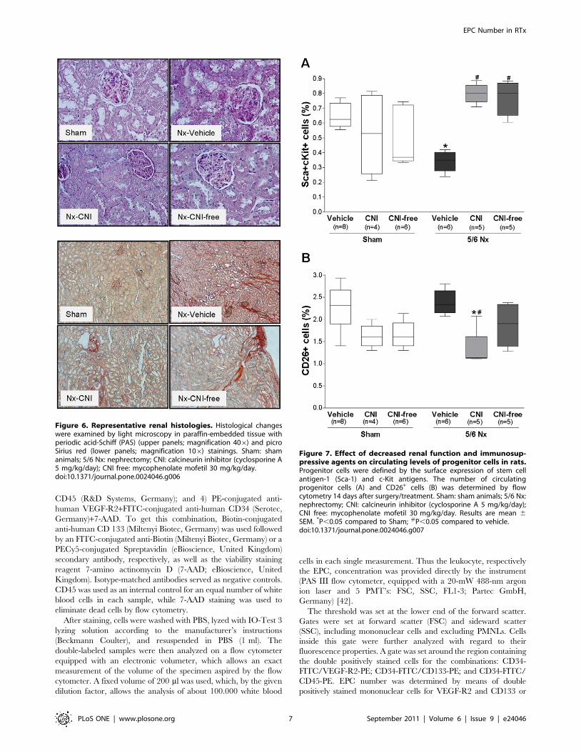
days after surgery (Table 2) and histological analysis (Figure 6), we
can state that 5/6 Nx leads to decreased renal function (increased
serum creatinine and blood urea nitrogen and decreased creatinine
clearance) and histological changes in the kidneys such as interstitial
fibrosis, glomerular sclerosis, and tubular atrophy. However,
treatment with cyclosporine A and MMF do not further deteriorate
renal function or kidney injury, but significantly ameliorated
albuminuria/proteinuria.
Progenitor cells were defined by the surface expression of stem
cell antigen-1 (Sca-1) and c-Kit antigens. This cell population
represents highly immature cells that account for a small fraction
of circulating mononuclear cells and include endothelial-commit-
ted precursors involved in compensatory angiogenesis at ischemic
sites [16]. As expected, 5/6 Nx rats presented decreased number
of circulating Sca+cKit+ cells when compared to sham-operated
rats. CNI- and CNI-free-treated rats presented not only an
increased number of progenitor cells in comparison to vehicle-
treated 5/6 Nx rats (Figure 7A), but also in comparison to sham-
operated rats (0.80%60.04 vs. 0.61%60.05, mean 6 SEM, sham
vs. CNI, Mann Whitney test P = 0.02; 0.77%60.05 vs. 0.61%6
0.05, sham vs. CNI-free, P = 0.08). CNI or CNI-free therapy given
to sham rats did not interfere with EPC numbers.
Attenuation of the CD26 system can lead to increased
concentration of SDF-1. Rats with renal failure and CNI
treatment had lower circulating CD26+ cells number than sham
and vehicle-treated 5/6 Nx rats. In CNI-free-treated rats the
CD26+ cells number was slightly lower (Figure 7B). These results
are in agreement with previous results of our group that show
increased SDF-1 levels in CNI-treated rats [17].
Discussion
Few studies have yet reported on EPC counts in RTx (Table
S2). Previous studies demonstrated reduced EPC levels in CKD
[7], whereas graft function seems to influence EPC number and
function in RTx recipients [10,18–20]. We herein show that RTx
recipients on immunosuppressive medication present increased
number of circulating EPCs when compared to controls subjects.
Furthermore, EPC levels were found to be independently
associated with plasma SDF-1 levels, a chemokine responsible
for the homing and mobilization of progenitor cells.
EPC can be characterized by hematopoietic stem cell markers
(clusters of differentiation) such as CD34 or CD133 combined with
the expression analysis of an endothelial surface marker (VEGFR2
or KDR, von Willebrand factor, VE cadherin, CD146, CD31),
uptake of Dil-acetylated lipoprotein, and lectin binding [5,21].
CD34 is an early marker expressed by bone marrow cells and
EPCs, and also by endothelial and hematopoietic cells. Co-
expression of CD34 and VEGFR2 has been used in various studies
to identify circulating progenitor cells [22,23]. Alternatively and
more recently, CD133, a marker of more immature hematopoietic
stem cells, was used for identification of these cells. Double staining
for CD133 and VEGFR2 performs better than CD34 staining
only to identify immature progenitor cells because CD34+/
Figure 4. Circulating levels of EPC in renal transplant recipientsaccording to their immunosuppressive therapy. (A) CD133+ andVEGF-R2+; and (B) CD34+ and VEGF-R2+. CNI, calcineurin inhibitor. Pvalue compared to control group is indicated (Mann-Whitney test).doi:10.1371/journal.pone.0024046.g004
Table 1. Relation of different parameters with circulating EPCnumbers.
CD133+EPC
No stand. B p-value
Full Model (backward selection):
RTx (yes or no) 0.157 0.760
eGFR (ml/min/1.73 m3) 20.001 0.789
CNI (yes or no) 0 037 0.908
CNI free (yes or no) 20.037 0.898
Statin (yes or no) 0.265 0.239
Antihypertensive therapy (yes or no) 0.135 0.505
Diabetes mellitus (yes or no) 20.119 0.644
SDF-1 alpha (pg/ml) 0.001 0.020
Result:
SDF-1 0.001 0.000
Multivariate analysis. CD133+EPC was transformed to natural logarithm. B, nostandardized regression coefficient beta. CNI, calcineurin inhibitor; eGFR,estimated glomerular filtration rate; SDF-1, stromal cell-derived factor 1 alpha;RTx, Kidney transplantation.doi:10.1371/journal.pone.0024046.t001
EPC Number in RTx
PLoS ONE | www.plosone.org 4 September 2011 | Volume 6 | Issue 9 | e24046
VEGFR2+ cells may also act as reserve cells embedded in the
vessel wall or as endothelial cells with a mature phenotype. Finally,
it is commonly agreed that the CD133+/VEGFR2+ cell fraction is
a population with characteristics of EPCs [24,25]. Thus, we
analyzed 3 cellular markers of EPCs: CD34, CD133, and
VEGFR2 in the present study.
To exclude influences of blood pressure, anti-hypertensive
medication and other co-morbidities, we have chosen a control
group of mostly hypertensive patients with normal kidney function
for comparison to RTx recipients. Even though a higher mean
blood pressure was associated with lower EPC counts in a study
with RTx recipients [20], hypertension itself seems not to influence
the number or functional activity of EPC [26–28], and all patients
recruited in our study achieved normotension by treatment.
It has been shown that the graft function is an important
determinant of EPC number and function in RTx recipients
[10,18]. De Groot et al. found a positive correlation between renal
function and EPC level [10]. Interestingly, the simple removal of
the uremic state contributed substantially to the restoration of EPC
count in these patients [10]. Taken all currently available studies
on EPC and RTx together [10,18–20,29], one study found a
decreased EPC count, and four studies demonstrated that RTx
recipients present EPC levels comparable to healthy subjects
(Table S2). Two studies present additional functional data. While
Soler et al. [19] assessed reduced EPC function, Herbig et al. [18]
observed improved EPC function after RTx when compared
to controls. De Groot et al. have shown that the number of
mononuclear cell-derived EPCs in culture is similar in RTx and
healthy controls. Nevertheless, the level of CD34+ hematopoietic
progenitor cells are significantly higher in patients than in controls
[10]. To note, CD34 positivity alone or in different combinations
has also been used to define circulating EPCs [30]. The results
obtained with the latter method are in full agreement with our
findings, since we have observed increased CD34+/VEGFR2+and CD133+/VEGFR2+ cell levels in RTx patients. These
contradictory results might be related to the different methodol-
ogies and markers employed in the study of EPC number.
Moreover, different studies might include patients with different
cumulative cardiovascular risk profiles. This might be relevant,
given that the EPC number can be affected negatively or positively
by individual cardiovascular risks [30].
Three factors could have influenced the EPC number in RTx
recipients: the immunosuppressive therapy, RAS blockade and
statin use. Indeed, we did not find a correlation between EPC
levels and statin use or RAS blockade, drugs which have
previously been reported to increase EPC numbers [20,31,32].
RAS blockade did not differ between groups and, even though
RTx patients who received statins presented an increased EPC
number compared to controls, so did all the other patients in our
study. In agreement with our findings, previous results have also
described the inability of statins to influence EPC count in end-
stage renal patients [28,33]. Finally, in our study population, RTx
presented increased CD133+/VEGFR2+EPC number indepen-
dently of the immunosuppressive drugs, while CD34+/VEG-
FR2+EPC cells were improved only in CNI-treated patients.
To dissociate the impact of kidney transplantation itself and
concomitant medication from the impact of immunosuppressive
drugs (CNI vs. CNI-free) on the number of circulating EPCs, we
have employed a defined animal model, the 5/6 nephrectomy to
minimize confounders. 5/6 nephrectomy led to a decreased renal
function (around 50% reduction in the GFR) which was
comparable to the decreased renal function of RTx patients. 5/
6 rats received no other drugs besides cyclosporine A or
mycophenolate mofetil. Interestingly, as seen in uremic patients,
vehicle-treated 5/6 Nx rats presented decreased circulating EPC
counts, confirming that decreased renal function is directly
associated to decreased EPC numbers. Even under this detrimen-
tal condition (uremia), CNI and CNI-free therapies improved the
number of circulating progenitor cells in comparison to vehicle-
treated 5/6 rats.
However, both immunosuppressants were not able to increase
EPC number in sham-operated rats. These results suggest that,
besides the immunosuppressive therapy, ischemic stress is also
necessary to affect progenitor cell count in our model [34]. In
agreement with our results, Wang et al. have already shown that
without ischemia, treatment with cyclosporine A does not lead to
significant differences in the circulating levels of progenitor cells, as
well as in the concentrations of EPC-associated cytokines [6]. In
addition, by using the same model, we have recently shown that
CNI-treated rats present not only increased mobilization of stem/
progenitor cells, but also that these cells are able to incorporate
into sites of injury, thereby conferring cardioprotection in these
rats [17].
Figure 5. Circulating levels of EPC in renal transplant recipientsare not associated with statin use. A) CD133+/VEGFR2+ EPCs; B)CD34+/VEGFR2+ EPCs.doi:10.1371/journal.pone.0024046.g005
EPC Number in RTx
PLoS ONE | www.plosone.org 5 September 2011 | Volume 6 | Issue 9 | e24046
EPCs participate in the repair of endothelial dysfunction [8], a
process divided into 3 different stages: mobilization from bone
marrow, homing into the sites of injury, and incorporation into the
endothelium [5]. Cytokines released by e.g. damaged tissues
mobilize EPCs, which in turn migrate and promote local
neovascularization. Recent studies indicate that the interplay
between SDF-1 and EPC is the main driving force behind the
mobilization and recruitment process [35].
SDF-1 is constitutively expressed by most organs in the body.
Interestingly, after kidney injury, its level is not only increased in
the kidney, but also in the circulation [36]. Herein, we have shown
that plasma SDF-1 levels are increased in RTx patients. Elevated
circulating SDF-1 concentrations can result from inhibition of the
CD26 (dipeptidylpeptidase IV), a membrane-bound extracellular
peptidase with the ability to cleave the cytokine [6]. In the
circulation, lymphocytes are the main source for CD26 [37]. We
and others have already demonstrated that kidney recipients
receiving immunosuppressant drugs exhibit lower CD26 activity/
availability when compared to healthy individuals (Figure S1,
Table S3, Ref. [38]), and that CsA treatment decreases the
number of circulating CD26+ cells in the peripheral blood of rats
[6,17]. In addition, in the hindlimb ischemia model, the same
effects (reduced circulating CD26+ cells/decreased enzymatic
activity) were associated to cyclosporine A therapy [6]. Altogether,
these results suggest that the use of an immunosuppressive therapy
lowers the CD26/dipeptidyl peptidase IV enzymatic activity in
peripheral blood, therefore avoiding SDF-1 inactivation and
promoting its increase in the circulation. Increased serum SDF-1
concentration increases EPC mobilization from the bone marrow
to the circulation. EPCs are then able to home into sites of tissue
hypoxia and/or damage.
Finally, a strong evidence for the functional relevance of EPC
for the positive effects of CNI/CNI-free on endothelial repair in
the 5/6 model is the fact that treated rats presented reduced
urinary albumin-to-creatinine and protein-to-creatinine ratios in
comparison to vehicle-treated animals. It is well established that
albuminuria/proteinuria reflects not only glomerular, but also
generalized endothelial dysfunction, which explains its prognostic
value (a sensitive marker) for renal and cardiovascular risks
[39,40].
In conclusion, we found that kidney transplantation and its
associated use of immunosuppressive drugs lead to improved
number of circulating EPCs. The nature and size of our study do
not permit us to determine whether high levels of these cells can
affect endothelial function in RTx cases. Rather, we can speculate
that this increase in EPC count is associated with increased SDF-1
levels, suggesting increased endothelial repair and function in these
patients.
Materials and Methods
Characteristics of patients and control subjectsFifty two kidney transplant patients were included from the
Transplantation Unit of the Department of Internal Medicine D,
University Clinics Munster, Germany. As the most of the patients
were hypertensive (70%), 16 age-matched subjects - of whom 11
with essential hypertension and normal kidney function - served as
a control group to exclude implications of blood pressure as well as
of antihypertensive treatment. Hypertension was controlled by
medication in both groups.
EDTA-blood was obtained from all control subjects and
patients. The blood samples of the patient cases were collected
50646 and 77662 months (mean 6 SD) after kidney transplan-
tation in both, CNI- and CNI free-groups, respectively.
The protocol was approved by the medical ethical committee of
the University Clinics Munster (permit number 4IX Kosch-Lang).
Written informed consent was obtained from all patients and
control subjects.
Flow cytometry of human circulating endothelialprogenitor cells (EPC)
The total number of circulating EPCs was analyzed by flow
cytometry as previously described [25,41]. EDTA-blood samples
taken from controls and patients (four aliquots of 100 ml) were
incubated for 30 minutes in the dark with the following antibody
combinations: 1) PE-conjugated mouse IgG2a (Serotec, Germa-
ny)+FITC-conjugated mouse IgG1 (Serotec, Germany)+Strepta-
vidin-PECy5; 2) PE-conjugated anti-human VEGF-R2 (R&D
Systems, Germany)+CD133-FITC+7-AAD; 3) PE-conjugated
anti-human VEGF-R2+CD133-PECy5+FITC-conjugated anti-human
Table 2. Animal functional data (day 14 after surgery).
Sham 5/6 Nx
Vehicle CNI CNI free
n = 8 n = 6 n = 5 n = 5
Body weight 310615 302618 309610 27065
Urine volume (ml/24 h) 1762 2964 2868 3265
Serum creatinine (mg/dl) 0.260.09 0.560.06* 0.560.06* 0.460.01
Blood urea nitrogen (mg/dl) 1862 3666* 5068* 3463*
Urinary Albumin (mg/mg Cr) 0.1060.03 4.161.13* 0.860.58# 0.460.08#
Urinary Protein (mg/mg Cr) 0.4560.06 8.862.7* 2.260.43# 1.2560.10#
CrCl (ml/min/100 g) 0.960.04 0.460.07* 0.560.07* 0.560.07*
BUN-Cl (ml/min/100 g) 0.460.05 0.260.04* 0.260.03* 0.360.02
(CrCl+BUN)/2-Cl (ml/min/100 g) 0.660.05 0.360.05* 0.360.05* 0.460.02*
Results are mean 6 SEM. BUN, blood urea nitrogen; Cr, creatinine, CrCl, creatinine clearance. Sham: sham animals. 5/6-nephrectomised rats (Nx) were treated (i.p) with:saline (vehicle); cyclosporine A 5 mg/kg/day (calcineurin inhibitior; CNI) and mycofenolat mofetil 30 mg/kg/day (CNI free). Results are mean 6 SEM.*P,0.05 compared to Sham.doi:10.1371/journal.pone.0024046.t002
EPC Number in RTx
PLoS ONE | www.plosone.org 6 September 2011 | Volume 6 | Issue 9 | e24046
CD45 (R&D Systems, Germany); and 4) PE-conjugated anti-
human VEGF-R2+FITC-conjugated anti-human CD34 (Serotec,
Germany)+7-AAD. To get this combination, Biotin-conjugated
anti-human CD 133 (Miltenyi Biotec, Germany) was used followed
by an FITC-conjugated anti-Biotin (Miltenyi Biotec, Germany) or a
PECy5-conjugated Spreptavidin (eBioscience, United Kingdom)
secondary antibody, respectively, as well as the viability staining
reagent 7-amino actinomycin D (7-AAD; eBioscience, United
Kingdom). Isotype-matched antibodies served as negative controls.
CD45 was used as an internal control for an equal number of white
blood cells in each sample, while 7-AAD staining was used to
eliminate dead cells by flow cytometry.
After staining, cells were washed with PBS, lyzed with IO-Test 3
lyzing solution according to the manufacturer’s instructions
(Beckmann Coulter), and resuspended in PBS (1 ml). The
double-labeled samples were then analyzed on a flow cytometer
equipped with an electronic volumeter, which allows an exact
measurement of the volume of the specimen aspired by the flow
cytometer. A fixed volume of 200 ml was used, which, by the given
dilution factor, allows the analysis of about 100.000 white blood
cells in each single measurement. Thus the leukocyte, respectively
the EPC, concentration was provided directly by the instrument
(PAS III flow cytometer, equipped with a 20-mW 488-nm argon
ion laser and 5 PMT’s: FSC, SSC, FL1-3; Partec GmbH,
Germany) [42].
The threshold was set at the lower end of the forward scatter.
Gates were set at forward scatter (FSC) and sideward scatter
(SSC), including mononuclear cells and excluding PMNLs. Cells
inside this gate were further analyzed with regard to their
fluorescence properties. A gate was set around the region containing
the double positively stained cells for the combinations: CD34-
FITC/VEGF-R2-PE; CD34-FITC/CD133-PE; and CD34-FITC/
CD45-PE. EPC number was determined by means of double
positively stained mononuclear cells for VEGF-R2 and CD133 or
Figure 6. Representative renal histologies. Histological changeswere examined by light microscopy in paraffin-embedded tissue withperiodic acid-Schiff (PAS) (upper panels; magnification 406) and picroSirius red (lower panels; magnification 106) stainings. Sham: shamanimals; 5/6 Nx: nephrectomy; CNI: calcineurin inhibitor (cyclosporine A5 mg/kg/day); CNI free: mycophenolate mofetil 30 mg/kg/day.doi:10.1371/journal.pone.0024046.g006
Figure 7. Effect of decreased renal function and immunosup-pressive agents on circulating levels of progenitor cells in rats.Progenitor cells were defined by the surface expression of stem cellantigen-1 (Sca-1) and c-Kit antigens. The number of circulatingprogenitor cells (A) and CD26+ cells (B) was determined by flowcytometry 14 days after surgery/treatment. Sham: sham animals; 5/6 Nx:nephrectomy; CNI: calcineurin inhibitor (cyclosporine A 5 mg/kg/day);CNI free: mycophenolate mofetil 30 mg/kg/day. Results are mean 6SEM. *P,0.05 compared to Sham; #P,0.05 compared to vehicle.doi:10.1371/journal.pone.0024046.g007
EPC Number in RTx
PLoS ONE | www.plosone.org 7 September 2011 | Volume 6 | Issue 9 | e24046

VEGF-R2 and CD34 (CD133+/VEGFR2+ and CD34+/VEGFR2+
cells, respectively) [43].
The reproducibility and variability of the method per patient
over time had been previously determined by Rustemeyer et al.
Moreover, our method presented high correlation with a cell
culture method where the cytometrically purified stem cells (EPC)
demonstrated their colony forming capacity [25]. The CD133+and VEGF-R2+ cells from the cell sorter (FACSAria, BD
Biosciences, USA) were cultured in a human methylcellulose base
media (R&D Systems, USA) supplemented with b-EGF, IL-3 and
SCF. All cell cultures were maintained at 37uC with 5% CO2 in a
humidified atmosphere. After 2 weeks colonies were counted by
two or three independent investigators. These colonies showed the
typical shape of early EPC-colonies with round immature cells in
the center and dendritic or spindle cell-shaped peripheral cells.
For further characterization cytospins of colonies were made.
Cells were stained with 496-diamidino-2-phenylindole (DAPI,
Sigma-Aldrich, Germany) and unconjugated monoclonal antibod-
ies against von Willebrand Factor (vWF; Dako, Denmark).
Immunodetection was visualized by FITC-labeled goat-anti-
mouse-antibody (Dako, Denmark).
In addition, sorted CD133+/VEGFR2+ cells were directly
transferred to a glass slide coated with poly-L-lysine (Sigma
Aldrich, Germany), fixed with 4% paraformaldehyde and
subsequently submitted to immunohistochemical analysis by using
a polyclonal antibody against vWF (dilution 1:100; Abbiotec,
USA) and HRP-conjugated secondary antibody (dilution 1:200;
Vector laboratories, USA). Omission of the primary antibody was
used as negative control.
CD26 and SDF-1 determinationCD26 and stromal-derived factor 1 alpha (SDF-1) levels were
measured in patients’ and controls’ plasma by using commercial
ELISA kits (human DPPIV/CD26 and human CXCL12/SDF-1
alpha immunoassay, respectively, R&D Systems). Samples for
CD26 determination were 100-fold diluted in Calibrator Diluent
according to manufacturer’s specifications, while SDF-1 determi-
nation does not require dilution. In both assays, the antibodies
were raised against the human recombinant factors.
Animal model of renal disease: 5/6 nephrectomized ratRenal disease was induced in Sprague Dawley rats by 5/6-
resection of renal tissue as previously described [44,45]. Experi-
ments were approved by a governmental committee on animal
welfare (Landesamt fur Natur, Umwelt und Verbraucherschutz
Nordrhein-Westfalen, permit number 8.87-50.10.36.08.230) and
were performed in accordance with national animal protection
guidelines. In short, the 5/6-nephrectomy involved midline
incision to remove the right kidney and ligation of branches of
the left renal artery to infarct approximately 2/3 of the kidney
mass. Surgery was performed under general anesthesia (ketamine
(100 mg/kg)/xylazine (10 mg/kg)). Further ketamine was supple-
mented as necessary. Sham operation consisted of decapsulation of
the right kidney. After surgery, the rats were randomized into 4
groups: 1) Sham+vehicle (Sham, n = 8); 2) 5/6-nephrectomized
rats+vehicle (5/6 Nx, n = 6); 3) 5/6 Nx+cyclosporine A 5 mg/kg/
day (CNI, n = 6); and 4) 5/6 Nx+mycophenolate mofetil 30 mg/
kg/day (CNI free, n = 5). The treatment started on the day of
surgery and lasted for 14 days. All drugs and saline were applied
intraperitoneally (i.p.). At day 13, rats were housed in metabolic
cages for 24 hours. Blood (EDTA-blood and serum) was collected
by puncturing the tail vein. Whole EDTA-blood was immediately
used for flow cytometry analysis. Urine and serum samples were
subsequently analyzed for protein (Bradford Blue; BioRad
Laboratories, Germany), creatinine (enzymatic assay; Creatinine-
Pap, Roche Diagnostics, Germany), blood urea nitrogen (BUN,
urease-GLDH method), and electrolytes (ISE) on a Roche
Diagnostic analyzer (Modular P, Roche Diagnostics). Albuminuria
was detected by using the Nephrat ELISA Kit (rat urinary
albumin, Exocell). At the end of the experiment, rats were
sacrificed by decapitation under anesthesia with isoflurane (2-
chloro-2-(difluoromethoxy)-1,1,1-trifluoro-ethane). The kidney
was excised, fixed in 4% buffered formaldehyde and embedded
in paraffin. Five-mm thick sections were then cut, deparaffinised,
rehydrated with graded ethanol, and stained with periodic acid
Schiff (PAS) and picro-sirius red (fibrosis staining).
Flow cytometry of rat circulating stem/progenitor cellsCirculating stem/progenitor cells and CD26+ cells were
analyzed by flow cytometry as previously described [17]. Briefly,
100 ml of EDTA-blood samples obtained from the tail vein were
incubated at 4uC for 30 minutes in the dark with the following
antibody combinations: 1) IgG2b-PE+IgG1-FITC; 2) PE-conju-
gated anti-mouse Sca-1 (Cederlane, Canada)+FITC-conjugated
anti-mouse c-Kit (BD Pharmingen); 3) PE-conjugated anti-mouse
Sca-1 (Cederlane, Canada)+biotin-conjugated anti-rat CD31 (BD
Pharmingen)+Streptavidin-FITC (BD Pharmingen); and 4) PE-
conjugated anti-rat CD26 antibody (BD Pharmingen). Isotype-
matched antibodies served as negative controls. After staining,
cells were washed with PBS, lyzed with IO-Test 3 lyzing solution
according to the manufacturer’s instructions (Beckmann Coulter),
and resuspended in PBS (,1 ml). Samples were analyzed on a BD
FACSCanto II (BD Biosciences). Gates were set at forward scatter
(FSC) and sideward scatter (SSC), including lymphocytes and
excluding monocytes and granulocytes. Cells inside this gate were
further analyzed with regard to their fluorescence properties. Data
were processed using the BDFACSDiva 6.0 Software (BD
Biosciences) and analyzed using FlowJo (TreeStar).
Statistical analysisAnalyses were performed with the PASW, Version 18.0 (SPSS
Inc., Chicago, IL). Non-normal data are presented as median and
interquartil range; data found to be normally distributed are
presented as means 6 SD. The Mann-Whitney test and Kruskal-
Wallis test were used to compare two or all three groups,
respectively. Variables based on proportions were analyzed by
chi-square test. Multivariate regression analyses were performed to
assess associations between CD133+EPC number and other
parameters with regards to potentially confounding factors. Results
are described as regression coefficient Beta (Stand. B). The two-
sided p,0.05 was considered to reflect statistical significance.
Experimental data is presented as mean 6 SEM. Comparison
among groups was performed by Kruskal-Wallis test. A level of
P,0.05 was accepted as statistically significant. Analyses were
performed using GraphPad Prism version 4.0.
Supporting Information
Figure S1 Concentration of plasma CD26 (A) and stromal cell-
derived factor 1 alpha (SDF-1) (B) in control and renal transplant
patients according to their immunosuppressive therapy regimen.
CNI: calcineurin inhibitor. The clinical characteristics of this
specific control and patient population are given in Table S3.
Results are mean 6 SEM. P value compared to control group is
indicated (Krulkal-Wallis test).
(PDF)
Table S1 Clinical characteristics of kidney transplant patients.
(PDF)
EPC Number in RTx
PLoS ONE | www.plosone.org 8 September 2011 | Volume 6 | Issue 9 | e24046
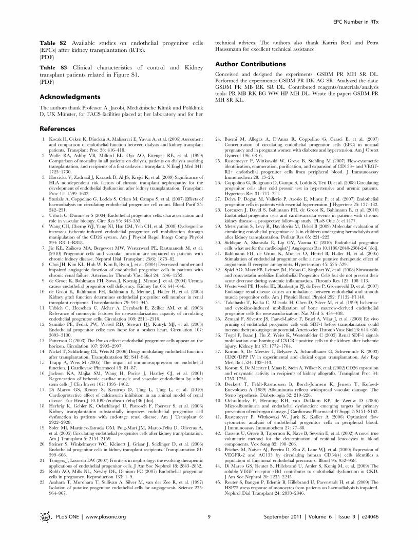
Table S2 Available studies on endothelial progenitor cells
(EPCs) after kidney transplantation (RTx).
(PDF)
Table S3 Clinical characteristics of control and Kidney
transplant patients related in Figure S1.
(PDF)
Acknowledgments
The authors thank Professor A. Jacobi, Medizinische Klinik und Poliklinik
D, UK Munster, for FACS facilities placed at her laboratory and for her
technical advices. The authors also thank Katrin Beul and Petra
Haussmann for excellent technical assistance.
Author Contributions
Conceived and designed the experiments: GSDM PR MH SR DL.
Performed the experiments: GSDM PR DK AG SR. Analyzed the data:
GSDM PR MB RK SR DL. Contributed reagents/materials/analysis
tools: PR MB RK BG WW HP MH DL. Wrote the paper: GSDM PR
MH SR KL.
References
1. Kocak H, Ceken K, Dinckan A, Mahsereci E, Yavuz A, et al. (2006) Assessment
and comparison of endothelial function between dialysis and kidney transplant
patients. Transplant Proc 38: 416–418.2. Wolfe RA, Ashby VB, Milford EL, Ojo AO, Ettenger RE, et al. (1999)
Comparison of mortality in all patients on dialysis, patients on dialysis awaitingtransplantation, and recipients of a first cadaveric transplant. N Engl J Med 341:
1725–1730.
3. Horcicka V, Zadrazil J, Karasek D, Al JS, Krejci K, et al. (2009) Significance ofHLA nondependent risk factors of chronic transplant nephropathy for the
development of endothelial dysfunction after kidney transplantation. TransplantProc 41: 1599–1603.
4. Sturiale A, Coppolino G, Loddo S, Criseo M, Campo S, et al. (2007) Effects ofhaemodialysis on circulating endothelial progenitor cell count. Blood Purif 25:
242–251.
5. Urbich C, Dimmeler S (2004) Endothelial progenitor cells: characterization androle in vascular biology. Circ Res 95: 343–353.
6. Wang CH, Cherng WJ, Yang NI, Hsu CM, Yeh CH, et al. (2008) Cyclosporineincreases ischemia-induced endothelial progenitor cell mobilization through
manipulation of the CD26 system. Am J Physiol Regul Integr Comp Physiol
294: R811–R818.7. Jie KE, Zaikova MA, Bergevoet MW, Westerweel PE, Rastmanesh M, et al.
(2010) Progenitor cells and vascular function are impaired in patients withchronic kidney disease. Nephrol Dial Transplant 25(6): 1875–82.
8. Choi JH, Kim KL, Huh W, Kim B, Byun J, et al. (2004) Decreased number andimpaired angiogenic function of endothelial progenitor cells in patients with
chronic renal failure. Arterioscler Thromb Vasc Biol 24: 1246–1252.
9. de Groot K, Bahlmann FH, Sowa J, Koenig J, Menne J, et al. (2004) Uremiacauses endothelial progenitor cell deficiency. Kidney Int 66: 641–646.
10. de Groot K, Bahlmann FH, Bahlmann E, Menne J, Haller H, et al. (2005)Kidney graft function determines endothelial progenitor cell number in renal
transplant recipients. Transplantation 79: 941–945.
11. Urbich C, Heeschen C, Aicher A, Dernbach E, Zeiher AM, et al. (2003)Relevance of monocytic features for neovascularization capacity of circulating
endothelial progenitor cells. Circulation 108: 2511–2516.12. Szmitko PE, Fedak PW, Weisel RD, Stewart DJ, Kutryk MJ, et al. (2003)
Endothelial progenitor cells: new hope for a broken heart. Circulation 107:3093–3100.
13. Patterson C (2003) The Ponzo effect: endothelial progenitor cells appear on the
horizon. Circulation 107: 2995–2997.14. Nickel T, Schlichting CL, Weis M (2006) Drugs modulating endothelial function
after transplantation. Transplantation 82: S41–S46.15. Trapp A, Weis M (2005) The impact of immunosuppression on endothelial
function. J Cardiovasc Pharmacol 45: 81–87.
16. Jackson KA, Majka SM, Wang H, Pocius J, Hartley CJ, et al. (2001)Regeneration of ischemic cardiac muscle and vascular endothelium by adult
stem cells. J Clin Invest 107: 1395–1402.17. Di Marco GS, Reuter S, Kentrup D, Ting L, Ting L, et al. (2010)
Cardioprotective effect of calcineurin inhibition in an animal model of renal
disease. Eur Heart J 10.1093/eurheartj/ehq436 [doi].18. Herbrig K, Gebler K, Oelschlaegel U, Pistrosch F, Foerster S, et al. (2006)
Kidney transplantation substantially improves endothelial progenitor celldysfunction in patients with end-stage renal disease. Am J Transplant 6:
2922–2928.19. Soler MJ, Martinez-Estrada OM, Puig-Mari JM, Marco-Feliu D, Oliveras A,
et al. (2005) Circulating endothelial progenitor cells after kidney transplantation.
Am J Transplant 5: 2154–2159.20. Steiner S, Winkelmayer WC, Kleinert J, Grisar J, Seidinger D, et al. (2006)
Endothelial progenitor cells in kidney transplant recipients. Transplantation 81:599–606.
21. Tongers J, Losordo DW (2007) Frontiers in nephrology: the evolving therapeutic
applications of endothelial progenitor cells. J Am Soc Nephrol 18: 2843–2852.22. Robb AO, Mills NL, Newby DE, Denison FC (2007) Endothelial progenitor
cells in pregnancy. Reproduction 133: 1–9.23. Asahara T, Murohara T, Sullivan A, Silver M, van der Zee R, et al. (1997)
Isolation of putative progenitor endothelial cells for angiogenesis. Science 275:964–967.
24. Buemi M, Allegra A, D’Anna R, Coppolino G, Crasci E, et al. (2007)
Concentration of circulating endothelial progenitor cells (EPC) in normal
pregnancy and in pregnant women with diabetes and hypertension. Am J ObstetGynecol 196: 68–6.
25. Rustemeyer P, Wittkowski W, Greve B, Stehling M (2007) Flow-cytometricidentification, enumeration, purification, and expansion of CD133+ and VEGF-
R2+ endothelial progenitor cells from peripheral blood. J Immunoassay
Immunochem 28: 13–23.26. Coppolino G, Bolignano D, Campo S, Loddo S, Teti D, et al. (2008) Circulating
progenitor cells after cold pressor test in hypertensive and uremic patients.Hypertens Res 31: 717–724.
27. Delva P, Degan M, Vallerio P, Arosio E, Minuz P, et al. (2007) Endothelialprogenitor cells in patients with essential hypertension. J Hypertens 25: 127–132.
28. Lorenzen J, David S, Bahlmann FH, de Groot K, Bahlmann E, et al. (2010)
Endothelial progenitor cells and cardiovascular events in patients with chronickidney disease–a prospective follow-up study. PLoS One 5: e11477.
29. Metsuyanim S, Levy R, Davidovits M, Dekel B (2009) Molecular evaluation ofcirculating endothelial progenitor cells in children undergoing hemodialysis and
after kidney transplantation. Pediatr Res 65: 221–225.
30. Siddique A, Shantsila E, Lip GY, Varma C (2010) Endothelial progenitorcells: what use for the cardiologist? J Angiogenes Res 10.1186/2040-2384-2-6 [doi].
31. Bahlmann FH, de Groot K, Mueller O, Hertel B, Haller H, et al. (2005)Stimulation of endothelial progenitor cells: a new putative therapeutic effect of
angiotensin II receptor antagonists. Hypertension 45: 526–529.32. Spiel AO, Mayr FB, Leitner JM, Firbas C, Sieghart W, et al. (2008) Simvastatin
and rosuvastatin mobilize Endothelial Progenitor Cells but do not prevent their
acute decrease during systemic inflammation. Thromb Res 123: 108–113.33. Westerweel PE, Hoefer IE, Blankestijn PJ, de Bree P, Groeneveld D, et al. (2007)
End-stage renal disease causes an imbalance between endothelial and smoothmuscle progenitor cells. Am J Physiol Renal Physiol 292: F1132–F1140.
34. Takahashi T, Kalka C, Masuda H, Chen D, Silver M, et al. (1999) Ischemia-
and cytokine-induced mobilization of bone marrow-derived endothelialprogenitor cells for neovascularization. Nat Med 5: 434–438.
35. Zemani F, Silvestre JS, Fauvel-Lafeve F, Bruel A, Vilar J, et al. (2008) Ex vivopriming of endothelial progenitor cells with SDF-1 before transplantation could
increase their proangiogenic potential. Arterioscler Thromb Vasc Biol 28: 644–650.36. Togel F, Isaac J, Hu Z, Weiss K, Westenfelder C (2005) Renal SDF-1 signals
mobilization and homing of CXCR4-positive cells to the kidney after ischemic
injury. Kidney Int 67: 1772–1784.37. Korom S, De Meester I, Belyaev A, Schmidbauer G, Schwemmle K (2003)
CD26/DPP IV in experimental and clinical organ transplantation. Adv ExpMed Biol 524: 133–143.
38. Korom S, De Meester I, Maas E, Stein A, Wilker S, et al. (2002) CD26 expression
and enzymatic activity in recipients of kidney allografts. Transplant Proc 34:1753–1754.
39. Deckert T, Feldt-Rasmussen B, Borch-Johnsen K, Jensen T, Kofoed-Enevoldsen A (1989) Albuminuria reflects widespread vascular damage. The
Steno hypothesis. Diabetologia 32: 219–226.
40. Ochodnicky P, Henning RH, van Dokkum RP, de Zeeuw D (2006)Microalbuminuria and endothelial dysfunction: emerging targets for primary
prevention of end-organ damage. J Cardiovasc Pharmacol 47 Suppl 2: S151–S162.41. Rustemeyer P, Wittkowski W, Jurk K, Koller A (2006) Optimized flow
cytometric analysis of endothelial progenitor cells in peripheral blood.J Immunoassay Immunochem 27: 77–88.
42. Cassens U, Greve B, Tapernon K, Nave B, Severin E, et al. (2002) A novel true
volumetric method for the determination of residual leucocytes in bloodcomponents. Vox Sang 82: 198–206.
43. Peichev M, Naiyer AJ, Pereira D, Zhu Z, Lane WJ, et al. (2000) Expression ofVEGFR-2 and AC133 by circulating human CD34(+) cells identifies a
population of functional endothelial precursors. Blood 95: 952–958.
44. Di Marco GS, Reuter S, Hillebrand U, Amler S, Konig M, et al. (2009) Thesoluble VEGF receptor sFlt1 contributes to endothelial dysfunction in CKD.
J Am Soc Nephrol 20: 2235–2245.45. Reuter S, Bangen P, Edemir B, Hillebrand U, Pavenstadt H, et al. (2009) The
HSP72 stress response of monocytes from patients on haemodialysis is impaired.Nephrol Dial Transplant 24: 2838–2846.
EPC Number in RTx
PLoS ONE | www.plosone.org 9 September 2011 | Volume 6 | Issue 9 | e24046