doi:10.1182/blood-2006-12-062471 Prepublished online February 27, 2007; Juan M Melero-Martin, Zia A Khan, Arnaud Picard, Xiao Wu, Sailaja Paruchuri and Joyce Bischoff progenitor cells In vivo vasculogenic potential of human blood-derived endothelial (166 articles) Stem Cells in Hematology (2494 articles) Hemostasis, Thrombosis, and Vascular Biology (3198 articles) Hematopoiesis and Stem Cells Articles on similar topics can be found in the following Blood collections http://bloodjournal.hematologylibrary.org/site/misc/rights.xhtml#repub_requests Information about reproducing this article in parts or in its entirety may be found online at: http://bloodjournal.hematologylibrary.org/site/misc/rights.xhtml#reprints Information about ordering reprints may be found online at: http://bloodjournal.hematologylibrary.org/site/subscriptions/index.xhtml Information about subscriptions and ASH membership may be found online at: digital object identifier (DOIs) and date of initial publication. the indexed by PubMed from initial publication. Citations to Advance online articles must include final publication). Advance online articles are citable and establish publication priority; they are appeared in the paper journal (edited, typeset versions may be posted when available prior to Advance online articles have been peer reviewed and accepted for publication but have not yet Copyright 2011 by The American Society of Hematology; all rights reserved. 20036. the American Society of Hematology, 2021 L St, NW, Suite 900, Washington DC Blood (print ISSN 0006-4971, online ISSN 1528-0020), is published weekly by For personal use only. by guest on January 24, 2014. bloodjournal.hematologylibrary.org From For personal use only. by guest on January 24, 2014. bloodjournal.hematologylibrary.org From

Welcome message from author
This document is posted to help you gain knowledge. Please leave a comment to let me know what you think about it! Share it to your friends and learn new things together.
Transcript

doi:10.1182/blood-2006-12-062471Prepublished online February 27, 2007;
Juan M Melero-Martin, Zia A Khan, Arnaud Picard, Xiao Wu, Sailaja Paruchuri and Joyce Bischoff progenitor cellsIn vivo vasculogenic potential of human blood-derived endothelial
(166 articles)Stem Cells in Hematology � (2494 articles)Hemostasis, Thrombosis, and Vascular Biology �
(3198 articles)Hematopoiesis and Stem Cells �Articles on similar topics can be found in the following Blood collections
http://bloodjournal.hematologylibrary.org/site/misc/rights.xhtml#repub_requestsInformation about reproducing this article in parts or in its entirety may be found online at:
http://bloodjournal.hematologylibrary.org/site/misc/rights.xhtml#reprintsInformation about ordering reprints may be found online at:
http://bloodjournal.hematologylibrary.org/site/subscriptions/index.xhtmlInformation about subscriptions and ASH membership may be found online at:
digital object identifier (DOIs) and date of initial publication. theindexed by PubMed from initial publication. Citations to Advance online articles must include
final publication). Advance online articles are citable and establish publication priority; they areappeared in the paper journal (edited, typeset versions may be posted when available prior to Advance online articles have been peer reviewed and accepted for publication but have not yet
Copyright 2011 by The American Society of Hematology; all rights reserved.20036.the American Society of Hematology, 2021 L St, NW, Suite 900, Washington DC Blood (print ISSN 0006-4971, online ISSN 1528-0020), is published weekly by
For personal use only. by guest on January 24, 2014. bloodjournal.hematologylibrary.orgFrom For personal use only. by guest on January 24, 2014. bloodjournal.hematologylibrary.orgFrom
1
In vivo vasculogenic potential of human blood-derived endothelial
progenitor cells
Juan M. Melero-Martin, Zia A. Khan, Arnaud Picard, Xiao Wu, Sailaja Paruchuri
and Joyce Bischoff
Vascular Biology Program and Department of Surgery, Children's Hospital
Boston, Harvard Medical School, MA, USA
Short title: Vasculogenic potential of blood-derived EPCs
Keywords: VASCULAR BIOLOGY, Endothelial cells; blood vessels; endothelial
progenitor cells; tissue-engineering; vasculogenesis
Corresponding Author:
Dr. Joyce Bischoff
Vascular Biology Research Program and Department of Surgery
Children’s Hospital Boston
Harvard Medical School
Boston, MA 02115
Tel.: (617) 919-2192
Fax: (617) 730-0231
Email: [email protected]
Blood First Edition Paper, prepublished online February 27, 2007; DOI 10.1182/blood-2006-12-062471
Copyright © 2007 American Society of Hematology
For personal use only. by guest on January 24, 2014. bloodjournal.hematologylibrary.orgFrom
2
Abstract
Vascularization of tissues is a major challenge of tissue-engineering (TE). We
hypothesize that blood-derived endothelial progenitor cells (EPCs) have the
required proliferative and vasculogenic activity to create vascular networks in
vivo. To test this, EPCs isolated from human umbilical cord blood or from adult
peripheral blood, and human saphenous vein smooth muscle cells (HSVSMCs)
as a source of perivascular cells, were combined in Matrigel and implanted
subcutaneously into immunodeficient mice. Evaluation of implants at one week
revealed an extensive network of human-specific lumenal structures containing
erythrocytes, indicating formation of functional anastomoses with the host
vasculature. Quantitative analyses showed the microvessel density was
significantly superior to that generated by human dermal microvascular
endothelial cells (HDMECs) but similar to that generated by human umbilical vein
ECs. We also found that as EPCs were expanded in culture, their morphology,
growth kinetics and proliferative responses toward angiogenic factors
progressively resembled those of HDMECs, indicating a process of in vitro
maturation. This maturation correlated with a decrease in the degree of
vascularization in vivo, which could be compensated by increasing the number of
EPCs seeded into the implants. Our findings strongly support the use of human
EPCs to form vascular networks in engineered organs and tissues.
For personal use only. by guest on January 24, 2014. bloodjournal.hematologylibrary.orgFrom
3
INTRODUCTION
TE holds promise as a new approach for creating replacement tissue to repair
congenital defects or diseased tissue 1. One strategy is to seed the appropriate
cells on a biodegradable scaffold engineered with the desired mechanical
properties, followed by stimulation of cell growth and differentiation in vitro, such
that, on implantation in vivo, the engineered construct undergoes remodeling and
maturation into functional tissue 2. Examples of this approach include blood
vessels and cardiovascular substitutes, where autologous vascular cells have
been used for this purpose without immune rejection 3-5. Despite advances in this
field, TE still faces important constraints. There are no TE constructs presently
available that have an inherent microvascular bed ready to be connected to the
host vascular system. Consequently, tissues implanted with a volume greater
than 2 to 3 mm3 cannot obtain appropriate provision of nutrients, gas exchange,
and elimination of waste products since all these mechanisms are limited by the
diffusion distance 6. To overcome the problem of vascularization, strategies such
as embedding angiogenic factors into the scaffold to promote ingrowth of
microvessels, fabrication technologies to create polymers containing vessel-like
networks, and pre-vascularization of matrices prior to cell seeding have been
proposed 7-11.
The need for prefabricated channels or growth factor-induced angiogenesis could
be avoided by exploiting the inherent vasculogenic ability of endothelial cells
(ECs). Using human umbilical vein ECs (HUVECs), microvascular networks in
collagen/fibronectin gels were formed within 31 days of implantation into
immunodeficient mice 12. Similar results have been reported with human
microvascular ECs (HDMECs) seeded on biopolymer matrices, where functional
microvessels were evident 7-10 days after implantation into mice 13.
Nevertheless, the clinical use of mature ECs derived from autologous vascular
tissue presents some important limitations: 1) the isolation relies on an invasive
For personal use only. by guest on January 24, 2014. bloodjournal.hematologylibrary.orgFrom
4
procedure, 2) mature ECs cells show relatively low proliferative potential, and 3)
the difficulty of obtaining a sufficient number of cells from a small biopsy of
autologous tissue. These limitations have instigated the search for other sources
of ECs with more proliferative and vasculogenic activities such as those derived
from both embryonic and adult stem and progenitor cells 14. One recent example
showed how seeding of endothelial cells derived from embryonic stem cells
along with myoblast and embryonic fibroblasts resulted in the formation of
skeletal muscle tissue 15. However, ethical considerations along with a poor
understanding of the mechanisms controlling the differentiation of embryonic
stem cells are hurdles that need to be overcome before these cells can be used
in a clinical setting.
For clinical applications, the presence of endothelial progenitor cells (EPCs) in
circulation represents a promising opportunity to non-invasively obtain the
required endothelial population 16. In previous work we showed the creation of
microvascular networks in vitro using biodegradable scaffolds seeded with EPCs
that had been isolated from human umbilical cord blood and expanded in vitro as
mature ECs 17. Using a sheep model, we also showed that blood-derived EPCs
could endothelialize small-diameter blood vessels 5. We now propose that human
blood-derived EPCs constitute a robust source of ECs with the potential to form
functional capillary networks in vivo. To test this, we used a xenograft model
where human cells were mixed in Matrigel and implanted subcutaneously into
immunodeficient mice. Our goal was to advance feasibility studies by evaluating
the ease with which highly purified and phenotypically defined human EPCs can
create microvascular structures that form functional anastomoses with the host
vasculature.
MATERIAL AND METHODS
Isolation and culture of blood-derived EPCs
For personal use only. by guest on January 24, 2014. bloodjournal.hematologylibrary.orgFrom
5
Human umbilical cord blood was obtained from the Brigham and Women’s
Hospital in accordance with an Institutional Review Board-approved protocol.
Adult peripheral blood was collected from volunteer donors in accordance with a
protocol approved by Children’s Hospital Boston Committee on Clinical
Investigation and was obtained with informed consent according to the
Declaration of Helsinki and under a protocol approved by the Committee on
Clinical Investigation. Both cord blood-derived EPCs (cbEPCs) and adult
peripheral blood-derived EPCs were obtained from the mononuclear cell (MNC)
fractions similarly to other authors 18-20. MNCs were seeded on 1% gelatin-coated
tissue culture plates using Endothelial Basal Medium (EBM-2) supplemented
with SingleQuots (except for hydrocortisone) (Cambrex BioScience, Walkersville,
MD), 20% FBS (Hyclone, Logan, UT), 1x glutamine-penicillin-streptomycin (GPS;
Invitrogen, Carlsbad, CA) and 15% autologous plasma 17. Unbound cells were
removed at 48 hours for cord blood and at 4 days for adult blood. In both cases,
the bound cell fraction was then maintained in culture using EBM-2
supplemented with 20% FBS, SingleQuots (except for hydrocortisone) and 1x
GPS (this medium is referred to as EBM-2/20%). Colonies of endothelial-like
cells were allowed to grow until confluence, trypsinized and purified using CD31-
coated magnetic beads (Dynal Biotech, Brown Deer, WI) (see Supplemental
Figures 1 and 5). CD31-selected EPCs were serially passaged and cultured on
fibronectin-coated (FN; 1 ug/cm2; Chemicon International, Temecula, CA) plates
at 5x10³ cell/cm2 in EBM-2/20%. HDMECs from newborn foreskin cultured in the
same condition as cbEPCs were used as positive controls 21. Human saphenous
vein smooth muscle cells (HSVSMCs) grown in DMEM (Invitrogen), 10% FBS, 1x
GPS and 1x Non essential amino acids (Sigma-Aldrich, St. Louis, MO) were
used as negative controls for endothelial phenotype.
Phenotypic characterization of cbEPCs
Methods for flow cytometry, indirect immunofluorescence and RT-PCR are
described in the on-line Supplement.
For personal use only. by guest on January 24, 2014. bloodjournal.hematologylibrary.orgFrom
6
In vitro maturation of cbEPCs
Expansion potential of cbEPCs
cbEPCs and adult EPCs, isolated as described above, were expanded for 112
and 60 days, respectively. All passages were performed by plating the cells onto
1 µg/cm2 FN-coated tissue culture plates at 5x10³ cell/cm2 using EBM-2/20%.
Medium was refreshed every 2-3 days and cells were harvested by trypsinization
and re-plated in the same culture conditions for the next passage. Cumulative
values of total cell number were calculated by counting the cells at the end of
each passage using a haemocytometer.
Growth kinetics assay
Growth curves of cbEPCs were evaluated at different passages. Cells were
plated in triplicates onto 1 µg/cm2 FN-coated 24-well tissue culture plates at
5x10³ cell/cm2 in 0.5 ml of EBM-2/20%. Medium was refreshed every two days
and cell numbers evaluated at 24 hour intervals for 7 days by counting the cells
after trypsinization using a haemocytometer. Doubling time profiles were
calculated from the mean values obtained from each growth curve at different
passages 22.
Cell size measurements
Morphological differences of cbEPCs were evaluated at different passages.
Confluent cell monolayers were immunostained with VE-cadherin antibody for
cell surface and DAPI for nuclear visualization as described above. The areas
occupied by cell bodies and cell nuclei were measured by analysis (ImageJ
software, NIH) of the images obtained from randomly selected fields from three
separate cultures after immunostaining. All values were normalized to the value
of total cell area.
Proliferation assay
Cells were seeded in triplicates onto 1 µg/cm2 FN-coated 24-well plates at 5x10³
cell/cm2 using EMB-2 supplemented with 5% FBS and 1x GPS (control medium);
For personal use only. by guest on January 24, 2014. bloodjournal.hematologylibrary.orgFrom
7
plating efficiency was determined at 24 hours, then cells were treated for 48
hours using control medium in the presence or absent of either 10 ng/ml of
VEGF-A (R&D Systems) or 1 ng/ml bFGF (Roche Applied Science, Indianapolis,
IN). Cells were trypsinized and counted using a haemocytometer. Values were
normalized to the cell numbers determined at 24 hours.
In vivo vasculogenesis experiments
Matrigel implantations
Unless otherwise indicated, 1.5x10⁶ EPCs were mixed with 0.375x10⁶ HSVSMCs
(4:1 ratio) and resuspended in 200 µl of Phenol Red-free Matrigel (BD
Bioscience, San Jose, CA) on ice. The mixture was implanted on the back of a
six-week-old male athymic nu/nu mouse (Charles River Laboratories, Boston,
MA) by subcutaneous injection using a 25-gauge needle. One implant was
injected per mouse. Each experimental condition was performed with 4 mice.
Histology and immunohistochemistry
Matrigel implants were removed at one week after xenografting, fixed in 10%
buffered formalin overnight, embedded in paraffin, and sectioned. Hematoxylin
and eosin (H&E) stained 7 µm-thick sections were examined for the presence of
lumenal structures containing red blood cells. For immunohistochemistry, 7-µm-
thick sections were deparaffinized, blocked for 30 minutes in 5% horse serum,
and incubated with human-specific CD31 monoclonal antibody (1:50,
DakoCytomation), anti-human α-SMA (1:750, Sigma-Aldrich), or mouse IgG
(DakoCytomation) for 1 hour at room temperature. Horseradish peroxidase-
conjugated secondary antibody and 3,3’-diaminobenzidine (DAB) were used for
detection. The sections were counterstained with hematoxilin and mounted using
Permount (Fisher Scientific).
Microvessel density analysis
Microvessels were detected by the evaluation of H&E stained sections taken
from the middle part of the implants. The full area of each individual section was
For personal use only. by guest on January 24, 2014. bloodjournal.hematologylibrary.orgFrom
8
evaluated. Microvessels were identified and counted as lumenal structures
containing red blood cells. The area of each section was estimated by image
analysis. Microvessels density was calculated by dividing the total number of red
blood cell-filled microvessels by the area of each section (expressed as
vessels/mm2). Values reported for each experimental condition correspond to the
average values obtained from four individual animals.
Statistical analysis
The data were expressed as means ± SD. Where appropriate, data were
analyzed by analysis of variance (ANOVA) followed by two-tailed Student’s
unpaired t-tests. P value < 0.05 was considered to indicate a statistically
significant difference.
RESULTS
Phenotypic characterization of cbEPCs
We isolated EPCs from the MNC fraction of human umbilical cord blood samples
(n=19) similarly to other authors 18,19. Cord blood-derived endothelial colonies
(identified by typical cobblestone morphology) emerged in culture after one week
(see Supplemental Figure 1). The size, frequency, and time of appearance of
these colonies varied as already reported 18 (data not shown). Endothelial
colonies were left to grow in the original culture plates until confluence and
purified thereafter (at passage 1) by selection of CD31-positive cells
(Supplemental Figure 1). This procedure resulted in superior cell yields
compared to our previous isolation protocol based on double selection of
CD34+/CD133+ cells from the MNC fraction 17. However, since CD31 is not a
specific marker of EPCs and due to the heterogeneity of blood preparations, both
phenotypical and functional characterization were performed. This was especially
important considering that earlier studies have shown that some EPC colonies
isolated from MNCs contain cells that express the hematopoietic-specific cell-
For personal use only. by guest on January 24, 2014. bloodjournal.hematologylibrary.orgFrom
9
surface antigen CD45 14,23,24 , raising questions about the cellular origin of
circulating EPCs.
The endothelial phenotype of the isolated cbEPCs was confirmed by different
methods. Flow cytometric analysis of cbEPCs showed remarkably uniform
expression of EC markers CD34, VEGF-R2, CD146, CD31, vWF and CD105
(Figure 1a). In addition, cells were negative for mesenchymal marker CD90 and
hematopoietic markers CD45 and CD14, confirming that the cells were not
contaminated with either mesenchymal or hematopoietic cells. Additionally, RT-
PCR analyses showed the expression of EC markers CD34, VEGF-R2, CD31,
VE-cadherin, vWF and eNOS at the mRNA level (Figure 1b). Indirect
immunofluorescent staining was performed to further examine the expression of
EC markers. The results showed that cbEPCs expressed CD31, VE-cadherin
and vWF (Figure 1c). Importantly, the localization of CD31 and VE-cadherin at
the cell-cell borders and vWF in a punctuate pattern in the cytoplasm showed
clear indications of EC properties.
In addition, we tested whether cbEPCs were able to up-regulate leukocyte
adhesion molecules in response to the inflammatory cytokine TNF-α. The low-to-
undetectable levels of E-selectin, ICAM-1 and VCAM-1 in the untreated cbEPC
cultures were up-regulated upon 5 hour incubation with TNF-α (Figure 1d). This
response to an inflammatory cytokine is characteristic of ECs and suggests that
the use of cbEPC in the formation of microvascular vessels could also provide
physiologic proinflammatory properties.
In summary, this combination of analyses provides a definitive demonstration
that the cells isolated from umbilical cord blood were ECs and discards the
possibility of hematopoietic/monocytic cells in the culture 20. Based on the
isolation methodology and the phenotypical characteristics, our isolated cells are
similar to those referred to by other authors as late-EPCs or endothelial
For personal use only. by guest on January 24, 2014. bloodjournal.hematologylibrary.orgFrom
10
outgrowth cells 19,24. The characterization depicted in Figure 1 corresponded to
cbEPCs at passage 6. However, a detailed characterization was performed at
passages 4, 9, 12 and 15 with similar results (Supplemental Figure 2, panels a-
d), indicating a stable endothelial phenotype through long term culture.
Furthermore, we provide additional characterization of the cbEPCs at passage 6
(Supplemental Figure 3) to show that cbEPCs express two other VEGF-
receptors, neuropilin-1 and Flt-1 (panel a), and that the cbEPCs do not express
the smooth muscle/mesenchymal cell markers PDGF-Rβ (panel b), α-SMA, or
calponin (panel c).
In vivo vasculogenic potential of cbEPCs
Our previous work showed the creation of microvascular network in vitro by
culturing cbEPCs and HSVSMCs on biodegradable scaffolds 17. To answer the
question of whether cbEPCs were capable of forming functional capillary
networks in vivo, we implanted cbEPCs in Matrigel subcutaneously into nude
mice for one week. For this experiment, 1.5x10⁶ of cbEPCs (passage 6) were
combined with 0.375x10⁶ HSVSMCs in 200 µl of Matrigel, resulting in a ratio of
cbEPCs to HSVSMCs of 4 to 1, and injected subcutaneously. This ratio of
cbEPCs to HSVSMCs was less than the 1:1 ratio previously used 17, with the
intention to minimize the contribution of smooth muscle cells. After harvesting the
Matrigel implants, H&E staining revealed the presence of lumenal structures
containing murine erythrocytes throughout the implants (Figure 2a). Similar
results were obtained with cbEPCs isolated from three different cord blood
samples, yielding an average of 47.5 ± 8 microvessels/mm2 (data not shown).
Importantly, implants with either cbEPCs or HSVSMCs alone failed to form any
detectable microvessels after one week (Figures 2b and 2c). Injections of
Matrigel alone resulted in the appearance of few host cells infiltrated into the
borders of the implants (Figure 2d), indicating that Matrigel itself was not
responsible for the presence of vascular structures within the implants.
For personal use only. by guest on January 24, 2014. bloodjournal.hematologylibrary.orgFrom
11
To further characterize the microvascular structures detected, sections of the
implant were immunohistochemically stained using a human-specific CD31
antibody. As depicted in Figure 2k, nearly all of the lumenal structures stained
positive for human CD31, confirming that those lumens were formed by the
implanted human cbEPCs and not by the host cells. This result was important
because it demonstrated that the formation of microvascular vessels within the
implant is the result of a process of in vivo vasculogenesis carried out by the
implanted cells and it is not due to blood vessel invasion and sprouting, i.e., an
angiogenic response from nearby host vasculature. The specificity of the anti-
human CD31 antibody 25,26 was confirmed by the negative reaction obtained
when mouse lung tissue sections were stained in parallel (Supplemental Figure
4). Taken together, the human endothelial identity of the lumenal structures
(Figure 2k) and the presence of murine erythrocytes within those structures
(Figure 2a,2h and 2j), it was evident that vasculogenesis occurred and, in
addition, the newly created microvessels formed functional anastomoses with the
host circulatory system. Next, the time course of vasculogenesis in the Matrigel
was analyzed by harvesting implants at 2, 4 and 7 days after xenografting. At 2
days, a low degree of cellular organization was seen (Figure 2e). At 4 days, a
high degree of organization with clear alignment of cells throughout the implant
was observed, suggesting formation of cellular cords (Figure 2f). The presence of
functional microvascular vessels, defined by the presence of red blood cells
within the lumen, was appreciable one week after implantation (Figure 2g, h and
j).
The location of the HSVSMCs was also examined by immunohistochemical
staining using anti-α-SMA. Smooth muscle cells were detected both around the
lumenal structures and throughout the Matrigel implants (Figure 2l), suggesting
an ongoing process of vessel maturation and stabilization 27-29. However, the α-
SMA antibody is not human-specific, as shown by the positive staining of control
tissue sections obtained from mouse lung (Supplemental Figure 4). Therefore,
For personal use only. by guest on January 24, 2014. bloodjournal.hematologylibrary.orgFrom
12
the observed α-SMA positive cells could corresponded to the implanted
HSVSMCs or murine cells recruited from the host, or a combination of these.
Maturation of cbEPC during in vitro expansion
cbEPCs were serially passaged to determine their expansion potential.
Remarkably, 1014 cells could theoretically be obtained after only 40 days in
culture, and thereafter cells were expanded up to 70 population doublings (Figure
3a), which is consistent with previous studies 18. Significant expansion of adult
blood EPCs (108 cells) was also achieved under the same conditions using 50
milliliters of adult peripheral blood (Figure 3a). In addition to this enormous
proliferative capacity, cbEPCs expressed and maintained a definitive endothelial
phenotype in vitro as shown in Figure 1. However, neither the expansion
potential nor the phenotypical stability rules out the possibility of cbEPCs
undergoing cellular changes during their expansion in vitro. To investigate
potential changes, the growth kinetics of cbEPCs at different passages were
examined by the generation of growth curves (Figure 3b). We found that cells
from earlier passages presented superior growth kinetics and reached higher cell
densities at confluence. The former was confirmed by the generation of the
doubling time profiles (Figure 3c), where lower passage number corresponded
with shorter doubling times. The u-shape of these profiles is the result of
mechanisms controlling cell growth in vitro: longer doubling times were found
during both the early and late stages of the culture corresponding to the initial lag
phase and the inhibition of cell growth by cell-cell contacts, respectively. Taking
the minimum values as representative of the dividing capacity, cbEPCs
presented minimum doubling times of 14, 17, 18, 29 and 35 hours at passages 4,
6, 9, 12, and 15 respectively. These results illustrated the remarkable dividing
capacity of cbEPCs at low passage numbers, and showed that as cbEPCs were
expanded in vitro, their growth kinetics progressively slowed.
For personal use only. by guest on January 24, 2014. bloodjournal.hematologylibrary.orgFrom
13
Serially passaging of cbEPCs also resulted in evident morphological differences.
As they were expanded, cells progressively occupied larger areas in culture
(Figure 3d). While the areas occupied by the cell nuclei remained constant at
each passage, cbEPCs were found to be significantly (P < .05) smaller than the
control HDMECs, with the exception of passage 15. As cbEPCs were expanded
in vitro, the average area occupied by the cells increased towards that of
HDMECs. The mean area of cbEPCs ranged from values 75% smaller than
HDMECs at passage 4 to 17% smaller at passage 15. These results were
consistent with the differences found in cell density at confluence (Figure 3b).
We next compared the proliferative response of cbEPCs at different passages to
stimulation by angiogenic factors VEGF or bFGF (Figure 4). We found that both
angiogenic factors produced a proliferative response in all the cases evaluated
as compared to basal proliferation in the presence of 5% serum (control). The
response was statistically significant (P < .05) in all the groups treated with
bFGF. Interestingly, the proliferative response to bFGF was progressively
reduced as passage number increased, and ranged from 5.4-fold at passage 4 to
2-fold at passage 15. When compared to HDMECs, the response toward bFGF
was found significantly higher in cbEPCs at passages 4, 6 and 9, but not in the
later passages. In the case of VEGF treatment, the response was statistically
significant (P < .05) at passages 4 and 6 as compared to basal proliferation.
Again, the proliferative response was progressively reduced as passage number
increased, and varied from 3.1-fold in the earliest passage to 1.3-fold in the latest
passage group. Collectively, these in vitro experiments demonstrate that despite
the consistent and stable expression of endothelial markers, cbEPCs undergo
cellular and functional changes as they are expanded in culture. Their
morphology, growth kinetics and proliferative responses toward angiogenic
growth factors progressively resembled those of HDMECs, indicating a process
of in vitro cell maturation over time. We showed previously that proliferative
For personal use only. by guest on January 24, 2014. bloodjournal.hematologylibrary.orgFrom
14
responses of HDMECs isolated in our laboratory do not change from passage 3-
12 21.
Effect of in vitro expansion of cbEPCs on in vivo vasculogenesis
We next tested whether the maturation of cbEPCs observed during their
expansion in vitro has any effect on their vasculogenic ability in vivo. To answer
this question, cbEPCs at different passages (3, 6, and 12) were implanted
subcutaneously into nude mice in the presence of HSVSMCs. Examination after
one week of the H&E-stained implants (Figure 5a-d) revealed a difference in the
level of in vivo vascularization. Quantification of the red blood cell-containing
microvessels (Figure 5e) showed that the differences among the groups were
statistically significant (P < .05) in all the cases, with values ranging from 93 ± 18
vessels/mm2 when using cbEPCs at passage 3 to 11 ± 13 vessels/mm2 with
passage 12. These results show that expansion of the cell population in vitro has
indeed a significant impact in the subsequent performance in vivo. Parallel
evaluation using mature HDMECs also revealed the presence of 23 ± 19 vessels
/mm2. This number of microvessels was inferior to those generated by the
earliest passages of cbEPCs (passages 3 and 6), with values significantly higher
in the case of cbEPCs at passage 3. In contrast, HUVECs combined with
HSVSMCs formed 52 ±9 vessels/mm2 (data not shown), indicating a robust
vasculogenic potential from this source of ECs.
We tested whether the lower vasculogenic ability observed in expanded cbEPCs
could be compensated by increasing the initial number of EPCs seeded into the
implants. To evaluate this, we implanted either 0.5x10⁶ (referred to as x1/3),
1.5x10⁶ (x1) or 4.5x10⁶ (x3) cbEPCs at passages 6 and 12 (Figure 6) in the
presence of HSVSMCs at a constant 4:1 ratio. One week after xenografting,
examination of the H&E-stained implants (Figure 6a-f) revealed that an increase
in the number of cbEPCs resulted in a higher degree of in vivo vascularization.
Quantification of the microvessel densities (Figure 6j) showed that the
For personal use only. by guest on January 24, 2014. bloodjournal.hematologylibrary.orgFrom
15
differences among the groups of cbEPCs at passage 6 were statistically
significant (P < .05), with values ranging from 6 ± 7 vessels/mm2 to 117 ± 23
vessels/mm2 when using x1/3 or x3 respectively. Consistent with the previous
results (Figure 5), the values of microvessel density in implants of cbEPCs at
passage 6 were always higher than those at passage 12 when the same
numbers of cbEPCs were used; indeed no microvessels were detected with x1/3
passage 12 cells. Nevertheless, at passage 12, the partial loss of vasculogenic
potential was compensated by increasing the number of seeded cells. As seen
in Figure 6, panels d-f and j, by simply seeding the implants with 3 times higher
density of cbEPCs at passage 12, microvessel density was raised from 10 ± 6
vessels/mm2 (x1) to 46 ± 28 vessels/mm2 (x3). Furthermore, the microvessel
level achieved with x3 cells passage 12 cells was similar to the level achieved
with passage 6 cells at x1 (P=0.56).
We evaluated whether a similar approach (i.e., increasing the number of EPCs
seeded) would result in increased vasculogenesis when using EPCs isolated
from blood of adult volunteers (Supplemental Figure 5). To test this, we
implanted either 1.5x10⁶ (x1) or 4.5x10⁶ (x3) adult EPCs at passages 6 in the
presence of HSVSMCs (4:1 ratio). One week after xenografting, examination of
the H&E-stained sections (Figure 6h and 6i) and human CD31-specific
immunostaining (Figure 6g) revealed the presence of human microvessels
containing red blood cells in both cases. As occurred with cbEPCs, we found that
an increase in the number of adult EPCs resulted in a higher degree of in vivo
vascularization with values ranging from 8 ± 8 lumens/mm2 to 23 ± 4
lumens/mm2 when using x1 or x3 adult EPCs respectively. Quantification of the
microvessel densities (Figure 6j) showed that adult EPCs at x3 was similar to
cbEPC-P6 x1 (P=0.10) and cbEPC-P12 x3 (P=0.2). In summary, these in vivo
experiments clearly show that in addition to the cellular and functional changes
observed in vitro, the vasculogenic ability of expanded EPCs progressively
For personal use only. by guest on January 24, 2014. bloodjournal.hematologylibrary.orgFrom
16
diminished but that this effect can be compensated by increasing the number of
EPCs initially seeded in Matrigel.
DISCUSSION
Tissue vascularization is one of the major challenges to be addressed for the
therapeutic success of TE applications. Here, we show that blood-derived EPCs
have an inherent vasculogenic ability that can be exploited to create functional
microvascular networks in vivo. Implantation of EPCs with HSVSMCs resulted in
the formation of an extensive blood vessel network after one week. The presence
of human EC-lined lumens containing murine erythrocytes throughout the
implants indicated not only a process of vasculogenesis by the implanted cells,
but also the formation of functional anastomoses with the host circulatory system.
Our results are the first to demonstrate the in vivo vasculogenic potential of
blood-derived EPCs in a TE setting. Based on our results, we speculate that
microvascular networks could be formed in many types of TE constructs with use
of blood-derived EPCs.
Previous investigators have suggested the possibility of creating microvascular
networks using mature ECs derived from vascular tissue. Both HUVECs and
HDMECs, seeded into collagen/fibronectin gels and biopolymer matrices
respectively, were shown to form complex vascular structures perfused by the
host circulation after implantation into immunodeficient mice 12,13. Alternatively,
the use of fat-derived vessel fragments embedded into collagen gels was shown
to generate perfused microvessels in immunodeficient mice 30. Nevertheless, the
necessity of invasive procedures together with their limited proliferative and
vasculogenic ability represents important constraints for the clinical use of mature
ECs derived from autologous vascular tissue.
For personal use only. by guest on January 24, 2014. bloodjournal.hematologylibrary.orgFrom
17
For therapeutic applications, one critical requisite will be to isolate defined
populations of cells so that growth and differentiation can be controlled and
regulated during tissue development. Since first identified 16, EPCs have been
isolated from the MNC fraction of blood in numerous studies 5,18-20,24,31,32.
However, their phenotypical characterization has been often controversial 20. In
our study, we show that 1011 homogeneous EPCs can be obtained from 25-50 ml
of cord blood after 30 days in culture and 108 EPCs from 50 ml of adult
peripheral blood. These cell numbers are likely to exceed, in the case of cord
blood, and be sufficient, in the case of adult blood, what would be needed for
most TE applications. Hence, we foresee that adult blood will also be a feasible
source of EPCs for autologous TE and regenerative therapies. In summary, this
remarkable yield confirms peripheral blood as a robust source of EC for
autologous TE. Furthermore, our data demonstrate that these cells maintain both
the expression of endothelial markers and functions through prolonged periods in
culture.
As we previously demonstrated in vitro 17, the presence of vascular smooth
muscle cells in the implants was found to be critical. Seeding cbEPCs with
HSVSMCs dramatically enhanced the assembly of CD31+ cells into microvessel
structures. Although the mechanisms by which the implanted smooth muscle
cells facilitate the formation of vascular structures need to be further investigated,
this finding is consistent with the extensive literature on endothelium-smooth
muscle cells interactions in vascular development 27-29. In our implants, α-SMA
positive cells were detected both around the lumenal structures and throughout
the Matrigel, suggesting an ongoing process of vessel assembly, maturation and
stabilization 27,28. We are currently investigating alternative sources that could
provide autologous smooth muscle cells without the necessity of invasive
procedures.
For personal use only. by guest on January 24, 2014. bloodjournal.hematologylibrary.orgFrom
18
For TE applications, it is of most interest to determine the time course and the
sequence of events that leads to the formation of functional microvessels. Time
course analyses of the implants revealed that cells appeared dispersed
throughout the implant by Day 2. Thereafter, cells became organized into tubular
structures without red blood cells by Day 4, and formed functional erythrocyte-
filled microvessels by Day 7. Hence, our in vivo model of tissue vascularization is
well-suited for the study of the physiology of microvessel development and for
the investigation of strategies to accelerate neovascularization. We speculate
that, in Matrigel, these vessels would regress after some period of time due to
the lack of metabolic demand. In future studies, the usefulness of this approach
for tissue vascularization will be tested by incorporating EPCs into tissue or
organ constructs that requires a blood supply.
Another important finding of our study is related to the consequences the in vitro
expansion had on vasculogenic ability. In previous work, we reported that far
from remaining constant, the migratory capacity of EPCs in vitro decreased over
time in culture 33. Consistent with this concept, we have now shown that as
cbEPCs were expanded in culture, their morphology, growth kinetics and
proliferative responses toward angiogenic growth factors progressively
resembled those of HDMECs, indicating a process of in vitro cell maturation over
time. In addition, this maturation correlated with a decrease in the degree of
vascularization in vivo (Figure 5). Even though at first examination this finding
may seem to impose a limitation on the extent to which cbEPCs could be
expanded in vitro prior to implantation, we showed that the partial loss of
vasculogenic ability can be compensated by increasing the number of EPCs
seeded into the implants (Figure 6). The number of EPCs in adult blood is known
to be significantly lower than in cord blood 34, which implies a more extensive
expansion in vitro or greater starting volume will be needed to obtain a sufficient
number. In addition, adult EPCs are also known to have an inherent lower
proliferative capacity in vitro 18, which agrees with their lower vasculogenic ability
For personal use only. by guest on January 24, 2014. bloodjournal.hematologylibrary.orgFrom
19
in vivo reported here. Nevertheless, as occurred with cbEPCs after extensive
expansion, these apparent limitations were overcome by increasing the number
of adult EPCs seeded into the implants (Figure 6). Therefore, we speculate that
the in vivo vasculogenic ability of EPCs from cord blood or adult peripheral blood
can be modulated to the desired degree of vascularization.
In summary, our results strongly support the therapeutic potential of using human
EPCs to form vascular networks that will allow sufficient vascularization of
engineered organs and tissues. For infants, other sources such as HUVECs may
also be isolated and used for this purpose. Further efforts are required to
implement strategies for controlled vasculogenesis in tissue engineered
constructs using both autologous vascular endothelial and smooth muscle cells
obtained from adult blood.
ACKNOWLEDGMENTS
We thank Dr. M. Aikawa for providing HSVSMCs (Brigham and Women’s
Hospital) and thank J. Wylie-Sears and T. Bartch for technical assistance. This
research was supported by funding from the US Army Medical Research and
Materiel Command (W81XWH-05-1-0115).
Author contributions: Juan M. Melero-Martin designed, executed and interpreted
all experiments and wrote the first draft of the manuscript. Zia A. Khan performed
RT-PCR analyses and provided intellectual advice and assistance with all
aspects of the animal studies. Arnaud Picard contributed intellectual advice and
technical assistance with mouse studies. Xiao Wu provided invaluable expertise
in the early stages of isolating cord blood cells. Sailaja Paruchuri performed
analyses of adhesion molecule expression. Joyce Bischoff was involved in
conceptual design of this project, interpretation of experimental results, and
writing and editing drafts of the manuscript and figures.
For personal use only. by guest on January 24, 2014. bloodjournal.hematologylibrary.orgFrom
20
FIGURES LEGENDS
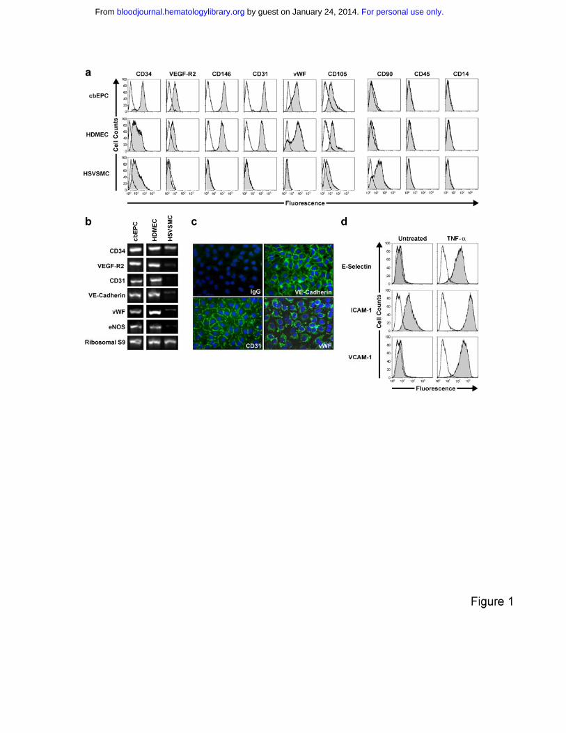
Figure 1. Phenotypic characterization of EPCs. CD31-selected cbEPCs were
evaluated at passage 6. HDMECs and HSVSMCs served as positive and
negative controls respectively. (a) Cytometric analysis of cultured cbEPCs for
endothelial markers CD34, VEGF-R2, CD146, CD31, vWF and CD105, the
mesenchymal marker CD90, and hematopoietic/monocytic markers CD45 and
CD14. Solid gray histograms represent cells stained with fluorescent antibodies.
Isotype-matched controls are overlaid in a black line on each histogram. (b) RT-
PCR analysis of cbEPCs for endothelial markers CD31, CD34, VEGF-R2, VE-
cadherin, vWF and eNOS (the lanes were re-arranged from the same picture to
match panel a). (c) Indirect immunofluorescence of cultured cbEPCs grown in
confluent monolayer showing positive staining for CD31, VE-cadherin, and vWF.
(d) Up-regulation of E-selectin, ICAM-1 and VCAM-1 in cultured cbEPC in
response to TNF-α. Solid gray histograms represent cells stained with
fluorescent antibodies while black lines correspond to the isotype-matched
control fluorescent antibodies.
Figure 2. In vivo vasculogenic potential of EPCs. Matrigel implants containing
cbEPCs and/or HSVSMCs evaluated after one week. (a) H&E staining of
implants (x100) containing a combination of cbEPCs (passage 6) and
HSVSMCs, (b) only cbEPCs, (c) only HSVSMCs, (d) and Matrigel alone. H&E
staining of implants containing both cbEPCs and HSVSMCs evaluated at day 2
(e), day 4 (f), and day 7 (g, x100; h, x400) after xenografting. (i-i) Matrigel plug
containing cbEPCs and HSVSMCs harvested one week after implantation. (i)
Macroscopic view of explanted Matrigel plug, (j) H&E staining showing high
power view of one microvessel containing hematopoietic cells. (k)
Immunohistochemical staining at one week with human specific CD31 antibody
(x400) and with (l) α-SMA antibody. All images are representative of implants
harvested from four different animals. (Black scale bar, 250 µm; white scale bar,
50 µm).
For personal use only. by guest on January 24, 2014. bloodjournal.hematologylibrary.orgFrom
21
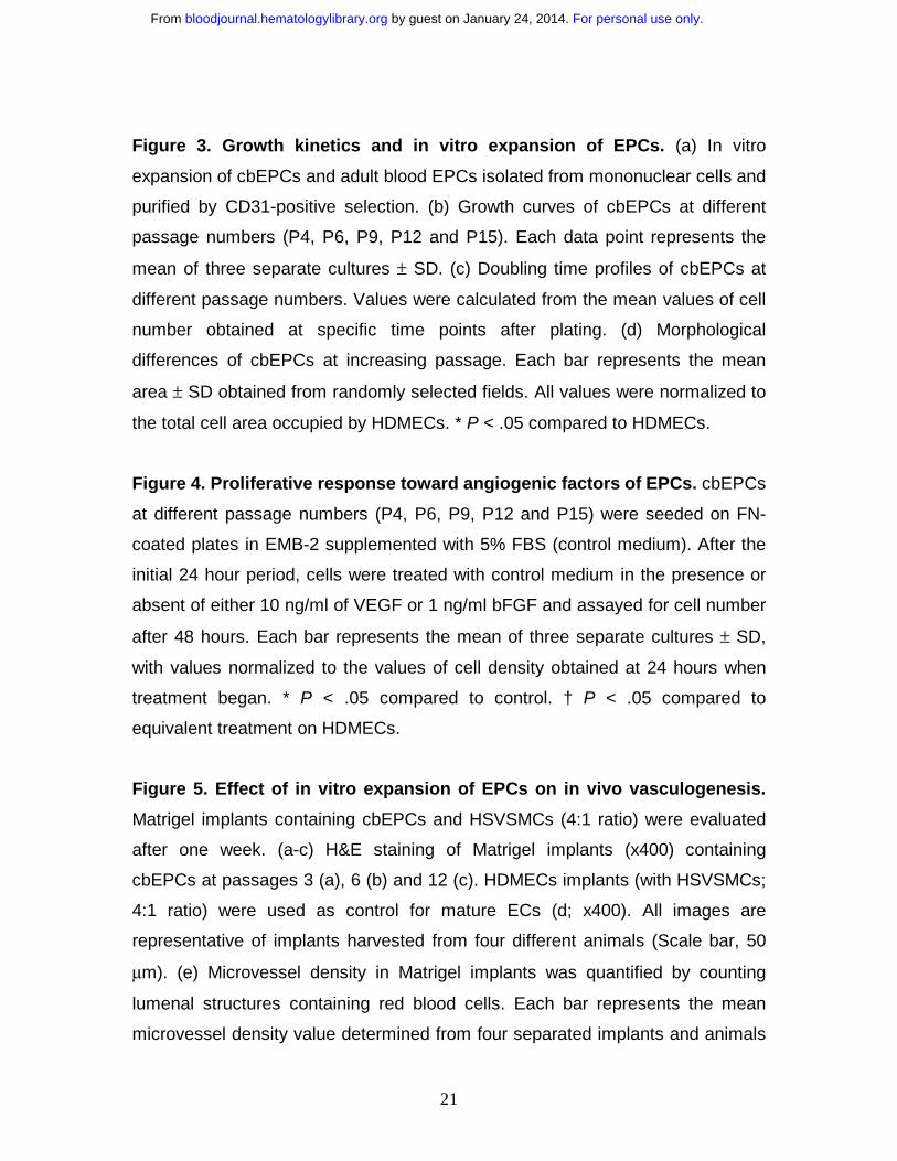
Figure 3. Growth kinetics and in vitro expansion of EPCs. (a) In vitro
expansion of cbEPCs and adult blood EPCs isolated from mononuclear cells and
purified by CD31-positive selection. (b) Growth curves of cbEPCs at different
passage numbers (P4, P6, P9, P12 and P15). Each data point represents the
mean of three separate cultures ± SD. (c) Doubling time profiles of cbEPCs at
different passage numbers. Values were calculated from the mean values of cell
number obtained at specific time points after plating. (d) Morphological
differences of cbEPCs at increasing passage. Each bar represents the mean
area ± SD obtained from randomly selected fields. All values were normalized to
the total cell area occupied by HDMECs. * P < .05 compared to HDMECs.
Figure 4. Proliferative response toward angiogenic factors of EPCs. cbEPCs
at different passage numbers (P4, P6, P9, P12 and P15) were seeded on FN-
coated plates in EMB-2 supplemented with 5% FBS (control medium). After the
initial 24 hour period, cells were treated with control medium in the presence or
absent of either 10 ng/ml of VEGF or 1 ng/ml bFGF and assayed for cell number
after 48 hours. Each bar represents the mean of three separate cultures ± SD,
with values normalized to the values of cell density obtained at 24 hours when
treatment began. * P < .05 compared to control. † P < .05 compared to
equivalent treatment on HDMECs.
Figure 5. Effect of in vitro expansion of EPCs on in vivo vasculogenesis.
Matrigel implants containing cbEPCs and HSVSMCs (4:1 ratio) were evaluated
after one week. (a-c) H&E staining of Matrigel implants (x400) containing
cbEPCs at passages 3 (a), 6 (b) and 12 (c). HDMECs implants (with HSVSMCs;
4:1 ratio) were used as control for mature ECs (d; x400). All images are
representative of implants harvested from four different animals (Scale bar, 50
µm). (e) Microvessel density in Matrigel implants was quantified by counting
lumenal structures containing red blood cells. Each bar represents the mean
microvessel density value determined from four separated implants and animals
For personal use only. by guest on January 24, 2014. bloodjournal.hematologylibrary.orgFrom
22
± SD. * P < .05 compared to HDMEC. † P < .05 compared to cbEPC-P3. ‡ P <
.05 compared to cbEPC-P6.
Figure 6. Effect of implanted cell number on vasculogenic performance of
EPCs. Matrigel implants containing either 0.5x10⁶ (x1/3) (panels a, d), 1.5x10⁶
(x1) (panels b, e, h) or 4.5x10⁶ (x3) (panels c, f, i) EPCs in the presence of
HSVSMCs (4:1 EPCs/HSVSMCs ratio) were evaluated after one week. (a-c)
H&E staining of Matrigel implants containing cbEPCs at passages 6 (x400); (d-f)
cbEPCs at passage 12 (x400) and (g-i) adult EPCs at passage 6 (x400). (g) Anti-
human CD31 immunostained section of adult EPCs at passage 6 seeded at x3.
All images are representative of implants harvested from four different animals
(Scale bar, 50 µm). (j) Microvessel density was quantified by counting lumenal
structures containing red blood cells. Each bar represents the mean microvessel
density value determined from four separated implants and animals ± SD. * P <
.05 compared to x1/3. † P < .05 compared to x1.
For personal use only. by guest on January 24, 2014. bloodjournal.hematologylibrary.orgFrom

23
REFERENCES
1. Vacanti JP, Langer R. Tissue engineering: the design and fabrication of living replacement devices for surgical reconstruction and transplantation. Lancet. 1999;354 Suppl 1:SI32-34. 2. Langer R, Vacanti JP. Tissue engineering. Science. 1993;260:920-926. 3. Stock UA, Sakamoto T, Hatsuoka S, et al. Patch augmentation of the pulmonary artery with bioabsorbable polymers and autologous cell seeding. J Thorac Cardiovasc Surg. 2000;120:1158-1167; discussion 1168. 4. Hoerstrup SP, Sodian R, Daebritz S, et al. Functional living trileaflet heart valves grown in vitro. Circulation. 2000;102:III44-49. 5. Kaushal S, Amiel GE, Guleserian KJ, et al. Functional small-diameter neovessels created using endothelial progenitor cells expanded ex vivo. Nat Med. 2001;7:1035-1040. 6. Shieh SJ, Vacanti JP. State-of-the-art tissue engineering: from tissue engineering to organ building. Surgery. 2005;137:1-7. 7. Chrobak KM, Potter DR, Tien J. Formation of perfused, functional microvascular tubes in vitro. Microvasc Res. 2006;71:185-196. 8. Nomi M, Atala A, Coppi PD, Soker S. Principals of neovascularization for tissue engineering. Mol Aspects Med. 2002;23:463-483. 9. Lee H, Cusick RA, Browne F, et al. Local delivery of basic fibroblast growth factor increases both angiogenesis and engraftment of hepatocytes in tissue-engineered polymer devices. Transplantation. 2002;73:1589-1593. 10. Ochoa ER, Vacanti JP. An overview of the pathology and approaches to tissue engineering. Ann N Y Acad Sci. 2002;979:10-26; discussion 35-18. 11. Peters MC, Polverini PJ, Mooney DJ. Engineering vascular networks in porous polymer matrices. J Biomed Mater Res. 2002;60:668-678. 12. Schechner JS, Nath AK, Zheng L, et al. In vivo formation of complex microvessels lined by human endothelial cells in an immunodeficient mouse. Proc Natl Acad Sci U S A. 2000;97:9191-9196. 13. Nor JE, Peters MC, Christensen JB, et al. Engineering and characterization of functional human microvessels in immunodeficient mice. Lab Invest. 2001;81:453-463. 14. Rafii S, Lyden D. Therapeutic stem and progenitor cell transplantation for organ vascularization and regeneration. Nat Med. 2003;9:702-712. 15. Levenberg S, Rouwkema J, Macdonald M, et al. Engineering vascularized skeletal muscle tissue. Nat Biotechnol. 2005;23:879-884. 16. Asahara T, Murohara T, Sullivan A, et al. Isolation of putative progenitor endothelial cells for angiogenesis. Science. 1997;275:964-967. 17. Wu X, Rabkin-Aikawa E, Guleserian KJ, et al. Tissue-engineered microvessels on three-dimensional biodegradable scaffolds using human endothelial progenitor cells. Am J Physiol Heart Circ Physiol. 2004;287:H480-487.
For personal use only. by guest on January 24, 2014. bloodjournal.hematologylibrary.orgFrom

24
18. Ingram DA, Mead LE, Tanaka H, et al. Identification of a novel hierarchy of endothelial progenitor cells using human peripheral and umbilical cord blood. Blood. 2004;104:2752-2760. 19. Lin Y, Weisdorf DJ, Solovey A, Hebbel RP. Origins of circulating endothelial cells and endothelial outgrowth from blood. J Clin Invest. 2000;105:71-77. 20. Yoder MC, Mead LE, Prater D, et al. Re-defining endothelial progenitor cells via clonal analysis and hematopoietic stem/progenitor cell principals. Blood. 2006. 21. Kraling BM, Bischoff J. A simplified method for growth of human microvascular endothelial cells results in decreased senescence and continued responsiveness to cytokines and growth factors. In Vitro Cell Dev Biol Anim. 1998;34:308-315. 22. Melero-Martin JM, Dowling MA, Smith M, Al-Rubeai M. Optimal in-vitro expansion of chondroprogenitor cells in monolayer culture. Biotechnol Bioeng. 2006;93:519-533. 23. Rehman J, Li J, Orschell CM, March KL. Peripheral blood "endothelial progenitor cells" are derived from monocyte/macrophages and secrete angiogenic growth factors. Circulation. 2003;107:1164-1169. 24. Gulati R, Jevremovic D, Peterson TE, et al. Diverse origin and function of cells with endothelial phenotype obtained from adult human blood. Circ Res. 2003;93:1023-1025. 25. Parums DV, Cordell JL, Micklem K, Heryet AR, Gatter KC, Mason DY. JC70: a new monoclonal antibody that detects vascular endothelium associated antigen on routinely processed tissue sections. J Clin Pathol. 1990;43:752-757. 26. Levenberg S, Huang NF, Lavik E, Rogers AB, Itskovitz-Eldor J, Langer R. Differentiation of human embryonic stem cells on three-dimensional polymer scaffolds. Proc Natl Acad Sci U S A. 2003;100:12741-12746. 27. Folkman J, D'Amore PA. Blood vessel formation: what is its molecular basis? Cell. 1996;87:1153-1155. 28. Darland DC, D'Amore PA. Blood vessel maturation: vascular development comes of age. J Clin Invest. 1999;103:157-158. 29. Darland DC, D'Amore PA. Cell-cell interactions in vascular development. Curr Top Dev Biol. 2001;52:107-149. 30. Shepherd BR, Chen HY, Smith CM, Gruionu G, Williams SK, Hoying JB. Rapid perfusion and network remodeling in a microvascular construct after implantation. Arterioscler Thromb Vasc Biol. 2004;24:898-904. 31. Shi Q, Rafii S, Wu MH, et al. Evidence for circulating bone marrow-derived endothelial cells. Blood. 1998;92:362-367. 32. Hristov M, Erl W, Weber PC. Endothelial progenitor cells: isolation and characterization. Trends Cardiovasc Med. 2003;13:201-206. 33. Khan ZA, Melero-Martin JM, Wu X, et al. Endothelial progenitor cells from infantile hemangioma and umbilical cord blood display unique cellular responses to endostatin. Blood. 2006;108:915-921.
For personal use only. by guest on January 24, 2014. bloodjournal.hematologylibrary.orgFrom

25
34. Peichev M, Naiyer AJ, Pereira D, et al. Expression of VEGFR-2 and AC133 by circulating human CD34(+) cells identifies a population of functional endothelial precursors. Blood. 2000;95:952-958.
For personal use only. by guest on January 24, 2014. bloodjournal.hematologylibrary.orgFrom
For personal use only. by guest on January 24, 2014. bloodjournal.hematologylibrary.orgFrom
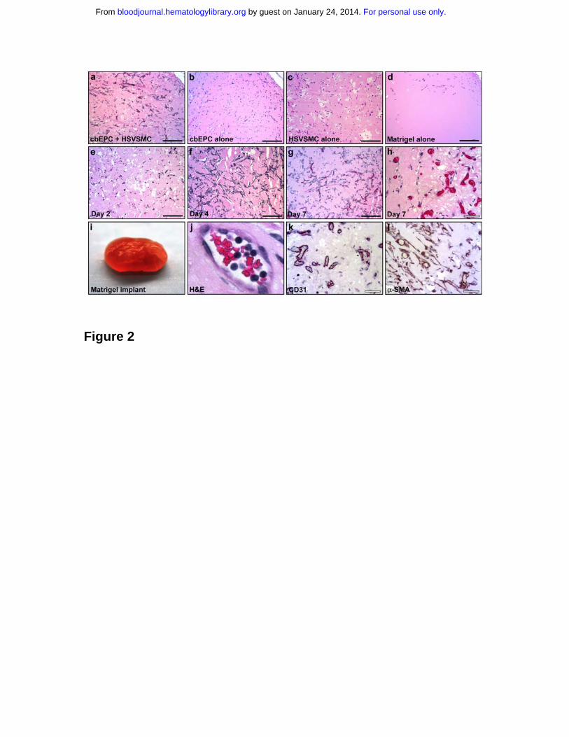
Figure 2
For personal use only. by guest on January 24, 2014. bloodjournal.hematologylibrary.orgFrom
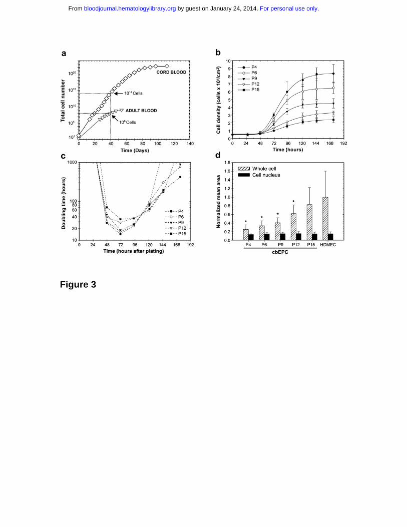
Figure 3
For personal use only. by guest on January 24, 2014. bloodjournal.hematologylibrary.orgFrom
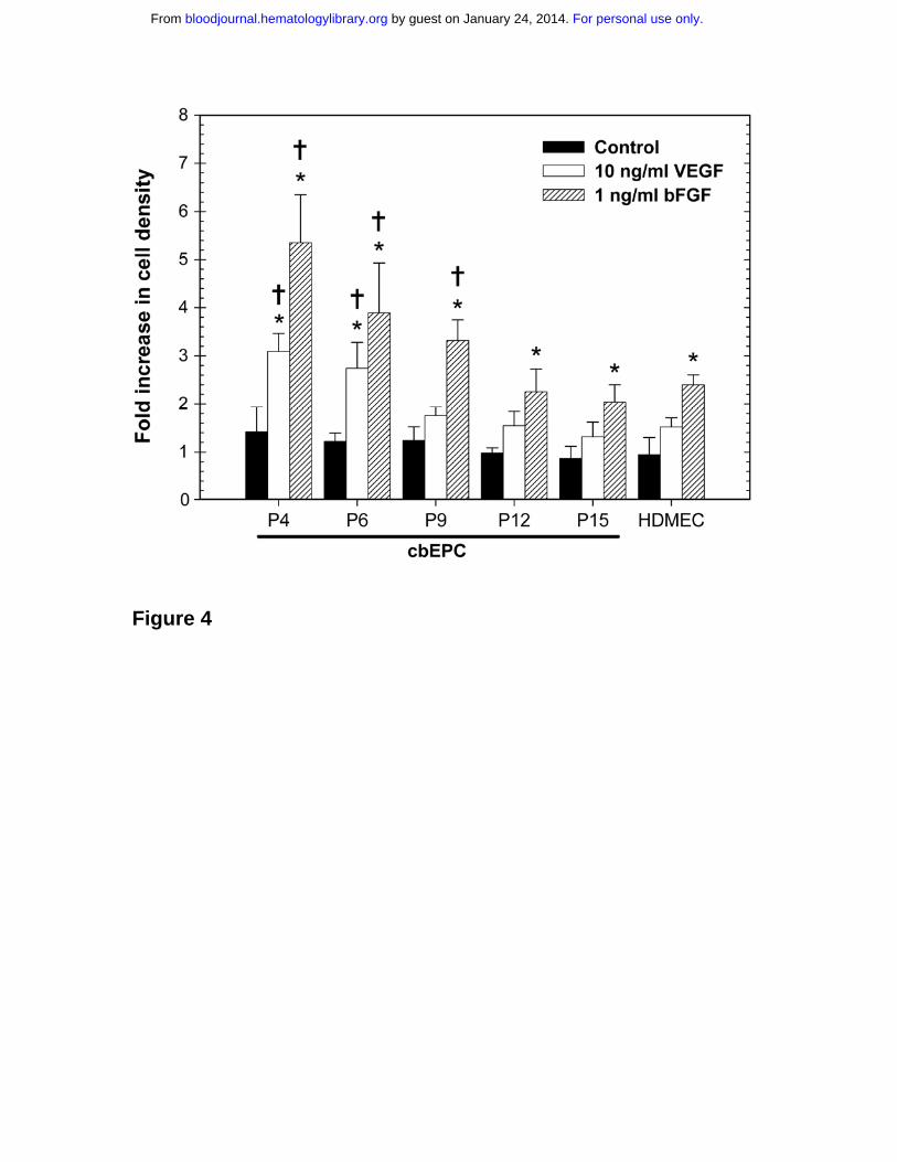
Figure 4
For personal use only. by guest on January 24, 2014. bloodjournal.hematologylibrary.orgFrom
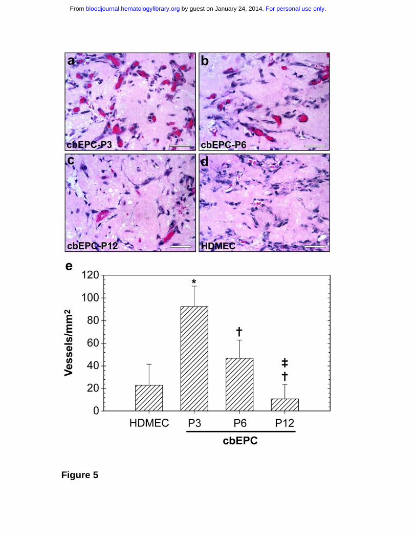
Figure 5
For personal use only. by guest on January 24, 2014. bloodjournal.hematologylibrary.orgFrom
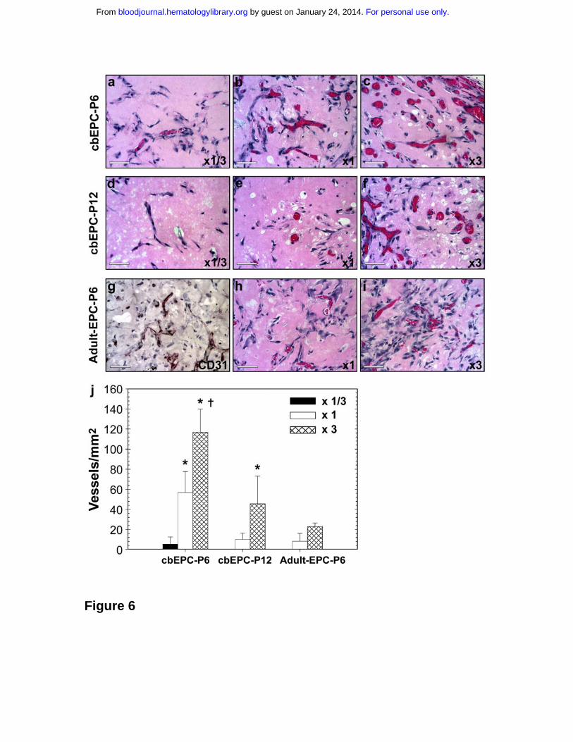
Figure 6
For personal use only. by guest on January 24, 2014. bloodjournal.hematologylibrary.orgFrom
Related Documents