ARTICLES NATURE MEDICINE ADVANCE ONLINE PUBLICATION 1 Improved neovascularization is an important therapeutic goal after myocardial infarction or limb ischemia 1 . In recent years, our under- standing of the processes responsible for the formation of new blood vessels after tissue ischemia has changed.Vascularization of ischemic tissue in adults was once thought to be restricted to migration and proliferation of mature endothelial cells, a process termed ‘angiogen- esis’ 2 . However, increasing evidence suggests that circulating EPCs home to sites of ischemia and contribute to the formation of new blood vessels 3–5 . In analogy to the embryonic development of blood vessels from primitive endothelial progenitors (angioblasts), this process is referred to as ‘vasculogenesis’ 6,7 . The importance of circu- lating progenitor cells is demonstrated by the fact that genetic inhibi- tion of their recruitment inhibits tumor angiogenesis 8 , whereas intravenous infusion of bone marrow–derived progenitor cells aug- ments neovascularization 9 . EPCs can be mobilized from the bone marrow into the circulation by VEGF or stromal cell–derived factor- 1 (SDF-1) 10–12 . Maintenance and mobilization of stem cells in the bone marrow is determined by the local microenvironment, which consists of fibroblasts and endothelial cells 13–15 . VEGF- and SDF-1- induced progenitor cell mobilization is dependent on local secretion of MMP-9 by the hematopoietic and stromal compartments of the bone marrow, which results in release of soluble Kit ligand (KitL; also known as stem cell factor) 11 . This permits transfer of endothelial and hematopoietic progenitor cells from the quiescent to the proliferative niche 11 . Nos3 –/– mice, which are deficient in eNOS, are characterized by impaired ischemia-induced neovascularization 16 . Mobilization of EPCs from the bone marrow into the circulation is required for postnatal neovascularization 4 . Here we show that mice lacking eNOS have defective hematopoietic recovery and progenitor cell mobilization, resulting in increased mortality after myelosuppres- sion and reduced VEGF-induced mobilization of EPCs. Intravenous infusion, but not bone marrow transplantation, of wild-type stem and progenitor cells rescued the impaired neovascularization of Nos3 –/– mice in a model of hind-limb ischemia. RESULTS Mobilization of progenitor cells in Nos3 –/– mice Basal numbers of EPCs did not significantly differ between Nos3 –/– and wild-type mice (Fig. 1a). However, whereas the number of peripheral EPCs significantly (P < 0.05) increased after VEGF mobi- lization in wild-type mice, Nos3 –/– mice did not mobilize EPCs in response to VEGF (Fig. 1a). These data were confirmed by directly measuring the number of CD34 + Flk-1 + cells by fluorescence-acti- vated cell sorting (FACS) analysis (data not shown). To assess whether the reduced mobilization capacity in Nos3 –/– mice conse- quently affects hematopoietic reconstitution after myelosuppression, Nos3 –/– mice were treated with 5-fluorouracil (5-FU), which allows for the study of factors that regulate recruitment of stem cells during hematopoietic recovery 11 . Nos3 –/– mice had significantly (P < 0.02) increased mortality compared with wild-type mice (Fig. 1b). The recovery of the white blood cell count was significantly delayed in Nos3 –/– mice (P = 0.01; Fig. 1c and Supplementary Fig. 1 online). In line with these findings, we observed that the increase in cellularity 1 Molecular Cardiology, Department of Internal Medicine IV, University of Frankfurt, Theodor-Stern-Kai 7, 60590 Frankfurt, Germany. 2 Department of Pathology, University of Freiburg, Albertstrasse 19, 79104 Freiburg, Germany. 3 These authors contributed equally to this work. Correspondence should be addressed to S.D. ([email protected]). Published online 12 October 2003; doi:10.1038/nm948 Essential role of endothelial nitric oxide synthase for mobilization of stem and progenitor cells Alexandra Aicher 1,3 , Christopher Heeschen 1,3 , Christiane Mildner-Rihm 1 , Carmen Urbich 1 , Christian Ihling 2 , Katja Technau-Ihling 2 , Andreas M Zeiher 1 & Stefanie Dimmeler 1 Endothelial nitric oxide synthase (eNOS) is essential for neovascularization. Here we show that the impaired neovascularization in mice lacking eNOS is related to a defect in progenitor cell mobilization. Mice deficient in eNOS (Nos3 –/– ) show reduced vascular endothelial growth factor (VEGF)-induced mobilization of endothelial progenitor cells (EPCs) and increased mortality after myelosuppression. Intravenous infusion of wild-type progenitor cells, but not bone marrow transplantation, rescued the defective neovascularization of Nos3 –/– mice in a model of hind-limb ischemia, suggesting that progenitor mobilization from the bone marrow is impaired in Nos3 –/– mice. Mechanistically, matrix metalloproteinase-9 (MMP-9), which is required for stem cell mobilization, was reduced in the bone marrow of Nos3 –/– mice. These findings indicate that eNOS expressed by bone marrow stromal cells influences recruitment of stem and progenitor cells. This may contribute to impaired regeneration processes in ischemic heart disease patients, who are characterized by a reduced systemic NO bioactivity. © 2003 Nature Publishing Group http://www.nature.com/naturemedicine

Welcome message from author
This document is posted to help you gain knowledge. Please leave a comment to let me know what you think about it! Share it to your friends and learn new things together.
Transcript

A R T I C L E S
NATURE MEDICINE ADVANCE ONLINE PUBLICATION 1
Improved neovascularization is an important therapeutic goal aftermyocardial infarction or limb ischemia1. In recent years, our under-standing of the processes responsible for the formation of new bloodvessels after tissue ischemia has changed. Vascularization of ischemictissue in adults was once thought to be restricted to migration andproliferation of mature endothelial cells, a process termed ‘angiogen-esis’2. However, increasing evidence suggests that circulating EPCshome to sites of ischemia and contribute to the formation of newblood vessels3–5. In analogy to the embryonic development of bloodvessels from primitive endothelial progenitors (angioblasts), thisprocess is referred to as ‘vasculogenesis’6,7. The importance of circu-lating progenitor cells is demonstrated by the fact that genetic inhibi-tion of their recruitment inhibits tumor angiogenesis8, whereasintravenous infusion of bone marrow–derived progenitor cells aug-ments neovascularization9. EPCs can be mobilized from the bonemarrow into the circulation by VEGF or stromal cell–derived factor-1 (SDF-1)10–12. Maintenance and mobilization of stem cells in thebone marrow is determined by the local microenvironment, whichconsists of fibroblasts and endothelial cells13–15. VEGF- and SDF-1-induced progenitor cell mobilization is dependent on local secretionof MMP-9 by the hematopoietic and stromal compartments of thebone marrow, which results in release of soluble Kit ligand (KitL; alsoknown as stem cell factor)11. This permits transfer of endothelial andhematopoietic progenitor cells from the quiescent to the proliferativeniche11.
Nos3–/– mice, which are deficient in eNOS, are characterized byimpaired ischemia-induced neovascularization16. Mobilization of
EPCs from the bone marrow into the circulation is required forpostnatal neovascularization4. Here we show that mice lackingeNOS have defective hematopoietic recovery and progenitor cellmobilization, resulting in increased mortality after myelosuppres-sion and reduced VEGF-induced mobilization of EPCs. Intravenousinfusion, but not bone marrow transplantation, of wild-type stemand progenitor cells rescued the impaired neovascularization ofNos3–/– mice in a model of hind-limb ischemia.
RESULTSMobilization of progenitor cells in Nos3–/– miceBasal numbers of EPCs did not significantly differ between Nos3–/–
and wild-type mice (Fig. 1a). However, whereas the number ofperipheral EPCs significantly (P < 0.05) increased after VEGF mobi-lization in wild-type mice, Nos3–/– mice did not mobilize EPCs inresponse to VEGF (Fig. 1a). These data were confirmed by directlymeasuring the number of CD34+Flk-1+ cells by fluorescence-acti-vated cell sorting (FACS) analysis (data not shown). To assesswhether the reduced mobilization capacity in Nos3–/– mice conse-quently affects hematopoietic reconstitution after myelosuppression,Nos3–/– mice were treated with 5-fluorouracil (5-FU), which allowsfor the study of factors that regulate recruitment of stem cells duringhematopoietic recovery11. Nos3–/– mice had significantly (P < 0.02)increased mortality compared with wild-type mice (Fig. 1b). Therecovery of the white blood cell count was significantly delayed inNos3–/– mice (P = 0.01; Fig. 1c and Supplementary Fig. 1 online). Inline with these findings, we observed that the increase in cellularity
1Molecular Cardiology, Department of Internal Medicine IV, University of Frankfurt, Theodor-Stern-Kai 7, 60590 Frankfurt, Germany. 2Department of Pathology,University of Freiburg, Albertstrasse 19, 79104 Freiburg, Germany. 3These authors contributed equally to this work. Correspondence should be addressed to S.D.([email protected]).
Published online 12 October 2003; doi:10.1038/nm948
Essential role of endothelial nitric oxide synthase formobilization of stem and progenitor cellsAlexandra Aicher1,3, Christopher Heeschen1,3, Christiane Mildner-Rihm1, Carmen Urbich1, Christian Ihling2,Katja Technau-Ihling2, Andreas M Zeiher1 & Stefanie Dimmeler1
Endothelial nitric oxide synthase (eNOS) is essential for neovascularization. Here we show that the impaired neovascularizationin mice lacking eNOS is related to a defect in progenitor cell mobilization. Mice deficient in eNOS (Nos3–/–) show reducedvascular endothelial growth factor (VEGF)-induced mobilization of endothelial progenitor cells (EPCs) and increased mortalityafter myelosuppression. Intravenous infusion of wild-type progenitor cells, but not bone marrow transplantation, rescued thedefective neovascularization of Nos3–/– mice in a model of hind-limb ischemia, suggesting that progenitor mobilization from thebone marrow is impaired in Nos3–/– mice. Mechanistically, matrix metalloproteinase-9 (MMP-9), which is required for stem cellmobilization, was reduced in the bone marrow of Nos3–/– mice. These findings indicate that eNOS expressed by bone marrowstromal cells influences recruitment of stem and progenitor cells. This may contribute to impaired regeneration processes inischemic heart disease patients, who are characterized by a reduced systemic NO bioactivity.
©20
03 N
atu
re P
ub
lish
ing
Gro
up
h
ttp
://w
ww
.nat
ure
.co
m/n
atu
rem
edic
ine

A R T I C L E S
2 ADVANCE ONLINE PUBLICATION NATURE MEDICINE
and hematopoietic cell clusters detected in histological bone marrowsections of wild-type mice, 6–10 d after 5-FU treatment, was incom-plete in Nos3–/– mice (Fig. 1d). These data indicate that eNOS isessential for mobilization and hematopoietic reconstitution.
Effect of progenitor cells on neovascularizationWe used a hind-limb ischemia model to assess whether impairedmobilization of EPCs, which may contribute to the reduced neo-vascularization in Nos3–/– mice, can be counteracted by infusion ofwild-type stem and progenitor cells. As previously reported16,Nos3–/– mice showed significantly reduced limb perfusion 2 weeksafter induction of ischemia, compared with wild-type mice (P < 0.01; Fig. 2a,b). The impaired limb perfusion was rescued byintravenous infusion of wild-type EPCs or isolated wild-type bonemarrow cells (BMCs; sca-1+Lin–; Fig. 2a,b). Histological analysisof ischemic thigh muscles indicated that infusion of wild-typeEPCs or BMCs substantially increased capillary density (Fig. 2c,d)and number of conductant vessels (Fig. 2e) in Nos3–/– mice.Physical incorporation of intravenously infused wild-type cellsinto endothelial structures was confirmed by immunostaining forCD31 or mouse pan–endothelial cell antigen-32 (MECA-32) (Fig.3).
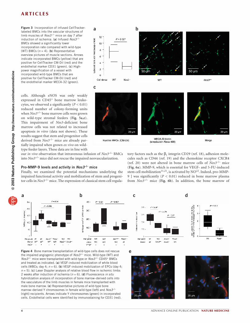
To investigate whether impaired stem cell–stromal cell interactioncontributes to defective neovascularization in Nos3–/– mice, bonemarrow cells depleted of stromal cells by CD45 magnetic bead selec-tion were transplanted into lethally irradiated Nos3–/– mice. Aftercompletely reconstituting the bone marrow of Nos3–/– mice withwild-type stem cells, the mice were treated with VEGF for three con-secutive days. VEGF-induced mobilization of white blood cells orEPCs was blunted in the Nos3–/– mice transplanted with wild-typebone marrow (Fig. 4a,b). In contrast, VEGF significantly (P = 0.006)increased peripheral white blood cell counts in wild-type micetransplanted with Nos3–/– bone marrow (Fig. 4a). Similarly, VEGFsignificantly increased c-Kit+Sca-1+ peripheral blood cells in wild-type mice transplanted with wild-type bone marrow (211 ± 43%increase; P = 0.03 compared with unstimulated mice; n = 6 pergroup), and in wild-type mice engrafted with Nos3–/– bone marrow
(398 ± 91% increase; P = 0.01 versus unstimulated mice). In con-trast, no significant increase was detected when VEGF was injectedinto Nos3–/– mice transplanted with wild-type bone marrow (141 ±26%; P = 0.186).
We used a model of hind-limb ischemia to investigate the func-tional relevance of this impaired mobilization in Nos3–/– mice. Incontrast to the improvement of neovascularization induced byintravenous infusion of wild-type progenitor cells (Fig. 2), bonemarrow reconstitution with wild-type cells did not rescue theimpaired neovascularization phenotype of Nos3–/– mice (Fig. 4c).Taken together, these data suggest that wild-type stem and progeni-tor cells are only effective when applied to the circulation, but can-not be mobilized from the bone marrow of Nos3–/– mice. The dataalso provide evidence that eNOS expression in the bone marrow isessential for mobilization of stem and progenitor cells.
Bypassing the mobilization of BMCs by intravenously infusingNos3–/– cells instead of wild-type cells is insufficient to compensatefor the impaired recovery of Nos3–/– mice after ischemia (Fig. 2a–d).Consistently, a significant (P < 0.05) impairment of neovasculariza-tion was observed when Nos3–/– BMCs were transplanted into wild-type mice (Fig. 4c), despite the effective mobilization of white bloodcells by VEGF (Fig. 4a). These data suggest that eNOS is not onlyessential for mobilization of stem and progenitor cells from thebone marrow, but also determines their angiogenic capacity inischemic tissue. The data also underscore the relative importance ofbone marrow–derived cells for the angiogenic response to ischemia.
To obtain further insight into the impaired proangiogenic activ-ity of Nos3–/– cells after intravenous application, we injectedlabeled wild-type or Nos3–/– cells into Nos3–/– mice and counted thecells that incorporated into the capillaries of the ischemic limbafter 7 days. A large number of systemically injected wild-type cellsincorporated into the microvessels of the ischemic limb andexpressed endothelial marker proteins such as CD31 or MECA-32,whereas incorporation of Nos3–/– cells was significantly reduced (P = 0.027; Fig. 3). In addition, fewer Y-chromosome-positive cellswere detected in capillaries when wild-type male BMCs were trans-planted into female Nos3–/– recipients (Fig. 4d,e). Because of the
a b
c
d
Figure 1 In vivo recruitment ofhematopoietic stem cells and EPCs into the circulation. (a) Peripheral EPC levelsafter mobilization with human VEGF (n = 5).WT, wild type. (b) Kaplan-Meier survivalcurves for Nos3–/– and wild-type mice aftertreatment with 5-FU (n = 10). (c) Recoveryof the white blood cell (WBC) count (n = 5)after 5-FU treatment. Data are given asmean ± s.e.m. (d) H&E staining of femurbone marrow sections 10 d after 5-FUtreatment (magnification, ×20; n = 5).
©20
03 N
atu
re P
ub
lish
ing
Gro
up
h
ttp
://w
ww
.nat
ure
.co
m/n
atu
rem
edic
ine

A R T I C L E S
NATURE MEDICINE ADVANCE ONLINE PUBLICATION 3
limited efficiency of Y-chromosome detection in histological sec-tions (40%; ref. 17), the estimated percentage of capillaries incor-porating bone marrow–derived cells after ischemia in our studywas ∼ 5% in wild-type mice previously transplanted with wild-typebone marrow. This figure was confirmed using wild-type micetransplanted with green fluorescent protein–expressing bone mar-row. We found that 7 ± 11% of the vessels incorporated bone mar-row–derived cells expressing endothelial marker proteins(Supplementary Figs. 2–5 online). Taken together, these data sug-gest that wild-type cells are efficiently incorporated into vessels andexpress endothelial marker proteins, whereas eNOS-deficient cellsare impeded by a marked reduction in homing and incorporationinto microvessels.
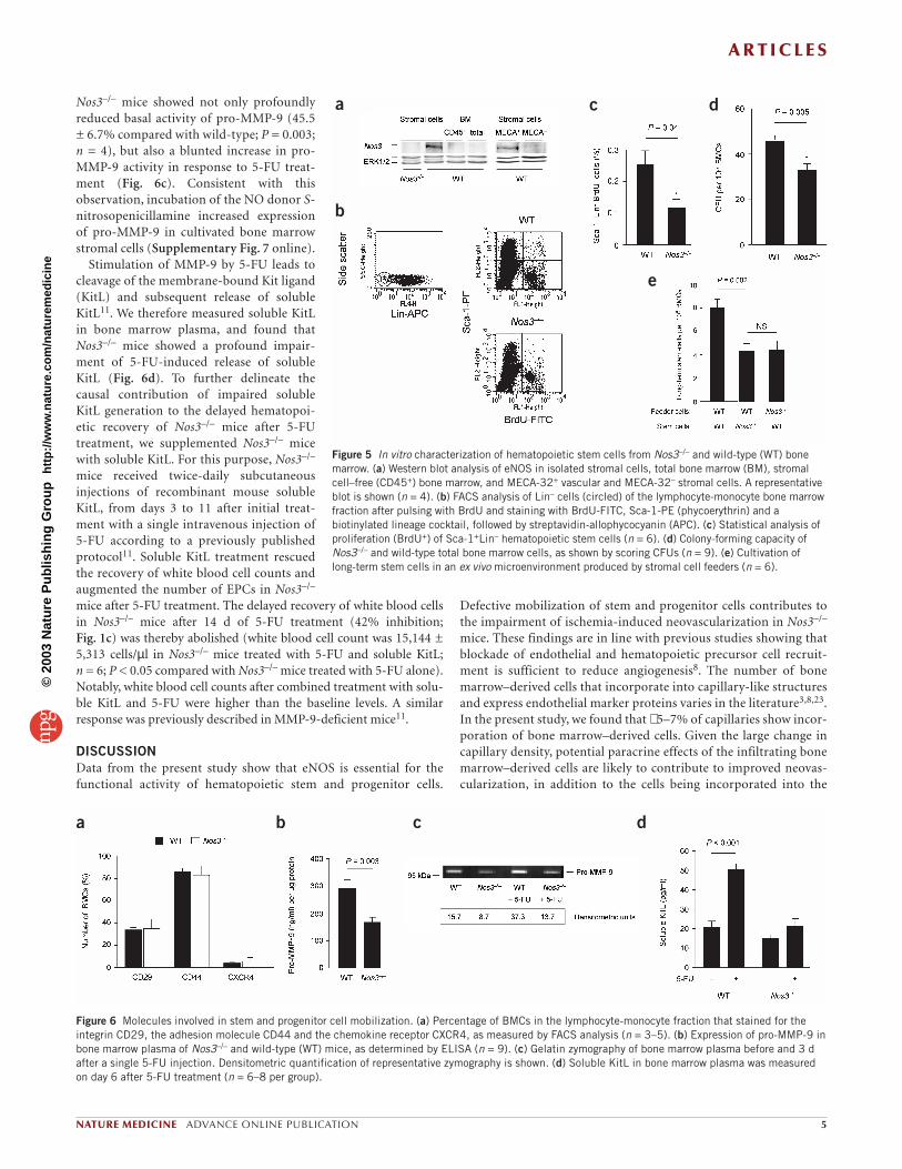
Stromal and progenitor cell functions in vitroBecause our data indicate an impaired interaction between stemcells and stromal cells in the bone marrow of Nos3–/– mice, we ana-lyzed the expression pattern of eNOS in different cellular compo-nents of bone marrow from wild-type mice. Whereas stromal cellsstrongly express eNOS, selected CD45+ bone marrow leukocytes
showed only very faint eNOS expression (Fig. 5a). Additional sub-fractionation of cultivated stromal cells revealed that eNOS is pre-dominantly expressed in MECA-32+ vascular cells (Fig. 5a andSupplementary Fig. 6 online). The effect of eNOS on stem and pro-genitor cell function was further investigated by determining theproliferative and colony-forming activity of bone marrow stemcells. In the bone marrow of Nos3 deficient mice, the numbers ofproliferating bromodeoxyuridine (BrdU)-positive Lin–Sca-1+ cells(Fig. 5b,c) and colony-forming units (Fig. 5d) were significantlyreduced (P < 0.05). Because the bone marrow microenvironmentcreated by stromal cells may influence the growth of stem and pro-genitor cells in the bone marrow, we isolated stromal cells fromNos3–/– or wild-type mice to generate feeder cells for functionallong-term in vitro stem cell assays. We then cultured total bone mar-row from wild-type mice on Nos3–/– or wild-type feeder cells togrow stem cells with repopulating potential and colony-formingcapacity. When bone marrow mononuclear cells were grown onNos3–/– feeders, the number of colony-forming units was signifi-cantly reduced (P < 0.01; Fig. 5e), indicating that stromalcell–derived NO is important for the culture of long-term stem
a
b
c d
e
Figure 2 Rescue of impaired angiogenic phenotype in eNOS-deficient mice by intravenous (i.v.) injection of wild-type cells. (a) Laser Doppler–derived relativeblood flow in Nos3–/– mice 14 d after induction of hind-limb ischemia and injection of wild-type or Nos3–/– cells (Sca-1+Lin– BMCs or ex vivo–cultured EPCs) (n = 6). Wild-type animals were used as a positive control. (b) Representative laser Doppler scans, with low or no perfusion displayed in dark blue andhighest perfusion displayed in red. Arrows indicate ischemic leg. (c,d) Capillary density in wild-type and Nos3–/– mice relative to wild-type control animals(n = 6). Representative overview images are shown in d. (e) Left, numbers of conductant vessels in 4-mm2 muscle sections from wild-type and Nos3–/–
mice and wild-type control animals. Right, conductant vessels were identified by size (>20 µm) and α-actin immunostaining. a,c,e, *, P < 0.01 comparedwith wild-type; **, P < 0.01 compared with untreated Nos3–/– mice.
©20
03 N
atu
re P
ub
lish
ing
Gro
up
h
ttp
://w
ww
.nat
ure
.co
m/n
atu
rem
edic
ine
A R T I C L E S
4 ADVANCE ONLINE PUBLICATION NATURE MEDICINE
cells. Although eNOS was only weaklyexpressed in CD45+ bone marrow leuko-cytes, we observed a significantly (P < 0.01)reduced number of colony-forming unitswhen Nos3–/– bone marrow cells were grownon wild-type stromal feeders (Fig. 5a,e).This impairment of Nos3-deficient bonemarrow cells was not related to increasedapoptosis in vitro (data not shown). Theseresults suggest that stem and progenitor cellsderived from Nos3–/– mice are already par-tially impaired when grown ex vivo on wild-type feeder layers. These data are in line withour in vivo observation that intravenous infusion of Nos3–/– BMCsinto Nos3–/– mice did not rescue the impaired neovascularization.
Pro-MMP-9 levels and activity in Nos3–/– miceFinally, we examined the potential mechanisms underlying theimpaired functional activity and mobilization of stem and progeni-tor cells in Nos3–/– mice. The expression of classical stem cell regula-
tory factors such as the β1 integrin CD29 (ref. 18), adhesion mole-cules such as CD44 (ref. 19) and the chemokine receptor CXCR4(ref. 20) were not altered in bone marrow cells of Nos3–/– mice (Fig. 6a). MMP-9, which is essential for VEGF- and 5-FU-inducedstem cell mobilization11,21, is activated by NO22. Indeed, pro-MMP-9 ] was significantly (P < 0.01) reduced in bone marrow plasmafrom Nos3–/– mice (Fig. 6b). In addition, the bone marrow of
a b
c
Figure 3 Incorporation of infused CellTracker-labeled BMCs into the vascular structures oflimb muscles of Nos3–/– mice on day 7 afterinduction of ischemia. (a) Infused Nos3–/–
BMCs showed a significantly lowerincorporation rate compared with wild-type(WT) BMCs (n = 4). (b) Representativeoverview pictures of muscle sections. Arrowsindicate incorporated BMCs (yellow) that arepositive for CellTracker CM-Dil (red) and theendothelial marker CD31 (green). (c) High-power magnification of a vessel withincorporated wild-type BMCs that arepositive for CellTracker CM-Dil (red) and the endothelial marker MECA-32 (green).
a b c d
eFigure 4 Bone marrow transplantation of wild-type cells does not rescuethe impaired angiogenic phenotype of Nos3–/– mice. Wild-type (WT) andNos3–/– mice were transplanted with wild-type or Nos3–/– CD45+ BMCsand treated as indicated. (a) VEGF-induced mobilization of white bloodcells (WBCs; day 4; n = 6). (b) VEGF-induced mobilization of EPCs (day 4;n = 5). (c) Laser Doppler analysis of relative blood flow in ischemic limbs2 weeks after induction of ischemia (n = 6). (d) Fluorescence in situhybridization analysis of incorporation of bone marrow–derived cells intothe vasculature of the limb muscles in female mice transplanted with male bone marrow. (e) Representative pictures of wild-type bonemarrow–derived Y chromosomes in female wild-type (left) and Nos3–/–
(right) recipients. Arrows indicate Y chromosomes (green) in incorporatedcells. Endothelial cells were identified by immunostaining for CD31 (red).
©20
03 N
atu
re P
ub
lish
ing
Gro
up
h
ttp
://w
ww
.nat
ure
.co
m/n
atu
rem
edic
ine
A R T I C L E S
NATURE MEDICINE ADVANCE ONLINE PUBLICATION 5
Nos3–/– mice showed not only profoundlyreduced basal activity of pro-MMP-9 (45.5± 6.7% compared with wild-type; P = 0.003;n = 4), but also a blunted increase in pro-MMP-9 activity in response to 5-FU treat-ment (Fig. 6c). Consistent with thisobservation, incubation of the NO donor S-nitrosopenicillamine increased expressionof pro-MMP-9 in cultivated bone marrowstromal cells (Supplementary Fig. 7 online).
Stimulation of MMP-9 by 5-FU leads tocleavage of the membrane-bound Kit ligand(KitL) and subsequent release of solubleKitL11. We therefore measured soluble KitLin bone marrow plasma, and found thatNos3–/– mice showed a profound impair-ment of 5-FU-induced release of solubleKitL (Fig. 6d). To further delineate thecausal contribution of impaired solubleKitL generation to the delayed hematopoi-etic recovery of Nos3–/– mice after 5-FUtreatment, we supplemented Nos3–/– micewith soluble KitL. For this purpose, Nos3–/–
mice received twice-daily subcutaneousinjections of recombinant mouse solubleKitL, from days 3 to 11 after initial treat-ment with a single intravenous injection of5-FU according to a previously publishedprotocol11. Soluble KitL treatment rescuedthe recovery of white blood cell counts andaugmented the number of EPCs in Nos3–/–
mice after 5-FU treatment. The delayed recovery of white blood cellsin Nos3–/– mice after 14 d of 5-FU treatment (42% inhibition;Fig. 1c) was thereby abolished (white blood cell count was 15,144 ±5,313 cells/µl in Nos3–/– mice treated with 5-FU and soluble KitL;n = 6; P < 0.05 compared with Nos3–/– mice treated with 5-FU alone).Notably, white blood cell counts after combined treatment with solu-ble KitL and 5-FU were higher than the baseline levels. A similarresponse was previously described in MMP-9-deficient mice11.
DISCUSSIONData from the present study show that eNOS is essential for thefunctional activity of hematopoietic stem and progenitor cells.
Defective mobilization of stem and progenitor cells contributes tothe impairment of ischemia-induced neovascularization in Nos3–/–
mice. These findings are in line with previous studies showing thatblockade of endothelial and hematopoietic precursor cell recruit-ment is sufficient to reduce angiogenesis8. The number of bonemarrow–derived cells that incorporate into capillary-like structuresand express endothelial marker proteins varies in the literature3,8,23.In the present study, we found that ∼ 5–7% of capillaries show incor-poration of bone marrow–derived cells. Given the large change incapillary density, potential paracrine effects of the infiltrating bonemarrow–derived cells are likely to contribute to improved neovas-cularization, in addition to the cells being incorporated into the
a
b
c d
e
Figure 5 In vitro characterization of hematopoietic stem cells from Nos3–/– and wild-type (WT) bonemarrow. (a) Western blot analysis of eNOS in isolated stromal cells, total bone marrow (BM), stromalcell–free (CD45+) bone marrow, and MECA-32+ vascular and MECA-32– stromal cells. A representativeblot is shown (n = 4). (b) FACS analysis of Lin– cells (circled) of the lymphocyte-monocyte bone marrowfraction after pulsing with BrdU and staining with BrdU-FITC, Sca-1-PE (phycoerythrin) and abiotinylated lineage cocktail, followed by streptavidin-allophycocyanin (APC). (c) Statistical analysis ofproliferation (BrdU+) of Sca-1+Lin– hematopoietic stem cells (n = 6). (d) Colony-forming capacity ofNos3–/– and wild-type total bone marrow cells, as shown by scoring CFUs (n = 9). (e) Cultivation oflong-term stem cells in an ex vivo microenvironment produced by stromal cell feeders (n = 6).
a b c d
Figure 6 Molecules involved in stem and progenitor cell mobilization. (a) Percentage of BMCs in the lymphocyte-monocyte fraction that stained for theintegrin CD29, the adhesion molecule CD44 and the chemokine receptor CXCR4, as measured by FACS analysis (n = 3–5). (b) Expression of pro-MMP-9 inbone marrow plasma of Nos3–/– and wild-type (WT) mice, as determined by ELISA (n = 9). (c) Gelatin zymography of bone marrow plasma before and 3 dafter a single 5-FU injection. Densitometric quantification of representative zymography is shown. (d) Soluble KitL in bone marrow plasma was measuredon day 6 after 5-FU treatment (n = 6–8 per group).
©20
03 N
atu
re P
ub
lish
ing
Gro
up
h
ttp
://w
ww
.nat
ure
.co
m/n
atu
rem
edic
ine

A R T I C L E S
6 ADVANCE ONLINE PUBLICATION NATURE MEDICINE
endothelial linings. The concept that bone marrow–derived cellscan promote neovascularization without being physically incorpo-rated into the endothelial lining has been proposed by a recentreport that activation of a suicide gene in bone marrow–derivedcells can reduce tumor angiogenesis, although none of the infiltrat-ing cells showed coexpression of endothelial markers in that experi-mental model23.
Deficiency in eNOS also reduced hematopoietic recovery andsubsequently increased mortality after myelosuppression. A similarphenotype in response to 5-FU and VEGF was recently reported forMMP-9-deficient animals, which show reduced transfer of endothe-lial and hematopoietic stem cells from quiescent to proliferativeniches in the bone marrow11. Mice deficient in Nos3 show not onlyreduced basal expression and activity of MMP-9, but also a signifi-cantly blunted increase in MMP-9 activity in response to 5-FU.Thus, a reduction in MMP-9 may indeed underlie the mobilizationdefect in Nos3–/– mice described in the present study. This hypothe-sis is supported by recent data identifying MMP-9 as a major targetfor NO, which activates MMP-9 by S-nitrosylation22. In addition toregulating MMPs, NO also modulates proliferation, differentiationand apoptosis in various cell types24–26. Although our studyexcludes an effect on stem and progenitor cell apoptosis in vitro, wecannot rule out the possibility that endothelium-derived NO inter-feres with cell cycle progression and/or differentiation of stem cellsin the bone marrow. Previous ex vivo studies have shown that lowconcentrations of NO donors, equivalent to those produced byeNOS in vivo, can specifically increase the colony-forming activityof white blood cell precursors from CD34+ cells27.
Our data also indicate that NO in the bone marrow acts primarilyin a paracrine manner, as it is generated mainly by vascular cells ofthe bone marrow stroma. The stromal cell population also containsendothelial cells, and ∼ 30% of the stromal cells express VEGF recep-tor-2 (Flk-1). Hypoxia-induced upregulation of VEGF activateseNOS predominantly through Flk-1-mediated, Akt-dependenteNOS phosphorylation28,29. It is thus possible that VEGF-stimu-lated NO synthesis in bone marrow stromal cells regulates MMP-9,which in turn induces mobilization of hematopoietic and endothe-lial progenitor cells30. However, increased expression of inducibleNOS may also partially compensate for the lack of eNOS, and maythus be responsible for the moderate phenotype observed in Nos3–/–
mice. Further studies are required to elucidate whether doubleknockout of the genes encoding eNOS and inducible NOS mightcreate an even more pronounced phenotype.
In addition to the essential role of eNOS in mobilization of stemand progenitor cells from the bone marrow, eNOS is also requiredfor angiogenesis in ischemic tissue. This is indicated by our findingthat intravenous infusion of Nos3–/– cells or bone marrow engraft-ment of Nos3–/– cells into wild-type animals does not rescue theimpaired neovascularization. These data are in accordance with pre-vious studies showing that eNOS is essential for the survival, migra-tion and angiogenic response of mature endothelial cells24–26. Thedefinitive mechanism underlying the impaired capacity of Nos3–/–
to rescue neovascularization is not clear. In our study, Nos3–/– cellsshowed significantly reduced incorporation into ischemic tissue.Nos3–/– cells might be more sensitive to apoptosis in vivo, leading toa reduction in cell numbers, or might have reduced migratorycapacity, resulting in impaired transmigration into the ischemic tis-sue. However, we did not detect an increase in apoptosis when theNos3–/– bone marrow cells used in the infusion studies were kept exvivo for 24 h, ruling out the possibility that eNOS deficiency directlyaffects progenitor cell survival.
The results of the present study may have important implicationsfor cardiovascular disease progression. It is well established thatendothelial NO bioavailability, which determines the developmentof coronary artery disease31, is not only impaired in atheroscleroticcoronary arteries, but is also reduced systemically in patients at riskfor coronary artery disease32. Consequently, endothelial NO synthe-sis may also be reduced in the bone marrow of heart patients, result-ing in reduced mobilization of progenitor cells. Indeed, patients atrisk for developing coronary artery disease have significantlyreduced numbers of circulating EPCs33,34. In addition, reducednumbers of circulating EPCs were recently shown to be associatedwith endothelial dysfunction in healthy adults35. Thus, the defectivemobilization of progenitor cells caused by reduced NO bioavailabil-ity may impair endogenous vascular repair mechanisms, therebycontributing to the acceleration of vascular aging as well as toreduced neovascularization and cardiac regeneration in patientswith ischemic heart disease.
METHODSAnimal experiments. Nos3–/– mice and their age- and sex-matched wild-typelittermates (C57BL/6J) were purchased from Jackson Laboratories. HumanVEGF165 (500 µg per kg per d) or saline were subcutaneously administeredonce a day for a period of 3 d, along with 5-FU in a single dose of 250 mg/kg.Recombinant mouse soluble KitL was injected twice daily (150 µg/kg).
Bone marrow transplantation. CD45+ bone marrow cells were isolatedusing CD45-bound microbeads and an automated separation device(Miltenyi Biotec). Recipient mice were lethally irradiated with 9.5 Gy andreceived an intravenous injection of 106 donor bone marrow cells 24 h afterirradiation. As a control for transplantation efficiency, bone marrow fromCD45.1 mice was transplanted into Nos3–/– and wild-type mice (bothCD45.2; Supplementary Fig. 8 online)36. Hind-limb ischemia was induced 6 weeks after bone marrow transplantation.
Hind-limb ischemia. The proximal femoral artery, including the superficialand deep branches, and the distal portion of the saphenous artery were lig-ated in the right hind limb37. Two weeks later, we measured blood flow ratiosin the ischemic (right) versus normal (left) limbs, using a laser Dopplerblood flow imager (Moor Instruments; see Supplementary Fig. 9 online fortime course). Capillary density in the semimembraneous and adductor mus-cles was determined in 5-µm cryosections using FITC-conjugated mono-clonal antibody to CD31 (BD PharMingen). The endothelial phenotype wasconfirmed by immunostaining for the pan-endothelial marker MECA-32(BD PharMingen). Injected cells were labeled with CellTracker CM-Dil(Molecular Probes). Bone marrow–derived cells were identified by fluores-cence in situ hybridization38 (probe from Cambio).
BrdU proliferation assay. Bone marrow was collected aseptically by flushingtibias and femurs, and was pulsed with 10 µM BrdU (BrdU flow kit, BDPharMingen) for 1 h before immunostaining for surface antigens. Cells wereincubated with phycoerythrin-conjugated rat antibody to mouse Sca-1, and acocktail of biotinylated antibodies to the lineage markers CD3, B220, CD11b,Gr-1 and TER119 (BD PharMingen) for 20 min at 4 °C, followed by strepta-vidin-allophycocyanin (BD PharMingen). Cells were fixed and permeabilizedwith Cytofix/Cytoperm buffer (BD PharMingen) according to the manufac-turer’s instructions, stained with FITC-conjugated antibody to BrdU (BrdUflow kit, BD PharMingen) and fixed in 1% formaldehyde and PBS. The analy-sis was done using a FACSCalibur with CellQuest software (BD PharMingen).
Colony-forming-unit assay. Total bone marrow cells (105 per well) wereplated in 1.5 ml of 1% methylcellulose (Methocult, StemCell Technologies)containing 15% FBS, 1% BSA, 10 µg/ml human insulin, 200 µg/ml humantransferrin, 10–4 M 2-mercaptoethanol, 2 mM L-glutamine, 50 ng/ml mousestem cell factor, 50 ng/ml human VEGF, 10 ng/ml mouse interleukin-3,10 ng/ml human interleukin-6 and 3 U/ml human erythropoietin. Colonies(>50 cells) were scored on day 7.
©20
03 N
atu
re P
ub
lish
ing
Gro
up
h
ttp
://w
ww
.nat
ure
.co
m/n
atu
rem
edic
ine

A R T I C L E S
NATURE MEDICINE ADVANCE ONLINE PUBLICATION 7
Stromal cell culture. Adherent cells from total bone marrow were culturedin IMDM with 10% FCS, 50 µM 2-mercaptoethanol and antibiotics for 6 weeks, until the formation of 80% confluent monolayers. MECA-32-positive cells were selected using magnetic beads (antibody from BDPharMingen).
Long-term stem cell assay. Bone marrow stromal layers were irradiated (20 Gy), overlaid with 106 wild-type or Nos3–/– total bone marrow cells andcultured in IMDM supplemented with 10% FCS, 10% horse serum, 10–5 Mhydrocortisone (Sigma) and antibiotics in 12-well plates. Cells were fedtwice a week with IMDM. After 35 d, nonadherent cells were flushed fromthe stromal feeder layers, 105 cells were plated in the methylcellulose clonalassay and colonies were scored after 7 d.
EPC assay. For EPC culture, mononuclear cells (4 × 10⁶ cells/well) were iso-lated from spleen-cell homogenates as previously described39, in a modifiedprotocol using 100 ng/ml VEGF (Peprotech) in the culture medium. Theidentity of EPCs was confirmed by Dil-labeled acetylated low-densitylipoprotein/lectin staining and Flk-1 expression.
Detection of MMP-9 and soluble KitL. For gelatin zymography, bone mar-row plasma was collected by flushing femurs and tibias with a total of 0.5 mlPBS. After spinning, supernatants were analyzed for metalloproteinaseactivity by gelatinolytic zymography as described40. Mouse pro-MMP-9 (20 µg protein) and soluble KitL expression were measured by ELISA (R&DSystems).
Western blot analysis. Cells were lysed and western blot analysis was doneas described28, using monoclonal antibody to eNOS (BD Biosciences) fol-lowed by mouse antibody to horseradish peroxidase (Santa CruzBiotechnology) and reprobing for ERK1/2 (Cell Signaling Technology).
Statistical analysis. Comparisons between groups were analyzed by two-sided t-test or ANOVA for experiments with more than two subgroups. Posthoc range tests and pairwise multiple comparisons were done using the two-sided t-test with Bonferroni adjustment. The Kaplan-Meier method wasused to construct survival curves, and the probability of survival betweenpairs was compared with a log rank test. P < 0.05 was considered statisticallysignificant. All analyses were done with SPSS 11.5 software.
Note: Supplementary information is available on the Nature Medicine website.
ACKNOWLEDGEMENTSWe thank M. Muhly-Reinholz, S. Rhiel, T. Röxe and A. Knau for their excellenttechnical assistance, and H.E. Schaefer (University of Freiburg) for helpfuldiscussion. This work was supported by the Sonderforschungsbereich (SFB 553)and the Alfried Krupp-Stiftung to S.D.
COMPETING INTERESTS STATEMENTThe authors declare that they have no competing financial interests.
Received 24 July; accepted 22 September 2003Published online at http://www.nature.com/naturemedicine/
1. Isner, J.M. & Losordo, D.W. Therapeutic angiogenesis for heart failure. Nat.Med. 5, 491–492 (1999).
2. Folkman, J. Angiogenesis in cancer, vascular, rheumatoid and other disease.Nat. Med. 1, 27–31 (1995).
3. Asahara, T. et al. Isolation of putative progenitor endothelial cells for angiogen-esis. Science 275, 964–967 (1997).
4. Takahashi, T. et al. Ischemia- and cytokine-induced mobilization of bone mar-row-derived endothelial progenitor cells for neovascularization. Nat. Med. 5,434–438 (1999).
5. Shi, Q. et al. Evidence for circulating bone marrow-derived endothelial cells.Blood 92, 362–367 (1998).
6. Risau, W. Mechanisms of angiogenesis. Nature 386, 671–674 (1997).7. Carmeliet, P. Mechanisms of angiogenesis and arteriogenesis. Nat. Med. 6,
389–395 (2000).8. Lyden, D. et al. Impaired recruitment of bone-marrow-derived endothelial and
hematopoietic precursor cells blocks tumor angiogenesis and growth. Nat. Med.7, 1194–1201 (2001).
9. Shintani, S. et al. Augmentation of postnatal neovascularization with autolo-gous bone marrow transplantation. Circulation 103, 897–903 (2001).
10. Moore, M.A. et al. Mobilization of endothelial and hematopoietic stem and pro-genitor cells by adenovector-mediated elevation of serum levels of SDF-1, VEGF,and angiopoietin-1. Ann. NY Acad. Sci. 938, 36–45 (2001).
11. Heissig, B. et al. Recruitment of stem and progenitor cells from the bone mar-row niche requires MMP-9 mediated release of kit-ligand. Cell 109, 625–637(2002).
12. Asahara, T. et al. VEGF contributes to postnatal neovascularization by mobiliz-ing bone marrow-derived endothelial progenitor cells. EMBO J. 18, 3964–3972(1999).
13. Morrison, S.J., Shah, N.M. & Anderson, D.J. Regulatory mechanisms in stemcell biology. Cell 88, 287–298 (1997).
14. Kiger, A.A., White-Cooper, H. & Fuller, M.T. Somatic support cells restrictgermline stem cell self-renewal and promote differentiation. Nature 407,750–754 (2000).
15. Lapidot, T. & Petit, I. Current understanding of stem cell mobilization: the rolesof chemokines, proteolytic enzymes, adhesion molecules, cytokines, and stro-mal cells. Exp. Hematol. 30, 973–981 (2002).
16. Murohara, T. et al. Nitric oxide synthase modulates angiogenesis in response totissue ischemia. J. Clin. Invest. 101, 2567–2578 (1998).
17. Taylor, D.A., Hruban, R., Rodriguez, E.R. & Goldschmidt-Clermont, P.J. Cardiacchimerism as a mechanism for self-repair: does it happen and if so to whatdegree? Circulation 106, 2–4 (2002).
18. Prosper, F., Stroncek, D., McCarthy, J.B. & Verfaillie, C.M. Mobilization andhoming of peripheral blood progenitors is related to reversible downregulation ofα4β1 integrin expression and function. J. Clin. Invest. 101, 2456–2467(1998).
19. Vermeulen, M. et al. Role of adhesion molecules in the homing and mobilizationof murine hematopoietic stem and progenitor cells. Blood 92, 894–900(1998).
20. Peled, A. et al. Dependence of human stem cell engraftment and repopulationof NOD/SCID mice on CXCR4. Science 283, 845–848 (1999).
21. Hattori, K. et al. Placental growth factor reconstitutes hematopoiesis by recruit-ing VEGFR1(+) stem cells from bone-marrow microenvironment. Nat. Med. 8,841–849 (2002).
22. Gu, Z. et al. S-nitrosylation of matrix metalloproteinases: signaling pathway toneuronal cell death. Science 297, 1186–1190 (2002).
23. De Palma, M., Venneri, M.A., Roca, C. & Naldini, L. Targeting exogenous genesto tumor angiogenesis by transplantation of genetically modified hematopoieticstem cells. Nat. Med. 9, 789–795 (2003).
24. Ziche, M. et al. Nitric oxide mediates angiogenesis in vivo and endothelial cellgrowth and migration in vitro promoted by substance P. J. Clin. Invest. 94,2036–2044 (1994).
25. Murohara, T. et al. Role of endothelial nitric oxide synthase in endothelial cellmigration. Arterioscler. Thromb. Vasc. Biol. 19, 1156–1161 (1999).
26. Dimmeler, S., Haendeler, J., Nehls, M. & Zeiher, A.M. Suppression of apoptosisby nitric oxide via inhibition of ICE-like and CPP32-like proteases. J. Exp. Med.185, 601–608 (1997).
27. Shami, P.J. & Weinberg, J.B. Differential effects of nitric oxide on erythroid andmyeloid colony growth from CD34+ human bone marrow cells. Blood 87,977–982 (1996).
28. Dimmeler, S. et al. Activation of nitric oxide synthase in endothelial cells viaAkt-dependent phosphorylation. Nature 399, 601–605 (1999).
29. Fulton, D. et al. Regulation of endothelium-derived nitric oxide production bythe protein kinase Akt. Nature 399, 597–601 (1999).
30. Eriksson, U. & Alitalo, K. VEGF receptor 1 stimulates stem-cell recruitment andnew hope for angiogenesis therapies. Nat. Med. 8, 775–777 (2002).
31. Schachinger, V., Britten, M.B. & Zeiher, A.M. Prognostic impact of coronaryvasodilator dysfunction on adverse long-term outcome of coronary heart dis-ease. Circulation 101, 1899–1906 (2000).
32. Zeiher, A.M. Endothelial vasodilator dysfunction: pathogenetic link to myocar-dial ischaemia or epiphenomenon? Lancet 348, S10–S12 (1996).
33. Vasa, M. et al. Number and migratory activity of circulating endothelial progen-itor cells inversely correlate with risk factors for coronary artery disease. Circ.Res. 89, E1–E7 (2001).
34. Tepper, O.M. et al. Human endothelial progenitor cells from type II diabeticsexhibit impaired proliferation, adhesion, and incorporation into vascular struc-tures. Circulation 106, 2781–2786 (2002).
35. Hill, J.M. et al. Circulating endothelial progenitor cells, vascular function, andcardiovascular risk. N. Engl. J. Med. 348, 593–600 (2003).
36. Fiering, S.N. et al. Improved FACS-Gal: flow cytometric analysis and sorting ofviable eukaryotic cells expressing reporter gene constructs. Cytometry 12,291–301 (1991).
37. Heeschen, C. et al. Nicotine stimulates angiogenesis and promotes tumorgrowth and atherosclerosis. Nat. Med. 7, 833–839 (2001).
38. Mezey, E. et al. Transplanted bone marrow generates new neurons in humanbrains. Proc. Natl. Acad. Sci. USA 100, 1364–1369 (2003).
39. Dimmeler, S. et al. HMG-CoA reductase inhibitors (statins) increase endothelialprogenitor cells via the PI 3-kinase/Akt pathway. J. Clin. Invest. 108, 391–397(2001).
40. Rajagopalan, S., Meng, X.P., Ramasamy, S., Harrison, D.G. & Galis, Z.S.Reactive oxygen species produced by macrophage-derived foam cells regulatethe activity of vascular matrix metalloproteinases in vitro. Implications for ath-erosclerotic plaque stability. J. Clin. Invest. 98, 2572–2579 (1996).
©20
03 N
atu
re P
ub
lish
ing
Gro
up
h
ttp
://w
ww
.nat
ure
.co
m/n
atu
rem
edic
ine
Related Documents