Citation: Salem, S.S.; Ali, O.M.; Reyad, A.M.; Abd-Elsalam, K.A.; Hashem, A.H. Pseudomonas indica-Mediated Silver Nanoparticles: Antifungal and Antioxidant Biogenic Tool for Suppressing Mucormycosis Fungi. J. Fungi 2022, 8, 126. https:// doi.org/10.3390/jof8020126 Academic Editor: Vincent Bruno Received: 14 January 2022 Accepted: 25 January 2022 Published: 27 January 2022 Publisher’s Note: MDPI stays neutral with regard to jurisdictional claims in published maps and institutional affil- iations. Copyright: © 2022 by the authors. Licensee MDPI, Basel, Switzerland. This article is an open access article distributed under the terms and conditions of the Creative Commons Attribution (CC BY) license (https:// creativecommons.org/licenses/by/ 4.0/). Fungi Journal of Article Pseudomonas indica-Mediated Silver Nanoparticles: Antifungal and Antioxidant Biogenic Tool for Suppressing Mucormycosis Fungi Salem S. Salem 1 , Omar M. Ali 2, * , Ahmed M. Reyad 3,4 , Kamel A. Abd-Elsalam 5, * and Amr H. Hashem 1, * 1 Department of Botany and Microbiology, Faculty of Science, Al-Azhar University, Nasr City, Cairo 11884, Egypt; [email protected] 2 Department of Chemistry, Turabah University College, Turabah Branch, Taif University, Taif 21944, Saudi Arabia 3 Biology Department, Faculty of Science, Jazan University, Jazan 82817, Saudi Arabia; [email protected] 4 Botany and Microbiology Department, Faculty of Science, Beni-Suef University, Beni-Suef 62511, Egypt 5 Plant Pathology Research Institute, Agricultural Research Centre, Giza 12619, Egypt * Correspondence: [email protected] (O.M.A.); [email protected] (K.A.A.-E.); [email protected] (A.H.H.) Abstract: Mucormycosis is considered one of the most dangerous invasive fungal diseases. In this study, a facile, green and eco-friendly method was used to biosynthesize silver nanoparticles (AgNPs) using Pseudomonas indica S. Azhar, to combat fungi causing mucormycosis. The biosynthesis of AgNPs was validated by a progressive shift in the color of P. indica filtrate from colorless to brown, as well as the identification of a distinctive absorption peak at 420 nm using UV-vis spectroscopy. Fourier-transform infrared spectroscopy (FTIR) results indicated the existence of bioactive chemicals that are responsible for AgNP production. AgNPs with particle sizes ranging from 2.4 to 53.5 nm were discovered using transmission electron microscopy (TEM). Pattern peaks corresponding to the 111, 200, 220, 311, and 222 planes, which corresponded to face-centered cubic forms of metallic silver, were also discovered using X-ray diffraction (XRD). Moreover, antifungal activity measurements of biosynthesized AgNPs against Rhizopus Microsporus, Mucor racemosus, and Syncephalastrum racemosum were carried out. Results of antifungal activity analysis revealed that the biosynthesized AgNPs exhibited outstanding antifungal activity against all tested fungi at a concentration of 400 μg/mL, where minimum inhibitory concentrations (MIC) were 50, 50, and 100 μg/mL toward R. microsporus, S. racemosum, and M. racemosus respectively. In addition, the biosynthesized AgNPs revealed antioxidant activity, where IC 50 was 31 μg/mL when compared to ascorbic acid (0.79 μg/mL). Furthermore, the biosynthesized AgNPs showed no cytotoxicity on the Vero normal cell line. In conclusion, the biosynthesized AgNPs in this study can be used as effective antifungals with safe use, particularly for fungi causing mucormycosis. Keywords: silver nanoparticles; green biosynthesis; antifungal activity; mucormycosis; antioxidant activity 1. Introduction More than 1.2 billion people are infected by pathogenic fungus each year, resulting in at least 1.7 million fatalities [1,2]. Fungal pathogens now outnumber drug-resistant Mycobacterium tuberculosis in terms of mortality, and they even outnumber malaria [3]. Mucormycosis is one of the most dangerous fungal diseases for humans. Mucormycosis is a disease caused by a group of fungi called mucormycetes, which includes various genera such as Rhizopus, Mucor, Synsephalastrum, Absidia, and Cunninghamella [4,5]. These fungi invade people with a history of diabetes, stem cell transplants, cancer, injection drug use, J. Fungi 2022, 8, 126. https://doi.org/10.3390/jof8020126 https://www.mdpi.com/journal/jof

Welcome message from author
This document is posted to help you gain knowledge. Please leave a comment to let me know what you think about it! Share it to your friends and learn new things together.
Transcript
�����������������
Citation: Salem, S.S.; Ali, O.M.;
Reyad, A.M.; Abd-Elsalam, K.A.;
Hashem, A.H. Pseudomonas
indica-Mediated Silver Nanoparticles:
Antifungal and Antioxidant Biogenic
Tool for Suppressing Mucormycosis
Fungi. J. Fungi 2022, 8, 126. https://
doi.org/10.3390/jof8020126
Academic Editor: Vincent Bruno
Received: 14 January 2022
Accepted: 25 January 2022
Published: 27 January 2022
Publisher’s Note: MDPI stays neutral
with regard to jurisdictional claims in
published maps and institutional affil-
iations.
Copyright: © 2022 by the authors.
Licensee MDPI, Basel, Switzerland.
This article is an open access article
distributed under the terms and
conditions of the Creative Commons
Attribution (CC BY) license (https://
creativecommons.org/licenses/by/
4.0/).
FungiJournal of
Article
Pseudomonas indica-Mediated Silver Nanoparticles:Antifungal and Antioxidant Biogenic Tool for SuppressingMucormycosis FungiSalem S. Salem 1 , Omar M. Ali 2,* , Ahmed M. Reyad 3,4 , Kamel A. Abd-Elsalam 5,*and Amr H. Hashem 1,*
1 Department of Botany and Microbiology, Faculty of Science, Al-Azhar University, Nasr City,Cairo 11884, Egypt; [email protected]
2 Department of Chemistry, Turabah University College, Turabah Branch, Taif University,Taif 21944, Saudi Arabia
3 Biology Department, Faculty of Science, Jazan University, Jazan 82817, Saudi Arabia; [email protected] Botany and Microbiology Department, Faculty of Science, Beni-Suef University, Beni-Suef 62511, Egypt5 Plant Pathology Research Institute, Agricultural Research Centre, Giza 12619, Egypt* Correspondence: [email protected] (O.M.A.); [email protected] (K.A.A.-E.);
[email protected] (A.H.H.)
Abstract: Mucormycosis is considered one of the most dangerous invasive fungal diseases. In thisstudy, a facile, green and eco-friendly method was used to biosynthesize silver nanoparticles (AgNPs)using Pseudomonas indica S. Azhar, to combat fungi causing mucormycosis. The biosynthesis ofAgNPs was validated by a progressive shift in the color of P. indica filtrate from colorless to brown,as well as the identification of a distinctive absorption peak at 420 nm using UV-vis spectroscopy.Fourier-transform infrared spectroscopy (FTIR) results indicated the existence of bioactive chemicalsthat are responsible for AgNP production. AgNPs with particle sizes ranging from 2.4 to 53.5 nmwere discovered using transmission electron microscopy (TEM). Pattern peaks corresponding to the111, 200, 220, 311, and 222 planes, which corresponded to face-centered cubic forms of metallic silver,were also discovered using X-ray diffraction (XRD). Moreover, antifungal activity measurements ofbiosynthesized AgNPs against Rhizopus Microsporus, Mucor racemosus, and Syncephalastrum racemosumwere carried out. Results of antifungal activity analysis revealed that the biosynthesized AgNPsexhibited outstanding antifungal activity against all tested fungi at a concentration of 400 µg/mL,where minimum inhibitory concentrations (MIC) were 50, 50, and 100 µg/mL toward R. microsporus, S.racemosum, and M. racemosus respectively. In addition, the biosynthesized AgNPs revealed antioxidantactivity, where IC50 was 31 µg/mL when compared to ascorbic acid (0.79 µg/mL). Furthermore,the biosynthesized AgNPs showed no cytotoxicity on the Vero normal cell line. In conclusion, thebiosynthesized AgNPs in this study can be used as effective antifungals with safe use, particularlyfor fungi causing mucormycosis.
Keywords: silver nanoparticles; green biosynthesis; antifungal activity; mucormycosis; antioxidantactivity
1. Introduction
More than 1.2 billion people are infected by pathogenic fungus each year, resultingin at least 1.7 million fatalities [1,2]. Fungal pathogens now outnumber drug-resistantMycobacterium tuberculosis in terms of mortality, and they even outnumber malaria [3].Mucormycosis is one of the most dangerous fungal diseases for humans. Mucormycosis isa disease caused by a group of fungi called mucormycetes, which includes various generasuch as Rhizopus, Mucor, Synsephalastrum, Absidia, and Cunninghamella [4,5]. These fungiinvade people with a history of diabetes, stem cell transplants, cancer, injection drug use,
J. Fungi 2022, 8, 126. https://doi.org/10.3390/jof8020126 https://www.mdpi.com/journal/jof
J. Fungi 2022, 8, 126 2 of 15
skin injury due to surgery, burns, or wounds [6–8]. Mucormycosis is a serious infectionand needs to be treated with prescription antifungal medicine, usually amphotericin B,posaconazole, or isavuconazole [9].The increasing usage of antifungal medications hasresulted in the emergence of fungal strains such as Candida albicans [10], Lichtheimia corymb-ifera, R. microsporus, R. arrhizus, and M. circinelloids that are resistant to the majority ofantifungal treatments [11,12]. Most harmful fungi, similar to bacteria, have recently devel-oped antibiotic resistance. To combat drug-resistant fungus, novel antifungal medicinesbased on current biotechnology must be investigated.
Nanoparticles (NPs) are a diverse class of materials that appeal to several researchersdue to their tiny size (1–100 nm), exceptional properties, large surface area, enhanced reac-tivity, capacity to access the body easily, and multiple uses in modern science, including theindustrial and medicinal sciences [13–20]. Selenium, silver, copper, magnesium, zinc, andtitanium are some of the nanoparticles that have been reported [21–30]. AgNPs are widelyutilized nanoparticles in numerous sectors of study, such as optical devices, ophthalmology,pharmaceutical, and the health sciences, for the creation of drug carriers, chemotherapeutic,nano-sensors, gene therapy, and other applications [31–36]. AgNPs have been shownto exhibit high antibacterial, anti-inflammatory, antibiofilm, and anticancer characteris-tics, with the antimicrobial capabilities of AgNPs being used to prevent infection againstharmful microorganisms, such as eukaryotic microorganisms, bacteria, and viruses [37–40].AgNPs are widely used in the biomedical control of diseases such as candidiasis [35] andaspergillosis [41] [42,43]. The efficacy of AgNPs as antimicrobial against microorganismsmay be due to their link with enzymes, reactive oxygen species (ROS) levels, and changingstructure contents that alter the membrane integrity and morphology [44–46]. Because oftheir antibacterial, anti-inflammatory, antibiofilm, anticoagulant, and anticancer properties,AgNPs are becoming a popular choice in the medical and biological fields [22,47,48]. Physi-cal and chemical approaches for the synthesis of NPs are extensively utilized, but they haveseveral downsides, such as the usage of high energy or dangerous chemicals, their high cost,and the generation of vast volumes of toxic byproducts that pollute the environment [49].To overcome the constraints of physical and chemical procedures, low-cost, ecofriendly,simple, and nontoxic approaches that eliminate the use of hazardous and pricey solventsare required for metallic NPs manufacturing [50]. Biological synthesis of metal nanoparti-cles should be stable, biologically safe and ecofriendly [51–55]. Bacteria are one of the mostsignificant groups of microorganisms, since they are employed in bioprocessing, enzymemanufacturing, acid generation, and nanotechnology, among other uses [56–58]. Microbialvariety may be found in abundance in soil, and these microorganisms can be harnessed tobenefit humans. Soil bacteria also require a low cost and low-nutrient medium for growth,making them an ideal option for use [47]. In this work, the bacterial strain P. indica S. Azharwas isolated from a soil sample and utilized to synthesize AgNPs in a simple, quick, andenvironmentally friendly manner. AgNPs were investigated and characterized by UV-vis,Ft-IR, TEM, and XRD. An attempt was made to investigate the antifungal and antioxidantactivities of AgNPs against three mold strains (Rhizopus microsporus, Mucor racemosus, andSyncephalastrum racemosum) which cause mucormycosis in vitro.
2. Materials and Methods2.1. Isolation and Identification of Strain S. Azhar
The bacterial strain P. indica S. Azhar was isolated from a soil sample collected fromthe garden of the Faculty of Science, Al-Azhar University, Cairo, Egypt. For the isolationof bacterial strains, nutrient agar medium was used. The 16S rRNA gene sequencing forbacteria was used to identify it genetically. The improved approach was used to extract bac-terial genomic DNA. Single colonies were taken from agar plates and suspended in steriledeionized water in 50 µL. The cell suspension was then incubated in a water bath for 10 minat 97 ◦C before being centrifuged for 10 min at 15,000 rpm to separate the DNA-containingcell lysate. Using a genomic DNA template and two universal primers, PCR was utilizedto amplify the 16S rRNA gene. The primers 27-f (5-AGAGTTTGATCCTGGCTCAG-3)
J. Fungi 2022, 8, 126 3 of 15
and 1492-r were utilized (5-GGTTACCTTGTTACGACTT-3). Amounts of 1× PCR buffer,0.5 mM MgCl2, 2.5 U Taq-polymerase (QIAGEN, Hilden, Germany), 0.25 mM dNTP, 0.5 Mof each primer, and 1 µL of isolated genomic DNA were added into the PCR mixture (50 µL).Sigma Scientific Services Company (Cairo, Egypt) used a Thermal Cycler to execute thePCR, which included a 3 min hot start at 94 ◦C, 30 cycles at 94 ◦C for 30 s, 55 ◦C for 30 s,and 72 ◦C for 1 min, and 10 min of gene extension at 72 ◦C. GATC Company used an ABI3730x1 DNA sequencer to evaluate the sequencing (Ebersberg, Germany). Using the NCBIBLAST software, the acquired sequences were compared to those in the GenBank database.The bootstrap technique was used to create the phylogenetic tree.
2.2. Extracellular Biosynthesis of AgNPs2.2.1. Bacterial Filtrate Preparation
P. indica cells were suspended in nutrient and injected in broth media for fermentationat 36 ± 2 ◦C for 48 h in an orbital shaker (120 rpm). The biomass was collected using filterpaper No. 1 and rinsed in sterilized distilled water to eliminate any medium componentsbefore being suspended in 100 mL distilled water. At 34 ± 2 ◦C, the mixture was stirred for24 h. Finally, the biomass filtrate was produced by passing it through Whatman filter paperNo. 1 and centrifuging it for 5 min at 2000 rpm to sediment any remaining cell debris. Thesupernatant was used for AgNPs biosynthesis.
2.2.2. Biosynthesis of AgNPs by Biomass Filtrate
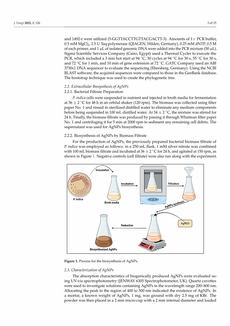
For the production of AgNPs, the previously prepared bacterial biomass filtrate ofP. indica was employed as follows: in a 250 mL flask, 1 mM silver nitrate was combinedwith 100 mL biomass filtrate and incubated at 36 ± 2 ◦C for 24 h, and agitated at 150 rpm, asshown in Figure 1. Negative controls (cell filtrate) were also run along with the experiment.
Figure 1. Process for the biosynthesis of AgNPs.
2.3. Characterization of AgNPs
The absorption characteristics of biogenically produced AgNPs were evaluated us-ing UV-vis spectrophotometry (JENWAY 6305 Spectrophotometer, UK). Quartz cuvetteswere used to investigate solutions containing AgNPs in the wavelength range 200–800 nm.Allocating the peak in the region of 400 to 500 nm indicated the existence of AgNPs. Ina mortar, a known weight of AgNPs, 1 mg, was ground with dry 2.5 mg of KBr. Thepowder was then placed in a 2 mm micro-cup with a 2 mm internal diameter and loaded
J. Fungi 2022, 8, 126 4 of 15
onto a FT-IR set at 26 ◦C ± 1 ◦C. The sample was scanned using infra-red in the range of400–4000 cm−1, utilizing Fourier transform-infrared spectrometty. To determine the sizeand form of nanoparticles, TEM (JEOL 1010, Tokyo, Japan) was used to characterize AgNPs.Drop-coating the AgNPs solution onto the carbon-coated copper grid and loading it onto aspecimen holder were used to produce the sample. The sizes and shapes of AgNPs werevalidated using TEM micrographs. Stress analysis, residual–austenite quantitation, crystal-lite size/lattice, crystallite calculation, and materials analysis by overlaid X-ray diffractionpatterns with a Shimadzu apparatus, using a nickel filter and Cu–Ka target (Shimadzu-Scientific Instruments (SSI), Kyoto, Japan), were obtained with the XRD 6000 series. Theaverage crystallite size of AgNPs can also be measured utilizing the Debye-scherrer equa-tion: D = kλ/β Cos θ.
2.4. Antifungal Activity
Some genera of the Mucorales group were used for evaluation of the biosynthesizedAgNPs as antifungal agent. Rhizopus microsporus (Accession no. MK623262.1), Mucorracemosus (Accession no. MG547571.1), and Syncephalastrum racemosum (Accession no.MK621186.1) were obtained from the Mycology Lab., Faculty of Science, Al-Azhar Uni-versity. These three fungal strains were inoculated on malt extract agar (MEA) (Oxoid)plates, incubated for 3–5 days at 28 ± 2 ◦C, and then stored at 4 ◦C for future use [59–62].Antifungal activity of AgNPs at 400 µg/mL were tested according to methods used byDacrory et al. [5] with minor modifications. Briefly, 100 µL of AgNPs were added to plates,prepared previously on MEA media streaked with purified fungal strains (107 spores/mL).The plates were incubated for 2–3 days at 30 ± 2 ◦C. Minimum inhibitory concentrationof AgNPs toward all tested fungal strains was assessed using the broth microdilutiontechnique, according to the standard EUCAST methodology [63].
2.5. Antioxidant Activity
1,1-Diphenyl-2-picrylhydrazyl (DPPH) free radical-scavenging activity of the biosyn-thesized AgNPs was determined according to the method used by Khalil et al. [64], withminor modifications. Different concentrations of biosynthesized AgNPs (400–0.78 µg/mL)were used to determine the antioxidant activity. The DPPH solution (800 µL) was combinedwith 200 µL of the sample concentration and maintained at 25 ◦C in the dark for 30 min.After that, the absorbance was measured at 517 nm against a blank after centrifugation for5 min at 13,000 rpm. The standard for this experiment was ascorbic acid. The followingformula was used to determine antioxidant activity:
Antioxidant activity (%) =Control absorbance – Sample absorbance
Control absorbance× 100
2.6. In-Vitro Cytotoxicity
The cytotoxicity of AgNPs, at concentrations ranging from 31.25 to 1000 µg/mL, wastested against normal Vero cell lines obtained from ATCC using the MTT procedure [65]with minor modifications. As mentioned in Equation (1), the viability percentages werecomputed as follows:
Viability % =Test OD
Control OD× 100 (1)
3. Results3.1. Bacterial Strain Identification
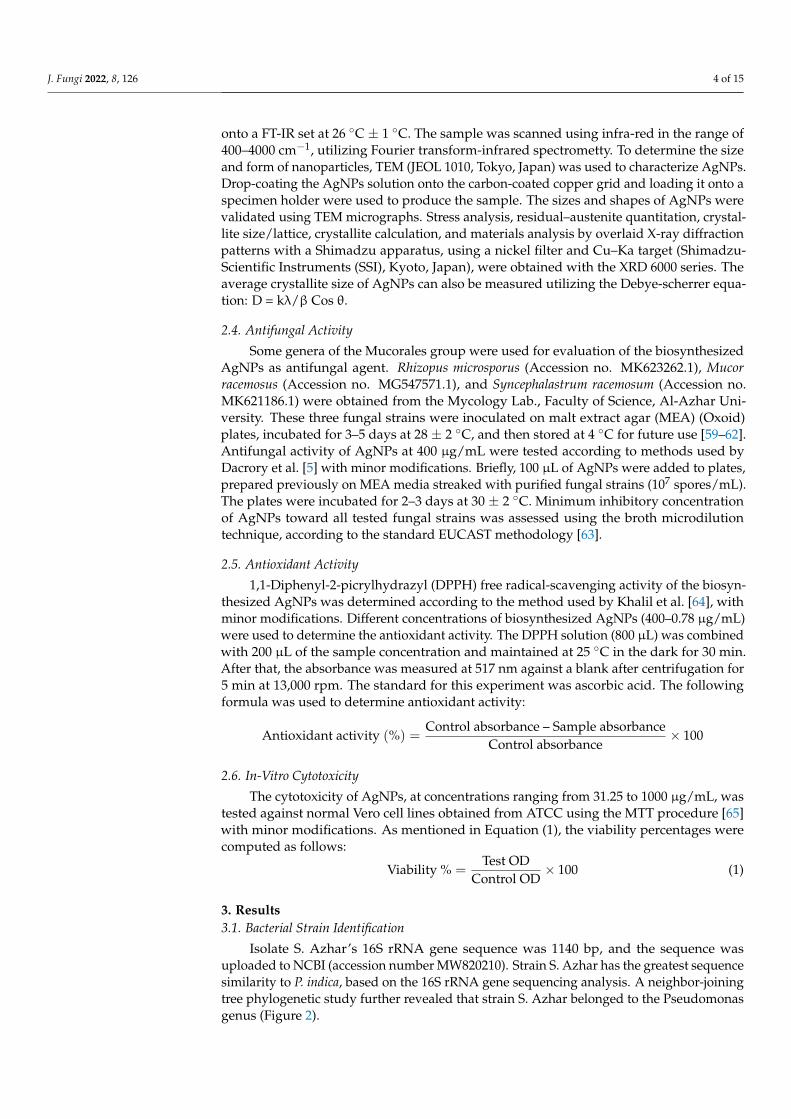
Isolate S. Azhar’s 16S rRNA gene sequence was 1140 bp, and the sequence wasuploaded to NCBI (accession number MW820210). Strain S. Azhar has the greatest sequencesimilarity to P. indica, based on the 16S rRNA gene sequencing analysis. A neighbor-joiningtree phylogenetic study further revealed that strain S. Azhar belonged to the Pseudomonasgenus (Figure 2).
J. Fungi 2022, 8, 126 5 of 15
Figure 2. A neighbor-joining (NJ) tree based on 16S rRNA gene sequence analysis was used to showthe phylogenetic relationships of the isolated strain S. Azhar with comparable type strains.
3.2. Synthesis and Characterization of AgNPs
In this study, biomass filtrate of P. indica was incubated with 1 mM AgNO3 for 24 hin dark conditions. Appearance of brown color after contacting of the filtrate of P. indica S.Azhar with precursor (AgNO3) at the reaction completion indicated AgNPs’ formation;maximum absorbance peaks at 420 nm were seen in the UV-vis spectral analysis, whichmight correlate to spherical AgNPs, as seen in Figure 3A. Surface plasmon resonance(SPR) excitation might be responsible for the color alteration. After calcination, AgNPswere obtained as a black powder. The functional groups of those found in AgNPs werecharacterized using FT-IR analysis. As seen in Figure 3B. The occurrence of functionalassemblies of biomolecules was discovered using FTIR wavelengths of 400 to 4000 cm−1.Ten prominent peaks in the FTIR spectra of biosynthesized AgNPs were found at 474.4,617.1, 1110.8, 1382.7, 1617.9, 1637.2, 2032.6, 2921.6, 3235.9, and 3415.3 cm−1 (Figure 3B). Thepeaks at 3235.9 and 3415.3 cm−1 correspond to the alcohol O–H stretching group or thesecondary amine N–H stretching group. NH stretching of the protein’s amide I band isshown by the bands at 1637.2and 1617.9 cm−1. Alkyne stretch bands are represented by thepeaks at 2032.6 and 2921.6 cm−1. Furthermore, the bands seen at 1382.7 and 1110.8 cm−1
might be attributed to aromatic and aliphatic amine C-N stretching vibrations. Finally,the peaks at 617.1 and 474.4 cm−1 correspond to the bending of alkene (C=H) groups.As a result, the current work reveals that proteins or bacterial extracts attach quickly toAgNPs via the proteins’ free amino or carboxyl groups; moreover, the acquired form ofNPs changes as the protein binding with AgNPs varies.
The most effective approach for identifying morphological features, such as the sizeand form, of biosynthesized AgNPs is TEM examination. Figure 4 reveals the effectivemanufacture of spherical AgNPs using metabolites found in P. indica S. Azhar’s filtrate,with typical sizes ranging from 2.4 to 53.5 nm. Furthermore, the biologically producednanoparticles were evenly diffused, with no aggregation or morphological discrepancy.
XRD based AgNPs characterization exhibited five peaks at 2θ values: 38.2◦, 44.46◦,64.22◦, 77.52◦, and 81.22◦, which were assigned to planes 111, 200, 220, 311, and 222,respectively for AgNPs Figure 5A. The average sizes of crystallite Ag- particles werecalculated using Scherrer’s equation. In this context, the size of Ag particles rangedbetween 8 to 80 nm, which were the outputs from the analysis of the equation. In linewith our clarification of the results, we reported the successful fabrication of crystallite,monoclinic-phase AgNPs at the same XRD diffraction planes utilizing metabolites ofP. indica S. Azhar.

J. Fungi 2022, 8, 126 6 of 15
Figure 3. UV–Vis spectrophotometer (A) and FT-IR spectra (B) of AgNPs synthesized by P. indica S. Azhar.
Figure 4. TEM image of AgNPs synthesized by of P. indica S. Azhar.
Figure 5. XRD pattern (A) and SAED pattern (B) of biosynthesized AgNPs.
J. Fungi 2022, 8, 126 7 of 15
3.3. Antifungal Activity
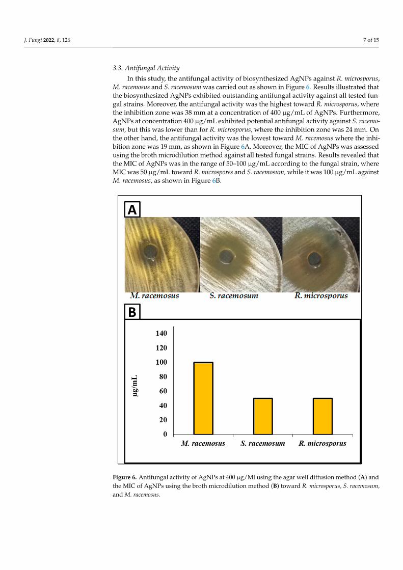
In this study, the antifungal activity of biosynthesized AgNPs against R. microsporus,M. racemosus and S. racemosum was carried out as shown in Figure 6. Results illustrated thatthe biosynthesized AgNPs exhibited outstanding antifungal activity against all tested fun-gal strains. Moreover, the antifungal activity was the highest toward R. microsporus, wherethe inhibition zone was 38 mm at a concentration of 400 µg/mL of AgNPs. Furthermore,AgNPs at concentration 400 µg/mL exhibited potential antifungal activity against S. racemo-sum, but this was lower than for R. microsporus, where the inhibition zone was 24 mm. Onthe other hand, the antifungal activity was the lowest toward M. racemosus where the inhi-bition zone was 19 mm, as shown in Figure 6A. Moreover, the MIC of AgNPs was assessedusing the broth microdilution method against all tested fungal strains. Results revealed thatthe MIC of AgNPs was in the range of 50–100 µg/mL according to the fungal strain, whereMIC was 50 µg/mL toward R. microspores and S. racemosum, while it was 100 µg/mL againstM. racemosus, as shown in Figure 6B.
Figure 6. Antifungal activity of AgNPs at 400 µg/Ml using the agar well diffusion method (A) andthe MIC of AgNPs using the broth microdilution method (B) toward R. microsporus, S. racemosum,and M. racemosus.
J. Fungi 2022, 8, 126 8 of 15
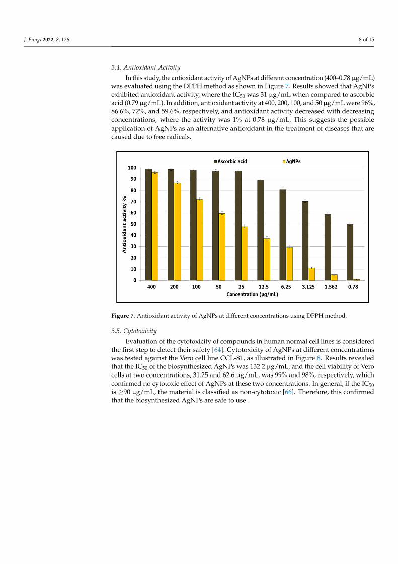
3.4. Antioxidant Activity
In this study, the antioxidant activity of AgNPs at different concentration (400–0.78 µg/mL)was evaluated using the DPPH method as shown in Figure 7. Results showed that AgNPsexhibited antioxidant activity, where the IC50 was 31 µg/mL when compared to ascorbicacid (0.79 µg/mL). In addition, antioxidant activity at 400, 200, 100, and 50 µg/mL were 96%,86.6%, 72%, and 59.6%, respectively, and antioxidant activity decreased with decreasingconcentrations, where the activity was 1% at 0.78 µg/mL. This suggests the possibleapplication of AgNPs as an alternative antioxidant in the treatment of diseases that arecaused due to free radicals.
Figure 7. Antioxidant activity of AgNPs at different concentrations using DPPH method.
3.5. Cytotoxicity
Evaluation of the cytotoxicity of compounds in human normal cell lines is consideredthe first step to detect their safety [64]. Cytotoxicity of AgNPs at different concentrationswas tested against the Vero cell line CCL-81, as illustrated in Figure 8. Results revealedthat the IC50 of the biosynthesized AgNPs was 132.2 µg/mL, and the cell viability of Verocells at two concentrations, 31.25 and 62.6 µg/mL, was 99% and 98%, respectively, whichconfirmed no cytotoxic effect of AgNPs at these two concentrations. In general, if the IC50is ≥90 µg/mL, the material is classified as non-cytotoxic [66]. Therefore, this confirmedthat the biosynthesized AgNPs are safe to use.

J. Fungi 2022, 8, 126 9 of 15
Figure 8. Cytotoxicity of AgNPs on Vero cell line.
4. Discussion
Green biosynthesis of AgNPs has risen in prominence as a potential alternative tochemical and physical techniques. Metabolites secreted by P. indica S. Azhar are effectivein the formation of AgNPs, besides stabilizing the formed NPs. P. indica S. Azhar filtratewas utilized as a bio-reactor for the formation of AgNPs through harnessing bioactivemacromolecules that are secreted from it. P. indica nanoparticles are currently in the earlyphases of development. P. indica-derived biogenic AgNPs are more appealing and lesshazardous to the environment than other approaches. The proteins or enzymes involved incell-free filtrates of P. indica that change nitrate to nitrite, and then reduced silver ions tosilver in the metallic form, may be responsible for the synthesis of AgNPs. The absorbancepeak at 420 nm in the UV-visible spectrum corresponded to the typical band of AgNPsgenerated by the P. indica cell free filtrate. The absorption peak at around 400–450 nm isattributed to AgNPs surface plasmon resonance, confirming the production of AgNPs [67].Similarly, Alsharif et al. [68] observed that AgNPs generated by Bacillus cereus presented asingle symmetric maximum at wavelength 420 nm, which is linked to spherical structure.The capping of biosynthesized AgNPs was discovered using Fourier transform-infraredspectroscopy. The FT-IR spectral analysis of AgNPs revealed N–H aliphatic amino grouppeaks at 3415.3 and 3235.9 cm−1, whereas the –CH2 group is indicated by the peak at

J. Fungi 2022, 8, 126 10 of 15
2921.6 cm−1 [69]. The (OH) stretch of the carboxyl group is shown by the spectrum peak at2032.6 cm−1. The binding of the amide I band of protein with the (N–H) stretch is associatedwith the peaks at 1637.2 and 1617.9 cm−1 [22]. The C–H and C–O stretching bands maybe recognized at 1382.9 and 1110.8 cm−1, respectively [47]. The relationship between Agand physiologically active chemicals that are responsible for the production and stability ofAgNPs as capping agents was investigated using FTIR. The infrared area of the spectrumis designated by the carboxylic acid group (CHOO–), obtained with an amine (NH2) inthe amino acid of proteins [70]. The most efficient approach for identifying morphologicalfeatures, such as the size and form of produced AgNPs, is TEM examination. Accordingto TEM data, the biosynthesized AgNPs had a spherical form with a size range of 2.4 to53.5 nm. According to a previous study, researchers successfully formed spherical AgNPswith a size range of 6–50 nm using TEM, as seen in Alsharif et al. [68]. Other reports showeda difference in the shape and size of AgNPs biologically formed by bacteria [71,72]. The sizerange of AgNPs generated by B. licheniformis filtrate during incubation with 1 and 3 mMAgNO3 was (3–130 nm) and (45–170 nm), respectively, according to Sarangadharan andNallusamy [73]. When the biomass of B. licheniformis was incubated with 1 mM AgNO3,the mean size of AgNPs reached around 50 nm in another investigation [74]. Specific peaksin the XRD spectra were used to illustrate the XRD pattern of the AgNPs biosynthesizedby P. indica. The face center cubic (fcc) nano-structures of AgNPs (111), (200), (220), (311),and (222), were characterized by five diffraction peaks at 2θ values of 38.2◦, 44.46◦, 64.22◦,77.52◦, and 81.22◦, respectively, with an average size 8–80 nm. According to another study,the diffraction peaks at 2θ = 38.2◦, 46.2◦, 64.6◦, and 77.6◦ exemplify the 111, 200, 220, and311 Bragg’s reflection of Ag nanoparticles’ face-centered-cubic structure [75]. Previousresearch that demonstrated the creation of AgNPs utilizing microbes had comparable XRDresults [69,76,77].
Mucormycosis disease is very dangerous to humans and is caused by membersof the Mucorales genera, such as Rhizopus, Mucor, and Sycephalastrum. Therefore, thecontrol of these fungi by biomaterials is required. The biosynthesized AgNPs in thisstudy revealed promising antifungal activity toward Mucorales genera species such asR. microsporus, M. racemosus, and S. racemosum. Medda et al. [78] synthesized AgNPsusing Aloe vera leaf extract, and found antifungal activity toward Rhizopus sp. andAspergillus sp. Alananbeh et al. [79] revealed that promising antifungal activity of AgNPstoward M. hemalis and R. arrhizus. AgNPs might be critical in breaking down such resis-tance. AgNPs’ efficacy can be attributed to a variety of mechanisms, including cell walldisintegration, surface protein degradation, nucleic acid damage caused by the formationand buildup of reactive oxygen and nitrogen species (ROS and free radicals), and protonpump blocking. AgNPs are thought to cause a buildup of silver ions, which obstructsrespiration by causing intracellular ion efflux, causing harm to the electron transportsystem [80].
Oxidative stress is a condition in which the balance between a cell’s antioxidative de-fense and oxidants is disturbed as a result of oxidant excess [78]. Furthermore, the presenceof oxidants causes oxidative changes of biological systems at the molecular level (unsat-urated bonds of lipids, proteins, DNA, and so on), resulting in damage and, ultimately,hastened cellular death [79]. Antioxidants are natural or manmade compounds that canhelp to prevent or postpone oxidative cell damage (ROS, RNS, free radicals, other unstablemolecules) [79]. Therefore, antioxidant compounds are continuously required to resist ox-idative stress. The biosynthesized AgNPs in the current study revealed antioxidant activitywhere the IC50 was 31 µg/mL. Previous studies have confirmed the antioxidant activity ofAgNPs from different sources [81–83]. González-Ballesteros et al. [82] reported that AgNPs,silver nanoparticles, can be made from Lactobacillus brevis exopolysaccharides, and thesewere tested for antioxidant capabilities. At 100 g/mL, AgNPs had a moderate DPPHradical scavenging potential of 34.09% [83]. Evaluation of the cytotoxicity of compounds onhuman normal cell lines is considered the first step to detect their safety [64]. Therefore, the
J. Fungi 2022, 8, 126 11 of 15
cytotoxicity of biosynthesized AgNPs was assessed, where results confirmed that AgNPsare safe in use.
5. Conclusions
In the current study, a fast, green, eco-friendly method was used to synthesize AgNPsby P. indica S. Azhar for controlling fungi that cause mucormycosis, as well as antioxidantactivity. Results revealed that AgNPs were fabricated using metabolites of P. indica, then, thebiosynthesized AgNPs were characterized using different modern techniques. Moreover,the biosynthesized AgNPs exhibited outstanding antifungal activity against R. microsporus,M. racemosus, and S. racemosum. Furthermore, results revealed antioxidant activity withoutany cytotoxicity on the Vero normal cell line. The results of this study warrant furtherin vivo experiments.
Author Contributions: Conceptualization, S.S.S. and A.H.H.; methodology, S.S.S. and A.H.H.; vali-dation, S.S.S. and A.H.H.; formal analysis, S.S.S. and A.H.H.; investigation, S.S.S., O.M.A., A.M.R.,K.A.A.-E. and A.H.H.; software S.S.S. and A.H.H.; resources, S.S.S. and A.H.H.; data curation, S.S.S.and A.H.H.; Funding O.M.A. and K.A.A.-E.; writing—original draft preparation, S.S.S. and A.H.H.;writing—review and editing, S.S.S., O.M.A., A.M.R., K.A.A.-E. and A.H.H.; visualization, S.S.S.,O.M.A., A.M.R., K.A.A.-E. and A.H.H. All authors have read and agreed to the published version ofthe manuscript.
Funding: This study was supported by the Taif University Researchers Supporting Project (TURSP-2020/81) at Taif University in Taif, Saudi Arabia.
Institutional Review Board Statement: Not applicable.
Informed Consent Statement: Not applicable.
Data Availability Statement: The data used to support the conclusions of this study are accessibleupon request from the corresponding author.
Acknowledgments: The authors express their sincere thanks to Faculty of Science (Boys), Al-AzharUniversity, Cairo, Egypt for providing the necessary research facilities.
Conflicts of Interest: The authors declare that they have no conflict of interest.
References1. Chang, Y.-L.; Yu, S.-J.; Heitman, J.; Wellington, M.; Chen, Y.-L. New facets of antifungal therapy. Virulence 2017, 8, 222–236.
[CrossRef] [PubMed]2. Campoy, S.; Adrio, J.L. Antifungals. Biochem. Pharmacol. 2017, 133, 86–96. [CrossRef] [PubMed]3. Brown, G.D.; Denning, D.W.; Gow, N.A.; Levitz, S.M.; Netea, M.G. White TC (2012) Hidden killers: Human fungal infections. Sci.
Transl. Med. 2012, 4, 165rv113. [CrossRef] [PubMed]4. Kontoyiannis, D.P.; Lewis, R.E. Agents of mucormycosis and entomophthoramycosis. In Mandell, Douglas, and Bennett’s Principles
and Practice of Infectious Diseases; Elsevier: Amsterdam, The Netherlands, 2014; pp. 2909–2919.5. Dacrory, S.; Hashem, A.H.; Hasanin, M. Synthesis of cellulose based amino acid functionalized nano-biocomplex: Characterization,
antifungal activity, molecular docking and hemocompatibility. Environ. Nanotechnol. Monit. Manag. 2021, 15, 100453. [CrossRef]6. Roden, M.M.; Zaoutis, T.E.; Buchanan, W.L.; Knudsen, T.A.; Sarkisova, T.A.; Schaufele, R.L.; Sein, M.; Sein, T.; Chiou, C.C.;
Chu, J.H.; et al. Epidemiology and outcome of zygomycosis: A review of 929 reported cases. Clinical infectious diseases: Anofficial publication of the Infectious Diseases Society of America. Clin. Infect. Dis. 2005, 41, 634–653. [CrossRef]
7. Petrikkos, G.; Skiada, A.; Lortholary, O.; Roilides, E.; Walsh, T.J.; Kontoyiannis, D.P. Epidemiology and clinical manifestations ofmucormycosis. Clinical infectious diseases: An official publication of the Infectious Diseases Society of America. Clin. Infect. Dis.2012, 54 (Suppl. 1), S23–S34. [CrossRef]
8. Walsh, T.J.; Gamaletsou, M.N.; McGinnis, M.R.; Hayden, R.T.; Kontoyiannis, D.P. Early clinical and laboratory diagnosis ofinvasive pulmonary, extrapulmonary, and disseminated mucormycosis (zygomycosis). Clinical infectious diseases: An officialpublication of the Infectious Diseases Society of America. Clin. Infect. Dis. 2012, 54 (Suppl. 1), S55–S60. [CrossRef]
9. Skiada, A.; Lass-Floerl, C.; Klimko, N.; Ibrahim, A.; Roilides, E.; Petrikkos, G. Challenges in the diagnosis and treatment ofmucormycosis. Med. Mycol. 2018, 56 (Suppl. 1), S93–S101. [CrossRef]
10. Sanglard, D.; Odds, F.C. Resistance of Candida species to antifungal agents: Molecular mechanisms and clinical consequences.Lancet Infect. Dis. 2002, 2, 73–85. [CrossRef]
J. Fungi 2022, 8, 126 12 of 15
11. Dannaoui, E. Antifungal resistance in mucorales. Int. J. Antimicrob. Agents 2017, 50, 617–621. [CrossRef]12. Pfaller, M.A. Antifungal drug resistance: Mechanisms, epidemiology, and consequences for treatment. Am. J. Med. 2012, 125,
S3–S13. [CrossRef] [PubMed]13. Salem, S.S.; Fouda, A. Green Synthesis of Metallic Nanoparticles and Their Prospective Biotechnological Applications: An
Overview. Biol. Trace Elem. Res. 2021, 199, 344–370. [CrossRef] [PubMed]14. Shaheen, T.I.; Fouda, A.; Salem, S.S. Integration of Cotton Fabrics with Biosynthesized CuO Nanoparticles for Bactericidal Activity
in the Terms of Their Cytotoxicity Assessment. Ind. Eng. Chem. Res. 2021, 60, 1553–1563. [CrossRef]15. Fouda, A.; Salem, S.S.; Wassel, A.R.; Hamza, M.F.; Shaheen, T.I. Optimization of green biosynthesized visible light active
CuO/ZnO nano-photocatalysts for the degradation of organic methylene blue dye. Heliyon 2020, 6, e04896. [CrossRef] [PubMed]16. Salem, S.S.; Fouda, M.M.G.; Fouda, A.; Awad, M.A.; Al-Olayan, E.M.; Allam, A.A.; Shaheen, T.I. Antibacterial, Cytotoxicity and
Larvicidal Activity of Green Synthesized Selenium Nanoparticles Using Penicillium corylophilum. J. Clust. Sci. 2021, 32, 351–361.[CrossRef]
17. Elfeky, A.S.; Salem, S.S.; Elzaref, A.S.; Owda, M.E.; Eladawy, H.A.; Saeed, A.M.; Awad, M.A.; Abou-Zeid, R.E.; Fouda, A.Multifunctional cellulose nanocrystal /metal oxide hybrid, photo-degradation, antibacterial and larvicidal activities. Carbohydr.Polym. 2020, 230, 115711. [CrossRef] [PubMed]
18. Sharaf, O.M.; Al-Gamal, M.S.; Ibrahim, G.A.; Dabiza, N.M.; Salem, S.S.; El-ssayad, M.F.; Youssef, A.M. Evaluation and characteri-zation of some protective culture metabolites in free and nano-chitosan-loaded forms against common contaminants of Egyptiancheese. Carbohydr. Polym. 2019, 223, 115094. [CrossRef]
19. Hasanin, M.; Al Abboud, M.A.; Alawlaqi, M.M.; Abdelghany, T.M.; Hashem, A.H. Ecofriendly synthesis of biosynthesized coppernanoparticles with starch-based nanocomposite: Antimicrobial, antioxidant, and anticancer activities. Biol. Trace Elem. Res. 2021,1–14. [CrossRef]
20. Hasanin, M.; Hashem, A.H.; Lashin, I.; Hassan, S.A.M. In vitro improvement and rooting of banana plantlets using antifungalnanocomposite based on myco-synthesized copper oxide nanoparticles and starch. Biomass Convers. Biorefin. 2021, 1–11.[CrossRef]
21. Hashem, A.H.; Khalil, A.M.A.; Reyad, A.M.; Salem, S.S. Biomedical Applications of Mycosynthesized Selenium NanoparticlesUsing Penicillium expansum ATTC 36200. Biol. Trace Elem. Res. 2021, 199, 3998–4008. [CrossRef]
22. Eid, A.M.; Fouda, A.; Niedbała, G.; Hassan, S.E.D.; Salem, S.S.; Abdo, A.M.; Hetta, H.F.; Shaheen, T.I. Endophytic streptomyceslaurentii mediated green synthesis of Ag-NPs with antibacterial and anticancer properties for developing functional textile fabricproperties. Antibiotics 2020, 9, 641. [CrossRef] [PubMed]
23. Hashem, A.H.; Salem, S.S. Green and ecofriendly biosynthesis of selenium nanoparticles using Urtica dioica (stinging nettle) leafextract: Antimicrobial and anticancer activity. Biotechnol. J. 2021, 2100432. [CrossRef] [PubMed]
24. Badawy, A.A.; Abdelfattah, N.A.H.; Salem, S.S.; Awad, M.F.; Fouda, A. Efficacy assessment of biosynthesized copper oxidenanoparticles (Cuo-nps) on stored grain insects and their impacts on morphological and physiological traits of wheat (Triticumaestivum L.) plant. Biology 2021, 10, 233. [CrossRef]
25. Mohamed, A.A.; Abu-Elghait, M.; Ahmed, N.E.; Salem, S.S. Eco-friendly Mycogenic Synthesis of ZnO and CuO Nanoparticles forIn Vitro Antibacterial, Antibiofilm, and Antifungal Applications. Biol. Trace Elem. Res. 2021, 199, 2788–2799. [CrossRef]
26. Saied, E.; Eid, A.M.; Hassan, S.E.D.; Salem, S.S.; Radwan, A.A.; Halawa, M.; Saleh, F.M.; Saad, H.A.; Saied, E.M.; Fouda, A. Thecatalytic activity of biosynthesized magnesium oxide nanoparticles (Mgo-nps) for inhibiting the growth of pathogenic microbes,tanning effluent treatment, and chromium ion removal. Catalysts 2021, 11, 821. [CrossRef]
27. Abdelmoneim, H.E.M.; Wassel, M.A.; Elfeky, A.S.; Bendary, S.H.; Awad, M.A.; Salem, S.S.; Mahmoud, S.A. Multiple Applicationsof CdS/TiO2 Nanocomposites Synthesized via Microwave-Assisted Sol–Gel. J. Clust. Sci. 2021, 1–10. [CrossRef]
28. Hashem, A.H.; Abdelaziz, A.M.; Askar, A.A.; Fouda, H.M.; Khalil, A.M.A.; Abd-Elsalam, K.A.; Khaleil, M.M. Bacillus megaterium-Mediated Synthesis of Selenium Nanoparticles and Their Antifungal Activity against Rhizoctonia solani in Faba Bean Plants.J. Fungi 2021, 7, 195. [CrossRef]
29. Hashem, A.H.; Al Abboud, M.A.; Alawlaqi, M.M.; Abdelghany, T.M.; Hasanin, M. Synthesis of Nanocapsules Based onBiosynthesized Nickel Nanoparticles and Potato Starch: Antimicrobial, Antioxidant and Anticancer Activity. Starch-Stärke 2022,74, 2100165. [CrossRef]
30. Mohamed, A.A.; Fouda, A.; Abdel-Rahman, M.A.; Hassan, S.E.-D.; El-Gamal, M.S.; Salem, S.S.; Shaheen, T.I. Fungal strainimpacts the shape, bioactivity and multifunctional properties of green synthesized zinc oxide nanoparticles. Biocatal. Agric.Biotechnol. 2019, 19, 101103. [CrossRef]
31. Jain, N.; Jain, P.; Rajput, D.; Patil, U.K. Green synthesized plant-based silver nanoparticles: Therapeutic prospective for anticancerand antiviral activity. Micro Nano Syst. Lett. 2021, 9, 5. [CrossRef]
32. Abdel-Khalek, A.A.; Al-Quraishy, S.; Abdel-Gaber, R. Silver Nanoparticles Induce Time- and Tissue-Specific Genotoxicity inOreochromis niloticus: Utilizing the Adsorptive Capacities of Fruit Peels to Minimize Genotoxicity. Bull. Environ. Contam. Toxicol.2021, 1–9. [CrossRef] [PubMed]
33. Nayak, S.; Manjunatha, K.B.; Goveas, L.C.; Rao, C.V.; Sajankila, S.P. Investigation of Nonlinear Optical Properties of AgNPsSynthesized Using Cyclea peltata Leaf Extract Post OVAT Optimization. BioNanoScience 2021, 11, 884–892. [CrossRef]
J. Fungi 2022, 8, 126 13 of 15
34. Beyene, H.D.; Werkneh, A.A.; Bezabh, H.K.; Ambaye, T.G. Synthesis paradigm and applications of silver nanoparticles (AgNPs),a review. Sustain. Mater. Technol. 2017, 13, 18–23. [CrossRef]
35. Elbahnasawy, M.A.; Shehabeldine, A.M.; Khattab, A.M.; Amin, B.H.; Hashem, A.H. Green biosynthesis of silver nanoparticlesusing novel endophytic Rothia endophytica: Characterization and anticandidal activity. J. Drug Deliv. Sci. Technol. 2021, 62, 102401.[CrossRef]
36. Hasanin, M.; Elbahnasawy, M.A.; Shehabeldine, A.M.; Hashem, A.H. Ecofriendly preparation of silver nanoparticles-basednanocomposite stabilized by polysaccharides with antibacterial, antifungal and antiviral activities. BioMetals 2021, 34, 1313–1328.[CrossRef]
37. Anees Ahmad, S.; Sachi Das, S.; Khatoon, A.; Tahir Ansari, M.; Afzal, M.; Saquib Hasnain, M.; Kumar Nayak, A. Bactericidalactivity of silver nanoparticles: A mechanistic review. Mater. Sci. Energy Technol. 2020, 3, 756–769. [CrossRef]
38. Korkmaz, N.; Ceylan, Y.; Hamid, A.; Karadag, A.; Bülbül, A.S.; Aftab, M.N.; Çevik, Ö.; Sen, F. Biogenic silver nanoparticlessynthesized via Mimusops elengi fruit extract, a study on antibiofilm, antibacterial, and anticancer activities. J. Drug Deliv. Sci.Technol. 2020, 59, 101864. [CrossRef]
39. Pilaquinga, F.; Morey, J.; Torres, M.; Seqqat, R.; Piña, M.D.L.N. Silver nanoparticles as a potential treatment against SARS-CoV-2:A review. WIREs Nanomed. Nanobiotechnol. 2021, 13, e1707. [CrossRef]
40. Ciriminna, R.; Albo, Y.; Pagliaro, M. New Antivirals and Antibacterials Based on Silver Nanoparticles. ChemMedChem 2020, 15,1619–1623. [CrossRef]
41. Bocate, K.P.; Reis, G.F.; de Souza, P.C.; Oliveira Junior, A.G.; Durán, N.; Nakazato, G.; Furlaneto, M.C.; de Almeida, R.S.; Panagio,L.A. Antifungal activity of silver nanoparticles and simvastatin against toxigenic species of Aspergillus. Int. J. Food Microbiol. 2019,291, 79–86. [CrossRef]
42. Alam, A.; Tanveer, F.; Khalil, A.T.; Zohra, T.; Khamlich, S.; Alam, M.M.; Salman, M.; Ali, M.; Ikram, A.; Shinwari, Z.K.; et al. Silvernanoparticles biosynthesized from secondary metabolite producing marine actinobacteria and evaluation of their biomedicalpotential. Antonie van Leeuwenhoek 2021, 114, 1497–1516. [CrossRef] [PubMed]
43. Alomar, T.S.; AlMasoud, N.; Awad, M.A.; El-Tohamy, M.F.; Soliman, D.A. An eco-friendly plant-mediated synthesis of silvernanoparticles: Characterization, pharmaceutical and biomedical applications. Mater. Chem. Phys. 2020, 249, 123007. [CrossRef]
44. Shaheen, T.I.; Salem, S.S.; Fouda, A. Current Advances in Fungal Nanobiotechnology: Mycofabrication and Applications. InMicrobial Nanobiotechnology: Principles and Applications; Lateef, A., Gueguim-Kana, E.B., Dasgupta, N., Ranjan, S., Eds.; Springer:Singapore, 2021; pp. 113–143. [CrossRef]
45. Aref, M.S.; Salem, S.S. Bio-callus synthesis of silver nanoparticles, characterization, and antibacterial activities via Cinnamomumcamphora callus culture. Biocatal. Agric. Biotechnol. 2020, 27, 101689. [CrossRef]
46. Fouda, A.; Abdel-Maksoud, G.; Abdel-Rahman, M.A.; Salem, S.S.; Hassan, S.E.-D.; El-Sadany, M.A.-H. Eco-friendly approachutilizing green synthesized nanoparticles for paper conservation against microbes involved in biodeterioration of archaeologicalmanuscript. Int. Biodeterior. Biodegrad. 2019, 142, 160–169. [CrossRef]
47. Huq, M.A. Green Synthesis of Silver Nanoparticles Using Pseudoduganella eburnea MAHUQ-39 and Their Antimicrobial Mecha-nisms Investigation against Drug Resistant Human Pathogens. Int. J. Mol. Sci. 2020, 21, 1510. [CrossRef] [PubMed]
48. Mohamed, A.A.; Fouda, A.; Elgamal, M.S.; EL-Din Hassan, S.; Shaheen, T.I.; Salem, S.S. Enhancing of cotton fabric antibacterialproperties by silver nanoparticles synthesized by new Egyptian strain Fusarium keratoplasticum A1-3. In Proceedings of the8th International Conference of The Textile Research Division (ICTRD 2017), National Research Centre, Cairo, Egypt, 25–27September 2017; pp. 63–71.
49. Mohmed, A.A.; Saad, E.; Fouda, A.; Elgamal, M.S.; Salem, S.S. Extracellular biosynthesis of silver nanoparticles using Aspergillussp. and evaluation of their antibacterial and cytotoxicity. J. Appl. Life Sci. Int. 2017, 11, 1–12. [CrossRef]
50. Tehri, N.; Vashishth, A.; Gahlaut, A.; Hooda, V. Biosynthesis, antimicrobial spectra and applications of silver nanoparticles:Current progress and future prospects. Inorg. Nano-Met. Chem. 2020, 52, 1–19. [CrossRef]
51. Zhang, D.; Ma, X.-L.; Gu, Y.; Huang, H.; Zhang, G.-W. Green Synthesis of Metallic Nanoparticles and Their Potential Applicationsto Treat Cancer. Front. Chem. 2020, 8, 799. [CrossRef] [PubMed]
52. Shehabeldine, A.M.; Hashem, A.H.; Wassel, A.R.; Hasanin, M. Antimicrobial and Antiviral Activities of Durable Cotton FabricsTreated with Nanocomposite Based on Zinc Oxide Nanoparticles, Acyclovir, Nanochitosan, and Clove Oil. Appl. Biochem.Biotechnol. 2021, 1–18. [CrossRef] [PubMed]
53. lashin, I.; Hasanin, M.; Hassan, S.A.M.; Hashem, A.H. Green biosynthesis of zinc and selenium oxide nanoparticles using callusextract of Ziziphus spina-christi: Characterization, antimicrobial, and antioxidant activity. Biomass Convers. Biorefin. 2021, 1–14.[CrossRef]
54. El-Naggar, M.E.; Hasanin, M.; Hashem, A.H. Eco-Friendly Synthesis of Superhydrophobic Antimicrobial Film Based on CelluloseAcetate/Polycaprolactone Loaded with the Green Biosynthesized Copper Nanoparticles for Food Packaging Application.J. Polym. Environment. 2021, 1–13. [CrossRef]
55. Abd Elkodous, M.; El-Husseiny, H.M.; El-Sayyad, G.S.; Hashem, A.H.; Doghish, A.S.; Elfadil, D.; Radwan, Y.; El-Zeiny, H.M.;Bedair, H.; Ikhdair, O.A. Recent advances in waste-recycled nanomaterials for biomedical applications: Waste-to-wealth. Nan-otechnol. Rev. 2021, 10, 1662–1739. [CrossRef]
56. Angelin, J.; Kavitha, M. Exopolysaccharides from probiotic bacteria and their health potential. Int. J. Biol. Macromol. 2020, 162,853–865. [CrossRef] [PubMed]
J. Fungi 2022, 8, 126 14 of 15
57. Adetunji, A.I.; Olaniran, A.O. Production and potential biotechnological applications of microbial surfactants: An overview.Saudi J. Biol. Sci. 2021, 28, 669–679. [CrossRef] [PubMed]
58. Moreno, V.M.; Álvarez, E.; Izquierdo-Barba, I.; Baeza, A.; Serrano-López, J.; Vallet-Regí, M. Bacteria as Nanoparticles Carrier forEnhancing Penetration in a Tumoral Matrix Model. Adv. Mater. Interfaces 2020, 7, 1901942. [CrossRef] [PubMed]
59. Fouda, A.; Khalil, A.; El-Sheikh, H.; Abdel-Rhaman, E.; Hashem, A. Biodegradation and detoxification of bisphenol-A byfilamentous fungi screened from nature. J. Adv. Biol. Biotechnol. 2015, 2, 123–132. [CrossRef]
60. Hashem, A.H.; Hasanin, M.S.; Khalil, A.M.A.; Suleiman, W.B. Eco-green conversion of watermelon peels to single cell oils using aunique oleaginous fungus: Lichtheimia corymbifera AH13. Waste Biomass Valoris. 2019, 11, 5721–5732. [CrossRef]
61. Suleiman, W.; El-Sheikh, H.; Abu-Elreesh, G.; Hashem, A. Recruitment of Cunninghamella echinulata as an Egyptian isolate toproduce unsaturated fatty acids. Res. J. Pharm. Biol. Chem. Sci. 2018, 9, 764–774.
62. Suleiman, W.; El-Skeikh, H.; Abu-Elreesh, G.; Hashem, A. Isolation and screening of promising oleaginous Rhizopus sp. anddesigning of Taguchi method for increasing lipid production. J. Innov. Pharm. Biol. Sci. 2018, 5, 8–15.
63. Rodriguez-Tudela, J.; Arendrup, M.; Arikan, S.; Barchiesi, F.; Bille, J.; Chryssanthou, E.; Cuenca-Estrella, M.; Dannaoui, E.;Denning, D.; Donnelly, J. Eucast Definitive Document E. Def 9.1: Method for the determination of broth dilution minimuminhibitory concentrations of antifungal agents for conidia forming moulds. Def 2008, 9, 1–13.
64. Khalil, A.; Abdelaziz, A.; Khaleil, M.; Hashem, A. Fungal endophytes from leaves of Avicennia marina growing in semi-aridenvironment as a promising source for bioactive compounds. Lett. Appl. Microbiol. 2021, 72, 263–274. [CrossRef] [PubMed]
65. Van de Loosdrecht, A.; Beelen, R.; Ossenkoppele, G.; Broekhoven, M.; Langenhuijsen, M. A tetrazolium-based colorimetric MTTassay to quantitate human monocyte mediated cytotoxicity against leukemic cells from cell lines and patients with acute myeloidleukemia. J. Immunol. Methods 1994, 174, 311–320. [CrossRef]
66. Ioset, J.-R.; Brun, R.; Wenzler, T.; Kaiser, M.; Yardley, V. Drug Screening for Kinetoplastids Diseases: A Training Manual for Screening inNeglected Diseases; DNDi and Pan-Asian Screening Network: Geneva, Switzerland, 2009.
67. Wypij, M.; Czarnecka, J.; Swiecimska, M.; Dahm, H.; Rai, M.; Golinska, P. Synthesis, characterization and evaluation ofantimicrobial and cytotoxic activities of biogenic silver nanoparticles synthesized from Streptomyces xinghaiensis OF1 strain. WorldJ. Microbiol. Biotechnol. 2018, 34, 23. [CrossRef] [PubMed]
68. Alsharif, S.M.; Salem, S.S.; Abdel-Rahman, M.A.; Fouda, A.; Eid, A.M.; Hassan, S.E.D.; Awad, M.A.; Mohamed, A.A. Multi-functional properties of spherical silver nanoparticles fabricated by different microbial taxa. Heliyon 2020, 6, e03943. [CrossRef][PubMed]
69. Salem, S.S.; El-Belely, E.F.; Niedbała, G.; Alnoman, M.M.; Hassan, S.E.D.; Eid, A.M.; Shaheen, T.I.; Elkelish, A.; Fouda, A.Bactericidal and in-vitro cytotoxic efficacy of silver nanoparticles (Ag-NPs) fabricated by endophytic actinomycetes and their useas coating for the textile fabrics. Nanomaterials 2020, 10, 2082. [CrossRef]
70. Alsamhary, K.I. Eco-friendly synthesis of silver nanoparticles by Bacillus subtilis and their antibacterial activity. Saudi J. Biol. Sci.2020, 27, 2185–2191. [CrossRef]
71. Saeed, S.; Iqbal, A.; Ashraf, M.A. Bacterial-mediated synthesis of silver nanoparticles and their significant effect against pathogens.Environ. Sci. Pollut. Res. 2020, 27, 37347–37356. [CrossRef]
72. Ibrahim, E.; Zhang, M.; Zhang, Y.; Hossain, A.; Qiu, W.; Chen, Y.; Wang, Y.; Wu, W.; Sun, G.; Li, B. Green-Synthesization of SilverNanoparticles Using Endophytic Bacteria Isolated from Garlic and Its Antifungal Activity against Wheat Fusarium Head BlightPathogen Fusarium graminearum. Nanomaterials 2020, 10, 219. [CrossRef]
73. Sarangadharan, S.; Nallusamy, S. Biosynthesis and characterization of silver nanoparticles produced by Bacillus licheniformis. Int.J. Pharma Med. Biol. Sci. 2015, 4, 236.
74. Kalimuthu, K.; Babu, R.S.; Venkataraman, D.; Bilal, M.; Gurunathan, S. Biosynthesis of silver nanocrystals by Bacillus licheniformis.Colloids Surf. B Biointerfaces 2008, 65, 150–153. [CrossRef]
75. Fouad, H.; Hongjie, L.; Yanmei, D.; Baoting, Y.; El-Shakh, A.; Abbas, G.; Jianchu, M. Synthesis and characterization of silvernanoparticles using Bacillus amyloliquefaciens and Bacillus subtilis to control filarial vector Culex pipiens pallens and its antimicrobialactivity. Artif. Cells Nanomed. Biotechnol. 2017, 45, 1369–1378. [CrossRef] [PubMed]
76. Du, J.; Singh, H.; Yi, T.-H. Biosynthesis of silver nanoparticles by Novosphingobium sp. THG-C3 and their antimicrobial potential.Artif. Cells Nanomed. Biotechnol. 2017, 45, 211–217. [CrossRef] [PubMed]
77. Zonooz, N.F.; Salouti, M. Extracellular biosynthesis of silver nanoparticles using cell filtrate of Streptomyces sp. ERI-3. ScientiaIranica 2011, 18, 1631–1635. [CrossRef]
78. Medda, S.; Hajra, A.; Dey, U.; Bose, P.; Mondal, N.K. Biosynthesis of silver nanoparticles from Aloe vera leaf extract and antifungalactivity against Rhizopus sp. and Aspergillus sp. Appl. Nanosci. 2015, 5, 875–880. [CrossRef]
79. Alananbeh, K.; Al-Refaee, W.; Al-Qodah, Z. Antifungal effect of silver nanoparticles on selected fungi isolated from raw andwaste water. Indian J. Pharm. Sci. 2017, 79, 559–567. [CrossRef]
80. Du, H.; Lo, T.-M.; Sitompul, J.; Chang, M.W. Systems-level analysis of Escherichia coli response to silver nanoparticles: The roles ofanaerobic respiration in microbial resistance. Biochem. Biophys. Res. Commun. 2012, 424, 657–662. [CrossRef] [PubMed]
81. Azeez, L.; Lateef, A.; Adebisi, S.A. Silver nanoparticles (AgNPs) biosynthesized using pod extract of Cola nitida enhancesantioxidant activity and phytochemical composition of Amaranthus caudatus Linn. Appl. Nanosci. 2017, 7, 59–66. [CrossRef]
J. Fungi 2022, 8, 126 15 of 15
82. González-Ballesteros, N.; Rodríguez-Argüelles, M.C.; Prado-López, S.; Lastra, M.; Grimaldi, M.; Cavazza, A.; Nasi, L.; Salviati, G.;Bigi, F. Macroalgae to nanoparticles: Study of Ulva lactuca L. role in biosynthesis of gold and silver nanoparticles and of theircytotoxicity on colon cancer cell lines. Mater. Sci. Eng. C 2019, 97, 498–509. [CrossRef]
83. Patra, J.K.; Baek, K.-H. Antibacterial Activity and Synergistic Antibacterial Potential of Biosynthesized Silver Nanoparticlesagainst Foodborne Pathogenic Bacteria along with its Anticandidal and Antioxidant Effects. Front. Microbiol. 2017, 8, 167.[CrossRef]
Related Documents