plants Article Dihydrophenanthrenes from a Sicilian Accession of Himantoglossum robertianum (Loisel.) P. Delforge Showed Antioxidant, Antimicrobial, and Antiproliferative Activities Natale Badalamenti 1, * ,† , Sabino Russi 2,† , Maurizio Bruno 1,3 , Viviana Maresca 4, * , Alessandro Vaglica 1 , Vincenzo Ilardi 1 , Anna Zanfardino 4 , Michela Di Napoli 4 , Mario Varcamonti 4 , Piergiorgio Cianciullo 4 , Giovanni Calice 2 , Simona Laurino 2, * , Geppino Falco 4 and Adriana Basile 4 Citation: Badalamenti, N.; Russi, S.; Bruno, M.; Maresca, V.; Vaglica, A.; Ilardi, V.; Zanfardino, A.; Di Napoli, M.; Varcamonti, M.; Cianciullo, P.; et al. Dihydrophenanthrenes from a Sicilian Accession of Himantoglossum robertianum (Loisel.) P. Delforge Showed Antioxidant, Antimicrobial, and Antiproliferative Activities. Plants 2021, 10, 2776. https:// doi.org/10.3390/plants10122776 Academic Editors: Laura Cornara and Antonella Smeriglio Received: 26 November 2021 Accepted: 13 December 2021 Published: 15 December 2021 Publisher’s Note: MDPI stays neutral with regard to jurisdictional claims in published maps and institutional affil- iations. Copyright: © 2021 by the authors. Licensee MDPI, Basel, Switzerland. This article is an open access article distributed under the terms and conditions of the Creative Commons Attribution (CC BY) license (https:// creativecommons.org/licenses/by/ 4.0/). 1 Department of Biological, Chemical and Pharmaceutical Sciences and Technologies (STEBICEF), Università degli Studi di Palermo, Viale delle Scienze, ed. 17, 90128 Palermo, Italy; [email protected] (M.B.); [email protected] (A.V.); [email protected] (V.I.) 2 IRCCS CROB—Referral Cancer Center of Basilicata, 85028 Rionero in Vulture, Italy; [email protected] (S.R.); [email protected] (G.C.) 3 Centro Interdipartimentale di Ricerca “Riutilizzo Bio-Based degli Scarti da Matrici Agroalimentari” (RIVIVE), Università di Palermo, 90128 Palermo, Italy 4 Department of Biology, University of Naples Federico II, 80126 Naples, Italy; [email protected] (A.Z.); [email protected] (M.D.N.); [email protected] (M.V.); [email protected] (P.C.); [email protected] (G.F.); [email protected] (A.B.) * Correspondence: [email protected] (N.B.); [email protected] (V.M.); [email protected] (S.L.); Tel.: +39-081-2538508 (V.M.) † These authors contributed equally to this work. Abstract: The peculiar aspect that emerges from the study of Orchidaceae is the presence of various molecules, which are particularly interesting for pharmaceutical chemistry due to their wide range of biological resources. The aim of our study was to investigate the properties of two dihydrophenan- threnes, isolated, for the first time, from Himantoglossum robertianum (Loisel.) P. Delforge (Orchi- daceae) bulbs and roots. Chemical and spectroscopic study of the bulbs and roots of Himantoglossum robertianum (Loisel.) P. Delforge resulted in the isolation of two known dihydrophenanthrenes— loroglossol and hircinol—never isolated from this plant species. The structures were evaluated based on 1 H-NMR, 13 C-NMR, and two-dimensional spectra, and by comparison with the literature. These two molecules have been tested for their possible antioxidant, antimicrobial, antiproliferative, and proapoptotic activities. In particular, it has been shown that these molecules cause an increase in the activity of superoxide dismutase (SOD), catalase (CAT), and glutathione S-transferase (GST) in polymorphonuclear leukocytes (PMN); show antimicrobial activity against Escherichia coli and Staphy- lococcus aureus, and have anti-proliferative effects on gastric cancer cell lines, inducing apoptosis effects. Therefore, these two molecules could be considered promising candidates for pharmaceutical and nutraceutical preparations. Keywords: dihydrophenanthrenes; Himantoglossum robertianum; NMR; antioxidant enzymes; antimi- crobial activity; anti-proliferative activity; proapoptotic activity 1. Introduction Orchids, the representatives of the Orchidaceae family, are distributed across all continents, except in Antarctica, and they are more prevalent in tropical and subtropical regions. The Orchidaceae family, widely used for ornamental purposes, assembles species with a wide range of applications, such as cosmetics, perfumes, and pharmaceuticals [1]. The genus Himantoglossum Spreng, belonging to the Orchidaceae family, was divided, based on molecular and morphometric investigations, into three subgenera, Himantoglos- sum, Barlia, and Comperia. subgen. Barlia (Parl.) enumerates only two species, Himantoglos- sum metlesicsianum (W.P. Teschner) P. Delforge [2], endemic to the Canary Islands and H. Plants 2021, 10, 2776. https://doi.org/10.3390/plants10122776 https://www.mdpi.com/journal/plants

Welcome message from author
This document is posted to help you gain knowledge. Please leave a comment to let me know what you think about it! Share it to your friends and learn new things together.
Transcript

plants
Article
Dihydrophenanthrenes from a Sicilian Accession ofHimantoglossum robertianum (Loisel.) P. Delforge ShowedAntioxidant, Antimicrobial, and Antiproliferative Activities
Natale Badalamenti 1,*,† , Sabino Russi 2,† , Maurizio Bruno 1,3 , Viviana Maresca 4,* , Alessandro Vaglica 1 ,Vincenzo Ilardi 1, Anna Zanfardino 4 , Michela Di Napoli 4, Mario Varcamonti 4, Piergiorgio Cianciullo 4 ,Giovanni Calice 2 , Simona Laurino 2,* , Geppino Falco 4 and Adriana Basile 4
�����������������
Citation: Badalamenti, N.; Russi, S.;
Bruno, M.; Maresca, V.; Vaglica, A.;
Ilardi, V.; Zanfardino, A.; Di Napoli,
M.; Varcamonti, M.; Cianciullo, P.;
et al. Dihydrophenanthrenes from a
Sicilian Accession of Himantoglossum
robertianum (Loisel.) P. Delforge
Showed Antioxidant, Antimicrobial,
and Antiproliferative Activities.
Plants 2021, 10, 2776. https://
doi.org/10.3390/plants10122776
Academic Editors: Laura Cornara
and Antonella Smeriglio
Received: 26 November 2021
Accepted: 13 December 2021
Published: 15 December 2021
Publisher’s Note: MDPI stays neutral
with regard to jurisdictional claims in
published maps and institutional affil-
iations.
Copyright: © 2021 by the authors.
Licensee MDPI, Basel, Switzerland.
This article is an open access article
distributed under the terms and
conditions of the Creative Commons
Attribution (CC BY) license (https://
creativecommons.org/licenses/by/
4.0/).
1 Department of Biological, Chemical and Pharmaceutical Sciences and Technologies (STEBICEF),Università degli Studi di Palermo, Viale delle Scienze, ed. 17, 90128 Palermo, Italy;[email protected] (M.B.); [email protected] (A.V.); [email protected] (V.I.)
2 IRCCS CROB—Referral Cancer Center of Basilicata, 85028 Rionero in Vulture, Italy;[email protected] (S.R.); [email protected] (G.C.)
3 Centro Interdipartimentale di Ricerca “Riutilizzo Bio-Based degli Scarti da Matrici Agroalimentari” (RIVIVE),Università di Palermo, 90128 Palermo, Italy
4 Department of Biology, University of Naples Federico II, 80126 Naples, Italy; [email protected] (A.Z.);[email protected] (M.D.N.); [email protected] (M.V.); [email protected] (P.C.);[email protected] (G.F.); [email protected] (A.B.)
* Correspondence: [email protected] (N.B.); [email protected] (V.M.);[email protected] (S.L.); Tel.: +39-081-2538508 (V.M.)
† These authors contributed equally to this work.
Abstract: The peculiar aspect that emerges from the study of Orchidaceae is the presence of variousmolecules, which are particularly interesting for pharmaceutical chemistry due to their wide range ofbiological resources. The aim of our study was to investigate the properties of two dihydrophenan-threnes, isolated, for the first time, from Himantoglossum robertianum (Loisel.) P. Delforge (Orchi-daceae) bulbs and roots. Chemical and spectroscopic study of the bulbs and roots of Himantoglossumrobertianum (Loisel.) P. Delforge resulted in the isolation of two known dihydrophenanthrenes—loroglossol and hircinol—never isolated from this plant species. The structures were evaluated basedon 1H-NMR, 13C-NMR, and two-dimensional spectra, and by comparison with the literature. Thesetwo molecules have been tested for their possible antioxidant, antimicrobial, antiproliferative, andproapoptotic activities. In particular, it has been shown that these molecules cause an increase inthe activity of superoxide dismutase (SOD), catalase (CAT), and glutathione S-transferase (GST) inpolymorphonuclear leukocytes (PMN); show antimicrobial activity against Escherichia coli and Staphy-lococcus aureus, and have anti-proliferative effects on gastric cancer cell lines, inducing apoptosiseffects. Therefore, these two molecules could be considered promising candidates for pharmaceuticaland nutraceutical preparations.
Keywords: dihydrophenanthrenes; Himantoglossum robertianum; NMR; antioxidant enzymes; antimi-crobial activity; anti-proliferative activity; proapoptotic activity
1. Introduction
Orchids, the representatives of the Orchidaceae family, are distributed across allcontinents, except in Antarctica, and they are more prevalent in tropical and subtropicalregions. The Orchidaceae family, widely used for ornamental purposes, assembles specieswith a wide range of applications, such as cosmetics, perfumes, and pharmaceuticals [1].
The genus Himantoglossum Spreng, belonging to the Orchidaceae family, was divided,based on molecular and morphometric investigations, into three subgenera, Himantoglos-sum, Barlia, and Comperia. subgen. Barlia (Parl.) enumerates only two species, Himantoglos-sum metlesicsianum (W.P. Teschner) P. Delforge [2], endemic to the Canary Islands and H.
Plants 2021, 10, 2776. https://doi.org/10.3390/plants10122776 https://www.mdpi.com/journal/plants
Plants 2021, 10, 2776 2 of 16
robertianum (Loisel.) P. Delforge [2] (Syn. Aceras longibracteatum Rchb.f.; Barlia longibracteata(Rchb.f.) Parl.; Barlia robertiana (Loisel.) Greuter; Himantoglossum longibracteatum (Rchb.f.)Schltr.; Loroglossum longibracteatum (Rchb.f.) Moris ex Ardoino; Orchis robertiana Loisel.) [3],with Steno-Mediterranean distribution, which extends from Portugal to Anatolia [4].
H. robertianum is a large orchid, whose flowering occurs in Sicily during the wintermonths (December-February). It presents with 2(–3) large ovoid RhizoTubes from whichnumerous and thick roots branch off, emitting a robust stem that often exceeds up to 10 mmin diameter. The basal leaves are large, up to 30 cm in length and 10 cm in width, andglossy. The plant is able to grow even up to 1700 m of altitude and is called “giant orchid”,as it exceeds most of the European wild orchids in height [5]. The inflorescence (up to65 flowers), more or less cylindrical, is dense and rich. The color of the inflorescence variesfrom greenish–white to purplish red [4]. In Sicily, the species is frequently found alongroadsides, uncultivated fields, garrigue, and scrubs, particularly on the chalky substratesof central-southern Sicily, up to 1000 m s.l.m.
The species, in Italy, is protected at the national level but, in some areas around Catania(Sicily), the plant is roasted and eaten [6]. In Turkey, there are several cases of cultivation forornamental purposes [7], while in Iran, food uses are reported for the species H. hircinum(L.) Spreng. (Syn. H. affine), H. jankae Somlyay, Kreutz and Óvári and H. comperianum(Steven) P. Delforge, which they are used to produce salep flour, drinks, and ice cream [8].
The peculiar aspect that emerges from the study of Orchidaceae is the presence of vari-ous derivatives based on stilbenes [9], particularly interesting for pharmaceutical chemistrydue to their wide range of biological resources [10]. The molecules frequently found in thisfamily are stilbenes, bibenzyls, or dihydrostylbenes, bis(bibenzyls), phenanthrenes, and9,10-dihydrophenanthrenes. Phenanthrenes, secondary metabolites of many higher plants,are widely conjugated aromatic compounds, and they essentially differ in the position ofthe different substituents in the rings and in the presence of ketone and hydroxyl groups. Alarge proportion of these metabolites were found in Bletilla, Bulbophyllum, Coelogyna, Cym-bidium, Dendrobium, Ephemerantha, Epidendrum, Eria, and Maxillaria genera, all belonging toOrchidaceae family [10]. Many natural phenanthrenes also exist as 9,10-dihydro-, or dehy-dro derivatives. Many reports have described the isolation and structure characterizationof a significant number of phenanthropyrans and stilbenoids in different orchids [11–13].
The literature emphasized excellent properties of phenanthrene derivatives, exhibitingdiverse and promising biological activities, including anti-inflammatory, antimicrobial,spasmolytic, anti-platelet, antioxidant, and anti-allergic activities [14]. Moreover, thecytotoxic effects of phenanthrene derivatives against the growth of several human cancercell lines were reported [15].
The literature is lacking in the study of the phytochemical and biological proper-ties of H. robertianum [16]. The only one study to perform phytochemical analyses onhydroalcoholic extract obtained from H. robertianum flowers, highlighted how flavonesand flavan-3-oils had represented the most abundant compounds (42.91%), followed byscopoletin (33.79%) and phenolic acids (23.3%) [16]. The extract also showed excellent an-tioxidant power, classifying oxygen radical absorbance capacity (ORAC) < ferric-reducingantioxidant power (FRAP) < trolox equivalent antioxidant capacity (TEAC) < whitening of-carotene < 2,2-diphenyl-1-picrylhydrazyl (DPPH) < chelation of iron.
Consequently, in the frame of our ongoing research on endemic Sicilian plants [17–19]and on the biological activity [20–22], the aim of our study was to investigate the biologicalproperties of two dihydrophenanthrenes, loroglossol and hircinol, isolated, for the first time,from Himantoglossum robertianum (Loisel.) P. Delforge bulbs and roots, testing the activity ofsuperoxide dismutase (SOD), catalase (CAT), and glutathione S-transferase (GST) enzymesin polymorphonuclear leukocytes (PMN); antimicrobial activity against Escherichia coliDH5α and Staphylococcus aureus ATCC 6538P, antiproliferative and proapoptotic activityon gastric cancer cell lines.

Plants 2021, 10, 2776 3 of 16
2. Results and Discussion2.1. Chemical Profiling
The CHCl3 extract of the air-dried bulbs and roots from H. robertianum was subjectedto several chromatographic separations to give two compounds: loroglossol (1) and hircinol(2) (Figure 1), they were identified by 1H-NMR, 13C-NMR, 2D-NMR, and HPLC-MS.
Figure 1. Structures of loroglossol (1) and hircinol (2).
Compound 1 was obtained as a colorless amorphous powder. HPLC-MS showed amolecular ion at m/z 257.1181 [M + H]+ (calcd. for C16H16O3+[H]+, m/z 257.1172). The 1H-NMR spectrum of 1 (Figure 2) showed characteristic signals for a 9,10-dihydrophenanthrene:4 aliphatic protons H-9 and H-10 (δ = 2.72 ppm, br s, 4H), which confirm the absenceof double bond between C-9 and C-10; the coupling between H-1 and H-3 protons(dd, J = 2.5 Hz) for a meta-di-substituted aromatic ring, highlighted the presence of twomethoxy-substituents on C-2 and C-4 (δC = 159.5 and 155.0 ppm, respectively) (Figure 3).The HMBC correlations of H-1 (δ = 6.62 ppm) with the carbons C-2 (δ = 159.5 ppm) and thecorrelation of C-2 with methoxy protons -OCH3 at 3.97 ppm established the methoxy groupon C-2 carbon (Figure 4). The presence of other substituents (a methoxy group on C-4) wasconfirmed by correlations between H-3 and C-4, and C-4 with methoxy protons at 3.87 ppm(Figure 4). The aromatic signals between 6.88 and 7.16 ppm showed a mono-substituted(hydroxy group) on C-5. The values of chemical shift (δC) presented in literature were reg-istered only in CD3OD [23], but our correct structural evaluation of 1 has been confirmedby 2D-NMR and HPLC-MS spectra.
Figure 2. 1H-NMR spectrum (400 MHz, CDCl3) of loroglossol.

Plants 2021, 10, 2776 4 of 16
Figure 3. 13C-NMR spectrum (100 MHz, CDCl3) of loroglossol.
Figure 4. HMBC spectrum showing correlations in loroglossol.
HPLC-MS of amorphous powder compound 2 showed a molecular ion at m/z 243.0948[M + H]+ (calcd. for [M + H]+, m/z 243.1016), which agreed with the molecular formulaC15H14O3. It is a saturated phenanthrene in position C9-C10 (31.7 and 31.9 ppm, respec-tively) with two hydroxyl groups in position C-2 (158.6 ppm) and C-5 (154.8 ppm) and asingle methoxy group in position C-4 (156.4 ppm). The absence of double bond was con-firmed by a resonance system at 2.67 ppm (4H, m, H2-9,-10), while the presence of a singlemethoxy-group was verified from signal at 3.92 ppm (3H, s, -OCH3). The C ring showedthe same proton signals of compound 1 (Figures 5 and 6). The impossibility of exploitingthe HMBC correlations (due to the overlapping between the H-1 and H-3 protons) did not
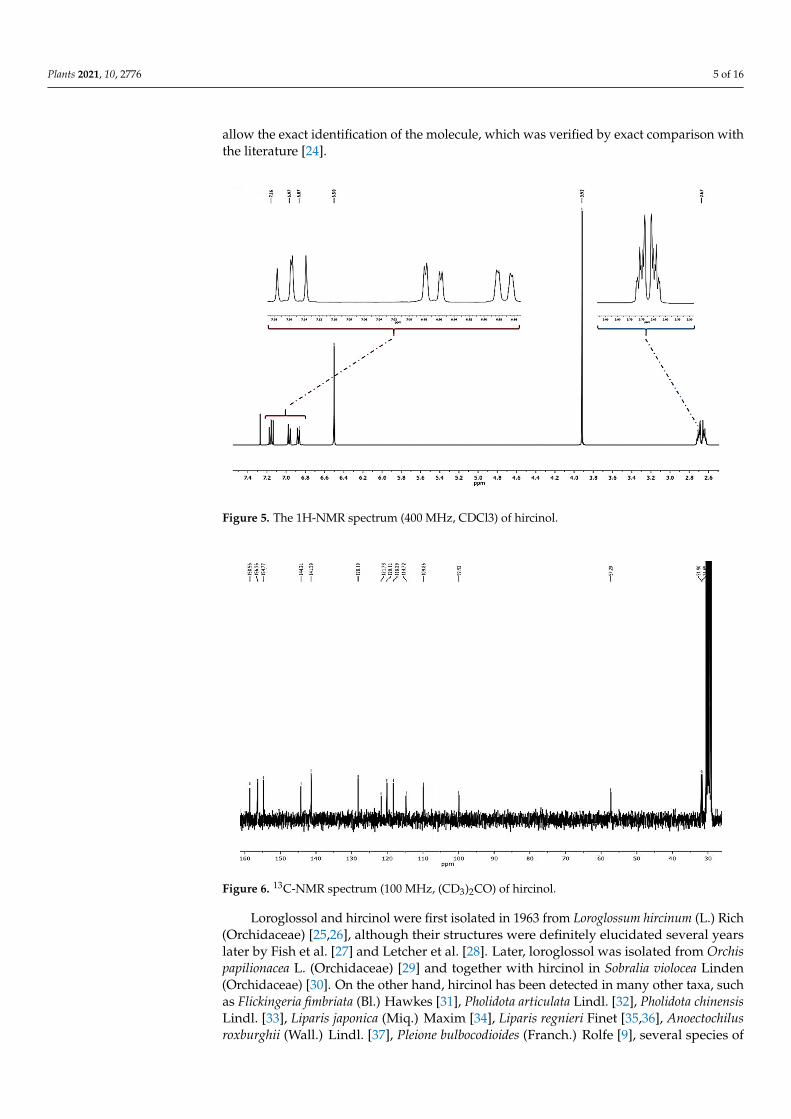
Plants 2021, 10, 2776 5 of 16
allow the exact identification of the molecule, which was verified by exact comparison withthe literature [24].
Figure 5. The 1H-NMR spectrum (400 MHz, CDCl3) of hircinol.
Figure 6. 13C-NMR spectrum (100 MHz, (CD3)2CO) of hircinol.
Loroglossol and hircinol were first isolated in 1963 from Loroglossum hircinum (L.) Rich(Orchidaceae) [25,26], although their structures were definitely elucidated several yearslater by Fish et al. [27] and Letcher et al. [28]. Later, loroglossol was isolated from Orchispapilionacea L. (Orchidaceae) [29] and together with hircinol in Sobralia violocea Linden(Orchidaceae) [30]. On the other hand, hircinol has been detected in many other taxa, suchas Flickingeria fimbriata (Bl.) Hawkes [31], Pholidota articulata Lindl. [32], Pholidota chinensisLindl. [33], Liparis japonica (Miq.) Maxim [34], Liparis regnieri Finet [35,36], Anoectochilusroxburghii (Wall.) Lindl. [37], Pleione bulbocodioides (Franch.) Rolfe [9], several species of
Plants 2021, 10, 2776 6 of 16
Dendrobium [38–45], all belonging to Orchidaceae, and in Dioscorea rotundata Poir [24] andDioscorea opposita Thunb. [46] of the Dioscoreaceae family.
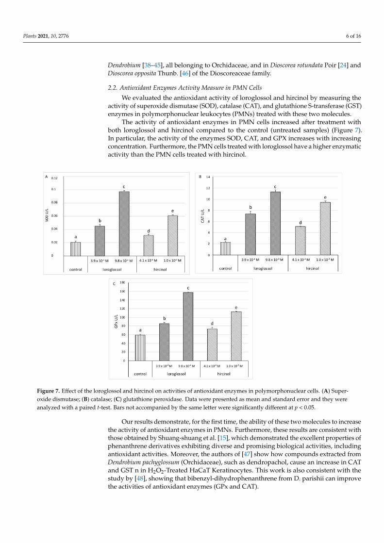
2.2. Antioxidant Enzymes Activity Measure in PMN Cells
We evaluated the antioxidant activity of loroglossol and hircinol by measuring theactivity of superoxide dismutase (SOD), catalase (CAT), and glutathione S-transferase (GST)enzymes in polymorphonuclear leukocytes (PMNs) treated with these two molecules.
The activity of antioxidant enzymes in PMN cells increased after treatment withboth loroglossol and hircinol compared to the control (untreated samples) (Figure 7).In particular, the activity of the enzymes SOD, CAT, and GPX increases with increasingconcentration. Furthermore, the PMN cells treated with loroglossol have a higher enzymaticactivity than the PMN cells treated with hircinol.
Figure 7. Effect of the loroglossol and hircinol on activities of antioxidant enzymes in polymorphonuclear cells. (A) Super-oxide dismutase; (B) catalase; (C) glutathione peroxidase. Data were presented as mean and standard error and they wereanalyzed with a paired t-test. Bars not accompanied by the same letter were significantly different at p < 0.05.
Our results demonstrate, for the first time, the ability of these two molecules to increasethe activity of antioxidant enzymes in PMNs. Furthermore, these results are consistent withthose obtained by Shuang-shuang et al. [15], which demonstrated the excellent properties ofphenanthrene derivatives exhibiting diverse and promising biological activities, includingantioxidant activities. Moreover, the authors of [47] show how compounds extracted fromDendrobium pachyglossum (Orchidaceae), such as dendropachol, cause an increase in CATand GST n in H2O2-Treated HaCaT Keratinocytes. This work is also consistent with thestudy by [48], showing that bibenzyl-dihydrophenanthrene from D. parishii can improvethe activities of antioxidant enzymes (GPx and CAT).
Plants 2021, 10, 2776 7 of 16
However, the antioxidant properties of essential oils cannot be assessed by simplylooking at the increased activity of antioxidant enzymes. On the other hand, the increase intheir activity is linked to oxidative stress. Generally, the activity of the enzymes CAT, SOD,and GST increases following an increase in ROS production, in order to counteract thenegative effects induced by stress, as reported in [49]. In this case, these are not stressfulconditions, but we suggest that an increase in these enzymes may indicate an increase intheir antioxidant properties.
2.3. Antimicrobial Assays
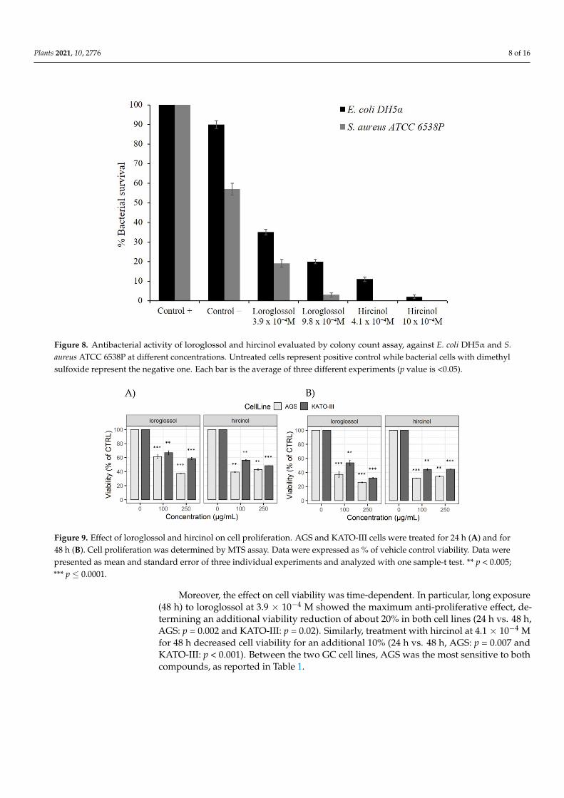
In order to evaluate the antibacterial activity, first, an antimicrobial test (Kirby–Bauerassay) was performed with both molecules on two different bacterial strains, a Gram-negative E. coli DH5α and a Gram-positive S. aureus ATCC 6538P. E. coli represents amodel strain to test antimicrobial activity, while S. aureus is an important human pathogenthat is responsible for most of the bacterial skin and soft tissue infections in humanstoo. This strain can also become more invasive and cause life-threatening infections,such as bacteremia, pneumonia, abscesses of various organs, meningitis, osteomyelitis,endocarditis, and sepsis. These infections represent a major public health threat because oftheir considerable number and spread [50]. Both molecules exhibited an inhibition haloagainst the strains, almost comparable to the antibiotic control (Supplementary MaterialsFigure S1). We used ampicillin as a positive control, in order to inhibit E. coli and S. aureuscell growth. No growth inhibition was seen with the DMSO used to dilute oils.
These first experiments allowed us to deepen the study on loroglossol and hircinolantimicrobial activity. Using the same indicator strains as in previous experiments, weperformed another assay in determining the substance antimicrobial efficiency by bacterialcounts. Figure 8 shows that both molecules extracted from orchids possess good antimi-crobial activity, more directly towards S. aureus. In particular, hircinol is very effectiveagainst the Gram-negative and Gram-positive strains, even at relatively low concentrations.Subsequently antimicrobial activity of loroglossol and hircinol was analyzed accordinglyto the broth microdilution method. By performing this assay, minimal inhibitory concentra-tion values were found to be comprised between 3.9 × 10⁻⁴ M and >1.2 × 10⁻³ M againstthe tested strains (Supplementary Materials Table S1). Other phenanthrene derivativesare reported in the literature. In particular, six biphenanthrenes extracted from Bletillastriata (Orchidaceae) show antimicrobial activity (8–128 µg/mL) against different bacte-rial strains [51]. Instead, two new dihydrophenanthrofurans and two new bisbibenzylderivatives isolated from Dendrobium nobile, were evaluated against Gram-positive bacterialstrains Staphylococcus aureus, Bacillus subtilis, and Gram-negative bacteria, Pseudomonasaeruginosa, Escherichia coli, by a microdilution technique, but neither was active [52]. Fur-ther studies will be needed to determine the mechanism of action of these substances as apotential antimicrobial agent.
2.4. Anti-Proliferative Effects on Gastric Cancer Cell Lines
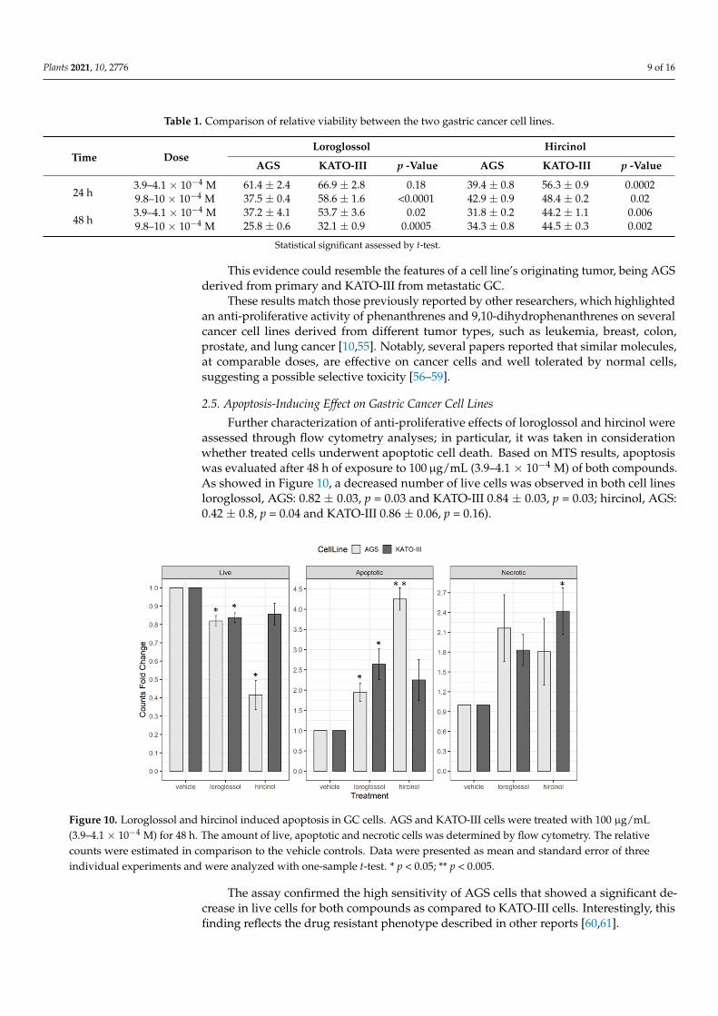
Since the literature reported the cytotoxic activity of extracts from several orchidplants, rich in phenanthrene and dihydrophenanthrene compounds [53,54], we evaluatedthe effect of loroglossol and hircinol on viability of two different gastric cancer (GC) celllines (AGS, and KATO-III) through MTS assay. Results were reported as a percentage ofcontrol viability (cells exposed to DMSO). Both compounds significantly reduced viabilityof gastric cancer cell lines. Interestingly, we observed a decrease of about 40% after 24 hof exposure to 100 µg/mL (3.9 × 10⁻⁴ M for loroglossol: AGS = 61.4 ± 2.4, p < 0.0001;KATO-III = 66.9± 2.8, p = 0.0003 and 4.1× 10⁻⁴ M for hircinol: AGS = 39.4± 0.8, p = 0.0002;KATO-III = 56.3. ± 0.9, p = 0.0004) (Figure 9).
Plants 2021, 10, 2776 8 of 16
Figure 8. Antibacterial activity of loroglossol and hircinol evaluated by colony count assay, against E. coli DH5α and S.aureus ATCC 6538P at different concentrations. Untreated cells represent positive control while bacterial cells with dimethylsulfoxide represent the negative one. Each bar is the average of three different experiments (p value is <0.05).
Figure 9. Effect of loroglossol and hircinol on cell proliferation. AGS and KATO-III cells were treated for 24 h (A) and for48 h (B). Cell proliferation was determined by MTS assay. Data were expressed as % of vehicle control viability. Data werepresented as mean and standard error of three individual experiments and analyzed with one sample-t test. ** p < 0.005;*** p ≤ 0.0001.
Moreover, the effect on cell viability was time-dependent. In particular, long exposure(48 h) to loroglossol at 3.9 × 10⁻⁴ M showed the maximum anti-proliferative effect, de-termining an additional viability reduction of about 20% in both cell lines (24 h vs. 48 h,AGS: p = 0.002 and KATO-III: p = 0.02). Similarly, treatment with hircinol at 4.1 × 10⁻⁴ Mfor 48 h decreased cell viability for an additional 10% (24 h vs. 48 h, AGS: p = 0.007 andKATO-III: p < 0.001). Between the two GC cell lines, AGS was the most sensitive to bothcompounds, as reported in Table 1.
Plants 2021, 10, 2776 9 of 16
Table 1. Comparison of relative viability between the two gastric cancer cell lines.
Time DoseLoroglossol Hircinol
AGS KATO-III p -Value AGS KATO-III p -Value
24 h3.9–4.1 × 10⁻⁴ M 61.4 ± 2.4 66.9 ± 2.8 0.18 39.4 ± 0.8 56.3 ± 0.9 0.00029.8–10 × 10−4 M 37.5 ± 0.4 58.6 ± 1.6 <0.0001 42.9 ± 0.9 48.4 ± 0.2 0.02
48 h3.9–4.1 × 10⁻⁴ M 37.2 ± 4.1 53.7 ± 3.6 0.02 31.8 ± 0.2 44.2 ± 1.1 0.0069.8–10 × 10−4 M 25.8 ± 0.6 32.1 ± 0.9 0.0005 34.3 ± 0.8 44.5 ± 0.3 0.002
Statistical significant assessed by t-test.
This evidence could resemble the features of a cell line’s originating tumor, being AGSderived from primary and KATO-III from metastatic GC.
These results match those previously reported by other researchers, which highlightedan anti-proliferative activity of phenanthrenes and 9,10-dihydrophenanthrenes on severalcancer cell lines derived from different tumor types, such as leukemia, breast, colon,prostate, and lung cancer [10,55]. Notably, several papers reported that similar molecules,at comparable doses, are effective on cancer cells and well tolerated by normal cells,suggesting a possible selective toxicity [56–59].
2.5. Apoptosis-Inducing Effect on Gastric Cancer Cell Lines
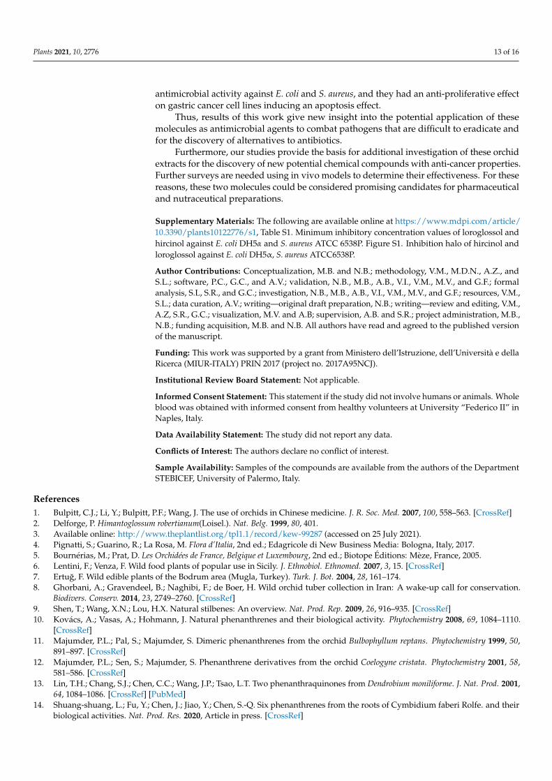
Further characterization of anti-proliferative effects of loroglossol and hircinol wereassessed through flow cytometry analyses; in particular, it was taken in considerationwhether treated cells underwent apoptotic cell death. Based on MTS results, apoptosiswas evaluated after 48 h of exposure to 100µg/mL (3.9–4.1 × 10⁻⁴ M) of both compounds.As showed in Figure 10, a decreased number of live cells was observed in both cell linesloroglossol, AGS: 0.82 ± 0.03, p = 0.03 and KATO-III 0.84 ± 0.03, p = 0.03; hircinol, AGS:0.42 ± 0.8, p = 0.04 and KATO-III 0.86 ± 0.06, p = 0.16).
Figure 10. Loroglossol and hircinol induced apoptosis in GC cells. AGS and KATO-III cells were treated with 100 µg/mL(3.9–4.1 × 10−4 M) for 48 h. The amount of live, apoptotic and necrotic cells was determined by flow cytometry. The relativecounts were estimated in comparison to the vehicle controls. Data were presented as mean and standard error of threeindividual experiments and were analyzed with one-sample t-test. * p < 0.05; ** p < 0.005.
The assay confirmed the high sensitivity of AGS cells that showed a significant de-crease in live cells for both compounds as compared to KATO-III cells. Interestingly, thisfinding reflects the drug resistant phenotype described in other reports [60,61].

Plants 2021, 10, 2776 10 of 16
Analyzing the apoptotic cells, it was observed that loroglossol treatment determineda significant increase of apoptosis in both AGS (1.95 ± 0.23, p = 0.03) and KATO-III(2.64 ± 0.38, p = 0.02) cells, while the treatment with hircinol caused a significant increaseof apoptosis only in AGS cell line (4.25 ± 0.28, p = 0.002). Necrotic cells were significantincreased only in KATO-III cells treated with hircinol. These data showed the apoptoticeffects of loroglossol and hircinol on two different gastric cancer cell lines highlighting ananti-tumor potential of these two molecules.
In line with these results, an apoptogenic effect of orchid’s phenanthrene derivativeson various human cancer cells, including osteosarcoma cells [62], breast cancer cells [63],and gastric cancer cells [64], has been reported.
Moreover, several compounds isolated from orchids have demonstrated significantanticancer activities [65]. The isolation of orchid extract whit cytotoxic potential could laythe bases for their chemical engineering into more effective anticancer drugs. This kind oforchid compound, being semi-synthetic, could help to reduce adverse reactions associatedwith current cancer treatment [66].
3. Materials and Methods3.1. Plant Materials
Bulbs and roots of H. robertianum were collected from wild populations growing atSutera (37◦30′49′ ′ N, 13◦45′112′ ′ E, 468 m s.l.m.), in the province of Caltanissetta, Sicily,Italy, in February 2020, and identified by Professor Vincenzo Ilardi (Università degli Studidi Palermo). A voucher specimen was deposited at Department STEBICEF, University ofPalermo, Italy, under number (PAL109715).
3.2. Extraction and Isolation
The bulbs and roots of H. robertianum (315 g), cut in small pieces, were lyophilized,and extracted with CHCl3 (3 × 0.5 L). The resulting extracts were evaporated to dryness,to yield 4.5 g of residue separated by silica gel column chromatography with hexane-EtOAc (60:40) eluent to give the main fractions, A–E. Each fraction was rechromatographedby silica gel column chromatography in CHCl3-MeOH (98:2) to afford: from fraction A,loroglossol (23 mg); from fraction D, hircinol (28 mg).
3.3. Spectroscopic Data3.3.1. Loroglossol
Colorless needles; 1H-NMR (400 MHz, CDCl3): δ = 2.72 (4H, br s, H2-9,-10), 3.87 (3H, s,-OCH3), 3.97 (3H, s, -OCH3), 6.57 (1H, d, J = 2.5 Hz, H-3), 6.62 (1H, d, J = 2.5 Hz, H-1), 6.88(1H, dd, J = 7.2, 1.4 Hz, H-8), 6.96 (1H, dd, J = 8.1, 1.4 Hz, H-6), 7.16 (1H, dd, J = 8.1, 7.2 Hz,H-7); 13C-NMR (100 MHz, CDCl3): δ = 107.1 (C-1), 159.5 (C-2), 98.5 (C-3), 155.0 (C-4), 153.5(C-5), 117.8 (C-6), 127.9 (C-7), 119.7 (C-8), 30.9 (C-9), 31.4 (C-10), 115.5 (C-4a), 120.6 (C-4b),140.6 (C-8a), 143.6 (C-10a), 55.4 (C2-OCH3), 57.2 (C4-OCH3); ESIMS m/z 257.1181 [M + H]+,(calcd. for C16H16O3+[H]+, m/z 257.1172).
3.3.2. Hircinol
Amorphous powder; 1H-NMR (400 MHz, CDCl3): δ = 2.67 (4H, m, H2-9,-10), 3.92 (3H,s, -OCH3), 6.50 (2H, br s, H-1, H-3), 6.87 (1H, dd, J = 7.3, 1.4 Hz, H-8), 6.97 (1H, dd, J = 8.1,1.4 Hz, H-6), 7.16 (1H, dd, J = 8.1, 7.3 Hz, H-7); 13C-NMR (100 MHz, [(CD3)2CO]: δ = 109.9(C-1), 158.6 (C-2), 99.9 (C-3), 156.4 (C-4), 154.8 (C-5), 118.3 (C-6), 128.2 (C-7), 120.1 (C-8), 31.7(C-9), 31.9 (C-10), 114.7 (C-4a), 121.7 (C-4b), 141.4 (C-8a), 144.2 (C-10a), 57.3 (C2-OCH3);ESIMS m/z 243.0948 [M + H]+, (calcd. for C15H14O3+[H]+, m/z 243.1016).
3.4. General Experimental Procedures
Column chromatography was performed using silica gel (70-230 mesh ASTM, MerckNo. 7734) deactivated with 15% deionized water. Lyophilization was conducted usingCoolSafe instrument (4-15L Freeze Dryers). The NMR spectra were recorded on a Bruker

Plants 2021, 10, 2776 11 of 16
Avance II instrument (400 MHz for 1H-NMR and 100 MHz for 13C-NMR). The thin-layerchromatography used aluminum oxide 60 F254 neutral (Merck KGaA, Darmstadt, Ger-many). Mass spectrum was obtained using a HPLC/ESI/Q-TOF HRMS apparatus. HPLCconditions were as follows: water, acetonitrile, and formic acid were of HPLC/MS grade;the HPLC system was an Agilent 1260 Infinity; a reversed-phase C18 column (ZORBAXExtended-C18 2.1 × 50 mm, 1.8 µm) with a Phenomenex C18 security guard column(4 × 3 mm) were used; the flow-rate was 0.4 mL/min, and the column temperature was setto 30 ◦C. The mass spectra was recorded using an Agilent 6540 UHD accurate-mass Q-TOFspectrometer equipped with a Dual AJS ESI source working in both negative and positivemodes. All solvents used were of analytical grade (Honeywell, Charlotte, NC, USA).
3.5. Blood Collection and Polymorphonuclear Leukocytes (PMN) Isolation
Whole blood was obtained with informed consent from healthy volunteers at Univer-sity “Federico II” in Naples, Italy. Between 08.00 and 09.00 a.m., three healthy fasting donorswere subjected to peripheral blood sampling with K3EDTA vacutainers (Becton Dickinson,Plymouth, UK). A discontinuous gradient, consisting of 100% (density 1.1294 g/mL) and70% (density 1.090 g/mL) isotonic Percoll (Pharmacia, Uppsala, Sweden) in calcium andmagnesium-free phosphate buffered saline pH 7.4 (PBS; Sigma-Aldrich, Saint Louis, MO,USA) was used to isolate PMNs [67]. Subsequently, the samples were centrifuged for20 min at 250× g at room temperature. The PMN layer was collected and washed twice inPBS. May-Grunwald Giemsa-stained cytocentrifuge smears were used to determine theisolated PMN purity, while, the trypan blue dye exclusion test was employed to check cellviability. Both ranged between 90% and 95%.
3.6. Antioxidant Enzymes Measured in PMN Cells
A commercial kit (BioAssay System, San Diego, CA, USA) was used to determine su-peroxide dismutase (SOD), catalase (CAT), and glutathione S-transferase (GST) enzymaticactivity in PMN cells according to the manufacturer’s recommendations. The activity of en-zymes was expressed as U/L [68]. Loroglossol and hircinol were tested at the concentrationof 100 µg/mL (3.9–4.1 × 10⁻⁴ M) and 250 µg/mL (9.8–10 × 10−4 M).
3.7. Antimicrobial Activity Assays
The presence of antimicrobial molecules in the extracted from Orchid flower was de-tected using Kirby–Bauer assay [69] against two bacterial strains: E. coli DH5α and S. aureusATCC 6538P. Both orchid molecules were used at 50 µg and ampicillin used at 10 µg as apositive control, to inhibit E. coli and S. aureus cells growth. Another method to evaluatethe antimicrobial activity involved the Gram-negative E. coli DH5a and Gram-positive S.aureus ATCC 6538P strains cell viability counting [70]. Bacterial cells were incubated withloroglossol and hircinol at two different concentrations: 100 µg/mL (3.9–4.1 × 10⁻⁴ M) and250 µg/mL (9.8–10 × 10−4 M). Bacterial cells immersed in the oil-free buffer representedpositive control, while bacterial cells plus 80% DMSO (volume corresponding to the maxi-mum concentration) and buffer represented the negative one [71]. Each experiment wasperformed in triplicate, and the reported result was an average of three independent experi-ments (p value was <0.05). Subsequently to determine the minimal inhibitory concentrationvalues, assays were performed as previously described elsewhere [72]. Briefly, bacteriawere grown to a midlogarithmic phase at 37 ◦C and then diluted to 1 × 10⁶ CFU/mLin Difco 0.5× Nutrient Broth (Becton-Dickenson, Franklin Lakes, NJ, USA) containingincreasing amounts of loroglossol and hircinol. Starting from a compound stock solution,2-fold serial dilutions were sequentially carried out, accordingly to the broth microdilutionmethod [73]. DMSO was tested at the same concentrations. Following overnight incuba-tion, MIC100 values were determined as the lowest molecule concentrations responsiblefor no visible bacterial growth.

Plants 2021, 10, 2776 12 of 16
3.8. Cell Cultures and Treatments
Human GC cell lines, AGS (ATCC® CRL-1739™) and KATO-III (ATCC® HTB-103),were purchased from ATCC (Manassas, VA, USA) and routinely cultured in their specificmedium according to manufacturer instructions. In particular, AGS were cultured inDulbecco’s Modified Eagle Medium (DMEM, GIBCO, Grand Island, NY, USA) supple-mented with 10% inactivated Fetal Bovine Serum (FBS, GIBCO, Grand Island, NY, USA),100 U/mL of penicillin and 100 µg/mL of streptomycin. KATO-III cells were grown inIscove’s Modified Dulbecco’s Medium (IMDM, GIBCO, Grand Island, NY, USA) with 20%inactivated FBS (GIBCO, Grand Island, NY, USA) and 1% penicillin/streptomycin. All cellswere incubated at 37 ◦C in 5% CO2 changing medium every 2 days.
Loroglossol and hircinol were first resuspended in DMSO and then diluted in DMEMor IMDM to obtain a stock solution of 10,000 µg/mL. GC cells were incubated with bothmolecules at two different concentrations: 100 µg/mL (3.9–4.1 × 10⁻⁴ M) and 250 µg/mL(9.8–10 × 10−4 M) for 24 and 48 h. As untreated control, DMSO at final concentration of0.5% was used.
3.9. Cell Proliferation and Apoptosis Analysis
Cell proliferation was assessed using a MTS assay. AGS and KATO-III cells wereplated at a density of 5× 10³ cells/well into 96-well plates and grown overnight beforetreatments with each compound or with an equivalent amount of DMSO for 24 and 48 h. Asolution of CellTiter 96® Aqueous MTS Reagent (Promega, Madison, WI, USA) was addedto each well measuring, after 1.5 h of incubation at 37 ◦C, the absorbance at 490 nm. Theproliferation rate in each well was calculated using the DMSO treated cells as reference,which represents the 100% of viability. Three replicate wells per point were used to obtainmeasures of cell proliferation from three independent experiments. Data were reportedas mean ± SE. Differences between treatments and control groups were estimated by onesample t test. p values < 0.05 were considered significant. Analyses were performed usingR software.
To evaluate apoptosis involvement, GC cells were seeded in 6-well plates and culturedto 70% confluence prior to being treated with 100 µg/mL (3.9–4.1 × 10⁻⁴ M) of bothcompounds, or with their vehicle, for 48 h. Briefly, cells were harvested, washed twice incold PBS, centrifuged at 1500 rpm at 4 ◦C, and then resuspended in Binding Buffer (BDBiosciences, Franklin Lakes, NJ, USA). Cells were double-stained with FITC-Annexin Vand propidium iodide (BD Biosciences, Franklin Lakes, NJ, USA) and incubated for 15 minat room temperature in the dark. Number of live, apoptotic, and necrotic cells was detectedusing a Navios flow cytometer (Beckman Coulter, Miami, FL, USA). Data were analyzedby Kaluza analysis software 2.1. Counts from flow cytometry evaluation of apoptosiswere reported as fold increases compared with vehicle controls. Data were reported asmean ± SE. Differences between treatments and control groups were estimated by onesample t test. p values < 0.05 were considered significant. Analyses were performed usingR software.
4. Conclusions
Phytoalexins are produced by plants in response to pathogenic fungal infections.The phytoalexins of orchids are phenanthrene and dihydrophenanthrene, such as hircinoland loroglossol. Phenanthrenes are a promising and expanding group of biologicallyactive natural compounds whose potential has not yet been sufficiently studied [10]. Theirantifungal abilities were studied on Candida lipolytica as early as 1973. However, very littleis known about the antimicrobial activity of these molecules against gram positive andnegative bacteria. This research resulted in the isolation and identification of the loroglossoland hircinol from the Orchids H. robertianum bulbs and roots. The two molecules showeddifferent bioactivity; they induced an increase in the activity of superoxide dismutase,catalase, and glutathione S-transferase in polymorphonuclear leukocytes; they showed an
Plants 2021, 10, 2776 13 of 16
antimicrobial activity against E. coli and S. aureus, and they had an anti-proliferative effecton gastric cancer cell lines inducing an apoptosis effect.
Thus, results of this work give new insight into the potential application of thesemolecules as antimicrobial agents to combat pathogens that are difficult to eradicate andfor the discovery of alternatives to antibiotics.
Furthermore, our studies provide the basis for additional investigation of these orchidextracts for the discovery of new potential chemical compounds with anti-cancer properties.Further surveys are needed using in vivo models to determine their effectiveness. For thesereasons, these two molecules could be considered promising candidates for pharmaceuticaland nutraceutical preparations.
Supplementary Materials: The following are available online at https://www.mdpi.com/article/10.3390/plants10122776/s1, Table S1. Minimum inhibitory concentration values of loroglossol andhircinol against E. coli DH5α and S. aureus ATCC 6538P. Figure S1. Inhibition halo of hircinol andloroglossol against E. coli DH5α, S. aureus ATCC6538P.
Author Contributions: Conceptualization, M.B. and N.B.; methodology, V.M., M.D.N., A.Z., andS.L.; software, P.C., G.C., and A.V.; validation, N.B., M.B., A.B., V.I., V.M., M.V., and G.F.; formalanalysis, S.L, S.R., and G.C.; investigation, N.B., M.B., A.B., V.I., V.M., M.V., and G.F.; resources, V.M.,S.L.; data curation, A.V.; writing—original draft preparation, N.B.; writing—review and editing, V.M.,A.Z, S.R., G.C.; visualization, M.V. and A.B; supervision, A.B. and S.R.; project administration, M.B.,N.B.; funding acquisition, M.B. and N.B. All authors have read and agreed to the published versionof the manuscript.
Funding: This work was supported by a grant from Ministero dell’Istruzione, dell’Università e dellaRicerca (MIUR-ITALY) PRIN 2017 (project no. 2017A95NCJ).
Institutional Review Board Statement: Not applicable.
Informed Consent Statement: This statement if the study did not involve humans or animals. Wholeblood was obtained with informed consent from healthy volunteers at University “Federico II” inNaples, Italy.
Data Availability Statement: The study did not report any data.
Conflicts of Interest: The authors declare no conflict of interest.
Sample Availability: Samples of the compounds are available from the authors of the DepartmentSTEBICEF, University of Palermo, Italy.
References1. Bulpitt, C.J.; Li, Y.; Bulpitt, P.F.; Wang, J. The use of orchids in Chinese medicine. J. R. Soc. Med. 2007, 100, 558–563. [CrossRef]2. Delforge, P. Himantoglossum robertianum(Loisel.). Nat. Belg. 1999, 80, 401.3. Available online: http://www.theplantlist.org/tpl1.1/record/kew-99287 (accessed on 25 July 2021).4. Pignatti, S.; Guarino, R.; La Rosa, M. Flora d’Italia, 2nd ed.; Edagricole di New Business Media: Bologna, Italy, 2017.5. Bournérias, M.; Prat, D. Les Orchidées de France, Belgique et Luxembourg, 2nd ed.; Biotope Éditions: Mèze, France, 2005.6. Lentini, F.; Venza, F. Wild food plants of popular use in Sicily. J. Ethnobiol. Ethnomed. 2007, 3, 15. [CrossRef]7. Ertug, F. Wild edible plants of the Bodrum area (Mugla, Turkey). Turk. J. Bot. 2004, 28, 161–174.8. Ghorbani, A.; Gravendeel, B.; Naghibi, F.; de Boer, H. Wild orchid tuber collection in Iran: A wake-up call for conservation.
Biodivers. Conserv. 2014, 23, 2749–2760. [CrossRef]9. Shen, T.; Wang, X.N.; Lou, H.X. Natural stilbenes: An overview. Nat. Prod. Rep. 2009, 26, 916–935. [CrossRef]10. Kovács, A.; Vasas, A.; Hohmann, J. Natural phenanthrenes and their biological activity. Phytochemistry 2008, 69, 1084–1110.
[CrossRef]11. Majumder, P.L.; Pal, S.; Majumder, S. Dimeric phenanthrenes from the orchid Bulbophyllum reptans. Phytochemistry 1999, 50,
891–897. [CrossRef]12. Majumder, P.L.; Sen, S.; Majumder, S. Phenanthrene derivatives from the orchid Coelogyne cristata. Phytochemistry 2001, 58,
581–586. [CrossRef]13. Lin, T.H.; Chang, S.J.; Chen, C.C.; Wang, J.P.; Tsao, L.T. Two phenanthraquinones from Dendrobium moniliforme. J. Nat. Prod. 2001,
64, 1084–1086. [CrossRef] [PubMed]14. Shuang-shuang, L.; Fu, Y.; Chen, J.; Jiao, Y.; Chen, S.-Q. Six phenanthrenes from the roots of Cymbidium faberi Rolfe. and their
biological activities. Nat. Prod. Res. 2020, Article in press. [CrossRef]
Plants 2021, 10, 2776 14 of 16
15. Pant, B.; Ram Paudel, M.; Raj Joshi, P. Orchids as potential sources of anticancer agents: Our experience. Annapurna J. Health Sci.2021, 1, 42–51. [CrossRef]
16. Bazzicalupo, M.; Burlando, B.; Denaro, M.; Barreca, D.; Trombetta, D.; Smeriglio, A.; Cornara, A. Polyphenol characterization andskin-preserving properties of hydroalcoholic flower extract from Himantoglossum robertianum (Orchidaceae). Plants 2019, 8, 502.[CrossRef] [PubMed]
17. Gagliano Candela, R.; Ilardi, V.; Badalamenti, N.; Bruno, M.; Rosselli, S.; Maggi, F. Essential oil compositions of Teucrium fruticans,T. scordium subsp. scordioides and T. siculum growing in Sicily and Malta. Nat. Prod. Res. 2021, 35, 3460–3469. [CrossRef]
18. Ilardi, V.; Badalamenti, N.; Bruno, M. Chemical composition of the essential oil from different vegetative parts of Foeniculumvulgare subsp. piperitum (Ucria) Coutinho (Umbelliferae) growing wild in Sicily. Nat. Prod. Res. 2020, in press. [CrossRef]
19. Badalamenti, N.; Ilardi, V.; Rosselli, S.; Bruno, M.; Maggi, F.; Leporini, M.; Falco, T.; Loizzo, M.R.; Tundis, R. Ferulago nodosa subsp.geniculata (Guss.) Troia & Raimondo from Sicily (Italy): Isolation of essential oil and evaluation of its bioactivity. Molecules 2020,25, 3249.
20. Di Napoli, M.; Maresca, V.; Varcamonti, M.; Bruno, M.; Badalamenti, N.; Basile, A.; Zanfardino, A. (+)-(E)-Chrysanthenyl acetate:A molecule with interesting biological properties contained in the Anthemis secundiramea (Asteraceae) flowers. Appl. Sci. 2020, 10,6808. [CrossRef]
21. Badalamenti, N.; Ilardi, V.; Bruno, M.; Pavela, R.; Boukouvala, M.C.; Kavallieratos, N.G.; Maggi, F.; Canale, A.; Benelli, G.Chemical composition and broad-spectrum insecticidal activity of the flower essential oil from an ancient Sicilian food plant,Ridolfia segetum. Agriculture 2021, 11, 304. [CrossRef]
22. Rosselli, S.; Tundis, R.; Bruno, M.; Leporini, M.; Falco, T.; Candela, R.G.; Badalamenti, N.; Loizzo, M.R. Ceiba speciosa (A. St.-Hil.)seeds oil: Fatty acids profiling by GC-MS and NMR and bioactivity. Molecules 2020, 25, 1037. [CrossRef]
23. Stoessl, A.; Stothers, J.B. Carbon-13NMR studies. 96†—carbon-13 spectra of several polyhydroxylated 9,10-dihydrophenanthreneand phenanthrene derivatives. Org. Magn. Res. 1982, 20, 166. [CrossRef]
24. Coxon, D.T.; Ogundana, S.K.; Dennis, C. Antifungal phenanthrenes in yam tubers. Phytochemistry 1982, 21, 1389–1392. [CrossRef]25. Hardegger, E.; Schellenbaum, M.; Corrodi, H. Welkstoffe und Antibiotika. 27. Mitteilung. Über induzierte Abwehrstoffe bei
Orchideen II. Helv. Chim. Acta 1963, 46, 1171–1180. [CrossRef]26. Urech, J.; Fechtig, B.; Nüesch, J.; Vischer, E. Hircinol, eine antifungisch wirksame Substanz aus Knollen von Loroglossum hircinum
(L.) Rich. Helv. Chim. Acta 1963, 46, 2758–2766. [CrossRef]27. Fisch, M.H.; Flick, B.H.; Arditti, J. Structure and antifungal activity of hircinol, loroglossol and orchinol. Phytochemistry 1973, 12,
437–441. [CrossRef]28. Letcher, R.M.; Nhamo, L.R.M. Structure of orchinol, loroglossol, and hircinol. J. Chem. Soc. Perkin Trans. 1973, 1, 1263–1265.
[CrossRef]29. Pagani, F. Isolation and identification of loroglossin and of some flavonol heterosides from Orchis papilionacea L. (Orchidaceae).
Boll. Chim. Farm. 1982, 121, 174–177.30. Oechslin-Merkel, K.; Konig, G.M.; Oechslin St., M.; Miyagawa, M.; Wright, A.D.; Sticher, O. A new glycoside from Sobralia violacea
related to phytoalexins from orchids. Planta Med. 1991, 57, A126. [CrossRef]31. Wu, Y.P.; Liu, W.J.; Zhong, W.J.; Chen, Y.J.; Chen, D.N.; He, F.; Jiang, L. Phenolic compounds from the stems of Flickingeria fimbriata.
Nat. Prod. Res. 2017, 31, 1518–1522. [CrossRef] [PubMed]32. Zhu, X.Y.; Zhou, Y.T.; Yan, H.G.; Fu, H.; Guo, J.; Yang, M.H. Chemical constituents from ethyl acetate extracts of Pholidota articulata.
Chin. Tradit. Herb. Drugs 2020, 51, 6151–6156.33. Wang, J.; Wang, L.; Kitanaka, S. Stilbene and dihydrophenanthrene derivatives from Pholidota chinensis and their nitric oxide
inhibitory and radical-scavenging activities. J. Nat. Med. 2007, 61, 381–386. [CrossRef]34. Song, S.P.; Jiang, F.; Li, C.H.; Wei, T.; Yu, Y.F.; Tian, X.H. Chemical constituents from Liparis japonica. Chin. Pharm. J. 2018, 53,
104–108.35. Ren, J.; Fan, C.; Guo, Y.G.; Yan, S.K.; Ye, R.D.; Zhang, Y.; Jin, H.Z.; Zhang, W.D. New biphenantherene and nervogenic acid
derivatives from Liparis regnieri Finet and their inhibitory activities against NF-κB activation. Tetrahedron 2017, 73, 1611–1617.[CrossRef]
36. Ren, J.; Qian, X.P.; Guo, Y.G.; Li, T.; Yan, S.K.; Jin, H.Z.; Zhang, W.D. Two new phenanthrene glycosides from Liparis regnieri Finetand their antibacterial activities. Phytochem. Lett. 2016, 18, 64–67. [CrossRef]
37. Wang, Y.; Chen, S.; Lu, D.P.; Zhang, L.R. Chemical constituents of Anoectochilus roxburghii. Chin. Tradit. Herb. Drugs 2017, 48,2619–2624.
38. Wang, C.; Han, S.W.; Cui, B.S.; Wang, X.J.; Li, S. Chemical constituents from Pleione bulbocodioides. Zhongguo Zhongyao Zazhi 2014,39, 442–447.
39. Zhou, X.M.; Zheng, C.J.; Gan, L.S.; Chen, G.Y.; Zhang, X.P.; Song, X.P.; Li, G.N.; Sun, C.G. Bioactive phenanthrene and bibenzylderivatives from the stems of Dendrobium nobile. J. Nat. Prod. 2016, 79, 1791–1797. [CrossRef] [PubMed]
40. Zhao, N.; Yang, G.; Zhang, Y.; Chen, L.; Chen, Y. A new 9,10-dihydrophenanthrene from Dendrobium moniliforme. Nat. Prod. Res.2016, 30, 174–179. [CrossRef]
41. Sritularak, B.; Anuwat, M.; Likhitwitayawuid, K. A new phenanthrenequinone from Dendrobium draconis. J. Asian Nat. Prod. Res.2011, 13, 251–255. [CrossRef] [PubMed]

Plants 2021, 10, 2776 15 of 16
42. Hwang, J.S.; Lee, S.A.; Hong, S.S.; Han, X.H.; Lee, C.; Kang, S.J.; Lee, D.; Kim, Y.; Hong, J.T.; Lee, M.K.; et al. Phenanthrenes fromDendrobium nobile and their inhibition of the LPS-induced production of nitric oxide in macrophage RAW 264.7 cells. Bioorg. Med.Chem. Lett. 2010, 20, 3785–3787. [CrossRef] [PubMed]
43. Zhang, C.F.; Shao, L.; Huang, W.H.; Wang, L.; Wang, Z.T.; Xu, L.S. Phenolic components from herbs of Dendrobium aphyllum.Zhongguo Zhongyao Zazhi 2008, 33, 2922–2925.
44. Hu, J.M.; Chen, J.J.; Yu, H.; Zhao, Y.X.; Zhou, J. Two novel bibenzyls from Dendrobium trigonopus. J. Asian Nat. Prod. Res. 2008, 10,647–651. [CrossRef] [PubMed]
45. Zhang, G.N.; Zhang, C.F.; Wang, Z.T.; Xu, L.S. Studies on chemical constituents of Dendrobium thyrsiflorum Rchb. Chin. J. Nat.Med. 2004, 2, 78–81.
46. Yang, M.H.; Yoon, K.D.; Chin, Y.W.; Park, J.H.; Kim, J. Phenolic compounds with radical scavenging and cyclooxygenase-2(COX-2) inhibitory activities from Dioscorea opposita. Bioorg. Med. Chem. 2009, 17, 2689–2694. [CrossRef] [PubMed]
47. Warinhomhoun, S.; Muangnoi, C.; Buranasudja, V.; Mekboonsonglarp, W.; Rojsitthisak, P.; Likhitwitayawuid, K.; Sritularak, B.Antioxidant Activities and Protective Effects of Dendropachol, a New Bisbibenzyl Compound from Dendrobium pachyglossum, onHydrogen Peroxide-Induced Oxidative Stress in HaCaT Keratinocytes. Antioxidants 2021, 10, 252. [CrossRef]
48. Li, Y.; Wang, C.L.; Wang, Y.J.; Wang, F.F.; Guo, S.X.; Yang, J.S.; Xiao, P.G. Four new bibenzyl derivatives from Dendrobium candidum.Chem. Pharm. Bull. 2009, 57, 997–999. [CrossRef]
49. Haydari, M.; Maresca, V.; Rigano, D.; Taleei, A.; Shahnejat-Bushehri, A.A.; Hadian, J.; Sorbo, S.; Guida, M.; Manna, C.; Piscopo, M.Salicylic Acid and Melatonin Alleviate the Effects of Heat Stress on Essential Oil Composition and Antioxidant Enzyme Activityin Mentha × piperita and Mentha arvensis L. Antioxidants 2019, 8, 547. [CrossRef]
50. Krishna, S.; Miller, L.S. Innate and adaptive immune responses against Staphylococcus aureus skin infections. Semin. Im-munopathol. 2012, 34, 261–280. [CrossRef]
51. Qian, C.-D.; Jiang, F.-S.; Yu, H.-S.; Shen, Y.; Fu, Y.-H.; Cheng, D.-Q.; Gan, L.-S.; Ding, Z.-S. Antibacterial Biphenanthrenes from theFibrous Roots of Bletilla striata. J. Nat. Prod. 2015, 78, 939–943. [CrossRef]
52. Cheng, L.; Guo, D.L.; Zhang, M.S.; Linghu, L.; Fu, S.B.; Deng, Y.; He, Y.-Q.; Xiao, S.-J. Dihydrophenanthrofurans and bisbibenzylderivatives from the stems of Dendrobium nobile. Fitoterapia 2020, 143, 104586. [CrossRef]
53. Yang, M.; Cai, L.; Tai, Z.; Zeng, X.; Ding, Z. Four new phenanthrenes from Monomeria barbata Lindl. Fitoterapia 2010, 81, 992–997.[CrossRef]
54. Paudel, M.R.; Chand, M.B.; Pant, B.; Pant, B. Antioxidant and cytotoxic activities of Dendrobium moniliforme extracts and thedetection of related compounds by GC-MS. BMC Complementary Altern. Med. 2018, 18, 134. [CrossRef] [PubMed]
55. Lee, C.-L.; Chang, F.-R.; Yen, M.-H.; Yu, D.; Liu, Y.-N.; Bastow, K.F.; Morris-Natschke, S.L.; Wu, Y.-C.; Lee, K.-H. Cytotoxicphenanthrenequinones and 9,10-dihydrophenanthrenes from Calanthe arisanensis. J. Nat. Prod. 2009, 72, 210–213. [CrossRef]
56. Bús, C.; Kúsz, N.; Kincses, A.; Szemerédi, N.; Spengler, G.; Bakacsy, L.; Purger, D.; Berkecz, R.; Hohmann, J.; Hunyadi, A.; et al.Antiproliferative Phenanthrenes from Juncus tenuis: Isolation and diversity-oriented semisynthetic modification. Molecules 2020,25, E5983. [CrossRef]
57. Itharat, A.; Thongdeeying, P.; Ruangnoo, S. Isolation and characterization of a new cytotoxic dihydrophenanthrene from Dioscoreamembranacea rhizomes and its activity against five human cancer cell lines. J. Ethnopharmacol. 2014, 156, 130–134. [CrossRef]
58. Kil, Y.S.; Park, J.; Han, A.R.; Woo, H.; Seo, E.K. A new 9, 10-dihydrophenanthrene and cell proliferative 3, 4-δ-dehydrotocopherolsfrom Stemona tuberosa. Molecules 2015, 20, 5965–5974. [CrossRef] [PubMed]
59. Zhan, R.; Wang, Z.C.; Yin, B.L.; Liu, Y.; Chen, Y.G. Novel 9, 10-dihydrophenanthrene derivatives from Eria bambusifolia withcytotoxicity aganist human cancer cells in vitro. Chin. J. Nat. Med. 2016, 14, 621–625. [CrossRef] [PubMed]
60. Laurino, S.; Mazzone, P.; Ruggieri, V.; Zoppoli, P.; Calice, G.; Lapenta, A.; Ciuffi, M.; Ignomirelli, O.; Vita, G.; Sgambato, A.;et al. Cationic channel TRPV2 overexpression promotes resistance to cisplatin-induced apoptosis in gastric cancer cells. Front.Pharmacol. 2021, 12, 746628. [CrossRef] [PubMed]
61. Lu, R.; Zhao, G.; Yang, Y.; Jiang, Z.; Cai, J.; Hu, H. Inhibition of CD133 overcomes cisplatin resistance through inhibitingPI3K/AKT/MTOR signaling pathway and autophagy in CD133-positive gastric cancer cells. Technol. Cancer Res. Treat. 2019, 18,1533033819864311. [CrossRef]
62. Zhang, Y.; Zhang, Q.; Xin, W.; Liu, N.; Zhang, H. Nudol, a phenanthrene derivative from Dendrobium nobile, induces cell cyclearrest and apoptosis and inhibits migration in osteosarcoma Cells. Drug Des. Dev. Ther. 2019, 13, 2591–2601. [CrossRef]
63. Shyur, L.-F.; Chen, C.-H.; Lo, C.-P.; Wang, S.-Y.; Kang, P.-L.; Sun, S.-J.; Chang, C.A.; Tzeng, C.-M.; Yang, N.-S. Induction ofapoptosis in MCF-7 human breast cancer cells by phytochemicals from Anoectochilus formosanus. J. Biomed. Sci. 2004, 11, 928–939.[CrossRef]
64. Song, J.I.; Kang, Y.J.; Yong, H.-Y.; Kim, Y.C.; Moon, A. Denbinobin, a Phenanthrene from Dendrobium Nobile, Inhibits Invasionand Induces Apoptosis in SNU-484 Human Gastric Cancer Cells. Oncol. Rep. 2012, 27, 813–818.
65. Joshi, P.R.; Paudel, M.R.; Chand, M.B.; Pradhan, S.; Pant, K.K.; Joshi, G.P.; Bohara, M.; Wagner, S.H.; Pant, B.; Pant, B. Cytotoxiceffect of selected wild orchids on two different human cancer cell lines. Heliyon 2020, 6, e03991. [CrossRef] [PubMed]
66. Tripathi, P.; Aditi, S. Indigenous Asian plants against cancer: A Comprehensive review. Int. J. Plant Res. 2015, 5, 80–86.67. Harbeck, R.J.; Homan, A.A.; Redecker, S.; Biundo, T.; Kurnick, J. The isolation and functional activity of polymorphonuclear
leukocytes and lymphocytes separated from whole blood on a single percoll density gradient. Clin. Immunol. Immunopathol. 1982,23, 682–690. [CrossRef]

Plants 2021, 10, 2776 16 of 16
68. Barbosa, P.O.; Pala, D.; Silva, C.T.; de Souza, M.O.; do Amaral, J.F.; Vieira, R.A.L.; de Freitas Folly, G.A.; Volp, A.C.P.; de Freitas,R.N. Açai (Euterpe oleracea Mart.) pulp dietary intake improves cellular antioxidant enzymes and biomarkers of serum in healthywomen. Nutrition 2016, 32, 674–680. [CrossRef]
69. Bauer, A.W.; Kirby, W.M.; Sherris, J.C.; Turck, M. Antibiotic susceptibility testing by a standardized single disk method. Am. J.Clin. Pathol. 1966, 45, 493–496. [CrossRef]
70. D’Alessio, G.; Zanfardino, A.; Pizzo, E.; Di Maro, A.; Varcamonti, M. The bactericidal action on Escherichia coli of ZF-RNase-3 istriggered by the suicidal action of the bacterium OmpT protease. FEBS J. 2010, 277, 1921–1928.
71. Di Napoli, M.; Varcamonti, M.; Basile, A.; Bruno, M.; Maggi, F.; Zanfardino, A. Anti-Pseudomonas aeruginosa activity of hemlock(Conium maculatum, Apiaceae) essential oil. Nat. Prod. Res. 2019, 33, 3436–3440. [CrossRef]
72. Prencipe, F.; Zanfardino, A.; Di Napoli, M.; Rossi, F.; D’errico, S.; Piccialli, G.; Mangiatordi, G.F.; Saviano, M.; Ronga, L.;Varcamonti, M.; et al. Silver (I) n-heterocyclic carbene complexes: A winning and broad spectrum of antimicrobial properties. Int.J. Mol. Sci. 2021, 22, 2497. [CrossRef]
73. Wiegand, I.; Hilpert, K.; Hancock, R.E. Agar and broth dilution methods to determine the minimal inhibitory concentration (MIC)of antimicrobial substances. Nat. Protoc. 2008, 3, 163–175. [CrossRef] [PubMed]
Related Documents