Molecular Cell
Article
A Hierarchical Network of TranscriptionFactors Governs AndrogenReceptor-Dependent Prostate Cancer GrowthQianben Wang,1 Wei Li,2 X. Shirley Liu,2 Jason S. Carroll,1 Olli A. Janne,3 Erika Krasnickas Keeton,1
Arul M. Chinnaiyan,4,6 Kenneth J. Pienta,5,6 and Myles Brown1,*1 Division of Molecular and Cellular Oncology, Department of Medical Oncology, Dana-Farber Cancer Institute and Harvard Medical
School, Boston, MA 02115, USA2 Department of Biostatistics and Computational Biology, Dana-Farber Cancer Institute and Harvard School of Public Health,
Boston, MA 02115, USA3 Biomedicum Helsinki, Institute of Biomedicine (Physiology), University of Helsinki, FI-00014 Helsinki, Finland4 Department of Pathology5 Department of Medicine6 Department of Urology
University of Michigan Medical School, Ann Arbor, MI 48109, USA
*Correspondence: [email protected] 10.1016/j.molcel.2007.05.041
SUMMARY
Androgen receptor (AR) is a ligand-dependenttranscription factor that plays a key role in pros-tate cancer. Little is known about the nature ofAR cis-regulatory sites in the human genome.We have mapped the AR binding regions on twochromosomes in human prostate cancer cellsby combining chromatin immunoprecipitation(ChIP) with tiled oligonucleotide microarrays.We find that the majority of AR binding regionscontain noncanonical AR-responsive elements(AREs). Importantly, we identify a noncanonicalARE as a cis-regulatory target of AR action inTMPRSS2, a gene fused to ETS transcriptionfactors in the majority of prostate cancers. Inaddition, through the presence of enrichedDNA-binding motifs, we find other transcriptionfactors including GATA2 and Oct1 that cooper-ate in mediating the androgen response. Thesecollaborating factors, together with AR, form aregulatory hierarchy that governs androgen-dependent gene expression and prostate cancergrowth and offer potential new opportunitiesfor therapeutic intervention.
INTRODUCTION
The androgen receptor (AR) plays a key role in the growth
and maintenance of the normal prostate and the onset and
progression of prostate cancer (Heinlein and Chang,
2004). AR is a ligand-dependent transcription factor be-
longing to the nuclear hormone receptor (NR) superfamily
(Mangelsdorf et al., 1995). NRs regulate transcription
through recruitment of a number of non-DNA-binding cor-
380 Molecular Cell 27, 380–392, August 3, 2007 ª2007 Elsevi
egulatory factors including coactivators and corepressors
to the cis-regulatory regions of target genes. Although
NRs share common coregulatory complexes, the tempo-
ral, spatial, and functional binding pattern of each receptor
such as AR and its coregulatory factors to genuine chro-
matin targets is distinct. Previous chromatin immunopre-
cipitation (ChIP) studies focus primarily on AR regulation
of only a single target gene, prostate-specific antigen
(PSA), which is commonly used for prostate cancer diag-
nosis and for monitoring therapeutic response (Balk et al.,
2003). In the presence of androgens, AR and its coactiva-
tors increasingly bind to PSA-regulatory regions over time,
loading primarily on the PSA enhancer. Following en-
hancer occupancy, the AR coactivator complex commu-
nicates with the PSA promoter through chromosomal
looping and RNA polymerase II (Pol II) tracking (Jia et al.,
2004; Louie et al., 2003; Wang et al., 2005). By contrast,
antiandrogens facilitate AR and corepressor recruitment
primarily to the PSA promoter (Kang et al., 2004; Shang
et al., 2002).
Despite the well-characterized dynamics of AR tran-
scription complex loading on PSA-regulatory regions, few
other AR cis-regulatory sites across the genome have
been analyzed in detail. Furthermore, while there has been
considerable progress in describing the role of AR coregu-
lators in androgen-dependent gene regulation, little is
known concerning the roles of other DNA-binding tran-
scription factors that may collaborate with AR in mediating
the androgen response. Addressing these issues is critical
to the elucidation of the mechanism of androgen-stimu-
lated prostate cancer growth, to understanding so-called
‘‘androgen-independent’’ prostate cancer, and for the
identification of new therapeutic targets. For example, the
androgen-regulated gene TMPRSS2 has been recently re-
ported to fuse with oncogenes ERG or ETV1 (or other ETS
transcription factors) and mediate androgen-responsive
overexpression of ERG or ETV1 in 80% of prostate tumors
er Inc.

Molecular Cell
Collaborating Transcription Factors in AR Action
Figure 1. Map of AR Binding Sites on Human Chromosomes 21 and 22
RefSeq genes are shown in dark bars, and AR binding regions are shown as blue bars. Ten binding regions for further detailed analyses are indicated.
(Tomlins et al., 2005). However, how AR and other poten-
tial cooperating factors regulate the TMPRSS2 or the
TMPRSS2-ETS fusion genes is unknown.
ChIP combined with tiled DNA oligonucleotide microar-
ray analysis (ChIP-on-chip) has recently evolved as an un-
biased technique for mapping the in vivo binding sites of
transcription factors in the genome (Carroll et al., 2005,
2006). Here we used this approach to map AR binding
sites on chromosomes 21 and 22 in a prostate cancer
cell line. We identify 90 AR binding sites on chromosomes
21 and 22. Interestingly, the majority of these sites contain
noncanonical AREs. Importantly, we identified a nonca-
nonical ARE as a previously unknown site of AR binding
upstream of the TMPRSS2 coding region that serves as
an androgen-stimulated enhancer and regulates the ex-
pression of TMPRSS2 and the TMPRSS2-ETS fusion
genes. We further find that three transcription factor DNA
recognition motifs are significantly enriched within the AR
binding regions. By combining the ChIP-on-chip data with
gene expression profiles from prostate cancer, we show
that three specific transcription factors, namely FoxA1,
GATA2, and Oct1, recognize the motifs found by ChIP-
on-chip. Finally, we demonstrate that GATA2 and Oct1,
together with AR, form a regulatory hierarchy that controls
androgen-mediated transcription and prostate cancer cell
growth.
RESULTS
Identification of 90 AR Binding Sites
on Chromosomes 21 and 22
We performed AR ChIP-on-chip assays in the prostate
cancer cell line LNCaP that expresses endogenous AR
protein (Horoszewicz et al., 1980). Because the occupancy
of AR on PSA-regulatory regions increases gradually fol-
lowing androgen exposure and peaks at 16 hr (Jia et al.,
2004; Louie et al., 2003; Wang et al., 2005), we treated
LNCaP cells for a brief (1 hr) or prolonged (16 hr) period
Mol
of time with saturating levels of the physiological androgen
5a-dihydrotestosterone (DHT) and performed ChIP with an
anti-AR antibody. The ChIP-enriched, AR-associated DNA
was amplified, labeled, and hybridized to Affymetrix-tiled
oligonucleotide microarrays that cover the entire nonre-
petitive regions of human chromosomes 21 and 22 (Carroll
et al., 2005). Genomic regions enriched for AR binding
were identified by an analysis using generalized Mann-
Whitney U test (see the Supplemental Experimental Proce-
dures in the Supplemental Data available with this article
online). Based on a stringent threshold (p value < 1E-05),
we identified 90 AR binding sites on chromosomes 21
and 22 (Figure 1). We then carried out quantitative anti-
AR ChIP followed by real-time PCR to validate a subset
of these sites including a majority that were randomly se-
lected. The PSA enhancer region (chromosome 19) was
used as a positive control (Wang et al., 2005), and the
XBP-1 promoter region (chromosome 22) served as a neg-
ative control (Carroll et al., 2005). Androgen induced sig-
nificant enrichment of AR binding to all of the 29 tested
binding regions (R5-fold) (Figures 2A). In addition, the
fold enrichment determined by real-time PCR is highly cor-
related with ChIP-on-chip p-value (DHT 1 hr r = 0.83,
p value 9.51E-08; DHT 16 hr r = 0.77, p value 2.92E-06),
suggesting that very few of the identified AR binding sites
are false positives. Interestingly, the occupancy of AR on
most AR binding sites increases after longer androgen ex-
posure, generalizing our long-term AR recruitment model
developed for the PSA gene (Wang et al., 2005).
In order to determine the dose response of AR recruit-
ment, we conducted directed AR ChIP to both the PSA
enhancer and the newly identified B39 site over a range
of DHT concentrations. We found that AR recruitment
was little changed between 1 and 100 nM DHT (the
saturating level used for ChIP-on-chip) (Figure 2B). In
addition, consistent with a previous report (Lin et al.,
1998), we found that, while 10 nM DHT is the optimal con-
centration for the stimulation of LNCaP cell proliferation,
ecular Cell 27, 380–392, August 3, 2007 ª2007 Elsevier Inc. 381
Molecular Cell
Collaborating Transcription Factors in AR Action
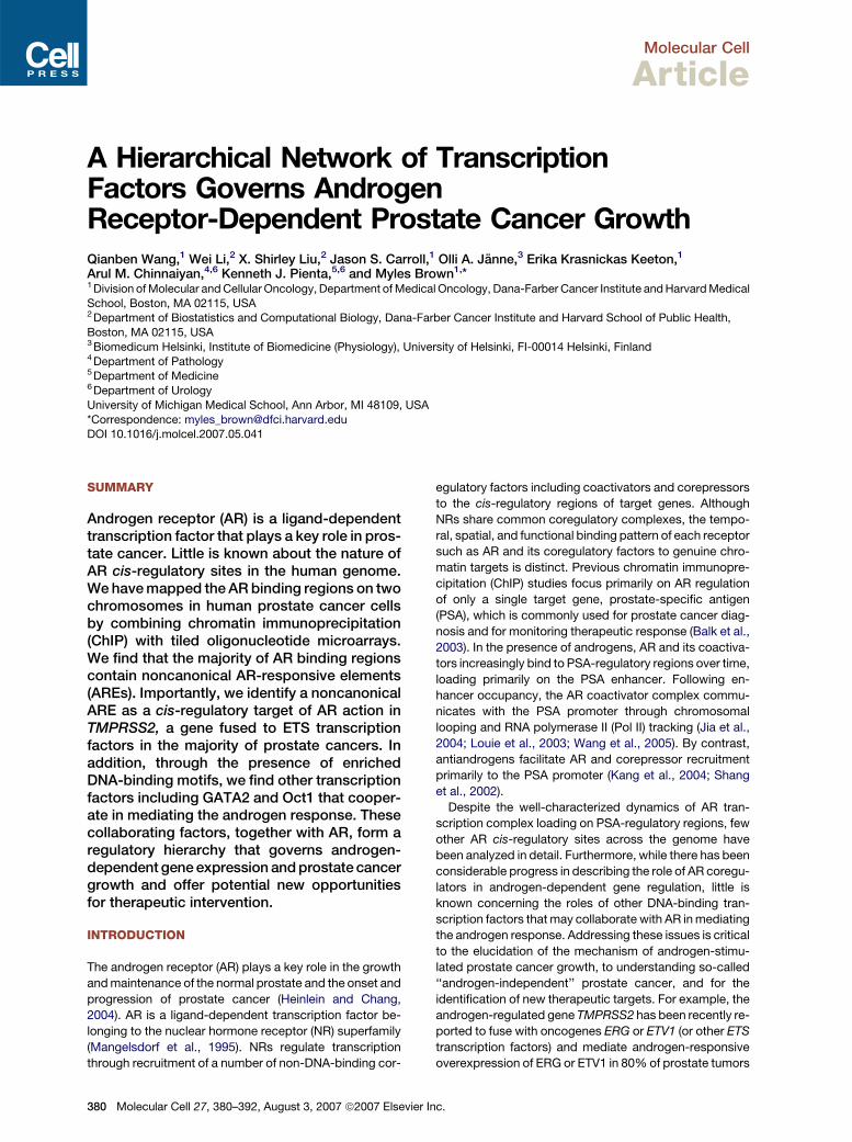
Figure 2. Characterization of AR Binding Sites on Chromosomes 21 and 22
(A) Standard ChIP assays of AR recruitment to various potential AR binding regions. LNCaP cells were treated with 100 nM DHT for 1 or 16 hr. ChIP
assays were performed with anti-AR antibodies. Immunoprecipitated DNA was quantified by real-time PCR using primers (Table S1) spanning various
potential AR binding regions.
(B and C) Effects of DHT concentration on AR binding to chromatin and cell proliferation. (B) Androgen-depleted LNCaP cells were treated with 1, 10,
or 100 nM DHT for 1 hr. ChIP assays were performed with an anti-AR antibody, and DNA precipitates were measured with real-time PCR using primers
spanning the PSA enhancer and the B39 site. (C) LNCaP cells were cultured in hormone-depleted medium for 3 days and then treated with vehicle or
DHT from 0.1 to 1000 nM for another 3 days. Cell proliferation was measured using the WST-1 assay.
(D) Standard ChIP assays of Pol II recruitment to various potential AR binding regions. ChIP assays were performed with anti-Pol II antibodies.
(E) Distribution of the types of AR binding motifs within AR binding sites and the chromosome 21 and 22 genomic background.
(F) AR binding regions function as enhancers. Ten selected AR binding regions, mutant AR binding regions with deleted AREs (B21 and B39), and the
FKBP5 enhancer were subcloned into the pGL3-promoter vector and transfected into androgen-depleted LNCaP cells. Empty pGL3 promoter vector
was used as a negative control. Cells were stimulated with 100 nM DHT or vehicle for 24 hr. The data were presented as the mean ± SE of two to three
replicates, (A)–(D) and (F).
concentrations as high as 1000 nM induce significant cell
growth (Figure 2C).
Selected AR Binding Sites Play Transcriptional Roles
We and others have previously found that RNA Pol II asso-
ciates with the AR binding regions of the PSA enhancer
and promoter, which is critical for PSA transcription (Louie
et al., 2003; Shang et al., 2002; Wang et al., 2005). To ad-
dress whether Pol II is recruited to the AR binding sites on
chromosomes 21 and 22, we performed Pol II ChIP on
382 Molecular Cell 27, 380–392, August 3, 2007 ª2007 Elsevie
several AR binding sites. Interestingly, Pol II was signifi-
cantly recruited to 50% of binding sites tested in an andro-
gen-dependent manner (R3-fold) (Figure 2D), supporting
the assertion that a significant number of binding sites
play transcriptional regulatory roles. We further performed
a comparative genomic analysis of 90 AR binding sites
between genomes of seven vertebrate species (chimp,
dog, mouse, rat, chicken, fugu, and zebrafish). The results
showed a strong conservation at the center of the AR
binding sites (Figure S1), consistent with the notion that
r Inc.

Molecular Cell
Collaborating Transcription Factors in AR Action
noncoding sequences that have survived selection play
important roles in gene regulation (Hardison, 2000).
Most AR Binding Sites Are Far from the Promoters
of Androgen-Regulated Genes
To correlate AR binding with androgen-dependent gene
expression, we examined the gene expression profile in
LNCaP cells in response to DHT using an expression
microarray. LNCaP cells were deprived of hormone for
72 hr and then treated with vehicle or DHT for 4 and
16 hr. Total RNA was extracted and then hybridized to
Affymetrix U133 plus 2.0 expression array. We found 62
transcripts on chromosomes 21 and 22 that were differen-
tially expressed in response to androgen (Table S2). Con-
sistent with previous studies (Nelson et al., 2002; Velasco
et al., 2004), we find that the expression of only a small
number of transcripts on chromosomes 21 and 22 is very
highly changed by androgens. In order to avoid a bias to-
ward this small number of highly differentially expressed
genes, we have included all transcripts that were differen-
tially expressed by at least 1.1-fold. Real-time RT-PCR
validation of five selected genes and PSA as a positive
control confirmed the expression array results (Figure S2).
Interestingly, on chromosomes 21 and 22, most AR bind-
ing sites are far from transcription start sites of androgen-
regulated genes (>10 kb) (Table S3). In total, only 34 out
of 90 binding sites (38%) were located within 500 kb of
transcriptional start sites of genes regulated by androgens
under these conditions in LNCaP cells.
Noncanonical AR-Responsive Elements Mediate
AR-Dependent Transcription
We next performed a DNA-binding motif search on the 90
AR binding regions. Surprisingly, we found that, although
78% of the binding regions contain the AR binding half-
site motif TGTTCT, only 10% of the binding regions have
a canonical class I NR (AGAACAnnnTGTTCT) binding
motif when we allow up to two positions to vary from the
palindromic consensus with 3 nt spacing (for detail, see
‘‘Sequence Analysis’’ in the Supplemental Experimental
Procedures) (Verrijdt et al., 2003) (Figure 2E and Table
S4). Of the 90 binding sites, 68% have one or more nonca-
nonical AREs including isolated ARE half-sites, head-to-
head AREs (AGAACA [0–8 n] TGTTCT, ns3), tail-to-tail
AREs (TGTTCT [0–8 n] AGAACA), and ARE direct repeats
(AGAACA [0–8 n] AGAACA) (Table S4 and ‘‘Sequence
Analysis’’ in the Supplemental Experimental Procedures).
To further address whether AR binding regions contain-
ing noncanonical AREs can function as enhancers, we
subcloned 10 AR binding sites and the recently character-
ized enhancer of the androgen-regulated gene FKBP5
(Magee et al., 2006) as a positive control, upstream of an
SV40 promoter in the pGL3 luciferase vector. The pGL3
luciferase vector with the minimal SV40 promoter alone
served as a negative control. These constructs were
transfected into androgen-depleted LNCaP cells, and lu-
ciferase activity was measured after 24 hr treatment with
vehicle alone or in the presence of saturating concentra-
Mol
tions of DHT. As shown in Figure 2F, all AR binding regions
tested demonstrate enhancer activity (R2-fold) with vary-
ing degrees of androgen dependence comparable to
the activity of the FKBP5 enhancer. To confirm that the
enhancer characteristics of the AR binding sites require
AREs, we deleted the canonical ARE within the B21 and
the noncanonical ARE within the B39 enhancer constructs
and transfected these mutated vectors into LNCaP cells.
As expected, deletion of the single typical ARE within
B21 completely abolished its enhancer activity (Figure 2F).
More importantly, deletion of the single noncanonical
(head-to-head, 8 nt spacing) ARE from B39 also com-
pletely abolished the enhancer activity, thus demonstrat-
ing that the noncanonical AREs within AR binding regions
are required for their enhancer function (Figure 2F).
Characterization of the TMPRSS2-Regulatory
Region
As mentioned above, most prostate cancers have recently
been shown to harbor either TMPRSS2:ERG or TMPRSS2:
ETV1 fusions (Tomlins et al., 2005). However, how the
TMPRSS2 gene or the TMPRSS2-ETS fusions are regu-
lated by AR is unknown. Using quantitative ChIP, we con-
firmed AR binding to site B39, which is�13.5 kb upstream
of the TMPRSS2 mRNA start site both in LNCaP cells that
do not harbor a rearranged TMPRSS2 gene (Figures 2A)
and in VCaP cells that contain the TMPRSS2:ERG fusion
(Figure 3A). In order to test whether this AR binding site
functions as an AR-dependent enhancer and communi-
cates with the TMPRSS2 promoter, we performed a ChIP
combined with chromosome conformation capture (5C)
assay in order to detect looped chromatin structures
in vivo (Wang et al., 2005). Hormone-depleted cells were
stimulated with vehicle or androgen. The fixed chromatin
was digested with EcoRI followed by anti-AR ChIP and
ligation. After reversing the crosslinks, one primer in the
TMPRSS2 enhancer and one primer in the promoter region
were used for PCR amplification of equal amounts of
eluted DNA. As shown in Figure 3B, the specific PCR prod-
uct is formed only in the presence of ligase and is signifi-
cantly enhanced by androgen stimulation, while control
PCR for the level of input chromatin was equivalent under
all conditions (Figure 3B). The 383 bp PCR amplified band
was sequenced to confirm that it indeed represents the
product of the correct ligation of the TMPRSS2 enhancer
and promoter. This result demonstrates that the TMPRSS2
enhancer is in close proximity to the promoter in vivo. As
negative controls, the interaction of the PSA enhancer
with the TMPRSS2 promoter and the TMPRSS2 enhancer
with the PSA promoter was tested by 5C. In neither case
was a specific PCR product formed (data not shown), sup-
porting the specificity of the 5C product formed between
the TMPRSS2 enhancer and promoter. In addition, a con-
trol region 9 kb upstream of the TMPRSS2 transcription
start site that contains two putative AR-responsive
element (ARE) motifs that were not found to bind AR did
not contact the TMPRSS2 promoter by 5C (Figure S3).
Interestingly, an AR binding site 462 kb downstream of
ecular Cell 27, 380–392, August 3, 2007 ª2007 Elsevier Inc. 383

Molecular Cell
Collaborating Transcription Factors in AR Action
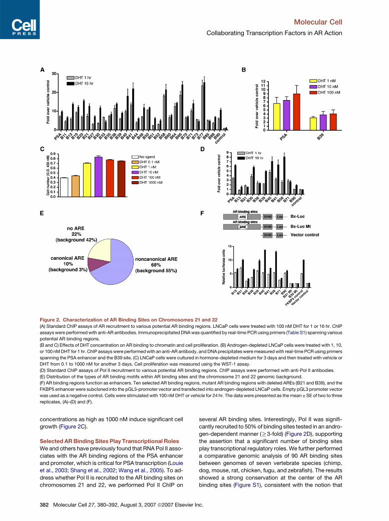
Figure 3. Characterization of TMPRSS2-Regulatory Regions
(A) ChIP analyses of AR occupancy on the PSA and B39 in VCaP cells. VCaP cells (a prostate cancer cell line that expresses TMPRSS2:ERG fusion
gene) were treated with 100 nM DHT for 1 hr. ChIP assays were performed with an anti-AR antibody.
(B) Spatial communication between the TMPRSS2 enhancer and promoter. 5C was performed using fixed or EcoRI- or BtgI-digested chromatin from
vehicle- or DHT-treated LNCaP cells. Primers (Table S1) flanking the�13.5 kb or 462 kb AR binding region and�700 bp promoter region were used to
PCR amplify DNA after ligation. Control PCR was performed using chromatin before restriction enzyme digestion.
(C) Schematic representation of the TMPRSS2 14 kb upstream regulatory region. Potential five ARE clusters and fourteen 1 kb fragments are shown.
(D) ChIP analyses of AR recruitment to five potential ARE regions. ChIP assays were performed with anti-AR antibodies in LNCaP cells treated with
vehicle or 100 nM DHT for 4 or 16 hr.
(E) Systematic mapping of enhancer elements within the TMPRSS2 14 kb upstream regulatory region. Fourteen 1 kb TMPRSS2 upstream sequences
were subcloned into the pGL3-promoter vector and transfected into LNCaP cells. Cells were treated with vehicle or 100 nM DHT for 24 hr. The data
are presented as the mean ± SE of two to three replicates, (A), (D), and (E).
TMPRSS2 (B38) also failed to form a loop with the
TMPRSS2 promoter (Figure 3B). This result serves as an-
other negative control and is consistent with the conclu-
sion that regions upstream of TMPRSS2 that are retained
in the TMPRSS2-ETS fusions rather than those down-
stream harbor the important functional elements. These
data demonstrate that the�13.5 kb AR binding site specif-
ically interacts with the TMPRSS2 promoter region, sup-
porting the formation of a chromosomal loop between
this upstream enhancer and the proximal promoter. The
identification of this upstream TMPRSS2 enhancer also
provides a molecular explanation of how expression
TMPRSS2-ETS factor fusions are driven in an androgen-
dependent manner. Interestingly, a newly identified pros-
tate cancer oncogene resulting from the fusion of
384 Molecular Cell 27, 380–392, August 3, 2007 ª2007 Elsevie
TMPRSS2 and the ETS transcription factor ETV4 does not
include any of the TMPRSS2 coding exons, as the break-
point is �8 kb upstream of the mRNA start site though
�5.5 kb downstream of this newly identified TMPRSS2 en-
hancer (Tomlins et al., 2006), suggesting that this enhancer
acts to drive ETV4 expression in this setting.
Although we found that the �13.5 kb B39 binding site
in the TMPRSS2 gene is a functional androgen-regulated
enhancer (Figure 2F) and is in close proximity to the
TMPRSS2 promoter in vivo (Figure 3B), we investigated
the possibility that other functionally important AR binding
sites located between�13.5 kb and the transcription start
site may have been missed by the ChIP-on-chip assay.
We therefore performed an AR motif search within the
14 kb region upstream of the TMPRSS2 gene and found
r Inc.
Molecular Cell
Collaborating Transcription Factors in AR Action
five clusters of potential AREs (Figure 3C). Despite the
presence of AREs, quantitative AR ChIP analyses found
only significant AR binding to ARE region V corresponding
exactly to the region identified by ChIP-on-chip (�13.5 kb)
(Figure 3D). In addition, to further address whether other
regions within the 14 kb upstream sequence might play
transcriptional roles, we systematically subcloned four-
teen 1 kb regions (Figure 3C) upstream of an SV40 pro-
moter in the pGL3 luciferase vector and transfected these
into androgen-depleted LNCaP cells. As shown in Fig-
ure 3E, construct A (�14 to �13 kb) corresponding to
the B39 element had the most significant enhancer activ-
ity. In addition, construct N (�1 kb to +24 bp) that contains
the TMPRSS2 promoter also demonstrated weak en-
hancer activity in this reporter assay. Significantly, quanti-
tative AR ChIP showed that AR was only weakly recruited
to the endogenous TMPRSS2 promoter region. Therefore
the B39 binding site is the primary site of AR binding and
functional enhancer activity within the 14 kb upstream of
the TMPRSS2 gene. Of course, we cannot rule out the
possibility that other cis-regulatory regions outside of the
14 kb region (Figure S4) may also play a role in TMPRSS2
expression.
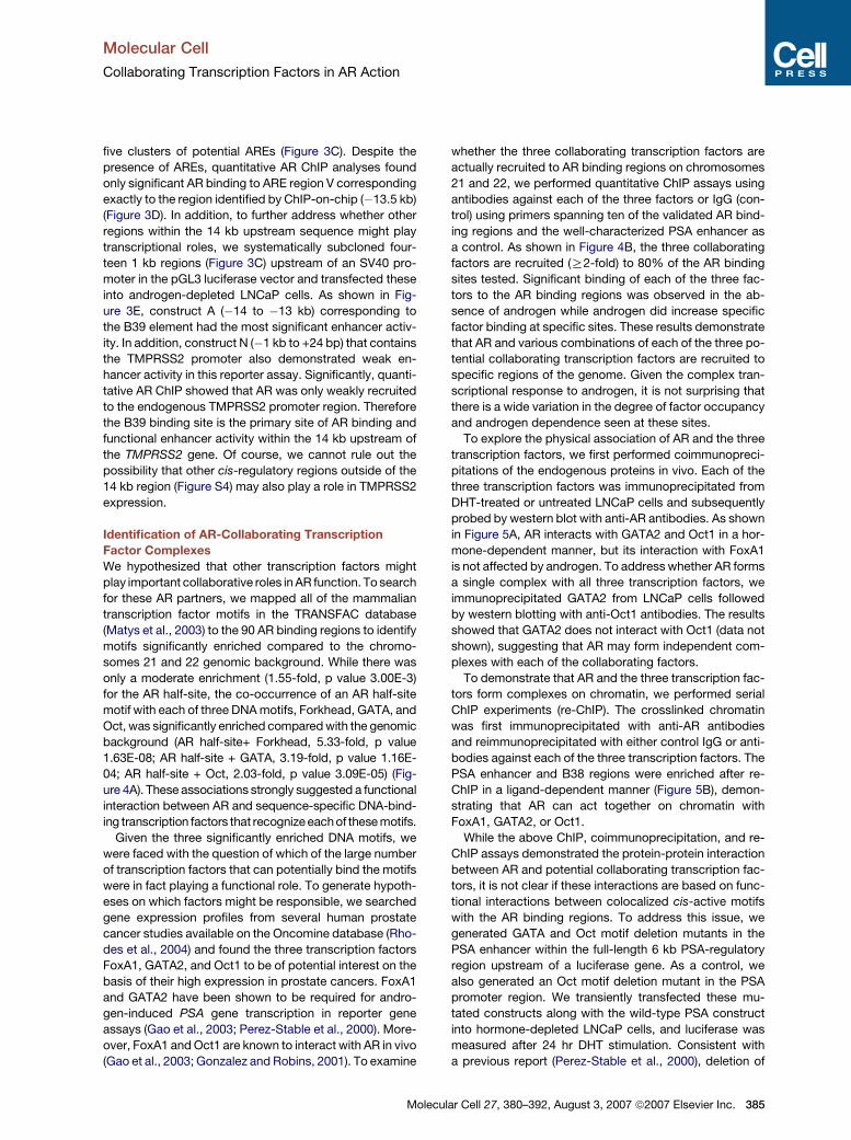
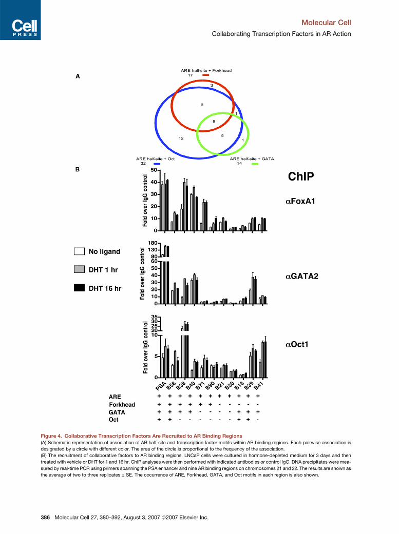
Identification of AR-Collaborating Transcription
Factor Complexes
We hypothesized that other transcription factors might
play important collaborative roles in AR function. To search
for these AR partners, we mapped all of the mammalian
transcription factor motifs in the TRANSFAC database
(Matys et al., 2003) to the 90 AR binding regions to identify
motifs significantly enriched compared to the chromo-
somes 21 and 22 genomic background. While there was
only a moderate enrichment (1.55-fold, p value 3.00E-3)
for the AR half-site, the co-occurrence of an AR half-site
motif with each of three DNA motifs, Forkhead, GATA, and
Oct, was significantly enriched compared with the genomic
background (AR half-site+ Forkhead, 5.33-fold, p value
1.63E-08; AR half-site + GATA, 3.19-fold, p value 1.16E-
04; AR half-site + Oct, 2.03-fold, p value 3.09E-05) (Fig-
ure 4A). These associations strongly suggested a functional
interaction between AR and sequence-specific DNA-bind-
ing transcription factors that recognize each of these motifs.
Given the three significantly enriched DNA motifs, we
were faced with the question of which of the large number
of transcription factors that can potentially bind the motifs
were in fact playing a functional role. To generate hypoth-
eses on which factors might be responsible, we searched
gene expression profiles from several human prostate
cancer studies available on the Oncomine database (Rho-
des et al., 2004) and found the three transcription factors
FoxA1, GATA2, and Oct1 to be of potential interest on the
basis of their high expression in prostate cancers. FoxA1
and GATA2 have been shown to be required for andro-
gen-induced PSA gene transcription in reporter gene
assays (Gao et al., 2003; Perez-Stable et al., 2000). More-
over, FoxA1 and Oct1 are known to interact with AR in vivo
(Gao et al., 2003; Gonzalez and Robins, 2001). To examine
Molec
whether the three collaborating transcription factors are
actually recruited to AR binding regions on chromosomes
21 and 22, we performed quantitative ChIP assays using
antibodies against each of the three factors or IgG (con-
trol) using primers spanning ten of the validated AR bind-
ing regions and the well-characterized PSA enhancer as
a control. As shown in Figure 4B, the three collaborating
factors are recruited (R2-fold) to 80% of the AR binding
sites tested. Significant binding of each of the three fac-
tors to the AR binding regions was observed in the ab-
sence of androgen while androgen did increase specific
factor binding at specific sites. These results demonstrate
that AR and various combinations of each of the three po-
tential collaborating transcription factors are recruited to
specific regions of the genome. Given the complex tran-
scriptional response to androgen, it is not surprising that
there is a wide variation in the degree of factor occupancy
and androgen dependence seen at these sites.
To explore the physical association of AR and the three
transcription factors, we first performed coimmunopreci-
pitations of the endogenous proteins in vivo. Each of the
three transcription factors was immunoprecipitated from
DHT-treated or untreated LNCaP cells and subsequently
probed by western blot with anti-AR antibodies. As shown
in Figure 5A, AR interacts with GATA2 and Oct1 in a hor-
mone-dependent manner, but its interaction with FoxA1
is not affected by androgen. To address whether AR forms
a single complex with all three transcription factors, we
immunoprecipitated GATA2 from LNCaP cells followed
by western blotting with anti-Oct1 antibodies. The results
showed that GATA2 does not interact with Oct1 (data not
shown), suggesting that AR may form independent com-
plexes with each of the collaborating factors.
To demonstrate that AR and the three transcription fac-
tors form complexes on chromatin, we performed serial
ChIP experiments (re-ChIP). The crosslinked chromatin
was first immunoprecipitated with anti-AR antibodies
and reimmunoprecipitated with either control IgG or anti-
bodies against each of the three transcription factors. The
PSA enhancer and B38 regions were enriched after re-
ChIP in a ligand-dependent manner (Figure 5B), demon-
strating that AR can act together on chromatin with
FoxA1, GATA2, or Oct1.
While the above ChIP, coimmunoprecipitation, and re-
ChIP assays demonstrated the protein-protein interaction
between AR and potential collaborating transcription fac-
tors, it is not clear if these interactions are based on func-
tional interactions between colocalized cis-active motifs
with the AR binding regions. To address this issue, we
generated GATA and Oct motif deletion mutants in the
PSA enhancer within the full-length 6 kb PSA-regulatory
region upstream of a luciferase gene. As a control, we
also generated an Oct motif deletion mutant in the PSA
promoter region. We transiently transfected these mu-
tated constructs along with the wild-type PSA construct
into hormone-depleted LNCaP cells, and luciferase was
measured after 24 hr DHT stimulation. Consistent with
a previous report (Perez-Stable et al., 2000), deletion of
ular Cell 27, 380–392, August 3, 2007 ª2007 Elsevier Inc. 385
Molecular Cell
Collaborating Transcription Factors in AR Action
Figure 4. Collaborative Transcription Factors Are Recruited to AR Binding Regions
(A) Schematic representation of association of AR half-site and transcription factor motifs within AR binding regions. Each pairwise association is
designated by a circle with different color. The area of the circle is proportional to the frequency of the association.
(B) The recruitment of collaborative factors to AR binding regions. LNCaP cells were cultured in hormone-depleted medium for 3 days and then
treated with vehicle or DHT for 1 and 16 hr. ChIP analyses were then performed with indicated antibodies or control IgG. DNA precipitates were mea-
sured by real-time PCR using primers spanning the PSA enhancer and nine AR binding regions on chromosomes 21 and 22. The results are shown as
the average of two to three replicates ± SE. The occurrence of ARE, Forkhead, GATA, and Oct motifs in each region is also shown.
386 Molecular Cell 27, 380–392, August 3, 2007 ª2007 Elsevier Inc.

Molecular Cell
Collaborating Transcription Factors in AR Action
Figure 5. AR Interacts with Collaborative Transcription Factors In Vivo
(A) AR coimmunopreciptates with its collaborating factors in vivo. LNCaP cells were grown in the presence or absence of DHT for 24 hr. Whole-cell
lysates were immunoprecipitated (IP) with indicated antibodies or control IgG. Western blot (WB) was performed with indicated antibodies.
(B) AR-collaborating factor complexes form on chromatin. LNCaP cells were treated with or without DHT for 16 hr. ChIP assays were performed with
anti-AR antibodies. The immunoprecipitated chromatin was eluted, reimmunoprecipitated with the indicated antibodies or control IgG, and amplified
by PCR using primers flanking the PSA and B38 enhancer regions.
(C and D) Colocalized cis-active elements are required for AR-dependent transcription. A wild-type PSA reporter construct and PSA reporter con-
struct with deleted GATA or Oct motifs within the PSA enhancer or promoter regions (C) or B39 reporter wild-type construct and B39 reporters
with GATA or Oct motif deletions (D) were transfected into LNCaP cells treated with vehicle or 100 nM DHT for 24 hr. Luciferase activities were
determined, and results are presented as the mean ± SE of the triplicate experiments.
the GATA motif within the PSA enhancer region dramati-
cally decreased androgen-stimulated transcriptional ac-
tivities (Figure 5C). Interestingly, deletion of the Oct motif
within the PSA enhancer, but not the promoter region,
also significantly attenuated androgen-induced transcrip-
tional activity (Figure 5C). These data suggest a functional
interaction among colocalized cis-active elements within
the PSA enhancer region. Significantly, both the Oct and
GATA motifs were also required for the activity of the
TMPRSS2 enhancer (Figure 5D).
AR-Collaborating Transcription Factors Regulate
AR-Mediated Transcription and Prostate
Cancer Cell Growth
To address the functional roles of the three collaborating
transcription factors in mediating AR-dependent tran-
scription, we reduced expression levels of the three fac-
Mole
tors by RNA interference (Figure 6A). We then investigated
the effect of silencing of the three factors on androgen-
stimulated expression of two previously reported andro-
gen-regulated genes PSA and TMPRSS2. We transiently
transfected siRNA targeting each of the three factors
into LNCaP cells. Forty-eight hours after transfection, cells
were treated with DHT (1 or 100 nM) for 4 or 16 hr. Quan-
titative RT-PCR was then performed to measure andro-
gen-regulated mRNA levels. As shown in Figure 6B, re-
duction of GATA2 or Oct1 levels significantly decreased
PSA at 16 hr and TMPRSS2 mRNA expression at 4 hr
while not changing control GADPH expression levels. In-
terestingly, TMPRSS2 expression returns to control levels
at 16 hr, suggesting the existence of gene-specific bypass
pathways. In contrast, FoxA1 silencing had no effect on
the expression of these two genes (Figures 6B). While
this result may suggest that FoxA1 does not play a critical
cular Cell 27, 380–392, August 3, 2007 ª2007 Elsevier Inc. 387

Molecular Cell
Collaborating Transcription Factors in AR Action
Figure 6. Functional Analyses of Collaborating Transcription Factors in Mediating AR-Dependent Transcription of the PSA and
TMPRSS2 Genes
(A) Suppression of AR-collaborating factor levels by RNAi. LNCaP cells were transfected with siRNA targeting each factor and a control siRNA. Forty-
eight hours posttransfection, cells were treated with or without 100 nM DHT for 16 hr, and western blots were performed using the antibodies indi-
cated.
(B) Effects of siRNA on PSA, TMPRSS2, and GADPH gene expression. Forty-eight hours after siRNA transfection, cells were treated with or without 1
or 100 nM DHT for 4 and 16 hr. Total RNA was isolated and amplified by real-time RT-PCR using transcript-specific primers (Table S1). The no-ligand
control was measured at 4 hr.
(C) Effects of silencing GATA2 and Oct1 on AR, Pol II, Oct1, and GATA2 recruitment to the PSA and TMPRSS2 enhancers. AR, Pol II, Oct1, and GATA2
ChIPs were performed after vehicle or 4 hr 100 nM DHT treatment of siLuc, siGATA2, or siOct1-transfected cells. Graphical representations of the
mean ± SE of two to three independent experiments are shown in (B) and (C).
role in androgen signaling, it is also possible that FoxA1
function is required for only a subset of AR targets or is
redundant with another Forkhead family member.
To investigate the mechanism of GATA2 and Oct1 ac-
tion on PSA and TMPRSS2 expression, we performed
ChIP assays to study the effect of GATA2 and Oct1 silenc-
ing on AR and Pol II recruitment to the PSA and TMPRSS2
enhancers. LNCaP cells were transiently transfected with
siGATA2, siOct1, or control siLuc; cultured for 48 hr; and
then stimulated with 100 nM DHT for 4 hr. As shown in
Figure 6C, recruitment of AR and Pol II to the PSA and
TMPRSS2 enhancers was significantly decreased in si-
GATA2 transfected cells as compared with the siLuc con-
trol, both in the absence of ligand and, to a greater extent,
in the presence of hormone. In contrast, silencing of Oct1
did not affect AR binding but greatly reduced Pol II loading
on the PSA and TMPRSS2 enhancers, both in the absence
388 Molecular Cell 27, 380–392, August 3, 2007 ª2007 Elsevier
and presence of hormone (Figure 6C). Interestingly, Oct1
recruitment to the PSA and TMPRSS2 enhancers was
similarly attenuated when GATA2 was silenced, whereas
GATA2 binding to these enhancers was not affected by
Oct1 silencing (Figure 6C). Western blot analyses showed
that silencing of GATA2 and Oct1 had no obvious effect on
the level of AR or Pol II expression (Figure 6A). This sug-
gests that GATA2 and Oct1 play necessary roles both in
the basal recruitment of AR and/or Pol II to chromatin and
in their recruitment following androgen stimulation. More
interestingly, these data suggest that GATA2 and Oct1
act at distinct steps in AR signaling, with GATA2 acting
upstream of Oct1 recruitment.
In order to extend these findings to a gene not previ-
ously known to be androgen regulated, we analyzed the
role of GATA2, Oct1, and FoxA1 in the expression of the
PDE9A gene. PDE9A is a member of the cyclic nucleotide
Inc.

Molecular Cell
Collaborating Transcription Factors in AR Action
Figure 7. Functional Roles of Collaborating Transcription Factors in Mediating the PDE9A Gene Transcription and Prostate
Cancer Cell Proliferation
(A) AR binding sites relative to the PDE9A gene. The black blocks represent AR binding sites. The PDE9A gene is shown in its 50-30 orientation, and the
blue arrows indicate the direction of the gene (June 05, University of California, Santa Cruz [UCSC], known genes).
(B) Effects of FoxA1, GATA2, and Oct1 silencing on PDE9A mRNA expression. siRNA-RTPCR analyses were performed as described in Figure 6B.
(C) Effects of silencing GATA2 and Oct1 on AR, Pol II, Oct1, and GATA2 recruitment to the PDE9A enhancers. siRNA-ChIP analyses were performed
as described in Figure 6C.
(D) Effects of AR and cofactor silencing on androgen-stimulated cell cycle entry. Forty-eight hours after siRNA transfection, cells were treated with
or without 1 or 10 nM DHT for 24 hr. Cells were then fixed, stained with propidium iodide, and analyzed by flow cytometry. Values represent the
mean ± SE of two to three independent experiments (B) to (D).
phosphodiesterase (PDE) family that plays a critical role in
regulating intracellular concentrations of cyclic nucleo-
tides and is highly expressed in prostate (Fisher et al.,
1998; Guipponi et al., 1998). Our ChIP-on-chip analysis
identified two intronic AR binding sites (B40 and B41)
that are 18.4 kb and 77.8 kb downstream of the PDE9A
transcription start site, respectively (Figure 7A). In addi-
tion, our gene expression studies identified PDE9A as a
gene significantly upregulated by androgen in LNCaP cells
(Table S2 and Figure S2). Similar to our findings with the
PSA and TMPRSS2 genes, we find that GATA2 and Oct1,
but not FoxA1, play necessary collaborative and hierarchi-
cal roles in AR-mediated PDE9A gene expression (Figures
7B and 7C).
Mole
As AR is essential for the growth of prostate cancer cells
(Heinlein and Chang, 2004) and previous studies have
shown AR silencing inhibits prostate cancer cell prolifera-
tion (Wright et al., 2003; Zegarra-Moro et al., 2002), we
next tested the effects of silencing individually or in com-
bination the collaborating transcription factors on cell
cycle progression. Forty-eight hours after siRNA transfec-
tion, LNCaP cells were treated with or without 1 or 10 nM
DHT for 24 hr. Cells were then stained with propidium io-
dide for DNA content. Silencing of GATA2 and Oct1 alone
or in combination significantly decreased androgen-
induced cell cycle progression and to the same extent
as silencing AR itself (Figures 7D). To control for potential
off-target effects, silencing GATA2 and Oct1 with an
cular Cell 27, 380–392, August 3, 2007 ª2007 Elsevier Inc. 389
Molecular Cell
Collaborating Transcription Factors in AR Action
independent set of siRNAs also inhibited androgen-de-
pendent cell cycle progression (Figure S5). In addition,
the response to the silencing of GATA2 and Oct1 was spe-
cific, as their silencing had no effect on cell cycle progres-
sion in HeLa cells (Figure S6). Interestingly, consistent with
its nonessential role in androgen-stimulated gene expres-
sion, silencing of FoxA1, a chromatin ‘‘pioneer’’ factor
(Cirillo et al., 2002), did not affect androgen-stimulated
cell cycle progression (Figure S7). Thus, GATA2 and Oct
1, but not FoxA1, play essential roles in androgen-stimu-
lated cell cycle progression in prostate cancer cells.
DISCUSSION
Whereas previous ChIP studies have provided useful in-
formation pertaining to AR transcription complex assem-
bly (Jia et al., 2004; Louie et al., 2003; Kang et al., 2004;
Shang et al., 2002; Wang et al., 2005), the data are mainly
restricted to the PSA gene. In this study, we used ChIP-
on-chip to map 90 previously unknown AR binding sites
on chromosomes 21 and 22. As chromosomes 21 and
22 represent approximately 2% of the entire nonrepetitive
human genome, these results predict �4500 AR binding
sites across the entire human genome.
Interestingly, most of the binding sites identified on chro-
mosomes 21 and 22 are far from androgen-regulated
genes. We previously have found that most estrogen re-
ceptor (ER) binding sites are also distant from estrogen-
regulated genes, consistent with their functioning as en-
hancers rather than promoters (Carroll et al., 2005, 2006).
Whether distance-independent binding is a general rule
for NR action awaits similar studies of other NRs. While it
is likely that many of these sites act as transcriptional en-
hancers of neighboring genes, other functions may also
exist. In addition, it is likely that only a subset of the binding
sites are functional in LNCaP under the specific experi-
mental conditions tested and may be functional in different
cell types and/or under different conditions, as previously
suggested (van Steensel, 2005). It is also possible that
some AR binding sites are indeed nonfunctional. Finally,
while a proportion of androgen-regulated genes have AR
binding sites associated with the gene, androgen-stimu-
lated transcription may in some cases be independent of
AR binding to chromatin.
Interestingly, we found that the majority of AR binding
sites on chromosomes 21 and 22 contain noncanonical
AREs and that a significant proportion of those tested can
function as AR-dependent enhancers and that the nonca-
nonical AREs are necessary for this activity. Although
in vitro DNA-binding assays demonstrated AR binds to
canonical AREs with highest affinity (Kallio et al., 1994),
a small number of noncanonical AREs such as ARE direct
repeats have been previously reported (Verrijdt et al.,
2003). In the genuine chromatin environment, it is likely
that collaborating factors may assist AR in binding to non-
canonical AREs. Our finding of a preponderance of atypi-
cal modes of AR binding suggests that transcription factor
binding motifs defined in the pre-ChIP era need to be
390 Molecular Cell 27, 380–392, August 3, 2007 ª2007 Elsevi
readdressed in the face of the ability to find large numbers
of additional binding sites using ChIP-on-chip.
In contrast to NR coregulatory factors identified by bio-
chemical methods (McKenna and O’Malley, 2002; Perissi
and Rosenfeld, 2005), we used a systems approach unit-
ing diverse data types combining chromosome-scale
ChIP-on-chip analysis and gene expression profiling to
identify a network of AR-collaborating transcription fac-
tors. Our data demonstrate a physical interaction between
AR and these transcription factors and that the collaborat-
ing partners have distinct functional roles in androgen-
dependent gene transcription and cell proliferation. Inter-
estingly, we found that GATA2 and Oct1 hierarchically
regulate androgen-dependent transcription. Previous
studies have found that other GATA family members can
open the compact chromatin through their conserved zinc
finger motifs (Boyes et al., 1998; Cirillo et al., 2002). Given
our finding that GATA2 is required for AR binding, it may
function similarly in this case as a pioneer factor that alters
chromatin to allow AR binding. In contrast, Oct1 is not re-
quired for AR to bind the PSA, TMPRSS2, and PDE9A en-
hancers, suggesting it functions at a step subsequent to
GATA2 action and in conjunction with AR. Consistent with
the roles of GATA2 and Oct1 in androgen target gene ex-
pression, silencing of these two collaborating transcrip-
tion factors significantly decreases androgen-induced cell
cycle progression. In other systems it has been reported
that GATA2 and Oct1 promote cell proliferation through
mechanisms including a p53-dependent pathway in the
case of GATA2 (Tsai and Orkin, 1997) and activation of his-
tone H2B transcription in the case of Oct1 (Ewen, 2000).
There is abundant evidence that AR plays a critical role
both in early hormone-dependent prostate cancer and in
so-called advanced androgen-independent prostate can-
cer (Debes and Tindall, 2004) where AR can be amplified.
The elucidation of the authentic AR cis-regulatory ele-
ments allows an understanding of how critical androgen-
stimulated targets such as TMPRSS2-ETS fusions are
regulated. Finally, the identification of the critical AR-
cooperating transcription factors provides potential new
targets for therapeutic intervention.
EXPERIMENTAL PROCEDURES
ChIP-on-Chip Assay
ChIP was performed with an anti-AR antibody (N20, Santa Cruz Bio-
technology, CA) as previously described (Wang et al., 2005) except
that 100 mg/ml yeast tRNA was used as blocking reagent instead of
2 mg salmon sperm DNA. Ligation-mediated PCR and labeling were
performed as previously described (Carroll et al., 2005). Microarrays
used were Affymetrix Genechip chromosome 21/22 tiling set P/N
900545. Biological triplicates were performed. The ChIP-on-chip raw
data are accessible at http://chip.dfci.harvard.edu/�wli/Androgen.
Receptor.ChIP2/. Details for ChIP-on-chip data and sequence analy-
ses are available in the Supplemental Experimental Procedures.
ChIP and Re-ChIP
ChIP and re-ChIP were performed as previously described (Shang
et al., 2000; Wang et al., 2005). Details are available in the Supplemen-
tal Experimental Procedures.
er Inc.

Molecular Cell
Collaborating Transcription Factors in AR Action
Cell Proliferation Assay
Cell proliferation was measured using a WST-1 kit (Roche, Indianapo-
lis, IN).
Expression Array Experiments
Hormone-depleted LNCaP cells were grown in the absence or pres-
ence of 100 nM DHT for 4 and 16 hr. Total RNA was isolated using
an RNeasy kit (QIAGEN, Valencia, CA). Total RNA from three biological
replicates was hybridized at the Dana-Farber Cancer Institute Microar-
ray Core Facility to human U133 plus 2.0 expression array. The data
were analyzed by using dChip microarray software (Li and Wong,
2001).
ChIP Combined Chromosome Conformation Capture Assay
5C was performed as previously described (Wang et al., 2005). The
primers used are listed in Table S1. The PCR amplified band was
sequence verified.
Construction of Plasmids
AR binding sites were PCR amplified from LNCaP genomic DNA. Four-
teen 1 kb TMPRSS2 upstream sequences were PCR amplified from
BAC RP11-814F13. These PCR products were subcloned into pGL3-
promoter vector (Promega, Madison, WI). ARE, GATA, and Oct motif
deletion mutants were sequence verified. The PCR primers are listed
in Table S1.
Reporter Gene Assays
Hormone-depleted LNCaP cells were transfected using Lipofectamine
2000 (Invitrogen, Carlsbad, CA). Details are available in the Supple-
mental Experimental Procedures.
Coimmunoprecipitation and Western Blotting
Coimmunoprecipitation and western blotting were performed as previ-
ously described (Wang et al., 2002) with a few modifications. Details
are available in the Supplemental Experimental Procedures.
Small Interfering RNA
Small interfering RNA (siRNA) duplexes were transfected into LNCaP
cells using Lipofectamine 2000 (Invitrogen). Forty-eight hours after
siRNA transfection, cells were treated with DHT or vehicle and then
harvested. The siRNA sequences are listed in Table S1.
Real-Time RT-PCR
Real-time RT-PCR was performed as previously described (Wang
et al., 2005). Primers for RT-PCR are listed in Table S1.
Flow Cytometry
siRNA-transfected LNCaP cells were treated with DHT for 24 hr. Cells
were collected and stained with propidium iodide. DNA contents were
analyzed by Dana-Farber Cancer Institute Cytometry Core Facility.
Supplemental Data
Supplemental Data include Supplemental Experimental Procedures,
seven figures, and four tables and can be found with this article online
at http://www.molecule.org/cgi/content/full/27/3/380/DC1/.
ACKNOWLEDGMENTS
This work was supported by the Claudia Adams Barr Program in Inno-
vative Basic Cancer Research (M.B.), Prostate Cancer SPORE Grants
P50 CA90381 (M.B.) and P50 CA69568 (K.J.P.), Department of De-
fense award W81XWH-05-01-0023 (Q.W.), and European Union con-
tract LSHM-CT-2005-018652 (O.A.J.). K.J.P. is an American Cancer
Society Clinical Research Professor. The authors also thank Drs. Jer-
ome Eeckhoute and Kirsten Fertuck for helpful discussions.
Molec
Received: November 7, 2006
Revised: March 15, 2007
Accepted: May 31, 2007
Published: August 2, 2007
REFERENCES
Balk, S.P., Ko, Y.J., and Bubley, G.J. (2003). Biology of prostate-
specific antigen. J. Clin. Oncol. 21, 383–391.
Boyes, J., Byfield, P., Nakatani, Y., and Ogryzko, V. (1998). Regulation
of activity of the transcription factor GATA-1 by acetylation. Nature
396, 594–598.
Carroll, J.S., Liu, X.S., Brodsky, A.S., Li, W., Meyer, C.A., Szary, A.J.,
Eeckhoute, J., Shao, W., Hestermann, E.V., Geistlinger, T.R., et al.
(2005). Chromosome-wide mapping of estrogen receptor binding
reveals long-range regulation requiring the forkhead protein FoxA1.
Cell 122, 33–43.
Carroll, J.S., Meyer, C.A., Song, J., Li, W., Geistlinger, T.R., Eeckhoute,
J., Brodsky, A.S., Keeton, E.K., Fertuck, K.C., Hall, G.F., et al. (2006).
Genome-wide analysis of estrogen receptor binding sites. Nat. Genet.
38, 1289–1297.
Cirillo, L.A., Lin, F.R., Cuesta, I., Friedman, D., Jarnik, M., and Zaret,
K.S. (2002). Opening of compacted chromatin by early developmental
transcription factors HNF3 (FoxA) and GATA-4. Mol. Cell 9, 279–289.
Debes, J.D., and Tindall, D.J. (2004). Mechanisms of androgen-refrac-
tory prostate cancer. N. Engl. J. Med. 351, 1488–1490.
Ewen, M.E. (2000). Where the cell cycle and histones meet. Genes
Dev. 14, 2265–2270.
Fisher, D.A., Smith, J.F., Pillar, J.S., St Denis, S.H., and Cheng, J.B.
(1998). Isolation and characterization of PDE9A, a novel human
cGMP-specific phosphodiesterase. J. Biol. Chem. 273, 15559–15564.
Gao, N., Zhang, J., Rao, M.A., Case, T.C., Mirosevich, J., Wang, Y.,
Jin, R., Gupta, A., Rennie, P.S., and Matusik, R.J. (2003). The role of
hepatocyte nuclear factor-3 alpha (Forkhead Box A1) and androgen
receptor in transcriptional regulation of prostatic genes. Mol. Endocri-
nol. 17, 1484–1507.
Gonzalez, M.I., and Robins, D.M. (2001). Oct-1 preferentially interacts
with androgen receptor in a DNA-dependent manner that facilitates
recruitment of SRC-1. J. Biol. Chem. 276, 6420–6428.
Guipponi, M., Scott, H.S., Kudoh, J., Kawasaki, K., Shibuya, K., Shin-
tani, A., Asakawa, S., Chen, H., Lalioti, M.D., Rossier, C., et al. (1998).
Identification and characterization of a novel cyclic nucleotide phos-
phodiesterase gene (PDE9A) that maps to 21q22.3: alternative splicing
of mRNA transcripts, genomic structure and sequence. Hum. Genet.
103, 386–392.
Hardison, R.C. (2000). Conserved noncoding sequences are reliable
guides to regulatory elements. Trends Genet. 16, 369–372.
Heinlein, C.A., and Chang, C. (2004). Androgen receptor in prostate
cancer. Endocr. Rev. 25, 276–308.
Horoszewicz, J.S., Leong, S.S., Chu, T.M., Wajsman, Z.L., Friedman,
M., Papsidero, L., Kim, U., Chai, L.S., Kakati, S., Arya, S.K., and Sand-
berg, A.A. (1980). The LNCaP cell line—a new model for studies on
human prostatic carcinoma. Prog. Clin. Biol. Res. 37, 115–132.
Jia, L., Choong, C.S., Ricciardelli, C., Kim, J., Tilley, W.D., and Coet-
zee, G.A. (2004). Androgen receptor signaling: mechanism of interleu-
kin-6 inhibition. Cancer Res. 64, 2619–2626.
Kallio, P.J., Palvimo, J.J., Mehto, M., and Janne, O.A. (1994). Analysis
of androgen receptor-DNA interactions with receptor proteins pro-
duced in insect cells. J. Biol. Chem. 269, 11514–11522.
Kang, Z., Janne, O.A., and Palvimo, J.J. (2004). Coregulator recruit-
ment and histone modifications in transcriptional regulation by the
androgen receptor. Mol. Endocrinol. 18, 2633–2648.
ular Cell 27, 380–392, August 3, 2007 ª2007 Elsevier Inc. 391

Molecular Cell
Collaborating Transcription Factors in AR Action
Li, C., and Wong, W.H. (2001). Model-based analysis of oligonucleo-
tide arrays: expression index computation and outlier detection.
Proc. Natl. Acad. Sci. USA 98, 31–36.
Lin, M.F., Meng, T.C., Rao, P.S., Chang, C., Schonthal, A.H., and Lin,
F.F. (1998). Expression of human prostatic acid phosphatase corre-
lates with androgen-stimulated cell proliferation in prostate cancer
cell lines. J. Biol. Chem. 273, 5939–5947.
Louie, M.C., Yang, H.Q., Ma, A.H., Xu, W., Zou, J.X., Kung, H.J., and
Chen, H.W. (2003). Androgen-induced recruitment of RNA polymerase
II to a nuclear receptor-p160 coactivator complex. Proc. Natl. Acad.
Sci. USA 100, 2226–2230.
Magee, J.A., Chang, L.W., Stormo, G.D., and Milbrandt, J. (2006). Di-
rect, androgen receptor-mediated regulation of the FKBP5 gene via
a distal enhancer element. Endocrinology 147, 590–598.
Mangelsdorf, D.J., Thummel, C., Beato, M., Herrlich, P., Schutz, G.,
Umesono, K., Blumberg, B., Kastner, P., Mark, M., Chambon, P.,
et al. (1995). The nuclear receptor superfamily: the second decade.
Cell 83, 835–839.
Matys, V., Fricke, E., Geffers, R., Gossling, E., Haubrock, M., Hehl, R.,
Hornischer, K., Karas, D., Kel, A.E., Kel-Margoulis, O.V., et al. (2003).
TRANSFAC: transcriptional regulation, from patterns to profiles.
Nucleic Acids Res. 31, 374–378.
McKenna, N.J., and O’Malley, B.W. (2002). Combinatorial control of
gene expression by nuclear receptors and coregulators. Cell 108,
465–474.
Nelson, P.S., Clegg, N., Arnold, H., Ferguson, C., Bonham, M., White,
J., Hood, L., and Lin, B. (2002). The program of androgen-responsive
genes in neoplastic prostate epithelium. Proc. Natl. Acad. Sci. USA 99,
11890–11895.
Perez-Stable, C.M., Pozas, A., and Roos, B.A. (2000). A role for GATA
transcription factors in the androgen regulation of the prostate-specific
antigen gene enhancer. Mol. Cell. Endocrinol. 167, 43–53.
Perissi, V., and Rosenfeld, M.G. (2005). Controlling nuclear receptors:
the circular logic of cofactor cycles. Nat. Rev. Mol. Cell Biol. 6, 542–
554.
Rhodes, D.R., Yu, J., Shanker, K., Deshpande, N., Varambally, R.,
Ghosh, D., Barrette, T., Pandey, A., and Chinnaiyan, A.M. (2004).
ONCOMINE: a cancer microarray database and integrated data-
mining platform. Neoplasia 6, 1–6.
Shang, Y., Hu, X., DiRenzo, J., Lazar, M.A., and Brown, M. (2000).
Cofactor dynamics and sufficiency in estrogen receptor-regulated
transcription. Cell 103, 843–852.
392 Molecular Cell 27, 380–392, August 3, 2007 ª2007 Elsevie
Shang, Y., Myers, M., and Brown, M. (2002). Formation of the andro-
gen receptor transcription complex. Mol. Cell 9, 601–610.
Tomlins, S.A., Rhodes, D.R., Perner, S., Dhanasekaran, S.M., Mehra,
R., Sun, X.W., Varambally, S., Cao, X., Tchinda, J., Kuefer, R., et al.
(2005). Recurrent fusion of TMPRSS2 and ETS transcription factor
genes in prostate cancer. Science 310, 644–648.
Tomlins, S.A., Mehra, R., Rhodes, D.R., Smith, L.R., Roulston, D.,
Helgeson, B.E., Cao, X., Wei, J.T., Rubin, M.A., Shah, R.B., and Chin-
naiyan, A.M. (2006). TMPRSS2:ETV4 gene fusions define a third mo-
lecular subtype of prostate cancer. Cancer Res. 66, 3396–3400.
Tsai, F.Y., and Orkin, S.H. (1997). Transcription factor GATA-2 is re-
quired for proliferation/survival of early hematopoietic cells and mast
cell formation, but not for erythroid and myeloid terminal differentia-
tion. Blood 89, 3636–3643.
van Steensel, B. (2005). Mapping of genetic and epigenetic regulatory
networks using microarrays. Nat. Genet. Suppl. 37, S18–S24.
Velasco, A.M., Gillis, K.A., Li, Y., Brown, E.L., Sadler, T.M., Achilleos,
M., Greenberger, L.M., Frost, P., Bai, W., and Zhang, Y. (2004). Iden-
tification and validation of novel androgen-regulated genes in prostate
cancer. Endocrinology 145, 3913–3924.
Verrijdt, G., Haelens, A., and Claessens, F. (2003). Selective DNA rec-
ognition by the androgen receptor as a mechanism for hormone-spe-
cific regulation of gene expression. Mol. Genet. Metab. 78, 175–185.
Wang, Q., Sharma, D., Ren, Y., and Fondell, J.D. (2002). A coregulatory
role for the TRAP-mediator complex in androgen receptor-mediated
gene expression. J. Biol. Chem. 277, 42852–42858.
Wang, Q., Carroll, J.S., and Brown, M. (2005). Spatial and temporal
recruitment of androgen receptor and its coactivators involves chro-
mosomal looping and polymerase tracking. Mol. Cell 19, 631–642.
Wright, M.E., Tsai, M.J., and Aebersold, R. (2003). Androgen receptor
represses the neuroendocrine transdifferentiation process in prostate
cancer cells. Mol. Endocrinol. 17, 1726–1737.
Zegarra-Moro, O.L., Schmidt, L.J., Huang, H., and Tindall, D.J. (2002).
Disruption of androgen receptor function inhibits proliferation of andro-
gen-refractory prostate cancer cells. Cancer Res. 62, 1008–1013.
Accession Numbers
The microarray data in this study have been deposited in NCBI’s Gene
Expression Omnibus (GEO, http://www.ncbi.nlm.nih.gov/geo/) and
are accessible under GEO Series accession number GSE7868.
r Inc.