Tumorigenesis and Neoplastic Progression Androgen Receptor Remains Critical for Cell-Cycle Progression in Androgen-Independent CWR22 Prostate Cancer Cells Xin Yuan,* Tong Li,* Hongyun Wang,* Tao Zhang,* Moumita Barua,* Robert A. Borgesi,* Glenn J. Bubley,* Michael L. Lu, † and Steven P. Balk* From the Cancer Biology Program,* Division of Hematology/Oncology, Department of Medicine, Beth Israel Deaconess Medical Center and the Urology Research Laboratory, † Brigham and Women’s Hospital, Harvard Medical School, Boston, Massachusetts The majority of prostate cancers (PCa) that relapse after androgen deprivation therapy (androgen-in- dependent PCa) continue to express androgen re- ceptor (AR). To study the functional importance of AR in these tumors, we derived androgen-indepen- dent CWR22 PCa xenografts in castrated mice and generated a cell line from one of these xenografts (CWR22R3). Similarly to androgen-independent PCa in patients, the relapsed xenografts and cell line expressed AR and were resistant to treatment with bicalutamide. However, expression of the AR- regulated PSA gene in the CWR22R3 cell line was markedly decreased compared to the relapsed xenografts in vivo. Transfections with androgen- regulated reporter genes further indicated that the cells lacked androgen-independent AR transcrip- tional activity and were not hypersensitive to low androgen concentrations despite constitutive acti- vation of the Erk/MAP kinases. Nonetheless, AR remained essential for androgen-independent growth because retroviral shRNA-mediated AR down-regulation resulted in marked long-term growth suppression. This was associated with in- creased levels of p27 kip1 and hypophosphorylation of retinoblastoma protein but not with decreases in D-type cyclin levels or MAP kinase activation. These results reveal a potentially critical function of AR in androgen-independent PCa that is distinct from its previously described transcriptional or nontran- scriptional functions. (Am J Pathol 2006, 169:682– 696; DOI: 10.2353/ajpath.2006.051047) Prostate cancer (PCa) most frequently presents as an androgen-dependent disease, and suppression of an- drogen receptor (AR) activity by surgical or medical cas- tration (androgen-deprivation therapy) results in tumor regression and alleviation of cancer-related symptoms in most patients. 1 However, patients with initially favorable responses to androgen-deprivation therapy eventually relapse with tumors that are termed hormone refractory or androgen-independent PCa. Importantly, the high-level expression of the AR and renewed expression of andro- gen-regulated genes such as prostate-specific antigen ( PSA) suggest that the AR regains its activity and plays an important role at this stage of the disease. 2,3 Some patients who relapse after androgen-deprivation therapy may still respond to secondary hormonal treatments with AR antagonists such as bicalutamide or with agents such as ketoconozole that suppress residual androgen pro- duction from the adrenal glands, suggesting that the tumors are responding to the low levels of androgen that remain after surgical or medical castration. 4,5 Indeed, studies using androgen-dependent PCa cells have shown that long-term growth under reduced androgen conditions can select for cells in which the AR can be activated by very low levels of androgen, and down- regulation of AR expression in these cells can suppress their growth. 6 –11 Mechanisms that may mediate PCa adaptation to decreased androgen levels include AR amplification, AR mutations, increased expression of transcriptional co-activator proteins, and activation of other signal transduction pathways. Previous studies showed that AR mRNA was highly expressed, the AR gene was frequently amplified in androgen-independent PCa, 12,13 and increased expression of AR was a con- Supported by the National Institutes of Health (grants R01-CA65647 to S.P.B. and K01-DK64739 to X.Y.), Massachusetts State (prostate cancer research grant to X.Y.), the Department of Defense (grant DAMD17-03- 0164 to X.Y.), and the Hershey Family Prostate Cancer Research Fund. Accepted for publication May 6, 2006. Address reprint requests to Xin Yuan, Hematology/Oncology Division, Beth Israel Deaconess Medical Center, 330 Brookline Ave., Boston, MA 02215. E-mail: [email protected]. The American Journal of Pathology, Vol. 169, No. 2, August 2006 Copyright © American Society for Investigative Pathology DOI: 10.2353/ajpath.2006.051047 682

Welcome message from author
This document is posted to help you gain knowledge. Please leave a comment to let me know what you think about it! Share it to your friends and learn new things together.
Transcript
Tumorigenesis and Neoplastic Progression
Androgen Receptor Remains Critical for Cell-CycleProgression in Androgen-Independent CWR22Prostate Cancer Cells
Xin Yuan,* Tong Li,* Hongyun Wang,*Tao Zhang,* Moumita Barua,* Robert A. Borgesi,*Glenn J. Bubley,* Michael L. Lu,† andSteven P. Balk*
From the Cancer Biology Program,* Division of
Hematology/Oncology, Department of Medicine, Beth Israel
Deaconess Medical Center and the Urology Research Laboratory,†
Brigham and Women’s Hospital, Harvard Medical School,
Boston, Massachusetts
The majority of prostate cancers (PCa) that relapseafter androgen deprivation therapy (androgen-in-dependent PCa) continue to express androgen re-ceptor (AR). To study the functional importance ofAR in these tumors , we derived androgen-indepen-dent CWR22 PCa xenografts in castrated mice andgenerated a cell line from one of these xenografts(CWR22R3). Similarly to androgen-independentPCa in patients , the relapsed xenografts and cellline expressed AR and were resistant to treatmentwith bicalutamide. However , expression of the AR-regulated PSA gene in the CWR22R3 cell line wasmarkedly decreased compared to the relapsedxenografts in vivo. Transfections with androgen-regulated reporter genes further indicated that thecells lacked androgen-independent AR transcrip-tional activity and were not hypersensitive to lowandrogen concentrations despite constitutive acti-vation of the Erk/MAP kinases. Nonetheless , ARremained essential for androgen-independentgrowth because retroviral shRNA-mediated ARdown-regulation resulted in marked long-termgrowth suppression. This was associated with in-creased levels of p27kip1 and hypophosphorylationof retinoblastoma protein but not with decreases inD-type cyclin levels or MAP kinase activation. Theseresults reveal a potentially critical function of AR inandrogen-independent PCa that is distinct from itspreviously described transcriptional or nontran-scriptional functions. (Am J Pathol 2006, 169:682–696;DOI: 10.2353/ajpath.2006.051047)
Prostate cancer (PCa) most frequently presents as anandrogen-dependent disease, and suppression of an-drogen receptor (AR) activity by surgical or medical cas-tration (androgen-deprivation therapy) results in tumorregression and alleviation of cancer-related symptoms inmost patients.1 However, patients with initially favorableresponses to androgen-deprivation therapy eventuallyrelapse with tumors that are termed hormone refractory orandrogen-independent PCa. Importantly, the high-levelexpression of the AR and renewed expression of andro-gen-regulated genes such as prostate-specific antigen(PSA) suggest that the AR regains its activity and playsan important role at this stage of the disease.2,3 Somepatients who relapse after androgen-deprivation therapymay still respond to secondary hormonal treatments withAR antagonists such as bicalutamide or with agents suchas ketoconozole that suppress residual androgen pro-duction from the adrenal glands, suggesting that thetumors are responding to the low levels of androgen thatremain after surgical or medical castration.4,5 Indeed,studies using androgen-dependent PCa cells haveshown that long-term growth under reduced androgenconditions can select for cells in which the AR can beactivated by very low levels of androgen, and down-regulation of AR expression in these cells can suppresstheir growth.6–11
Mechanisms that may mediate PCa adaptation todecreased androgen levels include AR amplification,AR mutations, increased expression of transcriptionalco-activator proteins, and activation of other signaltransduction pathways. Previous studies showedthat AR mRNA was highly expressed, the AR genewas frequently amplified in androgen-independentPCa,12,13 and increased expression of AR was a con-
Supported by the National Institutes of Health (grants R01-CA65647 toS.P.B. and K01-DK64739 to X.Y.), Massachusetts State (prostate cancerresearch grant to X.Y.), the Department of Defense (grant DAMD17-03-0164 to X.Y.), and the Hershey Family Prostate Cancer Research Fund.
Accepted for publication May 6, 2006.
Address reprint requests to Xin Yuan, Hematology/Oncology Division,Beth Israel Deaconess Medical Center, 330 Brookline Ave., Boston, MA02215. E-mail: [email protected].
The American Journal of Pathology, Vol. 169, No. 2, August 2006
Copyright © American Society for Investigative Pathology
DOI: 10.2353/ajpath.2006.051047
682
sistent finding in multiple xenograft models of andro-gen-independent PCa.10 The overall incidence of ARmutations in androgen-independent PCa is low, but themutations that do occur enhance AR responsiveness tononandrogen steroid hormones and to AR antagonistssuch as flutamide and bicalutamide.12,14 –17 Moreover,AR mutations that convert flutamide from an AR antag-onist to an agonist occur primarily in patients treatedwith combined flutamide and androgen-deprivationtherapy, indicating a selective pressure to maintainAR transcriptional activity in androgen-independentPCa.16 Increased expression of AR transcriptional co-activator proteins, such as steroid receptor co-activa-tor (SRC)-1 and -2, can enhance AR activity and hasbeen reported in androgen-independent PCa.18 Alter-natively, activation of extracellular signal-regulated ki-nase/mitogen-activated protein kinase (Erk/MAP ki-nases) and the phosphatidylinositol 3-kinase (PI3kinase) signal transduction pathway (the latter throughthe loss of the phosphatase and tensin homologuedeleted on chromosome 10, PTEN) occur frequently inadvanced androgen-independent PCa and may en-hance the activity of AR co-activator proteins.19 –21
Although the current data indicate that multiple mech-anisms may enhance AR transcriptional activity at lowandrogen levels in androgen-independent PCa, the re-sponses to secondary hormonal therapies designed tofurther suppress AR activity are generally partial andmost tumors progress despite postcastration androgenlevels and treatment with even high doses of AR antag-onists such as bicalutamide.4,22 These progressive an-drogen-independent tumors continue to express AR andAR-regulated genes such as PSA. It is not clear whetherAR still plays a critical role in this stage of disease andthus remains a valid target for more effective therapy. Toassess the role of AR in bicalutamide-resistant, progres-sive androgen-independent PCa, we examined theCWR22 PCa xenograft model.23,24 Androgen-dependentCWR22 xenografts responded initially to castration andthen relapsed with androgen-independent tumors thatwere resistant to high-dose bicalutamide treatment andmaintained AR and PSA expression at levels comparableto the androgen-dependent CWR22 xenografts. A cellline (CWR22R3) from a relapsed and bicalutamide-resis-tant xenograft was established and propagated long termin steroid hormone-depleted medium. Similarly to its pa-rental androgen-independent xenograft in vivo, the cellline continued to express AR and was resistant to bica-lutamide. However, in the absence of added ligand, theexpression of the AR-regulated PSA gene was markedlylower than the xenografts. Transient transfections withandrogen-responsive element (ARE)-regulated reportergenes further indicated that the AR in the CWR22R3 cellline lacked androgen-independent transcriptional activityand was not hypersensitive to androgen at low levels.Nonetheless, AR down-regulation resulted in a markedG0/G1 cell-cycle arrest, with increased levels of p27kip1
and hypophosphorylation of retinoblastoma protein(pRb), but no decrease in the constitutive activation ofErk-1 and -2 MAP kinases or in D-type cyclin expression.These results demonstrate a critical function for AR in
androgen-independent PCa that is independent of itsconventional transcriptional activity and distinct from pre-viously reported nontranscriptional functions.
Materials and Methods
Plasmids and Antibodies
Expression vectors for AR, SRC-1, SRC-2, SRC-3, ARE4-luciferase reporter, PSA-luciferase reporter, and pGL3-promoter and Renilla luciferase control vector (pGL3,pRL-CMV; Promega, Madison, WI) have been describedpreviously.25,26 Antibodies were from the followingsources: Ber-EP4 (DAKO, Carpinteria, CA), anti-PSA(Biodesign, Saco, ME), anti-tubulin (Sigma, St. Louis,MO), and anti-AR (Upstate Biotechnology, Lake Placid,NY, and Santa Cruz Biotechnology, Santa Cruz, CA).Additional antibodies were from Cell Signaling Technol-ogy (Beverly, MA) including, anti-phospho-Erk1 and 2(T202/Y204), anti-phospho-Akt (S473), anti-Erk1 and 2,anti-Akt, anti-phospho-pRb (S780), anti-p27kip1, and anti-PARP. Secondary anti-mouse and anti-rabbit antibodieswere from Promega.
CWR22 Xenografts and Cell Lines
CWR22 xenografts were very generously provided byDr. Tom Pretlow (Case Western Reserve University,Cleveland, OH).23,24,27 The cells from CWR22 xenografts(�106) in 50% Matrigel (Becton Dickinson, Bedford, MA)were implanted into the flanks of male NCr nude mice(Taconic, Germantown, NY), which were supplementedwith 12.5 mg of sustained-releasing testosterone pellets(Innovative Research of America, Sarasota, FL). Whenthe tumors were established and reached the size of 1cm in largest dimension, the host mice were castratedand the testosterone pellets removed. Tumors that re-lapsed in castrated mice were subsequently treated with1 mg of bicalutamide (Astra-Zeneca, Wilmington, DE) indimethyl sulfoxide as a 0.1-ml intraperitoneal injectionthree times per week. Mice were bled periodicallythrough the retro-orbital sinus to assess serum PSA. Tu-mor excision biopsies obtained before castration, at theinitiation of bicalutamide treatment, or at the time of sac-rificing, were either frozen at �80°C or fixed in 10%buffered formalin.
At the time of sacrificing, tumors were aseptically ex-cised under anesthesia and minced into pieces of �1mm3 in phosphate-buffered saline (PBS). The tissueswere then spun down and treated with trypsin-ethylenediamine tetraacetic acid (EDTA) (0.05% trypsin/0.53mmol/L EDTA; Life Technologies, Inc., Rockville, MD) at37°C for 10 minutes with frequent agitation. The cellsuspensions were again centrifuged and plated on10-cm tissue culture dishes in Dulbecco’s modified Ea-gle’s medium supplemented with 10% fetal bovine serum(FBS) or 10 to 20% charcoal/dextran-stripped FBS (ste-roid hormone-depleted) (CDS-FBS; Hyclone, Logan, UT)and antibiotics. The tumor cells derived from the initialxenografts grew more rapidly in the CDS medium; thus all
AR in Androgen-Independent PCa Cells 683AJP August 2006, Vol. 169, No. 2
of the subsequent cultures were performed with mediumcontaining 10 to 20% CDS-FBS. The cell were trypsinizedand passaged approximately every 5 to 10 days.
Differential trypsinization was performed to separateepithelial tumor cells from stromal cells. Cells weretreated with trypsin/EDTA for 1 to 5 minutes and followedmicroscopically for the detachment of epithelial cells,which were collected and replated. Alternatively, com-pletely trypsinized cells were placed into medium, andstromal cells were depleted by allowing them to adhere totissue culture plates for 10 to 60 minutes, at which timethe less adherent epithelial tumor cells were recoveredand plated in a separate dish. Additional enrichment ofepithelial cells was performed with immunomagneticbeads. Cells were trypsinized and incubated with mag-netic protein A beads conjugated with a monoclonal an-tibody against human epithelial antigen (Ber-EP4) (Dyna-beads Protein A; Dynal, Oslo, Norway). After three roundsof washing, the cells attached to the magnetic beadswere replated in Dulbecco’s modified Eagle’s mediumwith 20% CDS-FBS medium. After several passages thecultures were weaned to medium with 10% CDS-FBS.
Cell Growth and PSA Measurements
For cell growth measurement, cells were plated in 96-wellculture dishes and treated as indicated. The viable cellsat the end of the treatment were measured either usingthe CellTiter 96 AQueous One Solution cell proliferationassay (Promega), as suggested by the manufacturer; orthe cells were directly counted using a hemocytometer ora Coulter Z1 counter (Coulter, Miami, FL). PSA in serumfrom xenografted animals was measured by enzyme-linked immunosorbent assay using a clinical kit (Hybrid-tech, San Diego, CA).
Immunoblotting
Tumor xenografts were excised and minced in PBS into1-mm3 pieces, homogenized with a glass Dounce ho-mogenizer, and sonicated in RIPA lysis buffer (50 mmol/LTris-HCl, pH 8.0, 150 mmol/L NaCl, 1.0% Nonidet P-40,0.5% deoxycholate, 0.1% sodium dodecyl sulfate, 1mmol/L EDTA, and 1 mmol/L EGTA) containing proteaseand phosphatase inhibitors. Cultured cells were directlylysed with RIPA buffer containing protease and phospha-tase inhibitors. Protein quantity was determined by Brad-ford assay (Bio-Rad Laboratories, Inc., Hercules, CA).Proteins were separated by sodium dodecyl sulfate-poly-acrylamide gel electrophoresis under reducing condi-tions and then transferred to 0.45-�m nitrocellulose mem-branes by electroblotting. The membranes were blockedwith 5% nonfat powdered milk in PBS and then probedwith primary antibodies at a 1:1000 dilution in Tris-buff-ered saline containing 0.2% Tween 20 (TBST) with 5%milk. The membranes were then washed extensively withTBST and probed with horseradish peroxidase-conju-gated secondary antibodies at 1:2000 dilutions in TBSTwith 5% milk. After further washing in TBST, the mem-branes were developed with the ECL Western blotting
detection system (Pierce Biotech, Rockford, IL). The Im-ageJ program (Wayne Rasband, National Institutes ofHealth, Bethesda, MD) was used to quantify the inter-ested protein band density on Western blots according tothe author’s instruction.
Immunohistochemistry
Five-�m sections from paraffin-embedded tissue blockswere deparaffinized, rehydrated, and subjected to anti-gen retrieval by autoclaving for 20 minutes in 50 mmol/Lcitrate buffer (pH 6.0). After cooling to room temperature,the tissue sections were blocked using 10% goat serumand avidin blocking solution (Vector Laboratories, Burlin-game, CA). Primary antibodies were then added andincubated overnight at 4°C. The anti-phospho-Erk anti-body was used at 1:100 whereas the anti-AR antibodywas used at 1:50. After four washes in PBS, the antibod-ies were detected using biotinylated goat anti-rabbit an-tibody at 1:400 followed by streptavidin-horseradish per-oxidase at 1:400 (Vector Laboratories). Slides weredeveloped with 3,3�-diaminobenzidine and counter-stained with hematoxylin.
Immunofluorescence
The cells were grown on a cover slide and fixed with 5%paraformaldehyde at room temperature for 10 minutes.The cells were washed in PBS and permeabilized in PBSwith 1% Triton X-100 (PBST). The cells were then incu-bated with an anti-AR antibody (1:30 in PBST) at roomtemperature for 45 minutes. The cells were subsequentlywashed three times in PBST and incubated with an AlexaFluor 488-conjugated anti-rabbit antibody (1:400 inPBST; Invitrogen, Carlsbad, CA) for an additional 45 min-utes at room temperature. The cells were then washedand cover slides mounted and examined with a Nikonfluorescent microscope (Nikon Corporation, Tokyo,Japan).
Fluorescence-Activated Cell Sorting
CWR22R3 or LNCaP cells were trypsinized and fixed andpermeabilized with BD Cytofix/Cytoperm Plus/Fixation/Permeabilization kit according to the manufacturer’s sug-gestion. The cells were incubated with anti-Ber-EP4 oranti-AR antibodies at 1:50 on ice for 30 minutes. Afterwashing, the cells were labeled with fluorescein isothio-cyanate-anti-mouse antibody (1:100) for 30 minutes onice. After washing, the cells were analyzed with flowcytometry.
Transfection
One day before transfection, cells were plated into a 24-or 48-well plate at a density of 70 to 80%. The cells weretransfected with mixtures of DNA and/or siRNA and Lipo-fectamine 2000 (Invitrogen) for 24 hours, according to themanufacturer’s recommendations. Cells were then
684 Yuan et alAJP August 2006, Vol. 169, No. 2
switched to fresh medium containing various treatmentreagents for another 24 hours, and then lysed with pas-sive lysis buffer and analyzed for luciferase activity usingthe dual-luciferase measurement system (Promega). Theanti-AR siRNA sequence was AR siRNA (5�-AAAGGUU-CUCUGCUAGACGAC-3�). Its control Mut siRNA (5�-AAAGUGUCCUUGCUGAACGAC-3�) differed from ARsiRNA in position switches among three neighboring nu-cleotide pairs (bold letters). The siRNA sequences werecompared to sequences in GenBank and revealed tohave no greater than 76% homology to any genes otherthan human AR.
Real-Time Reverse Transcriptase-PolymeraseChain Reaction (RT-PCR)
Total RNA was isolated from CWR22 xenografts orcultured cells using the RNeasy protect mini kit (Qia-gen, Valencia, CA). The amount of total RNA was de-termined by spectrophotometer, and 100 ng of totalRNA from each sample were used to determine thespecific RNA level by TaqMan real-time RT-PCR usingABI Prism 7000 (Applied Biosystems, ABI, Foster City,CA). The primer sequences for human PSA are: 5�-TCTCCATGAGCTACAGGGCC-3� (forward), 5�-GGAG-GGAGAGCTAGCACTTGC-3� (reverse) and 5�-TGC-ATCCAGGGTGATCTAGTAATTGCAGAAC-3� (probe).The manufacturer quality-controlled and -validated18S human rRNA and OAS1 primers and probes werepurchased from ABI. The amounts of the target mRNAin each sample were normalized to its 18S rRNA andpresented as the relative fold difference of a referencesample (relative quantification method recommendedby ABI).
Retrovirus-Mediated shRNA
The following oligomers were used: 5�-GATCCCCAGG-TTCTCTGCTAGACGACTTCAAGAGAGTCGTCTAGCAG-AGAACCTTTTTT-3� (pSuper-1981 sense), 5�-AGCTA-AAAAAGGTTCTCTGCTAGAC-GACTCTCTCGAAGTCGTCTAGCAGAGAACCTGGG-3� (pSuper-1981 anti-sense),5�-GATCCCCGGACAATTACTTAGGGGGCTTCAAGAG-AGCCCCCTAAGTAATTGTCCTTTTT-3� (pSuper-1774sense), 5�-AGCTAAAAAGGACAATTACTTAGGGG-GCTCTCTTGAAGCCCCCTAAGTAATTGTCCGGG-3�(pSuper-1774 anti-sense), 5�-GATCCCCAGTGTCCTTG-CTGAACGACTTCAAGAGAGTCGTTCAGCAAGGAC-ACTTTTTT-3� (pSuper-1981-mut sense), 5�-AGCTAAAA-AAGTGTCCTTGCTGAACGACTCTCTTGAAGTCGTT-CTGCAAGGACACTGGG-3� (pSuper-1981mut anti-sense). The corresponding pairs of primers (pSuper-1981, pSuper-1774, or pSuper-1981mut) were annealedand ligated into BglII-HindIII linearized pSuper-retro purovector (Oligoengine, Seattle, WA). The correct insertswere verified by DNA sequencing. To generate retrovi-ruses, the pSuper vectors were co-transfected with PCG-gagpol and PCG-VSV-G into amphotrophic Phoenix cells(Orbigen, San Diego, CA), and the media were collected
during days 2 to 3 after transfection. CWR22R3 cells (2 �106) were plated into a 10-cm2 culture dish the daybefore infection, and were then infected with the retrovi-rus by incubating with the Phoenix cell culture mediummixed with 4 �g/ml of polybrene (Calbiochem, La Jolla,CA). During days 3 to 5 after infection, the infected cellswere selected with 2 �g/ml puromycin, and the survivinginfected cells were further studied.
Cell-Cycle Analysis
At 7 days after retroviral infection, cells were rinsed inPBS, trypsinized, washed three times with PBS, and fixedwith 95% ethanol at 4°C for 15 minutes. The cells werethen treated with propidium iodide and RNase A at roomtemperature for 30 minutes and subsequently analyzedby flow cytometry. The results were analyzed usingCellQuest-Pro software.
Results
Androgen-Independent and Bicalutamide-Resistant CWR22 Xenografts
To develop progressive androgen-independent PCamodels, we started with CWR22, which is a humanandrogen-dependent PCa xenograft that undergoesregression after castration and can relapse after arelatively long interval of many months as androgen-independent tumors that continue to express AR andAR-regulated genes such as PSA.24,28 Androgen-de-pendent CWR22 PCa xenografts were established sub-cutaneously in the flanks of male NCr nude mice sup-plemented with sustained-release testosterone pellets.When tumors reached �1 cm in diameter, the testos-terone pellets were removed and the mice were cas-trated. Consistent with previous results, the CWR22xenografts regressed in response to castration, andtwo of the four mice followed long term relapsed �7months after castration with increasing tumor size andserum PSA levels.
To determine whether the relapsed CWR22 xenografts(CWR22R3 and CWR22R4) would respond to further di-rect AR blockade, the tumor-bearing mice were treatedwith high doses of the AR antagonist bicalutamide, whichis effective against the H874Y mutant AR expressed byCWR22.29 The bicalutamide dose used (1 mg intraperi-toneally three times per week) was �50-fold greater thanthe dose used in patients (50 mg/day orally) and wasconfirmed to be effective during initial pilot experimentsbased on rapid involution of seminal vesicles in mice withintact testes (data not shown). There was no regression ineither mouse throughout a 4-week treatment periodbased on tumor size or tumor-derived serum PSA, indi-cating that the relapsed tumors were intrinsically resistantto bicalutamide.
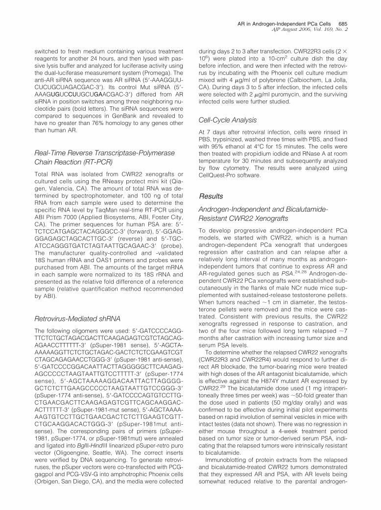
Immunoblotting of protein extracts from the relapsedand bicalutamide-treated CWR22 tumors demonstratedthat they expressed AR and PSA, with AR levels beingsomewhat reduced relative to the parental androgen-
AR in Androgen-Independent PCa Cells 685AJP August 2006, Vol. 169, No. 2
dependent CWR22 xenografts (Figure 1, A and B). DNAsequencing of the entire AR mRNA coding region fromthe relapsed tumors showed the codon 874 mutationidentical to that in the parental CWR22 xenograft30 butdid not reveal any additional mutations (data not shown).
Overall, these results demonstrated that the relapsedCWR22 tumors, with respect to expression of AR andPSA and resistance to AR antagonists, were similar toandrogen-independent PCa that occur in patients afterinitial responses to androgen deprivation therapies.
686 Yuan et alAJP August 2006, Vol. 169, No. 2
Establishment of CWR22R3 Cell Line
To further assess AR function in the relapsed andbicalutamide-resistant tumors, we established celllines from the two relapsed and bicalutamide-resistantCWR22R xenografts. The cells were cultured in steroidhormone-depleted medium (Dulbecco’s modified Ea-gle’s medium with 10% charcoal-dextran-strippedFBS, CDS medium). During the initial passages, therewas an outgrowth of both tumor and stromal cells.Differential trypsinization, as well as immunomagneticbeads conjugated with a monoclonal antibody againstan epithelial antigen (Ber-EP4), were used during somepassages to positively select for tumor cells. The tumorcells initially grew clustered in small aggregates sur-rounded by stromal cells and continued to grow inclusters even after the depletion of stromal cells. Oneof the cell lines, CWR22R3, was established as a long-term (�2 years) line free of stromal cell contaminationbecause flow-cytometry analysis demonstrated a sim-ilar profile to LNCaP cells in their expression of Ber-EP4epithelial marker and AR (Figure 1C). This line was thefocus of further detailed studies.
After more than 2 years of culture in CDS medium,the CWR22R3 cell line continued to express AR at highlevels comparable to the AR levels in the LNCaP PCacell line (Figure 1D). AR DNA sequencing again re-vealed the codon 874 mutation and no other mutationsin the CWR22R3 cell line (data not shown). The amountof AR in both cell lines was increased after dihydrotes-tosterone (DHT; 10 nmol/L) treatment for 24 hours,consistent with the stabilization of AR protein by li-gand.7 The DHT-mediated increase in AR protein levelwas also demonstrated in CWR22R3 cells by immuno-fluorescence, with AR being predominantly nuclear ineither the absence or presence of DHT (Figure 1E). NoPSA protein was detected in CWR22R3 cells, evenafter DHT stimulation, whereas PSA protein was readilydetected in LNCaP cells and was markedly increasedby DHT (Figure 1D). Assessment of PSA mRNA levelsby quantitative real-time RT-PCR confirmed that ex-pression was markedly decreased in the CWR22R3cell line relative to the in vivo xenografts (Figure 1F).PSA mRNA expression in the CWR22R3 cells could bestimulated by DHT, but the level of stimulation (40%increase) was small relative to the DHT-stimulated in-
crease in the LNCaP cell line. Consistent with the lackof response to bicalutamide in the in vivo xenografts,growth of the CWR22R3 cell line was not repressed bybicalutamide, whereas growth could be modestly stim-ulated by DHT (Figure 1G).
Activation of Erk/MAP Kinase and PI3 Kinase inCWR22R3 Cells
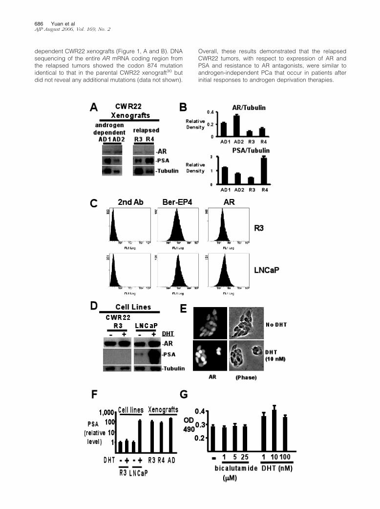
The progression of PCa in patients to more aggressive,androgen-independent disease has been associatedwith activation of the Erk1 and Erk2 MAP kinases as wellas with PTEN loss and the subsequent activation of thePI3 kinase/Akt pathway.31,32 Therefore, the CWR22R3cells were next assessed for activation of these kinases.Significantly, CWR22R3 cells grown in medium with 10%FBS or steroid hormone-depleted CDS medium had highlevels of activated Erk1 and 2, as assessed by immuno-blotting with a phospho-specific Erk1/2 antibody (Figure2A). This activation was not diminished by serum starva-tion for 24 hours and was only modestly enhanced byserum stimulation of the starved cells (Figure 2A). Asshown previously, Erk phosphorylation was not detect-able in LNCaP cells grown in FBS or CDS medium. Incontrast to Erk activation, Akt was phosphorylated at verylow levels in the CWR22R3 cells in comparison to thePTEN-deficient LNCaP cells (Figure 2B). Consistent withthis result, PTEN expression was readily detectable byimmunoblotting in the CWR22R3 cells, indicating thatprogression to androgen independence was not associ-ated with PTEN loss or PI3 kinase activation in these cells(data not shown).
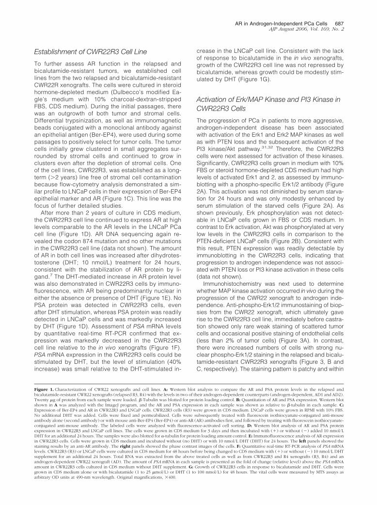
Immunohistochemistry was next used to determinewhether MAP kinase activation occurred in vivo during theprogression of the CWR22 xenograft to androgen inde-pendence. Anti-phospho-Erk1/2 immunostaining of biop-sies from the CWR22 xenograft, which ultimately gaverise to the CWR22R3 cell line, immediately before castra-tion showed only rare weak staining of scattered tumorcells and occasional positive staining of endothelial cells(less than 2% of tumor cells) (Figure 3A). In contrast,there were increased numbers of cells with strong nu-clear phospho-Erk1/2 staining in the relapsed and bicalu-tamide-resistant CWR22R3 xenografts (Figure 3, B andC, respectively). The staining pattern is patchy and within
Figure 1. Characterization of CWR22 xenografts and cell lines. A: Western blot analysis to compare the AR and PSA protein levels in the relapsed andbicalutamide-resistant CWR22 xenografts (relapsed R3, R4) with the levels in two of their androgen-dependent counterparts (androgen-dependent, AD1 and AD2).Twenty �g of protein from each sample were loaded. �-Tubulin was blotted for protein loading control. B: Quantitation of AR and PSA expression. Western blotshown in A was analyzed with the ImageJ program, and the AR and PSA expression in each sample was shown as relative to �-tubulin in each sample. C:Expression of Ber-EP4 and AR in CWR22R3 and LNCaP cells. CWR22R3 cells (R3) were grown in CDS medium. LNCaP cells were grown in RPMI with 10% FBS.No additional DHT was added. Cells were fixed and permeabilized. Cells were subsequently treated with fluorescein isothiocyanate-conjugated anti-mouseantibody alone (second antibody) or with mouse anti-Ber-EP4 (Ber-EP4) or anti-AR (AR) antibodies first, and followed by treating with fluorescein isothiocyanate-conjugated anti-mouse antibody. The labeled cells were analyzed with fluorescence-activated cell sorting. D: Western blot analysis of AR and PSA proteinexpression in CWR22R3 and LNCaP cell lines. The cells were grown in CDS medium for 3 days and then incubated with (�) or without (�) added 10 nmol/LDHT for an additional 24 hours. The samples were also blotted for �-tubulin for protein loading amount control. E: Immunofluorescence analysis of AR expressionin CWR22R3 cells. Cells were grown in CDS medium and incubated without (no DHT) or with 10 nmol/L DHT (DHT) for 24 hours. The left panels showed thestaining results by an anti-AR antibody. The right panels showed the phase contrast images of the cells. F: Quantitative real-time RT-PCR analysis of PSA mRNAlevels. CWR22R3 (R3) or LNCaP cells were cultured in CDS medium for 48 hours before being changed to CDS medium with (�) or without (�) 10 nmol/L DHTsupplement for an additional 24 hours. Total RNA was extracted from the above treated cells as well as from CWR22R3 and R4 xenografts (R3, R4) and anandrogen-dependent CWR22 xenograft (AD). The amount of PSA mRNA in each sample is presented as the fold of change (relative level) above the PSA mRNAamount in CWR22R3 cells cultured in CDS medium without DHT supplement. G: Growth of CWR22R3 cells in response to bicalutamide and DHT. Cells weregrown in CDS medium alone or with bicalutamide (1 to 25 �mol/L) or DHT (1 to 100 nmol/L) for 48 hours. The vital cells were measured by MTS assays asarbitrary OD units at 490-nm wavelength. Original magnifications, �400.
AR in Androgen-Independent PCa Cells 687AJP August 2006, Vol. 169, No. 2
positively stained area, increased percentage of tumorcells is positive for p-Erk (20 to 30% in postcastrationxenograft and 50 to 60% in postcastration and bicalut-amide-resistant xenograft). The arrangement of positivecells varied from scattered to large aggregates. AR ex-
pression in above described xenografts by immunostain-ing demonstrated a more even distributed pattern andthe vast majority of cells (80 to 90%) in all tumors wereAR-positive (Figure 3, D–F). In summary, these resultsindicated that Erk1/2 activation in the CWR22R3 xeno-
Figure 2. Erk activation in the CWR22R3 cell line. CWR22R3 or LNCaP cells were grown in FBS (FBS�) or CDS medium (CDS�) for 24 hours. Additional CWR22R3cells were grown in serum-free medium (FBS�, CDS�, stim�) for 24 hours, with or without subsequent serum stimulation with CDS medium for 20 minutes(FBS�, CDS�, stim�, or FBS�, CDS�, stim�). The cell lysates were blotted with anti-phospho-Erk (p-p44 Erk1/p-p42 Erk2) and anti-total Erk (total Erk)antibodies (A, top); or anti-phospho-Akt (p-Akt) and anti-total Akt (total Akt) antibodies (B, top). The bottom panels showed the quantification of Erkphosphorylation (A, p-p42/p42) or Akt phosphorylation (B, p-Akt/Akt) by ImageJ analysis of the respective top panels.
Figure 3. Erk activation during the relapse of CWR22R3 xenograft. Immunohistochemical staining with phospho-Erk-specific antibodies (A–C) or anti-ARantibodies (D–E) on CWR22R3 xenograft biopsy samples prepared before castration (A, D), after castration but before the initiation of bicalutamide treatment (B,E), or at the end of bicalutamide treatment (C, F). Original magnifications, �200.
688 Yuan et alAJP August 2006, Vol. 169, No. 2
graft occurred in vivo during the progression to androgenindependence, and Erk1/2 activation was maintained inthe CWR22R3 cell line.
CWR22R3 Cells Are Not Hypersensitive to LowLevels of Androgen
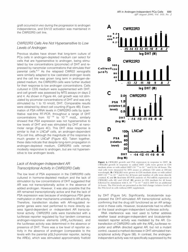
Previous studies have shown that long-term culture ofPCa cells in androgen-depleted medium can select forcells that are hypersensitive to androgen, being stimu-lated by low concentrations (picomolar) of DHT and re-pressed by nanomolar concentrations that stimulate theirparental cells.6,7 As the relapsed CWR22 xenograftswere similarly adapted to low castrated androgen levelsand the cell line was grown long term in androgen-de-pleted medium, the CWR22R3 cells were further studiedfor their response to low androgen concentrations. Cellscultured in CDS medium were supplemented with DHT,and cell growth was assessed by MTS assays on days 2and 4. As shown in Figure 4A, cell growth was not stim-ulated by picomolar concentrations of DHT and was onlystimulated by 1 to 10 nmol/L DHT. Comparable resultswere obtained by direct cell counting (Figure 4B). Exam-ination of PSA mRNA levels in CWR22R3 cells by quan-titative real-time RT-PCR, throughout a range of DHTconcentrations from 10�14 to 10�8 mol/L, similarlyshowed that PSA expression was not hypersensitive tolow levels of DHT and was stimulated by DHT at nano-molar range (Figure 4C). This DHT dose response issimilar to that in LNCaP cells, an androgen-dependentPCa cell line, although the magnitude of the response ismuch greater in LNCaP (Figure 4D). Taken together,these data indicate that despite long-term propagation inandrogen-depleted medium, CWR22R3 cells remainmodestly responsive to androgen, but are not hypersen-sitive to low androgen levels.
Lack of Androgen-Independent ARTranscriptional Activity in CWR22R3 Cells
The low level of PSA expression in the CWR22R3 cellscultured in hormone-depleted medium and the lack ofstimulation by low concentrations of DHT suggested thatAR was not transcriptionally active in the absence ofadded androgen. However, it was also possible that theAR remained transcriptionally active and that the lack ofPSA expression was because of endogenous PSA genemethylation or other mechanisms unrelated to AR activity.Therefore, transfection studies with AR-regulated re-porter genes were next performed to more directly as-sess ligand-dependent or -independent AR transcrip-tional activity. CWR22R3 cells were transfected with aluciferase reporter regulated by four tandem consensusandrogen-responsive elements (ARE4-luciferase), andthe luciferase activity was determined in the absence orpresence of DHT. There was a low level of reporter ac-tivity in the absence of androgen (comparable to thelevels with the parental pGL3-promoter reporter, lackingthe AREs), which was stimulated approximately fivefold
by DHT (Figure 5A). Significantly, bicalutamide sup-pressed the DHT-stimulated AR transcriptional activity,confirming that the drug still functioned as an AR antag-onist in these cells. However, bicalutamide had no effecton the basal androgen-independent luciferase activity.
RNA interference was next used to further addresswhether basal androgen-independent and bicalutamide-resistant reporter activity was mediated by the AR. Co-transfection of CWR22R3 cells with the ARE4-luciferase re-porter and siRNA directed against AR, but not a mutantcontrol, caused a marked decrease in DHT-stimulated tran-scriptional activity (Figure 5B). In contrast, the androgen-independent activity was not specifically suppressed by the
Figure 4. CWR22R3 growth and PSA expression in response to DHT. A:CWR22R3 growth in response to added DHT. Cells were grown in CDSmedium alone or with added DHT (10�15 to 10�7 mol/L) for 48 hours. Thevital cells were measured by MTS assays as arbitrary OD units at 490-nmwavelength. B: CWR22R3 were grown in CDS medium alone or with addedDHT (10�13 to 10�7 mol/L) for 48 hours and number of cells were directlycounted. C and D: Real-time RT-PCR analysis of PSA mRNA expression inCWR22R3 cells (C) and LNCaP (D) grown in CDS medium for 48 hoursfollowed by stimulation with 0, 10�14 to 10�8 mol/L DHT for an additional24 hours. The PSA levels are presented as the fold change (relative level) ofthe PSA level in cells without DHT supplement.
AR in Androgen-Independent PCa Cells 689AJP August 2006, Vol. 169, No. 2
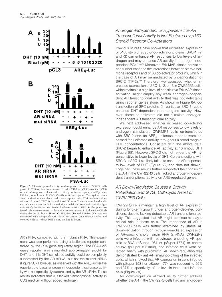
AR siRNA, compared with the mutant siRNA. This experi-ment was also performed using a luciferase reporter con-trolled by the PSA gene regulatory region. The PSA-lucif-erase reporter was stimulated approximately sixfold byDHT, and this DHT-stimulated activity could be completelysuppressed by the AR siRNA, but not the mutant siRNA(Figure 5C). However, as observed with the ARE4-luciferasereporter, the basal androgen-independent luciferase activ-ity was not specifically suppressed by the AR siRNA. Theseresults indicated that AR lacked transcriptional activity inCDS medium without added androgen.
Androgen-Independent or Hypersensitive ARTranscriptional Activity Is Not Restored by p160Steroid Receptor Co-Activators
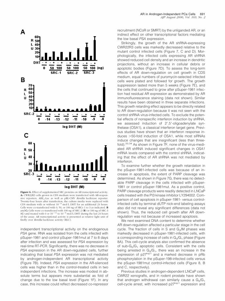
Previous studies have shown that increased expressionof p160 steroid receptor co-activator proteins (SRC-1, -2,and -3) can enhance AR responses to low levels of an-drogen and may enhance AR activity in androgen-inde-pendent PCa.18,26 Moreover, Erk MAP kinase activationcan further enhance the interactions between steroid hor-mone receptors and p160 co-activator proteins, which inthe case of AR may be mediated by phosphorylation ofSRC-2 (TIF-2).19 Therefore, we assessed whether in-creased expression of SRC-1, -2, or -3 in CWR22R3 cells,which maintain a high level of constitutive Erk MAP kinaseactivation, might amplify any weak androgen-indepen-dent AR transcriptional activity that was not detectableusing reporter genes alone. As shown in Figure 6A, co-transfection of SRC proteins (in particular SRC-3) couldenhance DHT-dependent reporter gene activity. How-ever, these co-activators did not stimulate androgen-independent AR transcriptional activity.
We next addressed whether increased co-activatorexpression could enhance AR responses to low levels ofandrogen stimulation. CWR22R3 cells co-transfectedwith SRC-2 and an ARE4-luciferase reporter were as-sessed for luciferase activity throughout a broad range ofDHT concentrations. Consistent with the above data,SRC-2 began to enhance AR activity at 10 nmol/L DHT(Figure 6B). However, SRC-2 did not render the AR hy-persensitive to lower levels of DHT. Co-transfections withSRC-3 or SRC-1 similarly failed to enhance AR responsesto low levels of DHT (Figure 6C, and data not shown).Together, these results further supported the conclusionthat AR in the CWR22R3 cells lacked androgen-indepen-dent transcriptional activity on ARE-regulated genes.
AR Down-Regulation Causes a GrowthRetardation and G0/G1 Cell-Cycle Arrest ofCWR22R3 Cells
CWR22R3 cells maintain a high level of AR expressionduring long-term growth under androgen-depleted con-ditions, despite lacking detectable AR transcriptional ac-tivity. This suggested that AR might continue to play acritical role in these cells. The importance of AR inCWR22R3 cells was further examined by stable ARdown-regulation through retrovirus-mediated expressionof AR-specific short hairpin RNA (shRNA). CWR22R3cells were infected with retroviruses encoding AR-spe-cific shRNA (pSuper-1981 or pSuper-1774) or controlshRNA (pSuper-1981mut), and infected cells were se-lected briefly with puromycin. AR down-regulation wasdemonstrated by anti-AR immunoblotting of the infectedcells, which showed that AR expression in cells infectedwith pSuper-1981 or pSuper-1774 was reduced to 29%and 37%, respectively, of the level in the control infectedcells (Figure 7A).
AR down-regulation allowed us to further addresswhether the AR in the CWR22R3 cells had any androgen-
Figure 5. AR transcriptional activity on AR-responsive reporters. CWR22R3 cellsgrown in CDS medium were transfected with ARE-less pGL3/promoter (pGL3)or with AR-responsive element-driven firefly luciferase reporters, ARE4-Luc orPSA-Luc, as well as a pRL-CMV Renilla luciferase reporter. Twenty-four hoursafter transfection, the culture media were replaced with CDS medium with orwithout 10 nmol/L DHT for an additional 24 hours. The cells were lysed at theend of the treatment and AR transcriptional activity is presented as relative lightunits (firefly luciferase over Renilla luciferase activity, RLU). A: The posttrans-fected cells were co-treated with various concentrations of bicalutamide (Bical)during the last 24 hours. B and C: ARE4-Luc (B) and PSA-Luc (C) were co-transfected with AR-specific (AR siRNA) or control (mut siRNA) siRNAs andcultured with or without DHT during the last 24 hours.
690 Yuan et alAJP August 2006, Vol. 169, No. 2
independent transcriptional activity on the endogenousPSA gene. RNA was isolated from the cells infected withpSuper-1981 and control pSuper-1981mut at 7 to 8 daysafter infection and was assessed for PSA expression byreal-time RT-PCR. Significantly, there was no decrease inPSA expression in the AR down-regulated cells, furtherindicating that basal PSA expression was not mediatedby androgen-independent AR transcriptional activity(Figure 7B). Indeed, PSA expression in the AR-silencedcells was higher than in the control-treated cells in twoindependent infections. The increase was modest in ab-solute terms but appears more substantial as fold ofchange due to the low basal level (Figure 1F). In anycase, this increase could reflect decreased co-repressor
recruitment (NCoR or SMRT) by the unliganded AR, or anindirect effect on other transcriptional factors mediatingthe low basal PSA expression.
Strikingly, the growth of the AR shRNA-expressingCWR22R3 cells was markedly decreased relative to themutant control infected cells (Figure 7, C and D). Mor-phologically, the infected cells expressing AR shRNAshowed reduced cell density and an increase in dendriticprojections, without an increase in cellular debris orapoptotic bodies (Figure 7D). To assess the long-termeffects of AR down-regulation on cell growth in CDSmedium, equal numbers of puromycin-selected infectedcells were plated and followed for growth. The growthsuppression lasted more than 5 weeks (Figure 7E), andthe cells that continued to grow after pSuper-1981 infec-tion had residual AR expression as demonstrated by ARimmunofluorescence staining (data not shown). Similarresults have been obtained in three separate infections.This growth retarding effect appears to be directly relatedto AR down-regulation because it was not seen with thecontrol shRNA virus-infected cells. To exclude the poten-tial effects of nonspecific interferon induction by shRNA,we assessed induction of 2�,5�-oligoadenylate syn-thetase (OSA1), a classical interferon target gene. Previ-ous studies have shown that an interferon response in-duces �50-fold induction of OSA1, while most siRNAsinduce changes that are insignificant (less than three-fold).33,34 As shown in Figure 7F, none of the virus-medi-ated AR shRNA induced significant changes in OSA1mRNA levels compared with the control shRNA, indicat-ing that the effect of AR shRNA was not mediated byinterferon.
To examine further whether the growth retardation inthe pSuper-1981-infected cells was because of an in-crease in apoptosis, the extent of PARP cleavage wasdetermined. As shown in Figure 7G, there was no detect-able PARP cleavage in the cells infected with pSuper-1981 or control pSuper-1981mut. As a positive control,PARP cleavage products were readily detected in LNCaPcells treated with the PI3 kinase inhibitor LY294002. Com-parison of cell apoptosis in pSuper-1981- versus control-infected cells by terminal dUTP nick-end labeling assaysalso did not reveal any significant differences (data notshown). Thus, the reduced cell growth after AR down-regulation was not because of increased apoptosis.
We next examined DNA content to determine whetherAR down-regulation affected a particular stage in the cellcycle. The fraction of cells in S and G2/M phases wasmarkedly decreased in pSuper-1981-infected cells, witha corresponding increase of cells in G0/G1 phase (Figure8A). This cell-cycle analysis also confirmed the absenceof sub-G0/G1 apoptotic cells. Consistent with the cellsbeing arrested in G0/G1, there was an increase in theexpression of p27kip1 and a marked decrease in pRbphosphorylation in the pSuper-198-infected cells versusthe pSuper-1981mut control-infected cells (Figure 8, Band C, respectively).
Previous studies in androgen-dependent LNCaP cells,CWR22 xenografts, and in rodent prostate have shownthat androgen withdrawal can similarly cause a G0/G1
cell-cycle arrest, with increased p27kip1 expression and
Figure 6. Effect of supplemented SRC proteins on AR transcriptional activity.A: CWR22R3 cells grown in CDS medium were transfected with AR-respon-sive reporters, ARE4-Luc as well as pRL-CMV Renilla luciferase reporter.Twenty-four hours after transfection, the culture media were replaced withCDS medium with or without 10�8 mol/L DHT for an additional 24 hours.Cells were co-transfected with 0, 50, or 100 ng of SRCs 1 to 3 as indicated. Band C: Cells were co-transfected with 100 ng of SRC-2 (B) or 100 ng of SRC-3(C) and treated with 0 or 10�14 to 10�8 mol/L DHT during the last 24 hoursof the assay. AR transcriptional activity is presented as relative light unit offirefly over Renilla luciferase activity (RLU).
AR in Androgen-Independent PCa Cells 691AJP August 2006, Vol. 169, No. 2
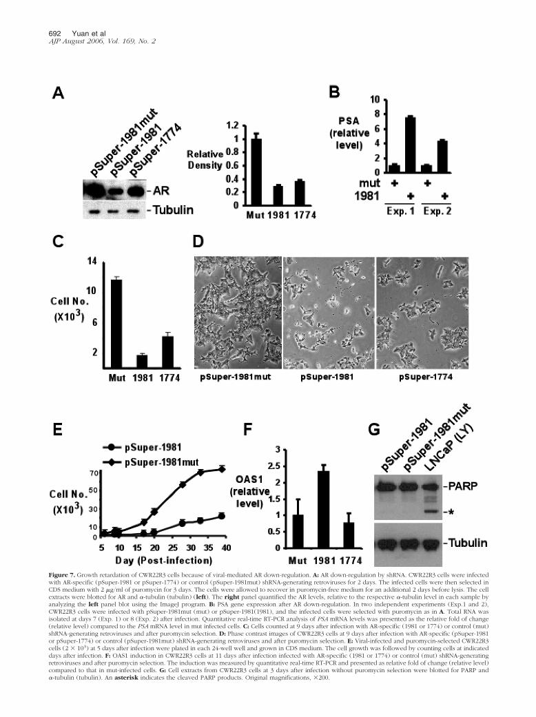
Figure 7. Growth retardation of CWR22R3 cells because of viral-mediated AR down-regulation. A: AR down-regulation by shRNA. CWR22R3 cells were infectedwith AR-specific (pSuper-1981 or pSuper-1774) or control (pSuper-1981mut) shRNA-generating retroviruses for 2 days. The infected cells were then selected inCDS medium with 2 �g/ml of puromycin for 3 days. The cells were allowed to recover in puromycin-free medium for an additional 2 days before lysis. The cellextracts were blotted for AR and �-tubulin (tubulin) (left). The right panel quantified the AR levels, relative to the respective �-tubulin level in each sample byanalyzing the left panel blot using the ImageJ program. B: PSA gene expression after AR down-regulation. In two independent experiments (Exp.1 and 2),CWR22R3 cells were infected with pSuper-1981mut (mut) or pSuper-1981(1981), and the infected cells were selected with puromycin as in A. Total RNA wasisolated at days 7 (Exp. 1) or 8 (Exp. 2) after infection. Quantitative real-time RT-PCR analysis of PSA mRNA levels was presented as the relative fold of change(relative level) compared to the PSA mRNA level in mut infected cells. C: Cells counted at 9 days after infection with AR-specific (1981 or 1774) or control (mut)shRNA-generating retroviruses and after puromycin selection. D: Phase contrast images of CWR22R3 cells at 9 days after infection with AR-specific (pSuper-1981or pSuper-1774) or control (pSuper-1981mut) shRNA-generating retroviruses and after puromycin selection. E: Viral-infected and puromycin-selected CWR22R3cells (2 � 103) at 5 days after infection were plated in each 24-well well and grown in CDS medium. The cell growth was followed by counting cells at indicateddays after infection. F: OAS1 induction in CWR22R3 cells at 11 days after infection infected with AR-specific (1981 or 1774) or control (mut) shRNA-generatingretroviruses and after puromycin selection. The induction was measured by quantitative real-time RT-PCR and presented as relative fold of change (relative level)compared to that in mut-infected cells. G: Cell extracts from CWR22R3 cells at 3 days after infection without puromycin selection were blotted for PARP and�-tubulin (tubulin). An asterisk indicates the cleaved PARP products. Original magnifications, �200.
692 Yuan et alAJP August 2006, Vol. 169, No. 2
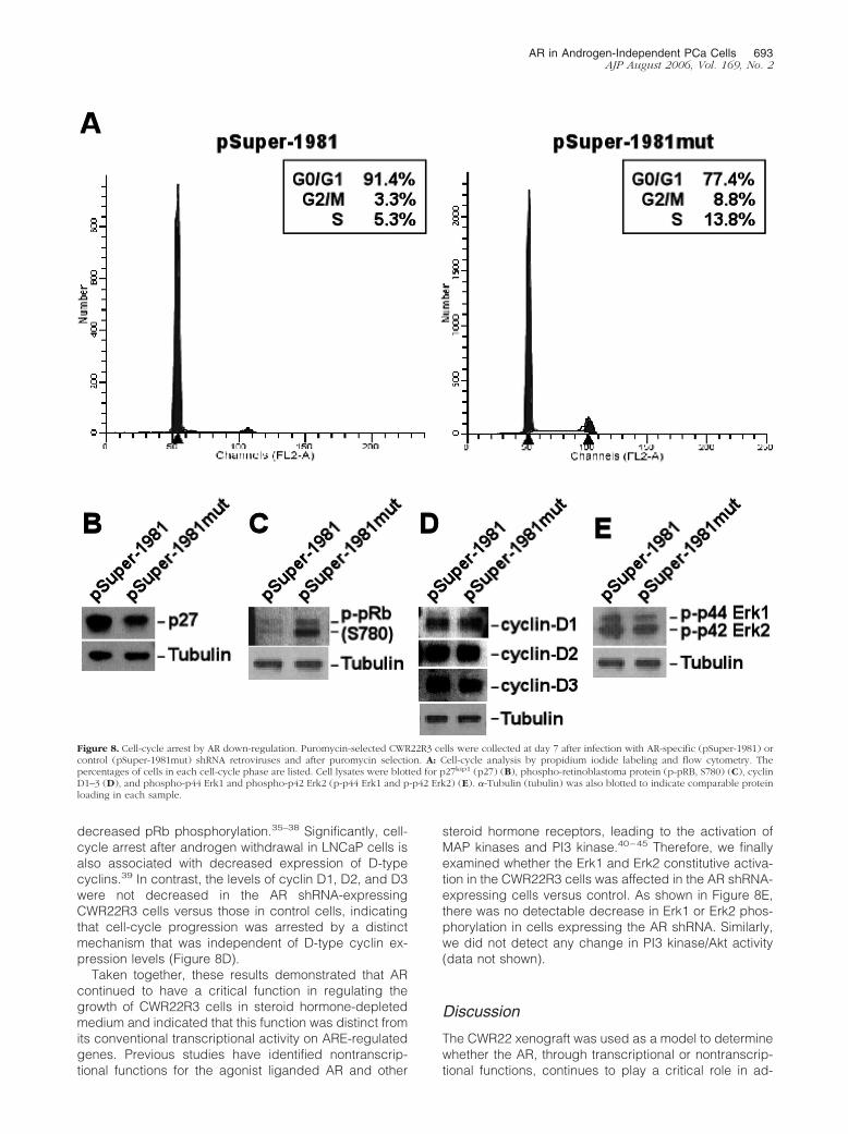
decreased pRb phosphorylation.35–38 Significantly, cell-cycle arrest after androgen withdrawal in LNCaP cells isalso associated with decreased expression of D-typecyclins.39 In contrast, the levels of cyclin D1, D2, and D3were not decreased in the AR shRNA-expressingCWR22R3 cells versus those in control cells, indicatingthat cell-cycle progression was arrested by a distinctmechanism that was independent of D-type cyclin ex-pression levels (Figure 8D).
Taken together, these results demonstrated that ARcontinued to have a critical function in regulating thegrowth of CWR22R3 cells in steroid hormone-depletedmedium and indicated that this function was distinct fromits conventional transcriptional activity on ARE-regulatedgenes. Previous studies have identified nontranscrip-tional functions for the agonist liganded AR and other
steroid hormone receptors, leading to the activation ofMAP kinases and PI3 kinase.40–45 Therefore, we finallyexamined whether the Erk1 and Erk2 constitutive activa-tion in the CWR22R3 cells was affected in the AR shRNA-expressing cells versus control. As shown in Figure 8E,there was no detectable decrease in Erk1 or Erk2 phos-phorylation in cells expressing the AR shRNA. Similarly,we did not detect any change in PI3 kinase/Akt activity(data not shown).
Discussion
The CWR22 xenograft was used as a model to determinewhether the AR, through transcriptional or nontranscrip-tional functions, continues to play a critical role in ad-
Figure 8. Cell-cycle arrest by AR down-regulation. Puromycin-selected CWR22R3 cells were collected at day 7 after infection with AR-specific (pSuper-1981) orcontrol (pSuper-1981mut) shRNA retroviruses and after puromycin selection. A: Cell-cycle analysis by propidium iodide labeling and flow cytometry. Thepercentages of cells in each cell-cycle phase are listed. Cell lysates were blotted for p27kip1 (p27) (B), phospho-retinoblastoma protein (p-pRB, S780) (C), cyclinD1–3 (D), and phospho-p44 Erk1 and phospho-p42 Erk2 (p-p44 Erk1 and p-p42 Erk2) (E). �-Tubulin (tubulin) was also blotted to indicate comparable proteinloading in each sample.
AR in Androgen-Independent PCa Cells 693AJP August 2006, Vol. 169, No. 2
vanced androgen-independent PCa. Relapsed CWR22tumors in castrated mice expressed AR and PSA at levelsthat were comparable to the androgen-dependentCWR22 xenografts and were resistant to high doses ofbicalutamide. The CWR22R3 cell line established from arelapsed CWR22 xenograft after long-term culture in ste-roid hormone-depleted medium continued to express ARand was resistant to bicalutamide, but PSA expressionwas markedly reduced relative to the in vivo xenograft.Consistent with this low level of PSA expression, transienttransfection experiments with AR-regulated reportergenes indicated that the AR in CWR22R3 cells lackedandrogen-independent transcriptional activity and wasnot hypersensitive to low levels of androgen. Nonethe-less, stable shRNA-mediated down-regulation of AR ex-pression resulted in marked suppression of cell growthand an accumulation of cells in G0/G1. These resultsidentify a critical function for AR in regulating cell-cycleprogression in androgen-independent PCa, and demon-strate that this AR function is not mediated through itsconventional transcriptional activity.
Similar to other steroid hormone receptors, the AR hasbeen reported to have rapid nontranscriptional signalingfunctions in response to its ligands, with several studiesindicating that AR can directly interact with c-Src and PI3kinase.40–45 The biological significance of nontranscrip-tional AR functions has been most clearly demonstratedin oocytes, in which the hormone-stimulated AR initiatesoocyte maturation via such a nontranscriptional mecha-nism.46 However, these reported AR nontranscriptionalfunctions (similarly to transcriptional activities) are ligandstimulated, and their physiological importance in normalprostate epithelium or in PCa has not been established.Significantly, in CWR22R3 cells, AR remains functional inthe absence of ligand stimulation, and we did not ob-serve changes in Erk1, Erk2 MAP kinase or PI3 kinase/Aktactivities in response to AR down-regulation, furtherindicating that AR was functioning through distinctmechanisms.
Androgen binding induces a conformational changein the AR ligand-binding domain that stimulates ho-modimerization and generates a binding site for tran-scriptional co-activator proteins through LXXLL motifs.1
Nonetheless, multiple proteins can also bind to the AR viasites that are not dependent on androgen-mediated con-formational changes in the ligand-binding domain, andsuch interactions could possibly mediate androgen-inde-pendent effects of AR on cell-cycle progression. Thesecandidate AR-interacting proteins include cyclin D1 andpRb, which have been reported to interact with the ARN-terminus and suppress (cyclin D1) or enhance (pRb)androgen-stimulated AR transcriptional activity.47–49 Themolecular mechanism of AR-mediated growth mainte-nance in CWR22R3 cells, including the roles of AR-inter-acting proteins are currently under investigation. Finally,it should be emphasized that although our studies did notdetect residual AR transcriptional activity in the absenceof ligand on conventional androgen-regulated genes thatcontain multiple AREs, it cannot be completely excludedthat AR may nonetheless remain critical for the expres-sion of some genes through alternative mechanisms that
are not dependent on ARE binding, such as throughinteractions with other transcription factors.
Previous studies have shown that the growth of LNCaPor LAPC4 PCa cells that have been adapted to growunder androgen-depleted conditions could also be sup-pressed when AR was down-regulated by a transfectedAR-specific siRNA, ribozyme, or by anti-AR antibody in-jection.8–11,50 These findings are consistent with our re-sults in CWR22R3 cells. However, in these reported stud-ies, the androgen-independent PCa cells expressedsubstantial levels of PSA, suggesting that AR remainedtranscriptionally active. Therefore, they did not addresswhether growth suppression was due to abrogation of ARtranscriptional activity mediated by residual androgens(with one study using cells grown in medium that was notdepleted of steroid hormones) or by other mechanisms.Moreover, these previous studies did not assess effectsof AR down-regulation on cell cycle or cell-cycle regula-tory proteins, in particular on D-type cyclin expression(which was not repressed by AR shRNA in the CWR22R3cells). In addition, AR down-regulation in LNCaP-abl cellsresults in significant apoptotic cell death,47 which is notobserved in CWR22R3 cells. Therefore, although theseprevious studies further support the hypothesis that ARremains as a critical regulator of cell growth and survivalin androgen-independent PCa, it remains to be deter-mined whether AR has similar or distinct functions in theLNCaP and LAPC4 cells versus in the CWR22R3 cells.
Although the AR in the CWR22R3 cell line had nodetectable transcriptional activity in the absence of an-drogens, the CWR22R3 and CWR22R4 xenografts in vivoin castrated mice expressed PSA at levels that werecomparable to the androgen-dependent CWR22 xeno-graft. The high-level expression of PSA and other andro-gen-regulated genes in the relapsed CWR22 xenograftmodel, and in patients with androgen-independent PCa,strongly indicates that the AR retains transcriptional ac-tivity during the progression to androgen indepen-dence.28 Although relapsed PCa after castration and an-ti-androgen treatment are termed androgen-independentPCa, in reality these tumors may still be responsive toandrogen. The AR transcriptional activity may be medi-ated by low levels of residual androgens in vivo, withmultiple mechanisms including MAP kinase activationenhancing androgen responses. Significantly, the re-lapsed CWR22 xenografts were also resistant to bicalu-tamide, which may reflect the low AR affinity of this an-tagonist and is consistent with the intrinsic resistance tobicalutamide in most patients who relapse after androgendeprivation therapy.4,22 The loss of AR transcriptionalactivity in the CWR22R3 cell line is consistent with themore controlled and stringent depletion of androgens invitro in the medium with charcoal-dextran-stripped se-rum. In addition, this loss of transcriptional activity mayalso be due to the loss of additional factors because theAR activity in the CWR22R3 cells was not hypersensitiveto low levels of DHT and was not restored by adding backAR cofactors, SRC1 to SRC3.
In summary, this study demonstrates that the AR re-mains critical for androgen-independent growth of theCWR22R3 cell line. Moreover, the lack of detectable AR
694 Yuan et alAJP August 2006, Vol. 169, No. 2
transcriptional activity in the absence of androgens or inresponse to low levels of added androgens indicates thatAR function in the CWR22R3 cells is mediated by amechanism that is distinct from its conventional transcrip-tional function. Further characterization of this in vitrocritical androgen-independent AR activity, whether anontranscriptional or a novel transcriptional function, isnecessary to determine the extent to which it contributesto in vivo tumor growth and whether it may provide a newtherapeutic target for drug development.
Acknowledgments
We thank Tom Pretlow (Case Western Reserve UniversitySchool of Medicine and Cancer Center, Cleveland, OH)for very generously providing the CWR22 xenografts, andmembers of the Balk Laboratory for reviewing the manu-script and helpful suggestions.
References
1. Gelmann EP: Molecular biology of the androgen receptor. J ClinOncol 2002, 20:3001–3015
2. Feldman BJ, Feldman D: The development of androgen-independentprostate cancer. Nature Rev Cancer 2001, 1:34–45
3. Balk SP: Androgen receptor as a target in androgen-independentprostate cancer. Urology 2002, 60:132–138
4. Joyce R, Fenton MA, Rode P, Constantine M, Gaynes L, KolvenbagG, DeWolf W, Balk S, Taplin ME, Bubley GJ: High dose bicalutamidefor androgen independent prostate cancer: effect of prior hormonaltherapy. J Urol 1998, 159:149–153
5. Small EJ, Halabi S, Dawson NA, Stadler WM, Rini BI, Picus J, GableP, Torti FM, Kaplan E, Vogelzang NJ: Antiandrogen withdrawal aloneor in combination with ketoconazole in androgen-independent pros-tate cancer patients: a phase III trial (CALGB 9583). J Clin Oncol2004, 22:1025–1033
6. Kokontis JM, Hay N, Liao S: Progression of LNCaP prostate tumorcells during androgen deprivation: hormone-independent growth, re-pression of proliferation by androgen, and role for p27Kip1 in andro-gen-induced cell cycle arrest. Mol Endocrinol 1998, 12:941–953
7. Gregory CW, Johnson Jr RT, Mohler JL, French FS, Wilson EM:Androgen receptor stabilization in recurrent prostate cancer is asso-ciated with hypersensitivity to low androgen. Cancer Res 2001,61:2892–2898
8. Zegarra-Moro OL, Schmidt LJ, Huang H, Tindall DJ: Disruption ofandrogen receptor function inhibits proliferation of androgen-refrac-tory prostate cancer cells. Cancer Res 2002, 62:1008–1013
9. Wright ME, Tsai MJ, Aebersold R: Androgen receptor represses theneuroendocrine transdifferentiation process in prostate cancer cells.Mol Endocrinol 2003, 17:1726–1737
10. Chen CD, Welsbie DS, Tran C, Baek SH, Chen R, Vessella R, Rosen-feld MG, Sawyers CL: Molecular determinants of resistance to anti-androgen therapy. Nat Med 2004, 10:33–39
11. Haag P, Bektic J, Bartsch G, Klocker H, Eder IE: Androgen receptordown regulation by small interference RNA induces cell growth inhi-bition in androgen sensitive as well as in androgen independentprostate cancer cells. J Steroid Biochem Mol Biol 2005, 96:251–258
12. Taplin ME, Bubley GJ, Shuster TD, Frantz ME, Spooner AE, OgataGK, Keer HN, Balk SP: Mutation of the androgen-receptor gene inmetastatic androgen-independent prostate cancer. N Engl J Med1995, 332:1393–1398
13. Visakorpi T, Hyytinen E, Koivisto P, Tanner M, Keinanen R, PalmbergC, Palotie A, Tammela T, Isola J, Kallioniemi OP: In vivo amplificationof the androgen receptor gene and progression of human prostatecancer. Nat Genet 1995, 9:401–406
14. Culig Z, Hobisch A, Cronauer MV, Cato AC, Hittmair A, Radmayr C,Eberle J, Bartsch G, Klocker H: Mutant androgen receptor detected
in an advanced-stage prostatic carcinoma is activated by adrenalandrogens and progesterone. Mol Endocrinol 1993, 7:1541–1550
15. Zhao XY, Boyle B, Krishnan AV, Navone NM, Peehl DM, Feldman D:Two mutations identified in the androgen receptor of the new humanprostate cancer cell line MDA PCa 2a. J Urol 1999, 162:2192–2199
16. Taplin ME, Bubley GJ, Ko YJ, Small EJ, Upton M, Rajeshkumar B,Balk SP: Selection for androgen receptor mutations in prostatecancers treated with androgen antagonist. Cancer Res 1999,59:2511–2515
17. Taplin ME, Rajeshkumar B, Halabi S, Werner CP, Woda BA, Picus J,Stadler W, Hayes DF, Kantoff PW, Vogelzang NJ, Small EJ: Androgenreceptor mutations in androgen-independent prostate cancer: Can-cer and Leukemia Group B Study 9663. J Clin Oncol 2003,21:2673–2678
18. Gregory CW, He B, Johnson RT, Ford OH, Mohler JL, French FS,Wilson EM: A mechanism for androgen receptor-mediated prostatecancer recurrence after androgen deprivation therapy. Cancer Res2001, 61:4315–4319
19. Gregory CW, Fei X, Ponguta LA, He B, Bill HM, French FS, Wilson EM:Epidermal growth factor increases coactivation of the androgen re-ceptor in recurrent prostate cancer. J Biol Chem 2004,279:7119–7130
20. Bakin RE, Gioeli D, Sikes RA, Bissonette EA, Weber MJ: Constitutiveactivation of the Ras/mitogen-activated protein kinase signaling path-way promotes androgen hypersensitivity in LNCaP prostate cancercells. Cancer Res 2003, 63:1981–1989
21. Mellinghoff IK, Vivanco I, Kwon A, Tran C, Wongvipat J, Sawyers CL:HER2/neu kinase-dependent modulation of androgen receptor func-tion through effects on DNA binding and stability. Cancer Cell 2004,6:517–527
22. Scher HI, Liebertz C, Kelly WK, Mazumdar M, Brett C, Schwartz L,Kolvenbag G, Shapiro L, Schwartz M: Bicalutamide for advancedprostate cancer: the natural versus treated history of disease. J ClinOncol 1997, 15:2928–2938
23. Wainstein MA, He F, Robinson D, Kung HJ, Schwartz S, Giaconia JM,Edgehouse NL, Pretlow TP, Bodner DR, Kursh ED: CWR22: andro-gen-dependent xenograft model derived from a primary human pros-tatic carcinoma. Cancer Res 1994, 54:6049–6052
24. Nagabhushan M, Miller CM, Pretlow TP, Giaconia JM, Edgehouse NL,Schwartz S, Kung HJ, Vere White RW, Gumerlock PH, Resnick MI,Amini SB, Pretlow TG: CWR22: the first human prostate cancer xeno-graft with strongly androgen-dependent and relapsed strains both invivo and in soft agar. Cancer Res 1996, 56:3042–3046
25. Yuan X, Lu ML, Li T, Balk SP: SRY interacts with and negativelyregulates androgen receptor transcriptional activity. J Biol Chem2001, 276:46647–46654
26. Masiello D, Cheng S, Bubley GJ, Lu ML, Balk SP: Bicalutamidefunctions as an androgen receptor antagonist by assembly of atranscriptionally inactive receptor. J Biol Chem 2002,277:26321–26326
27. Kochera M, Depinet TW, Pretlow TP, Giaconia JM, Edgehouse NL,Pretlow TG, Schwartz S: Molecular cytogenetic studies of a seriallytransplanted primary prostatic carcinoma xenograft (CWR22) andfour relapsed tumors. Prostate 1999, 41:7–11
28. Gregory CW, Hamil KG, Kim D, Hall SH, Pretlow TG, Mohler JL,French FS: Androgen receptor expression in androgen-independentprostate cancer is associated with increased expression of andro-gen-regulated genes. Cancer Res 1998, 58:5718–5724
29. Fenton MA, Shuster TD, Fertig AM, Taplin ME, Kolvenbag G, BubleyGJ, Balk SP: Functional characterization of mutant androgen recep-tors from androgen-independent prostate cancer. Clin Cancer Res1997, 3:1383–1388
30. Tan J, Sharief Y, Hamil KG, Gregory CW, Zang DY, Sar M, GumerlockPH, DeVere White RW, Pretlow TG, Harris SE, Wilson EM, Mohler JL,French FS: Dehydroepiandrosterone activates mutant androgen re-ceptors expressed in the androgen-dependent human prostate can-cer xenograft CWR22 and LNCaP cells. Mol Endocrinol 1997,11:450–459
31. Gioeli D, Mandell JW, Petroni GR, Frierson HF, Weber MJ: Activationof mitogen-activated protein kinase associated with prostate cancerprogression. Cancer Res 1999, 59:279–284
32. McMenamin ME, Soung P, Perera S, Kaplan I, Loda M, Sellers WR:Loss of PTEN expression in paraffin-embedded primary prostate
AR in Androgen-Independent PCa Cells 695AJP August 2006, Vol. 169, No. 2
cancer correlates with high Gleason score and advanced stage.Cancer Res 1999, 59:4291–4296
33. Bridge AJ, Pebernard S, Ducraux A, Nicoulaz AL, Iggo R: Induction ofan interferon response by RNAi vectors in mammalian cells. NatGenet 2003, 34:263–264
34. Scacheri PC, Rozenblatt-Rosen O, Caplen NJ, Wolfsberg TG,Umayam L, Lee JC, Hughes CM, Shanmugam KS, Bhattacharjee A,Meyerson M, Collins FS: Short interfering RNAs can induce unex-pected and divergent changes in the levels of untargeted proteins inmammalian cells. Proc Natl Acad Sci USA 2004, 101:1892–1897
35. Chen Y, Robles AI, Martinez LA, Liu F, Gimenez-Conti IB, Conti CJ:Expression of G1 cyclins, cyclin-dependent kinases, and cyclin-de-pendent kinase inhibitors in androgen-induced prostate proliferationin castrated rats. Cell Growth Differ 1996, 7:1571–1578
36. Knudsen KE, Arden KC, Cavenee WK: Multiple G1 regulatory ele-ments control the androgen-dependent proliferation of prostatic car-cinoma cells. J Biol Chem 1998, 273:20213–20222
37. Waltregny D, Leav I, Signoretti S, Soung P, Lin D, Merk F, Adams JY,Bhattacharya N, Cirenei N, Loda M: Androgen-driven prostate epi-thelial cell proliferation and differentiation in vivo involve the regula-tion of p27. Mol Endocrinol 2001, 15:765–782
38. Gregory CW, Johnson Jr RT, Presnell SC, Mohler JL, French FS:Androgen receptor regulation of G1 cyclin and cyclin-dependentkinase function in the CWR22 human prostate cancer xenograft. JAndrol 2001, 22:537–548
39. Chen Y, Martinez LA, LaCava M, Coghlan L, Conti CJ: Increased cellgrowth and tumorigenicity in human prostate LNCaP cells by over-expression to cyclin D1. Oncogene 1998, 16:1913–1920
40. Peterziel H, Mink S, Schonert A, Becker M, Klocker H, Cato AC: Rapidsignalling by androgen receptor in prostate cancer cells. Oncogene1999, 18:6322–6329
41. Castoria G, Lombardi M, Barone MV, Bilancio A, Di Domenico M, deFalco A, Varricchio L, Bottero D, Nanayakkara M, Migliaccio A, Au-ricchio F: Rapid signalling pathway activation by androgens in epi-thelial and stromal cells. Steroids 2004, 69:517–522
42. Kousteni S, Bellido T, Plotkin LI, O’Brien CA, Bodenner DL, Han L,Han K, DiGregorio GB, Katzenellenbogen JA, Katzenellenbogen BS,Roberson PK, Weinstein RS, Jilka RL, Manolagas SC: Nongenotropic,sex-nonspecific signaling through the estrogen or androgenreceptors: dissociation from transcriptional activity. Cell 2001,104:719–730
43. Castoria G, Lombardi M, Barone MV, Bilancio A, Di Domenico M,Bottero D, Vitale F, Migliaccio A, Auricchio F: Androgen-stimulatedDNA synthesis and cytoskeletal changes in fibroblasts by a nontran-scriptional receptor action. J Cell Biol 2003, 161:547–556
44. Sun M, Yang L, Feldman RI, Sun XM, Bhalla KN, Jove R, Nicosia SV,Cheng JQ: Activation of phosphatidylinositol 3-kinase/Akt pathway byandrogen through interaction of p85alpha, androgen receptor, andSrc. J Biol Chem 2003, 278:42992–43000
45. Baron S, Manin M, Beaudoin C, Leotoing L, Communal Y, VeyssiereG, Morel L: Androgen receptor mediates non-genomic activation ofphosphatidylinositol 3-OH kinase in androgen-sensitive epithelialcells. J Biol Chem 2004, 279:14579–14586
46. Hammes SR: Steroids and oocyte maturation—a new look at an oldstory. Mol Endocrinol 2004, 18:769–775
47. Yeh S, Miyamoto H, Nishimura K, Kang H, Ludlow J, Hsiao P, WangC, Su C, Chang C: Retinoblastoma, a tumor suppressor, is a coacti-vator for the androgen receptor in human prostate cancer DU145cells. Biochem Biophys Res Commun 1998, 248:361–367
48. Lu J, Danielsen M: Differential regulation of androgen and glucocor-ticoid receptors by retinoblastoma protein. J Biol Chem 1998,273:31528–31533
49. Knudsen KE, Cavenee WK, Arden KC: D-type cyclins complex withthe androgen receptor and inhibit its transcriptional transactivationability. Cancer Res 1999, 59:2297–2301
50. Liao X, Tang S, Thrasher JB, Griebling TL, Li B: Small-interferingRNA-induced androgen receptor silencing leads to apoptotic celldeath in prostate cancer. Mol Cancer Ther 2005, 4:505–515
696 Yuan et alAJP August 2006, Vol. 169, No. 2
Related Documents