Genome-wide Expression Profiling Reveals Transcriptomic Variation and Perturbed Gene Networks in Androgen-dependent and Androgen-independent Prostate Cancer Cells Ajay P. Singh 1 , Sangeeta Bafna 1 , Kunal Chaudhary 1 , Ganesh Venkatraman 1 , Lynette Smith 2 , James D. Eudy 3 , Sonny L. Johansson 4,5 , Ming-Fong Lin 1,5 , and Surinder K. Batra 1,5,* 1 Department of Biochemistry and Molecular Biology, University of Nebraska Medical Center, Omaha, Nebraska 2 Department of preventive and Societal Medicine, University of Nebraska Medical Center, Omaha, Nebraska 3 Department of Genetics, Cell Biology and Anatomy, University of Nebraska Medical Center, Omaha, Nebraska 4 Department of Pathology and Microbiology, University of Nebraska Medical Center, Omaha, Nebraska 5 Eppley Institute for Research in Cancer and Allied Diseases, University of Nebraska Medical Center, Omaha, Nebraska Abstract Previously, we have developed a unique in vitro LNCaP cell model, which includes androgen- dependent (LNCaP-C33), androgen-independent (LNCaP-C81) and an intermediate phenotype (LNCaP-C51) cell lines resembling the stages of prostate cancer progression to hormone independence. This model is advantageous in overcoming the heterogeneity associated with the prostate cancer up to a certain extent. We characterized and compared the gene expression profiles in LNCaP-C33 (androgen-dependent) and LNCaP-C81 (androgen-independent) cells using Affymetrix GeneChip array analyses. Multiple genes were identified exhibiting differential expression during androgen-independent progression. Among the important genes upregulated in androgen-independent cells were PCDH7, TPTE, TSPY, EPHA3, HGF, MET, EGF, TEM8, etc., whereas many candidate tumor suppressor genes (HTATIP2, CDKN2A, CDKN2B, CDKN1C, TP53, TP73, ICAM1, SOCS1/2, SPRY2, PPP2CA, PPP3CA, etc.) were decreased. Pathway prediction analysis identified important gene networks associated with growth-promoting and apoptotic signaling that were perturbed during androgen-independent progression. Further investigation of one of the genes, PPP2CA, which encodes the catalytic subunit of a serine phosphatase PP2A, a potent tumor suppressor, revealed that its expression was decreased in prostate cancer compared to adjacent normal/benign tissue. Furthermore, the downregulated expression of PPP2CA was significantly correlated with tumor stage and Gleason grade. Future studies on the identified differentially- *Correspondence to: Surinder K. Batra, Ph.D., Department of Biochemistry and Molecular Biology, The Eppley Institute for Research in Cancer and Allied Diseases, University of Nebraska Medical Center, 985870 Nebraska Medical Center, Omaha, NE 68198-5870, Tel.: (402) 559-5455, Fax (402) 559-6650, [email protected]. Publisher's Disclaimer: This is a PDF file of an unedited manuscript that has been accepted for publication. As a service to our customers we are providing this early version of the manuscript. The manuscript will undergo copyediting, typesetting, and review of the resulting proof before it is published in its final citable form. Please note that during the production process errors may be discovered which could affect the content, and all legal disclaimers that apply to the journal pertain. NIH Public Access Author Manuscript Cancer Lett. Author manuscript; available in PMC 2009 November 29. Published in final edited form as: Cancer Lett. 2008 January 18; 259(1): 28–38. doi:10.1016/j.canlet.2007.09.018. NIH-PA Author Manuscript NIH-PA Author Manuscript NIH-PA Author Manuscript

Welcome message from author
This document is posted to help you gain knowledge. Please leave a comment to let me know what you think about it! Share it to your friends and learn new things together.
Transcript
Genome-wide Expression Profiling Reveals TranscriptomicVariation and Perturbed Gene Networks in Androgen-dependentand Androgen-independent Prostate Cancer Cells
Ajay P. Singh1, Sangeeta Bafna1, Kunal Chaudhary1, Ganesh Venkatraman1, LynetteSmith2, James D. Eudy3, Sonny L. Johansson4,5, Ming-Fong Lin1,5, and Surinder K.Batra1,5,*1Department of Biochemistry and Molecular Biology, University of Nebraska Medical Center,Omaha, Nebraska2Department of preventive and Societal Medicine, University of Nebraska Medical Center, Omaha,Nebraska3Department of Genetics, Cell Biology and Anatomy, University of Nebraska Medical Center,Omaha, Nebraska4Department of Pathology and Microbiology, University of Nebraska Medical Center, Omaha,Nebraska5Eppley Institute for Research in Cancer and Allied Diseases, University of Nebraska MedicalCenter, Omaha, Nebraska
AbstractPreviously, we have developed a unique in vitro LNCaP cell model, which includes androgen-dependent (LNCaP-C33), androgen-independent (LNCaP-C81) and an intermediate phenotype(LNCaP-C51) cell lines resembling the stages of prostate cancer progression to hormoneindependence. This model is advantageous in overcoming the heterogeneity associated with theprostate cancer up to a certain extent. We characterized and compared the gene expression profilesin LNCaP-C33 (androgen-dependent) and LNCaP-C81 (androgen-independent) cells usingAffymetrix GeneChip array analyses. Multiple genes were identified exhibiting differentialexpression during androgen-independent progression. Among the important genes upregulated inandrogen-independent cells were PCDH7, TPTE, TSPY, EPHA3, HGF, MET, EGF, TEM8, etc.,whereas many candidate tumor suppressor genes (HTATIP2, CDKN2A, CDKN2B, CDKN1C, TP53,TP73, ICAM1, SOCS1/2, SPRY2, PPP2CA, PPP3CA, etc.) were decreased. Pathway predictionanalysis identified important gene networks associated with growth-promoting and apoptoticsignaling that were perturbed during androgen-independent progression. Further investigation of oneof the genes, PPP2CA, which encodes the catalytic subunit of a serine phosphatase PP2A, a potenttumor suppressor, revealed that its expression was decreased in prostate cancer compared to adjacentnormal/benign tissue. Furthermore, the downregulated expression of PPP2CA was significantlycorrelated with tumor stage and Gleason grade. Future studies on the identified differentially-
*Correspondence to: Surinder K. Batra, Ph.D., Department of Biochemistry and Molecular Biology, The Eppley Institute for Researchin Cancer and Allied Diseases, University of Nebraska Medical Center, 985870 Nebraska Medical Center, Omaha, NE 68198-5870, Tel.:(402) 559-5455, Fax (402) 559-6650, [email protected]'s Disclaimer: This is a PDF file of an unedited manuscript that has been accepted for publication. As a service to our customerswe are providing this early version of the manuscript. The manuscript will undergo copyediting, typesetting, and review of the resultingproof before it is published in its final citable form. Please note that during the production process errors may be discovered which couldaffect the content, and all legal disclaimers that apply to the journal pertain.
NIH Public AccessAuthor ManuscriptCancer Lett. Author manuscript; available in PMC 2009 November 29.
Published in final edited form as:Cancer Lett. 2008 January 18; 259(1): 28–38. doi:10.1016/j.canlet.2007.09.018.
NIH
-PA Author Manuscript
NIH
-PA Author Manuscript
NIH
-PA Author Manuscript
expressed genes and signaling pathways may be helpful in understanding the biology of prostatecancer progression and prove useful in developing novel prognostic biomarkers and therapy forandrogen-refractory prostate cancer.
KeywordsProstate Cancer; Gene expression; Transcriptomic variation; Androgenin-independence; Gene-networks
INTRODUCTIONProstate cancer is the most commonly diagnosed malignancy and second leading cause ofcancer death in men in the United States [1]. It has been estimated that 218,890 new cases ofprostate cancer will be diagnosed and 27,050 people will die due to prostate cancer in the year2007 [1]. Clinical progression of prostate cancer is characterized by a transition from anandrogen-dependent (AD) to an androgen-independent (AI) phenotype. Most patients treatedby androgen deprivation therapy initially exhibit a dramatic regression of the androgen-dependent cancer cells; however, the tumor eventually progresses to an androgen-independentstage, resulting in a poor prognosis [2]. Therefore, an improved understanding of the molecularmechanisms underlying the AI progression of prostate cancer is required to develop noveleffective therapies.
Initial insight into the molecular nature of prostate cancer progression can be gained byexamining the gene expression patterns associated with AD and AI phenotypes. Prostatecancer, however, poses significant challenges to this approach due to the heterogeneity ofprimary and metastatic tumors [3]. To overcome this limitation, we have developed an in-vitro prostate cancer progression model consisting of early passage AD (LNCaP-C33), latepassage AI (LNCaP-C81) and an intermediate-phenotype (LNCaP-C51) cell lines [4]. Thismodel closely resembles with different progressive stages to hormone-independency and hasbeen used previously to understand the disease mechanisms [4–6].
In this study, we have performed a genome-wide expression profiling and pathway predictionanalyses in AD and AI prostate cancer cells to characterize the transcriptomic variation andidentify the perturbed gene networks associated with the prostate cancer progression. Our studyprovides a list of candidate genes that could be useful for the development of new diagnostic/prognostic markers for human prostate cancer. Furthermore, it reveals that the androgen-independent progression of prostate cancer mainly involves a repression of cell signalingpathways. Functional studies on the identified differentially-expressed genes may be helpfulin understanding the biology of prostate cancer progression and prove useful in developingnovel treatment for androgen-refractory prostate cancer.
Materials and MethodsCancer cell lines and tissue specimens
Human prostate cancer cell lines (LNCaP-C33, LNCaP-C81, LNCaP-C4-2, PC3 and DU145)were utilized in the study. LNCaP-C33 (androgen-sensitive) and LNCaP-C81 (androgen-independent) cell lines are of same genotypic lineage and serve as a good model for prostatecancer progression [4]. Furthermore, LNCaP cells express functional androgen receptors as isthe case in majority of prostate carcinomas. All cell lines were maintained in the ATCCspecified culture media supplemented with 10% FBS and 100µg/ml of penicillin-streptomycin(Gibco BRL, Grand Island, NY). Growth media were changed alternate days and the cells weretrypsinized at near confluence. Prostate cancer tissue microarray containing 2 spots each from
Singh et al. Page 2
Cancer Lett. Author manuscript; available in PMC 2009 November 29.
NIH
-PA Author Manuscript
NIH
-PA Author Manuscript
NIH
-PA Author Manuscript
35 cancer cases (formalin-fixed and paraffin-embedded) along with 1 spot from adjacentnormal/benign tissue were obtained from a commercial source (Accumax™ Array, PetagenInc., Seoul, Korea).
RNA isolationTotal RNA was extracted from cancer cell lines by using guanidine isothiocyanate-cesiumchloride ultracentrifugation method and/or by using an RNeasy RNA isolation kit (Qiagen Inc.,Valencia, CA). RNA concentration was measured spectrophotometrically, and its integrity wasanalyzed by electrophoresis on a formaldehyde agarose gel.
Affymetrix GeneChip array analysisThe mRNA expression profiles of LNCaP-C33 and -C81 cells were examined by AffymetrixGenechip microarray (Affymetrix, Santa Clara, CA, USA). Total RNA (5 µg) was reverse-transcribed, and biotin-labeled cRNA probes were generated using the Affymetrix labeling kitas per manufacturer’s instructions. Biotinylated fragmented cRNA probes were hybridized tothe HGU133 plus2 Genechips (Affymetrix). Hybridization was performed at 45°C for 16 h ina hybridization oven (Affymetrix). The Genechips were then automatically washed and stainedwith streptavidin–phycoerythrin conjugate in an Affymetrix Genechip Fluidics Station.Fluorescence intensities were scanned using the Affymetrix GeneChip 3000 scanner in theUNMC microarray core facility. Quality metric parameters including noise level, background,and the efficiency of reverse transcription were ascertained for all hybridizations. The resultantmicroarray datasets were scaled to a target signal intensity of 500 using Affymetrix GCOSsoftware. To identify differentially expressed genes and associated fold-change differences,the scaled intensities were compared to each other using Affymetrix comparison analysissoftware.
Pathway analysisPathway prediction analysis on the differentially expressed genes was performed using a web-based application ‘Ingenuity Pathway Analysis’ (Ingenuity Systems, Mountain View, CA).This web-delivered application searches its database to place differentially expressed genes ingene clusters linked to a molecular pathway(s) and is helpful in postulating the functionalassumption from the large amount of gene expression data.
Quantitative reverse transcription-polymerase chain reaction (Q-RT-PCR)Total RNA (2µg) from each of the prostate cancer cell line was reverse transcribed using thefirst-strand cDNA synthesis kit (Perkin Elmer, Branchburg, NJ) and oligo-d(T) primersaccording to the manufacturer’s instructions. Real-time PCR amplifications were carried outwith 100 ng of first strand cDNA in 10-µl reaction volumes. The reaction mixture was subjectedto a two step cyclic program (95°C for 10 min. followed by 40 cycles of 95°C for 15 sec. and60°C for 1 min.) as per manufacturer’s protocol on ABI 7500 sequence detection system(Applied Biosystems, Foster City, CA) with SYBR chemistry. Pre-designed PCR primers forSPRY2, ALCAM, TPTE, HGF, MET, PTK6, PCDH7 and GAPDH were purchased from acommercial source (Superarray Biosciences Corporation, Frederick, MD). The relative folddifference in gene expression was calculated by ΔΔCT method [7] using GAPDH asnormalization control.
Western-blot analysisTotal protein was extracted from prostate cancer cell lines, resolved and analyzed by westernblotting as previously described [8]. In brief, cells were washed with cold-phosphate buffered-saline (PBS), scraped in RIPA buffer (100 mM Tris, 5 mM EDTA, 5% NP40, pH-8.0)containing protease inhibitors cocktail (Roche diagnostics, Mannheim, Germany) and allowed
Singh et al. Page 3
Cancer Lett. Author manuscript; available in PMC 2009 November 29.
NIH
-PA Author Manuscript
NIH
-PA Author Manuscript
NIH
-PA Author Manuscript

for lysis for at least 30 min on ice with intermittent vortexing. Cells were subjected to furtherlysis by one freeze-thaw cycle and centrifuged at 14,000×g for 30 min at 4°C. Supernatantswere carefully removed and protein concentrations were determined by Bio-Rad-DC proteinestimation kit. Electrophoresis was performed on polyacrylamide gel (10%) using equalamounts of protein samples under reducing conditions. Resolved proteins were transferred tothe PVDF membranes and probed with specific primary antibodies [anti-PP2A rabbit Mab,Clone Y119 (Epitomics, Inc., Burlingame, California); anti-PGK rabbit polyclonal antibodies(a gift from Dr. J.K. Vishwanatha, UNMC)] followed by incubation with correspondinghorseradish peroxidase-conjugated secondary antibodies (Amarsham BiosciencesBuckinghamshire, UK). Signal was detected with ECL electro-chemiluminescence (ECL) Kit(Amarsham Biosciences).
ImmunohistochemistryTissue microarray sections were deparaffinized using EZ-DeWax™ (Bio Genex, San Ramon,CA) and then rehydrated with graded alcohols. Heat-induced antigen retrieval was performedin citrate buffer (pH 6.0) by heating slides in a microwave oven at 700 W for 15 min. and thesections were processed for immunohistochemistry as described previously [8]. In brief, thetissue sections were washed three times with phosphate buffered saline (PBS) and incubatedwith 0.3% H2O2 in methanol: PBS (1:1) solution for 30 min to quench the endogenousperoxidase activity. Slides were then washed with PBS and incubated with normal serum(Vectastain ABC kit, Vector Laboratories, Burlingame, CA) for 30 min. for blocking the non-specific immunostaining. The sections were then incubated with a 1:100 dilution of anti-PP2Aantibody at room temperature for one hour. Slides were washed (3 × 5 min) with PBS containing0.05% Tween-20 (PBS-T) and incubated with biotinylated- secondary antibody for 30 min.After washing (3 × 5 min) with PBS-T, the slides were incubated with ABC solution (VectorLaboratories) at room temperature. The reaction color was developed by treating the tissuesections with 3, 3 –diaminobenzidine (DAB) substrate (DAB substrate kit, VectorLaboratories) as per the manufacturer's instructions. A reddish-brown precipitate indicatedpositive immunoreactivity. The slides were washed with water, counter-stained withhematoxylin, dehydrated and mounted with Vectamount permanent mounting media (VectorLaboratories).
Assessment of antigen stainingAll slides were analyzed using a Nikon Eclipse E400 Microscope (Japan) and the intensity ofimmunoreactivity was scored. Staining intensity was graded on a 0 to 3 scale (0 for no staining,+ for weak immunoreactivity; ++ for moderate immunoreactivity; and +++ for strongimmunoreactivity). The percentage of cells that showed positive immunoreactivity within thenormal epithelial/cancerous region of the section was scored as follows: 1 for 0–25%; 2 for26–50%; 3 for 51–75%; and 4 for 76–100%. The values of the staining intensity and the percentof immunoreactive cells were multiplied to obtain a composite score ranging from 0–12
Statistical analysisPaired t-tests were used to see if there was significant difference in the composite score valuesof PP2A, when comparing benign and prostate cancer samples.
RESULTSA genome-wide survey of differentially-expressed genes in androgen dependent versusandrogen-independent prostate cancer cells
Progression of prostate cancer from an AD phenotype to AI nature may involve alterations inthe expression of multiple genes. To identify these genes, we aimed at characterizing and
Singh et al. Page 4
Cancer Lett. Author manuscript; available in PMC 2009 November 29.
NIH
-PA Author Manuscript
NIH
-PA Author Manuscript
NIH
-PA Author Manuscript

comparing the genome-wide expression profiles in LNCaP-C33 (AD) and LNCaP-C81 (AI)prostate cancer cells. LNCaP human prostate cancer cell model was employed because thismodel recapitulate many characteristics associated with progression of prostate cancer cellsfrom AD to androgen-refractory phenotype [4].
Total RNA was extracted from the cell lines and analyzed using Affymetrix Genechips (HumanGenome U133plus2 GeneChip) to determine the global gene expression patterns. The HumanGenome U133plus2 GeneChip contains greater than 54,000 probe sets and can be used toanalyze the expression level of more than 47,000 transcripts and variants, includingapproximately 38,500 well-characterized human genes. Over 2,000 probe sets were determinedto be differentially expressed (with equal to and/or more than two-fold difference) in LNCaP-C33 vs. LNCaP-C81 cells (data not shown). Notably, in our study, more differentially-expressed genes were found to be under-expressed (n=1460) than upregulated (n=876) in AIprostate cancer cells. Table 1 and Table 2 present the list of some of the selected genes over-expressed and under-expressed, respectively, during the AI growth of LNCaP prostate cancercells. We have selected these genes based on their fold-change in expression, comparison withthe Oncomine cancer profiling database (www.oncomine.org) and established pathologicalsignificance in prostate and/or other cancers.
Validation of Affymetrix data by quantitative reverse transcriptase–polymerase chainreaction
To confirm that gene expression changes observed in microarray analysis were indeedassociated with androgen-independent phenotype, we examined the expression of a few selectgenes by quantitative real-time reverse transcription–polymerase chain reaction (RT–PCR)(Figure 1). Consistent with the GeneChip data, Q-RT–PCR confirmed an increased expressionof PCDH7, HGF, Met, and TPTE and a decreased expression of ALCAM, SPRY2 and PTK6in AI (LNCaP-C81) cells after normalizing the data with that of a housekeeping gene,glyceraldehyde-3-phophate dehydrogenase (GAPDH).
Identification of signaling pathways associated with the progression of prostate cancer cellstoward an androgen-refractory phenotype
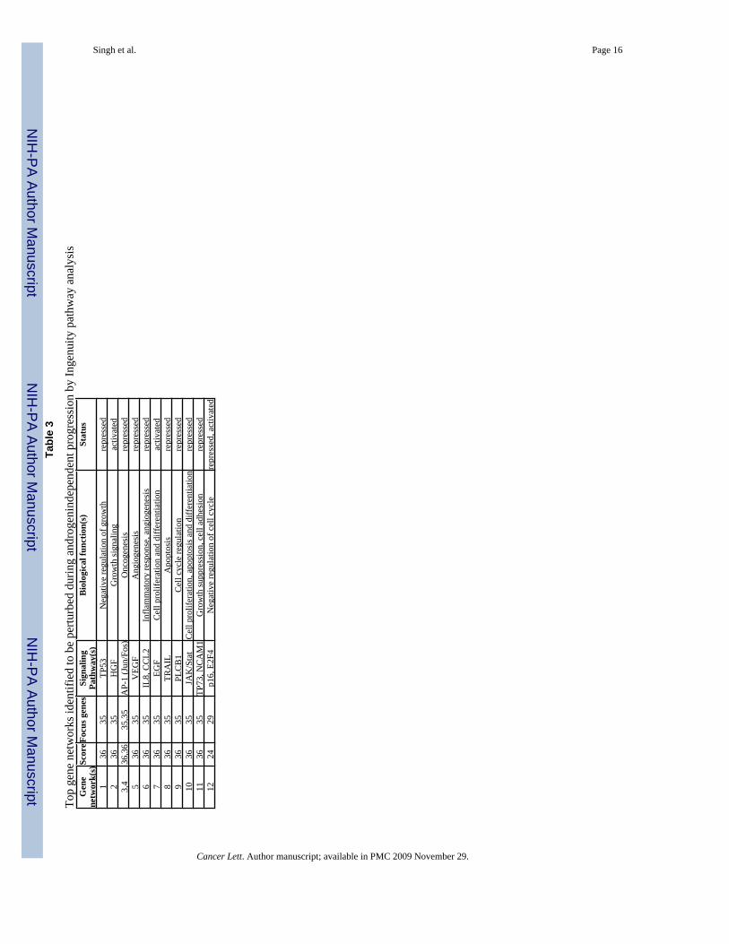
To identify the genetic networks altered during the androgen-refractory progression of prostatecancer cells, we utilized microarray data to perform a pathway prediction analysis using web-based application ‘Ingenuity pathway analysis’ (Ingenuity Systems, Mountain View, CA). Thisweb-delivered soft-ware searches its database to place differentially expressed genes in geneclusters linked to a molecular pathway(s). We identified several genetic networks to beperturbed during androgen-independent progression (Table 3). Most of the signaling pathwayscentral to the identified gene networks were found to be suppressed, except EGF, HGF andE2F4 (Table 3). Thus, our data suggest that the prostate cancer mainly involves a repressionof cell signaling pathways to attain an androgen-refractory phenotype.
Immunohistochemical analyses of protein phosphatase 2, catalytic subunit, (PPP2CA) inprostate cancer
Genome-wide survey of the differential gene expression resulted in the identification ofmultiple genes with potential pathobiological importance in androgen-refractory progressionof prostate cancer. From the long list of genes, we chose PPP2CA for further analysis at proteinlevel in human prostate cancer tissues to predict its prospective significance. PPP2CA encodesthe catalytic subunit (alpha-isoform) of protein phosphatase 2A (PP2A), which is a serinephosphatase implicated in the negative control of the cell division and survival. Immunoblotanalysis demonstrated its variable expression pattern in AD and AI prostate cancer cell lines(Figure 2). Consistent with the microarray data, PP2A was downregulated in LNCaP-C81 (AI)cells compared to the LNCaP-C33 (AD) cells. Furthermore, data from immunohistochemical
Singh et al. Page 5
Cancer Lett. Author manuscript; available in PMC 2009 November 29.
NIH
-PA Author Manuscript
NIH
-PA Author Manuscript
NIH
-PA Author Manuscript
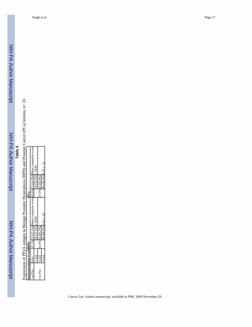
analysis also showed an overall downregulation of PP2A in cancer lesions compared to theadjacent normal/benign tissue (Figure 3). Out of the 35 human tissue samples, 22 (62.9%)showed positive reactivity in normal/benign region, whereas only 20 (57.0%) cancer caseswere positive for PP2A (Table 4). No intense immunoreactivity was observed in either normal/benign or cancer cells. Composite score values for normal/benign cells ranged from 0 to 6 witha mean composite score of 1.6. In cancer cells, however, the composite score values rangedfrom 0 to 3 (mean composite score=0.7) due to less number of immunoreactive cells (%positivity). The difference between mean composite scores of benign and cancer samples wasstatistically significant (P=0.0073). Interestingly, when we compared the composite scoredifferences in the stage and Gleason grade subgroups, the composite score were significantlygreater in late stage [stages 3 & 4] (P=0.015) and high Gleason grade [Gleason score 8–10](P=0.042) subgroups as compared to early stage and low Gleason grade subgroups,respectively.
DISCUSSIONThe present study examined the differential gene expression in androgen-dependent (AD)LNCaP-C33 and androgen-independent (AI) LNCaP-C81 cells via genome-wide expressionprofiling. Our study resulted in the identification of many key genes that could be of clinicalimportance and future targets for prostate cancer therapy. Furthermore, our data indicated thatthe androgen-refractory progression of prostate cancer mainly involves a repressionmechanism as a majority of the differentially-expressed genes and signalling networks weredownregulated in AI prostate cancer cells.
Over the last decade, DNA array technique has contributed significantly in identifying novelmolecules that play potential role in the disease progression as well as in the classification ofvarious cancers [9–11]. Moreover, recent technological advances have enabled thecomprehensive surveying of a cell’s transcriptional landscape. Genome-wide analysis allowsthe simultaneous monitoring of whole transcriptome, and is helpful in identifying the functionaland biochemical pathway(s) that are perturbed during the disease progression and targeted bythe therapeutic drugs [12;13]. In prostate cancer, several studies have been conducted ondifferent types of DNA arrays using tumor biopsy specimens, tumor cell lines, and morerecently tissues obtained from rapid autopsies [14;15]. What we have learnt thus far is thatprostate cancer is a heterogeneous group of diseases [3] and therefore a crucial examinationof current or newly identified biomarkers is needed, while considering their potential asdiagnostic, prognostic and therapeutic targets.
In this study, we utilized a unique in vitro LNCaP cell model, which includes different LNCaPcells (AD: C33 and AI: C81), closely resembling early and late stages of tumor progression[4]. One advantage of using this model is that it overcomes the heterogeneity, associated withthe prostate cancer [3] to a certain extent. Furthermore, LNCaP cells express functionalandrogen receptors as is the case in majority of prostate carcinomas. We hypothesized thatidentification of differentially regulated genes between AD (C33) and AI (C81) prostate cancercells may provide insights into its clinical course of progression and may be exploited for avariety of clinical applications. Genome-wide expression analysis generated a large amount ofinteresting data and identified many genes that were differentially regulated in AI prostatecancer cells. Genes identified in our study include those involved in cell proliferation,apoptosis, angiogenesis, hormone response, etc. These genes are of potential interest forfollow-up studies. While some have already been reported to have potential as clinicalbiomarkers [15–17], many others seem to be novel in context to their association with AIphenotype. Among the selected overexpressed genes in androgen-independent (C81) cells, two(TPTE and TSPY) have previously been shown to exhibit a testis-specific restricted pattern ofexpression in normal conditions [18;19]. TPTE (transmembrane phosphatase with tensin
Singh et al. Page 6
Cancer Lett. Author manuscript; available in PMC 2009 November 29.
NIH
-PA Author Manuscript
NIH
-PA Author Manuscript
NIH
-PA Author Manuscript

homology) encodes a PTEN-related tyrosine phosphatase (also referred as PTEN2) with fourpotential transmembrane domains [19]. PTEN (phosphatase and tensin homolog) acts as atumor suppressor and its multiple implications in prostate cancer progression have also beenreported [20]. No clear biological function has yet been assigned to TPTE and future studiesare warranted to understand its role in tumor progression. In contrast to our finding, anotherstudy has reported recurrent loss of a gene cluster on Y-chromosome involving TSPY (tumor-specific protein-Y linked) in prostate cancer [21]. This further supports the heterogeneousnature of AI prostate cancer as previously described [3] and suggests the existence of differentsignaling mechanisms among subtypes of metastatic prostate cancer. Nonetheless, a recentstudy has shown the oncogenic potential of TSPY in Hela and NIH3T3 cells [22]. Otherimportant overexpressed genes were HGF (hepatocyte growth factor) and MET/HGFR(hepatocyte growth factor receptor). HGF and its receptor, c-Met, have been demonstrated toelicit a number of key functions in numerous tissues that are important in the progression,invasion and metastasis of cancer [23]. Involvement of HGF-Met signalling in metastaticprostate cancer development has also been reported [24].
It is interesting to note that relatively more differentially-expressed genes were found to beunder-expressed (n=1460) than overexpressed (n=876) in AI prostate cancer cells (data notshown). In our study, Angiopoietin 2 (ANGP2), whose expression has been shown to correlatewith increasing grade of prostate cancer [25], was found to be downregulated in AI prostatecancer cells. Thrombospondin 1 (THBS1) is another important gene that was highly repressedin AI prostate cancer cells. In previous studies, it has been shown to possess anti-angiogenicactivity [26–28]. The intratumoral administration of a THBS1 expression vector into androgen-independent DU-145 prostate cancer xenografts was shown to inhibit tumor growth in vivothrough inhibition of tumor angiogenesis [28]. Interestingly, androgens have been shown torepress the expression of thrombospondin 1 in normal and neoplastic prostate tissues [27].HTATIP2, another tumor and metastasis suppressor gene [29], was also repressed in androgen-refractory PC cells. Ectopic expression of HTATIP2 in v-SCLC cells induced expression of anumber of genes including the apoptosis-related genes Bad and Siva, as well as metastasissuppressor NM23-H2 [30]. SPRY2, an inhibitor of receptor tyrosine kinase (RTK) signaling,found to be under-expressed in our study has been shown to be epigenetically inactivated inprostate cancer [31]. Progressive loss of ALCAM, which is downregulated in AI prostate cancercells, has been reported in high-grade prostate carcinomas [32]. Among other genes ofimportance exhibiting downregulated expression in AI prostate cancer cells were CDKN2B/TP53, CDKN1C/TP57, TP73. These genes are known tumor suppressors and are frequentlysilenced in cancers [33–35].
To better understand the functional premise related to androgen-independent progression, wesearched for patterns of gene expression using Ingenuity pathway analysis. Many signalingnetworks (Table 3) were identified, suggesting that perturbation of these molecular pathwaysis associated with the gain of androgen-refractory phenotype. In the literature, these pathwayshave been shown to be linked with various aspects of tumor progression [36–40]. Of particularinterest is the finding that most of the pathways identified in our analysis were repressed inandrogen-refractory prostate cancer cells. Importantly, many (TP53, TP73, p16, and TRAIL)among these downregulated pathways are associated with growth inhibitory cell signaling.
PPP2CA, which encodes for the catalytic-subunit (alpha-isoform) of the protein phosphatase2A (PP2AC), was one of several genes of interest that exhibited a downregulated expressionin our analysis in AI prostate cancer cells. PP2A is a potent tumor suppressor and is involvedin broad cellular functions [41]. In light of recent observations, it seems to be of potentialinterest in prostate cancer [42–45]. Data mining analysis using Oncomine cancer profilingdatabase (www.oncomine.org) demonstrate that PPP2CA is significantly downregulated inprostate cancer compared to normal/benign prostate (4 studies) and in metastatic prostate
Singh et al. Page 7
Cancer Lett. Author manuscript; available in PMC 2009 November 29.
NIH
-PA Author Manuscript
NIH
-PA Author Manuscript
NIH
-PA Author Manuscript
cancer compared to primary prostate carcinoma (3 studies). Interestingly, in a recent report theexpression of the beta-isoform of catalytic-subunit of PP2A (PPP2CB) is also shown to bedownregulated in prostate cancer [43]. Our immunohistochemical analysis in normal/benignand matched prostate cancer tissues corroborated the downregulated expression of PP2A inprostate cancer. However, we were not able to obtain patient-matched, androgen-dependentand androgen-independent specimens to examine the expression of PP2A during progressionto AI phenotype.
In summary, our study offers a significant contribution to our knowledge of the molecularcharacteristics of AI prostate cancer. Overall comparison of the expression data between ADand AI prostate cancer cells is intriguing demonstrating the decreased expression of severaltumor suppressor genes in the AI tumor cells, while many genes that may potentially promotegrowth and metastasis were upregulated. Protein phosphatase 2A was identified asdifferentially-expressed protein and its downregulated expression may be of potentialimportance during the prostate cancer development and further during AI progression of thedisease. Many of these differentially-expressed genes may be important as potential diagnostic/prognostic and therapeutic targets for the prostate cancer.
AcknowledgmentsThis work was supported by the U.S. Department of Defense- Idea Award (PC04502). The UNMC Microarray CoreFacility receives partial support from the INBRE Program of the National Center for Research Resources (NIH grantnumber P20 RR016469). We thank the Molecular Biology Core Facility, UNMC, for oligonucleotide synthesis, Ms.Fen F. Lin for technical help in cell culture and Kristi L.W. Berger (Eppley Institute) for editorial assistance.
REFERENCES1. Jemal A, Siegel R, Ward E, Murray T, Xu J, Thun MJ. Cancer statistics, 2007. CA Cancer J.Clin
2007;57:43–66. [PubMed: 17237035]2. Feldman BJ, Feldman D. The development of androgen-independent prostate cancer. Nat.Rev.Cancer
2001;1:34–45. [PubMed: 11900250]3. Shah RB, Mehra R, Chinnaiyan AM, Shen R, Ghosh D, Zhou M, Macvicar GR, Varambally S, Harwood
J, Bismar TA, Kim R, Rubin MA, Pienta KJ. Androgen-independent prostate cancer is a heterogeneousgroup of diseases: lessons from a rapid autopsy program. Cancer Res 2004;64:9209–9216. [PubMed:15604294]
4. Igawa T, Lin FF, Lee MS, Karan D, Batra SK, Lin MF. Establishment and characterization of androgen-independent human prostate cancer LNCaP cell model. Prostate 2002;50:222–235. [PubMed:11870800]
5. Karan D, Schmied BM, Dave BJ, Wittel UA, Lin MF, Batra SK. Decreased androgen-responsivegrowth of human prostate cancer is associated with increased genetic alterations. Clin.Cancer Res2001;7:3472–3480. [PubMed: 11705865]
6. Veeramani S, Igawa T, Yuan TC, Lin FF, Lee MS, Lin JS, Johansson SL, Lin MF. Expression of p66(Shc) protein correlates with proliferation of human prostate cancer cells. Oncogene 2005;24:7203–7212. [PubMed: 16170380]
7. Livak KJ, Schmittgen TD. Analysis of relative gene expression data using real-time quantitative PCRand the 2(-Delta Delta C(T)) Method. Methods 2001;25:402–408. [PubMed: 11846609]
8. Singh AP, Chauhan SC, Bafna S, Johansson SL, Smith LM, Moniaux N, Lin MF, Batra SK. Aberrantexpression of transmembrane mucins, MUC1 and MUC4, in human prostate carcinomas. Prostate2006;66:421–429. [PubMed: 16302265]
9. Hayes DN, Monti S, Parmigiani G, Gilks CB, Naoki K, Bhattacharjee A, Socinski MA, Perou C,Meyerson M. Gene expression profiling reveals reproducible human lung adenocarcinoma subtypesin multiple independent patient cohorts. J.Clin.Oncol 2006;24:5079–5090. [PubMed: 17075127]
10. Pusztai L, Mazouni C, Anderson K, Wu Y, Symmans WF. Molecular classification of breast cancer:limitations and potential. Oncologist 2006;11:868–877. [PubMed: 16951390]
Singh et al. Page 8
Cancer Lett. Author manuscript; available in PMC 2009 November 29.
NIH
-PA Author Manuscript
NIH
-PA Author Manuscript
NIH
-PA Author Manuscript

11. Sorlie T. Molecular portraits of breast cancer: tumour subtypes as distinct disease entities.Eur.J.Cancer 2004;40:2667–2675. [PubMed: 15571950]
12. Bono H, Okazaki Y. Functional transcriptomes: comparative analysis of biological pathways andprocesses in eukaryotes to infer genetic networks among transcripts. Curr.Opin.Struct.Biol2002;12:355–361. [PubMed: 12127455]
13. Liu S, Seidel-Dugan C. In search of p53 target genes for the therapeutic manipulation of cancer.Curr.Opin.Drug Discov.Devel 2006;9:176–183.
14. Varambally S, Dhanasekaran SM, Zhou M, Barrette TR, Kumar-Sinha C, Sanda MG, Ghosh D, PientaKJ, Sewalt RG, Otte AP, Rubin MA, Chinnaiyan AM. The polycomb group protein EZH2 is involvedin progression of prostate cancer. Nature 2002;419:624–629. [PubMed: 12374981]
15. Varambally S, Yu J, Laxman B, Rhodes DR, Mehra R, Tomlins SA, Shah RB, Chandran U, MonzonFA, Becich MJ, Wei JT, Pienta KJ, Ghosh D, Rubin MA, Chinnaiyan AM. Integrative genomic andproteomic analysis of prostate cancer reveals signatures of metastatic progression. Cancer Cell2005;8:393–406. [PubMed: 16286247]
16. Knudsen BS, Gmyrek GA, Inra J, Scherr DS, Vaughan ED, Nanus DM, Kattan MW, Gerald WL,Vande Woude GF. High expression of the Met receptor in prostate cancer metastasis to bone. Urology2002;60:1113–1117. [PubMed: 12475693]
17. Naughton M, Picus J, Zhu X, Catalona WJ, Vollmer RT, Humphrey PA. Scatter factor-hepatocytegrowth factor elevation in the serum of patients with prostate cancer. J.Urol 2001;165:1325–1328.[PubMed: 11257710]
18. Krick R, Jakubiczka S, Arnemann J. Expression, alternative splicing and haplotype analysis oftranscribed testis specific protein (TSPY) genes. Gene 2003;302:11–19. [PubMed: 12527192]
19. Wu Y, Dowbenko D, Pisabarro MT, llard-Telm L, Koeppen H, Lasky LA. PTEN 2, a Golgi-associatedtestis-specific homologue of the PTEN tumor suppressor lipid phosphatase. J.Biol.Chem2001;276:21745–21753. [PubMed: 11279206]
20. Mulholland DJ, Dedhar S, Wu H, Nelson CC. PTEN and GSK3beta: key regulators of progressionto androgen-independent prostate cancer. Oncogene 2006;25:329–337. [PubMed: 16421604]
21. Vijayakumar S, Hall DC, Reveles XT, Troyer DA, Thompson IM, Garcia D, Xiang R, Leach RJ,Johnson-Pais TL, Naylor SL. Detection of recurrent copy number loss at Yp11.2 involving TSPYgene cluster in prostate cancer using array-based comparative genomic hybridization. Cancer Res2006;66:4055–4064. [PubMed: 16618725]
22. Oram SW, Liu XX, Lee TL, Chan WY, Lau YF. TSPY potentiates cell proliferation and tumorigenesisby promoting cell cycle progression in HeLa and NIH3T3 cells. BMC.Cancer 2006;6(154):154.[PubMed: 16762081]
23. Hurle RA, Davies G, Parr C, Mason MD, Jenkins SA, Kynaston HG, Jiang WG. Hepatocyte growthfactor/scatter factor and prostate cancer: a review. Histol.Histopathol 2005;20:1339–1349. [PubMed:16136515]
24. MacDougall CA, Vargas M, Soares CR, Holzer RG, Ide AE, Jorcyk CL. Involvement of HGF/SF-Met signaling in prostate adenocarcinoma cells: evidence for 21 alternative mechanisms leading toa metastatic phenotype in Pr-14c. Prostate 2005;64:139–148. [PubMed: 15678502]
25. Lind AJ, Wikstrom P, Granfors T, Egevad L, Stattin P, Bergh A. Angiopoietin 2 expression is relatedto histological grade, vascular density, metastases, and outcome in prostate cancer. Prostate2005;62:394–399. [PubMed: 15378518]
26. Bocci G, Francia G, Man S, Lawler J, Kerbel RS. Thrombospondin 1, a mediator of the antiangiogeniceffects of low-dose metronomic chemotherapy. Proc.Natl.Acad.Sci.U.S.A 2003;100:12917–12922.[PubMed: 14561896]
27. Colombel M, Filleur S, Fournier P, Merle C, Guglielmi J, Courtin A, Degeorges A, Serre CM, BouvierR, Clezardin P, Cabon F. Androgens repress the expression of the angiogenesis inhibitorthrombospondin-1 in normal and neoplastic prostate. Cancer Res 2005;65:300–308. [PubMed:15665307]
28. Jin RJ, Kwak C, Lee SG, Lee CH, Soo CG, Park MS, Lee E, Lee SE. The application of an anti-angiogenic gene (thrombospondin-1) in the treatment of human prostate cancer xenografts. CancerGene Ther 2000;7:1537–1542. [PubMed: 11228532]
Singh et al. Page 9
Cancer Lett. Author manuscript; available in PMC 2009 November 29.
NIH
-PA Author Manuscript
NIH
-PA Author Manuscript
NIH
-PA Author Manuscript

29. Ito M, Jiang C, Krumm K, Zhang X, Pecha J, Zhao J, Guo Y, Roeder RG, Xiao H. TIP30 deficiencyincreases susceptibility to tumorigenesis. Cancer Res 2003;63:8763–8767. [PubMed: 14695192]
30. Xiao H, Palhan V, Yang Y, Roeder RG. TIP30 has an intrinsic kinase activity required for up-regulation of a subset of apoptotic genes. EMBO J 2000;19:956–963. [PubMed: 10698937]
31. McKie AB, Douglas DA, Olijslagers S, Graham J, Omar MM, Heer R, Gnanapragasam VJ, RobsonCN, Leung HY. Epigenetic inactivation of the human sprouty2 (hSPRY2) homologue in prostatecancer. Oncogene 2005;24:2166–2174. [PubMed: 15735753]
32. Kristiansen G, Pilarsky C, Wissmann C, Stephan C, Weissbach L, Loy V, Loening S, Dietel M,Rosenthal A. ALCAM/CD166 is up-regulated in low-grade prostate cancer and progressively lost inhigh-grade lesions. Prostate 2003;54:34–43. [PubMed: 12481253]
33. Herman JG, Jen J, Merlo A, Baylin SB. Hypermethylation-associated inactivation indicates a tumorsuppressor role for p15INK4B. Cancer Res 1996;56:722–727. [PubMed: 8631003]
34. Lodygin D, Epanchintsev A, Menssen A, Diebold J, Hermeking H. Functional epigenomics identifiesgenes frequently silenced in prostate cancer. Cancer Res 2005;65:4218–4227. [PubMed: 15899813]
35. Yang B, House MG, Guo M, Herman JG, Clark DP. Promoter methylation profiles of tumorsuppressor genes in intrahepatic and extrahepatic cholangiocarcinoma. Mod.Pathol 2005;18:412–420. [PubMed: 15467712]
36. Crosby ME, Almasan A. Opposing roles of E2Fs in cell proliferation and death. Cancer Biol.Ther2004;3:1208–1211. [PubMed: 15662116]
37. Eferl R, Wagner EF. AP-1: a double-edged sword in tumorigenesis. Nat.Rev.Cancer 2003;3:859–868. [PubMed: 14668816]
38. Igney FH, Krammer PH. Death and anti-death: tumour resistance to apoptosis. Nat.Rev.Cancer2002;2:277–288. [PubMed: 12001989]
39. Kim WY, Sharpless NE. The regulation of INK4/ARF in cancer and aging. Cell 2006;127:265–275.[PubMed: 17055429]
40. Melino G, De L, V, Vousden KH. p73: Friend or foe in tumorigenesis. Nat.Rev.Cancer 2002;2:605–615. [PubMed: 12154353]
41. Janssens V, Goris J, Van HC. PP2A: the expected tumor suppressor. Curr.Opin.Genet.Dev2005;15:34–41. [PubMed: 15661531]
42. Li L, Ren CH, Tahir SA, Ren C, Thompson TC. Caveolin-1 maintains activated Akt in prostate cancercells through scaffolding domain binding site interactions with and inhibition of serine/threonineprotein phosphatases PP1 and PP2A. Mol.Cell Biol 2003;23:9389–9404. [PubMed: 14645548]
43. Prowatke I, Devens F, Benner A, Grone EF, Mertens D, Grone HJ, Lichter P, Joos S. Expressionanalysis of imbalanced genes in prostate carcinoma using tissue microarrays. Br.J.Cancer2007;96:82–88. [PubMed: 17146477]
44. Yang X, Chen MW, Terry S, Vacherot F, Bemis DL, Capodice J, Kitajewski J, de la TA, BensonMC, Guo Y, Buttyan R. Complex regulation of human androgen receptor expression by Wnt signalingin prostate cancer cells. Oncogene 2006;25:3436–3444. [PubMed: 16474850]
45. Yang CS, Vitto MJ, Busby SA, Garcia BA, Kesler CT, Gioeli D, Shabanowitz J, Hunt DF, RundellK, Brautigan DL, Paschal BM. Simian virus 40 small t antigen mediates conformation-dependenttransfer of protein phosphatase 2A onto the androgen receptor. Mol.Cell Biol 2005;25:1298–1308.[PubMed: 15684382]
Singh et al. Page 10
Cancer Lett. Author manuscript; available in PMC 2009 November 29.
NIH
-PA Author Manuscript
NIH
-PA Author Manuscript
NIH
-PA Author Manuscript

Figure 1.Expression analysis of differential gene expression by real-time RT-PCR. Total RNA wasreverse-transcribed and subsequently amplified by a real-time PCR using gene-specificprimers. A fold-change in expression was calculated relative to LNCaP-C33 cells usingGAPDH as an internal control by ΔΔCT method. Each experiment was repeated at least threetimes and data presented as mean ± standard deviation.
Singh et al. Page 11
Cancer Lett. Author manuscript; available in PMC 2009 November 29.
NIH
-PA Author Manuscript
NIH
-PA Author Manuscript
NIH
-PA Author Manuscript

Figure 2.Immunoblot analysis of PPP2CA (alpha isoform of catalytic subunit of protein phosphatase2A). Consistent with the microarray analysis, a reduced expression of PPP2CA is observed inandrogen-independent (AI) LNCaP-C81 cells compared to androgen-dependent (AD) LNCaP-C33 cells. Except DU145, a reduced expression of PPP2CA is also evident in other AI celllines (C4-2 and PC3). Phospho-glycerol kinase (PGK) was used as an internal control.
Singh et al. Page 12
Cancer Lett. Author manuscript; available in PMC 2009 November 29.
NIH
-PA Author Manuscript
NIH
-PA Author Manuscript
NIH
-PA Author Manuscript
Figure 3.Immunohistochemical analysis of PPP2CA expression on a prostate cancer tissue array. A andB, Normal/benign tissue at 10X and 40 × magnifications, respectively. C, Prostate cancersection with various grade lesions (10× magnification), D and E, magnified (40X) view ofsection depicting lesions with Gleason grade 6 (3+3) and 8 (4+4), respectively. A decreasedexpression of PPP2CA is observed in prostate cancer, which further correlates with Gleasongrade.
Singh et al. Page 13
Cancer Lett. Author manuscript; available in PMC 2009 November 29.
NIH
-PA Author Manuscript
NIH
-PA Author Manuscript
NIH
-PA Author Manuscript
NIH
-PA Author Manuscript
NIH
-PA Author Manuscript
NIH
-PA Author Manuscript
Singh et al. Page 14
Table 1
List of selected genes that are overexpressed in androgen-independent (LNCaP-C81) compared to androgen-dependent (LNCaP-33) prostate cancer cells.
Gene Product Fold-change(Logbase2)
Function Genomiclocation
PCDH7 Protocadherin 7 7.9 Cell adhesion 4p15NRXN3 Neurexin 3 7.7 " 14q31TPTE/PTEN2
Transmembrane phosphatase with tensinhomology
5.4 Signal transduction 21p11
EPHA3 EPH receptor A3 5.3 " 3p11.2TSPY Testis specific protein, Y-linked 4.5 Hormone-response Yp11.2HGF Hepatocyte growth factor 4.4 Signal transduction 7q21.1PKIA Protein kinase inhibitor alpha 4.4 " 8q21.12PRLR Prolactin receptor 3.8 " 5p14-p13NR2F1 Nuclear receptor subfamily 2, group f,
member 13.6 Hormone-response 5q14
TNS Tensin 3.6 Signal transduction, cell migration 2q35-q36MET Hepatocyte growth factor receptor 3.2 Signal transduction 7q31CALN1 Calneuron 1 3.2 Calcium-binding protein 7q11ADAM-TS3 A disintegrin and metalloprotease with
thrombospondin motifs-33.1 propeptidase 4q13.3
NPL4 Nuclear protein localization 4 3 Protein trafficking 17qterMYO3A Myosin IIIA 3 Actin-dependent motor protein 10p11.1GNA11 Guanine nucleotide binding protein (G
protein), alpha 112.8 Signal transduction 19p13.3
GAGEB1/PAGE1
P antigen family, member 1 (prostateassociated)
2.7 Tumor-linked protein, functionunknown
Xp11.23
AKAP9 A kinase anchor protein 1 2.6 Signal transduction 17q21-q23TCF4 Transcription factor 4 2.6 " 18q21.1MYLK Myosin, light polypeptide kinase 2.6 cell migration 3q21KLK2 Kallikrein 2 2.4 peptidase 19q13.41HOXA10 Homeo box A10 2.3 Signal transduction 7p15-p14DDR2 Discoidin domain receptor family, member 2 2.2 " 1q12-q23EGF Epidermal growth factor 2.2 Cell growth 4q25TEM8 Tumor endothelial marker 8 2.1 angiogenesis 2p13.1XAGE-1 X antigen family, member 1 2 Cancer/testis-associated gene,
unknown functionXp11.22-p11.21
GAS2 Growth arrest-specific 2 2 Regulation of cell shape, apoptosis 11p14.3-p15.2
Cancer Lett. Author manuscript; available in PMC 2009 November 29.

NIH
-PA Author Manuscript
NIH
-PA Author Manuscript
NIH
-PA Author Manuscript
Singh et al. Page 15
Table 2
List of selected genes that are under-expressed in androgen-independent (LNCaP-C81) compared to androgen-dependent (LNCaP-33) prostate cancer cells.
Gene Product Fold-change(Logbase2)
Function Genomiclocation
ANGPT2 Angiopoietin 2 7.8 Context-dependent vascular regression and angiogenesis 8p23.1THBS1 Thrombospondin 1 7.3 Cell-cell and cell-extracellular matrix interaction 15q15CCL5 Chemokine (c-c motif) ligand 5 6.2 Chemokine, immunoregulatory 17q11.2-12PTK6 Protein tyrosine kinase 6 6.1 Signal transduction 20q13.3CXCL11 Chemokine (c-x-c motif) ligand 11 5.8 Chemokine, immunoregulatory 4q21.2S100P S100 calcium binding protein p 5.1 Hormone response 4p16PSMD5 Proteasome 26s non-ATPase subunit, 5 4.9 Multi-subunit protease 9q33.2CDKN2B Cyclin-dependent kinase inhibitor 2b 4.7 tumor suppressor 9p21PEG3 Paternally expressed 3 4.5 " 19q13.4HTATIP2 Hiv-1 tat interactive protein 2 4.4 " 11p15.1CXCL2 Chemokine (c-x-c motif) ligand 2 4.4 Chemokine, immunoregulatory 4q21RND1 Rho family gtpase 1 4.2 Cell motility 12q12-q13NK4 Natural killer4/Interleukin 32 4.1 cytokine 16p13.3ACPP Prostatic acid phosphotase 4.1 Hormone response, growth signaling 3q21-q23ICAM1 Intercellular adhesion molecule 1 4 tumor suppressor, cell migration 19p13.3-13.2FOSL2 Fos-like antigen 2 3.9 Cell proliferation/differentiation 2p23.3SFN Stratifin 3.7 Tumor suppressor 1p36.11PLAT Plasminogen activator, tissue 3.5 Cell migration, tissue remodeling 8p12INHBE Inhibin, beta E 3 tumor suppressor 12q13.3CDKN1C Cyclin-dependent kinase inhibitor 1c 2.9 tumor suppressor 11p15.5TP73 Tumor protein p73 2.7 tumor suppressor 1p36.3SOCS2 Suppressor of cytokine signaling 2 2.7 growth suppressor 12qBMPR1B Bone morphogenetic protein receptor, type 1B 2.7 Signal transduction 4q22-q24CCL2 Chemokine (c-c motif) ligand 2 2.6 chemokine 17q11.2-q21.1VIM Vimentin 2.6 Mesenchymal cytoskeletal protein 10p13SPRY2 Sprouty 2 2.6 tumor suppressor 13q31.1CXCL10 Chemokine (c-x-c motif) ligand 10 2.6 Chemokine, angiostatic 4q21LCN2 Lipocalin 2 2.5 Tumor suppressor/promoter 9q34AKAP12 A kinase anchor protein 12 2.4 tumor/metastasis suppressor 6q24-q25KLF4 Kruppel-like factor 4 2.4 Transcriptional activator 9q31ATF3 Activating transcription factor 3 2.4 apoptosis, cell migration 1q32.3FBN1 Fibrillin 1 2.4 ECM protein, cell attachment 15q21.1TWIST1 Twist homolog 1 2.4 Invasion, metastasis 7p21.2ALCAM Activated leukocyte cell adhesion molecule 2.3 Cell adhesion 3q13.1MSX2 Msh homeo box homolog 2 2.2 tumor suppressor 5q34-q35LOX Lysyl oxidase 2.2 Cross-linking of ECM proteins 5q23.2BTG1 B-cell translocation gene 1 protein 2.2 Hormone response, anti-proliferative 12q22SOCS1 Suppressor of cytokine signaling 1 2.2 tumor suppressor 16p13.13PPP2CA Protein phosphatase 2 (formerly 2a), catalytic
subunit, alpha isoform1.5 Serine phosphatase, control of cell
division5q23-q31
PPP3CA Protein phosphatase 3 (formerly 2b), catalyticsubunit, alpha isoform
1.3 Serine phosphatase, apoptosis 4q21-q24
Cancer Lett. Author manuscript; available in PMC 2009 November 29.
NIH
-PA Author Manuscript
NIH
-PA Author Manuscript
NIH
-PA Author Manuscript
Singh et al. Page 16Ta
ble
3
Top
gene
net
wor
ks id
entif
ied
to b
e pe
rturb
ed d
urin
g an
drog
enin
depe
nden
t pro
gres
sion
by
Inge
nuity
pat
hway
ana
lysi
sG
ene
netw
ork(
s)Sc
oreF
ocus
gen
esSi
gnal
ing
Path
way
(s)
Bio
logi
cal f
unct
ion(
s)St
atus
136
35TP
53N
egat
ive
regu
latio
n of
gro
wth
repr
esse
d2
3635
HG
FG
row
th si
gnal
ing
activ
ated
3,4
36,3
635
,35
AP-
1 (J
un/F
os)
Onc
ogen
esis
repr
esse
d5
3635
VEG
FA
ngio
gene
sis
repr
esse
d6
3635
IL8,
CC
L2In
flam
mat
ory
resp
onse
, ang
ioge
nesi
sre
pres
sed
736
35EG
FC
ell p
rolif
erat
ion
and
diff
eren
tiatio
nac
tivat
ed8
3635
TRA
ILA
popt
osis
repr
esse
d9
3635
PLC
B1
Cel
l cyc
le re
gula
tion
repr
esse
d10
3635
JAK
/Sta
tC
ell p
rolif
erat
ion,
apo
ptos
is a
nd d
iffer
entia
tion
repr
esse
d11
3635
TP73
, NC
AM
1G
row
th su
ppre
ssio
n, c
ell a
dhes
ion
repr
esse
d12
2429
p16,
E2F
4N
egat
ive
regu
latio
n of
cel
l cyc
lere
pres
sed,
act
ivat
ed
Cancer Lett. Author manuscript; available in PMC 2009 November 29.
NIH
-PA Author Manuscript
NIH
-PA Author Manuscript
NIH
-PA Author Manuscript
Singh et al. Page 17Ta
ble
4
Expr
essi
on o
f PP2
A a
ntig
en in
Ben
ign
Pros
tatic
Hyp
erpl
asia
(BPH
) and
Pro
stat
e C
ance
r (PC
a) le
sion
s, n=
35
Imm
unos
tain
ing
Gra
deB
PHPC
a−v
e (%
)0
(n)
37.1
%13
(37.
1%)M
ean
com
posi
te sc
ore
± SE
M43
.0%
15 (4
2.8%
)Mea
n co
mpo
site
scor
e±
SEM
+ve
(%)
+1 (n
)62
.9%
20 (5
7.2%
)57
.0%
19 (5
4.3%
)+2
(n)
02 (0
5.7%
) 1.63
± .2
501
(02.
9%) 0.
70 ±
.13
+3 (n
)00
(00.
0%)
00 (0
0.0%
)
Cancer Lett. Author manuscript; available in PMC 2009 November 29.
Related Documents