Faculty of Health Sciences Department of Pharmacy Drug Transport and Delivery Research Group Mucus-penetrating drug carriers for vaginal drug delivery — By Kristina Rybak Master thesis for the degree Master of Pharmacy May 2015 Supervisors PhD student May Wenche Jøraholmen Professor Nataša Škalko-Basnet

Welcome message from author
This document is posted to help you gain knowledge. Please leave a comment to let me know what you think about it! Share it to your friends and learn new things together.
Transcript
Faculty of Health Sciences
Department of Pharmacy
Drug Transport and Delivery Research Group
Mucus-penetrating drug carriers for vaginal drug delivery — By Kristina Rybak Master thesis for the degree Master of Pharmacy May 2015 Supervisors PhD student May Wenche Jøraholmen Professor Nataša Škalko-Basnet

I

II

III

IV
Acknowledgments This study was performed at the Drug Transport and Delivery Research Group, Department
of Pharmacy, University of Tromsø - The Arctic University of Norway, from October 2014 to
May 2015.
To start with, I would like to express immense gratitude to my supervisors Professor Nataša
Škalko-Basnet and PhD student May Wenche Jøraholmen for their support, valuable guidance
and contributions during this master thesis. Thank you Nataša for taking such good care of
me, making me feel welcome and guiding me through difficulties with your limitless wisdom.
Thank you May Wenche Jøraholmen for your patience and challenges, overcoming which
made me stronger.
I am very grateful to Cristiane Jacobsen for her kindness and care, readiness to help and
motivation. Thank you!
I would like to give thanks to my wonderful lab mates: Lisa Hemmingsen, Iren Wu and
Ayantu Chemeda. We have encountered many difficulties on our way, but you were always
there supporting and encouraging. Thank you for pleasant time we had together!
I also wish to thank my fellow students. We have traveled a long and challenging journey
together and it will be sad to see you go, but nonetheless the priceless moments will always
be with me.
I am grateful to my family for their help and contributions! Most of all, I would like to thank
my husband, Hogne Berg Jensen, for immense understanding, and my children Maximillian
Emmanuel and Angelica Sophie for being my biggest motivation! I would not have made it
without you!
May 2015
Kristina Rybak

V
VI
Sammendrag Vaginal legemiddeladministrering er utfordrende grunnet kroppens naturlige
forsvarsmekanismer og krever en spesiell tilnærming i utvikling av legemidler. Likevel er
topikal tilførsel foretrukket fremfor systemisk behandling der det er mulig. Fordeler med lokal
applikasjon er at man unngår nedbrytning i fordøyelsessystemet samt førstepassasjeeffekt.
Dessuten kan lokal noninvasiv anvendelse befri pasienter fra potensielt ubehagelige
prosedyrer.
Målet med dette prosjektet var å utvikle og optimalisere mukuspenetrerende liposomer for
vaginal behandling av human papilloma virus (HPV). Et naturlig forekommende protein,
interferon α-2b (IFN α-2b), brukes blant annet i behandling av vaginale infeksjoner.
Mukoadhesive nanovesikler har vist utilstrekkelig vaginal oppholdstid på grunn av fornyelse
av vaginale sekresjoner. Med dette utgangspunktet var det et ønske å forbedre terapeutisk
legemiddeleffekt ved å designe nye, mukuspenetrerende partikler. For å oppnå
gjennomtrenging av mukusbarrieren ble partiklenes overflate modifisert med polyethylene
glykol (PEG), en lav molekylvekt polymer.
PEGylerte liposomer med IFN α-2b ble tilberedt ved hjelp av den såkalte “thin film
hydration” metode. Vesikkelstørrelse ble redusert ved hjelp av ekstrudering gjennom
polykarbonatmembraner. Størrelse, polydispersitet, zetapotensialet og grad av
legemiddelinkorporering for liposomene ble karakterisert. En velegnet størrelse (185 ± 3 nm)
ble målt og lav polydispersitet (PI 0.09) indikerte uniform størrelses distribusjon.
Zetapotensialet var negativt (-12.2 ± 1.4 mV). Fritt legemiddel ble separert fra inkorporert
legemiddel ved hjelp av gel kolonne kromatografi, og inkorporeringsgrad (88%) var bestemt
ved hjelp av IFN α ELISA kit. Det nylig utviklede systemet for lokal IFN α-2b levering
innehar potensialet til å behandle HPV infeksjoner.
Nøkkelord: Mukuspenetrerende liposomer, PEG, vaginal tilførsel av legemidler, IFN α-2b
VII
Abstract Vaginal drug administration is a challenging approach due to the body´s natural defense
mechanisms and specificity in formulation design. However, where applicable, topical drug
delivery is preferable to systemic therapy. Firstly, it allows averting hepatic first pass effect
and degradation by GI enzymes. Secondly, non-invasive application provides closer and
direct contact with the affected area and relieves user from an unpleasant procedure.
The aim of this project was development and optimization of mucus-penetrating liposomes
for vaginal treatment of human papilloma virus (HPV). The naturally occurring protein
interferon α-2b (IFN α-2b) is commonly used in treatment of vaginal infections. Due to
continuous vaginal fluid renewal the residence time of mucoadhesive nanoparticles is shown
to be insufficient. Treatment efficacy can be increased by designing novel, mucus-penetrating
particles. To overcome the mucosal barrier, surface modification with the low molecular
weight polymer polyethylene glycol (PEG), was applied.
PEGylated liposomes containing IFN α-2b were prepared by thin film hydration method.
Vesicle size was reduced by extrusion through polycarbonate membranes. Liposomal size,
polydispersity, surface charge and IFN α-2b entrapment were determined. An adequate
vesicle size (185 ± 3 nm) was obtained and a low polydispersity (PI 0.09) indicated a
monodisperse size distribution. Net surface charge was measured to be -12.2 ± 1.4 mV. Free
drug was separated from liposomally encapsulated IFN α-2b by gel column chromatography,
and entrapment efficiency (88%) was determined using human IFN α ELISA kit. The newly
developed system for local IFN α-2b delivery has a potential to treat HPV infections.
Keywords: Mucus-penetrating liposomes, PEG, vaginal drug delivery, IFN α-2b
VIII
Table of Contents
1. General introduction ......................................................................................................... 1
2. Introduction ....................................................................................................................... 3
2.1 Vagina ................................................................................................................................ 3
2.1.1 Vaginal mucus ............................................................................................................. 4
2.1.2 Vagina as a site for drug delivery ............................................................................... 5
2.2 Mucoadhesion vs. mucus-penetration ............................................................................... 6
2.2.1 Mucoadhesion ............................................................................................................. 7
2.2.2 Mucus penetration ....................................................................................................... 8
2.3 Liposomes ........................................................................................................................ 10
2.3.1 Liposomes as drug delivery system .......................................................................... 12
2.3.2 Preparation of liposomes ........................................................................................... 12
2.4 Vaginal infection ............................................................................................................. 13
2.4.1 General ...................................................................................................................... 13
2.4.2 Human Papillomavirus (HPV) .................................................................................. 14
2.4.3 Pathogenesis of HPV ................................................................................................ 15
2.4.4 Current treatment of HPV infections ........................................................................ 16
2.5 Interferon α-2b (IFN α-2b) .............................................................................................. 17
2.5.1 General ...................................................................................................................... 17
2.5.2 Classification ............................................................................................................. 18
2.5.3 Application ................................................................................................................ 19
3. Aims of the study ............................................................................................................. 21
4. Materials and Methods ................................................................................................... 23
4.1 Materials .......................................................................................................................... 23
4.2 Computer programs ......................................................................................................... 24
4.3 Instruments ...................................................................................................................... 24
4.4 Equipment ........................................................................................................................ 24
4.5 Methods ........................................................................................................................... 25
4.5.1 Preparation of IFN α-2b buffer ................................................................................. 25
4.5.2 Preparation of IFN α-2b solution .............................................................................. 25
4.5.3 Preparation of empty liposomes ................................................................................ 25
IX
4.5.4 Preparation of liposomes with IFN α-2b ................................................................... 26
4.5.5 Vesicle size reduction ............................................................................................... 26
4.5.6 Particle size analysis ................................................................................................. 26
4.5.7 Zeta potential determination ..................................................................................... 26
4.5.8 Gel column preparation ............................................................................................. 27
4.5.9 Separation of a free drug ........................................................................................... 27
4.5.10 Preparation of 0.3 % Triton buffer .......................................................................... 27
4.5.11 Enzyme-linked immunoassay (ELISA) .................................................................. 27
4.5.12 Preparation of acetate buffer ................................................................................... 28
4.5.13 In vitro drug release study ....................................................................................... 28
5. Results and discussion ..................................................................................................... 31
5.1 Liposome characterization ............................................................................................... 31
5.1.1 Liposomal size .......................................................................................................... 31
5.1.2 Liposomal zeta potential ........................................................................................... 33
5.1.3 IFN α-2b entrapment ................................................................................................. 34
5.2 In vitro release of IFN α-2b ............................................................................................. 38
6. Conclusion ........................................................................................................................ 39
7. Perspectives ...................................................................................................................... 41
8. References ........................................................................................................................ 43
X
List of figures Figure 1: Schematic drawing of the vaginal mucosa. 1: capillary vessels; 2: artery; 3: vein
(das Neves and Bahia, 2006) ...................................................................................................... 3
Figure 2: Nanoparticle adhesion to mucus. Two steps of the process (Carvalho et al., 2010) . 7
Figure 3: Schematic illustration of the fate of CP and MPP administered to mucosal surface
(Lai et al., 2009) ......................................................................................................................... 9
Figure 4: Structural formula of polyethylene glycol (Medicines Complete) ............................ 9
Figure 5: General structure of liposomes (Encyclopædia Britannica) .................................... 10
Figure 6: Structure of a phosphatidyl choline molecule (Electronic Journal of Biomedicine)
.................................................................................................................................................. 11
Figure 7: Molecular structure of cholesterol (Medicines Complete) ...................................... 11
Figure 8: A model of Human Papillomavirus (Virusworld) ................................................... 14
Figure 9: Illustration of HPV pathogenesis (Groves and Coleman, 2015) ............................. 15
Figure 10: IFN α 2-b protein structure (Drug Bank) ............................................................... 17
Figure 11: Classification of IFNs ............................................................................................ 18
Figure 12: Gel column separation of liposomes. Fractions 15-51 .......................................... 36
Figure 13: Gel column separation of liposomes. Fractions 52-100 ........................................ 36

XI
XII
List of tables Table 1: The size of pre-extruded liposomes (n=4) ................................................................ 32
Table 2: The size of liposomes after extrusion through 400 nm membrane (n=4) ................. 32
Table 3: The size of liposomes after extrusion through 200 nm membrane (n=4) ................. 32
Table 4: Zeta potential values of liposomes (n=4) .................................................................. 34
XIII
Abbreviations C2H4O2 Glacial acetic acid
CH3COONH4 Ammonium acetate
CP Conventional particles
EDTA Ethylenedinitrilo tetraacetic acid, disodium salt
dehydrate
ELISA Enzyme-linked immunoassay
HPV Human papilloma virus
HRP Horse reddish peroxide
IFN α-2b Interferon α-2b
IU International units
LMV Large multilamellar vesicles
LUV Large unilamellar vesicles
MPP Mucus-penetrating particles
Na2HPO4 × 2H2O di-Sodium hydrogen phosphate dehydrate
NaCl Sodium chloride
NaH2PO4 × H2O Sodium hydrogen phosphate monohydrate
PCS Photon correlation spectroscopy
PEG Polyethylene glycol
PI Polydispersity index
PVA Polyvinyl alcohol
STD Sexually transmitted disease
SUV Smal unilamellar vesicles
TMB Tetramethylbenzidine
1
1. General introduction Conventional routes of administration (oral and topical) are used to obtain either systemic or
local effect respectively. Other application routes were not widely practiced since health care
providers had responsibility for drug administration which left almost no room for discreet
and private treatment (Alexander et al., 2004). In spite of the extended knowledge on vaginal
physiology and the potential for local drug delivery, this route of administration has not been
extensively explored (Hussain and Ahsan, 2005). However, there is growing interest in
evolving drug delivery systems for vaginal administration (Hussain and Ahsan, 2005).
A central aim in designing drug delivery systems for local administration is the close
proximity to an affected area and more target-oriented treatment. Topical administration of
the controlled release treatment formulations, on the other hand, will allow lower dosing and
influence intake regimen. Painless and discreet self-administration makes it easy to use, and
increases compliance. The additional advantage of local treatment is minimizing interference
with other orally taken drugs (Alexander et al., 2004).
The scope of conventional vaginal dosage forms is diverse (tablets, creams, foams, gels and
suppositories) (Khan and Saha, 2015), however they have certain limitations. Itching and
local irritation, messiness during application and low residence time due to the self-cleansing
action of vaginal tract are the most common (Robinson and Bologna, 1994, Vermani and
Garg, 2000, Khan and Saha, 2015). In order to improve vaginal drug delivery the attention
was turned to developing novel delivery systems that should be able to meet both
pharmaceutical and patient requirements. Such systems include controlled/sustained release
vaginal tablets, vaginal ring, vaginal microspheres and nanoparticles (Khan and Saha, 2015).
It was mentioned earlier that residence time of a drug in vaginal tract should be prolonged to
increase bioavailability (Robinson and Bologna, 1994). However, increased residence time
will not necessarily better distribution because the risk of being entrapped by vaginal
secretions is high (Ensign et al., 2012a, Ensign et al., 2012b). Instead, avoiding vaginal
mucus entrapment and reaching epithelial cell lining might improve bioavailability profile
(Lai et al., 2007).
For the last decade or so, a significant amount of work has been done in order to find better
and more effective approaches of drug application (Tong et al., 2014). The field of nano-scale
2
materials gives an opportunity to slightly open a door to a new world of nanomedicine.
Design and medical applications of “smart” therapeutics provides with the opportunity to
achieve enhanced efficacy, reduced toxicity, and to revolutionize treatment (Vanić and
Škalko-Basnet, 2013). Liposomes are an example of such “smart” therapeutics. They are
composed of phospholipids with bilayer membrane structure and possess a wide application
list as pharmaceutical carriers for drugs and genes (Sawant and Torchilin, 2012). Liposomes
vary in size; form nanometers to microns and can be loaded with a variety of drugs (Lasic,
1998). The advantageous properties of liposomes such as biocompatibility, biodegradability,
low toxicity and a capacity to modify the pharmacokinetic profile of the loaded drug are
valuable in drug delivery purposes (Sawant and Torchilin, 2012).
Most of the liposomal formulations that are on the market or in clinical trials nowadays are
administered intravenously, although the application range is wide (Bozzuto and Molinari,
2015). Nonetheless, there are abundant amount of liposomal formulations also for topical
treatment under development.
The prevalence of sexually transmitted diseases (STDs) is increasing rapidly across the world
and infections do not have age or race limits (Nardis et al., 2013). STDs are no longer
restricted to third world countries, but occur frequently in industrial and developed
megalopolises. The prevalence of HPV constitute 11-12 % worldwide and approximately 1 of
10 sexually active individuals is a carrier at some point during their lifetime (Forman et al.,
2012). HPV is an infectious disease, which infects a wide variety of organisms including
humans. Current way of contamination is skin-to-skin intimate contact which makes it one of
the most common sexually transmitted diseases in both genders (Mohammad and Zargar,
2014). Physico-clinical manifestations of this disease are anogenital warts that vary in size
and complexity (King et al., 2013). The numbers are large and intimidating, that is why
painless, easy to access treatment that does not intervene with daily routines is in demand, not
only as a cure but also as a preventative measure.
IFN α-2b is used to treat various diseases, including vaginal viral infections and new
applications for vaginal treatment are on their way to the market (Foldvari and Kumar, 2012).
3
2. Introduction
2.1 Vagina The curved form of the vagina consists of two distinct portions: a lower convex portion and a
wider upper portion (Alexander et al., 2004) and is 6-10 cm long (Khan and Saha, 2015). It is
extensively supplied with blood through a vast vascular network (Figure 1) that encompasses
the vagina from various sources (Alexander et al., 2004). Vaginal epithelium presents an
uneven and extensively folded lining (rugae) that is able to strech when undergoing either
external or internal strains (for example during childbirth or coitus) (Ensign et al., 2012b).
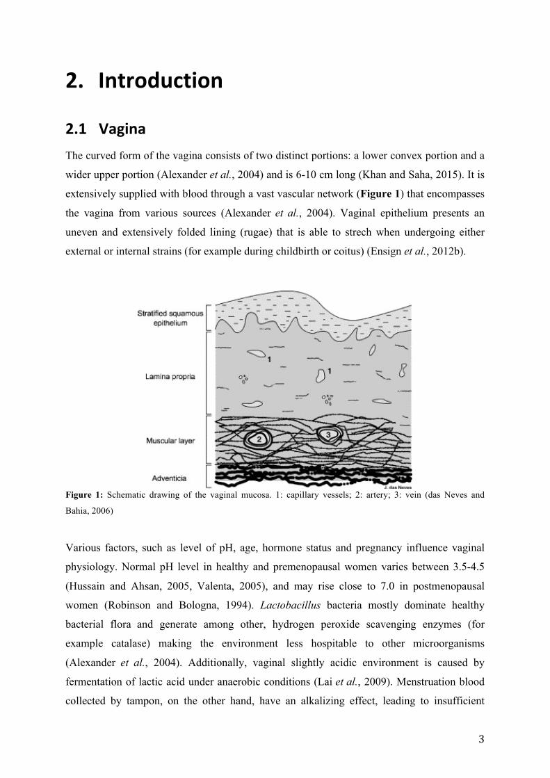
Figure 1: Schematic drawing of the vaginal mucosa. 1: capillary vessels; 2: artery; 3: vein (das Neves and
Bahia, 2006)
Various factors, such as level of pH, age, hormone status and pregnancy influence vaginal
physiology. Normal pH level in healthy and premenopausal women varies between 3.5-4.5
(Hussain and Ahsan, 2005, Valenta, 2005), and may rise close to 7.0 in postmenopausal
women (Robinson and Bologna, 1994). Lactobacillus bacteria mostly dominate healthy
bacterial flora and generate among other, hydrogen peroxide scavenging enzymes (for
example catalase) making the environment less hospitable to other microorganisms
(Alexander et al., 2004). Additionally, vaginal slightly acidic environment is caused by
fermentation of lactic acid under anaerobic conditions (Lai et al., 2009). Menstruation blood
collected by tampon, on the other hand, have an alkalizing effect, leading to insufficient
4
protective properties of Lactobacillus (Alexander et al., 2004). The following induces
pathogen bacterial colonization, thus increasing the vaginal pH. Also, the presence of semen
(pH 7.0 – 8.0) turns slightly acidic vaginal environment to somewhat basic by raising normal
pH level (Vermani and Garg, 2000, Alexander et al., 2004). Maintenance of natural pH is
important to avoid microbial growth and vaginal infections.
Female reproductive hormone (estrogen) controls the thickness of the vaginal epithelium
(Alexander et al., 2004). Small amount of estrogen leads to dryness and vaginal atrophy,
while constant level of the hormone keeps the thickness of epithelium lining stable. The level
of estrogen declines with increasing age (for example in post-menopausal women), which
commonly leads to discomfort and unpleasant nuisances (Khan and Saha, 2015). However,
the thickness of vaginal epithelium increases during puberty, reaches a plateau, followed by a
decline during menopause (Justin-temu et al., 2004).
Vaginal epithelium appears to be the primary physical barrier with a protective function. Its
stratified construction (25 layers thick with estrogen present) makes it hard for toxins and
small organism to invade the basement of membrane (Alexander et al., 2004).
2.1.1 Vaginal mucus
The vaginal mucus is a heterogeneous mesh network of mucin fibers of a gel-like appearance
(Lai et al., 2010). The mucus is essentially composed of 90-95% water, 1-2% mucin, and
other low-content constituents such as cells, bacteria, lipids, salts, proteins and
macromolecules (Lai et al., 2009, das Neves et al., 2011b).
A single mucin is a long fiber, 5-10 nm in diameter, flexible and highly glycosylated protein
(Lai et al., 2010), however several mucin fibers self-condense into network and the diameter
of formed mesh-spaces is estimated at 20-200 nm (das Neves et al., 2011b) and is able to
increase up to 340 nm (Lai et al., 2010). Mucin fibers also have short hydrophobic domains
(lipid-coated, non-glycosylated and cysteine-rich domains) interspersed between long
glycosylated regions. The negatively charged glycosylated domains likely repel each other,
but the hydrophobic domains may cause mucins to self-condense and/or bundle together thus
creating a network with bigger pore sizes (Lai et al., 2010). In this manner, mucins that are
small constituents of mucus lining present an excellent line of defense, as it was mentioned
earlier. Mucus is continuously secreted which induces shedding of foreign particles and
limiting their residence time on the surface. Being aware and being able to predict mucus
5
clearance time, presents an opportunity that might be exploited in developing nanoparticles
for vaginal administration, where one might be able to penetrate the first line of defense at
rates faster than mucus renewal (Ensign et al., 2012a).
In addition to shedding, mucus gel traps molecules by forming polyvalent adhesive
interactions. Hydrophobic interactions between large particles and lipophilic parts of mucin
contribute to bundling of mucin strands into thick cables resulting in immobilization of
foreign particles (Lai et al., 2009).
2.1.2 Vagina as a site for drug delivery The conventional route of administration is preferred, however, under certain conditions local
treatment is chosen. For example, in treatment of vaginal microbial, fungal and viral
infections local drug delivery route will be preferential due to the proximity to cite of action
and ability to escape systemic drug effect. In addition, such application averts hepatic first
pass metabolism that allows administration of a safer lower therapeutic dose. Drugs that are
poorly absorbed after oral administration can be delivered via vaginal route of administration
as well (Hussain and Ahsan, 2005). Easily accessible local application may enhance
compliance regimen by increasing the intervals between the doses (Alexander et al., 2004).
Furthermore, large surface area due to folded rugae presents a promising site for vaginal drug
delivery (das Neves and Bahia, 2006).
However, vaginal drug delivery route encounters for some limitations. Firstly, such treatment
is gender specific, and secondly, vaginal permeability is strongly influenced by estrogen
concentrations (Alexander et al., 2004). Changes in environment arise certain challenges in
development of delivery systems for local application. Examples of factors that may affect
vaginal drug delivery (das Neves et al., 2011a):
1. Menstrual cycle; Escalated shedding of vaginal fluids during menstruation may hinder
residence time of a drug formulation and make it hard to apply. In addition, menstrual
cycle has an effect on vaginal pH (increases) and epithelial layer (thickening)
(Valenta, 2005). In post-menopausal women, for example, decrease in epithelial
thickness will change the drug absorption rates
2. Intravaginal practices; Daily and excessive douching can for example disrupt the
effect of intended prolonged release
6
3. Health; Reoccurring vaginal infections not only disturb natural microflora but also
affect normal vaginal pH gradient which plays significant role in drug absorption thus
important for drug delivery systems
4. Sexual activity; Increased sexual activity predisposes to specific cautions during
treatment, for example regulation of drug administration timeframe (hours, minutes,
before or after coitus). During penile penetration the formed friction may disturb for
example mucus-entangled particles and lead to shedding of the latter. Compared to the
non-stimulated state where level of secretion is regular, lubrication efficiency
increases during sexual arousal
Another important fact to consider while developing formulations for vaginal therapy is
consumer´s preferences. It should be odorless and colorless, non-leaking and avoid causing
the feeling of messiness and fullness (Vermani and Garg, 2000). Most of all, the product and
its metabolites should be non-toxic, biodegradable, not cause local irritation, burning, itching
or swelling and not interfere with normal immune functions. The convenience of application
and dosage regimen plays an essential role in development.
The search for modified and improved treatment using vaginal delivery route is in progress
and constantly new approaches are being developed.
2.2 Mucoadhesion vs. mucus-‐penetration Adherence to the surface and penetration through the biological barrier to the underlying
epithelial layer (the site of action) is the aim when developing nanosystems for mucosal
surface (das Neves et al., 2011b, das Neves et al., 2012). The significant advantage is the
prolonged residual time that can benefit the total drug payload to the surface and underlying
layers (das Neves et al., 2011b). On the other hand, the prolonged retention time and nanosize
may contribute to the uptake by off-target epithelial cells or other cell types present at the
mucosal surface, or even cross the mucosal barrier and continue its migration through the
surrounding tissue (das Neves et al., 2011b).
7
2.2.1 Mucoadhesion
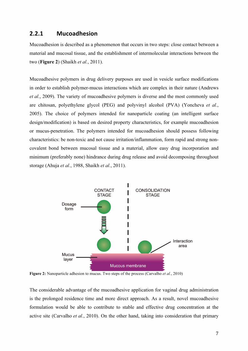
Mucoadhesion is described as a phenomenon that occurs in two steps: close contact between a
material and mucosal tissue, and the establishment of intermolecular interactions between the
two (Figure 2) (Shaikh et al., 2011).
Mucoadhesive polymers in drug delivery purposes are used in vesicle surface modifications
in order to establish polymer-mucus interactions which are complex in their nature (Andrews
et al., 2009). The variety of mucoadhesive polymers is diverse and the most commonly used
are chitosan, polyethylene glycol (PEG) and polyvinyl alcohol (PVA) (Yoncheva et al.,
2005). The choice of polymers intended for nanoparticle coating (an intelligent surface
design/modification) is based on desired property characteristics, for example mucoadhesion
or mucus-penetration. The polymers intended for mucoadhesion should possess following
characteristics: be non-toxic and not cause irritation/inflammation, form rapid and strong non-
covalent bond between mucosal tissue and a material, allow easy drug incorporation and
minimum (preferably none) hindrance during drug release and avoid decomposing throughout
storage (Ahuja et al., 1988, Shaikh et al., 2011).
Figure 2: Nanoparticle adhesion to mucus. Two steps of the process (Carvalho et al., 2010)
The considerable advantage of the mucoadhesive application for vaginal drug administration
is the prolonged residence time and more direct approach. As a result, novel mucoadhesive
formulation would be able to contribute to stable and effective drug concentration at the
active site (Carvalho et al., 2010). On the other hand, taking into consideration that primary
8
vaginal defense mechanism is mucus clearance mucoadhesive, formulations will simply lack
time to discharge therapeutic agents and provide the optimal therapeutic effect (Knowles and
Boucher, 2002, Ensign et al., 2012a).
2.2.2 Mucus penetration
It was considered that mucus gels sterically exclude pathogens and other particles that are
larger than the estimated pore sizes in the mesh network. However, it was observed that even
small pathogens (50 nm) can be “captured” by adhesive interactions with mucin. Due to
disturbed hydrophobic interactions, followed by pore size reduction, particle free diffusion
slows down significantly (Lai et al., 2009).
Small polymeric nanoparticles (around 100 nm) have shown to be less diffusive than the
larger ones (200-500 nm) (Lai et al., 2007, Ensign et al., 2012a). This paradox can be
explained by turning attention to mucus structure. Small molecules pass easily through
narrow channels but are retained in small pockets of mucin network (das Neves et al., 2012).
Analogically, large particles will diffuse easily thorough wide channels with reduced
viscosity. However, it was observed that small viruses could get fast and efficiently through
the first line of defense and thereby infect underlying epithelial cells (Lai et al., 2010). It was
also noted that viruses, which were able to rapidly penetrate mucus lining, were densely
coated with both positive and negative charges, thus creating a hydrophilic and net-neutral
shell that minimizes mucoadhesive interactions (Lai et al., 2007). The physicochemical
characteristics that govern the rapid transport of specific viruses allow them to avoid
mucoadhesion. Applying the knowledge on essential properties of a virus to the development
of nanoparticles for drug delivery, may improve local treatment of vaginal infections.
Considering uneven vaginal epithelium (rugae), much of the folded lining can be left
untreated and unprotected. Mucuoadhesive particles are excluded from the rugae because they
are trapped in the upper mucosal layer (Ensign et al., 2012b). Mucus-penetrating particles
have shown to provide more homogenous distribution than the conventional mucoadhesive
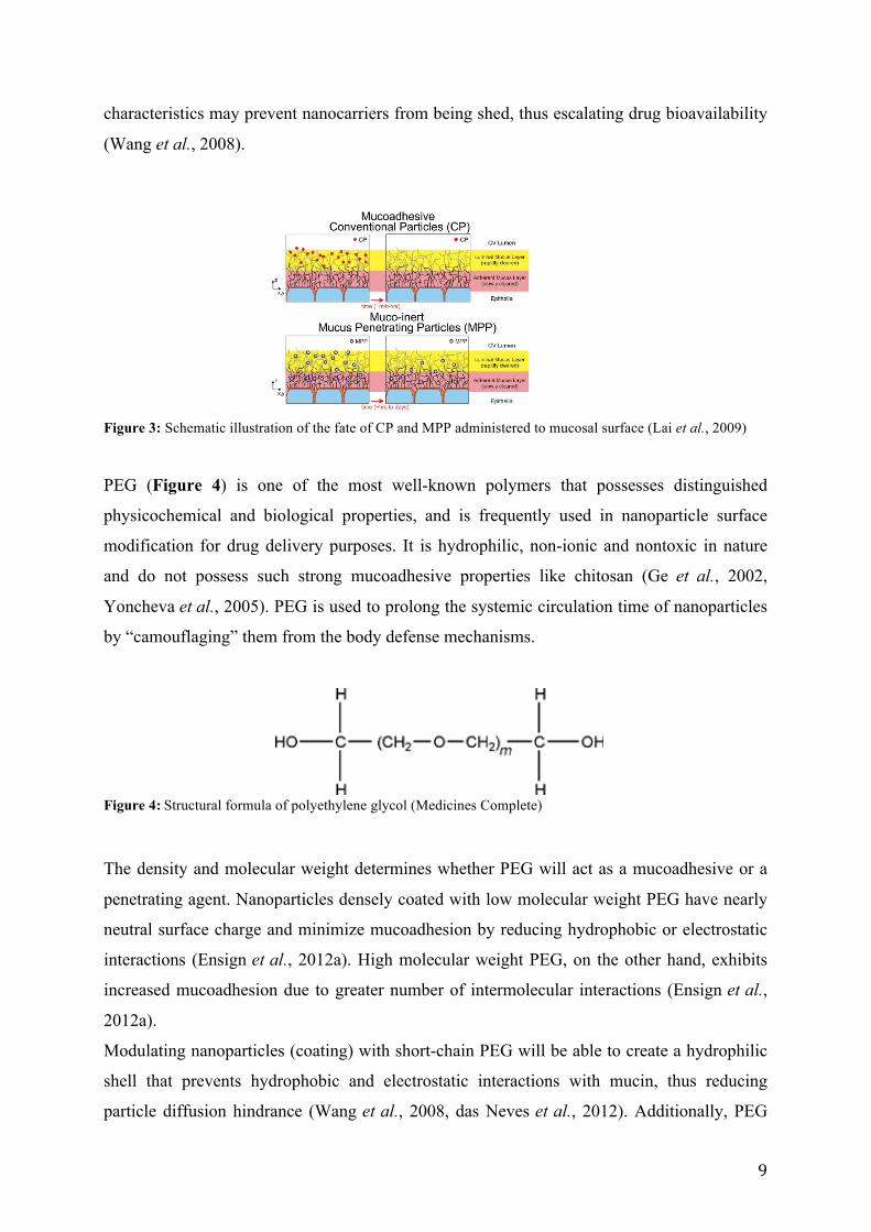
nanoparticles (Figure 3) thus leading to increased local drug delivery (Ensign et al., 2012b).
However, uniform distribution of a system is not yet enough to boost drug bioavailability.
Avoiding mucus entrapment and rapidly penetrating first line of defense is beneficial in
reaching underlying epithelial cells (Lai et al., 2007). Manipulating formulation
9
characteristics may prevent nanocarriers from being shed, thus escalating drug bioavailability
(Wang et al., 2008).
Figure 3: Schematic illustration of the fate of CP and MPP administered to mucosal surface (Lai et al., 2009)
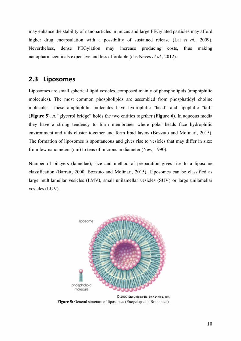
PEG (Figure 4) is one of the most well-known polymers that possesses distinguished
physicochemical and biological properties, and is frequently used in nanoparticle surface
modification for drug delivery purposes. It is hydrophilic, non-ionic and nontoxic in nature
and do not possess such strong mucoadhesive properties like chitosan (Ge et al., 2002,
Yoncheva et al., 2005). PEG is used to prolong the systemic circulation time of nanoparticles
by “camouflaging” them from the body defense mechanisms.
Figure 4: Structural formula of polyethylene glycol (Medicines Complete)
The density and molecular weight determines whether PEG will act as a mucoadhesive or a
penetrating agent. Nanoparticles densely coated with low molecular weight PEG have nearly
neutral surface charge and minimize mucoadhesion by reducing hydrophobic or electrostatic
interactions (Ensign et al., 2012a). High molecular weight PEG, on the other hand, exhibits
increased mucoadhesion due to greater number of intermolecular interactions (Ensign et al.,
2012a).
Modulating nanoparticles (coating) with short-chain PEG will be able to create a hydrophilic
shell that prevents hydrophobic and electrostatic interactions with mucin, thus reducing
particle diffusion hindrance (Wang et al., 2008, das Neves et al., 2012). Additionally, PEG
10
may enhance the stability of nanoparticles in mucus and large PEGylated particles may afford
higher drug encapsulation with a possibility of sustained release (Lai et al., 2009).
Nevertheless, dense PEGylation may increase producing costs, thus making
nanopharmaceuticals expensive and less affordable (das Neves et al., 2012).
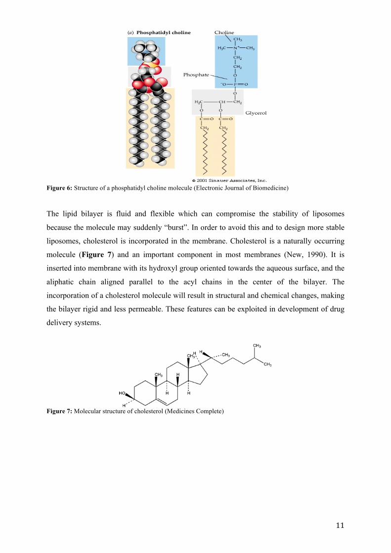
2.3 Liposomes Liposomes are small spherical lipid vesicles, composed mainly of phospholipids (amphiphilic
molecules). The most common phospholipids are assembled from phosphatidyl choline
molecules. These amphiphilic molecules have hydrophilic “head” and lipophilic “tail”
(Figure 5). A “glycerol bridge” holds the two entities together (Figure 6). In aqueous media
they have a strong tendency to form membranes where polar heads face hydrophilic
environment and tails cluster together and form lipid layers (Bozzuto and Molinari, 2015).
The formation of liposomes is spontaneous and gives rise to vesicles that may differ in size:
from few nanometers (nm) to tens of microns in diameter (New, 1990).
Number of bilayers (lamellae), size and method of preparation gives rise to a liposome
classification (Barratt, 2000, Bozzuto and Molinari, 2015). Liposomes can be classified as
large multilamellar vesicles (LMV), small unilamellar vesicles (SUV) or large unilamellar
vesicles (LUV).
Figure 5: General structure of liposomes (Encyclopædia Britannica)
11
Figure 6: Structure of a phosphatidyl choline molecule (Electronic Journal of Biomedicine)
The lipid bilayer is fluid and flexible which can compromise the stability of liposomes
because the molecule may suddenly “burst”. In order to avoid this and to design more stable
liposomes, cholesterol is incorporated in the membrane. Cholesterol is a naturally occurring
molecule (Figure 7) and an important component in most membranes (New, 1990). It is
inserted into membrane with its hydroxyl group oriented towards the aqueous surface, and the
aliphatic chain aligned parallel to the acyl chains in the center of the bilayer. The
incorporation of a cholesterol molecule will result in structural and chemical changes, making
the bilayer rigid and less permeable. These features can be exploited in development of drug
delivery systems.
Figure 7: Molecular structure of cholesterol (Medicines Complete)

12
2.3.1 Liposomes as drug delivery system
Liposomes were first proposed as biological carriers in 1971 (Gregoriadis et al., 1971). The
ability of liposomes to function as drug carriers depends on factors such as physiochemical
membrane properties, the nature of compositional elements, size, surface charge and lipid
organization (Bozzuto and Molinari, 2015). The greatest value of liposomes is that they are
composed of natural constituents and can nearly be tailor-made in order to achieve the desired
properties, both chemically and structurally (Singh and Lillard Jr, 2009). Liposomes can be
designed to be target-specific and release its content only under favorable conditions (for
example specific pH value or temperature). The release timeframe may be prolonged if
needed to establish sustained drug discharge over a period of hours or even days at the site of
action (Singh and Lillard Jr, 2009). The latter can be achieved with surface modifications
and/or using biodegradable materials. Taking into consideration that certain amount
(compared to local treatment) of an active ingredient taken orally is required to achieve and
thereafter maintain therapeutic effect, the development of “smart” pharmaceutics predisposes
to dose reduction and improving of bioavailability.
Liposome properties as variation in size and composition provide a unique opportunity to
incorporate active ingredients both on the outer membrane, inside the phospholipid bilayer
and within the aqueous core. Liposomes can be constructed in such a manner that will ensure
the best encapsulation and targeted delivery of a therapeutic agent.
As a result of their properties, liposomes have already been used as drug delivery systems in
treatment and prevention of vaginal viral infections and cervical cancer, though the need for
new formulations is persistent (Vanić and Škalko-Basnet, 2013).
2.3.2 Preparation of liposomes
Liposomes can be prepared using several methods: mechanical method, methods based on
replacement of organic solvent(s) by aqueous media and methods based on detergent removal
(Wagner and Vorauer-Uhl, 2011). The thin film method, a type of mechanical method, is a
widely used technique that produces heterogeneous population of multilamellar liposomes,
where vesicle size is influenced by the lipid charge (Wagner and Vorauer-Uhl, 2011, Bozzuto
and Molinari, 2015). A convenience of this method is that it can be applied for various lipid
compositions. Further it is easy to perform and high encapsulation of both lipid and aqueous

13
soluble substances can be achieved, since the molecules are amphiphilic in nature and high
lipid concentration may be used (Wagner and Vorauer-Uhl, 2011). However, the scale of
production is limited and not well suited for industrial manufacturing (Wagner and Vorauer-
Uhl, 2011).
2.4 Vaginal infection
2.4.1 General
The human body presents extraordinary machinery that is capable of self-inspection and
control. Physical barriers (skin) as well as biological barriers (pH) shelter our body from
external exposure. Symbiosis with microorganisms (microbiota) that reside in the body of a
host is favorable for both the recipient and microorganisms (Reid et al., 2011). During healthy
state, vagina is colonized with microbiota, for example Lactobacilli that make intravaginal pH
(3.5 - 4.5) slightly acidic due to lactic fermentation (Petrova et al., 2013). These relations are
valuable (symbiosis) and benefit both the host and organisms.
On entering the cervicovaginal tract, viruses compromise the acidic pH, epithelial barrier,
mucus lining and innate immune system. This activates an immune response. The latter
consists of four general steps (Kumamoto and Iwasaki, 2012):
1. Recognition of virus by innate immune system, thus leading to activation of defense
mechanisms, for example secretion of cytokines
2. Processing and presentation of the virus antigens by adaptive immunity
3. Elimination of a pathogen
4. Establishing long-term memory
Vaginal infections are not limited to viruses (for example HIV and HPV), but can also be
caused by pathogenic bacteria (E.coli) and yeast (Candidas) with their own disease
progression.
14
2.4.2 Human Papillomavirus (HPV)
HPV is an infectious disease, which infects a wide variety of organisms including humans.
Current way of contamination is close skin-to-skin intimate contact which makes it the most
common sexually transmitted diseases in both genders (Mohammad and Zargar, 2014).
Physico-clinical manifestations are anogenital warts which vary in size and complexity (King
et al., 2013).
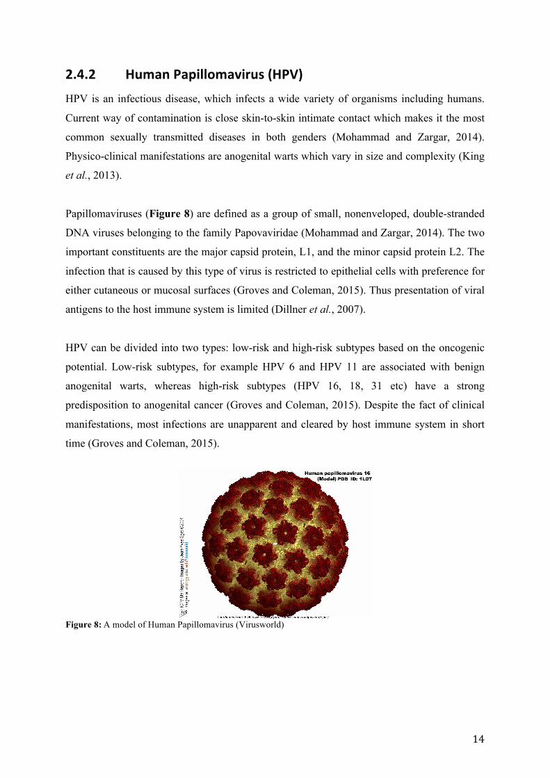
Papillomaviruses (Figure 8) are defined as a group of small, nonenveloped, double-stranded
DNA viruses belonging to the family Papovaviridae (Mohammad and Zargar, 2014). The two
important constituents are the major capsid protein, L1, and the minor capsid protein L2. The
infection that is caused by this type of virus is restricted to epithelial cells with preference for
either cutaneous or mucosal surfaces (Groves and Coleman, 2015). Thus presentation of viral
antigens to the host immune system is limited (Dillner et al., 2007).
HPV can be divided into two types: low-risk and high-risk subtypes based on the oncogenic
potential. Low-risk subtypes, for example HPV 6 and HPV 11 are associated with benign
anogenital warts, whereas high-risk subtypes (HPV 16, 18, 31 etc) have a strong
predisposition to anogenital cancer (Groves and Coleman, 2015). Despite the fact of clinical
manifestations, most infections are unapparent and cleared by host immune system in short
time (Groves and Coleman, 2015).
Figure 8: A model of Human Papillomavirus (Virusworld)
15
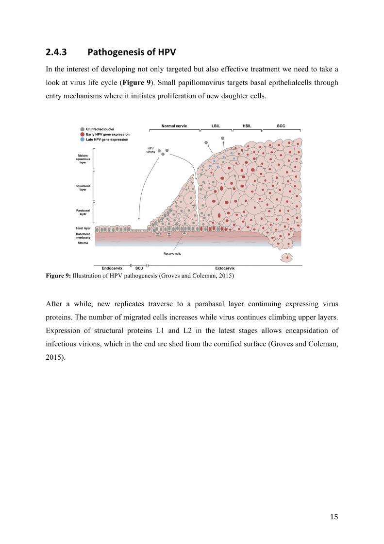
2.4.3 Pathogenesis of HPV
In the interest of developing not only targeted but also effective treatment we need to take a
look at virus life cycle (Figure 9). Small papillomavirus targets basal epithelialcells through
entry mechanisms where it initiates proliferation of new daughter cells.
Figure 9: Illustration of HPV pathogenesis (Groves and Coleman, 2015)
After a while, new replicates traverse to a parabasal layer continuing expressing virus
proteins. The number of migrated cells increases while virus continues climbing upper layers.
Expression of structural proteins L1 and L2 in the latest stages allows encapsidation of
infectious virions, which in the end are shed from the cornified surface (Groves and Coleman,
2015).
16
2.4.4 Current treatment of HPV infections
HPV infections are difficult to cure, because of the high re-occurrence rate (King et al.,
2013). Current treatment of human papillomavirus includes:
1. Vaccination as a preventative measure, composed of virus-like particles (Gardasil® and
Cervarix®) (Dillner et al., 2007)
2. Electrocoagulation; the use of intense heat generated by electric current
3. Cryotherapy; use of extreme cold and
4. Laser ablation or local surgery.
Alternative topical treatment is Veregene® ointment (podophyllotoxin, sinecathechin), 5-
fluorouracil and trichloroacetic acid that are applied only on lesions thus avoiding healthy
tissue (Foldvari and Kumar, 2012). Interferon alpha is a currently approved treatment of
anogenital warts and can be administered as intramuscular (for treating exophytic, visible
lesions) or intralesional injection(Foldvari and Kumar, 2012). ZyclaraTM and AldaraTM that
contain Imiquimod, an IFN inducing agent, are also used for treatment of external and
perianal warts (Foldvari and Kumar, 2012).
Nonetheless, these treatments (except vaccination) are effective as long as the disease is
localized (Mohammad and Zargar, 2014). The largest limitation of injection include pain and
systemic exposure thus elevating the possibility of side effect occurrence (King et al., 2013).
Topical treatment on the other hand will contribute to better compliance from the patient side,
avoid first metabolic passage and thereafter degradation by liver enzymes, and make localized
approach more accessible.
There have been made attempts to expand the possibility of topical application of IFNs using
such conventional formulations as creams and gels with an active pharmaceutical ingredient
(Foldvari, 2010, Foldvari and Kumar, 2012). Unfortunately, the results were neither
conclusive nor consistent. This demonstrates a persistent need for improving existing drug
delivery systems.
17
2.5 Interferon α-‐2b (IFN α-‐2b)
2.5.1 General
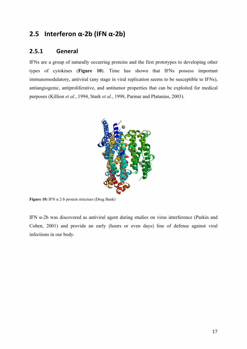
IFNs are a group of naturally occurring proteins and the first prototypes to developing other
types of cytokines (Figure 10). Time has shown that IFNs possess important
immunomodulatory, antiviral (any stage in viral replication seems to be susceptible to IFNs),
antiangiogenic, antiproliferative, and antitumor properties that can be exploited for medical
purposes (Killion et al., 1994, Stark et al., 1998, Parmar and Platanias, 2003).
Figure 10: IFN α 2-b protein structure (Drug Bank)
IFN α-2b was discovered as antiviral agent during studies on virus interference (Parkin and
Cohen, 2001) and provide an early (hours or even days) line of defense against viral
infections in our body.
18
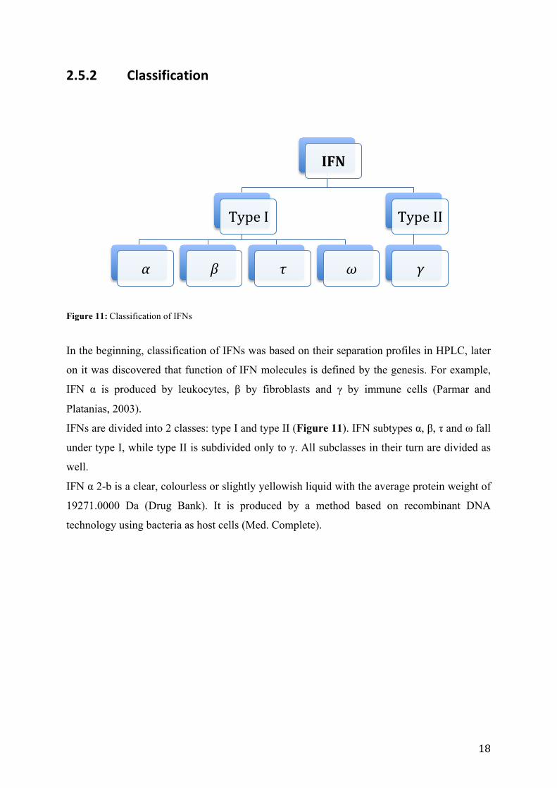
2.5.2 Classification
Figure 11: Classification of IFNs
In the beginning, classification of IFNs was based on their separation profiles in HPLC, later
on it was discovered that function of IFN molecules is defined by the genesis. For example,
IFN α is produced by leukocytes, β by fibroblasts and γ by immune cells (Parmar and
Platanias, 2003).
IFNs are divided into 2 classes: type I and type II (Figure 11). IFN subtypes α, β, τ and ω fall
under type I, while type II is subdivided only to γ. All subclasses in their turn are divided as
well.
IFN α 2-b is a clear, colourless or slightly yellowish liquid with the average protein weight of
19271.0000 Da (Drug Bank). It is produced by a method based on recombinant DNA
technology using bacteria as host cells (Med. Complete).
IFN
Type I
α β τ ω
Type II
γ
19
2.5.3 Application
IFNs have a wide range of application. They target several viral diseases such as chronic
hepatitis B and C, HPV and show promising anticancer activities (Hamidi et al., 2007, Med.
Complete).
On the other hand, IFNs have certain limitations as short circulation time and unwanted
effects of non-targeted tissues (Hamidi et al., 2007). It is worth noticing that interferons are
not absorbed from the gastrointestinal tract, and when attached to large molecules, reduce the
rate of excretion and increase the plasma concentration (Med. Complete).
Improving existing formulations, by for example using “smart” technologies the application
range widens. That will give an additional freedom in designing and choosing the appropriate
treatment. IFN-containing liposomes have been evolving and developing for many years and
the application range is still growing (Hamidi et al., 2007).

20
21
3. Aims of the study The aim of this study was the development and characterization of mucus-penetrating
liposomes as a model for antiviral drugs in localized vaginal delivery. Surface modified
vesicles (liposomes) were expected to encapsulate sufficient drug amount and enhance
penetrating properties at a vaginal site.
The aims were divided as following:
• Development of mucus-penetrating liposomes by modification of liposomal surface
• Characterization of liposomes in respect to size, polydispersity, surface charge and
entrapment efficiency
• In vitro drug release testing

22
23
4. Materials and Methods
4.1 Materials • Acetic acid (glacial) anhydrous GR for analysis, Merck KGaA, Darmstadt, Germany
• Ammonium acetate, VWR International, Leuven, Belgium
• Chloroform, Sigma-Aldrich, Chemie GmbH, Steinheim, Germany
• Cholesterol (from Lanolin) for GC, Sigma-Aldrich, Chemie GmbH, Steinheim,
Germany
• Di-Sodium hydrogen phosphate dehydrate GR for analysis, Merck KGaA, Darmstadt,
Germany
• Distilled water
• Ethanol 96% (v/v), Sigma-Aldrich, Chemie GmbH, Steinheim, Germany
• (Ethylenedinitrilo)tetraacetic acid, disodium salt dehydrate (EDTA dinatriumsalt),
Merck KGaA, Darmstadt, Germany
• IntronA® 50 million IU/ml injection fluid in multiple dose pen (active pharmaceutical
ingredient Interferon alpha-2b), MSD AS, Drammen, Norway
• Lipoid S 100 (soyabean lecithin, >94% phosphatidylcholine), Lipoid GMBH,
Ludwigshafen, Germany
• Methanol CHROMASOLV® for HPLC, Sigma-Aldrich, Chemie GmbH, Steinheim,
Germany
• N-(Carbonyl-methoxypolyethylene glycol-2000)-1,2-distearyl-sn-glycero-3-
phosphoethanolamine, (sodium salt), Lipoid GMBH, Ludwigshafen, Germany
• Polysorbatum 80, Norsk Medisinal Depot, Harstad, Norway
• Sephadex® G-25, for molecular biology, DNA grade superfine, Sigma-Aldrich,
Chemie GmbH, Steinheim, Germany
• Sodium chloride for AT, Sigma-Aldrich, Chemie GmbH, Steinheim, Germany
• Sodium dihydrogen phosphate monohydrate, Merck KGaA, Darmstadt, Germany
• Triton® X-100, Sigma-Aldrich, Chemie GmbH, Steinheim, Germany
• VeriKine™ Human IFN α Multi-Subtype ELISA Kit, Pestka Biomedical Laboratories
Inc, PBL Assay Science, Piscataway, USA
24
4.2 Computer programs • Nicomp Particle Sizing System, CW 388 version 1.68
• Soft Max Pro®, Molecular Devices Corporation
• UV-Visible Chem Station Software, Agilent Technologies
• Zetasizer Software
4.3 Instruments • Agilent 8453 UV-visible spectroscopy system, Agilent technologies, Santa Clara,
USA
• Branson 1510, Bath sonicator, Branson Ultrasonics, Danbury, USA
• Büchi rotavapor R-124, Büchi, Flawil, Switzerland
• Büchi Vacuum Controller B-721, Büchi, Flawil, Switzerland
• Büchi Vac® V-500 vacuum pump, Büchi, Flawil, Switzerland
• Büchi waterbath B-480, Büchi, Flawil, Switzerland
• Julabo Refrigerated/Heating circulator F12-ED, Julabo Labortechnik GmbH,
Seelbach, Germany
• Metrohm 744 pH Meter, Metrohm AG, Herisau, Switzerland
• Microplate spectrophotometer SpectraMAX 190, Molecular Devices, Sunnyvale, USA
• PermeGea Ink, Diffusion cells and Systems, Hellertown, USA
• Sartorius BP2HD, Analytical Scale, VWR International, Oslo, Norway
• Submicron Particle Sizer, model 370, Nicomp, Santa Barbara, USA
• Vortex Genie 2™, Bender & Hobein AG, Zurich, Switzerland
• Zetasizer Malvern, Malvern Zetasizer Nano L, Oxford, UK
4.4 Equipment • Cellophane, Bringmann, Wendelstein, Germany
• Filter 0.22 µm non-sterile syringe filters, Pall Life Sciences, Acrodisc®, Cornwall, UK
• Glass wool superfine, Assistent®, Kebolab, Darmstadt, Germany
• Microtubes, 6×50 mm, Borosilicate Glass, Disposable Culture tubes, Kimble Chase,
Vineland, USA
25
• Nuclepore® Track-etched Membranes, 0.2 µm, 0.4 µm, 0.8 µm, Nuclepore®
Polycarbonate (PC), Whatman International Ltd, Whatman House, Kent, UK
• Round bottom flask, 50 ml, NS 29/32, Boro 3.3, VWR, Darmstadt, Germany
4.5 Methods
4.5.1 Preparation of IFN α-‐2b buffer
IFN α-2b buffer was prepared by dissolving NaCl (15 g), Na2HPO4 × 2H2O (3.6 g), NaH2PO4
× H2O (2.6 g), Polysorbatum 80 (0.2 g) and EDTA (0.2 g) in distilled water, and the volume
was adjusted to 2 L. Measured pH was 6.77.
4.5.2 Preparation of IFN α-‐2b solution
IFN α-2b solution (10 million IU) was transferred from dose pen to a 5 ml volumetric flask
and diluted with buffer prepared for IFN α-2b. The concentration of the IFN α-2b solution
was calculated to be 2 million IU/ml.
4.5.3 Preparation of empty liposomes
Liposomes were prepared by thin film method (New, 1990). In brief, Lipoid S 100 (200 mg),
PEG-2000 (36.3 mg) and Cholesterol (10 mg) were weighed in the round bottom flask and
dissolved in methanol and chloroform solution (1:1). Using rotoevaporator, for at least 90 min
at 50 mm Hg and 51°C, the solvent composition was evaporated and thin lipid layer observed.
Lipid composition in the round bottom flask was flushed with nitrogen for 1 min to make sure
that all solvent was evaporated. The remaining film was re-suspended with buffer solution
prepared for IFN α-2b and shaken vigorously in order to dislodge all the film. If necessary,
vortex was used. Liposomal suspension was stored in the refrigerator (4-8°C) overnight prior
to further use.
26
4.5.4 Preparation of liposomes with IFN α-‐2b
Liposomes containing IFN α-2b were prepared using the same method described above, only
liposomal film was re-suspended in 5 ml of IFN α-2b solution. Liposomal suspension with
IFN α-2b was stored in the refrigerator (4-8°C) overnight prior to further use.
4.5.5 Vesicle size reduction
Liposomal suspension was extruded through 0.8 µm, 0.4 µm and 0.2 µm polycarbonate
filters. Extrusion was performed 5 times on each filter. Extruded liposomes were stored in the
refrigerator (4-8°C) overnight prior to further use.
4.5.6 Particle size analysis
The analysis of liposomal particle size was performed by photon correlation spectroscopy
(Nicomp model 370). In order to avoid interference, microtubes that were used in particle size
analysis were sonicated for 10 min in ultrasonic bath and then rinsed twice with distilled,
filtered water (0.2 µm pore size syringe filter) prior to further use. Small amounts of the
liposome dispersions were diluted with freshly filtered distilled water to achieve the intensity
of approximately 250-350 kHz (Ingebrigtsen and Brandl, 2002). All preparations were done
in a laminar airflow bench. Each sample was analyzed for 3 cycles with time duration 10 min
each. Gaussian and NICOMP distribution analysis were used accordingly.
4.5.7 Zeta potential determination
The zetasizer capillary cells were rinsed with 96% ethanol (one time) and filtered water (3
times) prior to experiment conduction. The liposome samples were diluted 1:19 with filtered
water. Zeta potential was measured for 3 cycles with a voltage of 4 mV.
27
4.5.8 Gel column preparation
Gel column was prepared by blending Sephadex G-25® (15 g) with 120 ml IFN α-2b buffer
(Gel Filtration Bok). The components were gently stirred in a beaker and placed for swelling
overnight at 4°C prior to further use. Before packing, the mixture of Sephadex was brought to
room temperature (23-24°C) and the opening on the bottom of burette was covered with an
adequate amount of glass wool. The viscous mixture was transferred to a burette in a
continuous speed to avoid formation of air bubbles. The column was equilibrated with 100 ml
of a buffer solution and stored in room temperature prior to further use.
4.5.9 Separation of a free drug
Before applying the sample, the top of the column was freed from buffer to avoid further
dilution of the active ingredient. Liposomal IFN α-2b solution was applied evenly on the top
of the column and thereafter was pulled further into the column by gravitational force. After
gel separation, fractions containing liposomes were determined by UV-spectrophotometer.
The wavelength was set to 205 nm.
4.5.10 Preparation of 0.3 % Triton buffer
In this experiment, Triton buffer (Yang et al., 2006) is needed for lysing liposomes for further
analysis. It was prepared from 300 mg Triton X-100 solved in buffer solution for IFN α-2b in
a volumetric flask. The volume was adjusted to 100 ml and stored at a room temperature prior
to further use.
4.5.11 Enzyme-‐linked immunoassay (ELISA)
Preparation of samples includes merging and dilution of liposomal fractions. IFN α-2b
standards (10 000 pg/ml) (ELISA) were diluted to appropriate concentrations with IFN α-2b
buffer.
Wash solution concentrate (50 ml) (ELISA) was diluted with distilled water up to 1 L in a
volumetric flask and stored at a room temperature prior to further use. Diluted HRP solution
was in its turn prepared by blending HRP concentrate (80 µL) (ELISA) and concentrate
28
diluent (12 ml) (ELISA). Diluted antibody solution was prepared by merging antibody
concentrate (120 µL) (ELISA) and dilution buffer (12 ml) (ELISA).
• Step 1
Samples, standards and blank (100 µl) were applied in wells, thereafter covered with plate
sealer and incubated for 1 h. After 1 h the content was emptied and wells washed once
with diluted wash buffer.
• Step 2
Diluted antibody solution (100 µL) was added to each well, covered with plate sealer and
incubated for 1 h. After 1 h the content was emptied and wells washed 3 times with
diluted wash buffer.
• Step 3
Diluted HRP (100 µL) solution was added to each well, covered with plate and incubated
for 1 h. During this hour TMB substrate solution was brought to room temperature. After
1 h the wells were emptied and washed 4 times with diluted wash buffer.
• Step 4
TMB substrate solution (100 µL) was added to each well. The plate was covered with
aluminium foil and incubated in dark for 15 min.
• Step 5
After 15 min, 100 µL of stop solution was added. The drug content of samples was
determined spectrophotometrically at 450 nm by microplate reader.
4.5.12 Preparation of acetate buffer
Acetate buffer was prepared by dissolving 38.55 g of ammonium acetate (CH3COONH4) in
distilled water, afterwards 35 ml of glacial acetic acid (C2H4O2) was added and the volume
adjusted to 1L with distilled water (Ph.Eur). Measured pH 4.51.
4.5.13 In vitro drug release study
Before use, Franz Diffusion cells were washed once with methanol (30 min) and twice with
distilled water (30 min). The acceptor chambers were 12.0 and 12.1 ml. The temperature was
set to 37°C and cellophane membrane was soaked in acetate buffer for at least 30 min prior to
use. The reception chamber was filled with acetate buffer and covered with pre-soaked
cellophane membrane. Samples (600 µl) were applied in the donor cells and the system was

29
completely sealed. Samples (500 µl) were collected after 1, 2, 3, 4, 5, 6, 7 and 8 h. An equal
amount of buffer was added to replace extracted sample. Drug amount was assessed by
ELISA kit.

30
31
5. Results and discussion
5.1 Liposome characterization
5.1.1 Liposomal size
The most common methods for size reduction are sonication, extrusion and high-pressure
homogenization (Bozzuto and Molinari, 2015). Extrusion is characterized as size reduction by
passing through a membrane with a defined pore size. Additionally, going from multimodal to
unimodal distribution allows correct size estimation and evaluation. Berger et.al has shown
that small variations in extrusion method (for example continuous or discontinuous extrusion)
gave rise to populations that deviated from the target size (Berger et al., 2001). Their finding
indicated that the choice of extrusion method might influence the outcome. In this project
membrane extrusion was used with pore sizes 800, 400 and 200 nm. The choice of size was
based on vaginal mucus physiology, where the diameter of mesh spaces is estimated to be
between 200 and 340 nm (Lai et al., 2010, das Neves et al., 2011a). Current reduction method
was also used by Karau et.al and Li et.al to yield reproducible 200 nm IFN α-2b liposomes
(Karau et al., 1996, Li et al., 2011). Thus the obtained vesicle size (Table 3) was considered
to be well suited for vaginal drug delivery.
Vesicle size was estimated by photon correlation spectroscopy (PCS) using Gaussian and
NICOMP distribution analysis accordingly. Liposomes re-suspended in distilled water (L1),
IFN buffer (L2) and buffer containing IFN α-2b (L3) were analyzed and liposomal size
distribution was determined before extrusion (Table 1) and after extrusion through 400 nm
(Table 2) and 200 nm (Table 3) pore size membranes.
32
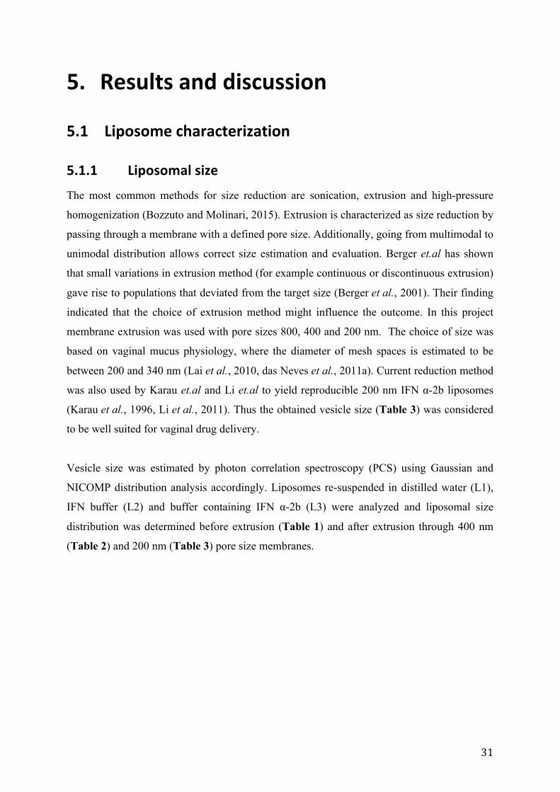
Table 1: The size of pre-extruded liposomes (n=4)
Sample Peak 1
(nm)
Intensity
(%)
Peak 2
(nm)
Intensity
(%)
Peak 3
(nm)
Intensity
(%)
PI
L1 35 ± 25 0.9 181 ± 58 16.4 890 ± 46 82.3 0.40
L2 71 ± 4 8.6 107 ± 1 12.2 905 ± 0.1 81.0 0.67
L3 90 ± 37 9.5 146 ± 37 16.0 845 ± 202 78.0 0.61 L1: liposomes re-suspended in distilled water, L2: liposomes re-suspended in IFN buffer and L3: liposomes containing IFN α-2b. PI - polydispersity index. The values denote the average of three cycles ± SD
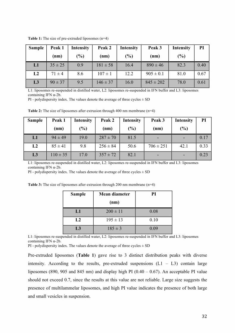
Table 2: The size of liposomes after extrusion through 400 nm membrane (n=4)
Sample Peak 1
(nm)
Intensity
(%)
Peak 2
(nm)
Intensity
(%)
Peak 3
(nm)
Intensity
(%)
PI
L1 94 ± 49 19.0 287 ± 70 81.5 - - 0.17
L2 85 ± 41 9.8 256 ± 84 50.6 706 ± 251 42.1 0.33
L3 110 ± 35 17.0 357 ± 72 82.1 - - 0.23 L1: liposomes re-suspended in distilled water, L2: liposomes re-suspended in IFN buffer and L3: liposomes containing IFN α-2b. PI - polydispersity index. The values denote the average of three cycles ± SD Table 3: The size of liposomes after extrusion through 200 nm membrane (n=4)
Sample Mean diameter
(nm)
PI
L1 200 ± 11 0.08
L2 195 ± 13 0.10
L3 185 ± 3 0.09
L1: liposomes re-suspended in distilled water, L2: liposomes re-suspended in IFN buffer and L3: liposomes containing IFN α-2b. PI - polydispersity index. The values denote the average of three cycles ± SD Pre-extruded liposomes (Table 1) gave rise to 3 distinct distribution peaks with diverse
intensity. According to the results, pre-extruded suspensions (L1 – L3) contain large
liposomes (890, 905 and 845 nm) and display high PI (0.40 – 0.67). An acceptable PI value
should not exceed 0.7, since the results at this value are not reliable. Large size suggests the
presence of multilammelar liposomes, and high PI value indicates the presence of both large
and small vesicles in suspension.
33
Liposomes were first extruded through 800 nm membrane as an additive measure in order to
minimize vesicle resistance and avoid spillage in further extrusion steps. Particle size was not
measured during this step.
We could clearly observe that the average vesicle size and PI was reduced after extrusion
through 400 nm membrane for liposomes re-suspended in water (L1) and liposomes after
extrusion through 200 nm membrane (L3), but not for liposomes re-suspended in IFN buffer
(L2) (Table 1 and Table 2). However there are still some smaller vesicles in formulation with
the size range between 85 and 110 nm (Table 2). Liposomes containing only IFN buffer
(Table 2) showed an additional third peak (706 nm) with a relatively high intensity (42.1 %)
and there is no clear size reduction observed. Particular liposomal behavior is unexpected,
especially after extrusion through 400 nm membrane and might indicate liposomal
agglomeration as a result of inability to form stable vesicles in this size range. Nevertheless,
the desired liposome size has not been reached.
After extrusion through 200 nm membrane, the IFN α-2b containing liposomes display values
close to the desired size and uniformity (Table 3). Experimentally received diameter (185
nm) slightly deviates from the desired (200 nm) but is still in the accepted size range. Tables
1 - 3 illustrate the effectiveness of extrusion as size reduction method and uniformity of
vesicles in a suspension. Liposomal size in formulations L1 and L3 decreases and displays a
more uniform size distribution. We can observe that the size reduces with every extrusion step
and PI value decreases. Uniformity, described by PI, is an important tool in drug delivery due
to possible prediction of entrapment and even drug distribution.
5.1.2 Liposomal zeta potential
Zeta potential presents “the potential difference between the dispersion medium and the
stationary layer of fluid attached to the dispersed particle” (Honary and Zahir, 2013) and is
very important factor in targeting drug delivery. Charged lipids form smaller liposomes with
less lamellae (Wagner and Vorauer-Uhl, 2011) and vice versa. It has earlier been discussed
that charged drug loaded nanocarriers are expected to interact with mucus layer by forming
electrostatic interactions (Honary and Zahir, 2013), thereby nanoparticles with net charge
close to neutral may aid in achieving mucus-penetrating properties by avoiding interactions
with mucin (Cu and Saltzman, 2008).
34
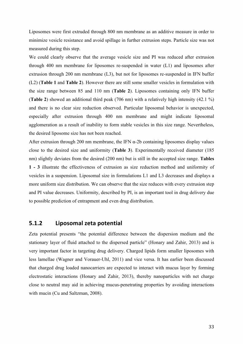
Table 4: Zeta potential values of liposomes (n=4)
Zeta potential (mV)
Sample L1 L2 L3
Pre-extruded -20.5 ± 0.8 -10.7 ± 2.3 -11.4 ± 1.4
400 nm -17.4 ± 1.4 -10.5 ± 0.9 -12.2 ± 1.4
200 nm -15.1 ± 1.3 -11.1 ± 1.4 -12.2 ± 1.4 L1: are liposomes re-suspended in distilled water, L2: liposomes re-suspended in IFN buffer and L3: liposomes containing IFN α-2b. The values denote the average of three cycles ± SD
The net charge of pre-extruded non-drug loaded liposomes (L1) presented in Table 4 is
negative (-20.5 ± 0.8 mV), however, the charge decreases alongside vesicle size (-15.1 ± 1.3
mV). Zeta potential of liposomes re-dispersed in IFN buffer (L2), on the other hand, remains
approximately the same in pre-extruded (-10.7 ± 2.3 mV) and in extruded suspensions (-11 ±
1.4) (Table 4). The outcome (-12.2 ±1.4 mV) deviates from the expected (close to neutral)
and might influence mucus-penetrating properties of IFN α-2b-loaded liposomes, because
negative charge can establish electrostatic interactions with the mucus layer. There is no
difference in surface charge between L2 and L3 after extrusion through 200 nm membrane. It
indicates that the charge remains the same despite the addition of IFN α-2b.
5.1.3 IFN α-‐2b entrapment
Separation of liposome encapsulated and free IFN α-2b is an important step in vesicle
characterization, since the outcome allows estimating drug-loading capacity. This knowledge
will aid in formulation design and may propose lower drug amount for an improved
therapeutic effect, for example vaginal administration route.
As described in section 4.5.8, liposomal formulation containing IFN α-2b was separated
through gel column. Separation principle is based on size exclusion, where large molecules
are expected to elute first, and small molecules (free drug) last. Drifting through gel pores,
small molecules use more time on eluting, large particles, on the other hand, evade tiny
“pockets” and appear earlier. Figure 12 and Figure 13 demonstrate gel separation of
nanoparticle suspension where we can observe liposomes appearing already in fraction 20.
The peak is reached after 23 fractions (Figure 12). Majority of liposomes are eluted in a few
fractions, confirming size uniformity. However, a short elongation after liposome elution was
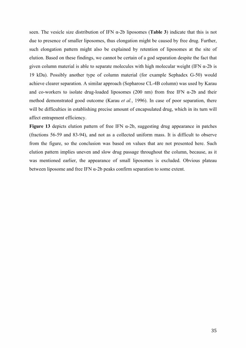
35
seen. The vesicle size distribution of IFN α-2b liposomes (Table 3) indicate that this is not
due to presence of smaller liposomes, thus elongation might be caused by free drug. Further,
such elongation pattern might also be explained by retention of liposomes at the site of
elution. Based on these findings, we cannot be certain of a god separation despite the fact that
given column material is able to separate molecules with high molecular weight (IFN α-2b is
19 kDa). Possibly another type of column material (for example Sephadex G-50) would
achieve clearer separation. A similar approach (Sepharose CL-4B column) was used by Karau
and co-workers to isolate drug-loaded liposomes (200 nm) from free IFN α-2b and their
method demonstrated good outcome (Karau et al., 1996). In case of poor separation, there
will be difficulties in establishing precise amount of encapsulated drug, which in its turn will
affect entrapment efficiency.
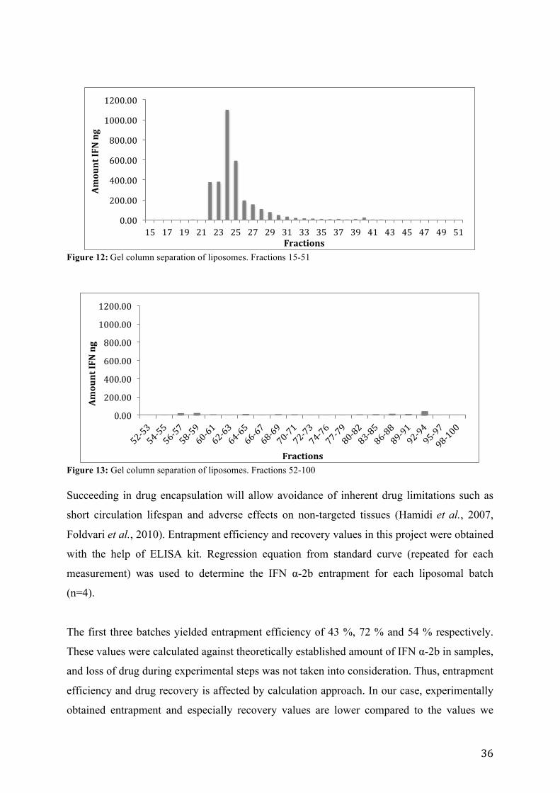
Figure 13 depicts elution pattern of free IFN α-2b, suggesting drug appearance in patches
(fractions 56-59 and 83-94), and not as a collected uniform mass. It is difficult to observe
from the figure, so the conclusion was based on values that are not presented here. Such
elution pattern implies uneven and slow drug passage throughout the column, because, as it
was mentioned earlier, the appearance of small liposomes is excluded. Obvious plateau
between liposome and free IFN α-2b peaks confirm separation to some extent.
36
Figure 12: Gel column separation of liposomes. Fractions 15-51
Figure 13: Gel column separation of liposomes. Fractions 52-100 Succeeding in drug encapsulation will allow avoidance of inherent drug limitations such as
short circulation lifespan and adverse effects on non-targeted tissues (Hamidi et al., 2007,
Foldvari et al., 2010). Entrapment efficiency and recovery values in this project were obtained
with the help of ELISA kit. Regression equation from standard curve (repeated for each
measurement) was used to determine the IFN α-2b entrapment for each liposomal batch
(n=4).
The first three batches yielded entrapment efficiency of 43 %, 72 % and 54 % respectively.
These values were calculated against theoretically established amount of IFN α-2b in samples,
and loss of drug during experimental steps was not taken into consideration. Thus, entrapment
efficiency and drug recovery is affected by calculation approach. In our case, experimentally
obtained entrapment and especially recovery values are lower compared to the values we
0.00
200.00
400.00
600.00
800.00
1000.00
1200.00
15 17 19 21 23 25 27 29 31 33 35 37 39 41 43 45 47 49 51
Amount IFN ng
Fractions
0.00
200.00
400.00
600.00
800.00
1000.00
1200.00
Amount IFN ng
Fractions
37
would have received if correct total values were used. Moreover, the measurements seem to
be quite variable while more uniform results are expected. This is an unwelcome feature in
developing new formulations since it will be impossible to predict amount of incorporated
active ingredient. It is useful to mention that variation (43 %, 72 % and 54 %) might be
questionable, because we are not confident it would be present if we have used correct value
of total drug amount. Consequently, the need of corroboration of entrapment efficiency
variation is required.
We ran an additional experiment where actual drug amount in sample was considered. The
entrapment efficiency was found to be 88 % with a drug recovery of 97 %. Unfortunately, due
to the fact that this is based on a single experiment (n=1), the reliability is questionable;
nevertheless we could have recommended such approach for further investigation. However,
our findings are in accordance with results presented by Yang and co-workers who prepared
reproducible liposomes with entrapment efficiency over 80 % (Yang et al., 2006). Although,
they have used different preparation method (multiple step hydration-dilution technique),
homogenization to reduce the size and ultracentrifugation to separate drug-loaded liposomes
from free IFN α-2b. Karau et.al, have investigated the effect of lipid composition (das Neves
et al.) and size reduction method (homogenization vs. extrusion) of liposomes on IFN α-2b
entrapment efficiency (Karau et al., 1996). They have found that liposomes with negative
charge resulted in increased IFN α-2b entrapment compared to neutral liposomal composition.
In our case, negative charge is not optional because of the potential interactions with mucin
fibers during vaginal mucus penetration. Karau et.al have also shown that less drug was lost
throughout extrusion than during homogenization (Karau et al., 1996).. Foldvari et.al, on the
other hand, have demonstrated that multilammelar liposomes (50-200 nm) prepared by
modified solvent evaporation method yielded vesicles with high IFN α-2b incorporation
degree (91.7 ± 2.2%) (Foldvari and Moreland, 1997). Despite the differences in preparation
methods, our outcome seems to be comparable with the results demonstrated by Foldvari
et.al, indicating the effectiveness and potential of using liposomes for IFN α-2b entrapment.
To confirm experimentally received high encapsulation efficiency (88%) and reproducibility,
the experiment must be repeated. By all means, such high entrapment percentage is a
desirable result, since drug amount needed to establish good therapeutic effect, decrease
application frequency and extend drug release time will be reduced.
38
5.2 In vitro release of IFN α-‐2b Over the years, Franz diffusion cell system has become one of the most widely used methods
for measuring in vitro drug release. It provides some insight in relationship between drug,
formulation and the barrier, thus being useful for designing novel formulations (Ng et al.,
2010). Against this background, Franz diffusion cell system was chosen to establish
efficiency of IFN α-2b release from liposomes in vitro.
Acetate buffer (pH 4.6) was chosen as a receptor medium based on its preferential pH value,
since healthy vaginal pH varies between 3.5 - 4.5 (Hussain and Ahsan, 2005, Valenta, 2005).
The temperature was set to 37 °C to imitate natural value. The sample was drawn from the
acceptor cell every hour for in total 8 hours. The timeframe of the experiment was set due to
the mucosal physiological properties such as vaginal shedding (das Neves et al., 2011a). The
experiment was run in 3 parallels (data not shown). However collected results were both
inconsistent and unpredictable. Possible source of error are mistakes in the experimental
performance. Seemingly, little has been done on establishing diffusion degree of IFN α-2b
encapsulated liposome molecules through mucus lining. On this basis it is difficult to discuss
the anticipated results and draw a conclusion regarding possible origin of experimental
mistakes.
39
6. Conclusion Surface modified (PEGylated) liposomes containing IFN α-2b prepared by thin film method
showed to yield high drug entrapment. The method is easy to perform and produces highly
reproducible uniform liposomes. Vesicle size reduction by extrusion method proved to be
suitable both in regard to size distribution and in maintaining sufficient entrapment efficiency.
PEGylated liposomes appear to be well suited as IFN α-2b carriers.
Designing mucus-penetrating drug carriers for vaginal delivery will allow more direct
approach by bringing the active ingredient closer to the sight of action. This approach
presents an alternative to painful and unpleasant invasive current treatment and opens up to
treatment of non-visible lesions.

40
41
7. Perspectives PEGylated IFN α-2b containing liposomes prepared during this project will be further
analyzed in regard to characterization and properties as a drug delivery system. IFN α-2b
entrapment efficiency and reproducibility of the method needs to be confirmed as well as in
vitro drug release testing should be repeated. Furthermore, this experiment should be
extended to ex vivo testing, and the stability of the drug delivery system in the presence of
vaginal fluid simulant and the in vivo safety should be determined before animal studies.

42
43
8. References Drug Bank. Recombinant Interferon Alfa-‐2b. Retrieved 07.03.2015.
http://www.drugbank.ca/drugs/db00105 Electronic Journal of biomedicine. Phosphatidyl choline. Retrieved 27.04.2015.
http://biomed.uninet.edu/2009/n3/erhan.html Encyclopædia Britannica. Structure of liposomes. Retrieved 27.04.2015.
http://www.britannica.com/EBchecked/topic/342808/lipid/images-‐videos Gel Filtration Book. Principles and Methods. Retrieved 02.12.2014.
http://sevierlab.vet.cornell.edu/resources/Gel_Filtration_Book.pdf Medicines Complete. Interferon Alfa. Retrieved 20.04.2015.
https://www.medicinescomplete.com/mc/ahfs/current/a301061.htm?q=interferon alpha 2-‐b&t=search&ss=text&p=1 -‐ _hit
Medicines complete. Cholesterol. Retrieved 20.04.2015.
https://www.medicinescomplete.com/mc/excipients/current/images/ExccholesterolC001_default.png
Medicines Complete. Polyethylene glycol. Retrieved 20.04.2015.
https://www.medicinescomplete.com/mc/excipients/current/1001943522.htm?q=polyethylene glycol&t=search&ss=text&p=1 -‐ _hit
European Pharmacopoeia Online 8.4. Retrieved 18.02.2015.
http://online6.edqm.eu/ep804/ Virusworld. A model of Human Papillomavirus. Retrieved 27.04.2015.
http://www.virology.wisc.edu/virusworld/viruslist.php?virus=hpv Ahuja, A., Khar, R. K. and Ali, J. (1988). "Mucoadhesive drug delivery systems". Drug
development and industrial pharmacy 23 (5): 489–515 Alexander, N. J., Baker, E., Kaptein, M., Karck, U., Miller, L. and Zampaglione, E. (2004).
"Why consider vaginal drug administration?". Fertility and Sterility 82 (1): 1-‐12 Andrews, G. P., Laverty, T. P. and Jones, D. S. (2009). "Mucoadhesive polymeric platforms
for controlled drug delivery". European Journal of Pharmaceutics and Biopharmaceutics 71 (3): 505-‐518
Barratt, G. M. (2000). "Therapeutic applications of colloidal drug carriers".
Pharmaceutical Science & Technology Today 3 (5): 163-‐171 Berger, N., Sachse, A., Bender, J., Schubert, R. and Brandl, M. (2001). "Filter extrusion of
liposomes using different devices: comparison of liposome size, encapsulation
44
efficiency, and process characteristics". International Journal of Pharmaceutics 223 (1–2): 55-‐68
Bozzuto, G. and Molinari, A. (2015). "Liposomes as nanomedical devices". International
journal of nanomedicine 25 (10): 975-‐999 Carvalho, F. C., Bruschi, M. L., Evangelista, R. C. and Gremião, M. P. D. (2010).
"Mucoadhesive drug delivery systems". Brazilian Journal of Pharmaceutical Sciences 46 (1): 1-‐18
Cu, Y. and Saltzman, W. M. (2008). "Controlled Surface Modification with
Poly(ethylene)glycol Enhances Diffusion of PLGA Nanoparticles in Human Cervical Mucus". Molecular pharmaceutics 6 (1): 173-‐181
Das Neves, J., Amiji, M. and Sarmento, B. (2011a). "Mucoadhesive nanosystems for
vaginal microbicide development: friend or foe?". Wiley Interdisciplinary Reviews: Nanomedicine and Nanobiotechnology 3 (4): 389-‐399
Das Neves, J. and Bahia, M. F. (2006). "Gels as vaginal drug delivery systems".
International Journal of Pharmaceutics 318 (1–2): 1-‐14 Das Neves, J., Bahia, M. F., Amiji, M. M. and Sarmento, B. (2011b). "Mucoadhesive
nanomedicines: characterization and modulation of mucoadhesion at the nanoscale". Expert opinion on drug delivery 8 (8): 1085-‐1104
Das Neves, J., Rocha, C. M., Goncalves, M. P., Carrier, R. L., Amiji, M., Bahia, M. F. and
Sarmento, B. (2012). "Interactions of microbicide nanoparticles with a simulated vaginal fluid". Molecular pharmaceutics 9 (11): 3347-‐3356
Dillner, J., Arbyn, M. and Dillner, L. (2007). "Translational Mini-‐Review Series on
Vaccines: Monitoring of human papillomavirus vaccination". Clinical & Experimental Immunology 148 (2): 199-‐207
Ensign, L. M., Schneider, C., Suk, J. S., Cone, R. and Hanes, J. (2012a). "Mucus penetrating
nanoparticles: biophysical tool and method of drug and gene delivery". Advanced Materials 24 (28): 3887-‐3894
Ensign, L. M., Tang, B. C., Wang, Y. Y., Tse, T. A., Hoen, T., Cone, R. and Hanes, J. (2012b).
"Mucus-‐penetrating nanoparticles for vaginal drug delivery protect against herpes simplex virus". Science Transltional Medicine 4 (138): 138-‐179
Foldvari, M. (2010). "Biphasic vesicles: a novel topical drug delivery system". Journal of
Biomedical Nanotechnology 6 (5): 543-‐557 Foldvari, M., Badea, I., Wettig, S., Baboolal, D., Kumar, P., Creagh, A. L. and Haynes, C. A.
(2010). "Topical Delivery of Interferon Alpha by Biphasic Vesicles: Evidence for a Novel Nanopathway across the Stratum Corneum". Molecular pharmaceutics 7 (3): 751-‐762
45
Foldvari, M. and Kumar, P. (2012). "Recent progress in the application of nanotechnology for prevention and treatment of human papillomavirus infection". Therapeutic delivery 3 (8): 1005-‐1017
Foldvari, M. and Moreland, A. (1997). "Clinical observations with topical liposome-‐
encapsulated interferon alpha for the treatment of genital papillomavirus infections". Journal of liposome research 7 (1): 115-‐126
Forman, D., De Martel, C., Lacey, C. J., Soerjomataram, I., Lortet-‐Tieulent, J., Bruni, L.,
Vignat, J., Ferlay, J., Bray, F., Plummer, M. and Franceschi, S. (2012). "Global Burden of Human Papillomavirus and Related Diseases". Vaccine 30 (5): F12-‐F23
Ge, H., Hu, Y., Jiang, X., Cheng, D., Yuan, Y., Bi, H. and Yang, C. (2002). "Preparation,
characterization, and drug release behaviors of drug nimodipine-‐loaded poly(ε-‐caprolactone)-‐poly(ethylene oxide)-‐poly(ε-‐caprolactone) amphiphilic triblock copolymer micelles". Journal of pharmaceutical sciences 91 (6): 1463-‐1473
Gregoriadis, G., Leathwood, P. D. and Ryman, B. E. (1971). "Enzyme entrapment in
liposomes". FEBS Letters 14 (2): 95-‐99 Groves, I. J. and Coleman, N. (2015). "Pathogenesis of human papillomavirus-‐associated
mucosal disease". Journal of Pathology 235 (4): 527-‐538 Hamidi, M., Zarrin, A. and Foroozesh, M. (2007). "Novel delivery systems for
interferons". Critical reviews in biotechnology 27 (3): 111-‐127 Honary, S. and Zahir, F. (2013). "Effect of Zeta potential on the Properties of Nano-‐Drug
Delivery systems -‐ A Review (Part 1)". Tropical Journal of Pharmaceutical Research 12 (2): 255-‐264
Hussain, A. and Ahsan, F. (2005). "The vagina as a route for systemic drug delivery".
Journal of Controlled Release 103 (2): 301-‐313 Ingebrigtsen, L. and Brandl, M. (2002). "Determination of the size distribution of
liposomes by SEC fractionation, and PCS analysis and enzymatic assay of lipid content". AAPS PharmSciTech 3 (2): 1-‐7
Justin-‐Temu, M., Damian, F., Kinget, R. and Mooter, G. V. D. (2004). "Intravaginal Gels as
Drug Delivery Systems". Journal of Women's Health 13 (7): 834-‐844 Karau, C., Petszulat, M. and Schmidt, P. C. (1996). "Preparation and stability of
interferon-‐α-‐containing liposomes". International Journal of Pharmaceutics 128 (1–2): 89-‐98
Khan, A. B. and Saha, C. (2015). "A Review on Vaginal Drug Delivery System". Rajiv
Gandhi University of Health Sciences Journal of Pharmaceutical Sciences 4 (4): 142-‐147
46
Killion, J. J., Fishbeck, R., Bar-‐Eli, M. and Chernajovsky, Y. (1994). "Delivery of interferon to intracellular pathways by encapsulation of interferon into multilamellar liposomes is independent of the status of interferon receptors". Cytokine 6 (4): 443-‐449
King, M., Kumar, P., Michel, D., Batta, R. and Foldvari, M. (2013). "In vivo sustained
dermal delivery and pharmacokinetics of interferon alpha in biphasic vesicles after topical application". European Journal of Pharmaceutics and Biopharmaceutics 84 (3): 532-‐539
Knowles, M. R. and Boucher, R. C. (2002). "Mucus clearance as a primary innate defense
mechanism for mammalian airways". The Journal of clinical investigation 109 (5): 571-‐577
Kumamoto, Y. and Iwasaki, A. (2012). "Unique features of antiviral immune system of
the vaginal mucosa". Current Opinion in Immunology 24 (4): 411-‐416 Lai, S. K., O'hanlon, D. E., Harrold, S., Man, S. T., Wang, Y. Y., Cone, R. and Hanes, J. (2007).
"Rapid transport of large polymeric nanoparticles in fresh undiluted human mucus". Proceedings of the Natlonal Academy of Sciences 104 (5): 1482-‐1487
Lai, S. K., Wang, Y. Y. and Hanes, J. (2009). "Mucus-‐penetrating nanoparticles for drug
and gene delivery to mucosal tissues". Advanced drug delivery reviews 61 (2): 158-‐171
Lai, S. K., Wang, Y. Y., Hida, K., Cone, R. and Hanes, J. (2010). "Nanoparticles reveal that
human cervicovaginal mucus is riddled with pores larger than viruses". Proceedings of the Natlonal Academy of Sciences 107 (2): 598-‐603
Lasic, D. D. (1998). "Novel applications of liposomes". Trends in Biotechnology 16 (7):
307-‐321 Li, X., Chen, D., Le, C., Zhu, C., Gan, Y., Hovgaard, L. and Yang, M. (2011). "Novel mucus-‐
penetrating liposomes as a potential oral drug delivery system: preparation, in vitro characterization, and enhanced cellular uptake". International journal of nanomedicine 2011 (6): 3151-‐3162
Mohammad, A. a. S. T. a. S. O. and Zargar, A. M. A. (2014). "Review of the current
knowledge on the epidemiology, pathogenesis, and prevention of human papillomavirus infection". European Journal of Cancer Prevention 23 (3): 206–224 Nardis, C., Mosca, L. and Mastromarino, P. (2013). "Vaginal microbiota and viral sexually
transmitted diseases". Ann Ig 25 (5): 443-‐456 New, R. R. C. (1990). "Liposomes a practical approach". Oxford University Press. New
York
47
Ng, S.-‐F., Rouse, J. J., Sanderson, F. D., Meidan, V. and Eccleston, G. M. (2010). "Validation of a Static Franz Diffusion Cell System for In Vitro Permeation Studies". American Association of Pharmacetical Scientists Pharmaceutical Science Technology 11 (3): 1432-‐1441
Parkin, J. and Cohen, B. (2001). "An overview of the immune system". The Lancet 357
(9270): 1777-‐1789 Parmar, S. and Platanias, L. C. (2003). "Interferons: Mechanisms of action and clinical
applications". Current Opinion in Oncology 15 (6): 431-‐439 Petrova, M. I., Van Den Broek, M., Balzarini, J., Vanderleyden, J. and Lebeer, S. (2013).
"Vaginal microbiota and its role in HIV transmission and infection". Federation of European Microbiological Societies 37 (5): 762-‐792
Reid, G., Younes, J. A., Van Der Mei, H. C., Gloor, G. B., Knight, R. and Busscher, H. J. (2011).
"Microbiota restoration: natural and supplemented recovery of human microbial communities". Nature Reviews Microbiology 9 (1): 27-‐38
Robinson, J. R. and Bologna, W. J. (1994). "Vaginal and reproductive system treatments
using a bioadhesive polymer". Journal of Controlled Release 28 (1–3): 87-‐94 Sawant, R. R. and Torchilin, V. P. (2012). "Challenges in development of targeted
liposomal therapeutics". American association ofpharmaceutical scientists Journal 14 (2): 303-‐315
Shaikh, R., Raj Singh, T. R., Garland, M. J., Woolfson, A. D. and Donnelly, R. F. (2011).
"Mucoadhesive drug delivery systems". Journal of Pharmacy and Bioallied Sciences 3 (1): 89-‐100
Singh, R. and Lillard Jr, J. W. (2009). "Nanoparticle-‐based targeted drug delivery".
Experimental and Molecular Pathology 86 (3): 215-‐223 Stark, G. R., Kerr, I. M., Williams, B. R., Silverman, R. H. and Schreiber, R. D. (1998). "How
cells respond to interferons". Annual review of biochemistry 67 227-‐264 Tong, S., Fine, E. J., Lin, Y., Cradick, T. J. and Bao, G. (2014). "Nanomedicine: tiny particles
and machines give huge gains". Annals of Biomedical Engineering 42 (2): 243-‐259 Valenta, C. (2005). "The use of mucoadhesive polymers in vaginal delivery". Advanced
drug delivery reviews 57 (11): 1692-‐1712 Vanić, Ž. and Škalko-‐Basnet, N. (2013). "Nanopharmaceuticals for improved topical
vaginal therapy: Can they deliver?". European Journal of Pharmaceutical Sciences 50 (1): 29-‐41
Vermani, K. and Garg, S. (2000). "The scope and potential of vaginal drug delivery".
Pharmaceutical Science & Technology Today 3 (10): 359-‐364

48
Wagner, A. and Vorauer-‐Uhl, K. (2011). "Liposome Technology for Industrial Purposes". Journal of Drug Delivery 2011 (2011): 1-‐9
Wang, Y.-‐Y., Lai, S. K., Suk, J. S., Pace, A., Cone, R. and Hanes, J. (2008). "Addressing the
PEG Mucoadhesivity Paradox to Engineer Nanoparticles that “Slip” through the Human Mucus Barrier". Angewandte Chemie International Edition 47 (50): 9726-‐9729
Yang, L., Yang, W., Bi, D. and Zeng, Q. (2006). "A novel method to prepare highly
encapsulated interferon-‐alpha-‐2b containing liposomes for intramuscular sustained release". European Journal of Pharmaceutics and Biopharmaceutics 64 (1): 9-‐15
Yoncheva, K., Gomez, S., Campanero, M. A., Gamazo, C. and Irache, J. M. (2005).
"Bioadhesive properties of pegylated nanoparticles". Expert Opinion Drug Delivery 2 (2): 205-‐218

49
Related Documents