Skin-Gut-Breast Microbiota Axes Printed Edition of the Special Issue Published in Journal of Clinical Medicine www.mdpi.com/journal/jcm Lorenzo Drago Edited by

Welcome message from author
This document is posted to help you gain knowledge. Please leave a comment to let me know what you think about it! Share it to your friends and learn new things together.
Transcript
Skin-Gut-Breast Microbiota Axes • Lorenzo Drago
Skin-Gut-Breast Microbiota Axes
Printed Edition of the Special Issue Published in Journal of Clinical Medicine
www.mdpi.com/journal/jcm
Lorenzo DragoEdited by

Skin-Gut-Breast Microbiota Axes


Skin-Gut-Breast Microbiota Axes
Editor
Lorenzo Drago
MDPI • Basel • Beijing • Wuhan • Barcelona • Belgrade • Manchester • Tokyo • Cluj • Tianjin

Editor
Lorenzo Drago
Department of Biochemical
Sciences for Health,
University of Milan
Italy
Editorial Office
MDPI
St. Alban-Anlage 66
4052 Basel, Switzerland
This is a reprint of articles from the Special Issue published online in the open access journal
Journal of Clinical Medicine (ISSN 2077-0383) (available at: https://www.mdpi.com/journal/jcm/
special issues/SGB MA).
For citation purposes, cite each article independently as indicated on the article page online and as
indicated below:
LastName, A.A.; LastName, B.B.; LastName, C.C. Article Title. Journal Name Year, Volume Number,
Page Range.
ISBN 978-3-0365-0898-6 (Hbk)
ISBN 978-3-0365-0899-3 (PDF)
© 2021 by the authors. Articles in this book are Open Access and distributed under the Creative
Commons Attribution (CC BY) license, which allows users to download, copy and build upon
published articles, as long as the author and publisher are properly credited, which ensures maximum
dissemination and a wider impact of our publications.
The book as a whole is distributed by MDPI under the terms and conditions of the Creative Commons
license CC BY-NC-ND.

Contents
About the Editor . . . . . . . . . . . . . . . . . . . . . . . . . . . . . . . . . . . . . . . . . . . . . . vii
Preface to ”Skin-Gut-Breast Microbiota Axes” . . . . . . . . . . . . . . . . . . . . . . . . . . . . . ix
Ivan Kushkevych, Olga Lescanova, Dani Dordevic, Simona Jancıkova, Jan Hosek, Monika
Vıtezova, Leona Bunkova and Lorenzo Drago
The Sulfate-Reducing Microbial Communities and Meta-Analysis of Their Occurrence duringDiseases of Small–Large Intestine AxisReprinted from: J. Clin. Med. 2019, 8, 1656, doi:10.3390/jcm8101656 . . . . . . . . . . . . . . . . . 1
Justyna Pełka-Wysiecka, Mariusz Kaczmarczyk, Agat , Paweł Liskiewicz,
Michał Wronski, Karolina Skonieczna-Zydecka, Wojciech Marlicz, Błazej Misiak, TeresaStarzynska, Jolanta Kucharska-Mazur, Igor Łoniewski and Jerzy Samochowiec
Analysis of Gut Microbiota and Their Metabolic Potential in Patients with SchizophreniaTreated with Olanzapine: Results from a Six-Week Observational Prospective Cohort StudyReprinted from: J. Clin. Med. 2019, 8, 1605, doi:10.3390/jcm8101605 . . . . . . . . . . . . . . . . . 13
Ivan Kushkevych, Dani Dordevic, Peter Kollar, Monika Vıtezova and Lorenzo Drago
Hydrogen Sulfide as a Toxic Product in the Small–Large Intestine Axis and its Role inIBD DevelopmentReprinted from: J. Clin. Med. 2019, 8, 1054, doi:10.3390/jcm8071054 . . . . . . . . . . . . . . . . . 31
Francesco Folino, Luca Ruggiero, Pasquale Capaccio, Ilaria Coro, Stefano Aliberti, Lorenzo
Drago, Paola Marchisio and Sara Torretta
Upper Respiratory Tract Microbiome and Otitis Media Intertalk: Lessons from the LiteratureReprinted from: J. Clin. Med. 2020, 9, 2845, doi:10.3390/jcm9092845 . . . . . . . . . . . . . . . . . 43
Lorenzo Drago, Gian Vincenzo Zuccotti, Carlo Luca Romano, Karan Goswami, Jorge Hugo
Villafane, Roberto Mattina and Javad Parvizi
Oral–Gut Microbiota and Arthritis: Is There an Evidence-Based Axis?Reprinted from: J. Clin. Med. 2019, 8, 1753, doi:10.3390/jcm8101753 . . . . . . . . . . . . . . . . . 69
Lorenzo Drago, Simona Panelli, Claudio Bandi, Gianvincenzo Zuccotti, Matteo Perini and
Enza D’Auria
What Pediatricians Should Know before Studying Gut MicrobiotaReprinted from: J. Clin. Med. 2019, 8, 1206, doi:10.3390/jcm8081206 . . . . . . . . . . . . . . . . . 83
v


About the Editor
Lorenzo Drago is a professor of clinical microbiology at the University of Milan. He was
the director of the laboratory medical hubs in two large Italian hospitals. He is a project
leader—at the Italian and European levels—of microbiological networks focusing on microorganisms
and infections, antibiotic resistance, and microbiota and probiotics. Prof. Lorenzo Drago is
an international leader in laboratory diagnosis, molecular technology, next-generation sequencing
systems, hospital infection control and management, antibiotic resistance, and microbiota studies.
He is a co-founder of the World Association of Infection in Orthopedics and Trauma (WAIOT) and
the former president of the International Society of Microbiota.
vii

Preface to ”Skin-Gut-Breast Microbiota Axes”
The “skin-gut-breast microbiota axis” comprises the network of connections—involving
multiple biological systems—that allows for relational communication between the gut-skin axis,
breast bacteria, and our body. This system is finely regulated, and it is crucial for maintaining the
homeostasis of skin integrity, the gastrointestinal tract, and the central nervous system of humans.
This network of microorganisms is known to be relevant to our health. This book describes the
mechanisms of, opportunities for, and approaches to studying this system and how to harness it
to improve human health.
Lorenzo Drago
Editor
ix


Journal of
Clinical Medicine
Article
The Sulfate-Reducing Microbial Communities andMeta-Analysis of Their Occurrence during Diseasesof Small–Large Intestine Axis
Ivan Kushkevych 1,*, Ol’ga Lešcanová 1, Dani Dordevic 2, Simona Jancíková 2, Jan Hošek 3,
Monika Vítezová 1, Leona Bunková 4 and Lorenzo Drago 5
1 Department of Experimental Biology, Faculty of Science, Masaryk University, Kamenice 753/5, 62500 Brno,Czech Republic; [email protected] (O.L.); [email protected] (M.V.)
2 Department of Plant Origin Foodstuffs Hygiene and Technology, Faculty of Veterinary Hygiene and Ecology,University of Veterinary and Pharmaceutical Sciences, 61242 Brno, Czech Republic;[email protected] (D.D.); [email protected] (S.J.)
3 Regional Centre of Advanced Technologies and Materials, Faculty of Science, Palacky University inOlomouc, 78371 Olomouc, Czech Republic; [email protected]
4 The Department of Environmental Protection Engineering, Faculty of Technology, Tomas Bata University inZlín, 76001 Zlín, Czech Republic; [email protected]
5 Department of Biomedical Sciences for Health, University of Milan, 20122 Milan, Italy;[email protected]
* Correspondence: [email protected]; Tel.: +420-549-495-315
Received: 28 August 2019; Accepted: 9 October 2019; Published: 11 October 2019
Abstract: Sulfate-reducing bacteria (SRB) are often isolated from animals and people with ulcerativecolitis and can be involved in the IBD development in the gut–intestine axis. The background of theresearch consisted of obtaining mixed cultures of SRB communities from healthy mice and mice withcolitis, finding variation in the distribution of their morphology, to determine pH and temperaturerange tolerance and their possible production of hydrogen sulfide in the small–large intestinalenvironment. The methods: Microscopic techniques, biochemical, microbiological, and biophysicalmethods, and statistical processing of the results were used. The results: Variation in the distributionof sulfate-reducing microbial communities were detected. Mixed cultures from mice with ulcerativecolitis had 1.39 times higher production of H2S in comparison with samples from healthy mice.The species of Desulfovibrio genus play an important role in diseases of the small–large intestine axis.Meta-analysis was also used for the observation about an SRB occurrence in healthy and not healthyindividuals and the same as their metabolic processes. Conclusions: This finding is important for itspossible correlation with inflammation of the intestine, where the present of SRB in high concentrationplays a major part. It can be a good possible indicator of the occurrence of IBD.
Keywords: bowel disease; colitis; small–large intestine axis; sulfate reduction; hydrogen sulfide
1. Introduction
Sulfate-reducing bacteria (SRB) represent probably a trigger for the occurrence of inflammatorybowel diseases (IBD) since studies are connecting their presence with these diseases, especially theirmetabolic end product H2S in the gut [1,2]. Other ailments (including rheumatic diseases and withankylosing spondylitis) occur also in their presence [3]. SRB use sulfate as an electron acceptor in theprocess of dissimilatory sulfate reduction. The final product of this process is hydrogen sulfide [4].Constant microorganism cultivation is happening in the large intestine since certain undigested foodremains in it. [1,2]. Around 200 g of digestive material is found in the large intestine of an adulthuman [2,3,5,6]. These bacteria are in the fermentation process can cleave complex organic compounds
J. Clin. Med. 2019, 8, 1656; doi:10.3390/jcm8101656 www.mdpi.com/journal/jcm1
J. Clin. Med. 2019, 8, 1656
and form molecular hydrogen, different acids (acetic and lactic), same as other compounds. Lactic acidbacteria fermentative properties are directly responsible for the production of lactate [4]. Other groupsof microorganisms can also use lactate and acetate, serving as electron donors and carbon sources [7–12].The important role of human physiological processes is their capability to absorb sulfate and developamino acids out of it (cysteine and methionine). The amount of the sulfate present in the intestineis related to human diet [13–16], meaning that it is highly influenced by individual’s eating habits.The importance of daily sulfate intake can be overseen by the fact that staple food commoditiesrepresent high sulfate sources (>10 μmol/g) [13].
Although, sulfate amounts that are not used in amino acid synthesis represent good conditionsfor SRB [1,4,17–21]. SRB needs electron acceptor (sulfate serves this purpose) and they form hydrogensulfide as their final product [22–27]. An exogenic electron donor, including lactate can be alsoused and oxidized to acetate [18,28]. The dominant SRB in the intestine of humans is Desulfovibriogenus [5,22,28]. The studies are emphasized connections between the presence of SRB in the intestinesand the prevalence of ailments, such as cholecystitis, brain abscesses, and abdominal cavity ulcerativeenterocolitis. Sulfate-reducing bacteria are not the only ones that produce H2S in the intestinal content.Numerous bacterial groups convert cysteine to H2S, pyruvate, and ammonia by cysteine desulfhydraseactivity [2–4,12].
Though connections have been found, it is still not clear how these processes are affecting theprevalence of certain ailments. Meta-analysis is used widely in medical research, as in natural science.It is included in systematic reviews as a rigorous method for mapping the evidence gained by manyauthors. The meta-analysis should provide unbiased overviews of multiple results and should assessevidence quality and synthesize it. The first step of a systematic review is the research question thatis deconstructed by sample consideration, the second step is intervention and then come outcomeand comparator. The outcome of the meta-analysis depends on the study field, but in many cases,quantitative results are used [29].
The aim of the research was to compare a variation in the morphological distribution ofsulfate-reducing microbial communities from healthy mice and mice with colitis, their production ofhydrogen sulfide, and to study the occurrence of these bacterial populations during diseases of thesmall–large intestine axis.
2. Experimental Section
2.1. Manipulation with Animals
Male C57Bl/6 mice (20 g ± 2 g) were obtained from the Animal Breeding Facility of MasarykUniversity (Brno, Czech Republic). They were kept under standard conditions (22 ± 2 ◦C, 50 ± 10%relative humidity) and alternating 12 h light/dark cycles. The animals had access to a standard diet anddrinking water ad libitum. Manipulations with the animals were carried out according to the bioethicalrules as per the principles of the “European Convention for the Protection of Vertebrate AnimalsUsed for Experimental and Other Scientific Purposes” adopted in Strasbourg in 1986. The study wasalso approved by the “Commission for the Protection of Animals against Cruelty” and the EthicsCommittee of the University of Veterinary and Pharmaceutical Sciences in Brno, Czech Republic.In total, six animals in two groups (4 + 2 animals in the first and second group, respectively) wererandomly separated and used in this experiment. In the dextran sulfate sodium (DSS) group (n = 4),colitis was induced by administering 5% (w/v) DSS (MP Biomedicals, Illkirch-Graffenstaden, France,MW 36,000–50,000 Da) in drinking water for 7 days. The mice in the intact group (n = 2) receiveddrinking water only. On the last day of the experiment, the animals were killed by decapitationunder isoflurane anesthesia. The isolated distal colonic segments were selected for the analysis of thequalitative and quantitative composition of intestinal microflora of both groups of the animals.
2

J. Clin. Med. 2019, 8, 1656
2.2. Bacterial Mixed Cultures
The material used for the study consisted out of mixed sulfate-reducing bacteria cultures thatwere isolated from feces of healthy and with ulcerative colitis mice. After the autopsy, the sampleswere placed in the tubes. The bacteria were studied as mixed cultures because the aim of the study wasnot the purification of SRB. Mixed cultures were kept at the Laboratory of Anaerobic Microorganismsof the Department of Experimental Biology at Masaryk University (Brno, Czech Republic).
2.3. Cultivation of SRB Cultures
SRB cultures were cultivated according to Kovac and Kushkevych (2017) [30] and Postgate(1984) in a modified Postgate C medium [23]. Mohr’s salt (ammonium iron sulfate hexahydrate,Sigma-Aldrich, Prague, Czech Republic) was used as a simple growth detection. Ferrous salt formsreacted with sulfide produced by SRB (dark black precipitate of FeS) and indicated the presence ofSRB (the presence of dissimilatory sulfate reduction). Due to the method, it was possible to opticallydetermine the presence of metabolic activity qualitatively and quantitatively.
The cultures were kept in medium with Mohr’s salt and without is since color changes are notdesirable for spectrophotometric and turbidimetric methods. In cultures kept in medium withoutMohr’s salt, the SRB can be detected by the sharp smell of hydrogen sulfide same as by optical turbidity.The medium was sterilized (pH 7.5–7.7, Eh = −100 mV). Redox potential was adjusted by Na2S(Sigma-Aldrich, Prague, Czech Republic) and ascorbic acid (Sigma-Aldrich, Prague, Czech Republic).The anoxic atmosphere was ensured by the nitrogen gas addition, inhibiting oxygen from the air todiffuse into the medium. The oxygen proof layer was secured by the addition of paraffin (Sigma-Aldrich,Prague, Czech Republic) drops to each cultivation tube. The strains were able to grow 10 days underthese conditions.
The long storage (up to one month) conditions for cultures were provided by Postgate B mediumwith the addition of Mohr’s salt. In this medium there is always tending of bacteria to descend to thebottom of the tube due to the presence of the precipitate. Bacteria usually stick to the walls of the tubewhen is used modified Postgate C medium.
2.4. Description of Morphology
Microscope Olympus BX50 (lympus, Japan) was used for the observation of cells.Phase-contrast microscopy is a technique that allows images of transparent specimens (living
cells). The advantage of this technique is the possibility to do the measuring without cell killing sincecells can be monitored with real-time motility. The bacterial suspension (a drop) was placed on a glassslide. The slide (cover glass added to the top of bacterial suspension) was analyzed immediately afterimmersion and with 100× objective.
The Gram staining method provides observation of gram-positive and gram-negative bacteriaby differential staining with the use of crystal violet-iodine complex and a safranin counterstain.Gram-positive bacteria appear purple after treatment with alcohol while gram-negative bacteria appearpink. After drying samples were microscopically observed, including oil immersion 100× objective.
Capsule staining. Acidic and basic stains cannot be used for bacterial capsules. Therefore, the bestway to visualize them is to stain the background using an acidic dye (e.g., nigrosine, Congo red) and tostain the cell itself using a basic stain (e.g., crystal violet, safranin, methylene blue). One drop of Congored dye was mixed with one drop of bacterial suspension on a glass slide. After spreading throughoutthe slide and letting dry, it was immersed in hydrochloric acid (4 mol/L) and after a few seconds, it waslet dry again. Subsequently, methylene blue dye was added on the slide and it was let standing forthree minutes. After three minutes, the slide was washed with deionized water, dried, and observedwith immersion oil and 100× objective. The cells were stained blue and their capsules remained whiteand visible on a dark background.
3

J. Clin. Med. 2019, 8, 1656
DAPI (4′,6-diamidino-2-phenylindole) staining is a fluorescent dye, binding by preference to theAT-rich regions of DNA [31]. Microorganisms with thick cell walls can be stained with DAPI afterpermeabilization of the cell wall by ethanol. For this type of microscopy, using a 48-hour old culturewas found most suitable. A 48-h-old cell suspension of a volume 25 μL to 100 μL was diluted in severalml of MiliQ deionized water and washed by vacuum filtration. After washing, the filtration paper withcells was let dry. Consequently, 20 μL of DAPI stain (Sigma-Aldrich, Prague, Czech Republic) wasapplied and the filtration paper with cells was kept in the dark in a refrigerator for 10 min. After that,the filtration paper was washed in water, ethanol, and water, respectively, and let dry. Next, it was puton a glass slide with immersion oil applied both under and over the filtration paper with cells, and theslide was observed in a microscope, using WU filter (Sigma-Aldrich, Prague, Czech Republic) and100× objective.
2.5. pH Tolerance and Temperature Range Test
As measured before, the optimal pH for the cultivation of intestinal SRB is from 7 to 8 [15].The measuring was done by performing a simple pH test. The modified Postgate C medium wasprepared by adjusting various pH values, performed by adding drops of sodium hydroxide (aqueoussolution) and hydrochloric acid (aqueous solution), respectively. CyberScan 510 pH-meter (PreSens,Regensburg, Germany) was used to measure the exact pH values (pH ranged from 4 to 12). Mediawere heated to 37 ◦C in Wasserman tubes inoculums (obtained from healthy and not healthy mice) ofcultures. Paraffin oil (500 μL) was added on the top of the medium to provide an oxygen-proof layer.The optical density of the suspension was measured at 430 nm using spectrophotometer SpectronicsGenesys 5 (Thermo Fisher Scientific, Prague, Czech Republic). Blank samples were media withoutinoculum. Optical density was measured after 24 h of cultivation again. Bacteria were added inEppendorf tubes and placed in thermostats (1-CUBE, Havlickuv Brod, Czech Republic) set at 5, 25,35, 45, 50, and 60 ◦C. Optical density was measured at 430 nm using Spectronic Genesys 5, after 72 hof cultivation.
2.6. Production of Hydrogen Sulfide
Spectrophotometrical methylene blue method was used for measuring the presence of hydrogensulfide in solution [32]. The bacterial suspension (1 mL) was pipetted to 5 mL of aqueous zinc acetate(5 g/L). 2 mL of p-aminodimethylaniline (Sigma-Aldrich, Prague, Czech Republic) solution (0.75 g/L in2 M sulfuric acid) was added immediately and the solution was let stand at room temperature for 5 min.0.5 mL of ferric chloride (FeCl3) (12 g/L in 0.015 M sulfuric acid) solution was consequently added.The solution was centrifuged at 2200 RPM (10 ◦C for 5 min). After centrifuging, the samples lost theoriginal light pink color and had a blue color. The absorbance was measured at 665 nm by SpectronicGenesys 5 spectrophotometer. The procedure for blank sample preparation included preparation thata clear cultivation medium was added in step 1. The concentrations used for calibration solutionsranged from 6 μmol/L to 100 μmol/L (Figure 1).
Figure 1. The calibration used for the determination of sulfide concentrations.
4

J. Clin. Med. 2019, 8, 1656
2.7. Statistical Analysis
Using the experimental data, the basic statistical parameters (M—mean, m—standard error,M ±m) were calculated. The accurate approximation was when p ≤ 0.0533 [33]. Statistical analysiswas done by SPSS 20 statistical software (IBM Corporation, Armonk, NY, USA). Plots were built bysoftware package Origin 7.0 (Northampton, MA, USA).
Meta-analysis consisted of studies found on the WEB OF KNOWLEDGE database. The databasefound 38 studies, from the year 1945 to 2019.considering sulfate-reducing bacteria. Only six studieswere included in the meta-analysis since other studies did not satisfy the specific hypothesis of thestudy. The Review Manager Software (Cochrane, Brno, Czech-Republic) (number 5.3 developedby Cochrane Collaboration) was used. In the included studies the data consisted of the number ofparticipants with the positive occurrence of the SRB bacteria in the group of healthy people andpeople with ulcerative colitis. In other studies, the data consisted of the mean, standard deviation andthe number of the measurements. Heterogeneity was expressed by the I2 test, where the higher I2
represented a higher heterogeneity.
3. Results
The vibrio shape was a dominant shape of the cells, as expected. Though they are very small andthin that makes them very often hard to be observed. These cells were marked as Desulfovibrio sp.Due to their characteristic shape, gram negativity and flagellar motility (Figure 2). Very abundant werealso cells, oval form. Chain and cluster shaped had cocci that were larger than vibrios, same as somerod shape cells were observed too. Rods have almost similar characteristics as cocci. Not abundantlyspirilloid forms of bacteria were present too. They had long shape and were very thin, curved multipletimes (maximum twelve curves) (Figure 2A). They had long, polar flagella that are responsible forrapid movement. Gram-negative bacteria only were not only present in SRB cultures isolated fromrodents (Figure 2B).
Figure 2. Sulfate-reducing bacteria (SRB) mixed culture: native slide (A), Gram staining (B), capsulestaining (C), DAPI staining (D).
5
J. Clin. Med. 2019, 8, 1656
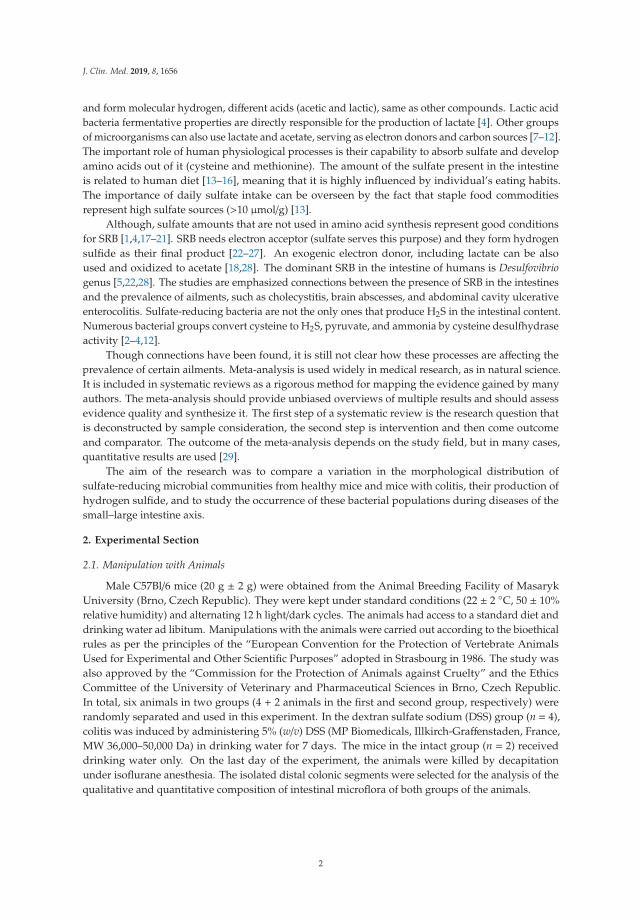
Desulfotomaculum is rod-shaped (stained Gram-positive) (representing non-SRB genera in the gut)can be seen in Figure 2C since it has a short rod oval shape. According to the previous microscopictechnique, cocci can be encapsulated or not. More often encapsulated cocci are present in pairs.The formation of capsules occurs probably due to a non-favorable environment, such as high hydrogensulfide concentrations due to sulfate-reducing bacteria presence. It is important to stress out thatcapsule formation is not defined as SRB characteristic. DAPI (4′,6-diamidino-2-phenylindole) stainingis compliant with the observations made by the previous technique (Figure 2D). The most abundant wasvibrio cell-shape. SRB present in the gut isolate was probably Desulfovibrio sp., according to literaturedata that is describing them as the most frequently isolated species in the intestinal inflammationenvironment. Cocci were confirmed by DAPI staining since they are significantly brighter andlarger than other cells. The findings that DAPI cultures bind to DNA molecules indicate that someoval-shaped have more DNA than others, meaning that they are unrelated to each other. Differentsizes of cocci, gained by previous techniques, is supporting this interpretation. These cells were foundin multiple isolates because thin rods of exceeding length were found by DAPI staining. These cellsrepresent a common microbiome in the intestines that are capable to survive in conditions designed forSRB cultivation.
The fastest bacterial growth and viability, measured spectrophotometrically OD430 (Figure 3),was detected after 24 h of cultivation at 37 ◦C and pH from 8.0 to 9.0. A significant drop in viabilitywas observed at pH 10. The absence of black precipitate was observed in tubes with Mohr’s salt andpH > 10 (Figure 3A). This result is indicating a threshold limit pH ≥ 10 both for sulfate-reducers andother (contaminating) species. The values did not reach zero value but were stabilized at around30–40% of maximum bacterial growth. It means that bacteria were capable to survive and divide atthis pH, reaching an optical density of 0.3. Black precipitate occurred at all pH values, meaning thatbacteria can survive a longer time period before starting to metabolize and produce hydrogen sulfide.The changing of color in the tubes at pH 11 and 12 occurred due to basic conditions. It means that themeasured values of optical density can be explained by the extreme pH effect.
Figure 3. Various pH (A) and temperature (B) influence on relative viability of SRB cultures.
After 72 h of cultivation bacterial growth of all samples was observed. SRB cultures can grow atvarious ranges of temperature conditions, not only at 37 ◦C, though the fastest growth occurred attemperature ranges from 37 ◦C to 45 ◦C. Another observation was that cells survived for three days at50 ◦C and died on the temperatures higher than 60 ◦C and at the temperature of 5 ◦C (no bacterialgrowth, no hydrogen sulfide production, black precipitate not occurred and low OD430 values weremeasured. The growth was slow at a temperature of 25 ◦C. The relative viability values of SRB areshown in Figure 3B.
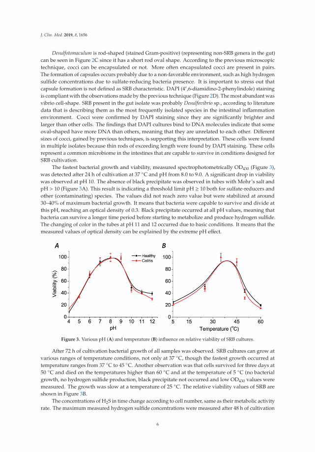
The concentrations of H2S in time change according to cell number, same as their metabolic activityrate. The maximum measured hydrogen sulfide concentrations were measured after 48 h of cultivation
6
J. Clin. Med. 2019, 8, 1656
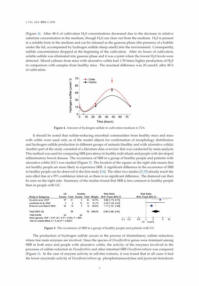
(Figure 4). After 48 h of cultivation H2S concentrations decreased due to the decrease in relativesubstrate concentration in the medium, though H2S can clear out from the medium. H2S is presentin a soluble form in the medium and can be released as the gaseous phase (the presence of a bubbleunder the lid, accompanied by hydrogen sulfide sharp smell) into the environment. Consequently,sulfide concentrations dropped at the beginning of the cultivation. After six hours of cultivation,soluble sulfide was eliminated into gaseous phase and it was a point where the lowest H2S levels weredetected. Mixed cultures from mice with ulcerative colitis had 1.39 times higher production of H2Sin comparison with samples from healthy mice. The maximal difference was 20 μmol/L after 48 hof cultivation.
Figure 4. Amount of hydrogen sulfide in cultivation medium in 72 h.
It should be noted that sulfate-reducing microbial communities from healthy mice and micewith colitis were used only as of the model objects for confirmation of morphology distributionand hydrogen sulfide production in different groups of animals (healthy and with ulcerative colitis).Another part of the study consisted of a literature data overview that was conducted by meta-analysis.This method was used for comparing SRB prevalence in healthy individuals and people with developedinflammatory bowel disease. The occurrence of SRB in a group of healthy people and patients withulcerative colitis (UC) was studied (Figure 5). The location of the square on the right side means thatnot healthy people are more likely to experience SRB. A significant difference in the occurrence of SRBin healthy people can be observed in the first study [34]. The other two studies [5,35] already touch thezero effect line at a 95% confidence interval, so there is no significant difference. The diamond can thenbe seen on the right side. Summary of the studies found that SRB is less common in healthy peoplethan in people with UC.
Figure 5. The occurrence of SRB in a group of healthy people and patients with UC.
The production of hydrogen sulfide occurs in the process of dissimilatory sulfate reduction,where tree main enzymes are involved. Since the species of Desulfovibrio genus were dominant amongSRB in both mice and people with ulcerative colitis, the activity of the enzymes involved in theprocesses of sulfate reduction in Desulfovibrio and other intestinal SRB Desulfomicrobium was compared(Figure 6). In the case of enzyme activity in cell-free extracts, it was found that in all cases it hadthe lower enzymatic activity of Desulfomicrobium sp. phosphotransacetylase and pyruvate-ferredoxin
7

J. Clin. Med. 2019, 8, 1656
activity was more or less the same in Desulfovibrio bacteria. Thus, it can be argued that the activityof Na+/K+ ATPase is the highest of the investigated enzymes in the cell-free extracts of Desulfovibrio.Similar results were observed in soluble fractions. The activity of Na+/K+ ATPase is highest inDesulfovibrio than Desulfomicrobium in all enzymes examined. In the case of sediment fractions, higherNa+/K+ ATPase activity was again found in Desulfovibrio bacteria and no activity was observed inboth Desulfovibrio and Desulfomicrobium in the other investigated enzymes, phosphotransacetylase,and pyruvate-ferredoxin oxidoreductase.
Figure 6. Enzyme activity in Desulfovibrio and Desulfomicrobium.
Thus, the contribution of sulfate-reducing microbial communities, especially of the Desulfovibriogenus, in both groups of healthy people and patients with UC and enzymatic activities of bacterialcells is based on a meta-analysis is obvious. Though, the number of studies is certainly not enough fora stronger conclusion.
4. Discussion
Important factors that influence the intestinal environment are sulfate consumption, sulfideproduction, lactate consumption and acetate accumulation [7–10]. Very often Desulfovibrio genus ispresent in the intestines and feces of people and animals with inflammatory bowel disease, meaningthat this genus plays an important role in the development and occurrence of this ailment. Sulfate isused as a terminal electron acceptor by these bacteria, the same as organic compounds are used aselectron donors in their metabolism [6,7]. Leading us to the conclusion that sulfate in food commodities(some bread, soya flour, dried fruits, brassicas, and sausages, as well as some beers, ciders, and wines)play an important role in the development of bowel disease [13].
The principal component analysis showed that the Desulfovibrio strains from individuals withcolitis grouped in one cluster by biomass accumulation and sulfide production, while the strainsfrom healthy individuals formed another cluster that included the same parameters. A negativecorrelation (Pearson correlations, p< 0.01) was found between sulfate and lactate consumption. Biomassaccumulation and hydrogen sulfide showed lower linear regression (R2). The kinetic parameters,biomass accumulation, and sulfide production have an important role in bowel inflammation, includingulcerative colitis. Acetate produced by SRB probably has a synergy interaction with H2S since sulfateconsumption and lactate oxidation represent minor factors in bowel disease [16].
Optimum growing conditions for the bacteria were provided by the study. The intensive growthof D. piger Vib-7 was observed in the presence of higher electron acceptor and donor concentrations.
8

J. Clin. Med. 2019, 8, 1656
Consequently, the intensive accumulation of sulfide and acetate occurs too. According to previousstudies and literature data, these conditions are the probable cause of ulcerative colitis, leading tobowel cancer. Hydrogen sulfide negatively affects intestinal mucosa, epithelial cells, the growthof colonocytes [4,14–18,36–39], causes phagocytosis, causes the death of intestinal bacteria [4,12,24],and induces hyperproliferation and metabolic abnormalities of epithelial cells [12]. The presence of SRBand high level of metabolites are also connected with colon inflammation [4,6,38]. Hydrogen sulfideconcentrations are regulating the integrity of colonocytes [37–39]. In the samples of individuals withulcerative colitis was also found that SRB sulfide production is higher [5,6]. According to another studydealing with the SRB metabolic process was found that the strains isolated from people with colitisshifted to the right side of the Y-axis by biomass accumulation, sulfate consumption, lactate oxidation,same as hydrogen sulfide and acetate production, in comparison with the strains isolated from healthyindividuals. The percentages were differences observed in shifting to the right side of the Y-axis:biomass accumulation 26%, sulfate consumption 1.5%, and sulfide production 5% [14]. The intestinalmicrobiota is a complex system, interactions occur between clostridia, methanogens, lactic acid bacteria,etc. Though, SRB plays a central role in the development of IBD, including ulcerative colitis [1–3,11].Lactic acid bacteria, methanogens, and many other intestinal microorganisms can be inhibited byhydrogen sulfide produced by SRB [2].
Preservatives added to food often contain sulfur oxides, sulfate polysaccharides (mucin),chondroitin sulfate, carrageenan, and other food commodities represent the source of sulfate and leadto evaluated sulfate intake in the daily diet that leads to increase of hydrogen sulfide concentrationsproduced by SRB. The western diet contains over 16.6 mmol sulfate/day [13] and the feces of about50% of healthy individuals contain SRB (Desulfovibrio: up to 92%) [1,5,24]. On the other hand, theconcentrations of hydrogen sulfide are toxic not only for the intestinal environment but also for theirproducers. The concentrations higher than 6 mM stop the growth of Desulfovibrio, but metabolic activitywas not 100% inhibited (the results supported by cross-correlation and principal component analysis).5 mM concentrations of H2S resulted in two times and eight times longer lag phase and generation time,respectively [18]. It should be noted that clostridia can also produce hydrogen sulfide, but in smallerquantities and can be interacted with SRB [40] Terminal oxidative processes in the large intestine ofhumans can be also included in the activities of SRB. The connections between SRB presence andactivity in the intestine and occurrence of ulcerative colitis were also found in animal studies whereSRB isolated from mice with UC produced 1.14 times (higher hydrogen sulfide production rate candamage aggressively intestinal mucosa) more sulfide ions than SRB isolates from healthy mice [6].
It is of crucial importance that all issues concerning H2S metabolic processes and its influence onthe gastrointestinal environment are well studied and tested. Since it has been observed in animalstudies that H2S-releasing agents can be seen as promising therapeutic agents for many indications [41].H2S is confirmed to represent an important signaling factor for cardiovascular and nervous systemsstatute [42]. The way how cecal musoca protects itself from the toxical effects of H2S is the conversionto thiosulfate. Consequently, these metabolic pathways play an important role in the occurrence ofulcerative colitis [43]. The importance of similar studies can be seen through the fact that mechanismsleading to Chron’s disease still remain unclear [44].
According to meta-analysis, SRB occurs more often in patients with UC. The finding can beexplained by the fact that counts of SRB are lower (though still detectable) in healthy individuals.Oppositely, in patients with developed inflammatory bowel disease, the production of H2S reachestoxic levels and also destroyed its producers (sulfate-reducing bacteria) [15].
5. Conclusions
Sulfate-reducing bacteria are present in various environments and they make a high impact onanimal and human health since their presence is a possible contributing factor in the developmentof inflammatory bowel diseases. Their morphology (vibrio, spiral, rods, and cocci) and diversityare highly influenced by environmental conditions including temperature, pH, oxygen presence and
9

J. Clin. Med. 2019, 8, 1656
substrate availability. Unique in nature is anaerobic sulfate-reducing bacteria metabolism in whichhydrogen sulfide is produced in the process of electron acceptors (mainly sulfate ions) reduction (theprocess of dissimilatory sulfate reduction). The study clearly showed that mixed SRB cultures obtainedfrom healthy and with ulcerative mice were equally polymorphic (the most often vibrio and coccusshape occurred). Though, the production of hydrogen sulfide differs significantly among isolatedcultures. It was observed that isolates from not healthy mice produced higher hydrogen sulfideamounts. This observation is emphasizing correlations between intestine inflammation occurrence andhydrogen sulfide concentrations. The meta-analysis confirmed these correlations. Presently, it is stillnot fully understood the occurrence processes of inflammatory bowel diseases, including ulcerativecolitis. Though, the study is emphasizing one more time that the occurrence of SRB in the sampleswith developed IBD is pointing out the importance of issues concerning sulfate-reducing bacteria.
Author Contributions: Conceptualization, I.K., O.L., and D.D.; methodology, I.K., O.L., and M.V.; validation,S.J., M.V., and D.D.; formal analysis, S.J., M.V., and L.D.; investigation, O.L., I.K.; resources, I.K.; data curation,L.B., D.D.; writing—original draft preparation, I.K., D.D., S.J., and M.V.; writing—review and editing, I.K., J.H.,L.B., and L.D.; visualization, I.K.; supervision, M.V.; project administration, I.K.; funding acquisition, I.K., D.D.,and M.V.
Funding: This research was supported by Grant Agency of the Masaryk University (MUNI/A/0902/2018).
Conflicts of Interest: The authors declare no conflict of interest.
References
1. Gibson, G.R.; Cummings, J.H.; Macfarlane, G.T. Growth and activities of sulphate-reducing bacteria in gutcontents of health subjects and patients with ulcerative colitis. FEMS Microbiol. Ecol. 1991, 86, 103–112.[CrossRef]
2. Gibson, G.R.; Macfarlane, S.; Macfarlane, G.T. Metabolic interactions involving sulphate-reducing andmethanogenic bacteria in the human large intestine. FEMS Microbiol. Ecol. 1993, 12, 117–125. [CrossRef]
3. Cummings, J.H.; Macfarlane, G.T.; Macfarlane, S. Intestinal Bacteria and Ulcerative Colitis. Curr. IssuesIntest. Microbiol. 2003, 4, 9–20. [PubMed]
4. Barton, L.L.; Hamilton, W.A. Sulphate-Reducing Bacteria Environmental and Engineered Systems; CambridgeUniversity Press: Cambridge, UK, 2017.
5. Loubinoux, J.; Bronowicji, J.P.; Pereira, I.A. Sulphate-reducing bacteria in human feces and their associationwith inflammatory diseases. FEMS Microbiol. Ecol. 2002, 40, 107–112. [CrossRef] [PubMed]
6. Kovác, J.; Vítezová, M.; Kushkevych, I. Metabolic activity of sulfate-reducing bacteria from rodents withcolitis. Open Med. 2018, 13, 344–349. [CrossRef] [PubMed]
7. Kushkevych, I.; Vítezová, M.; Fedrová, P.; Vochyanová, Z.; Paráková, L.; Hošek, J. Kinetic properties ofgrowth of intestinal sulphate-reducing bacteria isolated from healthy mice and mice with ulcerative colitis.Acta Vet. Brno 2017, 86, 405–411. [CrossRef]
8. Kushkevych, I.; Fafula, R.; Parak, T.; Bartoš, M. Activity of Na+/K+-activated Mg2+-dependent ATP hydrolasein the cell-free extracts of the sulfate-reducing bacteria Desulfovibrio piger Vib-7 and Desulfomicrobium sp.Rod-9. Acta Vet. Brno 2015, 84, 3–12. [CrossRef]
9. Kushkevych, I.V. Activity and kinetic properties of phosphotransacetylase from intestinal sulfate-reducingbacteria. Acta Biochem. Pol. 2015, 62, 1037–1108. [CrossRef]
10. Kushkevych, I.V. Kinetic Properties of Pyruvate Ferredoxin Oxidoreductase of Intestinal Sulfate-ReducingBacteria Desulfovibrio piger Vib-7 and Desulfomicrobium sp. Rod-9. Pol. J. Microbiol. 2015, 64, 107–114.
11. Loubinoux, J.; Mory, F.; Pereira, I.A.; Le Faou, A.E. Bacteremia caused by a strain of Desulfovibrio related tothe provisionally named Desulfovibrio fairfieldensis. J. Clin. Microbiol. 2000, 38, 931–934.
12. Pitcher, M.C.; Cummings, J.H. Hydrogen sulphide: A bacterial toxin in ulcerative colitis? Gut 1996, 39, 1–4.[CrossRef] [PubMed]
13. Florin, T.H.; Neale, G.; Goretski, S. Sulfate in food and beverages. J. Food Compos. Anal. 1993, 6, 140–151.[CrossRef]
14. Kushkevych, I.; Dordevic, D.; Vítezová, M.; Kollár, P. Cross-correlation analysis of the Desulfovibrio growthparameters of intestinal species isolated from people with colitis. Biologia 2018, 73, 1137–1143. [CrossRef]
10

J. Clin. Med. 2019, 8, 1656
15. Kushkevych, I.; Dordevic, D.; Vítezová, M. Analysis of pH dose-dependent growth of sulfate-reducingbacteria. Open Med. 2019, 14, 66–74. [CrossRef] [PubMed]
16. Kushkevych, I.; Dordevic, D.; Kollar, P. Analysis of physiological parameters of Desulfovibrio strains fromindividuals with colitis. Open Life Sci. 2018, 13, 481–488. [CrossRef]
17. Kushkevych, I.; Vítezová, M.; Kos, J.; Kollár, P.; Jampilek, J. Effect of selected 8-hydroxyquinoline-2-carboxanilides on viability and sulfate metabolism of Desulfovibrio piger. J. Appl. Biomed. 2018, 16,241–246. [CrossRef]
18. Kushkevych, I.; Dordevic, D.; Vítezová, M. Toxicity of hydrogen sulfide toward sulfate-reducing bacteriaDesulfovibrio piger Vib-7. Arch. Microbiol. 2019, 201, 389–397. [CrossRef] [PubMed]
19. Kushkevych, I.; Kollar, P.; Suchy, P.; Parak, T.; Pauk, K.; Imramovsky, A. Activity of selected salicylamidesagainst intestinal sulfate-reducing bacteria. Neuroendocrinol. Lett. 2015, 36, 106–113. [PubMed]
20. Kushkevych, I.; Kollar, P.; Ferreira, A.L.; Palma, D.; Duarte, A.; Lopes, M.M.; Bartos, M.; Pauk, K.;Imramovsky, A.; Jampilek, J. Antimicrobial effect of salicylamide derivatives against intestinalsulfate-reducing bacteria. J. Appl. Biomed. 2016, 14, 125–130. [CrossRef]
21. Kushkevych, I.; Kos, J.; Kollar, P.; Kralova, K.; Jampilek, J. Activity of ring-substituted8-hydroxyquinoline-2-carboxanilides against intestinal sulfate-reducing bacteria Desulfovibrio piger.Med. Chem. Res. 2018, 27, 278–284. [CrossRef]
22. Loubinoux, J.; Valente, F.M.A.; Pereira, I.A.C. Reclassification of the only species of the genus Desulfomonas,Desulfomonas pigra, as Desulfovibrio piger comb. nov. Int. J. Syst. Evol. Microbiol. 2002, 52, 1305–1308.[PubMed]
23. Postgate, J.R. The Sulfate Reducing Bacteria; Cambridge University Press: Cambridge, UK, 1984.24. Rowan, F.E.; Docherty, N.G.; Coffey, J.C.; O’Connell, P.R. Sulphate-reducing bacteria and hydrogen sulphide
in the aetiology of ulcerative colitis. Br. J. Surg. 2009, 96, 151–158. [CrossRef] [PubMed]25. Kushkevych, I.; Vítezová, M.; Vítez, T.; Bartoš, M. Production of biogas: Relationship between methanogenic
and sulfate-reducing microorganisms. Open Life Sci. 2017, 12, 82–91.26. Kushkevych, I.; Vítezová, M.; Vítez, T.; Kovac, J.; Kaucká, P.; Jesionek, W.; Bartoš, M.; Barton, L.
A new combination of substrates: Biogas production and diversity of the methanogenic microorganisms.Open Life Sci. 2018, 13, 119–128. [CrossRef]
27. Kushkevych, I.; Kovác, J.; Vítezová, M.; Vítez, T.; Bartoš, M. The diversity of sulfate-reducing bacteria in theseven bioreactors. Arch. Microbiol. 2018, 200, 945–950. [CrossRef] [PubMed]
28. Kushkevych, I.; Dordevic, D.; Kollar, P.; Vítezová, M.; Drago, L. Hydrogen Sulfide as a Toxic Product inthe Small–Large Intestine Axis and its Role in IBD Development. J. Clin. Med. 2019, 8, 1054. [CrossRef][PubMed]
29. Mallett, R.; Hagen-Zanker, J.; Slater, R.; Duvendack, M. The benefits and challenges of using systematicreviews in international development research. J. Dev. Eff. 2012, 4, 445–455. [CrossRef]
30. Kovác, J.; Kushkevych, I. New modification of cultivation medium for isolation and growth of intestinalsulfate-reducing bacteria. In Proceedings of the International PhD Students Conference MendelNet, Brno,Czech Republic, 6–7 November 2019; pp. 702–707.
31. Stan-Lotter, H.; Leuko, S.; Legat, A.; Fendrihan, S. The Assessment of the Viability of HalophilicMicroorganisms in Natural Communities. Methods Microbiol. 2006, 35, 569–584.
32. Cline, J.D. Spectrophotometric determination of hydrogen sulfide in natural water. Limnol. Oceanogr. 1969,14, 454–458. [CrossRef]
33. Bailey, N.T.J. Statistical Methods in Biology; Cambridge University Press: Cambridge, UK, 1995.34. Coutinho, C.M.L.M.; Coutinho-Silva, R.; Zinkevich, V.; Pearce, C.B.; Ojcius, D.M.; Beech, I. Sulphate-reducing
bacteria from ulcerative colitis patients induce apoptosis of gastrointestinal epithelial cells. Microb. Pathog.2017, 112, 126–134. [CrossRef]
35. Zinkevich, V.V.; Beech, I.B. Screening of sulfate-reducing bacteria in colonoscopy samples from healthy andcolitic human gut mucosa. FEMS Microbiol. Ecol. 2000, 34, 147–155. [CrossRef] [PubMed]
36. Attene-Ramos, M.S.; Wagner, E.D.; Plewa, M.J.; Gaskins, H.R. Evidence that hydrogen sulfide is a genotoxicagent. Mol. Cancer Res. 2006, 4, 9–14. [CrossRef] [PubMed]
37. Beauchamp, R.O.; Bus, J.S.; Popp, J.A.; Boreiko, C.J.; Andjelkovich, D.A.; Leber, P. A critical review of theliterature on hydrogen sulfide toxicity. CRC Crit. Rev. Toxicol. 1984, 13, 25–97. [CrossRef] [PubMed]
11

J. Clin. Med. 2019, 8, 1656
38. Blachier, F.; Davila, A.M.; Mimoun, S. Luminal sulfide and large intestine mucosa: Friend or foe? Amino Acids2010, 39, 335–347. [CrossRef] [PubMed]
39. Grieshaber, M.K.; Völkel, S. Animal adaptations for tolerance and exploitation of poisonous sulfide. Annu. Rev.Physiol. 1998, 60, 33–53. [CrossRef] [PubMed]
40. Cerný, M.; Vítezová, M.; Vítez, T.; Bartoš, M.; Kushkevych, I. Variation in the Distribution of HydrogenProducers from the Clostridiales Order in Biogas Reactors Depending on Different Input Substrates. Energies2018, 11, 3270. [CrossRef]
41. Wallace, J.L.; Ferraz, J.G.; Muscara, M.N. Hydrogen sulfide: An endogenous mediator of resolution ofinflammation and injury. Antioxid. Redox Signal. 2012, 17, 58–67. [CrossRef] [PubMed]
42. Szabó, C. Hydrogen sulphide and its therapeutic potential. Nat. Rev. Drug Discov. 2007, 6, 917–935.[CrossRef]
43. Levitt, M.D.; Furne, J.; Springfield, J.; Suarez, F.; DeMaster, E. Detoxification of hydrogen sulfide andmethanethiol in the cecal mucosa. J. Clin. Investing. 1999, 104, 1107–1114. [CrossRef]
44. Mottawea, W.; Chiang, C.K.; Mühlbauer, M.; Starr, A.E.; Butcher, J.; Abujamel, T.; Hajibabaei, M. Alteredintestinal microbiota–host mitochondria crosstalk in new onset Crohn’s disease. Nat. Commun. 2016, 7, 13419.[CrossRef]
© 2019 by the authors. Licensee MDPI, Basel, Switzerland. This article is an open accessarticle distributed under the terms and conditions of the Creative Commons Attribution(CC BY) license (http://creativecommons.org/licenses/by/4.0/).
12
Journal of
Clinical Medicine
Article
Analysis of Gut Microbiota and Their MetabolicPotential in Patients with Schizophrenia Treated withOlanzapine: Results from a Six-Week ObservationalProspective Cohort Study
Justyna Pełka-Wysiecka 1, Mariusz Kaczmarczyk 2, Agata Baba-Kubis 1, Paweł Liskiewicz 1,
Michał Wronski 1, Karolina Skonieczna-Zydecka 3, Wojciech Marlicz 4, Błazej Misiak 5,
Teresa Starzynska 4, Jolanta Kucharska-Mazur 1, Igor Łoniewski 1,* and Jerzy Samochowiec 1
1 Department of Psychiatry, Pomeranian Medical University in Szczecin, Broniewskiego 26, 71-460 Szczecin,Poland; [email protected] (J.P.-W.); [email protected] (A.B.-K.); [email protected] (P.L.);[email protected] (M.W.); [email protected] (J.K.-M.); [email protected] (J.S.)
2 Department of Clinical and Molecular Biochemistry, Pomeranian Medical University in Szczecin,Powstanców Wielkopolskich 72, 70-111 Szczecin, Poland; [email protected]
3 Department of Human Nutrition and Metabolomics, Pomeranian Medical University in Szczecin,Broniewskiego 24, 71-460 Szczecin, Poland; [email protected]
4 Department of Gastroenterology, Pomeranian Medical University in Szczecin, Unii Lubelskiej 1,71-252 Szczecin, Poland; [email protected] (W.M.); [email protected] (T.S.)
5 Department of Genetics, Wroclaw Medical University, Marcinkowskiego 1, 50-368 Wrocław, Poland;[email protected]
* Correspondence: [email protected]; Tel.: +48-91-441-4806
Received: 13 August 2019; Accepted: 25 September 2019; Published: 3 October 2019
Abstract: Accumulating evidence indicates the potential effect of microbiota on the pathogenesisand course of schizophrenia. However, the effects of olanzapine, second-generation antipsychotics,on gut microbiota have not been investigated in humans. This study aimed to analyze fecalmicrobiota in schizophrenia patients treated with olanzapine during six weeks of their hospitalstay. After a seven-day washout from all psychotropic medications, microbiota compositions wereevaluated at baseline and after six weeks of hospitalization using 16S rRNA sequencing. The studywas conducted in 20 inpatients, who followed the same hospital routine and received 5–20 mgdaily doses of olanzapine. Olanzapine treatment was associated with clinical improvements in allpatients and significant increases in body mass index in females, but not changes in gut microbiotacompositions and predicted function. The severity of symptoms at the beginning of treatment variedin accordance with the predicted metabolic activity of the bacteria. The present findings indicate thatthe microbiota of schizophrenia patients is highly individual and has different taxonomical (Type 1,with a predominance of Prevotella, and Type 2 with a higher abundance of Bacteroides, Blautia andClostridium) and functional clusters, and it does not change following six weeks of olanzapine therapy;in addition, the microbiota is not associated with either the weight gain observed in women or theeffectiveness of olanzapine therapy.
Keywords: microbiota; schizophrenia; olanzapine administration; weight gain
1. Introduction
More than 21 million people worldwide suffer from schizophrenia (SZ) [1]. A growing body ofstudies has shown the role of the gut–brain axis dysregulation in the pathophysiology of SZ. Subclinicalinflammation, aberrant monoamine metabolism, and abnormal hypothalamic–pituitary–adrenal axis
J. Clin. Med. 2019, 8, 1605; doi:10.3390/jcm8101605 www.mdpi.com/journal/jcm13
J. Clin. Med. 2019, 8, 1605
activation have been widely reported in patients with SZ [2–5] and are associated with microbiotaalterations [6–9]. For instance, Schwartz et al. [10] found elevated abundance of Lactobacillaceae,Halothiobacillaceae, Brucellaceae, and Micrococcineae and lowered counts of Veillonellaceae in acohort of SZ patients; in addition, greater microbial abnormalities, lower remission rates, and poorerresponses to therapy, as well as decreased microbiome α-diversity index and altered gut microbialcomposition, were observed in SZ patients [11]. Although mechanisms underlying the potential effectof microbiota on the pathogenesis and course of SZ are yet to be determined, chronic inflammation [12]and altered tryptophan metabolism [13,14] have been suggested to be implicated in the pathogenesisof SZ. However, gut microbiota-associated biomarkers that would hold clinical utility have not beenindicated to date.
Olanzapine (OLZ), one of the most widely used second-generation antipsychotics (SGAs) [15],has multiple adverse effects, including weight gain, dyslipidemia, impaired glucose metabolism,and hypertension [16–19]. These metabolic adversities may occur shortly after treatment implementationand progress with treatment duration [20–22]. Importantly, the first year of antipsychotic treatmentis a critical period for weight gain and other metabolic adverse effects [23]. Notably, weight gain atthe beginning of OLZ therapy can be used to predict long-term outcomes related to cardiovascularcomorbidity. Therefore, dietary counseling and weight management, including regular bodyweightmeasurements, should be implemented as soon as the OLZ therapy begins [24,25]. However, weight gainis of multifactorial nature [20,26–28], and, to date, no effective therapeutic strategies could preventweight gain in patients treated with OLZ.
A few studies have demonstrated that OLZ administration plays a role in weight gain and metabolicmalfunctions. Davey et al. [29] found that OLZ treatment induced metabolic alterations via microbiotachanges, and the metabolic alterations could be reversed by treatment with antibiotics; in addition,microbial, inflammatory, and metabolic adversities related to OLZ treatment were sex-dependent [30].Moreover, Morgan et al. [31] observed that weight gain depended on gut microbiota, and specificbacteria were responsible for weight gain. Furthermore, Flowers et al. [32] revealed that clusters ofgut microbiota were associated with pharmacological treatment in patients with bipolar disorder.However, to the best of our knowledge, the effects of OLZ on gut microbiota in patients with SZ havenot been investigated. We hypothesized that short-term treatment with OLZ in controlled conditions(unified dietary intake and environmental factors) affects fecal microbiota compositions, and microbiotacan affect body weight and treatment efficacy. Accordingly, this study analyzed microbiota compositionsof stool samples collected from a cohort of SZ inpatients. The cohort comprised of acutely-relapsed SZinpatients who were followed-up for six weeks during OLZ treatment.
2. Materials and Methods
2.1. Patients
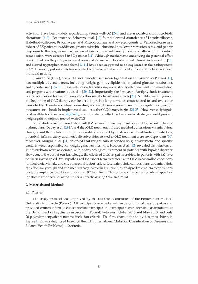
The study protocol was approved by the Bioethics Committee of the Pomeranian MedicalUniversity in Szczecin (Poland). All participants received a written description of the study aims andprovided written informed consent before participation. Participants were recruited as inpatients atthe Department of Psychiatry in Szczecin (Poland) between October 2016 and May 2018, and only20 psychiatric inpatients met the inclusion criteria. The flow chart of the study design is shown inFigure 1. SZ was diagnosed based on the ICD (International Statistical Classification of Diseases andRelated Health Problems) −10 criteria.
14

J. Clin. Med. 2019, 8, 1605
Figure 1. Flow chart of the study design. SZ, schizophrenia.
2.2. Study Protocol
All participants were subjected to the same daily activities, including physical exercise(daily morning exercise and a walk with a therapist), occupational therapy, and psycho-educationalactivities. Two senior psychiatrists performed the psychiatric and basic physical examinations, and agastroenterologist conducted a comprehensive physical examination.
Patients received a standard hospital diet (i.e., 2995 ± 93 kcal, 106 ± 14 g total protein, 420 ± 24 gcarbohydrates, and 102 ± 10 g fat per day), balanced by a hospital dietician, in accordance withthe Polish standards for hospitalized patients [33]. Detailed nutritional data on the diet duringhospitalization, including fiber consumption, are presented in Supplementary Table S1.
This study included 20 patients, with 11 males and 9 females. After admission to the hospital ward,they were all subjected to a 7-day washout from psychiatric medications, received the standard hospitaldiet, and had a similar hospital routine. The first stool samples were collected after the washout period(W0), and subsequently, OLZ treatment was administered (initially 5 mg/day; doses were individuallyadjusted up to 20 mg/day). After 6 weeks of treatment, the second stool samples (W6) were collected(Supplementary Figure S1).
Clinical responses were defined as follows: Early responders, 30% reduction in positive andnegative syndrome scale (PANNS) total score at 4 weeks; late responders, 40% reduction in PANNStotal score at end-point [34]; Clinical global impression-improvement scale (CGI-I) responders, score of3 points (much improvement); and non-responders, clinical global impression-severity (CGI-S) scoresof 4 (minimal improvement) or 5 (no improvement).
2.3. Processing of Raw Data and Statistical Analysis
Sequencing of the V4 region of 16S rRNA gene was performed by the uBiome, Inc. (San Francisco, CA,USA). The 16S amplicons from each sample were individually barcoded and sequenced in the multiplexin the NextSeq 500 platform in a 150 bp (base pair) paired-end modality. The initial quality check ofthe 16S sequences was conducted using the AfterQC (version 0.9.7) software with default settings [35].Subsequently, forward and reverse reads were, respectively, capped at 125 and 124 bp and thenjoined together with an in-between padding sequence (8 of “Ns” with a base score quality of 40).Each sequence was assigned the number of expected errors, and the sequences were filtered to have amaximum expected error of 1.0. The above steps were conducted using the VSEARCH (2.8.0) tool [36].
15

J. Clin. Med. 2019, 8, 1605
The sequences were processed using mothur (v.1.41.3) [37]. Briefly, sequences were aligned to theSILVA bacterial reference alignment (release 132), and were then screened to drop those not aligning topositions 13,148 and 25,277 of the SILVA alignment and were pre-clustered to allow two differencesbetween sequences. The chimeras were identified and removed using VSEARCH implemented inmothur. Subsequently, sequences were classified using a Wang method with the Greengenes 16S rRNADatabase version 13.8. Finally, sequences were clustered into OTUs using opticlust algorithm andMatthews correlation coefficient metric.
Metagenomic predictions from 16S rRNA marker genes (corrected for predicted 16S rRNAcopy number) were carried out using PICRUSt (version 1.1.3) [38], and a list of the KEGG (KyotoEncyclopedia of Genes and Genomes) functional orthologs (KO) was created. Reference genomecoverage of the samples was calculated using the weighted nearest sequenced taxon index (NSTI) [38].The PICRUSt predicted a median NSTI score of 0.11 (interquartile range, IQR of 0.05). The predictedmetagenomes were analyzed with HUMAnN [39] and LEfSe [40]. The KO list was submitted as inputdata to HUMAnN, which generated KEGG modules and KEGG pathway abundances.
Downstream data analysis was performed using the R software (version 3.5.1, https://cran.r-project.org/), R based tools (such as Phyloseq package (version 1.24.2)) [41] and ComplexHeatmap [42],and custom-made scripts. Before calculating alpha diversity, the samples were rarefied to 3680sequences per sample. Prior to beta diversity analysis, the taxa with the prevalence of less than 5%were removed (the prevalence of taxa was defined as the proportion of samples in which the taxaappeared at least once). Beta diversity was analyzed using principal coordinate analysis (PCoA) onBray–Curtis distance matrices generated from the relative OTU abundances. To analyze the changesin bacterial community composition, a change in the principal coordinate 1 (PC1) was examined.The statistical analysis methods included the Wilcoxon rank-sum test, paired Wilcoxon signed-ranktest, t-test for one sample, and Spearman rho correlation coefficient. p-values were adjusted using theBenjamini–Hochberg’s false discovery rate (FDR) controlling procedure. Numerical data are presentedas median, lower quartile, and upper quartile.
3. Results
3.1. Microbiota Compositions
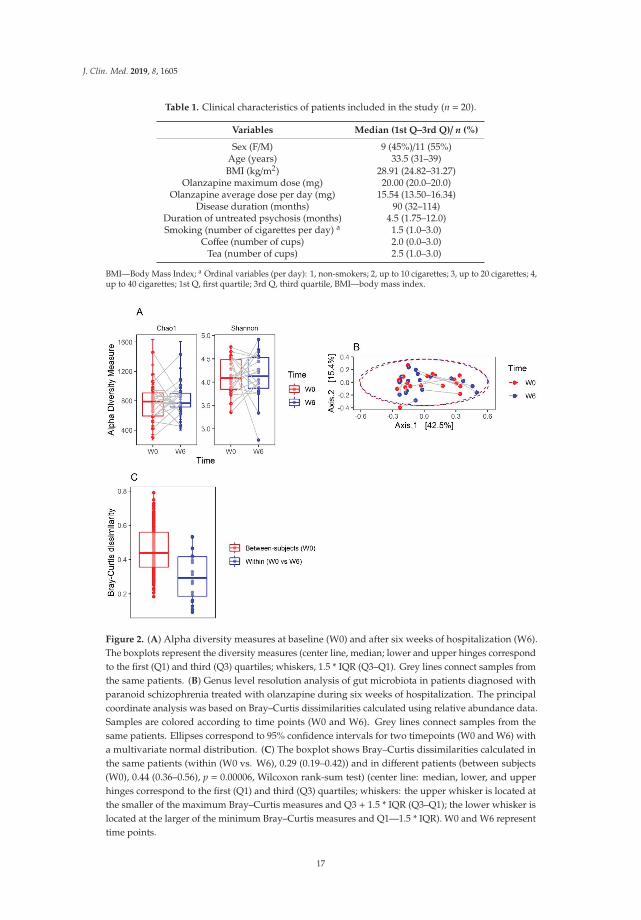
General characteristics of patients are shown in Table 1. There was no significant change inalpha diversity as measured by Chao1 and Shannon indexes (p = 0.955 and p = 0.808, respectively;Figure 2A). The PCoA with Bray–Curtis dissimilarity is presented in Figure 2B. Samples were separatedinto distinct regions, mainly along the PC1 (Axis.1) that explained 42.5% of the intersample variance.The gut microbiome was individually specific, and the Bray–Curtis distances between the same sampleswere significantly smaller than those between all W0 samples (p = 0.00006; Figure 2C). The direction ofchange along the PC1 was not consistent (Supplementary Figure S2). The mean change in the PC1was not significantly different from 0 (0.0012, (95% confidence interval: −0.0946, 0.0970), t = −0.03,df = 19, p = 0.979), suggesting that the gut microbial community composition does not change after sixweeks of treatment. In line with this observation, no OTUs were differentially abundant (from thegenus to phylum level) between W0 and W6 (Supplementary Figures S3–S5). There was no changein the ratio of Firmicutes to Bacteroidetes (F/B) in the whole group, as well as in males and females(Supplementary Figure S6). In addition, there were no significant differences in the abundance of theKEGG orthologs, modules, and pathways between W0 and W6 samples in the whole group, as well asin men and women (Supplementary Figure S7).
16
J. Clin. Med. 2019, 8, 1605
Table 1. Clinical characteristics of patients included in the study (n = 20).
Variables Median (1st Q–3rd Q)/ n (%)
Sex (F/M) 9 (45%)/11 (55%)Age (years) 33.5 (31–39)BMI (kg/m2) 28.91 (24.82–31.27)
Olanzapine maximum dose (mg) 20.00 (20.0–20.0)Olanzapine average dose per day (mg) 15.54 (13.50–16.34)
Disease duration (months) 90 (32–114)Duration of untreated psychosis (months) 4.5 (1.75–12.0)Smoking (number of cigarettes per day) a 1.5 (1.0–3.0)
Coffee (number of cups) 2.0 (0.0–3.0)Tea (number of cups) 2.5 (1.0–3.0)
BMI—Body Mass Index; a Ordinal variables (per day): 1, non-smokers; 2, up to 10 cigarettes; 3, up to 20 cigarettes; 4,up to 40 cigarettes; 1st Q, first quartile; 3rd Q, third quartile, BMI—body mass index.
Figure 2. (A) Alpha diversity measures at baseline (W0) and after six weeks of hospitalization (W6).The boxplots represent the diversity measures (center line, median; lower and upper hinges correspondto the first (Q1) and third (Q3) quartiles; whiskers, 1.5 * IQR (Q3–Q1). Grey lines connect samples fromthe same patients. (B) Genus level resolution analysis of gut microbiota in patients diagnosed withparanoid schizophrenia treated with olanzapine during six weeks of hospitalization. The principalcoordinate analysis was based on Bray–Curtis dissimilarities calculated using relative abundance data.Samples are colored according to time points (W0 and W6). Grey lines connect samples from thesame patients. Ellipses correspond to 95% confidence intervals for two timepoints (W0 and W6) witha multivariate normal distribution. (C) The boxplot shows Bray–Curtis dissimilarities calculated inthe same patients (within (W0 vs. W6), 0.29 (0.19–0.42)) and in different patients (between subjects(W0), 0.44 (0.36–0.56), p = 0.00006, Wilcoxon rank-sum test) (center line: median, lower, and upperhinges correspond to the first (Q1) and third (Q3) quartiles; whiskers: the upper whisker is located atthe smaller of the maximum Bray–Curtis measures and Q3 + 1.5 * IQR (Q3–Q1); the lower whisker islocated at the larger of the minimum Bray–Curtis measures and Q1—1.5 * IQR). W0 and W6 representtime points.
17
J. Clin. Med. 2019, 8, 1605
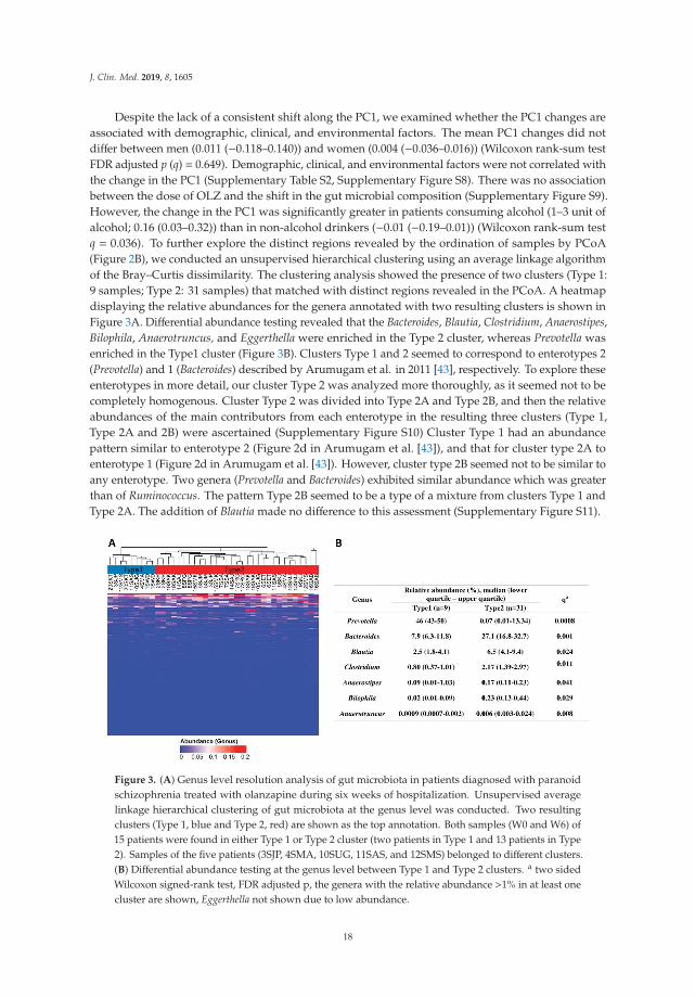
Despite the lack of a consistent shift along the PC1, we examined whether the PC1 changes areassociated with demographic, clinical, and environmental factors. The mean PC1 changes did notdiffer between men (0.011 (−0.118–0.140)) and women (0.004 (−0.036–0.016)) (Wilcoxon rank-sum testFDR adjusted p (q) = 0.649). Demographic, clinical, and environmental factors were not correlated withthe change in the PC1 (Supplementary Table S2, Supplementary Figure S8). There was no associationbetween the dose of OLZ and the shift in the gut microbial composition (Supplementary Figure S9).However, the change in the PC1 was significantly greater in patients consuming alcohol (1–3 unit ofalcohol; 0.16 (0.03–0.32)) than in non-alcohol drinkers (−0.01 (−0.19–0.01)) (Wilcoxon rank-sum testq = 0.036). To further explore the distinct regions revealed by the ordination of samples by PCoA(Figure 2B), we conducted an unsupervised hierarchical clustering using an average linkage algorithmof the Bray–Curtis dissimilarity. The clustering analysis showed the presence of two clusters (Type 1:9 samples; Type 2: 31 samples) that matched with distinct regions revealed in the PCoA. A heatmapdisplaying the relative abundances for the genera annotated with two resulting clusters is shown inFigure 3A. Differential abundance testing revealed that the Bacteroides, Blautia, Clostridium, Anaerostipes,Bilophila, Anaerotruncus, and Eggerthella were enriched in the Type 2 cluster, whereas Prevotella wasenriched in the Type1 cluster (Figure 3B). Clusters Type 1 and 2 seemed to correspond to enterotypes 2(Prevotella) and 1 (Bacteroides) described by Arumugam et al. in 2011 [43], respectively. To explore theseenterotypes in more detail, our cluster Type 2 was analyzed more thoroughly, as it seemed not to becompletely homogenous. Cluster Type 2 was divided into Type 2A and Type 2B, and then the relativeabundances of the main contributors from each enterotype in the resulting three clusters (Type 1,Type 2A and 2B) were ascertained (Supplementary Figure S10) Cluster Type 1 had an abundancepattern similar to enterotype 2 (Figure 2d in Arumugam et al. [43]), and that for cluster type 2A toenterotype 1 (Figure 2d in Arumugam et al. [43]). However, cluster type 2B seemed not to be similar toany enterotype. Two genera (Prevotella and Bacteroides) exhibited similar abundance which was greaterthan of Ruminococcus. The pattern Type 2B seemed to be a type of a mixture from clusters Type 1 andType 2A. The addition of Blautia made no difference to this assessment (Supplementary Figure S11).
Figure 3. (A) Genus level resolution analysis of gut microbiota in patients diagnosed with paranoidschizophrenia treated with olanzapine during six weeks of hospitalization. Unsupervised averagelinkage hierarchical clustering of gut microbiota at the genus level was conducted. Two resultingclusters (Type 1, blue and Type 2, red) are shown as the top annotation. Both samples (W0 and W6) of15 patients were found in either Type 1 or Type 2 cluster (two patients in Type 1 and 13 patients in Type2). Samples of the five patients (3SJP, 4SMA, 10SUG, 11SAS, and 12SMS) belonged to different clusters.(B) Differential abundance testing at the genus level between Type 1 and Type 2 clusters. a two sidedWilcoxon signed-rank test, FDR adjusted p, the genera with the relative abundance >1% in at least onecluster are shown, Eggerthella not shown due to low abundance.
18
J. Clin. Med. 2019, 8, 1605
Taken together, our results suggest that the gut microbiota is highly individually specific, and themicrobial community compositional changes during six weeks of OLZ treatment are not consistentacross the patients.
3.2. Clinical Improvement and BMI Changes
We found that OLZ treatment was associated with significantly improved treatment efficacy asmeasured by PANNS, 36-item short form survey (SF36), and CGI-S scales (Supplementary Table S3).We further investigated whether these improvements are correlated with the change in microbiotacompositions (as measured by a change in the PC1 component) and with demographic and clinicalcharacteristics. No significant correlations were observed between clinical improvements and changesin microbiota composition (Supplementary Figure S12) or demographic and clinical characteristics,except the duration of untreated psychosis (DUP) (Supplementary Table S4).
In contrast to changes in the symptom severity of schizophrenia (Supplementary Table S3),there was no significant change in the patients’ BMI during OLZ treatment (q= 0.763). However, the BMIchange (W6 vs. W0 difference) was significantly higher in women than in men (SupplementaryFigure S13) but did not correlate significantly with age, OLZ average dose per day, OLZ maximumdose, disease duration, or duration of untreated psychosis.
Because we found clear differences in gut microbiome compositions in all 40 samples (Figure 3),we next sought to determine whether similar differences in microbial community compositionsand metabolic potentials exist in baseline samples and whether those differences could affect thepatients’ clinical improvement and change in BMI within six weeks. We performed the unsupervisedaverage linkage hierarchical clustering of the Bray–Curtis dissimilarity among the baseline samples(W0, Supplementary Figure S14), as well as that of the relative abundances of the predicted KEGGorthologs, modules, and pathways (Supplementary Figures S15–S17). Regarding the microbiomecompositions, we were able to demonstrate different groups of patients (clusters) using hierarchicalclustering of KEGG features in the W0 samples: KEGG orthologs (Supplementary Figure S15),modules (Supplementary Figure S16), and pathways (Supplementary Figure S17). Differentialabundance testing revealed that only the Prevotella genus differed between the two clusters (Type 1,0.01% (0.006–0.004) vs. Type 2, 27.4% (17.7–43.1); two-sided Wilcoxon signed-rank test, FDR adjustedp= 0.033; Supplementary Figure S14). To identify differentially abundant genes, modules, and pathwaysbetween clusters, we conducted a linear discriminant analysis with effect size (LEfSe) method (Figure 4).
Subsequently, we compared the baseline symptom scales and BMI between Type 1 and Type 2clusters. We found significant differences in the baseline PANNS, PANNS G, and CGI-S between thegroups created from the clustering of the pathway abundance (Table 2). The patients classified into aType 2 cluster had significantly more severe symptoms at baseline. The improvement in symptomseverity after OLZ treatment assessed by PANNS, SF36, and CG1I was not associated with microbialcommunity compositions (Supplementary Figure S14, Table S5) or KEGG features at baseline (Table 2;Supplementary Figures S15–S17 and Tables S6 and S7). Likewise, no associations were found betweenbaseline gut microbiota (Supplementary Figure S14, Supplementary Table S5) or its metabolic potentials(Table 2 and Supplementary Figures S15–S17 and Supplementary Tables S6 and S7) and the BMI changein the whole group or separately in women or men.
19
J. Clin. Med. 2019, 8, 1605
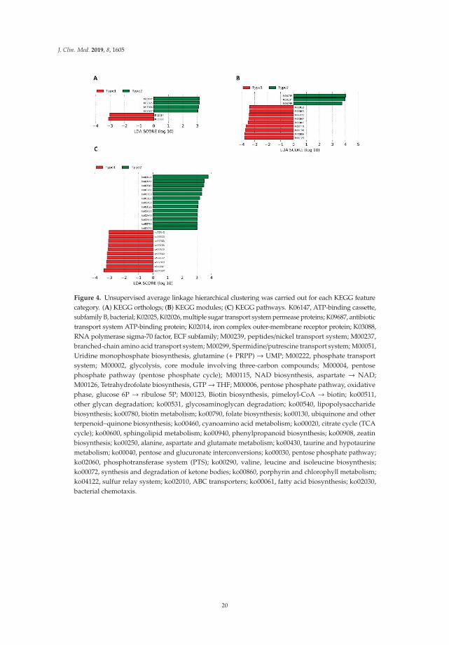
Figure 4. Unsupervised average linkage hierarchical clustering was carried out for each KEGG featurecategory. (A) KEGG orthologs; (B) KEGG modules; (C) KEGG pathways. K06147, ATP-binding cassette,subfamily B, bacterial; K02025, K02026, multiple sugar transport system permease proteins; K09687, antibiotictransport system ATP-binding protein; K02014, iron complex outer-membrane receptor protein; K03088,RNA polymerase sigma-70 factor, ECF subfamily; M00239, peptides/nickel transport system; M00237,branched-chain amino acid transport system; M00299, Spermidine/putrescine transport system; M00051,Uridine monophosphate biosynthesis, glutamine (+ PRPP) → UMP; M00222, phosphate transportsystem; M00002, glycolysis, core module involving three-carbon compounds; M00004, pentosephosphate pathway (pentose phosphate cycle); M00115, NAD biosynthesis, aspartate → NAD;M00126, Tetrahydrofolate biosynthesis, GTP→ THF; M00006, pentose phosphate pathway, oxidativephase, glucose 6P → ribulose 5P; M00123, Biotin biosynthesis, pimeloyl-CoA → biotin; ko00511,other glycan degradation; ko00531, glycosaminoglycan degradation; ko00540, lipopolysaccharidebiosynthesis; ko00780, biotin metabolism; ko00790, folate biosynthesis; ko00130, ubiquinone and otherterpenoid–quinone biosynthesis; ko00460, cyanoamino acid metabolism; ko00020, citrate cycle (TCAcycle); ko00600, sphingolipid metabolism; ko00940, phenylpropanoid biosynthesis; ko00908, zeatinbiosynthesis; ko00250, alanine, aspartate and glutamate metabolism; ko00430, taurine and hypotaurinemetabolism; ko00040, pentose and glucuronate interconversions; ko00030, pentose phosphate pathway;ko02060, phosphotransferase system (PTS); ko00290, valine, leucine and isoleucine biosynthesis;ko00072, synthesis and degradation of ketone bodies; ko00860, porphyrin and chlorophyll metabolism;ko04122, sulfur relay system; ko02010, ABC transporters; ko00061, fatty acid biosynthesis; ko02030,bacterial chemotaxis.
20
J. Clin. Med. 2019, 8, 1605
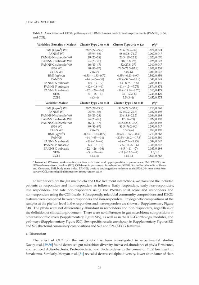
Table 2. Associations of KEGG pathways with BMI changes and clinical improvements (PANNS, SF36,and CGI).
Variables (Females +Males) Cluster Type 2 (n = 5) Cluster Type 3 (n = 12) p/qa
BMI (kg/m2) W0 28.7 (27–29.9) 29.6 (24.4–32) 0.874/0.874PANNS W0 95 (94–98) 68 (62.8–74.2) 0.007/0.047
PANNS N subscale W0 28 (23–28) 20.5 (17–22.2) 0.020/0.070PANNS P subscale W0 24 (23–26) 20 (15.8–22) 0.026/0.073PANNS G subscale W0 46 (43–47) 32 (27.8–37) 0.010/0.047
SF36 W0 90 (83–97) 76.5 (72.5–83.8) 0.102/0.238CGI-S W0 7 (6–7) 5 (5–6) 0.009/0.047
BMI (kg/m2) −0.53 (−1.33–0.72) 0.35 (−0.23–0.90) 0.562/0.656PANNS −44 (−65–−31) −37 (−39.5–−21.8) 0.342/0.749
PANNS N subscale −10 (−17–−9) −6 (−8.75–−4.5) 0.205/0.410PANNS P subscale −12 (−18–−6) −11 (−15–−7.75) 0.874/0.874PANNS G subscale −22 (−26–−16) −16 (−17.8–−8.75) 0.315/0.479
SF36 −5 (−18–−4) −3 (−12.2–6) 0.245/0.429CGI-I 4 (3–4) 3.5 (3–4) 0.452/0.575
Variable (Males) Cluster Type 2 (n = 5) Cluster Type 3 (n = 4) p/qa
BMI (kg/m2) W0 28.7 (27–29.9) 30.5 (27.5–32.2) 0.713/0.768PANNS W0 95 (94–98) 67 (59.2–76.5) 0.037/0.198
PANNS N subscale W0 28 (23–28) 20 (18.8–22.2) 0.084/0.198PANNS P subscale W0 24 (23–26) 17 (14–19) 0.027/0.198PANNS G subscale W0 46 (43–47) 30.5 (24.8–37.5) 0.065/0.198
SF36 W0 90 (83–97) 83.5 (76.2–90) 0.391/0.547CGI-S W0 7 (6–7) 5.5 (5–6) 0.050/0.198
BMI (kg/m2) −0.53 (−1.33–0.72) −0.92 (−1.97–−0.30) 0.713/0.768PANNS −44 (−65–−31) −20.5 (−26.2–−17.8) 0.140/0.280
PANNS N subscale −10 (−17–−9) −6 (−7.5–−5.75) 0.389/0.547PANNS P subscale −12 (−18–−6) −7.5 (−8.25–−6) 0.389/0.547PANNS G subscale −22 (−26–−16) −8.5 (−11–−7) 0.085/0.198
SF36 −5 (−18–−4) −11 (−13.5–−7) 1.0/1.0CGI-I 4 (3–4) 4 (4–4) 0.661/0.768
a Two-sided Wilcoxon rank-sum test, median with lower and upper quartiles in parentheses; BMI, PANNS, andSF36—changes from baseline (W0); CGI-I—an improvement from baseline; KEGG, Kyoto Encyclopedia of Genesand Genomes; BMI, body mass index; PANNS, positive and negative syndrome scale; SF36, 36- item short formsurvey; CGI, clinical global impression-improvement scale.
To further explore the gut microbiota and OLZ treatment interactions, we classified the includedpatients as responders and non-responders as follows: Early responders, early non-responders,late responders, and late non-responders using the PANNS total score and responders andnon-responders using the CGI-I scale. Subsequently, microbial community compositions and KEGGfeatures were compared between responders and non-responders. Phylogenetic compositions of thesamples at the phylum level in the responders and non-responders are shown in Supplementary FigureS18. The phyla were not differentially abundant in responders and non-responders, regardless ofthe definition of clinical improvement. There were no differences in gut microbiome compositions atother taxonomic levels (Supplementary Figure S19), as well as in the KEGG orthologs, modules, andpathways (Supplementary Figure S20). Sex-specific results are shown in Supplementary Figures S21and S22 (bacterial community composition) and S23 and S24 (KEGG features).
4. Discussion
The effect of OLZ on the microbiota has been investigated in experimental studies.Davey et al. [29,30] found decreased gut microbiota diversity, increased abundance of phyla Firmicutes,and reduced Actinobacteria, Proteobacteria, and Bacteroidetes in the course of OLZ treatment infemale rats. Similarly, Morgan et al. [31] revealed decreased alpha diversity, lower abundance of class
21

J. Clin. Med. 2019, 8, 1605
Bacteroidia, and increased abundances of Erysipelotrichia, Actinobacteria, and Gammaproteobacteriain female mice treated with OLZ. However, Kao et al. [44] demonstrated no significant effects of OLZon gut microbiota in female rats. To the best of our knowledge, this study is the first to analyzefecal microbiota compositions in patients hospitalized due to acute relapse of SZ. We did not findthe impact of six-week OLZ treatment on bacterial diversity, abundance, and predicted metabolicfunction, and patients with SZ had individualized and stable gut microbiota in the course of six-weekOLZ treatment in terms of both composition and function. Because of the inconsistent findings above,further studies are needed to clarify the effect of OLZ on gut microbiota.
Although gut microbiota could be compositionally and functionally clustered into similargroups, the classification could not be used to predict the responses to OLZ treatment or theoccurrence of weight gain (observed only in women) during OLZ treatment. As mentioned above,OLZ causes weight gain in female rats [29,44] and mice [31]. This metabolic effect is not observedduring antibiotic therapy [29] and gnotobiosis (germ-free mouse model) and is enhanced during theadministration of the high-fat diet regimen that is responsible for alterations of microbiota similar tothose observed in metabolic syndromes [31]. In addition, Davey et al. [30] demonstrated metabolicdisturbances, inflammation, and microbiota alterations in female mice treated with OLZ and foundonly slight alterations in male mice treated with OLZ, and metabolic effects of OLZ were linked togut microbiota alterations. Notably, antibiotics reversed these effects due to reduced gut microbiota.Therefore, changed gut microbiota plays a pivotal role in weight gain. The lack of association betweenfecal microbiota compositions and weight gain in this study may be due to the low number ofparticipants and the short period of OLZ administration. In addition, other factors might also beresponsible for the increase in body mass index associated with the OLZ administration [20,26–28].
In the present study two bacterial enterotypes (clusters) were found, Type 1, with a predominanceof Prevotella, and Type 2 with a higher abundance of Bacteroides, Blautia and Clostridium. Cluster Type2 seemed not to be completely homogenous (with Types 2A and 2B), which initially suggested thepossibility of the occurrence of a third enterotype similar to that found by Arumugam et al. [43].Further analysis did not confirm this hypothesis and a higher abundance of Ruminococcus or Blautiain sub-cluster Type 2B was not seen. This sub-cluster seemed to be a type of mixture from clustersType 1 and Type 2A. Due to this we took into consideration in further analyses only two enterotypes(original clusters) of bacteria. Moreover, patients with SZ were clustered at the level of KEGG genes,modules, and pathways. The severity of symptoms measured at the beginning of treatment varied,depending on the predicted metabolic activity of the bacteria. Other studies also have observeda relationship between the composition of bacteria and the severity of symptoms in SZ patients.Zheng et al. [11] demonstrated that PANSS was negatively correlated with Veillonellaceae and waspositively correlated with Bacteroidaceae, Streptococcaceae, and Lachnospiraceae. Schwartz et al. [10]found greater microbial abnormalities in SZ patients than in controls. In addition, increases inthe number of Lactobacillus group bacteria were positively correlated with the severity of varioussymptom domains in SZ patients and were negatively correlated with the global assessment offunctioning. Moreover, responses to the treatment were worse in patients with severe microbiotaalterations. Furthermore, Shen et al. [45], using the PICRUSt analysis, infer that vitamin B6 and fattyacid metabolic potential differed significantly between SZ patients and controls. Therefore, there arepotential relationships between predicted metabolic changes and the severity of symptoms in SZpatients, as shown in Table 3. It is important to note that the PICRUSt approach using in prediction ofbacterial metabolic activity should be treated with caution and followed by metagenomic analysesto explain such findings in humans. The median NSTI score was 0.11 (interquartile range of 0.05)suggesting a reasonable accuracy of the prediction, however, some closely related reference genomeswere not available.
22
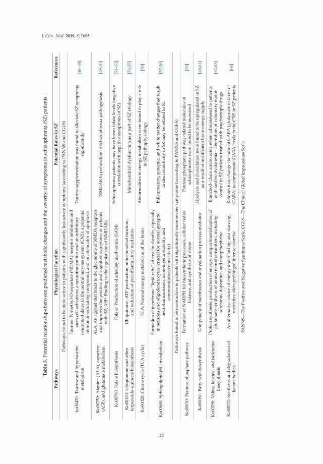
J. Clin. Med. 2019, 8, 1605
Ta
ble
3.
Pote
ntia
lrel
atio
nshi
psbe
twee
npr
edic
ted
met
abol
icch
ange
san
dth
ese
veri
tyof
sym
ptom
sin
schi
zoph
reni
a(S
Z)p
atie
nts.
Path
way
sP
hy
sio
log
ical
Fu
nct
ion
Po
ten
tial
Ro
les
inS
ZR
efe
ren
ces
Path
way
sfo
und
tobe
mor
eac
tive
inpa
tien
tsw
ith
sign
ifica
ntly
less
seve
resy
mpt
oms
(acc
ordi
ngto
PAN
NS
and
CG
I-S)
ko00
430:
Taur
ine
and
hypo
taur
ine
met
abol
ism
Taur
ine:
N-m
ethy
l-D
-asp
arta
te(N
DM
A)r
ecep
tor
inhi
biti
onan
dst
emce
llac
tiva
tion
;ane
urot
rans
mit
ter
and
anin
hibi
tory
neur
omod
ulat
orin
the
cent
raln
ervo
ussy
stem
(CN
S);a
pote
ntia
lim
mun
omod
ulat
ing
com
poun
d,an
dan
atte
nuat
orof
apop
tosi
s
Taur
ine
supp
lem
enta
tion
was
foun
dto
alle
viat
eSZ
sym
ptom
ssi
gnifi
cant
ly[4
6–48
]
Ko0
0250
:Ala
nine
(ALA
),as
part
ate
(ASP
),an
dgl
utam
ate
met
abol
ism
ALA
:An
agon
istt
hatb
inds
toth
egl
ycin
esi
teof
NM
DA
rece
ptor
san
dim
prov
esth
epo
siti
vean
dco
gnit
ive
sym
ptom
sof
pati
ents
wit
hSZ
;ASP
:Bin
ding
toth
eag
onis
tsit
eof
NM
DA
Rs
NM
DA
Rhy
pofu
ncti
onin
schi
zoph
reni
apa
thog
enes
is[4
9,50
]
Ko0
0790
:Fol
ate
bios
ynth
esis
Fola
te:P
rodu
ctio
nof
aden
osyl
met
hion
ine
(SA
M)
Schi
zoph
reni
apa
tient
sm
ayha
velo
wer
fola
tele
vels
(neg
ativ
eco
rrel
atio
nw
ith
nega
tive
sym
ptom
sof
SZ)
[51–
53]
Ko0
0130
:Ubi
quin
one
and
othe
rte
rpen
oids
–qui
none
bios
ynth
esis
Ubi
quin
one:
ATP
prod
ucti
on,m
itoc
hond
rial
func
tion
,an
dre
duct
ion
ofpr
oinfl
amm
ator
ym
edia
tors
Mit
ocho
ndri
aldy
sfun
ctio
nas
apa
rtof
SZet
iolo
gy[5
4,55
]
Ko0
0020
:Cit
rate
cycl
e(T
CA
cycl
e)TC
A:N
orm
alen
ergy
met
abol
ism
ofth
ebr
ain
Abn
orm
aliti
esin
ener
gym
etab
olis
mw
ere
foun
dto
play
aro
lein
SZpa
thop
hysi
olog
y[5
6]
Ko0
0600
:Sph
ingo
lipid
(SL)
met
abol
ism
Form
atio
nof
mem
bran
e“l
ipid
raft
s”of
mye
linsh
eath
s,es
peci
ally
inne
uron
san
dol
igod
endr
ocyt
es(c
ruci
alfo
rno
rmal
syna
ptic
neur
otra
nsm
issi
on,a
xon-
mye
linst
abili
ty,a
ndco
mm
unic
atio
n/co
nnec
tivi
ty)
Infla
mm
ator
y,sy
napt
ic,a
ndw
hite
mat
ter
chan
ges
that
resu
ltin
disc
onne
ctiv
ity
inSZ
may
bere
late
dto
SL[5
7,58
]
Path
way
sfo
und
tobe
mor
eac
tive
inpa
tien
tsw
ith
sign
ifica
ntly
mor
ese
vere
sym
ptom
s(a
ccor
ding
toPA
NN
San
dC
GI-
S)
Ko0
0030
:Pen
tose
phos
phat
epa
thw
ayFo
rmat
ion
ofN
AD
PHfo
rbi
osyn
thet
icpr
oces
ses,
cellu
lar
redo
xba
lanc
e,an
dsy
nthe
sis
ofri
bose
Pent
ose
phos
phat
epa
thw
ay-r
elat
edm
olec
ules
insc
hizo
phre
nia
wer
efo
und
tobe
incr
ease
d[5
9]
Ko0
0061
:Fat
tyac
idbi
osyn
thes
isC
ompo
nent
ofm
embr
anes
and
mye
linat
ion
proc
ess
med
iato
rLi
poly
sis
andβ
-oxi
datio
nw
ere
foun
dto
beup
regu
late
din
SZ,
asa
resu
ltof
insuffi
cien
tbra
inen
ergy
supp
ly[6
0,61
]
Ko0
0290
:Val
ine,
leuc
ine,
and
isol
euci
nebi
osyn
thes
is
Prot
ein
synt
hesi
s,pr
oduc
tion
ofen
ergy
,com
part
men
taliz
atio
nof
glut
amat
esy
nthe
sis
ofam
ine
neur
otra
nsm
itte
rs,i
nclu
ding
sero
toni
n,do
pam
ine,
and
nore
pine
phri
ne
Bran
ched
-cha
inam
ino
acid
sw
hen
adm
inis
tere
dto
pati
ents
wit
hta
rdiv
edy
skin
esia
—ab
erra
tion
ofvo
lunt
ary
mot
orco
ntro
lin
SZpa
tien
tstr
eate
dw
ith
psyc
hotr
opic
drug
s[6
2,63
]
Ko0
0072
:Syn
thes
isan
dde
grad
atio
nof
keto
nebo
dies
An
alte
rnat
ive
sour
ceof
ener
gyun
der
fast
ing
and
star
ving
;re
stri
ctiv
edi
ets
prol
onge
din
tens
eex
erci
seK
eton
esm
aych
ange
the
ratio
ofG
ABA
(glu
tam
ate
infa
vor
ofG
ABA
)to
com
pens
ate
GA
BAle
vels
inth
eC
NS
inSZ
patie
nts
[64]
PAN
NS—
The
Posi
tive
and
Neg
ativ
eSy
ndro
me
Scal
e,C
GI-
S—Th
eC
linic
alG
loba
lIm
pres
sion
sSc
ale.
23
J. Clin. Med. 2019, 8, 1605
Our study has several strengths that should be highlighted. (1) The applied treatment resulted inexpected clinical effects. The relationship between duration of untreated psychosis (DUP) and poorgeneral symptomatic outcomes was confirmed, and the longer DUP was associated with more severepositive and negative symptoms. Additionally, OLZ treatment caused weight gain. This observationis in agreement with that in another experimental study [65]. (2) During the treatment, the patientswere under the same controlled hospital conditions (diet, drug intake, and clinical monitoring), and awashout was used before treatment, thus providing a "unification" of the environmental impact onthe fecal microbiota pattern. Consequently, we speculate that such conditions diminish the impact ofcommon environmental factors that permanently shape gut microbiota composition and underline theassociation between the disease and treatment. (3) Weight gain at the beginning of OLZ treatment isvery important because it determines the further development of cardiometabolic risk factors [22,23,66].(4) Although the study group was not homogeneous, the symptoms were observed every day in ourpsychiatric clinic. Patients were previously treated with other pharmaceuticals, which might haveaffected the microbiota composition. Such situations might lead to resistance against psychotropicdrugs, probably leading to no impact of OLZ on the microbiota.
There are certain limitations of our study that need to be discussed. First, the sample sizewas small and heterogeneous (drug-naive and previously-treated patients). No formal samplesize calculations were employed for this analysis, but the cohort size was based on what waspreviously sufficient to test microbiotic changes in schizophrenia patients [67], and/or the influenceof antibiotics [68] and risperidone administration [69] on gut microbiota. This limitation should beattributed to rigorous inclusion and exclusion criteria as well as the short duration of the whole study(17 months). Thus, studies with a greater sample size are needed to further examine the associationsbetween OLZ treatment and gut microbiota structure. Second, the composition of intestinal bacteriavaried among individuals, and inter-individual variation within the gut ecosystem of patients washigh. Third, in individual studies (also experimental), various taxonomic groups of bacteria wereanalyzed only in stools. The composition of bacteria in feces is more stable and is not influenced byexternal factors compared with the composition of bacteria in the small intestine. Changes in themicrobiota of the small bowel have a much greater effect on the metabolic functions of the human body.Therefore, further experimental studies should pay more attention to this issue [70,71], although aninvasive way of sampling intestine biological material remains difficult and holds several ethicalconcerns. Fourth, there was a lack of long-term follow-up, which is especially important in case ofmetabolic consequences of OLZ treatment. Fifth, we did not compare the results between SZ patientsand healthy subjects or patients receiving placebos. Matched controls with similar lifestyle should beused to exclude false-positive results. However, the general lifestyle in patients diagnosed with SZwas found to be divergent from that observed in healthy people [72]. Therefore, a placebo approachwas impossible mainly due to ethical and organizational concerns. Sixth, changes in dietary and livingconditions during the hospital stay might be another limitation of our study. However, enterotypesPrevotella and Bacteroides are strongly associated with long-term diet. It was shown that microbiomecomposition changed detectably within 24 h of initiating a high-fat/low-fiber or low-fat/high-fiber diet,but that enterotype identity remained stable during the 10-day study [73]. Therefore, a change ofdiet after admission to hospital should not affect W0 microbiota. After this all patients received thesame diet, and it seems that this factor should also not significantly affect the influence of OLZ on W6microbiota composition.
5. Conclusions
In conclusion, the present findings indicate that the microbiota in patients with the schizophreniaepisode is highly individualized, although it can be clustered into different taxonomical (Type 1, with apredominance of Prevotella, and Type 2 with a higher abundance of Bacteroides, Blautia, and Clostridium)and functional groups; the microbiota does not change during six weeks of treatment with OLZ and isnot associated with the weight gain that occurs in women treated with OLZ, as well as the treatment
24
J. Clin. Med. 2019, 8, 1605
effectiveness. This study provides some insights into the metabolic effects of psychotropic drugs ongut microbiota in SZ patients. Further long-term and placebo-controlled studies are needed to clarifythe effect of OLZ on gut microbiota.
Supplementary Materials: The following are available online at http://www.mdpi.com/2077-0383/8/10/1605/s1.
Author Contributions: Conceptualization, J.P.-W., A.B.-K. and J.S.; data curation, J.P.-W., M.K., A.B.-K., P.L., M.W.and I.Ł.; formal analysis, J.P.-W., M.K., P.L., K.S.-Z., W.M., B.M., J.K.-M., I.Ł. and J.S.; investigation, J.P.-W., M.K.,A.B.-K., P.L., M.W., K.S.-Z., W.M., J.K.-M., I.Ł. and J.S.; methodology, J.S.; software, M.K.; supervision, J.P.-W. andJ.S.; visualization, M.K.; writing—original draft, J.P.-W., M.K. and I.Ł.; writing—review and editing, J.P.-W., M.K.,A.B.-K., M.W., K.S.-Z., W.M., B.M., T.S., I.Ł. and J.S.
Funding: The study was funded within the framework of the project Fundusz Stymulacji Nauki (grant number:FSN-312-05/15) provided by Pomeranian Medical University at Szczecin (Poland) and National Science Centre(UMO-2018/31/B/NZ5/00527). The funding sources had no role in concept design, selection of articles, the decisionto publish, or the preparation of the manuscript.
Acknowledgments: We would like to thank Editage (www.editage.com) for English language editing.
Conflicts of Interest: I.Ł. and W.M. are cofounders and shareholders at Sanprobi company. However, the contentof this study was not constrained by this fact. Other authors declare no conflicts of interest.
References
1. WHO Schizophrenia. Available online: https://www.who.int/news-room/fact-sheets/detail/schizophrenia(accessed on 2 June 2019).
2. Müller, N. Inflammation in Schizophrenia: Pathogenetic Aspects and Therapeutic Considerations.Schizophr. Bull. 2018, 44, 973–982. [CrossRef] [PubMed]
3. Purves-Tyson, T.D.; Owens, S.J.; Rothmond, D.A.; Halliday, G.M.; Double, K.L.; Stevens, J.; McCrossin, T.;Weickert, C.S. Putative presynaptic dopamine dysregulation in schizophrenia is supported by molecularevidence from post-mortem human midbrain. Transl. Psychiatry 2017, 7, e1003. [CrossRef] [PubMed]
4. Guest, F.L.; Martins-de-Souza, D.; Rahmoune, H.; Bahn, S.; Guest, P.C. The effects of stress onhypothalamic-pituitary-adrenal (HPA) axis function in subjects with schizophrenia. Arch. Clin. Psychiatry(São Paulo) 2013, 40, 20–27. [CrossRef]
5. Guest, P.C.; Martins-De-Souza, D.; Vanattou-Saifoudine, N.; Harris, L.W.; Bahn, S. Abnormalities inMetabolism and Hypothalamic–Pituitary–Adrenal Axis Function in Schizophrenia. Int. Rev. Neurobiol. 2011,101, 145–168.
6. Clemente, J.C.; Manasson, J.; Scher, J.U. The role of the gut microbiome in systemic inflammatory disease.BMJ 2018, 360, j5145. [CrossRef]
7. Sudo, N.; Chida, Y.; Aiba, Y.; Sonoda, J.; Oyama, N.; Yu, X.-N.; Kubo, C.; Koga, Y. Postnatal microbialcolonization programs the hypothalamic–pituitary–adrenal system for stress response in mice. J. Physiol.2004, 558, 263–275. [CrossRef]
8. Waclawiková, B.; El Aidy, S. Role of Microbiota and Tryptophan Metabolites in the Remote Effect of IntestinalInflammation on Brain and Depression. Pharmaceuticals (Basel) 2018, 11, 63. [CrossRef]
9. Rogers, G.B.; Keating, D.J.; Young, R.L.; Wong, M.-L.; Licinio, J.; Wesselingh, S. From gut dysbiosis to alteredbrain function and mental illness: Mechanisms and pathways. Mol. Psychiatry 2016, 21, 738–748. [CrossRef]
10. Schwarz, E.; Maukonen, J.; Hyytiäinen, T.; Kieseppä, T.; Orešic, M.; Sabunciyan, S.; Mantere, O.; Saarela, M.;Yolken, R.; Suvisaari, J. Analysis of microbiota in first episode psychosis identifies preliminary associationswith symptom severity and treatment response. Schizophr. Res. 2018, 192, 398–403. [CrossRef]
11. Zheng, P.; Zeng, B.; Liu, M.; Chen, J.; Pan, J.; Han, Y.; Liu, Y.; Cheng, K.; Zhou, C.; Wang, H.; et al.The gut microbiome from patients with schizophrenia modulates the glutamate-glutamine-GABA cycle andschizophrenia-relevant behaviors in mice. Sci. Adv. 2019, 5, eaau8317. [CrossRef]
12. Rakoff-Nahoum, S.; Paglino, J.; Eslami-Varzaneh, F.; Edberg, S.; Medzhitov, R. Recognition of CommensalMicroflora by Toll-Like Receptors Is Required for Intestinal Homeostasis. Cell 2004, 118, 229–241. [CrossRef][PubMed]
13. Barry, S.; Clarke, G.; Scully, P.; Dinan, T.G. Kynurenine pathway in psychosis: Evidence of increasedtryptophan degradation. J. Psychopharmacol. 2009, 23, 287–294. [CrossRef] [PubMed]
25
J. Clin. Med. 2019, 8, 1605
14. Miller, C.L.; Llenos, I.C.; Dulay, J.R.; Weis, S. Upregulation of the initiating step of the kynurenine pathwayin postmortem anterior cingulate cortex from individuals with schizophrenia and bipolar disorder. Brain Res.2006, 1073, 25–37. [CrossRef] [PubMed]
15. Hálfdánarson, Ó.; Zoëga, H.; Aagaard, L.; Bernardo, M.; Brandt, L.; Fusté, A.C.; Furu, K.; Garuoliené, K.;Hoffmann, F.; Huybrechts, K.F.; et al. International trends in antipsychotic use: A study in 16 countries,2005–2014. Eur. Neuropsychopharmacol. 2017, 27, 1064–1076.
16. Vancampfort, D.; Correll, C.U.; Galling, B.; Probst, M.; De Hert, M.; Ward, P.B.; Rosenbaum, S.; Gaughran, F.;Lally, J.; Stubbs, B. Diabetes mellitus in people with schizophrenia, bipolar disorder and major depressivedisorder: A systematic review and large scale meta-analysis. World Psychiatry 2016, 15, 166–174. [CrossRef]
17. Galling, B.; Roldán, A.; Nielsen, R.E.; Nielsen, J.; Gerhard, T.; Carbon, M.; Stubbs, B.; Vancampfort, D.;De Hert, M.; Olfson, M.; et al. Type 2 Diabetes Mellitus in Youth Exposed to Antipsychotics. JAMA Psychiatry2016, 73, 247–259. [CrossRef]
18. Galling, B.; Correll, C.U. Do antipsychotics increase diabetes risk in children and adolescents? Expert Opin.Drug Saf. 2015, 14, 219–241. [CrossRef]
19. De Hert, M.; Vancampfort, D.; Correll, C.U.; Mercken, V.; Peuskens, J.; Sweers, K.; Van Winkel, R.; Mitchell, A.J.Guidelines for screening and monitoring of cardiometabolic risk in schizophrenia: Systematic evaluation.Br. J. Psychiatry 2011, 199, 99–105. [CrossRef]
20. Bak, M.; Fransen, A.; Janssen, J.; Van Os, J.; Drukker, M. Almost All Antipsychotics Result in Weight Gain:A Meta-Analysis. PLoS ONE 2014, 9, e94112. [CrossRef]
21. Musil, R.; Obermeier, M.; Russ, P.; Hamerle, M. Weight gain and antipsychotics: A drug safety review.Expert Opin. Drug Saf. 2015, 14, 73–96. [CrossRef]
22. Spertus, J.; Horvitz-Lennon, M.; Abing, H.; Normand, S.-L. Risk of weight gain for specific antipsychoticdrugs: A meta-analysis. NPJ Schizophr. 2018, 4, 12. [CrossRef] [PubMed]
23. Pérez-Iglesias, R.; Martínez-García, O.; Pardo-Garcia, G.; Amado, J.A.; Garcia-Unzueta, M.T.; Tabares-Seisdedos, R.;Crespo-Facorro, B. Course of weight gain and metabolic abnormalities in first treated episode of psychosis: The firstyear is a critical period for development of cardiovascular risk factors. Int. J. Neuropsychopharmacol. 2014, 17, 41–51.[CrossRef] [PubMed]
24. American Diabetes Association; American Psychiatric Association; American Association of ClinicalEndocrinologists; North American Association for the Study of Obesity Consensus development conferenceon antipsychotic drugs and obesity and diabetes. Diabetes Care 2004, 27, 596–601.
25. De Hert, M.; Dekker, J.M.; Wood, D.; Kahl, K.G.; Holt, R.I.G.; Möller, H.-J. Cardiovascular disease anddiabetes in people with severe mental illness position statement from the European Psychiatric Association(EPA), supported by the European Association for the Study of Diabetes (EASD) and the European Societyof Cardiology (ESC). Eur. Psychiatry 2009, 24, 412–424. [CrossRef]
26. Albaugh, V.L.; Henry, C.R.; Bello, N.T.; Hajnal, A.; Lynch, S.L.; Halle, B.; Lynch, C.J. Hormonal and metaboliceffects of olanzapine and clozapine related to body weight in rodents. Obesity (Silver Spring) 2006, 14, 36–51.[CrossRef]
27. Basson, B.R.; Kinon, B.J.; Taylor, C.C.; Szymanski, K.A.; Gilmore, J.A.; Tollefson, G.D. Factors InfluencingAcute Weight Change in Patients with Schizophrenia Treated with Olanzapine, Haloperidol, or Risperidone.J. Clin. Psychiatry 2001, 62, 231–238. [CrossRef]
28. Ou, J.-J.; Xu, Y.; Chen, H.-H.; Fan, X.; Gao, K.; Wang, J.; Guo, X.-F.; Wu, R.-R.; Zhao, J.-P. Comparison ofmetabolic effects of ziprasidone versus olanzapine treatment in patients with first-episode schizophrenia.Psychopharmacology 2013, 225, 627–635. [CrossRef]
29. Davey, K.J.; Cotter, P.D.; O’Sullivan, O.; Crispie, F.; Dinan, T.G.; Cryan, J.F.; O’Mahony, S.M. Antipsychotics andthe gut microbiome: Olanzapine-induced metabolic dysfunction is attenuated by antibiotic administrationin the rat. Transl. Psychiatry 2013, 3, e309. [CrossRef]
30. Davey, K.J.; O’Mahony, S.M.; Schellekens, H.; O’Sullivan, O.; Bienenstock, J.; Cotter, P.D.; Dinan, T.G.;Cryan, J.F. Gender-dependent consequences of chronic olanzapine in the rat: Effects on body weight,inflammatory, metabolic and microbiota parameters. Psychopharmacology 2012, 221, 155–169. [CrossRef]
31. Morgan, A.P.; Crowley, J.J.; Nonneman, R.J.; Quackenbush, C.R.; Miller, C.N.; Ryan, A.K.; Bogue, M.A.;Paredes, S.H.; Yourstone, S.; Carroll, I.M.; et al. The Antipsychotic Olanzapine Interacts with the GutMicrobiome to Cause Weight Gain in Mouse. PLoS ONE 2014, 9, e115225. [CrossRef]
26

J. Clin. Med. 2019, 8, 1605
32. Flowers, S.A.; Evans, S.J.; Ward, K.M.; McInnis, M.G.; Ellingrod, V.L. Interaction Between AtypicalAntipsychotics and the Gut Microbiome in a Bipolar Disease Cohort. Pharmacother. J. Hum. Pharmacol. DrugTher. 2017, 37, 261–267. [CrossRef] [PubMed]
33. Jarosz, M.; Rychlik, E.; Stos, K.; Wierzejska, R.; Wojtasik, A.; Charzewska, J.; Mojska, H.; Szponar, L.; Sajór, I.;Kłosiewicz-Latoszek, L.; et al. Normy Zywienia dla Populacji Polski; Instytut Zywnosci i Zywienia: Warszawa,Poland, 2017; ISBN 978-83-86060-89-4.
34. Ascher-Svanum, H.; Zhao, F.; Detke, H.C.; Nyhuis, A.W.; Lawson, A.H.; Stauffer, V.L.; Montgomery, W.;Witte, M.M.; McDonnell, D.P. Early response predicts subsequent response to olanzapine long-acting injectionin a randomized, double-blind clinical trial of treatment for schizophrenia. BMC Psychiatry 2011, 11, 152.[CrossRef] [PubMed]
35. Chen, S.; Huang, T.; Zhou, Y.; Han, Y.; Xu, M.; Gu, J. AfterQC: Automatic filtering, trimming, error removingand quality control for fastq data. BMC Bioinform. 2017, 18, 80. [CrossRef] [PubMed]
36. Rognes, T.; Flouri, T.; Nichols, B.; Quince, C.; Mahé, F. VSEARCH: A versatile open source tool formetagenomics. PeerJ 2016, 4, e2584. [CrossRef] [PubMed]
37. Schloss, P.D.; Westcott, S.L.; Ryabin, T.; Hall, J.R.; Hartmann, M.; Hollister, E.B.; Lesniewski, R.A.;Oakley, B.B.; Parks, D.H.; Robinson, C.J.; et al. Introducing mothur: Open-Source, Platform-Independent,Community-Supported Software for Describing and Comparing Microbial Communities. Appl. Environ. Microbiol.2009, 75, 7537–7541. [CrossRef]
38. Langille, M.G.I.; Zaneveld, J.; Caporaso, J.G.; McDonald, D.; Knights, D.; Reyes, J.A.; Clemente, J.C.;Burkepile, D.E.; Thurber, R.L.V.; Knight, R.; et al. Predictive functional profiling of microbial communitiesusing 16S rRNA marker gene sequences. Nat. Biotechnol. 2013, 31, 814–821. [CrossRef]
39. Abubucker, S.; Segata, N.; Goll, J.; Schubert, A.M.; Izard, J.; Cantarel, B.L.; Rodriguez-Mueller, B.; Zucker, J.;Thiagarajan, M.; Henrissat, B.; et al. Metabolic Reconstruction for Metagenomic Data and Its Application tothe Human Microbiome. PLoS Comput. Biol. 2012, 8, e1002358. [CrossRef]
40. Segata, N.; Izard, J.; Waldron, L.; Gevers, D.; Miropolsky, L.; Garrett, W.S.; Huttenhower, C. Metagenomicbiomarker discovery and explanation. Genome Biol. 2011, 12, R60. [CrossRef]
41. McMurdie, P.J.; Holmes, S. phyloseq: An R Package for Reproducible Interactive Analysis and Graphics ofMicrobiome Census Data. PLoS ONE 2013, 8, e61217. [CrossRef]
42. Gu, Z.; Eils, R.; Schlesner, M. Complex heatmaps reveal patterns and correlations in multidimensionalgenomic data. Bioinformatics 2016, 32, 2847–2849. [CrossRef]
43. Arumugam, M.; Raes, J.; Pelletier, E.; Le Paslier, D.; Yamada, T.; Mende, D.R.; Fernandes, G.R.; Tap, J.;Bruls, T.; Batto, J.-M.; et al. Enterotypes of the human gut microbiome. Nature 2011, 473, 174–180. [CrossRef][PubMed]
44. Kao, A.C.-C.; Spitzer, S.; Anthony, D.C.; Lennox, B.; Burnet, P.W.J. Prebiotic attenuation of olanzapine-inducedweight gain in rats: Analysis of central and peripheral biomarkers and gut microbiota. Transl. Psychiatry2018, 8, 66. [CrossRef] [PubMed]
45. Shen, Y.; Xu, J.; Li, Z.; Huang, Y.; Yuan, Y.; Wang, J.; Zhang, M.; Hu, S.; Liang, Y. Analysis of gut microbiotadiversity and auxiliary diagnosis as a biomarker in patients with schizophrenia: A cross-sectional study.Schizophr. Res. 2018, 197, 470–477. [CrossRef] [PubMed]
46. Firth, J.; Rosenbaum, S.; Ward, P.B.; Curtis, J.; Teasdale, S.B.; Yung, A.R.; Sarris, J. Adjunctive nutrientsin first-episode psychosis: A systematic review of efficacy, tolerability and neurobiological mechanisms.Early Interv. Psychiatry 2018, 12, 774–783. [CrossRef] [PubMed]
47. Leppik, L.; Kriisa, K.; Koido, K.; Koch, K.; Kajalaid, K.; Haring, L.; Vasar, E.; Zilmer, M. Profiling of AminoAcids and Their Derivatives Biogenic Amines Before and After Antipsychotic Treatment in First-EpisodePsychosis. Front. Psychol. 2018, 9, 155. [CrossRef] [PubMed]
48. O’Donnell, C.P.; Allott, K.A.; Murphy, B.P.; Yuen, H.P.; Proffitt, T.-M.; Papas, A.; Moral, J.; Pham, T.;O’Regan, M.K.; Phassouliotis, C.; et al. Adjunctive Taurine in First-Episode Psychosis: A Phase 2,Double-Blind, Randomized, Placebo-Controlled Study. J. Clin. Psychiatry 2016, 77, e1610–e1617. [CrossRef][PubMed]
49. Hatano, T.; Ohnuma, T.; Sakai, Y.; Shibata, N.; Maeshima, H.; Hanzawa, R.; Suzuki, T.; Arai, H. Plasmaalanine levels increase in patients with schizophrenia as their clinical symptoms improve—Results from theJuntendo University Schizophrenia Projects (JUSP). Psychiatry Res. Neuroimaging 2010, 177, 27–31. [CrossRef]
27
J. Clin. Med. 2019, 8, 1605
50. Errico, F.; Nuzzo, T.; Carella, M.; Bertolino, A.; Usiello, A. The Emerging Role of Altered d-AspartateMetabolism in Schizophrenia: New Insights from Preclinical Models and Human Studies. Front. Psychol.2018, 9, 559. [CrossRef]
51. Cao, B.; Wang, D.-F.; Xu, M.-Y.; Liu, Y.-Q.; Yan, L.-L.; Wang, J.-Y.; Lu, Q.-B. Lower folate levels in schizophrenia:A meta-analysis. Psychiatry Res. 2016, 245, 1–7. [CrossRef]
52. Hussein, H.; El Mawella, S.A.; Ahmed, T. Folate, vitamin B12, and negative symptoms in schizophrenia.Egypt. J. Psychiatry 2018, 39, 89. [CrossRef]
53. Hill, M.; Shannahan, K.; Jasinski, S.; Macklin, E.A.; Raeke, L.; Roffman, J.L.; Goff, D.C. Folate supplementationin schizophrenia: A possible role for MTHFR genotype. Schizophr. Res. 2011, 127, 41–45. [CrossRef][PubMed]
54. Maguire, Á.; Hargreaves, A.; Gill, M. Coenzyme Q10 and neuropsychiatric and neurological disorders:Relevance for schizophrenia. Nutr. Neurosci. 2018, 21, 1–14. [CrossRef] [PubMed]
55. Schmelzer, C.; Döring, F. Identification of LPS-inducible genes downregulated by ubiquinone in humanTHP-1 monocytes. BioFactors 2010, 36, 222–228. [CrossRef] [PubMed]
56. Bubber, P.; Hartounian, V.; Gibson, G.E.; Blass, J.P. Abnormalities in the tricarboxylic acid (TCA) cycle in thebrains of schizophrenia patients. Eur. Neuropsychopharmacol. 2011, 21, 254–260. [CrossRef]
57. Castillo, R.I.; Rojo, L.E.; Henríquez-Henríquez, M.; Silva, H.; Maturana, A.; Villar, M.J.; Fuentes, M.;Gaspar, P.A. From Molecules to the Clinic: Linking Schizophrenia and Metabolic Syndrome throughSphingolipids Metabolism. Front. Mol. Neurosci. 2016, 10, 488. [CrossRef]
58. Weston-Green, K.; Babic, I.; De Santis, M.; Pan, B.; Montgomery, M.K.; Mitchell, T.; Huang, X.-F.; Nealon, J.Disrupted sphingolipid metabolism following acute clozapine and olanzapine administration. J. Biomed. Sci.2018, 25, 40. [CrossRef]
59. Liu, M.-L.; Zhang, X.-T.; Du, X.-Y.; Fang, Z.; Liu, Z.; Xu, Y.; Zheng, P.; Xu, X.-J.; Cheng, P.-F.; Huang, T.; et al.Severe disturbance of glucose metabolism in peripheral blood mononuclear cells of schizophrenia patients:A targeted metabolomic study. J. Transl. Med. 2015, 13, 226. [CrossRef]
60. Yang, X.; Sun, L.; Zhao, A.; Hu, X.; Qing, Y.; Jiang, J.; Yang, C.; Xu, T.; Wang, P.; Liu, J.; et al. Serum fatty acidpatterns in patients with schizophrenia: A targeted metabonomics study. Transl. Psychiatry 2017, 7, e1176.[CrossRef]
61. Peters, B.D.; Machielsen, M.W.J.; Hoen, W.P.; Caan, M.W.A.; Malhotra, A.K.; Szeszko, P.R.; Duran, M.;Olabarriaga, S.D.; de Haan, L. Polyunsaturated fatty acid concentration predicts myelin integrity inearly-phase psychosis. Schizophr. Bull. 2013, 39, 830–838. [CrossRef]
62. Fernstrom, J.D. Branched-chain amino acids and brain function. J. Nutr. 2005, 135, 1539S–1546S. [CrossRef]63. Bevans, M.L.; Read, L.L.; Chao, H.M.; Clelland, J.D.; Suckow, R.F.; Maher, T.J.; Citrome, L.; Richardson, M.A.
Efficacy of the Branched-Chain Amino Acids in the Treatment of Tardive Dyskinesia in Men. Am. J. Psychiatry2003, 160, 1117–1124.
64. Włodarczyk, A.; Wiglusz, M.S.; Cubała, W.J. Ketogenic diet for schizophrenia: Nutritional approach toantipsychotic treatment. Med. Hypotheses 2018, 118, 74–77. [CrossRef] [PubMed]
65. Penttilä, M.; Jaaskelainen, E.; Hirvonen, N.; Isohanni, M.; Miettunen, J. Duration of untreated psychosis aspredictor of long-term outcome in schizophrenia: Systematic review and meta-analysis. Br. J. Psychiatry2014, 205, 88–94. [CrossRef] [PubMed]
66. Lin, C.-H.; Lin, S.-C.; Huang, Y.-H.; Wang, F.-C.; Huang, C.-J. Early prediction of olanzapine-induced weightgain for schizophrenia patients. Psychiatry Res. 2018, 263, 207–211. [CrossRef] [PubMed]
67. Castro-Nallar, E.; Bendall, M.L.; Pérez-Losada, M.; Sabuncyan, S.; Severance, E.G.; Dickerson, F.B.;Schroeder, J.R.; Yolken, R.H.; Crandall, K.A. Composition, taxonomy and functional diversity of theoropharynx microbiome in individuals with schizophrenia and controls. PeerJ 2015, 3, e1140. [CrossRef]
68. Palleja, A.; Mikkelsen, K.H.; Forslund, S.K.; Kashani, A.; Allin, K.H.; Nielsen, T.; Hansen, T.H.; Liang, S.;Feng, Q.; Zhang, C.; et al. Recovery of gut microbiota of healthy adults following antibiotic exposure.Nat. Microbiol. 2018, 3, 1255–1265. [CrossRef]
69. Bahr, S.M.; Tyler, B.C.; Wooldridge, N.; Butcher, B.D.; Burns, T.L.; Teesch, L.M.; Oltman, C.L.;Azcarate-Peril, M.A.; Kirby, J.R.; Calarge, C.A. Use of the second-generation antipsychotic, risperidone,and secondary weight gain are associated with an altered gut microbiota in children. Transl. Psychiatry 2015,5, e652. [CrossRef]
28

J. Clin. Med. 2019, 8, 1605
70. Marlicz, W.; Yung, D.E.; Skonieczna-Zydecka, K.; Loniewski, I.; Van Hemert, S.; Loniewska, B.;Koulaouzidis, A. From clinical uncertainties to precision medicine: The emerging role of the gut barrierand microbiome in small bowel functional diseases. Expert Rev. Gastroenterol. Hepatol. 2017, 11, 961–978.[CrossRef]
71. Skonieczna-Zydecka, K.; Łoniewski, I.; Misera, A.; Stachowska, E.; Maciejewska, D.; Marlicz, W.; Galling, B.Second-generation antipsychotics and metabolism alterations: A systematic review of the role of the gutmicrobiome. Psychopharmacology 2018, 236, 1491–1512. [CrossRef]
72. Rao, T.S.S.; Asha, M.R.; Ramesh, B.N.; Rao, K.S.J. Understanding nutrition, depression and mental illnesses.Indian J. Psychiatry 2008, 50, 77–82.
73. Wu, G.D.; Chen, J.; Hoffmann, C.; Bittinger, K.; Chen, Y.-Y.; Keilbaugh, S.A.; Bewtra, M.; Knights, D.;Walters, W.A.; Knight, R.; et al. Linking long-term dietary patterns with gut microbial enterotypes. Science2011, 334, 105–108. [CrossRef] [PubMed]
© 2019 by the authors. Licensee MDPI, Basel, Switzerland. This article is an open accessarticle distributed under the terms and conditions of the Creative Commons Attribution(CC BY) license (http://creativecommons.org/licenses/by/4.0/).
29

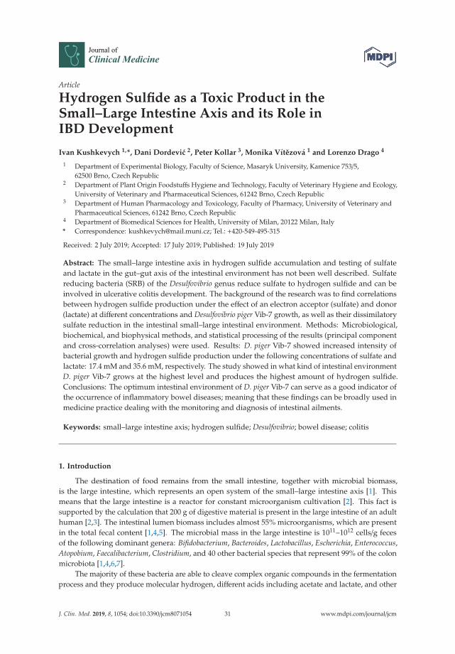
Journal of
Clinical Medicine
Article
Hydrogen Sulfide as a Toxic Product in theSmall–Large Intestine Axis and its Role inIBD Development
Ivan Kushkevych 1,*, Dani Dordevic 2, Peter Kollar 3, Monika Vítezová 1 and Lorenzo Drago 4
1 Department of Experimental Biology, Faculty of Science, Masaryk University, Kamenice 753/5,62500 Brno, Czech Republic
2 Department of Plant Origin Foodstuffs Hygiene and Technology, Faculty of Veterinary Hygiene and Ecology,University of Veterinary and Pharmaceutical Sciences, 61242 Brno, Czech Republic
3 Department of Human Pharmacology and Toxicology, Faculty of Pharmacy, University of Veterinary andPharmaceutical Sciences, 61242 Brno, Czech Republic
4 Department of Biomedical Sciences for Health, University of Milan, 20122 Milan, Italy* Correspondence: [email protected]; Tel.: +420-549-495-315
Received: 2 July 2019; Accepted: 17 July 2019; Published: 19 July 2019
Abstract: The small–large intestine axis in hydrogen sulfide accumulation and testing of sulfateand lactate in the gut–gut axis of the intestinal environment has not been well described. Sulfatereducing bacteria (SRB) of the Desulfovibrio genus reduce sulfate to hydrogen sulfide and can beinvolved in ulcerative colitis development. The background of the research was to find correlationsbetween hydrogen sulfide production under the effect of an electron acceptor (sulfate) and donor(lactate) at different concentrations and Desulfovibrio piger Vib-7 growth, as well as their dissimilatorysulfate reduction in the intestinal small–large intestinal environment. Methods: Microbiological,biochemical, and biophysical methods, and statistical processing of the results (principal componentand cross-correlation analyses) were used. Results: D. piger Vib-7 showed increased intensity ofbacterial growth and hydrogen sulfide production under the following concentrations of sulfate andlactate: 17.4 mM and 35.6 mM, respectively. The study showed in what kind of intestinal environmentD. piger Vib-7 grows at the highest level and produces the highest amount of hydrogen sulfide.Conclusions: The optimum intestinal environment of D. piger Vib-7 can serve as a good indicator ofthe occurrence of inflammatory bowel diseases; meaning that these findings can be broadly used inmedicine practice dealing with the monitoring and diagnosis of intestinal ailments.
Keywords: small–large intestine axis; hydrogen sulfide; Desulfovibrio; bowel disease; colitis
1. Introduction
The destination of food remains from the small intestine, together with microbial biomass,is the large intestine, which represents an open system of the small–large intestine axis [1]. Thismeans that the large intestine is a reactor for constant microorganism cultivation [2]. This fact issupported by the calculation that 200 g of digestive material is present in the large intestine of an adulthuman [2,3]. The intestinal lumen biomass includes almost 55% microorganisms, which are presentin the total fecal content [1,4,5]. The microbial mass in the large intestine is 1011–1012 cells/g fecesof the following dominant genera: Bifidobacterium, Bacteroides, Lactobacillus, Escherichia, Enterococcus,Atopobium, Faecalibacterium, Clostridium, and 40 other bacterial species that represent 99% of the colonmicrobiota [1,4,6,7].
The majority of these bacteria are able to cleave complex organic compounds in the fermentationprocess and they produce molecular hydrogen, different acids including acetate and lactate, and other
J. Clin. Med. 2019, 8, 1054; doi:10.3390/jcm8071054 www.mdpi.com/journal/jcm31
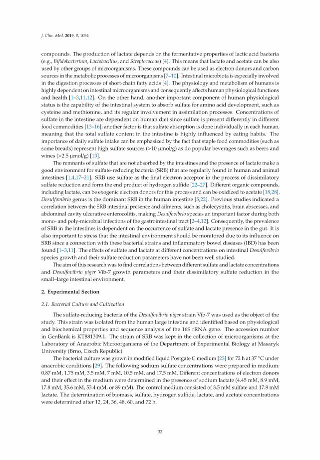
J. Clin. Med. 2019, 8, 1054
compounds. The production of lactate depends on the fermentative properties of lactic acid bacteria(e.g., Bifidobacterium, Lactobacillus, and Streptococcus) [4]. This means that lactate and acetate can be alsoused by other groups of microorganisms. These compounds can be used as electron donors and carbonsources in the metabolic processes of microorganisms [7–10]. Intestinal microbiota is especially involvedin the digestion processes of short-chain fatty acids [4]. The physiology and metabolism of humans ishighly dependent on intestinal microorganisms and consequently affects human physiological functionsand health [1–3,11,12]. On the other hand, another important component of human physiologicalstatus is the capability of the intestinal system to absorb sulfate for amino acid development, such ascysteine and methionine, and its regular involvement in assimilation processes. Concentrations ofsulfate in the intestine are dependent on human diet since sulfate is present differently in differentfood commodities [13–16]; another factor is that sulfate absorption is done individually in each human,meaning that the total sulfate content in the intestine is highly influenced by eating habits. Theimportance of daily sulfate intake can be emphasized by the fact that staple food commodities (such assome breads) represent high sulfate sources (>10 μmol/g) as do popular beverages such as beers andwines (>2.5 μmol/g) [13].
The remnants of sulfate that are not absorbed by the intestines and the presence of lactate make agood environment for sulfate-reducing bacteria (SRB) that are regularly found in human and animalintestines [1,4,17–21]. SRB use sulfate as the final electron acceptor in the process of dissimilatorysulfate reduction and form the end product of hydrogen sulfide [22–27]. Different organic compounds,including lactate, can be exogenic electron donors for this process and can be oxidized to acetate [18,28].Desulfovibrio genus is the dominant SRB in the human intestine [5,22]. Previous studies indicated acorrelation between the SRB intestinal presence and ailments, such as cholecystitis, brain abscesses, andabdominal cavity ulcerative enterocolitis, making Desulfovibrio species an important factor during bothmono- and poly-microbial infections of the gastrointestinal tract [2–4,12]. Consequently, the prevalenceof SRB in the intestines is dependent on the occurrence of sulfate and lactate presence in the gut. It isalso important to stress that the intestinal environment should be monitored due to its influence onSRB since a connection with these bacterial strains and inflammatory bowel diseases (IBD) has beenfound [1–3,11]. The effects of sulfate and lactate at different concentrations on intestinal Desulfovibriospecies growth and their sulfate reduction parameters have not been well studied.
The aim of this research was to find correlations between different sulfate and lactate concentrationsand Desulfovibrio piger Vib-7 growth parameters and their dissimilatory sulfate reduction in thesmall–large intestinal environment.
2. Experimental Section
2.1. Bacterial Culture and Cultivation
The sulfate-reducing bacteria of the Desulfovibrio piger strain Vib-7 was used as the object of thestudy. This strain was isolated from the human large intestine and identified based on physiologicaland biochemical properties and sequence analysis of the 16S rRNA gene. The accession numberin GenBank is KT881309.1. The strain of SRB was kept in the collection of microorganisms at theLaboratory of Anaerobic Microorganisms of the Department of Experimental Biology at MasarykUniversity (Brno, Czech Republic).
The bacterial culture was grown in modified liquid Postgate C medium [23] for 72 h at 37 ◦C underanaerobic conditions [29]. The following sodium sulfate concentrations were prepared in medium:0.87 mM, 1.75 mM, 3.5 mM, 7 mM, 10.5 mM, and 17.5 mM. Different concentrations of electron donorsand their effect in the medium were determined in the presence of sodium lactate (4.45 mM, 8.9 mM,17.8 mM, 35.6 mM, 53.4 mM, or 89 mM). The control medium consisted of 3.5 mM sulfate and 17.8 mMlactate. The determination of biomass, sulfate, hydrogen sulfide, lactate, and acetate concentrationswere determined after 12, 24, 36, 48, 60, and 72 h.
32

J. Clin. Med. 2019, 8, 1054
2.2. Bacterial Biomass Determination
In total, 1 mL of liquid medium without Mohr’s salt in a plastic cuvette was measured in abiophotometer (Eppendorf BioPhotometer®D30, Hamburg, Germany) as a blank. The same procedurewith the bacterial suspension was performed. The optical density (OD340) was always measured beforethe experiments to provide approximately the same amount of SRB in each experiment [7].
2.3. Sulfate Determination
The sulfate concentration in the liquid medium was measured by turbidimetric method after 12 hintervals of cultivation. In total, 40 mg/L BaCl2 solution was prepared in 0.12 M HCl and mixed withglycerol in a 1:1 ratio. The supernatant of the sample was obtained by centrifugation at 5000× g at 23 ◦Cand 1 mL was added to 10 mL of BaCl2:glycerol solution and carefully mixed. The absorbance of themixed solution was measured after 10 min at 520 nm (Spectrosonic Genesis 5, Ecublens, Switzerland).A cultivation medium without bacteria growth was used as a control [30].
2.4. Hydrogen Sulfide Determination
The concentration of hydrogen sulfide was determined in cultivation medium after different timeintervals. In total, 1 mL of the sample was added to 10 mL of a 5 g/L solution of zinc acetate and 2 mLof 0.75 g/mL p-aminodimethylaniline in a solution of sulfuric acid (2 M). The mixture stood for 5 min atroom temperature. After that, 0.5 mL of 12 g/L solution of ferric chloride dissolved in 15 mM sulfuricacid was added. After standing another 5 min at room temperature, the mixture was centrifuged5000× g at 23 ◦C. The absorbance of the mixture was determined at a wavelength of 665 nm by aspectrophotometer (Cecil Aquarius CE 7200 Double Beam Spectrophotometer, London, UK) [31,32].
2.5. Lactate and Acetate Determination
The measurement was repeated in the same manner using a cultivation medium and it served asthe control sample. Measurements of lactate concentration using a lactate assay kit (Sigma-Aldrich,Catalog Number MAK064, Prague, Czech Republic) were carried out. Accumulation of acetate ions inthe process of bacterial growth in the medium was determined using the acetate assay kit (Abnova,Colorimetric, Catalog Number KA3764, Prague, Czech Republic).
2.6. Statistical Analysis
Using the experimental data, the basic statistical parameters (M—mean, m—standard error,M ±m) were calculated. The accurate approximation was when p ≤ 0.0533 [33]. Statistical significancewas measured with the use of principal component analysis (PCA) that gave overall differences amongcompared groups. Statistical analysis was done by SPSS 20 statistical software (IBM Corporation,Armonk, NY, USA). Plots were built by software package Origin7.0 (Northampton, UK).
3. Results
Intestinal sulfate-reducing bacteria, D. piger Vib-7, showed the highest rate (biomass accumulation,sulfate and lactate consumption, and sulfide and acetate production), both increasing and decreasingtrends, until the 60th h of cultivation in the control (3.5 mM of sulfate and 17.3 mM of lactate) medium(Figure 1). The stationary growth phase was achieved after 60 h of cultivation and the followingpercentage decreases and increases in contents were measured: biomass (increased by 87%), sulfate(decreased by 95%), sulfide (increased by 83%), lactate (decreased by 88%) and acetate (increased by91%). Relative growth and survival of D. piger Vib-7 achieved the highest percentages at 7 mM ofsulfate and 35.6 mM of lactate. Higher concentrations than these resulted in the stability of relativegrowth and it stayed at the same level during 12 to 48 h. Lower concentrations of sulfate (<3.5 mM)and lactate (<17.8 mM) were not enough for the achievement of maximum growth parameters.
33

J. Clin. Med. 2019, 8, 1054
Figure 1. The growth of D. piger Vib-7 and their sulfate reduction.
As can be seen in Figure 2, the consumption of sulfate was highly dependent on its differentconcentrations in cultivation medium, time of cultivation, and the presence of lactate donors (it wasconstant at 17.8 mM of lactate).
Figure 2. Growth of D. piger Vib-7, and their survival and sulfate reduction parameters during 12, 24, 36,and 48 h of cultivation: the effect of electron acceptor (sulfate)/donor (lactate) at different concentrations(columns: first = 12 h, second = 24 h, third = 36 h, fourth = 48 h).
34
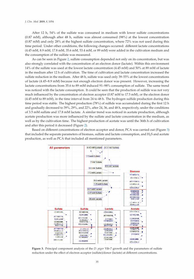
J. Clin. Med. 2019, 8, 1054
After 12 h, 54% of the sulfate was consumed in medium with lower sulfate concentrations(0.87 mM), although after 48 h, sulfate was almost consumed (98%) at the lowest concentration(0.87 mM) and only 28% at the highest sulfate concentration, where 72% was not used during thistime period. Under other conditions, the following changes occurred: different lactate concentrations(4.45 mM, 8.9 mM, 17.8 mM, 35.6 mM, 53.4 mM, or 89 mM) were added in the cultivation medium andthe consumption of the sulfate was measured.
As can be seen in Figure 2, sulfate consumption depended not only on its concentration, but wasalso strongly correlated with the concentration of an electron donor (lactate). Within this environment14% of the sulfate was used at the lowest lactate concentration (4.45 mM) and 50% at 89 mM of lactatein the medium after 12 h of cultivation. The time of cultivation and lactate concentration increased thesulfate reduction in the medium. After 48 h, sulfate was used only 39–55% at the lowest concentrationsof lactate (4.45–8.9 mM) because not enough electron donor was present. However, increasing thelactate concentrations from 35.6 to 89 mM induced 91–98% consumption of sulfate. The same trendwas noticed with the lactate consumption. It could be seen that the production of sulfide was not verymuch influenced by the concentration of electron acceptor (0.87 mM to 17.5 mM), or the electron donor(4.45 mM to 89 mM), in the time interval from 24 to 48 h. The hydrogen sulfide production during thistime period was stable. The highest production (78%) of sulfide was accumulated during the first 12 hand gradually decreased to 39%, 29%, and 22%, after 24, 36, and 48 h, respectively, under the conditionsof 3.5 mM sulfate and 17.8 mM lactate. A similar trend was noticed in acetate production, althoughacetate production was more influenced by the sulfate and lactate concentration in the medium, aswell as by the cultivation time. The highest production of acetate was until the 36th h of cultivationand after this period it decreased (Figure 2).
Based on different concentrations of electron acceptor and donor, PCA was carried out (Figure 3)that included the separate parameters of biomass, sulfate and lactate consumption, and H2S and acetateproduction, as well as PCA that included all mentioned parameters.
Figure 3. Principal component analysis of the D. piger Vib-7 growth and the parameters of sulfatereduction under the effect of electron acceptor (sulfate)/donor (lactate) at different concentrations.
35
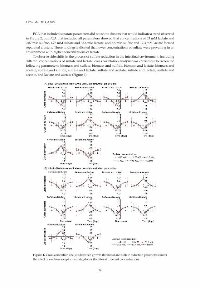
J. Clin. Med. 2019, 8, 1054
PCA that included separate parameters did not show clusters that would indicate a trend observedin Figure 2, but PCA that included all parameters showed that concentrations of 53 mM lactate and0.87 mM sulfate, 1.75 mM sulfate and 35.6 mM lactate, and 3.5 mM sulfate and 17.3 mM lactate formedseparated clusters. These findings indicated that lower concentrations of sulfate were prevailing in anenvironment with higher concentrations of lactate.
To observe side shifts in the process of sulfate reduction in the intestinal environment, includingdifferent concentrations of sulfate and lactate, cross correlation analysis was carried out between thefollowing parameters: biomass and sulfate, biomass and sulfide, biomass and lactate, biomass andacetate, sulfate and sulfide, sulfate and lactate, sulfate and acetate, sulfide and lactate, sulfide andacetate, and lactate and acetate (Figure 4).
Figure 4. Cross-correlation analysis between growth (biomass) and sulfate reduction parameters underthe effect of electron acceptor (sulfate)/donor (lactate) at different concentrations.
36
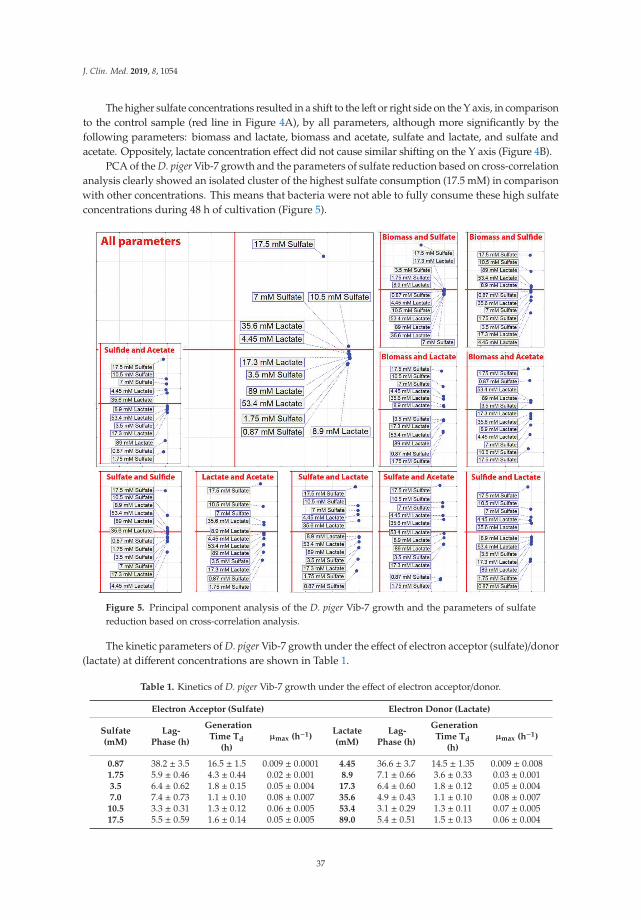
J. Clin. Med. 2019, 8, 1054
The higher sulfate concentrations resulted in a shift to the left or right side on the Y axis, in comparisonto the control sample (red line in Figure 4A), by all parameters, although more significantly by thefollowing parameters: biomass and lactate, biomass and acetate, sulfate and lactate, and sulfate andacetate. Oppositely, lactate concentration effect did not cause similar shifting on the Y axis (Figure 4B).
PCA of the D. piger Vib-7 growth and the parameters of sulfate reduction based on cross-correlationanalysis clearly showed an isolated cluster of the highest sulfate consumption (17.5 mM) in comparisonwith other concentrations. This means that bacteria were not able to fully consume these high sulfateconcentrations during 48 h of cultivation (Figure 5).
Figure 5. Principal component analysis of the D. piger Vib-7 growth and the parameters of sulfatereduction based on cross-correlation analysis.
The kinetic parameters of D. piger Vib-7 growth under the effect of electron acceptor (sulfate)/donor(lactate) at different concentrations are shown in Table 1.
Table 1. Kinetics of D. piger Vib-7 growth under the effect of electron acceptor/donor.
Electron Acceptor (Sulfate) Electron Donor (Lactate)
Sulfate(mM)
Lag-Phase (h)
GenerationTime Td
(h)μmax (h−1)
Lactate(mM)
Lag-Phase (h)
GenerationTime Td
(h)μmax (h−1)
0.87 38.2 ± 3.5 16.5 ± 1.5 0.009 ± 0.0001 4.45 36.6 ± 3.7 14.5 ± 1.35 0.009 ± 0.0081.75 5.9 ± 0.46 4.3 ± 0.44 0.02 ± 0.001 8.9 7.1 ± 0.66 3.6 ± 0.33 0.03 ± 0.0013.5 6.4 ± 0.62 1.8 ± 0.15 0.05 ± 0.004 17.3 6.4 ± 0.60 1.8 ± 0.12 0.05 ± 0.0047.0 7.4 ± 0.73 1.1 ± 0.10 0.08 ± 0.007 35.6 4.9 ± 0.43 1.1 ± 0.10 0.08 ± 0.007
10.5 3.3 ± 0.31 1.3 ± 0.12 0.06 ± 0.005 53.4 3.1 ± 0.29 1.3 ± 0.11 0.07 ± 0.00517.5 5.5 ± 0.59 1.6 ± 0.14 0.05 ± 0.005 89.0 5.4 ± 0.51 1.5 ± 0.13 0.06 ± 0.004
37

J. Clin. Med. 2019, 8, 1054
Under sulfate concentrations of 10.5 mM the shortest lag phase was measured and specificmaximum rate of growth (μmax) was the fastest at 7.0 mM of sulfate. Under electron donor (lactate)concentrations the shortest lag phase and the fastest specific maximum rate of growth were detected at53.4 mM and 35.6 mM of lactate, respectively.
4. Discussion
The sulfate consumption and sulfide production, and the lactate consumption and acetateaccumulation are important factors influencing the intestinal environment [7–10]. Intestinalsulfate-reducing bacteria, especially Desulfovibrio genus, are often found in the intestines and feces ofpeople and animals with IBD. One of the main roles in the development of colitis, among other factors,can also be the species of this genus. These bacteria use sulfate as a terminal electron acceptor andorganic compounds as electron donors in their metabolism [6,7]. This fact leads us to the conclusionthat sulfate present in the daily diet plays an important role in the development of bowel disease.Sulfate is present mainly in the following food commodities: some breads, soya flour, dried fruits,brassicas, and sausages, as well as some beers, ciders, and wines. These data indicate that sulfateintake is highly dependent on diet and the small–large intestine axis [13].
In our previous research, principal component analysis indicated that the Desulfovibrio strains fromindividuals with colitis were grouped in one cluster by biomass accumulation and sulfide production,and the strains from healthy individuals formed another cluster by the same parameters. Sulfate andlactate consumption measured over time showed a negative correlation (Pearson correlations, p < 0.01).The linear regression (R2) was lower in biomass accumulation and hydrogen sulfide production. Thus,biomass accumulation and sulfide production, together with measured kinetic parameters, play animportant factor in bowel inflammation, including ulcerative colitis. Additionally, acetate producedby SRB can also be in synergic interaction with H2S, while sulfate consumption and lactate oxidationlikely represent minor factors in bowel disease [16].
Our results provide an opportunity to find the optimum growing point of the bacteria. The studyconfirmed an intense growth of D. piger Vib-7 in the presence of higher concentrations of electronacceptor and donor, though the consequence is an intensive accumulation of sulfide and acetate. Datafrom the literature indicate that these conditions can be the cause of ulcerative colitis that can leadto cancer of the bowel. This statement is supported by the fact that hydrogen sulfide negativelyaffects intestinal mucosa and epithelial cells, inhibits the growth of colonocytes [4,14–18,34–37], causesphagocytosis, causes the death of intestinal bacteria [4,12,24], and induces hyperproliferation andmetabolic abnormalities of epithelial cells [12]. The high level of metabolites and the presence ofSRB are connected with the inflammation of the colon [4,6,36]. Therefore, the integrity of colonocytesis maintained by hydrogen sulfide concentration [35–37]. Sulfide production is higher among SRBisolated from individuals with ulcerative colitis [5,6].
Other research describing cross-correlation parameters of the SRB metabolic process indicated thatthe strains isolated from people with colitis shifted to the right side of the Y axis by biomass accumulation,sulfate consumption, lactate oxidation, as well as hydrogen sulfide and acetate production, comparedwith the strains isolated from healthy individuals. Different percentages were observed in shiftingto the right side of the Y axis: biomass accumulation 26%, sulfate consumption 1.5%, and sulfideproduction 5% [14]. It should be noted that the intestinal microbiota is a very complex system thatmay limit this study. There are a lot of interactions with clostridia, methanogens, lactic acid bacteria,etc. However, a central role in the development of IBD, especially ulcerative colitis, is SRB [1–3,11].This bacterial group, producing hydrogen sulfide, can inhibit other microbiota, including lactic acidbacteria, methanogens, and many other intestinal microorganisms [2].
A diet high in sulfate ions (preservatives added to food often contain sulfur oxides) causes anincrease in hydrogen sulfide concentration by SRB in rumens. The studies have revealed that thewestern diet contains over 16.6 mmol sulfate/day [13] and the feces of approximately 50% of healthyindividuals contain SRB (up to 92% belong to the genus Desulfovibrio) [1,5]. Sulfate polysaccharides
38

J. Clin. Med. 2019, 8, 1054
such as mucin, chondroitin sulfate, and carrageenan are broadly consumed, and they represent goodsources of sulfate for SRB [24]. It should also be noted that hydrogen sulfide can be toxic not only forintestinal cells, but also for its producers. The highest toxicity of H2S was measured in the presence ofconcentrations higher than 6 mM, where growth was stopped, though metabolic activities were not100% inhibited. These findings are confirmed by cross correlation and principal component analysisthat clearly support the above mentioned results. The presence of 5 mM H2S resulted in a two timeslonger lag phase and generation time was eight times longer. The results confirmed toxicity of H2Stoward Desulfovibrio [18]. Beside sulfate and lactate, terminal oxidative processes in the human largeintestine could be involved in the activities of SRB, and consequently the production of hydrogensulfide in high concentrations that can cause inflammatory bowel disease development.
5. Conclusions
The study gave more information about the intestinal environment in vitro concerning sulfate andlactate concentrations and their effects on the growth parameters of Desulfovibrio piger Vib-7. Almosttotal consumption of sulfate and lactate was achieved after 60 h of cultivation, though the best relativegrowth and stability was measured at 7 mM and 35.6 mM of sulfate and lactate, respectively. PCAincluding separated parameters did not show combined clusters, but PCA based on all parametersshowed that different concentrations of sulfate and lactate formed separated clusters. These obtainedresults represent the main findings of the research, indicating that SRB would grow at the highest levelunder these experimentally simulated conditions. These conditions are an indicator of higher SRBactivity that can lead to the development of IBD, and further studies will certainly focus more on theintestinal environment concerning SRB not only in vitro, but also in vivo.
Author Contributions: Conceptualization, I.K. and D.D.; methodology, I.K. and M.V.; validation, P.K., M.V., andD.D.; formal analysis, M.V. and L.D.; investigation, I.K.; resources, I.K.; data curation, D.D.; writing—originaldraft preparation, I.K., D.D., and M.V.; writing—review and editing, I.K. and L.D.; visualization, I.K.; supervision,M.V.; project administration, I.K.; funding acquisition, I.K., D.D., and P.K.
Funding: This research was supported by Grant Agency of the Masaryk University (MUNI/A/0902/2018).
Conflicts of Interest: The authors declare no conflict of interest.
References
1. Gibson, G.R.; Cummings, J.H.; Macfarlane, G.T. Growth and activities of sulphate-reducing bacteria in gutcontents of health subjects and patients with ulcerative colitis. FEMS Microbiol. Ecol. 1991, 86, 103–112.[CrossRef]
2. Gibson, G.R.; Macfarlane, S.; Macfarlane, G.T. Metabolic interactions involving sulphate-reducing andmethanogenic bacteria in the human large intestine. FEMS Microbiol. Ecol. 1993, 12, 117–125. [CrossRef]
3. Cummings, J.H.; Macfarlane, G.T.; Macfarlane, S. Intestinal Bacteria and Ulcerative Colitis. Curr. Issues Intest.Microbiol. 2003, 4, 9–20. [PubMed]
4. Barton, L.L.; Hamilton, W.A. Sulphate-Reducing Bacteria Environmental and Engineered Systems; CambridgeUniversity Press: Cambridge, UK, 2017.
5. Loubinoux, J.; Bronowicji, J.P.; Pereira, I.A. Sulphate-reducing bacteria in human feces and their associationwith inflammatory diseases. FEMS Microbiol. Ecol. 2002, 40, 107–112. [CrossRef] [PubMed]
6. Kovác, J.; Vítezová, M.; Kushkevych, I. Metabolic activity of sulfate-reducing bacteria from rodents withcolitis. Open Med. 2018, 13, 344–349.
7. Kushkevych, I.; Vítezová, M.; Fedrová, P.; Vochyanová, Z.; Paráková, L.; Hošek, J. Kinetic properties ofgrowth of intestinal sulphate-reducing bacteria isolated from healthy mice and mice with ulcerative colitis.Acta Vet. Brno 2017, 86, 405–411. [CrossRef]
8. Kushkevych, I.; Fafula, R.; Parak, T.; Bartoš, M. Activity of Na+/K+-activated Mg2+-dependent ATP hydrolasein the cell-free extracts of the sulfate-reducing bacteria Desulfovibrio piger Vib-7 and Desulfomicrobium sp.Rod-9. Acta Vet. Brno 2015, 84, 3–12. [CrossRef]
39
J. Clin. Med. 2019, 8, 1054
9. Kushkevych, I.V. Activity and kinetic properties of phosphotransacetylase from intestinal sulfate-reducingbacteria. Acta Biochem. Pol. 2015, 62, 1037–1108. [CrossRef]
10. Kushkevych, I.V. Kinetic Properties of Pyruvate Ferredoxin Oxidoreductase of Intestinal Sulfate-ReducingBacteria Desulfovibrio piger Vib-7 and Desulfomicrobium sp. Rod-9. Pol. J. Microbiol. 2015, 64, 107–114.
11. Loubinoux, J.; Mory, F.; Pereira, I.A.; Le Faou, A.E. Bacteremia caused by a strain of Desulfovibrio related tothe provisionally named Desulfovibrio fairfieldensis. J. Clin. Microbiol. 2000, 38, 931–934.
12. Pitcher, M.C.; Cummings, J.H. Hydrogen sulphide: A bacterial toxin in ulcerative colitis? Gut 1996, 39, 1–4.[CrossRef] [PubMed]
13. Florin, T.H.; Neale, G.; Goretski, S. Sulfate in food and beverages. J. Food Compos. Anal. 1993, 6, 140–151.[CrossRef]
14. Kushkevych, I.; Dordevic, D.; Vítezová, M.; Kollár, P. Cross-correlation analysis of the Desulfovibrio growthparameters of intestinal species isolated from people with colitis. Biologia 2018, 73, 1137–1143. [CrossRef]
15. Kushkevych, I.; Dordevic, D.; Vítezová, M. Analysis of pH dose-dependent growth of sulfate-reducingbacteria. Open Med. 2019, 14, 66–74. [CrossRef] [PubMed]
16. Kushkevych, I.; Dordevic, D.; Kollar, P. Analysis of physiological parameters of Desulfovibrio strains fromindividuals with colitis. Open Life Sci. 2018, 13, 481–488. [CrossRef]
17. Kushkevych, I.; Vítezová, M.; Kos, J.; Kollár, P.; Jampilek, J. Effect of selected 8-hydroxyquinoline-2-carboxanilides on viability and sulfate metabolism of Desulfovibrio piger. J. Appl. Biomed. 2018, 16, 241–246.[CrossRef]
18. Kushkevych, I.; Dordevic, D.; Vítezová, M. Toxicity of hydrogen sulfide toward sulfate-reducing bacteriaDesulfovibrio piger Vib-7. Arch. Microbiol. 2019, 201, 1–9. [CrossRef] [PubMed]
19. Kushkevych, I.; Kollar, P.; Suchy, P.; Parak, T.; Pauk, K.; Imramovsky, A. Activity of selected salicylamidesagainst intestinal sulfate-reducing bacteria. Neuroendocrinol. Lett. 2015, 36, 106–113. [PubMed]
20. Kushkevych, I.; Kollar, P.; Ferreira, A.L.; Palma, D.; Duarte, A.; Lopes, M.M.; Bartos, M.; Pauk, K.;Imramovsky, A.; Jampilek, J. Antimicrobial effect of salicylamide derivatives against intestinal sulfate-reducing bacteria. J. Appl. Biomed. 2016, 14, 125–130. [CrossRef]
21. Kushkevych, I.; Kos, J.; Kollar, P.; Kralova, K.; Jampilek, J. Activity of ring-substituted 8-hydroxyquinoline-2-carboxanilides against intestinal sulfate-reducing bacteria Desulfovibrio piger. Med. Chem. Res. 2018, 27,278–284. [CrossRef]
22. Loubinoux, J.; Valente, F.M.A.; Pereira, I.A.C. Reclassification of the only species of the genus Desulfomonas,Desulfomonas pigra, as Desulfovibrio piger comb. nov. Int. J. Syst. Evol. Microbiol. 2002, 52, 1305–1308.[PubMed]
23. Postgate, J.R. The Sulfate Reducing Bacteria; Cambridge University Press: Cambridge, UK, 1984.24. Rowan, F.E.; Docherty, N.G.; Coffey, J.C.; O’Connell, P.R. Sulphate-reducing bacteria and hydrogen sulphide
in the aetiology of ulcerative colitis. Br. J. Surg. 2009, 96, 151–158. [CrossRef] [PubMed]25. Kushkevych, I.; Vítezová, M.; Vítez, T.; Bartoš, M. Production of biogas: Relationship between methanogenic
and sulfate-reducing microorganisms. Open Life Sci. 2017, 12, 82–91. [CrossRef]26. Kushkevych, I.; Vítezová, M.; Vítez, T.; Kovac, J.; Kaucká, P.; Jesionek, W.; Bartoš, M.; Barton, L. A new
combination of substrates: Biogas production and diversity of the methanogenic microorganisms. Open LifeSci. 2018, 13, 119–128. [CrossRef]
27. Kushkevych, I.; Kovác, J.; Vítezová, M.; Vítez, T.; Bartoš, M. The diversity of sulfate-reducing bacteria in theseven bioreactors. Arch. Microbiol. 2018, 200, 945–950. [CrossRef] [PubMed]
28. Cerný, M.; Vítezová, M.; Vítez, T.; Bartoš, M.; Kushkevych, I. Variation in the Distribution of HydrogenProducers from the Clostridiales Order in Biogas Reactors Depending on Different Input Substrates. Energies2018, 11, 3270.
29. Kovác, J.; Kushkevych, I. New modification of cultivation medium for isolation and growth of intestinalsulfate-reducing bacteria. In Proceedings of the International PhD Students Conference MendelNet, Brno,Czechia, 6–7 November 2019; pp. 702–707.
30. Kolmert, A.; Wikstrom, P.; Hallberg, K.B. A fast and simple turbidimetric method for the determination ofsulfate in sulfate-reducing bacterial cultures. J. Microbiol. Methods 2000, 41, 179–184. [CrossRef]
31. Cline, J.D. Spectrophotometric determination of hydrogen sulfide in natural water. Limnol. Oceanogr. 1969,14, 454–458. [CrossRef]
40
J. Clin. Med. 2019, 8, 1054
32. Bailey, T.S.; Pluth, M.D. Chemiluminescent detection of enzymatically produced hydrogen sulfide: Substratehydrogen bonding influences selectivity for H2S over biological thiols. J. Am. Chem. Soc. 2013, 135,16697–16704. [CrossRef]
33. Bailey, N.T.J. Statistical Methods in Biology; Cambridge University Press: Cambridge, UK, 1995.34. Attene-Ramos, M.S.; Wagner, E.D.; Plewa, M.J.; Gaskins, H.R. Evidence that hydrogen sulfide is a genotoxic
agent. Mol. Cancer Res. 2006, 4, 9–14. [CrossRef]35. Beauchamp, R.O.; Bus, J.S.; Popp, J.A.; Boreiko, C.J.; Andjelkovich, D.A.; Leber, P. A critical review of the
literature on hydrogen sulfide toxicity. CRC Crit. Rev. Toxicol. 1984, 13, 25–97. [CrossRef] [PubMed]36. Blachier, F.; Davila, A.M.; Mimoun, S. Luminal sulfide and large intestine mucosa: Friend or foe? Amino Acids
2010, 39, 335–347. [CrossRef] [PubMed]37. Grieshaber, M.K.; Völkel, S. Animal adaptations for tolerance and exploitation of poisonous sulfide. Annu. Rev.
Physiol. 1998, 60, 33–53. [CrossRef] [PubMed]
© 2019 by the authors. Licensee MDPI, Basel, Switzerland. This article is an open accessarticle distributed under the terms and conditions of the Creative Commons Attribution(CC BY) license (http://creativecommons.org/licenses/by/4.0/).
41

Journal of
Clinical Medicine
Review
Upper Respiratory Tract Microbiome and OtitisMedia Intertalk: Lessons from the Literature
Francesco Folino 1,*, Luca Ruggiero 2, Pasquale Capaccio 3,4, Ilaria Coro 1,2, Stefano Aliberti 1,5,
Lorenzo Drago 6, Paola Marchisio 1,2 and Sara Torretta 3,7
1 Department of Pathophysiology and Transplantation, University of Milan, 20122 Milan, Italy;[email protected] (I.C.); [email protected] (S.A.); [email protected] (P.M.)
2 Pediatric Highly Intensive Care Unit, Fondazione IRCCS Ca’ Granda Ospedale Maggiore Policlinico,20122 Milan, Italy; [email protected]
3 Department of Otolaryngology and Head and Neck Surgery, Fondazione IRCCS Ca’ Granda OspedaleMaggiore Policlinico, 20122 Milan, Italy; [email protected] (P.C.); [email protected] (S.T.)
4 Department of Biomedical Surgical Dental Science, University of Milan, 20122 Milan, Italy5 Internal Medicine Department, Respiratory Unit and Adult Cystic Fibrosis Center, Fondazione IRCCS Ca’
Granda Ospedale Maggiore Policlinico, 20122 Milan, Italy6 Laboratory of Clinical Microbiology, Department of Biomedical Science for Health, University of Milan,
20122 Milan, Italy; [email protected] Department of Clinical Sciences and Community Health, University of Milan, 20122 Milan, Italy* Correspondence: [email protected]
Received: 19 July 2020; Accepted: 31 August 2020; Published: 2 September 2020
Abstract: Otitis media (OM) is one of the most common diseases occurring during childhood.Microbiological investigations concerning this topic have been primarily focused on the fourclassical otopathogens (Streptococcus pneumoniae, Haemophilus influenzae, Moraxella catarrhalis andStreptococcus pyogenes) mainly because most of the studies have been conducted with culture-dependentmethods. In recent years, the introduction of culture-independent techniques has allowedhigh-throughput investigation of entire bacterial communities, leading to a better comprehensionof the role of resident flora in health and disease. The upper respiratory tract (URT) is a region ofmajor interest in otitis media pathogenesis, as it could serve as a source of pathogens for the middleear (ME). Studies conducted with culture-independent methods in the URT and ME have providednovel insights on the pathogenesis of middle ear diseases through the identification of both possiblenew causative agents and of potential protective bacteria, showing that imbalances in bacterialcommunities could influence the natural history of otitis media in children. The aim of this review is toexamine available evidence in microbiome research and otitis media in the pediatric age, with a focuson its different phenotypes: acute otitis media, otitis media with effusion and chronic suppurativeotitis media.
Keywords: otitis media; microbiota; upper respiratory tract; adenoid; middle ear; microbiota axes
1. Introduction
The human microbiota consists of ecological communities of commensal, symbiotic and pathogenicmicroorganisms that colonize several body sites, as the gastrointestinal tract, respiratory system, oralcavity, skin and female reproductive system [1]. In past years, microbiological investigations have beenpredominantly conducted with culture-dependent methods, therefore many sites in the human bodyhave been considered sterile until recently. However, the introduction of culture-independenttechniques has allowed investigation of entire bacterial communities [2], leading to a bettercomprehension of the role of resident flora in health and disease. These microorganisms and
J. Clin. Med. 2020, 9, 2845; doi:10.3390/jcm9092845 www.mdpi.com/journal/jcm43

J. Clin. Med. 2020, 9, 2845
their products play indeed a critical role in the regulation of many homeostatic processes, includingimmune response and inflammation [3] and defense against pathogens [4]. A diseased alterationin the composition of these bacterial communities, defined dysbiosis, can therefore lead to manypathological conditions, including infections [5].
Most of these studies have been conducted with a marker gene analysis based on a broad-rangePCR, using primers that target a segment of the 16SrRNA gene, a highly conserved region containedin bacterial genomes. This method, combined with next-generation sequencing technologies, permitsthe simultaneous characterization of an entire community [6]. This approach allows a fast andcost-effective analysis that provides a low-resolution view of a microbial community. However, thereare also some limitations that should be taken into account when interpreting data derived from thesestudies: it is not possible to determine whether taxa detected are alive or dead, active or inactive, thusthere is limited functional information; it is susceptible to over amplification bias, especially withlow biomass samples such as middle ear fluid; as a short segment of 16SrRNA gene is amplified andsequenced, taxonomic resolution is usually limited to family or genus level; there is great variabilitydepending on technical aspects as region selection, amplicon size, sampling, storage, sequencingapproach, and bioinformatic analysis. Full-gene 16S rRNA gene sequencing and metagenome andmetatrascriptome analyses may overcome some of these limitations but are less adopted as they arerelative expensive and complex to perform [7]. Middle ear infections and diseases are widespreadin pediatric age. Acute otitis media (AOM) is the most common bacterial infection in childhood [8]and the leading cause of antibiotic prescription in pediatric patients [9]; similarly, otitis media witheffusion (OME) is prevalent in the first years of life, as up to 80% of children suffer from one or moreepisodes by 10 years of age; however, it should be considered that the prevalence of OME varies acrosspopulation and could be difficult to define accurately, as this condition is often asymptomatic [10].
The upper respiratory tract (URT) is a region of major interest in otitis media pathogenesis:According to the Pathogen Reservoir Hypothesis (PRH), the adenoid pad serves as a source ofpathogens that can grow in this region and further spread to the respiratory system and middle ear,leading to infections and diseases [11–14].
The URT extends from the nostrils to the portion of the larynx above the vocal cords and harborsthe highest bacterial density in the whole respiratory system [15]; however, these bacterial communitieshave been studied with more effort and from an ecological perspective only in recent years, afterthe introduction of culture-independent techniques [16].
Scientific interest has been focused on the comprehension of the characteristics of a healthyURT microbiota and the mechanism that guarantees its balance, as mounting evidence shows thatresident bacteria are able to inhibit colonization and growth of otopathogens [14,17,18]. Thosemicroorganisms that are essential in maintaining balance and function of a bacterial community aredefined keystone species (see Table 1 for definitions of common terms used in microbiota analysis).In the URT, Dolosigranulum spp. and Corynebacterium spp. have been identified as potential keystonespecies, as they have been associated with respiratory health and exclusion of otopathogens in severalstudies [19–22].
Reconstitution of healthy microbial communities through administration of probiotics forthe prevention of middle ear diseases in children is a topic of major clinical and scientific interest.Several trials have been conducted, but results lack consistency [23,24]. Deepening our knowledgeon the physiological features of the URT microbiota and understanding how modifications in itsbalance relate to the pathogenesis of otitis media could be of remarkable importance in developingprobiotic therapies. Furthermore, middle ear microbiota involvement in this field has been gaininginterest in recent years, although less studies are available in comparison with URT microbiota, due tothe different feasibility in collecting samples.
The aim of this review is to examine evidence available in microbiome research on otitis mediain children. We will describe the most important factors that impact on microbiota developmentin the first years of life and that could influence the natural history of otitis media; then, we will
44
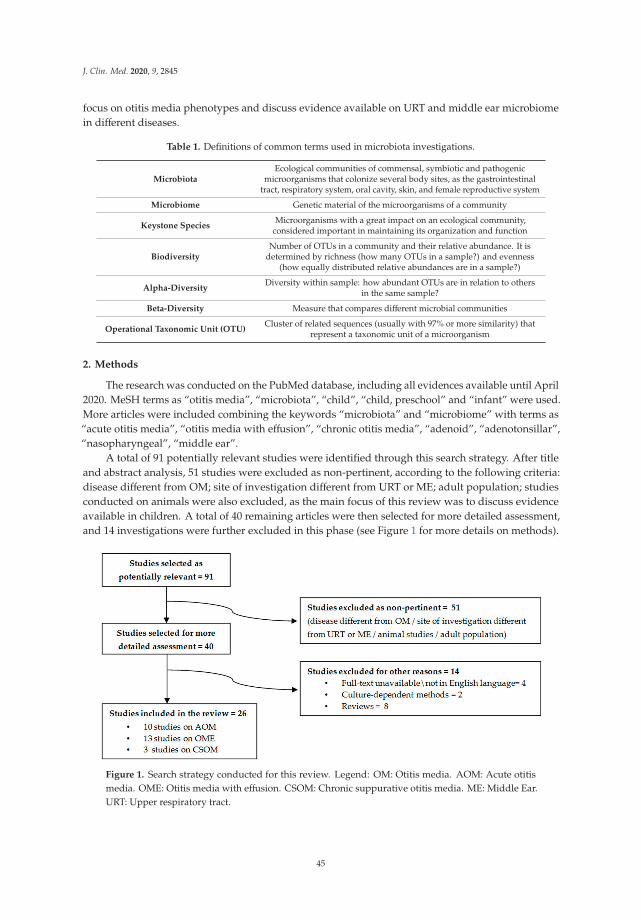
J. Clin. Med. 2020, 9, 2845
focus on otitis media phenotypes and discuss evidence available on URT and middle ear microbiomein different diseases.
Table 1. Definitions of common terms used in microbiota investigations.
MicrobiotaEcological communities of commensal, symbiotic and pathogenic
microorganisms that colonize several body sites, as the gastrointestinaltract, respiratory system, oral cavity, skin, and female reproductive system
Microbiome Genetic material of the microorganisms of a community
Keystone SpeciesMicroorganisms with a great impact on an ecological community,
considered important in maintaining its organization and function
BiodiversityNumber of OTUs in a community and their relative abundance. It is
determined by richness (how many OTUs in a sample?) and evenness(how equally distributed relative abundances are in a sample?)
Alpha-DiversityDiversity within sample: how abundant OTUs are in relation to others
in the same sample?
Beta-Diversity Measure that compares different microbial communities
Operational Taxonomic Unit (OTU)Cluster of related sequences (usually with 97% or more similarity) that
represent a taxonomic unit of a microorganism
2. Methods
The research was conducted on the PubMed database, including all evidences available until April2020. MeSH terms as “otitis media”, “microbiota”, “child”, “child, preschool” and “infant” were used.More articles were included combining the keywords “microbiota” and “microbiome” with terms as“acute otitis media”, “otitis media with effusion”, “chronic otitis media”, “adenoid”, “adenotonsillar”,“nasopharyngeal”, “middle ear”.
A total of 91 potentially relevant studies were identified through this search strategy. After titleand abstract analysis, 51 studies were excluded as non-pertinent, according to the following criteria:disease different from OM; site of investigation different from URT or ME; adult population; studiesconducted on animals were also excluded, as the main focus of this review was to discuss evidenceavailable in children. A total of 40 remaining articles were then selected for more detailed assessment,and 14 investigations were further excluded in this phase (see Figure 1 for more details on methods).
Figure 1. Search strategy conducted for this review. Legend: OM: Otitis media. AOM: Acute otitismedia. OME: Otitis media with effusion. CSOM: Chronic suppurative otitis media. ME: Middle Ear.URT: Upper respiratory tract.
45

J. Clin. Med. 2020, 9, 2845
After this process, 26 studies were included in this review: 10 studies on acute otitis media (AOM,1321 subject enrolled in all studies), 13 studies on otitis media with effusion (OME, 501 subjects enrolledin all studies), 3 studies on chronic suppurative otitis media (CSOM, 217 subjects enrolled in all studies).
3. Environmental Factors and Microbiota Development in the First Years of Life
The microbial communities that colonize the human organism are dynamic and change throughoutlife under the effect of several environmental factors, but infancy and early childhood representthe critical period in shaping their composition [25,26]. These external factors can impair the homeostaticfunctions mediated by the microbiota, leading to immediate consequences or impacting the healthstatus in the later stages of life [27]. This is particularly evident for the URT microbiota, as this regionis interconnected with middle ear, lower respiratory tract, and gastrointestinal tract, and representsthe interface between these systems and the external environment.
Immediately after birth, in the first hours of life, the URT in healthy neonates becomes colonizedby microorganisms of maternal origin [28]. Niche differentiation starts in the first week of life, witha predominance of Staphylococcus spp., followed by an enrichment of Corynebacterium, Dolosigranulum,and Moraxella [29].
The first months of life are of remarkable importance in the development of URT microbialcommunities and their composition: Biesbroek et al. described eight distinct microbiota profilesin the URT of healthy infants, showing that a distinct bacterial profile could be identified by the sixthweek of life; moreover, this early bacterial colonization plays a pivotal role in the stability of microbialcommunities: profiles dominated by Moraxella and Dolosigranulum/Corynebacterium are associated witha stable microbiota and with lower rates of respiratory infections in later stages of life, while less stableprofiles are associated with high abundance of Haemophilus and Streptococcus [30].
Theo et al. confirmed the role of Corynebacterium and found a positive role of Alloiococcusin the first year of life in the development of URT microbial communities; moreover, authors reporteddata on nasopharyngeal (NP) microbiota in children with respiratory diseases, concluding that someMoraxella spp. were associated with an increased risk of disease rather than respiratory health [31].
Several environmental factors, discussed below, can influence the shaping of the URT microbiotacomposition in the first years of life.
3.1. Delivery Route
As it is generally known, children born by caesarian-section (C-section) suffer from a higher incidenceof respiratory illness and morbidity in comparison with children born by vaginal delivery [32,33].
In one of the first reports concerning nasopharyngeal microbiota and route of delivery, swabsfrom different body sites were collected from healthy neonates immediately after birth: Authors foundthat undifferentiated microbial communities in vaginally delivered children were similar to maternalvaginal microbiota, while those who were born by C-section had microbial communities resemblingmaternal skin surface [28].
A subsequent longitudinal study on this theme analyzed nasopharyngeal swabs collected from102 children in the first 6 months of life, showing a predominance of bacteria previously associatedto microbiome stability and respiratory health in early stages of life (Moraxella, Corynebacterium,and Dolosigranulum) in children born by vaginal delivery [29]. These microorganisms are likely derivedfrom maternal skin (Staphylococcus and Corynebacterium) [34] or from vaginal tract (Dolosigranulum,Staphylococcus, or Streptococcus) [35].
However, by contrast, another study evidenced that differences related to delivery route aretransient and disappear by six weeks of age, suggesting that the development of the microbiotain the postnatal period is more related to the body site that harbors a community [36].
46

J. Clin. Med. 2020, 9, 2845
3.2. Breastfeeding
Breastfeeding is a significant protective factor against infections [37,38]. This effect is related notonly to the presence of antibacterial substances in maternal milk [39], as it is known that breastfeedingcan significantly facilitate the development of a healthy microbiota.
Biesbroek et al. showed that breastfed infants develop a bacterial profile enriched by Dolosigranulumand Corynebacterium at six weeks of age in comparison with formula fed infants; moreover,Dolosigranulum abundance was inversely associated with wheezing episodes and a number of parentalreported respiratory tract infections, even after correction for feeding type. [40].
Similar data were provided by Bosch et al.: Children who suffered from a higher number ofrespiratory infections had an aberrant nasopharyngeal microbiota development in the first monthof life, that coincided with a prolonged reduction of Dolosigranulum and Corynebacterium; authorsfound that breastfeeding was an independent driver of this aberrant development, as a prolongeddominance of these bacteria was observed in breastfed infants. However, similarly to delivery route,these dissimilarities are transient and disappear around six months of age [41].
3.3. Antibiotic Therapy
Antibiotic therapy can significantly impair composition and balance of the microbiome [42].This is particularly relevant in pediatric age, in which antibiotic prescription and misuse is quitecommon [43].
In the URT, antibiotic administration causes a reduction of the abundance of potential beneficialbacteria, such as Dolosigranulum and Corynebacterium, and an increase in Haemophilus, Streptococcus,and Moraxella [31]. Moreover, in children with AOM, a recent antibiotic therapy induces a reductionof Streptococcaceae and Corynebacteriaceae and an increased abundance of Enterobacteriaceae andPasturellaceae in the URT [44]. Subsequent longitudinal studies confirmed how antibiotic treatmentcan induce a reduction in the abundance of potential beneficial bacteria, as Dolosigranulum andCorynebacterium [41,45].
3.4. Pneumococcal Vaccination
The introduction of the pneumococcal conjugate vaccination (PCV) in the pediatric populationhas led to an important reduction of OM episodes caused by the serotypes included in the vaccine [46].On the other hand, the introduction of pneumococcal vaccination programs has resulted in importantmodifications in OM microbiology: H. influenzae has become the most common otopathogen andOM episodes caused by M. catarrhalis have become more frequent; moreover, serotypes not includedin PCVs have been more frequently identified as causative agents of diseases [47,48].
These findings suggest that the introduction of PCVs might have induced modificationsin the composition of the microbial communities in the respiratory system. However, evidencesupporting these findings has been focused prevalently on otopathogens, while studies conductedwith high-throughput methods and looking at whole bacterial communities in the URT are lackingand show conflicting results.
In one of the first investigations on the whole NP microbiota and AOM, Hilty et al. reportedthat a previous exposure to PCV-7 in children with AOM was associated with reduced abundance ofcommensal families (Streptococcaceae and Corynebacteriaceae) [44].
A possible influence of this vaccination on NP microbiota was later reported by Biesbroek et al.In this investigation, NP swabs were collected from healthy children who received PCV-7 and fromunvaccinated children: vaccination affected the URT microbiota causing a shift in composition andstructure of the bacterial community, with an increase of Veillonella, Prevotella, Fusobacterium, Leptotrichia,Actinomyces, Rothia, and non pneumococcal streptococci, in addition to an increased bacterial diversityand inter-individual variability [49].
47
J. Clin. Med. 2020, 9, 2845
Longitudinal data on this theme were further provided in another study conducted in Switzerlandby Mika et al., who compared NP microbiota in healthy children who were vaccinated with PCV-7 orPCV-13, showing that those who received PCV-13 had a more diverse and stable URT microbiota anda lower pneumococcal carriage rate compared to those who received PCV-7 [50].
However, in contrast to these findings, other available studies suggest that PCV might not havesuch a relevant impact on the URT microbiota. Faezel et al. performed a randomized controlled trialin Kenya comparing NP microbiota of children who received a 10-valent pneumococcal vaccine vs.children who received Hepatitis A vaccine. In this longitudinal study, NP swabs were collected beforethe administration of the vaccine and after 6 months. The authors found that PCV did not cause anysignificant alteration in the abundance or prevalence of otopathogens [51].
Moreover, a more detailed longitudinal study conducted in Gambia analyzed NP swabs collectedperiodically from birth to the first year of life. Children were divided in three groups according tovaccination schedule: Two groups received two different types of PCV-7, while the third group wascomposed by unvaccinated children. Again, bacterial communities were comparable across groups,as there were no significant differences in richness, diversity, and composition. Interestingly, PCV-7vaccination reduced the nasopharyngeal carriage of vaccine serotypes, but pneumococcal carriageremained high among vaccinated infants, probably because of an immediate expansion of non-vaccineserotypes [52].
Interesting data were provided by Andrade et al. in a complex investigation that compared53 children vaccinated with PCV-10 vs. 27 unvaccinated children. The strength of this study isthe integrated metagenomic and transcriptomic analysis: no difference were found in nasopharyngealcarriage rates of S. pneumoniae, S. aureus, H. influenzae, or M. catarrhalis by either transcriptomicormetagenomics analysis, but unvaccinated children had higher metabolic rates for S. pneumoniae,compared to PCV-10 vaccinated children [53].
Available evidence thus suggest that PCV has a direct impact on pneumococcal carriage, whichin turn might indirectly affect the whole bacterial community in the URT. However, results areconflicting: a possible explanation could be found in the variation of pneumococcal carriage ratesin relation to the geographic region and socio-economic status: the effects of PCV might indeed bedifferent while considering developed or developing countries [54].
This particular theme was investigated in a study conducted in Fiji, in which NP microbiota fromtwo ethnic groups (iTaukei and Fijians of Indian descent) was analyzed. These groups are known tohave a different carriage prevalence of S. pneumoniae and a different burden of pneumococcal disease,which is higher in the iTaukei population. NP swabs were collected from 132 total children belongingto the two ethnic groups that were further divided in two subgroups based on whether children hadbeen previously vaccinated or not with PCV-7. The vaccination had no overall impact on microbialdiversity or composition, but significant modifications were evident when stratifying by ethnicity:vaccinated iTaukei children had a lower relative abundance of Streptococcus and Haemophilus comparedwith unvaccinated ones, while vaccinated Indian descent children had a higher relative abundance ofDolosigranulum compared with those unvaccinated [55].
3.5. Smoking
Studies conducted in adult subjects suggest that active smoking impairs URT microbiotacomposition [56]. It is likely that similar effects involve the pediatric population; however, studiesconcerning active and passive smoking effects on URT microbiome in children are lacking.
4. Acute Otitis Media
Acute otitis media (AOM) is defined by the presence of fluid in the middle ear associated to signsand symptoms of acute infection. It affects the majority of children in the first 3 years of life andbecomes recurrent in almost 50% of cases [10]. Recurrent acute otitis media (RAOM) is defined as fouror more AOM episodes in one year or three or more episodes in 6 months [57].
48

J. Clin. Med. 2020, 9, 2845
Laufer et al. performed one of the first studies comparing NP microbiome in children withAOM to healthy children. The authors evidenced that a higher relative abundance of Corynebacteriumand Dolosigranulum, in addition to Propionibacterium, Lactococcus, and Staphylococcus, was associatedwith a lower incidence of pneumococcal colonization and AOM. The same study showed that a lessdiverse and a less even microbiota was associated with colonization by S. pneumoniae, highlightingthe correlation between a higher biodiversity and better outcomes [20].
These data were confirmed in a subsequent investigation conducted by the same group on240 children aged 6 months–3 years, that evidenced that a lower biodiversity was associated witha higher colonization rate not only from S. pneumoniae but also from H. influenzae and M. catharralis;moreover, authors compared diversity indices between health status and during an acute upperrespiratory infection (URTI), showing that biodiversity was significantly higher in healthy childrenthan during disease [19].
These findings on biodiversity during URTI are coherent with data provided by Hilty et al. thatevidenced how NP bacterial density is lower in children during an AOM episode compared withthe same in healthy status. Moreover, interesting insights were provided on how the infants’ microbiotaundergoes changes during an AOM episode, as the classical otopathogens predominated overcommensal families (Staphylococcaceae, Flavobacteriaceae, Carnobacteriaceae, and Comamonadaceae) [44].
In 2017 Chonmaitree et al. performed a longitudinal study on 139 healthy neonates, followedsince birth for the first 12 months of life or until the occurrence of the first AOM episode, collecting971 swabs performed monthly and during an URTI or AOM. In particular, as it is known that URTIoften precedes an AOM episode, authors studied the characteristics of the NP microbiome duringtransitional phase from URTI to AOM. Data revealed that an unstable microbiota during an URTIepisode with the predominance of otopathogens were associated with the occurrence of symptomaticviral infection and with a higher risk of transition from URTI to AOM. Interestingly, otopathogenswere not predominant during otherwise asymptomatic viral infections [45].
Evidence on otitis-prone children, i.e., those already suffering for RAOM were provided byDirain et al.: Authors compared the microbial flora on adenoid tissue in a small group of subjectsundergoing adenoidectomy for RAOM (n = 5) or obstructive sleep apnea (OSA) (n = 5), finding thatthe relative abundance of S. pneumoniae and M. catharralis was higher in the RAOM group [58].
A complex study with a higher sample size was subsequently performed on an Australianpopulation, comparing NP microbiome of 103 healthy children vs. 93 otitis-prone children undergoinggrommet insertion for RAOM, in order to identify potential protective genera. This investigationconfirmed the pivotal role of Dolosigranulum and Corynebacterium in NP microbiome, as these twogenera have been found to be significantly more abundant in the NP of healthy children comparedwith otitis-prone children. As for biodiversity, in contrast with previous findings, this study found thatotitis-prone children had a significantly more diverse microbiome than controls. In addition, authorsanalyzed middle ear fluid (MEF) microbiome collected from children undergoing surgery from RAOMand performed a paired comparison with the NP microbiome of the same subject. Results showed thatthese two niches were not highly concordant: In particular, the interesting data is that Alloiococcus andTuricella have been found to be abundant in MEF but almost absent in the NP [22].
The MEF microbiome during an AOM episode was further investigated on 79 subjects aged5–42 months. This report confirmed that the classical otopathogens are the predominant species in MEFduring AOM: S. pneumoniae was dominant in 16% of samples, H. influenzae in 17%, and M. catarrhalisin 5.6%; moreover, Turicell aotitidis was detected as a clearly dominant bacteria in two samples,suggesting that it could be a rare but true causative agent; Alloiococcus otitidis was detected onlyin 3 samples; Staphylococcus auricolaris was predominant in two samples, but authors speculatedthat this finding could be related to potential contamination from the external auditory canal (EAC);however, A. otitidis and T. otitidis could be also related to EAC contamination [59].
Xu et al. compared the MEF microbiota during AOM episode to the NP microbiota analyzedon nasal wash (NW) samples: A significantly higher abundance of A. otitidis was detected in MEF
49
J. Clin. Med. 2020, 9, 2845
during AOM, compared with NW in health and disease; authors concluded that the ME could harbora resident microbiome that becomes different from NP after the onset of an infection. Moreover,NP microbiome was analyzed prior to the onset of AOM vs. at AOM onset: In line with previous data,NP microbiome during health was significantly more diverse than during AOM [60].
Paired analysis of NP and MEF microbiome during an AOM episode was subsequently performedon a larger population, collecting 286 NP swabs in children aged 0–6 years; 42/286 episodes werecharacterized by spontaneous tympanic membrane perforation (STMP), and thus, MEF microbiomewas analyzed in these cases. Authors found that diversity was strictly related to age: in particular, olderchildren had a higher richness and showed more personalized bacterial profiles, that develop towardthe end of the sixth year of life. The transition to an adult-like microbiome appeared in children olderthan 3 years and was defined by an increase in Staphylococcaceae and Corynebacteriaceae. Furthermore,authors found concordance between NP and MEF microbiome when the predominant bacteria in MEFwas S. pyogenes, H. influenzae, or S. pneumoniae. However, even this event appeared to be age-related,as the concordance between NP and MEF microbiome became weaker as children got older. Authorsthus concluded that the NP microbiota does not necessarily resembles the one in ME: The URTin children with AOM serves as a moderate proxy for MEF at a very young age but becomes morediverse at a more advanced age [61].
The most frequently observed complication of AOM in clinical practice is the spontaneoustympanic membrane perforation (STMP) [62]. However, evidence on microbiota in children withhistory of RAOM with STMP is lacking. We believe that this condition represents a distinct phenotypeof disease in otitis-prone children [63], and more effort should be directed to this category of patients,since their clinical management is often very challenging, and the most important AOM preventivemeasurements are often less effective [64–66].
Man et al. conducted a study on 94 children with tympanostomy tubes who suffered fromear discharge. In this case, authors observed a substantial concordance between paired NP andMEF microbiota, thus supporting the pathogen reservoir hypothesis: in particular, Pseudomonasaeruginosa, Staphylococcus aureus, Streptococcus pyogenes, Turicella otitidis, Klebsiella pneumoniae,and Haemophilus spp. were correlated between these two sites. Moraxella spp., Streptococcus pneumoniae,and Corynebacterium/Dolosigranulum were predominant in NP rather than in MEF, confirming theirrole as keystone bacteria of the URT; by contrast, Turicella, P. aeruginosa and S. aureus were stronglyassociated to MEF. Of interest, abundance of Corynebacterium and Dolosigranulum in NP related toa shorter course of the disease and better clinical outcomes [67].
Evidence available on AOM display that Dolosigranulum and Corynebacterium might act as potentialkeystone taxa in the URT, as they have been associated to a healthy status and to a lower colonizationrate by otopathogens such as S. pneumoniae. Moreover, studies conducted on MEF identify A. otitidisand T. otitidis as possible novel otopathogens, although the theme of sample contamination fromthe EAC deserves major clarification.
An overview on microbiome study in AOM previously discussed is reported in Table 2.
50

J. Clin. Med. 2020, 9, 2845
Ta
ble
2.
Ove
rvie
wof
inve
stig
atio
nson
mic
robi
ota
and
acut
eot
itis
med
iadi
scus
sed
inth
isre
view
.
Tit
le(Y
ear
of
Pu
bli
cati
on
)[R
ef]
Stu
dy
Desi
gn
N.o
fS
ub
ject
sA
ge
Sit
eo
fIn
vest
igati
on
Main
Fin
din
gs
Mic
robi
alC
omm
unit
ies
ofth
eU
pper
Res
pira
tory
Trac
tand
Oti
tis
Med
iain
Chi
ldre
n(2
011)
[20]
Com
pari
son
ofN
Pm
icro
bial
com
mun
itie
sin
child
ren
wit
han
dw
itho
utO
M
108
(25
wit
hA
OM
;83
wit
hout
AO
M)
6–78
mN
P
•M
icro
bial
com
mun
itie
sw
ith
S.pn
eum
onia
ew
ere
sign
ifica
ntly
less
dive
rse
and
less
even
•H
ighe
rre
lati
veab
unda
nce
ofC
oryn
ebac
teri
uman
dD
olos
igra
nulu
m,i
nad
diti
onto
Prop
ioni
bact
eriu
m,
Lact
ococ
cus,
and
Stap
hylo
cocc
us,w
asas
soci
ated
wit
ha
low
erin
cide
nce
ofpn
eum
ococ
calc
olon
izat
ion
and
low
erri
skof
AO
M
Nas
opha
ryng
ealM
icro
biot
ain
Infa
nts
wit
hA
cute
Oti
tis
Med
ia(2
012)
[44]
Com
pari
son
ofN
Pm
icro
bial
com
mun
itie
sin
child
ren
wit
han
dw
itho
utO
M
163
(153
wit
hA
OM
;10
wit
hout
AO
M)
<2
yN
P
•N
Pba
cter
iald
ensi
tyw
aslo
wer
duri
ngan
AO
Mep
isod
ein
com
pari
son
tohe
alth
•O
topa
thog
ens
pred
omin
ated
over
com
men
sal
fam
ilies
duri
ngA
OM
Upp
erR
espi
rato
ryTr
actM
icro
bial
Com
mun
itie
s,A
cute
Oti
tis
Med
iaPa
thog
ens,
and
Ant
ibio
tic
Use
inH
ealt
hyan
dSi
ckC
hild
ren
(201
2)[1
9]
Com
pari
son
ofN
Pm
icro
bial
com
mun
itie
sin
heal
thy
child
ren
vs.c
hild
ren
wit
hU
RTI
wit
han
dw
itho
utco
ncur
rent
AO
M
240
(73
heal
thy
subj
ects
;95
subj
ects
wit
hU
RTI
wit
hout
conc
urre
ntA
OM
;72
subj
ects
with
UR
TIw
ithco
ncur
rent
AO
M)
6m
–3y
NP
•Lo
wer
dive
rsit
yw
asas
soci
ated
wit
ha
high
erco
loni
zati
onra
teby
S.pn
eum
onia
e,H
.infl
uenz
ae,
and
M.c
atar
rhal
is•
Biod
iver
sity
leve
lsw
ere
sign
ifica
ntly
high
erin
heal
thy
child
ren
than
duri
ngdi
seas
e•
Chi
ldre
nw
ithan
tibio
ticus
ein
the
past
6m
onth
san
da
high
erab
unda
nce
ofLa
ctoc
occu
san
dPr
opio
niba
cter
ium
had
alo
wer
risk
ofA
OM
•C
hild
ren
wit
hno
anti
biot
icus
ein
the
past
6m
onth
s,a
low
abun
danc
eof
Stre
ptoc
occu
san
dH
aem
ophi
lus,
and
ahi
ghab
unda
nce
ofC
oryn
ebac
teri
uman
dD
olos
igra
nulu
mha
da
low
erri
skof
AO
M
51

J. Clin. Med. 2020, 9, 2845
Ta
ble
2.
Con
t.
Tit
le(Y
ear
of
Pu
bli
cati
on
)[R
ef]
Stu
dy
Desi
gn
N.o
fS
ub
ject
sA
ge
Sit
eo
fIn
vest
igati
on
Main
Fin
din
gs
Nas
opha
ryng
ealm
icro
biot
ain
infa
nts
and
chan
ges
duri
ngvi
ralu
pper
resp
irat
ory
trac
tinf
ecti
onan
dac
ute
otit
ism
edia
(201
7)[4
5]
NP
mic
robi
ota
anal
ysis
ofch
ildre
nfo
llow
edfr
omne
arbi
rth
for
the
first
12m
onth
sof
life
orun
tilt
heoc
curr
ence
ofth
efir
stA
OM
epis
ode.
NP
swab
sco
llect
edm
onth
lyor
duri
ngea
chU
RTI
orA
OM
epis
ode.
139
pati
ents
(971
sam
ples
)<
1y
NP
•Ba
cter
iald
iver
sity
was
low
erin
cult
ure-
sam
ples
posi
tive
for
S.pn
eum
onia
ean
dH
.infl
uenz
aeco
mpa
red
tocu
ltur
ed-n
egat
ive
sam
ples
•O
topa
thog
enco
loni
zati
onw
asre
late
dto
high
erin
cide
nce
ofU
RT
I•
Hig
her
abun
danc
eof
otop
atho
gens
and
low
erab
unda
nce
ofPs
eudo
mon
as,M
yroi
des,
Yers
inia
,an
dSp
hing
omon
asdu
ring
UR
TIa
ndA
OM
•H
ighe
rot
opat
hoge
nab
unda
nce
duri
ngsy
mpt
omat
icvi
rali
nfec
tion
butn
otdu
ring
asym
ptom
atic
infe
ctio
n•
An
unst
able
mic
robi
ota
duri
ngU
RT
Iand
the
pred
omin
ance
ofot
opat
hoge
nsw
asas
soci
ated
wit
ha
high
erri
skof
tran
siti
onfr
omU
RT
Ito
AO
M
The
Ade
noid
Mic
robi
ome
inR
ecur
rent
Acu
teO
titi
sM
edia
and
Obs
truc
tive
Slee
pA
pnea
(201
7)[5
8]
Com
pari
son
ofad
enoi
dm
icro
biot
ain
subj
ects
unde
rgoi
ngsu
rger
yfo
rR
AO
Mor
OSA
10(5
AO
M;
5O
SA)
2–11
yA
deno
id
•H
.infl
uenz
ae,M
.cat
arrh
alis
,S.p
neum
onia
e,P.
aeru
gino
sa,a
ndS.
aure
usw
ere
pred
omin
ant
inal
lsam
ples
•R
elat
ive
abun
danc
eof
S.pn
eum
onia
ean
dM
.ca
thar
ralis
was
high
erin
the
RA
OM
grou
p•
The
mic
robi
alpr
ofile
sas
soci
ated
wit
hR
AO
Mw
ere
diff
eren
tfro
m,b
utov
erla
pped
wit
hO
SA
Nex
t-G
ener
atio
nSe
quen
cing
Com
bine
dw
ith
Spec
ific
PCR
Ass
ays
ToD
eter
min
eth
eBa
cter
ial1
6SrR
NA
Gen
ePr
ofile
sof
Mid
dle
Ear
Flui
dC
olle
cted
from
Chi
ldre
nw
ith
Acu
teO
titi
sM
edia
(201
7)[5
9]
ME
mic
robi
ota
anal
ysis
duri
ngA
OM
epis
odes
79su
bjec
ts(9
0sa
mpl
es)
5–42
mM
E
•S.
pneu
mon
iae
was
dete
cted
in31
%of
sam
ples
,H.
influ
enza
ein
27%
,M.c
atar
rhal
isin
20%
,Sta
phyl
ococ
cus
spp.
in23
%,T
.otit
idis
in5.
6%,A
.otit
idis
in3.
3%•
S.pn
eum
onia
ew
asth
edo
min
antp
atho
gen
in16
%of
sam
ples
,H.i
nflue
nzae
in17
%,M
.cat
arrh
alis
in5.
6%
52
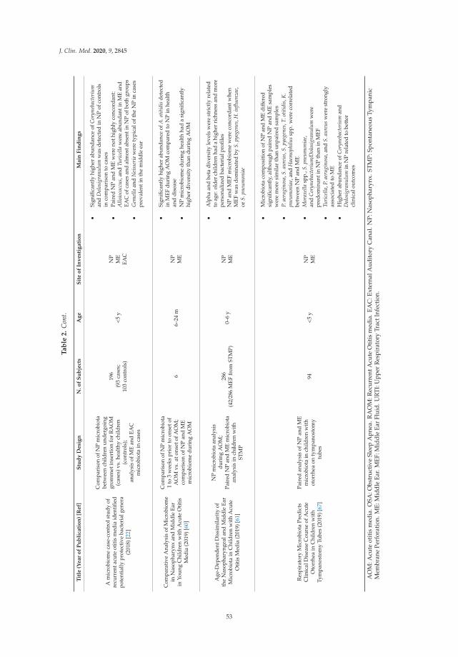
J. Clin. Med. 2020, 9, 2845
Ta
ble
2.
Con
t.
Tit
le(Y
ear
of
Pu
bli
cati
on
)[R
ef]
Stu
dy
Desi
gn
N.o
fS
ub
ject
sA
ge
Sit
eo
fIn
vest
igati
on
Main
Fin
din
gs
Am
icro
biom
eca
se-c
ontr
olst
udy
ofre
curr
enta
cute
otiti
sm
edia
iden
tified
pote
ntia
llypr
otec
tive
bact
eria
lgen
era
(201
8)[2
2]
Com
pari
son
ofN
Pm
icro
biot
abe
twee
nch
ildre
nun
derg
oing
grom
met
inse
rtio
nfo
rR
AO
M(c
ases
)vs.
heal
thy
child
ren
(con
trol
s);
anal
ysis
ofM
Ean
dEA
Cm
icro
biot
ain
case
s
196
(93
case
s;10
3co
ntro
ls)
<5
yN
PM
EEA
C
•Si
gnifi
cant
lyhi
gher
abun
danc
eof
Cor
yneb
acte
rium
and
Dol
osig
ranu
lum
was
dete
cted
inN
Pof
cont
rols
inco
mpa
riso
nto
case
s•
Pair
edN
Pan
dM
Ew
ere
noth
ighl
yco
ncor
dant
:A
lloio
cocc
us,a
ndTu
rice
llaw
ere
abun
dant
inM
Ean
dEA
Cof
case
san
dal
mos
tabs
enti
nN
Pof
both
grou
ps•
Gem
ella
and
Nei
sser
iaw
ere
typi
calo
fthe
NP
inca
ses
prev
alen
tin
the
mid
dle
ear
Com
para
tive
Ana
lysi
sof
Mic
robi
ome
inN
asop
hary
nxan
dM
iddl
eEa
rin
Youn
gC
hild
ren
wit
hA
cute
Oti
tis
Med
ia(2
019)
[60]
Com
pari
son
ofN
Pm
icro
biot
a1
to3
wee
kspr
ior
toon
seto
fA
OM
vs.a
tons
etof
AO
M;
com
pari
son
ofN
Pan
dM
Em
icro
biom
edu
ring
AO
M
66–
24m
NP
ME
•Si
gnifi
cant
lyhi
gher
abun
danc
eof
A.o
titid
isde
tect
edin
MEF
duri
ngA
OM
com
pare
dto
NP
inhe
alth
and
dise
ase
•N
Pm
icro
biom
edu
ring
heal
thha
da
sign
ifica
ntly
high
erdi
vers
ity
than
duri
ngA
OM
Age
-Dep
ende
ntD
issi
mila
rity
ofth
eN
asop
hary
ngea
land
Mid
dle
Ear
Mic
robi
ota
inC
hild
ren
wit
hA
cute
Oti
tis
Med
ia(2
019)
[61]
NP
mic
robi
ota
anal
ysis
duri
ngA
OM
;Pa
ired
NP
and
ME
mic
robi
ota
anal
ysis
inch
ildre
nw
ith
STM
P
286
(42/
286
MEF
from
STM
P)0–
6y
NP
ME
•A
lpha
and
beta
dive
rsit
yle
vels
wer
est
rict
lyre
late
dto
age:
olde
rch
ildre
nha
da
high
erri
chne
ssan
dm
ore
pers
onal
ized
bact
eria
lpro
files
•N
Pan
dM
EFm
icro
biom
ew
ere
conc
orda
ntw
hen
MEF
was
dom
inat
edby
S.py
ogen
es,H
.infl
uenz
ae,
orS.
pneu
mon
iae
Res
pira
tory
Mic
robi
ota
Pred
icts
Clin
ical
Dis
ease
Cou
rse
ofA
cute
Oto
rrhe
ain
Chi
ldre
nw
ith
Tym
pano
stom
yTu
bes
(201
9)[6
7]
Pair
edan
alys
isof
NP
and
ME
mic
robi
ota
inch
ildre
nw
ith
otor
rhea
onty
mpa
nost
omy
tube
s
94<
5y
NP
ME
•M
icro
biot
aco
mpo
siti
onof
NP
and
ME
diff
ered
sign
ifica
ntly
,alt
houg
hpa
ired
NP
and
ME
sam
ples
wer
em
ore
sim
ilar
than
unpa
ired
sam
ples
•P.
aeru
gino
sa,S
.aur
eus,
S.py
ogen
es,T
.otit
idis
,K.
pneu
mon
iae,
and
Hae
mop
hilu
ssp
p.w
ere
corr
elat
edbe
twee
nN
Pan
dM
E•
Mor
axel
lasp
p.,S
.pne
umon
iae,
and
Cor
yneb
acte
rium/D
olos
igra
nulu
mw
ere
pred
omin
anti
nN
Pth
anin
MEF
•Tu
rice
lla,P
.aer
ugin
osa,
and
S.au
reus
wer
est
rong
lyas
soci
ated
toM
E•
Hig
her
abun
danc
eof
Cor
yneb
acte
rium
and
Dol
osig
ranu
lum
inN
Pre
late
dto
bett
ercl
inic
alou
tcom
es
AO
M:A
cute
otit
ism
edia
.OSA
:Obs
truc
tive
Slee
pA
pnea
.RA
OM
:Rec
urre
ntA
cute
Oti
tis
med
ia.E
AC
:Ext
erna
lAud
itor
yC
anal
.NP:
Nas
opha
rynx
.ST
MP:
Spon
tane
ous
Tym
pani
cM
embr
ane
Perf
orat
ion.
ME:
Mid
dle
Ear.
MEF
:Mid
dle
Ear
Flui
d.U
RTI
:Upp
erR
espi
rato
ryTr
actI
nfec
tion
.
53
J. Clin. Med. 2020, 9, 2845
5. Otitis Media with Effusion
Otitis media with effusion (OME) is defined as the presence of middle ear fluid without signs orsymptoms of acute infection. It is defined chronic otitis media with effusion (COME) whether it persistsfor more than 3 months [10].
The first study on this topic with a high-throughput molecular approach was conducted byLiu et al., through the investigation of the microbiota of middle ear, adenoid, and tonsils in an 8-yearold child with chronic middle ear effusion undergoing adenotonsillectomy and bilateral tympanictube insertion. Middle ear microbiota was dominated by Pseudomonadaceae, and tonsil microbiotashowed a predominance by Streptococcaceae; adenoid microbiota was the most complex, includingPseudomonadaceae, Streptococcaceae, Fusobacteriaceae, and Pasteurellaceae, and shared microorganismsfound both in tonsils and middle ear, supporting the hypothesis that the adenoid pad could actas a reservoir for both of these sites [68].
Relevant new insights on OME were subsequently provided in an Australian study analyzingNP swabs, MEF, and adenoid specimens from 11 indigenous children undergoing surgery: MEFmicrobiome was characterized by low diversity indices and predominance of a single bacteria, in mostcases A. otitidis, H. influenzae, or Streptococcus spp. In particular, A. otitidis was the most commontaxa in MEF and was not detected in any NP or adenoid samples. Thus, authors speculated that itsorigin from NP was unlikely and that it could represent a typical microorganism of the ME niche;however, as A. otitidis is a known commensal of the ear canal [69], further studies were warrantedto understand its role and the influence of the ear canal flora, especially in children who suffer fromrecurrent tympanic membrane perforations [70].
Fago-Olsen et al. analyzed microbiota of palatine tonsils and adenoids from children undergoingsurgery for adenoid/tonsillar hyperplasia vs. subjects undergoing surgery for secretory otitismedia (SOM), showing that several microorganisms were occasionally co-detected in both sites,but H. influenzae, S. pneumoniae, and M. catarrhalis were significantly more abundant in the adenoidsand almost absent from palatine tonsils, indicating that adenoids but not palatine tonsils could actas main reservoir of pathogens leading to OM. However, it should be noted that this study did notinclude MEF microbiota analysis [71].
Data concerning dissimilarities between NP and MEF microbiome were provided in a followinginvestigation including 10 children undergoing adenotonsillectomy and grommet insertion for OME.The authors reported that adenoid and tonsil microbiota shared a higher similarity than adenoid andME, thus questioning the PRH in OME. According to previous findings, Alloiococcus and Turicella weredetected only in MEF samples; however, the most abundant genera in middle ear were Fusobacteriumand Staphylococcus [72]. These data were subsequently confirmed in an investigation by Ari et al.on a larger population of children with OME: ME microbiome was characterized by a significantpredominance of Alloicoccus otitidis (44%), Turicella otitidis (6%), and Staphylococcus auricularis (3%),while adenoid harbored a high relative abundance of Rothia, Staphylococcus, and Granulicatella. As fordiversity indices, no significant dissimilarities in alpha-diversity were found between MEF and adenoidniches [73].
The potential role of A. otitidis as a key bacteria of the ME was confirmed in an investigation byChan et al., through the analysis of paired MEF samples and adenoid swabs from children undergoinggrommet insertion for OME and of adenoid swabs from healthy subjects. Data evidenced a differentcomposition in microbial communities between paired MEF and adenoid, as 13 of the 17 most abundantgenera showed a statistically significant difference in relative abundance. In particular, A. otitidis wasthe predominant OTU in MEF (23% mean relative abundance), while it was almost absent in adenoidsamples (<1% relative abundance). Interestingly, this taxa was found in greater abundance in childrenwith unilateral effusion. Authors postulated that the dissimilarities between the MEF and adenoidmicrobiota could question the PRH in children with OME: Adenoidal hypertrophy and Eustachiantube dysfunction predispose to OME, but subsequent modifications in the ME environment determine
54
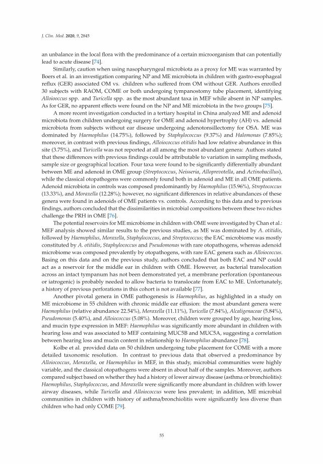
J. Clin. Med. 2020, 9, 2845
an unbalance in the local flora with the predominance of a certain microorganism that can potentiallylead to acute disease [74].
Similarly, caution when using nasopharyngeal microbiota as a proxy for ME was warranted byBoers et al. in an investigation comparing NP and ME microbiota in children with gastro-esophagealreflux (GER) associated OM vs. children who suffered from OM without GER. Authors enrolled30 subjects with RAOM, COME or both undergoing tympanostomy tube placement, identifyingAlloiooccus spp. and Turicella spp. as the most abundant taxa in MEF while absent in NP samples.As for GER, no apparent effects were found on the NP and ME microbiota in the two groups [75].
A more recent investigation conducted in a tertiary hospital in China analyzed ME and adenoidmicrobiota from children undergoing surgery for OME and adenoid hypertrophy (AH) vs. adenoidmicrobiota from subjects without ear disease undergoing adenotonsillectomy for OSA. ME wasdominated by Haemophilus (14.75%), followed by Staphylococcus (9.37%) and Halomonas (7.85%);moreover, in contrast with previous findings, Alloiococcus otitidis had low relative abundance in thissite (3.75%), and Turicella was not reported at all among the most abundant genera: Authors statedthat these differences with previous findings could be attributable to variation in sampling methods,sample size or geographical location. Four taxa were found to be significantly differentially abundantbetween ME and adenoid in OME group (Streptococcus, Neisseria, Alloprevotella, and Actinobacillus),while the classical otopathogens were commonly found both in adenoid and ME in all OME patients.Adenoid microbiota in controls was composed predominantly by Haemophilus (15.96%), Streptococcus(13.33%), and Moraxella (12.28%); however, no significant differences in relative abundances of thesegenera were found in adenoids of OME patients vs. controls. According to this data and to previousfindings, authors concluded that the dissimilarities in microbial compositions between these two nicheschallenge the PRH in OME [76].
The potential reservoirs for ME microbiome in children with OME were investigated by Chan et al.:MEF analysis showed similar results to the previous studies, as ME was dominated by A. otitidis,followed by Haemophilus, Moraxella, Staphylococcus, and Streptococcus; the EAC microbiome was mostlyconstituted by A. otitidis, Staphylococcus and Pseudomonas with rare otopathogens, whereas adenoidmicrobiome was composed prevalently by otopathogens, with rare EAC genera such as Alloiococcus.Basing on this data and on the previous study, authors concluded that both EAC and NP couldact as a reservoir for the middle ear in children with OME. However, as bacterial translocationacross an intact tympanum has not been demonstrated yet, a membrane perforation (spontaneousor iatrogenic) is probably needed to allow bacteria to translocate from EAC to ME. Unfortunately,a history of previous perforations in this cohort is not available [77].
Another pivotal genera in OME pathogenesis is Haemophilus, as highlighted in a study onME microbiome in 55 children with chronic middle ear effusion: the most abundant genera wereHaemophilus (relative abundance 22.54%), Moraxella (11.11%), Turicella (7.84%), Alcaligenaceae (5.84%),Pseudomonas (5.40%), and Alloiococcus (5.08%). Moreover, children were grouped by age, hearing loss,and mucin type expression in MEF: Haemophilus was significantly more abundant in children withhearing loss and was associated to MEF containing MUC5B and MUC5A, suggesting a correlationbetween hearing loss and mucin content in relationship to Haemophilus abundance [78].
Kolbe et al. provided data on 50 children undergoing tube placement for COME with a moredetailed taxonomic resolution. In contrast to previous data that observed a predominance byAlloiococcus, Moraxella, or Haemophilus in MEF, in this study, microbial communities were highlyvariable, and the classical otopathogens were absent in about half of the samples. Moreover, authorscompared subject based on whether they had a history of lower airway disease (asthma or bronchiolitis):Haemophilus, Staphylococcus, and Moraxella were significantly more abundant in children with lowerairway diseases, while Turicella and Alloiococcus were less prevalent; in addition, ME microbialcommunities in children with history of asthma/bronchiolitis were significantly less diverse thanchildren who had only COME [79].
55
J. Clin. Med. 2020, 9, 2845
Nasopharyngeal microbiome is less diverse in children suffering from OME than in controls,as highlighted by two case-control studies [80,81]. In particular, Walker et al. showed that the nasalmicrobiome in children with OME is composed of a higher abundance of pathogens, with a lowerabundance of commensals as alpha-hemolytic Streptococci and Lactococcus. Moreover, cluster analysisrevealed that profiles dominated by Corynebacterium, Streptococcus, or Moraxella were associatedwith COME, while healthy children had a more mixed bacterial profile with higher abundance ofcommensals [81].
In conclusion, investigations on OME discussed above confirm the role of the known otopathogens,in particular H. influenzae, as the predominant taxa in MEF during disease. Moreover, as previouslydescribed for AOM, A. otitidis and T. otitidis are frequently identified as abundant members of the MEmicrobiota. Studies have so far failed to define the possible reservoir for ME microbiome, and it is notpossible to exclude a sample contamination from the EAC, especially in a low biomass environment asthe ME. Concerning this theme, we believe that further studies should also be focused on patients witha history of tympanic membrane perforation, which might be the entryway for microorganisms thatcolonize the EAC.
An overview on microbiome study in OME previously discussed is reported in Table 3.
56
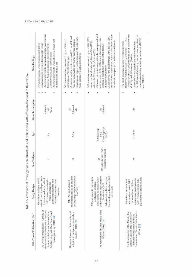
J. Clin. Med. 2020, 9, 2845
Ta
ble
3.
Ove
rvie
wof
inve
stig
atio
nson
mic
robi
ota
and
otit
ism
edia
wit
heff
usio
ndi
scus
sed
inth
isre
view
.
Tit
le(Y
ear
of
Pu
bli
cati
on
)[R
ef]
Stu
dy
Desi
gn
N.o
fS
ub
ject
sA
ge
Sit
eo
fIn
vest
igati
on
Main
Fin
din
gs
The
Oto
logi
cM
icro
biom
e:A
Stud
yof
the
Bact
eria
lMic
robi
ota
ina
Pedi
atri
cPa
tien
twit
hC
hron
icSe
rous
Oti
tis
Med
iaU
sing
16Sr
RN
AG
ene-
Base
dPy
rose
quen
cing
(201
1)[6
8]
Mic
robi
ota
anal
ysis
inM
E,ad
enoi
d,an
dto
nsil
spec
imen
sfr
omon
epe
diat
ric
pati
ent
wit
hch
roni
cse
rous
otit
ism
edia
unde
rgoi
ngad
enot
onsi
llect
omy
and
bila
tera
ltym
pani
ctu
bein
sert
ion
18
yA
deno
idM
ETo
nsil
•Ps
eudo
mon
adac
eae
wer
epr
edom
inan
tin
ME
•St
rept
ococ
cace
aew
ere
pred
omin
anti
nto
nsil
•A
deno
idm
icro
biot
ain
clud
edm
ulti
ple
pred
omin
ant
bact
eria
:Pse
udom
onad
acea
e,St
rept
ococ
cace
ae,
Fuso
bact
eria
ceae
,and
Past
eure
llace
ae•
Ade
noid
mic
robi
ota
shar
edba
cter
iafo
und
both
into
nsils
and
mid
dle
ear
The
mic
robi
ome
ofot
itis
med
iaw
ith
effus
ion
inIn
dige
nous
Aus
tral
ian
child
ren
(201
5)[7
0]
MEF
,NP,
and
aden
oid
mic
robi
ota
anal
ysis
inch
ildre
nun
derg
oing
surg
ical
trea
tmen
tfo
rO
ME
113–
9y
NP
Ade
noid
ME
•M
Em
icro
biot
aw
asdo
min
ated
byA
.otit
idis
,H.
influ
enza
e,or
Stre
ptoc
occu
ssp
p.•
A.o
titid
isw
asth
em
ostc
omm
onO
TU
inM
EFan
dw
asno
tdet
ecte
din
any
NP
orad
enoi
dsa
mpl
es•
Stre
ptoc
occu
ssp
p.,H
.infl
uenz
ae,a
ndM
.cat
arrh
alis
wer
eco
mm
onto
alls
ampl
ety
pes
The
Mic
robi
ome
ofO
titi
sM
edia
wit
hEff
usio
n(2
016)
[74]
ME
and
aden
oid
mic
robi
ota
anal
ysis
inch
ildre
nun
derg
oing
aden
oide
ctom
yw
ithve
ntila
tion
tube
inse
rtio
nfo
rch
roni
cO
ME.
Ade
noid
mic
robi
ota
anal
ysis
from
heal
thy
subj
ects
enro
lled
asco
ntro
ls
33(2
3su
bjec
tsw
ith
OM
E;10
heal
thy
cont
rols
)
OM
Egr
oup:
1–8
yC
ontr
olgr
oup:
1–12
y
ME
Ade
noid
•M
Em
icro
biot
aw
asdo
min
ated
byA
.otit
idis
(23%
mea
nre
lati
veab
unda
nce)
,Hae
mop
hilu
s(2
2%),
Mor
axel
la(5
%),
and
Stre
ptoc
occu
s(5
%)
•D
iffer
entm
icro
bial
com
posi
tion
betw
een
pair
edM
EFan
dad
enoi
d:13/1
7of
the
mos
tabu
ndan
tgen
era
show
eda
stat
isti
cally
sign
ifica
ntdiff
eren
cein
rela
tive
abun
danc
e•
A.o
titid
isw
asth
epr
edom
inan
tOT
Uin
MEF
(23%
mea
nre
lativ
eab
unda
nce)
,whi
leit
was
alm
osta
bsen
tin
aden
oid
sam
ples
(<1%
rela
tive
abun
danc
e)
The
Rel
atio
nshi
pof
the
Mid
dle
Ear
Effus
ion
Mic
robi
ome
toSe
cret
ory
Muc
inPr
oduc
tion
inPe
diat
ric
Pati
ents
wit
hC
hron
icO
titi
sM
edia
(201
6)[7
8]
Mic
robi
ota
anal
ysis
and
muc
inde
tect
ion
inM
EFco
llect
edfr
omch
ildre
nun
derg
oing
myr
ingo
tom
yw
ith
tym
pano
stom
ytu
bepl
acem
entf
orch
roni
cO
ME
553–
176
mM
E
•T
hem
osta
bund
antg
ener
aw
ere
Hae
mop
hilu
s(r
elat
ive
abun
danc
e22
.54%
),M
orax
ella
(11.
11%
),Tu
rice
lla(7
.84%
),A
lcal
igen
acea
e(5
.84%
),Ps
eudo
mon
as(5
.40%
),an
dA
lloio
cocc
us(5
.08%
)•
Hae
mop
hilu
sw
assi
gnifi
cant
lym
ore
abun
dant
inch
ildre
nw
ith
hear
ing
loss
and
was
asso
ciat
edto
sam
ples
cont
aini
ngse
cret
ory
muc
ins
asM
UC
5Ban
dM
UC
5A
57
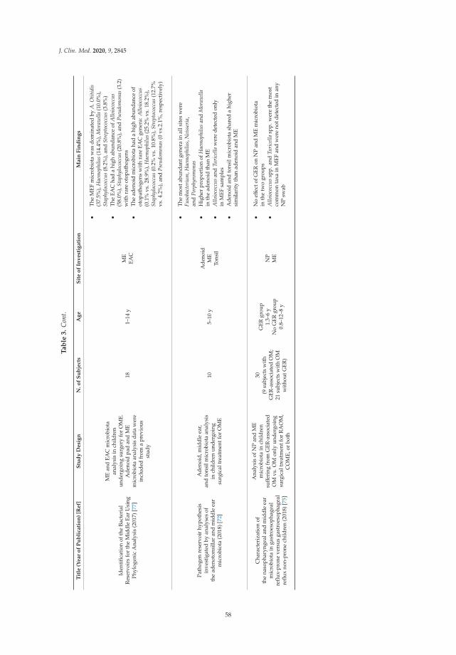
J. Clin. Med. 2020, 9, 2845
Ta
ble
3.
Con
t.
Tit
le(Y
ear
of
Pu
bli
cati
on
)[R
ef]
Stu
dy
Desi
gn
N.o
fS
ub
ject
sA
ge
Sit
eo
fIn
vest
igati
on
Main
Fin
din
gs
Iden
tific
atio
nof
the
Bact
eria
lR
eser
voir
sfo
rth
eM
iddl
eEa
rU
sing
Phyl
ogen
icA
naly
sis
(201
7)[7
7]
ME
and
EAC
mic
robi
ota
anal
ysis
inch
ildre
nun
derg
oing
surg
ery
for
OM
E.A
deno
idpa
dan
dM
Em
icro
biot
aan
alys
isda
taw
ere
incl
uded
from
apr
evio
usst
udy
181–
14y
ME
EAC
•T
heM
EFm
icro
biot
aw
asdo
min
ated
byA
.Otit
idis
(37.
5%),
Hae
mop
hilu
s(1
4.4%
),M
orax
ella
(10.
0%),
Stap
hylo
cocc
us(8
.2%
),an
dSt
rept
ococ
cus
(3.8
%)
•T
heEA
Cha
da
high
abun
danc
eof
Allo
ioco
ccus
(58.
0%),
Stap
hylo
cocc
us(2
0.8%
),an
dPs
eudo
mon
as(3
.2)
wit
hra
reot
opat
hoge
ns•
The
aden
oid
mic
robi
ota
had
ahi
ghab
unda
nce
ofot
opat
hoge
nsw
ith
rare
EAC
gene
ra:A
lloio
cocc
us(0
.1%
vs.2
8.9%
),H
aem
ophi
lus
(25.
2%vs
.18.
2%),
Stap
hylo
cocc
us(0
.2%
vs.1
0.8%
),St
rept
ococ
cus
(12.
7%vs
.4.2
%),
and
Pseu
dom
onas
(0vs
.2.1
%,r
espe
ctiv
ely)
Path
ogen
rese
rvoi
rhy
poth
esis
inve
stig
ated
byan
alys
esof
the
aden
oton
silla
ran
dm
iddl
eea
rm
icro
biot
a(2
018)
[72]
Ade
noid
,mid
dle
ear,
and
tons
ilm
icro
biot
aan
alys
isin
child
ren
unde
rgoi
ngsu
rgic
altr
eatm
entf
orO
ME
105–
10y
Ade
noid
ME
Tons
il
•T
hem
osta
bund
antg
ener
ain
alls
ites
wer
eFu
soba
cter
ium
,Hae
mop
hilu
s,N
eiss
eria
,an
dPo
rphy
rom
onas
•H
ighe
rpr
opor
tion
ofH
aem
ophi
lus
and
Mor
axel
lain
the
aden
oid
than
ME
•A
lloio
cocc
usan
dTu
rice
llaw
ere
dete
cted
only
inM
EFsa
mpl
es•
Ade
noid
and
tons
ilm
icro
biot
ash
ared
ahi
gher
sim
ilari
tyth
anad
enoi
dan
dM
E
Cha
ract
eriz
atio
nof
the
naso
phar
ynge
alan
dm
iddl
eea
rm
icro
biot
ain
gast
roes
opha
geal
reflu
x-pr
one
vers
usga
stro
esop
hage
alre
flux
non-
pron
ech
ildre
n(2
018)
[75]
Ana
lysi
sof
NP
and
ME
mic
robi
ota
inch
ildre
nsuff
erin
gfr
omG
ER-a
ssoc
iate
dO
Mvs
.OM
only
unde
rgoi
ngsu
rgic
altr
eatm
entf
orR
AO
M,
CO
ME,
orbo
th
30(9
subj
ects
wit
hG
ER-a
ssoc
iate
dO
M;
21su
bjec
tsw
ith
OM
wit
hout
GER
)
GER
grou
p1.
3–6
yN
oG
ERgr
oup
0.8–
12–8
y
NP
ME
•N
oeff
ecto
fGER
onN
Pan
dM
Em
icro
biot
ain
the
two
grou
ps•
Allo
ioco
ccus
spp.
and
Turi
cella
spp.
wer
eth
em
ost
com
mon
taxa
inM
EFan
dw
ere
notd
etec
ted
inan
yN
Psw
ab
58
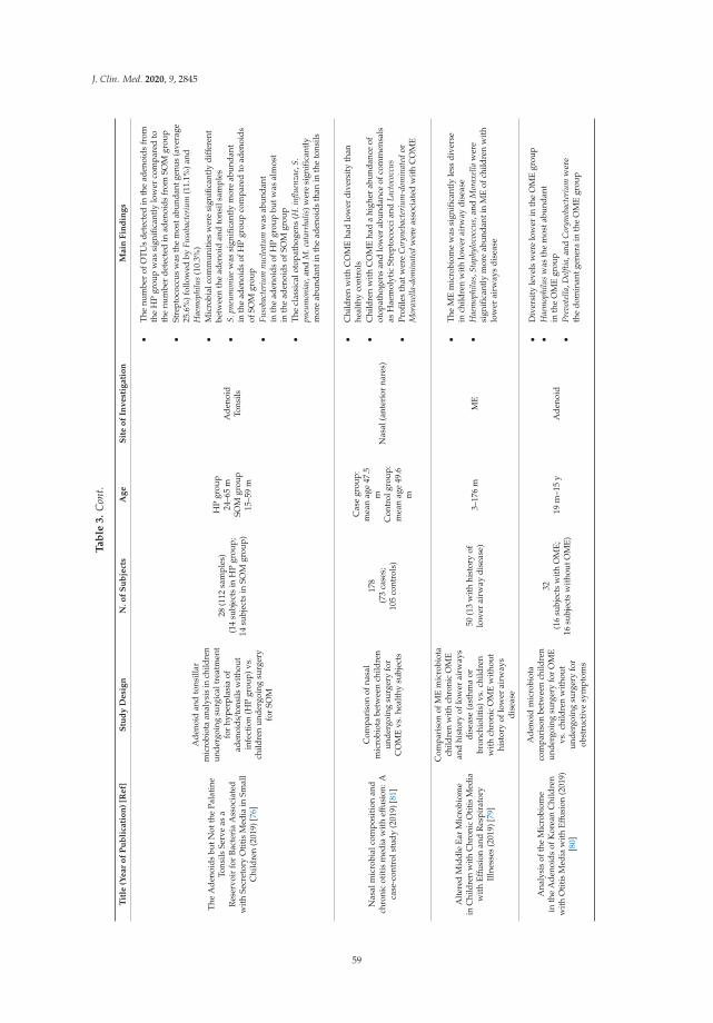
J. Clin. Med. 2020, 9, 2845
Ta
ble
3.
Con
t.
Tit
le(Y
ear
of
Pu
bli
cati
on
)[R
ef]
Stu
dy
Desi
gn
N.o
fS
ub
ject
sA
ge
Sit
eo
fIn
vest
igati
on
Main
Fin
din
gs
The
Ade
noid
sbu
tNot
the
Pala
tine
Tons
ilsSe
rve
asa
Res
ervo
irfo
rBa
cter
iaA
ssoc
iate
dw
ith
Secr
etor
yO
titi
sM
edia
inSm
all
Chi
ldre
n(2
019)
[76]
Ade
noid
and
tons
illar
mic
robi
ota
anal
ysis
inch
ildre
nun
derg
oing
surg
ical
trea
tmen
tfo
rhy
perp
lasi
aof
aden
oids/t
onsi
lsw
itho
utin
fect
ion
(HP
grou
p)vs
.ch
ildre
nun
derg
oing
surg
ery
for
SOM
28(1
12sa
mpl
es)
(14
subj
ects
inH
Pgr
oup;
14su
bjec
tsin
SOM
grou
p)
HP
grou
p24
–65
mSO
Mgr
oup
15–5
9m
Ade
noid
Tons
ils
•T
henu
mbe
rof
OT
Us
dete
cted
inth
ead
enoi
dsfr
omth
eH
Pgr
oup
was
sign
ifica
ntly
low
erco
mpa
red
toth
enu
mbe
rde
tect
edin
aden
oids
from
SOM
grou
p•
Stre
ptoc
occu
sw
asth
em
osta
bund
antg
enus
(ave
rage
25.6
%)f
ollo
wed
byFu
soba
cter
ium
(11.
1%)a
ndH
aem
ophi
lus
(10.
3%)
•M
icro
bial
com
mun
itie
sw
ere
sign
ifica
ntly
diff
eren
tbe
twee
nth
ead
enoi
dan
dto
nsil
sam
ples
•S.
pneu
mon
iae
was
sign
ifica
ntly
mor
eab
unda
ntin
the
aden
oids
ofH
Pgr
oup
com
pare
dto
aden
oids
ofSO
Mgr
oup
•Fu
soba
cter
ium
nucl
eatu
mw
asab
unda
ntin
the
aden
oids
ofH
Pgr
oup
butw
asal
mos
tin
the
aden
oids
ofSO
Mgr
oup
•T
hecl
assi
calo
topa
thog
ens
(H.i
nflue
nzae
,S.
pneu
mon
iae,
and
M.c
atar
rhal
is)w
ere
sign
ifica
ntly
mor
eab
unda
ntin
the
aden
oids
than
inth
eto
nsils
Nas
alm
icro
bial
com
posi
tion
and
chro
nic
otit
ism
edia
wit
heff
usio
n:A
case
-con
trol
stud
y(2
019)
[81]
Com
pari
son
ofna
sal
mic
robi
ota
betw
een
child
ren
unde
rgoi
ngsu
rger
yfo
rC
OM
Evs
.hea
lthy
subj
ects
178
(73
case
s;10
5co
ntro
ls)
Cas
egr
oup:
mea
nag
e47
.5m
Con
trol
grou
p:m
ean
age
49.6
m
Nas
al(a
nter
ior
nare
s)
•C
hild
ren
wit
hC
OM
Eha
dlo
wer
dive
rsit
yth
anhe
alth
yco
ntro
ls•
Chi
ldre
nw
ith
CO
ME
had
ahi
gher
abun
danc
eof
otop
atho
gens
and
low
erab
unda
nce
ofco
mm
ensa
lsas
Hae
mol
ytic
Stre
ptoc
occi
and
Lact
ococ
cus
•Pr
ofile
sth
atw
ere
Cor
yneb
acte
rium
-dom
inat
edor
Mor
axel
la-d
omin
ated
wer
eas
soci
ated
wit
hC
OM
E
Alt
ered
Mid
dle
Ear
Mic
robi
ome
inC
hild
ren
with
Chr
onic
Otit
isM
edia
wit
hEff
usio
nan
dR
espi
rato
ryIl
lnes
ses
(201
9)[7
9]
Com
pari
son
ofM
Em
icro
biot
ach
ildre
nw
ith
chro
nic
OM
Ean
dhi
stor
yof
low
erai
rway
sdi
seas
e(a
sthm
aor
bron
chio
litis
)vs.
child
ren
wit
hch
roni
cO
ME
wit
hout
hist
ory
oflo
wer
airw
ays
dise
ase
50(1
3w
ith
hist
ory
oflo
wer
airw
aydi
seas
e)3–
176
mM
E
•T
heM
Em
icro
biom
ew
assi
gnifi
cant
lyle
ssdi
vers
ein
child
ren
wit
hlo
wer
airw
aydi
seas
e•
Hae
mop
hilu
s,St
aphy
loco
ccus
,and
Mor
axel
law
ere
sign
ifica
ntly
mor
eab
unda
ntin
ME
ofch
ildre
nw
ith
low
erai
rway
sdi
seas
e
Ana
lysi
sof
the
Mic
robi
ome
inth
eA
deno
ids
ofK
orea
nC
hild
ren
with
Otit
isM
edia
with
Effus
ion
(201
9)[8
0]
Ade
noid
mic
robi
ota
com
pari
son
betw
een
child
ren
unde
rgoi
ngsu
rger
yfo
rO
ME
vs.c
hild
ren
wit
hout
unde
rgoi
ngsu
rger
yfo
rob
stru
ctiv
esy
mpt
oms
32(1
6su
bjec
tsw
ith
OM
E;16
subj
ects
wit
hout
OM
E)19
m–1
5y
Ade
noid
•D
iver
sity
leve
lsw
ere
low
erin
the
OM
Egr
oup
•H
aem
ophi
lus
was
the
mos
tabu
ndan
tin
the
OM
Egr
oup
•Pr
evot
ella
,Del
ftia,
and
Cor
yneb
acte
rium
wer
eth
edo
min
antg
ener
ain
the
OM
Egr
oup
59

J. Clin. Med. 2020, 9, 2845
Ta
ble
3.
Con
t.
Tit
le(Y
ear
of
Pu
bli
cati
on
)[R
ef]
Stu
dy
Desi
gn
N.o
fS
ub
ject
sA
ge
Sit
eo
fIn
vest
igati
on
Main
Fin
din
gs
The
bact
erio
me
ofot
itis
med
iaw
ith
effus
ion:
does
itor
igin
ate
from
the
aden
oid?
(201
9)[7
3]
Ade
noid
and
ME
mic
robi
ota
anal
ysis
inch
ildre
nun
derg
oing
surg
ery
for
OM
E25
1.5–
9y
Ade
noid
ME
•M
Em
icro
biom
ew
asdo
min
ated
byA
.otit
is(4
4%),
T.ot
itidi
s(6
%),
and
S.au
ricu
lari
s(3
%)
•A
deno
idm
icro
biom
ew
asdo
min
ated
byR
othi
a,St
aphy
loco
ccus
,and
Gra
nulic
atel
la•
No
stat
isti
cally
sign
ifica
ntdiff
eren
cein
alph
adi
vers
ity
betw
een
the
two
nich
es;a
deno
idsa
mpl
escl
uste
red
inth
ebe
tadi
vers
ity
grap
h
The
mic
robi
omes
ofad
enoi
dan
dm
iddl
eea
rin
child
ren
wit
hot
itis
med
iaw
ith
effus
ion
and
hype
rtro
phy
from
ate
rtia
ryho
spit
alin
Chi
na(2
020)
[76]
Ade
noid
and
ME
mic
robi
ota
anal
ysis
inch
ildre
nun
derg
oing
surg
ical
trea
tmen
tfo
rO
ME
vs.a
deno
idm
icro
biot
aan
alys
isin
child
ren
unde
rgoi
ngsu
rger
yfo
rO
SAw
itho
utea
rdi
seas
e
30(1
5in
OM
Egr
oup;
15in
OSA
grou
p)
OM
Egr
oup
60–1
08m
OSA
grou
p8–
96m
Ade
noid
ME
•M
Ein
OM
Ew
asdo
min
ated
byH
aem
ophi
lus
(14.
75%
),St
aphy
loco
ccus
(9.3
7%),
and
Hal
omon
as(7
.85%
)•
Low
abun
danc
eof
A.o
titis
(3.7
5%)i
nM
Ein
OM
Egr
oup
•A
deno
idm
icro
biot
ain
OM
Egr
oup
was
dom
inat
edby
Hae
mop
hilu
s(2
1.87
%),
Stre
ptoc
occu
s(1
9.65
%),
and
Nei
sser
ia(5
.8%
)•
Ade
noid
mic
robi
ota
inO
SAw
asdo
min
ated
byH
aem
ophi
lus
(15.
96%
),St
rept
ococ
cus
(13.
33%
),an
dM
orax
ella
(12.
28%
)•
No
sign
ifica
ntdiff
eren
ces
inal
pha-
dive
rsity
betw
een
ME
and
aden
oids
inO
ME
grou
p•
Beta
dive
rsit
yan
alys
essh
owed
that
the
mic
robi
ome
stru
ctur
eof
ME
was
diss
imila
rth
ead
enoi
don
ein
OM
Epa
tien
ts:t
axa
foun
dto
besi
gnifi
cant
lydiff
eren
tially
abun
dant
betw
een
thes
etw
osi
tes
wer
eSt
rept
ococ
cus,
Nei
sser
ia,A
llopr
evot
ella
,an
dA
ctin
obac
illus
OM
E:O
titi
sM
edia
wit
hEff
usio
n.C
OM
E:C
hron
icO
titi
sM
edia
wit
hEff
usio
n.N
P:N
asop
hary
nx.M
E:M
idd
leE
ar.M
EF:
Mid
dle
Ear
Flui
d.G
ER
:Gas
tro-
esop
hage
alre
flux
.RA
OM
:R
ecur
rent
acut
eot
itis
med
ia.S
OM
:Sec
reti
veot
itis
med
ia.E
AC
:Ext
erna
laud
itor
yC
anal
.OTU
:Ope
rati
onal
Taxo
nom
icU
nit.
OSA
:Obs
truc
tive
slee
pap
nea.
60

J. Clin. Med. 2020, 9, 2845
6. Chronic Suppurative Otitis Media
Chronic suppurative otitis media (CSOM) is defined as a chronic inflammation of the middleear and mastoid cavity, with recurrent or persistent ear discharge through a non-intact tympanicmembrane [10]. Less evidence is available on microbial communities in pediatric patients sufferingfrom this condition.
Neef et al. compared 24 children with CSOM undergoing mastoid surgery to 22 healthy controlsundergoing ear surgery for other conditions as cochlear implantation or benign brain tumor removal.Microbiota analysis and conventional culture were performed on swabs collected from middle earand mastoid cavity during surgery. Authors did not observe a typical bacterial profile associated toCSOM, but highlighted the limits of the conventional culture-based approach, as no bacteria weredetected by culture in healthy subjects. By contrast, molecular analysis detected potential pathogensas Staphylococcus, Pseudomonas, and Haemophilus even in healthy controls. As for diversity, authorsobserved a major inter-personal difference among CSOM patients, whereas this finding was notobserved for controls. This data supported the hypothesis that microbial communities’ disruption anddysbiosis could be implicated in CSOM pathogenesis [82].
These dissimilarities among patients suffering from CSOM are age-related, as reported byMinami et al. In this investigation, middle ear swabs were collected during surgery in pediatric andadult patients undergoing tympanoplasty for wet or dry COM vs. subjects undergoing surgery fromother conditions than otitis media. Proteobacteria was the predominant phylum detected in normalsubjects, both adults and children. However, the normal middle ear microbiota differed significantlyaccording to age: Authors concluded that this dissimilarity between adults and children could berelated to the higher incidence of Staphylococcus (Firmicutes phylum) in adults. Subjects with activeinflammation and wet COM had a lower abundance of Proteobacteria and a higher incidence ofFirmicutes: Authors warranted this finding to be considered in the pathogenesis of active inflammationin COM, in relation to the potential penetration of several exogenous pathogens through a chronicperforation. On the other hand, microbiome of dry COM was not significantly different from normalmiddle ear [83].
Santos-Cortez et al. previously performed an investigation comparing ME and EAC microbiomein 16 indigenous Filipino subject with chronic tympanic membrane perforation, showing thatthe microbial communities between these two niches were similar, probably due to a cross-contaminationprocess through the perforated eardrum. Moreover, authors investigated microbiota compositionin subjects who were carrier of the A2ML1 gene, which encodes an alpha-2 macroglobulin-like1 protein, previously identified as a genetically determined risk factor for of otitis media [84].Authors detected a higher relative abundance of Fusobacterium, Porphyromonas, Peptostreptococcus,Parvimonas, and Bacteroides in the ME of A2ML1-carrier patients, while Alloiococcus, Staphylococcus,Proteus, and Haemophilus were more abundant in ME of non-carrier subjects. Authors speculatedthat the expected loss-of-function of A2ML1 protein could influence ME microbiota compositionpromoting survival and growth of specific microorganism. This findings warrant further investigationson the relationship between host genotype and microbiota in OM [85].
Evidence on CSOM is lacking and does not show peculiar features of microbial communitiesin this OM phenotype. Moreover, investigations discussed above include both adults and children,thus it is difficult to draw any general conclusion in the pediatric population. The penetration ofmicroorganisms residing in the EAC from the chronic tympanic membrane perforation has beenconsidered in the pathogenesis of the active inflammation in CSOM, but further studies are needed todefine with major detail this aspect.
An overview on microbiome study in CSOM previously discussed is reported in Table 4.
61
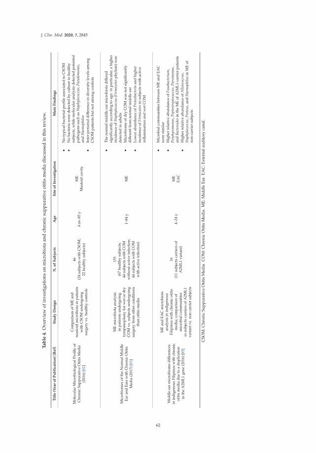
J. Clin. Med. 2020, 9, 2845
Ta
ble
4.
Ove
rvie
wof
inve
stig
atio
nson
mic
robi
ota
and
chro
nic
supp
urat
ive
otit
ism
edia
disc
usse
din
this
revi
ew.
Tit
le(Y
ear
of
Pu
bli
cati
on
)[R
ef]
Stu
dy
Desi
gn
N.o
fS
ub
ject
sA
ge
Sit
eo
fIn
vest
igati
on
Main
Fin
din
gs
Mol
ecul
arM
icro
biol
ogic
alPr
ofile
ofC
hron
icSu
ppur
ativ
eO
titi
sM
edia
(201
6)[8
2]
Com
pari
son
ofM
Ean
dm
asto
idm
icro
biot
ain
patie
nts
wit
hC
SOM
unde
rgoi
ngsu
rger
yvs
.hea
lthy
cont
rols
46(2
4su
bjec
tsw
ith
CSO
M;
22he
alth
ysu
bjec
ts)
6m
–85
yM
EM
asto
idca
vity
•N
oty
pica
lbac
teri
alpr
ofile
asso
ciat
edto
CSO
M•
No
bact
eria
wer
ede
tect
edby
cult
ure
inhe
alth
ysu
bjec
ts,w
hile
mol
ecul
aran
alys
isde
tect
edpo
tent
ial
path
ogen
ssu
chas
Stap
hylo
cocc
us,P
seud
omon
as,
and
Hae
mop
hilu
s•
Inte
r-pe
rson
aldiff
eren
cein
dive
rsit
yle
vels
amon
gC
SOM
pati
ents
butn
otam
ong
cont
rols
Mic
robi
omes
ofth
eN
orm
alM
iddl
eEa
ran
dEa
rsw
ith
Chr
onic
Oti
tis
Med
ia(2
017)
[83]
ME
mic
robi
ota
anal
ysis
inpa
tien
tsun
derg
oing
tym
pano
plas
tyfo
rw
etor
dry
CO
Mvs
.sub
ject
sun
derg
oing
surg
ery
from
othe
rco
ndit
ions
than
otit
ism
edia
155
(67
heal
thy
subj
ects
;44
subj
ects
wit
hC
OM
wit
hout
acti
vein
fect
ion;
44su
bjec
tsw
ith
CO
Mw
ith
acti
vein
fect
ion)
1–84
yM
E
•T
heno
rmal
mid
dle
ear
mic
robi
ota
diff
ered
sign
ifica
ntly
acco
rdin
gto
age:
inpa
rtic
ular
,ahi
gher
inci
denc
eof
Stap
hylo
cocc
us(F
irm
icut
esph
ylum
)was
dete
cted
inad
ults
•M
icro
biom
eof
dry
CO
Mw
asno
tsig
nific
antl
ydiff
eren
tfro
mno
rmal
mid
dle
ear
•Lo
wer
abun
danc
eof
Prot
eoba
cter
iaan
dhi
gher
inci
denc
eof
Firm
icut
esin
subj
ects
wit
hac
tive
infla
mm
atio
nan
dw
etC
OM
Mid
dle
ear
mic
robi
ome
diff
eren
ces
inin
dige
nous
Filip
inos
wit
hch
roni
cot
itis
med
iadu
eto
adu
plic
atio
nin
the
A2M
L1ge
ne(2
016)
[85]
ME
and
EAC
mic
robi
ota
anal
ysis
inin
dige
nous
Filip
inos
wit
hch
roni
cot
itis
med
ia;c
ompa
riso
nof
mic
robi
alco
mm
unit
ies
insu
bjec
tsca
rrie
rsof
A2M
L1va
rian
tvs.
non
carr
ier
subj
ects
16(1
1su
bjec
tsca
rrie
rsof
A2M
L1va
rian
t)4–
24y
ME
EAC
•M
icro
bial
com
mun
itie
sbe
twee
nM
Ean
dEA
Cw
ere
sim
ilar
•H
ighe
rre
lati
veab
unda
nce
ofFu
soba
cter
ium
,Po
rphy
rom
onas
,Pep
tost
rept
ococ
cus,
Parv
imon
as,
and
Bact
eroi
des
inth
eM
Eof
A2M
L1-c
arri
erpa
tien
ts•
Hig
her
rela
tive
abun
danc
eof
Allo
ioco
ccus
,St
aphy
loco
ccus
,Pro
teus
,and
Hae
mop
hilu
sin
ME
ofno
n-ca
rrie
rsu
bjec
ts
CSO
M:C
hron
icSu
ppur
ativ
eO
titi
sM
edia
.CO
M:C
hron
icO
titi
sM
edia
.ME:
Mid
dle
Ear.
EAC
:Ext
erna
laud
itor
yca
nal.
62

J. Clin. Med. 2020, 9, 2845
7. Probiotic Therapy
Prevention of OM in children represents one of the most difficult aspects in the clinical managementof these patients.
Restoration of dysbiosis through administration of probiotic strains is a preventive strategy thathas gained major clinical and scientific interest in recent years in several diseases, including otitis media.
Probiotics are defined as “live microorganisms that, when administered in adequate amounts,confer a health benefit on the host” [86]. The introduction of high-throughput sequencing methods hasallowed the investigation of entire bacterial communities and the identification of microorganismsassociated to health status in various conditions.
As previously discussed, evidence on microbiota in children suffering from OM suggest thatCorynebacterium spp. and Dolosigranulum pigrum are potential keystone taxa in the URT; thus, majorinterest has been directed towards these two microorganisms and their potential use as probiotics.
A detailed discussion of evidence available on probiotic therapy in OM goes beyond the scope ofthis review, as it has been recently extensively reviewed elsewhere.
A recent review by van den Broek et al. described novel insights on probiotic therapy in OM [87].Basing on Koch’s postulates, authors introduced the “probiotic postulates” to define the ideal probioticstrain to be used in clinical practice: The microorganism can be found in high abundance in healthstatus and decreased abundance during disease; the microorganism can be isolated from a healthyorganism and grown in pure culture; the cultured organism should promote health when introducedinto a diseased organism; it should be possible to re-isolate these microorganisms as identical tothe original agent from the healthy host. According to available evidence and to this postulates, authorsidentified Dolosigranulum as a prime candidate for the development of probiotic therapy.
However, current knowledge is still not sufficient to define probiotic efficacy for preventing OM.A recent systematic review included 13 studies on this subject, concluding that available evidence onprobiotics use for the prevention of AOM is limited; among the various formulations, possible benefitcould derive from nasal administration [88].
The most important limitations in evidence on this topic are poor to moderate quality ofthe investigations and great heterogeneity in route of administration (oral vs. intranasal), probioticstrains included in formulations, duration of therapy, and outcome measures.
8. Conclusions
The introduction of the modern molecular techniques and the subsequent investigations onmicrobial communities in the human organisms have changed our conceptions of health and diseaseand our approach to infectious conditions.
It is indeed well known that health and disease status are not merely determined by the presenceor the absence of a pathogen but depend on a complex balance established among pathogens, residentmicrobiota, and host immune response.
Investigations previously described in this review have provided novel insights on the pathogenesisof middle ear diseases and led to the identification of both possible new causative agents and ofpotential protective bacteria, showing that imbalances in bacterial communities of the URT and MEcould influence the natural history of otitis media in children.
However, scientific data on this topic are often difficult to compare because of methodologicaldifferences in specimen collection and analysis, in the site of investigation, and in data reporting.Moreover, a lack of standard diagnostic criteria for OM across countries often influences the enrollmentphase and contributes to increase the heterogeneity among populations under investigation.
Another element that complicates data interpretation and deserves standardization is the useof different databases during OTUs assignment. This is a relevant issue that should be taken intoconsideration for two main reasons: different databases might lead to heterogeneous results; sometaxa could be misclassified with certain databases, as reported for A. otitidis and T. otitidis [89].
We believe that future investigation should be focused on the following aspects:
63

J. Clin. Med. 2020, 9, 2845
• Defining standard criteria of specimen collection, analysis, and data reporting, in order to facilitatedata comparison across studies;
• Deepening our knowledge on the impact of various exogenous factors that have been less explored,such as active/passive smoking, vaccines, and viral infections;
• Confirming the role of Corynebacterium and/or Dolosigranulum as keystone taxa, in order to evaluatetheir possible use as probiotics;
• Understanding the development of URT and ME microbiota at different ages, in order to identifya potential “window of opportunity” in which therapeutic interventions as probiotic administrationcould be more effective, before the establishment of a stable microbial community that could bemodulated with difficulty;
• Investigating the concordance between NP and ME microbiota, in order to better define the roleof adenoid pad as a proxy for ME;
• Providing data on microbial communities in ME, which is no longer considered a sterile site;• Defining with major detail the features of NP and ME microbial communities in different OM
phenotypes, in particular in children with recurrent STMP.
Author Contributions: P.M. and S.T. conceived the paper; F.F. conducted the literature search and wrotethe manuscript; I.C. contributed to the literature search; P.M., S.T., L.D., P.C. and S.A. revised the paper; L.R.revised the technical aspects. All authors have read and agreed to the published version of the manuscript.
Funding: This research received no external funding.
Conflicts of Interest: The authors declare no conflict of interest.
References
1. The NIH HMP Working Group; Peterson, J.; Garges, S.; Giovanni, M.; McInnes, P.; Wang, L.; Schloss, J.A.;Bonazzi, V.; McEwen, J.E.; Wetterstrand, K.A.; et al. The NIH Human Microbiome Project. Genome Res. 2009,19, 2317–2323. [CrossRef] [PubMed]
2. Patel, A.H.; Harris, K.A.; Fitzgerald, F. What is broad-range 16S rDNA PCR? Arch. Dis. Child. Educ. Pract. Ed.2017, 102, 261–264. [CrossRef] [PubMed]
3. Samuelson, D.R.; Welsh, D.A.; Shellito, J.E. Regulation of lung immunity and host defense by the intestinalmicrobiota. Front. Microbiol. 2015, 6, 1085. [CrossRef] [PubMed]
4. Buffie, C.G.; Pamer, E.G. Microbiota-mediated colonization resistance against intestinal pathogens. Nat. Rev.Immunol. 2013, 13, 790–801. [CrossRef] [PubMed]
5. Honda, K.; Littman, D.R. The microbiome in infectious disease and inflammation. Annu. Rev. Immunol. 2012,30, 759–795. [CrossRef] [PubMed]
6. Schuster, S.C. Next-generation sequencing transforms today’s biology. Nat. Methods 2007, 5, 16–18. [CrossRef][PubMed]
7. Knight, R.; Vrbanac, A.; Taylor, B.C.; Aksenov, A.; Callewaert, C.; Debelius, J.; González, A.; Kosciolek, T.;McCall, L.-I.; McDonald, D.; et al. Best practices for analysing microbiomes. Nat. Rev. Genet. 2018, 16,410–422. [CrossRef]
8. Paradise, J.L.; Rockette, H.E.; Colborn, D.K.; Bernard, B.S.; Smith, C.G.; Kurs-Lasky, M.; Janosky, J.E. OtitisMedia in 2253 Pittsburgh-Area Infants: Prevalence and Risk Factors During the First Two Years of Life.Pediatrics 1997, 99, 318–333. [CrossRef]
9. Marom, T.; Marchisio, P.G.; Tamir, S.O.; Torretta, S.; Gavriel, H.; Esposito, S. Complementary and AlternativeMedicine Treatment Options for Otitis Media. Medicine 2016, 95, e2695. [CrossRef]
10. Schilder, A.G.; Chonmaitree, T.; Cripps, A.W.; Rosenfeld, R.M.; Casselbrant, M.L.; Haggard, M.P.;Venekamp, R.P. Otitis media. Nat. Rev. Dis. Prim. 2016, 2, 16063. [CrossRef]
11. Bernstein, J.M.; Reddy, M.S.; Scannapieco, F.A.; Faden, H.S.; Ballow, M. The Microbial Ecology and Immunologyof the Adenoid: Implications for Otitis Media. Ann. N.Y. Acad. Sci. 1997, 830, 19–31. [CrossRef] [PubMed]
12. Nistico, L.; Kreft, R.; Gieseke, A.; Coticchia, J.M.; Burrows, A.; Khampang, P.; Liu, Y.; Kerschner, J.E.; Post, J.C.;Lonergan, S.; et al. Adenoid Reservoir for Pathogenic Biofilm Bacteria. J. Clin. Microbiol. 2011, 49, 1411–1420.[CrossRef] [PubMed]
64

J. Clin. Med. 2020, 9, 2845
13. Hoa, M.; Tomovic, S.; Nistico, L.; Hall-Stoodley, L.; Stoodley, P.; Sachdeva, L.; Berk, R.; Coticchia, J.M.Identification of adenoid biofilms with middle ear pathogens in otitis-prone children utilizing SEM and FISH.Int. J. Pediatr. Otorhinolaryngol. 2009, 73, 1242–1248. [CrossRef] [PubMed]
14. Bogaert, D.; De Groot, R.; Hermans, P. Streptococcus pneumoniae colonisation: The key to pneumococcaldisease. Lancet Infect. Dis. 2004, 4, 144–154. [CrossRef]
15. Man, W.H.; Piters, W.A.A.D.S.; Bogaert, D. The microbiota of the respiratory tract: Gatekeeper to respiratoryhealth. Nat. Rev. Genet. 2017, 15, 259–270. [CrossRef] [PubMed]
16. Piters, W.A.A.D.S.; Sanders, E.A.M.; Bogaert, D. The role of the local microbial ecosystem in respiratoryhealth and disease. Philos. Trans. R. Soc. B Biol. Sci. 2015, 370, 20140294. [CrossRef]
17. Tano, K.; Grahn-Håkansson, E.; Holm, S.E.; Hellström, S. Inhibition of OM pathogens by alpha-hemolyticstreptococci from healthy children, children with SOM and children with rAOM. Int. J. Pediatr. Otorhinolaryngol.2000, 56, 185–190. [CrossRef]
18. Tano, K.; Olofsson, C.; Grahn-Håkansson, E.; Holm, S.E. In vitro inhibition of S. pneumoniae, nontypableH. influenzae and M. catharralis by alpha-hemolytic streptococci from healthy children. Int. J.Pediatr. Otorhinolaryngol. 1999, 47, 49–56. [CrossRef]
19. Pettigrew, M.M.; Laufer, A.S.; Gent, J.F.; Kong, Y.; Fennie, K.; Metlay, J.P. Upper Respiratory TractMicrobial Communities, Acute Otitis Media Pathogens, and Antibiotic Use in Healthy and Sick Children.Appl. Environ. Microbiol. 2012, 78, 6262–6270. [CrossRef]
20. Laufer, A.S.; Metlay, J.P.; Gent, J.F.; Fennie, K.; Kong, Y.; Pettigrew, M.M. Microbial Communities of the UpperRespiratory Tract and Otitis Media in Children. mBio 2011, 2, e00245–e00310. [CrossRef]
21. Bomar, L.; Brugger, S.D.; Yost, B.H.; Davies, S.S.; Lemon, K.P. Corynebacterium accolensReleasesAntipneumococcal Free Fatty Acids from Human Nostril and Skin Surface Triacylglycerols. mBio 2016, 7,01725. [CrossRef] [PubMed]
22. Lappan, R.; Imbrogno, K.; Sikazwe, C.; Anderson, D.; Mok, D.; Coates, H.; Vijayasekaran, S.; Bumbak, P.;Blyth, C.; Jamieson, S.E.; et al. A microbiome case-control study of recurrent acute otitis media identifiedpotentially protective bacterial genera. BMC Microbiol. 2018, 18, 13. [CrossRef] [PubMed]
23. Tano, K.; Håkansson, E.G.; Holm, S.E.; Hellström, S. A nasal spray with alpha-haemolytic streptococci as longterm prophylaxis against recurrent otitis media. Int. J. Pediatr. Otorhinolaryngol. 2002, 62, 17–23. [CrossRef]
24. Marchisio, P.G.; Santagati, M.C.; Scillato, M.; Baggi, E.; Fattizzo, M.; Rosazza, C.; Stefani, S.; Esposito, S.;Principi, N. Streptococcus salivarius 24SMB administered by nasal spray for the prevention of acute otitismedia in otitis-prone children. Eur. J. Clin. Microbiol. Infect. Dis. 2015, 34, 2377–2383. [CrossRef] [PubMed]
25. Costello, E.K.; Lauber, C.L.; Hamady, M.; Fierer, N.; Gordon, J.I.; Knight, R. Bacterial Community Variationin Human Body Habitats Across Space and Time. Science 2009, 326, 1694–1697. [CrossRef]
26. Thomas, S.; Izard, J.; Walsh, E.; Batich, K.; Chongsathidkiet, P.; Clarke, G.; Sela, D.A.; Muller, A.J.; Mullin, J.M.;Albert, K.; et al. The Host Microbiome Regulates and Maintains Human Health: A Primer and Perspectivefor Non-Microbiologists. Cancer Res. 2017, 77, 1783–1812. [CrossRef]
27. Charbonneau, M.R.; Blanton, L.V.; DiGiulio, D.B.; Relman, D.A.; Lebrilla, C.B.; Mills, D.A.; Gordon, J.I.A microbial perspective of human developmental biology. Nature 2016, 535, 48–55. [CrossRef]
28. Dominguez-Bello, M.G.; Costello, E.K.; Contreras, M.; Magris, M.; Hidalgo, G.; Fierer, N.; Knight, R. Deliverymode shapes the acquisition and structure of the initial microbiota across multiple body habitats in newborns.Proc. Natl. Acad. Sci. USA 2010, 107, 11971–11975. [CrossRef]
29. Bosch, A.A.; Levin, E.; Van Houten, M.A.; Hasrat, R.; Kalkman, G.; Biesbroek, G.; Piters, W.A.A.D.S.;De Groot, P.-K.C.; Pernet, P.; Keijser, B.J.; et al. Development of Upper Respiratory Tract Microbiota in Infancyis Affected by Mode of Delivery. EBioMedicine 2016, 9, 336–345. [CrossRef]
30. Biesbroek, G.; Tsivtsivadze, E.; Sanders, E.A.M.; Montijn, R.; Veenhoven, R.H.; Keijser, B.J.F.; Bogaert, D.Early Respiratory Microbiota Composition Determines Bacterial Succession Patterns and Respiratory Healthin Children. Am. J. Respir. Crit. Care Med. 2014, 190, 1283–1292. [CrossRef]
31. Teo, S.M.; Mok, D.; Pham, K.; Kusel, M.; Serralha, M.; Troy, N.; Holt, B.J.; Hales, B.J.; Walker, M.L.; Hollams, E.;et al. The infant nasopharyngeal microbiome impacts severity of lower respiratory infection and risk ofasthma development. Cell Host Microbe 2015, 17, 704–715. [CrossRef] [PubMed]
32. Guibas, G.V.; Moschonis, G.; Xepapadaki, P.; Roumpedaki, E.; Androutsos, O.; Manios, Y.; Papadopoulos, N.G.Conception viain vitrofertilization and delivery by Caesarean section are associated with paediatric asthmaincidence. Clin. Exp. Allergy 2013, 43, 1058–1066. [CrossRef] [PubMed]
65

J. Clin. Med. 2020, 9, 2845
33. Kristensen, K.; Fisker, N.; Haerskjold, A.; Ravn, H.; Simões, E.A.F.; Stensballe, L. Caesarean Section andHospitalization for Respiratory Syncytial Virus Infection: A population-based study. Pediatr. Infect. Dis. J.2015, 34, 145–148. [CrossRef] [PubMed]
34. Grice, E.A.; Segre, J.A. The skin microbiome. Nat. Rev. Genet. 2011, 9, 244–253. [CrossRef]35. Mendling, W. Vaginal Microbiota. Adv. Exp. Med. Biol. 2016, 902, 83–93. [CrossRef]36. Chu, D.M.; Ma, J.; Prince, A.L.; Antony, K.M.; Seferovic, M.D.; Aagaard, K.M. Maturation of the infant
microbiome community structure and function across multiple body sites and in relation to mode of delivery.Nat. Med. 2017, 23, 314–326. [CrossRef]
37. Tarrant, M.; Kwok, M.K.; Lam, T.-H.; Leung, G.M.; Schooling, C.M. Breast-feeding and ChildhoodHospitalizations for Infections. Epidemiology 2010, 21, 847–854. [CrossRef]
38. Duijts, L.; Jaddoe, V.W.V.; Hofman, A.; Moll, H.A. Prolonged and Exclusive Breastfeeding Reduces the Riskof Infectious Diseases in Infancy. Pediatrics 2010, 126, e18–e25. [CrossRef]
39. Labbok, M.; Clark, D.; Goldman, A.S. Breastfeeding: Maintaining an irreplaceable immunological resource.Nat. Rev. Immunol. 2004, 4, 565–572. [CrossRef]
40. Biesbroek, G.; Bosch, A.A.; Wang, X.; Keijser, B.J.F.; Veenhoven, R.H.; Sanders, E.A.; Bogaert, D. The Impactof Breastfeeding on Nasopharyngeal Microbial Communities in Infants. Am. J. Respir. Crit. Care Med. 2014,190, 298–308. [CrossRef]
41. Bosch, A.A.T.M.; Wouter, A.A.d.S.P.; Van Houten, M.A.; Chu, M.L.J.N.; Biesbroek, G.; Kool, J.; Pernet, P.; DeGroot, P.-K.C.M.; Eijkemans, M.J.C.; Keijser, B.J.F.; et al. Maturation of the Infant Respiratory Microbiota,Environmental Drivers, and Health Consequences. A Prospective Cohort Study. Am. J. Respir. Crit. CareMed. 2017, 196, 1582–1590. [CrossRef] [PubMed]
42. Sullivan, Å.; Edlund, C.; Nord, C.E. Effect of antimicrobial agents on the ecological balance of humanmicroflora. Lancet Infect. Dis. 2001, 1, 101–114. [CrossRef]
43. Hicks, L.A.; Taylor, T.H.; Hunkler, R.J. U.S. Outpatient Antibiotic Prescribing, 2010. N. Engl. J. Med. 2013,368, 1461–1462. [CrossRef] [PubMed]
44. Hilty, M.; Qi, W.; Brugger, S.D.; Frei, L.; Agyeman, P.; Frey, P.M.; Aebi, S.; Mühlemann, K. NasopharyngealMicrobiota in Infants with Acute Otitis Media. J. Infect. Dis. 2012, 205, 1048–1055. [CrossRef]
45. Chonmaitree, T.; Jennings, K.; Golovko, G.; Khanipov, K.; Pimenova, M.; Patel, J.A.; McCormick, D.P.;Loeffelholz, M.J.; Fofanov, Y. Nasopharyngeal microbiota in infants and changes during viral upperrespiratory tract infection and acute otitis media. PLoS ONE 2017, 12, e0180630. [CrossRef]
46. Pettigrew, M.M.; Alderson, M.R.; Bakaletz, L.O.; Barenkamp, S.J.; Hakansson, A.P.; Mason, K.M.;Nokso-Koivisto, J.; Patel, J.; Pelton, S.I.; Murphy, T.F. Panel 6: Vaccines. Otolaryngol. Neck Surg. 2017, 156(Suppl. 4), S76–S87. [CrossRef]
47. Ngo, C.C.; Massa, H.M.; Thornton, R.B.; Cripps, A.W. Predominant Bacteria Detected from the Middle EarFluid of Children Experiencing Otitis Media: A Systematic Review. PLoS ONE 2016, 11, e0150949. [CrossRef]
48. Gladstone, R.A.; Jefferies, J.M.; Tocheva, A.S.; Beard, K.R.; Garley, D.; Chong, W.W.; Bentley, S.D.; Faust, S.N.;Clarke, S.C. Five winters of pneumococcal serotype replacement in UK carriage following PCV introduction.Vaccine 2015, 33, 2015–2021. [CrossRef]
49. Biesbroek, G.; Wang, X.; Keijser, B.J.; Eijkemans, R.M.; Trzcinski, K.; Rots, N.Y.; Veenhoven, R.H.; Sanders, E.A.;Bogaert, D. Seven-Valent Pneumococcal Conjugate Vaccine and Nasopharyngeal Microbiota in HealthyChildren. Emerg. Infect. Dis. 2014, 20, 201–210. [CrossRef]
50. Mika, M.; Maurer, J.; Korten, I.; Allemann, A.; Aebi, S.; Brugger, S.D.; Qi, W.; Frey, U.; Latzin, P.; Hilty, M.Influence of the pneumococcal conjugate vaccines on the temporal variation of pneumococcal carriage andthe nasal microbiota in healthy infants: A longitudinal analysis of a case–control study. Microbiome 2017, 5,85. [CrossRef]
51. Feazel, L.M.; Santorico, S.A.; Robertson, C.E.; Bashraheil, M.; Scott, J.A.G.; Frank, D.; Hammitt, L.L. Effectsof Vaccination with 10-Valent Pneumococcal Non-Typeable Haemophilus influenza Protein D ConjugateVaccine (PHiD-CV) on the Nasopharyngeal Microbiome of Kenyan Toddlers. PLoS ONE 2015, 10, e0128064.[CrossRef] [PubMed]
52. Kwambana-Adams, B.A.; Hanson, B.; Worwui, A.; Agbla, S.; Foster-Nyarko, E.; Ceesay, F.; Ebruke, C.;Egere, U.; Zhou, Y.; Ndukum, M.; et al. Rapid replacement by non-vaccine pneumococcal serotypes maymitigate the impact of the pneumococcal conjugate vaccine on nasopharyngeal bacterial ecology. Sci. Rep.2017, 7, 1–11. [CrossRef] [PubMed]
66
J. Clin. Med. 2020, 9, 2845
53. Andrade, D.C.; Borges, I.C.; Bouzas, M.L.; Oliveira, J.R.; Fukutani, K.F.; Queiroz, A.T.; De Oliveira, C.I.;Barral, A.; Van Weyenbergh, J.; Nascimento-Carvalho, C. 10-valent pneumococcal conjugate vaccine (PCV10)decreases metabolic activity but not nasopharyngeal carriage of Streptococcus pneumoniae and Haemophilusinfluenzae. Vaccine 2017, 35, 4105–4111. [CrossRef]
54. Adegbola, R.A.; DeAntonio, R.; Hill, P.C.; Roca, A.; Usuf, E.; Hoet, B.; Greenwood, B.M. Carriage ofStreptococcus pneumoniae and Other Respiratory Bacterial Pathogens in Low and Lower-Middle IncomeCountries: A Systematic Review and Meta-Analysis. PLoS ONE 2014, 9, e103293. [CrossRef] [PubMed]
55. Boelsen, L.K.; Dunne, E.M.; Mika, M.; Eggers, S.; Nguyen, C.D.; Ratu, F.T.; Russell, F.M.; Mulholland, E.K.;Hilty, M.; Satzke, C. The association between pneumococcal vaccination, ethnicity, and the nasopharyngealmicrobiota of children in Fiji. Microbiome 2019, 7, 106. [CrossRef]
56. Charlson, E.S.; Chen, J.; Custers-Allen, R.; Bittinger, K.; Li, H.; Sinha, R.; Hwang, J.; Bushman, F.D.;Collman, R.G. Disordered Microbial Communities in the Upper Respiratory Tract of Cigarette Smokers.PLoS ONE 2010, 5, e15216. [CrossRef]
57. Teele, D.W.; Klein, J.O.; Rosner, B. Epidemiology of Otitis Media During the First Seven Years of Lifein Children in Greater Boston: A Prospective, Cohort Study. J. Infect. Dis. 1989, 160, 83–94. [CrossRef]
58. Dirain, C.O.; Silva, R.C.; Collins, W.O.; Antonelli, P.J. The Adenoid Microbiome in Recurrent Acute OtitisMedia and Obstructive Sleep Apnea. J. Int. Adv. Otol. 2017, 13, 333–339. [CrossRef]
59. Sillanpää, S.; Kramna, L.; Oikarinen, S.; Sipilä, M.; Rautiainen, M.; Aittoniemi, J.; Laranne, J.; Hyöty, H.;Cinek, O. Next-Generation Sequencing Combined with Specific PCR Assays To Determine the Bacterial 16SrRNA Gene Profiles of Middle Ear Fluid Collected from Children with Acute Otitis Media. mSphere 2017, 2,00006–00017. [CrossRef]
60. Xu, Q.; Gill, S.; Xu, L.; Gonzalez, E.; Pichichero, M.E. Comparative Analysis of Microbiome in Nasopharynxand Middle Ear in Young Children With Acute Otitis Media. Front. Genet. 2019, 10, 1–7. [CrossRef]
61. Brugger, S.D.; Kraemer, J.G.; Qi, W.; Bomar, L.; Oppliger, A.; Hilty, M. Age-Dependent Dissimilarity ofthe Nasopharyngeal and Middle Ear Microbiota in Children With Acute Otitis Media. Front. Genet. 2019, 10,555. [CrossRef] [PubMed]
62. Berger, G. Nature of spontaneous tympanic membrane perforation in acute otitis media in children.J. Laryngol. Otol. 1989, 103, 1150–1153. [CrossRef] [PubMed]
63. Torretta, S.; Marchisio, P. Otitis media in children: A proposal for a new nosological classification. Int. J.Pediatr. Otorhinolaryngol. 2017, 93, 174–175. [CrossRef] [PubMed]
64. Marchisio, P.G.; Nazzari, E.; Torretta, S.; Esposito, S.; Principi, N. Medical prevention of recurrent acute otitismedia: An updated overview. Expert Rev. Anti Infect. Ther. 2014, 12, 611–620. [CrossRef]
65. Marchisio, P.G.; Esposito, S.; Bianchini, S.; Dusi, E.; Fusi, M.; Nazzari, E.; Picchi, R.; Galeone, C.; Principi, N.Efficacy of Injectable Trivalent Virosomal-Adjuvanted Inactivated Influenza Vaccine in Preventing AcuteOtitis Media in Children With Recurrent Complicated or Noncomplicated Acute Otitis Media. Pediatr. Infect.Dis. J. 2009, 28, 855–859. [CrossRef]
66. Marchisio, P.G.; Consonni, D.; Baggi, E.; Zampiero, A.; Bianchini, S.; Terranova, L.; Tirelli, S.; Esposito, S.;Principi, N. Vitamin D Supplementation Reduces the Risk of Acute Otitis Media in Otitis-prone Children.Pediatr. Infect. Dis. J. 2013, 32, 1055–1060. [CrossRef]
67. Man, W.H.; Van Dongen, T.M.; Venekamp, R.P.; Pluimakers, V.G.; Chu, M.L.J.; Van Houten, M.A.; Sanders, E.A.;Schilder, A.G.M.; Bogaert, D. Respiratory Microbiota Predicts Clinical Disease Course of Acute Otorrheain Children With Tympanostomy Tubes. Pediatr. Infect. Dis. J. 2019, 38, e116–e125. [CrossRef]
68. Liu, C.M.; Cosetti, M.K.; Aziz, M.; Buchhagen, J.L.; Contente-Cuomo, T.L.; Price, L.B.; Keim, P.; Lalwani, A.K.The Otologic MicrobiomeA Study of the Bacterial Microbiota in a Pediatric Patient With Chronic SerousOtitis Media Using 16SrRNA Gene-Based Pyrosequencing. Arch. Otolaryngol. Head Neck Surg. 2011, 137,664–668. [CrossRef]
69. Frank, D.N.; Spiegelman, G.B.; Davis, W.; Wagner, E.; Lyons, E.; Pace, N.R. Culture-independent molecularanalysis of microbial constituents of the healthy human outer ear. J. Clin. Microbiol. 2003, 41, 295–303.[CrossRef]
70. Jervis-Bardy, J.; Rogers, G.B.; Morris, P.S.; Smith-Vaughan, H.C.; Nosworthy, E.; Leong, L.E.X.; Smith, R.J.;Weyrich, L.S.; De Haan, J.; Carney, A.S.; et al. The microbiome of otitis media with effusion in IndigenousAustralian children. Int. J. Pediatr. Otorhinolaryngol. 2015, 79, 1548–1555. [CrossRef]
67

J. Clin. Med. 2020, 9, 2845
71. Fagö-Olsen, H.; Dines, L.M.; Sørensen, C.H.; Jensen, A. The Adenoids but Not the Palatine Tonsils Serveas a Reservoir for Bacteria Associated with Secretory Otitis Media in Small Children. mSystems 2019, 4,e00169–e00218. [CrossRef] [PubMed]
72. Johnston, J.; Hoggard, M.; Biswas, K.; Astudillo-García, C.; Radcliff, F.J.; Mahadevan, M.; Douglas, R.G.Pathogen reservoir hypothesis investigated by analyses of the adenotonsillar and middle ear microbiota.Int. J. Pediatr. Otorhinolaryngol. 2019, 118, 103–109. [CrossRef]
73. Ari, O.; Karabudak, S.; Kalcioglu, M.T.; Gunduz, A.Y.; Durmaz, R. The bacteriome of otitis media with effusion: Doesit originate from the adenoid? Int. J. Pediatr. Otorhinolaryngol. 2019, 126, 109624. [CrossRef] [PubMed]
74. Chan, C.L.; Wabnitz, D.; Bardy, J.J.; Bassiouni, A.; Wormald, P.-J.; Vreugde, S.; Psaltis, A.J. The microbiome ofotitis media with effusion. Laryngoscope 2016, 126, 2844–2851. [CrossRef] [PubMed]
75. Boers, S.A.; De Zeeuw, M.; Jansen, R.; Van Der Schroeff, M.P.; Van Rossum, A.M.C.; Hays, J.P.; Verhaegh, S.J.C.Characterization of the nasopharyngeal and middle ear microbiota in gastroesophageal reflux-prone versusgastroesophageal reflux non-prone children. Eur. J. Clin. Microbiol. Infect. Dis. 2018, 37, 851–857. [CrossRef] [PubMed]
76. Xu, J.; Dai, W.; Liang, Q.; Ren, D. The microbiomes of adenoid and middle ear in children with otitis mediawith effusion and hypertrophy from a tertiary hospital in China. Int. J. Pediatr. Otorhinolaryngol. 2020, 134,110058. [CrossRef]
77. Chan, C.L.; Wabnitz, D.; Bassiouni, A.; Wormald, P.-J.; Vreugde, S.; Psaltis, A.J. Identification of the Bacterial Reservoirsfor the Middle Ear Using Phylogenic Analysis. JAMA Otolaryngol. Neck Surg. 2017, 143, 155–161. [CrossRef]
78. Krueger, A.; Val, S.; Pérez-Losada, M.; Panchapakesan, K.; Devaney, J.; Duah, V.; DeMason, C.; Poley, M.;Rose, M.; Preciado, D.; et al. Relationship of the Middle Ear Effusion Microbiome to Secretory Mucin Productionin Pediatric Patients With Chronic Otitis Media. Pediatr. Infect. Dis. J. 2017, 36, 635–640. [CrossRef] [PubMed]
79. Kolbe, A.R.; Castro-Nallar, E.; Preciado, D.; Pérez-Losada, M. Altered Middle Ear Microbiome in ChildrenWith Chronic Otitis Media With Effusion and Respiratory Illnesses. Front. Microbiol. 2019, 9, 1–10. [CrossRef]
80. Kim, S.K.; Hong, S.J.; Pak, K.H.; Hong, S.M. Analysis of the Microbiome in the Adenoids of Korean Childrenwith Otitis Media with Effusion. J. Int. Adv. Otol. 2019, 15, 379–385. [CrossRef]
81. Walker, R.E.; Walker, C.G.; Camargo, C.A., Jr.; Bartley, J.; Flint, D.; Thompson, J.M.D.; Mitchell, E.A. Nasalmicrobial composition and chronic otitis media with effusion: A case-control study. PLoS ONE 2019, 14,e0212473. [CrossRef] [PubMed]
82. Neeff, M.; Biswas, K.; Hoggard, M.; Taylor, M.W.; Douglas, R.G. Molecular Microbiological Profile of ChronicSuppurative Otitis Media. J. Clin. Microbiol. 2016, 54, 2538–2546. [CrossRef] [PubMed]
83. Minami, S.B.; Mutai, H.; Suzuki, T.; Horii, A.; Oishi, N.; Wasano, K.; Katsura, M.; Tanaka, F.; Takiguchi, T.;Fujii, M.; et al. Microbiomes of the normal middle ear and ears with chronic otitis media. Laryngoscope 2017,127, E371–E377. [CrossRef] [PubMed]
84. Santos-Cortez, R.L.P.; University of Washington Center for Mendelian Genomics; Chiong, C.M.;Reyes-Quintos, M.R.T.; Tantoco, M.L.C.; Wang, X.; Acharya, A.; Abbe, I.; Giese, A.P.J.; Smith, J.D.; et al. RareA2ML1 variants confer susceptibility to otitis media. Nat. Genet. 2015, 47, 917–920. [CrossRef]
85. Santos-Cortez, R.L.P.; Hutchinson, D.S.; Ajami, N.J.; Reyes-Quintos, M.R.T.; Tantoco, M.L.C.; Labra, P.J.;Lagrana, S.M.; Pedro, M.; Llanes, E.G.D.V.; Gloria-Cruz, T.L.; et al. Middle ear microbiome differencesin indigenous Filipinos with chronic otitis media due to a duplication in the A2ML1 gene. Infect. Dis. Poverty2016, 5, 97. [CrossRef]
86. Hill, C.; Guarner, F.; Reid, G.; Gibson, G.R.; Merenstein, D.J.; Pot, B.; Morelli, L.; Canani, R.B.; Flint, H.J.; Salminen, S.;et al. The International Scientific Association for Probiotics and Prebiotics consensus statement on the scope andappropriate use of the term probiotic. Nat. Rev. Gastroenterol. Hepatol. 2014, 11, 506–514. [CrossRef]
87. Broek, M.F.L.V.D.; De Boeck, I.; Kiekens, F.; Boudewyns, A.; Vanderveken, O.M.; Lebeer, S. Translating RecentMicrobiome Insights in Otitis Media into Probiotic Strategies. Clin. Microbiol. Rev. 2019, 32, 1–33. [CrossRef]
88. Chen, T.Y.; Hendrickx, A.; Stevenson, D.S.; Bird, P.; Walls, T. No evidence from a systematic review forthe use of probiotics to prevent otitis media. Acta Paediatr. 2020. [CrossRef] [PubMed]
89. Lappan, R.; Jamieson, S.E.; Peacock, C.S. Reviewing the Pathogenic Potential of the Otitis-Associated BacteriaAlloiococcus otitidis and Turicella otitidis. Front. Cell Infect. Microbiol. 2020, 10, 51. [CrossRef]
© 2020 by the authors. Licensee MDPI, Basel, Switzerland. This article is an open accessarticle distributed under the terms and conditions of the Creative Commons Attribution(CC BY) license (http://creativecommons.org/licenses/by/4.0/).
68
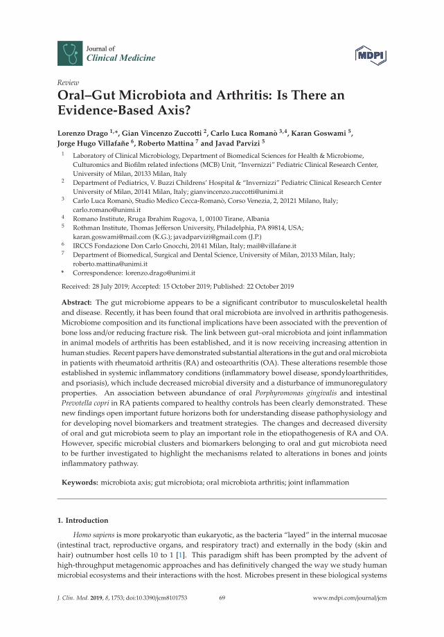
Journal of
Clinical Medicine
Review
Oral–Gut Microbiota and Arthritis: Is There anEvidence-Based Axis?
Lorenzo Drago 1,*, Gian Vincenzo Zuccotti 2, Carlo Luca Romanò 3,4, Karan Goswami 5,
Jorge Hugo Villafañe 6, Roberto Mattina 7 and Javad Parvizi 5
1 Laboratory of Clinical Microbiology, Department of Biomedical Sciences for Health & Microbiome,Culturomics and Biofilm related infections (MCB) Unit, “Invernizzi” Pediatric Clinical Research Center,University of Milan, 20133 Milan, Italy
2 Department of Pediatrics, V. Buzzi Childrens’ Hospital & “Invernizzi” Pediatric Clinical Research CenterUniversity of Milan, 20141 Milan, Italy; [email protected]
3 Carlo Luca Romanò, Studio Medico Cecca-Romanò, Corso Venezia, 2, 20121 Milano, Italy;[email protected]
4 Romano Institute, Rruga Ibrahim Rugova, 1, 00100 Tirane, Albania5 Rothman Institute, Thomas Jefferson University, Philadelphia, PA 89814, USA;
[email protected] (K.G.); [email protected] (J.P.)6 IRCCS Fondazione Don Carlo Gnocchi, 20141 Milan, Italy; [email protected] Department of Biomedical, Surgical and Dental Science, University of Milan, 20133 Milan, Italy;
[email protected]* Correspondence: [email protected]
Received: 28 July 2019; Accepted: 15 October 2019; Published: 22 October 2019
Abstract: The gut microbiome appears to be a significant contributor to musculoskeletal healthand disease. Recently, it has been found that oral microbiota are involved in arthritis pathogenesis.Microbiome composition and its functional implications have been associated with the prevention ofbone loss and/or reducing fracture risk. The link between gut–oral microbiota and joint inflammationin animal models of arthritis has been established, and it is now receiving increasing attention inhuman studies. Recent papers have demonstrated substantial alterations in the gut and oral microbiotain patients with rheumatoid arthritis (RA) and osteoarthritis (OA). These alterations resemble thoseestablished in systemic inflammatory conditions (inflammatory bowel disease, spondyloarthritides,and psoriasis), which include decreased microbial diversity and a disturbance of immunoregulatoryproperties. An association between abundance of oral Porphyromonas gingivalis and intestinalPrevotella copri in RA patients compared to healthy controls has been clearly demonstrated. Thesenew findings open important future horizons both for understanding disease pathophysiology andfor developing novel biomarkers and treatment strategies. The changes and decreased diversityof oral and gut microbiota seem to play an important role in the etiopathogenesis of RA and OA.However, specific microbial clusters and biomarkers belonging to oral and gut microbiota needto be further investigated to highlight the mechanisms related to alterations in bones and jointsinflammatory pathway.
Keywords: microbiota axis; gut microbiota; oral microbiota arthritis; joint inflammation
1. Introduction
Homo sapiens is more prokaryotic than eukaryotic, as the bacteria “layed” in the internal mucosae(intestinal tract, reproductive organs, and respiratory tract) and externally in the body (skin andhair) outnumber host cells 10 to 1 [1]. This paradigm shift has been prompted by the advent ofhigh-throughput metagenomic approaches and has definitively changed the way we study humanmicrobial ecosystems and their interactions with the host. Microbes present in these biological systems
J. Clin. Med. 2019, 8, 1753; doi:10.3390/jcm8101753 www.mdpi.com/journal/jcm69
J. Clin. Med. 2019, 8, 1753
are deeply integrated in our daily life, and emerging research has sought to decipher this complexinter-kingdom communication network present in our body and immune system. The gastrointestinal(GI) tract has the highest density and variety of microorganisms (more than 100 trillion microbes andapproximately 1500 species). Early life host–microbe interactions, especially in the gut, drive thedevelopment of immunity and the establishment of a stable complex microbial community, commonlyreferred to as the commensal microbiota [2,3]. Extensive research has focused on gut microbiota andhost immune response effects in the context of protection against pathogenic gut microbes and thepathophysiology of chronic inflammatory/autoimmune disease states [4,5]. For example, it has beenreported that in patients with Crohn’s disease, there is a relationship between dysbiosis and responseto treatment. Hence, microbiota could be a target of the treatment of chronic intestinal diseases [6].
Emerging scientific reports have also highlighted the immunomodulatory effects of gut microbiotaon other pathologic conditions, which often involve distant anatomical sites, such as the liver, thebrain, the heart and the skeleton [7–9].
Furthermore, several mechanisms and factors have been implicated to explain the role of microbiotain bone and joint health [10]. The gut microbiome is indeed a source of a number of key vitamins,such as cobalamin (B12), biotin (B7), folate, thiamine (B1), pyridoxal phosphate, pantothenic acid (B5),niacin (B3), vitamin K, and tetrahydrofolate, which are particularly important for the health of themusculoskeletal system [11].
Steves et al. highlighted how the gut microbiome can alter the inflammatory state of an individualby influencing both the host metabolic potential and its innate and adaptive immune system [12]. Theseauthors further discussed the role of microbiota diversity on some prevalent age-related disorders,such as osteoporosis, osteoarthritis, gout, rheumatoid arthritis, frailty and sarcopenia.
In the last decade, the alteration of gut microbiota has been reported in rheumatic disease andarthritis, most notably in juvenile idiopathic arthritis (JIA), rheumatoid arthritis (RA), psoriasis, andthe related spondyloarthritides (SpA), including ankylosing spondylitis (AS) and reactive arthritis(ReA) [13]. In a similar fashion to inflammatory bowel disease (IBD), it has been suggested that gutbacteria play important role in the etiopathogenesis of these aforementioned conditions.
RA is an autoimmune disorder which occurs when the immune system affects the fluid thatnourishes the cartilage and lubricates the joints (synovium) and their soft tissues. Generally, the rootcauses of arthritis include an increase in inflammatory processes and a decrease of the normal amountof cartilage present at the joint. A correct diet and gut balance can improve these diseases [14]. Indeed,inflammation-reducing foods containing antioxidants, such as fresh fruits, vegetables, or a gluten-poordiet may improve symptoms and disease progression by restoring intestinal microbiota. Findings haveprovided a model of how genetic and environmental factors, in association, cause autoimmune diseasessuch as RA. Sakaguchi S. et al. reported that the causal genetic anomaly of ZAP-70, a polymorphism ofthe MHC gene, significantly contributes to determining genetic susceptibility to autoimmune arthritisin SKG mice. Furthermore, they demonstrated that the disease initiation requires the interaction ofboth genetic and environmental factors, in particular the type of microbial colonization.
One of the most common form of arthritis is osteoarthritis (OA). This disease commonly occurswhen the protective cartilage on the ends of bones wears down over time by damaging any jointof the hands, knees, hips and spine. OA is characterized by a chronic, low-grade inflammationwhich is mediated primarily by the innate immune system, making it distinct from that observed inRA. Several dietary factors have been reported to be involved in the pathogenesis of OA. Vitamins,magnesium, and especially amino acids, i.e., little amounts of single amino acids supplementation suchas 0.5% (w/w) l-arginine or 1.0% (w/w) l-glutamine, have shown a significant influence on intestinalmicrobiota, especially the ratio of Firmicutes/Bacteroidetes. Chitosan supplementation can also alterthe component of intestinal microbiota, causing a lowering of the ratio Firmicutes/Bacteroidetes, inparticular a decreasing of Bacteroidales and an increasing of the Lactobacillales in the feces [15,16].
The alteration of gut microbiota can thus lead to an increased translocation of microbial associatedmolecular patterns (MAMPs) across the gut endothelium into the systemic circulation. MAMPs include
70
J. Clin. Med. 2019, 8, 1753
factors such as lipopolysaccharide (LPS), peptidoglycan, and bacterial DNA. These factors can triggerpro-inflammatory pathways by stimulating immune receptors in the resident immune cells of bone,cartilage and synovium [17,18].
RA has long been associated with periodontal disease [19], and recent evidence on the oralmicrobiome has emphasized its role in the arthritis. Using a metagenomics approach and molecularinvestigations, common opinion has been formed that each individual carries over 700 species inthe oral cavity, and this microbiome is the second largest microbial niche after the gastrointestinaltract [20]. Oral bacteria may penetrate through the gingival pockets and enter into the bloodstream.The translocation of microbiota-derived molecules into systemic circulation is considered one route forthe microbiome to mediate arthritis by stimulating specific cytokines (see below).
There is not so much evidence on microbiota association with some musculoskeletal diseasesrelated to age, as RA and OA. However, it seems that these clinical issues are associated withinflammatory changes, which could be specifically related to microbiota changes or be associated withage. Some studies described below on microbiota and arthritis were age-matched, presuming that theshaping of microbiota may have a role in the developing and maintaining these diseases independentlyby age.
The present review aims to address the most recent findings regarding the oral and gut microbiomesand their relationship with RA and OA, respectively.
2. Oral Microbiota in RA and OA
RA is an autoimmune disease affecting the synovium and cartilage with bony erosion. Recently,the relationship between the oral microbiome and systemic diseases has been explored [21,22]. Sher etal. demonstrated that overall exposure to Porphyromonas gingivalis was similar between patients withRA and controls. These authors found an abundance of Anaeroglobus geminatus that correlates with thepresence of rheumatoid factors, and Prevotella and Leptotrichia species are the only taxa that have beenobserved in patients with new-onset RA [22].
Chen et al. showed that RA has a distinct oral microbiome and may be affected by its dynamicvariations [23]. In this study comparing the oral microbiome in RA, OA and healthy patients usingrRNA gene amplicon sequencing, eight oral bacterial biomarkers (Prevotella melaninogenica, Veillonelladispar, Prevotella, Neisseria, Porphyromonas, Veillonella, Haemophilus, Rothia, Streptococcus, Actinomyces,Granulicatella, Leptotrichia, Lautropia, and Fusobacterium) were identified to differentiate RA from OA. Inaddition, the authors found that patients with RA and OA had oral microbiota with higher microbialdiversity compared to healthy subjects, indicating that there could be more pathobionts in the oralcavity of patients with RA that are able to negatively influence the outcome of the disease. The mostcommon phyla were Proteobacteria, Firmicutes, Bacteroidetes, Actinobacteria and Fusobacteria. Therelative abundance of Proteobacteria in healthy subjects was significantly higher than in patients withRA and OA, and the relative abundance of Firmicutes in patients with OA is significantly higher thanthose in patients with RA. Table 1a,b reports the different taxa and species of oral and gut microbiotaobserved in RA and OA, respectively.
Persson et al. previously noted P. gingivalis to be directly linked to RA through citrullinationand induction of antipeptidyl citrulline antibodies reacting to citrullinated human self-proteins [24].Interestingly, P. gingivalis, which is mainly abundant in the oral microbiome of RA patients, shares82% homology of α-enolase with human α-enolase. Consequently, human antibodies against bacterialenolase can promote an increase of antibody production. Lundberg et al. [25] showed that the levels ofanti-citrullinated human α-enolase antibodies and bacterial α-enolase correlates with the severity ofRA. P. gingivalis can be also found in the gut, but nothing exists between the P. gingivalis oral–gut axiscorrelation and arthritis, and the presence of this bacteria in the gut is not an inflammatory triggerof RA.
71

J. Clin. Med. 2019, 8, 1753
Table 1. Differences of bacterial abundance (taxa) in oral and gut microbiota of rheumatoid arthritis(RA) (a) and osteoarthritis (OA) (b) patients compared to the healthy controls.
(a)
Type of Arthritis AbundanceOral Microbiota
Profile (Taxa)Abundance
Gut MicrobiotaProfile (Taxa)
RA Increase ([23])
Neisseria subfava,Haemophilusparainfuenzae,Veillonella dispar,Prevotella tannerae,Actinobacillusparahaemolyticus,Neisseria,Haemophilus,Prevotella,Veillonella,Fusobacterium,Aggregatibacter,Actinobacillus
Decrease([26,27])
Bacteroides,Akkermansia,F.prausnitziiPrevotella,Ruminococcus
RA Increase ([24]) Porphyromonasgingivalis
RA Decrease ([28]) Haemophilus spp. Decrease ([28]) Haemophilus spp.
RA Increase ([28]) Lactobacillussalivarius Increase ([28]) Lactobacillus
salivarius
RA Increase ([29])
Plaque:Actinomyces meyeriPrevotella nigrescensTreponema socranskiiTreponema spp.EubacteriuminfirmumPrevotella orisActimomycesmassiliensisCatonella spp.
Increase ([30,31]) Prevotella copri
RA Increase ([29])
Saliva:Prevotella spp.Veillonella spp.Centipeda spp.Solobacterium moreiPrevotella pallensAtopobium parvulumButyrivibrio spp.
Decrease ([30]) Bacteroides
RA Increase ([32])
P. melaninogenicaP. denticolaP. histicola,P. nigrescens,P. oulorumP. maculosaSelenomonas noxiaS. sputigenaAnaeroglobusgeminatusAggregaticbacteractinomycetemcomitansParvimonas micraOtherGram-negatives
72
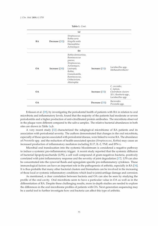
J. Clin. Med. 2019, 8, 1753
Table 1. Cont.
(a)
RA Decrease ([32])
StreptococcusRothia aeriaKingella oralisHaemophilusActinomyces
(b)
OA Increase ([28])
Rothia dentocariosa,Ruminococcusgnavus,Streptococcus,Actinomyces,Lautropia,Rothia,Granulicatella,Ruminococcus,Oribacterium,Abiotrophia
Increase ([33]) Lactobacillus spp.Methanobrevibacter
OA Increase ([33])
C. coccoides,C. leptum,Clostridium clustersXI-I, Roseburia spp.,Lactobacillus spp.
OA Decrease ([33]) BacteroidesPrevotella spp.
Eriksson et al. [29], by investigating the periodontal health of patients with RA in relation to oralmicrobiota and inflammatory levels, found that the majority of the patients had moderate or severeperiodontitis and a higher production of anti-citrullinated protein antibodies. The microbiota observedin the plaque were different compared to the saliva samples. The relative bacterial abundances in bothsites are shown in Table 1a,b.
A very recent study [32] characterized the subgingival microbiome of RA patients and itsassociation with periodontal severity. The authors demonstrated that changes in the oral microbiota,especially of those species associated with periodontal disease, were linked to worse RA. The abundanceof Prevotella spp. and the reduction of health-associated species (Streptococcus, Rothia) may cause anincreased production of inflammatory mediators including IL17, IL-2, TNF, and IFN-γ.
Microbial oral translocation into the systemic bloodstream is considered a negative pathwayto induce a systemic pro-inflammatory trigger. A recent study reported that the systemic diffusionof bacterial lipopolysaccharide (LPS), a cell wall compound of gram-negatives bacteria, positivelycorrelated with joint inflammatory response and the severity of joint degradation [17]. LPS can alsobe concentrated into the synovial fluids and upregulate specific pro-inflammatory cytokines. Theseimmunological factors can have an important role in the pathogenesis of arthritis, especially in RA [34].It is thus probable that many other bacterial clusters and biomarkers can be involved in the increasingof those local or systemic inflammatory conditions which lead to joint/cartilage damage and corrosion.
As mentioned, a clear correlation between bacteria and OA can also be seen by studying theprofile of the oral cavity. Oral microbiota seem to have a particular value in OA as well as in thedifferentiation of RA. Despite these challenging results, more in-depth studies are needed to explorethe differences in the oral microbiome profiles of patients with OA. Next-generation sequencing maybe a useful tool to further investigate how oral bacteria can affect this type of arthritis.
73

J. Clin. Med. 2019, 8, 1753
3. Gut Microbiota in RA and OA
The hypothesis that not only oral but also intestinal microbiota can be associated with thedevelopment of RA is supported by many data. Zhang et al. published a case-control metagenome-wideassociation study (MGWAS) of the fecal, dental and salivary microbiomes of a cohort of treatment-naiveand treated RA patients. They found that the RA-associated microbiome deviated significantly fromhealthy controls in all sites [28]. In this study, Haemophilus spp. was depleted in individuals with RA atthe fecal and oral levels, whereas Lactobacillus salivarius was over-represented in individuals with RAat both microbiota sites.
Older patients often manifest more severe diseases, and this appears connected to age-relatedgut dysbiosis. Alterations in the microbiota provide plausible candidate mechanisms for driving bothinflammation and altering the immune response and host metabolism, which in turn may modulatethe development of musculoskeletal problems (see the Prevotella copri case below). The microbiome isthus a highly plausible target for the modulation of diseases of aging owing to its close relationshipwith innate and adaptive immune systems. Components of intestinal microbiota can indeed direct keyaspects of host immunity, in particular effector T-cell differentiation, which may impact susceptibilityto autoimmune diseases and RA in particular [35].
Different studies investigating the etiology of RA have established the involvement of regulatoryT (T-reg) cells, which are defective at suppressing IFN-γ and TNF-α production by conventionalT cells in the peripheral blood of active RA patients [36,37]. It has been well established that thegut microbiota–immune interaction and homeostasis, via balancing pro- and anti-inflammatorymechanisms, can regulate the differentiation of various T cell types, especially T-reg cells [38]. A clearexample is the potential therapeutic effect of SCFAs (short chain fatty acids), which are microbialfermentation products found in the bowel, that have demonstrated a profound influence on T-reg celldifferentiation in a variety of experimental models of autoimmunity or inflammatory T-cell-mediateddiseases [39,40].
An elegant collagen-induced arthritis mouse model published by Hui et al. demonstrated thatbutyrate (a functional SCFA) administration inhibited arthritis by suppressing the expression ofinflammatory cytokines [41]. The authors suggested that modulation was likely mediated by thedifferentiation of CD4 T cells towards T-reg cells, which produce anti-inflammatory cytokine IL-10,and thus influenced the function of Th17 cells.
As mentioned, an altered microbiota profile has also been associated with juvenile idiopathicarthritis (JIA). Current evidence indeed suggests that the perturbation of gut microbiota may contributeto the development of JIA [42,43].
It remains difficult, however, to establish a definitive microbial marker or specific enterotypesthat are associated with RA. It has been hypothesized that the alteration of single bacterial genuscould have direct impact on driving inflammation, as suggested for Bacteroides, Akkermansia or theanti-inflammatory Faecalobacterium prausnitzii, which has been noted to be depleted in RA patients,while Prevotella and Ruminococcus were more prevalent [26,27]. Increasing Prevotella copri and decreasingBacteroides concentrations in the stool have both been associated with new onset, untreated RA inhumans [30].
The above studies, although not always homogenous, have directly or indirectly demonstratedthat genetic risk may be modulated by alterations in the microbiome and that the presence of particularmicrobial markers can be predictive of disease. As mentioned, intestinal microbiota are also knownto change with age. Many of the clinical issues, including OA, are related to the inflammatorychange—either specific to disease or associated with age. OA is indeed considered a degenerationof joint cartilage and the underlying bone which commonly occurs from middle age onward. Theprecise etiology of OA remains unknown thus far, even if various risk factors have been associatedwith presence of the disease, including age, sex, obesity, and diet, and local joint injury [44].
Most of these factors are associated with drastic changes in the intestinal microbiota. Age, in fact,shifts the intestinal microbiota with significant differences between younger adults and older people,
74

J. Clin. Med. 2019, 8, 1753
showing a lower diversity of gut microbiota, a greater proportion of Bacteroides spp., and a distinctabundance pattern of Clostridium groups [45]. In addition, obesity is associated with phylum-levelchanges in the microbiota (i.e., ratio of Firmicutes/Bacteroidetes), reduced bacterial diversity, and analtered representation of bacterial genes and metabolic pathways [46]. Finally, diet can shape gutmicrobiota and consequently change the composition and metabolism of intestinal microbiota as wellas impact host immune responses [47].
For these reasons, OA is now considered an induced inflammatory condition in which the role ofthe microbiome has emerging as one of the most important factors. Several publications have reporteda clear demonstration of the link between osteoarthritis and gut microbiota. For instance, animalswith a low-grade chronic systemic inflammation due to a high-fat diet have developed osteoarthritis,and others with an increased body weight due to diet have shown an increased progression ofosteoarthritis [48,49]. Metcalfe et al. proposed that metabolic endotoxemia (raised LPS blood andsynovial concentrations) caused by impaired gastric mucosa and low-grade chronic inflammation, maycontribute to the onset and progression of OA in obese patients [50].
Collins et al. also demonstrated that changes in the Mankin score (a histopathological classificationof the severity of osteoarthritic cartilage lesions) seen in a rat model of osteoarthritis were correlatedwith alterations of gut microbiota [33]. The translocation of bacteria or related compounds (i.e., LPS andpeptidoglycans) across the gut barrier into the systemic circulation was found to mediate osteoarthritis.Together, Lactobacillus species and Methanobrevibacter spp. abundance have shown a strong predictiverelationship with the Mankin Score (p < 0.001).
Huang and colleagues further demonstrated that systemic and synovial concentrations of bacterialLPS were positively correlated with the joint inflammatory response [17]. This study enrolled 25patients in whom osteophyte score, joint space narrowing, and pain were measured.
Th epolymerase chain reaction (PCR) analyses and next generation sequencing (NGS) ofosteoarthritic synovial fluid and synovial tissue have also revealed the presence of bacterial DNA,raising the possibility that live bacteria or bacterial products are present in the joint during diseaseprogression [51,52].
Other studies have delineated the use of specific probiotics to rebalance gut microbiota and reducethe grade of inflammation. Studies in OA animal models have demonstrated that the oral administrationof Lactobacillus casei with type II collagen and glucosamine as prebiotic reduces pain, cartilage destruction,and lymphocyte infiltration and leads to a reduced expression of numerous pro-inflammatory cytokinesand matrix metalloproteinases, as well as an upregulation of anti-inflammatory cytokines IL-10 andIL-4 [53]. The results observed after the oral intake of a combination of Lactobacillus casei and Lactobacillusacidophilus in a rat model of collagen-induced arthritis seemed even more protective versus thoseafter indomethacin administration, with regard to oxidative stress parameters in synovial effusateand arthritis scores [54]. A very recent study conducted in a rat model with OA demonstrated thata probiotic diet plus chondroitin sulfate administration reduced the expression of the markers ofinflammation and collagen degradation [55].
The exact role of gut microbiota’s involvement in the pathophysiology of OA remains underinvestigation; all these aforementioned observations raise the possibility that the microbiome or part ofit may mediate the effects and outcome of this highly prevalent and widespread disease.
4. Discussion
The first description of the possible involvement of microbiota in the pathology of arthritis waspublished in 1970s when rats raised in germ-free conditions developed severe joint inflammation with100% penetrance in an adjuvant-induced arthritis model, while conventionally raised controls showedonly mild disease at a very low incidence [56].
A fine equilibrium between ‘peace-keeping’ and potentially pro-inflammatory intestinal and oralbacteria is necessary to keep gut immunity in check and prevent a state of dysbiosis, which mightlead to local and distant deleterious consequences in the host. A crucial driver of changes in the gut
75

J. Clin. Med. 2019, 8, 1753
and oral environments is the inflammatory response of the host. Intestinal and oral inflammationsin people are associated with an imbalance in the microbiota, the dysbiosis, which is characterizedby a reduced diversity of microbes, a reduced abundance of obligate anaerobic bacteria, and anexpansion of facultative anaerobic bacteria in the phylum Proteobacteria, mostly members of thefamily Enterobacteriaceae.
In regards to RA and gut microbiota, single microorganisms such as P. copri might correlate withthe development of RA. Pianta A. et al. reported massive concentrations of antibodies against P. copriin the sera of RA patients [31]. Impressive advances in sequencing technologies, compelling animaldata, and mounting human evidence have suggested that gut microbiota indeed play a part in thepathogenesis of diseases such as autoimmune arthritis. The few studies addressing potential linksbetween the gut microbiota and human inflammatory joint disease have identified dysbiotic patternsthat may contribute to initiate or to perpetuate the disease. Obviously, age can greatly contribute to theincrease of systemic inflammation (inflammaging), and the microbiota shaped by aging can negativelymodulate the outcome of joint diseases. However, the gut microbiota of RA patients seem to be moredysbiotic than those of healthy patients, thus confirming their role as independent of age. An indirectdemonstration of the role of microbiota is that gut microbiome (the same for the oral) is perturbed inrheumatoid arthritis and partly normalized after RA-specific treatment [28].
Dysbiosis occurring, for instance, in jejunoileal-bypass, used as alternative to bariatric banding,seems to be associated with arthritis. In these patients, studies have reported a bacterial overgrowthand a deposition of resultant immune complexes in the synovium [57]. However, a very comprehensivehuman model fitting with the gut–joint axis and dysbiosis can be represented by Whipple’s disease,in which the presence of a single bacterium, Tropheryma whipplei, overgrowth in the small intestine issufficient for the development of joint inflammation in predisposed individuals. Similar results havebeen appointed by some authors regarding the high quantity of Streptococci in milk as a theoreticalcause of RA [58,59].
A strong evidence of the gut–bone axis and its role in arthritis outcomes has been reported ingerm free mice studies. It has been evidenced that these animals do not show arthritis; however,the mono-colonization of particular intestinal bacterium is sufficient to induce arthritis. Thus, gutmicrobiota have been further confirmed to be a cause of relevant immunological triggers occurring inarthritis pathogenesis [8,60].
Periodontal disease also correlates with new-onset RA patients, and many bacterial clustersrelated to this disease have been faced in different studies [22–24]. Gene sequencing studies haveinvestigated the subgingival microbiome of patients with RA and compared the results of subjects withosteoarthritis and healthy controls with or without periodontitis. In both cases, literature revealedthat specific bacteria biomarker abundance may influence the severity of the osteoarthritic disease.Unfortunately, no unique microbial oral cluster has been identified so far.
Only one study [28] has reported results on the simultaneous effect of oral–gut microbiota in RApatients. By collecting fecal, dental and salivary samples in a cohort of RA and healthy donors, thisstudy demonstrated a rate of dysbiosis in the gut and oral microbiomes of RA patients, suggesting anoverlap in the abundance and function of species at different body sites that could be partially resolvedafter RA treatment.
Despite findings which are suggestive of microbiota–bone axis correlation with inflammatory jointdisease, research to date remains inconclusive with regard to the final mechanism. We therefore needto identify the priorities for research in order to substantiate and translate these findings. An importantand recent review analyzing nine clinical studies [61] compared changes in diversity and taxa presentin the microbiome of RA patients with age, gender and weight-matched controls. Despite microbiomediversity being a generic tool to define whether microbial disturbance in the oral or gut environmentshas occurred, the study of specific bacterial clusters is of great interest to establish the possibleetiopathogenetic role of microbiota in arthritis. In RA, a correlation between a pro-inflammatorygenotype-HLA related bacteria and some bacterial clusters has been strongly postulated. However,
76
J. Clin. Med. 2019, 8, 1753
well-defined human studies using NGS and metabolomic approaches are needed to better understandif and when intestinal community composition in patients with joint inflammation differs (in additionto improving therapies) by looking at specific bacterial markers for disease presence and progression.Prospective studies evaluating the microbiome–host relationship are indeed necessary to establish notonly the potential etiology but also the effects of immunosuppressive or anti-inflammatory therapieson microbiota. Another final aim will be to establish how the microbiota can influence therapies per sein OA or RA patients and, subsequently, how they may impact the host’s well-being. Table 1a,b showsthe main taxa abundances in oral and gut microbiota in OA and RA. To date, interesting and exhaustivedata have shown that a connection between microbiomes and joint diseases exists in RA. Other diseases,OA in particular, have received little attention so far, despite some promising, suggestive findings.The gut microbiome, indeed, could be the culprit behind arthritis and joint pain for obese people. Arecent paper by Schott E.M. et al. [62] demonstrated that changes in the gut microbiomes of the micecoincided with signs of body-wide inflammation, including in their knees, where the authors inducedosteoarthritis with a meniscal tear. Compared to lean mice, osteoarthritis progressed much morequickly in the obese mice, with nearly all of their cartilage disappearing within 12 weeks of the tear.
Though studies have specifically investigated the influence of gut microbiota in OA, pre-clinicaldata and some observational investigations in humans have suggested a potential relationship betweenthe gut and risk factors of OA. The role of some confounding factors (genes, sex, age, diet, livingconditions) needs to be better explored to fully understand the role of gut bacterial biomarkers in OA.
Thus, a deeper understanding of the biological complexities of our ‘two genomes’ (host andmicrobial) will help elucidate the factors that trigger inflammation and finally bridge the gap in ourknowledge regarding the role of gene–environment interactions in other autoimmune and inflammatoryprocesses involved in disease pathogenesis. Next generation sequencing, metatranscriptomic analysis,and metabolomic approaches may provide yet-greater insight and help to further understandthese mechanisms.
There is a justified association between oral and gut microbiomes in arthritis, although the currentevidence that the microbiome causes arthritis is far from conclusive. Strategic future studies aimingto improve the understanding of the combined role of gut–oral axis in arthritis as well as the use of“microbiome influencers,” such as the probiotics, are mandatory.
5. Highlights of Future Perspectives
Boxes 1–5 report Microbiome definition and its involvement in RA and OA as well as the need forfurther studies.
Box 1. Microbiome definition.
The microbiome is defined as the totality of microorganisms and their genes inhabiting a unique environment;the human microbiome outnumbers human genes by several orders of magnitude.
Box 2. Tools for studying microbiome.
Understanding of the role of microorganisms in modulating health and disease by NGS and metabolomictechnologies will be the new era.
Box 3. Microbiome and RA link.
Despite the fact that precise causation of RA has not yet been established, several clinical investigations havedemonstrated the role of some microorganisms in RA pathogenesis, independently of age.
77
J. Clin. Med. 2019, 8, 1753
Box 4. Microbiome and OA link.
OA is the most common disorder of the musculoskeletal system. The literature has considered the microbiomeand the use of some selected probiotics as a possible future therapeutic approach.
Box 5. Need for further studies.
More studies are needed to assess the role of the microbiome in human arthritis and related diseases in theorder to finally elucidate their mechanisms and therapeutic targets.
Author Contributions: L.D. conceived and write the paper; G.V.Z., C.L.R., R.M., J.H.V. revised the paper; K.G.and J.P. revised the English and improved the manuscript.
Funding: This research received no external funding
Conflicts of Interest: The authors declare no conflict of interest
References
1. Qin, J.; Li, R.; Raes, J.; Arumugam, M.; Burgdorf, K.S.; Manichanh, C.; Nielsen, T.; Pons, N.; Levenez, F.;Yamada, T.; et al. A human gut microbial gene catalogue established by metagenomic sequencing. Nature2010, 464, 59–65. [CrossRef] [PubMed]
2. Ivanov, I.I.; Honda, K. Intestinal commensal microbes as immune modulators. Cell Host Microbe 2012, 12,496–508. [CrossRef] [PubMed]
3. Sommer, F.; Bäckhed, F. The gut microbiota–masters of host development and physiology. Nat. Rev. Microbiol.2013, 11, 227–238. [CrossRef] [PubMed]
4. Bäumler, A.J.; Sperandio, V. Interactions between the microbiota and pathogenic bacteria in the gut. Nature2016, 535, 85–93. [CrossRef] [PubMed]
5. Kamada, N.; Seo, S.-U.; Chen, G.Y.; Núñez, G. Role of the gut microbiota in immunity and inflammatorydisease. Nat. Rev. Immunol. 2013, 13, 321–335. [CrossRef] [PubMed]
6. Ribaldone, D.G.; Caviglia, G.P.; Abdulle, A.; Pellicano, R.; Ditto, M.C.; Morino, M.; Fusaro, E.; Saracco, G.M.;Bugianesi, E.; Astegiano, M. Adalimumab Therapy Improves Intestinal Dysbiosis in Crohn’s Disease. J. Clin.Med. 2019, 8, 1646. [CrossRef]
7. Schroeder, B.O.; Bäckhed, F. Signals from the gut microbiota to distant organs in physiology and disease.Nat. Med. 2016, 22, 1079–1089. [CrossRef]
8. Wu, H.-J.; Ivanov, I.I.; Darce, J.; Hattori, K.; Shima, T.; Umesaki, Y.; Littman, D.R.; Benoist, C.; Mathis, D.Gut-residing segmented filamentous bacteria drive autoimmune arthritis via T helper 17 cells. Immunity2010, 32, 815–827. [CrossRef]
9. Li, J.-Y.; Chassaing, B.; Tyagi, A.M.; Vaccaro, C.; Luo, T.; Adams, J.; Darby, T.M.; Weitzmann, M.N.; Mulle, J.G.;Gewirtz, A.T.; et al. Sex steroid deficiency-associated bone loss is microbiota dependent and prevented byprobiotics. J. Clin. Investig. 2016, 126, 2049–2063. [CrossRef]
10. Hernandez, C.J.; Guss, J.D.; Luna, M.; Goldring, S.R. Links Between the Microbiome and Bone. J. Bone Miner.Res. 2016, 31, 1638–1646. [CrossRef]
11. Kau, A.L.; Ahern, P.P.; Griffin, N.W.; Goodman, A.L.; Gordon, J.I. Human nutrition, the gut microbiome andthe immune system. Nature 2011, 474, 327–336. [CrossRef] [PubMed]
12. Steves, C.J.; Bird, S.; Williams, F.M.K.; Spector, T.D. The Microbiome and Musculoskeletal Conditions ofAging: A Review of Evidence for Impact and Potential Therapeutics. J. Bone Miner. Res. Off. J. Am. Soc. BoneMiner. Res. 2016, 31, 261–269. [CrossRef] [PubMed]
13. De Filippo, C.; Di Paola, M.; Giani, T.; Tirelli, F.; Cimaz, R. Gut microbiota in children and altered profiles injuvenile idiopathic arthritis. J. Autoimmun. 2019, 98, 1–12. [CrossRef] [PubMed]
14. Li, Y.; Luo, W.; Deng, Z.; Lei, G. Diet-Intestinal Microbiota Axis in Osteoarthritis: A Possible Role. Mediat.Inflamm. 2016, 2016, 3495173. [CrossRef]
78
J. Clin. Med. 2019, 8, 1753
15. Sakaguchi, S.; Tanaka, S.; Tanaka, A.; Ito, Y.; Maeda, S.; Sakaguchi, N.; Hashimoto, M. Thymus, innateimmunity and autoimmune arthritis: Interplay of gene andenvironment. FEBS Lett. 2011, 585, 3633–3639.[CrossRef]
16. Conti, V.; Leone, M.C.; Casato, M.; Nicoli, M.; Granata, G.; Carlesimo, M. High prevalence of gluten sensitivityin a cohort of patients with undifferentiated connective tissue disease. Eur. Ann. Allergy Clin. Immunol. 2015,47, 54–57.
17. Huang, Z.Y.; Stabler, T.; Pei, F.X.; Kraus, V.B. Both systemic and local lipopolysaccharide (LPS) burden areassociated with knee OA severity and inflammation. Osteoarthr. Cartil. 2016, 24, 1769–1775. [CrossRef]
18. Hernandez, C.J. The Microbiome and Bone and Joint Disease. Curr. Rheumatol. Rep. 2017, 19, 77. [CrossRef]19. Ogrendik, M. Rheumatoid arthritis is linked to oral bacteria: Etiological association. Mod. Rheumatol. 2009,
19, 453–456. [CrossRef]20. Diaz, P.I.; Hoare, A.; Hong, B. Subgingival Microbiome Shifs and Community Dynamics in Periodontal
Diseases. J. Calif. Dent. Assoc. 2016, 44, 397–472.21. Igari, K.; Kudo, T.; Toyofuku, T.; Inoue, Y.; Iwai, T. Association between periodontitis and the development
of systemic diseases. Oral Biol. Dent. 2014, 2, 4. [CrossRef]22. Scher, J.U.; Ubeda, C.; Equinda, M.; Khanin, R.; Buischi, Y.; Viale, A.; Lipuma, L.; Attur, M.; Pillinger, M.H.;
Weissmann, G.; et al. Periodontal disease and the oral microbiota in new-onset rheumatoid arthritis. ArthritisRheum. 2012, 64, 3083–3094. [CrossRef] [PubMed]
23. Chen, B.; Zhao, Y.; Li, S.; Yang, L.; Wang, H.; Wang, T.; Shi, B.; Gai, Z.; Heng, X.; Zhang, C.; et al. Variations inoral microbiome profiles in rheumatoid arthritis and osteoarthritis with potential biomarkers for arthritisscreening. Sci. Rep. 2018, 8, 17126. [CrossRef] [PubMed]
24. Persson, G.R. Rheumatoid arthritis and periodontitis—Inflammatory and infectious connections. Review ofthe literature. J. Oral Microbiol. 2012, 4, 11829. [CrossRef] [PubMed]
25. Lundberg, K.; Kinloch, A.; Fisher, B.A.; Wegner, N.; Wait, R.; Charles, P. Antibodies to citrullinatedalpha-enolase peptide 1 are specific for rheumatoid arthritis and cross-react with bacterial enolase. ArthritisRheum. 2008, 58, 3009–3019. [CrossRef] [PubMed]
26. Wells, C.L.; van de Westerlo, E.M.; Jechorek, R.P.; Feltis, B.A.; Wilkins, T.D.; Erlandsen, S.L. Bacteroidesfragilis enterotoxin modulates epithelial permeability and bacterial internalization by HT-29 enterocytes.Gastroenterology 1996, 110, 1429–1437. [CrossRef]
27. Derrien, M.; Vaughan, E.E.; Plugge, C.M.; de Vos, W.M. Akkermansia muciniphila gen. nov., sp. nov., ahuman intestinal mucin-degrading bacterium. Int. J. Syst. Evol. Microbiol. 2004, 54, 1469–1476. [CrossRef]
28. Zhang, X.; Zhang, D.; Jia, H.; Feng, Q.; Wang, D.; Liang, D.; Wu, X.; Li, J.; Tang, L.; Li, Y.; et al. The oral andgut microbiomes are perturbed in rheumatoid arthritis and partly normalized after treatment. Nat. Med.2015, 21, 895–905. [CrossRef]
29. Eriksson, K.; Fei, G.; Lundmark, A.; Benchimol, D.; Lee, L.; Hu, Y.; Kats, A.; Saevarsdottir, S.; Catrina, A.I.;Klinge, B.; et al. Periodontal Health and Oral Microbiota in Patients with Rheumatoid Arthritis. J. Clin. Med.2019, 8, 630. [CrossRef]
30. Scher, J.U.; Sczesnak, A.; Longman, R.S.; Segata, N.; Ubeda, C.; Bielski, C.; Rostron, T.; Cerundolo, V.;Pamer, E.G.; Abramson, S.B.; et al. Expansion of intestinal Prevotella copri correlates with enhancedsusceptibility to arthritis. eLife 2013, 2, e01202. [CrossRef]
31. Pianta, A.; Arvikar, S.; Strle, K.; Drouin, E.E.; Wang, Q.; Costello, C.E.; Steere, A.C. Evidence of the ImmuneRelevance of Prevotella copri, a Gut Microbe, in Patients with Rheumatoid Arthritis. Arthritis Rheumatol.2017, 69, 964–975. [CrossRef] [PubMed]
32. Corrêa, J.D.; Fernandes, G.R.; Calderaro, D.C.; Mendonça, S.M.S.; Silva, J.M.; Albiero, M.L.; Cunha, F.Q.;Xiao, E.; Ferreira, G.A.; Teixeira, A.L.; et al. Oral microbial dysbiosis linked to worsened periodontalcondition in rheumatoid arthritis patients. Sci. Rep. 2019, 9, 8379. [CrossRef] [PubMed]
33. Collins, K.H.; Paul, H.A.; Reimer, R.A.; Seerattan, R.A.; Hart, D.A.; Herzog, W. Relationship betweeninflammation, the gut microbiota, and metabolic osteoarthritis development: Studies in a rat model.Osteoarthr. Cartil. 2015, 23, 1989–1998. [CrossRef] [PubMed]
34. Pretorius, E.; Akeredolu, O.O.; Soma, P.; Kell, D.B. Major involvement of bacterial components in rheumatoidarthritis and its accompanying oxidative stress, systemic inflammation and hypercoagulability. Exp. Biol.Med. (Maywood) 2017, 242, 355–373. [CrossRef]
79

J. Clin. Med. 2019, 8, 1753
35. McLean, M.H.; Dieguez, D.; Miller, L.M.; Young, H.A. Does the microbiota play a role in the pathogenesis ofautoimmune diseases? Gut 2015, 64, 332–341. [CrossRef]
36. Ehrenstein, M.R.; Evans, J.G.; Singh, A.; Moore, S.; Warnes, G.; Isenberg, D.A.; Mauri, C. Compromisedfunction of regulatory T cells in rheumatoid arthritis and reversal by anti-TNFalpha therapy. J. Exp. Med.2004, 200, 277–285. [CrossRef]
37. Flores-Borja, F.; Jury, E.C.; Mauri, C.; Ehrenstein, M.R. Defects in CTLA-4 are associated with abnormalregulatory T cell function in rheumatoid arthritis. Proc. Natl. Acad. Sci. USA 2008, 105, 19396–19401.[CrossRef]
38. Goodrich, J.K.; Waters, J.L.; Poole, A.C.; Sutter, J.L.; Koren, O.; Blekhman, R.; Beaumont, M.; Van Treuren, W.;Knight, R.; Bell, J.T.; et al. Human genetics shape the gut microbiome. Cell 2014, 159, 789–799. [CrossRef]
39. Furusawa, Y.; Obata, Y.; Fukuda, S.; Endo, T.A.; Nakato, G.; Takahashi, D.; Nakanishi, Y.; Uetake, C.; Kato, K.;Kato, T.; et al. Commensal microbe-derived butyrate induces the differentiation of colonic regulatory T cells.Nature 2013, 504, 446–450. [CrossRef]
40. Haghikia, A.; Jörg, S.; Duscha, A.; Berg, J.; Manzel, A.; Waschbisch, A.; Hammer, A.; Lee, D.-H.; May, C.;Wilck, N.; et al. Dietary Fatty Acids Directly Impact Central Nervous System Autoimmunity via the SmallIntestine. Immunity 2015, 43, 817–829. [CrossRef]
41. Hui, W.; Yu, D.; Cao, Z.; Zhao, X. Butyrate inhibit collagen-induced arthritis via Treg/IL-10/Th17 axis. Int.Immunopharmacol. 2019, 68, 226–233. [CrossRef] [PubMed]
42. Stoll, M.L.; Weiss, P.F.; Weiss, J.E.; Nigrovic, P.A.; Edelheit, B.S.; Bridges, S.L.; Danila, M.I.; Spencer, C.H.;Punaro, M.G.; Schikler, S.L.; et al. Age and fecal microbial strain-specific differences in patients withspondyloarthritis. Arthritis Res. Ther. 2018, 20, 14. [CrossRef] [PubMed]
43. Stoll, M.L.; Kumar, R.; Lefkowitz, E.J.; Cron, R.Q.; Morrow, C.D.; Barnes, S. Fecal metabolomics in pediatricspondyloarthritis implicate decreased metabolic diversity and altered tryptophan metabolism as pathogenicfactors. Genes Immun. 2016, 17, 400–405. [CrossRef] [PubMed]
44. Malfait, A.M. Osteoarthritis year in review 2015: Biology. Osteoarthr. Cartil. 2016, 24, 21–26. [CrossRef][PubMed]
45. O’Toole, P.W.; Jeffery, I.B. Gut microbiota and aging. Science 2015, 350, 1214–1215. [CrossRef] [PubMed]46. Turnbaugh, P.J.; Hamady, M.; Yatsunenko, T.; Cantarel, B.L.; Duncan, A.; Ley, R.E.; Sogin, M.L.; Jones, W.J.;
Roe, B.A.; Affourtit, J.P.; et al. A core gut microbiome in obese and lean twins. Nature 2009, 457, 480–484.[CrossRef] [PubMed]
47. Ren, W.; Chen, S.; Yin, J.; Duan, J.; Li, T.; Liu, G.; Feng, Z.; Tan, B.; Yin, Y.; Wu, G. Dietary ArginineSupplementation of Mice Alters the Microbial Population and Activates Intestinal Innate Immunity. J. Nutr.2014, 144, 988–995. [CrossRef]
48. Mooney, R.A.; Sampson, E.R.; Lerea, J.; Rosier, R.N.; Zuscik, M.J. High-fat diet accelerates progression ofosteoarthritis after meniscal/ligamentous injury. Arthritis Res. Ther. 2011, 13, R198. [CrossRef]
49. Griffin, T.M.; Huebner, J.L.; Kraus, V.B.; Yan, Z.; Guilak, F. Induction of osteoarthritis and metabolicinflammation by a very high-fat diet in mice: Effects of short-term exercise. Arthritis Rheum. 2012, 64, 443–453.[CrossRef]
50. Metcalfe, D.; Harte, A.L.; Aletrari, M.O.; Al Daghri, N.M.; Al Disi, D.; Tripathi, G.; McTernan, P.G. Doesendotoxaemia contribute to osteoarthritis in obese patients? Clin. Sci. Lond. Engl. 2012, 123, 627–634.[CrossRef]
51. Siala, M.; Gdoura, R.; Fourati, H.; Rihl, M.; Jaulhac, B.; Younes, M.; Sibilia, J.; Baklouti, S.; Bargaoui, N.;Sellami, S.; et al. Broad-range PCR, cloning and sequencing of the full 16S rRNA gene for detection ofbacterial DNA in synovial fluid samples of Tunisian patients with reactive and undifferentiated arthritis.Arthritis Res. Ther. 2009, 11, R102. [CrossRef] [PubMed]
52. Tarabichi, M.; Shohat, N.; Goswami, K.; Alvand, A.; Silibovsky, R.; Belden, K.; Parvizi, J. Diagnosis ofPeriprosthetic Joint Infection: The Potential of Next-Generation Sequencing. J. Bone Jt. Surg. Am. 2018, 100,147–154. [CrossRef] [PubMed]
53. So, J.-S.; Song, M.-K.; Kwon, H.-K.; Lee, C.-G.; Chae, C.-S.; Sahoo, A.; Jash, A.; Lee, S.H.; Park, Z.Y.; Im, S.-H.Lactobacillus casei enhances type II collagen/glucosamine-mediated suppression of inflammatory responsesin experimental osteoarthritis. Life Sci. 2011, 88, 358–366. [CrossRef] [PubMed]
80
J. Clin. Med. 2019, 8, 1753
54. Amdekar, S.; Singh, V.; Kumar, A.; Sharma, P.; Singh, R. Lactobacillus casei and Lactobacillus acidophilusregulate inflammatory pathway and improve antioxidant status in collagen-induced arthritic rats. J. InterferonCytokine Res. 2013, 33, 1–8. [CrossRef] [PubMed]
55. Korotkyi, O.H.; Vovk, A.A.; Dranitsina, A.S.; Falalyeyeva, T.M.; Dvorshchenko, K.O.; Fagoonee, S.;Ostapchenko, L.I. The influence of probiotic diet and chondroitin sulfate administration on Ptgs2, Tgfb1 andCol2a1 expression in rat knee cartilage during monoiodoacetate-induced osteoarthritis. Minerva Med. 2019,110, 419–424. [CrossRef] [PubMed]
56. Kohashi, O.; Kuwata, J.; Umehara, K.; Uemura, F.; Takahashi, T.; Ozawa, A. Susceptibility to adjuvant-inducedarthritis among germfree, specific-pathogen-free, and conventional rats. Infect. Immun. 1979, 26, 791–794.
57. Ross, C.B.; Scott, H.W.; Pincus, T. Jejunoileal bypass arthritis. Baillieres Clin. Rheumatol. 1989, 3, 339–355.[CrossRef]
58. Moos, V.; Schneider, T. Changing paradigms in Whipple’s disease and infection with Tropheryma whipplei.Eur. J. Clin. Microbiol. Infect. Dis. 2011, 30, 1151–1158. [CrossRef]
59. Svartz, N. The primary cause of rheumatoid arthritis is an infection—The infectious agent exists in milk.Acta Med. Scand. 1972, 192, 231–239. [CrossRef]
60. Abdollahi-Roodsaz, S.; Joosten, L.A.; Koenders, M.I.; Devesa, I.; Roelofs, M.F.; Radstake, T.R.;Heuvelmans-Jacobs, M.; Akira, S.; Nicklin, M.J.; Ribeiro-Dias, F.; et al. Stimulation of TLR2 and TLR4differentially skews the balance of T cells in a mouse model of arthritis. J. Clin. Investig. 2008, 118, 205–216.[CrossRef]
61. Wells, P.M.; Williams, F.M.K.; Matey-Hernandez, M.L.; Menni, C.; Steves, C.J. RA and the microbiome: Dohost genetic factors provide the link? J. Autoimmun. 2019, 99, 104–115. [CrossRef] [PubMed]
62. Schott, E.M.; Farnsworth, C.W.; Grier, A.; Lillis, J.A.; Soniwala, S.; Dadourian, G.H.; Bell, R.D.; Doolittle, M.L.;Villani, D.A.; Awad, H.; et al. Targeting the gut microbiome to treat the osteoarthritis of obesity. JCI Insight2018, 3, e95997. [CrossRef] [PubMed]
© 2019 by the authors. Licensee MDPI, Basel, Switzerland. This article is an open accessarticle distributed under the terms and conditions of the Creative Commons Attribution(CC BY) license (http://creativecommons.org/licenses/by/4.0/).
81


Journal of
Clinical Medicine
Review
What Pediatricians Should Know before StudyingGut Microbiota
Lorenzo Drago 1,2,*, Simona Panelli 2, Claudio Bandi 2,3, Gianvincenzo Zuccotti 4, Matteo Perini 2
and Enza D’Auria 4
1 Department of Biomedical Sciences for Health, Università di Milano, 20133 Milan, Italy2 Department of Biomedical and Clinical Sciences “L. Sacco”, Pediatric Clinical Research Center “Invernizzi”,
Università di Milano, 20157 Milan, Italy3 Department of Biosciences, Università di Milano, 20133 Milan, Italy4 Department of Pediatrics, Children’s Hospital Vittore Buzzi, Università di Milan, 20141 Milan, Italy* Correspondence: [email protected]
Received: 2 July 2019; Accepted: 9 August 2019; Published: 12 August 2019
Abstract: Billions of microorganisms, or “microbiota”, inhabit the gut and affect its homeostasis,influencing, and sometimes causing if altered, a multitude of diseases. The genomes of the microbesthat form the gut ecosystem should be summed to the human genome to form the hologenome dueto their influence on human physiology; hence the term “microbiome” is commonly used to refer tothe genetic make-up and gene–gene interactions of microbes. This review attempts to provide insightinto this recently discovered vital organ of the human body, which has yet to be fully explored. Weherein discuss the rhythm and shaping of the microbiome at birth and during the first years leadingup to adolescence. Furthermore, important issues to consider for conducting a reliable microbiomestudy including study design, inclusion/exclusion criteria, sample collection, storage, and variabilityof different sampling methods as well as the basic terminology of molecular approaches, data analysis,and clinical interpretation of results are addressed. This basic knowledge aims to provide thepediatricians with a key tool to avoid data dispersion and pitfalls during child microbiota study.
Keywords: gut microbiota; microbiome; maternal–fetal interface; newborn; child; pediatricdisease; dysbiosis
1. Introduction
The field of microbiome research is quickly evolving and unravelling. Causal links betweendistinct microbial consortia, their collective functions, and host pathophysiology during the variousstages of life are becoming increasingly clear. Studies of microbiome plasticity, composition, andfunction based on a distinction of the host phenotypes may lay the foundation for both therapeuticand preventive interventions [1]. Indeed, new practical aspects of microbiome studies will be focusedon the personalization of actions as well as on an understanding of the inherent individual variabilityof microbiomes at different ages, stages of development, conditions, and internal or external influences.These studies will allow the comprehension of physiological features to explain, or predict, humanhealth and disease states. Therefore, clinical studies need to be well designed and the subject/patientphenotype properly selected. Age and many other factors have the potential to strongly influencethe results, thus clinical studies on microbiota in children should take into account the differencesthat naturally occur during growth. Other technical challenges that need to be addressed are linkedto properly establishing, harmonizing, and standardizing clinical protocols for sample collection,processing, sequencing, and analysis that also takes into account the “microbiome’s age”. The issuesof diet, environment, host immune system, and genetics as key factors for determining microbiomeand microbiota profiles have not been fully resolved yet. All of these influences can impact on the
J. Clin. Med. 2019, 8, 1206; doi:10.3390/jcm8081206 www.mdpi.com/journal/jcm83

J. Clin. Med. 2019, 8, 1206
microbiota composition at any age and may sometimes be difficult to harmonize and standardizeduring clinical investigation.
Clinical and microbiological translation urgently needs to implement the main information onmicrobiota. This review aims to give a rapid overview of child microbiota in order to guide pediatriciansto a better understanding of the field while trying to limit biases and intrinsic pitfalls before the studydesign and starting any clinical trials. Even if most of the reported literature and data specifically referto the best studied community, in other words, the one inhabiting the gut, the knowledge discussed inthe text, together with more practical aspects and recommendations, can also be adapted to the studyof other medically-relevant communities (e.g., in nasal-oral cavities).
2. Basic Knowledge on Gut Microbes
The human body harbors trillions of microbial cells mainly represented by bacteria, but alsoincludes archea, viruses, fungi, and parasites. These communities establish extensive networks ofcross-feeding (trophic) interactions, consuming, producing, and exchanging hundreds of metaboliteswith each other and with their human host, with whom they constitute a unique ecological entity called“holobiont” [2,3]. Their highest density is reached in the intestinal compartment, particularly in thelower segments. Here, bacteria are estimated to reach a number of 1014 cells and their density in stoolhave been calculated in the order of 1011 per gram of dry material [4]. Although less-well studied, manyother body habitats within healthy individuals are occupied by microbial communities such as themouth and oral tract, nostrils, skin, vagina. The term ‘microbiota’ literally means all living organismswithin a body-site habitat. More specifically, the term “gut microbiota” indicates the resident intestinalbacterial communities, and from a practical point of view, it is generally investigated, with obviousbiases, through the analysis of fecal samples, which are easy and non-invasive to collect. The term‘microbiome’ is used instead to refer to the genetic content of these microorganisms. Conventionally,research in the field is mainly focused on bacterial microbiome, but further fascinating results havecome from the study of “virome”, or the viruses inhabiting the gut, of “mycome”, which revealsanother intriguing world of gut fungi, and of “parasitome”.
New genetic and sequencing technologies have opened the way to the ‘metagenomic’ approach,which directly analyzes the total microbial genomes contained in a sample, that in turn, allowsinformation to be acquired on the genomic links between function and phylogenetic evolution. Otherapproaches faced in the field include ‘metatranscriptomics’, the study of the whole RNA repertoire froma microbial community; ‘metaproteomics’, the study of the entire protein content from the community;and ‘meta-metabolomics’, the study of small-molecule metabolites produced through the interactionof diet and microbiome [5–7].
The analysis of the gene coding for the ribosomal 16S rRNA is very useful for studying gut bacteria.16S rRNA is a component of the prokaryotic ribosome and is coded by a gene spanning about 1500 bp.The 16S rRNA gene is highly conserved between different species of bacteria, but presents nine variable(“V”) regions that allow identification at the genus or species level. After amplification of, typically,2–3 V regions, the obtained sequences are clustered into nearly-identical tags called ‘phylotypes’ or‘operational taxonomic units’ (OTUs). These terms refer to a group of microbes generally throughthe threshold of sequence homology between their 16S rRNA genes (e.g., ≥98% for a ‘species’-levelphylotype) [8].
Eukaryotic components of the microbiota (e.g., fungi and protozoans) can be analyzed throughhomologous ribosomal gene sequences (small-subunit rRNA, SSU rRNA), while viral communitiesthat lack ribosomal genes are investigated through shotgun DNA sequencing, or via primers targetedon conserved sequences in viral families. The above approaches are referred to as culture-independent,while culturomics is a culturing approach that uses multiple culture conditions, combined with theMALDI-TOF mass spectrometry and/or the 16S rRNA sequencing, for the isolation and identificationof the largest possible number of bacterial species [9].
84
J. Clin. Med. 2019, 8, 1206
The gut hosts taxonomically diverse archaea, bacteria, fungi, and viruses. Studies report at least22 bacterial phyla in the body, mainly represented (>90%) by Actinobacteria, Firmicutes, Proteobacteria,and Bacteroidetes. In the gut, Bacteroidetes and Firmicutes represent the predominant phyla [10–12]. Inaddition to taxonomic composition, taxonomic diversity also needs to be considered in evaluatingthe homeostasis of microbiota. In particular, two parameters are routinely employed for this purpose:alpha diversity (within-sample diversity, how many taxa or lineages are present in a sample), andbeta diversity (between-sample diversity, to which extent the guts of different subjects or patientsshare taxa or lineages). Parameters that need to be evaluated when computing these ecological indicesare richness (i.e., how many bacterial taxa) and evenness, which also takes into account the relativeabundance of taxa, in addition to presence/absence, and compares it between subjects or patients [13].
In this context, measures of species richness (for example, the number of observed species or theChao1 index, which is an abundance-based estimator of species diversity) and phylogenetic measures(Faith’s phylogenetic diversity) are sensitive to the number of sequences per sample, whereas this istrue to a much minor extent for metrics that combine richness and evenness (Shannon index).
Statistical and computational analyses still remain the main challenge in microbiome research.Some methods currently used for their power and effect size analysis are based on PERMANOVA,Dirichlet Multinomial, or random forest analysis [14]. Parametric statistical tests (for example, theStudent’s t-test and ANOVA) as well as measures of correlation including Spearman’s rank correlationcan be used on the basis of the phenotypes under study and the type of information the researcherwants to capture.
3. The Intestinal Microbiota from Birth Throughout Childhood
Addressing neonatal and early-life microbiota is pivotal as many of the events capable of shapingmicrobial communities even in adults take place during this phase of life: gestational age at birth, type ofdelivery, breast vs. formula feeding, weaning, use of antibiotics, etc. [15,16]. When neonatal microbiotabegins is still a subject of great debate. The “sterile womb paradigm”, in other words, the notion that,under physiological conditions, the human fetal environment is sterile and microbial colonizationbegins with birth, has been accepted for decades. Recently, with the burst of metagenomic studies,there has been a group of papers that have found traces of a lowly abundant bacterial colonization inthe placenta, endometrium, amniotic fluid, and meconium in healthy, full-term pregnancies (see NatureEditorial by C. Willyard, 2018, [17] and references therein). This has led some researchers to dateback the seeding of the microbiota to before birth (“in utero colonization hypothesis”). The fieldis still the subject of much debate, and the results appear in general to be controversial. Recently,several scientists have underlined that, even if it is possible that not all healthy babies are bornsterile as previously thought, particular caution is necessary when working on samples bearing alow microbial biomass due to the heavy contamination issues notoriously connaturated with suchsamples when using molecular approaches based on next-generation sequencing [17]. Other importantpoints that have been raised are the difficulty of maintaining a strict sterility when collecting samplesrelated to the in utero environment within a clinical setting, and the impossibility of using NGS-basedtechniques to discriminate DNA from viable cells and DNA belonging to dead organisms or derivedfrom translocation from the blood stream [15,17].
The human intestine at birth is an aerobic environment, as such, while the adult gut microbiotais dominated by obligate anaerobes belonging to the Firmicutes and Bacteroidetes phyla, the neonatalpioneer flora is composed by aerotolerant taxa, mainly belonging to the Enterobacteriaceae family(phylum: Proteobacteria). In a matter of days, however, these microorganisms will reduce oxygenlevels, and the intestinal lumen becomes anaerobic. This allows the colonization by strict anaerobes,dominated by Bifidobacterium (phylum: Actinobacteria); Clostridium (phylum: Firmicutes); and Bacteroides(phylum: Bacteroidetes) [18,19]. During the first months, the diet of the infant is almost exclusively milk,favoring milk oligosaccharide fermenters as the already cited Bifidobacterium, represented, at this stage,
85

J. Clin. Med. 2019, 8, 1206
by many species. Other predominant bacterial taxa are represented by Enterococcaceae, Streptococcaceae,and Lactobacillaceae [15].
A very recent paper [20] addressed the development of gut microbiota in a large cohort of children,comprising cases who seroconverted to islet cell autoantibody positivity, children who developed type1 diabetes (T1D), and matched controls (healthy). This interesting analysis followed the longitudinalmaturation of the microbiome from 3 to 46 months of age and determined the covariates that significantlyaffected its development. Globally, this study harmonized data by collecting 12,500 stool samples from903 children in three different European countries and three US states. Breastfeeding and birth moderesulted in being the main factors able to drive gut microbiome during the developmental phase bychanging some relevant bacterial clusters. The authors proposed three distinct phases of microbiomeprogression: a developmental phase (months 3–14), a transitional phase (months 15–30), and a stablephase (≥31 months). The Shannon diversity index changed significantly during the first two phases,unchanging only during the stable phase. This study represents a very nice model of how to harmonizethe age of the children with other covariate factors. Figure 1 presents a proposal for pediatricians touse a personalized staging of the enrolled individuals to differentiate relevant microbial clusters anddominating phyla.
Figure 1. The figure represents the seven golden steps that the pediatrician should follow before theenrollment of individuals/patients in the microbiota study.
4. Issues to be Considered for Studying Microbiome in Clinical Studies
Study Design and Patient Selections
Pediatricians should select children cohorts by trying to limit the confounding factors that havethe potential of diluting the statistical estimates of the effect sizes of the microbiome. Thus, as anexample, when defining disease-specific signatures, the diseased population should be recruited withparticular care in choosing patients who display a relatively homogeneous clinical phenotype. Thechoice of controls is also a challenging question: a good control population includes patients with aclinical phenotype that is a clear contrast from the one under study, while matching other relevantcriteria. To reduce the heterogeneity of the cohort, it is indeed mandatory to clearly define inclusionand exclusion criteria by considering the factors affecting microbiota analysis (see below) and matching,accordingly, cases and controls. In this regard, it is crucially important to collect information aboutpotential confounding factors, among which age group, for moderating influences that can artifactuallyalter results and the outcomes of interest. This is important in order to decrease co-variability andheterogeneity during the enrollment, by increasing the power of the analysis in parallel. The collectedinformation will form part of the “metadata” (covariates) surrounding the sample and will later be used
86
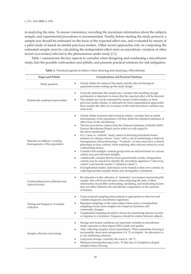
J. Clin. Med. 2019, 8, 1206
in analyzing the data. To ensure consistency, recording the maximum information about the subjects,sample, and experimental procedures is recommended. Finally, before starting the study protocol, asample size should be estimated on the basis of the expected effect size, and evaluated by means ofa pilot study or based on similar previous studies. Other recent approaches rely on computing theestimated sample sizes by calculating the independent effect sizes on microbiota variation of otherfactors (covariates) relevant to the phenomenon under study [21].
Table 1 summarizes the key aspects to consider when designing and conducting a microbiomestudy, lists the possible confounders and pitfalls, and presents practical solutions for risk mitigation.
Table 1. Practical aspects to follow when drawing and studying a Microbiome.
Stages and Pitfalls Considerations and Practical Solutions
Study question • Clearly define the aim(s) of the study and the relevant biologicalquestion(s) before setting up the study design.
Statistically underpowered studies
• Correctly determine the sample size: consider that enrolling enoughparticipants is important to ensure that the expected effect will be detected.
• The sample size can be estimated by means of pilot studies, or fromprevious similar studies, or alternatively from computational approachesthat consider the effect of covariates on the total microbiota variation (seemain text).
Selection of subjects: avoidingheterogeneity of the population
• Clearly define inclusion and exclusion criteria: consider that an initialheterogeneity of the population will then dilute the statistical estimates ofeffect sizes on the microbiome.
• The list of exclusion criteria from the National Institutes of Health (NIH)Human Microbiome Project can be relied on with regard tothe above-mentioned.
• In a “cases vs. controls” study, aimed at detecting microbiota-basedmarkers of a disease, choose “cases” with a care in maintaining a relativelyhomogeneous clinical phenotype. “Controls”, in turn, must have a clinicalphenotype in clear contrast, while matching other relevant criteria to avoidconfounding factors.
• Consider that multiple controls groups that are selected based on variouscriteria may provide more insights.
• Additionally consider that for more generalizable results, independentcohorts may be selected to identify the microbiota signatures (“discoverycohort”) and test the results (“validation cohort”).
• In longitudinal studies, individuals can be treated as their own controls, bycollecting baseline samples before and during/after a treatment.
Confounding factors (lifestyle andclinical factors)
• Be exhaustive in the collection of “metadata” (covariates) surrounding thesample: this will be pivotal later, when analyzing the data. Collectinformation on possible confounding, mediating, and moderating factorsthat can either influence the microbiome composition or the outcomeof interest.
Timing and frequency of samplecollection
• Cross-sectional sampling from patients is appropriate to discover andvalidate diagnostic microbiome signatures.
• Repeated samplings of the same subject (time series or longitudinalsampling) ensure more insights into temporal dynamics andcommunity changes.
• Longitudinal sampling should be chosen for monitoring disease severityor response to a treatment. Frequency should be similar between subjects.
Sample collection and storage
• Storage and transit conditions are important variables in microbiomestudy outcomes as they impact DNA yields and quality.
• After collecting samples, freeze immediately. When immediate freezing isnot possible, short-term refrigeration (+4 ◦C) is helpful. An alternative isto use stabilizing solutions.
• Long-term storage: currently the norm is −80 ◦C.• Minimize freezing-thawing cycles. To this aim, it is helpful to aliquot
samples before freezing.
87

J. Clin. Med. 2019, 8, 1206
Table 1. Cont.
Stages and Pitfalls Considerations and Practical Solutions
Experimental Lab procedures
• Use the same procedures and reagents throughout the study. Documenteverything and be consistent. If, for example, different batches of anenzyme are used, document it among the metadata.
• DNA extraction: This is an important source of variation and bias becauseof the differential resistance to lysis of microbial cells. Combine chemicaland mechanical lysing procedures to capture the most accuratecommunity composition.
• Contamination may significantly impact results, especially if working onlow-biomass samples. It may derive from laboratory contaminants (e.g.,previously produced amplicons), from reagents and commercial kits(“kitome”). It is recommendable to separate pre- and post-PCR areas andto introduce appropriate negative controls in different sample processingsteps (e.g., blank extraction control: DNA-free water undergoes DNAextraction and all subsequent experimental procedures; blank PCR control:DNA-free water undergoes PCR and all subsequent procedures).
• Selection of 16S primers: Rely on previous studies and consider thatdifferent couples of universal 16S primers may be biased toward (oragainst) certain bacterial taxa, thus giving artefactual over- (orunder-representations) of them. For example, the 27F/338R primer sets(targeting the V1–V3 regions) is biased against the amplification ofBifidobacteria. Another possible pitfall is given by primer sets poorlyresolving specific taxa.
• PCR amplification: Low DNA template concentration and high number ofPCR cycles introduce biases. To reduce their effects, minimize PCR cycles,use a standard (and relatively high) DNA template concentration, andpool multiple PCR (e.g., triplicates) for each sample. The use ofproof-reading DNA polymerases and longer annealing times (to reducechimera formation) is also recommended.
Sequencing
• Use positive controls to calibrate the sequencing method: (i) pure strainsof, e.g., Escherichia coli that produce strong PCR bands of a known size;and (ii) a synthetic mock microbial community to ensure thatamplification, sequencing, and taxonomic classification workflows havenot introduced substantial bias or distortions in the expected microbiomeprofiles. Consider that, in addition to the DNA extraction and PCR steps,errors can be introduced during library preparation, sequencing, imaging,and data analysis.
Data analysis
• The design and choice of the analyses is strictly connected with theresearch objectives of the study.
• Be consistent with the procedures and software used for analyzing data.Consider that different software versions can behave differently.
• Integrate non-microbiome sources of data (e.g., clinical parameters) withmicrobiome data to answer the biological questions that primed the study.
• Consider that microbiota data are high-dimensional in nature, with thetotal number of variable measurements far exceeding the numberof samples.
• Incorporate the patient and experimental covariates collected in the“metadata” file of the analysis. Evaluate if some of them act asconfounding factors.
• Repeat the analyses introducing some changes (e.g., change someparameters or algorithms, include or exclude metadata) and the evaluatereproducibility of results.
• The complexity of questions in a translational study makes its useful totest multiple statistical models using several combinations ofindependent-dependent variables.
• If a variable is continuous, using it directly in the model is substantiallymore informative than using a categorical or binary encoding.
• Remember that DNA-based techniques are not able to reveal if themicrobes under study are alive or dead. If precise information on this isneeded, consider performing meta-transcriptomics.
Risk-benefit assessment • Studies need to be designed to ensure that short term and long-termreliable data are collected.
88

J. Clin. Med. 2019, 8, 1206
5. Major Pre-, Peri-, and Post-Natal Factors Affecting the Child Gut Microbiota
A schematic representation of the factors that are able to affect the dynamics and composition ofthe intestinal microbiota is given in Figure 2.
Figure 2. Infant microbiota composition (a) and the main “major” and “minor” factors affecting analysisand results in microbiota studies (b).
5.1. Maternal Factors Influencing Infant Microbiota
5.1.1. Changes Related to Vertical Transmission of Maternal Metabolites
During gestation, bacteria in the mother’s intestine have been shown to drive the future immunematuration of the neonatal gut through the passage of soluble molecules from the placenta in theabsence of direct colonization and of the vertical transmission of viable bacterial cells [22,23]. Thesebacteria are able to induce specific changes in the gut of newborns, creating new microbiota profiles.
5.1.2. Changes Related to Dietary Patterns and Lifestyle
The intestinal microbiota is strongly personalized and influenced by a plethora of environmentaland inter-individual variables including body mass index (BMI), exercise frequency, and dietarypatterns and habits (which in turn, are strongly related with cultural factors and lifestyle). It has
89
J. Clin. Med. 2019, 8, 1206
been reported that the infant’s fecal microbiota composition is influenced by the BMI and weightgain of the mother during pregnancy [24,25]. In general, the maternal microbial reservoir plays acrucial role in the acquisition and development of early infant microbiota, which in turn is the key toestablishing a healthy host–microbiome symbiosis with long-lasting health effects. Therefore, it canbe easily understood as to why maternal diet and lifestyle should be monitored and categorized asrelevant metadata in infant microbiota studies. In an early phase, after the huge microbial “inoculum”at birth, the infant continues to directly acquire maternal gut strains from different sources (e.g., fromskin, mouth, milk) and these are likely to become stable colonizers of the infant gut. Later in life,increasingly important roles are also played by other factors such as shared diet and lifestyle.
5.2. Genetic Factors
There is growing evidence that geographical origin and host genetic makeup influence theacquisition and development of the gut microbiota, with clear associations reported between thehost genotype and the relative abundances of different bacterial taxa. For example, Bonder et al. [26]described a single nucleotide polymorphism (SNP) in the LCT locus (coding for human lactase) that isrelated to varying abundances of Bifidobacterium. Goodrich et al. [27], by comparing microbiota acrosssamples belonging to either monozygotic and dizygotic twin pairs, reported a number of microbial taxawhose abundances were strongly influenced by host genetics. Among such taxa, the Christensellaceae,considered a microbiome-based marker of obesity and is significantly enriched in individuals with lowBMI, resulted in the most highly heritable taxon. Any data related to the genetic hardware of the childshould then be noticed.
5.3. Mode of Delivery
At birth, the infant gut communities tend to resemble the maternal vagina or skin microbiotain cases of vaginal or cesarean section (C-section) delivery, respectively [19,28]. Even later, whenthese “pioneer” foundation populations have been replaced, the birth mode seems to exert significantlong-term effects on the structure of the gut microbiota. At 24 months of age, the gut microbialcommunities of cesarean delivered infants still appear to be less diverse [15]. Even in children as old asseven years, some authors have reported the enduring influence of the mode of delivery, but data aresomewhat contrasting regarding this point [19]. Vaginally delivered infants tend to be colonized byLactobacillus and Prevotella, while C-section neonates are preferentially colonized by microorganismsfrom maternal skin, and the hospital staff or environment.
5.4. Mode of Infant Feeding
Breastfed infants receive, from their mothers’ milk, a complex mix that will affect the milieuwithin which their own microbiota will develop. This mix is made up of nutrients, antimicrobialproteins, short chain fatty acids (SCFA), secretory IgA, non-digestible oligosaccharides (HMOs, humanmilk oligosaccharides, that promote the proliferation of specific gut bacterial taxa in the neonate),and live bacteria, even if previously considered germ-free [15]. The source of the “milk microbiota”,which has a transient nature and declines rapidly at weaning, has recently been another subject ofdebate. At least some of the bacteria is thought to reach the mammary gland through an endogenousroute called the enteromammary pathway, which has not been fully elucidated yet. It has also beensuggested that mammary skin microbiota can travel via the lymphatic and vascular circulations to thebreast ([15,16] and references therein). Gut microbiota differences between breastfed and formula-fedinfants are indeed well documented. The former exhibit lower diversity indexes, indicative of a moreuniform population where Bifidobacterium and Lactobacillus dominate. The latter are characterized bymore diverse communities, with higher proportions of Bacteroides, Clostridium, Streptococcus, Veillonella,Atopobium, and Enterobacteriaceae [29]. Finally, compositional differences in microbial communities inhuman milk sampled from different geographical locations have been studied and reported to createstrong variability between newborn microbiota [30].
90

J. Clin. Med. 2019, 8, 1206
5.5. Gestational Age
While in full-term infants, delivery and feeding mode are reported to represent the major driversof microbiota development, in preterm (PT) infants (<37 weeks of gestation), the gestational age seemsto have the biggest impact on the assembly of gut communities [19,31,32]. PT neonates experiencea number of unique challenges in the establishment of their microbiota. Their colonization patternsare characterized by the involvement of peculiar microbial sources, mainly bacteria deriving fromthe neonatal intensive care unit (NICU) environment [33]. Not rarely, these are strains implicated innosocomial infections such as Enterococcus spp., Staphylococcus aureus, Klebsiella pneumoniae, Acinetobacterspp., Pesudomonas aeruginosa, and other Enterobacteriaceae [34] with their burden of antibiotic resistancegenes. Other relevant features of this peculiar colonization trajectory are its extreme inter-individualvariability, and the fact that, across studies, it does not appear to be univocally linked to healthoutcomes as necrotizing enterocolitis and late-onset sepsis. Instead, the colonization process seemsto reflect the co-occurrence of a variety of nosocomial “variables” [35], among which are parenteralnutrition and antibiotic usage (see below). Antibiotics, normally administered to these patients, in turnperturbate the colonization process by killing bacteria acquired during birth and promoting the growthof taxa significantly different from those found in more physiological situations [31]. In conclusion,the PT microbiota appears to be more unstable than that of full-term equivalents and is believed tobe associated with a delay in the establishment of an adult-type signature microbiota [16]. All theseindividuals should be carefully selected and clearly categorized by the clinician before enrollment intothe microbiota study.
5.6. Antibiotics
Specific properties of antibiotics, as a mode of action and antimicrobial spectrum, might act aspowerful forces for the selection of intestinal bacterial populations, especially if the infant is exposed toantibiotics too early and/or for long periods of time [3,15]. Antibiotics are able to alter the abundancesof resident bacteria, significantly impact the growth of otherwise dominant bacterial phyla, and lead toan overall decrease in microbial diversity. A study by Fouhy and colleagues [36] showed that infantsexposed to ampicillin and gentamicin shortly after birth harbored higher proportions of Proteobacteriaand Actinobacteria, and the genus Lactobacillus for up to four weeks after concluding treatment. Anotherstudy reported an attenuation in colonization with Bifidobacterium and an increase of Enterococcus insubjects receiving oral or intravenous antibiotics during the first four days of life [37].
This variability among individuals suggests caution when including subjects who have beentreated with antibiotics [38]. Indeed, the exclusion criteria from the NIH Human Microbiome Project(HMP, dbGAP, see the url https://www.ncbi.nlm.nih.gov/projects/gap/cgi-bin/study.cgi?study_id=phs000228.v4.p1) include the use of systemic antibiotics, antifungals, antivirals, or antiparasitics withinsix months of sampling. However, this criterion, although optimal, may not be easily applicable withsubjects in the pediatric age. For this reason, shorter time windows are often considered. In any case, itis mandatory to accurately document, within the metadata file, any history of antibiotics as well asother medication use.
5.7. Weaning
The transition to more varied, solid food is an important step in the development of the early-lifegut microbiota; infants begin to be exposed to a much larger array of substrates and non-digestiblecarbohydrates that promote the survival and proliferation of more various bacterial taxa. As aconsequence, the alpha diversity increases; moreover, Proteobacteria and Actinobacteria are replacedby Firmicutes and Bacteroidetes as the dominant phyla, in a more adult-like compositional structure.The cessation of exclusive milk feeding correlates with the decrease of saccharolytic bacteria asBifidobacteriaceae (phylum: Actinobacteria). The increased protein intake is thought to be associated with
91
J. Clin. Med. 2019, 8, 1206
an increase of Lachnospiraceae (phylum: Firmicutes), while the ingestion of fibers with that of higherlevels of Prevotellaceae (phylum: Bacteroidetes) [39].
In general, the relative abundance of our intestinal microbes is highly influenced by dietary patternsand habits [11], that should therefore be taken into account in clinical studies targeting microbiota.
6. Minor Factors Affecting Gut Microbiota
Various minor factors can affect and modify the gut microbiota, which can occur at any stage of life.Insomnia and circadian rhythm disruption, latitude with time zone shift and intercontinental flights(with the consequent jet lag), household siblings, and companion animals as well as seasonal changescan modify gut microbiota and determine different microbiota profiles with high inter-individualvariability to responses to the different factors [40–42]. All of these factors can influence the resultsand should be carefully considered before starting a clinical study and accurately reported in themetadata to then be considered later in the downstream bioinformatics and statistical analyses. Othersimilar confounder factors such as bowel movement preparations, evacuants or laxatives, or anymicroorganism-supplemented food (such as probiotics) can act as deep and long-time gut modifiers,thus a plot-to-plot variation needs to be addressed with nested statistical tests.
7. Sample Collection
Donors/patients to enroll, their genetic or disease phenotypes as well as the expertise of theclinician in methodology used for collecting samples are very relevant in designing a correct study.The number of samples and patients to be enrolled is an intriguing and still hotly debated topic.Sample stability as well as shipping and storage requirements need to be more appropriate andwill surely be improved and standardized in the future. Researchers may find some proceduresat http://www.microbiome-standards.org or at https://www.hmpdacc.org/resources/metagenomics_sequencing_analysis.php and other papers [43–45].
Concerning the practical aspects, an important question is how often to collect samples becausethe microbiome ecology is intrinsically dynamic. This largely depends on what question one is tryingto answer. If, for gastrointestinal disorders, remarkable changes can be observed between one day andthe next (e.g., in times surrounding surgery or in correspondence with periods of activity or remissionof the pathology), changes induced by other factors (e.g., diet) often take place on a longer timescale.Collection of multiple samples from the same patient is preferred to allow for better standardizationon the basis of the type of patients, centers involved, and statistical power. Whether or not samplescollected from the same individual can be pooled before analysis is another topic to be standardized.An important point is that sampling and storage do affect microbiota composition in healthy as wellas in diseased subjects. The most widely accepted protocols include immediate homogenizationand freezing either with dry ice or in liquid nitrogen, followed by storage at −80 ◦C. However, thisapproach is not always practical, particularly for stool samples, or in the case of stool collection from alarge scale cohort or remote/rural areas. Whether samples must be immediately frozen (and at whattemperature) or whether they can withstand a period of room temperature remains controversial. Theabove-mentioned studies showed that the effects of short-term storage conditions on the structureand diversity of communities are quite small in general. In particular, storage at −80 ◦C, −20 ◦C for aweek, or 4 ◦C for 24 h were found to not significantly affect the ecological indexes of between-samplediversity or the abundance of major taxa [45]. In contrast, the number of freeze–thaw cycles seems tohave an effect on the composition of the microbial community, thus it is strongly recommended toaliquot samples at the beginning. Of course, some DNA stabilizers can be used to prolong the stabilityof samples. In the study of Choo et al. [46] Omnigene Gut and Tris EDTA appeared to show the sameperformance as storage in an ultrafreezer (−80 ◦C). In addition to feces, swabs can be an alternativestarting material for DNA extractions, especially within hospital settings, even if some studies haveshown that the stool swabs of some subjects had limited and not detectable bacterial DNA. A recentstudy by Christine M. Bassis [47], by comparing stool versus rectal swab samples and their storage
92
J. Clin. Med. 2019, 8, 1206
conditions, demonstrated minor differences in the bacterial community profiles between the stool andswab from the same subject as well as when samples were stored up to 27 h at +4 ◦C before freezing at−80 ◦C. Interestingly, this study also concluded that it was possible to thaw and refreeze samples alimited number of times under particular conditions (i.e., immediately frozen at −20 ◦C, first thawcycle, refrozen at −80 ◦C; immediately frozen at −20 ◦C, first thaw cycle, refrozen at −20 ◦C, secondthaw cycle and frozen at −80 ◦C) without strong effects on the community composition. A word ofcaution is, however, due on this point, as the consensus recommendations are different, as detailedabove. Finally, it is to be underlined that as the collection of stool can be difficult from some subjectsunder certain experimental conditions, swab collection may be useful in such cases, which also hasthe advantage that they are more easily shipped and handled. A further recent study confirmed thatswab samples reliably replicate the stool microbiota bacterial composition when swabs are processedquickly (≤2 days) [48].
Finally, special considerations are needed if addressing peculiar samples such as the newborn’sfirst intestinal discharge (meconium). The debate about “when” the neonatal microbiota begins hasbeen previously mentioned. Recently, several scientists have underlined that, even if it is possiblethat not all healthy babies are born sterile as previously thought, particular caution is due whenworking on samples bearing low microbial biomass such as meconia because of the contaminationissues connaturated with molecular approaches based on PCR amplification and next-generationsequencing [17,49,50]. The presence of contaminating DNA in laboratory reagents (so-called “kitome”)is a serious challenge in these cases; low levels of target bacterial DNA in a sample have been reportedto correlate with a high proportion of sequences being attributable to contamination [51,52].
8. Discussion
The Anna Karenina principle, based on Leo Tolstoy’s great book and cited in 1878 (All happyfamilies are alike: each unhappy family is unhappy in its own way), has been recently translated byZaneveld et al. [53] as the response to stress against the stability of animal microbiomes. These authorsdiscussed how healthy microbiomes may be quite similar between individuals, but each dysbioticmicrobiota is dysbiotic in its own way. The associations between microbiome instability/variabilityand many confounding factors as well as with diseases, suggest that microbiome may have many andsimultaneous multiple faces.
This “stochastic” drift, occurring at any stage of life under stress conditions, can create severalphenotypes that need to be known and harmonized when planning a study on microbiota.
Early childhood possesses distinct microbiota tracts compared with later ones, where differentclusters and phyla may be differently represented. One common characteristic during this early stageof life is that bacterial richness and diversity increase during growth. Therefore, pediatricians shouldknow that there are several age-related microbiota profiles, and should also be aware of the need tocategorize each individual in a defined, monthly range by carefully considering the above-mentionedinterference factors.
Several specialties need to be involved in this aim as well as the combination of differentknowledge. The “Clinical Microbiota Expert” is not only a new job, but represents a step forwardto create competence in this field where clinical microbiologists, clinicians, and bioinformaticiansare merged into one. This new job-role will have to create awareness on the study of the “dynamicbody” such as the gut microbiota during early age by creating novel models and approaches as well assolutions to solve and interpret the clinical microbiology results. Therefore, translational methodologiesto approach a new way of designing clinical trials need to use feasibility and efficacy tools, and a deeperpreparation in the field to avoid uncontrolled errors, unsubstantiated results, and excessive costs.
93
J. Clin. Med. 2019, 8, 1206
9. Conclusions and Future Perspectives
Next-generation sequencing methodologies still remain expensive and the diagnostic market isoffering different solutions, thus a proper, and especially judicious, use of these methods is definitivelymandatory. The clinical microbiota expert and pediatricians involved in the field will also have toguide through this jungle by trying to avoid false myths and promises that could be difficult to realize.In the near future, all of these studies and experiences will necessarily lead to a better understandingof the real key phases of microbiome progression from birth throughout childhood.
A final consideration to underline is that the metagenomics community still needs to fully convergetoward standardized methods and procedures, leading to an investigation of the sources of variabilityand bias at each step of the workflow, and to an improved reproducibility and comparability betweenstudies. This is a necessary premise for moving from correlation studies to causation investigationsand to answer complex questions in a translational setting.
Author Contributions: L.D. designed and conceived the study; S.P. revised the paper and the technical aspectsand checked the literature; C.B. and G.Z. revised the manuscript; M.P. conceived the figures and revised themanuscript; E.D. revised the clinical aspects and the manuscript.
Acknowledgments: The authors wish to thank the Fondazione “Romeo ed Enrica Invernizzi”.
Conflicts of Interest: The authors declare no conflicts of interest.
References
1. Zmora, N.; Zilberman-Schapira, G.; Suez, J.; Mor, U.; Dori-Bachash, M.; Bashiardes, S.; Kotler, E.; Zur, M.;Regev-Lehavi, D.; Brik, R.B.Z.; et al. Personalized Gut Mucosal Colonization Resistance to Empiric ProbioticsIs Associated with Unique Host and Microbiome Features. Cell 2018, 174, 1388–1405. [CrossRef] [PubMed]
2. Yatsunenko, T.; Rey, F.E.; Manary, M.J.; Trehan, I.; Dominguez-Bello, M.G.; Contreras, M.; Magris, M.;Hidalgo, G.; Baldassano, R.N.; Anokhin, A.P.; et al. Human gut microbiome viewed across age andgeography. Nature 2012, 486, 222–227. [CrossRef] [PubMed]
3. Rinninella, E.; Raoul, P.; Cintoni, M.; Franceschi, F.; Miggiano, G.A.D.; Gasbarrini, A.; Mele, M.C. What is theHealthy Gut Microbiota Composition? A Changing Ecosystem across Age, Environment, Diet, and Diseases.Microorganisms 2019, 7, 14. [CrossRef] [PubMed]
4. Lozupone, C.A.; Stombaugh, J.I.; Gordon, J.I.; Jansson, J.K.; Knight, R. Diversity, stability and resilience ofthe human gut microbiota. Nature 2012, 489, 220–230. [CrossRef] [PubMed]
5. Williams, S.C. The other microbiome. Proc. Natl. Acad. Sci. USA 2013, 110, 2682–2684. [CrossRef] [PubMed]6. Thomas, T.; Gilbert, J.; Meyer, F. Metagenomics—A guide from sampling to data analysis. Microb. Inform.
Exp. 2012, 2, 3. [CrossRef] [PubMed]7. Turnbaugh, P.J.; Gordon, J.I. An invitation to the marriage of metagenomics and metabolomics. Cell 2006,
134, 708–713. [CrossRef]8. Trivedi, B. Microbiome: The surface brigade. Nature 2012, 492, S60–S61. [CrossRef]9. Lagier, J.C.; Dubourg, G.; Million, M.; Cadoret, F.; Bilen, M.; Fenollar, F.; Levasseur, A.; Rolain, J.M.;
Fournier, P.E.; Raoult, D. Culturing the human microbiota and culturomics. Nat. Rev. Genet. 2018, 16,540–550. [CrossRef]
10. Peterson, J.; Garges, S.; Giovanni, M.; McInnes, P.; Wang, L.; Schloss, J.A.; Bonazzi, V.; McEwen, J.E.;Wetterstrand, K.A.; Deal, C.; et al. The NIH human microbiome project. Genome Res. 2009, 19, 2317–2323.
11. Costello, E.K.; Lauber, C.L.; Hamady, M.; Fierer, N.; Gordon, J.I.; Knight, R. Bacterial Community Variationin Human Body Habitats Across Space and Time. Science 2009, 326, 1694–1697. [CrossRef] [PubMed]
12. Qin, J.; Li, R.; Raes, J.; Arumugam, M.; Burgdorf, K.S.; Manichanh, C.; Nielsen, T.; Pons, N.; Levenez, F.;Yamada, T.; et al. A human gut microbial gene catalogue established by metagenomic sequencing. Nature2010, 464, 59–65. [CrossRef] [PubMed]
13. Human Microbiome Project Consortium. Structure, function and diversity of the healthy human microbiome.Nature 2012, 486, 207–214. [CrossRef] [PubMed]
94

J. Clin. Med. 2019, 8, 1206
14. Kelly, B.J.; Gross, R.; Bittinger, K.; Sherrill-Mix, S.; Lewis, J.D.; Collman, R.G.; Bushman, F.D.; Li, H. Power andsample-size estimation for microbiome studies using pairwise distances and PERMANOVA. Bioinformatics2015, 31, 2461–2468. [CrossRef]
15. Arrieta, M.C.; Stiemsma, L.T.; Amenyogbe, N.; Brown, E.M.; Finlay, B. The Intestinal Microbiome in EarlyLife: Health and Disease. Front. Immunol. 2014, 5, 427. [CrossRef]
16. Milani, C.; Duranti, S.; Bottacini, F.; Casey, E.; Turroni, F.; Mahony, J.; Belzer, C.; Palacio, S.D.; Montes, S.A.;Mancabelli, L.; et al. The First Microbial Colonizers of the Human Gut: Composition, Activities, and HealthImplications of the Infant Gut Microbiota. Microbiol. Mol. Biol. Rev. 2017, 81, e00036-17. [CrossRef]
17. Willyard, C. Could baby’s first bacteria take root before birth? Nature 2018, 553, 264–266. [CrossRef]18. Matamoros, S.; Guen, C.G.L.; Le Vacon, F.; Potel, G.; De La Cochetière, M.F. Development of intestinal
microbiota in infants and its impact on health. Trends Microbiol. 2013, 21, 167–173. [CrossRef]19. Hill, C.J.; Lynch, D.B.; Murphy, K.; Ulaszewska, M.; Jeffery, I.B.; O’Shea, C.A.; Watkins, C.; Dempsey, E.;
Mattivi, F.; Touhy, K.; et al. Evolution of gut microbiota composition from birth to 24 weeks in theINFANTMET Cohort. Microbiome 2017, 5, 4. [CrossRef]
20. Stewart, C.J.; Ajami, N.J.; O’Brien, J.L.; Hutchinson, D.S.; Smith, D.P.; Wong, M.C.; Ross, M.C.; Lloyd, R.E.;Doddapaneni, H.; Metcalf, G.A.; et al. Temporal development of the gut microbiome in early childhoodfrom the TEDDY study. Nature 2018, 562, 583. [CrossRef]
21. Falony, G.; Joonssens, M.; Vieine-Silva, S.; Wang, J.; Darzi, Y.; Faust, K.; Kurilshikov, A.; Bonder, M.J.;Valles-Colomer, M.; Vandeputte, P.; et al. Population-level analysis of gut microbiota variation. Science 2016,352, 560–564. [CrossRef] [PubMed]
22. De Agüero, M.G.; Ganal-Vonarburg, S.C.; Fuhrer, T.; Rupp, S.; Uchimura, Y.; Li, H.; Steinert, A.;Heikenwalder, M.; Hapfelmeier, S.; Sauer, U.; et al. The maternal microbiota drives early postnatalinnate immune development. Science 2016, 351, 1296–1302. [CrossRef] [PubMed]
23. Rakoff-Nahoum, S. Another reason to thank mum: Gestational effects of microbiota metabolites. Cell HostMicrobe 2016, 19, 425–427. [CrossRef] [PubMed]
24. MacPherson, A.J.; De Agüero, M.G.; Ganal-Vonarburg, S.C. How nutrition and the maternal microbiotashape the neonatal immune system. Nat. Rev. Immunol. 2017, 17, 508–517. [CrossRef] [PubMed]
25. Kearney, J.F.; Patel, P.; Stefanov, E.K.; King, R.G. Natural Antibody Repertoires: Development and FunctionalRole in Inhibiting Allergic Airway Disease. Annu. Rev. Immunol. 2015, 33, 475–504. [CrossRef] [PubMed]
26. Bonder, M.J.; Kurilshikov, A.; Tigchelaar, E.F.; Mujagic, Z.; Imhann, F.; Vila, A.V.; Deelen, P.; Vatanen, T.;Schirmer, M.; Smeekens, S.P.; et al. The effect of host genetics on the gut microbiome. Nat. Genet. 2016, 48,1407–1412. [CrossRef] [PubMed]
27. Goodrich, J.K.; Waters, J.L.; Poole, A.C.; Sutter, J.L.; Koren, O.; Blekhman, R.; Beaumont, M.; Van Treuren, W.;Knight, R.; Bell, J.T.; et al. Human genetics shape the gut microbiome. Cell 2014, 159, 789–799. [CrossRef][PubMed]
28. Dominguez-Bello, M.G.; Costello, E.K.; Contreras, M.; Magris, M.; Hidalgo, G.; Fierer, N.; Knight, R. Deliverymode shapes the acquisition and structure of the initial microbiota across multiple body habitats in newborns.Proc. Natl. Acad. Sci. USA 2010, 107, 11971–11975. [CrossRef] [PubMed]
29. Fallani, M.; Young, D.; Scott, J.; Norin, E.; Amarri, S.; Adam, R.; Aguilera, M.; Khanna, S.; Gil, A.;Edwards, A.C.; et al. Intestinal Microbiota of 6-week-old Infants Across Europe: Geographic InfluenceBeyond Delivery Mode, Breast-feeding, and Antibiotics. J. Pediatr. Gastroenterol. Nutr. 2010, 51, 77–84.[CrossRef]
30. Drago, L.; Toscano, M.; De Grandi, R.; Grossi, E.; Padovani, E.M.; Peroni, D.G. Microbiota network andmathematic microbe mutualism in colostrum and mature milk collected in two different geographic areas:Italy versus Burundi. ISME J. 2017, 11, 875–884. [CrossRef]
31. Groer, M.W.; Luciano, A.A.; Dishaw, L.J.; Ashmeade, T.L.; Miller, E.; Gilbert, A.J. Development of the preterminfant gut microbiome: A research priority. Microbiome 2014, 2, 38. [CrossRef] [PubMed]
32. Stewart, C.J.; Embleton, N.D.; Clements, E.; Luna, P.N.; Smith, D.P.; Fofanova, T.Y.; Nelson, A.; Taylor, G.;Orr, C.H.; Petrosino, J.F.; et al. Cesarean or Vaginal Birth Does Not Impact the Longitudinal Development ofthe Gut Microbiome in a Cohort of Exclusively Preterm Infants. Front. Microbiol. 2017, 8, 1008. [CrossRef][PubMed]
95

J. Clin. Med. 2019, 8, 1206
33. Brooks, B.; Olm, M.R.; Firek, B.A.; Baker, R.; Thomas, B.C.; Morowitz, M.J.; Banfield, J.F. Strain-resolvedanalysis of hospital rooms and infants reveals overlap between the human and room microbiome.Nat. Commun. 2017, 8, 1814. [CrossRef] [PubMed]
34. Hu, H.; Johani, K.; Gosbell, I.; Jacombs, A.; Almatroudi, A.; Whiteley, G.; Deva, A.; Jensen, S.; Vickery, K.Intensive care unit environmental surfaces are contaminated by multidrug-resistant bacteria in biofilms:Combined results of conventional culture, pyrosequencing, scanning electron microscopy, and confocal lasermicroscopy. J. Hosp. Infect. 2015, 91, 35–44. [CrossRef] [PubMed]
35. Wandro, S.; Osborne, S.; Enriquez, C.; Bixby, C.; Arrieta, A.; Whiteson, K. The microbiome and metabolome ofpreterm onfant stool are personalized and not driven by health outcomes, including necrotizing enterocolitisand late-onset sepsis. Msphere 2018, 3, e00104–e00118. [CrossRef] [PubMed]
36. Fouhy, F.; Guinane, C.M.; Hussey, S.; Wall, R.; Ryan, C.A.; Dempsey, E.M.; Murphy, B.; Ross, R.P.;Fitzgerald, G.F.; Stanton, C.; et al. High-Throughput Sequencing Reveals the Incomplete, Short-TermRecovery of Infant Gut Microbiota following Parenteral Antibiotic Treatment with Ampicillin and Gentamicin.Antimicrob. Agents Chemother. 2012, 56, 5811–5820. [CrossRef] [PubMed]
37. Tanaka, S.; Kobayashi, T.; Songjinda, P.; Tatejama, A.; Tsubouchi, M.; Kiyohara, C.; Shirakawa, T.; Sonomoto, J.;Nakayama, J. Influence of antibiotic exposure in the early postnatal period in the development of intestinalmicrobiota. FEMS Immunol. Med. Microbiol. 2009, 56, 80–87. [CrossRef]
38. Dethlefsen, L.; Relman, D.A. Incomplete recovery and individualized responses of the human distal gutmicrobiota to repeated antibiotic perturbation. Proc. Natl. Acad. Sci. USA 2011, 108, 4554–4561. [CrossRef]
39. Koenig, J.E.; Spor, A.; Scalfone, N.; Fricker, A.D.; Stombaugh, J.; Knight, R.; Angenent, L.T.; Ley, R.E.Succession of microbial consortia in the developing infant gut microbiome. Proc. Natl. Acad. Sci. USA 2011,108, S4578–S4585. [CrossRef]
40. Thaiss, C.A.; Levy, M.; Korem, T.; Dohnalová, L.; Shapiro, H.; Jaitin, D.A.; David, E.; Winter, D.R.;Gury-BenAri, M.; Tatirovsky, E.; et al. Microbiota Diurnal Rhythmicity Programs Host TranscriptomeOscillations. Cell 2016, 167, 1495–1510. [CrossRef]
41. Misic, A.M.; Davis, M.F.; Tyldsley, A.S.; Hodkinson, B.P.; Tolomeo, P.; Hu, B.; Nachamkin, I.; Lautenbach, E.;Morris, O.D.; Grice, A.E. The shared microbiota of humans and companion animals as evaluated fromStaphylococcus carriage sites. Microbiome 2015, 3, 2. [CrossRef] [PubMed]
42. Davenport, E.R.; Mizrahi-Man, O.; Michelini, K.; Barreiro, L.B.; Ober, C.; Gilad, Y. Seasonal Variation inHuman Gut Microbiome Composition. PLoS ONE 2014, 9, e90731. [CrossRef] [PubMed]
43. Costea, P.I.; Zeller, G.; Sunagawa, S.; Pelletier, E.; Alberti, A.; Levenez, F.; Tramontano, M.; Driessen, M.;Hercog, R.; Jung, F.E.; et al. Towards standards for human fecal sample processing in metagenomic studies.Nat. Biotechnol. 2017, 35, 1069–1076. [CrossRef] [PubMed]
44. Jordan, S.; Baker, B.; Dunn, A.; Edwards, S.; Ferranti, E.; Mutic, A.D.; Yang, I.; Rodriguez, J. Maternal–ChildMicrobiome: Specimen Collection, Storage and Implications for Research and Practice. Nurs. Res. 2017, 66,175–183. [CrossRef] [PubMed]
45. Tedjo, D.I.; Jonkers, D.M.A.E.; Savelkoul, P.H.; Masclee, A.A.; Van Best, N.; Pierik, M.J.; Penders, J. The Effectof Sampling and Storage on the Fecal Microbiota Composition in Healthy and Diseased Subjects. PLoS ONE2015, 10, 0126685. [CrossRef] [PubMed]
46. Choo, J.M.; Leong, L.E.; Rogers, G.B. Sample storage conditions significantly influence faecal microbiomeprofiles. Sci. Rep. 2015, 5, 16350. [CrossRef]
47. Bassis, C.M.; Moore, N.M.; Lolans, K.; Seekatz, A.M.; Weinstein, R.A.; Young, V.B.; Hayden, M.K. CDCPrevention Epicenters Program. Comparison of stool versus rectal swab samples and storage conditions onbacterial community profiles. BMC Microbiol. 2017, 17, 78–85. [CrossRef]
48. Bokulich, N.A.; Maldonado, J.; Kang, D.W.; Krajmalnik-Brown, R.; Caporaso, J.G. Rapidly Processed StoolSwabs Approximate Stool Microbiota Profiles. mSphere 2019, 4, e00208–e00219. [CrossRef]
49. Lauder, A.P.; Roche, A.M.; Sherrill-Mix, S.; Bailey, A.; Laughlin, A.L.; Bittinger, K.; Leite, R.; Elovitz, M.A.;Parry, S.; Bushman, F.D. Comparison of placenta samples with contamination controls does not provideevidence for a distinct placenta microbiota. Microbiome 2016, 4, 29. [CrossRef]
50. Perez-Munoz, M.E.; Arrieta, M.C.; Ramer-Tai, A.E.; Walter, J. A critical assessment of the “sterile womb” and“in utero colonization” hypothesis: Implications for research of the pioneer infant microbiome. Microbiome2017, 5, 48–67. [CrossRef]
96

J. Clin. Med. 2019, 8, 1206
51. Panelli, S.; Schneider, L.; Comandatore, F.; Bandi, C.; Zuccotti, G.V.; D’Auria, E.; Dauria, E. Is there life in themeconium? A challenging, burning question. Pharmacol. Res. 2018, 137, 148–149. [CrossRef] [PubMed]
52. Ferri, E.; Genco, F.; Gulminetti, R.; Bandi, C.; Novati, S.; Casiraghi, M.; Sambri, V. Plasma Levels of BacterialDNA in HIV Infection: The Limits of Quantitative Polymerase Chain Reaction. J. Infect. Dis. 2010, 202,176–177. [CrossRef] [PubMed]
53. Zaneveld, J.R.; McMinds, R.; Thurber, R.V. Stress and stability: Applying the Anna Karenina principle toanimal microbiomes. Nat. Microbiol. 2017, 2, 17121. [CrossRef] [PubMed]
© 2019 by the authors. Licensee MDPI, Basel, Switzerland. This article is an open accessarticle distributed under the terms and conditions of the Creative Commons Attribution(CC BY) license (http://creativecommons.org/licenses/by/4.0/).
97


MDPISt. Alban-Anlage 66
4052 BaselSwitzerland
Tel. +41 61 683 77 34Fax +41 61 302 89 18
www.mdpi.com
Journal of Clinical Medicine Editorial OfficeE-mail: [email protected]
www.mdpi.com/journal/jcm


MDPI St. Alban-Anlage 66 4052 Basel Switzerland
Tel: +41 61 683 77 34 Fax: +41 61 302 89 18
www.mdpi.com ISBN 978-3-0365-0899-3
Related Documents