nanomaterials Review Advances in Non-Animal Testing Approaches towards Accelerated Clinical Translation of Novel Nanotheranostic Therapeutics for Central Nervous System Disorders Mark J. Lynch * and Oliviero L. Gobbo * Citation: Lynch, M.J.; Gobbo, O.L. Advances in Non-Animal Testing Approaches towards Accelerated Clinical Translation of Novel Nanotheranostic Therapeutics for Central Nervous System Disorders. Nanomaterials 2021, 11, 2632. https:// doi.org/10.3390/nano11102632 Academic Editor: Anna Roig Received: 24 August 2021 Accepted: 1 October 2021 Published: 7 October 2021 Publisher’s Note: MDPI stays neutral with regard to jurisdictional claims in published maps and institutional affil- iations. Copyright: © 2021 by the authors. Licensee MDPI, Basel, Switzerland. This article is an open access article distributed under the terms and conditions of the Creative Commons Attribution (CC BY) license (https:// creativecommons.org/licenses/by/ 4.0/). School of Pharmacy and Pharmaceutical Sciences, Panoz Building, Trinity College Dublin, D02 PN40 Dublin, Ireland * Correspondence: [email protected] (M.J.L.); [email protected] (O.L.G.) Abstract: Nanotheranostics constitute a novel drug delivery system approach to improving systemic, brain-targeted delivery of diagnostic imaging agents and pharmacological moieties in one rational carrier platform. While there have been notable successes in this field, currently, the clinical trans- lation of such delivery systems for the treatment of neurological disorders has been limited by the inadequacy of correlating in vitro and in vivo data on blood–brain barrier (BBB) permeation and biocompatibility of nanomaterials. This review aims to identify the most contemporary non-invasive approaches for BBB crossing using nanotheranostics as a novel drug delivery strategy and current non-animal-based models for assessing the safety and efficiency of such formulations. This review will also address current and future directions of select in vitro models for reducing the cumbersome and laborious mandate for testing exclusively in animals. It is hoped these non-animal-based mod- elling approaches will facilitate researchers in optimising promising multifunctional nanocarriers with a view to accelerating clinical testing and authorisation applications. By rational design and appropriate selection of characterised and validated models, ranging from monolayer cell cultures to organ-on-chip microfluidics, promising nanotheranostic particles with modular and rational design can be screened in high-throughput models with robust predictive power. Thus, this article serves to highlight abbreviated research and development possibilities with clinical translational relevance for developing novel nanomaterial-based neuropharmaceuticals for therapy in CNS disorders. By generating predictive data for prospective nanomedicines using validated in vitro models for sup- porting clinical applications in lieu of requiring extensive use of in vivo animal models that have notable limitations, it is hoped that there will be a burgeoning in the nanotherapy of CNS disorders by virtue of accelerated lead identification through screening, optimisation through rational design for brain-targeted delivery across the BBB and clinical testing and approval using fewer animals. Additionally, by using models with tissue of human origin, reproducible therapeutically relevant nanomedicine delivery and individualised therapy can be realised. Keywords: nanotheranostics; blood–brain barrier; advanced drug delivery; in vitro modelling; organ-on-chip; in silico testing 1. Introduction The diagnosis and treatment of central nervous system (CNS) disorders constitute a notable challenge in the field of modern therapeutics and advanced drug delivery systems, and it would appear such disorders are on the rise despite increasing appreciation and elucidation of underlying aetiology and pathophysiological mechanisms [1,2] The CNS therapeutics market is set to grow to EUR 114.4 billion in 2025, and a resurgence in the neuroscience field is predicted, which will be bolstered by novel drug delivery systems and new chemical entities (NCE’s). The most recent global burden of disease (GBD) study published in the Lancet journal shows that the global burden of neurological disorder approaches the 14% value of overall disease modelled over a decade ago, accounting Nanomaterials 2021, 11, 2632. https://doi.org/10.3390/nano11102632 https://www.mdpi.com/journal/nanomaterials

Welcome message from author
This document is posted to help you gain knowledge. Please leave a comment to let me know what you think about it! Share it to your friends and learn new things together.
Transcript

nanomaterials
Review
Advances in Non-Animal Testing Approaches towardsAccelerated Clinical Translation of Novel NanotheranosticTherapeutics for Central Nervous System Disorders
Mark J. Lynch * and Oliviero L. Gobbo *
Citation: Lynch, M.J.; Gobbo, O.L.
Advances in Non-Animal Testing
Approaches towards Accelerated
Clinical Translation of Novel
Nanotheranostic Therapeutics for
Central Nervous System Disorders.
Nanomaterials 2021, 11, 2632. https://
doi.org/10.3390/nano11102632
Academic Editor: Anna Roig
Received: 24 August 2021
Accepted: 1 October 2021
Published: 7 October 2021
Publisher’s Note: MDPI stays neutral
with regard to jurisdictional claims in
published maps and institutional affil-
iations.
Copyright: © 2021 by the authors.
Licensee MDPI, Basel, Switzerland.
This article is an open access article
distributed under the terms and
conditions of the Creative Commons
Attribution (CC BY) license (https://
creativecommons.org/licenses/by/
4.0/).
School of Pharmacy and Pharmaceutical Sciences, Panoz Building, Trinity College Dublin,D02 PN40 Dublin, Ireland* Correspondence: [email protected] (M.J.L.); [email protected] (O.L.G.)
Abstract: Nanotheranostics constitute a novel drug delivery system approach to improving systemic,brain-targeted delivery of diagnostic imaging agents and pharmacological moieties in one rationalcarrier platform. While there have been notable successes in this field, currently, the clinical trans-lation of such delivery systems for the treatment of neurological disorders has been limited by theinadequacy of correlating in vitro and in vivo data on blood–brain barrier (BBB) permeation andbiocompatibility of nanomaterials. This review aims to identify the most contemporary non-invasiveapproaches for BBB crossing using nanotheranostics as a novel drug delivery strategy and currentnon-animal-based models for assessing the safety and efficiency of such formulations. This reviewwill also address current and future directions of select in vitro models for reducing the cumbersomeand laborious mandate for testing exclusively in animals. It is hoped these non-animal-based mod-elling approaches will facilitate researchers in optimising promising multifunctional nanocarrierswith a view to accelerating clinical testing and authorisation applications. By rational design andappropriate selection of characterised and validated models, ranging from monolayer cell cultures toorgan-on-chip microfluidics, promising nanotheranostic particles with modular and rational designcan be screened in high-throughput models with robust predictive power. Thus, this article serves tohighlight abbreviated research and development possibilities with clinical translational relevancefor developing novel nanomaterial-based neuropharmaceuticals for therapy in CNS disorders. Bygenerating predictive data for prospective nanomedicines using validated in vitro models for sup-porting clinical applications in lieu of requiring extensive use of in vivo animal models that havenotable limitations, it is hoped that there will be a burgeoning in the nanotherapy of CNS disordersby virtue of accelerated lead identification through screening, optimisation through rational designfor brain-targeted delivery across the BBB and clinical testing and approval using fewer animals.Additionally, by using models with tissue of human origin, reproducible therapeutically relevantnanomedicine delivery and individualised therapy can be realised.
Keywords: nanotheranostics; blood–brain barrier; advanced drug delivery; in vitro modelling;organ-on-chip; in silico testing
1. Introduction
The diagnosis and treatment of central nervous system (CNS) disorders constitute anotable challenge in the field of modern therapeutics and advanced drug delivery systems,and it would appear such disorders are on the rise despite increasing appreciation andelucidation of underlying aetiology and pathophysiological mechanisms [1,2] The CNStherapeutics market is set to grow to EUR 114.4 billion in 2025, and a resurgence in theneuroscience field is predicted, which will be bolstered by novel drug delivery systemsand new chemical entities (NCE’s). The most recent global burden of disease (GBD) studypublished in the Lancet journal shows that the global burden of neurological disorderapproaches the 14% value of overall disease modelled over a decade ago, accounting
Nanomaterials 2021, 11, 2632. https://doi.org/10.3390/nano11102632 https://www.mdpi.com/journal/nanomaterials
Nanomaterials 2021, 11, 2632 2 of 47
for 250,000 deaths relating to brain and central nervous system cancer and 100 milliondisability-adjusted life years relating to neurological disorders [3]. These figures mirrorthe 2016 systematic review of GBD 1990–2016 [4], which found that deaths increased by39% and DALYs by 27% over this period, and that reductions were only seen for infectiouscauses (encephalitis, meningitis and tetanus).
This considerable mortality and disease burden particularly in relation to chronicdisability means that prompt and efficient intervention is required at the earliest possiblestages to improve clinical outcomes and prognosis for affected patients, which is likelyto have greater urgency due to the increasing median age of the worldwide population.As Eroom’s law would suggest [5], much of the empirical regimens available to cliniciansconstitute the vast majority of those agents that will be readily available for developmentand marketability, and so the pharmaceutical fraternity is tasked with turning to noveldelivery systems for delivery of this suite of potent agents. The inefficiency of delivery andconsequent inadequacy of conventional formulations in empirical regimens is largely dueto the presence of the blood–brain barrier (BBB), and indeed this has been the culprit formany novel entities failing to reach clinical translation as they cannot bypass this robustphysical barrier [6–8].
Nanomedicines have constituted one of the major breakthroughs in such novel drugdelivery efforts, and has been the focus of intensified efforts in the past twenty years.Although they are not the “magic bullet” purported by Nobel laureate Paul Ehrlich assubstances that seek out specific disease causing agents, they have led to a notable ad-vancement particularly in the field of oncological diagnostics and chemotherapy [9]. Oneof their fundamental limitations is in delivery efficiency, as mean nanoparticle deliveryefficiency is in the region of 0.7% to 5% for a single intravenous administered dose, whichprimarily relies on passive targeting approaches [10]. Targeted delivery thus requires thedevelopment of rational nanocarriers that are functionalised to actively and specificallyreach the principal organ of interest following administration in therapeutically significantconcentrations and, in the case of the brain, to cross the BBB.
The culmination of such efforts has arisen in the form of nanotheranostics, a portman-teau to encapsulate the multifunctionality of such nanoplatforms that can simultaneouslyprovide targeted non-invasive disease imaging and drug therapy [11]. In spite of theirremarkable potential for revolutionising medical treatment and contribution to the bur-geoning of the personalised medicine treatment protocols, no such nanotheranostics havereached the clinic. Indeed, much of the available literature suggests that these have beendeveloped as isolated efforts by numerous small academic research groups worldwide,and that there is a notable gap in the translational effort from “bench to bedside” [12].As the Gartner hype cycle [13] would suggest, reaching the slope of enlightenment fornanotheranostics with their commercial realisation would require a redress of this gap,which would require improved regulatory frameworks for their development, more com-prehensive understanding of their interaction with biological systems, demonstration ofbiocompatibility, and, most arguably, the development of predictive orthogonal models toillustrate in vitro and in vivo quality, safety and efficacy to reduce animal testing and pavethe way for clinical development [14].
If clinicians have at their disposal in vitro models that can be used for high-throughputscreening, more hits will inevitably be ascertained in a shorter timeframe. Furthermore,by utilising human-based tissues in their construction, robust biorelevant models arerealised that recapitulate pivotal aspects of the BBB, thus dispensing with the requisiteof cumbersome animal testing. The net outcome is accelerated development cycles, inwhich more attention can then be placed on the modularity and rational design of thenanotheranostic particles themselves for optimisation of brain-targeting delivery efficiency,which will be explored in a subsequent section. Due to the versatility of the models onceappropriately characterised and validated, the researcher can focus primarily on optimisingthe delivery across the BBB, rather than exclusively considering the design of a particularnanotheranostic platform for one disease only.
Nanomaterials 2021, 11, 2632 3 of 47
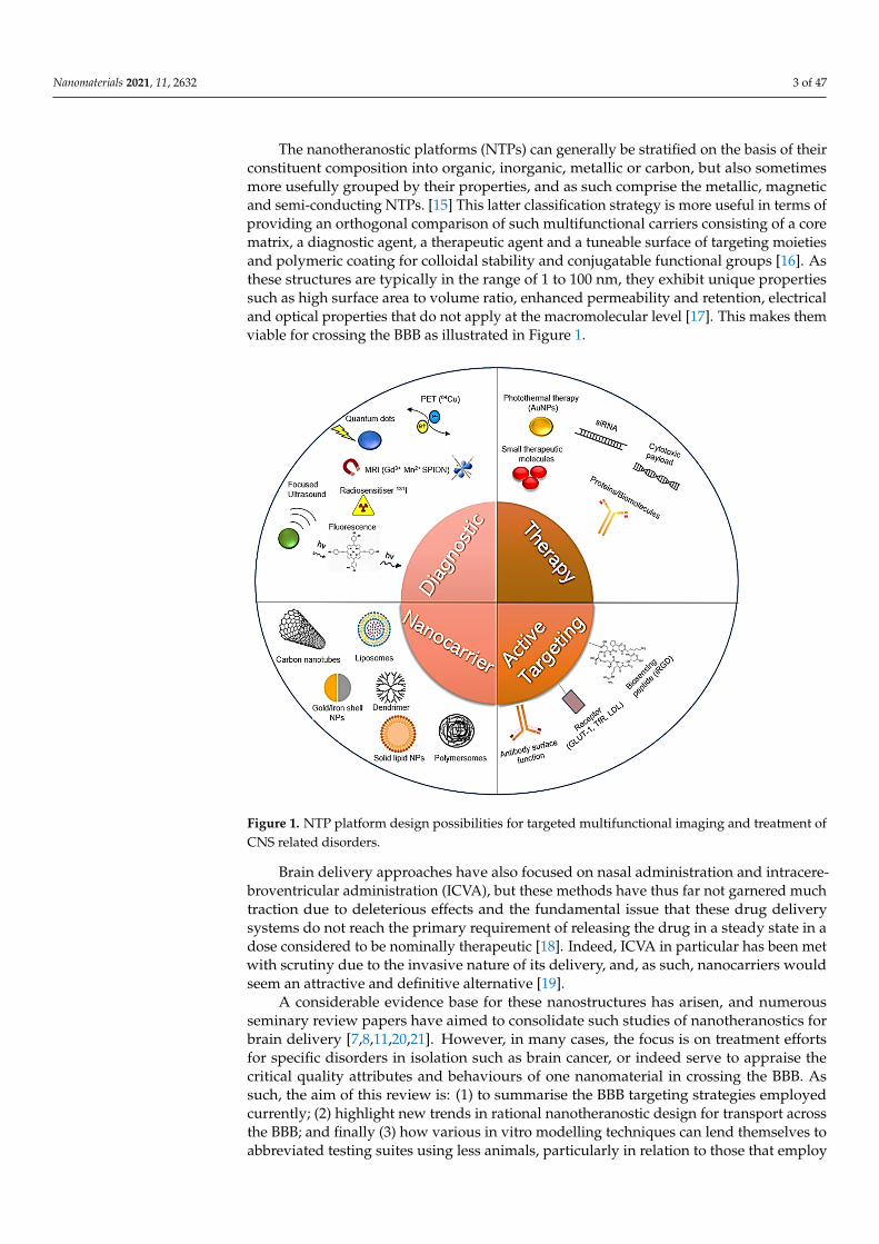
The nanotheranostic platforms (NTPs) can generally be stratified on the basis of theirconstituent composition into organic, inorganic, metallic or carbon, but also sometimesmore usefully grouped by their properties, and as such comprise the metallic, magneticand semi-conducting NTPs. [15] This latter classification strategy is more useful in terms ofproviding an orthogonal comparison of such multifunctional carriers consisting of a corematrix, a diagnostic agent, a therapeutic agent and a tuneable surface of targeting moietiesand polymeric coating for colloidal stability and conjugatable functional groups [16]. Asthese structures are typically in the range of 1 to 100 nm, they exhibit unique propertiessuch as high surface area to volume ratio, enhanced permeability and retention, electricaland optical properties that do not apply at the macromolecular level [17]. This makes themviable for crossing the BBB as illustrated in Figure 1.
Nanomaterials 2021, 11, 2632 3 of 48
optimising the delivery across the BBB, rather than exclusively considering the design of a particular nanotheranostic platform for one disease only.
The nanotheranostic platforms (NTPs) can generally be stratified on the basis of their constituent composition into organic, inorganic, metallic or carbon, but also sometimes more usefully grouped by their properties, and as such comprise the metallic, magnetic and semi-conducting NTPs. [15] This latter classification strategy is more useful in terms of providing an orthogonal comparison of such multifunctional carriers consisting of a core matrix, a diagnostic agent, a therapeutic agent and a tuneable surface of targeting moieties and polymeric coating for colloidal stability and conjugatable functional groups [16]. As these structures are typically in the range of 1 to 100 nm, they exhibit unique properties such as high surface area to volume ratio, enhanced permeability and retention, electrical and optical properties that do not apply at the macromolecular level [17]. This makes them viable for crossing the BBB as illustrated in Figure 1.
Figure 1. NTP platform design possibilities for targeted multifunctional imaging and treatment of CNS related disorders.
Brain delivery approaches have also focused on nasal administration and intracere-broventricular administration (ICVA), but these methods have thus far not garnered much traction due to deleterious effects and the fundamental issue that these drug deliv-ery systems do not reach the primary requirement of releasing the drug in a steady state in a dose considered to be nominally therapeutic [18]. Indeed, ICVA in particular has been met with scrutiny due to the invasive nature of its delivery, and, as such, nanocarriers would seem an attractive and definitive alternative [19].
A considerable evidence base for these nanostructures has arisen, and numerous seminary review papers have aimed to consolidate such studies of nanotheranostics for brain delivery [7,8,11,20,21]. However, in many cases, the focus is on treatment efforts for specific disorders in isolation such as brain cancer, or indeed serve to appraise the critical quality attributes and behaviours of one nanomaterial in crossing the BBB. As such, the aim of this review is: (1) to summarise the BBB targeting strategies employed currently; (2) highlight new trends in rational nanotheranostic design for transport across the BBB;
Figure 1. NTP platform design possibilities for targeted multifunctional imaging and treatment ofCNS related disorders.
Brain delivery approaches have also focused on nasal administration and intracere-broventricular administration (ICVA), but these methods have thus far not garnered muchtraction due to deleterious effects and the fundamental issue that these drug deliverysystems do not reach the primary requirement of releasing the drug in a steady state in adose considered to be nominally therapeutic [18]. Indeed, ICVA in particular has been metwith scrutiny due to the invasive nature of its delivery, and, as such, nanocarriers wouldseem an attractive and definitive alternative [19].
A considerable evidence base for these nanostructures has arisen, and numerousseminary review papers have aimed to consolidate such studies of nanotheranostics forbrain delivery [7,8,11,20,21]. However, in many cases, the focus is on treatment effortsfor specific disorders in isolation such as brain cancer, or indeed serve to appraise thecritical quality attributes and behaviours of one nanomaterial in crossing the BBB. Assuch, the aim of this review is: (1) to summarise the BBB targeting strategies employedcurrently; (2) highlight new trends in rational nanotheranostic design for transport acrossthe BBB; and finally (3) how various in vitro modelling techniques can lend themselves toabbreviated testing suites using less animals, particularly in relation to those that employ
Nanomaterials 2021, 11, 2632 4 of 47
cells of human origin, as these constitute the gold standard in relation to predictive powerand bio-relevancy.
Despite the promise of nanotherapeutics as a drug delivery strategy for CNS disorders,and some 250 papers published on PubMed in 2020 alone in relation to their use for crossingthe BBB, as of this review there are only three nanomedicines licensed for use in brain cancer(Marqibo, Onivyde and Feraheme), with notable omissions of any licensed nanomedicaltreatment for Alzheimer’s disease (AD) and Parkinson’s disease (PD) [22]. This is bitterlydisappointing in light of the intensive research efforts that have been focused on theapplication of nanomedicine to these fields in particular in recent years.
This review thus aims to demonstrate the diversity of targeting strategies for crossingthe BBB with specific reference to nanotheranostics, identify the reasons for the lack of theclinical translation of research data generated thus far and identify trends and future direc-tions for in vitro BBB permeability testing, which will reduce the need in future for relianceon animal studies. While numerous exemplary efforts on nanoparticle engineering havebeen reviewed, many have not extensively reviewed the possibility of in vitro modellingapproaches for testing the merit of nanotheranostic candidates designed by formulationscientists. This review thus serves to inform formulation scientists and those workingin the nanotechnology research area in relation to alternatives to in vivo administrationfor testing the synthesised nanoparticles in relation to delivery efficiency and indeed forestablishing their biocompatibility and targeted delivery specifically to the brain acrossthe BBB. As such, an appraisal of the interaction of nanoparticles with biological systemsin relation to the formation of the protein corona, evasion of the mononuclear phagocyticsystem (MPS) and modulation of their physiochemical properties for enhanced colloidalstability, reduced clearance and increased accumulation by exploiting the EPR effect arebeyond the scope of this review as they have been extensively reviewed elsewhere. [23–25]The specific interest of this paper is overcoming the BBB and how nanomaterials can berationally designed and tested using modelling of the BBB to facilitate clinical translationof promising nanoplatforms, with abbreviated in vivo testing requirements.
2. Physiological Aspects of the Brain and Blood–Brain Barrier
The encephalon or brain is a complex organ that is responsible for regulating andintegrating a complex array of executive functions in mammals including wakefulness,memory, sleep, olfactory signal integration, motor function and perception. It is, however,a considerably fragile organ, and therefore found enclosed in the cranium of the skull.Despite this physical enclosure to resist mechanical insult, it must further be physicallyprotected from exposure to toxins and microorganisms present in the systemic circulationand the integrity of its physiological environment must be maintained. The BBB alsoacts as a strict barrier to the passage of xenobiotics, and, as a result, it is an aspect tocircumvent to achieve adequate pharmaceutical delivery. In the brain, there are three suchprincipal barriers: the blood–brain barrier (BBB), blood–leptomeningeal barrier (BLMB)and the blood–cerebrospinal fluid barrier (BCSFB), with the former constituting the keyhomeostatic regulator mediating transport between the peripheral circulation and theCNS [26].
The BBB is a cellular barrier constituted primarily by a concentric series of microves-sel non-fenestrated continuous endothelial cells with tight junction adjoints, which wasfirst described by Paul Ehrlich in 1885 [27]. This barrier thus exquisitely regulates thepassage of xenobiotics, microorganisms and endogenous entities such as macrophages andendopeptides. It is increasingly acknowledged that the BBB is in fact far more complicatedin composition, with contributions from several proteins (i.e., claudin-5 [28] and cell types(e.g., perivascular astrocytes), as the former provide high transendothelial potential re-sistances in the order of ~1500 Ohm/cm2 [29], thus significantly hampering paracellularmechanism of drug delivery, and the latter contribute to capillary phenotypic regulation.
This constitutes a significant hindrance to the development of therapeutic or diagnosticagents for brain-targeted drug delivery, as many agents may appear bioactive but cannot
Nanomaterials 2021, 11, 2632 5 of 47
be permitted across this barrier. There are also a number of less tightly modulated regionssuch as the chemoreceptor trigger zones (CTZ), which regulate blood composition [30],primarily localised in the subfornical organ and organum vasculosum, which are in effect adynamic permeable blood–brain barrier. However, these are less exploitable for therapeuticdelivery, as they would generally impose dose limiting nausea and vomiting constraints,which are already a hallmark issue in the administration of intravenous chemotherapy.
This is further confounded by the presence of a robust biochemical barrier of effluxtransporter proteins, which compromises the utility of the transcellular route for drugdelivery. The ATP-binding cassette transporter efflux pumps (ABCs), multi-drug resistanceproteins (ABCG2) and P-gp in particular are restrictive in facilitating antineoplastic andanti-human immunodeficiency virus pharmaceuticals delivery in conventional formula-tions [31]. The influx transporters such as the organic anionic transporters (OATs) are moreexploitable as they can regulate transport in both directions, and efforts are directly atpreferentially facilitating influx [32]. The blood–cerebrospinal fluid barrier (BCSFB) bycontrast is not as popular a candidate in the drug delivery strategy for CNS disorders,owing to the arguments presented that this rather constitutes a principle aspect of ICVAas aforementioned. However, this highly branched structure of choroid plexus epithelialcells, which is responsible for homeostatic CSF secretion and regulation, can contributeto the delivery efficiency, as it too has a polarised expression of numerous ion channels,transporters and receptors. Thus, while acting as an essential protector and regulator forthe brain itself, the BBB, BLMB and to a lesser extent the BCSFB pose a major issue toformulation scientists and academic researchers for drug delivery.
2.1. Modulating the BBB
A number of physical and chemical methods exist to modulate the integrity of the BBBin a temporary and reversible manner, using immune cells [33], techniques such as focusedultrasound [34] or more selectively using endogenous ligands as “Trojan Horses” [35], butthese have not reached fruition clinically as such, due to issues with reproducibility andlimitations with mapping biodistribution after administration. The neurosurgical methodsor direct administration of such agents to the brain have been largely precluded exceptin experimental circumstances for the foregoing reasons of invasiveness, pain and risk ofirreversible brain damage due to the unpredictability in the disruption to the BBB thatensues. [36,37] As such, the fundamental challenge remains in ensuring efficacious deliveryacross the BBB by the associated permeation mechanisms, and nanotheranostic deliverysystems are an ideal candidate for such purposes when it can be demonstrated that theytraverse the BBB and release their diagnostic and therapeutic payloads in a controlled andsite specific manner [38]. These include passive diffusion, carrier-mediated transport andadsorptive or receptor mediated endocytosis/transcytosis.
2.2. Rationale for Nanotheranostics over Standard Therapies
The rationale for nanotheranostics in the treatment of neurological disease by crossingthe BBB upon systemic administration is underpinned by the limitations of conventionalempirical therapy [39]. Despite numerous advances in this field, particularly in relation tobiotechnology and the revolution of monoclonal antibodies and immunotherapies, suchtherapeutics have hardly increased the delivery efficiency, nor significantly amelioratedthe competence of current clinical diagnostics and chemotherapeutic regimens in particu-lar [40,41]. One of the primary attributes of maladies of the brain is that prompt primarydiagnosis treatment that is tailored to the patient is at a premium. Indeed, it would seemthat nanotheranostics could potentially satisfy all of these pre-requisites and more, bylimiting off site action, dose limiting toxicities and potentially overcoming drug resistance,which have significantly hampered clinical efforts thus far [42].
Acute traumatic insult to the head is commonly encountered in the clinic from multiplesources, most notably as a consequence of engaging in physical sport. This trauma to thehead causes a multi-faceted pathophysiology to the BBB, including ischaemia, hypoxia,
Nanomaterials 2021, 11, 2632 6 of 47
pro-inflammatory factor release and increased tight junction leakiness, which is potentiallylife threatening and for which there is not a satisfactory treatment to date [43]. A numberof efforts including that by Campbell and colleagues [44] seek to identify such protocolsfor managing acute ischaemic head injuries, and nanotheranostics have potential in thisregard [45]. To accelerate the progression of such to the clinic, however, there is an evidentneed for high throughput robust in vitro models to test such novel strategies, and themodels outlined in this review hold great promise in this regard.
Despite this however, personalised medicine marks a paradigm shift in clinical modelsof care, and any such contributions to such should be heralded as a move away from thedogmatic stagnation of the neurotherapy field, in which superficially enhanced efficacyin vitro has been possibly given precedence over consideration of the in vivo translationand bio-compatibility of such delivery systems, which is the primary aim of any suchexercise [46]. This underscores the potential of nanotheranostics in that the neurologyand oncology fields are at a critical juncture as highlighted in a recent paper by Aldapeand colleagues [47], in which they posset that current clinical diagnostics are arguably notsensitive enough for diagnosing such conditions at the pivotal sub-clinical stage. There isalso an increasing recognition that the genomic, metabolomic and phenotypic heterogeneityof people is such that “one-fits all” empirical therapies are not optimal, and the incidenceof such diseases are on the rise at a global population level [47].
This means that if clinicians have an armament of multimodal nanoformulations thatcan: (1) be specifically used to diagnose a patient, (2) stratify such patients accordingto likelihood of response by biomarker and metabolomic screening, (3) specifically andefficiently target the affected CNS tissues reproducibly using the full repertoire of phar-macological agents including biotechnological products, (4) monitor the biodistribution,tissues or response in real time and (5) follow-up and recalibrate the therapy based onresponse, this would mark a golden age of therapeutics. Optimism must be tempered bythe fact, however, that as yet such “magic bullets” are confined to research settings, somepromising clinical candidates and a handful of commercially approved agents that haveone modality only, but concerted efforts towards such nanotheranostics will be reviewedforthwith in the specific context of systemic delivery targeting the brain via permeationacross the BBB.
2.3. Rationale for Modelling the BBB
While small molecules <400 Da such as glucose can readily perfuse this dynamicbarrier, in most cases a myriad of factors compromise the delivery of pharmacologicalagents [48], including lipophilicity, enzymatic degradation or metabolic conversion, associ-ation with non-transporting ligands or association with off-target tissues, and inefficienttraversal to the interstitial spaces of the brain once in the parenchyma. The predictabilityof the in vivo efficacy of nanotheranostic carriers in crossing the BBB thus requires thedevelopment and implementation of robust models, which can reflect the dynamic natureof the BBB. This is further evidenced by the high attrition rate for candidate drugs (~80%)coupled with the fact that those that are therapeutically approved only cross the BBB in~5% of cases [49].
Such in vitro and ex vivo models are indispensable at the pre-clinical stage, as themore extensive the information that can be garnered the less mandate there is for animaltesting, which is costly and ethically contentious. This is in accordance with realising the“Replacement” component of the 3 R principles sanctioned by several national authoritiesoutlined in the EU under Article 4 of EU Directive 2010/63/EU “on the protection ofanimals used for scientific purposes” [50].
The conventional studies of transfer across the BBB utilise membrane models, in vitrostatic and dynamic models or indeed animal studies, the latter of which has numerousestablished limitations in predicting in vivo behaviour due to inter-species variation [51].It is no surprise that while as an academic exercise a lab mouse or their cells is usefulfor studying a novel nanoformulation for its BBB permeation properties, in effect, the

Nanomaterials 2021, 11, 2632 7 of 47
translation of such to a human subject is virtually incomparable. As such, optimisation ofin vitro models and permeability assays would constitute a move towards more compre-hensive and predictive data, which would facilitate nanotheranostic candidate selectionand optimisation at earlier stages for accelerated discovery and development.
2.4. Overview of Current Modelling Approaches
In vitro BBB modelling has been a subject of intensive research since the 1980′s, andin many cases has been based on the use of transwell assays, which although utilitarianhave notable disadvantages [52]. They have been bolstered by the use of human inducedpluripotent stem cells (iPSCs), which are more representative of in vivo conditions, butlimitations associated with irregularities due to co-differentiation and the relative shortageof viable stem cell sources means that they have not been widely adopted to date [53]. Asstatic monolayer models, they do not recognise the significant influence of shear stresson the endothelial function due to blood flow, and for this reason dynamic models havebecome more ubiquitous. Advances in microfluidic technology and integrated sensorshas generated an organ-on-chip in vitro model of the BBB termed a µBBB, which canincorporate shear stress and can withstand high-throughput screening (HTS) [54].
Perhaps the most promising modelling approaches incorporate co-culturing of astro-cytes, pericytes and primary brain endothelial cells to form CNS organoids as illustratedby Cho and colleagues, which will effectively be biomimetic of the neurovascular unit(NVU), as all of this constitutively contributes to BBB integrity [55]. The ease of culturing,up-scale for HTS and the fact that unlike conventional transwell systems all cells are indirect contact with one another means that these are a promising technology. They can alsobe rotated at regulated speeds to simulate the shear stress that microfluidic technologiesprovide, without additional expertise or specialised equipment considerations, and canbe used to simultaneously investigate hundreds of compounds using automated roboticassisted confocal fluorescence microscopy and imaging mass spectroscopy [56]. The lattertechniques in particular are an exciting prospect, as confocal fluorescence microscopy canbe used for mapping biodistribution of nanotheranostics, which incorporate fluorescentdyes or quantum dots, while imaging mass spectroscopy facilitates detailed 3D imagingof the nanoparticles in situ for real-time clinical diagnostics and principal componentanalysis [57,58].
As evidenced by the foregoing, the selection of the BBB model is dependent on theintended purpose for conducting the study and the stage of development. The combinationof such models with in silico screening technology would arguably lead to the elucidation ofa greater number of viable leads and prediction of permeability and bio-fates at early stagesof nanotheranostic development to save time and costs [59]. In terms of in silico screeningof BBB permeation and ADMET parameters, the data selection process is imperativeand must be extensively validated to minimise the false positive rate. However, recentadvances in artificial intelligence and machine learning means that by producing initialrobust classification models consisting of reliable nanocarriers, theoretically, the biaseddata sets can be corrected [60].
As a general rule, using different immortalised cell lines is usually warranted toserve as orthogonal models to qualify the in vitro to in vivo extrapolation of findings [61].Signalling pathways and associated kinetics of transport are best suited to study by way ofmonolayers, as these are specific and simple [62]. For establishing structure activity rela-tionships, and, more crucially, for evaluating toxicological profiles, more sensitive modelssuch as iPSC models are warranted due to enhanced sensitivity and the critical natureof the information garnered in guiding subsequent optimisation of leads and generationof safety data as supporting information for clinical testing application submissions [63].Organoids in particular would seem the optimal candidate for analysing nanotheranosticsin tandem with organ-in-chip microfluidics, as they best recapitulate physiological condi-tions and integrity of the BBB and can be used to precisely determine cellular uptake andbiodistribution in related high-throughput assays in a cost-effective manner. The relative

Nanomaterials 2021, 11, 2632 8 of 47
strengths and weaknesses of such models are summarised in the graphic in Figure 2, whichfurther exemplifies the increase in choice with respect to time as more technologies comeon stream [54].
Nanomaterials 2021, 11, 2632 8 of 48
safety data as supporting information for clinical testing application submissions [63]. Or-ganoids in particular would seem the optimal candidate for analysing nanotheranostics in tandem with organ-in-chip microfluidics, as they best recapitulate physiological condi-tions and integrity of the BBB and can be used to precisely determine cellular uptake and biodistribution in related high-throughput assays in a cost-effective manner. The relative strengths and weaknesses of such models are summarised in the graphic in Figure 2, which further exemplifies the increase in choice with respect to time as more technologies come on stream [54].
Figure 2. Trends in BBB models 1991–2018. The opacity of the lines in the graphic on left refer to overall popularity, and the shaded boxes on the right represent a qualitative 1–6 score where lower scores imply limitations and higher scores indicate relative strengths of a particular model. PAMPA = parallel artificial membrane permeability assay, a cell free assay used to screen the permeability of compounds based on the pass from a donor to acceptor compartment separated by an artificial lipid membrane. Reproduced with permission from Oddo and colleagues, Trends in biotechnology; published by Cell Press 2019 [54] based on data in Mahto and colleagues [64].
3. NTP Delivery Approaches for Treating CNS Disorders Of the aforementioned mechanisms of transport across the BBB, adsorptive mediated
and receptor mediated endocytosis constitute the most pervasive explored by researchers for NTP mediated delivery of imaging contrast agents and therapeutic moieties. These biological mediated mechanisms and to a lesser extent cell mediated delivery will be the focus of this review in relation to testing the efficiency of NTP delivery across the BBB.
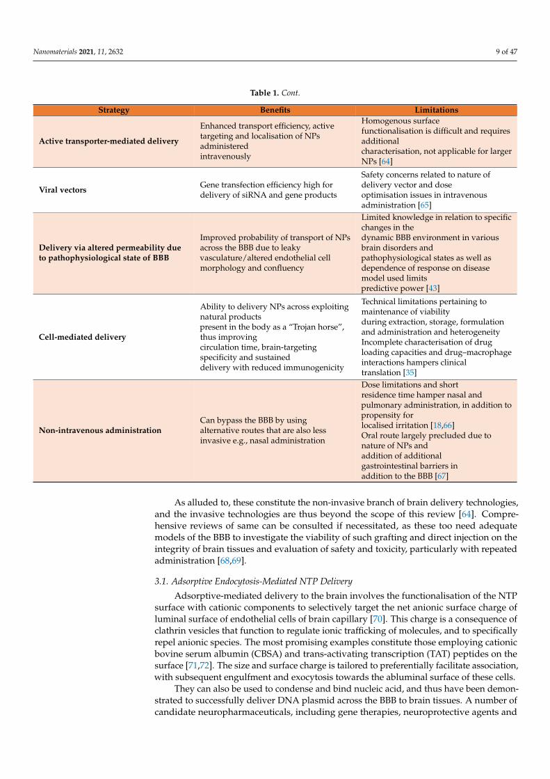
A summary of the main advantages and limitations of the various strategies em-ployed by nanoformulation scientists for brain-targeted NTP delivery are summarised in Table 1. When the contemporary literature is investigated, the most promising and readily tested NTP platforms are those that make use of surface functionalisation with either known ligands of the receptors highly expressed on BBB endothelial cell surface, or indeed by using inherent cellular components such as macrophages [35] and fatty acids to en-hance penetrance. The key factor is to determine not only whether the NTP can be deliv-ered across the BBB model in-vitro, but also to have a measurable index of the concentra-tion or number of particles that reach the brain (as well as accumulation in non-target tissues), as this is the true predictor of therapeutic response [7,8] and biocompatibility.
Figure 2. Trends in BBB models 1991–2018. The opacity of the lines in the graphic on left refer to overall popularity, and theshaded boxes on the right represent a qualitative 1–6 score where lower scores imply limitations and higher scores indicaterelative strengths of a particular model. PAMPA = parallel artificial membrane permeability assay, a cell free assay used toscreen the permeability of compounds based on the pass from a donor to acceptor compartment separated by an artificiallipid membrane. Reproduced with permission from Oddo and colleagues, Trends in biotechnology; published by Cell Press2019 [54].
3. NTP Delivery Approaches for Treating CNS Disorders
Of the aforementioned mechanisms of transport across the BBB, adsorptive mediatedand receptor mediated endocytosis constitute the most pervasive explored by researchersfor NTP mediated delivery of imaging contrast agents and therapeutic moieties. Thesebiological mediated mechanisms and to a lesser extent cell mediated delivery will be thefocus of this review in relation to testing the efficiency of NTP delivery across the BBB.
A summary of the main advantages and limitations of the various strategies employedby nanoformulation scientists for brain-targeted NTP delivery are summarised in Table 1.When the contemporary literature is investigated, the most promising and readily testedNTP platforms are those that make use of surface functionalisation with either knownligands of the receptors highly expressed on BBB endothelial cell surface, or indeed byusing inherent cellular components such as macrophages [35] and fatty acids to enhancepenetrance. The key factor is to determine not only whether the NTP can be deliveredacross the BBB model in-vitro, but also to have a measurable index of the concentration ornumber of particles that reach the brain (as well as accumulation in non-target tissues), asthis is the true predictor of therapeutic response [7,8] and biocompatibility.
Table 1. Main strategies for nanotheranostic drug delivery to the brain across the BBB.
Strategy Benefits Limitations
BBB disruption by focused ultrasoundTransient opening of the BBBfacilitates increasedconcentration of NPs in brain
Inter-species limitations andvariability of response between subjects’limit findings [34]
Magnetic field-guided delivery
Enhanced imaging capabilities fordiagnostics, in situmonitoring and follow-up oflocalisation and concentration anddelivery guided by external device
Balance must be struck to attain efficientand specifichyperthermia while maintaining viabilityof healthy surrounding tissues inaddition to observed development ofthermotolerance in several subjects [62]
Nanomaterials 2021, 11, 2632 9 of 47
Table 1. Cont.
Strategy Benefits Limitations
Active transporter-mediated delivery
Enhanced transport efficiency, activetargeting and localisation of NPsadministeredintravenously
Homogenous surfacefunctionalisation is difficult and requiresadditionalcharacterisation, not applicable for largerNPs [64]
Viral vectors Gene transfection efficiency high fordelivery of siRNA and gene products
Safety concerns related to nature ofdelivery vector and doseoptimisation issues in intravenousadministration [65]
Delivery via altered permeability dueto pathophysiological state of BBB
Improved probability of transport of NPsacross the BBB due to leakyvasculature/altered endothelial cellmorphology and confluency
Limited knowledge in relation to specificchanges in thedynamic BBB environment in variousbrain disorders andpathophysiological states as well asdependence of response on diseasemodel used limitspredictive power [43]
Cell-mediated delivery
Ability to delivery NPs across exploitingnatural productspresent in the body as a “Trojan horse”,thus improvingcirculation time, brain-targetingspecificity and sustaineddelivery with reduced immunogenicity
Technical limitations pertaining tomaintenance of viabilityduring extraction, storage, formulationand administration and heterogeneityIncomplete characterisation of drugloading capacities and drug–macrophageinteractions hampers clinicaltranslation [35]
Non-intravenous administrationCan bypass the BBB by usingalternative routes that are also lessinvasive e.g., nasal administration
Dose limitations and shortresidence time hamper nasal andpulmonary administration, in addition topropensity forlocalised irritation [18,66]Oral route largely precluded due tonature of NPs andaddition of additionalgastrointestinal barriers inaddition to the BBB [67]
As alluded to, these constitute the non-invasive branch of brain delivery technologies,and the invasive technologies are thus beyond the scope of this review [64]. Compre-hensive reviews of same can be consulted if necessitated, as these too need adequatemodels of the BBB to investigate the viability of such grafting and direct injection on theintegrity of brain tissues and evaluation of safety and toxicity, particularly with repeatedadministration [68,69].
3.1. Adsorptive Endocytosis-Mediated NTP Delivery
Adsorptive-mediated delivery to the brain involves the functionalisation of the NTPsurface with cationic components to selectively target the net anionic surface charge ofluminal surface of endothelial cells of brain capillary [70]. This charge is a consequence ofclathrin vesicles that function to regulate ionic trafficking of molecules, and to specificallyrepel anionic species. The most promising examples constitute those employing cationicbovine serum albumin (CBSA) and trans-activating transcription (TAT) peptides on thesurface [71,72]. The size and surface charge is tailored to preferentially facilitate association,with subsequent engulfment and exocytosis towards the abluminal surface of these cells.
They can also be used to condense and bind nucleic acid, and thus have been demon-strated to successfully deliver DNA plasmid across the BBB to brain tissues. A number ofcandidate neuropharmaceuticals, including gene therapies, neuroprotective agents and

Nanomaterials 2021, 11, 2632 10 of 47
chemotherapeutics, with enhanced permeability, were confirmed by images to have in-creased accumulation, and in some cases sustained release profiles [73,74]. The most viablematerials for such platforms are pegylated chitosan, lipid and polymeric nanoparticles suchas polylactic acid (PLA), poly-ε-caprolactone (PCL), cholesterol, poly(butyl cyanoacrylate)(PBCA) gelatin siloxane and mesoporous silica magnetic nanoparticles incorporating ironoxide (SiO2-Fe3O4) [75,76]. Some isolated instances of glutathione and sinapic acid-basedas well as MMP-2200 derivative functionalisation have also been investigated to remarkableresults [77].
However, the primary issue with this class of NTPs is that despite their potentialthey are notably more toxic than non-ionic or anionic counterparts, which must be ap-praised before recommending their scale-up and clinical testing. A paper published byLv and colleagues elucidated such structure–toxicity relationships, and determined thatfor such non-viral vectors, low molecular weight polymers such as PLA and PLGA arepreferable, and the biodegradability of the linker is pivotal [78]. In such cases, a carbamatelinker is preferred where viable, and for cationic lipids importing a heterocylic ring asthe head group in preference to a quaternary or tertiary amine is preferred. It is alsopurported and clarified with reference to more contemporary literature that engineeringof self-assembling amphiphilic carriers or water soluble lipopolymers including thosebased on poly(ethylenimine) (PEI) and poly(l-lysine) (PLL) and non-ionic actively targeted“niosomes” are the best strategies in relation to gene delivery in particular, which hasnotable implications in several CNS disorders including Huntington’s disease, AD, PD andglioblastoma multiforme (GBM) [64,79,80].
3.2. Receptor-Mediated Transcytosis
In keeping with the marked trend away from nanomedicines being designed andtested based primarily on the enhanced permeability and retention effect (EPR), whichhas notable limitations particularly with regards to the heterogeneity of response andlack of reproducibility in vivo, perhaps active targeting utilising functionalised receptorligands for active targeting is the most promising strategy for the novel nanotherapy drivendrug delivery systems. This is unsurprising given the exquisite regulatory function ofthe BBB and associated biochemical barriers to entry of exogenous compounds. As adirect consequence, by employing ligands that preferentially bind the iron transferrin,folate, insulin, and LDL cholesterol receptors, among others that have been studied, andpredictable pathways of internalisation to the brain, it can be appreciated that these are themost probable candidates, particularly when exploited synergistically [64].
3.2.1. Transferrin (TfR) Receptor-Mediated Transcytosis
The most widely studied of the foregoing is arguably the iron transferrin (TfR) re-ceptor, as they are very highly expressed in the brain endothelium in comparison to theperiphery, although the bone marrow, splenic and hepatocellular accumulation is alwaysa concern [81]. The lactoferrin receptor is also a notable member of this family and hasbeen targeted to varying success in some instances, such as that achieved by Kumari andcolleagues for temozolomide delivery, which was demonstrated both in vitro and in vivoto improve its pharmacokinetics and intratumoral accumulation by a pH-dependent re-sponsive mechanism [82].
A number of notable achievements have been made by careful optimisation of thephysiochemical composition of such nanocarriers, as it became increasingly evident thatnaked nanoparticles >200 nm would not garner a suitable therapeutic concentration dueto efflux and the requirement for recycling before selective accumulation in brain tissues.A number of immunoliposomes have been developed using antibodies such as OX26,which recognise alternative epitopes on the transferrin receptor, as illustrated by Kang andcolleagues for dopamine delivery in a rat model of PD, achieving an 8-fold increased uptakecompared to naked dopamine and 3-fold compared to pegylated liposome alone [83]. Suchimmunoliposomes achieve this enhanced delivery by occupying these alternative epitopic

Nanomaterials 2021, 11, 2632 11 of 47
sites as the receptors are usually saturated in a physiological condition with endogenousprotein.
This has been achieved to considerable success with gold nanoparticles (AuNPs),folate and transferrin dual conjugated doxorubicin loaded liposomes for glioma treatmentas demonstrated by Gao and colleagues, which can be further modulated to incorpo-rate imaging agents, paclitaxel, cisplatin and other notable therapeutic payloads such asamyloid β-inhibitors and siRNA [84–86]. The most notable requirement seems to be thatantibody targeted carriers require monovalent antibodies with carefully tailored affinitiessuch that the antibody does not bind too strongly and result in the receptor complex beingphagocytosed [87]. The prototypical example in this class would be JR-141 (PabinafuspAlfa), which was recently approved in Japan for the treatment of Hunter’s syndrome(mucopolysaccharidosis II, a rare heritable carbohydrate storage disease) [88]. JCR pharma-ceuticals have patented a proprietary BBB permeating technology “J-Brain Cargo”, whichutilises a fusion protein comprising an anti-TfR antibody and iduronate-2-sulfatase as anintravenous enzyme replacement therapy.
Despite its orphan designation in Japan, which was approved in March 2021, thisconstitutes a major breakthrough for such platforms, as this proprietary modular platformcan be potentially used for brain-targeted delivery for other diseases, such as mucopolysac-charidosis I, which is being evaluated using JR-171, a fusion protein of J-Brain Cargoand α-L-iduronidase (IDUA) [89]. Although its inclusion is on the basis that antibod-ies are essentially nanomedicines in their own right, it exemplifies the promise of suchreceptor-mediated delivery systems, with the primary consideration for testing ensuringthe model accounts for the inter-species TfR expression disparities (2.5-fold higher in micebrain microvessels) [90]. This again demonstrates that predictive BBB models need to besophisticated enough to account for such nuances, but may be preferential to resorting tousing human TfR knock in mice, which must also account for receptor “sinks” of periph-eral compartments potentially influencing the overall therapeutic concentration at targettissues.
3.2.2. Low-Density Lipoprotein (LDL) Receptor-Mediated Transcytosis
The low-density liprotein (LDL) gene family have crucial contributions to regulationof metabolism and nutrient transport in mammals, and this holds true for the CNS, par-ticularly in relation to apolipoprotein E (apoE) [91]. ApoE is synthesised by microgliaand astroglia, and it has been suggested increasingly that it has a role as a susceptibilitygene for AD and contributes to the neurobiology of disease following such insults inimmunomodulatory and neurotrophic as well as antioxidant contexts [92]. This givesan inherent degree of versatility to the construction of nanocarriers for these receptors,as a number of endogenous compounds can be used as biomimetic scaffolds for high-throughput screening. Solid lipid nanoparticles are the most widely employed carrierclass in this regard, although there has been notable disparity in terms of their success inpermeating the BBB, which potentially is a consequence of certain nanocarrier propertiesimparting an adsorptive-mediated transcytosis mechanism preference over directly usingthe lipoprotein receptor related protein (LRP) ligands [93,94].
The angiopep 2-based ligand in particular has a notable dual targeting functionalitywhich can be modelled in vitro for predictive response, as this ligand is expressed on gliomaand amyloid β cell surface as well as on the BBB. As a result, it enhances accumulationin the brain by receptor-mediated transcytosis, and successively facilitates localisation tosuch disordered tissues for mediating a clinical response, which has been demonstratedby Kafa and colleagues who employed targeted nanotubes in glioma in vitro and in vivomodels [95]. The BBB model of porcine brain endothelial cells (PBEC) co-cultured with ratastrocytes demonstrated diameter dependent accumulation at 24 h of approximately 2% ofthe injected dose/g brain. The natural HDL carriers are perhaps even more desirable dueto their enhanced stability, biocompatibility and long circulation with intrinsic biological
Nanomaterials 2021, 11, 2632 12 of 47
function properties, as intravenous administration of apolioprotein A1 nanoparticles alonehave reduced amyloid β levels in symptomatic APP/PS1 mice models for AD.
Both direct conjugation of apolipoproteins and indirect methods which employ non-ionic surfactants such as the polysorbates to promote subsequent apolipoprotein adsorptionin vivo have been explored. The literature seems to find agreement in the fact that ad-ministration route has another critical determinant influence on the efficiency of suchformulations, with pulmonary administration intriguingly leading to higher effective brainconcentrations of the nanoparticles when compared with intraperitoneal and intravenousadministration, though again one must consider the extrapolation of such data from mouseto human models of the BBB [96,97].
One notable limitation is the availability of primary LDL ligand materials, and assuch mimetics employ materials such as acrylic polymers, i.e., PBCA, phosphatidyl-choline, triglycerides and PLGA surface functionalised with Tweens and Spans, as wellas more contemporaneously with angio-pep 2-based ligand, they have been employedwith both in vitro and in vivo successes. Costagliola di Polidoro and colleagues [98]designed hyaluronic acid nanoparticles encapsulating an imaging agent (i.e., Gadolinium–diethylenetriamine penta-acetic acid) and irinotecan, which when surface functionalisedwith angio-pep 2 led to improved glioma imaging through enhanced T1 relaxometricproperties and cytotoxic efficacy at 24 h rather than 48 h, thus reducing irinotecan timeresponse. These have also explored tentative use of the oral route, which would be consid-ered the gold standard of administration routes due to acceptability and tolerability for thepatient. Dalargin, an anti-nociceptive peptide mimicking endogenous opioid peptides wassuccessfully found to localise in the brain endothelium following oral administration in aPBCA nanoparticle formulation surface coated with Tween-80 [67].
3.2.3. Other Notable Receptor-Mediated Approaches
Proteomic studies have generated invaluable information in relation to the endoge-nous regulation of the BBB and have recognised several other receptors that can potentiallybe commandeered by nanomaterials for passage into the brain [97]. For example, studiesof models of epilepsy have revealed that glutamate in particular can modulate in vivoBBB permeability, and as it is recognised by several receptors and is implicated in severaldisorders, i.e., anxiety, epilepsy, pain and addiction, this means that it holds noteworthypromise [99]. The glucose receptor (GLUT-1) is upregulated in brain tumours due to thehypoxic environment and may be an associated marker of radio-resistance and poor prog-nosis [100]. Additionally, a rapid glycemic increase is observed following fasting which hasbeen demonstrated by Wu and colleagues to impart rapid delivery character to a numberof nanomaterials including micelles, both in vitro and in vivo in models of head and necksquamous cell carcinoma (HSNCC) [101].
While insulin cannot itself be readily employed for mediated passage of the BBBdue to instability of the endogenous ligand and hypoglycemic potentiation, anti-insulinreceptor antibodies have successfully been conjugated to nanocarriers for active targetingof brain tissues [102]. Ulbrich and colleagues provide an eminent example of such a strat-egy employing 29B4 anti-insulin conjugated loperamide loaded human serum albuminnanoparticles versus immunoglobulin G conjugated nanoparticles in an antinociceptive tailflick test in ICR (CD-1) mice [103]. The fact that the latter had only marginal effectivenessdemonstrates the potential for anti-insulin antibodies in considerably increasing the deliv-ery efficiency. EGFR, folate and, more recently, interleukin receptors have been implicatedin cancer due to their high expression on tumour cell surfaces, and have been studiesas a targeting mechanism for several years [104,105]. Peptides, magnetic nanoparticlesand quantum dots have all been successfully used for enhanced chemotherapeutic andimaging applications by selective recognition as the folate receptor in particular is highlyexpressed on the BBB but not on healthy brain cells, and, as such, a dual targeting efficiencyis achieved both in terms of facilitating passage across the BBB and further in localising totumour tissues.

Nanomaterials 2021, 11, 2632 13 of 47
Cai and colleagues successfully designed and tested a nanotheranostic platformconsisting of an aggregation induced emission fluorogen for glioblastoma multiformetumour margin imaging and a high NIR absorbing semi conducting polymer for successivephotothermal therapy encapsulated in cRGD and folate surface functionalised nanopar-ticles. [106]. These nanoparticles had good biocompatibility and safety demonstrated byalmost complete clearance at 10 days, and, furthermore, the optical properties facilitatedvivid tumour size analysis up to a week following tumour implantation and offer selectiveGBM cell killing efficiency.
Another robust example is the EGFR variant III targeted by Peng and colleagues usingaptamer U2-gold nanoparticle complexes (U2-AUNPs), constituting a novel and promisingstrategy for GBM treatment [107]. In both the in vitro U87 cell line and in tumour bearingmice, significant antitumour efficacy was observed (effectively halving the percentage ofproliferating cells when treated with U2-AUNPs, versus a negligible response for AUNPsalone), and increasing survival times of treated mice (mean 30 days versus 24 days forthose treated with the NaCl control). While unlike Cai and colleagues this study doesnot significantly address safety concerns of using such AUNPs, what is evident is thatEGFR targeting is a viable strategy for treatment of gliomas by selectively inhibiting theassociated proliferation and DNA repair pathways.
3.3. Other Active Targeting Strategies
The foregoing notable advances in this field serve as a concise demonstration of theversatility and utility of rationally designed nanocarriers, which include various modularstructures, surface chemistries and formulation with the emergence of nano-emulsions, insitu nanogels and self-assembling nanosuspensions [108]. In general such strategies makefortuitous use of the fact that the neuropathophysiology of glioma, AD and PD among otherneurological disorders including ischaemia and acute neurological trauma involves aninnate disruption of the integrity of the BBB due to neuroinflammation and dysregulationresulting in increased permeability [109]. Focused ultrasound has garnered attentionfor synergistic therapies involving intravenous administration of ultrasound sensitivemicrobubble nanoformulations followed by ultrasound-guided temporary opening ofthe BBB [110]. This facilitates temporary reversible increased site-specific permeabilitychanges for subsequent administration of nanoparticles, imaging agents and cells, whichhas shown particularly promising results for magnetically guided superparamagneticiron oxide nanoparticles (SPIONs); the safety of such approaches remains dubious [111].These consolidated non-invasive strategies are perhaps best conceptualised by visualrepresentation as given by Figure 3 [112].
Nanomaterials 2021, 11, 2632 14 of 48
Figure 3. Summary of non-invasive transport mechanisms available for the delivery of nanoparticulate systems across the BBB. Reproduced from Nair and colleagues.
Where the BBB is in its intact physiological state, however, more exquisite strategies are required, such as active peptide sequence targeting, i.e., using iRGD for BBB and tu-mour penetration enhancement [114]. A number of shuttle peptides have been developed as a consequence of improvements in phage display technology, and cell-based transpor-tation technologies such as those highlighted by Li and colleagues and Batrakova and col-leagues, respectively, are propitious, despite admitted limitations associated with hetero-genous expression and limited loading capacities [115,116]. These approaches consequen-tially must also be accounted for when designing in vitro models of the BBB, as safety is the paramount concern, particularly for inorganic nanoparticles employing heavy metals or non-biodegradable moieties, despite their useful optical and magnetic properties [117]. The vast array of nanoparticulate systems in terms of design, materials and modulation in terms of therapeutic, diagnostic imaging agents and surface probes with divergent bi-odistributions and principal activities require a parallel robust toolset of viable predictive models to test their effectiveness.
4. Towards Consolidated NTP Testing Using Validated BBB Models If safety and biocompatibility can be unequivocally proven, then more liberal regu-
latory frameworks with abbreviated testing protocols would pave the way for accelerated development and approval. It would not seem useful to design an intact BBB for testing NTPs destined to be used in pathological states, but, by the same token, designing a dys-functional BBB may artificially lead to results constituting effective permeability of such nanocarriers when in fact this would not be clinically reproducible. Thus dynamic models are the gold standard, which are feasible due to improvements in microfluidics, cell engi-neering in tandem with in silico screening technological capability advancements which have been witnessed in the last decade.
As alluded to in Sections 2.3. and 2.4., the ultimate goal in research and development is to find universally acceptable and applicable in vitro BBB models that essentially recre-ate the neurovascular unit, as shown in Figure 4, in order to expedite research and devel-opment and reduce the associated financial and logistical implications of using animal testing as the primary source of supporting clinical information. Furthermore, they hold more constitutive properties when they can mimic physiological condition such as recep-tor expression, cellular regulation and stresses such as shear stress due to blood flow, which can then be used to rapidly evaluate a wide range of nanomaterials and nanocarrier platforms for their permeability efficiency. Such models are preferential to conducting in vivo studies on animals, and the trend of their development and increasing use by re-searchers is chronologically reviewed in a seminal paper published by Ribeiro and col-leagues [118].
Figure 3. Summary of non-invasive transport mechanisms available for the delivery of nanoparticulate systems across theBBB. Reproduced from Nair and colleagues.

Nanomaterials 2021, 11, 2632 14 of 47
Where the BBB is in its intact physiological state, however, more exquisite strategiesare required, such as active peptide sequence targeting, i.e., using iRGD for BBB and tumourpenetration enhancement [113]. A number of shuttle peptides have been developed as aconsequence of improvements in phage display technology, and cell-based transportationtechnologies such as those highlighted by Li and colleagues and Batrakova and colleagues,respectively, are propitious, despite admitted limitations associated with heterogenousexpression and limited loading capacities [114,115]. These approaches consequentiallymust also be accounted for when designing in vitro models of the BBB, as safety is theparamount concern, particularly for inorganic nanoparticles employing heavy metals ornon-biodegradable moieties, despite their useful optical and magnetic properties [116].The vast array of nanoparticulate systems in terms of design, materials and modulationin terms of therapeutic, diagnostic imaging agents and surface probes with divergentbiodistributions and principal activities require a parallel robust toolset of viable predictivemodels to test their effectiveness.
4. Towards Consolidated NTP Testing Using Validated BBB Models
If safety and biocompatibility can be unequivocally proven, then more liberal regula-tory frameworks with abbreviated testing protocols would pave the way for accelerateddevelopment and approval. It would not seem useful to design an intact BBB for testingNTPs destined to be used in pathological states, but, by the same token, designing adysfunctional BBB may artificially lead to results constituting effective permeability ofsuch nanocarriers when in fact this would not be clinically reproducible. Thus dynamicmodels are the gold standard, which are feasible due to improvements in microfluidics,cell engineering in tandem with in silico screening technological capability advancementswhich have been witnessed in the last decade.
As alluded to in Sections 2.3 and 2.4, the ultimate goal in research and development isto find universally acceptable and applicable in vitro BBB models that essentially recreatethe neurovascular unit, as shown in Figure 4, in order to expedite research and developmentand reduce the associated financial and logistical implications of using animal testing asthe primary source of supporting clinical information. Furthermore, they hold moreconstitutive properties when they can mimic physiological condition such as receptorexpression, cellular regulation and stresses such as shear stress due to blood flow, whichcan then be used to rapidly evaluate a wide range of nanomaterials and nanocarrierplatforms for their permeability efficiency. Such models are preferential to conductingin vivo studies on animals, and the trend of their development and increasing use byresearchers is chronologically reviewed in a seminal paper published by Ribeiro andcolleagues [117].
These include monolayer isolated brain capillary models, in vitro cell-based modelsusing human and animal-derived cells and cell-free models including microfluidic “brainon chip” models, which all have merit and associated challenges and limitations in relationto their application to studying nanomaterials. These are able in an orthogonal manner toaccount for such nuances and heterogeneity and hold promise for reducing in vivo testingstudies to prove their merit, which would be a remarkable achievement in the context ofregulatory and drug development models. As they cannot entirely reproduce the in vivoenvironment, knowing the limitations of a model or cell type in advance can be pivotal ingoverning their selection. The merits and challenges constituted by such models whichwill be discussed in detail in the next subsections in the context of their applications toNTP testing.

Nanomaterials 2021, 11, 2632 15 of 47Nanomaterials 2021, 11, 2632 15 of 48
Figure 4. Replicating the dynamic barrier. A cross sectional view of the neurovascular unit that con-stitutes the BBB. Graphic courtesy of Mr. Richard Kollath (accessed on 29 April 2021).
These include monolayer isolated brain capillary models, in vitro cell-based models using human and animal-derived cells and cell-free models including microfluidic “brain on chip” models, which all have merit and associated challenges and limitations in rela-tion to their application to studying nanomaterials. These are able in an orthogonal man-ner to account for such nuances and heterogeneity and hold promise for reducing in vivo testing studies to prove their merit, which would be a remarkable achievement in the con-text of regulatory and drug development models. As they cannot entirely reproduce the in vivo environment, knowing the limitations of a model or cell type in advance can be pivotal in governing their selection. The merits and challenges constituted by such models which will be discussed in detail in the next subsections in the context of their applications to NTP testing.
4.1. Validation Markers for the Reviewed Models While it is generally considered to be practically impossible to generate a full set of
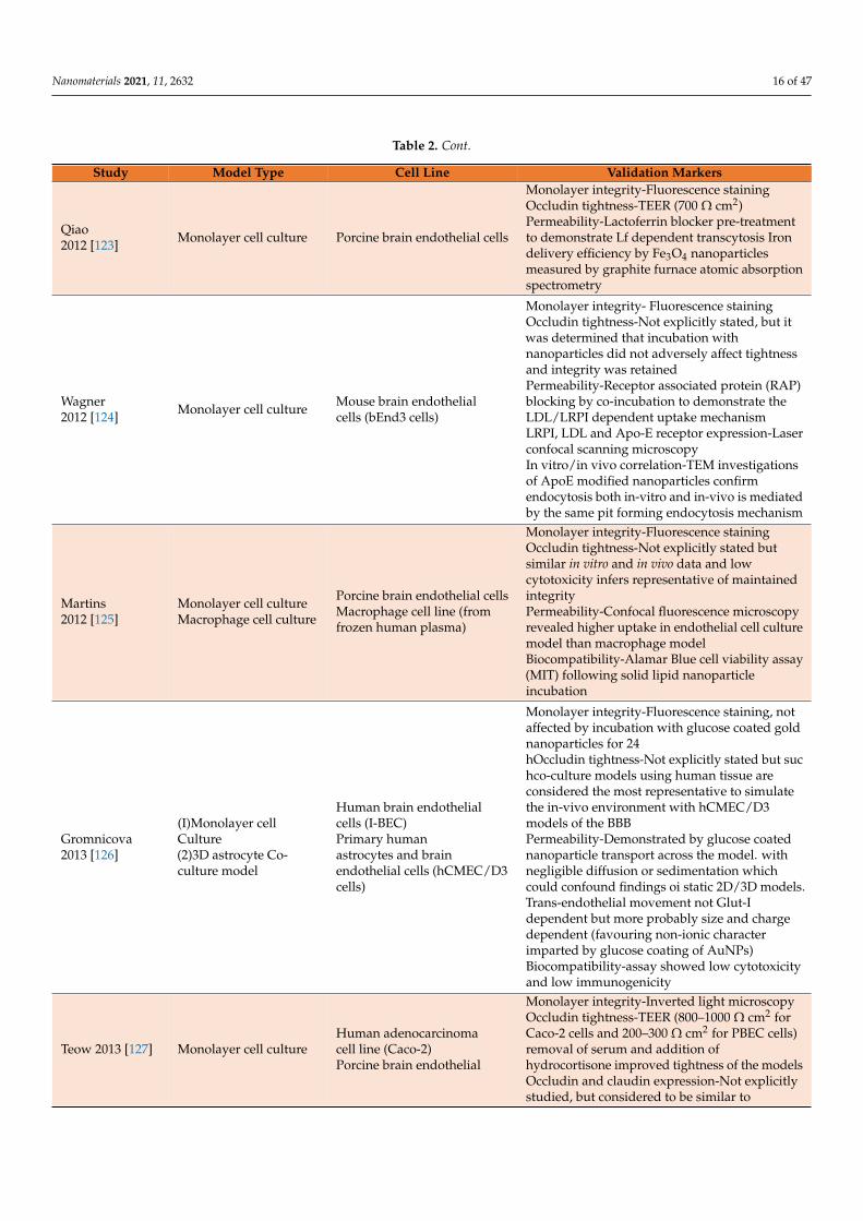
BBB characteristics to ensure the models recapitulate all features of the barrier, a number of key parameters aid in ensuring the model is suitable for its intended study application. A seminary paper published by Helms and colleagues should be consulted for in-depth guidelines on protocols for the general use of these models [119]. While there are several established sources of heterogeneity in any in vitro cell-based model study, and reproduc-ibility can be difficult, the lack of translatability of data is frequently due to incomplete characterisation of the models, nanomaterials and due to suboptimal handling and proto-cols for their use [120]. The following therefore constitutes an effective user guide for re-searchers in validating a model for the study of nanomaterials to ensure more robust data are generated, which will be more representative of the in vivo situation as presented in Table 2 [121–141].
Table 2. Executive summary of included cell-based models and associated validation markers.
Study Model Type Cell Line Validation Markers Chang 2009 [121]
Co-Culture Bovine brain endothelial cells Monolayer integrity-Fluorescence staining
Courtesy @ https://kollathdesign.com/
Figure 4. Replicating the dynamic barrier. A cross sectional view of the neurovascular unit thatconstitutes the BBB. Graphic courtesy of Mr. Richard Kollath (accessed on 29 April 2021).
4.1. Validation Markers for the Reviewed Models
While it is generally considered to be practically impossible to generate a full set ofBBB characteristics to ensure the models recapitulate all features of the barrier, a numberof key parameters aid in ensuring the model is suitable for its intended study application.A seminary paper published by Helms and colleagues should be consulted for in-depthguidelines on protocols for the general use of these models [118]. While there are severalestablished sources of heterogeneity in any in vitro cell-based model study, and repro-ducibility can be difficult, the lack of translatability of data is frequently due to incompletecharacterisation of the models, nanomaterials and due to suboptimal handling and pro-tocols for their use [119]. The following therefore constitutes an effective user guide forresearchers in validating a model for the study of nanomaterials to ensure more robustdata are generated, which will be more representative of the in vivo situation as presentedin Table 2 [120–140].
Table 2. Executive summary of included cell-based models and associated validation markers.
Study Model Type Cell Line Validation Markers
Chang2009 [120] Co-Culture
Bovine brain endothelial cellsRat mixed glial cells (60%astrocytes, 20%oligodendrocytes, and 20%microglia)
Monolayer integrity-Fluorescence stainingOccludin tightness-Not explicitly stated but tightjunction, LDL, TfR and y-glutanyl transpeptidase(y-GT) activity considered to be retained as perCecchelli and colleagues 2007 [121]Permeability-Transferrin receptor inhibitorpre-treatment to demonstrate the specific TfRmediated endocytosisIn vitro/in vivo correlation-Not explicitlyreported but referenced as method comparableto that described in Dehouck and colleagues1992 [122]
Georgieva2011 [75] Plasma membrane Human brain endothelial cells
[hCMEC/D3 cells]
Monolayer integrity-Fluorescence stainingOccludin tightness-TEER (50 Ω cm2)Permeability-Hydrophilic tracers(sucrose/inulin) PECAM, ZO-I and MRP-Iexpression-Laser scanning confocal microscopy
Nanomaterials 2021, 11, 2632 16 of 47
Table 2. Cont.
Study Model Type Cell Line Validation Markers
Qiao2012 [123] Monolayer cell culture Porcine brain endothelial cells
Monolayer integrity-Fluorescence stainingOccludin tightness-TEER (700 Ω cm2)Permeability-Lactoferrin blocker pre-treatmentto demonstrate Lf dependent transcytosis Irondelivery efficiency by Fe3O4 nanoparticlesmeasured by graphite furnace atomic absorptionspectrometry
Wagner2012 [124] Monolayer cell culture Mouse brain endothelial
cells (bEnd3 cells)
Monolayer integrity- Fluorescence stainingOccludin tightness-Not explicitly stated, but itwas determined that incubation withnanoparticles did not adversely affect tightnessand integrity was retainedPermeability-Receptor associated protein (RAP)blocking by co-incubation to demonstrate theLDL/LRPI dependent uptake mechanismLRPI, LDL and Apo-E receptor expression-Laserconfocal scanning microscopyIn vitro/in vivo correlation-TEM investigationsof ApoE modified nanoparticles confirmendocytosis both in-vitro and in-vivo is mediatedby the same pit forming endocytosis mechanism
Martins2012 [125]
Monolayer cell cultureMacrophage cell culture
Porcine brain endothelial cellsMacrophage cell line (fromfrozen human plasma)
Monolayer integrity-Fluorescence stainingOccludin tightness-Not explicitly stated butsimilar in vitro and in vivo data and lowcytotoxicity infers representative of maintainedintegrityPermeability-Confocal fluorescence microscopyrevealed higher uptake in endothelial cell culturemodel than macrophage modelBiocompatibility-Alamar Blue cell viability assay(MIT) following solid lipid nanoparticleincubation
Gromnicova2013 [126]
(I)Monolayer cellCulture(2)3D astrocyte Co-culture model
Human brain endothelialcells (I-BEC)Primary humanastrocytes and brainendothelial cells (hCMEC/D3cells)
Monolayer integrity-Fluorescence staining, notaffected by incubation with glucose coated goldnanoparticles for 24hOccludin tightness-Not explicitly stated but suchco-culture models using human tissue areconsidered the most representative to simulatethe in-vivo environment with hCMEC/D3models of the BBBPermeability-Demonstrated by glucose coatednanoparticle transport across the model. withnegligible diffusion or sedimentation whichcould confound findings oi static 2D/3D models.Trans-endothelial movement not Glut-Idependent but more probably size and chargedependent (favouring non-ionic characterimparted by glucose coating of AuNPs)Biocompatibility-assay showed low cytotoxicityand low immunogenicity
Teow 2013 [127] Monolayer cell cultureHuman adenocarcinomacell line (Caco-2)Porcine brain endothelial
Monolayer integrity-Inverted light microscopyOccludin tightness-TEER (800–1000 Ω cm2 forCaco-2 cells and 200–300 Ω cm2 for PBEC cells)removal of serum and addition ofhydrocortisone improved tightness of the modelsOccludin and claudin expression-Not explicitlystudied, but considered to be similar to

Nanomaterials 2021, 11, 2632 17 of 47
Table 2. Cont.
Study Model Type Cell Line Validation Markers
Teow 2013 [127] Monolayer cell cultureHuman adenocarcinomacell line (Caco-2)Porcine brain endothelial
described in Patabendige and colleagues 2012.Papp measurements of paclitaxel in bothdirections demonstrated the expression of p-gpin the monolayer models [128]Permeability—TEER measurements before andafter experiments/incubation. Apparentpermeability coefficient (Pan) was calculatedfrom the equation Papp (cm/s) −(dQ/dt)/(CoxA)dQ/dt. which constitutes arobust quantitative value which facilitatesorthogonal comparisons with other studiesBiocompatibility-LDH assay showed lowcytotoxicity of the dendrimer nanocarriers, andconverse high cytotoxicity (antitumour activity)when conjugated with paclitaxel
Rempe 2014 [129] Monolayer cell culture Porcine brain endothelial cells
Monolayer integrity-Fluorescence staining andimmunocytochemical analysisOccludin tightness-TEER measurements,although stated as percentages rather thanabsolute valuesPermeability-Hydrophilic tracers NC-sucroseand fluorescein isothiocyanate labelled bovineserum albumin (FITC-BSA). Found maximalpermeability after four hours due to decrease inTEER and maximum values of Papp (cm/s)P-gp, occludin expression-Immunocytochemicalanalysis and implied from experimental datashowing disruption of model integrity after fourhours when incubated with thepoly(cyanobutylacrylate) NPs, following byrecovery of integrity to 80 % baseline TEERvaluesBiocompatibility-Critical solids content of 26.62µg/mL led to irreversible monolayer disruption,while those below half this value i.e., <13.31µg/mL led to complete recovery of barrierintegrity
Cramer2014 [130] Monolayer cell culture Porcine brain endothelial cells
Capillary choroid plexus cells
Occludin tightness-TEER, being expressed inpercentages than absolute valuesOccludin expression-Western blot andimmunochemistryPermeability-TEER measurements before andafter treatment with AgNPs, confirmed byFITC-dextran Papp measurements, which were inagreementBiocompatibility-Neutral red uptake assay andmicroscopy to monitor cell morphology afterincubation with AgNPs. The ethylene oxidenanoparticles were notably more cytotoxic thantheir citrate counterparts, with a criticalconcentration dependence (75 µg/mL) ofmonolayer disruptionPro-inflammatory capacity-Reactive oxygennitrogen species, MMP-2 and COX-2 activity

Nanomaterials 2021, 11, 2632 18 of 47
Table 2. Cont.
Study Model Type Cell Line Validation Markers
Cramer2014 [130] Monolayer cell culture Porcine brain endothelial cells
Capillary choroid plexus cells
measured by zymography which wasupregulated by ethylene oxide AgNPs but notfor citrate AgNPs at standard concentrations(25 µg/mL)
Bramini2014 [131] Monolayer cell culture
Human brain capillarymicrovascular endothelialcells (hCMEC/D3 cell line)
Monolayer integrity -Fluorescence stainingOccludin tightness-TEER measurements andconfocal microscopy, which found holes of total200 µm2, and although these may have anexaggerating effect on the overall flux, they areaccounted for in the mode. This would beconsistent with those found in similar models,although this is frequently not investigated orreported Claudin expression Western blot andconfocal microscopyPermeability-TEER measurements andfluorescent labelled permeability assay, Spinningdisk confocal fluorescence microscopy and totalinternal reflection fluorescence microscopy(TIRFM) was used to quantify the translocationof the nanopartic1es in real time with 10 minexposure times of the carboxylated polystyreneNPs (40 nm and 100 nm sizes), demonstrating apreferential lysosomal accumulation within themodel rather than true translocation
Hanada2014 [132] Co-Culture
Rat brain microvascularendothelial cellsRat brain pericytes
Monolayer Integrity-Fluorescence stainingOccludin tightness-TEER measurements beforepermeability measurements (150–300 Ω cm2)Perrneability—Papp (cm/s) of 30 nm, 100 nm,400 nm silica nanoparticles compared With Pappof tracer sulforhodamine B. Papp studies ofquantum dots with different surface chargefunctionalisationsBiocompatability—Histological data confirmsome degree of BBB disruption implied bythinning of the endothelial cell layers followinghematoxylin and eosin (H&E) staining, thoughlong term permeability assays indicatednegligible adverse effects on BBB functionalityIn vitro/in vivo correlation-Not explicitlyinvestigated but commercial BBB model usedwhich has been previously validated byNakagawa and colleagues using a suite of drugmolecules including known substrates of MRP-Iand p-gp [133]
Shilo 2015 [134] Monolayer cellculture
Mouse brain endothelial cells(bEnd3 cells)
Monolayer integrity-Fluorescence stainingOccludin tightness-Most parameters were notexplicitly investigated, but the bEnd3 monolayeris a validated and well established model, andimaging demonstrated it formed similarly toother studies Permeability-Flame atomicabsorption spectrometry to quantify the AuNPuptake after 30 min incubation With varioussizes of NPs, revealing preferential selection Of70 nm barbiturate functionalized AuNPs for CT

Nanomaterials 2021, 11, 2632 19 of 47
Table 2. Cont.
Study Model Type Cell Line Validation Markers
Shilo 2015 [134] Monolayer cellculture
Mouse brain endothelial cells(bEnd3 cells)
imaging applications (most total Au uptake), and20 nm for drug loading (highest free surface area)In-vitro/ln-vivo correlation—Fluorescentconfocal microscopy investigating interaction Ofbarbiturate loaded AuNPs with the modelindicated specific pinocytosis mediated transportacross the barrier, and some degree ofassociation with the barrier itself
Xu 2015 [135] Co-Culture
Rat microvascular endothelialcellsRat pericytesRat astrocytes
Monolayer integrity—Fluorescence stainingOccludin tightness-TEER (>200 Ω cm2)ZO-I, claudin 5 expression-Confocal microscopyPermeability-TEER measurements before andafter incubation with AgNPs and polystyreneNPs as control, demonstrating BBB disruption bydecreased resistance values after 24 h for the10 µg/mL—AgNPs only (1 µg/mL AgNPs andcontrol were unaffected)Biocompatibility—AgNPs at 10 µg/mLdemonstrated reduced ZO-I expression,mitochondrial shrinkage, apoptosis and alteredgene expression by immunostaining andmicroarray analysis of astrocytesIn vitro/ln vivo correlation—Triple co-culturemodel gives high TJ protein expression andtightness for evaluating mechanisms ofnanotoxicity and vasoactive compounds
De Jong2018 [136]
Filter free monolayer cellculture
Human microvascular brainendothelial cells (hCMEC/D3cells)
Monolayer integrity—Fluorescence stainingOccludin tightness—Not explicitly stated butmodel validated with permeabilitymeasurementsZO-I expression—Fluorescence microscopyPermeability—Model validated with Papp(cm/min) measurements for 4 kDa and 2000 kDadextran, which were in agreement with 3Dmicrofluidic organ on a chip models of the BBB.Also validated by collagenase A digestion ofapical, cellular and basolateral fractionsfacilitating quantitative assessment of active LDLmediated transcytosis by fluorescencespectroscopy illustrating the quantitative modeof the modelIn vitro/ln vivo correlation—Filter-free model in ahuman cell line allowing quantitative and realtime imaging of nanoparticle transport across themembrane
Zhang2020 [137]
Transcellularmonolayer cellculture
Mouse brain endothelial cells(bEnd3 cells)
Monolayer integrity—Fluorescence stainingOccludin tightness—Not explicitly investigated,but permeability measurements used to validatethe model and same protocols used as otherstudies which generated confluent monolayerswith high TJ protein expressionPermeability—Papp measurements of neutralnanoparticles used to validate the modelquantitatively by fluorescence spectroscopyIn vitro/in vivo correlation—Model

Nanomaterials 2021, 11, 2632 20 of 47
Table 2. Cont.
Study Model Type Cell Line Validation Markers
Zhang2020 [137]
Transcellularmonolayer cellculture
Mouse brain endothelial cells(bEnd3 cells)
mathematically expressed as a 2D barrier interms of its bending rigidity, surface tension,viscoelasticity and surface charge, as well as ionconcentration of the medium and size andcharge properties of nanoparticles. Therefore,recapitulates several key aspects ofelectrochemical gradient driven endocytosisrather than receptor mediated targeting, whichallows elucidation of key rational designproperties for NP delivery to the brain
Sokolova2020 [138] Spheroid model
Human brain microvascularendothelial cellsHuman brain pericytesHuman astrocytesHuman microglia (iPSCderived)Human oligodendrocytes(iPSC derived)Human cortical neurons (iPSCderived)
Monolayer integrity-Fluorescence staining,confocal scanning microscopyOccludin tightness-Not explicitly investigated,but characterisation was conducted as per Zhouand colleagues who have extensively establishedand Validated this model (140]ZO-1, claudin-5, CD31, P-gp. GLUT-Iexpression—immunohistochemistryBiocompatibility-ATP production as a cellviability assay following incubation with dye(FAM-Alkyne) conjugated AuNPs for up to 24 h.showing negligible change demonstrating lack ofclinically significant cytotoxicityIn vitro/ in vivo correlation-3D Model employingsix types of human or human related tissueswhich comprise the NVU, Hypoxia condition e.g.following ischaemic stroke recapitulated todetermine influence of pathophysiology onnanoparticle behaviour and distribution
Kumarasamy2021 [141] Spheroid model
Human brain microvascularendothelial cellsHuman brain vascularpericytesHuman astrocytesRat neuronsRat microglia
Monolayer integrity-Fluorescence STEMOccludin tightness-Confocal laser scanningfluorescence microscopy, RNA-sequencingVE-cadherin, claudin-5, NG2 proteoglycan,GFAP β-III tubulin, iNOS, MAP-2expression-Immunohistochemistry, Western Blot,SDS PAGE,ABC, GLUT 1,3,5, SLC, p-gp expression—RNAsequencing and PCAPermeability—FITC labelling and incubation Ofnanoparticles with model for 24 h, followed byimagingBiocompatibility—Metabolic and morphologicalstudies on endothelial and epithelial cellsfollowing incubation with different classes Ofnanocarriers including graphene nanoplatesrcarbon dots, polymeric and metallicnanoparticlesIn vitro/in vivo correlation-3D Modelemploying five cell types encompassing the BBBand associated microglia and neural networkswith exquisite audio-visual data and extensivecharacterisation of an essentially ex-vivo NVU isthe most biomimetic model type to date, thoughusing rat and human derived cells can limit thetranslation and reproduction of results
A notable inclusion is the junctional tightness, as this arguably is the pivotal propertythat will influence the robustness of the model in mimicking the integrity of the BBB [141].
Nanomaterials 2021, 11, 2632 21 of 47
In the vast majority of studies, the tightness is measured using the transendothelial electricalresistance (TEER), which, while useful, has notable implications with its use, includingdifferences in the techniques and apparatus used to measure it, and the size dependence ofthe compound of interest is largely ignored [142]. As such, permeability using hydrophilictracers such as fluorescent probes or small molecules such as sucrose (~340 Da) may providemore functional estimates, particularly when both measures are employed, in addition toensuring claudins are also included in the model to prevent BBB model leakage [143].
Expression and localisation of the efflux transporters and solute receptors outlinedin Section 3 are also fundamentally important to the study, and particular care must beexercised when using non-human in vitro models. It has been established by quantitative-targeted absolute proteomic (QTAP) studies that the human BBB is closest to that of thecynomolgus monkey and marmoset primates in terms of receptor expression, and thepoor efficiency of rodents is such that in most cases both receptor and efflux transporterexpression is greater than two-fold higher in such model organisms [144]. For example,P-gp expression has been found to be expressed in the region of ~6.00 fmol/µg totalprotein in humans, but the expression is ~14 fmol/µg total protein and ~19 fmol/µg totalprotein for mouse and rat, respectively, which has been validated using positron emissiontomography (PET) studies of the permeability of known P-gp substrates, which are farhigher in human models due to lower function of the P-gp efflux mechanism.
Conversely, studies of claudins have elucidated that claudin-5 is the critical proteinfor tight juncture closure in humans and is two-fold higher than in other primates andrats, while mice have 1000 fold expression to that of humans [145]. This complicates theability of certain models constituted by cells from rodent origin to be the most efficientfor translational research efforts. However, as outlined by Ohtsuki and colleagues [146],exogenous expression of such proteins is possible with subsequent transfection to therodent cell model without adverse effects, which constitutes a pragmatic workaround, andas will be outlined due to issues relating to cost and sourcing of human-based models,the in vitro animal models will continue to be imperative in the overall research anddevelopment process for nanotheranostics into the future.
4.2. Cell Culture Models4.2.1. Monolayer Cell Culture
The simplest models for the study of nanoparticle interaction with the BBB involvethe culturing of primary endothelial cells on a transwell insert [53], which creates a two-compartment model [Figure 5] in which the insert mimics the luminal side (blood com-partment) and the well mimics the abluminal side (parenchymal space). Although theseprimary cell lines are preferred due to their high TEER values (500–800 Ω cm2), high tightjunction expression and classic BBB receptor and enzymatic expression (such as claudin-5,P-gp and occludin), the task of isolating these cells directly before cell culturing is anarduous task [147]. Indeed, for the mouse, such vasculature accounts for 0.1% v/v of theoverall murine brain, which has a high propensity for contamination and additionallyrequires a large number of rodents to conduct one experiment.
Once a confluent monolayer general forms (after approximately 5 days for moststudies), the model should be validated with fluorescence staining and appropriate TEERmeasurements and permeability assay to show adequate tightness. The use of a microp-orous membrane (~0.3 µm) support, usually of polycarbonate or polyethylene terephthalateconstruction, is obviated for nanoparticle permeation studies as it facilitates the passageof small molecules while maintaining the two-compartment model [148]. However, apossible exception is illustrated by De Jong and colleagues [137], in that the quantificationof transendothelial delivery can be accurately determined by using a collagen gel cov-ered with a confluent monolayer and is more accurate as association within the filter andmembrane pores is eradicated.
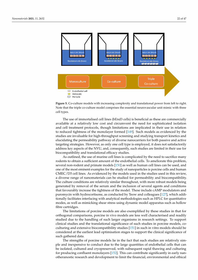
Nanomaterials 2021, 11, 2632 22 of 47
Nanomaterials 2021, 11, 2632 21 of 48
rats, while mice have 1000 fold expression to that of humans [146]. This complicates the ability of certain models constituted by cells from rodent origin to be the most efficient for translational research efforts. However, as outlined by Ohtsuki and colleagues [147], ex-ogenous expression of such proteins is possible with subsequent transfection to the rodent cell model without adverse effects, which constitutes a pragmatic workaround, and as will be outlined due to issues relating to cost and sourcing of human-based models, the in vitro animal models will continue to be imperative in the overall research and develop-ment process for nanotheranostics into the future.
4.2. Cell Culture Models 4.2.1. Monolayer Cell Culture
The simplest models for the study of nanoparticle interaction with the BBB involve the culturing of primary endothelial cells on a transwell insert [53], which creates a two-compartment model [Figure 5] in which the insert mimics the luminal side (blood com-partment) and the well mimics the abluminal side (parenchymal space). Although these primary cell lines are preferred due to their high TEER values (500–800 Ω cm2), high tight junction expression and classic BBB receptor and enzymatic expression (such as claudin-5, P-gp and occludin), the task of isolating these cells directly before cell culturing is an arduous task [148]. Indeed, for the mouse, such vasculature accounts for 0.1% v/v of the overall murine brain, which has a high propensity for contamination and additionally re-quires a large number of rodents to conduct one experiment.
Figure 5. Co-culture models with increasing complexity and translational power from left to right. Note that the triple co-culture model comprises the essential neurovascular unit mimic with three cell types.
Once a confluent monolayer general forms (after approximately 5 days for most stud-ies), the model should be validated with fluorescence staining and appropriate TEER measurements and permeability assay to show adequate tightness. The use of a mi-croporous membrane (~0.3 μm) support, usually of polycarbonate or polyethylene tereph-thalate construction, is obviated for nanoparticle permeation studies as it facilitates the passage of small molecules while maintaining the two-compartment model [149]. How-ever, a possible exception is illustrated by De Jong and colleagues [138], in that the quan-tification of transendothelial delivery can be accurately determined by using a collagen gel covered with a confluent monolayer and is more accurate as association within the filter and membrane pores is eradicated.
Figure 5. Co-culture models with increasing complexity and translational power from left to right.Note that the triple co-culture model comprises the essential neurovascular unit mimic with threecell types.
The use of immortalised cell lines (bEnd3 cells) is beneficial as these are commerciallyavailable at a relatively low cost and circumvent the need for sophisticated isolationand cell treatment protocols, though limitations are implicated in their use in relationto reduced tightness of the monolayer formed [149]. Such models as evidenced by thestudies are invaluable for high-throughput screening and studying transport kinetics andelucidating the permeability pathway of diverse nanocarriers for both passive and activetargeting strategies. However, as only one cell type is employed, it does not satisfactorilyaddress key aspects of the NVU, and, consequently, such studies are limited in their use forbiocompatibility and translational efficacy studies.
As outlined, the use of murine cell lines is complicated by the need to sacrifice manyrodents to obtain a sufficient amount of the endothelial cells. To ameliorate this problem,several non-rodent and primate models [150] as well as human cell lines can be used, andone of the most eminent examples for the study of nanoparticles is porcine cells and humanCMEC/D3 cell lines. As evidenced by the models used in the studies used in this review,a diverse range of nanomaterials can be studied for permeability and biocompatibility.The culture conditions are relatively similar throughout, with more robust models beinggenerated by removal of the serum and the inclusion of several agents and conditionsthat favourably increase the tightness of the model. These include cAMP modulators andpuromycin with hydrocortisone, as conducted by Teow and colleagues [127], which addi-tionally facilitates interfacing with analytical methodologies such as HPLC for quantitativemodes, as well as mimicking shear stress using dynamic model apparatus such as hollowfibre cartridges.
The limitations of porcine models are also exemplified by these studies in that fororthogonal comparisons, porcine in vivo models are less well characterised and readilystudied due to the handling of such larger organisms in research settings. To supportclinical studies and the translational significance of such studies in porcine models, co-culturing and extensive biocompatibility studies [151] in such in vitro models should beconsidered at the earliest lead optimisation stages to support the clinical significance ofsuch gathered data.
The strengths of porcine models lie in the fact that such studies are relatively sim-ple and inexpensive to conduct due to the large quantities of endothelial cells that canbe isolated, cultured and cryopreserved, with subsequent rapid thawing and culturingfor producing confluent monolayers [152]. This can contribute significantly in early nan-otheranostic research and development to limit the financial, environmental and ethical

Nanomaterials 2021, 11, 2632 23 of 47
implications of cytotoxicity and biocompatibility studies in research settings, potentiallyexpediting the subsequent stages of development by providing robust translational safetydata.
It is possibly no coincidence that the most exemplary efforts in transwell models areconstituted by those which employ human-derived cells, as evidently these will have themost predictive power in mimicking several aspects of the in vivo dynamic BBB envi-ronment. The human brain microvascular endothelial hCMEC/D3 cell line is the mostintensively studied and optimised monolayer model to date [153], with several notable fea-tures, including extensive characterisation of receptor, enzyme and tight junction proteins,but suffers from having lower TEER and permeability than other comparative models.These are most useful for biocompatibility and biodistribution studies as they have beeninterfaced synergistically with sophisticated imaging such as those observed by De Jongand colleagues [137], which facilitate real time imaging of translocation, in addition toassociation within the model itself for rapid analysis of nanoparticle–BBB interactions.
The primary limitation of human models such as these are the obvious ethical im-plications of resourcing human tissue and the relative paucity thereof, although this hasbeen partially offset by the increasing cell banking and commercial availability of same. Inaddition to this, resourceful researchers have developed efficient and well characterisedmonolayer cell cultures from other sources: stem cell lines to yield induced pluripotentstem cells (iPSC) [119], as well as non-cerebral origin-derived materials such as the humanimmortalised epithelial colorectal adenocarcinoma cell line (Caco-2) [127] and CSF-derivedhuman capillary choroid plexus endothelial cells [130] have all been employed successfullyfor studying nanoparticle bio-interactions.
For all the foregoing monolayer models, the fundamental commonality that precludestheir use for predictive permeation studies and biocompatibility studies of nanoparticleBBB integrity disruption is that they employ one cell type only. Despite the fact thatwhile these monolayer models are utilitarian in the alluded to instances, they are a grosssimplification of the NVU, and as they are 2D models, they do not fully depict the anatomicstructure and complexity of the BBB in vivo.
Efforts have been made to better express these 2D models in mathematical terms byZhang and colleagues [137], producing a transcellular model that can efficiently monitorthe determinant influence of surface charge, medium ionic concentration and viscoelasticproperties on nanoparticle permeability, but these do not fully capture the physiologicalrelevancy and otherwise rely on assumptions of the equations employed. Various celllines have been up-regulated to maintain endothelial cell relevancy following isolationby activation of canonical pathways, such as the WnT/β-catenin pathway, to promotephenotypic behaviour [154], but they do not adequately represent the endothelial cell–astrocyte crosstalk which can be better recreated by co-culturing.
4.2.2. Co-Cultured Cell Cultures
As alluded to, the primary goal of an in vitro cell-based model is to recreate as closelyas possible the in situ BBB environment and composition of the NVU in terms of tightnessand expression of transporters, as well as the key cross-regulatory and vesicular traffick-ing functions of endothelial cells, which are primarily modulated by the astrocytes andpericytes [155]. If such models are to essentially replace the classical in vivo methodolo-gies for quantitatively assessing CNS permeability of drug candidates that encompass insitu perfusion, CSF sampling and intracerebral microdialysis, as well as intravenous andintraperitoneal, as less invasive methods [156], then in the wider context of CNS drugdiscovery programs, the models must be more representative and maintainable over longerstudy periods. To this end, co-culturing and indeed tri-culturing afford opportunities inthis regard in maintaining the versatility, usability and high-throughput strengths of amonolayer model while consolidating their relevance to the in vivo situation.
While porcine and bovine sources of endothelial cells are advantageous for smallmolecule permeability studies such as nanocarrier BBB passage screening, the rodent
Nanomaterials 2021, 11, 2632 24 of 47
models are possibly more representative due to their closer homology to human proteinexpression [97], with the implied compromises as alluded to in Section 4.1. While se-lected studies have employed a co-culturing protocol, with improved functional tightness(correlated by TEER measurements and reduced Papp measurements), as Hatherell andcolleagues elucidated [157], direct cell-to-cell contact in such models is a prerequisite fortransitioning from 2D to 3D models, and astrocytes have a greater contribution to theirtightness than pericytes. This was demonstrated by the physical constraints of transwellsystems, in that adjacent co-cultured cells are generally unfeasible, and the endothelial cellsare instead seeded on the insert surface while the astrocytes are cultured underneath andthe pericytes at the bottom of the apparatus. The result is that the intercellular commu-nication is mediated only by soluble factors secreted into the medium, which is virtuallyimpossible to characterise and reproduce with any study-to-study homogeneity.
What Gromnicova and colleagues remarked, however, is that this geometrical prob-lem can be overcome by employing a 3D collagen gel under the confluent endothelialmonolayer [126]. This is further improved by the commercial availability of such co-culturesystems, such as that employed by Hanada and colleagues [132]. The co-culture systemshave a dual functionality of permitting rapid permeability studies and more comprehen-sive biocompatibility assessment of “nano-risk”, as it can also encompass the impact ofnanomaterials on astrocytes which they encounter directly after passage across the BBB.As astrocytes provide metabolic nutrients to neurons and are also neuroprotective, a fun-damental aspect of the compatibility of certain materials can be assessed by investigatingthe potential impact of these on the viability of the co-culture model, which, as Xu andcolleagues found, can consolidate the contradicting data in more rudimental studies in thebroader literature [135].
While the vast majority of studies favour models which utilise rat-derived cells,there is an increasing appreciation of the viability of employing synergistic mammaland rat-derived models [133]. One such model can be envisaged to include the useof human-derived cells or neurons in place of pericytes for modelling the blood–CSFbarrier in addition to the BBB. This would facilitate more complete in vivo correlations andextrapolation to humans using a triple culture model, which offers the highest functionaltightness and biorelevant properties [158]. The expertise required and laborious nature ofsuch models is a detrimental facet of their use, but with increasing sourcing of patentedmodels, these frozen ready to use kits may possess the answers to issues raised by isolatedefforts published to date in terms of reproducibility and characterisation of these modelsfor the study of nanomaterials.
Co-culture models are also superior for studying CNS conditions such as stroke andtraumatic brain injury, as Neuhaus and colleagues [159] have highlighted the immenseinfluence of astrocytes on the in vitro BBB model they developed. Such conditions requirebiorelevant models for investigating underlying pathophysiological mechanisms andtesting the potential merit of therapeutic strategies for their acute management. As wasalso alluded to, however, as for all transwell-based cell culture models, the contribution ofshear stress by blood flow to regulation of the endothelial cell layer cannot be understated,and so dynamic models of the BBB in 3D are desirable. Takeshita and colleagues [160]have developed this concept to realising a human cell-derived co-culture model thatsimulates the shear stress under flow and, furthermore, allows recovery of the analyteafter transmigration. While these evidently have great suitability and bio-relevancy, thediscriminant contribution of various satellite cells such as neurons and microglia arenot readily emulated, and in many cases, the study is confined to a 2D nanoparticlebio interaction study. Thus, while pivotal in R&D for high-throughput screening, leadoptimisation, permeability studies and toxicity screening for functioning as supportivedata for clinical testing applications in lieu of in vivo animal studies, testing on a morecomplex model is mandatory as an attractive and satisfactory alternative from a researchand regulatory standpoint.

Nanomaterials 2021, 11, 2632 25 of 47
4.2.3. Spheroid Cell Culture
With the foregoing considerations of the limitations of 2D models and the interactionof nanoparticles with dyes and artificially enhanced permeability leading to biased results,3D cell culture models have garnered attention in modern times. The marked shift towardsthe use of such models has also been a direct result of the increasing recognition of thenumerous advantages of induced pluripotent stem cells (iPSCs), both in terms of closephysiological relevancy, reproducibility, scalability and isogenic and individualised co-culturing protocols facilitating patient-specific integrated CNS models [161].
Perhaps the most exciting prospect of such 3D models is that they can spontaneouslyself-assemble to form scaffold free models of the BBB for permeability screening, i.e.,spheroids, which accurately represent the brain physiologically and spatially [162,163].More sophisticatedly, cells can form cerebral organoids in suitable scaffolding matrices,such as that developed by Nzou and colleagues [140]. For an excellent review of iPSC-derived BBB models including cerebral organoids for studying neurological disorders,consult the article recently published by Logan and colleagues [164] in comprehensivephysiology. While there is some degree of contention with regards to defining the differ-ences between a “spheroid” and ”organoid,” what can be agreed upon generally, and forthe purposes of this review, is that the difference lies primarily in the cell types used andthe culturing protocols, and will accordingly be discussed separately.
Generally speaking, they share a commonality in their self-assembly and the factthat they essentially are an organ mimetic with shared characteristics to the endogenousorgan [141]. Where they differ, however, is in the employment of differentiated or stemcells, which give rise to spheroids and organoids respectively. Therefore, in this section,the nanomedicine studies for cell culture are confined to spheroids/assembloids, andorganoids will be further discussed in the section on organ-on-chip microfluidics, in howthey can be used in tandem with microfluidics for studying nanoparticle–BBB interactionsas an alternative to in vitro cell cultures. As was shown by the findings of Sokolova andcolleagues [139], Nzou and colleagues [139], and Kumarasamy and colleagues [140], themost robust models must as a prerequisite include the primary elements of the NVU(endothelial cells, astrocytes and pericytes), but further yet, the necessity of the addition ofsatellite cells such as the microglia to spheroid models, the so-termed “third element” ofthe NVU by Szepesi and colleagues [164], which accounts for 10–15% of total brain cells, isincontrovertible.
To date, many studies have used the triple culture model only, and while this isgenerally a pragmatic compromise given the cost and technical constraints associated withsourcing, characterising, and culturing such auxiliary cells for low-adherence spheroidself-assembly, their importance in models for studying nanomaterials cannot be under-stated [165]. Indeed, Kumarasamy and colleagues [166] have extensively investigatedthe various contributions of microglia in mediating nose to brain transport of nanomate-rials, an alternative nanotheranostic delivery strategy under consideration, and indeedthe possibility exists of pathogens such as SARS- CoV-2 hijacking this mechanism forCNS entry [167]., Indeed, where such models are obviated but cost considerations wouldpreclude their use for small research groups, rather than resorting to simplified modelswith little predictive power, the use of a human-based triple culture supplemented with ratmicroglia and neurons would be the most advisable approach.
The strengths of such a model are best represented in the fact that dyes themselveswill not enter the model, but when encapsulated in a nanocarrier, advanced microscopytechniques such as confocal laser scanning microscopy can be used to map real-timepermeation and biodistribution in a vascularised model of the BBB. [138,140] As this isspatially resolved in highly resolved 3D audio-visual recordings, and the fact that themodel is more relevant than transwell culture models in terms of key modulator andreceptor expression and activity, means that these are versatile and powerful models fordrug screening [162]. In addition to this, pathophysiological states can be exquisitelymodelled, and as Sokolova and colleagues exemplified [138], this is a noteworthy trait as

Nanomaterials 2021, 11, 2632 26 of 47
nanomaterial-based management of hypoxia in acute phase ischaemic stroke and traumaticbrain injury can be investigated.
The main limitation is that although the model can be extensively characterised andvalidated, and an array of molecules can be studied for their cell penetrating, receptor-mediated or other associated transport mechanisms of delivery across the BBB, the influenceof shear stress is difficult to model unless the model can be extensively vascularised [168],which eludes all but the most experienced researchers. As such, advances in microfluidicscan be used to solve this problem and generate an organ-on-chip, which can simulate themechanics and physiology of the human brain in a micro physiological artificial organsystem [54].
5. Recent Trends and Future Directions for NTP Models5.1. Microfluidic Organ-on-Chip Technology (µBBB)
While parallel artificial membrane permeability assay (PAMPA) and cell-based tran-swell assays have been prevalent for the past two decades in CNS drug discovery, theysuffer from being oversimplified and limited in terms of their biorelevancy, as in mostcases they are a static two-compartment models, and for PAMPA, there is in fact no cellbasis [169]. While well suited to the preliminary stages of R&D for small molecule screeningwith high throughput and low costs, for subsequent lead optimisation and testing, 3Dmodels such as those offered by advances in microfluidics are warranted, particularly whenthey can be used synergistically with advanced cell culturing techniques, i.e., organoids.As has been demonstrated in several studies [170–172], near physiological shear stresscan be incorporated into the model to better capture in vivo tightness and endothelial cellbehaviour under the influence of simulated blood flow. This is of great importance forthe toxicology studies of biocompatibility as nanomaterial safety must be demonstrablefor species and disease-specific cumulative dosing studies of >1 month [64], which is notpossible with transwell studies as the integrity of the endothelial cells cannot be maintainedfor long periods.
An ideal microfluidic platform for NTP testing requires integration of a number ofkey considerations. Firstly, the platform must be able to recapitulate the BBB endothelialcell vessel-like structures in 3D, mimic the cellular crosstalk and cross-regulation, simulateshear stress under flow and have a biocompatible basal membrane [173]. These require-ments have been met to varying extents by modular configurations, ranging from theelementary sandwich design which evolved from transwell models, to parallel and 3Dtubular networks, in addition to experimental inclusion of de novo microvessel formationover the use of microneedles. The article published by Oddo and colleagues [54] should beconsulted for a more complete review of these designs, but, briefly, the drawbacks of theconventional sandwich configuration have been mitigated by more sophisticated designs,which will be considered here.
In general, the most successful models are comprised by co-culturing human or iPSCcells under constant perfusion in a glass synthetic microvasculature model [53], whichhas layered microchannels separated by microfabricated membranes with 3 µm gaps togenerate patient and disease-specific models of the BBB, which can have a high degree ofcontrol and flexibility in terms of key parameters (Figure 6: such parameters include thetightness measured by TEER, permeability measured by small molecules such as dextranand inulin, tight junction protein expression measurable by microscopy and degree ofshear stress). They can also be used to study the influence of pathogenesis, i.e., disease,such as AD/PD, ischaemic and hypoxic states, on the BBB behaviour and permeability ofnanoparticles [174,175].

Nanomaterials 2021, 11, 2632 27 of 47
Nanomaterials 2021, 11, 2632 26 of 48
screening with high throughput and low costs, for subsequent lead optimisation and test-ing, 3D models such as those offered by advances in microfluidics are warranted, partic-ularly when they can be used synergistically with advanced cell culturing techniques, i.e., organoids. As has been demonstrated in several studies [171–173], near physiological shear stress can be incorporated into the model to better capture in vivo tightness and endothelial cell behaviour under the influence of simulated blood flow. This is of great importance for the toxicology studies of biocompatibility as nanomaterial safety must be demonstrable for species and disease-specific cumulative dosing studies of >1 month [64], which is not possible with transwell studies as the integrity of the endothelial cells cannot be maintained for long periods.
An ideal microfluidic platform for NTP testing requires integration of a number of key considerations. Firstly, the platform must be able to recapitulate the BBB endothelial cell vessel-like structures in 3D, mimic the cellular crosstalk and cross-regulation, simulate shear stress under flow and have a biocompatible basal membrane [174]. These require-ments have been met to varying extents by modular configurations, ranging from the el-ementary sandwich design which evolved from transwell models, to parallel and 3D tub-ular networks, in addition to experimental inclusion of de novo microvessel formation over the use of microneedles. The article published by Oddo and colleagues [54] should be consulted for a more complete review of these designs, but, briefly, the drawbacks of the conventional sandwich configuration have been mitigated by more sophisticated de-signs, which will be considered here.
In general, the most successful models are comprised by co-culturing human or iPSC cells under constant perfusion in a glass synthetic microvasculature model [53], which has layered microchannels separated by microfabricated membranes with 3 μm gaps to gen-erate patient and disease-specific models of the BBB, which can have a high degree of control and flexibility in terms of key parameters (Figure 6: such parameters include the tightness measured by TEER, permeability measured by small molecules such as dextran and inulin, tight junction protein expression measurable by microscopy and degree of shear stress). They can also be used to study the influence of pathogenesis, i.e., disease, such as AD/PD, ischaemic and hypoxic states, on the BBB behaviour and permeability of nanoparticles [175,176].
While adsorption and immobilisation of the endothelial cells to the glass layers of the model is generally achieved by silanisation or oxygen plasma activation as used in the model developed by Kim and colleagues [177], for longer term studies such as biocom-patibility screening in cumulative dosing of NTPs, a covalent binding of the extracellular matrix is advantageous. Peng and colleagues [178] recently achieved a coated organ-in-chip using a photo cross-linkable copolymer that is amenable to in situ surface modifica-tion to model the contribution of the basal membrane to BBB formation and regulation, with high-throughput screening and simulated microchannel flow studies. The rate of medium flow through each chamber thus determines the shear stress, which can be regu-lated and modulated at will, which is much better for biorelevant studies than simple ro-tation devices and constrained geometries employed in dynamic transwell models.
Nanomaterials 2021, 11, 2632 27 of 48
Figure 6. Top-down schematic of a typical microfluidic model with separated channels (approxi-mately 2 cm in length. Bottom organ-on-chip model of BBB. Note each channel has microchannel separators marked by the colour interface of each channel (approximately 3 μm wide between blood and brain, and 50 μm between medium and brain)).
Additionally, pumps, sensors such as electrical impendence sensing (EIS for nano-toxicity assessments) and electrodes can be readily employed in these models to measure key properties such as the TEER, pH and ionic concentration gradients and ensure con-stant monitoring as developed by Liang and colleagues [179]. As the nanoparticles will have a homogenous distribution through the medium and each compartment can be read-ily sampled and imaged with high resolution microscopy in real time without the require-ment for labels and dyes, the effective delivered dose of nanoparticles and cumulative dose safety evaluations can be studied [64]. They are also a valuable tool as they will elim-inate the influence of aggregation, gravity and buoyancy due to the laminar flow and ar-tificial enhanced permeation and association of nanoparticles with the model itself as ob-served in transwell assays [52], thus giving more accurate and reliable data in relation to absolute nanoparticle delivery across the BBB.
While several papers give an excellent account of the advances in microfluidic mod-els for disease modelling applications, this review is to give an account of the application of organ-on-chip models to the study of nanomaterials. For an in-depth account of such technologies for disease modelling, please refer to Holloway and colleagues [172] and Van der Helm and colleagues [180]. Briefly, however, a number of models given in these re-views have potential for developing nanoparticle treatments in specific disease states, such as in AD and glioblastoma multiforme (GBM).
While a number of organ-on-chip models have been used for in vitro drug develop-ment for evaluating efficacy and toxicity of novel drug compounds, such as the liver chip [181], kidney chip [182], gastrointestinal chip [183] and lung chip [184], the NVU/BBB chip is still relatively in its infancy, as it is arguably the most difficult to capture efficiently.,,, Recent efforts have thus focused on developing more sophisticated models that employ synaptic activity in subcellular structures of the model, and optimising the models for specifically studying nanomaterial transport, as classically the models have been prefer-entially adopted for neuroscientific research applications. [185].
The search for biocompatible materials is also at a premium, as although that which has been used almost ubiquitously in the models published to date is utilitarian, the pri-mary limitation is that it can absorb small organic compounds [186], which would seem to confound the findings of permeability studies for nanoparticles where it is used as the micro-fabrication material. Biofabrication has improved in recent years to improve the functional tightness of these chips (>2000 Ω cm2) [187], and indeed the constraints associ-ated with employing two cell types only in a two compartment model has been recognised as being unsatisfactory. While Bang and colleagues [188] consolidated this latter issue and thus improved the models postulated by Booth and colleagues [189], and later Adriani and colleagues [190], to permit independent emulation of the internal and external
Figure 6. Top-down schematic of a typical microfluidic model with separated channels (approxi-mately 2 cm in length. Bottom organ-on-chip model of BBB. Note each channel has microchannelseparators marked by the colour interface of each channel (approximately 3 µm wide between bloodand brain, and 50 µm between medium and brain)).
While adsorption and immobilisation of the endothelial cells to the glass layers of themodel is generally achieved by silanisation or oxygen plasma activation as used in themodel developed by Kim and colleagues [176], for longer term studies such as biocom-patibility screening in cumulative dosing of NTPs, a covalent binding of the extracellularmatrix is advantageous. Peng and colleagues [177] recently achieved a coated organ-in-chipusing a photo cross-linkable copolymer that is amenable to in situ surface modificationto model the contribution of the basal membrane to BBB formation and regulation, withhigh-throughput screening and simulated microchannel flow studies. The rate of mediumflow through each chamber thus determines the shear stress, which can be regulated andmodulated at will, which is much better for biorelevant studies than simple rotation devicesand constrained geometries employed in dynamic transwell models.
Additionally, pumps, sensors such as electrical impendence sensing (EIS for nanotoxi-city assessments) and electrodes can be readily employed in these models to measure keyproperties such as the TEER, pH and ionic concentration gradients and ensure constantmonitoring as developed by Liang and colleagues [178]. As the nanoparticles will havea homogenous distribution through the medium and each compartment can be readilysampled and imaged with high resolution microscopy in real time without the requirementfor labels and dyes, the effective delivered dose of nanoparticles and cumulative dosesafety evaluations can be studied [64]. They are also a valuable tool as they will eliminatethe influence of aggregation, gravity and buoyancy due to the laminar flow and artificialenhanced permeation and association of nanoparticles with the model itself as observed intranswell assays [52], thus giving more accurate and reliable data in relation to absolutenanoparticle delivery across the BBB.
While several papers give an excellent account of the advances in microfluidic modelsfor disease modelling applications, this review is to give an account of the applicationof organ-on-chip models to the study of nanomaterials. For an in-depth account of suchtechnologies for disease modelling, please refer to Holloway and colleagues [171] andVan der Helm and colleagues [179]. Briefly, however, a number of models given in thesereviews have potential for developing nanoparticle treatments in specific disease states,such as in AD and glioblastoma multiforme (GBM).

Nanomaterials 2021, 11, 2632 28 of 47
While a number of organ-on-chip models have been used for in vitro drug devel-opment for evaluating efficacy and toxicity of novel drug compounds, such as the liverchip [180], kidney chip [181], gastrointestinal chip [182] and lung chip [183], the NVU/BBBchip is still relatively in its infancy, as it is arguably the most difficult to capture efficiently.Recent efforts have thus focused on developing more sophisticated models that employsynaptic activity in subcellular structures of the model, and optimising the models forspecifically studying nanomaterial transport, as classically the models have been preferen-tially adopted for neuroscientific research applications. [184].
The search for biocompatible materials is also at a premium, as although that whichhas been used almost ubiquitously in the models published to date is utilitarian, theprimary limitation is that it can absorb small organic compounds [185], which would seemto confound the findings of permeability studies for nanoparticles where it is used asthe micro-fabrication material. Biofabrication has improved in recent years to improvethe functional tightness of these chips (>2000 Ω cm2) [186], and indeed the constraintsassociated with employing two cell types only in a two compartment model has beenrecognised as being unsatisfactory. While Bang and colleagues [187] consolidated thislatter issue and thus improved the models postulated by Booth and colleagues [188], andlater Adriani and colleagues [189], to permit independent emulation of the internal andexternal vascular microenvironments, the use of rat-derived cells still implies cross-speciestranslatability limitations.
Campisi and colleagues [190] thus realised a state-of-the-art model that combines thestrengths of all of its forebearers, realising an iPSC-derived tri-culture model, which servesas a robust 3D platform for drug permeability studies. The perfusability and permeabilitywere validated and comparable to other literature-derived data, demonstrating its potentialfor automated high-throughput drug transport studies. Using a coating such as thatdeveloped by Peng and colleagues [177], and culturing under constant flow to generatemore BBB relevant microvascular formation and reduced permeability over a longer period,one could envisage a comprehensive BBB model for nanoparticle transport and safetystudies.
Developing these concepts further, the studies of Caballero and colleagues [191],and more recently Vatine and colleagues [192], exemplify the potential of microfluidicplatforms for personalised nanomedicine as cells obtained from affected patients can beused to create a disease-specific model of the BBB, with associated disruption of barrierintegrity and downregulated transporter expression. As many studies have implicatedreceptor-mediated pathways of entry for nanoparticles, it is of pivotal importance tomonitor how nanoparticles will behave with respect to an aberrant BBB microenvironmentand how novel strategies such as that proposed by Bonakdar and colleagues [193] can beused to modulate the BBB with subsequent efficient delivery of nanoparticle therapeuticsto the CNS.
A common feature of many CNS injuries and diseases is the induction of astrocytereactivity leading to neuroinflammation [194], which has been accurately modelled andvalidated by Ahn and colleagues [195], which permits precision sampling and nanoparticlequantification for assaying transport and distribution in homeostatic and pathologicalstates in their 3D organ-in-chip model. This could be integrated with other models togenerate effective body-in-chip models, which would be invaluable for investigating cancermetastasis and neurodegenerative illnesses recognising the contribution of the brain–gutaxis [172] and multiple system-mediated diseases such as hepatic encephalopathy andmuscle lesion-induced CNS damage [56].
Thus, as alluded to by Wang and colleagues [56], for developing and testing smallmolecule delivery strategies for CNS disorders such as brain-targeted nanotheranostics,microfluidics will be imperative in clarifying such brain–organ intercommunication, andfurther to evaluate the efficacy and potential of therapeutic strategies such as those consti-tuted by nanomedicines. As such, the trend towards personalised medicine and integratedin vitro models to reduce animal testing [170] will likely be resolved by iterations on the

Nanomaterials 2021, 11, 2632 29 of 47
themes and recent trends in microfluidics, and constant improvements in technologicalcapabilities and integrated understanding of the dynamic microenvironment of the BBBand how this nuanced complexity can be accurately represented in the lab, particularlywith the advent of brain organoids developed by co-culture of iPSC and patient-derivedcells, as discussed by Yu and colleagues [196] in a perspective article on this evolving field.
They note that while robust, many organoids have not achieved their full potential,and microfluidics may answer the current issues relating to the use of organoids as in vitromodels. These include improving scale-up and size constraints for high-throughput drugscreening; expediting the timescale for organoid formation, which is in the range of monthsat present; and recapitulating several key aspects, such as the contribution of microglia,shear stress and vascularisation to the integrated in vitro platforms and, moreover, simu-lating the dynamic nutrient, gas and waste exchange processes. Consolidating all of theseaspects of organoid models will pave the way for generating predictive and biorelevantpharmacokinetic/pharmacodynamic profiles in reproducible and well-characterised mod-els which are cost effective and commercially available to researchers in lieu of animaltesting.
5.2. In Silico Simulated NTP Transport Studies
While the vast majority of contemporary studies have trended towards using in vitromicrofluidics over transwell cell culture and more classical methods such PAMPA, forCNS-targeted nanoparticulate drugs, in silico screening strategies have also been reliedupon [197]. While not as sensitive and translationally meaningful as in vitro studies dueto most screening libraries lacking the necessary volume of data and permeability simpli-fication being predicted based on algorithms and previous experimental data, machinelearning and artificial intelligence mean that these methods have more predictive powerwhen studies are carefully designed and optimised [60].
One of the fundamental properties of such studies is that the dataset selection employsinformation that is reproducible and orthogonal in nature, such that the cumulative findingshave in vivo extrapolation significance. As Goodwin and colleagues [198] elucidate intheir article, the most widely reported measure is by estimation of the logBB, which isanalogous to the parameter measured in PAMPA, whereby passive diffusion is the assumedtransport mechanism, with the ratio of solute concentration in plasma and brain in thetwo compartment simulation governing CNS penetrability. While most high-throughputsoftware for nanomaterial studies in silico report this value, this largely proves inadequatefor lead optimisation stages of drug development as it does not discriminate betweenfree and plasma bound solute concentrations, does not map biodistribution and largelyignores the receptor-mediated mechanisms of transport that are pivotal for brain-targetingstrategies for nanotheranostic platforms [199].
While models have become more sophisticated to include the contribution of endothe-lial surface area (Log PS) [200] and the CSF solute concentration (Log CSF) [201] to thequantitative structure activity relationships of nanomaterials using known physiochemicalproperties and biophysical descriptors, i.e., particle size, zeta potential, shape and Log P,etc., the most robust models are constituted by those that employ comprehensive moleculardynamics simulation (MDS) software packages [202]. These frequently employ machinelearning and rule-based models of the BBB such as the modified Lipinski rule of five forpredicting CNS penetration using regression analysis and established in vitro data [203].
Due to limitations implicit in such software in terms of the simplification of transportmechanism modelled, size of the computational cell and simulation time scales to modelmillions of nanoparticles interacting with an artificial lipid membrane, coarse grain modelshave been developed [204], which can be further modified to synthetically model diseasestates, such as PD, AD, MS and the highly heterogenous tumour microenvironment inGBM [205]. These models employ “pseudo atoms” to represent the nanoparticles with lessdegrees of freedom [204], to consolidate the study design and enable nanomaterial risk andnanosafety evaluations [205] in tandem with permeability studies to establish CNS activity.

Nanomaterials 2021, 11, 2632 30 of 47
This is particularly imperative with the advent of elaborate nanotherapeutic engineeringstrategies, such as nanorobots [206], which are enhanced by optical or magnetic guidedtargeting to the brain as simulated by Pedram and colleagues [207].
As alluded to, all of these models require well-established and information-richtraining sets, which have been bolstered by seminary efforts by teams such as Gao andcolleagues [208]. For an in-depth review of recent computational molecular modellingstrategies and the mathematical significance of the measured parameters in such algorithms,consult the recently published reviews by Shityakov and colleagues [206] and Kisala andcolleagues [209]; a few essential features will be briefly discussed.
In silico models have an estimated 70% success rate in accurately predicting the LogBB [210], which is a pragmatic compromise given the constraints associated with modellinglog PS and reproducibly generating such data across studies where different protocols, al-gorithms and regression analysis are employed. While discerning and classifying potentialCNS compounds and nanoplatforms as CNS penetrating or non-penetrating (CNS+/CNS−)such as delphinidin-loaded nanoparticles for GBM, [205] hesperidin-loaded nanoparti-cles for carotid artery occlusion reperfusion [211] and cerium oxide nanoparticles for PDtreatment [212], care must be exercised to ensure the measures are reliable.
The role of P-gp cannot be overlooked, as a CNS+ compound may be artificiallydeemed as a hit or lead candidate, when in fact it is rapidly metabolised or effluxed [213].This is generally accounted for by the use of resampling and molecular docking simulations,which are simpler and faster than MDS. QSAR studies have thus benefitted from non-linearmodels, which have machine learning and resampling in-built, which has facilitated theadvent of computational neural networks for high throughput nanoparticle permeabilitystudies [214]. These models are more deterministic in nature for examining binding kineticsof nanoparticle–cell interactions, transport across the BBB and biodistribution/biofate. Asthe review of Singh and colleagues [206] highlights, the need for superior algorithms willonly be met by interdisciplinary collaboration of computer scientists and researchers. Thecontribution of computational methods would arguably be two-fold in expediting the drugdiscovery process and, moreover, would aid in shifting the regulatory framework by rapidnanotoxicity evaluation and demonstration of biocompatibility [215]. Integrating in vitroand in silico methodologies to reduce animal testing would thus, as a prerequisite, requiresuperior algorithms that incorporate active transport-mediated mechanisms in the modeland facilitate generation of de novo nanotherapeutics with desirable BBB properties.
6. Rational Nanotheranostic Design for Accelerated In Vitro Testing
While all of the foregoing models have admissible limitations, a commonality in favourof their adoption of animal testing is that it controls more variables in the testing data.Notably, when studies include data obtained from commercially available models withcharacterised cell features such as morphology, confluency and increasingly using tissueof human origin in their construction, several degrees of freedom causing inconsistentdata acquisition are removed. This in turn lends itself to allowing formulation scientists torationally design the nanoformulation in a simplified and expedited manner, as the “trialand error” approach generally is necessitated by the fact that no two research groups canacquire congruent data, or indeed in many cases within two runs of the one experiment.When the models are commercially available, the nanoengineering can become the mainfocus, with versatile in vitro platforms that minimise the need for administration directly toanimal subjects. This is an exciting prospect, particularly when one considers the laboriousnature of acquiring and interpreting such data, and the resulting issues with up-scalingand reproducibility.
As the models recapitulate the most fundamentally important features of the humanBBB, the attention can then be placed more on high-throughput screening and identifyingnovel strategies for crossing such a dynamic barrier, with additional NTP concentrationmeasurements by direct sampling of various compartments of the model. This is partic-ularly true of organ-on-chip models, which can simulate dynamic features such as shear
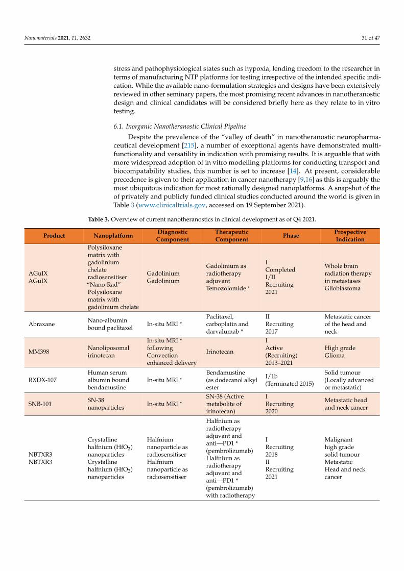
Nanomaterials 2021, 11, 2632 31 of 47
stress and pathophysiological states such as hypoxia, lending freedom to the researcher interms of manufacturing NTP platforms for testing irrespective of the intended specific indi-cation. While the available nano-formulation strategies and designs have been extensivelyreviewed in other seminary papers, the most promising recent advances in nanotheranosticdesign and clinical candidates will be considered briefly here as they relate to in vitrotesting.
6.1. Inorganic Nanotheranostic Clinical Pipeline
Despite the prevalence of the “valley of death” in nanotheranostic neuropharma-ceutical development [215], a number of exceptional agents have demonstrated multi-functionality and versatility in indication with promising results. It is arguable that withmore widespread adoption of in vitro modelling platforms for conducting transport andbiocompatability studies, this number is set to increase [14]. At present, considerableprecedence is given to their application in cancer nanotherapy [9,16] as this is arguably themost ubiquitous indication for most rationally designed nanoplatforms. A snapshot of theof privately and publicly funded clinical studies conducted around the world is given inTable 3 (www.clinicaltrials.gov, accessed on 19 September 2021).
Table 3. Overview of current nanotheranostics in clinical development as of Q4 2021.
Product Nanoplatform DiagnosticComponent
TherapeuticComponent Phase Prospective
Indication
AGuIXAGuIX
Polysiloxanematrix withgadoliniumchelateradiosensitiser“Nano-Rad”Polysiloxanematrix withgadolinium chelate
GadoliniumGadolinium
Gadolinium asradiotherapyadjuvantTemozolomide *
ICompletedI/IIRecruiting2021
Whole brainradiation therapyin metastasesGlioblastoma
Abraxane Nano-albuminbound paclitaxel In-situ MRI *
Paclitaxel,carboplatin anddarvalumab *
IIRecruiting2017
Metastatic cancerof the head andneck
MM398 Nanoliposomalirinotecan
In-situ MRI *followingConvectionenhanced delivery
Irinotecan
IActive(Recruiting)2013–2021
High gradeGlioma
RXDX-107Human serumalbumin boundbendamustine
In-situ MRI *Bendamustine(as dodecanol alkylester
I/1b(Terminated 2015)
Solid tumour(Locally advancedor metastatic)
SNB-101 SN-38nanoparticles In-situ MRI *
SN-38 (Activemetabolite ofirinotecan)
IRecruiting2020
Metastatic headand neck cancer
NBTXR3NBTXR3
Crystallinehalfnium (HfO2)nanoparticlesCrystallinehalfnium (HfO2)nanoparticles
Halfniumnanoparticle asradiosensitiserHalfniumnanoparticle asradiosensitiser
Halfnium asradiotherapyadjuvant andanti—PD1 *(pembrolizumab)Halfnium asradiotherapyadjuvant andanti—PD1 *(pembrolizumab)with radiotherapy
IRecruiting2018IIRecruiting2021
Malignanthigh gradesolid tumourMetastaticHead and neckcancer

Nanomaterials 2021, 11, 2632 32 of 47
Table 3. Cont.
Product Nanoplatform DiagnosticComponent
TherapeuticComponent Phase Prospective
Indication
Feraheme
Iron oxidenanoparticle withfunctionalisedcoating
Ferumoxytol asMRI enhancer
Macrophagepolarisation byferumoxytol *
IRecruiting 2017
Childhood brainneoplasm
Feraheme
Iron oxidenanoparticle withfunctionalisedcoating
Ferumoxytol asMRI enhancerFerumoxytol fordynamicsusceptibilitycontrast-enhancedMRIFerumoxytol forneuroinflammatoryimagingFerumoxytol andgadolinium MRI
Magnetic guidedTherapy *Gadolinium fordynamic contrastenhanced MRI/asradiosensitiser *Magnetic guidedtherapy *Magnetic guidedtherapy *
IIRecruiting2017N/A2018I (Early)CompletedIIRecruiting 2021
Brain neoplasmRecurrentchildhood brainneoplasmPrimary brainneoplasmCNS degenerativedisorderCNS infectiousdisorderCNS vascularmalformationHaemorrhagic andischaemic brainaccidentCNS neoplasmCranial nervedisorderMetastaticmalignantneoplasm in thebrain
MM398 plusFeraheme
Nanoliposomalirinotecan and ironoxidenanoparticles
Ferumoxytol Irinotecan ICompleted
Solid tumoursBreast cancer withactive brainmetastasis
CPC634Polymericmicelles ofdocetaxel
In-Situ MRI * Docetaxel ICompleted
Metastatic cancerSolid tumours
* Denotes implied or co-administered therapies or diagnostics that could be rationally designed as a consolidated nanoplatform.
In terms of rational design of nanotheranostic platforms, a number of key characteris-tics and design principles [16] should be considered from the outset in order to maximisethe probabilistic outcomes of: (1) transport across the BBB, (2) biocompatibility and (3) mea-surable concentrations that are therapeutically useful. In essence, the fundamental goalfor a formulation scientist is to design a nanotheranostic platform that uniformly crossesthe BBB in a predictive and reproducible way, localises in the target regions and tissues ofthe brain or associated tumour/embolism and elicits a therapeutic effect (ideally with asustained release profile) without causing significant accumulation or adverse effects.
Evidently, this is no trivial effort, particularly when considering the nuanced approachrequired for dose optimisation and balancing the diagnostic and therapeutic modalitiesin a homogenous and reproducible platform that is scalable to clinical settings [216]. Assuch, the in vitro modalities would frequently prove imperative to rational design ashigh-throughput and rapid screening of a platform can be conducted in inexpensive andsimplified transwell models [217]. While limited in terms of ultimate predictive power,these would at least be robust enough to permit rapid indicative assays that are reproducibleand a reliable indicator of probabilistic biocompatibility and BBB permeation.

Nanomaterials 2021, 11, 2632 33 of 47
6.2. On Current Engineering and Rational Design
Polymeric and metallic nanoplatforms constitute the most pervasive in the field for de-velopment of novel neuropharmaceuticals [16] due to the comparative lack of biomaterial-based studies. While numerous candidates, as alluded to earlier, have shown promisepreclinically in vitro, and in some settings have reached phase 2 trials, the vast number ofdiscontinued or unproven designs are a direct consequence of the “trial and error” phi-losophy [218], which has hampered research to date. Unfortunately, as a newly emergingfield, the regulatory landscape is such that rigorous characterisation and validation ofsuch elaborate nanoplatforms is mandated, yet clear guidance and approved specificationsuites are not available. Moreover, scalability and reproducibility concerns [219] are suchthat following unsatisfactory in vivo animal data, frequently the project is abandoned, orindeed the research team is forced to “start from scratch” and develop another platformwhich may meet a similar fate.
As such, what is urgently required is the ability to have rational design methodologiesthat not only improve hit probability in rapid in vitro screening in the models outlinedin this review but also facilitate reconfiguration and modular construction which can cir-cumvent commonly encountered troubleshooting in preclinical development. For instance,a commonality among the polymeric and metallic nanoparticulate systems such as den-drimers, gold and halfnium nanoparticles as well as SPIONS is that modification is limitedto chemical alteration of monomers or post-processing of a polymer [16]. This not onlylacks scope but can in numerous instances lead to compromises in terms of biocompatibility,drug encapsulation efficiency and prevention of premature release of either diagnostic ortherapeutic modality.
Due to the foregoing, a novel promising nanoplatform has been developed, known ascustomisable telodendrimers [220]. While conventional polymerics are limited in scopedue to heterogeneity due to radical or other polymerisation one-port reactions, these canbe engineered by stepwise dendrimer block co-polymer synthesis and subsequent self-assembly with exquisite control of chemistries and reproducibility on scale-up. This isparticularly useful in light of associated size-dependent limits (10 to 15 nm for AuNPsto permeate the BBB adequately [221]) for NTPs, shape-dependent permeation and therequirement for surface functionalisation and subsequent coating owing to the evolutionof the protein corona [222] in addition to the highly anionic nature of the BBB endothelialcell surface.
In this regard, polyethylene glycol, when used as the hydrophilic moiety, wouldseem to confer advantages in facilitating customisable rational design, which can beactively targeted to the brain in stable platforms [16], which become increasingly importantproperties when it is observed that at such sizes, NTPs frequently accumulate in theliver and the brain in a seemingly non-saturable fashion when administered in nakedform. While metallic NPs such as AGuIX have demonstrated good biocompatibility,accumulation on repeated administration within a chemotherapeutic regimen would bea cause of concern [223], particularly within the liver. Evidently, the ability to tailor thesize and functionalisation in addition to external device-assisted delivery such as theaforementioned magnetic-guided therapy and focused ultrasound, would invariably resultin preferential accumulation of therapeutically relevant concentrations of the NTPs acrossthe BBB within the target tissues of the brain, which could be rapidly tested in variousconfigurations and compositions in vitro to optimise the platform [224].
In their extensive nanoparticle engineering research efforts, Guo and colleagues [16]found that the best stability was obtained by using dendrimers composed of a low num-ber of hydrophobic side chains, thus minimising the occurrence of aggregation and un-favourable geometric morphology. They have successfully developed chemical synthesismethods that facilitate the introduction of amphiphilic groups and reversible cross-linkingand stimuli-responsive cleavage where appropriate [225]. These tuneable nanocarriershave numerous capabilities including enhanced encapsulation and drug loading efficiency,drug targeting and selective site-specific drug release (using pH or hypoxia responsive
Nanomaterials 2021, 11, 2632 34 of 47
labile ester functionality). In particular, cholic acid, when introduced in G2 and G3 of adendrimer, has resulted in the generation of robust micelles with self-assembly, biocompat-ibility and modular construction in that a wide array of drug binding moieties (DBMs) anddiagnostic agents can be encapsulated in a single nanoplatform, with additional surfacefunctionalisation with peptides for active targeting.
In the case of brain-specific delivery, one could envisage functionalisation with iRGDfor active targeting, encapsulation of synergistic chemotherapeutic moieties (of both ahydrophobic and hydrophilic nature by virtue of functional segregated three-layer telo-dendrimers) and a contrast agent for enhanced in situ MRI for diagnosis, monitoring andfollow-up. They have also successfully developed hybrid telodendrimers [226] that can fa-cilitate in situ protein encapsulation for advanced delivery of protein therapeutics, withoutinducing the inherent immunogenicity or protein denaturation, which has hampered theirconventional delivery. The binding affinity can be optimised based on the cargo proteincharacteristics rather than developing a nanoparticle and subsequently manipulating it toencapsulate a therapeutic protein.
In this way, in silico prediction and peptide chemistry can be fine tuned for optimiseddelivery and release. This could be further interfaced with rapid in vitro transwell assayto demonstrate permeability [227], and, due to their high stability and biocompatibility,it would be endeavoured that the nanosafety evaluation could be abbreviated, leadingto more time being allotted to optimising the nanoplatform for maximal brain delivery.Furthermore, the high drug loading capacity and permissibility to encapsulate hydrophobicand hydrophilic chemotherapies in synergistic combinations [226] would arguably reducethe effective number of nanoparticles and thus the dose of the drug (or diagnostic contrastagent) that would have to be administered.
Exemplary efforts such as these would arguably be bolstered by the available in vitromodels presented in this review for screening various formulations of such nanocarriers. Infact, there is ongoing research by Shi and colleagues [228] into the possibility to immobilisesuch telodendrimers in hydrogel resins for local controlled release depot formulations andfor engineering protein-binding dendrons in size-exclusive resins as “nano-traps” for sepsisimmune modulation, which could have potential applications in acute brain dysfunctionimmunomodulation associated with sepsis.
6.3. Unrealised Promise of Biomaterial-Inspired Rational NTP Design
Of the thus far explored nanotheranostics based on biomaterials, lipid-based systemsare the most pervasive. However, in spite of clinically available liposomal preparations ofchemotherapeutics such as doxorubicin (DOXIL), none have been approved to date for thetreatment of CNS disorders [229], which is disappointing given the obviated advanatages ofsuch nanocarriers that are capable of self-assembly and modularity with surfactant coatingsto generate “niosomes” [18]. Dependent on their surface character (zeta potential), they cancross the BBB by receptor-mediated or adsorptive-mediated transport mechanisms [230].As alluded to in previous sections, surface functionalisation with ligands such as CBSA,transferrin and glutathione or peptides such as RGD have generated potentially usefulactive-targeted nanocarriers for receptor-mediated transport across the BBB. However, innumerous instances, it is becoming increasingly clear that the advent of new technolo-gies such as phage display [231] means that cell-derived nanoparticles have unrealisedpossibilities for advanced drug delivery to the CNS.
By directly leveraging the biomaterials on cell membranes, for example, the issuesassociated with traditional synthetic preparation methods and associated heterogeneityare largely circumvented [16]. Moreover, critical interactions such as the demonstrabledeterministic nature of the evolution of the protein corona on administration [232] can bemore precisely predicted and the biomimetic platform tailored as necessary. In one studyconducted by Chen and colleagues, minor changes in lipid composition for formulatingsolid lipid nanoparticles (SLN) had a profound influence on surface charge propertiesand consequentially on biodistribution and tissue penetrance. They also determined that
Nanomaterials 2021, 11, 2632 35 of 47
apoliprotein rather than vitronectin-rich corona optimises delivery to tumour cells forcancer nanotherapy, which is mirrored by other studies that demonstrated the potential useof apolipoprotein (apoE3) surface coating [233] for preferential delivery of porphrin-lipidnanotheranostics across the BBB.
In this regard, in vitro platforms will prove indispensable for studying such nuancedinteractions, particularly where the cell models incorporate cells predominantly fromhuman origin. The more information that can be garnered in terms of the evolution ofthe protein corona, corona–nanoplatform interactions and nanoplatform protein coronainteractions with tumour cells and environments, the more customisable the platforms willbecome. There is endeavoured potential for patient-specific and tissue-specific delivery ofbiomimetic nanoplatforms [38] in the future by virtue of developing models based on ex-tracted patient cells and media to tailor therapy and minimise adverse events. There is alsoscope for hybrid nanoparticles, in which an inorganic nanoparticle can be biosynthesised,leveraged for its unique properties and made more biomimetic by using cell-based coatingand functionalisation [234].
Examples of this approach include red blood cell coating of nanoparticles [235], cancercell membrane coating [236], bacterial cell membrane coating [237] and indeed macrophage-derived coatings [238]. The latter in particular are advantageous in that they facilitateenhanced stability, circulation times, reduced immunogenicity and sustained delivery likeother cell-based delivery methods, but further augment delivery based on infiltration in re-sponse to inflammatory pathophysiology. The potential of such disease-dependent deliveryacross the BBB has been explored by various researchers for treating neuroinflammation fol-lowing acute brain injury or sepsis, HIV [35], PD and various cancer indications [239] dueto the enhanced penetrance and concentration of macrophages (with associated nanopar-ticles) in the tumour microenvironment. The in vitro models may prove fortuitous instudying macrophage interactions with drug cargo and associated issues relating to prema-ture degradation by endolysin. They can also be utilised to test various formulations andloading strategies, as well as for demonstrating the maintenance of bioactivity followingextraction and storage [240] as these are acknowledged issues limiting the scale-up andclinical translation of macrophage-mediated nanoparticle drug delivery.
7. Conclusions and Future Outlooks
While there have been notable advances in the treatment of CNS disorders due toincreasing understanding of underlying pathophysiology, improved diagnostic capabilitiesand novel therapeutic strategies, the BBB remains a critical barrier to the treatment of anumber of brain disorders, including classical neurodegenerative diseases such as AD, PDand MS, oncology, ischaemic stroke and traumatic brain injury. While nanotheranosticsare an exciting prospect in regard to targeted and personalised therapy of therapeutic andimaging modalities that are otherwise impermeable or unacceptably toxic to the CNS, theclinical translation remains elusive with high attrition rates despite a number of promisingpreclinical candidates by industrious researchers.
One of the primary reasons for this is the need to better understand the intricate andoften multi-faceted interactions of nanomaterials with biological systems highlighted byan increasing acknowledgement of the protein corona and the requirements of testingbiocompatibility. While there is an increasing trend away from the dogmatic principlesof relying on the EPR effect for nanomaterials and the properties that govern preferentialentry across the BBB, elucidation of the various receptor-mediated and transport processeshas not been entirely realised.
The other reason for a lack of translation is the issue with reproducibility and stabilityin relation to nanotheranostic design, synthesis, scale-up and testing, particularly in regardto characterising acceptable in vitro and in vivo models. For more predictive and meaning-ful studies to be conducted, coupled with an increasing desire to move away from in vivotesting with associated costs, ethical and logistical considerations along with inter-speciesdifferences potentially confounding the results of clinical data, more robust clinical models

Nanomaterials 2021, 11, 2632 36 of 47
are mandated. As has been outlined in this review, no one true model can be employed tocapture the dynamic and sophisticated nature of all aspects of the BBB, and so the selectionof model for testing potential clinical nanotheranostics in novel drug delivery strategiesrequires a practical consideration of the aim of the study and the stage of research anddevelopment.
For lead identification, in silico screening followed by high-throughput screening inmonolayer models seems the most feasible option to identify potential candidates withacceptable permeability characteristics. A co-culture or similar transwell model wouldthen seem prudent for rapid biocompatibility assessments of nanotheranostic platformsconsisting of different nanomaterials of both organic and synthetic origin, preferentially inhuman-derived or iPSC cell lines where possible. This will likely become a reality withincreasing commercial availability of standardised and validated sources of immortalisedor human-derived cells, or indeed the ready-to-use models themselves.
For lead optimisation and pre-clinical nanosafety evaluation and efficacy studies,microfluidic models including organ-on-chip and organoid models would seem to be themost acceptable choice. These lend themselves well to biorelevant studies that can capturekey aspects of the BBB, including the key contribution of shear stress forces due to bloodflow. Remarkably, advancements of such models have realised a recapitulation of notonly the intact BBB but also of modelling the disruption of the BBB inherent to severaldisease states, and thus can rapidly and dynamically facilitate assessment of the potentialof nanotheranostic platforms for acute management and therapeutic intervention in suchconditions.
These are very welcome as a formulation strategy in light of the increased focus of thepharmaceutical industry in the use of biotechnological products such as antibodies, pep-tides and siRNA for the management of CNS disorders including glioblastoma multiforme,PD and AD. By their nature, such biopharmaceuticals require robust delivery vectors thatcan protect such delicate biopayloads and selectively deliver them to the target tissues ofthe brain across the BBB, prerequisites which seem to be met uniquely by nanoparticulatedelivery systems.
Further still, it is endeavoured that brain-targeted nanotheranostic drug delivery sys-tems can answer outstanding clinical questions for which there has not been a satisfactoryanswer to date, such as disappointments in the treatment of AD and PD, as well as the acuteand chronic management of traumatic brain injury. It has been increasingly understood byresearch teams such as Piot-Grosjean and colleagues [240] that such insult to and disruptionof the BBB imposes life threatening complications in the acute phase and often debilitatingconsequences for chronic cases with poor prognosis, particularly arising from physicalsports.
Unfortunately, as of yet, and despite promising efforts such as those outlined in thisreview, there are few candidate neuroprotectants and associated delivery strategies thatproceed past the in vitro setting
The result for clinicians is a dearth of interventions for use in emergency situations tointervene at critical care points such as on admission to emergency departments for casesrelating to traumatic brain injuries and ischaemia and the associated excitotoxicity andoxidative stress [45]. Understanding such pathophysiological process using sophisticatedmodels such as organoids and microfluidics and the subsequent rational design of nanoth-erapeutic interventions could be life saving and also life changing for affected individuals.The use of validated biorelevant models of the BBB for drug discovery and formulationdevelopment will incontrovertibly improve both (1) the clinical pipeline affording a suite oftreatments at the disposal of clinicians and (2) patient outcomes, which are the pre-eminentgoal of any clinical research.
While targeted nanomaterial-mediated brain delivery across the BBB constitutes apromising direction for CNS disease treatment, to reach clinical significance, the consolida-tion of the seminary efforts by researchers in nanotechnological fields will hopefully be metby increasing availability and use of the appropriate robust modular in vitro technologies

Nanomaterials 2021, 11, 2632 37 of 47
for nanoparticle testing reviewed here. By employing validated models at the pivotal stagesof R&D to demonstrate prospective nanotheranostic biocompatibility and efficacy in vitro,increased success of clinical testing applications and reduced animal testing would beobserved. Moreover, the neuropharmaceutical industry would burgeon with a witnessedresurgence of an expedited and cost-effective drug development pipeline. This would un-deniably garner increased regulatory approvals of novel multifunctional therapeutics withthe potential to revolutionise diagnostics and personalised therapeutics for neurologicaldisorders in an ageing worldwide population.
Perhaps these models additionally offer a crucial beacon of hope in the contentiousongoing debate on the merit of “nanomedicine” in the context of advanced drug delivery.While the use of animal models has been the mainstay of clinical R&D, perhaps, as observedhere, the expediency and efficiency of data collection generated from validated in vitromodels can also serve to reduce and in the future potentially eliminate the requirementof laborious animal testing. Moreover, with exciting prospects in nano-engineering andself-assembly of rationally designed nanoplatforms, it would be hoped that abbreviatedcharacterisation and troubleshooting would be observed. The direct result would be moretime and resources being allocated by researchers to optimising the delivery across theBBB in preclinical testing to identify clinical candidates and a greater number of clinicaltrials to pave the way for nanomaterial-based CNS theranostics. The precedent set bysuch rationally designed therapies would constitute a basis upon which further agentscould enter the pipeline and offset the appreciably high attrition rate and translational gapsobserved in the field at the present time.
As exemplified by notable clinical candidates in the pipeline and approved nanomedi-cal interventions to date, the novel phenomena that nanotheranostic interventions exhibitare not readily tested reproducibly in animals, and scale-up issues, as aforementioned,render the data garnered indeterminate in relation to potential human use. Rather, pilotstudies in animals can be prioritised at early lead optimisation stages where warrantedand justified, while in vitro and indeed in silico modelling approaches should become theobvious choice for expediting development and clinical testing.
Rather than observing this as an expensive and uncharted ground in drug researchefforts, as presented, the increasing availability of validated and commercially availablemodels should be viewed by researchers as an exciting next chapter in the ongoing searchfor cutting edge nanotechnology driven drug delivery systems in the wider context ofpersonalised medicine. By reducing the number of animals being used in animal testing infavour of these in vitro approaches, a number of ethical, economical and data potentialityissues can be simultaneously consolidated generating platforms for nanotheranostic drugdevelopment with bolstered clinical potential for improving the treatment outcomes inCNS disorders. Solving the conundrum posed by crossing the robust BBB is best envisagedas a composite of a series of nano-discoveries using novel testing strategies outlined herein,rather than hoping for a macro-sized breakthrough garnered from repetitive and unreliabletesting in the classical settings, which, to date, have not produced any promising results.To continue in this vein would seem to be a missed opportunity and in a sense potentiallycompromise the viability of nanomedical research, which would be an inexpiable develop-ment given the untold potential of such therapeutics and in light of the exemplary effortsof research teams worldwide for improving population quality of life and longevity.
Author Contributions: Conceptualization, M.J.L. and O.L.G.; methodology, M.J.L. and O.L.G.;investigation, M.J.L. and O.L.G.; writing—original draft preparation, M.J.L.; writing—review andediting, O.L.G.; visualization, M.J.L.; supervision, O.L.G.; project administration, O.L.G.; fundingacquisition, O.L.G. All authors have read and agreed to the published version of the manuscript.
Funding: This project has received funding from the European Union’s Horizon 2020 research andinnovation programme under grant agreement No. 952259.
Acknowledgments: The authors of this publication wish to thank the Trinity College Dublin Schoolof Pharmacy for their continued support and contribution to all work discussed herein. They
Nanomaterials 2021, 11, 2632 38 of 47
would also like to acknowledge the work of all researchers included in this review, and to ElsevierPublications and Richard Kollath for the reproduction of figures.
Conflicts of Interest: The authors declare no conflict of interest. The funders had no role in the designof the study; in the collection, analyses, or interpretation of data; in the writing of the manuscript, orin the decision to publish the results.
References1. Howes, O.D.; Mehta, M.A. Challenges in CNS drug development and the role of imaging. Psychopharmacology 2021, 238,
1229–1230. [CrossRef]2. Karami, Z.; Zanjani, M.R.S.; Hamidi, M. Nanoemulsions in CNS drug delivery: Recent developments, impacts and challenges.
Drug Discov. Today 2019, 24, 1104–1115. [CrossRef]3. Vos, T.; Lim, S.S.; Abbafati, C.; Abbas, K.M.; Abbasi, M.; Abbasifard, M.; Abbasi-Kangevari, M.; Abbastabar, H.; Abd-Allah, F.;
Abdelalim, A.; et al. Global burden of 369 diseases and injuries in 204 countries and territories, 1990–2019: A systematic analysisfor the Global Burden of Disease Study 2019. Lancet 2020, 396, 1204–1222. [CrossRef]
4. Feigin, V.L.; Nichols, E.; Alam, T.; Bannick, M.S.; Beghi, E.; Blake, N.; Culpepper, W.J.; Dorsey, E.R.; Elbaz, A.; Ellenbogen, R.G.;et al. Global, regional, and national burden of neurological disorders, 1990–2016: A systematic analysis for the Global Burden ofDisease Study 2016. Lancet Neurol. 2019, 18, 459–480. [CrossRef]
5. Borton, D.A.; Dawes, H.E.; Worrell, G.A.; Starr, P.A.; Denison, T.J. Developing Collaborative Platforms to Advance Neurotechnol-ogy and Its Translation. Neuron 2020, 108, 286–301. [CrossRef] [PubMed]
6. Bors, L.A.; Erdo, F. Overcoming the Blood–Brain Barrier. Challenges and Tricks for CNS Drug Delivery. Sci. Pharm. 2019, 87, 6.[CrossRef]
7. Xie, J.; Shen, Z.; Anraku, Y.; Kataoka, K.; Chen, X. Nanomaterial-based blood-brain-barrier (BBB) crossing strategies. Biomaterials2019, 224, 119491. [CrossRef] [PubMed]
8. Wong, H.L.; Wu, X.Y.; Bendayan, R. Nanotechnological advances for the delivery of CNS therapeutics. Adv. Drug Deliv. Rev. 2012,64, 686–700. [CrossRef]
9. Rashid, M.; Ahmad, Q.Z. Tajuddin Trends in Nanotechnology for Practical Applications. In Applications of Targeted Nano Drugsand Delivery Systems; Elsevier: Amsterdam, The Netherlands, 2019. [CrossRef]
10. Shi, Y.; Van Der Meel, R.; Chen, X.; Lammers, T. The EPR effect and beyond: Strategies to improve tumor targeting and cancernanomedicine treatment efficacy. Theranostics 2020, 10, 7921–7924. [CrossRef] [PubMed]
11. Wong, X.Y.; Sena-Torralba, A.; Álvarez-Diduk, R.; Muthoosamy, K.; Merkoçi, A. Nanomaterials for Nanotheranostics: TuningTheir Properties According to Disease Needs. ACS Nano 2020, 14, 2585–2627. [CrossRef] [PubMed]
12. Shi, J.; Xiao, Z.; Kamaly, N.; Farokhzad, O.C. Self-Assembled Targeted Nanoparticles: Evolution of Technologies and Bench toBedside Translation. Acc. Chem. Res. 2011, 44, 1123–1134. [CrossRef] [PubMed]
13. O’Leary, D.E. Gartner’s hype cycle and information system research issues. Int. J. Account. Inf. Syst. 2008, 9, 240–252. [CrossRef]14. Alexander, A.A.; Jotterand, F. Market Considerations for Nanomedicines. In Cancer Theranostics; Academic Press: Cambridge,
MA, USA, 2014; pp. 471–491. [CrossRef]15. Selvan, S.T.; Narayanan, K. Introduction to Nanotheranostics; Springer: Singapore, 2016. [CrossRef]16. Guo, D.; Ji, X.; Luo, J. Rational nanocarrier design towards clinical translation of cancer nanotherapy. Biomed. Mater. 2021, 16,
032005. [CrossRef] [PubMed]17. Wu, Q.; Miao, W.-S.; Zhang, Y.-D.; Gao, H.-J.; Hui, D. Mechanical properties of nanomaterials: A review. Nanotechnol. Rev. 2020, 9,
259–273. [CrossRef]18. Gharbavi, M.; Amani, J.; Kheiri-Manjili, H.; Danafar, H.; Sharafi, A. Niosome: A Promising Nanocarrier for Natural Drug Delivery
through Blood-Brain Barrier. Adv. Pharmacol. Sci. 2018, 2018, 6847971. [CrossRef] [PubMed]19. Kuo, A.; Smith, M.T. Theoretical and practical applications of the intracerebroventricular route for CSF sampling and drug
administration in CNS drug discovery research: A mini review. J. Neurosci. Methods 2014, 233, 166–171. [CrossRef] [PubMed]20. Upadhyay, R.K. Drug Delivery Systems, CNS Protection, and the Blood Brain Barrier. BioMed Res. Int. 2014, 2014, 869269.
[CrossRef]21. Sharma, M.; Dube, T.; Chibh, S.; Kour, A.; Mishra, J.; Panda, J.J. Nanotheranostics, a future remedy of neurological disorders.
Expert Opin. Drug Deliv. 2019, 16, 113–128. [CrossRef] [PubMed]22. Anselmo, A.C.; Mitragotri, S. Nanoparticles in the clinic: An update. Bioeng. Transl. Med. 2019, 4, e10143. [CrossRef] [PubMed]23. Caracciolo, G.; Farokhzad, O.C.; Mahmoudi, M. Biological Identity of Nanoparticles In Vivo: Clinical Implications of the Protein
Corona. Trends Biotechnol. 2017, 35, 257–264. [CrossRef]24. Zhou, Y.; Dai, Z. New Strategies in the Design of Nanomedicines to Oppose Uptake by the Mononuclear Phagocyte System and
Enhance Cancer Therapeutic Efficacy. Chem. Asian J. 2018, 13, 3333–3340. [CrossRef] [PubMed]25. Bazak, R.; Houri, M.; El Achy, S.; Hussein, W.; Refaat, T. Passive targeting of nanoparticles to cancer: A comprehensive review of
the literature. Mol. Clin. Oncol. 2014, 2, 904–908. [CrossRef] [PubMed]26. Strazielle, N.; Ghersi-Egea, J.-F. Potential Pathways for CNS Drug Delivery Across the Blood-Cerebrospinal Fluid Barrier. Curr.
Pharm. Des. 2016, 22, 5463–5476. [CrossRef]27. Dyrna, F.; Hanske, S.; Krueger, M.; Bechmann, I. The Blood-Brain Barrier. J. Neuroimmune Pharmacol. 2013, 8, 763–773. [CrossRef]

Nanomaterials 2021, 11, 2632 39 of 47
28. Berndt, P.; Winkler, L.; Cording, J.; Breitkreuz-Korff, O.; Rex, A.; Dithmer, S.; Rausch, V.; Blasig, R.; Richter, M.; Sporbert, A.; et al.Tight junction proteins at the blood–brain barrier: Far more than claudin-5. Cell. Mol. Life Sci. 2019, 76, 1987–2002. [CrossRef]
29. Kim, J.; Ahn, S.I.; Kim, Y. Nanotherapeutics engineered to cross the blood-brain barrier for advanced drug delivery to the centralnervous system. J. Ind. Eng. Chem. 2019, 73, 8–18. [CrossRef] [PubMed]
30. MacDougall, M.R.; Sharma, S. Physiology, Chemoreceptor Trigger Zone. StatPearls [Internet]. 2020. Available online:https://www.ncbi.nlm.nih.gov/books/NBK537133/ (accessed on 29 April 2021).
31. Shen, S.; Zhang, W. ABC Transporters and Drug Efflux at the Blood-Brain Barrier. Rev. Neurosci. 2010, 21, 29–53. [CrossRef][PubMed]
32. Eisenblätter, T.; Hüwel, S.; Galla, H.-J. Characterisation of the brain multidrug resistance protein (BMDP/ABCG2/BCRP)expressed at the blood–brain barrier. Brain Res. 2003, 971, 221–231. [CrossRef]
33. Charabati, M.; Rabanel, J.-M.; Ramassamy, C.; Prat, A. Overcoming the Brain Barriers: From Immune Cells to Nanoparticles.Trends Pharmacol. Sci. 2020, 41, 42–54. [CrossRef] [PubMed]
34. Chen, K.-T.; Wei, K.-C.; Liu, H.-L. Theranostic Strategy of Focused Ultrasound Induced Blood-Brain Barrier Opening for CNSDisease Treatment. Front. Pharmacol. 2019, 10, 86. [CrossRef]
35. Choi, M.R.; Bardhan, R.; Stanton-Maxey, K.J.; Badve, S.; Nakshatri, H.; Stantz, K.M.; Cao, N.; Halas, N.; Clare, S.E. Delivery ofnanoparticles to brain metastases of breast cancer using a cellular Trojan horse. Cancer Nanotechnol. 2012, 3, 47–54. [CrossRef][PubMed]
36. Casaos, J.; Gorelick, N.; Tyler, B. Neurosurgical Implant-Based Strategy for Brain Cancer Therapy. In Nanotherapy for Brain TumorDrug Delivery; Humana: New York, NY, USA, 2021; pp. 225–244. [CrossRef]
37. Viderman, D.; Brotfain, E.; Bilotta, F.; Zhumadilov, A. Risk Factors and Mechanisms of Postoperative Delirium after IntracranialNeurosurgical Procedures. Asian J. Anesthesiol. 2020, 2020, 1–9. [CrossRef]
38. Kumar, A.; Chaudhary, R.K.; Singh, R.; Singh, S.P.; Wang, S.-Y.; Hoe, Z.-Y.; Pan, C.-T.; Shiue, Y.-L.; Wei, D.-Q.; Kaushik, A.C.; et al.Nanotheranostic Applications for Detection and Targeting Neurodegenerative Diseases. Front. Neurosci. 2020, 14, 305. [CrossRef][PubMed]
39. Kanwar, J.R.; Sriramoju, B.; Kanwar, R.K. Neurological disorders and therapeutics targeted to surmount the blood–brain barrier.Int. J. Nanomed. 2012, 7, 3259–3278. [CrossRef]
40. Gklinos, P.; Papadopoulou, M.; Stanulovic, V.; Mitsikostas, D.; Papadopoulos, D. Monoclonal Antibodies as NeurologicalTherapeutics. Pharmaceuticals 2021, 14, 92. [CrossRef] [PubMed]
41. Hu, X.; Leak, R.; Thomson, A.W.; Yu, F.; Xia, Y.; Wechsler, L.R.; Chen, J. Promises and limitations of immune cell-based therapiesin neurological disorders. Nat. Rev. Neurol. 2018, 14, 559–568. [CrossRef]
42. Zottel, A.; Paska, A.V.; Jovcevska, I. Nanotechnology Meets Oncology: Nanomaterials in Brain Cancer Research, Diagnosis andTherapy. Materials 2019, 12, 1588. [CrossRef] [PubMed]
43. Chodobski, A.; Zink, B.J.; Szmydynger-Chodobska, J. Blood–Brain Barrier Pathophysiology in Traumatic Brain Injury. Transl.Stroke Res. 2011, 2, 492–516. [CrossRef] [PubMed]
44. Campbell, M.; Hanrahan, F.; Gobbo, O.; Kelly, M.E.; Kiang, A.-S.; Humphries, M.M.; Nguyen, A.T.; Ozaki, E.; Keaney, J.;Blau, C.W.; et al. Targeted suppression of claudin-5 decreases cerebral oedema and improves cognitive outcome followingtraumatic brain injury. Nat. Commun. 2012, 3, 849. [CrossRef] [PubMed]
45. Liao, R.; Wood, T.R.; Nance, E. Nanotherapeutic modulation of excitotoxicity and oxidative stress in acute brain injury.Nanobiomedicine 2020, 7, 1849543520970819. [CrossRef]
46. Torrice, M. Does Nanomedicine Have a Delivery Problem? ACS Cent. Sci. 2016, 2, 434–437. [CrossRef] [PubMed]47. Aldape, K.; Brindle, K.M.; Chesler, L.; Chopra, R.; Gajjar, A.; Gilbert, M.R.; Gottardo, N.; Gutmann, D.; Hargrave, D.; Holland, E.C.;
et al. Challenges to curing primary brain tumours. Nat. Rev. Clin. Oncol. 2019, 16, 509–520. [CrossRef] [PubMed]48. Abbott, N.J. Blood–brain barrier structure and function and the challenges for CNS drug delivery. J. Inherit. Metab. Dis. 2013, 36,
437–449. [CrossRef] [PubMed]49. Pamies, D.; Hartung, T.; Hogberg, H.T. Biological and medical applications of a brain-on-a-chip. Exp. Biol. Med. 2014, 239,
1096–1107. [CrossRef] [PubMed]50. Olsson, I.A.S.; da Silva, S.P.; Townend, D.; Sandøe, P. Protecting Animals and Enabling Research in the European Union: An
Overview of Development and Implementation of Directive 2010/63/EU. ILAR J. 2017, 57, 347–357. [CrossRef]51. Lacombe, O.; Videau, O.; Chevillon, D.; Guyot, A.-C.; Contreras, C.; Blondel, S.; Nicolas, L.; Ghettas, A.; Bénech, H.; Thévenot, E.A.;
et al. In Vitro Primary Human and Animal Cell-Based Blood−Brain Barrier Models as a Screening Tool in Drug Discovery. Mol.Pharm. 2011, 8, 651–663. [CrossRef] [PubMed]
52. Gumbleton, M.; Audus, K.L. Progress and limitations in the use of in vitro cell cultures to serve as a permeability screen for theblood-brain barrier. J. Pharm. Sci. 2001, 90, 1681–1698. [CrossRef]
53. Bagchi, S.; Chhibber, T.; Lahooti, B.; Verma, A.; Borse, V.; Jayant, R.D. In-vitro blood-brain barrier models for drug screening andpermeation studies: An overview. Drug Des. Dev. Ther. 2019, 13, 3591–3605. [CrossRef]
54. Oddo, A.; Peng, B.; Tong, Z.; Wei, Y.; Tong, W.Y.; Thissen, H.; Voelcker, N.H. Advances in Microfluidic Blood–Brain Barrier (BBB)Models. Trends Biotechnol. 2019, 37, 1295–1314. [CrossRef] [PubMed]
55. Bergmann, S.; Lawler, S.E.; Qu, Y.; Fadzen, C.M.; Wolfe, J.M.; Regan, M.S.; Pentelute, B.L.; Agar, N.Y.R.; Cho, C.-F. Blood–brain-barrier organoids for investigating the permeability of CNS therapeutics. Nat. Protoc. 2018, 13, 2827–2843. [CrossRef]

Nanomaterials 2021, 11, 2632 40 of 47
56. Wang, X.; Hou, Y.; Ai, X.; Sun, J.; Xu, B.; Meng, X.; Zhang, Y.; Zhang, S. Potential applications of microfluidics based blood brainbarrier (BBB)-on-chips for in vitro drug development. Biomed. Pharmacother. 2020, 132, 110822. [CrossRef]
57. Ng, T.S.; Garlin, M.A.; Weissleder, R.; Miller, M.A. Improving nanotherapy delivery and action through image-guided systemspharmacology. Theranostics 2020, 10, 968–997. [CrossRef]
58. Aalinkeel, R.; Kutscher, H.L.; Singh, A.; Cwiklinski, K.; Khechen, N.; Schwartz, S.A.; Prasad, P.N.; Mahajan, S.D. Neuroprotectiveeffects of a biodegradable poly(lactic-co-glycolic acid)-ginsenoside Rg3 nanoformulation: A potential nanotherapy for Alzheimer’sdisease? J. Drug Target. 2017, 26, 182–193. [CrossRef]
59. Irudayanathan, F.J.; Trasatti, J.P.; Karande, P.; Nangia, S. Molecular Architecture of the Blood Brain Barrier Tight JunctionProteins—A Synergistic Computational and In Vitro Approach. J. Phys. Chem. B 2016, 120, 77–88. [CrossRef] [PubMed]
60. Wang, Z.; Yang, H.; Wu, Z.; Wang, T.; Li, W.; Tang, Y.; Liu, G. In Silico Prediction of Blood–Brain Barrier Permeability ofCompounds by Machine Learning and Resampling Methods. ChemMedChem 2018, 13, 2189–2201. [CrossRef] [PubMed]
61. Yang, S.; Mei, S.; Jin, H.; Zhu, B.; Tian, Y.; Huo, J.; Cui, X.; Guo, A.; Zhao, Z. Identification of two immortalized cell lines, ECV304and bEnd3, for in vitro permeability studies of blood-brain barrier. PLoS ONE 2017, 12, e0187017. [CrossRef]
62. Ivask, A.; Pilkington, E.H.; Blin, T.; Kakinen, A.; Vija, H.; Visnapuu, M.; Quinn, J.F.; Whittaker, M.R.; Qiao, R.; Davis, T.P.; et al.Uptake and transcytosis of functionalized superparamagnetic iron oxide nanoparticles in an in vitro blood brain barrier model.Biomater. Sci. 2017, 6, 314–323. [CrossRef]
63. Appelt-Menzel, A.; Oerter, S.; Mathew, S.; Haferkamp, U.; Hartmann, C.; Jung, M.; Neuhaus, W.; Pless, O. Human iPSC-DerivedBlood-Brain Barrier Models: Valuable Tools for Preclinical Drug Discovery and Development? Curr. Protoc. Stem Cell Biol. 2020,55, 122. [CrossRef] [PubMed]
64. Dong, X. Current strategies for brain drug delivery. Theranostics 2018, 8, 1481. [CrossRef] [PubMed]65. Caffery, B.; Lee, J.S.; Alexander-Bryant, A.A. Vectors for Glioblastoma Gene Therapy: Viral & Non-Viral Delivery Strategies.
Nanomaterials 2019, 9, 105. [CrossRef]66. Dal Magro, R.; Ornaghi, F.; Cambianica, I.; Beretta, S.; Re, F.; Musicanti, C.; Rigolio, R.; Donzelli, E.; Canta, A.R.; Ballarini, E.; et al.
ApoE-modified solid lipid nanoparticles: A feasible strategy to cross the blood-brain barrier. J. Control. Release 2017, 249, 103–110.[CrossRef] [PubMed]
67. Das, D.; Lin, S. Double-Coated Poly (Butylcynanoacrylate) Nanoparticulate Delivery Systems for Brain Targeting of Dalargin viaOral Administration. J. Pharm. Sci. 2005, 94, 1343–1353. [CrossRef]
68. Tam, V.H.; Sosa, C.; Liu, R.; Yao, N.; Priestley, R.D. Nanomedicine as a non-invasive strategy for drug delivery across the bloodbrain barrier. Int. J. Pharm. 2016, 515, 331–342. [CrossRef]
69. Sharma, G.; Sharma, A.R.; Lee, S.-S.; Bhattacharya, M.; Nam, J.-S.; Chakraborty, C. Advances in nanocarriers enabled braintargeted drug delivery across blood brain barrier. Int. J. Pharm. 2019, 559, 360–372. [CrossRef]
70. Lu, W.; Tan, Y.-Z.; Hu, K.-L.; Jiang, X.-G. Cationic albumin conjugated pegylated nanoparticle with its transcytosis ability andlittle toxicity against blood–brain barrier. Int. J. Pharm. 2005, 295, 247–260. [CrossRef] [PubMed]
71. Kesharwani, P.; Jain, A.; Jain, A.; Jain, A.K.; Garg, N.K.; Tekade, R.K.; Singh, T.R.R.; Iyer, A.K. Cationic bovine serum albumin(CBA) conjugated poly lactic-co-glycolic acid (PLGA) nanoparticles for extended delivery of methotrexate into brain tumors. RSCAdv. 2016, 6, 89040–89050. [CrossRef]
72. Liu, L.; Guo, K.; Lu, J.; Venkatraman, S.S.; Luo, D.; Ng, K.C.; Ling, E.-A.; Moochhala, S.; Yang, Y.-Y. Biologically active core/shellnanoparticles self-assembled from cholesterol-terminated PEG–TAT for drug delivery across the blood–brain barrier. Biomaterials2008, 29, 1509–1517. [CrossRef] [PubMed]
73. Raval, N.; Mistry, T.; Acharya, N.; Acharya, S. Development of glutathione-conjugated asiatic acid-loaded bovine serum albuminnanoparticles for brain-targeted drug delivery. J. Pharm. Pharmacol. 2015, 67, 1503–1511. [CrossRef] [PubMed]
74. Michaelis, M.; Hoffmann, M.M.; Dreis, S.; Herbert, E.; Alyautdin, R.; Kreuter, J.; Langer, K. Covalent Linkage of ApolipoproteinE to Albumin Nanoparticles Strongly Enhances Drug Transport into the Brain. J. Pharmacol. Exp. Ther. 2006, 317, 1246–1253.[CrossRef]
75. Georgieva, J.V.; Kalicharan, D.; Couraud, P.-O.; Romero, I.; Weksler, B.; Hoekstra, D.; Zuhorn, I. Surface Characteristics ofNanoparticles Determine Their Intracellular Fate in and Processing by Human Blood–Brain Barrier Endothelial Cells In Vitro.Mol. Ther. 2011, 19, 318–325. [CrossRef] [PubMed]
76. Zhao, X.; Shang, T.; Zhang, X.; Ye, T.; Wang, D.; Rei, L. Passage of Magnetic Tat-Conjugated Fe3O4@SiO2 Nanoparticles Across InVitro Blood-Brain Barrier. Nanoscale Res. Lett. 2016, 11, 451. [CrossRef]
77. Mabrouk, O.S.; Falk, T.; Sherman, S.J.; Kennedy, R.T.; Polt, R. CNS penetration of the opioid glycopeptide MMP-2200: A micro-dialysis study. Neurosci. Lett. 2012, 531, 99–103. [CrossRef]
78. Lv, H.; Zhang, S.; Wang, B.; Cui, S.; Yan, J. Toxicity of cationic lipids and cationic polymers in gene delivery. J. Control. Release2006, 114, 100–109. [CrossRef] [PubMed]
79. Ingusci, S.; Verlengia, G.; Soukupova, M.; Zucchini, S.; Simonato, M. Gene Therapy Tools for Brain Diseases. Front. Pharmacol.2019, 10, 724. [CrossRef] [PubMed]
80. Zong, T.; Mei, L.; Gao, H.; Cai, W.; Zhu, P.; Shi, K.; Chen, J.; Wang, Y.; Gao, F.; He, Q. Synergistic Dual-Ligand DoxorubicinLiposomes Improve Targeting and Therapeutic Efficacy of Brain Glioma in Animals. Mol. Pharm. 2014, 11, 2346–2357. [CrossRef][PubMed]

Nanomaterials 2021, 11, 2632 41 of 47
81. Johnsen, K.B.; Burkhart, A.; Thomsen, L.B.; Andresen, T.L.; Moos, T. Targeting the transferrin receptor for brain drug delivery.Prog. Neurobiol. 2019, 181, 101665. [CrossRef] [PubMed]
82. Kumari, S.; Ahsan, S.M.; Kumar, J.M.; Kondapi, A.K.; Rao, N.M. Overcoming blood brain barrier with a dual purpose Temozolo-mide loaded Lactoferrin nanoparticles for combating glioma (SERP-17-12433). Sci. Rep. 2017, 7, 6602. [CrossRef] [PubMed]
83. Kang, Y.-S.; Jung, H.-J.; Oh, J.-S.; Song, D.-Y. Use of PEGylated Immunoliposomes to Deliver Dopamine across the Blood-BrainBarrier in a Rat Model of Parkinson’s Disease. CNS Neurosci. Ther. 2016, 22, 817–823. [CrossRef] [PubMed]
84. Gao, J.-Q.; Lv, Q.; Li, L.-M.; Tang, X.-J.; Li, F.-Z.; Hu, Y.-L.; Han, M. Glioma targeting and blood–brain barrier penetration bydual-targeting doxorubincin liposomes. Biomaterials 2013, 34, 5628–5639. [CrossRef]
85. Nam, L.; Coll, C.; Erthal, L.C.S.; De La Torre, C.; Serrano, D.; Martínez-Máñez, R.; Santos-Martínez, M.J.; Ruiz-Hernández, E. DrugDelivery Nanosystems for the Localized Treatment of Glioblastoma Multiforme. Materials 2018, 11, 779. [CrossRef] [PubMed]
86. Tosi, G.; Pederzoli, F.; Belletti, D.; Vandelli, M.A.; Forni, F.; Duskey, J.T.; Ruozi, B. Nanomedicine in Alzheimer’s disease: Amyloidbeta targeting strategy. Prog. Brain Res. 2019, 245, 57–88. [CrossRef]
87. Johnsen, K.B.; Bak, M.; Kempen, P.J.; Melander, F.; Burkhart, A.; Thomsen, M.S.; Nielsen, M.S.; Moos, T.; Andresen, T.L. Antibodyaffinity and valency impact brain uptake of transferrin receptor-targeted gold nanoparticles. Theranostics 2018, 8, 3416. [CrossRef]
88. Sonoda, H.; Morimoto, H.; Yoden, E.; Koshimura, Y.; Kinoshita, M.; Golovina, G.; Takagi, H.; Yamamoto, R.; Minami, K.;Mizoguchi, A.; et al. A Blood-Brain-Barrier-Penetrating Anti-human Transferrin Receptor Antibody Fusion Protein for Neurono-pathic Mucopolysaccharidosis II. Mol. Ther. 2018, 26, 1366–1374. [CrossRef] [PubMed]
89. Kida, S.; Kinoshita, M.; Tanaka, S.; Okumura, M.; Koshimura, Y.; Morimoto, H. Non-clinical evaluation of a blood-brainbarrier-penetrating enzyme for the treatment of mucopolysaccharidosis type I. Mol. Genet. Metab. 2019, 126, S83–S84. [CrossRef]
90. Zhang, W.; Liu, Q.Y.; Haqqani, A.S.; Leclerc, S.; Liu, Z.; Fauteux, F.; Baumann, E.; Delaney, C.E.; Ly, D.; Star, A.T.; et al. Differentialexpression of receptors mediating receptor-mediated transcytosis (RMT) in brain microvessels, brain parenchyma and peripheraltissues of the mouse and the human. Fluids Barriers CNS 2020, 17, 47. [CrossRef]
91. Dehouck, B.; Fenart, L.; Dehouck, M.-P.; Pierce, A.; Torpier, G.; Cecchelli, R. A New Function for the LDL Receptor: Transcytosisof LDL across the Blood–Brain Barrier. J. Cell Biol. 1997, 138, 877–889. [CrossRef]
92. Laskowitz, D.T.; Horsburgh, K.; Roses, A.D. Apolipoprotein E and the CNS response to injury. J. Cere. Blood Flow Metab. 1998, 18,465–471. [CrossRef]
93. Kim, H.R.; Andrieux, K.; Gil, S.; Taverna, M.; Chacun, H.; Desmaële, D.; Taran, F.; Georgin, D.; Couvreur, P. Translocation ofPoly(ethylene glycol-co-hexadecyl)cyanoacrylate Nanoparticles into Rat Brain Endothelial Cells: Role of Apolipoproteins inReceptor-Mediated Endocytosis. Biomacromolecules 2007, 8, 793–799. [CrossRef]
94. Hartl, N.; Adams, F.; Merkel, O.M. From Adsorption to Covalent Bonding: Apolipoprotein E Functionalization of PolymericNanoparticles for Drug Delivery across the Blood–Brain Barrier. Adv. Ther. 2021, 4, 2000092. [CrossRef] [PubMed]
95. Kafa, H.; Wang, J.T.-W.; Rubio, N.; Klippstein, R.; Costa, P.M.; Hassan, H.A.; Sosabowski, J.K.; Bansal, S.S.; Preston, J.E.;Abbott, N.J.; et al. Translocation of LRP1 targeted carbon nanotubes of different diameters across the blood–brain barrier in vitroand in vivo. J. Control. Release 2016, 225, 217–229. [CrossRef] [PubMed]
96. Ball, K.; Bouzom, F.; Scherrmann, J.-M.; Walther, B.; Decleves, X. Physiologically Based Pharmacokinetic Modelling of DrugPenetration Across the Blood–Brain Barrier—Towards a Mechanistic IVIVE-Based Approach. AAPS J. 2013, 15, 913–932. [CrossRef][PubMed]
97. Uchida, Y.; Ohtsuki, S.; Katsukura, Y.; Ikeda, C.; Suzuki, T.; Kamiie, J.; Terasaki, T. Quantitative targeted absolute proteomics ofhuman blood-brain barrier transporters and receptors. J. Neurochem. 2011, 117, 333–345. [CrossRef]
98. di Polidoro, A.C.; Zambito, G.; Haeck, J.; Mezzanotte, L.; Lamfers, M.; Netti, P.; Torino, E. Theranostic Design of Angiopep-2Conjugated Hyaluronic Acid Nanoparticles (Thera-ANG-cHANPs) for Dual Targeting and Boosted Imaging of Glioma Cells.Cancers 2021, 13, 503. [CrossRef]
99. Engin, A.B.; Engin, A. Nanoparticles and neurotoxicity: Dual response of glutamatergic receptors. Prog. Brain Res. 2019, 245,281–303. [CrossRef]
100. Jensen, R.L.; Chkheidze, R. The Role of Glucose Transporter-1 (GLUT-1) in Malignant Gliomas. In Tumours of the Central NervousSystem; Springer: Dordrecht, The Netherlands, 2011; Volume 1, pp. 99–108. [CrossRef]
101. Wu, T.-T.; Zhou, S.-H. Nanoparticle-Based Targeted Therapeutics in Head-And-Neck Cancer. Int. J. Med. Sci. 2015, 12, 187–200.[CrossRef]
102. Pardridge, W.M.; Kang, Y.-S.; Buciak, J.L.; Yang, J. Human insulin receptor monoclonal antibody undergoes high affinity bindingto human brain capillaries in vitro and rapid transcytosis through the blood-brain barrier in vivo in the primate. Pharm. Res.1995, 12, 807–816. [CrossRef] [PubMed]
103. Ulbrich, K.; Knobloch, T.; Kreuter, J. Targeting the insulin receptor: Nanoparticles for drug delivery across the blood–brain barrier(BBB). J. Drug Target. 2010, 19, 125–132. [CrossRef] [PubMed]
104. McCord, E.; Pawar, S.; Koneru, T.; Tatiparti, K.; Sau, S.; Iyer, A.K. Folate Receptors’ Expression in Gliomas May Possess PotentialNanoparticle-Based Drug Delivery Opportunities. ACS Omega 2021, 6, 4111–4118. [CrossRef] [PubMed]
105. Wang, B.; Lv, L.; Wang, Z.; Zhao, Y.; Wu, L.; Fang, X.; Xu, Q.; Xin, H. Nanoparticles functionalized with Pep-1 as potential gliomatargeting delivery system via interleukin 13 receptor α2-mediated endocytosis. Biomaterials 2014, 35, 5897–5907. [CrossRef][PubMed]

Nanomaterials 2021, 11, 2632 42 of 47
106. Cai, X.; Bandla, A.; Chuan, C.K.; Magarajah, G.; Liao, L.-D.; Teh, D.B.L.; Kennedy, B.K.; Thakor, N.V.; Liu, B. Identifyingglioblastoma margins using dual-targeted organic nanoparticles for efficient in vivo fluorescence image-guided photothermaltherapy. Mater. Horiz. 2018, 6, 311–317. [CrossRef]
107. Peng, L.; Liang, Y.; Zhong, X.; Liang, Z.; Tian, Y.; Li, S.; Liang, J.; Wang, R.; Zhong, Y.; Shi, Y.; et al. Aptamer-Conjugated GoldNanoparticles Targeting Epidermal Growth Factor Receptor Variant III for the Treatment of Glioblastoma. Int. J. Nanomed. 2020,15, 1363–1372. [CrossRef] [PubMed]
108. Vashist, A.; Atluri, V.; Raymond, A.; Kaushik, A.; Parira, T.; Huang, Z.; Durygin, A.; Tomitaka, A.; Nikkhah-Moshaie, R.;Vashist, A.; et al. Development of Multifunctional Biopolymeric Auto-Fluorescent Micro- and Nanogels as a Platform forBiomedical Applications. Front. Bioeng. Biotechnol. 2020, 8, 315. [CrossRef]
109. Salimi, H.; Klein, R.S. Disruption of the Blood-Brain Barrier during Neuroinflammatory and Neuroinfectious Diseases. InNeuroimmune Diseases: From Cells to the Living Brain; Springer: Cham, Switzerland, 2019; pp. 195–234. [CrossRef]
110. Åslund, A.K.; Berg, S.; Hak, S.; Mørch, Ý.; Torp, S.H.; Sandvig, A.; Widerøe, M.; Hansen, R.; de Lange Davies, C. Nanoparticledelivery to the brain—By focused ultrasound and self-assembled nanoparticle-stabilized microbubbles. J. Control. Release 2015,220, 287–294. [CrossRef] [PubMed]
111. Mainprize, T.; Lipsman, N.; Huang, Y.; Meng, Y.; Bethune, A.; Ironside, S.; Heyn, C.; Alkins, R.; Trudeau, M.; Sahgal, A.; et al.Blood-Brain Barrier Opening in Primary Brain Tumors with Non-invasive MR-Guided Focused Ultrasound: A Clinical Safetyand Feasibility Study. Sci. Rep. 2019, 9, 321. [CrossRef] [PubMed]
112. Nair, M.; Jayant, R.D.; Kaushik, A.; Sagar, V. Getting into the brain: Potential of nanotechnology in the management of NeuroAIDS.Adv. Drug Deliv. Rev. 2016, 103, 202–217. [CrossRef]
113. Kang, S.; Lee, S.; Park, S. iRGD Peptide as a Tumor-Penetrating Enhancer for Tumor-Targeted Drug Delivery. Polymers 2020, 12,1906. [CrossRef] [PubMed]
114. Li, J.; Feng, L.; Fan, L.; Zha, Y.; Guo, L.; Zhang, Q.; Chen, J.; Pang, Z.; Wang, Y.; Jiang, X.; et al. Targeting the brain with PEG–PLGAnanoparticles modified with phage-displayed peptides. Biomaterials 2011, 32, 4943–4950. [CrossRef] [PubMed]
115. Batrakova, E.V.; Gendelman, H.E.; Kabanov, A. Cell-mediated drug delivery. Expert Opin. Drug Deliv. 2011, 8, 415–433. [CrossRef]116. D’Agata, F.; Ruffinatti, F.A.; Boschi, S.; Stura, I.; Rainero, I.; Abollino, O.; Cavalli, R.; Guiot, C. Magnetic nanoparticles in the
central nervous system: Targeting principles, applications and safety issues. Molecules 2018, 23, 10009. [CrossRef]117. Ribeiro, M.; Castanho, M.A.R.B.; Serrano, I. In vitro blood-brain barrier models–latest advances and therapeutic applications in a
chronological perspective. Mini-Rev. Med. Chem. 2010, 10, 263–271. [CrossRef]118. Helms, H.C.C.; Abbott, N.J.; Burek, M.; Cecchelli, R.; Couraud, P.-O.; Deli, M.; Förster, C.; Galla, H.J.; Romero, I.; Shusta, E.V.; et al.
In vitro models of the blood–brain barrier: An overview of commonly used brain endothelial cell culture models and guidelinesfor their use. Br. J. Pharmacol. 2016, 36, 862–890. [CrossRef] [PubMed]
119. Delsing, L.; Herland, A.; Falk, A.; Hicks, R.; Synnergren, J.; Zetterberg, H. Models of the blood-brain barrier using iPSC-derivedcells. Mol. Cell. Neurosci. 2020, 107, 103533. [CrossRef]
120. Chang, J.; Jallouli, Y.; Kroubi, M.; Yuan, X.-B.; Feng, W.; Kang, C.-S.; Pu, P.-Y.; Betbeder, D. Characterization of endocytosis oftransferrin-coated PLGA nanoparticles by the blood–brain barrier. Int. J. Pharm. 2009, 379, 285–292. [CrossRef] [PubMed]
121. Cecchelli, R.; Berezowski, V.; Lundquist, S.; Culot, M.; Renftel, M.; Dehouck, M.-P.; Fenart, L. Modelling of the blood–brain barrierin drug discovery and development. Nat. Rev. Drug Discov. 2007, 6, 650–661. [CrossRef]
122. Dehouck, M.-P.; Jolliet-Riant, P.; Brée, F.; Fruchart, J.-C.; Cecchelli, R.; Tillement, J.-P. Drug Transfer across the Blood-Brain Barrier:Correlation Between In Vitro and In Vivo Models. J. Neurochem. 1992, 58, 1790–1797. [CrossRef]
123. Qiao, R.; Jia, Q.; Hüwel, S.; Xia, R.; Liu, T.; Gao, F.; Galla, H.-J.; Gao, M. Receptor-Mediated Delivery of Magnetic Nanoparticlesacross the Blood–Brain Barrier. ACS Nano 2012, 6, 3304–3310. [CrossRef] [PubMed]
124. Wagner, S.; Zensi, A.; Wien, S.L.; Tschickardt, S.E.; Maier, W.; Vogel, T.; Worek, F.; Pietrzik, C.U.; Kreuter, J.; Von Briesen, H.Uptake Mechanism of ApoE-Modified Nanoparticles on Brain Capillary Endothelial Cells as a Blood-Brain Barrier Model. PLoSONE 2012, 7, e32568. [CrossRef]
125. Martins, S.; Tho, I.; Reimold, I.; Fricker, G.; Souto, E.; Ferreira, D.; Brandl, M. Brain delivery of camptothecin by means of solidlipid nanoparticles: Formulation design, in vitro and in vivo studies. Int. J. Pharm. 2012, 439, 49–62. [CrossRef] [PubMed]
126. Gromnicova, R.; Davies, H.A.; Sreekanthreddy, P.; Romero, I.A.; Lund, T.; Roitt, I.M.; Phillips, J.B.; Male, D.K. Glucose-CoatedGold Nanoparticles Transfer across Human Brain Endothelium and Enter Astrocytes In Vitro. PLoS ONE 2013, 8, e81043.[CrossRef]
127. Teow, H.M.; Zhou, Z.; Najlah, M.; Yusof, S.R.; Abbott, N.J.; D’Emanuele, A. Delivery of paclitaxel across cellular barriers using adendrimer-based nanocarrier. Int. J. Pharm. 2013, 441, 701–711. [CrossRef] [PubMed]
128. Patabendige, A.; Skinner, R.A.; Abbott, N.J. Establishment of a simplified in vitro porcine blood–brain barrier model with hightransendothelial electrical resistance. Brain Res. 2013, 1521, 1–15. [CrossRef] [PubMed]
129. Rempe, R.; Cramer, S.; Qiao, R.; Galla, H.-J. Strategies to overcome the barrier: Use of nanoparticles as carriers and modulators ofbarrier properties. Cell Tissue Res. 2014, 355, 717–726. [CrossRef] [PubMed]
130. Cramer, S. The Influence of Silver Nanoparticles on the Blood-Brain and the Blood-Cerebrospinal Fluid Barrier in vitro. J. Nanomed.Nanotechnol. 2014, 5, 2157–7439. [CrossRef]
131. Bramini, M.; Ye, D.; Hallerbach, A.; Nic Raghnaill, M.; Salvati, A.; Åberg, C.; Dawson, K.A. Imaging Approach to MechanisticStudy of Nanoparticle Interactions with the Blood–Brain Barrier. ACS Nano 2014, 8, 4304–4312. [CrossRef] [PubMed]

Nanomaterials 2021, 11, 2632 43 of 47
132. Hanada, S.; Fujioka, K.; Inoue, Y.; Kanaya, F.; Manome, Y.; Yamamoto, K. Cell-Based in Vitro Blood–Brain Barrier Model CanRapidly Evaluate Nanoparticles’ Brain Permeability in Association with Particle Size and Surface Modification. Int. J. Mol. Sci.2014, 15, 1812–1825. [CrossRef] [PubMed]
133. Nakagawa, S.; Deli, M.A.; Kawaguchi, H.; Shimizudani, T.; Shimono, T.; Kittel, A.; Tanaka, K.; Niwa, M. A new blood–brainbarrier model using primary rat brain endothelial cells, pericytes and astrocytes. Neurochem. Int. 2009, 54, 253–263. [CrossRef][PubMed]
134. Shilo, M.; Sharon, A.; Baranes, K.; Motiei, M.; Lellouche, J.-P.M.; Popovtzer, R. The effect of nanoparticle size on the probability tocross the blood-brain barrier: An in-vitro endothelial cell model. J. Nanobiotechnol. 2015, 13, 19. [CrossRef] [PubMed]
135. Dan, M.; Xu, L.; Shao, A.; Cheng, X.; Zhang, C.; Yokel, R.A.; Takemura, T.; Hanagata, N.; Niwa, M.; Watanabe, D. Silvernanoparticles induce tight junction disruption and astrocyte neurotoxicity in a rat blood–brain barrier primary triple coculturemodel. Int. J. Nanomed. 2015, 10, 6105–6119. [CrossRef] [PubMed]
136. De Jong, E.; Williams, D.S.; Abdelmohsen, L.K.; Van Hest, J.C.; Zuhorn, I.S. A filter-free blood-brain barrier model to quantitativelystudy transendothelial delivery of nanoparticles by fluorescence spectroscopy. J. Control. Release 2018, 289, 14–22. [CrossRef][PubMed]
137. Zhang, L.; Fan, J.; Li, G.; Yin, Z.; Fu, B.M. Transcellular Model for Neutral and Charged Nanoparticles Across an In VitroBlood–Brain Barrier. Cardiovasc. Eng. Technol. 2020, 11, 607–620. [CrossRef] [PubMed]
138. Sokolova, V.; Mekky, G.; Van Der Meer, S.B.; Seeds, M.C.; Atala, A.J.; Epple, M. Transport of ultrasmall gold nanoparticles (2 nm)across the blood–brain barrier in a six-cell brain spheroid model. Sci. Rep. 2020, 10, 18033. [CrossRef] [PubMed]
139. Nzou, G.; Wicks, R.T.; Wicks, E.E.; Seale, S.A.; Sane, C.H.; Chen, A.; Murphy, S.V.; Jackson, J.D.; Atala, A.J. Human CortexSpheroid with a Functional Blood Brain Barrier for High-Throughput Neurotoxicity Screening and Disease Modeling. Sci. Rep.2018, 8, 7413. [CrossRef] [PubMed]
140. Kumarasamy, M.; Sosnik, A. Heterocellular spheroids of the neurovascular blood-brain barrier as a platform for personalizednanoneuromedicine. iScience 2021, 24, 102183. [CrossRef] [PubMed]
141. Srinivasan, B.; Kolli, A.R.; Esch, M.B.; Abaci, H.E.; Shuler, M.L.; Hickman, J.J. TEER Measurement Techniques for In Vitro BarrierModel Systems. J. Lab. Autom. 2015, 20, 107–126. [CrossRef]
142. Deli, M.A.; Ábrahám, C.S.; Kataoka, Y.; Niwa, M. Permeability Studies on In Vitro Blood–Brain Barrier Models: Physiology,Pathology, and Pharmacology. Cell. Mol. Neurobiol. 2005, 25, 59–127. [CrossRef] [PubMed]
143. Zhao, W.; Han, L.; Bae, Y.; Manickam, D.S. Lucifer Yellow—A Robust Paracellular Permeability Marker in a Cell Model of theHuman Blood-brain Barrier. J. Vis. Exp. 2019, 150, e58900. [CrossRef] [PubMed]
144. Aday, S.; Cecchelli, R.; Hallier-Vanuxeem, D.; Dehouck, M.P.; Ferreira, L. Stem Cell-Based Human Blood–Brain Barrier Models forDrug Discovery and Delivery. Trends Biotechnol. 2016, 34, 382–393. [CrossRef] [PubMed]
145. Urich, E.; Lazic, S.E.; Molnos, J.; Wells, I.; Freskgård, P.-O. Transcriptional Profiling of Human Brain Endothelial Cells Reveals KeyProperties Crucial for Predictive In Vitro Blood-Brain Barrier Models. PLoS ONE 2012, 7, e38149. [CrossRef]
146. Ohtsuki, S.; Sato, S.; Yamaguchi, H.; Kamoi, M.; Asashima, T.; Terasaki, T. Exogenous expression of claudin-5 induces barrierproperties in cultured rat brain capillary endothelial cells. J. Cell. Physiol. 2006, 210, 81–86. [CrossRef] [PubMed]
147. Czupalla, C.J.; Liebner, S.; Devraj, K. In Vitro Models of the Blood–Brain Barrier. In Cerebral Angiogenesis. Methods in MolecularBiology; Humana Press: New York, NY, USA, 2014; Volume 1135. [CrossRef]
148. Grobstein, C. Morphogenetic Interaction between Embryonic Mouse Tissues separated by a Membrane Filter. Nat. Cell Biol. 1953,172, 869–871. [CrossRef]
149. Watanabe, T.; Dohgu, S.; Takata, F.; Nishioku, T.; Nakashima, A.; Futagami, K.; Yamauchi, A.; Kataoka, Y. Paracellular Barrierand Tight Junction Protein Expression in the Immortalized Brain Endothelial Cell Lines bEND.3, bEND.5 and Mouse BrainEndothelial Cell 4. Biol. Pharm. Bull. 2013, 36, 492–495. [CrossRef] [PubMed]
150. Chaves, C.; Do, T.-M.; Cegarra, C.; Roudières, V.; Tolou, S.; Thill, G.; Rocher, C.; Didier, M.; Lesuisse, D. Non-Human PrimateBlood–Brain Barrier and In Vitro Brain Endothelium: From Transcriptome to the Establishment of a New Model. Pharmaceutics2020, 12, 967. [CrossRef] [PubMed]
151. Leite, P.E.C.; Pereira, M.R.; Granjeiro, J.M. Hazard effects of nanoparticles in central nervous system: Searching for biocompatiblenanomaterials for drug delivery. Toxicol. In Vitro 2015, 29, 1653–1660. [CrossRef] [PubMed]
152. Nielsen, S.S.E.; Siupka, P.; Georgian, A.; Preston, J.; Toth, A.E.; Yusof, S.R.; Abbott, N.J.; Nielsen, M.S. Improved Method for theEstablishment of an In Vitro Blood-Brain Barrier Model Based on Porcine Brain Endothelial Cells. J. Vis. Exp. 2017, 127, e56277.[CrossRef] [PubMed]
153. Poller, B.; Gutmann, H.; Krähenbühl, S.; Weksler, B.; Romero, I.; Couraud, P.-O.; Tuffin, G.; Drewe, J.; Huwyler, J. The humanbrain endothelial cell line hCMEC/D3 as a human blood-brain barrier model for drug transport studies. J. Neurochem. 2008, 107,1358–1368. [CrossRef]
154. Paolinelli, R.; Corada, M.; Ferrarini, L.; Devraj, K.; Artus, C.; Czupalla, C.J.; Rudini, N.; Maddaluno, L.; Papa, E.; Engelhardt, B.;et al. Wnt Activation of Immortalized Brain Endothelial Cells as a Tool for Generating a Standardized Model of the Blood BrainBarrier In Vitro. PLoS ONE 2013, 8, e70233. [CrossRef] [PubMed]
155. Erickson, M.A.; Banks, W.A. Neuroimmune Axes of the Blood–Brain Barriers and Blood–Brain Interfaces: Bases for PhysiologicalRegulation, Disease States, and Pharmacological Interventions. Pharmacol. Rev. 2018, 70, 278–314. [CrossRef] [PubMed]
Nanomaterials 2021, 11, 2632 44 of 47
156. Bicker, J.; Alves, G.; Fortuna, A.; Falcão, A. Blood–brain barrier models and their relevance for a successful development of CNSdrug delivery systems: A review. Eur. J. Pharm. Biopharm. 2014, 87, 409–432. [CrossRef]
157. Hatherell, K.; Couraud, P.-O.; Romero, I.A.; Weksler, B.; Pilkington, G.J. Development of a three-dimensional, all-human in vitromodel of the blood–brain barrier using mono-, co-, and tri-cultivation Transwell models. J. Neurosci. Methods 2011, 199, 223–229.[CrossRef] [PubMed]
158. Veszelka, S.; Bocsik, A.; Walter, F.; Hantosi, D.; Deli, M.A. Blood-brain carrier co-culture models to study nanoparticle penetration:Focus on co-culture systems. Acta Biol. Szeged. 2015, 59, 157–168. Available online: http://abs.bibl.u-szeged.hu/index.php/abs/article/view/2846 (accessed on 5 June 2021).
159. Neuhaus, W.; Gaiser, F.; Mahringer, A.; Franz, J.; Riethmüller, C.; Förster, C. The pivotal role of astrocytes in an in vitro strokemodel of the blood-brain barrier. Front. Cell. Neurosci. 2014, 8, 352. [CrossRef] [PubMed]
160. Takeshita, Y.; Obermeier, B.; Cotleur, A.; Sano, Y.; Kanda, T.; Ransohoff, R.M. An in vitro blood–brain barrier model combiningshear stress and endothelial cell/astrocyte co-culture. J. Neurosci. Methods 2014, 232, 165–172. [CrossRef] [PubMed]
161. Page, S.; Patel, R.; Raut, S.; Al-Ahmad, A. Neurological diseases at the blood-brain barrier: Stemming new scientific paradigmsusing patient-derived induced pluripotent cells. Biochim. Biophys. Acta (BBA)—Mol. Basis Dis. 2020, 1866, 165358. [CrossRef][PubMed]
162. Cho, C.-F.; Wolfe, J.M.; Fadzen, C.M.; Calligaris, D.; Hornburg, K.; Chiocca, E.A.; Agar, N.Y.R.; Pentelute, B.L.; Lawler, S.E.Blood-brain-barrier spheroids as an in vitro screening platform for brain-penetrating agents. Nat. Commun. 2017, 8, 15623.[CrossRef]
163. Logan, S.; Arzua, T.; Canfield, S.G.; Seminary, E.R.; Sison, S.L.; Ebert, A.D.; Bai, X. Studying Human Neurological Disorders UsingInduced Pluripotent Stem Cells: From 2D Monolayer to 3D Organoid and Blood Brain Barrier Models. Compr. Physiol. 2019, 9,565–611. [CrossRef]
164. Szepesi, Z.; Manouchehrian, O.; Bachiller, S.; Deierborg, T. Bidirectional Microglia–Neuron Communication in Health and Disease.Front. Cell. Neurosci. 2018, 12, 323. [CrossRef]
165. Song, L.; Yuan, X.; Jones, Z.; Vied, C.; Miao, Y.; Marzano, M.; Hua, T.; Sang, Q.-X.A.; Guan, J.; Ma, T.; et al. Functionalization ofBrain Region-specific Spheroids with Isogenic Microglia-like Cells. Sci. Rep. 2019, 9, 11055. [CrossRef]
166. Kumarasamy, M.; Sosnik, A. The Nose-To-Brain Transport of Polymeric Nanoparticles Is Mediated by Immune Sentinels and Notby Olfactory Sensory Neurons. Adv. Biosyst. 2019, 3, e1900123. [CrossRef]
167. Burks, S.M.; Rosas-Hernandez, H.; Ramirez-Lee, M.A.; Cuevas, E.; Talpos, J.C. Can SARS-CoV-2 infect the central nervous systemvia the olfactory bulb or the blood-brain barrier? Brain, Behav. Immun. 2021, 95, 7–14. [CrossRef] [PubMed]
168. Shirure, V.S.; Hughes, C.C.; George, S.C. Engineering Vascularized Organoid-on-a-Chip Models. Annu. Rev. Biomed. Eng. 2021,23, 141–167. [CrossRef]
169. Mensch, J.; Melis, A.; Mackie, C.; Verreck, G.; Brewster, M.E.; Augustijns, P. Evaluation of various PAMPA models to identifythe most discriminating method for the prediction of BBB permeability. Eur. J. Pharm. Biopharm. 2010, 74, 495–502. [CrossRef][PubMed]
170. Jiang, L.; Li, S.; Zheng, J.; Li, Y.; Huang, H. Recent Progress in Microfluidic Models of the Blood-Brain Barrier. Micromachines 2019,10, 375. [CrossRef]
171. Holloway, P.M.; Willaime-Morawek, S.; Siow, R.; Barber, M.; Owens, R.M.; Sharma, A.D.; Rowan, W.; Hill, E.; Zagnoni, M.Advances in microfluidic in vitro systems for neurological disease modeling. J. Neurosci. Res. 2021, 99, 1276–1307. [CrossRef][PubMed]
172. Raimondi, I.; Izzo, L.; Tunesi, M.; Comar, M.; Albani, D.; Giordano, C. Organ-On-A-Chip in vitro Models of the Brain and theBlood-Brain Barrier and Their Value to Study the Microbiota-Gut-Brain Axis in Neurodegeneration. Front. Bioeng. Biotechnol.2020, 7, 435. [CrossRef] [PubMed]
173. Wikswo, J.P.; Block, F.E.; Cliffel, D.E.; Goodwin, C.R.; Marasco, C.; Markov, D.A.; McLean, D.L.; McLean, J.A.; McKenzie, J.R.;Reiserer, R.S.; et al. Engineering Challenges for Instrumenting and Controlling Integrated Organ-on-Chip Systems. IEEE Trans.Biomed. Eng. 2013, 60, 682–690. [CrossRef]
174. Park, T.-E.; Mustafaoglu, N.; Herland, A.; Hasselkus, R.; Mannix, R.; Fitzgerald, E.A.; Prantil-Baun, R.; Watters, A.; Henry, O.;Benz, M.; et al. Hypoxia-enhanced Blood-Brain Barrier Chip recapitulates human barrier function and shuttling of drugs andantibodies. Nat. Commun. 2019, 10, 2621. [CrossRef] [PubMed]
175. Cong, Y.; Han, X.; Wang, Y.; Chen, Z.; Lu, Y.; Liu, T.; Wu, Z.; Jin, Y.; Luo, Y.; Zhang, X. Drug Toxicity Evaluation Based onOrgan-on-a-chip Technology: A Review. Micromachines 2020, 11, 381. [CrossRef] [PubMed]
176. Kim, J.A.; Kim, H.N.; Im, S.-K.; Chung, S.; Kang, J.Y.; Choi, N. Collagen-based brain microvasculature model in vitro usingthree-dimensional printed template. Biomicrofluidics 2015, 9, 024115. [CrossRef] [PubMed]
177. Peng, B.; Tong, Z.; Tong, W.Y.; Pasic, P.J.; Oddo, A.; Dai, Y.; Luo, M.; Frescene, J.; Welch, N.G.; Easton, C.D.; et al. In Situ SurfaceModification of Microfluidic Blood–Brain-Barriers for Improved Screening of Small Molecules and Nanoparticles. ACS Appl.Mater. Interfaces 2020, 12, 56753–56766. [CrossRef] [PubMed]
178. Liang, Y.; Yoon, J.-Y. In situ sensors for blood-brain barrier (BBB) on a chip. Sens. Actuators Rep. 2021, 3, 100031. [CrossRef]179. Van Der Helm, M.W.; van der Meer, A.; Eijkel, J.C.; Berg, A.V.D.; Segerink, L.I. Microfluidic organ-on-chip technology for
blood-brain barrier research. Tissue Barriers 2016, 4, e1142493. [CrossRef] [PubMed]

Nanomaterials 2021, 11, 2632 45 of 47
180. Beckwitt, C.; Clark, A.; Wheeler, S.; Taylor, D.L.; Stolz, D.B.; Griffith, L.; Wells, A. Liver ‘organ on a chip’. Exp. Cell Res. 2018, 363,15–25. [CrossRef] [PubMed]
181. Ashammakhi, N.; Wesseling-Perry, K.; Hasan, A.; Elkhammas, E.; Zhang, Y.S. Kidney-on-a-chip: Untapped opportunities. KidneyInt. 2018, 94, 1073–1086. [CrossRef] [PubMed]
182. Ashammakhi, N.; Nasiri, R.; de Barros, N.R.; Tebon, P.; Thakor, J.; Goudie, M.; Shamloo, A.; Martin, M.G.; Khademhosseini, A.Gut-on-a-chip: Current progress and future opportunities. Biomaterials 2020, 255, 120196. [CrossRef]
183. Geraili, A.; Jafari, P.; Hassani, M.S.; Araghi, B.H.; Mohammadi, M.H.; Ghafari, A.M.; Tamrin, S.H.; Modarres, H.P.; Kolahchi, A.R.;Ahadian, S.; et al. Controlling Differentiation of Stem Cells for Developing Personalized Organ-on-Chip Platforms. Adv. HealthMater. 2017, 7, 1700426. [CrossRef] [PubMed]
184. Jagadeesan, S.; Workman, M.J.; Herland, A.; Svendsen, C.N.; Vatine, G.D. Generation of a Human iPSC-Based Blood-Brain BarrierChip. J. Vis. Exp. 2020, 157, e60925. [CrossRef]
185. Raj, M.K.; Chakraborty, S. PDMS microfluidics: A mini review. J. Appl. Polym. Sci. 2020, 137, 48958. [CrossRef]186. Wang, Y.; Abaci, H.E.; Shuler, M.L. Microfluidic blood–brain barrier model provides in vivo-like barrier properties for drug
permeability screening. Biotechnol. Bioeng. 2017, 114, 184–194. [CrossRef] [PubMed]187. Bang, S.; Lee, S.-R.; Ko, J.; Son, K.; Tahk, D.; Ahn, J.; Im, C.; Jeon, N.L. A Low Permeability Microfluidic Blood-Brain Barrier
Platform with Direct Contact between Perfusable Vascular Network and Astrocytes. Sci. Rep. 2017, 7, 8083. [CrossRef]188. Booth, R.; Kim, H. Characterization of a microfluidic in vitro model of the blood-brain barrier (µBBB). Lab Chip 2012, 12, 1784–1792.
[CrossRef]189. Adriani, G.; Ma, D.; Pavesi, A.; Kamm, R.D.; Goh, E.L.K. A 3D neurovascular microfluidic model consisting of neurons, astrocytes
and cerebral endothelial cells as a blood–brain barrier. Lab Chip 2016, 17, 448–459. [CrossRef]190. Campisi, M.; Shin, Y.; Osaki, T.; Hajal, C.; Chiono, V.; Kamm, R.D. 3D self-organized microvascular model of the human
blood-brain barrier with endothelial cells, pericytes and astrocytes. Biomaterials 2018, 180, 117–129. [CrossRef] [PubMed]191. Caballero, D.; Kaushik, S.; Correlo, V.; Oliveira, J.; Reis, R.; Kundu, S.C. Organ-on-chip models of cancer metastasis for future
personalized medicine: From chip to the patient. Biomaterials 2017, 149, 98–115. [CrossRef] [PubMed]192. Vatine, G.D.; Barrile, R.; Workman, M.; Sances, S.; Barriga, B.K.; Rahnama, M.; Barthakur, S.; Kasendra, M.; Lucchesi, C.; Kerns, J.;
et al. Human iPSC-Derived Blood-Brain Barrier Chips Enable Disease Modeling and Personalized Medicine Applications. CellStem Cell 2019, 24, 995–1005. [CrossRef]
193. Bonakdar, M.; Graybill, P.M.; Davalos, R.V. A microfluidic model of the blood–brain barrier to study permeabilization by pulsedelectric fields. RSC Adv. 2017, 7, 42811–42818. [CrossRef] [PubMed]
194. Brown, J.A.; Codreanu, S.G.; Shi, M.; Sherrod, S.D.; Markov, D.A.; Neely, M.D.; Britt, C.M.; Hoilett, O.S.; Reiserer, R.S.;Samson, P.C.; et al. Metabolic consequences of inflammatory disruption of the blood-brain barrier in an organ-on-chip model ofthe human neurovascular unit. J. Neuroinflamm. 2016, 13, 306. [CrossRef] [PubMed]
195. Ahn, S.I.; Sei, Y.J.; Park, H.-J.; Kim, J.; Ryu, Y.; Choi, J.J.; Sung, H.-J.; Macdonald, T.J.; Levey, A.I.; Kim, Y. Microengineered humanblood–brain barrier platform for understanding nanoparticle transport mechanisms. Nat. Commun. 2020, 11, 175. [CrossRef]
196. Yu, F.; Hunziker, W.; Choudhury, D. Engineering Microfluidic Organoid-on-a-Chip Platforms. Micromachines 2019, 10, 165.[CrossRef] [PubMed]
197. Mensch, J.; Oyarzabal, J.; Mackie, C.; Augustijns, P. In vivo, in vitro and in silico methods for small molecule transfer across theBBB. J. Pharm. Sci. 2009, 98, 4429–4468. [CrossRef] [PubMed]
198. Goodwin, J.T.; Clark, D.E. In Silico Predictions of Blood-Brain Barrier Penetration: Considerations to “Keep in Mind”. J. Pharmacol.Exp. Ther. 2005, 315, 477–483. [CrossRef]
199. Wang, Y.; Gallagher, E.; Jorgensen, C.; Troendle, E.P.; Hu, D.; Searson, P.C.; Ulmschneider, M.B. An experimentally validatedapproach to calculate the blood-brain barrier permeability of small molecules. Sci. Rep. 2019, 9, 6117. [CrossRef] [PubMed]
200. Carpenter, T.; Kirshner, D.A.; Lau, E.Y.; Wong, S.E.; Nilmeier, J.P.; Lightstone, F.C. A Method to Predict Blood-Brain BarrierPermeability of Drug-Like Compounds Using Molecular Dynamics Simulations. Biophys. J. 2014, 107, 630–641. [CrossRef][PubMed]
201. Bendels, S.; Kansy, M.; Wagner, B.; Huwyler, J. In silico prediction of brain and CSF permeation of small molecules using PLSregression models. Eur. J. Med. Chem. 2008, 43, 1581–1592. [CrossRef] [PubMed]
202. Shamloo, A.; Pedram, M.Z.; Heidari, H.; Alasty, A. Computing the blood brain barrier (BBB) diffusion coefficient: A moleculardynamics approach. J. Magn. Magn. Mater. 2016, 410, 187–197. [CrossRef]
203. Nagpal, K.; Singh, S.K.; Mishra, D.N. Drug targeting to brain: A systematic approach to study the factors, parameters andapproaches for prediction of permeability of drugs across BBB. Expert Opin. Drug Deliv. 2013, 10, 927–955. [CrossRef] [PubMed]
204. Shityakov, S.; Roewer, N.; Broscheit, J.-A.; Förster, C. In silico models for nanotoxicity evaluation and prediction at the blood-brainbarrier level: A mini-review. Comput. Toxicol. 2017, 2, 20–27. [CrossRef]
205. Ozdemir-Kaynak, E.; Qutub, A.; Yesil-Celiktas, O. Advances in Glioblastoma Multiforme Treatment: New Models for NanoparticleTherapy. Front. Physiol. 2018, 9, 170. [CrossRef] [PubMed]
206. Singh, A.V.; Chandrasekar, V.; Janapareddy, P.; Mathews, D.E.; Laux, P.; Luch, A.; Yang, Y.; Garcia-Canibano, B.; Balakrishnan, S.;Abinahed, J.; et al. Emerging Application of Nanorobotics and Artificial Intelligence To Cross the BBB: Advances in Design,Controlled Maneuvering, and Targeting of the Barriers. ACS Chem. Neurosci. 2021, 12, 1835–1853. [CrossRef]

Nanomaterials 2021, 11, 2632 46 of 47
207. Pedram, M.Z.; Shamloo, A.; Alasty, A.; Ghafar-Zadeh, E. Optimal Magnetic Field for Crossing Super-Para-Magnetic Nanoparticlesthrough the Brain Blood Barrier: A Computational Approach. Biosensors 2016, 6, 25. [CrossRef] [PubMed]
208. Gao, Z.; Chen, Y.; Cai, X.; Xu, R. Predict drug permeability to blood–brain-barrier from clinical phenotypes: Drug side effects anddrug indications. Bioinformatics 2016, 33, 901–908. [CrossRef] [PubMed]
209. Kisala, J.; Heclik, K.I.; Pogocki, K.; Pogocki, D. Essentials and Perspectives of Computational Modelling Assistance for CNS-oriented Nanoparticle-based Drug Delivery Systems. Curr. Med. Chem. 2019, 25, 5894–5913. [CrossRef] [PubMed]
210. Subramanian, G.; Kitchen, D.B. Computational models to predict blood-brain barrier permeation and CNS activity. J. Comput.Mol. Des. 2003, 17, 643–664. [CrossRef]
211. Kumar, P.P.; Kumar, K.T.S.; Nainita, M.K.; Tarun, A.S.; Ramudu, B.G.R.; Deepika, K.; Pramoda, A.; Yasmeen, C. CerebroprotectivePotential of Hesperidin Nanoparticles Against Bilateral Common Carotid Artery Occlusion Reperfusion Injury in Rats and Insilico Approaches. Neurotox. Res. 2020, 37, 264–274. [CrossRef] [PubMed]
212. Kaushik, A.C.; Bharadwaj, S.; Kumar, S.; Wei, D.-Q. Nano-particle mediated inhibition of Parkinson’s disease using computationalbiology approach. Sci. Rep. 2018, 8, 9169. [CrossRef]
213. Shityakov, S.; Förster, C. Multidrug resistance protein P-gp interaction with nanoparticles (fullerenes and carbon nanotube) toassess their drug delivery potential: A theoretical molecular docking study. Int. J. Comput. Biol. Drug Des. 2013, 6, 343–357.[CrossRef]
214. Alsenan, S.; Al-Turaiki, I.; Hafez, A. A Recurrent Neural Network model to predict blood–brain barrier permeability. Comput.Biol. Chem. 2020, 89, 107377. [CrossRef] [PubMed]
215. Gottardo, S.; Mech, A.; Drbohlavová, J.; Małyska, A.; Bøwadt, S.; Sintes, J.R.; Rauscher, H. Towards safe and sustainableinnovation in nanotechnology: State-of-play for smart nanomaterials. NanoImpact 2021, 21, 100297. [CrossRef] [PubMed]
216. Raghavendra, P.; Pullaiah, T. Future of Cellular and Molecular Diagnostics. In Advances in Cell and Molecular Diagnostics; ElsevierBV: Amsterdam, The Netherlands, 2018; pp. 203–270.
217. Sherman, H.; Rossi, A.E. A Novel Three-Dimensional Glioma Blood-Brain Barrier Model for High-Throughput Testing ofTumoricidal Capability. Front. Oncol. 2019, 9, 351. [CrossRef] [PubMed]
218. Jeelani, S.; Reddy, R.J.; Maheswaran, T.; Asokan, G.S.; Dany, A.; Anand, B. Theranostics: A treasured tailor for tomorrow. J. Pharm.Bioallied Sci. 2014, 6 (Suppl. 1). [CrossRef] [PubMed]
219. Chen, Y.; Wu, Y.; Sun, B.; Liu, S.; Liu, H. Two-Dimensional Nanomaterials for Cancer Nanotheranostics. Small 2017, 13, 1603446.[CrossRef] [PubMed]
220. Shi, C.; Guo, D.; Xiao, K.; Wang, X.; Wang, L.; Luo, J. A drug-specific nanocarrier design for efficient anticancer therapy. Nat.Commun. 2015, 6, 7449. [CrossRef] [PubMed]
221. Ohta, S.; Kikuchi, E.; Ishijima, A.; Azuma, T.; Sakuma, I.; Ito, T. Investigating the optimum size of nanoparticles for their deliveryinto the brain assisted by focused ultrasound-induced blood–brain barrier opening. Sci. Rep. 2020, 10, 18220. [CrossRef] [PubMed]
222. Cox, A.; Andreozzi, P.; Magro, R.D.; Fiordaliso, F.; Corbelli, A.; Talamini, L.; Chinello, C.; Raimondo, F.; Magni, F.; Tringali, M.;et al. Evolution of Nanoparticle Protein Corona across the Blood–Brain Barrier. ACS Nano 2018, 12, 7292–7300. [CrossRef][PubMed]
223. Sancey, L.; Kotb, S.; Truillet, C.; Appaix, F.; Marais, A.; Thomas, E.; van der Sanden, B.; Klein, J.-P.; Laurent, B.; Cottier, M.; et al.Long-Term in Vivo Clearance of Gadolinium-Based AGuIX Nanoparticles and Their Biocompatibility after Systemic Injection.ACS Nano 2015, 9, 2477–2488. [CrossRef] [PubMed]
224. Shi, C.; Yuan, D.; Nangia, S.; Xu, G.; Lam, K.S.; Luo, J. A Structure–Property Relationship Study of the Well-Defined Teloden-drimers to Improve Hemocompatibility of Nanocarriers for Anticancer Drug Delivery. Langmuir 2014, 30, 6878–6888. [CrossRef]
225. Li, Y.; Xiao, W.; Xiao, K.; Berti, L.; Luo, J.; Tseng, H.P.; Fung, G.; Lam, K.S. Well-Defined, Reversible Boronate CrosslinkedNanocarriers for Targeted Drug Delivery in Response to Acidic pH Values andcis-Diols. Angew. Chem. 2012, 124, 2918–2923.[CrossRef]
226. Wang, X.; Shi, C.; Zhang, L.; Bodman, A.; Guo, D.; Wang, L.; Hall, W.A.; Wilkens, S.; Luo, J. Affinity-controlled proteinencapsulation into sub-30 nm telodendrimer nanocarriers by multivalent and synergistic interactions. Biomaterials 2016, 101,258–271. [CrossRef]
227. Huynh, L.; Neale, C.; Pomès, R.; Allen, C. Computational approaches to the rational design of nanoemulsions, polymeric micelles,and dendrimers for drug delivery. Nanomed. Nanotechnol. Biol. Med. 2012, 8, 20–36. [CrossRef] [PubMed]
228. Shi, C.; Wang, X.; Wang, L.; Meng, Q.; Guo, D.; Chen, L.; Dai, M.; Wang, G.; Cooney, R.; Luo, J. A nanotrap improves survival insevere sepsis by attenuating hyperinflammation. Nat. Commun. 2020, 11, 3384. [CrossRef]
229. Satapathy, M.; Yen, T.-L.; Jan, J.-S.; Tang, R.-D.; Wang, J.-Y.; Taliyan, R.; Yang, C.-H. Solid Lipid Nanoparticles (SLNs): An AdvancedDrug Delivery System Targeting Brain through BBB. Pharmaceutics 2021, 13, 1183. [CrossRef]
230. Lombardo, S.M.; Schneider, M.; Türeli, A.E.; Türeli, N.G. Key for crossing the BBB with nanoparticles: The rational design.Beilstein J. Nanotechnol. 2020, 11, 866–883. [CrossRef] [PubMed]
231. Xu, H.; Cao, B.; Li, Y.; Mao, C. Phage nanofibers in nanomedicine: Biopanning for early diagnosis, targeted therapy, andproteomics analysis. Wiley Interdiscip. Rev. Nanomed. Nanobiotechnol. 2020, 12, e1623. [CrossRef] [PubMed]
232. Chen, D.; Parayath, N.; Ganesh, S.; Wang, W.; Amiji, M. The role of apolipoprotein- and vitronectin-enriched protein corona onlipid nanoparticles for in vivo targeted delivery and transfection of oligonucleotides in murine tumor models. Nanoscale 2019, 11,18806–18824. [CrossRef] [PubMed]
Nanomaterials 2021, 11, 2632 47 of 47
233. Rajora, M.A.; Ding, L.; Valic, M.; Jiang, W.; Overchuk, M.; Chen, J.; Zheng, G. Tailored theranostic apolipoprotein E3 porphyrin-lipid nanoparticles target glioblastoma. Chem. Sci. 2017, 8, 5371–5384. [CrossRef] [PubMed]
234. Thorat, N.D.; Townley, H.E.; Patil, R.M.; Tofail, S.A.; Bauer, J. Comprehensive approach of hybrid nanoplatforms in drug deliveryand theranostics to combat cancer. Drug Discov. Today 2020, 25, 1245–1252. [CrossRef] [PubMed]
235. Xia, Q.; Zhang, Y.; Li, Z.; Hou, X.; Feng, N. Red blood cell membrane-camouflaged nanoparticles: A novel drug delivery systemfor antitumor application. Acta Pharm. Sin. B 2019, 9, 675–689. [CrossRef] [PubMed]
236. Wang, C.; Wu, B.; Wu, Y.; Song, X.; Zhang, S.; Liu, Z. Camouflaging Nanoparticles with Brain Metastatic Tumor Cell Membranes:A New Strategy to Traverse Blood–Brain Barrier for Imaging and Therapy of Brain Tumors. Adv. Funct. Mater. 2020, 30, 1909369.[CrossRef]
237. Tosi, G.; Vilella, A.; Veratti, P.; Belletti, D.; Pederzoli, F.; Ruozi, B.; Vandelli, M.A.; Zoli, M.; Forni, F. Exploiting Bacterial Pathwaysfor BBB Crossing with PLGA Nanoparticles Modified with a Mutated Form of Diphtheria Toxin (CRM197): In Vivo Experiments.Mol. Pharm. 2015, 12, 3672–3684. [CrossRef]
238. Liang, T.; Zhang, R.; Liu, X.; Ding, Q.; Wu, S.; Li, C.; Lin, Y.; Ye, Y.; Zhong, Z.; Zhou, M. Recent advances in macrophage-mediateddrug delivery systems. Int. J. Nanomed. 2021, 16, 2703. [CrossRef]
239. Nowacek, A.S.; Balkundi, S.; McMillan, J.; Roy, U.; Martinez-Skinner, A.; Mosley, R.L.; Kanmogne, G.; Kabanov, A.V.; Bronich, T.;Gendelman, H.E. Analyses of nanoformulated antiretroviral drug charge, size, shape and content for uptake, drug release andantiviral activities in human monocyte-derived macrophages. J. Control. Release 2011, 150, 204–211. [CrossRef] [PubMed]
240. Piot-Grosjean, O.; Wahl, F.; Gobbo, O.; Stutzmann, J.-M. Assessment of Sensorimotor and Cognitive Deficits Induced by aModerate Traumatic Injury in the Right Parietal Cortex of the Rat. Neurobiol. Dis. 2001, 8, 1082–1093. [CrossRef] [PubMed]
Related Documents