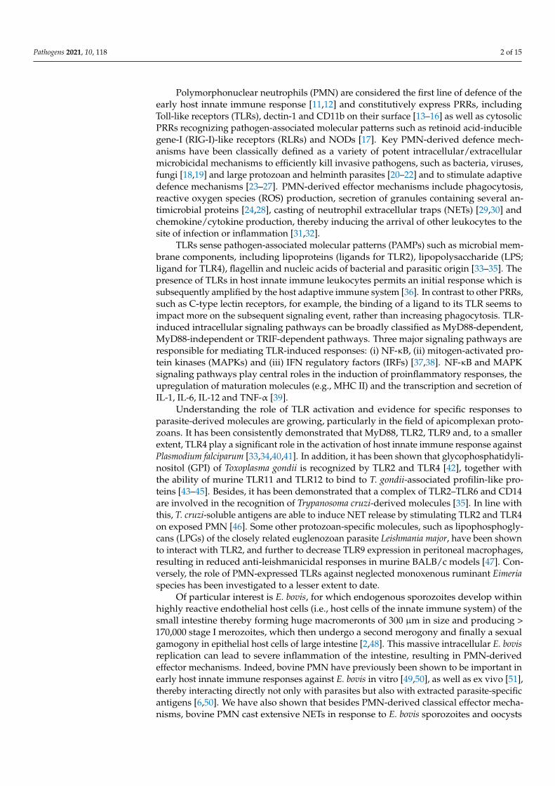
pathogens Article The Role of TLR2 and TLR4 in Recognition and Uptake of the Apicomplexan Parasite Eimeria bovis and Their Effects on NET Formation Tamara Muñoz-Caro 1,2 , Amanda J. Gibson 3,4 , Iván Conejeros 1 , Dirk Werling 3 , Anja Taubert 1 and Carlos Hermosilla 1, * Citation: Muñoz-Caro, T.; Gibson, A.J.; Conejeros, I.; Werling, D.; Taubert, A.; Hermosilla, C. The Role of TLR2 and TLR4 in Recognition and Uptake of the Apicomplexan Parasite Eimeria bovis and Their Effects on NET Formation. Pathogens 2021, 10, 118. https://doi.org/10.3390/pathogens 10020118 Academic Editors: Geoff Hide and Lawrence S. Young Received: 28 December 2020 Accepted: 20 January 2021 Published: 24 January 2021 Publisher’s Note: MDPI stays neutral with regard to jurisdictional claims in published maps and institutional affil- iations. Copyright: © 2021 by the authors. Licensee MDPI, Basel, Switzerland. This article is an open access article distributed under the terms and conditions of the Creative Commons Attribution (CC BY) license (https:// creativecommons.org/licenses/by/ 4.0/). 1 Institute of Parasitology, Justus Liebig University Giessen, 35392 Giessen, Germany; [email protected] (T.M.-C.); [email protected] (I.C.); [email protected] (A.T.) 2 Escuela de Medicina Veterinaria, Facultad de Recursos Naturales y Medicina Veterinaria, Universidad Santo Tomás, Santiago 3460000, Chile 3 Department of Pathobiology and Population Sciences, Royal Veterinary College, Hatfield AL9 7TA, UK; [email protected] (A.J.G.); [email protected] (D.W.) 4 Centre of Excellence in Bovine Tuberculosis, Institute for Biological, Environmental and Rural Sciences, Aberystwyth University, Wales SY23 3FD, UK * Correspondence: [email protected] Abstract: Background: Bovine polymorphonuclear neutrophils (PMN) constitutively express the Toll- like receptors (TLRs) TLR2 and TLR4 and have been shown to generate Neutrophil extracellular traps (NETs) upon exposure to Eimeria bovis. The present work investigated the role of TLR2 and TLR4 in the recognition and uptake of E. bovis sporozoites, IL-8 production and neutrophil extracellular trap (NET) formation. Methods: TLR expression was performed by flow cytometric analysis on PMN exposed to live carboxyfluorescein succinimidyl ester (CFSE)-stained sporozoites. Supernatants of PMN exposed to different E. bovis sporozoite preparations and antigens in the absence or presence of TLR antibodies were assessed for IL-8 secretion. Cells were exposed to sporozoite preparations and assessed for the activation of transcription factor NF-κB using a luciferase reporter assay. Immunofluorescence analysis was done to investigate TLR2 and TLR4 surface expression and NET formation on bovine PMN exposed to vital sporozoites. Results: we observed significantly increased TLR2 and TLR4 expression with a mean increase in expression that was greater for TLR2 than TLR4. This upregulation neither inhibited nor promoted sporozoite phagocytosis by bovine PMN. Live sporozoites together with anti-TLR2 mAb resulted in a significant enhancement of IL-8 production. NF-κB activation was more strongly induced in TLR2-HEK cells than in TLR4/MD2-HEK cells exposed to heat-killed sporozoites and antigens. Immunofluorescence analysis showed TLR-positive signals on the surface of PMN and concomitant NET formation. Conclusions: This is the first report on E. bovis-induced concomitant TLR2 and TLR4 expression during bovine PMN-derived NETosis. Keywords: E. bovis; TLR; IL-8; neutrophil extracellular traps 1. Introduction At least thirteen monoxenous apicomplexan Eimeria species have been reported to infect domestic cattle worldwide to date and, among these species, E. bovis is considered as one of the most pathogenic species, causing severe inflammation of the intestine with clinical manifestations such as haemorrhagic diarrhoea, dehydration, weight loss and poor growth rates, mainly affecting calves [1,2]. Underlying E. bovis infections are complex host adaptive [3–5] as well as host innate immunological regulation in vitro [6–8], ex vivo [9] and in vivo [10]; however, little is known about pathogen recognition receptors (PRRs) involved in early innate immune reactions against ruminant Eimeria species. Pathogens 2021, 10, 118. https://doi.org/10.3390/pathogens10020118 https://www.mdpi.com/journal/pathogens

Welcome message from author
This document is posted to help you gain knowledge. Please leave a comment to let me know what you think about it! Share it to your friends and learn new things together.
Transcript
pathogens
Article
The Role of TLR2 and TLR4 in Recognition and Uptake of theApicomplexan Parasite Eimeria bovis and Their Effects on NETFormation
Tamara Muñoz-Caro 1,2 , Amanda J. Gibson 3,4 , Iván Conejeros 1 , Dirk Werling 3 , Anja Taubert 1 andCarlos Hermosilla 1,*
�����������������
Citation: Muñoz-Caro, T.; Gibson,
A.J.; Conejeros, I.; Werling, D.;
Taubert, A.; Hermosilla, C. The Role
of TLR2 and TLR4 in Recognition and
Uptake of the Apicomplexan Parasite
Eimeria bovis and Their Effects on NET
Formation. Pathogens 2021, 10, 118.
https://doi.org/10.3390/pathogens
10020118
Academic Editors: Geoff Hide and
Lawrence S. Young
Received: 28 December 2020
Accepted: 20 January 2021
Published: 24 January 2021
Publisher’s Note: MDPI stays neutral
with regard to jurisdictional claims in
published maps and institutional affil-
iations.
Copyright: © 2021 by the authors.
Licensee MDPI, Basel, Switzerland.
This article is an open access article
distributed under the terms and
conditions of the Creative Commons
Attribution (CC BY) license (https://
creativecommons.org/licenses/by/
4.0/).
1 Institute of Parasitology, Justus Liebig University Giessen, 35392 Giessen, Germany;[email protected] (T.M.-C.); [email protected] (I.C.);[email protected] (A.T.)
2 Escuela de Medicina Veterinaria, Facultad de Recursos Naturales y Medicina Veterinaria,Universidad Santo Tomás, Santiago 3460000, Chile
3 Department of Pathobiology and Population Sciences, Royal Veterinary College, Hatfield AL9 7TA, UK;[email protected] (A.J.G.); [email protected] (D.W.)
4 Centre of Excellence in Bovine Tuberculosis, Institute for Biological, Environmental and Rural Sciences,Aberystwyth University, Wales SY23 3FD, UK
* Correspondence: [email protected]
Abstract: Background: Bovine polymorphonuclear neutrophils (PMN) constitutively express the Toll-like receptors (TLRs) TLR2 and TLR4 and have been shown to generate Neutrophil extracellular traps(NETs) upon exposure to Eimeria bovis. The present work investigated the role of TLR2 and TLR4 in therecognition and uptake of E. bovis sporozoites, IL-8 production and neutrophil extracellular trap (NET)formation. Methods: TLR expression was performed by flow cytometric analysis on PMN exposed tolive carboxyfluorescein succinimidyl ester (CFSE)-stained sporozoites. Supernatants of PMN exposedto different E. bovis sporozoite preparations and antigens in the absence or presence of TLR antibodieswere assessed for IL-8 secretion. Cells were exposed to sporozoite preparations and assessed forthe activation of transcription factor NF-κB using a luciferase reporter assay. Immunofluorescenceanalysis was done to investigate TLR2 and TLR4 surface expression and NET formation on bovinePMN exposed to vital sporozoites. Results: we observed significantly increased TLR2 and TLR4expression with a mean increase in expression that was greater for TLR2 than TLR4. This upregulationneither inhibited nor promoted sporozoite phagocytosis by bovine PMN. Live sporozoites togetherwith anti-TLR2 mAb resulted in a significant enhancement of IL-8 production. NF-κB activationwas more strongly induced in TLR2-HEK cells than in TLR4/MD2-HEK cells exposed to heat-killedsporozoites and antigens. Immunofluorescence analysis showed TLR-positive signals on the surfaceof PMN and concomitant NET formation. Conclusions: This is the first report on E. bovis-inducedconcomitant TLR2 and TLR4 expression during bovine PMN-derived NETosis.
Keywords: E. bovis; TLR; IL-8; neutrophil extracellular traps
1. Introduction
At least thirteen monoxenous apicomplexan Eimeria species have been reported toinfect domestic cattle worldwide to date and, among these species, E. bovis is consideredas one of the most pathogenic species, causing severe inflammation of the intestine withclinical manifestations such as haemorrhagic diarrhoea, dehydration, weight loss and poorgrowth rates, mainly affecting calves [1,2]. Underlying E. bovis infections are complex hostadaptive [3–5] as well as host innate immunological regulation in vitro [6–8], ex vivo [9]and in vivo [10]; however, little is known about pathogen recognition receptors (PRRs)involved in early innate immune reactions against ruminant Eimeria species.
Pathogens 2021, 10, 118. https://doi.org/10.3390/pathogens10020118 https://www.mdpi.com/journal/pathogens
Pathogens 2021, 10, 118 2 of 15
Polymorphonuclear neutrophils (PMN) are considered the first line of defence of theearly host innate immune response [11,12] and constitutively express PRRs, includingToll-like receptors (TLRs), dectin-1 and CD11b on their surface [13–16] as well as cytosolicPRRs recognizing pathogen-associated molecular patterns such as retinoid acid-induciblegene-I (RIG-I)-like receptors (RLRs) and NODs [17]. Key PMN-derived defence mech-anisms have been classically defined as a variety of potent intracellular/extracellularmicrobicidal mechanisms to efficiently kill invasive pathogens, such as bacteria, viruses,fungi [18,19] and large protozoan and helminth parasites [20–22] and to stimulate adaptivedefence mechanisms [23–27]. PMN-derived effector mechanisms include phagocytosis,reactive oxygen species (ROS) production, secretion of granules containing several an-timicrobial proteins [24,28], casting of neutrophil extracellular traps (NETs) [29,30] andchemokine/cytokine production, thereby inducing the arrival of other leukocytes to thesite of infection or inflammation [31,32].
TLRs sense pathogen-associated molecular patterns (PAMPs) such as microbial mem-brane components, including lipoproteins (ligands for TLR2), lipopolysaccharide (LPS;ligand for TLR4), flagellin and nucleic acids of bacterial and parasitic origin [33–35]. Thepresence of TLRs in host innate immune leukocytes permits an initial response which issubsequently amplified by the host adaptive immune system [36]. In contrast to other PRRs,such as C-type lectin receptors, for example, the binding of a ligand to its TLR seems toimpact more on the subsequent signaling event, rather than increasing phagocytosis. TLR-induced intracellular signaling pathways can be broadly classified as MyD88-dependent,MyD88-independent or TRIF-dependent pathways. Three major signaling pathways areresponsible for mediating TLR-induced responses: (i) NF-κB, (ii) mitogen-activated pro-tein kinases (MAPKs) and (iii) IFN regulatory factors (IRFs) [37,38]. NF-κB and MAPKsignaling pathways play central roles in the induction of proinflammatory responses, theupregulation of maturation molecules (e.g., MHC II) and the transcription and secretion ofIL-1, IL-6, IL-12 and TNF-α [39].
Understanding the role of TLR activation and evidence for specific responses toparasite-derived molecules are growing, particularly in the field of apicomplexan proto-zoans. It has been consistently demonstrated that MyD88, TLR2, TLR9 and, to a smallerextent, TLR4 play a significant role in the activation of host innate immune response againstPlasmodium falciparum [33,34,40,41]. In addition, it has been shown that glycophosphatidyli-nositol (GPI) of Toxoplasma gondii is recognized by TLR2 and TLR4 [42], together withthe ability of murine TLR11 and TLR12 to bind to T. gondii-associated profilin-like pro-teins [43–45]. Besides, it has been demonstrated that a complex of TLR2–TLR6 and CD14are involved in the recognition of Trypanosoma cruzi-derived molecules [35]. In line withthis, T. cruzi-soluble antigens are able to induce NET release by stimulating TLR2 and TLR4on exposed PMN [46]. Some other protozoan-specific molecules, such as lipophosphogly-cans (LPGs) of the closely related euglenozoan parasite Leishmania major, have been shownto interact with TLR2, and further to decrease TLR9 expression in peritoneal macrophages,resulting in reduced anti-leishmanicidal responses in murine BALB/c models [47]. Con-versely, the role of PMN-expressed TLRs against neglected monoxenous ruminant Eimeriaspecies has been investigated to a lesser extent to date.
Of particular interest is E. bovis, for which endogenous sporozoites develop withinhighly reactive endothelial host cells (i.e., host cells of the innate immune system) of thesmall intestine thereby forming huge macromeronts of 300 µm in size and producing >170,000 stage I merozoites, which then undergo a second merogony and finally a sexualgamogony in epithelial host cells of large intestine [2,48]. This massive intracellular E. bovisreplication can lead to severe inflammation of the intestine, resulting in PMN-derivedeffector mechanisms. Indeed, bovine PMN have previously been shown to be important inearly host innate immune responses against E. bovis in vitro [49,50], as well as ex vivo [51],thereby interacting directly not only with parasites but also with extracted parasite-specificantigens [6,50]. We have also shown that besides PMN-derived classical effector mecha-nisms, bovine PMN cast extensive NETs in response to E. bovis sporozoites and oocysts
Pathogens 2021, 10, 118 3 of 15
in vitro and in vivo [6,7,10], suggesting specific interactions of PMN with different E. bovisstages (e.g., sporozoites, merozoites, sporocysts, oocysts). All these parasite stages havedifferent antigens in order to overcome adverse early host innate immune reactions, andthe recognition of E. bovis-derived PAMPs might be mediated by bovine TLRs. More impor-tantly, bovine PMN have been described as expressing numerous PRRs [13,14]. In cattle,TLRs have been described on a number of innate immune cells and are associated withthe recognition of Mycobacterium tuberculosis and M. bovis by macrophages [52], pathogensinvolved in bovine respiratory disease [53] and Escherichia coli-mediated mastitis [54].Bovine PMN express TLR2 and TLR4 and the function of TLR2 has been demonstrated bystimulation with Pam3CSK4, inducing a proinflammatory response [16,55].
To date, few data exist showing the activation of TLRs in leukocytes of the bovineinnate immune system in response to parasite-derived molecules, with only one reportassessing the potential involvement of PMN receptors in early innate immune responsesagainst E. bovis [7]. As such, E. bovis-induced NETosis was shown to be mediated byCD11b expressed on parasite-exposed bovine PMN [7]. We therefore sought to explore thepossible role of TLR2 and TLR4 not only in the uptake of E. bovis sporozoites but also inPMN-derived pro-inflammatory reactions as well as NET extrusion.
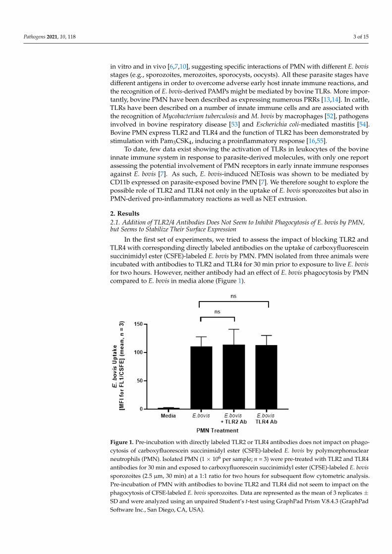
2. Results2.1. Addition of TLR2/4 Antibodies Does Not Seem to Inhibit Phagocytosis of E. bovis by PMN,but Seems to Stabilize Their Surface Expression
In the first set of experiments, we tried to assess the impact of blocking TLR2 andTLR4 with corresponding directly labeled antibodies on the uptake of carboxyfluoresceinsuccinimidyl ester (CSFE)-labeled E. bovis by PMN. PMN isolated from three animals wereincubated with antibodies to TLR2 and TLR4 for 30 min prior to exposure to live E. bovisfor two hours. However, neither antibody had an effect of E. bovis phagocytosis by PMNcompared to E. bovis in media alone (Figure 1).
Pathogens 2021, 10, x FOR PEER REVIEW 4 of 15
Figure 1. Pre-incubation with directly labeled TLR2 or TLR4 antibodies does not impact on phago-cytosis of carboxyfluorescein succinimidyl ester (CSFE)-labeled E. bovis by polymorphonuclear neutrophils (PMN). Isolated PMN (1 × 10⁶ per sample; n = 3) were pre-treated with TLR2 and TLR4 antibodies for 30 min and exposed to carboxyfluorescein succinimidyl ester (CFSE)-labeled E. bovis sporozoites (2.5 µm, 30 min) at a 1:1 ratio for two hours for subsequent flow cytometric analysis. Pre-incubation of PMN with antibodies to bovine TLR2 and TLR4 did not seem to impact on the phagocytosis of CFSE-labeled E. bovis sporozoites. Data are represented as the mean of 3 replicates ± SD and were analyzed using an unpaired Student’s t-test using GraphPad Prism V.8.4.3 (GraphPad Software Inc., San Diego, CA, USA).
(A) (B)
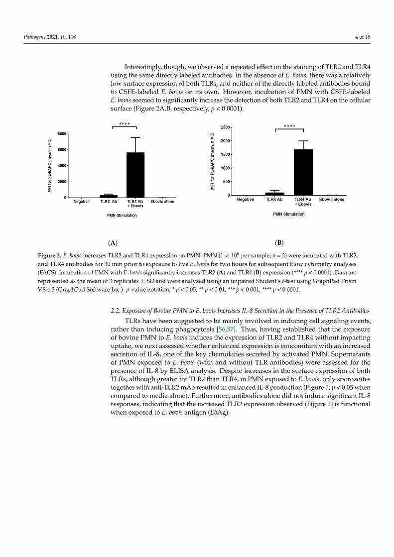
Figure 2. E. bovis increases TLR2 and TLR4 expression on PMN. PMN (1 × 10⁶ per sample; n = 3) were incubated with TLR2 and TLR4 antibodies for 30 min prior to exposure to live E. bovis for two hours for subsequent Flow cytometry analyses (FACS). Incubation of PMN with E. bovis significantly increases TLR2 (A) and TLR4 (B) expression (**** p < 0.0001). Data are represented as the mean of 3 replicates ± SD and were analyzed using an unpaired Student’s t-test using GraphPad Prism V.8.4.3 (GraphPad Software Inc.). p-value notation; * p < 0.05, ** p < 0.01, *** p < 0.001, **** p < 0.0001.
2.2. Exposure of Bovine PMN to E. bovis increases IL-8 Secretion in the Presence of TLR2 Antibodies
Figure 1. Pre-incubation with directly labeled TLR2 or TLR4 antibodies does not impact on phago-cytosis of carboxyfluorescein succinimidyl ester (CSFE)-labeled E. bovis by polymorphonuclearneutrophils (PMN). Isolated PMN (1 × 10⁶ per sample; n = 3) were pre-treated with TLR2 and TLR4antibodies for 30 min and exposed to carboxyfluorescein succinimidyl ester (CFSE)-labeled E. bovissporozoites (2.5 µm, 30 min) at a 1:1 ratio for two hours for subsequent flow cytometric analysis.Pre-incubation of PMN with antibodies to bovine TLR2 and TLR4 did not seem to impact on thephagocytosis of CFSE-labeled E. bovis sporozoites. Data are represented as the mean of 3 replicates ±SD and were analyzed using an unpaired Student’s t-test using GraphPad Prism V.8.4.3 (GraphPadSoftware Inc., San Diego, CA, USA).
Pathogens 2021, 10, 118 4 of 15
Interestingly, though, we observed a repeated effect on the staining of TLR2 and TLR4using the same directly labeled antibodies. In the absence of E. bovis, there was a relativelylow surface expression of both TLRs, and neither of the directly labeled antibodies boundto CSFE-labeled E. bovis on its own. However, incubation of PMN with CSFE-labeledE. bovis seemed to significantly increase the detection of both TLR2 and TLR4 on the cellularsurface (Figure 2A,B, respectively, p < 0.0001).
Pathogens 2021, 10, x FOR PEER REVIEW 4 of 15
Figure 1. Pre-incubation with directly labeled TLR2 or TLR4 antibodies does not impact on phago-cytosis of carboxyfluorescein succinimidyl ester (CSFE)-labeled E. bovis by polymorphonuclear neutrophils (PMN). Isolated PMN (1 × 10⁶ per sample; n = 3) were pre-treated with TLR2 and TLR4 antibodies for 30 min and exposed to carboxyfluorescein succinimidyl ester (CFSE)-labeled E. bovis sporozoites (2.5 µm, 30 min) at a 1:1 ratio for two hours for subsequent flow cytometric analysis. Pre-incubation of PMN with antibodies to bovine TLR2 and TLR4 did not seem to impact on the phagocytosis of CFSE-labeled E. bovis sporozoites. Data are represented as the mean of 3 replicates ± SD and were analyzed using an unpaired Student’s t-test using GraphPad Prism V.8.4.3 (GraphPad Software Inc., San Diego, CA, USA).
(A) (B)
Figure 2. E. bovis increases TLR2 and TLR4 expression on PMN. PMN (1 × 10⁶ per sample; n = 3) were incubated with TLR2 and TLR4 antibodies for 30 min prior to exposure to live E. bovis for two hours for subsequent Flow cytometry analyses (FACS). Incubation of PMN with E. bovis significantly increases TLR2 (A) and TLR4 (B) expression (**** p < 0.0001). Data are represented as the mean of 3 replicates ± SD and were analyzed using an unpaired Student’s t-test using GraphPad Prism V.8.4.3 (GraphPad Software Inc.). p-value notation; * p < 0.05, ** p < 0.01, *** p < 0.001, **** p < 0.0001.
2.2. Exposure of Bovine PMN to E. bovis increases IL-8 Secretion in the Presence of TLR2 Antibodies
Figure 2. E. bovis increases TLR2 and TLR4 expression on PMN. PMN (1 × 10⁶ per sample; n = 3) were incubated with TLR2and TLR4 antibodies for 30 min prior to exposure to live E. bovis for two hours for subsequent Flow cytometry analyses(FACS). Incubation of PMN with E. bovis significantly increases TLR2 (A) and TLR4 (B) expression (**** p < 0.0001). Data arerepresented as the mean of 3 replicates ± SD and were analyzed using an unpaired Student’s t-test using GraphPad PrismV.8.4.3 (GraphPad Software Inc.). p-value notation; * p < 0.05, ** p < 0.01, *** p < 0.001, **** p < 0.0001.
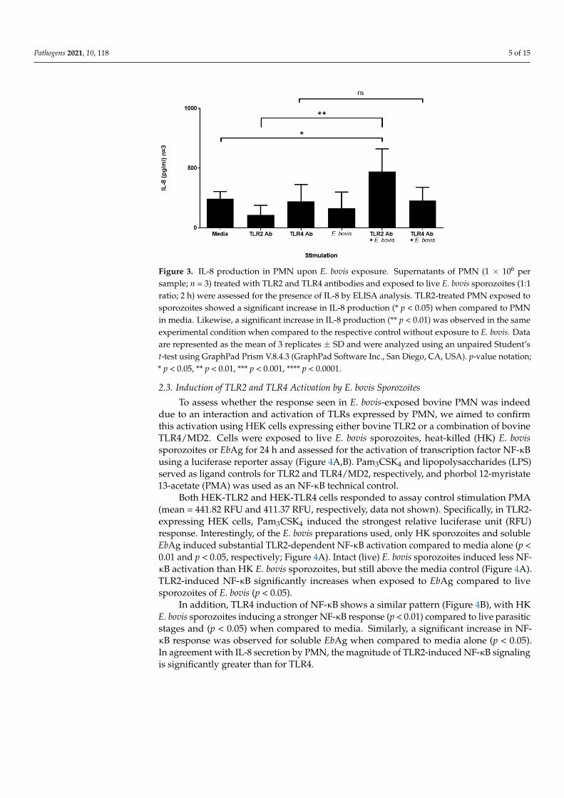
2.2. Exposure of Bovine PMN to E. bovis Increases IL-8 Secretion in the Presence of TLR2 Antibodies
TLRs have been suggested to be mainly involved in inducing cell signaling events,rather than inducing phagocytosis [56,57]. Thus, having established that the exposureof bovine PMN to E. bovis induces the expression of TLR2 and TLR4 without impactinguptake, we next assessed whether enhanced expression is concomitant with an increasedsecretion of IL-8, one of the key chemokines secreted by activated PMN. Supernatantsof PMN exposed to E. bovis (with and without TLR antibodies) were assessed for thepresence of IL-8 by ELISA analysis. Despite increases in the surface expression of bothTLRs, although greater for TLR2 than TLR4, in PMN exposed to E. bovis, only sporozoitestogether with anti-TLR2 mAb resulted in enhanced IL-8 production (Figure 3, p < 0.05 whencompared to media alone). Furthermore, antibodies alone did not induce significant IL-8responses, indicating that the increased TLR2 expression observed (Figure 1) is functionalwhen exposed to E. bovis antigen (EbAg).
Pathogens 2021, 10, 118 5 of 15
Pathogens 2021, 10, x FOR PEER REVIEW 5 of 15
TLRs have been suggested to be mainly involved in inducing cell signaling events, rather than inducing phagocytosis [56,57]. Thus, having established that the exposure of bovine PMN to E. bovis induces the expression of TLR2 and TLR4 without impacting up-take, we next assessed whether enhanced expression is concomitant with an increased secretion of IL-8, one of the key chemokines secreted by activated PMN. Supernatants of PMN exposed to E. bovis (with and without TLR antibodies) were assessed for the pres-ence of IL-8 by ELISA analysis. Despite increases in the surface expression of both TLRs, although greater for TLR2 than TLR4, in PMN exposed to E. bovis, only sporozoites to-gether with anti-TLR2 mAb resulted in enhanced IL-8 production (Figure 3, p < 0.05 when compared to media alone). Furthermore, antibodies alone did not induce significant IL-8 responses, indicating that the increased TLR2 expression observed (Figure 1) is functional when exposed to E. bovis antigen (EbAg).
Figure 3. IL-8 production in PMN upon E. bovis exposure. Supernatants of PMN (1 × 10⁶ per sam-ple; n = 3) treated with TLR2 and TLR4 antibodies and exposed to live E. bovis sporozoites (1:1 ratio; 2 h) were assessed for the presence of IL-8 by ELISA analysis. TLR2-treated PMN exposed to sporozoites showed a significant increase in IL-8 production (* p < 0.05) when compared to PMN in media. Likewise, a significant increase in IL-8 production (** p < 0.01) was observed in the same experimental condition when compared to the respective control without exposure to E. bovis. Data are represented as the mean of 3 replicates ± SD and were analyzed using an unpaired Stu-dent’s t-test using GraphPad Prism V.8.4.3 (GraphPad Software Inc. San Diego, CA, USA). p-value notation; * p < 0.05, ** p < 0.01, *** p < 0.001, **** p < 0.0001.
2.3. Induction of TLR2 and TLR4 Activation by E. bovis Sporozoites To assess whether the response seen in E. bovis-exposed bovine PMN was indeed due
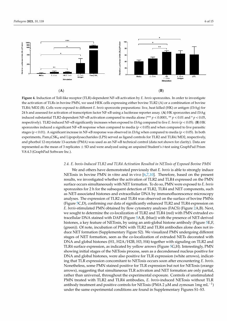
to an interaction and activation of TLRs expressed by PMN, we aimed to confirm this activation using HEK cells expressing either bovine TLR2 or a combination of bovine TLR4/MD2. Cells were exposed to live E. bovis sporozoites, heat-killed (HK) E. bovis spo-rozoites or EbAg for 24 h and assessed for the activation of transcription factor NF-κB using a luciferase reporter assay (Figure 4A,B). Pam3CSK4 and lipopolysaccharides (LPS) served as ligand controls for TLR2 and TLR4/MD2, respectively, and phorbol 12-myristate 13-acetate (PMA) was used as an NF-κB technical control.
Both HEK-TLR2 and HEK-TLR4 cells responded to assay control stimulation PMA (mean = 441.82 RFU and 411.37 RFU, respectively, data not shown). Specifically, in TLR2-expressing HEK cells, Pam3CSK4 induced the strongest relative luciferase unit (RFU) re-sponse. Interestingly, of the E. bovis preparations used, only HK sporozoites and soluble EbAg induced substantial TLR2-dependent NF-κB activation compared to media alone (p
Figure 3. IL-8 production in PMN upon E. bovis exposure. Supernatants of PMN (1 × 10⁶ persample; n = 3) treated with TLR2 and TLR4 antibodies and exposed to live E. bovis sporozoites (1:1ratio; 2 h) were assessed for the presence of IL-8 by ELISA analysis. TLR2-treated PMN exposed tosporozoites showed a significant increase in IL-8 production (* p < 0.05) when compared to PMNin media. Likewise, a significant increase in IL-8 production (** p < 0.01) was observed in the sameexperimental condition when compared to the respective control without exposure to E. bovis. Dataare represented as the mean of 3 replicates ± SD and were analyzed using an unpaired Student’st-test using GraphPad Prism V.8.4.3 (GraphPad Software Inc., San Diego, CA, USA). p-value notation;* p < 0.05, ** p < 0.01, *** p < 0.001, **** p < 0.0001.
2.3. Induction of TLR2 and TLR4 Activation by E. bovis Sporozoites
To assess whether the response seen in E. bovis-exposed bovine PMN was indeeddue to an interaction and activation of TLRs expressed by PMN, we aimed to confirmthis activation using HEK cells expressing either bovine TLR2 or a combination of bovineTLR4/MD2. Cells were exposed to live E. bovis sporozoites, heat-killed (HK) E. bovissporozoites or EbAg for 24 h and assessed for the activation of transcription factor NF-κBusing a luciferase reporter assay (Figure 4A,B). Pam3CSK4 and lipopolysaccharides (LPS)served as ligand controls for TLR2 and TLR4/MD2, respectively, and phorbol 12-myristate13-acetate (PMA) was used as an NF-κB technical control.
Both HEK-TLR2 and HEK-TLR4 cells responded to assay control stimulation PMA(mean = 441.82 RFU and 411.37 RFU, respectively, data not shown). Specifically, in TLR2-expressing HEK cells, Pam3CSK4 induced the strongest relative luciferase unit (RFU)response. Interestingly, of the E. bovis preparations used, only HK sporozoites and solubleEbAg induced substantial TLR2-dependent NF-κB activation compared to media alone (p <0.01 and p < 0.05, respectively; Figure 4A). Intact (live) E. bovis sporozoites induced less NF-κB activation than HK E. bovis sporozoites, but still above the media control (Figure 4A).TLR2-induced NF-κB significantly increases when exposed to EbAg compared to livesporozoites of E. bovis (p < 0.05).
In addition, TLR4 induction of NF-κB shows a similar pattern (Figure 4B), with HKE. bovis sporozoites inducing a stronger NF-κB response (p < 0.01) compared to live parasiticstages and (p < 0.05) when compared to media. Similarly, a significant increase in NF-κB response was observed for soluble EbAg when compared to media alone (p < 0.05).In agreement with IL-8 secretion by PMN, the magnitude of TLR2-induced NF-κB signalingis significantly greater than for TLR4.
Pathogens 2021, 10, 118 6 of 15
Pathogens 2021, 10, x FOR PEER REVIEW 6 of 15
< 0.01 and p < 0.05, respectively; Figure 4A). Intact (live) E. bovis sporozoites induced less NF-κB activation than HK E. bovis sporozoites, but still above the media control (Figure 4A). TLR2-induced NF-κB significantly increases when exposed to EbAg compared to live sporozoites of E. bovis (p < 0.05).
In addition, TLR4 induction of NF-κB shows a similar pattern (Figure 4B), with HK E. bovis sporozoites inducing a stronger NF-κB response (p < 0.01) compared to live para-sitic stages and (p < 0.05) when compared to media. Similarly, a significant increase in NF-κB response was observed for soluble EbAg when compared to media alone (p < 0.05). In agreement with IL-8 secretion by PMN, the magnitude of TLR2-induced NF-κB signaling is significantly greater than for TLR4.
(A) (B)
Figure 4. Induction of Toll-like receptor (TLR)-dependent NF-κB activation by E. bovis sporozoites. In order to investigate the activation of TLRs in bovine PMN, we used HEK cells expressing either bovine TLR2 (A) or a combination of bovine TLR4/MD2 (B). Cells were exposed to different E. bovis sporozoite preparations: live, heat killed (HK) or antigen (EbAg) for 24 h and assessed for activation of transcription factor NF-κB using a luciferase reporter assay. (A) HK sporozoites and EbAg induced substantial TLR2-dependent NF-κB activation compared to media alone (*** p < 0.0001, ** p < 0.01 and * p < 0.05, respectively). TLR2-induced NF-κB significantly increases when exposed to EbAg compared to live E. bovis (p < 0.05). (B) HK sporozoites induced a significant NF-κB response when compared to media (p < 0.05) and when compared to live parasitic stages (p < 0.01). A significant increase in NF-κB response was observed in EbAg when compared to media (p < 0.05). In both experiments, Pam3CSK4 and Lipopolysaccharides (LPS) served as ligand controls for TLR2 and TLR4/MD2, respectively, and phorbol 12-myristate 13-acetate (PMA) was used as an NF-κB technical control (data not shown for clarity). Data are represented as the mean of 3 replicates ± SD and were analyzed using an unpaired Student’s t-test using GraphPad Prism V.8.4.3 (GraphPad Software Inc.).
2.4. E. bovis-Induced TLR2 and TLR4 Activation Resulted in NETosis of Exposed Bovine PMN We and others have demonstrated previously that E. bovis is able to strongly induce
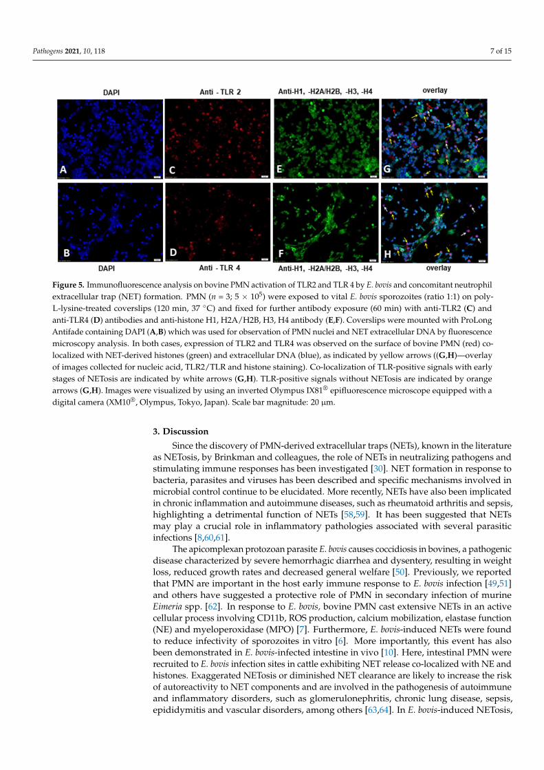
NETosis in bovine PMN in vitro and in vivo [6,7,10]. Therefore, based on the present re-sults, we investigated whether the activation of TLR2 and TLR4 expressed on the PMN surface occurs simultaneously with NET formation. To do so, PMN were exposed to E. bovis sporozoites for 2 h for the subsequent detection of TLR2, TLR4 and NET components, such as NET-associated histones and extracellular DNA by immunofluorescence micros-copy analyses. The expression of TLR2 and TLR4 was observed on the surface of bovine PMNs (Figure 5C,D), confirming our data of significantly enhanced TLR2 and TLR4 ex-pression on E. bovis-stimulated PMN obtained by flow cytometry analyses (FACS) (Figure 2A,B). Next, we sought to determine the co-localization of TLR2 and TLR4 (red) with PMN extruded extracellular DNA stained with DAPI (Figure 5A,B, (blue)) with the presence of NET-derived histones, a key feature of NETosis, by using an anti-global histone antibody (Figure 5E,F, (green)). Of note, incubation of PMN with TLR2 and TLR4 antibodies alone does not induce NET formation (Supplementary Figure S2). We visualized PMN under-going different stages of NET formation, seen as the co-localization of extruded NETs dec-orated with DNA and global histones (H1, H2A/H2B, H3, H4) together with signaling on TLR2 and TLR4 surface expression, as indicated by yellow arrows (Figure 5G,H). Inter-
Figure 4. Induction of Toll-like receptor (TLR)-dependent NF-κB activation by E. bovis sporozoites. In order to investigatethe activation of TLRs in bovine PMN, we used HEK cells expressing either bovine TLR2 (A) or a combination of bovineTLR4/MD2 (B). Cells were exposed to different E. bovis sporozoite preparations: live, heat killed (HK) or antigen (EbAg) for24 h and assessed for activation of transcription factor NF-κB using a luciferase reporter assay. (A) HK sporozoites and EbAginduced substantial TLR2-dependent NF-κB activation compared to media alone (*** p < 0.0001, ** p < 0.01 and * p < 0.05,respectively). TLR2-induced NF-κB significantly increases when exposed to EbAg compared to live E. bovis (p < 0.05). (B) HKsporozoites induced a significant NF-κB response when compared to media (p < 0.05) and when compared to live parasiticstages (p < 0.01). A significant increase in NF-κB response was observed in EbAg when compared to media (p < 0.05). In bothexperiments, Pam3CSK4 and Lipopolysaccharides (LPS) served as ligand controls for TLR2 and TLR4/MD2, respectively,and phorbol 12-myristate 13-acetate (PMA) was used as an NF-κB technical control (data not shown for clarity). Data arerepresented as the mean of 3 replicates ± SD and were analyzed using an unpaired Student’s t-test using GraphPad PrismV.8.4.3 (GraphPad Software Inc.).
2.4. E. bovis-Induced TLR2 and TLR4 Activation Resulted in NETosis of Exposed Bovine PMN
We and others have demonstrated previously that E. bovis is able to strongly induceNETosis in bovine PMN in vitro and in vivo [6,7,10]. Therefore, based on the presentresults, we investigated whether the activation of TLR2 and TLR4 expressed on the PMNsurface occurs simultaneously with NET formation. To do so, PMN were exposed to E. bovissporozoites for 2 h for the subsequent detection of TLR2, TLR4 and NET components, suchas NET-associated histones and extracellular DNA by immunofluorescence microscopyanalyses. The expression of TLR2 and TLR4 was observed on the surface of bovine PMNs(Figure 5C,D), confirming our data of significantly enhanced TLR2 and TLR4 expression onE. bovis-stimulated PMN obtained by flow cytometry analyses (FACS) (Figure 2A,B). Next,we sought to determine the co-localization of TLR2 and TLR4 (red) with PMN extruded ex-tracellular DNA stained with DAPI (Figure 5A,B, (blue)) with the presence of NET-derivedhistones, a key feature of NETosis, by using an anti-global histone antibody (Figure 5E,F,(green)). Of note, incubation of PMN with TLR2 and TLR4 antibodies alone does not in-duce NET formation (Supplementary Figure S2). We visualized PMN undergoing differentstages of NET formation, seen as the co-localization of extruded NETs decorated withDNA and global histones (H1, H2A/H2B, H3, H4) together with signaling on TLR2 andTLR4 surface expression, as indicated by yellow arrows (Figure 5G,H). Interestingly, PMNshowing initial stages of the NETosis process, seen as a decondensed nucleus positive forDNA and global histones, were also positive for TLR expression (white arrows), indicat-ing that TLR expression concomitant to NETosis occurs soon after encountering E. bovis.Nonetheless, some PMN stained positive for TLR expression but not for NETosis (orangearrows), suggesting that simultaneous TLR activation and NET formation are only partial,rather than universal, throughout the experimental exposure. Controls of unstimulatedPMN treated with TLR2 and TLR4 antibodies, E. bovis-induced NETosis without TLRantibody treatment and positive controls for NETosis (PMA 2 µM and zymosan 1mg mL−1)under the same experimental conditions are found in Supplementary Figures S1–S3.
Pathogens 2021, 10, 118 7 of 15
Pathogens 2021, 10, x FOR PEER REVIEW 7 of 15
estingly, PMN showing initial stages of the NETosis process, seen as a decondensed nu-cleus positive for DNA and global histones, were also positive for TLR expression (white arrows), indicating that TLR expression concomitant to NETosis occurs soon after encoun-tering E. bovis. Nonetheless, some PMN stained positive for TLR expression but not for NETosis (orange arrows), suggesting that simultaneous TLR activation and NET for-mation are only partial, rather than universal, throughout the experimental exposure. Controls of unstimulated PMN treated with TLR2 and TLR4 antibodies, E. bovis-induced NETosis without TLR antibody treatment and positive controls for NETosis (PMA 2 µM and zymosan 1mg mL−1) under the same experimental conditions are found in Supple-mentary Figures S1–S3.
Figure 5. Immunofluorescence analysis on bovine PMN activation of TLR2 and TLR 4 by E. bovis and concomitant neutro-phil extracellular trap (NET) formation. PMN (n = 3; 5 × 10⁵) were exposed to vital E. bovis sporozoites (ratio 1:1) on poly-L-lysine-treated coverslips (120 min, 37 °C) and fixed for further antibody exposure (60 min) with anti-TLR2 (C) and anti-TLR4 (D) antibodies and anti-histone H1, H2A/H2B, H3, H4 antibody (E,F). Coverslips were mounted with ProLong An-tifade containing DAPI (A,B) which was used for observation of PMN nuclei and NET extracellular DNA by fluorescence microscopy analysis. In both cases, expression of TLR2 and TLR4 was observed on the surface of bovine PMN (red) co-localized with NET-derived histones (green) and extracellular DNA (blue), as indicated by yellow arrows (G,H—overlay of images collected for nucleic acid, TLR2/TLR and histone staining). Co-localization of TLR-positive signals with early stages of NETosis are indicated by white arrows (G,H). TLR-positive signals without NETosis are indicated by orange arrows (G,H). Images were visualized by using an inverted Olympus IX81® epifluorescence microscope equipped with a digital camera (XM10®, Olympus, Tokyo, Japan). Scale bar magnitude: 20 µm.
3. Discussion Since the discovery of PMN-derived extracellular traps (NETs), known in the litera-
ture as NETosis, by Brinkman and colleagues, the role of NETs in neutralizing pathogens and stimulating immune responses has been investigated [30]. NET formation in response to bacteria, parasites and viruses has been described and specific mechanisms involved in microbial control continue to be elucidated. More recently, NETs have also been impli-cated in chronic inflammation and autoimmune diseases, such as rheumatoid arthritis and sepsis, highlighting a detrimental function of NETs [58,59]. It has been suggested that NETs may play a crucial role in inflammatory pathologies associated with several para-sitic infections [8,60,61].
The apicomplexan protozoan parasite E. bovis causes coccidiosis in bovines, a patho-genic disease characterized by severe hemorrhagic diarrhea and dysentery, resulting in weight loss, reduced growth rates and decreased general welfare [50]. Previously, we re-ported that PMN are important in the host early immune response to E. bovis infection
Figure 5. Immunofluorescence analysis on bovine PMN activation of TLR2 and TLR 4 by E. bovis and concomitant neutrophilextracellular trap (NET) formation. PMN (n = 3; 5 × 10⁵) were exposed to vital E. bovis sporozoites (ratio 1:1) on poly-L-lysine-treated coverslips (120 min, 37 ◦C) and fixed for further antibody exposure (60 min) with anti-TLR2 (C) andanti-TLR4 (D) antibodies and anti-histone H1, H2A/H2B, H3, H4 antibody (E,F). Coverslips were mounted with ProLongAntifade containing DAPI (A,B) which was used for observation of PMN nuclei and NET extracellular DNA by fluorescencemicroscopy analysis. In both cases, expression of TLR2 and TLR4 was observed on the surface of bovine PMN (red) co-localized with NET-derived histones (green) and extracellular DNA (blue), as indicated by yellow arrows ((G,H)—overlayof images collected for nucleic acid, TLR2/TLR and histone staining). Co-localization of TLR-positive signals with earlystages of NETosis are indicated by white arrows (G,H). TLR-positive signals without NETosis are indicated by orangearrows (G,H). Images were visualized by using an inverted Olympus IX81® epifluorescence microscope equipped with adigital camera (XM10®, Olympus, Tokyo, Japan). Scale bar magnitude: 20 µm.
3. Discussion
Since the discovery of PMN-derived extracellular traps (NETs), known in the literatureas NETosis, by Brinkman and colleagues, the role of NETs in neutralizing pathogens andstimulating immune responses has been investigated [30]. NET formation in response tobacteria, parasites and viruses has been described and specific mechanisms involved inmicrobial control continue to be elucidated. More recently, NETs have also been implicatedin chronic inflammation and autoimmune diseases, such as rheumatoid arthritis and sepsis,highlighting a detrimental function of NETs [58,59]. It has been suggested that NETsmay play a crucial role in inflammatory pathologies associated with several parasiticinfections [8,60,61].
The apicomplexan protozoan parasite E. bovis causes coccidiosis in bovines, a pathogenicdisease characterized by severe hemorrhagic diarrhea and dysentery, resulting in weightloss, reduced growth rates and decreased general welfare [50]. Previously, we reportedthat PMN are important in the host early immune response to E. bovis infection [49,51]and others have suggested a protective role of PMN in secondary infection of murineEimeria spp. [62]. In response to E. bovis, bovine PMN cast extensive NETs in an activecellular process involving CD11b, ROS production, calcium mobilization, elastase function(NE) and myeloperoxidase (MPO) [7]. Furthermore, E. bovis-induced NETs were foundto reduce infectivity of sporozoites in vitro [6]. More importantly, this event has alsobeen demonstrated in E. bovis-infected intestine in vivo [10]. Here, intestinal PMN wererecruited to E. bovis infection sites in cattle exhibiting NET release co-localized with NE andhistones. Exaggerated NETosis or diminished NET clearance are likely to increase the riskof autoreactivity to NET components and are involved in the pathogenesis of autoimmuneand inflammatory disorders, such as glomerulonephritis, chronic lung disease, sepsis,epididymitis and vascular disorders, among others [63,64]. In E. bovis-induced NETosis,
Pathogens 2021, 10, 118 8 of 15
it is suggested that released NETs might play an important role in very early host innatedefence reactions during coccidiosis, thereby contributing to the protection of infectedanimals and significantly altering the outcome of infection, as some parasitic stages (e.g.,sporozoites, merozoites) might be unable to invade host cells [6,7,10].
Activation of PMN in order to generate NETs can be mediated by several means,including hydrogen peroxide production, pro-inflammatory cytokines, such as IFNγ and IL-8, and microbial PAMPs [59,65,66]. PAMPs recognized by TLR2, TLR4, TLR7 and TLR9 havebeen implicated in recognition of P. falciparum, T. gondii, L. major and T. cruzi [33,41,45,46,67].Further, TLR2 and TLR4 are required for the complete induction of NETs when T. cruziis cultured with PMN in vitro [46], whereas TLR7 has been described to be critical forthe control of L. major in mice in vivo. This effect seems to be mediated by ROS andNETosis induction [67]. Bovine PMN constitutively express TLR2 and TLR4 and werepreviously shown to undergo NETosis in a ROS-dependent manner upon exposure toE. bovis; therefore, the present work was carried out to investigate in more detail the role ofTLR2 and TLR4 in the recognition and uptake of E. bovis sporozoites, pro-inflammatorycytokine production (IL-8) and NET formation.
Pre-incubation of PMN with directly labeled antibodies to TLR2 and TLR4 prior toE. bovis exposure had no impact on phagocytosis of E. bovis (Figure 2). Interestingly, though,a strongly enhanced TLR signal was observed on E. bovis-exposed PMN, which was nota result of the directly labeled antibodies binding to CSFE-labeled E. bovis sporozoites(Figure 1). At this moment in time, we are not able to explain this phenomenon, butassume that it is indeed an increased expression of TLR2/4 as a result of E. bovis exposure,and insufficient washing of excess antibodies. To assess whether E. bovis indeed signalsthrough TLR2 and TLR4, we next used TLR2- and TLR4/MD2-expressing HEK cells. Onlyheat-killed E. bovis sporozoites or EbAg induced NF-κB induction, whereas live E. bovissporozoites and media controls did not. This suggested that in this experimental system,live E. bovis neither activate TLR2 nor TLR4 (Figure 4). Interestingly, increased TLR2expression on PMN was significantly greater than for TLR4, which was also seen in regardto a stronger NF-κB activation induced in TLR2-HEK cells compared to TLR4/MD2-HEKcells in response to HK sporozoites and EbAg. In corroboration with the HEK cell reporterassay results, PMN do not produce increased IL-8 when incubated with live E. bovissporozoites, further indicating that attachment or uptake of these stages by PMN is notreliant on TLR activation (Figure 3). Taken together, these results strongly suggest that thedestruction of E. bovis sporozoites by heat treatment may expose a TLR2-specific ligand notreadily accessible in the viable organism. In addition, live parasites are not fully requiredfor TLR2 or TLR4 recognition of E. bovis, as demonstrated by a substantial increase inNF-κB induction via TLR2.
We observed TLR2- and TLR4-positive staining on the surface of PMN concomitantwith NET-derived extracellular DNA and global histones in PMN exposed to E. bovis(Figure 5). This co-localization was observed in PMN undergoing NETosis and also thosein the early stages of NET formation, although this event was not observed in all cellsexposed to E. bovis. ROS production has a central role in NETosis, and, interestingly, TLR2induction of ROS is responsible for NETs in response to L. major LPG [67]. Consequently,the release of NETs in response to E. bovis is also an active cellular death process involvingROS production [7]. TLR2 expression is associated with PMN-derived extracellular DNAwith histones induced by E. bovis further suggesting a role for TLR2 in NET induction(Figure 5). The involvement of TLRs in parasite-induced NETosis is complex; MyD88 wasfound not to be necessary for NET formation in response to T. gondii tachyzoites, however,TLR2 and TLR4 are required to recognize GPI extracted from T. gondii [42,68]. However,there appears to be an agreement that viable parasite stages are not always required forNETosis to occur. Indeed, soluble antigens (prepared by freeze–thawing similar to EbAgabove) from T. cruzi induced TLR2/4-dependent NET formation, highlighting that viableparasites are not always required for the recognition and activation of innate immuneleukocytes [46]. In the present study, we showed that by performing immunofluorescence
Pathogens 2021, 10, 118 9 of 15
microscopy analysis, live E. bovis sporozoites are able to induce TLR2 and TLR4 surfaceexpression on bovine PMNs and this signal occurs simultaneously to NETosis. Theseevents seem relevant to the initiation of stronger host innate immune responses againstthis parasite orchestrating other leukocyte-derived effector mechanisms [8,20]. However,future research should be performed in order to elucidate additional leukocyte populationsas well as pathways involved in E. bovis-triggered TLR activation and NET formation.Future molecular analyses of E. bovis merozoite-, gametocyte- and oocyst-derived antigens,probably all involved in TLR-derived host innate immune reactions during cattle coccidiosisin vivo, are needed.
4. Materials and Methods4.1. Parasites
The E. bovis (strain H) strain used in the present study was maintained by passages inHolstein–Friesian calves (n = 3) for oocyst production as described by [69]. Collection ofoocysts, oocyst sporulation and excystation of sporozoites were performed as previouslydescribed [69]. Released, free sporozoites were washed three times with sterile phosphate-buffered saline solution (PBS, 400× g, 10 min), counted in a Neubauer hemocytometerchamber and thereafter suspended at a final concentration of 2 × 10⁶/sporozoites/mL incell culture medium RPMI 1640 without phenol red (Gibco, Waltham, MA, USA) containingpenicillin/streptomycin (both 100 U/0.1 mg/mL, Gibco) until further experimental use.
For parasite antigen preparation, E. bovis sporozoites were homogenized by repeatedfreezing followed by sonication (20 kHz, 5 × 15 s pulses) on ice. After centrifugation(11,000× g, 4 ◦C, 20 min), the supernatants were passed through 0.2 µm sterile filters(Merk, Darmstadt, Germany). Protein concentration was determined using the Bradfordmethod [70]. The E. bovis-specific antigen (EbAg) was stored at −80 ◦C until further use.
For PMN phagocytosis assays, E. bovis sporozoites were stained with carboxyfluo-rescein succinimidyl ester (CFSE) following previously described protocols [9]. Briefly,vital sporozoites where incubated with a 2.5 µM final concentration of carboxyfluoresceinsuccinimidyl ester (CFSE, Invitrogen, Waltham, MA, USA) in sterile PBS (cell culture grade,0.1 µm filtered, endotoxin tested, Gibco) for 30 min at 37 ◦C prior to sterile PBS washing bycentrifugation (400× g, 10 min; three times). CFSE-stained sporozoites were suspended inRPMI 1640 cell culture without phenol red (Gibco) ready for incubation with bovine PMN.
4.2. Isolation of Bovine PMN
Whole blood was collected by venopuncture of the jugular vein from healthy parousfemale Holstein–Friesian cattle housed at Bolton’s Park Farm, Royal Veterinary College(RVC), London, United Kingdom, using acid citrate dextrose under Home Office licensePPL7009059. PMN were isolated by density centrifugation followed by flash lysis aspreviously described [16]. Briefly, whole blood was diluted with PBS + ethylenediaminete-traacetic acid (EDTA) 0.01% before layering onto Lymphoprep (Biocol, Millipore, Burling-ton, MA, USA) and centrifugation at 700× g for 30 min. The lower layer containingPMN was retained, lysed with distilled water for 40 s and recovered with Hank’s bal-anced salt solution (HBSS) without phenol red (Gibco) before washing by centrifugation.PMN were counted by a trypan blue (Sigma-Aldrich, Darmstadt, Germany) exclusiontest using FastRead® counting chambers (Immunosystems, Torquay, UK) and adjusted to1 × 106 cells/mL with RPMI 1640 cell medium without phenol red (Gibco). PMN puritywas assessed by microscopy after DiffQuick® (Reagena, Toivala, Finland) staining andisolations of at least 90% were used for E. bovis stimulation assays.
4.3. Blocking of TLRs and Phagocytosis Assay
All reagents used in these assays were either purchased from specified suppliers as“low LPS, culture grade”, or were tested for LPS in the TLR4 HEK cell system. IsolatedPMN (1 × 10⁶ per sample) from three animals were pre-treated with Alexa Fluor 647(far red spectrum) directly labeled antibodies to TLR2 and TLR4 (AbD Bio-Rad, Watford,
Pathogens 2021, 10, 118 10 of 15
Hertfordshire, UK), see Table A1) for 30 min before washing once. PMN were then exposedto CFSE-labeled E. bovis sporozoites (CFSE treatment: 2.5 µM, 30 min, 37 ◦C) at a 1:1 ratiofor 2 h. PMN supernatant was collected, clarified and stored for subsequent IL-8 ELISA(AbD Bio-Rad, Watford, Hertfordshire, UK), after which PMN were washed in PBS twiceand finally suspended in 400 µL FACSFlow for flow cytometric analysis. Ten thousandevents were acquired with a BD FACS Calibur running Cell Quest Pro (BD Biosciences,San Jose, CA, USA) and post-analyzed with FlowJo V10 software (BD Biosciences, San Jose,CA, USA).
4.4. IL-8 ELISA
IL-8 was detected in stimulated PMN cell-free supernatants by capture ELISA as previ-ously described [71]. Antibodies used (Table A1) were mouse anti-sheep IL-8 capture (AbDSerotec), rabbit anti-sheep IL-8 detection (AbD Serotec) and goat anti-rabbit horseradishperoxidase (HRP) detection (Agilent (DAKO), Stockport, Cheshire, UK) alongside recom-binant bovine IL-8 (Kingfisher Biotech, Saint Paul, MN, USA) to create a standard curve.Supernatants from stimulated PMN were collected, clarified by centrifugation and storedat −20 ◦C until the ELISA was performed. Results were visualized by the addition of3,3′,5,5′-Tetramethylbenzidine (TMB, Sigma-Aldrich, Dorset, UK) for 15 min before stop-ping the reaction with 0.5 M sulfuric acid (VWR, Poole, Dorset. UK) and plates were readusing a Tecan M200 pro plate reader® (Tecan, Reading, Berkshire, UK) and analyzed withGraphPad® Prism software (GraphPad Software, San Diego, CA, USA).
4.5. TLR Stimulation Assay
To assess TLR recognition of E. bovis-derived antigens (EbAg), HEK cells expressingbovine TLR2 or bovine TLR4 containing an NF-κB luciferase reporter (NF-κB-luc, Promega,Chilworth, Hampshire, UK) were used [56,72,73]. Briefly, HEK-TLR2 or HEK-TLR4 cellswere seeded at a density of 2.5 × 10⁵ cells in 6-well plates using Dulbecco’s ModifiedEagle Medium (DMEM Life Technologies, Paisley, Renfrewshire, UK) supplemented with10% fetal bovine serum (FBS, Life Technologies, Paisley, Renfrewshire, UK) and 1 mgmL−1 Geneticin (Life Technologies, Paisley, Renfrewshire, UK). After 24 h, HEK cells weretransfected with 250 ng p NF-κB-luc with TurboFectTM (Thermo Scientific, Waltham, MAUSA) using the manufacturers’ standard recommendations and allowed to recover for24 h. For experimental assays, one well of a 6-well plate was split into 6 wells of a 24-wellplate and each condition was assayed in triplicate. HEK-TLR2 and HEK-TLR4 cells wereexposed to E. bovis stimuli: live 2.5 × 10⁵; dead 2.5 × 10⁵ heat killed (60 ◦C, 30 min)and soluble EbAg 100 µg for 24 h with TLR2 (1 µg mL−1 Pam3CSK4), TLR4 (1 µg mL−1
LPS-EK) and NF-κB [100 ng mL−1 phorbol 12-myristate 13-acetate (PMA)] controls (allInvivogen, Toulouse, Midi-Pyrenees, France). NF-κB gene activation was determined usingthe Luciferase Reporter Assay System (Promega Chilworth, Hampshire, UK) following themanufacturer’s instructions. Cell lysates were clarified by centrifugation at 16,000× g for5 min and protein concentration determined by absorbance at 280 nm with a NanodropND-1000 for normalization, as described [74].
4.6. Induction of NETosis and TLR2 Expression Via Fluorescence Microscopy Analysis
In another set of experiments, E. bovis-induced TLR2 and TLR4 expression and NETosiswere analyzed via fluorescence microscopy analysis. Here, bovine PMN (n = 3; 5 × 10⁵)were exposed to vital E. bovis sporozoites at a ratio 1:1 on previously pre-coated poly-L-lysine-treated (Sigma-Aldrich, Darmstadt, Germany) coverslips (2 h, 37 ◦C) in a plastic 24-well plate (Greiner, Kremsmünster, Austria). Thereafter, the samples were fixed (overnight,4% paraformaldehyde on ice, Merck), for 20 min at room temperature (RT), washedthrice with PBS and stored at 4 ◦C until further use. Prior to antibody exposure, sampleswere washed three times with sterile PBS and blocked with bovine serum albumin (BSA2%, 30 min, Sigma-Aldrich, Darmstadt, Germany). Treatments with anti-TLR2 and anti-TLR4 antibodies (1 µg mL−1, in the dark, detailed in Table A1) were performed for 1 h.

Pathogens 2021, 10, 118 11 of 15
Thereafter, samples were carefully washed three times with sterile PBS. For the detectionof histones, cells were incubated with a pan-histone antibody detecting H1, H2A/H2B,H3, H4 (MAB3422; Merk, Darmstadt, Germany), diluted 1:200 for 1 h, washed washedtwice with sterile PBS and incubated with the secondary antibody (Alexa Fluor 488 goatanti-mouse, Life Technologies, Paisley, Renfrewshire, UK) diluted 1:500 in buffer (PBS 1×,3% BSA) for 1 h in the dark (for antibody information, see Table A1). Finally, specimenswere washed three times with sterile PBS and mounted in ProLong Antifade® containingDAPI (Thermo Fisher Scientific, Waltham, MA, USA) for 24 h in the dark, which were usedfor the detection of nuclei and NET extracellular DNA. Visualization of TLR expression onthe surface of PMN, NET structures based on co-localized extracellular DNA staining andhistone-derived signals was achieved by using an inverted Olympus IX81® epifluorescencemicroscope equipped with an XM10® digital camera (Olympus, Hamburg, Germany).Staining for controls consisting of unstimulated PMN incubated with antibodies to TLR2and TLR4, E. bovis-induced NETosis without TLR antibody treatment and positive controlsfor NETosis (PMA 2 µM; zymosan 1 mg mL−1; Sigma-Aldrich, Darmstadt, Germany) canbe found in the Supplementary Data.
4.7. Statistical Analysis
Data were analyzed using Microsoft Excel (Microsoft 2013, Redmond, Washing-ton, USA) and GraphPad® Prism. Differences were regarded as significant at a levelof p ≤ 0.05 (*).
Supplementary Materials: The following are available online at https://www.mdpi.com/2076-0817/10/2/118/s1. Figure S1: Control of PMN exposed to E. bovis without TLR treatment andsubsequent NET formation. PMN (n = 3; 5 × 10⁵) were exposed to vital E. bovis sporozoites (ratio 1:1)on poly-L-lysine-treated coverslips (120 min, 37 ◦C) and fixed in 4% paraformaldehyde (A) for furtherantibody exposure (60 min) with an anti-histone H1, H2A/H2B, H3, H4 antibody (C). Coverslipswere mounted with ProLong Antifade® containing DAPI (B) which was used for observation of PMNnuclei and NET extracellular DNA. Co-localization of E. bovis induced-bovine NET DNA (blue) withNET-derived histones (green) is observed (D). Images were visualized by using an inverted OlympusIX81® epifluorescence microscope equipped with a digital camera (XM10®, Olympus, Tokyo, Japan).Scale bar magnitude: 20 µm. Figure S2: Control TLR expression in unstimulated PMN + TLR2/4.PMNs (n = 3; 5 × 10⁵) were seeded on poly-L-lysine-treated coverslips (120 min, 37 ◦C) and fixedin 4% paraformaldehyde for further antibody exposure (60 min) with anti-TLR2 and anti-TLR4antibodies (red). Thereafter, coverslips were mounted with ProLong Antifade® containing DAPIwhich was used for observation of PMN nuclei (blue). Images were visualized by using an invertedOlympus IX81® epifluorescence microscope equipped with a digital camera (XM10®, Olympus,Tokyo, Janpan). Scale bar magnitude: 20 µm. Figure S3: Control of TLR expression and release ofextracellular DNA by PMN on PMN incubated with zymosan (1 mg mL−1) and PMA (2 µM). PMN(n = 3; 5 × 10⁵) were incubated with zymosan (1 mg mL−1) or PMA (2 µM) on poly-L-lysine-treatedcoverslips (120 min, 37 ◦C) and fixed in 4% paraformaldehyde for further antibody exposure (60 min)with anti-TLR2 and anti-TLR4 antibodies (red). Thereafter, coverslips were mounted with ProLongAntifade® containing DAPI which was used for observation of PMN nuclei (blue) and PMN-derivedextracellular DNA. Images were visualized by using an inverted Olympus IX81® epifluorescencemicroscope equipped with a digital camera (XM10®, Olympus, Tokyo, Japan). Scale bar magnitude:20 µm.
Author Contributions: D.W., C.H. and A.T. designed the experiments, participated in the organiza-tion of the study, analyzed the results and revised the manuscript. T.M.-C., A.J.G. and I.C. performedthe experiments, analyzed statistics of the results and drafted and revised the manuscript. All authorscontributed to the article and approved the submitted version. All authors have read and agreed tothe published version of the manuscript.
Funding: This study was partially funded by the DFG project: 216337519 (TA291/4-1) granted to A.T.and the Institute of Parasitology (Justus Liebig University Giessen), the Department of Pathobiologyand Population Sciences, Royal Veterinary College, Hawkshead Lane, UK and FONDECYT Projectgrant number 11200103 held by T.M. from the National Research and Development Agency of
Pathogens 2021, 10, 118 12 of 15
Chile (ANID). A.G. currently holds a Sêr Cymru II Lectureship funded by the European ResearchDevelopment Fund and Welsh Government.
Institutional Review Board Statement: The study was conducted according to the guidelines ofthe Declaration of Helsinki. All animal procedures were performed according to the Justus LiebigUniversity Animal Care Committee guidelines, approved by the Ethic Commission for ExperimentalAnimal Studies of the State of Hesse (Regierungspräsidium Giessen) and in accordance with thecurrent German animal protection laws. Identification number of animal care and project license: GI18/10-Nr. A51/2012 544_AZ952 (Eimeria bovis—oocyst production); GI 18/10-Nr.A9/2012 521_AZ877(bovine blood samples). European animal welfare legislation: ART13TFEU.
Informed Consent Statement: Not applicable.
Data Availability Statement: The data presented in this study are available on request from thecorresponding author. The data are not publicly available due to internal policies.
Acknowledgments: We thank Brigitte Hoffmann and Klaus Becker (Institute of Parasitology, Justus-Liebig-University Giessen, Germany) for technical assistance in laboratory procedures and duringEimeria bovis oocyst obtention from calves.
Conflicts of Interest: The authors declare no conflict of interest.
Appendix A
Table A1. Antibodies.
Antibody Supplier Details Isotype
Alexa Fluor 647 conjugatedanti-human TLR4 Novus NBP2-24773 (clone 76B357.1) IgG2a
Alexa Fluor 647 conjugatedanti-bovine TLR2 Bio-Rad HCA152A647 (clone
AbD12538) HuCal Fab
Mouse anti-sheep IL-8 Bio-Rad MCA 1660 (clone 8M6) IgG2a
Rabbit anti-sheep IL-8 Bio-Rad AHP425 (polyclonal) Polyclonal IgG
Goat anti-rabbit HRP DAKO P0448 -
Mouse anti-histone Merck MAB3422 (clone H11-4) IgG1
Alexa Fluor 488 goatanti-mouse IgG Life Technologies Recombinant polyclonal IgG
References1. Daugschies, A.; Najdrowski, M. Eimeriosis in Cattle: Current Understanding. J. Veter-Med. Ser. B 2005, 52, 417–427. [CrossRef]
[PubMed]2. López-Osorio, S.; Silva, L.; Taubert, A.; Chaparro-Gutiérrez, J.J.; Hermosilla, C.R. Concomitant in vitro development of Eimeria
zuernii- and Eimeria bovis-macromeronts in primary host endothelial cells. Parasitol. Int. 2018, 67, 742–750. [CrossRef] [PubMed]3. Hermosilla, C.; Bürger, H.-J.; Zahner, H. T cell responses in calves to a primary Eimeria bovis infection: Phenotypical and functional
changes. Vet. Parasitol. 1999, 84, 1–2. [CrossRef]4. Suhwold, A.; Hermosilla, C.; Seeger, T.; Zahner, H.; Taubert, A. T cell reactions of Eimeria bovis primary and challenge-infected
calves. Parasitol. Res. 2010, 106, 595–605. [CrossRef]5. Taubert, A.; Wimmers, K.; Ponsuksili, S.; Jimenez, C.A.; Zahner, H.; Hermosilla, C. Microarray-based transcriptional profiling of
Eimeria bovis-infected bovine endothelial host cells. Vet. Res. 2010, 41, 70. [CrossRef] [PubMed]6. Behrendt, J.H.; Ruiz, A.; Zahner, H.; Taubert, A.; Hermosilla, C. Neutrophil extracellular trap formation as innate immune
reactions against the apicomplexan parasite Eimeria bovis. Vet. Immunol. Immunopathol. 2010, 133, 1–8. [CrossRef]7. Muñoz-Caro, T.; Huertas, S.J.M.; Conejeros, I.; Alarcón, P.; Hidalgo, M.A.; Burgos, R.A.; Hermosilla, C.; Taubert, A. Eimeria
bovis-triggered neutrophil extracellular trap formation is CD11b-, ERK 1/2-, p38 MAP kinase- and SOCE-dependent. Vet. Res.2015, 46, 23. [CrossRef] [PubMed]
8. Silva, L.M.; Muñoz-Caro, T.; Burgos, R.A.; Hidalgo, M.A.; Taubert, A.; Hermosilla, C. Far beyond Phagocytosis: Phagocyte—Derived Ex tracellular Traps Act Efficiently against Protozoan Parasites In Vitro and In Vivo. Mediat. Inflamm. 2016, 5898074.[CrossRef]
Pathogens 2021, 10, 118 13 of 15
9. Hermosilla, C.; Stamm, I.; Taubert, A.; Lutz, K.; Zahner, H.; Menge, C. Fluorescent Eimeria bovis sporozoites and meront stagesin vitro: A helpful tool to study parasite-host cell interactions. Parasitol. Res. 2008, 102, 777–786. [CrossRef]
10. Munoz-Caro, T.; Machado Ribeiro da Silva, L.; Renteria-Solis, Z.; Taubert, A.; Hermosilla, C. Neutrophil extracellular traps in theintestinal mucosa of Eimeria-infected animals. Asian Pac. J. Trop Biomed. 2016, 6, 301–307. [CrossRef]
11. Faurschou, M.; Borregaard, N. Neutrophil granules and secretory vesicles in inflammation. Microbes Infect. 2003, 5, 1317–1327.[CrossRef] [PubMed]
12. Segal, A.W. How neutrophils kill microbes. Annu. Rev. Immunol. 2005, 23, 197–223. [CrossRef] [PubMed]13. Brown, G.D. Dectin-1: A signalling non-TLR pattern-recognition receptor. Nat. Rev. Immunol. 2006, 6, 33–43. [CrossRef] [PubMed]14. Chavakis, T.; Bierhaus, A.; Al-Fakhri, N.; Schneider, D.; Witte, S.; Linn, T.; Nagashima, M.; Morser, J.; Arnold, B.; Preissner, K.T.;
et al. The pattern recognition receptor (RAGE) is a counterreceptor for leukocyte integrins: A novel pathway for inflammatorycell recruitment. J. Exp. Med. 2003, 198, 1507–1515. [CrossRef] [PubMed]
15. Wagner, C.; Deppisch, R.; Denefleh, B.; Hug, F.; Andrassy, K.; Hansch, G.M. Expression patterns of the lipopolysaccharidereceptor CD14, and the FCgamma receptors CD16 and CD64 on polymorphonuclear neutrophils: Data from patients with severebacterial infections and lipopolysaccharide-exposed cells. Shock 2003, 19, 5–12. [CrossRef]
16. Conejeros, I.; Gibson, A.J.; Werling, D.; Muñoz-Caro, T.; Hermosilla, C.; Taubert, A.; Burgos, R.A. Effect of the synthetic Toll-likereceptor ligands LPS, Pam3CSK4, HKLM and FSL-1 in the function of bovine polymorphonuclear neutrophils. Dev. Comp.Immunol. 2015, 52, 215–225. [CrossRef]
17. Kubelkova, K.; Macela, A. Innate Immune Recognition: An Issue More Complex Than Expected. Front. Cell Infect. Microbiol. 2019,9, 241. [CrossRef]
18. Urban, C.F.; Reichard, U.; Brinkmann, V.; Zychlinsky, A. Neutrophil extracellular traps capture and kill Candida albicans yeastand hyphal forms. Cell Microbiol. 2006, 8, 668–676. [CrossRef]
19. Brinkmann, V.; Zychlinsky, A. Beneficial suicide: Why neutrophils die to make NETs. Nat. Rev. Microbiol. 2007, 5, 577–582.[CrossRef]
20. Hermosilla, C.; Caro, T.M.; Silva, L.M.; Ruiz, A.; Taubert, A. The intriguing host innate immune response: Novel anti-parasiticdefence by neutrophil extracellular traps. Parasitology 2014, 141, 1489–1498. [CrossRef]
21. Muñoz-Caro, T.; R, M.C.R.; Silva, L.; Magdowski, G.; Gärtner, U.; McNeilly, T.N.; Taubert, A.; Hermosilla, C. Leucocyte-derivedextracellular trap formation significantly contributes to Haemonchus contortus larval entrapment. Parasit Vectors 2015, 26, 607.[CrossRef] [PubMed]
22. Muñoz-Caro, T.; Conejeros, I.; Zhou, E.; Pikhovych, A.; Gärtner, U.; Hermosilla, C.; Kulke, D.; Taubert, A. Dirofilaria immitisMicrofilariae and Third-Stage Larvae Induce Canine NETosis Resulting in Different Types of Neutrophil Extracellular Traps.Front. Immunol. 2018, 8, 968. [CrossRef] [PubMed]
23. Bainton, D.F.; Ullyot, J.L.; Farquhar, M.G. The development of neutrophilic polymorphonuclear leukocytes in human bonemarrow. J. Exp. Med. 1971, 134, 907–934. [CrossRef] [PubMed]
24. Borregaard, N.; Cowland, J.B. Granules of the human neutrophilic polymorphonuclear leukocyte. Blood 1997, 89, 3503–3521.[CrossRef] [PubMed]
25. Nathan, C. Neutrophils and immunity: Challenges and opportunities. Nat. Rev. Immunol. 2006, 6, 173–182. [CrossRef]26. Brinkmann, V.; Zychlinsky, A. Neutrophil extracellular traps: Is immunity the second function of chromatin? J. Cell Biol. 2012,
198, 773–783. [CrossRef]27. Von Köckritz-Blickwede, M.; Nizet, V. Innate immunity turned inside-out: Antimicrobial defense by phagocyte extracellular
traps. J. Mol. Med. 2009, 87, 775–783. [CrossRef]28. Paape, M.J.; Bannerman, D.D.; Zhao, X.; Lee, J.W. The bovine neutrophil: Structure and function in blood and milk. Vet. Res. 2003,
34, 597–627. [CrossRef]29. Silva, L.; Caro, T.M.; Gerstberger, R.; Vila-Viçosa, M.J.M.; Cortes, H.C.E.; Hermosilla, C.; Taubert, A. The apicomplexan parasite
Eimeria arloingi induces caprine neutrophil extracellular traps. Parasitol. Res. 2014, 113, 2797–2807. [CrossRef]30. Brinkmann, V.; Reichard, U.; Goosmann, C.; Fauler, B.; Uhlemann, Y.; Weiss, D.S.; Weinrauch, Y.; Zychlinsky, A. Neutrophil
extracellular traps kill bacteria. Science 2004, 303, 1532–1535. [CrossRef]31. Hammond, M.E.; Lapointe, G.R.; Feucht, P.H.; Hilt, S.; Gallegos, C.A.; Gordon, C.A.; Giedlin, M.A.; Mullenbach, G.; Tekamp-
Olson, P. IL-8 induces neutrophil chemotaxis predominantly via type I IL-8 receptors. J. Immunol. 1995, 1, 1428–1433.32. Peixoto, R.; Silva, L.M.; López-Osório, S.; Zhou, E.; Gärtner, U.; Conejeros, I.; Taubert, A.; Hermosilla, C. Fasciola hepatica induces
weak NETosis and low production of intra- and extracellular ROS in exposed bovine polymorphonuclear neutrophils. Dev. Comp.Immunol. 2021, 114. [CrossRef]
33. Pichyangkul, S.; Yongvanitchit, K.; Kum-arb, U.; Hemmi, H.; Akira, S.; Krieg, A.M.; Heppner, D.G.; Stewart, V.A.; Hasegawa, H.;Looareesuwan, S.G.; et al. Malaria blood stage parasites activate human plasmacytoid dendritic cells and murine dendritic cellsthrough a Toll-like receptor 9-dependent pathway. J. Immunol. 2004, 172, 4926–4933. [CrossRef] [PubMed]
34. Krishnegowda, G.; Hajjar, A.M.; Zhu, J.; Douglass, E.J.; Uematsu, S.; Akira, S.; Woods, A.S.; Gowda, D.C. Induction ofproinflammatory responses in macrophages by the glycosylphosphatidylinositols of Plasmodium falciparum: Cell signalingreceptors, glycosylphosphatidylinositol (GPI) structural requirement, and regulation of GPI activity. J. Biol. Chem. 2005, 280,8606–8616. [CrossRef] [PubMed]
Pathogens 2021, 10, 118 14 of 15
35. Gazzinelli, R.T.; Denkers, E.Y. Protozoan encounters with Toll-like receptor signalling pathways: Implications for host parasitism.Nat. Rev. Immunol. 2006, 6, 895–906. [CrossRef] [PubMed]
36. Hou, B.; Reizis, B.; DeFranco, A.L. Toll-like receptors activate innate and adaptive immunity by using dendritic cell-intrinsic and-extrinsic mechanisms. Immunity 2008, 15, 272–282. [CrossRef] [PubMed]
37. Takeda, K.; Akira, S. Toll-like receptors in innate immunity. Int. Immunol. 2005, 17, 1–14. [CrossRef]38. Kawai, T.; Akira, S. Signaling to NF-kappaB by Toll-like receptors. Trends Mol. Med. 2007, 13, 460–469. [CrossRef]39. Liu, T.; Zhang, L.; Joo, D.; Sun, S.C. NF-κB signaling in inflammation. Signal. Transduct Target. Ther 2017, 2, 17023. [CrossRef]40. Parroche, P.; Lauw, F.N.; Goutagny, N.; Latz, E.; Monks, B.G.; Visintin, A.; Halmen, K.A.; Lamphier, M.; Olivier, M.; Bartholomeu,
D.C.; et al. Malaria hemozoin is immunologically inert but radically enhances innate responses by presenting malaria DNA toToll-like receptor 9. Proc. Natl. Acad. Sci. USA 2007, 104, 1919–1924. [CrossRef]
41. Zhu, J.; Krishnegowda, G.; Li, G.; Gowda, D.C. Proinflammatory responses by glycosylphosphatidylinositols (GPIs) of Plasmodiumfalciparum are mainly mediated through the recognition of TLR2/TLR1. Exp. Parasitol. 2011, 128, 205–211. [CrossRef] [PubMed]
42. Debierre-Grockiego, F.; Campos, M.A.; Azzouz, N.; Schmidt, J.; Bieker, U.; Resende, M.G.; Mansur, D.S.; Weingart, R.; Schmidt,R.R.; Golenbock, D.T.; et al. Activation of TLR2 and TLR4 by glycosylphosphatidylinositols derived from Toxoplasma gondii.J. Immunol. 2007, 179, 1129–1137. [CrossRef] [PubMed]
43. Andrade, W.A.; Souza Mdo, C.; Ramos-Martinez, E.; Nagpal, K.; Dutra, M.S.; Melo, M.B.; Bartholomeu, D.C.; Ghosh, S.;Golenbock, D.T.; Gazzinelli, R.T. Combined action of nucleic acid-sensing Toll-like receptors and TLR11/TLR12 heterodimersimparts resistance to Toxoplasma gondii in mice. Cell Host Microbe 2013, 13, 42–53. [CrossRef] [PubMed]
44. Koblansky, A.A.; Jankovic, D.; Oh, H.; Hieny, S.; Sungnak, W.; Mathur, R.; Hayden, M.S.; Akira, S.; Sher, A.; Ghosh, S. Recognitionof profilin by Toll-like receptor 12 is critical for host resistance to Toxoplasma gondii. Immunity 2013, 38, 119–130. [CrossRef][PubMed]
45. Raetz, M.; Kibardin, A.; Sturge, C.R.; Pifer, R.; Li, H.; Burstein, E.; Ozato, K.; Larin, S.; Yarovinsky, F. Cooperation of TLR12 andTLR11 in the IRF8-Dependent IL-12 Response to Toxoplasma gondii Profilin. J. Immunol. 2013, 191, 4818–4827. [CrossRef]
46. Sousa-Rocha, D.; Thomaz-Tobias, M.; Diniz, L.F.A.; Souza, P.S.S.; Pinge-Filho, P.; Toledo, K.A. Trypanosoma cruzi and Its SolubleAntigens Induce NET Release by Stimulating Toll-Like Receptors. PLoS ONE 2015, 10, e0139569. [CrossRef]
47. Srivastava, S.; Pandey, S.P.; Jha, M.K.; Chandel, H.S.; Saha, B. Leishmania expressed lipophosphoglycan interacts with Toll-likereceptor (TLR)-2 to decrease TLR-9 expression and reduce anti-leishmanial responses. Clin. Exp. Immunol. 2013, 172, 403–409.[CrossRef]
48. Hammond, D.; Bowman, G.; Davis, L.; Simms, B. The Endogenous Phase of the Life Cycle of Eimeria bovis. J. Parasitol. 1946, 32,409–427. [CrossRef]
49. Hermosilla, C.; Zahner, H.; Taubert, A. Eimeria bovis modulates adhesion molecule gene transcription in and PMN adhesion toinfected bovine endothelial cells. Int. J. Parasitol. 2006, 36, 423–431. [CrossRef]
50. Hermosilla, C.; Ruiz, A.; Taubert, A. Eimeria bovis: An update on parasite-host cell interactions. Int. J. Med. Microbiol. 2012, 302,210–215. [CrossRef]
51. Behrendt, J.H.; Hermosilla, C.; Hardt, M.; Failing, K.; Zahner, H.; Taubert, A. PMN-mediated immune reactions against Eimeriabovis. Vet. Parasitol. 2008, 151, 97–109. [CrossRef] [PubMed]
52. Werling, D.; Piercy, J.; Coffey, T.J. Expression of TOLL-like receptors (TLR) by bovine antigen-presenting cells—Potential role inpathogen discrimination? Veter-Immunol. Immunopathol. 2006, 112, 2–11. [CrossRef]
53. Hodgson, P.D.; Aich, P.; Manuja, A.; Hokamp, K.; Roche, F.M.; Brinkman, F.S.L.; Potter, A.; Babiuk, L.A.; Griebel, P. Effect of Stresson Viral–Bacterial Synergy in Bovine Respiratory Disease: Novel Mechanisms to Regulate Inflammation. Comp. Funct. Genom.2005, 6, 244–250. [CrossRef] [PubMed]
54. De Schepper, S.; De Ketelaere, A.; Bannerman, D.D.; Paape, M.J.; Peelman, L.; Burvenich, C. The toll-like receptor-4 (TLR-4)pathway and its possible role in the pathogenesis of Escherichia coli mastitis in dairy cattle. Vet. Res. 2008, 39, 5. [CrossRef][PubMed]
55. Conejeros, I.; Patterson, R.; Burgos, R.; Hermosilla, C.; Werling, D. Induction of reactive oxygen species in bovine neutrophils isCD11b, but not dectin-1-dependent. Veter-Immunol. Immunopathol. 2011, 139, 308–312. [CrossRef]
56. Hermosilla, C.; Barbisch, B.; Heise, A.; Kowalik, S.; Zahner, H. Development of Eimeria bovis in vitro: Suitability of several bovine,human and porcine endothelial cell lines, bovine fetal gastrointestinal, Madin-Darby bovine kidney (MDBK) and African greenmonkey kidney (VERO) cells. Parasitol. Res. 2001, 88, 301–307. [CrossRef]
57. Bradford, M.M. A rapid and sensitive method for the quantitation of microgram quantities of protein utilizing the principle ofprotein-dye binding. Anal. Biochem. 1976, 72, 248–254. [CrossRef]
58. Cronin, J.; Hodges, R.; Pedersen, S.; Sheldon, I.M. Enzyme Linked Immunosorbent Assay for Quantification of Bovine Interleukin-8 to Study Infection and Immunity in the Female Genital Tract. Am. J. Reprod. Immunol. 2015, 73, 372–382. [CrossRef]
59. Lizundia, R.; Sauter, K.-S.; Taylor, G.; Werling, D. Host species-specific usage of the TLR4-LPS receptor complex. Innate Immun.2008, 14, 223–231. [CrossRef]
60. Patterson, N.J.; Gã 14 Nther, J.; Gibson, A.J.; Eofford, V.; Coffey, T.J.; Esplitter, G.; Emonk, I.; Eseyfert, H.-M.; Werling, D. Two
TIR-like domain containing proteins in a newly emerging zoonotic Staphylococcus aureus strain sequence type 398 are potentialvirulence factors by impacting on the host innate immune response. Front. Microbiol. 2014, 5, 662. [CrossRef]
Pathogens 2021, 10, 118 15 of 15
61. Willcocks, S.; Offord, V.; Seyfert, H.-M.; Coffey, T.J.; Werling, D. Species-specific PAMP recognition by TLR2 and evidence forspecies-restricted interaction with Dectin-1. J. Leukoc. Biol. 2013, 94, 449–458. [CrossRef] [PubMed]
62. Liu, S.; Shi, X.; Bauer, I.; Günther, J.; Seyfert, H.-M. Lingual antimicrobial peptide and IL-8 expression are oppositely regulated bythe antagonistic effects of NF-κB p65 and C/EBPβ in mammary epithelial cells. Mol. Immunol. 2011, 48, 895–908. [CrossRef][PubMed]
63. Underhill, D.M.; Gantner, B. Integration of Toll-like receptor and phagocytic signaling for tailored immunity. Microbes Infect. 2004,6, 1368–1373. [CrossRef] [PubMed]
64. Clark, S.R.; Ma, A.C.; A Tavener, S.; McDonald, B.; Goodarzi, Z.; Kelly, M.M.; Patel, K.D.; Chakrabarti, S.; McAvoy, E.; Sinclair,G.D.; et al. Platelet TLR4 activates neutrophil extracellular traps to ensnare bacteria in septic blood. Nat. Med. 2007, 13, 463–469.[CrossRef] [PubMed]
65. Khandpur, R.; Carmona-Rivera, C.; Vivekanandan-Giri, A.; Gizinski, A.; Yalavarthi, S.; Knight, J.S.; Friday, S.; Li, S.; Patel, R.M.;Subramanian, V.; et al. NETs Are a Source of Citrullinated Autoantigens and Stimulate Inflammatory Responses in RheumatoidArthritis. Sci. Transl. Med. 2013, 5, 178ra40. [CrossRef] [PubMed]
66. Villagra-Blanco, R.; Silva, L.; Conejeros, I.; Taubert, A.; Hermosilla, C. Pinniped- and Cetacean-Derived ETosis Contributes toCombating Emerging Apicomplexan Parasites (Toxoplasma gondii, Neospora caninum) Circulating in Marine Environments.Biology 2019, 8, 12. [CrossRef]
67. Díaz-Godínez, C.; Carrero, J.C. The state of art of neutrophil extracellular traps in protozoan and helminthic infections. Biosci.Rep. 2019, 39, BSR20180916. [CrossRef]
68. Schito, M.L.; Barta, J.R. Nonspecific immune responses and mechanisms of resistance to Eimeria papillata infections in mice.Infect. Immun. 1997, 65, 3165–3170. [CrossRef]
69. Kaplan, M.J.; Radic, M. Neutrophil Extracellular Traps: Double-Edged Swords of Innate Immunity. J. Immunol. 2012, 189,2689–2695. [CrossRef]
70. Zambrano, F.; Schulz, M.; Pilatz, A.; Wagenlehner, F.; Schuppe, H.-C.; Conejeros, I.; Uribe, P.; Taubert, A.; Sánchez, R.; Hermosilla,C. Increase of leucocyte-derived extracellular traps (ETs) in semen samples from human acute epididymitis patients—A pilotstudy. J. Assist. Reprod. Genet. 2020, 37, 2223–2231. [CrossRef]
71. Fuchs, T.A.; Abed, U.; Goosmann, C.; Hurwitz, R.; Schulze, I.; Wahn, V.; Weinrauch, Y.; Brinkmann, V.; Zychlinsky, A. Novel celldeath program leads to neutrophil extracellular traps. J. Cell Biol. 2007, 176, 231–241. [CrossRef] [PubMed]
72. Keshari, R.S.; Jyoti, A.; Dubey, M.; Kothari, N.; Kohli, M.; Bogra, J.; Barthwal, M.K.; Dikshit, M. Cytokines Induced NeutrophilExtracellular Traps Formation: Implication for the Inflammatory Disease Condition. PLoS ONE 2012, 7, e48111. [CrossRef][PubMed]
73. Regli, I.B.; Passelli, K.; Martínez-Salazar, B.; Amore, J.; Hurrell, B.P.; Müller, A.J.; Tacchini-Cottier, F. TLR7 Sensing by NeutrophilsIs Critical for the Control of Cutaneous Leishmaniasis. Cell Rep. 2020, 31, 107746. [CrossRef] [PubMed]
74. Abdallah, D.S.A.; Lin, C.; Ball, C.J.; King, M.R.; Duhamel, G.E.; Denkers, E.Y. Toxoplasma gondii Triggers Release of Human andMouse Neutrophil Extracellular Traps. Infect. Immun. 2011, 80, 768–777. [CrossRef]
Related Documents