Niaspan Treatment Improves Neurological Functional Recovery in Experimental Autoimmune Encephalomyelitis Mice Jing Zhang 1 , Jieli Chen 1 , Yi Li 1 , Xu Cui 1 , Xuguang Zheng 1 , Cynthia Roberts 1 , Mei Lu 2 , Stanton B. Elias 1 , and Michael Chopp 1,3 1Department of Neurology, Henry Ford Health System, Detroit, MI, 48202 2Biostatistics and Research Epidemiology, Henry Ford Health System, Detroit, MI, 48202 3Department of Physics, Oakland University, Rochester, MI, 48309 Abstract We investigated the treatment of experimental autoimmune encephalomyelitis (EAE) in mice with Niaspan, an agent used to elevate high density lipoprotein (HDL). EAE mice were treated with Niaspan starting on the immunization or clinical onset day. Neurological functional recovery was significantly increased in the Niaspan treated mice (100mg/kgbw) compared to the controls. Inflammatory infiltrates were significantly reduced in the Niaspan treatment group compared to the EAE controls. HDL level, intact myelin area, newly formed oligodendrocytes, regenerating axons, gene and protein levels of sonic hedgehog (Shh)/Gli1 were significantly increased in the Niaspan treated mice compared to EAE controls. These data indicate that Niaspan treatment improved functional recovery after EAE, possibly, via reducing inflammatory infiltrates and demyelination areas, and stimulating oligodendrogenesis and axonal regeneration. Niaspan mediated activation of Shh/Gli1 pathway may promote functional recovery post EAE. Keywords experimental autoimmune encephalomyelitis; Niaspan; Niacin; high-density lipoprotein; oligodendrocyte cell; demyelination; oligodendrogenesis; axon; sonic hedgehog Introduction The myelin sheath is formed by extensions of oligodendrocyte cell membranes that wrap around the axon. Seventy to 80% of the dry weight of myelin consists of lipids, a proportion that is significantly higher than in most other cell membranes(Hu et al., 2004). Multiple sclerosis (MS) is an inflammatory demyelinating disease of the central nervous system (CNS) (Hemmer et al., 2002; Lucchinetti et al., 2000). Damage to the myelin sheath in MS occurs through membrane de-adhesion and swelling and ultimate vesiculation(Genain et al., 1999), and demyelinated axons are vulnerable to damage by inflammatory mediators, as proteolytic enzymes, cytokines, oxidative products, and free radicals are produced by activated immune and glial cells(Kuhlmann et al., 2002; Lassmann, 2003). EAE is an animal model of MS. Corresponding Author: Michael Chopp, Department of Neurology, Henry Ford Health Sciences Center, Education & Research Building, #3056, (313) 916-3936 Office, (313) 916-1318 Fax, 2799 West Grand Boulevard, Detroit, MI, 48202, [email protected]. Publisher's Disclaimer: This is a PDF file of an unedited manuscript that has been accepted for publication. As a service to our customers we are providing this early version of the manuscript. The manuscript will undergo copyediting, typesetting, and review of the resulting proof before it is published in its final citable form. Please note that during the production process errors may be discovered which could affect the content, and all legal disclaimers that apply to the journal pertain. NIH Public Access Author Manuscript Neurobiol Dis. Author manuscript; available in PMC 2009 November 1. Published in final edited form as: Neurobiol Dis. 2008 November ; 32(2): 273–280. doi:10.1016/j.nbd.2008.07.011. NIH-PA Author Manuscript NIH-PA Author Manuscript NIH-PA Author Manuscript

Welcome message from author
This document is posted to help you gain knowledge. Please leave a comment to let me know what you think about it! Share it to your friends and learn new things together.
Transcript

Niaspan Treatment Improves Neurological Functional Recovery inExperimental Autoimmune Encephalomyelitis Mice
Jing Zhang1, Jieli Chen1, Yi Li1, Xu Cui1, Xuguang Zheng1, Cynthia Roberts1, Mei Lu2,Stanton B. Elias1, and Michael Chopp1,3
1Department of Neurology, Henry Ford Health System, Detroit, MI, 48202
2Biostatistics and Research Epidemiology, Henry Ford Health System, Detroit, MI, 48202
3Department of Physics, Oakland University, Rochester, MI, 48309
AbstractWe investigated the treatment of experimental autoimmune encephalomyelitis (EAE) in mice withNiaspan, an agent used to elevate high density lipoprotein (HDL). EAE mice were treated withNiaspan starting on the immunization or clinical onset day. Neurological functional recovery wassignificantly increased in the Niaspan treated mice (100mg/kgbw) compared to the controls.Inflammatory infiltrates were significantly reduced in the Niaspan treatment group compared to theEAE controls. HDL level, intact myelin area, newly formed oligodendrocytes, regenerating axons,gene and protein levels of sonic hedgehog (Shh)/Gli1 were significantly increased in the Niaspantreated mice compared to EAE controls. These data indicate that Niaspan treatment improvedfunctional recovery after EAE, possibly, via reducing inflammatory infiltrates and demyelinationareas, and stimulating oligodendrogenesis and axonal regeneration. Niaspan mediated activation ofShh/Gli1 pathway may promote functional recovery post EAE.
Keywordsexperimental autoimmune encephalomyelitis; Niaspan; Niacin; high-density lipoprotein;oligodendrocyte cell; demyelination; oligodendrogenesis; axon; sonic hedgehog
IntroductionThe myelin sheath is formed by extensions of oligodendrocyte cell membranes that wraparound the axon. Seventy to 80% of the dry weight of myelin consists of lipids, a proportionthat is significantly higher than in most other cell membranes(Hu et al., 2004). Multiplesclerosis (MS) is an inflammatory demyelinating disease of the central nervous system (CNS)(Hemmer et al., 2002; Lucchinetti et al., 2000). Damage to the myelin sheath in MS occursthrough membrane de-adhesion and swelling and ultimate vesiculation(Genain et al., 1999),and demyelinated axons are vulnerable to damage by inflammatory mediators, as proteolyticenzymes, cytokines, oxidative products, and free radicals are produced by activated immuneand glial cells(Kuhlmann et al., 2002; Lassmann, 2003). EAE is an animal model of MS.
Corresponding Author: Michael Chopp, Department of Neurology, Henry Ford Health Sciences Center, Education & Research Building,#3056, (313) 916-3936 Office, (313) 916-1318 Fax, 2799 West Grand Boulevard, Detroit, MI, 48202, [email protected]'s Disclaimer: This is a PDF file of an unedited manuscript that has been accepted for publication. As a service to our customerswe are providing this early version of the manuscript. The manuscript will undergo copyediting, typesetting, and review of the resultingproof before it is published in its final citable form. Please note that during the production process errors may be discovered which couldaffect the content, and all legal disclaimers that apply to the journal pertain.
NIH Public AccessAuthor ManuscriptNeurobiol Dis. Author manuscript; available in PMC 2009 November 1.
Published in final edited form as:Neurobiol Dis. 2008 November ; 32(2): 273–280. doi:10.1016/j.nbd.2008.07.011.
NIH
-PA Author Manuscript
NIH
-PA Author Manuscript
NIH
-PA Author Manuscript

Although much effort has been expended on gene, immune and cell therapy of MS/EAE(Goldet al., 2006), currently approved MS treatments, however, are only partly effective and areoften limited by side-effects or toxicities. Generating safe and effective therapies remain achallenge.
In neurodegenerative diseases, large amounts of lipid are released from demyelination areas,and much of the cholesterol is stored by neural cells and reused during regeneration(Stewartet al., 1998). High-density lipoprotein (HDL) extracts cholesterol from cells containing excesscholesterol. Approximately, thirty percent of blood cholesterol is carried by HDL. HDL invitro, forms myelin buds (liposomes)(Adams and Abdulla, 1978). HDL possesses anti-inflammatory properties and reduces the expression of adhesion molecules in endothelial cells(Barter et al., 2004), maintains endothelial integrity, and inhibits blood cell adhesion to vascularendothelium(Calabresi et al., 2003).
Niacin, also known as nicotinic acid or vitamin B3, lowers the concentration of all atherogenicplasma lipids/lipoproteins and raises levels of the protective HDL. Niacin is used to preventand treat clinical atherosclerosis, and to promote the health of the myelin sheath(Adams andAbdulla, 1978; Nakashima and Suzue, 1982). Niaspan is a prolonged release formulation ofNiacin, which has considerable advantages over both immediate release and slow releaseformulations of this drug (Carlson, 2004). The major early side effect of immediate releaseNiacin, flushing, is reduced with Niaspan, and the hepatotoxic effects with slow release Niacinare not present with Niaspan (Carlson, 2004; Guyton, 2004; Vogt et al., 2006). Niaspan hasbeen clinically recommended to administer daily (Birjmohun et al., 2004; Capuzzi et al.,1998; Carlson, 2004; Goldberg, 1998; Guyton et al., 1998; Knopp et al., 1998; Morgan et al.,1998; Vogt et al., 2006).
It has long been thought that mature oligodendrocytes in the adult mammalian central nervoussystem (CNS) are post-mitotic and are unable to proliferate in response to injury (Ludwin,1984). However, abundant oligodendrocyte progenitor cells exist in the white and gray matterof normal CNS, and are present in MS lesions, represent a viable target for therapies intendedto enhance remyelination in MS patients (Chang et al., 2000). These proliferativeoligodendrocyte progenitor cells contribute to remyelination (Althaus et al., 1992; Carroll andJennings, 1994; Ffrench-Constant and Raff, 1986; Gensert and Goldman, 1996; Gensert andGoldman, 1997; Godfraind et al., 1989; Jiang et al., 2008; Keirstead et al., 1998; Ludwin,1979; Ludwin, 1984; Prayoonwiwat and Rodriguez, 1993; Prineas et al., 1989; Raff et al.,1983; Raine et al., 1988; Raine et al., 1981; Rodriguez, 1991; Vick et al., 1992).
N20.1 cells are premature oligodendrocytes (Paez et al., 2004; Verity et al., 1993); they arewidely used and useful cell models to study the cellular and molecular mechanisms involvedin the development, maturation and possibly formation of myelin by oligodendrocytes in themammalian brain (Allamargot and Gardinier, 2007; Boullerne et al., 2001; Campagnoni et al.,2001; Foster et al., 1995; Garcia et al., 2007; Newman et al., 1995; Paez et al., 2005; Paez etal., 2004; Paez et al., 2006; Studzinski and Benjamins, 2001; Studzinski et al., 1999; Zhang etal., 2008). In the present study, we will employ the N20.1 cells and focus on cell proliferationafter Niacin treatment, and elucidate the underlying mechanisms.
Sonic hedgehog (Shh) is a member of the family of the hedgehog proteins. It is critical foroligodendrocyte development, including induction, survival, proliferation and migration ofoligodendrocytes and control of axon growth(Dubois-Dalcq and Murray, 2000; Marti andBovolenta, 2002; Merchan et al., 2007; Seifert et al., 2005; Sussman et al., 2002). Despite thesevarious activities, it appears that the Shh signaling pathway is well conserved and that the samemechanisms are utilized to achieve a variety of cellular responses(Marti and Bovolenta,2002). Shh binds to the transmembrane receptor protein, patched, to activate the
Zhang et al. Page 2
Neurobiol Dis. Author manuscript; available in PMC 2009 November 1.
NIH
-PA Author Manuscript
NIH
-PA Author Manuscript
NIH
-PA Author Manuscript
transmembrane receptor, smoothened(Ingham and McMahon, 2001), and induces a complexseries of intracellular reactions that target the Gli family of transcription factors(Ruiz i Altabaet al., 2002). Gli1 is the principal effector of Shh signaling in neural progenitor cells(Ahn andJoyner, 2005) (Wang et al., 2007).
In this manuscript, we demonstrate that Niaspan is an effective restorative treatment for EAE,enhances functional recovery and white-matter remodeling, and stimulates the Shh pathway.
MethodsIn Vitro Oligodendrocyte Proliferation
We employed an oligodendrocyte cell line to measure cell proliferation. An immortalizedmouse premature oligodendrocyte cell line (N20.1, generously provided by Dr. AnthonyCampagnoni, University of California at Los Angeles) was obtained from mouse primarycultures of oligodendrocytes conditionally immortalized by transformation with a temperature-sensitive large T-antigen(Verity et al., 1993). N20.1 cells grow constantly in Dulbecco'smodified Eagle's medium (DMEM)/F12 with 10% fetal bovine serum (FBS) and G418 (100µg/ml) at 34°C (permissive temperature), and differentiate in DMEM/F12/1%FBS and G418at 39°C (nonpermissive temperature)(Paez et al., 2004). Therefore, in the present experiments,the N20.1 cell lines were placed in DMEM/F12 high glucose (Invitrogen), with 3.6 g/LDextrose anhydrous, 3.38 g/L HEPES, 2.16 g/L sodium bicarbonate, 90 mg/L Gentamicin, 1%FBS and 100 µg/ml G418 at 39°C (nonpermissive temperature) for 7 days.
Niacin (Sigma) was used in all the in vitro experiments. N20.1 cells were incubated in 4 groups(n=6/group, 6 wells were used in each group): (a) regular cell culture medium for control; (b)10mM Niacin; (c) 10mM Niacin with 5uM cyclopamine (Calbiochem), which is a specificinhibitor of smoothened(Wang et al., 2007); (d) 80ug/ml HDL (Calbiochem). N20.1 cells weretreated for 12h and 20ug/ml bromodeoxyuridine (BrdU, Sigma) was added to the cell culturesfor 2h. Proliferation (BrdU immunostaining) of the oligodendrocytes was measured. Numbersof BrdU+ cells were calculated by counting 10 random fields in each well with 6 wells pergroup. The results are presented as a percentage (positive cells divided by total cells). Anadditional set of experimental groups were employed for mRNA analyses (n=3/group).
EAE Induction and TreatmentAll experimental procedures were approved by the Institutional Animal Care and UseCommittee of Henry Ford Hospital.
EAE was induced in female SJL/J mice (8–10 week old, the Jackson Laboratory) bysubcutaneous injection with 100ug myelin proteolipid protein (PLP) (p139–151,HSLGKWLGHPDKF, SynPep Corporation), dissolved in complete Freund’s adjuvant (CFA,Difco Laboratories). On the day of immunization and 48 hours later, pertussis toxin (PT, ListBiological laboratories, Inc) 200ng in phosphate buffered saline (PBS) was injected into themouse tail vein(Zhang et al., 2005a). Mice were scored daily for clinical signs of EAE, asfollows: 0, healthy; 1, loss of tail tone; 2, hind limb weakness; 3, hind limb paralysis; 4, hindlimb and forelimb paralysis; 5, moribund or dead(Zhang et al., 2005a).
Mice were randomly divided into: 1). Niaspan treatment group: Niaspan (dissolved in saline;Kos Pharmaceuticals, Cranbury, NJ) at doses of 100mg, 200mg and 400mg/kgbw weregavaged once a day for 30 consecutive days starting on the day of clinical symptom onset(p.o., score ≥1). 2). Niaspan prevention group: the most beneficial dose determined from theNiaspan treatment groups, was gavaged daily starting on the day of immunization (p.i.) until30 days after clinical onset. 3). EAE control group: EAE mice gavaged with the same volume
Zhang et al. Page 3
Neurobiol Dis. Author manuscript; available in PMC 2009 November 1.
NIH
-PA Author Manuscript
NIH
-PA Author Manuscript
NIH
-PA Author Manuscript

of saline on the day of immunization until 30 days after clinical onset were used as a controlgroup.
BrdU (100 mg/kgbw) was intraperitoneally injected once a day for 14 consecutive days intoEAE mice starting on the day of clinical symptom onset.
Neurological functional tests were evaluated by an examiner blinded to the treatment status ofeach animal. Functional data were collected on a total of 47 mice in 5 treatment groupsincluding mice receiving Niaspan 100 mg/kgbw p.i. (n=9), 100 mg/kgbw (n=10), 200 mg/kgbw(n=6), and 400 mg/kgbw (n=7) p.o., and EAE control (n=15). Neurological assessments werereported on a scale from 1 to 5, with 5 being the most severe neurological deficit. Scores weremeasured daily, for 30 days. Mice that died before 30 days received a score of 5 for each dayfollowing the death. Average scores and cumulative scores for each treatment group werecompared to the EAE control group. Normality of the average and cumulative neurologicalscores were assessed, and data were not normal. Comparisons of average scores were madeusing Wilcoxon two-sample tests on data at days 7, 14, 21 and 30. For the cumulative scores,the Kolmogorov-Smirnov two-sample test was performed to calculate the percent neurologicalimprovement in the treated group compared to the control group with the significantimprovement detected at 0.05 level.
Tissue PreparationFor morphological study, EAE mice treated with or without Niaspan were euthanized at 30dp.o. Anesthetized mice were intracardiac perfused with saline and followed by 4%paraformaldehyde. The entire spinal cord was extracted from the vertebra and then immersedand fixed in 4% paraformaldehyde. The cervical, thoracic and lumbar spinal cord (C1–C4, T1–T6 and L1–L3) were embedded in paraffin and cut into serial 6-µm thick coronal slides. Forgene and protein analysis, normal mice and EAE mice treated with or without Niaspan wereeuthanized at 15d p.o. The entire spinal cord was extracted quickly and kept in −80°C.
Histopathology and QuantificationSlides were stained with hematoxylin and eosin (HE) to detect inflammatory infiltrates(Zhanget al., 2005a; Zhang et al., 2005b).
Oligodendrocytes were identified by antibodies O4 (1:100, Chemicon) or MBP (1:50, Abcam,Cambridge, MA). GAP43 (1:100, Abcam) was employed to mark new sprouting axons. Amouse monoclonal antibody (mAb) against BrdU (1:100, Boehringer Mannheim) wasemployed to identify cell proliferation. Double immunostaining for MBP and GAP43 was usedto demonstrate the relationship of myelin and regenerating axons. Double immunostaining O4and BrdU was performed to identify oligodendrocyte proliferation.
Immunostaining was performed following standard protocols. Slides were treated first withthe primary antibody, and then with the antibody conjugated to fluorescein isothiocyanate(FITC, Jackson ImmunoResearch). These slides were then treated with a second primaryantibody, and then incubated with antibody conjugated to Cy3 (Vector). Negative control slidesfor each animal received identical preparations for immunostaining, except that primaryantibodies were omitted.
For each animal, 15 transverse sections (5 from cervical, 5 from thoracic and 5 from lumbar,each taken from every 20th slides) were obtained, which encompass the entire spinal cord. Thenumbers of vessels with inflammatory infiltrates, myelin area and the immunoreactive cellswere measured in 10 fields in each 6-µm thick slide digitized under a 40x microscope (OlympusBX40) using a 3-CCD color video camera (Sony DXC-970 MD) interfaced with MicroComputer Imaging Device (MCID) image analysis system (Imaging Research Inc.). The
Zhang et al. Page 4
Neurobiol Dis. Author manuscript; available in PMC 2009 November 1.
NIH
-PA Author Manuscript
NIH
-PA Author Manuscript
NIH
-PA Author Manuscript

numbers of vessels were then divided by the total area of slides, and data are presented asnumbers per mm2. Areas of myelin were determined as the measured MBP+ areas of all of thestained transverse slides, and are presented as the proportional area. The numbers ofGAP43+ signals were calculated and divided by the measured areas, and presented as numbers(×102) per mm2.
Data are presented as mean ± SD. Significance between the two groups was examined by usingANOVA analysis. A value of p <0.05 was considered significant.
Real-time RT-PCR AnalysisQuantitative PCR was performed using the SYBR Green real-time PCR method. Total RNAwas isolated from spinal cord or cell cultures using the TRIzol (Invitrogene). Quantitative RT-PCR was performed on an ABI 7000 PCR instrument (Applied Biosystems, Foster City, CA)using three-stage program parameters provided by the manufacturer, as follows; 2 min at 50 °C, 10 min at 95 °C, and then 40 cycles of 15 s at 95 °C and 1 min at 60 °C. Specificity of theproduced amplification product was confirmed by examination of dissociation reaction plots.A distinct single peak indicated that a single DNA sequence was amplified during PCR. PCRproducts were run on 2% agarose gels to confirm that correct molecular sizes were present.Each sample was tested in triplicate, and samples obtained from three independent experimentswere used for analysis of relative gene expression using the 2−ΔΔCT method(Livak andSchmittgen, 2001). The following primers for real-time RT-PCR were designed using PrimerExpress software (ABI): Glyceraldehyde-3-phosphate dehydrogenase (GAPDH) (FWD,AGAACATCATCCCTGCATCC; REV, CACATTGGGGGTAGGAACAC); Shh (FWD,CCTTTACCCTACAAGCAGTTTATTG C; REV,GTAATTGGGGGTGAGTTCCTTAAATC); patched (FWD,TAGCGCCTTCTTCTTTTGGA; REV, GTGGAAGTTGGTGGACGAGT); Gli1 (FWD,TCCACACGCCCCCTAGTG; REV, TGGCAACATTTTCGGTGATG); Mash1 (FWD,TCTCCTGGGAATGGACTTTG; REV, GGTTGGCTGTCTGGTTTGTT). One-wayanalysis of variance followed by Student-Newman-Keuls test was performed. The data arepresented as means ± SD. A value of p<0.05 is considered significant.
Western Blot AnalysisWestern blots were performed according to published methods(Wang et al., 2007). Protein wasisolated from spinal cord or cell cultures using the TRIzol (Invitrogen). Protein concentrationin the supernatants of tissue or cell extract was determined using a BCA protein assay kit (PierceBiotechnology, Inc.). Equal amounts of proteins were loaded on 10% SDS-polyacrylamidegel. After electrophoresis, the proteins were transferred to nitrocellulose membranes, and theblots were subsequently probed with the following antibodies: Shh (N-19) (1:200; Santa CruzBiotechnology), Gli1 (1:5000; Abcam Inc.), Mash1 (1:250; BD Biosciences), and MBP(1:2000, Chemicon). For detection, horseradish peroxidase-conjugated secondary antibodieswere used (1:2000) followed by enhanced chemiluminescence development (Pierce).Normalization of results was ensured by running parallel Western blots with β-actin antibody.The optical density was quantified using an image processing and analysis program (ScionImage, Ederick, MA). One-way analysis of variance followed by Student-Newman-Keuls testwas performed. The data are presented as means ± SD. A value of p<0.05 is consideredsignificant.
High-Density Lipoprotein Cholesterol (HDL) MeasurementWhole blood HDL and total cholesterol were measured before treatment and 30 days afterNiaspan treatment using CardioChek P•A analyzer and HDL and total Cholesterol check strips(Polymer technology System, Inc. Indianapolis, IN), according to the manufacturer’s
Zhang et al. Page 5
Neurobiol Dis. Author manuscript; available in PMC 2009 November 1.
NIH
-PA Author Manuscript
NIH
-PA Author Manuscript
NIH
-PA Author Manuscript

instructions. 15ul of blood were collected from the tail vein. Measurement on each animal wasrepeated three times. The data are presented as mg/dl values.
Results1. Niacin Treatment Promotes Oligodendrocyte Cell Culture Proliferation
Under 1%FBS and 39°C conditions, most of N20.1 cells exhibited significantly decreasedgrowth rate (Figure 1A–C). Both Niacin and HDL treatment significantly increased the N20.1cell proliferation, as indicated by BrdU immunostaining, compared with the normal mediumgroup (p<0.01, Figure 1D~F, J~L, M). Niacin induced N20.1 proliferation was significantlyreduced by cyclopamine, a pharmacological inhibitor of the Shh signaling pathway (p<0.01,Figure 1G~I, M). RT-PCR derived that mRNA expression of Shh, patched, Gli1 and Mash1significantly increased in Niacin treated N20.1 cells compared to non treated N20.1 cells(Figure 1N).
2. Niaspan Treatment Improves Neurological Functional Recovery in EAE MiceGiven the significant oligodendrogenesis effect of Niacin in vitro, we sought to address whetherNiacin treatment affects the EAE, which is the major demyelination disease in the CNS. First,we evaluated whether the administration of different doses of Niaspan, a prolonged releaseformulation of Niacin, reduces neurological deficits in EAE mice. Neurological function ofEAE mice treated with or without Niaspan was tested daily until 30 days after clinical symptomonset (Figure 2A~B). Most of SJL/J mice developed a typical course of neurological disabilityat about 7d after PLP immunization which persisted for at least 30 days.
Comparisons of average scores were made using Wilcoxon two-sample tests on data at days7, 14, 21 and 30. Mice treated with Niaspan 100 mg/kgbw p.i. and p.o. had significantlydecreased average scores compared to EAE control mice at all observed time points (p<0.05).Mice treated with Niaspan 200 or 400 mg/kgbw did not have significantly different averagescores compared to controls at any of the observed time points (Figure 2A).
Results from the comparisons of cumulative scores show there were significant differencesbetween Niaspan 100 mg/kgbw (p.i. and p.o.) and controls at days 14, 21 and 30 (Figure 2B).No differences were observed between Niaspan 200 or 400 mg/kgbw and controls. Mice treatedwith Niaspan 100 mg/kgbw p.i. had 78% improvement on the cumulative neurological deficitsup to 14, 21, and 30 days, compared to controls (p<0.01). Mice treated with Niaspan 100 mg/kgbw p.o. had 77% improvement compared to controls at day 14 (p<0.01), 73% at day 21(p<0.01), and 60% at day 30 (p<0.05).
We, therefore, chose 100mg/kgbw as the most beneficial dose treatment group for theprophylactic protocol and morphological studies. Niaspan (100mg/kgbw) administrationinitiated after PLP immunization, for the prophylactic protocol, significantly delayed the onsetday (8.9±1.1 vs 7.1±0.7 days, p<0.01) and significantly attenuated the functional deficits ofEAE (Figure 2A~B).
3. Niaspan Treatment Reduces Inflammatory Infiltrates in the Spinal Cord of EAE MiceUsing H&E staining, inflammatory infiltrates adjacent to vessels in the spinal cord of EAEmice were evident. However, the numbers of vessels containing inflammatory cell infiltrationwere significantly reduced in both 100mg/kgbw Niaspan treatment and prevention groupscompared with the EAE control group (p<0.01, Figure 3A, B, I).
Zhang et al. Page 6
Neurobiol Dis. Author manuscript; available in PMC 2009 November 1.
NIH
-PA Author Manuscript
NIH
-PA Author Manuscript
NIH
-PA Author Manuscript

4. Niaspan Treatment Increases Oligodendrogenesis and Axonal Regeneration in the SpinalCord of EAE Mice
Given the significant and robust improvement in neurological outcome, we sought to addresswhether Niaspan treatment affects myelin protection, remyelination, and axonal regeneration.As indicated by MBP immunostaining, myelin damage and demyelination were obvious afterEAE onset (Figure 3C). The proportional area of intact myelin (MBP+) in the spinal cord wassignificantly increased in the Niaspan treatment and prevention groups compared with thecontrol group (Figure 3D, J). BrdU double immunostaining with the oligodendrocyte markerO4 shows that approximately 5.3±0.4% of oligodendrocytes proliferated in the spinal cordafter EAE. Niaspan treatment significantly increased oligodendrocyte proliferation (15±2%,p<0.05). GAP43 is expressed by regenerating axons and is widely used to specifically labeland score neuronal regeneration(Cafferty et al., 2004). GAP43+ signal is present in the spinalcord at 30d p.o., and significantly increased after Niaspan treatments (Figure 3E, F, K). Doubleimmunostaining shows regenerating axons in the demyelinated area (Figure 3G, H). Westernblots also show increased MBP protein level in the EAE spinal cord after Niaspan treatment(Figure 4B, C).
5. Niaspan Promotes the Shh/Gli1 Signaling PathwayThe Shh/Gli1 signaling pathway regulates oligodendrogenesis in the adult rodent CNS(Wanget al., 2007). To determine whether Niaspan induces Shh/Gli1 signaling activation, weexamined spinal cord expression of Shh, and its receptors patched and Gli1. Real-time RT-PCR analysis revealed that Shh, and Gli1 mRNA expression were significantly decreased inthe EAE spinal cord compared with normal mice, and there is no significant change of patchedmRNA; however, gene expression of Shh, patched and Gli1 significantly increased in the spinalcord of Niaspan treated mice (Figure 4A). Western blot analysis revealed that protein levelsof Shh and Gli1 were significantly decreased in the EAE spinal cord, and significantly increasedafter Niaspan treatment (Figure 4B, C).
Mash1, basic helix-loop-helix protein transcription factor, a downstream target of the Shhsignaling pathway, plays an important role in oligodendrogenesis(Battiste et al., 2007; Parraset al., 2004). To examine whether Mash1 is a potential target for Niaspan-inducedoligodendrogenesis, we examined Mash1 gene expression and protein level. After EAE,mRNA and protein level were significantly decreased in the spinal cord. Niaspan treatmentsignificantly increased Mash1 mRNA expression and protein level in the spinal cord,respectively, compared with that in the EAE control group (p<0.05, Figure 4A–C). These datasuggest that Niaspan activates the Shh/Gli1 pathway and Mash1 in the EAE spinal cord, andaffects oligodendrogenesis.
6. Niaspan Treatment Increases HDL LevelTo test whether Niaspan regulates serum HDL level, we measured HDL before treatment and30 days after Niaspan treatment. The data show that for all three groups, HDL levels were notsignificantly different before treatment (p>0.05); however, HDL level significantly increasedat 30 days in Niaspan treated and prophylactic groups compared with EAE controls (p<0.05,Figure 5). Total cholesterol level did not change among the 3 groups (data not shown).
DiscussionIn this study, we demonstrate for the first time that Niaspan treatment of EAE starting beforeor after clinical onset significantly improves neurological functional recovery. Concomitantwith this neurological benefit are significant increases of serum HDL level, reduction ofinflammation, protection of myelin and induction of oligodendrogenesis and axonal sprouting.
Zhang et al. Page 7
Neurobiol Dis. Author manuscript; available in PMC 2009 November 1.
NIH
-PA Author Manuscript
NIH
-PA Author Manuscript
NIH
-PA Author Manuscript
These data suggest that Niaspan treatment is an effective pharmacological therapy for EAEmice.
In the present study, we performed a dose-response experiment. The 200 and 400mg dosegroups did not provide a superior therapeutic response compared to the 100mg group. Theoptimal dose for treatment, i.e. best effect at the lowest dose, was therefore selected for thepresent study. The reason why 100mg group appears superior to the high dose groups is notknown. However, these data are consistent with a similar dose-response observed in theNiaspan treatment in the stroke rats (Chen et al., 2007).
Since EAE is an immune-mediated response that recognizes myelin peptide determinants andinitiates attacks directly against myelin constituents, and causes myelin destruction and axonalloss(Hemmer et al., 2002; Lucchinetti et al., 2000), the reasonable first step in combating EAEis to suppress the immune onslaught. Our study demonstrated that the inflammatory infiltrationwas significantly reduced in the Niaspan treatment group compared with the EAE controlgroup. Niaspan treatment increases HDL level, consistent with our previous and other’s studies(Birjmohun et al., 2004) (Chen et al., 2007). HDL possesses anti-inflammatory properties andreduces the expression of adhesion molecules in endothelial cells(Barter et al., 2004), maintainsendothelial integrity(Chen et al., 2007), and inhibits blood cell adhesion to vascularendothelium(Calabresi et al., 2003). Therefore, the Niaspan mediated increase in HDL levelmay contribute to the reduction of inflammatory cell infiltration after EAE.
HDL can not reach the CNS either through fenestrations in the capillary membranes or throughparacellular diffusion. However, apolipoprotein A-I (apoA-I), the primary protein constituentof HDL, can be bound by the HDL receptor scavenger receptor class B type I (SR-BI) whichis expressed in brain capillary endothelial cells and contributes to selective uptake of HDL-Cmovement across the BBB (Goti et al., 2001). HDL-C is taken up by distal axons andtransported to cell bodies for use in axonal regeneration (Posse De Chaves et al., 2000). Thesestudies suggest that HDL can be increased in the CNS through an intact blood brain barrier.However, we note, that in the present study, HDL-C or apoA-I were not measured in the CNSof EAE mice. We investigated the effects of Niaspan on myelin repair/remyelination.Widespread death of oligodendrocytes occurs in MS/EAE leading to demyelination(Gold etal., 2006). BrdU and O4 (an oligodendrocyte marker) double immunostaining demonstratedoligodendrocyte proliferation in the spinal cord of EAE mice; however, this process is largelyinsufficient to restore neurological function in the adult CNS. Niaspan significantly increasedmyelin area, BrdU+−O4+ cells, and MBP protein level in the spinal cord of EAE mice,suggesting that Niaspan treatment protects oligodendrocytes and promotes oligodendrogenesisand promotes their differentiation into myelinated oligodendrocytes. This hypothesis is alsosupported by our in vitro data that Niacin or HDL significantly increased oligodendrocyteproliferation.
We also investigated the effects of Niaspan on axon regeneration. Lipoproteins act to deliverlipids from degenerating cells to cells for new membrane synthesis or storage(Stewart et al.,1998) (Boyles et al., 1989). Axonal regeneration requires the expansion of axonal membranesby the addition of new membrane materials (proteins and lipids) to the growing axon(PosseDe Chaves et al., 2000). Lipoproteins in the vicinity of CNS neurons are HDL-sized(Posse DeChaves et al., 2000). Therefore, the Niaspan-mediated increase in HDL level may be a factorcontributing to axon regeneration after EAE.
The in vitro study demonstrates that blockage of the Shh pathway with the Shh antagonistcyclopamine abolished the effect of Niacin on oligodendrocyte proliferation. These datasuggest that activation of Shh/Gli1 pathway underlies Niacin or Niaspan inducedoligodendrogenesis. In MS victims, the total amount of Shh in white matter homogenates was
Zhang et al. Page 8
Neurobiol Dis. Author manuscript; available in PMC 2009 November 1.
NIH
-PA Author Manuscript
NIH
-PA Author Manuscript
NIH
-PA Author Manuscript

less than in normals (Mastronardi et al., 2003). Previous studies found Shh contributes tooligodendrocyte progenitor cell generation, proliferation and migration(Dubois-Dalcq andMurray, 2000) (Merchan et al., 2007; Murray et al., 2002; Nery et al., 2001; Orentas et al.,1999), facilitates remyelination(Seifert et al., 2005), and controls axon growth(Marti andBovolenta, 2002). Mash1, a bHLH transcription factor, is a downstream target of the Shhsignaling pathway, and may regulate cell survival, proliferation, and differentiation in theoligodendrocyte lineage(Wang et al., 2007) (Battiste et al., 2007; Parras et al., 2004). Our dataindicate that Niacin treatment increased Shh, patched, Gli1 and Mash1 mRNA expression invitro and in vivo after Niacin treatment, and in vivo protein data revealed that Shh, Gli1 andMash1 levels increased in the spinal cord compared with non Niacin treated animal, implyingNiacin enhances oligodendrogenesis which may be mediated by the Shh signaling pathwayand its downstream target, Mash1.
In summary, our data indicate that Niaspan treatment improves neurological functionalrecovery, after EAE in mice, possibly via, reducing inflammatory infiltrates and demyelination,and by increasing oligodendrogenesis and axonal regeneration. Activation of the Shh/Gli1pathway may underlie Niaspan’s restorative effects on EAE in mice.
AcknowledgementsThis work was supported by the Benson Ford Foundation, NIH grants PO1 NS42345 and RO1 NS45041.
The authors thank Mr. Alex Zacharek, Dr. Mark Katakowski and Qinge Lu for their technical assistance and DeborahJewell for secretarial support.
ReferenceAdams CW, Abdulla YH. The action of human high density lipoprotein on cholesterol crystals. Part 1.
Light-microscopic observations. Atherosclerosis 1978;31:465–471. [PubMed: 215176]Ahn S, Joyner AL. In vivo analysis of quiescent adult neural stem cells responding to Sonic hedgehog.
Nature 2005;437:894–897. [PubMed: 16208373]Allamargot C, Gardinier MV. Alternative isoforms of myelin/oligodendrocyte glycoprotein with variable
cytoplasmic domains are expressed in human brain. J Neurochem 2007;101:298–312. [PubMed:17402967]
Althaus HH, et al. Nerve growth factor induces proliferation and enhances fiber regeneration inoligodendrocytes isolated from adult pig brain. Neurosci Lett 1992;135:219–223. [PubMed: 1625798]
Barter PJ, et al. Antiinflammatory properties of HDL. Circ Res 2004;95:764–772. [PubMed: 15486323]Battiste J, et al. Ascl1 defines sequentially generated lineage-restricted neuronal and oligodendrocyte
precursor cells in the spinal cord. Development 2007;134:285–293. [PubMed: 17166924]Birjmohun RS, et al. Increasing HDL cholesterol with extended-release nicotinic acid: from promise to
practice. Neth J Med 2004;62:229–234. [PubMed: 15554597]Boullerne AI, et al. Role of calcium in nitric oxide-induced cytotoxicity: EGTA protects mouse
oligodendrocytes. J Neurosci Res 2001;63:124–135. [PubMed: 11169622]Boyles JK, et al. A role for apolipoprotein E, apolipoprotein A-I, and low density lipoprotein receptors
in cholesterol transport during regeneration and remyelination of the rat sciatic nerve. J Clin Invest1989;83:1015–1031. [PubMed: 2493483]
Cafferty WB, et al. Conditioning injury-induced spinal axon regeneration fails in interleukin-6 knock-out mice. J Neurosci 2004;24:4432–4443. [PubMed: 15128857]
Calabresi L, et al. Endothelial protection by high-density lipoproteins: from bench to bedside. ArteriosclerThromb Vasc Biol 2003;23:1724–1731. [PubMed: 12969988]
Campagnoni CW, et al. Identification of genes in the oligodendrocyte lineage through the analysis ofconditionally immortalized cell lines. Dev Neurosci 2001;23:452–463. [PubMed: 11872946]
Capuzzi DM, et al. Efficacy and safety of an extended-release niacin (Niaspan): a longterm study. Am JCardiol 1998;82:74U–81U.discussion 85U–86U
Zhang et al. Page 9
Neurobiol Dis. Author manuscript; available in PMC 2009 November 1.
NIH
-PA Author Manuscript
NIH
-PA Author Manuscript
NIH
-PA Author Manuscript

Carlson LA. Niaspan, the prolonged release preparation of nicotinic acid (niacin), the broad-spectrumlipid drug. Int J Clin Pract 2004;58:706–713. [PubMed: 15311728]
Carroll WM, Jennings AR. Early recruitment of oligodendrocyte precursors in CNS demyelination. Brain1994;117(Pt 3):563–578. [PubMed: 8032866]
Chang A, et al. NG2-positive oligodendrocyte progenitor cells in adult human brain and multiple sclerosislesions. J Neurosci 2000;20:6404–6412. [PubMed: 10964946]
Chen J, et al. Niaspan increases angiogenesis and improves functional recovery after stroke. Ann Neurol2007;62:49–58. [PubMed: 17557352]
Dubois-Dalcq M, Murray K. Why are growth factors important in oligodendrocyte physiology? PatholBiol (Paris) 2000;48:80–86. [PubMed: 10729915]
Ffrench-Constant C, Raff MC. Proliferating bipotential glial progenitor cells in adult rat optic nerve.Nature 1986;319:499–502. [PubMed: 3945333]
Foster LM, et al. Conditionally immortalized oligodendrocyte cell lines migrate to different brain regionsand elaborate 'myelin-like' membranes after transplantation into neonatal shiverer mouse brains. DevNeurosci 1995;17:160–170. [PubMed: 8549427]
Garcia CI, et al. Differential gene expression during development in two oligodendroglial cell linesoverexpressing transferrin: a cDNA array analysis. Dev Neurosci 2007;29:413–426. [PubMed:17119318]
Genain CP, et al. Identification of autoantibodies associated with myelin damage in multiple sclerosis.Nat Med 1999;5:170–175. [PubMed: 9930864]
Gensert JM, Goldman JE. In vivo characterization of endogenous proliferating cells in adult ratsubcortical white matter. Glia 1996;17:39–51. [PubMed: 8723841]
Gensert JM, Goldman JE. Endogenous progenitors remyelinate demyelinated axons in the adult CNS.Neuron 1997;19:197–203. [PubMed: 9247275]
Godfraind C, et al. In vivo analysis of glial cell phenotypes during a viral demyelinating disease in mice.J Cell Biol 1989;109:2405–2416. [PubMed: 2553746]
Gold R, et al. Understanding pathogenesis and therapy of multiple sclerosis via animal models: 70 yearsof merits and culprits in experimental autoimmune encephalomyelitis research. Brain2006;129:1953–1971. [PubMed: 16632554]
Goldberg AC. Clinical trial experience with extended-release niacin (Niaspan): dose-escalation study.Am J Cardiol 1998;82:35U–38U.discussion 39U–41U
Goti D, et al. Scavenger receptor class B, type I is expressed in porcine brain capillary endothelial cellsand contributes to selective uptake of HDL-associated vitamin E. J Neurochem 2001;76:498–508.[PubMed: 11208913]
Guyton JR. Extended-release niacin for modifying the lipoprotein profile. Expert Opin Pharmacother2004;5:1385–1398. [PubMed: 15163282]
Guyton JR, et al. Effectiveness of once-nightly dosing of extended-release niacin alone and incombination for hypercholesterolemia. Am J Cardiol 1998;82:737–743. [PubMed: 9761083]
Hemmer B, et al. New concepts in the immunopathogenesis of multiple sclerosis. Nat Rev Neurosci2002;3:291–301. [PubMed: 11967559]
Hu Y, et al. Synergistic interactions of lipids and myelin basic protein. Proc Natl Acad Sci U S A2004;101:13466–13471. [PubMed: 15353595]
Ingham PW, McMahon AP. Hedgehog signaling in animal development: paradigms and principles. GenesDev 2001;15:3059–3087. [PubMed: 11731473]
Jiang S, et al. Remyelination after chronic spinal cord injury is associated with proliferation of endogenousadult progenitor cells after systemic administration of guanosine. Purinergic Signal 2008;4:61–71.[PubMed: 18368534]
Keirstead HS, et al. Response of the oligodendrocyte progenitor cell population (defined by NG2labelling) to demyelination of the adult spinal cord. Glia 1998;22:161–170. [PubMed: 9537836]
Knopp RH, et al. Equivalent efficacy of a time-release form of niacin (Niaspan) given once-a-night versusplain niacin in the management of hyperlipidemia. Metabolism 1998;47:1097–1104. [PubMed:9751239]
Zhang et al. Page 10
Neurobiol Dis. Author manuscript; available in PMC 2009 November 1.
NIH
-PA Author Manuscript
NIH
-PA Author Manuscript
NIH
-PA Author Manuscript
Kuhlmann T, et al. Acute axonal damage in multiple sclerosis is most extensive in early disease stagesand decreases over time. Brain 2002;125:2202–2212. [PubMed: 12244078]
Lassmann H. Axonal injury in multiple sclerosis. J Neurol Neurosurg Psychiatry 2003;74:695–697.[PubMed: 12754330]
Livak KJ, Schmittgen TD. Analysis of relative gene expression data using real-time quantitative PCRand the 2(-Delta Delta C(T)) Method. Methods 2001;25:402–408. [PubMed: 11846609]
Lucchinetti C, et al. Heterogeneity of multiple sclerosis lesions: implications for the pathogenesis ofdemyelination. Ann Neurol 2000;47:707–717. [PubMed: 10852536]
Ludwin SK. An autoradiographic study of cellular proliferation in remyelination of the central nervoussystem. Am J Pathol 1979;95:683–696. [PubMed: 453329]
Ludwin SK. Proliferation of mature oligodendrocytes after trauma to the central nervous system. Nature1984;308:274–275. [PubMed: 6700730]
Marti E, Bovolenta P. Sonic hedgehog in CNS development: one signal, multiple outputs. TrendsNeurosci 2002;25:89–96. [PubMed: 11814561]
Mastronardi FG, et al. The amount of sonic hedgehog in multiple sclerosis white matter is decreased andcleavage to the signaling peptide is deficient. Mult Scler 2003;9:362–371. [PubMed: 12926841]
Merchan P, et al. Sonic hedgehog promotes the migration and proliferation of optic nerve oligodendrocyteprecursors. Mol Cell Neurosci 2007;36:355–368. [PubMed: 17826177]
Morgan JM, et al. A new extended-release niacin (Niaspan): efficacy, tolerability, and safety inhypercholesterolemic patients. Am J Cardiol 1998;82:29U–34U.discussion 39U–41U
Murray K, et al. Sonic hedgehog is a potent inducer of rat oligodendrocyte development from corticalprecursors in vitro. Mol Cell Neurosci 2002;19:320–332. [PubMed: 11906206]
Nakashima Y, Suzue R. Effect of nicotinic acid on myelin lipids in brain of developing rat. J Nutr SciVitaminol (Tokyo) 1982;28:491–500. [PubMed: 7161647]
Nery S, et al. Sonic hedgehog contributes to oligodendrocyte specification in the mammalian forebrain.Development 2001;128:527–540. [PubMed: 11171336]
Newman SL, et al. Myelinogenic potential of an immortalized oligodendrocyte cell line. J Neurosci Res1995;40:680–693. [PubMed: 7541477]
Orentas DM, et al. Sonic hedgehog signaling is required during the appearance of spinal cordoligodendrocyte precursors. Development 1999;126:2419–2429. [PubMed: 10226001]
Paez PM, et al. Overexpression of human transferrin in two oligodendroglial cell lines enhances theirdifferentiation. Glia 2005;52:1–15. [PubMed: 15892129]
Paez PM, et al. Apotransferrin promotes the differentiation of two oligodendroglial cell lines. Glia2004;46:207–217. [PubMed: 15042587]
Paez PM, et al. Expression of myelin basic protein in two oligodendroglial cell lines is modulated byapotransferrin through different transcription factors. J Neurosci Res 2006;83:606–618. [PubMed:16435391]
Parras CM, et al. Mash1 specifies neurons and oligodendrocytes in the postnatal brain. Embo J2004;23:4495–4505. [PubMed: 15496983]
Posse De Chaves EI, et al. Uptake of lipoproteins for axonal growth of sympathetic neurons. J Biol Chem2000;275:19883–19890. [PubMed: 10867025]
Prayoonwiwat N, Rodriguez M. The potential for oligodendrocyte proliferation during demyelinatingdisease. J Neuropathol Exp Neurol 1993;52:55–63. [PubMed: 8381162]
Prineas JW, et al. Multiple sclerosis. Oligodendrocyte proliferation and differentiation in fresh lesions.Lab Invest 1989;61:489–503. [PubMed: 2811298]
Raff MC, et al. A glial progenitor cell that develops in vitro into an astrocyte or an oligodendrocytedepending on culture medium. Nature 1983;303:390–396. [PubMed: 6304520]
Raine CS, et al. Oligodendrocyte proliferation and enhanced CNS remyelination after therapeuticmanipulation of chronic relapsing EAE. Ann N Y Acad Sci 1988;540:712–714. [PubMed: 2462842]
Raine CS, et al. Multiple sclerosis. Oligodendrocyte survival and proliferation in an active establishedlesion. Lab Invest 1981;45:534–546. [PubMed: 7321526]
Rodriguez M. Immunoglobulins stimulate central nervous system remyelination: electron microscopicand morphometric analysis of proliferating cells. Lab Invest 1991;64:358–370. [PubMed: 2002654]
Zhang et al. Page 11
Neurobiol Dis. Author manuscript; available in PMC 2009 November 1.
NIH
-PA Author Manuscript
NIH
-PA Author Manuscript
NIH
-PA Author Manuscript

Ruiz i Altaba A, et al. Gli and hedgehog in cancer: tumours, embryos and stem cells. Nat Rev Cancer2002;2:361–372. [PubMed: 12044012]
Seifert T, et al. Differential expression of sonic hedgehog immunoreactivity during lesion evolution inautoimmune encephalomyelitis. J Neuropathol Exp Neurol 2005;64:404–411. [PubMed: 15892298]
Stewart JE, et al. Receptor binding of an apolipoprotein E-rich subfraction of high density lipoprotein torat and human brain membranes. Int J Biochem Cell Biol 1998;30:407–415. [PubMed: 9611781]
Studzinski DM, Benjamins JA. Cyclic AMP differentiation of the oligodendroglial cell line N20.1switches staurosporine-induced cell death from necrosis to apoptosis. J Neurosci Res 2001;66:691–697. [PubMed: 11746389]
Studzinski DM, et al. Increased intracellular calcium alters myelin gene expression in the N20.1oligodendroglial cell line. J Neurosci Res 1999;57:633–642. [PubMed: 10462687]
Sussman CR, et al. Extracellular and intracellular regulation of oligodendrocyte development: roles ofSonic hedgehog and expression of E proteins. Glia 2002;40:55–64. [PubMed: 12237843]
Verity AN, et al. Expression of myelin protein genes and other myelin components in an oligodendrocyticcell line conditionally immortalized with a temperature-sensitive retrovirus. J Neurochem1993;60:577–587. [PubMed: 7678286]
Vick RS, et al. Role of adult oligodendrocytes in remyelination after neural injury. J Neurotrauma1992;9:S93–S103. [PubMed: 1588636]
Vogt A, et al. Evaluation of the safety and tolerability of prolonged-release nicotinic acid in a usual caresetting: the NAUTILUS study. Curr Med Res Opin 2006;22:417–425. [PubMed: 16466614]
Wang L, et al. The Sonic hedgehog pathway mediates carbamylated erythropoietin-enhancedproliferation and differentiation of adult neural progenitor cells. J Biol Chem 2007;282:32462–32470. [PubMed: 17804404]
Zhang J, et al. Human bone marrow stromal cell treatment improves neurological functional recovery inEAE mice. Exp Neurol 2005a;195:16–26. [PubMed: 15904921]
Zhang J, et al. Erythropoietin treatment improves neurological functional recovery in EAE mice. BrainRes 2005b;1034:34–39. [PubMed: 15713257]
Zhang J, et al. Bone marrow stromal cells protect oligodendrocytes from oxygen-glucose deprivationinjury. J Neurosci Res 2008;86:1501–1510. [PubMed: 18214988]
Zhang et al. Page 12
Neurobiol Dis. Author manuscript; available in PMC 2009 November 1.
NIH
-PA Author Manuscript
NIH
-PA Author Manuscript
NIH
-PA Author Manuscript

Figure 1.BrdU immunostaining in normal N20.1 cells (A~C), Niacin treated N20.1 cells without andwith cyclopamine (CY), (D~F, G~I) and HDL treated N20.1 cells (J~L). Quantitative data (M)show that the proliferation rate of N20.1 cells treated by Niacin or HDL significantly increasedcompared with normal cells (p<0.01), respectively. The N20.1 cell proliferation significantlydecreased after Niacin plus cyclopamine treatment compared with Niacin treatment. RT-PCRanalysis (N) shows the mRNA expression in N20.1 cells. mRNA expression of Shh, ptch1,Gli1 and Mash1 were significantly increased after Niacin treatment compared with the normalN20.1 cells (p<0.05). Scale bar in A~L=100µm.
Zhang et al. Page 13
Neurobiol Dis. Author manuscript; available in PMC 2009 November 1.
NIH
-PA Author Manuscript
NIH
-PA Author Manuscript
NIH
-PA Author Manuscript

Figure 2.The neurological response of EAE mice treated with or without Niaspan. A: Average clinicalscore shows that significant neurological improvement was present at all observed time points(day 7, 14, 21, 30) with 100mg/kgbw Niaspan treatment (p.i. and p.o.) compared with EAEcontrol (p<0.05); Mice treated with Niaspan 200 or 400 mg/kgbw did not have significantlydifferent average scores compared to controls at any of the observed time points. B: Thecumulative clinical score results show a similar trend as that of average clinical score. Micetreated with Niaspan 100 mg/kgbw p.i. had 78% improvement on the cumulative neurologicaldeficits up to 14, 21, and 30 days, compared to controls (p<0.01). Mice treated with Niaspan100 mg/kgbw p.o. had functional improvement compared to controls at day 14, 21 (p<0.01),and day 30 (p<0.05). No significant differences were observed between Niaspan 200 or 400mg/kgbw and controls.
Zhang et al. Page 14
Neurobiol Dis. Author manuscript; available in PMC 2009 November 1.
NIH
-PA Author Manuscript
NIH
-PA Author Manuscript
NIH
-PA Author Manuscript

Figure 3.H&E staining show inflammatory infiltrates adjacent to vessels (black arrows) in the spinalcord of EAE control mice (A) and Niaspan treated mice (B). Quantitative data show thenumbers of vessels containing inflammatory infiltrates (I) were significantly reduced, andintact myelin area (J) and axonal regeneration (K) were significantly increased in the Niaspan(100mg/kgbw) treatment and prevention groups compared with the control group (p<0.05).The double immunostaining by MBP and GAP-43 shows the myelin and regenerating axonsin the spinal cord of EAE mice treated with saline (C, E) or Niaspan (D, F). Doubleimmunostaining shows regenerating axons in the demyelinated area (arrows, the merged G,H). Scale bars in D-I=25µm.
Zhang et al. Page 15
Neurobiol Dis. Author manuscript; available in PMC 2009 November 1.
NIH
-PA Author Manuscript
NIH
-PA Author Manuscript
NIH
-PA Author Manuscript

Figure 4.A. RT-PCR analysis shows the mRNA expression of Shh, Gli1 and Mash1 were significantlydecreased in the EAE mice compared with the normal mice, and increased after Niaspantreatment compared with the EAE mice (p<0.05). B~C. Western blot analysis shows Shh, Gli1,Mash1 and MBP protein levels were significantly decreased in spinal cord of the EAE micecompared with normal mice; however, Niaspan treatment increased these protein levels inspinal cord subjected to EAE.
Zhang et al. Page 16
Neurobiol Dis. Author manuscript; available in PMC 2009 November 1.
NIH
-PA Author Manuscript
NIH
-PA Author Manuscript
NIH
-PA Author Manuscript
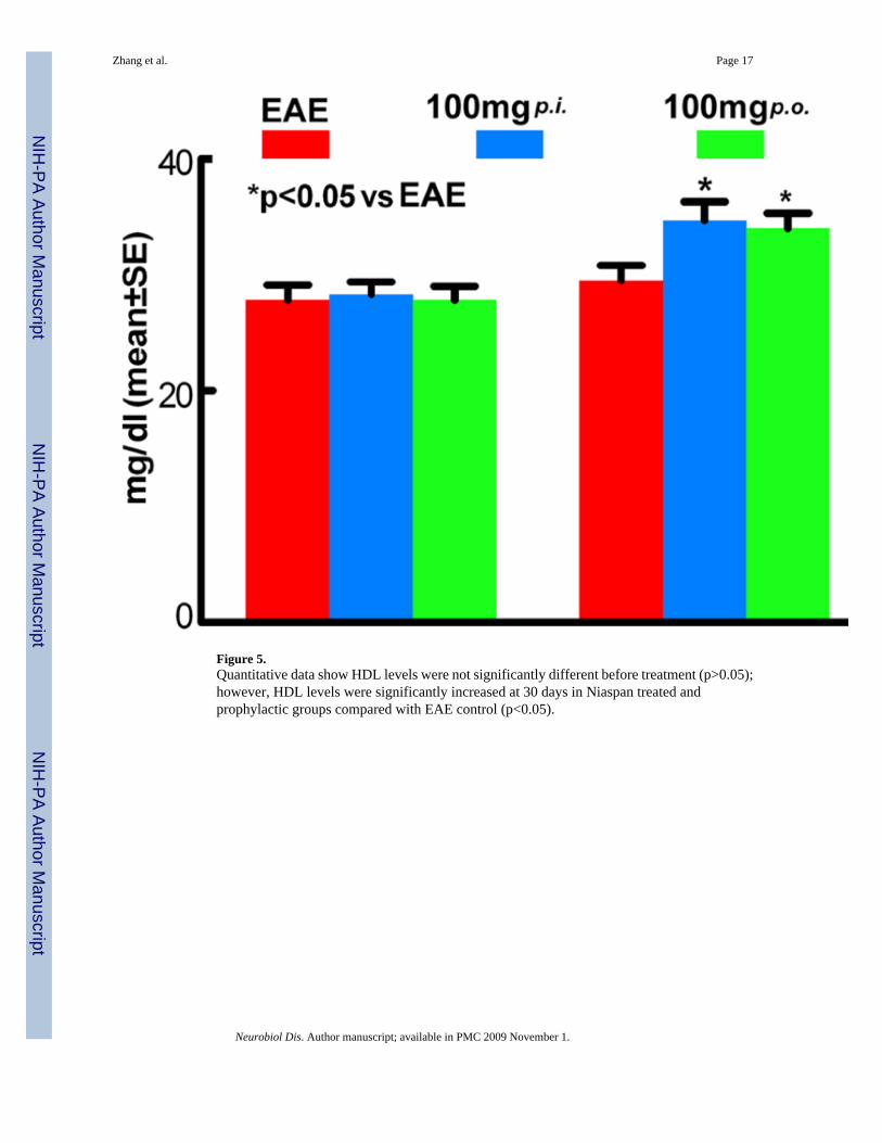
Figure 5.Quantitative data show HDL levels were not significantly different before treatment (p>0.05);however, HDL levels were significantly increased at 30 days in Niaspan treated andprophylactic groups compared with EAE control (p<0.05).
Zhang et al. Page 17
Neurobiol Dis. Author manuscript; available in PMC 2009 November 1.
NIH
-PA Author Manuscript
NIH
-PA Author Manuscript
NIH
-PA Author Manuscript
Related Documents


