THE JOURNAL OF COMPARATIVE NEUROLOGY 368~185-197 (1996) Distribution of Galanin-Like Immunoreactive Elements in the Brain of the Adult Lamprey Lampetra fluviatilis ANTONIO-J. JIMENEZ, JUAN-MIGUEL MANCERA, MANUEL A. POMBAL, JOSE-MANUEL PEREZ-F~GARES, AND PEDRO FERNANDEZ-LLEBREZ Departamentos de Biologia Celular y Genetica (A.-J.J., J.-M.P.-F.)y Biologia Animal (P.F.-Ll.), Facultad de Ciencias, Universidad de Malaga, Malaga, E-29071, Departamento de Biologia Animal, Facultad de Ciencias del Mar, Universidad de Cadiz, Puerto Real, Cadiz, E-11510 (J.-M.M.), Spain, and Nobel Institute for Neurophysiology (M.A.P.), Karolinska Institutet, S-104 01, Stockholm, Sweden ABSTRACT Galanin is a brain-gut peptide present in the central nervous system of vertebrates and invertebrates. The distribution of galanin-like immunoreactive perikarya and fibers in the brain of the river lamprey Lampetra fluviatilis (Agnatha) has been studied immunocytochemi- cally by using antisera against rat and porcine galanin. Galanin-like immunoreactive perikarya were seen in the telencephalon and mediobasal diencephalon. In the telencephalon, they were present in the nucleus olfactorius anterior, nucleus basalis, and especially, in the nucleus commissurae anterioris. The diencephalon contained most of the immunoreactive neurons. They were located in the nucleus commissurae praeinfundibularis, nucleus ventralis hypo- thalami, nucleus commissurae postinfundibularis, nucleus ventralis thalami, and nucleus dorsalis thalami pars medius. Most of the galanin-like immunoreactive infundibular neurons showed apical processes contacting the cerebrospinal fluid. Immunoreactive fibers and termi- nals were widely distributed throughout the neuraxis. In the telencephalon, the richest galaninergic innervation was found in the nucleus olfactorius anterior, lobus subhippocam- palis, corpus striatum, and around the nucleus septi and the nucleus praeopticus. In the diencephalon, the highest density of galanin-like immunoreactive fibers was seen in the nucleus commissurae postopticae, nucleus commissurae praeinfundibularis, nucleus ventralis hypo- thalami, nucleus dorsalis hypothalami, and neurohypophysis. In the mesencephalon and rhombencephalon, the distribution of immunoreactive fibers was heterogeneous, being most pronounced in a region between the nucleus nervi oculomotorii and the nucleus interpeduncu- laris mesencephali, in the nucleus isthmi, and in the raphe region. A subependymal plexus of immunoreactive fibers was found throughout the ventricular system. The distribution of immunoreactive neurons and fibers was similar to that of teleosts but different to those of other vertebrate groups. The possible hypophysiotropic and neuroregulatory roles of galanin are discussed. G 1996 Wiley-Liss, Inc. Indexing terms: immunocytochemical study, hypothalamus, neurohypophysis, cerebrospinal fluid- contacting neurons, Agnatha Galanin (GAL) is a 29-residue peptide that was originally isolated by Tatemoto et al. (1983) from the porcine intes- tine. Later it was shown that GAL is present in the central nervous system (CNS) of vertebrates. Immunocytochemi- cal studies have revealed galanin-like immunoreactive (GAL-ir) elements in the CNS of mammals, including man (Skofitsch and Jacabowitz, 1985; Melander et al., 1986; Levin et al., 1987; Palkovits et al., 1987; Blasco et al., 1989; Gaymann and Martin, 1989; Gentleman et al., 1989; Walker et al., 1989; Kordower and Mufson, 1990; Meister et al., 1990; Elmquist et al., 1992; Kordower et al., 19921, and in nonmammalian species, such as the dogfish (Vallarino et al., 19911, teleosts (Batten et al., 1990a,b; Cornbrooks and Accepted November 30,1995 Manuel A. Pombal’s present address is Departamento de Biologia Celular y Molecular, Facultad de Ciencias, Universidad de La Coruba, La Coruna, E-15071, Spain. Address reprint request to Dr. Antonio-J. Jimenez, Departamento de Biologia Celular y Genetica, Facultad de Ciencias, Universidad de Milaga, Malaga, E-29071, Spain. 8 1996 WILEY-LISS, INC.

Welcome message from author
This document is posted to help you gain knowledge. Please leave a comment to let me know what you think about it! Share it to your friends and learn new things together.
Transcript
THE JOURNAL OF COMPARATIVE NEUROLOGY 368~185-197 (1996)
Distribution of Galanin-Like Immunoreactive Elements in the Brain
of the Adult Lamprey Lampetra fluviatilis
ANTONIO-J. JIMENEZ, JUAN-MIGUEL MANCERA, MANUEL A. POMBAL, JOSE-MANUEL PEREZ-F~GARES, AND PEDRO FERNANDEZ-LLEBREZ
Departamentos de Biologia Celular y Genetica (A.-J.J., J.-M.P.-F.) y Biologia Animal (P.F.-Ll.), Facultad de Ciencias, Universidad de Malaga, Malaga, E-29071, Departamento de Biologia
Animal, Facultad de Ciencias del Mar, Universidad de Cadiz, Puerto Real, Cadiz, E-11510 (J.-M.M.), Spain, and Nobel Institute for Neurophysiology (M.A.P.),
Karolinska Institutet, S-104 01, Stockholm, Sweden
ABSTRACT Galanin is a brain-gut peptide present in the central nervous system of vertebrates and
invertebrates. The distribution of galanin-like immunoreactive perikarya and fibers in the brain of the river lamprey Lampetra fluviatilis (Agnatha) has been studied immunocytochemi- cally by using antisera against rat and porcine galanin. Galanin-like immunoreactive perikarya were seen in the telencephalon and mediobasal diencephalon. In the telencephalon, they were present in the nucleus olfactorius anterior, nucleus basalis, and especially, in the nucleus commissurae anterioris. The diencephalon contained most of the immunoreactive neurons. They were located in the nucleus commissurae praeinfundibularis, nucleus ventralis hypo- thalami, nucleus commissurae postinfundibularis, nucleus ventralis thalami, and nucleus dorsalis thalami pars medius. Most of the galanin-like immunoreactive infundibular neurons showed apical processes contacting the cerebrospinal fluid. Immunoreactive fibers and termi- nals were widely distributed throughout the neuraxis. In the telencephalon, the richest galaninergic innervation was found in the nucleus olfactorius anterior, lobus subhippocam- palis, corpus striatum, and around the nucleus septi and the nucleus praeopticus. In the diencephalon, the highest density of galanin-like immunoreactive fibers was seen in the nucleus commissurae postopticae, nucleus commissurae praeinfundibularis, nucleus ventralis hypo- thalami, nucleus dorsalis hypothalami, and neurohypophysis. In the mesencephalon and rhombencephalon, the distribution of immunoreactive fibers was heterogeneous, being most pronounced in a region between the nucleus nervi oculomotorii and the nucleus interpeduncu- laris mesencephali, in the nucleus isthmi, and in the raphe region. A subependymal plexus of immunoreactive fibers was found throughout the ventricular system. The distribution of immunoreactive neurons and fibers was similar to that of teleosts but different to those of other vertebrate groups. The possible hypophysiotropic and neuroregulatory roles of galanin are discussed. G 1996 Wiley-Liss, Inc.
Indexing terms: immunocytochemical study, hypothalamus, neurohypophysis, cerebrospinal fluid- contacting neurons, Agnatha
Galanin (GAL) is a 29-residue peptide that was originally isolated by Tatemoto et al. (1983) from the porcine intes- tine. Later it was shown that GAL is present in the central nervous system (CNS) of vertebrates. Immunocytochemi- cal studies have revealed galanin-like immunoreactive (GAL-ir) elements in the CNS of mammals, including man (Skofitsch and Jacabowitz, 1985; Melander et al., 1986; Levin et al., 1987; Palkovits et al., 1987; Blasco et al., 1989; Gaymann and Martin, 1989; Gentleman et al., 1989; Walker et al., 1989; Kordower and Mufson, 1990; Meister et al.,
1990; Elmquist et al., 1992; Kordower et al., 19921, and in nonmammalian species, such as the dogfish (Vallarino et al., 19911, teleosts (Batten et al., 1990a,b; Cornbrooks and
Accepted November 30,1995 Manuel A. Pombal’s present address is Departamento de Biologia Celular
y Molecular, Facultad de Ciencias, Universidad de La Coruba, La Coruna, E-15071, Spain.
Address reprint request to Dr. Antonio-J. Jimenez, Departamento de Biologia Celular y Genetica, Facultad de Ciencias, Universidad de Milaga, Malaga, E-29071, Spain.
8 1996 WILEY-LISS, INC.
186
Parsons, 1991a,b; Holmqvist and Ekstrom, 1991; Olivereau and Olivereau, 1991; Yamamoto et al., 1992; Magliulo- Cepriano et al., 1993; Anglade et al., 1994), amphibians (Wolfbauer and Skofitsch, 1989; Lazar et al., 1991; 01- ivereau and Olivereau, 1992), reptiles (Jimenez et al., 1994) and birds (Jozsa and Mess, 1993). In addition, GAL-ir elements have been found in the nervous system of inverte- brates (Lundqvist et al., 1991). In mammals, anatomical (Holmqvist and Carlberg, 1992) and physiological (Ottlecz et al., 1986; Koshiyama et al., 1987; Murakami et al., 1987; Sahu et al., 1987; Lopez et al., 1991) studies led to the proposal that GAL is a hypophysiotropic hormone. In the hypothalamus of mammals, GAL colocalizes with growth hormone releasing factor (GRF; Meister et al., 19901, vasopressin (Gai et al., 1990) and oxytocin (Landry et al., 1991). In fish, no such colocalization has been observed (Holmqvist and Ekstrom, 1991). Tracing studies demon- strated that in the rat, nearly 50% of the neurons of the hypothalamo-neurohypophyseal system contained immuno- reactive GAL (Arai et al., 1990). Galanin has been shown to be involved in nociceptive reception (Skofitsch and Jacobow- itz, 1985; Wiesenfeld-Hallin et al., 1990), learning and memory (Melander and Staines, 1986; Chan-Palay, 1988; Senut et al., 19891, and in cognitive, sensory, motor and autonomic processes (see Rokaeus, 1987; Kordover and Mufson, 1990). The list of probable functions involving GAL can even be bigger. Since the differentiation of the galaninergic system occurs early in the developing mamma- lian brain, a role of GAL in CNS differentiation has also been suggested (Sizer et al., 1990; Elmquist et al., 1992). The role of GAL in the CNS of nonmammalian vertebrates
A.-J. JIMENEZ ET AL.
is poorly understood. In fish, GAL has been implicated in olfactoryigustatory functions, central visual processing, somatosensory transmission, osmoregulation, and sex- specific behaviors (Cornbrooks and Parsons, 1991b; Holm- qvist and Carlberg, 1992).
Cyclostomes are representatives of the most primitive vertebrate group, the Agnatha. Due to this crucial position, the study of the galaninergic system in this group is of great phylogenetic interest. The present investigation was under- taken to study the distribution of GAL immunoreactivity in the brain of a cyclostome, the river lamprey (Lampetra fluuiatilis).
MATERIALS AND METHODS Animals
Investigation was performed on six adult (20-35 cm) river lampreys (Lampetru fluuiatilis L.). Animals were captured in the spring in Soderhamn (Sweden) during their upstream migratory phase, and kept in aerated freshwater aquaria at 4-1OoC, with a 12 hour:12 hour 1ight:dark cycle (illumination between 08:OO and 20:OO). Four animals were used to obtain transverse sections and two for sagittal sections. All brains used in this study were fixed in July, during the terminal spawning phase.
Immunocytochemical procedure The animals were anaesthetized with tricaine methane-
sulphonate (MS-222; 100 mg/l ; Sigma, Madrid, Spain) and killed by decapitation. The brains were quickly removed
AH C CGL CSF
ch CP CPt
D1 D2 D4 D4m D6 D7 D8 D9 D10 D11 D12 flm fr GO hc I I11 IV LLL M 1 M2 M3 M4 M5 M6 NB NH NL
cc
cs
adenohypophysis cerebellar plate corpus geniculatum laterale cerebrospinal fluid commissura cerebelli chiasma opticum commissura posterior commissura postectalis central stratum (tectum opticum) ganglion habenula dexter ganglion habenula sinister nucleus dorsalis thalami nucleus dorsalis thalami pars medius nucleus ventralis thalami nucleus tuberculi posterioris nucleus commissurae postopticae nucleus commissurae praeinfundibularis nucleus ventralis hypothalami nucleus dorsalis hypothalami nucleus commissurae postinfundibularis fasciculus longitudinalis medialis fasciculus retroflexus glomeruli olfactorii habenular commissure nucleus isthmi nucleus nervi oculomotorii nucleus trochlearis lateral line lobe nucleus commissurae posterioris nucleus fasciculi longitudinalis medialis tectum opticum torus semicircularis nucleus M5 of Schober nucleus interpeduncularis nucleus basalis neurohypophysis nucleus lentiformis mesencephali
Abbreviations
NP
P PP
PS Ra Rm Rr ri rPo rpos rpro sco T1 T2 T3 T4 T5 T6 T I T8 T9 T10 to tv V VII Vm vs vd vla
0s
PCh
ss
vm vr vt X
nucleus praetectalis optic stratum (tectum opticum) pineal organ parapineal organ plexus choroideus periventricular stratum (tectum opticum) raphe region mesencephalic reticular area rbombencephalic reticular area recessus infundibuli recessus postopticus recessus posterior recessus praeopticus subcommisural organ superficial stratum (tectum opticum) nucleus bulbi olfactorii nucleus septi nucleus olfactorius anterior nucleus commissurae anterior nucleus praeopticus primordium piriforme primordium hippocampi lobus subhippocampalis corpus striatum primordium pallii tractus opticus commissura tegmenti ventralis nucleus motorius nervi trigemini nucleus motorius nervi facialis radix motorius nervi trigemini radix sensorius nervi trigemini ventriculus diencephali ventriculus lateralis anterior telencephali ventriculus mesencephali ventriculus rhombencephali ventriculus medius telencephali nucleus vagus
DISTRIBUTION OF GALANIN IN THE LAMPREY BRAIN 187
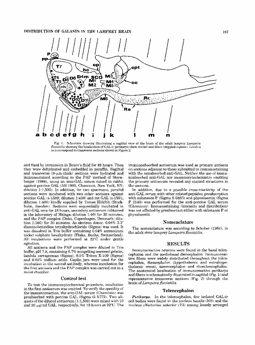
Fig. 1. Schematic drawing illustrating a sagittal view of the brain of the adult lamprey Lampetra fluuzatzlis showing the localization of GAL-ir perikarya (dark circles) and fibers (stippled regions). Levels a to n correspond to transverse sections shown in Figure 2.
and fixed by immersion in Bouin’s fluid for 48 hours. Then they were dehydrated and embedded in paraffin. Sagittal and transverse (8-pm-thick) sections were hydrated and immunostained according to the PAP method of Stern- berger (19861, using an anti-GAL serum raised in rabbit against porcine GAL (AE3 1985, Chemicon, New York, NY; dilution 1: 1,500). In addition, for two specimens, parallel sections were incubated with two other antisera against porcine GAL (s-1589; dilution 1:400) and rat GAL (s-1591; dilution 1:400) kindly supplied by Tomas Hokfelt (Stock- holm, Sweden). Sections were sequentially incubated in anti-GAL sera for 18 hours, secondary antiserum (obtained in the laboratory of Malaga; dilution 1:40) for 30 minutes, and the PAP complex (Dako, Copenhagen, Denmark; dilu- tion 1:lOO) for 30 minutes. As electron donor, 0.04% 3.3’ diaminobenzidine tetrahydrochloride (Sigma) was used. It was dissolved in Tris buffer containing 0.04% ammonium nickel sulphate hexahydrate (Fluka, Buchs, Switzerland). All incubations were performed at 22°C under gentle agitation.
All antisera and the PAP complex were diluted in Tris buffer, pH 7.8, containing 0.7% nongelling seaweed gelatin, lambda carrageenan (Sigma), 0.5% Triton X-100 (Sigma) and 0.02% sodium azide. Coplin jars were used for the incubation in the second antibody, whereas incubation for the first antisera and the PAP complex was carried out in a moist chamber.
Control test To test the immunocytochemical procedure, incubation
in the first antiserum was omitted. To verify the specifity of the immunoreaction, the anti-GAL serum (Chemicon) was preabsorbed with porcine GAL (Sigma G 5773). Two ali- quots of the diluted antiserum (1:1,500) were mixed with 10 and 20 pg/ml GAL, respectively, for 18 hours a t 22°C. The
immunoabsorbed antiserum was used as primary antisera on sections adjacent to those submitted to immunostaining with the nonabsorbed anti-GAL. Neither the use of immu- noabsorbed anti-GAL nor immunocytochemistry omitting the primary antiserum revealed any stained structures in the sections.
In addition, due to a possible cross-reactivity of the anti-GAL serum with other related peptides, preabsorption with substance P (Sigma S 6883) and physalaemin (Sigma P 2149) was performed for the anti-porcine GAL serum (Chemicon). Immunostaining (intensity and distribution) was not affected by preabsortion either with substance P or physalaemin.
Nomenclature The nomenclature was according to Schober (19641, in
the adult river lamprey Lampetra fluuiatilis.
RESULTS Immunoreactive neurons were found in the basal telen-
cephalon and the mediobasal diencephalon. Immunoreac- tive fibers were widely distributed throughout the telen- cephalon, diencephalon (hypothalamic and extrahypo- thalamic areas), mesencephalon and rhombencephalon. The anatomical localization of immunoreactive perikarya and fibers is schematically illustrated in sagittal (Fig. 1) and representative transverse sections (Fig. 2) through the brain of Lampetra fluuiatilis.
Telencephalon In the telencephalon, few isolated GAL-ir
cell bodies were found in the nucleus basalis (NB) and the nucleus olfactorius anterior (T3) among loosely arranged
Perikarya.
2
P
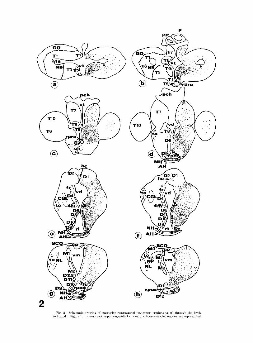
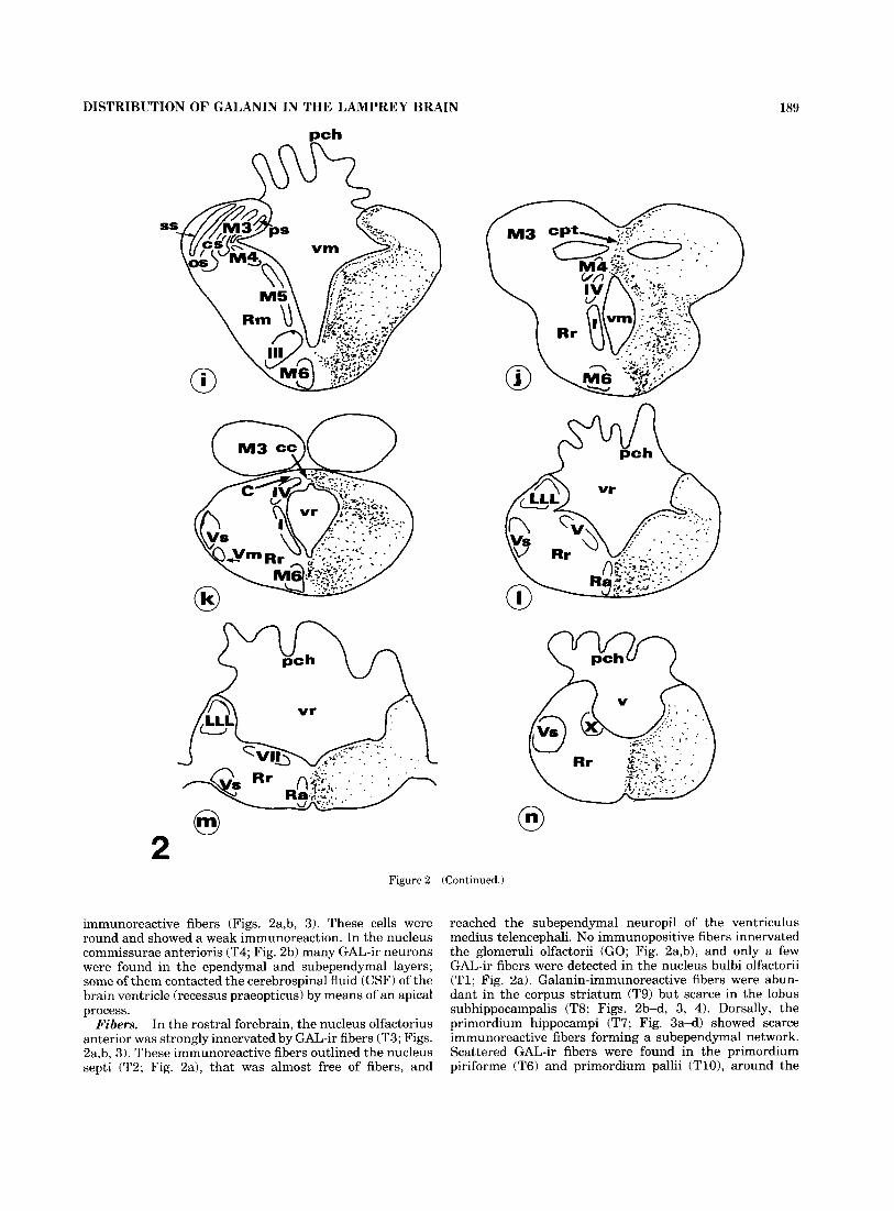
Fig. 2. Schematic drawing of successive rostrocaudal transverse sections (a-n) through the levels indicated in Figure 1. Immunoreactive perikarya (dark circles) and fibers (stippled regions) are represented.
DISTRIBUTION OF GALANIN IN THE LAMPREY BRAIN 189
Figure 2 (Continued.)
immunoreactive fibers (Figs. 2a,b, 3). These cells were round and showed a weak immunoreaction. In the nucleus commissurae anterioris (T4; Fig. 2b) many GAL-ir neurons were found in the ependymal and subependymal layers; some of them contacted the cerebrospinal fluid (CSF) of the brain ventricle (recessus praeopticus) by means of an apical process.
In the rostra1 forebrain, the nucleus olfactorius anterior was strongly innervated by GAL-ir fibers (T3; Figs. 2a,b, 3). These immunoreactive fibers outlined the nucleus septi (T2; Fig. 2a), that was almost free of fibers, and
Fibers.
reached the subependymal neuropil of the ventriculus medius telencephali. No immunopositive fibers innervated the glomeruli olfactorii (GO; Fig. 2a,b), and only a few GAL-ir fibers were detected in the nucleus bulbi olfactorii (Tl ; Fig. 2a). Galanin-immunoreactive fibers were abun- dant in the corpus striatum (T9) but scarce in the lobus subhippocampalis (T8; Figs. 2b-d, 3, 4). Dorsally, the primordium hippocampi (T7; Fig. 3a-d) showed scarce immunoreactive fibers forming a subependymal network. Scattered GAL-ir fibers were found in the primordium piriforme (T6) and primordium pallii (TlO), around the
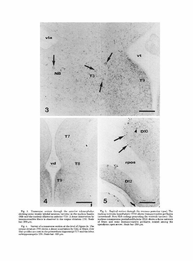
Fig. 3. Transverse section through the anterior telencephalon Fig. 5. Sagittal section through the recessus posterior (rpos). The showing some weakly labeled neurons (arrows) in the nucleus basalis nucleus ventralis hypothalami (D10) shows immunoreactive perikarya (NB) and the nucleus olfactorius anterior (T3). A dense innervation by (arrowhead). Note bleb endings protruding the ventricle (arrows). The immunoreactive fibers is observed in the corpus striatum (T9). Scale nucleus commissurae postinfundibularis (D12) shows a dense network bar: 200 pm. of fibers and some immunoreactive perikarya located among the
ependyma (open arrow). Scale bar: 200 pm. Survey of a transverse section at the level of Figure 2c. The
corpus striatum (T9) shows a dense innervation by GAL-ir fibers. Few fiber profiles are seen in the primordium hippocampi (T7) and the lobus subhippocampalis (T8). Scale bar: 400 pm.
Fig. 4.
DISTRIBUTION OF GALANIN IN THE LAMPREY BRAIN 191
ventriculus lateralis anterior telencephali (Fig. 2b-d). The area occupied by cell bodies in the nucleus praeopticus (T5) and the commissura anterior were devoid of GAL-ir fibers (Fig. 2b,c).
Diencephalon Perikarya. The hypothalamus contained most of the
GAL-ir neurons in the lamprey brain. Around the recessus postopticus, the nucleus commissurae praeinfundibularis (D9; Figs. 2d-f, 9) showed immunoreactive fusiform cell bodies arranged in 3-4 layers parallel to the ventricle. These cells had their long axis perpendicular to the epen- dyma and displayed an apical process that reached and protruded into the ventricle, and a long basal process that coursed deep into the lateral neuropil. The apical process passed through a dense subependymal network of GAL-ir fibers (Fig. 9). Some pear-shaped immunoreactive peri- karya were also found in the ependymal layer.
The most conspicuous population of GAL-ir cell bodies was found in the nucleus ventralis hypothalami (D10; Figs. 2 e g , 5-71, Immunoreactive cells were located among or beneath the ependyma, arranged into 3-5 layers parallel to the recessus infundibuli. Perikarya were bipolar, with a cerebrospinal fluid contacting process displaying a terminal bleb, and a basal process toward the lateral area. In the nucleus dorsalis thalami pars medius (D4m; Figs. 2e,f, 6) numerous fusiform GAL-ir neurons were arranged into layers parallel to the ventricle. These cells showed immuno- reactive beaded basal processes that ran toward the lateral neuropil. In the nucleus ventralis thalami (D6; Fig. 2e,f) few GAL-ir neurons were scattered among a dense immuno- reactive plexus.
In the floor of the recessus posterior, the nucleus commis- surae postinfundibularis (D12; Figs. 2h, 5, 6) showed few immunoreactive pear-shaped and cerebrospinal fluid- contacting GAL-ir neurons.
The hypothalamus of the lamprey displayed a very dense innervation of GAL-ir fibers in the nucleus commissurae postopticae (D8), the nucleus commissurae praeinfundibularis (D9), the nucleus ventralis hypothalami (DlO), and the nucleus dorsalis hypothalami (D11; Figs. 2d-h; 5-7; 9). A dense network of immunoreactive fibers was also present in the subependymal region and in the neuropil beneath these nuclei.
In the dorsal thalamus, the corpus geniculatum laterale (CGL) received a moderate innervation of GAL-ir fibers (Fig. 2 e 3 .
In the epithalamus, the habenula showed some GAL-ir fibers in its ventral part, and a few were seen to run along the habenular commissure (Fig. 2e,f). Galanin-like immuno- reactive fibers were absent from the pineal and parapineal organs (Fig. 2b).
In the pretectal region, GAL-ir fibers were seen in the nucleus praetectalis (NP), crossing the commissura poste- rior and bordering the subcommissural organ (Figs. 2g,h, 8). Occasional GAL-ir fibers coursed among cells of the subcommissural organ (Fig. 8).
The neural lobe of the hypophysis (NH) contained abun- dant GAL-ir fibers, both in its anterior and posterior aspects. At variance, the adenohypophysis (AH) lacked immunoreactive fibers (Figs. 2d-g, 9).
Within the diencephalon, the main fiber tracts, such as chiasma opticum, tractus opticus, commissura postoptica and fasciculus retroflexus, were free of immunoreactive fibers.
Fibers.
Mesencephalon and rhombencephalon Perikarya. Galanin-immunoreactive neurons were not
detected in the mesencephalon and the rhombencephalon of the lamprey.
Despite the absence of immunoreactive neu- rons, the mesencephalon and rhombencephalon showed a fairly rich innervation by GAL-ir fibers, mainly in the basal plate-derived regions. In the rostra1 mesencephalon, some GAL-ir fibers innervate the nucleus lentiformis mesen- cephali (NL; Fig. 2g,h). In the dorsal mesencephalon, the periventricular and central strata of the tectum opticum received a moderate innervation of GAL-ir fibers (Figs. 2ij, 11). At variance, the superficial and the optic strata were devoid of label. Many immunopositive fibers were found in the commissura posttectalis and around the torus semicir- cularis (M4; Fig. 2ij).
In the mesencephalon, the nuclei around the ventriculus mesencephali such as the nucleus commissurae posterioris (Ml), the nucleus fasciculi longitudinalis medialis (M2), and the nucleus M5 of Schober (M5; Figs. 2g-i; 10) lacked GAL-ir fibers. However, a subependymal network of immu- nopositive fibers was noted in these areas. The area occu- pied by mesencephalic reticular cells (Muller cells) and their dendrites (Rm) showed scarce GAL-ir fibers (Fig. 2i). Immunoreactive fibers were also seen in the neuropil among the nucleus oculomotorii (1111, the nucleus interpe- duncularis (M6), and the fasciculus retroflexus (Figs. 2i-k, 10).
In the isthmus rhombencephali, the density of GAL-ir fibers was particularly high in the periventricular areas near to the mesencephalon and in the subependymal area at the level of the trochlear nucleus (IV; Fig. 2j,k). Some innervation was detected in the nucleus isthmi (I; Fig. 2j,k). In addition, some immunoreactive fibers were seen in the cerebellar plate and crossing the commissura cerebelli (Figs. 2k, 11).
In the rhombencephalon proper, most GAL-ir fibers distributed in the basal plate, especially in the raphe region (Ra; Fig. 21,m). The rhombencephalic reticular area (Rr; Figs. 2j-n, 10) contained numerous GAL-ir fibers, whereas other areas such as the lateral line lobe (LLL; Fig. 21,m) and the motor nuclei (V, VII, X and nucleus glossopharingicus) were almost devoid of GAL-ir innervation. A sparse net- work of GAL-ir fibers was found in the subependymal region of the ventriculus rhombencephali. Some GAL-ir fibers were also intermingled, and sometimes in close apposition with the giant descending reticular axons.
Fibers.
DISCUSSION The amino acidic sequence of GAL varies among mam-
mals (cf. Rokaeus and Brownstein, 1986; Kaplan et al., 1988) and among nonmammalian vertebrates (McKeon et al., 1990; Anglade et al., 1994). However, anti-porcine GAL antisera have been reported to cross-react with GAL of mammals, including man (Skofitsch and Jacobowitz, 1985; Melander et al., 1986; Levin et al., 1987; Palkovits et al., 1987; Blasco et al., 1989; Gaymann and Martin, 19891, of nonmammalian vertebrates (teleosts: Batten et al., 1990a,b; Cornbrooks and Parsons, 1991a,b; Holmqvist and Ek- strom, 1991; Olivereau and Olivereau, 1991; amphibians: Lazar et al., 1991; Olivereau and Olivereau, 1992; reptiles: Jim6nez et al., 1994) and of invertebrates (Lundquist et al., 1991). The present study has been carried out using two antiporcine GAL sera and an anti-rat GAL serum in a
192 A.-J. JIMENEZ ET AL.
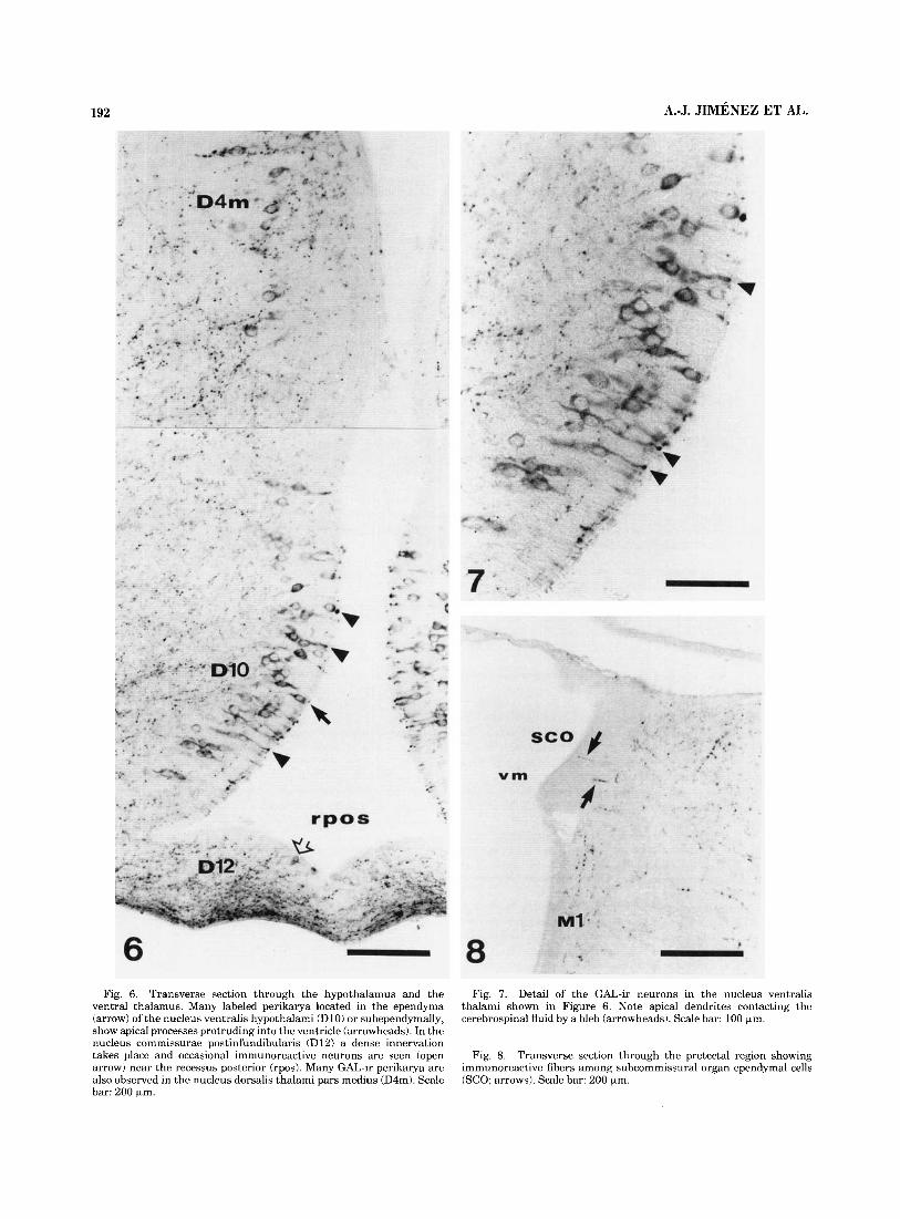
Fig. 6. Transverse section through the hypothalamus and the Fig. 7. Detail of the GAL-ir neurons in the nucleus ventralis thalami shown in Figure 6. Note apical dendrites contacting the cerebrospinal fluid by a bleb (arrowheads). Scale bar: 100 pm.
ventral thalamus. Many labeled perikarya located in the ependyma (arrow) of the nucleus ventralis hypothalami (D10) or subependymally, show apical processes protruding into the ventricle (arrowheads). In the nucleus commissurae postinfundibularis (D12) a dense innervation takes place and occasional immunoreactive neurons are seen (open arrow) near the recessus posterior (rpos). Many GAL-ir perikarya are also observed in the nucleus dorsalis thalami pars medius (D4m). Scale bar: 200 pm.
Fig. 8. Transverse section through the pretectal region showing immunoreactive fibers among subcommissural organ ependymal cells (SCO; arrows). Scale bar: 200 pm.
9 - ?
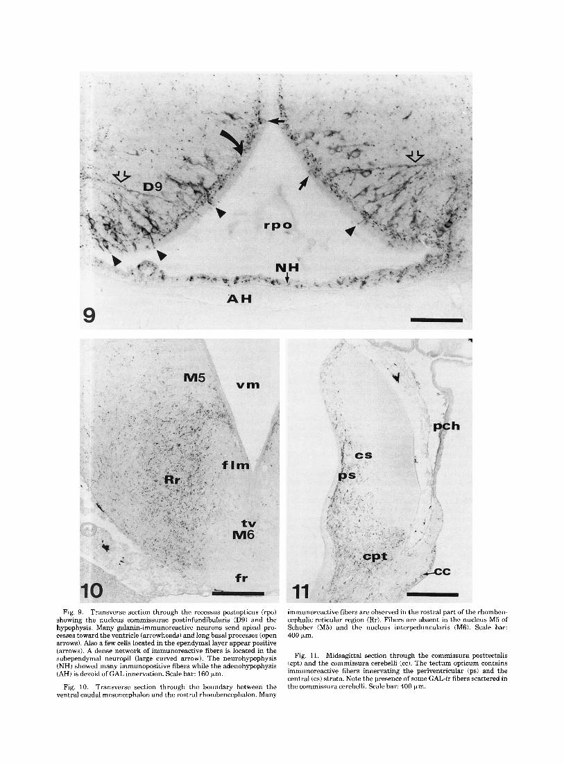
Fig. 9. Transverse section through the recessus postopticus (rpo) showing the nucleus commissurae postinfundibularis (D9) and the hypophysis. Many galanin-immunoreactive neurons send apical pro- cesses toward the ventricle (arrowheads) and long basal processes (open arrows). Also a few cells located in the ependymal layer appear positive (arrows). A dense network of immunoreactive fibers is located in the subependymal neuropil (large curved arrow). The neurohypophysis (NH) showed many immunopositive fibers while the adenohypophysis (AH) is devoid of GAL innervation. Scale bar: 160 pm.
Transverse section through the boundary between the ventral caudal mesencephalon and the rostral rhombencephalon. Many
Fig. 10.
immunoreactive fibers are observed in the rostral part of the rhomben- cephalic reticular region (Rr). Fibers are absent in the nucleus M5 of Schober (M5) and the nucleus interpeduncularis (M6). Scale bar: 400 p,m.
Fig. 11. Midsagittal section through the commissura posttectalis (cpt) and the commissura cerebelli (cc). The tectum opticum contains immunoreactive fibers innervating the periventricular (ps) and the central (cs) strata. Note the presence of some GAL-ir fibers scattered in the commissura cerebelli. Scale bar: 400 pm.
194 A.-J. JIMENEZ ET AL.
species of an ancient vertebrate line, the adult river lam- prey Lampetra fluviatilis. Perikarya and fibers were immu- nostained throughout the brain identically with all anti- sera. The reaction was abolished by preincubation with porcine GAL in one antiporcine GAL serum (Chemicon). Thus, a GAL-like peptide or a compound cross-reacting with an antiporcine GAL serum exists in the lamprey brain.
Since there is some molecular similarity among GAL, substance P and physaelamin (Rokaeus et al., 19841, cross- reactions of these substances with GAL antisera could be expected to occur. By use of immunocytochemistry, these closely related molecules have been located in the hypothala- mus of teleosts in structures different from those revealed by anti-GAL (Holmqvist and Ekstrom, 1991). In the pres- ent investigation, preincubation of the anti-GAL serum (Chemicon) with substance P and physaelamin was carried out as control. Since immunoreaction was unaffected it is unlikely that galanin-like substance revealed by the antipor- cine GAL serum is not substance P or physaelamin.
In general, the distribution of GAL immunoreactivity in the brain of Lampetra fluviatilis was quite similar to that in teleosts.
Hypothalamic areas The distribution of GAL-ir neurons in the lamprey is
quite similar to teleosts, having perikarya in the preoptic and tuberal hypothalamus (Batten et al., 1990a,b; Corn- brooks and Parsons, 1991a,b; Holmqvist and Ekstrom, 1991; Olivereau and Olivereau, 1991; Anglade et al., 1994). In amphibians and birds, in addition to the hypothalamus, the telencephalon, mesencephalon and rhomboencephalon were also reported to display GAL-ir neurons (Lazar et al., 1991; Olivereau and Olivereau, 1992; JBzsa and Mess, 1993). In the turtle, we have found GAL-ir perikarya in telencephalon, diencephalon, and rhombencephalon, but not in the mesencephalon (Jimenez et al., 1994). Immuno- reactive neurons have been found in extrahypothalamic areas of mammals (Skofitsch and Jacobowitz, 1985; Elm- quist et al., 1992; Kordower et al., 1992).
The present results show that the hypothalamus and the ventral thalamus of the lamprey contained many GAL-ir neurons in the periventricular areas. Most of these cells had CFS-contacting apical processes. Cerebrospinal fluid- contacting neurons containing immunoreactive GAL were reported before in teleosts (Holmqvist and Ekstrom, 1991), anurans (Lazar et al., 1991; Olivereau and Olivereau, 1992) and the turtle (Jimenez et al., 1994). In lampreys, CSF- contacting neurons have been found to be immunoreactive to antisera against several substances such as serotonin (Brodin et al., 1988a, 1990a; Pierre et al., 19921, somatosta- tin (Wright, 1986; YaAez et al., 1992), substance P (Nozaki and Gorbman, 19861, cholecystokinin (Brodin et al., 1988b), gonadotropin-releasing hormone (King et al., 1988), the molluscan cardio-excitatory tetrapeptide (Ohtomi et al., 1989) neurotensin (Brodin et al., 1990b), histamine (Brodin et al., 1990a), dopamine and tyrosine hydroxylase (Pierre et al., 19941, and opsins (Garcia-Fernandez and Foster, 1994). The brain of lamprey is considered to have conserved many characteristics of the primitive vertebrate brain (Nieuwen- huys, 1977). Neurons of the CSF-contacting type have been considered to reflect a primitive character of the vertebrate brain (Vigh-Teichmann and Vigh, 1989).
In the brain of Lampetra fluuiatilis, an interesting fact
tem, including the regions containing nuclei lacking galanin- ergic innervation. This galaninergic subependymal network could control the activity of other cerebrospinal fluid- contacting neurons or could be related with the activity of the ependymal cells.
Many GAL-ir fibers have been found in the neurohypophy- sis of Lampetra fluviatilis. The anterior neurohypophysis of lampreys is equivalent to the mammalian median emi- nence, whereas the posterior neurohypophysis corresponds to the neural lobe (see Tsuneki, 1988). The lamprey ante- rior neurophypophysis displays immunoreactivity to: arginine vasotocin (Goossens et al., 19771, luteinizing hormone-releas- ing hormone (Nozaki and Kobayashi, 19791, enkephalin (Dores et al., 1984; Nozaki and Gorbman, 1984) and substance P (Nozaki and Gorbman, 1986). The posterior neurohypophysis of the lamprey presents immunoreaction for: arginine vasoto- cin (Goossens et al., 1977), luteinizing hormone-releasing hor- mone (Crim et al., 1979; Nozaki and Gorbman, 1986), met- enkephalin (Nozaki and Gorbman, 1984), prolactin and growth hormone-like (Wright, 19861, and somatostatin (Yafiezet al., 1992). The presence of GAL-like immunoreactivity in the lamprey anterior and posterior neurohypophysis suggests that GAL could act either as a blood-borne hormone and/or as a hypophysiotropic factor. Alternatively, GAL could have a role as a neuromodulator of the release of neurohypophy- seal hormones and hypothalamic releasing factors.
Evidence for the control of adenohypophysis by GAL has been reported in vertebrates. Galanin can be released to the portal capillaries in the outer layer of the median eminence of amphibians (Lazar et al., 1991), the turtle (Jimenez et al., 19941, birds (Jozsa and Mess, 1993) and mammals (Skofitsch and Jacobowitz, 1985; Melander et al., 1986; Palkovits et al., 1987; Beal et al., 1988; Gaymann and Martin, 1989; Walker et al., 1989; Kordower et al., 1992). In this way it could modulate the activity of adenohypophyseal cells. Indeed, in mammals, GAL has been reported to exert a hypophysiotropic role affecting the release of adenohy- pophyseal hormones (Ottlecz et al., 1986; Koshiyama et al., 1987; Murakami et al., 1987; Sahu et al., 1987; Lopez et al., 1991). In teleosts lacking a hypophyseal portal vascular system, direct projections of GAL-ir fibers to the adenohy- pophysis were reported (Batten et al., 1990a,b; Holmqvist and Ekstrom, 1991; Olivereau and Olivereau, 1991; Ang- lade et al., 1994); this is in agreement with the presence of GAL receptors in the adenohypophysis of teleosts (Moonset al., 1991). Like teleosts, lampreys lack a hypothalamo- hypophyseal portal system; but unlike teleosts, the adenohy- pophysis of the lamprey is not directly innervated from the hypothalamus. Hence, different mechanisms have been proposed to exist for hypothalamic control of the adenohy- pophysis in lampreys; these include releasing of the neuro- secretory materials in the external layer of the neurohy- pophysis and subsequent transport through the underlying connective tissues or, less likely, releasing to the systemic circulation in the neurohypophysis or, indirectly, via the cerebrospinal fluid, after releasing in the third ventricle (see King et al., 1988). In Lampetra fluuiatilis, abundant fibers were found in all layers of the neurohypophysis including the external layer. The exact mechanism of delivery from hypothalamic neurons to adenohypophyseal target cells, if any, is unknown in Lampetra fluuiatilis.
Extrahypothalamic areas was the presence of an-extended subependymal network of immunoreactive fibers in virtually all the ventricular sys-
Mammals and birds have GAL-ir cell bodies in the septum (rat: Melander et al., 1986; monkey: Melander and

DISTRIBUTION OF GALANIN IN THE LAMPREY BRAIN 195
Staines, 1986; Kordower et al., 1992; human: Kordower et al., 1992; chicken: Jozsa and Mess, 1993). In this same location GAL-ir fibers were described in anuran (Lazar et al., 1991; Olivereau and Olivereau, 1992) and the turtle (Jimenez et al., 1994). In teleosts, a rich network of fibers is located rostrally in the telencephalic floor (Batten et al., 1990a,b; Holmqvist and Ekstrom, 1991; Olivereau and Olivereau, 1991; Anglade et al., 1994). In Lampetrafluuiati- lis few GAL-ir fibers were observed in the septum itself, but many of them were distributed in the periventricular and lateral neuropil bordering this area. In mammals, the septum has been involved in the control of water balance (Miselis, 1981; Tanaka et al., 1988).
It is known that the striatum of several vertebrates is involved in processing sensory information and in motor control. In mammals, GAL coexists with acetylcholine in cholinergic neurons of the basal forebrain involved in learning and memory (Ogren et al., 1992), and it was proposed that GAL could inhibit the activity of cholinergic neurons (Melander et al., 1986; Chan-Palay, 1988). Galanin also exerts an inhibitory effect in the serotoninergic system either by a presynaptic inhibition (Sundstrom and Melander, 1988) or by being an inhibitor of serotonin receptors (cf. Fuxe et al., 1988). A strong acetylcholinesterase activity has also been demonstrated in the corpus striatum of lampreys (Wachtler, 1974), while the surrounding neuropil has been found to contain immunoreactive fibers to antisera against serotonin (Brodin et al., 1988a, 1990a; Pierre et al., 19921, substance P (Nozaki and Gorbman, 1986), neurotensin (Brodin et al., 1990b), histamine (Brodin et al., 1990a), dopamine and tyrosine hydroxylase (Pierre et al., 1994). The dense GAL innervation of the corpus striatum of Lampetra fluuiatilis shown in this study suggests that GAL may modulate cholinergic and/or serotoninergic and other activities in this telencephalic region.
In the present study, no immunoreactive neurons were found in the brainstem of Lampetra fluuiatilis. This is also the case of some teleosts (see: Holmqvist and Ekstrom, 1991; Olivereau and Olivereau, 1991; Anglade et al., 1994). In contrast, mammals showed GAL-ir neurons throughout the brainstem (Skofitsch and Jacobowitz, 1985; Melander et al., 1986; Kordower et al., 1992). Although lacking GAL-ir neurons, the brainstem of the lamprey is richly innervated by GAL-ir fibers, indicating some role of GAL in this area.
In the tectum opticum of Lampetra fluuiatilis, GAL-ir fibers were found near the ventricle. A similar pattern was reported for teleosts (Batten et al., 1990a,b; Holmqvist and Ekstrom, 1991; Olivereau and Olivereau, 19911, anurans (Lazar et al., 1991; Olivereau and Olivereau, 19921, and the turtle (J imhez et al., 1994). At variance, few GAL-ir fibers were described in the tectum opticum of the chicken (Jozsa and Mess, 1993) and the superior colliculus of the rat (Skofitsch and Jacobowitz, 1985; Melander et al., 1986). In this respect, differences seem to exist between birds and mammals on one hand, and fishes, amphibians and the turtle on the other.
Unlike teleosts, the cerebellar plate of Lampetra fluviati- lis showed few GAL-ir fibers. This characteristic therefore seems to be exclusive of Agnatha.
Some scattered GAL-ir fibers were found between the descending reticular axons in the ventromedial rhomben- cephalon. It is known that reticulospinal neurons play a crucial role in the initiation of locomotion, by conveying information from the brainstem to the spinal cord
(Rovainen, 1967; Wickelgren, 1977; Brodin et al., 1988b). Sometimes GAL-ir fibers were seen to be closely associated with reticular axons, establishing putative en passant con- tacts. Galanin could therefore modulate this descending reticulospinal pathway.
CONCLUSION The present study indicates that a GAL-like substance is
present in the lamprey brain. The galaninergic system in the brain of lampreys is well developed as compared with that described in teleosts, but presents some differences with respect to other vertebrate groups.
ACKNOWLEDGMENTS This work was supported by DGICYT grant PB93.0979,
Madrid (Spain) to P.F.-LL., FIS 15/1591 to J.-M.P.-F., and by a postdoctoral fellowship from the Xunta de Galicia (Spain) to M.A.P. We thank Dr. S. Grillner for providing lamprey brains and Dr. T. Hokfelt for providing two of the antisera against GAL (s-1589 and s-1591). We also thank Dr. Ramon Anadon and Dr. Esteban M. Rodriguez for critical reading of the manuscript.
LITERATURE CITED Anglade, I., Y. Wang, J. Jensen, G. Tramu, 0. Kah, and J.M. Conlon (1994)
Characterization of trout galanin and its distribution in trout brain and pituitary. J. Comp. Neurol. 350:63-74.
Arai, R., B. Onteniente, A. Trembleau, M. Landry, and A. Calas (1990) Hypothalamic galanin-immunoreactive neurons projecting to the poste- rior lobe of the rat pituitary: A combined retrograde tracing and immunohistochemical study. J. Comp. Neurol. 299:405420.
Batten, T.F.C., M.L. Cambre, L. Moons, and F. Vandesande (1990a) Comparative distribution of neuropeptide-immunoreactive system in the brain of the green molly, Poeczlia latzpznna. J. Comp. Neurol. 302893- 919.
Batten, T.F.C., L. Moons, M. Cambre, and F. Vandesande (1990b) Anatomi- cal distribution of galanin-like immunoreactivity in the brain and pituitary ofteleost fishes. Neurosci. Lett. 111:lZ-17.
Beal, M.F.S., M. Gabriel, K.J. Swartz, and U. MacGarvey (1988) Distribution of galanin-like immunoreactivity in the baboon brain. Peptides 93447- 851.
Blasco, I., F.J. Alvarez, R.M. Villalba, M.L. Solano, R. Martinez-Murrillo, and J. Rodrigo (1989) Light and electron microscopic study of galanin- immunoreactive nerve fibers in the rat posterior thalamus. J. Comp. Neurol. 283:l-12.
Brodin, L., J.T. Buchanan, T. Hokfelt, S. Grillner, J.F. Rehfeld, P. Frey, A.A.J. Verhofstad, G.J. Dockray, and J.H. Walsh (1988a) Immunohisto- chemical studies of cholecystokinin-like peptides and their relation to 5-HT, CGRP, and bombesin immunoreactivities in the brainstem and spinal cord of lampreys. J. Comp. Neurol. 27l:l-18.
Brodin, L., S. Grillner, R. Dubuc, Y. Ohta, S. Kasicki, and T. Hokfelt (1988b) Reticulospinal neurons in lamprey: Transmitters, synaptic interactions and their role during locomotion. Arch. Ital. Biol. 126:317-345.
Brodin, L., T. Hokfelt, S. Grillner, and P. Panula (1990a) Distribution of histaminergic neurons in the brain of the lamprey Lampetra fluuiatslas as revealed by histamine-immunohistochemistry. J. Comp. Neurol. 292:435-442.
Brodin, L., E. Theodorsson, J. Christenson, S. Cullheim, T. Hokfelt, J.C. Brown, A. Buchan, P. Panula, A.A.J. Verhofstad, and M. Goldstein (1990b) Neurotensine-like peptides in the CNS of lampreys: Chromato- graphic characterization and immunohistochemical localization with reference to aminergic markers. Eur. J. Neurosci. 2:1095-1109.
Chan-Palay, V. (1988) Neurons with galanin innervate cholinergic cells in the human basal forebrain and galanin and acetylcholine coexist. Brain Res. Bull. 21:465-472.
Cornbrooks, E.B., and R.L. Parsons (1991a) Sexually dimorphic distribution of a galanin-like peptide in the central nervous system of the teleost fish Poecilia latipinnu. J. Comp. Neurol. 3043539-657.
196 A.-J. JIMENEZ ET AL.
Cornbrooks, E.B., and R.L. Parsons (1991b) Source of sexually dimorphic galanin-like immunoreactive projections in the teleost fish Poecilia latipinnu. J. Comp. Neurol. 304,658-665.
Crim, J.W., A. Urano, and A. Gorbman (19791 Immunocytochemical studies of luteinizing hormone-releasing hormone in brains of agnathan fishes. I. Comparisons of adult Pacific lamprey (Entosphenus tridentataj and the Pacific hagfish (Eptah-etus stouti). Gen. Comp. Endocrinol. 372296305.
Dores, R.M., T.E. Finger, and M.R. Gold (1984) Immunohistochemical localization of enkephalin-and ACTH-related substances in the pituitary of the lamprey. Cell Tissue Res. 235:107-115.
Elmquist, J.K., C.A. Fox, L.R. Ross, and C.D. Jacobson 11992) Galanin-like immunoreactivity in the adult and developing Brazilian opossum brain. Dev. Brain Res. 67:161-179.
Fuxe, K., S.O. Ogren, A. Jansson, A. Cintra, A. Hiirfstrand, and L.F. Agnati (1988) Intraventricular injections of galanin reduces 5-HT metabolism in the ventral limbic cortex, the hippocampal formation and the frontopa- rietal cortex of the male rat. Acta Physiol. Scand. 133,579-581.
Gai, W.P., L.B. Geffen, and W.W. Blessing (1990) Galanin immunoreactive neurons in the human hypothalamus: Colocalization with vasopressin- containing neurons. J. Comp. Neurol. 298:265-280.
Garcia-Fernandez, J.M., and R.G. Foster 11994) Immunocytochemical iden- tification of photoreceptor proteins in hypothalamic cerebrospinal fluid- contacting neurons of the larval lamprey (Petromyzon marinus). Cell Tissue Res. 275:319-326.
Gaymann, W., and R. Martin (19891 Immunoreactive galanin-like material in magnocellular hypothalamo-hypophysial neurones of the rat. Cell Tissue Res. 255:139-147.
Gentleman, S.M., P. Falkkai, B. Bogerts, M.T. Herrero, J.M. Polak, and G.W. Roberts (1989) Distribution of GAL-like immunoreactivity in the human brain. Brain Res. 505:311-315.
Goossens, N., K. Dierickx, and F. Vandesande (1977) Immunocytochemical demonstration of the hypothalamo-hypophysial vasotocinergic system of Lampetrafluuiatilis. Cell Tissue Res. 177:317-323.
Holmqvist, B.I., and M. Carlberg (1992) Galanin receptors in the brain of a teleost: Autoradiographic distribution of binding sites in the atlantic salmon. J. Comp. Neurol. 326:44-60.
Holmqvist, B.I., and P. Ekstrom (1991) Galanin-like immunoreactivity in the brain of teleosts: Distribution and relation to substance P, vasotocin, and isotocin in the atlantic salmon (Salmo salar). J. Comp. Neurol. 306:361-381.
Jimenez, A.J., J.M. Mancera, J.M. Perez-Figares, and P. Fernandez-Llebrez (1994) Distribution of galanin-like immunoreactivity in the brain of the turtle Mauremys caspica. J. Comp. Neurol. 349:73-84.
Jozsa, R., and B. Mess (1993) Galanin-like immunoreactivity in the chicken brain. Cell Tissue Res. 273:391-399.
Kaplan, L.M., E.R. Sinder, K.J. Isselbacher, and W.W. Chin 11988) Tissue specific expression of rat galanin gene. Proc. Natl. Acad. USA 85:10651069.
King, J.C., S.A. Sower, and E.L.P. Anthony (1988) Neuronal systems immunoreactive with antiserum to lamprey gonadotrophin-releasing hormone in the brain of Petrom,yzon marinus. Cell Tissue Res. 253:l-8.
Kordower, J.H., and E.J. Mufson 11990) Galanin-like immunoreactivity within the primate basal forebrain: Differential staining patterns be- tween humans and monkeys. J. Comp. Neurol. 294:281-292.
Kordower, J.H., H.K. Le, and E.J. Mufson (1992) Galanin immunoreactivity in the primate central nervous system. J. Comp. Neurol. 319,479-500.
Koshiyama, H., Y. &to, I. Tatsuhide, Y. Murakami, Y. Ishikawa, N. Yanaihara, and H. Imura 11987) Central galanin stimulates prolactin secretion in rats: Possible involvement of hypothalamic vasoactive intestinal polypeptide. Neurosci. Lett. 7549-54.
Landry, M., A. Trembleau, R. Arai, and A. Calas (1991) Evidence for a colocalization of oxitocin mRNA and galanin in magnocellular hypotha- lamic neurons: A study combining in situ hybridization and immunohis- tochemistry. Mol. Brain Res. 10:91-95.
LAzar, G., 2. Liposits, P. Toth, S.L. Trasti, J.L. Maderdrut, and I. Merchen- thaler (1991) Distribution of galanin-like immunoreactivity in the brain ofRana esculenta andxenopus laeuis. J. Comp. Neurol. 310:45-67.
Levin, M.C., P.E. Sawchenko, P.R.C. Howe, S.R. Bloom, and J.M. Polak (19871 Organization of galanin-immunoreactive inputs to the paraven- tricular nucleus with special reference to their relationship to catechol- aminergic afferents. J. Comp. Neurol. 261:562-582.
Lopez, F.J., I. Merchenthaler, M. Ching, M.G. Wisniewski, and A. Negro- Vilar (1991) Galanin: A hypothalamic hypophysiotropic hormone modu- lating reproductive function. Proc. Natl. Acad. Sci. USA 88:4508-4512.
Lundquist, C.T., A. Rokaeus, and R. Nassel(1991) Galanin immunoreactiv- ity in the blowfly nervous system: Localization and chromatographic analysis. J. Comp. Neurol. 31277-96.
Magliulo-Cepriano, L., M.P. Schreibman, and V. Blum I19931 The distribu- tion of immunoreactive FMRF-amide, neurotensin, and galanin in the brain and pituitary gland of three species of Xiphophorus from birth to sexual maturity. Gen. Comp. Endocrinol. 92269-280.
McKeon, T., R.E. Carravay, L.M. Konopka, and R.L. Parsons (1990) Distribution of galanin-like peptide in various tissues of Necturus musculosus. Cell Tissue Res. 262:461-466.
Meister, B., M.F. Scanlon, and T. Hokfelt (1990) Occurence of galanin-like immunoreactivity in growth hormone-releasing factor (GRF)-containing neurons of the monkey (Macaca fascicularis) infundibular nucleus and median eminence. Neurosci. Lett. 119:136-139.
Melander, T., and W.A. Staines (1986) A galanin-like peptide coexists in putative cholinergic neurons of the septum-basal forebrain complex and in acetylcholinesterase-containing fibers and varicosities within the hippocampus in the owl monkey IAotus triuirgatus). Neurosci. Lett. 68: 17-22.
Melander, T., T. Hiikfelt, and A. Rokaeus (1986) Distribution of galanin-like immunoreactivity in the rat central nervous system. J. Comp. Neurol. 248:475-517.
Miselis, R.R. (1981) The efferent projections of the subfornical organ of the rat: A circumventricular organ within a neural network subserving water balance. Brain Res. 23O:l-23.
Moons, L., T.F.C. Batten, and F. Vandesande (1991) Autoradiographic distribution of galanin-binding sites in the brain and pituitary in the sea bass (Dicentrarchus labraxl. Neurosci. Lett. 123.49-52.
Murakami, I., Y. Kato, H. Koshiyama, T. Inoue, N. Yanaihara, and H. Imura (1987) Galanin stimulates growth hormone (GH) secretion via GH- releasing factor (GRF) in conscious rats. Eur. J. Pharmacol. 136:415-418.
Nieuwenhuys, R. (1977) The brain of the lamprey in a comparative perspective. Ann. New York Acad. Sci. 299:97-145.
Nozaki, M., and A. Gorbman (1984) Distribution of immunoreactive sites for several components of pro-opiocortin in the pituitary and brain of adult lampreys, Petromyzon marinus and Entosphenus tridentatus. Gen. Comp. Endocrinol. 53:335-352.
Nozaki, M., and A. Gorbman (1986) Occurence and distribution of substance P-related immunoreactivity in the brain of adult lampreys, Petromyzon marinus and Entosphenus tridentatus. Gen. Comp. Endocrinol. 62:217- 229.
Nozaki, M., and H. Kobayashi 11979) Distribution of LHRH-like substance in the vertebrate brain as revealed by immunohistochemistry. Arch. Histol. Jpn. 42201-219.
Ogren, S.O., T. Hokfelt, K. Kask, U. Langel, and T. Bartfai (1992) Evidence for a role of the neuropeptide galanin in spatial learning. Neuroscience 51:l-5.
Ohtomi, M., K. Fujii, and H. Kobayashi (1989) Distribution of FMRFamide- like immunoreactivity in the brain and neurohypophysis of the lamprey, Lampetrajaponica. Cell Tissue Res. 256:581-584.
Olivereau, M., and J.M. Olivereau (1991) Immunocytochemical localization of a galanin-like peptidergic system in the brain and pituitary of some teleost fish. Histochemistry 96:343-354.
Olivereau, M., and J.M. Olivereau (19921 Immunocytochemical localization of a galanin-like peptidergic system in the brain of two urodele and two anuran species (Amphibia). Histochemistry 98:51-66.
Ottlecz, A., W.K. Samson, and S.M. McCann (19861 Galanin: Evidence for a hypothalamic site of action to release growth hormone. Peptides 7:51-53.
Palkovits, M., A. Rokaeus, F.A. Antoni, and A. Kiss (1987) Galanin in the hypothalamo-hypophysial system. Neuroendocrinology 46:417423.
Pierre, J., J. Reperant, R. Ward, N.P. Vesselkin, J.-P. Rio, D. Miceli, and I. Katskin (19921 The serotoninergic system of the brain of the lamprey, Lampetra fluuiatilis: An evolutionary perspective. J. Chem. Neuroanat. 5t195-219.
Pierre, J., J:P. Rio, M. Mahouche, and J. Reperant (1994) Catecholamine systems in the brain of cyclostomes, the lamprey, Lampetra fluuiatilis. In W.J.A.J. Smeets and A. Reiner (eds): Phylogeny and Development of Catecholamine Systems in the CNS of Vertebrates. Cambridge: Cam- bridge Univ. Press, pp. 7-19.
Rokaeus, A. (1987) Galanin: A newly isolated biologically active neuropep- tide. TINS 10:158-164.
Rokaeus, A,, and M.J. Brownstein (1986) Construction of a porcine adrenal medullary cDNA library and nucleotide sequence analysis of two clones encoding a galanin precursor. Proc. Natl. Acad. Sci. USA 83:6287-6291.
Rokaeus, A,, T. Melander, T. Hokfelt, J.M. Lundberg, K. Tatemoto, M. Carlquist, and V. Mutt 11984) A galanin-like peptide in the central nervous system and intestine of the rat. Neurosci. Lett. 47,161-166.

DISTRIBUTION OF GALANIN IN THE LAMPREY BRAIN 197
Rovainen, C.M. (1967) Physiological and anatomical studies on large neu- rons of the central nervous system of the sea lamprey (Petromyzon marinus) I. Miiller and Mauthner cells. J. Neurophysiol. 3Ut1000-1023.
Sahu, A,, W.R. Crowley, K. Tatemoto, A. Balasubramaniam, and S.P. Kalra (1987) Effect of neuropeptide Y, NPY analog (norleucine4-NPY), galanin and neuropeptide K on LH release in ovariectomized (om) and ovx estrogen, progesterone-treated rats, Peptides 8:92 1-926.
Schober, W. 11964) Vergleichend-anatomische Untersuchungen am Gehirn der Larven und adulten Tiere von Lampetra fluuiatilis (Linne, 1758) und Lampetraplaneri (Bloch, 1784). J. Hirnforsch. 7:107-209.
Senut, M.C., D. Menetrey, andY. Lamour (1989) Cholinergic andpeptidergic projections from the medial septum and the nucleus of the diagonal band of Broca to dorsal hippocampus, cingulate cortex and olfactory hulb: A combined wheat germ agglutinin-apohorseradish peroxidase-gold immu- nohistochemical study. Neuroscience 3Ck385-403.
Sizer, A.R., A. Rokaeus, and G.A. Foster (1990) Analysis of the ontogeny of galanin in the rat central nervous system by immunohistochemistry and radioimmunoassay. Int. J. Dev. Neurosci. 8:81-97.
Skofitsch, G., and D.M. Jacobowitz (1985) Immunohistochemical mapping of galmidike neurons in the rat central nervous system. Peptides 6:509-546.
Sternberger, L.A. (1986) Immunocytochemistry. New York; Chichester, Brisbane: Wiley.
Sundstrom, E., and T. Melander 11988) Effects of galanin on 5-HT neurons in the rat CNS. Eur. J. Pharmacol. 14fit327-329.
Tanaka, J., H. Saito, and K. Set0 (1988) Involvement of the septum in the regulation of paraventricular vasopressin neurons hy the subfornical organ in the rat. Neurosci. Lett. 92.187-191,
Tatemoto, K., A. Rokaeus, H. Jornvall, T.J. McDonald, and V. Mutt (1983) Galanin: A novel biologically active peptide from porcine intestine. FEBS Lett. 164.124-128,
Tsuneki, K. (1988) The neurohypophysis of cyclostomes as a primitive hypothalamic center of vertebrates. Zool. Sci. 521-32.
Vallarino, M., M. Feuilloley, F. Vandesande, and H. Vaudry (1991) Immuno- histochemical mapping of galanin-like immunoreactivity in the brain of the dogfish Scyliorhinus canicula. Peptides 12.351-357.
Vigh-Teichmann, I., and B. Vigh (1989) The cerebrospinal fluid-contacting neurons: A peculiar cell type of the central nervous system. Immunocyto- chemical aspect. Arch. Histol. Cytol. 52195-207.
Wachtler, K. (1974) The distribution of acetylcholinesterase in the cyclo- stome brain. I. Lampetraplaneri (L.). Cell Tiss. Res. 152259-270.
Walker, L.C., V.E. Koliatsos, C.A. Kitt, R.T. Richardson, A. Rokaeus, and D.L. Price (1989) Peptidergic neurones in the basal forebrain magnocel- lular complex of the rhesus monkey. J. Comp. Neurol. 28Ut272-282.
Wickelgren, W. (1977) Physiological and anatomical characteristics of reticulospinal neurons in lamprey. J. Physiol. 270:89-114.
Wiesenfeld-Hallin, X.J . Xu, M.J. Villar, and T. Hokfelt (1990) Intrathecal galanin potentiates the spinal analgesic effect of morphine: Electrophysi- ological and behavioural studies. Neurosci. Lett. lU9:217-221.
Wolfbauer, C., and G. Skofitsch (1989) Immunohistochemical localization of galanin and calcitonin-gene related peptide in the brain of the frog, Rana temporaria. Gen. Comp. Endocrinol. 49:195-209.
Wright, G.M. (1986) Immunocytochemical demonstration of growth hor- mone, prolactin and somatostatin-like immunoreactivities in the brain of larval, young adult and upstream migrant adult sea lamprey, Petromy- zon marinus. Cell Tissue Res. 246:23-31.
Yamamoto, T., L. Maler, and J.I. Nagy (1992) Organization of galanin-like immunoreactive neuronal systems in weakly electric fish (Apteronotus leptorhynchus). J. Chem. Neuroanat. 519-38.
Yanez, J., I. Rodriguez-Moldes, and R. Anadon (1992) Distribution of somatostatin-immunoreactivity in the brain of the larval lamprey IPetro- myzon marinus). J. Chem. Neuroanat. 5511-520.
Related Documents