Sónia Maria Gomes Batista
“Use of probiotics in sole (Solea senegalensis) diets: Effects on
growth performance, host defense, morphology and ecology of
the digestive tract.”
Tese de Candidatura ao grau de Doutor em Ciência
Animal, submetida ao Instituto de Ciências
Biomédicas Abel Salazar da Universidade do Porto.
Orientador: Professor Doutor Rodrigo Otávio de
Almeida Ozório
Categoria: Investigador Auxiliar/ Professor Afiliado
Afiliação: Centro Interdisciplinar de Investigação
Marinha e Ambiental (CIIMAR) e Instituto de
Ciências Biomédicas de Abel Salazar (ICBAS),
Universidade do Porto, Portugal.
Coorientadora: Professora Doutora Luísa Maria
Pinheiro Valente
Categoria: Professora Associada
Afiliação: Instituto de Ciências Biomédicas de Abel
Salazar (ICBAS) e Centro Interdisciplinar de
Investigação Marinha e Ambiental (CIIMAR),
Universidade do Porto, Portugal.
Coorientador: Professor Doutor Jorge Manuel de
Oliveira Fernandes
Categoria: Professor Catedrático
Afiliação: Faculty of Biosciences and Aquaculture,
Nord University, Norway.


LEGAL DETAILS
In compliance with what is stated in Decree Law n. º 115/2013 of August 7th, it is
hereby declared that the author of this thesis participated in the creation and
execution of the experimental work leading to the results shown, as well as in their
interpretation and the writing of respective manuscripts. Includes four scientific
papers published in international journals originating from part of the results
obtained in the experimental work referenced to as:
Batista S., Ramos M.A., Cunha S., Barros R., Cristóvão B., Rema P., Pires M.A.,
Valente L.M.P., Ozório R.O.A., 2015. Immune responses and gut morphology of
Senegalese sole (Solea senegalensis, Kaup 1858) fed monospecies and
multispecies probiotics, Aquaculture Nutrition 21: 625-634. DOI:
10.1111/anu.12191.
Batista S., Ozório R.O.A., Kollias S., Dhanasiri A.K., Lokesh J., Kiron V., Valente
L.M.P., Fernandes J.M.O., 2016. Changes in intestinal microbiota, immune- and
stress-related transcript levels in Senegalese sole (Solea senegalensis) fed plant
ingredients diets intercropped with probiotics or immunostimulants, Aquaculture
458: 149-157. DOI: 10.1016/j.aquaculture.2016.03.002.
Batista S., Medina A., Pires M. A., Moriñigo M. A., Fernandes J.M.O., Valente
L.M.P., Ozório R.O.A., 2016. Use of plant protein diets intercropped with probiotics
and immunostimulants in sole (Solea senegalensis) and their influence on the innate
immune response, intestine microbiota and histology, Applied Microbiology and
Biotechnology Epub ahead of print: 1-16. DOI: 10.1007/s00253-016-7592-7.
Batista S., Sitjà-Bobadilla A., Fouz B., Llorens A., Pires M. A., Kiron V., Sousa, S.,
Manaia, C.M., Gomes, A.M.P., Barros R., Cristóvão B., Fernandes J.M.O., Valente
L.M.P., Ozório R.O.A., 2016. Effects of autochthonous intestine bacteria on growth,
disease resistance, intestinal morphology and microbiota in Senegalese sole
infected with Photobacterium damselae sp. piscicida, Fish and Shellfish
Immunology (submitted ID: FSIM-D-16-00467).

Table of contents
Acknowledgements 1
Abstract 5
Resumo 9
Chapter 1 - General introduction 13
1.1. General aspects of Senegalese sole (Solea senegalensis) biology and
production 15
1.2. Nutrient requirements and vegetable ingredients in sole aquafeeds 17
1.3. Disease in sole aquaculture 18
1.4. Probiotic definition 20
1.5. Probiotic attributes 21
1.6. Regulation and safety assessment of the probiotics use for animal
nutrition in the European Union 21
1.7. Probiotics in aquaculture 23
1.8. Use of probiotics in sole farming 24
1.9. Factors affecting the immunomodulating capacity of probiotics 30
1.9.1. Type and form of strain 30
1.9.2. Dosage of probiotics 32
1.9.3. Mode of supplementation 32
1.9.4. Environmental conditions 33
1.9.5. Duration of treatment 33
1.9.6. Probiotic viability and survival 34
1.10. Probiotics and fish immunity 34
1.11. Probiotics effect on intestinal and liver morphology in fish 37
1.12. Probiotics and gut microbiota in fish 38
1.13. Probiotics and disease protection in fish 39
1.14. The use of immunostimulants in fish 39
1.15. Objectives 40
Chapter 2 - Immune responses and gut morphology of Senegalese sole
(Solea senegalensis, Kaup 1858) fed mono-species and multi-species
probiotics. 41
Abstract 44
2.1. Introduction 45
2.2. Materials and methods 46
2.2.1. Fish 46
2.2.2. Diet formulation and composition 47
2.2.3. Sampling Procedures 48
2.2.4. Chemical analyses of diets and body composition 49
2.2.5. Innate Immune parameters 50
2.2.5.1. Cellular parameters 50
2.2.5.2. Humoral parameters 50
2.2.6. Growth Performance and Nutrient Retention 50
2.2.7. Statistical analysis 51
2.3. Results 51
2.3.1. Growth and body composition 51
2.3.2. Gut histology 52
2.3.3. Innate immune parameters 52
2.4. Discussion 56
2.5. Conclusions 59

2.6. Acknowledgements 59
Chapter 3 - Changes in intestinal microbiota, immune- and stress-
related transcript levels in Senegalese sole (Solea senegalensis) fed
plant ingredients diets intercropped with probiotics or
immunostimulants. 61
Abstract 64
3.1 Introduction 65
3.2 Materials and methods 67
3.2.1 Feeding experiment 67
3.2.2 Sampling procedures 69
3.2.3 Chemical analyses of diets and body composition 69
3.2.4 Humoral innate immune parameters 70
3.2.5 Immune- and stress-related gene expression 70
3.2.5.1 RNA extraction and cDNA synthesis 70
3.2.5.2 Quantitative real-time PCR (qPCR) 72
3.2.6 PCR amplification and denaturing gradient gel electrophoresis
(PCR + DGGE) 72
3.2.7 Calculations of growth performance 73
3.2.8 Statistical analysis 73
3.3 Results 74
3.3.1 Growth performance 74
3.3.2 Humoral innate immune parameters 74
3.3.3 Immune- and stress-related gene expression 74
3.3.4 Probiotic detection and gut microbiota profiles 75
3.4 Discussion 80

3.5 Conclusion 83
3.6 Acknowledgements 83
3.7 Supplementary material 84
Chapter 4 – Innate immune response, intestinal morphology and
microbiota changes in Senegalese sole fed plant protein diets with
probiotics or autolyzed yeast 89
Abstract 92
4.1 Introduction 93
4.2 Materials and methods 94
4.2.1 Feed and fish 94
4.2.2 Sampling procedures 96
4.2.3 Chemical analyses of diets and body composition 97
4.2.4 Humoral innate immune parameters 97
4.2.5 Histological evaluation 98
4.2.6 Intestinal microbiota 99
4.2.7 Calculations of growth performance 100
4.2.8 Statistical analysis 100
4.3 Results 101
4.3.1 Growth performance 101
4.3.2 Humoral innate immune parameters 101
4.3.3 Histological evaluation 104
4.3.4 Intestinal microbiota 107
4.4 Discussion 111
4.5 Conclusion 115
4.6 Acknowledgements 116

Chapter 5 – Effects of autochthonous intestine bacteria on growth,
disease resistance, intestinal morphology and microbiota in
Senegalese sole infected with Photobacterium damselae sp. piscicida 117
Abstract 120
5.1. Introduction 122
5.2. Materials and methods 123
5.2.1. Screening procedure for candidate strain probiotics 124
5.2.2. Experimental diets 124
5.2.3. Fish and rearing conditions during growth trial 125
5.2.4. Pathogen challenge 126
5.2.4.1. Pathogen inoculum preparation and challenge dose validation 126
5.2.4.2. Bacterial challenge 127
5.2.5. Sampling procedures 127
5.2.6. Proximate composition 128
5.2.7. Detection of E. raffinosus and Ps. protegens in sole intestine 128
5.2.8. Humoral innate immune parameters 129
5.2.9. Intestine morphological evaluation 129
5.2.10. Intestinal microbiota composition 130
5.2.11. Calculations of growth performance 130
5.2.12. Statistical analysis 131
5.3. Results 131
5.3.1. Growth performance and body composition 131
5.3.2. Detection of E. raffinosus and Ps. protegens in the intestine 131
5.3.3. Cumulative mortality after challenge 134
5.3.4. Humoral innate immune parameters 134
5.3.5. Intestinal morphological evaluation 135
5.3.6. Assessment of Intestinal Microbiota 135
5.4. Discussion 141
5.5. Conclusion 144
5.6. Acknowledgements 145
Chapter 6 – General discussion and conclusions 147
6.1. General discussion 149
6.2. Highlighted conclusions 157
6.3. Future perspectives 159
Bibliographic references 161
1
Acknowledgements
During these years of work, many people contributed to the successful completion
of this Thesis. Here, I would like to deeply acknowledge their invaluable contribution
and my sincere appreciation.
First of all, I would like to thank my supervisors, Professor Rodrigo Ozório, Professor
Luísa Valente and Professor Jorge Fernandes for giving me the opportunity of
working with them. Thanks for all your guidance, academic advice, and
encouragement throughout my course.
Professor Rodrigo Ozório, thank you for your support and patience. You gave me
space to have my ideas and the orientation to be able to put them work. I'm stubborn
but I also know you see that as an added value of my personality.
Professor Luísa Valente, thank you for being always present and attentive to my
work as also to my personal problems and needs. Your energy, optimism and smile
were always very contagious and gave me the impetus to continue.
Professor Jorge Fernandes, thank you so much for your invaluable help. In a cold
country as Bodø, having your warm welcome and friendship it was a big gift. I am
extremely thankful for the opportunity to learn from you, receiving all your valuable
knowledge with all your kindness.
I want to thank Fundação para a Ciência e a Tecnologia (FCT) for the financial
support (Ref SFRH/BD/76668/2011). This work was also supported by
PROBIOSOLEA project with the financial support of Quadro de Referência
Estratégico Nacional – QREN and Programa Operacional Regional do Norte – ON2
(Ref. no. 13551), supported by the European fund for regional development FEDER.
I am also grateful to the management committee of the COST Action FA0801-3610,
who financed a Short Term Scientific Mission (COST-STSM-FA0801- 11573) within
the COST scientific programme on Critical success factors for fish larval production
in European Aquaculture: a multidisciplinary network (LARVANET), which granted
me a research stay at the University of Nordland, Norway. Also grateful to the
management committee of AQUAEXCEL project, who financed the last in vivo trial
2
at CSIC - Torre de la Sal (Ref: 0112/07/04/14a). And a mention to the structured
R&D&I Project INNOVMAR - Innovation and Sustainability in the Management and
Exploitation of Marine Resources (ref. NORTE-01-0145-FEDER-000035) within the
research line "INSEAFOOD - Innovation and valorization of seafood products:
meeting local challenges and opportunities", founded by the Northern Regional
Operational Programme (NORTE2020) through the European Regional
Development Fund (ERDF).
I would also like to acknowledge all the Institutions that contributed to the process
of all my work during these years. First to my host institutions CIIMAR/ ICBAS (UP)
and FBA (University of Nordland) for the use of the facilities, equipment and for all
technical supported. Them all the other Institutions also involved and of major
importance: Biomin®, Facultad de Ciencias - UMA (Málaga), UTAD (Vila Real),
Universidade Católica do Porto and CSIC – Instituto de acuicultura de la Torre la
Sal.
I acknowledge Professor Maria dos Anjos from UTAD, for performing the histological
cuts, and for always being so helpful and friendly. Also from UTAD, my thanks to
Professor Paulo Rema and all of his work team, for kindly providing the animal
facilities and laboratory conditions for the first experiment.
Concerning my stay at UMA, I want to first acknowledge Professor Miguel Angel
Moriñigo for having accepted me there and his collaboration in manuscript
reviewing. Also a specially thank to Salvador Arijo Andrade, Alberto Medina, Silvana
Tapia Paniagua for their work guidance and teaching during my stay.
It is with great appreciation that I thank to Dr. Ariadna Sitjà-Bobadilla from CSIC for
her friendship, guidance, academic advice and encouragement throughout my work
at Torre La Sal as well to Professor Jaime Pérez-Sánchez from CSIC and Dra. Belén
Fouz Rodríguez from Valencia University.
Also from my work at Torre La Sal I have to acknowledge to all people working there,
for their kindness and specially to some of them who become my sincerely friends:
Zhor Ameur, Inma Vicente, Raquel Del Pozo Barahona, Itziar Estensoro, David

3
Cordero, Vanesa Piquer Zorrilla, Rosa Badenes Casañ, Patricia Cabrera and
Francisco Valle.
I am very grateful to Dr Reid Hole and Nina Høiskar from FBA at the University of
Nordland for having accepted me there, and also to all the faculty staff that have
contributed to my pleasant stay there.
It was my privilege to be able to work with Professor Kiron Viswanath, for his
contribution in the preparation and running of all the experimental work at FBA and
also his collaboration in manuscript reviewing was extremely helpful. Furthermore,
it was very important his friendship, attention and patience.
A special thanks to Monica Brinchmann and to the technicians Tor Erik Jørgensen,
Susanne Hjemaas, Vigdis Edvardsen, Martina Kopp and Ingvild Berg from FBA for
the help they provided in the lab.
Moreover, I wish to thank my good friends and colleagues from Bodø, Spyros
Kollias, Arvind Sundaram, Teshome Bizuayehu, Kazue Nagasawa, Maren,
Mommens, Cindy Hornaff, Irina Smolina, Carlo Lazado, Ghana Vasanth, Prabhu
Fishco, Deepti Patel, Anusha Dhanasiri, Lokesh Jeppinamogeru, Yoichiro Kitani,
Andrea Bozman and Giulia Micallef for all their great support and friendship. And
there are a lot of others that I did not mentioned that were also so important to me,
very good people that I brought in my heart.
At my friends and colleagues from CIIMAR, I specially thanks to Pedro Borges,
Catarina Campos, Bruno Reis, Vera Sousa, André Amoedo, Andreia Domingues,
Lúcia Barriga Negra, Alexandra Marques, Inês Campos, Marta Conde, Emilio A.
Salas Leitón and Marco Custódio for their help and for the good times that we spent
together.
A very special and big thank for Paulo Faria, Bruno Ramos and Kanokwan
Sansuwan for their important help during some tasks, and to my supportive friend
Amélia Ramos, who always lends an ear, something that a grad student cannot be
without!
4
I would like to thank for the technical assistance and good advices from BOGA staff
Hugo Santos, Olga Martínez and Ricardo Branco.
To my long-time friends for being there for me, showing that life is easy and fun. I
will not mention anyone because all of you know who you are in my heart.
I need to mention my dog “Maia”, because her loyalty and unconditional love.
Finally, I would like to thank my family for their patience and understanding,
especially to my mother, who is my big support taking care of me and my life always
when I am not able to do it.

5
Abstract
Senegalese sole (Solea senegalensis) is a promising flatfish species for the
diversification of the European aquaculture, due to its high commercial value and
nutritional properties. Nevertheless, growth performance and survival of sole from
juvenile to market-size is not fully controlled and the intensification of sole production
has been severely halted due to various biological limitations and infectious
diseases. The presence of antibiotic residues in products that lead to increased
bacterial resistance, forced to limit their use in animal production. In this context,
probiotics represent an emerging tool increasingly used in aquaculture systems,
both in water and feed as prophylactic biological control agents.
Recent advances on nutritional requirements are contributing to overcome the
constraints for the establishment of sole aquaculture in a large commercial scale. In
fact, the ability of sole to efficiently use plant-based diets is an important finding
towards the intensification and commercialization of sole. Nevertheless, there are
indications suggesting some adverse impacts on gut morphology and physiology
when sole fed high plant-based content diets. Given the importance of nutrition to
fish health, there is an on-growing trend in exploring the functional attributes of
dietary components of a non-nutritional nature to improve fish welfare and growth
performance. The use of probiotics and/or immunostimulants may difficult the
intestinal colonization of bacterial pathogens by modulating the microbiota. The aim
of the current PhD thesis is to evaluate the effects of dietary probiotics and
immunostimulant supplementation in Senegalese sole, considering growth
performance, gut morphology, health status and disease resistance in order to
reduce economic and environmental losses in sole production.
The chapters comprising in this thesis were designed to address the following
questions:
Are probiotics able to affect Senegalese sole, bringing benefits concerning
growth performance, innate immune response and gut morphology? (Chapter 2)
Are probiotics or other immunostimulant raw materials, able to protect
Senegalese sole from the possible negative effects caused by the use of plant
ingredients as fishmeal replacement? (Chapter 3 and Chapter 4)

6
Are autochthonous bacteria from sole intestine able to have a protective effect
against bacterial pathogens? (Chapter 5)
In chapter 2, juveniles were fed for one month, with a diet supplemented with two
different commercial probiotics (multispecies and monospecies) at two different
concentrations. Growth performance as well as innate immune parameters
analyzed were not affected by the dietary treatments. The study indicates that the
use of the multispecies probiotic (Bacilli class) at low concentration may enhance
protection against pathogen outbreaks by affecting the muscular duodenal layer
thickness, whereas at the highest concentration could reduce fish size dispersion
among tanks.
Chapter 3 focuses on the ability of sole to grows well when fed diets containing
plant ingredients and the possible effect of probiotics and immunostimulants,
preventing some negative effects caused by high plant ingredient supplementation.
The probiotic used was the same multispecies tested in chapter 2, and an autolyzed
yeast was used as immunostimulant. Juvenile Senegalese sole were fed diets
formulated with low (35%) or high (72%) content of plant protein (PP) ingredients,
with or without probiotic or yeast supplementation during a 73-days trial. Overall,
fish fed diets with 72% of plant ingredients showed lower transcript levels of key
immune- and stress-related genes in distal intestine, rectum and head-kidney than
the fish fed the 35% diets. Inclusion of PP was associated with differences in gene
expression and a more diverse microbiota profile but without a significant effect on
growth performance. Moreover, probiotic supplementation resulted in up-regulation
of some genes transcript levels (hsp90b, gpx, cat and apoa1) in distal intestine
concomitantly with a growth rate reduction compared to non-supplemented fish.
In chapter 3 it was determined that a continuous stimulation of innate immune
system during 73-day administration period is possibly not the best option to detect
the potential activation of innate immune response. So, chapter 4 evaluated the
effect of both factors (PP content and supplementation) in the humoral innate
immune response and in the intestine histology and microbiota, during short- and
log-term administration periods (2, 17, 38 and 73 days). Curiously, PP content had
a stronger effect on the innate immune response than the dietary probiotic or yeast
supplementation. The results presented at chapter 4 suggested that short-term
7
feeding high dietary PP level may enhance the immune system (17 and 38 days of
feeding) and increase intestinal surface area for absorption (2 days of feeding).
However, this effect was reversed with long-term feeding (73 days), possibly by a
habituation to dietary treatments and/or immunosuppression, with a reduction in the
number of the goblet cells. The predominant bacteria found in sole intestine were
Vibrio sp., whereas dietary probiotic supplementation caused a reduction in Vibrio
content, regardless of the dietary PP level.
Finally, in chapter 5, a growth trial and a bacterial infection trial using
Photobacterium damselae subsp. piscicida as pathogenic agent, were carried out
using independent systems supplied with filtered open flow seawater. Chapter 5,
tested the use of two autochthonous bacteria (Enterococcus raffinosus and
Pseudomonas protegens) isolated from sole intestine as a dietary probiotic
treatment. No significant differences were observed in growth performance and
innate immune parameters of fish, after the 36-days feeding. However, during the
growth trial (36 days) fish fed E. raffinosus had higher muscular layer thickness and
number of goblet cells counts, indicating an enhancement in the protection against
pathogen outbreak. In fact, fed E. raffinosus had lower cumulative mortality after 17
days post infection, indicating a possible protective effect of E. raffinosus against
photobacteriosis. In addition, there was a very clear indication that the two
autochthonous bacteria, were able to modulate sole intestine microbiota, having
different profiles from fish fed control diet. Fish subjected to the P. damselae subsp.
piscicida infection, presented high similarity of intestinal microbiota, especially in the
proximal intestine (>60% of similarity), maybe showing the dominance of P.
damselae subsp. piscicida during disease. In addition, a decrease in the peroxidase
activity was observed in infected fish, revealing lowest antioxidant capacity.
Lastly, in chapter 6 (general discussion), the findings from chapters 2 to 5 are
reviewed. The effects of the dietary treatments on growth performance, immune
response and intestinal morphology and microbiota are summarized and discussed
for advancing the research presented in this thesis. Overall, the present thesis
shows that probiotics and immunostimulant effects may be controversial. They may
be useful reducing size dispersion among tanks despite not bringing a clear effect
on growth performance. They seem to be able to stimulate the innate immune
system in some cases, but such effect is lost in long-term administration periods.
Conversely, dietary PP supplementation showed to be more effective than

8
probiotics or immunostimulants to potentiate the immune response. Concerning the
intestinal microbiota, the predominant bacteria found in sole intestine were Vibrio
sp. and dietary multispecies probiotic used in our experiments seems to cause a
reduction in Vibrio content.

9
Resumo
O linguado senegalês (Solea senegalensis) é um peixe plano bastante promissor
para a diversificação da aquacultura Europeia, devido às suas propriedades
nutricionais e ao seu elevado valor comercial. No entanto, o crescimento e a
sobrevivência do linguado da idade juvenil até ao seu tamanho comercial, não se
encontram totalmente controlados, sendo a intensificação da produção
severamente afetada por várias limitações biológicas, tais como as doenças
infeciosas. Além disso, o uso de antibióticos na produção animal tem vindo a ser
limitado, devido ao aumento das resistências bacterianas. Neste contexto, os
probióticos, administrados na água ou através do alimento, surgiram como uma
ferramenta que pode ser utilizada nos sistemas de aquacultura como medida
profilática de controlo biológico.
Recentes avanços sobre os requerimentos nutricionais do linguado senegalês, têm
contribuído para ultrapassar algumas limitações que impediam a sua produção
aquícola em grande escala comercial. A capacidade do linguado em usar
eficientemente dietas de origem vegetal, é uma descoberta importante para a
intensificação da sua produção e comercialização. Porém, há indicações de que as
dietas vegetais podem causar alguns efeitos adversos tanto na morfologia como na
fisiologia intestinal. Devido à importância da alimentação na saúde, tem surgido
uma tendência em explorar os atributos funcionais de determinados componentes
alimentares, sem relevante valor nutritivo, para melhorar o bem-estar e o
crescimento dos peixes. O uso de probióticos e/ou imunoestimulantes podem
modular a microbiota intestinal e assim impedir a colonização por parte de bactérias
patogénicas. O objetivo da presente tese de doutoramento é avaliar os efeitos da
suplementação com probióticos e imunoestimulantes na dieta do linguado
senegalês, atendendo ao seu crescimento, morfologia intestinal, estado de saúde
e resistência à doença, de forma a reduzir as perdas económicas e ambientais no
processo da sua produção.
Os capítulos apresentados nesta tese foram desenhados de forma a responder às
seguintes questões:
10
Serão os probióticos capazes de afetar o linguado senegalês, trazendo
benefícios no crescimento, resposta imune inata e morfologia intestinal?
(Capítulo 2)
Serão os probióticos ou outras matérias-primas imunoestimulantes, capazes de
proteger o linguado senegalês de possíveis efeitos negativos causados pelo uso
de ingredientes vegetais como substitutos da farinha de peixe? (Capítulos 3 e
4)
Será que bactérias autóctones isoladas do intestino de linguado, terão algum
efeito protetor contra invasão de bactérias patogénicas? (Capítulo 5)
No capítulo 2, juvenis de linguado foram alimentados durante um mês com dietas
suplementadas com 2 probióticos comerciais distintos (multiespécie e
monoespécie) e cada um deles a duas concentrações diferentes. Tanto o
crescimento, como os parâmetros imunológicos inatos analisados não foram
afetados pelas dietas experimentais. O estudo indica que o uso do probiótico
multiespécie (da Classe Bacilli) na sua concentração mais baixa pode melhorar a
proteção intestinal, contra a entrada de agentes patogénicos, devido ao seu efeito
na espessura da parede muscular duodenal. Enquanto que a concentração mais
alta pode ajudar a reduzir a dispersão no tamanho dos peixes entre os tanques.
O capítulo 3 centra-se na capacidade do linguado em crescer eficientemente
quando alimentado com dietas vegetais e no uso de probióticos ou
imunoestimulantes na prevenção de possíveis efeitos negativos causados por
essas mesmas dietas. O probiótico utilizado na experiência descrita no capítulo 3,
foi o mesmo multiespécie testado no capitulo 2, assim como também foi testado
um imunoestimulante (levedura inativa autolisada). Os juvenis de linguado foram
alimentados durante um ensaio de 73 dias, com duas dietas de formulação distinta
quanto ao seu teor em ingredientes vegetais, uma de baixo teor (35%) e outra de
alto teor (72%), suplementadas com ou sem probiótico ou levedura. No geral, os
peixes alimentados com dietas com 72% de ingredientes vegetais mostraram níveis
mais baixos de expressão dos genes relacionados com o sistema imunitário e
resposta ao stress, que os peixes alimentados com as dietas com 35% de teor,
quando analisados ao nível do intestino distal, reto e rim anterior. A inclusão dos
11
ingredientes vegetais foi associada com diferenças na expressão dos genes assim
como com uma maior diversidade da microbiota intestinal, apesar de não ter tido
qualquer efeito significativo no crescimento dos peixes. Além disso, a
suplementação com o probiótico resultou num aumento da expressão de alguns
genes (hsp90b, gpx, cat and apoa1) no intestino distal em conjunto com uma
redução da taxa de crescimento comparativamente com os peixes não
suplementados.
No capítulo 3, conclui-se que a estimulação contínua do sistema imune inato,
durante 73 dias de administração das dietas experiemtais, talvez não seja a melhor
opção para detetar uma possível ativação dessa mesma resposta imune. Assim
sendo, o capítulo 4 avalia o efeito de ambos os fatores (dieta vegetal e
suplementação com probiótico ou levedura) na resposta imune inata humoral e na
morfologia e microbiota intestinais, considerando também períodos de curta
administração (2, 17, 48 e 73 dias). Curiosamente, o teor de ingredientes vegetais
na dieta, teve um efeito mais pronunciado na resposta inata que propriamente o
uso do probiótico ou da levedura. Os resultados apresentados no capítulo 4
sugerem que a administração da dieta vegetal 72% num curto período de tempo
(17 e 38 dias de alimentação) pode melhorar a resposta do sistema imunitário e
levar a um aumento da área de absorção intestinal (2 dias de alimentação). No
entanto, este efeito foi revertido com a continuidade da sua administração (73 dias),
possivelmente devido a uma habituação a essa dieta e/ou imunossupressão
evidenciada pela redução no número de células caliciformes nos peixes que
ingeriram essa dieta. Detetou-se que as bactérias predominantes no intestino de
linguado pertencem à espécie Vibrio, e que o uso do probiótico na dieta levou a
uma redução dessa mesma presença de Vibrio sp na microbiota,
independentemente da dieta vegetal testada (35 ou 72%).
Finalmente, no capítulo 5 foi efetuado um ensaio de crescimento (36 dias)
prosseguido por um ensaio de infeção bacteriana, usando como agente patogénico
o Photobacterium damselae subsp. piscicida. Estes dois ensaios, foram realizados
em sistemas independentes, abastecidos com água do mar filtrada e em fluxo
aberto. No capítulo 5 testou-se o uso de duas bactérias autóctones (Enterococcus
raffinosus e Pseudomonas protegens) previamente isoladas do intestino de
linguado senegalês e que foram identificadas in vitro como potenciais suplementos
probióticos. Após os 36 dias de alimentação, não se observaram diferenças

12
significativas na avaliação do crescimento nem na resposta imune inata dos peixes.
No entanto, os peixes alimentados com a bactéria E. raffinosus apresentaram uma
parede muscular duodenal mais espessa, assim como um maior número de células
caliciformes, indicando que estes animais possam ter ganho uma melhoria na
proteção contra a entrada de agentes patogénicos. De facto, após 17 dias da
infeção, os peixes alimentados com E. raffinosus manifestaram uma mortalidade
cumulativa mais baixa, revelando um possível efeito protetor da E. raffinosus contra
a photobacteriose. Além disso, verificou-se claramente que as duas bactérias
autóctones foram capazes de modular a microbiota intestinal, evidenciando perfis
bacterianos distintos da microbiota dos peixes alimentados com a dieta controlo.
Os peixes sujeitos à infeção com o P. damselae subsp. piscicida, mostraram uma
grande similaridade na microbiota intestinal, especialmente no intestino proximal
(>60% de similaridade), mostrando um possível domínio do P. damselae subsp.
piscicida. Além disso, os peixes infectados apresentaram um decréscimo da
atividade da peroxidase, revelando uma menor capacidade de resposta
antioxidativa desses peixes.
Por último, no capítulo 6 (discussão geral), efetuou-se uma revisão tendo por
base os resultados e conclusões dos capítulos anteriores. Os efeitos das dietas
experimentais na avaliação do crescimento, resposta imunitária e morfologia e
microbiota intestinais do linguado, são sumarizadas e discutidas. No geral, a
presente tese mostra que os efeitos do uso de probióticos podem ser controversos.
Apesar de não terem evidenciado um efeito claro no crescimento dos animais,
podem ser úteis reduzindo a dispersão do tamanho dos peixes entre tanques. Eles
parecem, em alguns casos, serem capazes de estimular a resposta imune inata,
mas esse efeito perde-se com um período mais longo de administração. Por outro
lado, o uso de ingredientes vegetais na dieta revelou-se ser mais eficiente em
estimular a resposta imunitária do que propriamente o uso do probiótico ou até
mesmo do imunoestimulante. No que diz respeito à microbiota intestinal, detetou-
se uma predominância de Vibrio sp, tendo o probiótico multiespécie testado no
nosso trabalho reduzido a presença de Vibrio sp. na microbiota.

13
CHAPTER 1 General introduction

14

CHAPTER 1
15
1.1. General aspects of Senegalese sole (Solea senegalensis) biology and
production
The Senegalese sole (Solea senegalensis Kaup, 1858) (order Pleuronectiformes
and family Soleidae) is a benthonic marine flatfish species found from the Gulf of
Biscay to the coasts of Senegal in sandy or muddy bottoms off the continental shelf,
up to 100 m depth (Imsland et al., 2003). In its natural environment, this species
feeds essentially on invertebrates living in the sediment, such as polychaetes,
bivalves, molluscs and small crustaceans (Cabral, 2000). Sexual maturity is reached
at age 3+ or when total length is around 32 cm. Sole spawning season occurs mostly
between the months of March and June, with each female ovulating and releasing
batches of eggs every few days over a period of several weeks (Imsland et al.,
2003). Its life cycle can be divided between the juvenile phase, which is
predominantly estuarine, and the adult phase, which is mainly marine (Cabral,
2003). Similarly, to other flatfish, this fish undergo a dramatic metamorphic process,
which starts around 8-12 days post hatching (dph) and involves a 90º rotation of the
body position and the migration of the left eye to join the other one on an ocular
upper side (Fernández-Díaz et al., 2001). During metamorphosis, there is a
rearrangement of the internal organs and digestive tract, with migration of the anus
towards the pelvic fin. Only around 30 dph the digestive system completes its
maturation (Ribeiro et al., 1999).
Solea senegalensis is a sole specie, found naturally in Atlantic and Mediterranean
waters, and is considered potentially important for marine aquaculture owing to their
high market value and consumer demand (Colen et al., 2014). Sole production
increased from 110 tonnes to 500 tonnes from 2008 to 2011, especially in Portugal
and Spain (Borges, 2014). The increase in Senegalese sole production has been
constantly halted due to disease outbreaks, causing high mortality, growth
depression and poor juvenile quality (Morais et al., 2014). Growth and survival from
juvenile to market-size is not fully controlled and one of the most serious problems
concerning sole production is the existence of bacterial infectious diseases (Arijo et
al., 2005a; Romalde, 2002; Zorrilla et al., 1999).
Sole has a nocturnal activity pattern, peaking during the first part of the dark period
(Bayarri et al., 2004) and higher metabolic rate during the dark phase (Castanheira
et al., 2011). However, aquaculture facilities for indoor on-growing use mostly a
12hL:12hD photoperiod and some shading in the tanks to keep light at the surface
CHAPTER 1
16
between 80 and 350 lux (Navarro et al., 2009; Salas-Leiton et al., 2008). Typical
rearing of Senegalese sole is done either following natural thermoperiod or
maintaining constant temperature around 20°C (Morais et al., 2014). Although
higher growth occurs at temperatures ranging from 20 to 25°C, temperatures above
22°C entail higher risk of pathological outbreaks (Cañavate, 2005). However, sole
can be exposed to high temperature fluctuations throughout its life time, which in
the wild can range between 12 ºC and 28 °C (Cabral and Costa, 1999; Vinagre et
al., 2006). Sole juveniles can tolerate salinities from 5 to 55 ppm (Arjona et al.,
2007). However, growth was shown to be depressed at a low salinity concentration,
with a clear impact on feed intake, energy metabolism and cortisol response when
fish is reared at salinities between 25 and 39 ppm (Arjona et al., 2009). Densities of
up to 30 kg m-2 have been tested with no effects on growth (Salas-Leiton et al.,
2008) although a relationship has been found between high stocking densities and
stress (Costas et al., 2008; Salas-Leiton et al., 2010), but it is unclear whether this
is due to density per se, or rather to deteriorating water quality.
Understanding the underlying mechanisms of growth in fish has been a major focus
for an effective and successful aquaculture production. Research on fish muscle
growth is also important for the rapidly developing global aquaculture industry,
particularly with respect to quality.That it is a very complex process involving
hyperplasia (increase in number of fibers) and hypertrophy (increase in fiber size),
which is controlled by an extensive network of genes (Johnston et al., 2011). Adult
muscle is a heterogeneous tissue composed of several cell types that interact to
affect growth patterns. Temperature is perhaps the most important single abiotic
factor known to have a marked effect on myogenesis in several fish species of
commercial importance, including Senegalese sole (Campos et al., 2014). Other
important factor is the composition of the diet. Recently, it has been shown that an
increase in the dietary lipid content or a decrease in the protein/fat ratio was shown
to have a negative effect on growth or feed efficiency of Senegalese sole juveniles
(Borges et al., 2009). Moreover, Campos et al. (2010) observed a decrease in the
expression of myogenic regulatory factors and myosins in the muscle of Senegalese
sole fed increasing dietary lipid levels, supporting the hypothesis that high lipid
levels somehow depress growth by reducing protein accretion.

CHAPTER 1
17
1.2. Nutrient requirements and plant ingredients in sole aquafeeds
Feeding strategies as well as specific dietary formulation is required to enhance
production and minimize costs. Furthermore, in the last decade the increasing
demand, price and world supply fluctuations of fish meal (FM) have emphasized the
need to look for alternative protein sources.
Sole has a high dietary protein requirement (53% dry matter, DM) to maintain good
overall growth performance (Rema et al., 2008). This represents an extremely high
cost in aquafeeds, since fish meal is the main protein source, which can account for
20 to 60% of the diet, and the most costly ingredient.
In most marine fish, a significant protein sparing can be achieved by increasing
digestible energy levels through an increase in fats and/or carbohydrates (Helland
and Grisdale-Helland, 1998; Kaushik, 1998). However, contrary to most marine fish
species, the ability of Senegalese sole juveniles to efficiently use high dietary lipid
levels seems limited, in both juvenile (Borges et al., 2009; Dias et al., 2004;
Guerreiro et al., 2012) and market-sized fish (Valente et al., 2011). Borges et al.
(2009) clearly demonstrated a low lipid tolerance in this species and recommended
a dietary lipid inclusion of up to 8% (dry matter basis) for optimal growth and feed
utilization efficiency.
High-quality FM is still the major protein source currently used in sole diets.
However, supplies of FM and fish oil are limited, and their replacement in aquafeed
formulations with ingredients from more available plant sources is needed (Tacon
and Metian, 2008). The replacement of marine-derived protein sources by plant
protein (PP) ingredients in Senegalese sole feed is feasible in both juvenile (Cabral
et al., 2011) and large-sized fish (Cabral et al., 2013; Valente et al., 2011). It was
further evidenced that sole juveniles can grow equally well with diets completely
devoid of fish meal, providing these diets of a well-balanced dietary aminoacid
profile (Silva et al., 2009).
Considering the effect of plant-based diets on the sensorial characteristics of
Senegalese sole flesh, the replacement of fish meal by a blend of plant ingredients
did not have a significant impact on the majority of volatile compounds (Moreira et
al., 2014; Silva et al., 2012) or in the sensory descriptors (Cabral et al., 2013).
Nevertheless, these plant-based diets contain some antinutrititional factors
(saponins, phytoestrogens, trypsin inhibitors, phytic acid, and allergens) which may
hamper growth and nutrient utilization of fish (Francis et al., 2001). Thus, the impact
CHAPTER 1
18
of long-term feeding high plant-based diets on gut integrity, liver function and
immune status should be addressed.
1.3. Disease in sole aquaculture
Infectious diseases are one of the most significant threats to successful aquaculture.
The high-density living conditions in aquaculture facilities and the increased animal
stress due to overcrowding lead to outbreaks of diseases that normally occur at low
levels in natural populations. In the aquaculture systems, fish are in permanent
contact with microbial communities and fish metabolites, a feature that can affect
their health and growth. The oscillation of environmental conditions (e.g.
temperatures, salinity, water quality, UV light), management factors (e.g. high
density and poor feeding) and host-related factors (stress, skin surface condition)
play a significant role on disease outbreaks.
One of the main factors hampered Senegalese sole farming has been the high
incidence and intensity of diseases (Padrós et al., 2003; Toranzo et al., 2003).
Currently, the main pathological problems are bacterial diseases, mainly
tenacibaculosis (or flexibacteriosis), photobacteriosis (or pasteurellosis) and
vibriosis.
Tenacibaculosis, which is mainly caused by Tenacibaculum maritimum (or
Flexibacter maritimum), can cause significant morbidity and mortality, limiting the
culture of economically important marine fish species (Santos et al., 1999). Cepeda
and Santos (2002) isolated for the first time T. maritimum from Senegalese sole in
south-west Spain, where it caused almost 100% mortality of the affected stocks.
Recently, Vilar et al. (2012) described particularly severe ulcerative disease
outbreaks in cultured Senegalese sole associated with T. maritimum. Affected sole
usually display several external signs including eroded mouth, rotten fins and skin
lesions with total loss of epidermis and dermis and extensive necrosis of the
muscular layers.
Photobacteriosis, caused by Photobacterium damsela ssp. piscicida, is responsible
for high losses in the aquaculture industry leading to massive mortalities in several
marine fish species such as gilthead sea bream (Toranzo et al., 1991), sea bass
(Balebona et al., 1992), and in the flatfish Japanese flounder (Fukuda et al., 1996),
among others. As it was first recorded in farmed Senegalese sole in southwest of
Spain (Zorrilla et al., 1999), several sole farms, mainly in the south of Spain, have

CHAPTER 1
19
suffered mortalities caused by this disease (Magariños et al., 2003). In most cases,
peracute mortalities without apparent lesions are the most typical manifestation
found mainly in juveniles. However, in subacute and chronic cases, external lesions
of infected fish included only unspecific symptoms such as dark skin coloration and
swelling of the abdominal cavity. This disease particularly affects Senegalese sole
at temperatures above 18°C and usually triggers severe acute cases in which
mortality can be extremely high (Padrós et al., 2003).
Vibriosis affecting Senegalese sole are usually detected as secondary infections
associated with other disease, but often they can also be primary infections and its
pathogenesis is still unclear (Padrós et al., 2003). Vibrio harveyi (Rico et al., 2008;
Zorrilla et al., 2003), V. parahaemolyticus (Zorrilla et al., 2003) and Vibrio
alfacsensis (Gomez-Gil et al., 2012) are pathogenic bacteria which were described
in some disease outbreaks of farmed sole in Spain. Main external signs of the
disease were skin ulcers and haemorrhagic areas near the fins and mouth (Zorrilla
et al., 2003).
Other bacteria have also been identified as causative of infectious disease in sole,
such as the Aeromona salmonicida subspecies salmonicida (Magariños et al.,
2011), and Edwarsiella tarda (Castro et al., 2012).
Vaccination strategies have been development against these diseases (Romalde et
al., 2005), and a divalent vaccine against P. damselae subsp. piscicida and V.
harveyi that provides short-term protection is being studied (Arijo et al., 2005b). In
addition, recent studies on the use of probiotics to control Photobacteriosis and
different Vibrio species have given encouraging results (García de la Banda et al.,
2012; Tapia-Paniagua et al., 2012).
As viral diseases, betanodaviruses have been detected in Senegalese sole (Cutrín
et al., 2007; Hodneland et al., 2011; Olveira et al., 2009; Thiéry et al., 2004) as well
birnavirus and lymphocystis virus (Alonso et al., 2005; Cano et al., 2010; Rodríguez
et al., 1997; Toranzo et al., 2003). Fish infected with betanodaviruses and
birnavirus, show abnormal swimming behaviour and moderate to high mortalities
(Hodneland et al., 2011; Rodríguez et al., 1997). Lymphocystis disease is caused
by an iridovirus, characterized by papilloma-like lesions typically on the skin, fins
and tail (Walker and Hill, 1980).
The main parasitic problem in cultured Senegalese sole is the systemic amoebic
disease. Although the condition was not associated with high mortalities, reduced

CHAPTER 1
20
growth and high morbidity were noted. Fish show protuberances on the skin surface
in addition to unspecific signs of disease (lethargy with sporadic and erratic
swimming) (Constenla and Padrós, 2010). Endolimax piscium (Archamoeba) is the
causative agent of this amoebiasis (Constenla et al., 2014), causing a
granulomatous inflammatory reaction mainly in muscular but also in different internal
organs of the host. Early detection of the parasite in the farm should be considered
a priority for the management of this disease in sole culture, as there is no known
effective treatment against these parasites.
1.4. Probiotic definition
The word “Probiotic” is derived from Latin word “pro”-for and Greek word “biotic”-
life. According to the currently adopted definition by Food and Agricultural
Organization/ World Health Organization (FAO, 2001), probiotics are “live
microorganisms which when administered in adequate amounts confer a health
benefit on the host”.
Dietary probiotic supplementation may beneficially affect the host by the production
of inhibitory compounds, competition for chemicals and adhesion sites, immune
modulation and stimulation, and improving the microbial balance (Fuller, 1989;
McCracken and Gaskins, 1999; Verschuere et al., 2000). Merrifield et al. (2010d),
proposed a distinct definition of probiotics, given the nature of fish farming and the
closer relationship with their water environment: “Probiotic is any microbial cell
provided via the diet or rearing water that benefits the host fish, fish farmer or fish
consumer, which is achieved by improving the microbial balance of the fish. In this
context, the direct benefits to the host are immune-stimulation, improvement of
disease resistance, reduction of stress response, improvement of intestinal
morphology. The benefits to the fish farmer or consumer are the improvement of
fish appetite, growth performance and feed utilization, improvement of carcass and
flesh quality and reduction of malformations. Therefore, several terms such as
“friendly”, “beneficial”, or “healthy” bacteria are commonly used to describe
probiotics (Wang et al., 2008a). Most probiotics are bacteria and lactic acid bacteria
are especially popular.
Prebiotics, on the other hand can be defined as non-digestible food ingredients that
selectively stimulate the growth and/or activity of one or limited microbes and

CHAPTER 1
21
“symbiotic”, the nutritional supplements combining probiotics and prebiotics
(Andersson et al., 2001; Morelli et al., 2003).
1.5. Probiotic attributes
The probiotic concept requires that the bacterial strains must meet selected
attributes: non-pathogenic (to the host species, aquatic animals and human
consumers); free of plasmid-encoded antibiotic resistance genes; survive through
the digestive tract (resistant to bile salts and low pH); adhere and colonise the
intestinal epithelial surface; improve growth performance of host by improving feed
efficiency, competing for energy sources and/ or produce relevant extracellular
digestive enzymes and/or vitamins; exhibit antagonistic properties towards one or
more key pathogens, among others (Gomez and Balcázar, 2008; Merrifield et al.,
2010c; Sáenz de Rodrigáñez et al., 2009; Tinh et al., 2008; Verschuere et al.,
2000).
Several works have studied the immunological and haematological stimulation of
fish defence mechanisms by probiotic bacteria (Arijo et al., 2008; Brunt et al., 2008;
Merrifield et al., 2010a; Merrifield et al., 2011; Merrifield et al., 2010d; Pieters et al.,
2008). Furthermore, probiotics may confer protection against intestinal aggression
(Sáenz de Rodrigáñez et al., 2009) caused by an increase in dietary antinutrients
or antinutrional factors, as a consequence of replacing of fish meal by plant
ingredients.
1.6. Regulation and safety assessment of the probiotics use for animal
nutrition in the European Union
The use of probiotics is associated with a proven efficacy on the gut microflora and
improved health status. Probiotics should have a role on the balance of gut
microflora, increasing the resistance to pathogenic agents, both through a
strengthening of the intestinal barrier and stimulating directly the immune system
(Anadón et al., 2006). Microorganisms used in animal feed in the EU are mainly
strains of Gram-positive bacteria belonging to the types Bacillus, Enterococcus,
Lactobacillus, Pediococcus, Streptococcus and strains of yeast belonging to the
Saccharomyces cerevisiae species and kluyveromyces (Anadón et al., 2006). While
most of Lactobacilli and bifidobacteria are apparently safe but certain
microorganisms may be problematic; particularly the enterococci, which are
CHAPTER 1
22
associated with infections and harbour transmissible antibiotic resistance
determinants (Wright, 2005).
Probiotics used in animal nutrition in the European Union must be registered as
microbial feed additives. The manufacturers demonstrate the safety, efficacy and
stability of their products by appropriate trials. Studies conducted in the laboratory
and under practical conditions follow the requirements of the European Community
for registration (Directive 70/524/EC and Regulation (EC) No. 1831/2003 on feed
additives in animal nutrition, respectively, and the guidelines for the assessment of
feed additives) (Busch et al., 2004). Registration comes into effect only after the
European Food Safety Authority (EFSA) and the experts of all Member States have
approved the quality and efficacy of the probiotic as well as its safety in humans,
animals and the environment. Once the probiotic is authorised, the microorganism
is registered as approved feed additives, with the dosage range and the approved
target species (Busch et al., 2004).
Briefly, the use of a given microorganism as probiotic requires its isolation,
characterization and testing to certify its probiotic efficiency. First a source of
microorganisms (e.g. digestive tract of healthy animals) must be selected.
Thereafter, the microorganisms are isolated and identified by means of selective
culture. Then, in vitro evaluations (inhibition of pathogens; pathogenicity to target
species; resistance conditions of host; among others) are performed only with the
colonies of interest. In case of the absence of restrictions on the use of the target
species, experiments with in vivo supplementation, are carried out to check if there
are real benefits to the host (Azevedo and Braga, 2012). Comprehensive and
accurate characterisation of the microorganism is necessary and microbiological
tests and selection procedures are carried out to evaluate their suitability for animal
nutrition. The behaviour of the microorganism in the animal is studied, i.e. whether
it survives intestinal passage, how long it remains in the intestine and how it
regulates the intestinal ecosystem. Then, for production purposes it is necessary
the microorganism is capable of large-scale proliferation and it remains genetically
stable (Busch et al., 2004). Finally, the probiotic that presented satisfactory results
can be produced utilized commercially (Azevedo and Braga, 2012)
Final formulation and standardisation are usually achieved by mixing with a carrier
to ensure a homogeneous distribution of the probiotic indifferent feed types (Busch
et al., 2004).

CHAPTER 1
23
Probiotics in aquaculture may act in a manner similar to that observed for terrestrial
animals. However, the relationship of aquatic organisms with the farming
environment is much more complex than the one involving terrestrial animals
(Azevedo and Braga, 2012). Currently Pediococcus acidilactici (CNCM MA 18/5M)
it the only strain authorized as a feed additive by the Regulation (CE) nº 911/2009
for salmonids and shrimps and by the Regulation (CE) nº 95/2013 for all fish except
salmonids, being classified in the additive category “zootechnical additives”.
1.7. Probiotics in aquaculture
Fish is one of the richest sources of animal protein and is the fastest food producing
sector in the world. Worldwide, 25% of animal protein come from fish and shellfish,
and dependence on fish protein continues to climb (Naylor et al., 2000). The
intensification of aquaculture and globalization of the seafood trade have led to
remarkable developments in the aquaculture industry with the addition of
commercial diets, growth promoters, antibiotics, and several other additives (Wang
et al., 2008a). However, serious economic losses could occur in the modern
aquaculture.
In fish farms, the control of bacterial pathogens is achieved by the administration of
chemotherapeutic agents, which are extensively employed, leading to potential risk
to public health and to environment by the emergence of drug-resistant
microorganism and antibiotic residues (Miranda and Zemelman, 2001; Radu et al.,
2003). Taking this into account, as well as the increasing demand for environment
friendly aquaculture, it is necessary to provide aquaculture industry with alternative
means to keep a microbiologically healthy environment and to enhance fish
production and economic profits (Díaz-Rosales et al., 2009). A variety of useful feed
additives, including probiotics and prebiotics were successfully used in aquaculture
to combat diseases, to improve growth performance and to stimulate immunity
response of fish (Irianto and Austin, 2002a). The use of probiotics has emerged as
a potential tool to reduce mortalities in the rearing of aquatic organisms (Gatesoupe,
1999; Gomez-Gil et al., 2000; Ringø and Gatesoupe, 1998; Verschuere et al., 2000)
by improving growth, being already available several commercial preparations that
could be introduced as feed additives or incorporated in the water.
Boyle et al. (2006) reviewed the safety of probiotics and highlighted deficiencies in
our understanding of their appropriate administration and their mechanisms of
CHAPTER 1
24
action. They found that probiotics should be used with caution in some cases,
because: a) of the risk of sepsis; b) of adverse metabolic effects from manipulation
of the microbiota, even if such manipulation is temporary; c) of immune deviation or
excessive immune stimulation; or even d) of possible transfer of antimicrobial
resistance from probiotic strains to pathogenic bacteria in the intestinal microbiota.
1.8. Use of probiotics in sole farming
Commercial rearing of sole recently became of great interest due to their high value
and increased market demand. Great research efforts have been devoted to the
evaluation of various plant ingredients as sustainable alternatives to fish meal.
Sealey et al. (2009) suggested that the amount of dietary plant ingredients can be
increased by adding probiotics to the diet, but there are scattered studies on this
matter. Moreover, several reports on the application of different probiotic strains on
Senegalese sole have provided encouraging results concerning growth and survival
against pathogens (Díaz-Rosales et al., 2009; García de La Banda et al., 2010;
Makridis et al., 2008). Different approaches have been used in order to estimate the
beneficial effects of different probiotic strains on feed efficiency and growth
performance, body composition, intestine and liver morphology, immune responses
(respiratory burst activity of phagocytes) and in vivo challenge with pathogens
(Avella et al., 2011; Díaz-Rosales et al., 2009; García de La Banda et al., 2010).
However, diets containing probiotics have generally been evaluated in Senegalese
sole in terms of their effect on disease resistance and immune response, with little
attention given to their effect on growth (Sáenz de Rodrigáñez et al., 2009).
Studies have been conducted to isolate and define the best bacterial species for
probiotic applications for sole (Table 1). In particular, several strains isolated from a
number of teleost species have been assessed in order to prevent bacterial
diseases. Nevertheless, additional information concerning (a) the mechanism of
action of probiotics on the digestive and absorptive processes within the gut of fish
and, (b) the in vivo interaction between host and microbes, (c) the optimization of
the dose and frequency of probiotic administration are still needed to define
adequate selection criteria for new potential probiotics (Sáenz de Rodrigáñez et al.,
2009).

Table 1 - Probiotic applications in Senegalese sole experimental farming.
Fish Objective Probiotic and origin Method Effects Reference
Solea senegalensis
Evaluate the adhesive ability to Senegalese sole mucus and the host specificity of several microbial isolates from farmed fish.
Ten isolates. Isolated from healthy farmed Senegalese sole.
In vitro test. Test their adhesion to skin and intestinal sole mucus, and were also screened for their antagonistic capability against P. damselae subsp. piscicida.
Mucus adhesion of certain isolates is strain-dependent rather than host-dependent. 21% of the microorganisms isolated exhibited antibacterial activity against P. damselae subsp. piscicida.
Chabrillón et al. (2005b)
Solea senegalensis juveniles
Evaluate the adhesive competitiveness of four potential probiotic strains isolated from the microbiota of a farmed fish, with the pathogen V. harveyi.
Pdp5 (Micrococcus) Pdp9 (Pseudomonodaceae) Pdp11 51M6 (Vibrionaceae) Recovered from skin mucus of healthy farmed gilthead sea bream.
In vivo and in vitro tests. 15 days trial. Bacteria were mixed with the daily feed dose in a blender to obtain a dose of 108 cfu g−1 feed. Challenge with V. harveyi.
Pdp11 was selected, based on its adhesion to intestinal mucus, its antagonistic effect on V. harveyi, and its inhibition of the attachment of the pathogen to intestinal mucus under exclusion and displacement conditions. Pdp11 significantly reduced mortality in challenged fish.
Chabrillón et al. (2005a)
Solea senegalensis larvae
Determine the effect of the candidate probiotic strains on: (a) survival of unfed sole yolk-
sac larvae (in vivo test) (b) survival of larvae and
postlarvae in a feeding experiment
(c) number of culturable
bacteria present in the water and the fish gut.
Three candidate probiotics, which had shown antimicrobial activity in vitro against two fish pathogens. Isolated from the culturable heterotrophic gut microflora of Senegalese sole juveniles fed natural prey.
In vivo and in vitro tests. During the first phase of the rearing (0–20 days after hatching), bacterial cultures were added daily to the water in tanks. In the second phase of the rearing (20–60 days after hatching), bacteria were added via bioencapsulation in Artemia.
Addition of probiotic bacteria increased the survival of the larvae during the first phase of rearing. In the second phase of rearing, showed a low rate of colonization of the gut and no increase of survival in the sole postlarvae.
Makridis et al. (2008)

Fish Objective Probiotic and origin Method Effects Reference
Solea senegalensis juveniles
Assess the effect of two probiotics on growth and feed efficiency, enzymatic activities of the brush-border membrane, intestine histology and microbial community.
Pdp11 (S. putrefaciens) Pdp13 (S. baltica) Isolated from the skin of gilthead seabream
In vivo test. 60 Days of supplementation period. Lyophilized bacterial cell suspension (109 cfu g−1 feed) sprayed into the feed under continuous agitation.
Increase growth and nutrient utilization in fish receiving probiotics. Accumulation of lipid droplets in the enterocytes of fish receiving the control diet, but not in those fed on probiotics.
Sáenz de Rodrigáñez et al. (2009)
Solea senegalensis juveniles
The effects of dietary administration of the two probiotics, on growth, respiratory burst activity of phagocytes, and survival of fish challenged with Photobacterium damselae subsp. piscicida.
Pdp11 (S. putrefaciens) Pdp13 (S. baltica) Isolated from the skin of gilthead seabream
In vivo test. 60 Days of supplementation period. Lyophilized bacterial cell suspension (109 cfu g−1 feed) sprayed into the feed under continuous agitation. Challenge with P.damselae subsp. Piscicida
Increase respiratory burst activity of phagocytes from fish fed diet Pdp11. Increase growth, and survival against the pathogen. Cumulative percentage of mortality after the challenge: 100% in the control diet groups, 75–100% in the Pdp11 and 65–80%, in the Pdp13.
Díaz-Rosales et al. (2009)
Solea senegalensis juveniles
Study of intestinal microbiota (PCR + DGGE) diversity following probiotic administration.
Pdp11 (S. putrefaciens). Isolated from the skin of gilthead seabream.
In vivo test. 60 Days of supplementation period. Fresh or lyophilized bacterial cells diluted in a suspension (109 cfu g−1 feed) sprayed into the feed under continuous agitation.
Incresase in the predominant species related to Vibrio genus in the intestinal microbiota. Differences in the microbial composition from fishes receiving the commercial diet, compared to those fed with a diet supplemented with fresh or lyophilized probiotics.
Tapia-Paniagua et al. (2010)

Fish Objective Probiotic and origin Method Effects Reference
Solea senegalensis juveniles
Evaluate the influence of dietary administration of two probiotic strains on growth, biochemical composition, histology and digestive microbiota.
Pdp11 (S. putrefaciens) Pdp13 (S. baltica) Isolated from the skin of gilthead seabream
In vivo test. Fish fed for 2 months. Pdp11 was incorporated at concentration of 109 cfu g−1
Challenge with Photobacterium damselae subsp. piscicida (intraperitoneal) was performed.
Probiotic confered protection against P. damselae subsp. Piscicida. Pdp11 diet promoted better digestive and liver condition. Fish fed the Pdp13 diet showed significant differences in growth and body composition. Intestinal microbiota was differently influenced depending on the strain assayed.
García de La Banda et al. (2010)
Solea senegalensis juveniles
Tested the health protection and nutritional effects of probiotic (fresh and lyophilized cells) on juveniles.
Pdp11 (S. putrefaciens). Isolated from the skin of gilthead seabream.
In vivo test. Fish fed for 2 months. Pdp11 was incorporated at concentration of 109 cfu g−1
Challenge with Photobacterium damselae subsp. piscicida (intraperitoneal) was performed.
Fresh Pdp11 enhanced growth performance. Both fresh and lyophilized Pdp11 cells conferred protection against P. damselaesubsp. piscicida.
García de la Banda et al. (2012)
Solea senegalensis larvae
Study the influence of probiotic supplementation on growth, body composition and gut microbiota, during larval and weaning development.
Pdp11 (S. putrefaciens). Isolated from the skin of gilthead seabream.
In vivo test. Pdp11 was incorporated using Artemia as live vector (2.5 × 10⁷ cfu mL-1)
Pdp11 modulated gut microbiota and increased protein contents and DHA/EPA ratios. Pdp11 promoted higher growth and a less heterogeneous fish size in length at 90 days after hatching.
Lobo et al. (2014)

Fish Objective Probiotic and origin Method Effects Reference
Solea senegalensis juveniles
Evaluate the effect of the dietary administration of two probiotics on the intestinal microbiota and on the fatty acid contents of their liver.
Pdp11 (S. putrefaciens) Pdp13 (S. baltica) Isolated from the skin of gilthead seabream
In vivo test. Fish fed for 69 days. Pdp11 was incorporated in a dose of 109 CFU/g feed
Modulation of intestinal microbiota by probiotic diets, increasing the presence of Shewanella spp and decreasing of Vibrio spp. Correlation between bacteria species observed in fish fed Pdp13 and liver linoleic and linolenic acid levels. Species comprising the intestinal microbiota in fish fed Pdp11 were related to lower lipid droplet presence in liver and enterocytes.
Tapia-Paniagua et al. (2014)
Solea senegalensis juveniles
Determine the effect of a dietary multispecies probiotic on growth, gut morphology and immune parameters.
Commercial probiotic (Bacillus sp., Pedicoccus sp., Enterococcus sp. and Lactobacillus sp.)
In vivo test. Fish fed for 72 days. A sub-lethal bath challenge with Photobacterium damselae subsp. piscicida was performed after the growth trial.
No significant differences were found in growth performance and humoural immune parameters. Gut morphology showed a significant increase in intestinal villi height of fish fed the probiotic. Probiotic supplementation increased thrombocytes levels whereas a decrease in the proportion of lymphocytes was observed. Bath challenge differentially affected leucocyte counts and increased peroxidase activity.
Barroso et al. (2014)

Fish Objective Probiotic and origin Method Effects Reference
Solea senegalensis juveniles
Evaluate the effect of the dietary administration of oxytetracycline (OTC) in isolation or combined with probiotic on the intestinal microbiota and hepatic expression of genes related to immunity, oxidative-stress and apoptosis in the liver.
Pdp11 (S. putrefaciens). Isolated from the skin of gilthead seabream.
In vivo test. Fish fed for 10 days. Pdp11 was incorporated at concentration of 109 cfu g−1
Richness and diversity of intestinal microbiota of fish was changed by the use of Pdp11. Fish received OTC and Pdp11 jointly showed a decreased intensity of the DGGE bands related to Vibrio genus and the presence of DGGE bands related to Lactobacillus and Shewanella genera. Pdp11 induced the up-regulation of genes related to antiapoptotic effects and oxidative stress regulation.
Tapia-Paniagua et al. (2015)

CHAPTER 1
30
1.9. Factors affecting the immunomodulation capacity of probiotics
Modulation of host immunity is one of the most alleged benefits of probiotics.
Selection of probiotics is very critical because inappropriate microorganisms can
lead to undesirable effects in the host. An ideal probiotic, regardless of its origin,
should be able to colonise, establish and multiply in the host gut (Gómez-Gil and
Roque, 1998).
1.9.1. Type and form of strain
The dominant groups of probiotics used in aquaculture are Gram+, especially lactic
acid bacteria (LAB) and bifid bacteria groups (Kesarcodi-Watson et al., 2008). Each
strain has unique properties and they greatly differ in their mode of action, including
the ability to activate immune system. The probiotic effects of a specific strain should
not be extrapolated to other strains (Boyle et al., 2006; Pineiro and Stanton, 2007).
Different strains of the same species may exert different effects on the host, as well
as strains of the same species can exert different, and sometimes, opposite effects
(Aureli et al., 2011). Recently a study in sole using Shewanella putrefaciens and
Shewanella baltica as probiotic bacteria showed difference that both bacteria have
different mechanisms in triggering the respiratory burst activity (Díaz-Rosales et al.,
2009).
Commercially available probiotics are sometimes ineffective. They are unable to
survive and/or remain viable at optimum concentration in gut, possibly due to their
non-fish origin (Abraham et al., 2008). Autochthonous bacteria isolated from fish
tissues and/or its natural environment or aquaculture systems are currently being
studied as the best approach for increasing efficacy as fish probiotic (Verschuere et
al., 2000). The strategy on isolating probiotics from the gut of mature animals and
then use in immature animals of the same species has been successfully applied in
fish (Gatesoupe, 1999; Gildberg et al., 1997; Gomez-Gil et al., 2000; Gram et al.,
1999).
These autochthonous probiotics have a greater chance of competing with resident
microbes and of becoming predominant within a short period of intake and persist
in the colonic environment for some time after the withdrawal of probiotics (Carnevali
et al., 2004). In this context, the identification of the strain is necessary for safety
reasons and to prove their beneficial action.

CHAPTER 1
31
It is unlikely to find a single probiotic that fulfil all the desirable characteristics of an
ideal probiotic. So several studies were designed to explore the possibilities of
simultaneously using probiotic blend or probiotics-prebiotics (termed symbiotic)
approaches (Patterson and Burkholder, 2003). According Timmerman et al. (2004),
multistrain and multispecies probiotics approach have shown to provide synergistic
bacteria with complementary modes of action with enhancing protection.
A wide range of probiotics, containing either mono- or multi-species microorganisms
are commercially available for aquaculture practices. The multispecies/multistrain
probiotic treatment may be considered more effective and more consistent than the
monospecies probiotic treatment, by promoting synergistic properties (Timmerman
et al., 2004). The use of multispecies probiotics in fish, may induce immune
response (Cabral and Costa, 1999; Irianto and Austin, 2002b; Salinas et al., 2006)
and be more effective in triggering the local gut immunity (Salinas et al., 2008).
Bacteria belonging to both spore former and non-spore formers are used as
probiotics. Several spore forming bacteria which produce a wide range of
antagonistic compounds can be valuable as probiotics (Moriarty, 2003). Among
spore formers, Bacillus spores are routinely being used as probiotics in human and
animal practices due to their immunostimulatory properties (Casula and Cutting,
2002; Hong et al., 2005). Due to the physical and biological spore forming bacteria
can resist adverse environmental conditions having a prolonged shelf life, they are
heat-stable and can survive transit across the stomach barrier, properties that
cannot be assured when using non-spore forming bacteria (Huang et al., 2008). The
production cost of probiotic from spore-forming bacteria is lower with respect to
production of purified components (Huang et al., 2008). However, the majority of
probiotics currently available are bacteria which are non-spore formers,
supplemented to fish diet in the vegetative form. Nevertheless, the combination of
both spore- and non-spore forming bacteria are also found to increase immunity in
fish (Salinas et al., 2008; Salinas et al., 2005; Taoka et al., 2006b).
Viability is an important property of any probiotics which enable them to adhere and
colonize the host intestine. Although, viable bacteria are better stimulator of immune
system (Taoka et al., 2006b), certain probiotic bacteria can potentially elicit similar
beneficial effects in host in inactivated form. Different probiotics in inactivated form
exhibited promising immunomodulatory and protection effects in various fish
species, under in vitro (Salinas et al., 2006) and in vivo (Irianto and Austin, 2003;

CHAPTER 1
32
Panigrahi et al., 2005) conditions. The immunomodulating activity of non-viable
probionts could possibly be attributed to the presence of certain conserved microbial
components such as capsular polysaccharides, peptidoglycans and lipoteichoic
acids which are the potent stimulator of fish immune system (Secombes et al.,
2001).
1.9.2. Dosage of probiotics
Determination of the adequate amount of live probiotic bacteria to be administered
to fish is not an easy task (Aureli et al., 2011). The inadequate dose of probiotics
treatment could limit to achieve the optimum effects. A lower dose can be insufficient
to stimulate the piscine immune system, whereas too high a dose can exert
deleterious effects. Nikoskelainen et al. (2001a), recorded higher percentage of
mortality in O. mykiss fed at high dose of L. rhamnosus (1012 CFU g feed-1)
compared to lower dose (109 CFU g feed-1). The optimum concentration of probiotics
is not only required for bacteria colonization and proliferation in the intestine but it is
also needed to effectively exert the beneficial effects, including immunostimulatory
activity, enhancing growth, protection and host protection, among others. Panigrahi
et al. (2005) observed that in vivo immune response of fish varies with the
concentration of probiotics. The dose of probiotics ingested is an important factor to
obtain high concentrations in the various compartments of the gastrointestinal tract.
It is often said that probiotic concentrations must be greater than or equal to 106
CFU mL-1 in the small intestine and 108 CFU g-1 in the colon (Sanders, 2003). In
aquaculture, the dose of probiotics usually varies from 106-10 CFU g feed-1, but the
optimum dose of a probiotics can vary with respect to host and also type of immune
parameters (Panigrahi et al., 2004). Furthermore, stimulation of a particular immune
response with respect to different tissue/organ also varies with dose. Therefore, the
dose of the individual probiotics needs to be determined for a particular host.
1.9.3. Mode of supplementation
In fish, probiotics are applied by different methods such as bath immersion,
suspension and dietary supplementation. Dietary supplementation is considered the
best method for successful colonization and establishment in gut. Oral
administration of probiotics is more effective in enhancing immunity as well as
subsequent protection compared to bath immersion (Taoka et al., 2006b). However,
CHAPTER 1
33
several probiotics are also directly used as water additives with health benefits to
the fish, but also to the rearing environmental. The application of probiotic directly
to the rearing water may play a significant role in the decomposition of organic
matter, reduction of nitrogen and phosphorus level as well as control of ammonia,
nitrite, and hydrogen sulfide (Boyd and Massaut, 1999; Zhou et al., 2010).
1.9.4. Environmental conditions
Several factors may influence the establishment of probiotics and subsequent
actions, namely water quality, water hardness, dissolved oxygen, temperature, pH,
osmotic pressure and mechanical friction (Das et al., 2008). Water temperature
plays an important role for probiotic settlement in the intestine. Fish are
poikilothermic (temperature in intestine is similar to the surround environment), thus
the probiotic bacteria activity is most effective if the fish rearing temperature
coincides with the optimum temperature range of the probiotic bacteria (Panigrahi
et al., 2007). Stress due to high stocking density can affect the performance of the
probiotics (Mehrim, 2009). Probiotics can help to overcome stress due to salinity
(Taoka et al., 2006b) or due to high temperature (Asli et al., 2007).
1.9.5. Duration of treatment
Duration of the probiotic feeding is another important factor that may affect the
establishment, persistence and subsequent induction of host immune responses. In
fish, most of the beneficial effects have been recorded within a dietary probiotics
feeding regime of 1-10 weeks (Nayak, 2010). The time course for optimum induction
of immune response differs with respect to probiotic strain and also with the type of
immune parameter to determine (Choi and Yoon, 2008). Similarly, difference in
stimulating specific immune parameter is also dependent on feeding duration. Díaz-
Rosales et al. (2009) observed significant enhancement of respiratory burst activity
by feeding trout with probiotics for 60 days. Nevertheless, Díaz-Rosales et al.
(2006b), did not observed an enhancement of respiratory burst activity when fed for
4 weeks the same probiotic.
Several probiotics are often found to stimulate the piscine immune system within 2
weeks of supplementation. Sharifuzzaman and Austin (2009) recorded highest
cellular and humoral immunity at 2 weeks of feeding regime and further
supplementation lead to lowering at weeks 3 and 4 of supplementation. However, a
CHAPTER 1
34
short-term probiotic feeding can also cause sharp decline in immune response in
fish (Panigrahi et al., 2005). Such type of decline may be due the failure of the
probiotic strains to establish and multiply in the fish gut.
1.9.6. Probiotic viability and survival
High levels of viable probiotics microorganisms are recommended for efficacy
(Gatesoupe, 1999). Consequently, the retention of high viability during preparation
and storage presents particular challenge in commercial probiotic production. Most
liquid/frozen probiotic cultures require refrigeration for storage and distribution,
adding expense and inconvenience to their widespread use in aquaculture. To
maintain confidence in probiotic products used in aquaculture, it is important to
demonstrate good survival of the bacteria in products during their storage (Wang et
al., 2008a). To be effective and confer health benefits, probiotic cultures must be
able to retain their properties after processing, and in sufficient numbers of viable
bacteris during shelf life/storage. However, the stability of probiotics is influenced by
various factors and so special attention and techniques are needed during the
process of probiotic production. Probiotic manufacturers should apply modern
molecular techniques to ensure that the species of bacteria used in their products
are correctly identified, for quality assurance as well as safety (Wang et al., 2008a).
1.10. Probiotics and fish immunity
Most of the earlier studies with probiotics in fish dealt with growth promoting and
disease protective ability, modulating various immunohaematological parameters in
teleosts (Nayak, 2010).
The main role of the innate immune system, also known as non-specific immune
system, is the first line of host defense in opposing pathogenic organisms and to
deal with any foreign material until the adaptive immune system is able and potent
enough to take over (Sinyakov et al., 2002). This means that the cells of the innate
system recognize and respond to pathogens in a generic way, but unlike the
adaptive immune system, it does not confer long-lasting or protective immunity to
the host (Alberts et al., 2002).
The adaptive immune system, also known as the specific immune system, is
composed of highly specialized, systemic cells and processes that eliminate or
prevent pathogenic growth (Janeway et al., 2001). The cells of the adaptive immune
CHAPTER 1
35
system are a type of leukocyte, called lymphocyte, being B and T cells the major
types (Janeway et al., 2001).
The major functions of the vertebrate innate immune system include (Alberts et al.,
2002; Janeway et al., 2001): 1) Recruiting immune cells to sites of infection; 2)
Activation of the complement cascade to identify bacteria, activate cells and to
promote clearance of dead cells or antibody complexes; 3) The identification and
removal of foreign substances present in organs, tissues, the blood and lymph, by
specialized white blood cells; 4) Activation of the adaptive immune system through
a process known as antigen presentation; 5) Acting as a physical and chemical
barrier to infectious agents.
The innate immune system is separated into two branches, the humoral immunity,
for which the protective function of immunization is observed in the humor (cell-free
bodily fluid or serum) and cellular immunity, for which the protective function of
immunization was associated within the cells (Janeway et al., 2001). Some
parameters allow us to evaluate the immune response, like the humoral activity of
some enzymes (lysozyme and peroxidase) and the system of complement activity
related degradation of pathogens by lysis or the cellular production of antibacterial
components by the macrophages (respiratory burst activity and the nitric oxide
production) (Nayak, 2010).
Lysozyme is an important bactericidal enzyme in the innate immunity and an
indispensable tool in the fight against infectious agents (Lindsay, 1986). Some
studies (Balcázar et al., 2006; Kim and Austin, 2006a; Panigrahi et al., 2004) show
that probiotics, individually or in combination, affect the level of lysozyme in teleost
fish. On the contrary, in other studies, dietary supplementation of probiotics in S.
trutta (Balcázar et al., 2007a) or in O. mykiss (Balcázar et al., 2007b; Panigrahi et
al., 2005) as well as in water supplementation in O. niloticus (Zhou et al., 2010)
failed to elevate lysozyme levels.
Peroxidase uses oxygen radicals to produce hypochlorous acid which kills the
pathogens, and it is mostly released by the azurophilic granules of neutrophils,
during oxidative respiratory burst (Nayak, 2010). Certain probiotics can successfully
elevate this activity in fish (Brunt et al., 2007; Sharifuzzaman and Austin, 2009), but
in Salinas et al. (2008), probiotic formulation failed to enhance the peroxidase
activity of head kidney leucocytes of S. aurata.

CHAPTER 1
36
The complement system is a biochemical cascade of more than 35 soluble and cell-
bound proteins, 12 of which are directly involved in the complement pathways
related to degradation and phagocytosis of pathogens by lysis (Janeway et al.,
2001). Three biochemical pathways activate the complement system of teleost fish:
a) the classical pathway, b) the alternative pathway; and c) the lectin pathway. All
three pathways converge to the lytic pathway, leading to opsonisation or direct killing
of microorganisms (Holland and Lambris, 2002). Basically, the classical
complement pathway typically requires antibodies for activation and is a specific
immune response, while the alternate pathway can be activated without the
presence of antibodies (Janeway et al., 2001). Many studies show that probiotics,
administered in the diet or the surrounding water, can improve the activity of natural
complement of the fish (Panigrahi et al., 2005; Panigrahi et al., 2007; Salinas et al.,
2008). It is also worth noting that non-viable probiotics can stimulate complement
components in fish, as observed by Choi and Yoon (2008).
Respiratory burst activity is an important innate defense mechanism of fish. The
findings of respiratory burst activity following probiotic treatment in fish are often
contradictory. While some studies indicate probiotics do not have significant impact
on this non-specific defense mechanism of fish (Díaz-Rosales et al., 2009;
Sharifuzzaman and Austin, 2009), several other studies showed significant increase
in respiratory burst activity by various probiotics in fish (Nikoskelainen et al., 2003;
Salinas et al., 2005; Salinas et al., 2006; Zhou et al., 2010).
The production of nitric oxide (NO) is known to play an integral part in the regulation
of the immune system. In fish, macrophage NO production by iNOS (inducible NO
synthases), plays an important role in the cellular defense mechanisms against
some viral, parasitic and bacterial infections, and it has been demonstrated in
stimulated macrophages in several fish species (Buentello and Gatlin III, 1999;
Neumann et al., 1995; Tafalla and Novoa, 2000). In teleosts fish, head-kidney is an
important haematopoietic organ, serving as a secondary lymphoid organ in the
induction and elaboration of immune responses. Furthermore, the head-kidney is
also the major site for antibody production and melanomacrophage accumulations
(Ronneseth et al., 2007).

CHAPTER 1
37
1.11. Probiotics effect on intestinal and liver morphology in fish
Health maintenance in aquaculture is a concept in which fish should be reared under
conditions that optimize the growth rate, feed conversion efficiency, and survival
while minimizing problems related to infectious, nutritional, and environmental
diseases (Plumb and Hanson, 2010). The overall effect of all these factors and
therefore the health condition of farmed fish can be unraveled by histological
analysis.
Cytoplasm alterations in fish hepatocytes are a very early and unspecific signal of
disturbance of hepatocellular homeostasis being difficult to establish a threshold for
what should be considered a fish farm healthy liver (Braunbeck, 1998). Hypertrophy,
vacuolar degeneration and increase of lipid droplets in hepatocytes of fish are some
of parameters normally analyzed to predict liver health and integrity. In farmed fish
it is known that commercial feed causes lipid droplet accumulation, hepatic cell
membrane degeneration, and hepatocyte vacuolization and can cause circulatory
disturbances (Bilen and Bilen, 2013; Coz-Rakovac et al., 2002). However, these
histological changes are not considered pathological if they did not cause extensive
hepatic necrosis (Saraiva et al., 2015).
The gastrointestinal tract is seen generally as an organ of digestion/ absorption of
nutrients, but nowadays great interest revolves around its role as a physical and
immunological barrier and as an organ involved in the immunity, since it is accepted
that digestion and immunity are complicated physiological processes that have
coevolved (Van Loo, 2007).
The distal intestine is the principal site for the endocytosis of proteins (Rombout et
al., 1985) and normally used to evaluate potential negative effect of diet on intestinal
histology. Integrity of intestine is assumed to be a key factor for the growth and
welfare of farmed fish. Some of the quantitative or semi-quantitative parameters
normally used to do the intestinal morphometric study, comply the detection of
changes in the epithelial integrity, presence of cell debris in the lumen, disintegrated
tight junctions, villus height, presence of goblet cells, leucocytes infiltrations,
microvilli disorganization/disruption, and oedema (Bakke-McKellep et al., 2007;
Caballero et al., 2003; Cerezuela et al., 2013; Dimitroglou et al., 2010; Pirarat et al.,
2011; Rombout et al., 2011; Sáenz de Rodrigáñez et al., 2009).
The increased use of plant feedstuffs in farmed fish diets can affect the gut integrity
and increase the deleterious effect of gut pathogens (Couto et al., 2014; Mourente
CHAPTER 1
38
et al., 2007; Oliva-Teles, 2012; Urán, 2008). Some tissue damage in fish has been
related to the use of certain plant protein sources such as soybean or vegetable oils,
and was reverted by probiotic administration (Caballero et al., 2003; Sáenz de
Rodrigáñez et al., 2009).
1.12. Probiotics and gut microbiota in fish
Given the significant role of microbiota in the homeostasis of numerous physiologic
processes, it is not surprising that an imbalance of microbiota has been implicated
in many disease states. Consequently, it is necessary to develop new techniques
that could reestablish a sustained balance in disrupted microbiota. For example, a
new successful technique showing great promise for treating many diseases in
humans, is the fecal microbiota transplantation, an innovative attempt to restore the
disturbed microbiota, by infusion of fecal suspension from a healthy individual (Woo
Jung et al., 2015).
The intestinal microbiota is an ecosystem formed by a variety of ecological niches,
made of several bacterial species and a very large amount of strains (Aureli et al.,
2011). Both microbiota and mucosa, along with mucus, form the mucosal barrier
against potentially pathogenic factors present in the lumen (Aureli et al., 2011). The
microbial community in the fish gut is strongly influenced by the environment and
within this complex ecosystem the microbes compete for space and nutrients for
their survival (Rombout et al., 2011). For a better appreciation of how the microbial
ecology of the gut influences the organism health, it is important to understand how
bacteria interact with each other within the gut to influence the dynamics of
colonisation and their subsequent activities (Swift et al., 2000). Quorum sensing is
one of the main forms of communication between bacteria, a two-component term
in which quorum means “threshold” and sensing means “feel” (Moghaddam et al.,
2014). The QS is used by bacteria populations to communicate and coordinate their
group interactions, which is typically applied by pathogens in infection processes
(Moghaddam et al., 2014). The evidence so far accumulated suggests that
population cell density and cell-to-cell communication will be an important factor in
the regulation of microbial activity within the high cell density bacterial population of
the gut (Swift et al., 2000).
Over the years various strategies to modulate the composition of the gut microbiota
for better growth, digestion, immunity, and disease resistance of the host have been
CHAPTER 1
39
investigated. The gut is the organ where probiotics get established and also execute
their functions including immunostimulatory activity (Nayak, 2010). So, there is an
important cross-talk between probiotics, epithelial cells and gut immune system.
Interactions between the endogenous gut microbiota and the fish host are integral
in mediating the development, maintenance and effective functionality of the
intestinal mucosa and gut associated lymphoid tissues (Dimitroglou et al., 2011).
These microbial populations also provide a level of protection against pathogenic
visitors to the gastrointestinal tract and aid host digestive function via the production
of exogenous digestive enzymes and vitamins (Dimitroglou et al., 2011).
Manipulation of these endogenous populations may provide an alternative method
to antibiotics to control disease and promote health management (Dimitroglou et al.,
2011). The innate immune system protects the host by maintaining the integrity of
the intestinal barrier, using the pathogen recognition receptors (Rombout et al.,
2011). The knowledge of the sole intestinal microflora is very scarce but Vibrio is
regarded as the dominant genus (Tapia-Paniagua et al., 2010).
1.13. Probiotics and disease protection in fish
Probiotic therapy offers a possible alternative for controlling pathogens thereby
overcoming the adverse consequences of antibiotics and chemotherapeutic agents.
The effectiveness of probiotics in terms of protection against infectious pathogens
is often attributed to elevated immunity. Protection against several diseases is
successfully accomplished through probiotics feeding (Irianto and Austin, 2003; Kim
and Austin, 2006b; Nikoskelainen et al., 2001a; Nikoskelainen et al., 2003; Raida et
al., 2003; Sharifuzzaman and Austin, 2009; Taoka et al., 2006b)
1.14. The use of immunostimulants in fish
Immunostimulants are substances (drugs and nutrients) that stimulate the immune
system by activating or increasing activity of any of its components.
Immunostimulants can be grouped as chemical agents, bacterial preparations,
polysaccharides, animal or plant extracts, nutritional factors and cytokines (Sakai,
1999).
Some immunostimulants are proved to facilitate the function of phagocytic cells
(Jørgensen et al., 1993; Sakai et al., 1993) and increase their bactericidal activities
(Grayson et al., 1987). Li and Lovell (1985) and Hardie et al. (1991) reported that

CHAPTER 1
40
fish given large amounts of vitamin C had increased levels of complement activity.
Atlantic salmon injected with yeast glucan also showed increased complement
activity (Engstad et al., 1992) and lysozyme activity was also influenced by the
administration of immunostimulants (Engstad et al., 1992; Jørgensen et al., 1993).
The most effective method of administration of immunostimulants to fish is by
injection. However oral administration is non-stressful and allows mass
administration regardless of fish size but its efficacy decreases with long-term
administration (Sakai, 1999). Anderson (1992) proposed that immunostimulants
should be applied before the outbreak of disease to reduce disease-related losses.
The effects of immunostimulants are not directly dose-dependent, and high doses
may not enhance and may inhibit the immune responses. For the effective use of
immunostimulants, the timing, dosages, method of administration and the
physiological condition of fish need to be taken into consideration (Sakai, 1999).
1.15. Objectives
The aim of this current PhD study was to evaluate the effects of dietary probiotic
supplementation in juvenile sole (Solea senegalensis) with emphasis on growth
performance, host defence and intestinal morphology and microbiota.
The chapters proposed in this thesis were designed to evaluate:
The beneficial effects of probiotics on growth performance, innate immune
response and gut morphology in Senegalese sole (Chapter 2);
The protective effects of probiotics and other immunostimulant raw materials
on high dietary plant ingredients supplementation (Chapter 3 and Chapter
4);
The use of autochthonous bacteria from sole intestine with probiotic effects
against bacterial pathogens (Chapter 5).
The PhD thesis was carried out under the project “PROBIO-SOLEA”, QREN
reference no. 13551. Sonia Batista received a PhD scholarship from FCT, under the
grant reference SFRH/BD/76668/2011.
41
CHAPTER 2 Immune responses and gut morphology of Senegalese
sole (Solea senegalensis, Kaup 1858) fed monospecies
and multispecies probiotics
Published: Batista et al. 2015. Aquaculture Nutrition 21: 625-634 (DOI:
10.1111/anu.12191).

42

CHAPTER 2
43
Immune responses and gut morphology of Senegalese sole (Solea
senegalensis, Kaup 1858) fed monospecies and multispecies probiotics
Batista S.1,2, Ramos M.A.2,3,4, Cunha, S.5, Barros R.6, Cristóvão B.6, Rema P.2,3,4,
Pires M. A.3,7, Valente L.M.P.1,2, Ozório R.O.A.2*
1 ICBAS – Instituto de Ciências Biomédicas de Abel Salazar, Universidade de Porto, Rua Jorge Viterbo Ferreira n.º 228,
4050-313 Porto, Portugal.
2 CIMAR/CIIMAR – Centro Interdisciplinar de Investigação Marinha e Ambiental, Rua dos Bragas, 289, 4050-123 Porto,
Portugal.
3 ECAV, UTAD – Universidade de Trás-os-Montes e Alto-Douro, Apartado 1013, 5001-801 Vila Real, Portugal.
4 ESAC – Escola Superior Agrária de Coimbra, Bencanta, 3040-316 Coimbra, Portugal.
5 CBQF - Centro de Biotecnologia e Química Fina – Laboratório Associado, Escola Superior de Biotecnologia, Universidade
Católica Portuguesa/Porto, Rua Dr. António Bernardino Almeida, 4200-072 Porto, Portugal
6 Bioinstrument - Consultoria de desenvolvimento de projetos bioquímicos S.A.
7 CECAV – Centro de Ciência Animal e Veterinária, UTAD, Apartado 1013, 5001-801 Vila Real, Portugal
CHAPTER 2
44
Abstract
The current study aimed to determine the effects of dietary probiotic
supplementation on growth, gut morphology and non-specific immune parameters
in Senegalese sole (Solea senegalensis) juveniles during a 1-month trial. Fish were
fed for 1 month two diets with 1.0 or 4.6 × 10⁹ CFU kg-1) of probiotic A (Bacillus sp.,
Pediococcus sp., Enterococcus sp. and Lactobacillus sp.) and two diets with 3.5 or
8.6 × 10⁸ CFU kg-1 of probiotic B (Pediococcus acidilactici), and tested against an
unsupplemented diet (control). Growth performance, as well as respiratory burst
activity, nitric oxide (NO), alternative complement pathway (ACH50), lysozyme and
peroxidase activities were not affected by the dietary treatments. Probiotic
supplementation tended to increased growth homogeneity between tanks having
diet A1 the best possible alternative to decrease costs associated to size grading.
Villus length and number of goblet cells of the anterior intestine did not vary among
treatments. Muscular duodenal layer was significantly thicker in fish fed probiotic A
compared to probiotic B, when included at the lowest level (A2 vs B2). The current
study indicates that the use of the multispecie probiotic at 1.0 × 10⁹ CFU kg-1 might
enhance protection against pathogen outbreak and increase nutrient absorption,
whereas at the highest concentration could reduced size dispersion among tanks.
CHAPTER 2
45
2.1. Introduction
Senegalese sole (Solea senegalensis) is a high-value flatfish that presents a great
potential for future farming at commercial scale in Mediterranean countries. The
major concern to the aquaculture industry is the eradication of disease outbreaks,
although, growth performance from juvenile to market-size is still not fully optimized
(Arijo et al., 2005a). In fish farms, bacterial diseases outbreak is a constant problem
in aquaculture and the control of bacterial diseases is achieved by the administration
of chemotherapeutic agents. However, the increasing prevalence of drug-resistant
bacteria poses a significant threat to aquaculture sector, public health and to
environment. The use of probiotics as prophylaxis may emerge as an alternative to
antibiotic treatment, but is controversial how effective is dietary probiotic
supplementation to enhance fish protection.
The functional integrity of the epithelial cells of gut mucosa depends on a
coordinated action involving mucus layers, intestinal epithelial cells, microbiota, and
host immune system (Merrifield et al., 2010c). Some studies demonstrated the
importance of the complex microbe-host interactions on gut well-being. Rawls et al.
(2007) observed in zebrafish (Danio rerio) a strong influence of endogenous
microbiota on the gut integrity and development. Moreover, they were successful to
correlate microbiota as a key element in the regulation of mucosal tolerance,
development and differentiation.
According to FAO (2001), probiotics are "live microorganisms which, when
administered in adequate amounts, confer a health benefit on the host". The
application of probiotic bacteria in the prevention of fish diseases has received
considerable attention in recent years. Díaz-Rosales et al. (2009) observed that
dietary supplementation of Shewanella putrefaciens (Pdp11) and Shewanella
baltica (PdP 13) were effective to improve growth and the survival of sole (Solea
senegalensis) against pseudotuberculosis caused by Photobacterium damselae
subsp. piscicida, in comparison with those fish receiving the control diet. These two
bacteria belonging to Shewanella genus, were isolated from gilthead seabream
(Sparus aurata) skin (Salinas et al., 2006).
The suitability of bacteria isolates for probiotic use is evaluated according to some
functional attributes (Bairagi et al., 2002; Gomez and Balcázar, 2008; Gram et al.,
1999; Kim and Austin, 2006a; Merrifield et al., 2010c; Sáenz de Rodrigáñez et al.,
2009; Sugita et al., 1996; Vine et al., 2004). The candidate bacteria should be non-

CHAPTER 2
46
pathogenic and free of antibiotic resistance genes. They should survive through the
digestive tract, competing for adhesion sites, growing and colonising the intestinal
surface. Ideally, they should be indigenous to the host or the rearing system and
exhibit antagonistic properties towards one or more key pathogens. From the
industry viewpoint, probiotics should be viable during storage conditions and
industrial processes. Recent studies showed activation of the immune response and
haematological traits of rainbow trout, Oncorhynchus mykiss (Kim and Austin,
2006a; Merrifield et al., 2010d; Nikoskelainen et al., 2003), Senegalese sole, Solea
senegalensis (Díaz-Rosales et al., 2009; Díaz-Rosales et al., 2006a; Sáenz de
Rodrigáñez et al., 2009) and seabream, Sparus aurata (Salinas et al., 2006) fed
with dietary probiotic supplementation.
The Pediococcus acidilactici is currently the only bacteria authorized in aquaculture
by the European Commission Regulation (EC) nº 911/2009 as a feed additive for
salmonids and shrimps. Nevertheless, multispecies probiotics may work
synergistically for greater benefits for fish health (Timmerman et al., 2004).
The aim of this study was to evaluate the effects of dietary supplementation of
commercially available multispecies (Bacillus sp., Pediococcus sp., Enterococcus
sp., Lactobacillus sp.) and monospecies (Pediococcus acidilactici) probiotics on the
innate immune response and gut morphology of Senegalese sole.
2.2. Materials and methods
2.2.1. Fish
One hundred and sixty-five juvenile soles (mean initial bodyweight: 82.70 ± 3.25 g)
were obtained from a commercial fish farm (Aquacria S.A., Portugal). Fish were
transported to the rearing facilities of University of Trás-os-Montes e Alto Douro
(UTAD - Vila Real, Portugal) and maintained for a 5-week period in
quarantine/acclimation. The five dietary treatments were randomly assigned to
triplicate 50 L fibreglass tanks (11 fish per tank), supplied with filtered and heated
closed recirculation seawater (2 L min−1) system. Temperature (17.2 ± 0.5 oC),
dissolved oxygen (9 ± 0.4 mg L-1), salinity (23.8 ± 0.5 ppm), pH (7.6 ± 0.3), NH4+
(0.10 ± 0.07 mg L-1), NO2- (0.63 ± 0.25 mg L-1) and photoperiod (12 h light : 12 h
darkness) were periodically monitored during the entire trial. Fish were hand-fed ad
libitum for 1 month, three times per day. The current study was conducted under the

CHAPTER 2
47
supervision of an accredited expert in laboratory animal science by the Portuguese
Veterinary Authority (1005/92, DGV-Portugal, following FELASA category C
recommendations), according to the guidelines on the protection of animals used
for scientific purposes from the European directive 2010/63/UE.
2.2.2. Diet formulation and composition
The experimental diets were formulated to be nutritionally identical (540 g kg-1 crude
protein, 178 g kg-1 crude lipid, 22 kJ g-1 gross energy, dry-matter basis, Table 1).
Table 1 - Feed ingredients and proximate composition of the experimental basal diet
DM, dry matter; 1 Soluble fish protein hydrolysate (75 g kg-1 crude protein, Sopropêche, France); 2 Vitamins (mg or IU kg−1diet): Vitamin A (retinyl acetate), 20000 IU; vitamin D3 (DL-cholecalciferol), 2000 IU; vitamin K3 (menadione sodium bisulfite), 25 mg; vitamin B1 (thiamine hydrochloride), 30 mg; vitamin B2 (riboflavin), 30 mg; vitamin B6 (pyridoxine hydrochloride), 20 mg; vitamin B12 (cyanocobalamin), 0.1 mg; vitamin B5 (pantothenic acid), 100 mg; vitamin B3 (nicotinic acid), 200 mg; vitamin B9 (folic acid), 15 mg; vitamin H (biotin), 3mg; betaine, 500 mg; inositol, 500 mg; choline chloride, 1000 mg; vitamin C (stay C), 1000 mg; vitamin E, 100 mg; 3 Minerals (g or mg kg−1diet): Mn (manganese oxyde), 9.6 mg; I (potassium iodide), 0.5 mg; Cu (cupric sulfate), 9 mg; Co (cobalt sulfate), 0.65 mg; Zn (zinc oxide), 7.5 mg; Se (sodium selenite), 0.01 mg; Fe (iron sulfate), 6 mg; Cl (sodium chloride), 2.41g; Ca (calcium carbonate), 18,6g; NaCl (sodium), 4g.
The dietary ingredients were mixed without fish oil and extruded (3 mm granules)
with a pilot-scale twin-screw extruder (Clextral BC45, St. Etienne, Firminy, France).
Thereafter, probiotics were blended in the fish oil and vacuum coated (Dinnisen
Pegasus vacuum mixer, PG-10VCLAB, Horsterweg, Sevenum, the Netherlands) to
the extruded diets. The basal diet (control) was supplemented with commercial
probiotic A [A1, 4.6 × 10⁹ CFU kg-1; A2, 1.0 × 10⁹ CFU kg-1 diet, colony-forming unit
Feed ingredients Basal diet (g kg-1)
Fishmeal (700g kg-1 crude protein) 185
Fishmeal (600 g kg-1 crude protein) 250
CPSP1 25
Squid meal 25
Soybean meal (micronized) 50
Soycomil PC 62
Pea (Lysamine GP) 100
Wheat meal 73
Corn gluten 50
Fish oil 93
Aquatex 50
Di-calcium phosphate 15
Binder (kilseghur) 10
Vit2 and Min Premix3 10
Proximate composition
Crude protein (g kg-1 DM) 548.1
Crude fat (g kg-1 DM) 178.0
Gross Energy (kJ g−1 DM) 21.81

CHAPTER 2
48
(CFU)] or with commercial probiotic B (B1, 8.6 × 10⁸ CFU kg-1; B2, 3.5 × 10⁸ CFU kg-
1 diet). Probiotic A is a blend of probiotic bacteria (Bacillus sp., Pediococcus sp.,
Enterococcus sp. and Lactobacillus sp.) and probiotic B is a live concentrate of lactic
acid bacteria, Pediococcus acidilactici. Diets were prepared according to the ISO
6887-1:1999, ISO-7218:2007 and ISO-6498:2012 normatives, for sample
preparation in microbiology of food and animal feeding stuffs. Thereafter, the
isolation and enumeration of bacteria in the diets followed by the BS EN 15788:2009
(Enterococcus spp.), BS EN 15787:2009 (Lactobacillus spp.), BS EN 15786:2009
(Pediococcus spp.) and BS EN 15784:2009 (Bacillus spp.).
2.2.3. Sampling Procedures
During sampling, all fish were quickly netted from each tank and anaesthetized with
ethyl 3-aminobenzoate methanesulfonate (MS-222, 200 mg L-1, Sigma, Portugal).
At the beginning and end of the trial all fish were individually weighed and measured.
Two fishes from each tank were collected and stored at −20 °C for subsequent
whole-body composition analysis. Liver and viscera were weighted for the
calculation of the hepatosomatic (HSI) and viscerosomatic (VSI) indexes. For
histological evaluation, the anterior intestinal tract of six fish from each treatment
was collected and fixed in phosphate buffered formalin 4 (v v-1), pH 7 (VWR,
Carnaxide, Portugal) for 24 hours. The samples were subsequently transversally
sectioned, dehydrated and embedded in paraffin according to standard histological
procedures. Thereafter, three micra sections were made and stained with
haematoxylin and eosin (H&E, Merck, Algés, Portugal) and Periodic acid-Schiff
(PAS, Merck, Algés, Portugal) and examined under a light microscope (cell^B
software, Olympus BX51, GmbH, Hamburg, Germany). The length of ten selected
villi was measured in three intestinal sections of each animal, from the submucosa
to the top of the enterocytes, according description of Pirarat et al. (2011). Three
different gut sections of each animal were used to count the goblets cells (mucus-
producing cells) positive to PAS. The results were expressed in average number of
goblet cells per section according description of Pirarat et al. (2011). The muscular
layer thickness, of intestinal wall, measured from serosa to submucosa was
determined from three sections of each animal (Fig. 1). For the evaluation of the
innate immune response, plasma and head-kidney were used for the measurements
of the humoral and cellular parameters. Blood was withdrawn with heparinized
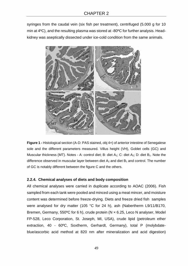
CHAPTER 2
49
syringes from the caudal vein (six fish per treatment), centrifuged (5.000 g for 10
min at 4ºC), and the resulting plasma was stored at -80ºC for further analysis. Head-
kidney was aseptically dissected under ice-cold condition from the same animals.
Figure 1 - Histological section (A-D: PAS stained, obj 4×) of anterior intestine of Senegalese
sole and the different parameters measured. Villus height (VH), Goblet cells (GC) and
Muscular thickness (MT). Notes - A: control diet; B: diet A1; C: diet A2; D: diet B1. Note the
difference observed in muscular layer between diet A2 and diet B1 and control. The number
of GC is notably different between the figure C and the others.
2.2.4. Chemical analyses of diets and body composition
All chemical analyses were carried in duplicate according to AOAC (2006). Fish
sampled from each tank were pooled and minced using a meat mincer, and moisture
content was determined before freeze-drying. Diets and freeze dried fish samples
were analysed for dry matter (105 °C for 24 h), ash (Nabertherm L9/11/B170,
Bremen, Germany, 550ºC for 6 h), crude protein (N × 6.25, Leco N analyser, Model
FP-528, Leco Corporation, St. Joseph, MI, USA), crude lipid (petroleum ether
extraction, 40 - 60ºC, Soxtherm, Gerhardt, Germany), total P (molybdate-
blue/ascorbic acid method at 820 nm after mineralization and acid digestion)
CHAPTER 2
50
according to AFNOR (1992) and gross energy (adiabatic bomb calorimeter, Werke
C2000, IKA, Staufen, Germany).
2.2.5. Innate Immune parameters
All analyses were conducted in triplicates on a Power-WaveTM microplate
spectrophotometer (BioTek Synergy HT, Vermont, USA).
2.2.5.1. Cellular parameters
Leucocytes from head-kidney were isolated and maintained as described by
Secombes (1990) and followed by Costas et al. (2011). Respiratory burst activity
(ROS) was based on the reduction of ferricytochrome C (Cyt C) method for the
detection of extracelular superoxide anion (O2-) production. Nitric oxide (NO)
production was based on the Griess reaction (Green et al., 1982) that quantifies the
nitrite content of the leucocytes supernatant (Neumann et al., 1995), modified by
(Tafalla and Novoa, 2000). The molar concentration of nitrite was quantified using
a standard curve produced with known concentrations of sodium nitrite (1.562 µM -
100 µM).
2.2.5.2. Humoral parameters
Lysozyme activity was determined using a turbidimetric assay based on the method
described by (Ellis, 1990) with minor modifications (Costas et al., 2011; Wu et al.,
2007). Total peroxidase activity was measured following the procedure described
by Quade and Roth (1997) and Costas et al. (2011). The peroxidase activity (units
mL-1 plasma) was determined by defining that one unit of peroxidase produces an
absorbance change of 1 OD. Alternative complement pathway (ACH50) was
measured using washed rabbit red blood cells (2.8 × 10⁸ cells mL-1, Probiológica,
Belas, Portugal) as target cells in the presence of ethylene glycol tetraacetic acid
(EGTA, Sigma, Portugal) and Mg2+ (MgCl2.6H2O, VWR, Portugal) as described by
Sunyer and Tort (1995).
2.2.6. Growth Performance and Nutrient Retention
Feed conversion ratio (FCR) was calculated as dry feed intake (g) × wet weight
gain−1 (g) and the daily growth index (DGI; g kg-1 BW day-1) as DGI = 100 × [(W1)1/3
− (W0)1/3]/ trial duration in days, where W0 and W1 are the initial and the final fish
CHAPTER 2
51
mean weights in grams. Voluntary feed intake (VFI; g kg-1 BW day-1) was calculated
as VFI = (dry feed intake /ABW/ trial duration), where average body weight (ABW)
was calculated as (W1+W0)/2. The protein efficiency ratio (PER) was calculated as
PER = weight gain (g) /protein ingested (g). Daily nutrient intake (g kg−1 ABW day−1)
was calculated as nutrient intake/ABW/trial duration. Daily nutrient gain, (g kg−1
ABW day−1) was calculated as (final body nutrient content − initial body nutrient
content)/ ABW/ trial duration. Nutrient retention (g kg-1 intake) was calculated as
(nutrient W1 – nutrient W0)/ nutrient intake. The HSI and VSI indexes (g kg-1, wet
weight basis) were calculated as HSI = liver weight/ whole body weight, and VSI =
100 × viscera weight / whole body weight.
2.2.7. Statistical analysis
Statistical analyses were carried out following the methods outlined by Zar (1999).
Results are expressed as mean ± standard deviation and with a P ≤ 0.05 as level of
significance. Data were analysed for normality (Shapiro-Wilk test) and homogeneity
of variance (Levene’s test) and were log-transformed whenever necessary. Data
were analysed by a one-way ANOVA (IBM SPSS STATISTICS, 17.0 package, IBM
Corporation, New York, USA) with probiotic treatment and respective concentration
level as a dependent variable. When significant differences were observed, Tukey’s
post hoc tests were carried out to identify significantly different groups fed the
experimental diets. When data did not meet the assumptions of ANOVA, the non-
parametric ANOVA equivalent (Kruskal–Wallis test) was performed.
2.3. Results
2.3.1. Growth and body composition
No measurable effects were observed on growth performance among treatments
(Table 2). Overall, size variation was lower in the dietary probiotic groups. The
coefficient of variation as a function of daily growth index (DGI) and weight gain
varied between 0.06 – 0.07 in A1, 0.13 – 0.15 in A2 and 0.11 – 0.13 in B1 groups and
0.24 in fish fed unsupplemented diet had, suggesting higher growth homogeneity in
fish fed probiotic diets (data not shown).
Hepatosomatic index varied from 12.1 ± 1.0 to 13.1 ± 4.4 g kg -1 and viscerosomatic
index varied from 15.4 ± 1.7 to 19.1 ± 0.6 g kg -1 (Table 3). Both indexes were not
CHAPTER 2
52
affected by dietary treatments. Whole body composition, dietary dry matter and
protein retentions (Table 3) were not significantly affected (P>0.05) by probiotic
supplementation, except ash, which was significantly lower in fish fed B1 than in
control fish (P<0.05).
2.3.2. Gut histology
Histological measurements of the intestinal mucosa of fish are present in Table 4.
No significant differences were observed among treatments (P < 0.05) for villus
height (µm). Thickness of muscular layer (µm) showed significantly differences (P <
0.05) between fish fed A2 (163 ± 32) and B2 (115 ± 14) probiotic diets. Goblet cells
counting did not vary among dietary treatments.
2.3.3. Innate immune parameters
Plasma lysozyme and peroxidase activities, expressed as Enzyme Unit (EU) mL-1
plasma, were not affected (P < 0.05) by the probiotic supplementation. Lysozyme
ranged from 160 (Control) to 500 EU mL-1 plasma (B2) and peroxidase from 88 (B2)
to 150 EU mL-1 plasma (A2). Alternative complement pathway activity (ACH50)
varied between 35 (Control) and 63 (B2), and was not significantly different (P <
0.05) between treatments. ROS and NO production showed no significant
differences among treatments (P < 0.05, Table 5). ROS (nmoles de O2-) range from
4.9 (A2) to 7.4 (B1) and NO (concentration of nitrites, μM) range from 6.41 (A1) to
6.46 (B2).
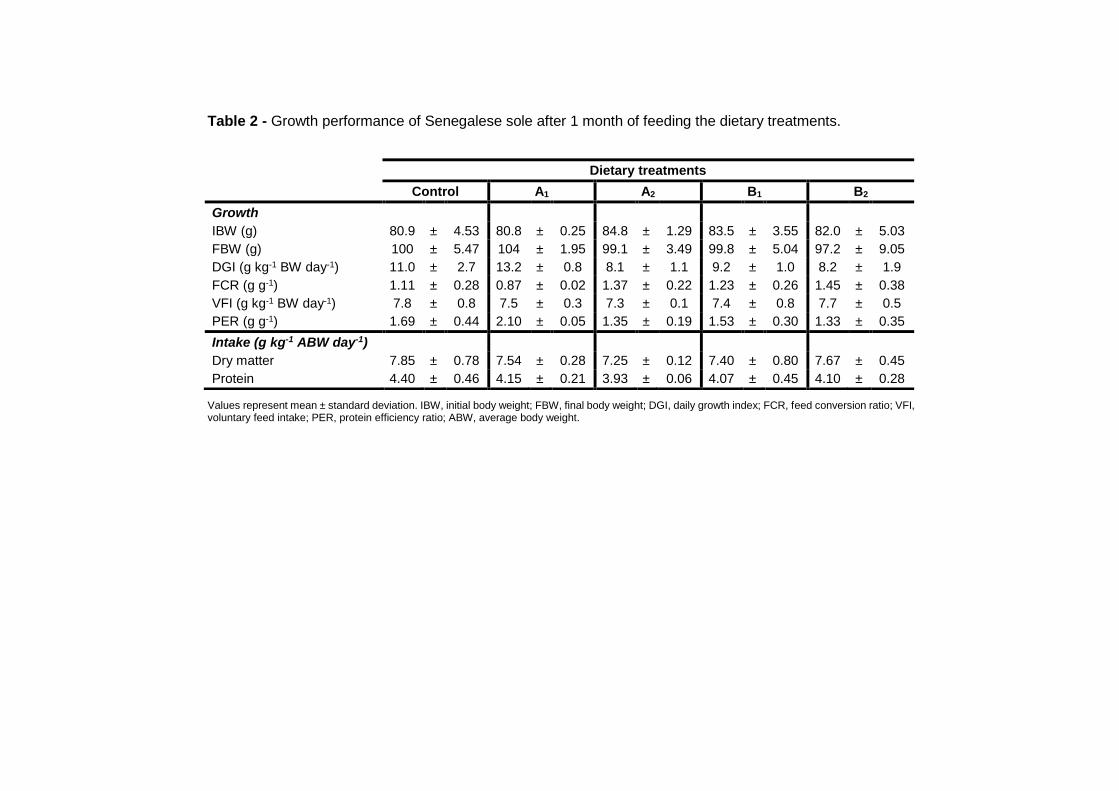
Table 2 - Growth performance of Senegalese sole after 1 month of feeding the dietary treatments.
Values represent mean ± standard deviation. IBW, initial body weight; FBW, final body weight; DGI, daily growth index; FCR, feed conversion ratio; VFI, voluntary feed intake; PER, protein efficiency ratio; ABW, average body weight.
Dietary treatments
Control A1 A2 B1 B2
Growth
IBW (g) 80.9 ± 4.53 80.8 ± 0.25 84.8 ± 1.29 83.5 ± 3.55 82.0 ± 5.03
FBW (g) 100 ± 5.47 104 ± 1.95 99.1 ± 3.49 99.8 ± 5.04 97.2 ± 9.05
DGI (g kg-1 BW day-1) 11.0 ± 2.7 13.2 ± 0.8 8.1 ± 1.1 9.2 ± 1.0 8.2 ± 1.9
FCR (g g-1) 1.11 ± 0.28 0.87 ± 0.02 1.37 ± 0.22 1.23 ± 0.26 1.45 ± 0.38
VFI (g kg-1 BW day-1) 7.8 ± 0.8 7.5 ± 0.3 7.3 ± 0.1 7.4 ± 0.8 7.7 ± 0.5
PER (g g-1) 1.69 ± 0.44 2.10 ± 0.05 1.35 ± 0.19 1.53 ± 0.30 1.33 ± 0.35
Intake (g kg-1 ABW day-1)
Dry matter 7.85 ± 0.78 7.54 ± 0.28 7.25 ± 0.12 7.40 ± 0.80 7.67 ± 0.45
Protein 4.40 ± 0.46 4.15 ± 0.21 3.93 ± 0.06 4.07 ± 0.45 4.10 ± 0.28

Table 3 - Whole body composition and nutrient utilization of Senegalese sole after 1 month of feeding the dietary treatments.
Dietary treatments
Control A1 A2 B1 B2
Somatic indexes (g Kg-1)
HSI 13.0 ± 2.1 13.1 ± 4.4 13.3 ± 1.2 12.1 ± 1.0 12.5 ± 1.3
VSI 19.1 ± 0.6 17.5 ± 1.8 18.6 ± 1.3 18.9 ± 2.7 15.4 ± 1.7
Whole body composition
Dry matter (g Kg-1) 266.5 ± 9.1 262.6 ± 6.6 261.9 ± 14.8 265.6 ± 6.0 277.3 ± 12.3
Ash (g Kg-1) 25.9 ± 1.4a 23.8 ± 1.5ab 22.9 ± 2.3ab 20.8 ± 0.8b 23.9 ± 0.6ab
Protein (g Kg-1) 181.4 ± 8.8 184.0 ± 4.4 180.0 ± 4.4 180.5 ± 3.4 184.2 ± 3.7
Lipid (g Kg-1) 57.6 ± 4.8 54.70 ± 3.4 60.4 ± 14.5 70.4 ± 2.9 73.9 ± 9.5
Energy (kJ) 62.1 ± 2.6 62.16 ± 3.5 61.9 ± 5.0 63.7 ± 2.4 66.8 ± 4.5
Gain (g kg-1 ABW day-1)
Dry matter 1.31 ± 0.80 1.53 ± 0.1 0.57 ± 0.45 0.92 ± 0.32 1.17 ± 0.77
Protein 0.92 ± 0.65 1.27 ± 0.1 0.48 ± 0.09 0.64 ± 0.16 0.66 ± 0.35
Retention (g Kg-1 intake)
Dry matter 169.9 ± 105.8 203.0 ± 22.2 78.5 ± 61.9 127.9 ± 57.9 155.9 ± 109.8
Protein 213.8 ± 152.3 306.6 ± 28.3 122.3 ± 23.0 163.3 ± 58.7 162.0 ± 94.8
Values represent mean ± standard deviation. In each line, different superscript letters indicate significant differences between treatments (P< 0.05). ABW, average body weight; HIS, hepatosomatic index; VSI, viscerosomatic index.

Table 4 - Intestinal morphology and goblet cells counting of Senegalese sole after 1 month of feeding the dietary
treatments.
Dietary treatments
Control A1 A2 B1 B2
Villus height (μm) 729 ± 102 741 ± 53 712 ± 93 724 ± 91 652 ± 154
Muscular layer thickness (μm) 144 ± 12ab 146 ± 22ab 163 ± 32a 141 ± 21ab 115 ± 14b
Goblet cells (number per section) 287 ± 173 298 ± 191 288 ± 169 235 ± 122 394 ± 155
Values represent mean ± standard deviation. In each line, different superscript letters indicate significant differences between treatments (P < 0.05).
Table 5 - Effects on humoral and celular non-specific immune parameters of Senegalese sole after 1 month of feeding the dietary
treatments.
Dietary treatments
Control A1 A2 B1 B2
Humoral parameters
Lysozyme (EU mL-1) 159.7 ± 109.5 298.6 ± 375.5 261.1 ± 250.7 302.8 ± 94.5 500.0 ± 270.9
Peroxidase (EU mL-1) 137.29 ± 30.3 124.0 ± 59.0 149.8 ± 27.0 102.8 ± 26.7 87.8 ± 12.7
ACH50 (units mL-1) 35.1 ± 14.6 40.6 ± 10.4 44.0 ± 4.6 49.5 ± 23.9 63.2 ± 55.7
Celular parameters
Respiratory burst activity (nmoles O2-) 6.63 ± 3.31 6.42 ± 2.82 4.85 ± 3.59 7.35 ± 2.37 4.62 ± 3.96
Nitric oxide production (nitrite μM) 6.44 ± 0.02 6.41 ± 0.10 6.45 ± 0.02 6.45 ± 0.02 6.46 ± 0.03
Values are means ± standard deviation.

CHAPTER 2
56
2.4. Discussion
The probiotics choice was based on their effects on fish health and growth in
aquaculture (Table 6). Some of these bacteria are known to present ability of spore-
forming, allowing greater viability after pelleting and high resistance to gastric
conditions (Hong et al., 2005).
Table 6 - Different applications of Bacillus sp., Pediococcus sp., Enterococcus sp.,
Lactobacillus sp. and Pediococcus acidilactici in aquaculture.
There is little information about the most effective dose and supplementation
duration time for the probiotics tested in this study. However, previous findings
showed that a short-term supplementation (3-6 weeks) was sufficient for their
successful colonization in the gut, stimulation of immune system and protection
against disease (Brunt et al., 2007; Newaj-Fyzul et al., 2007).
The beneficial effect of probiotics on the growth performance and dietary nutrient
utilization has been commonly reported for difference fish species (Carnevali et al.,
Aplication Probiotic bacteria Aquatic specie Reference
Growth
promoter
Bacillus sp
Bacillus coagulans
Lactobacillus helveticus
Lactobacillus lactis AR21
L. casei
Pediococcus acidilactici
Pediococcus acidilactici
Catfish
Cyprinus carpio koi
Scophthalmus maximus
Brachionus plicatilis
Poeciliopsis gracilis
Pollachius pollachius
Oncorhynchus mykiss
Queiroz and Boyd (1998)
Lin et al. (2012)
Gatesoupe (1999)
Shiri Harzevili et al. (1998)
Hernandez et al. (2010)
Gatesoupe (2002)
Merrifield et al. (2011)
Pathogen
inhibition
Bacillus sp
Bacillus spp, Enterococcus sp
Enterococcus faecium SF 68
L. rhamnosus ATCC53103
Lactobacillus acidophilus
Lactococcus lactis
Penaeids
Farfantepenaeus brasiliensis
Anguilla anguilla
Oncorhynchus mykiss
Clarias gariepinus
Epinephelus coioides
Moriarty (1998)
Souza et al. (2012)
Chang and Liu (2002)
Nikoskelainen et al. (2001a)
Al-Dohail et al. (2011)
Sun et al. (2012)
Adhesion
to
intestinal
mucosa
E. faecium, Bacillus spp, P.
acidilactici
Pediococcus acidilactici
Oncorhynchus mykiss
Oncorhynchus mykiss
Merrifield et al. (2010b)
Merrifield et al. (2011)
Nutrient
digestibility
L. helveticus
Lactobacillus acidophilus
Scophthalmus maximus
Clarias gariepinus
Gatesoupe (1999)
Al-Dohail et al. (2009)
Stress
tolerance
and Health
Lactobacillus delbrueckii
B. subtilis, L. acidophilus, S.
cerevisiae
L. casei
Pediococcus acidilactici
Pediococcus acidilactici
Dicentrarchus labrax
Paralichthys olivaceus
Poecilopsis gracilis
Litopenaeus stylirostris
Oncorhynchus mykiss
Carnevali et al. (2006)
Taoka et al. (2006a)
Hernandez et al. (2010)
Castex et al. (2009)
Merrifield et al. (2011)

CHAPTER 2
57
2006; El-Haroun et al., 2006; Taoka et al., 2006a; Wang et al., 2008b). Díaz-Rosales
et al. (2009), Sáenz de Rodrigáñez et al. (2009) and García de la Banda et al. (2012)
reported an improvement in growth performance in Senegalese sole fed dietary
probiotic supplementation. In the current study, fish fed the multispecies (Bacillus
sp., Pediococcus sp., Enterococcus sp., Lactobacillus sp.) and monospecies (P.
acidilactici) probiotics at different concentrations did not have any effect on growth
performance. Nevertheless, fish fed probiotic diet A1 presented the lowest growth
variation in traits such as growth rate and weight gain, suggesting higher growth
homogeneity in fish fed probiotic diets. This finding was also observed in previous
studies (García de la Banda et al., 2012; Sáenz de Rodrigáñez et al., 2009; Varela
et al., 2010; Wang et al., 2008b). Probiotics are seen to be effective in modulating
gut microbiota and reducing health problems (Djouvinov et al., 2005). As they
modulate gut microbiota and morphology, nutrient assimilation may improve and so
the growth performance, which might explain a reduction in variability within groups.
Merrifield et al. (2010d) showed that P. acidilactici significantly improve microvilli
length in proximal intestine of the rainbow trout compared to the control group, but
did not affect microvilli density. In our study, no significant differences were
observed on villus length, but villi length increased in fish fed A1. The increase in villi
length involves an increase of epithelial surface area, which may improve absorption
of available nutrients (Caspary, 1992) and ultimately improve growth performance.
The dietary probiotic supplementation may interfere with gut health, by altering the
height, width, and surface area of the villi and muscular layer thickness, improving
the tract absorption capacity of the intestine and enhancing the animal protection
against pathogen outbreak (Liu et al., 2007; Peinado et al., 2012). Tsirtsikos et al.
(2012) provides evidence in broilers that mucus layer thickness increased with
probiotic inclusion level. Although the changes in the muscular thickness have
already been mentioned in some nutritional experiments in different species
(broilers, rabbits and rats) the explanation of this phenomena it is yet poorly
understood. In our study, muscular layer thickness showed significant difference
between A2 and B2 groups. Fish fed B2 probiotic diet had the thinnest muscular layer
among treatments denoting a possible influence of the type of probiotic inclusion in
the intestine morphology.
Goblet cells are specialized cells that secrete mucins, glycoprotein compounds.
They are important in gut immunology binding pathogenic microorganisms and
CHAPTER 2
58
reducing their adherence to the intestinal mucosa (Blomberg et al., 1993). The
probiotic used in our experiment are foreign bacteria and the host may react by
producing more mucus in order to get rid of the probiont and reducing the protective
adherent to gut microbiota, however in our study, no differences were observed in
the number of goblet cells.
Serum peroxidase, lysozyme and ACH50 activities, are commonly used as
indicators of non-specific immune status in fish. Panigrahi et al. (2004), Kim and
Austin (2006a) and Newaj-Fyzul et al. (2007) showed that dietary probiotic
supplementation increased serum lysozyme activity in fish. Conversely, Balcázar et
al. (2007a), Merrifield et al. (2010a) and Merrifield et al. (2010b) did not find
significant effects of probiotics on serum lysozyme activity. In our study, the dietary
probiotic supplementation did not affect the immune parameters. Nevertheless, fish
fed dietary probiotic supplementation showed a tendency to increase lysozyme and
ACH50 activities. This trend was also observed by Díaz-Rosales et al. (2009)
working with Senegalese sole and Kim and Austin (2006a) in rainbow trout, where
probiotics induced a slightly increase in some of the studied immune parameters. In
fish, macrophage activity and NO production plays an important role in the non-
specific cellular defence mechanisms (Buentello and Gatlin III, 1999; Neumann et
al., 1995; Tafalla and Novoa, 2000). Thus, probiotics as immunomodulators may
have a stimulatory effect, increasing the production of reactive oxygen species by
macrophages in fish species (Díaz-Rosales et al., 2006a; Salinas et al., 2006).
However, we cannot forget the potential influence of the probiotic supplementation
duration time. Díaz-Rosales et al. (2009) noted that only the phagocytes of
Senegalese sole treated with probiotic for 60 days showed a significant increase of
ROS, while such effect was not detected in sole treated for 30 days. It is plausible
to infer that our trial duration was not long enough to observe the full effects of
dietary probiotic administration, although previous studies observed effects of
probiotic supplementation applying similar trial duration of probiotic administration
(Brunt et al., 2007; Newaj-Fyzul et al., 2007).
Under the current experimental conditions, the immune status did not vary
significantly between control and probiotic groups. Nevertheless, probiotic
supplementation tended to increased growth homogeneity, as a function of DGI and
weight gain, showing diet A1 the best possible alternative to decrease costs
associated to size grading. Gut morphology did not vary for the villus height and

CHAPTER 2
59
number of goblet cells, but the muscular layer thickness was influenced by the type
of probiotic included.
2.5. Conclusions
The beneficial effects of probiotics application in Senegalese sole remain with few
answers and further studies are needed, regarding doses, supplementation period
and type of bacteria to use. In addition, gut microbiota needs to be better evaluated
using PCR-DGGE techniques and the effect of dietary probiotic supplementation
should be tested against acute stress conditions, such as nutritional, environmental,
or infections by pathogen agent.
2.6. Acknowledgements
S. M. G. Batista would like to thank to UTAD and CIIMAR for the use of the facilities
and equipment and for technical support. S. M. G. Batista was supported by FCT –
SFRH/BD/76668/2011. The work was carried out as part of PROBIOSOLEA project
with the financial support of Quadro de Referência Estratégico Nacional – QREN
and Programa Operacional Regional do Norte – ON2 (Ref. no. 13551), supported
by the European fund for regional development FEDER. The histological analyzes
were supported by Project PEst-OE/AGR/UI0772/2011.

60

61
CHAPTER 3 Changes in intestinal microbiota, immune- and
stress-related transcript levels in Senegalese sole
(Solea senegalensis) fed plant ingredients diets
intercropped with probiotics or immunostimulants
Published: Batista et al. 2016. Aquaculture 458: 149-157 (DOI:
10.1016/j.aquaculture.2016.03.002)

62
CHAPTER 3
63
Changes in intestinal microbiota, immune- and stress-related transcript
levels in Senegalese sole (Solea senegalensis) fed plant ingredients diets
intercropped with probiotics or immunostimulants
Batista S.1,2,3, Ozório R.O.A.1,2, Kollias S.3,4, Dhanasiri A.K.3, Lokesh J.3, Kiron V.3,
Valente L.M.P.1,2, Fernandes J.M.O.*3
1 ICBAS – Instituto de Ciências Biomédicas de Abel Salazar, Universidade de Porto, Rua Jorge Viterbo Ferreira, 228, 4050-
313 Porto, Portugal
2 CIMAR/CIIMAR – Centro Interdisciplinar de Investigação Marinha e Ambiental, Rua dos Bragas, 289, 4050-123 Porto,
Portugal
3 FBA – Faculty of Biosciences and Aquaculture, Nord University, 8049 Bodø, Norway
4 Norwegian Sequencing Centre, Centre for Ecological and Evolutionary Synthesis, Department of Biosciences, University
of Oslo, PO Box 1066, NO-0316, Oslo, Norway
CHAPTER 3
64
Abstract
Senegalese sole (Solea senegalensis) is a highly valued flatfish that grows well with
diets containing plant ingredients but their effects on immune competence is still a
matter of debate. The current study aimed to examine changes in innate immune
parameters and gut microbiota in Senegalese sole fed with 35% or 72% of plant
ingredients with or without probiotic or yeast supplementation. Overall, fish fed diets
with 72% of plant ingredients showed lower transcript levels of key immune- and
stress-related genes in distal intestine, rectum and head-kidney than the 35% diets.
In particular, hsp90b mRNA levels in distal intestine were down-regulated by 70%
and 60% with the use of high content of plant ingredients in the diet containing the
multispecies probiotic and autolyzed yeast, respectively. Denaturing gradient gel
electrophoresis showed lower similarity values for distal intestine than rectum. Also
fish fed high content of plant ingredients displayed lower similarity values, pointing
to a difference in the microbial populations between fish fed different plant
ingredients content on the diet. Our data revealed that inclusion of plant ingredients
was associated with differences in gene expression and a more diverse microbiota
profile but without a significant effect on growth performance. Moreover, probiotic
supplementation resulted in up-regulation of hsp90b, gpx, cat and apoa1 transcript
levels in distal intestine concomitantly with a growth rate reduction compared to non-
supplemented fish.
CHAPTER 3
65
3.1 Introduction
Significant advances have been made in alternative protein diet formulations,
effectively replacing a large proportion of fishmeal in aquafeeds. Formulated
commercial feeds are composed of several ingredients, mixed in various proportions
to complement each other, and form a nutritionally complete compounded diet. In
Europe, the commercial aquafeeds are largely dependent on soybean products and
corn or wheat products (Rana et al., 2009). A number of different plant ingredients
have been successfully used as fishmeal replacement in fish feed (Kaushik et al.,
1995) without reducing growth performance. The ability of Senegalese sole (Solea
senegalensis) to efficiently use diets with high levels of plant protein (PP) sources
has been widely reported (Cabral et al., 2011; Silva et al., 2009; Valente et al., 2011)
and is an important asset towards the intensification and commercialization of this
species. Recent studies showed that sole could effectively use practical diets
containing up to 75% of PP sources, without affecting feed intake, growth
performance or nutrient utilization whilst preserving the nutritional fillet value (Cabral
et al., 2013). Nevertheless, the extent to which high levels of plant ingredients may
affect health conditions of fish, including the anti-oxidant and immune capacity
remains controversial. The suppression of the innate immune response upon
feeding high amounts of plant protein ingredients has been reported in rainbow trout
(Burrells et al., 1999) and in gilthead seabream, Sparus aurata (Sitjà-Bobadilla et
al., 2005). Conversely, another study using soybean in rainbow trout (Rumsey et
al., 1994) showed an increase in the innate immune parameters examined. There
are limited studies focused on the immune responses of the Senegalese sole,
particularly using a multifactorial approach; i.e. interactions between biotic and
abiotic stressors stressors (reviewed by Morais et al. (2014)).
Prebiotics and probiotics are increasingly used as preventive therapy, with high
success rates in reducing the incidence of diseases and increasing fish resistance
to infection (Gaggìa et al., 2010). The use of probiotics has increased in the
aquaculture sector due to its success in livestock production (Fulton et al., 2002)
and in human health (Gill, 2003). The probiotic genera commonly used in animal
feeding are Saccharomyces, Lactobacillus, Enterococcus and Bacillus (Gaggìa et
al., 2010). Lactobacillus are common bacteria in the gut microbiota of humans and
animals, whereas their occurrence and number are host dependent. Enterococcus
as well as Lactobacillus belong to the lactic acid bacteria (LAB) group and are found
CHAPTER 3
66
naturally in food products. Bacillus are Gram-positive spore-forming
microorganisms and are normally allochthonous microbes to the intestinal tract as
a result of ingestion (Gaggìa et al., 2010).
The use of a multispecies probiotic, from one or preferentially more genera, has
provided superior results over monostrain probiotic preparations (Timmerman et al.,
2004). Paubert-Braquet et al. (1995) tested mono and multistrain Lactobacillus in
mice challenged with Salmonella typhimurium and observed a clear protective effect
of the multistrain probiotic preparation. Modulation of the innate humoral and cellular
defences is one of the benefits of probiotic treatment described in fish (Nayak,
2010), since the innate immune system is the first line of host defense against
pathogenic organisms (Sinyakov et al., 2002). The inter-relationship between gut
mucosal epithelial cells, mucus, antimicrobial products, and organisms resident in
the gut and immune cells in the mucosa/sub-mucosa are vital for the health and
well-being of the fish (Merrifield, et al., 2010b). Several studies have demonstrated
the immunological and haematological stimulation of fish defence mechanisms by
probiotic bacteria (Brunt et al., 2008; Pieters et al., 2008). In fish most of the
beneficial effects, have been recorded within a dietary probiotics feeding regime of
1-10 weeks (Nayak, 2010). The time course for optimum induction of immune
response differs with respect to probiotic strain and also with the type of immune
parameter (Choi and Yoon, 2008). Batista et al. (2014) studied the effects of dietary
probiotic supplementation (monospecies and multispecies) in Senegalese sole
(Solea senegalensis) juveniles during a 1-month trial and this time duration was not
able to produce any effect on growth or humoral innate immune response. The
short-time probiotic feeding may lead to failure of the probiotic strains to establish
themselves in the fish gut and therefore long-time administration should be
investigated.
As the use of plant protein sources to replace fish meal is a major trend in
aquafeeds, it is important to gather health-related information to confirm the
suitability of the newly developed plant-based diets in farmed fish. Therefore, the
aim of this study is to evaluate the effects of two dietary plant protein levels
intercropped with probiotic supplementation on the growth performance, innate
immune response and intestine microbiota diversity in sole (Solea senegalensis).

CHAPTER 3
67
3.2 Materials and methods
The current study was conducted under the supervision of an accredited expert in
laboratory animal science by the Portuguese Veterinary Authority (1005/92, DGV-
Portugal, following FELASA category C recommendations), according to the
guidelines on the protection of animals used for scientific purposes from the
European directive 2010/63/UE.
3.2.1 Feeding experiment
The Senegalese sole feeding experiment took place at the CIIMAR facilities
(University of Porto, Porto, Portugal). Fish were fed for 73 days, in triplicate, with six
isoproteic (55% crude protein) and isolipidic (8% lipid) diets with similar amino-acid
composition (Table 1 and supplementary table S1). Two practical diets were
formulated to contain either low or high plant protein (PP) inclusion levels (35% and
72%, respectively). Each of these diets were supplemented with either PROB or
YEAST, and compared with the unsupplemented ones (UN).
The duration of the trial was selected as the best option to ensure the establishment
and multiplication of probiotic strains in the fish gut while allowing diets to impact on
fish growth. Diets were extruded (3 mm pellet) by the use of a pilot-scale twin-screw
extruder (Clextral BC45). PROB was a multispecies probiotic bacteria
(AquaStar® Growout: Bacillus sp, Pediococcus sp, Enterococcus sp, Lactobacillus
sp) incorporated at 1.34 × 10¹⁰ CFU kg-1 diet (CFU – colony forming unit) in the
extruded pellets by means of vacuum coating (Dinnisen Pegasus vacuum mixer,
PG-10VCLAB) using fish oil as a carrier. YEAST was autolyzed yeast
(Levabon® Aquagrow E: Saccharomyces cerevisiae) supplemented in the mixture
at 4g kg-1 diet. Diets were prepared according to the normatives ISO (6887-1:1999,
7218:2007, 6498:2012) for the microbiological examinations. Thereafter, the
isolation and enumeration of bacteria in the diets followed the European standards
for Enterococcus spp. (EN 15788:2009), Lactobacillus spp. (EN 15787:2009),
Pediococcus spp. (EN 15786:2009) and Bacillus spp. (EN 15784:2009).
Senegalese sole were obtained from a commercial fish farm (Aquacria S.A.,
Portugal) and transported to the rearing facilities of CIIMAR. After 5-week
acclimation, fish of similar weight and length were distributed into 18 fibreglass tanks
of 50 L, supplied by a recirculation system.
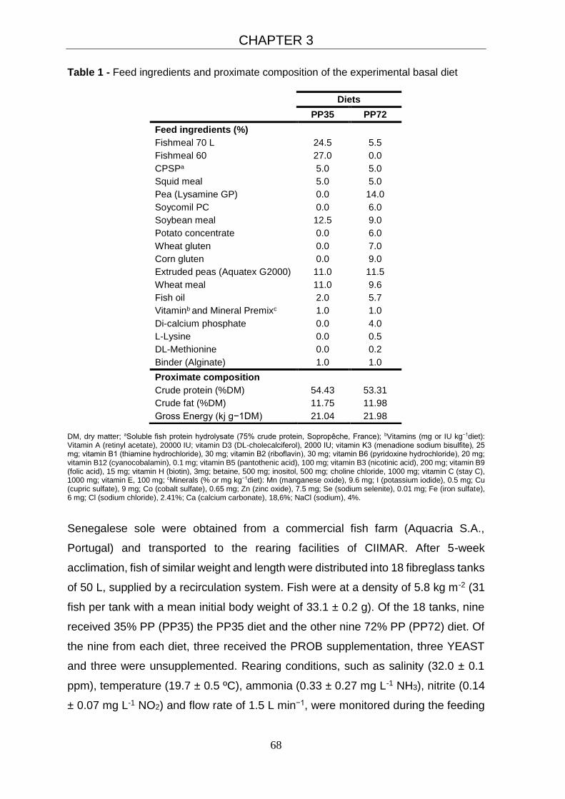
CHAPTER 3
68
Table 1 - Feed ingredients and proximate composition of the experimental basal diet
DM, dry matter; aSoluble fish protein hydrolysate (75% crude protein, Sopropêche, France); bVitamins (mg or IU kg−1diet): Vitamin A (retinyl acetate), 20000 IU; vitamin D3 (DL-cholecalciferol), 2000 IU; vitamin K3 (menadione sodium bisulfite), 25 mg; vitamin B1 (thiamine hydrochloride), 30 mg; vitamin B2 (riboflavin), 30 mg; vitamin B6 (pyridoxine hydrochloride), 20 mg; vitamin B12 (cyanocobalamin), 0.1 mg; vitamin B5 (pantothenic acid), 100 mg; vitamin B3 (nicotinic acid), 200 mg; vitamin B9 (folic acid), 15 mg; vitamin H (biotin), 3mg; betaine, 500 mg; inositol, 500 mg; choline chloride, 1000 mg; vitamin C (stay C), 1000 mg; vitamin E, 100 mg; cMinerals (% or mg kg−1diet): Mn (manganese oxide), 9.6 mg; I (potassium iodide), 0.5 mg; Cu (cupric sulfate), 9 mg; Co (cobalt sulfate), 0.65 mg; Zn (zinc oxide), 7.5 mg; Se (sodium selenite), 0.01 mg; Fe (iron sulfate), 6 mg; Cl (sodium chloride), 2.41%; Ca (calcium carbonate), 18,6%; NaCl (sodium), 4%.
Senegalese sole were obtained from a commercial fish farm (Aquacria S.A.,
Portugal) and transported to the rearing facilities of CIIMAR. After 5-week
acclimation, fish of similar weight and length were distributed into 18 fibreglass tanks
of 50 L, supplied by a recirculation system. Fish were at a density of 5.8 kg m-2 (31
fish per tank with a mean initial body weight of 33.1 ± 0.2 g). Of the 18 tanks, nine
received 35% PP (PP35) the PP35 diet and the other nine 72% PP (PP72) diet. Of
the nine from each diet, three received the PROB supplementation, three YEAST
and three were unsupplemented. Rearing conditions, such as salinity (32.0 ± 0.1
ppm), temperature (19.7 ± 0.5 ºC), ammonia (0.33 ± 0.27 mg L-1 NH3), nitrite (0.14
± 0.07 mg L-1 NO2) and flow rate of 1.5 L min−1, were monitored during the feeding
Diets
PP35 PP72
Feed ingredients (%)
Fishmeal 70 L 24.5 5.5
Fishmeal 60 27.0 0.0
CPSPa 5.0 5.0
Squid meal 5.0 5.0
Pea (Lysamine GP) 0.0 14.0
Soycomil PC 0.0 6.0
Soybean meal 12.5 9.0
Potato concentrate 0.0 6.0
Wheat gluten 0.0 7.0
Corn gluten 0.0 9.0
Extruded peas (Aquatex G2000) 11.0 11.5
Wheat meal 11.0 9.6
Fish oil 2.0 5.7
Vitaminb and Mineral Premixc 1.0 1.0
Di-calcium phosphate 0.0 4.0
L-Lysine 0.0 0.5
DL-Methionine 0.0 0.2
Binder (Alginate) 1.0 1.0
Proximate composition
Crude protein (%DM) 54.43 53.31
Crude fat (%DM) 11.75 11.98
Gross Energy (kj g−1DM) 21.04 21.98
CHAPTER 3
69
entire trial. The recirculation system was equipped with UV lights and ozone
generator to hamper bacteria growth in the rearing water. Fish were fed to apparent
satiety based on visual observation of acceptance and refusal of feed using
temporized automatic feeders. To prevent stressful conditions, high stocking
densities and repeated handling stress were avoided.
3.2.2 Sampling procedures
Prior to sampling, fish were fasted for 24 h and then sacrificed with an overdose of
ethyl 3-aminobenzoate methanesulfonate (MS-222, 200 mg L-1). All fish were
individually weighed and measured at the beginning and at the end of the
experiment in order to determine growth performance. Ten fish from the initial stock
and two fish from each tank at the end of the trial (6 fish per treatment) were sampled
and stored at -20 ºC for body composition analyses. For the evaluation of humoral
innate immune parameters (lysozyme, peroxidase and alternative complement
pathway – ACH50) blood was withdrawn from the caudal vein of 9 fish per treatment
using heparinised syringes and centrifuged at 5000 × g for 10 min at 4 ºC to collect
the plasma. The liver and viscera weights were recorded to calculate HSI
(hepatosomatic index) and VSI (viscerosomatic index). The posterior intestine was
aseptically sampled and divided in two sections (distal intestine and rectum). The
intestinal microbiota, including the probiotic bacteria tested, were determined in both
intestine sections, separately. The expression of immune-related genes, were
quantified (RT-qPCR) in the head-kidney, distal intestine and rectum. All samples
were stored at -80 ºC for further analyses.
3.2.3 Chemical analyses of diets and body composition
All chemical analyses were carried in duplicate according to AOAC (2006). Fish
were minced without thawing using a meat mincer, pooled and used to determine
their dry matter content (105 °C for 24 h). Fish were then freeze-dried and analysed
for dry matter, ash (Nabertherm L9/11/B170; Germany; 550ºC for 6 h), crude protein
(N×6.25, Leco N analyser, Model FP-528, Leco Corporation, St. Joseph, USA),
crude lipid (petroleum ether extraction, 40–60ºC, Soxtherm, Gerhardt, Germany)
and gross energy (adiabatic bomb calorimeter, Werke C2000, IKA, Germany).
CHAPTER 3
70
3.2.4 Humoral innate immune parameters
All measurements were done in triplicate on a Power-WaveTM microplate
spectrophotometer (BioTek Synergy HT, USA). Plasma lysozyme activity (EU ml-1)
was determined using a turbidimetric assay adapted to microtitre, as described by
Hutchinson and Manning (1996). One lysozyme enzyme unit (EU) was defined as
the amount of lysozyme that caused a decrease in absorbance of levels per min.
Plasma peroxidase activity (EU ml-1) was measured following the procedure
adapted to Solea senegalensis by Costas et al. (2011), defining that one unit of
peroxidase produces an absorbance change of 1 OD. Alternative complement
pathway (ACH50) was based on the lysis of rabbit red blood cells (2.8 × 10⁸ cells
mL-1; RaRBC), as reported by Sunyer and Tort (1995). ACH50 units were defined
as the concentration of plasma giving 50% lysis of RaRBC.
3.2.5 Immune- and stress-related gene expression
Except for lysozyme c (lyzc), target and reference genes used in this study were
based on published information (Table 2). The immune-related genes selected are:
lysozymes (lyzc and lyzg), heat shock proteins (hsp90a, hsp90b and hsp90b1), iron
chelating proteins (ftm and apoa1), complement factors (c3a1 and c3a2), the
cysteine protease casp3 and oxidative enzymes (cat and gpx).
3.2.5.1 RNA extraction and cDNA synthesis
Senegalese sole tissues (head-kidney, distal intestine or rectum) were
homogenised with Lysing matrix D beads for 20 s at 5500 rpm on a MagNA Lyser
instrument (Roche) and used for RNA extraction following the QIAzol protocol
(Qiagen). Assessment of RNA quality, RNA quantification and cDNA synthesis
followed the methodology described by Campos et al. (2010).
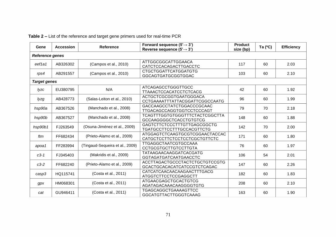
71
Table 2 – List of the reference and target gene primers used for real-time PCR
Gene Accession Reference Forward sequence (5’→ 3’) Reverse sequence (5’→ 3’)
Product size (bp)
Ta (ºC) Efficiency
Reference genes
eef1a1 AB326302 (Campos et al., 2010) ATTGGCGGCATTGGAACA CATCTCCACAGACTTGACCTC
117 60 2.03
rps4 AB291557 (Campos et al., 2010) CTGCTGGATTCATGGATGTG GGCAGTGATGCGGTGGAC
103 60 2.10
Target genes
lyzc EU380795 N/A ATCAGAGCCTGGGTTGCC TTAAACTCCACATCCTCTCACG
42 60 1.92
lyzg AB428773 (Salas-Leiton et al., 2010) ACTGCTCGCGGTGAATGGGACA CCTGAAAATTTATTACGGATTCGGCCAATG
96 60 1.99
hsp90a AB367526 (Manchado et al., 2008) GACCAAGCCTATCTGGACCCGCAAC TTGACAGCCAGGTGGTCCTCCCAGT
79 70 2.18
hsp90b AB367527 (Manchado et al., 2008) TCAGTTTGGTGTGGGTTTCTACTCGGCTTA GCCAAGGGGCTCACCTGTGTCG
148 60 1.88
hsp90b1 FJ263549 (Osuna-Jiménez et al., 2009) GAGTCTTCTCCCTTTGTTGAGCGGCTG TGATGCCTTCCTTTGCCACGTTCTG
142 70 2.00
ftm FF682434 (Prieto-Alamo et al., 2009) ATGGAGTCTCAAGTGCGTCGGAACTACCAC CATGCTCCTTCTCCTCCTCGCTGTTCTC
171 60 1.80
apoa1 FF283994 (Tingaud-Sequeira et al., 2009) TTGAGGCTAATCGTGCCAAA CCTGCGTGCTTGTCCTTGTA
76 60 1.97
c3-1 FJ345403 (Makridis et al., 2009) TATAAGAACAAGGATCACGATG GGTAGATGATCAATGAACCTC
106 54 2.01
c3-2 FF682240 (Prieto-Alamo et al., 2009) ACCTTAGACTGCCCTACTCTGCTGTCCGTG GCACTGCACACATCATCCGTCTCAGAC
147 60 2.26
casp3 HQ115741 (Costa et al., 2011) CATCATCAACAACAAGAACTTTGACG ATGGTCTTCCTCCGAGGCTT
182 60 1.83
gpx HM068301 (Costa et al., 2011) ATGAACGAGCTGCACTGTCG AGATAGACAAACAAGGGGTGTG
208 60 2.10
cat GU946411 (Costa et al., 2011) TGAGCAGGCTGAAAAGTTCC GGCATGTTACTTGGGTCAAAG
163 60 1.90
CHAPTER 3
72
3.2.5.2 Quantitative real-time PCR (qPCR)
Quantification of mRNA levels was done by qPCR in LightCycler 480 (Roche) using
SYBR green (Qiagen), as detailed elsewhere (Campos et al., 2010; Fernandes et
al., 2008). Briefly, 10 µL reactions were prepared in 96-well plates and included 4
µL of 25 ×-diluted cDNA template, 1 µL of each primer pair at 5 µM and 5 µL of
QuantiTect SYBR Green containing ROX as reference dye (Qiagen). Samples were
denatured (15 min at 95 ºC) and then amplified (45 cycles), according to the
following thermocycling profile: denaturation for 10 s at 95 ºC, annealing for 20s at
60 ºC and extension for 20 s at 72 ºC. Six-point standard curves of a 2-fold dilution
series (1:1–1:32) from pooled cDNA were used for PCR efficiency calculation.
Specificity of the qPCR reactions was determined by melting curve analysis and
further confirmed by Sanger sequencing of the PCR product, as reported by
Campos et al. (2010). Data were analysed by the relative quantification method after
normalization using the geometric mean of the best reference genes (Fernandes et
al., 2008).
3.2.6 PCR amplification and denaturing gradient gel electrophoresis (PCR +
DGGE)
The DGGE analysis of PCR-amplified genes coding for 16S rRNA was carried out
in the distal intestine and rectum (2 fish / treatment) to make a qualitative study of
the microbial community in the intestine. DNA was extracted using
cetyltrimethylammoniumbromide (Zhou et al., 1996). For the detection of the
multispecies bacteria PROB, the PCR amplification of DNA was carried out using
the primer combination of 341F with GC clamp, CCTACGGGAGGCAGCAG and
907R, CCGTCAATTCMTTTGAGTTT (Muyzer et al., 1995). Yeast was detected in
animals by PCR amplification using Saccharomyces cerevisiae specific primers
combination of SCIF, GTGCTTTTGTTATAGGACAATT and SI5R,
AGAGAAACCTCTCTTTGGA (Chang et al., 2007). PCR amplification by a modified
touchdown protocol was performed as described Dhanasiri et al. (2011), except for
the temperature of the initial denaturation step (96oC). DGGE was performed on the
PCR products from DNA samples using a 16 × 16 cm gel on the Bio-Rad DCode ™
system (Bio-Rad, USA), as described by Dhanasiri et al. (2011), but using a 30 to
55% (w/v) urea and formamide denaturing gradient. PCR products from distal

CHAPTER 3
73
intestine and rectum sections were loaded into two separated gels (6 fish/ treatment)
and electrophoresis was performed with 0.5% TAE buffer, at 20 V for 15 minutes
and then at a constant voltage of 75 V for 17 hours. DGGE profile analysis (Quantity
One® version 4.6.3 software, Bio-Rad) was performed for gels A and B (Fig. 1) to
determine similarity matrices (Dhanasiri et al., 2011). Bands of interest were
excised, purified and sequenced. The results were subjected to BLAST sequence
similarity search using BLASTN from the National Center for Biotechnology
Information (NCBI; http://www.ncbi.nlm.nih.gov).
3.2.7 Calculations of growth performance
Feed conversion ratio was calculated as: dry feed intake (g) / wet weight gain (g),
and the daily growth index (% BW day-1) as: 100 × [(W1)1/3 − (W0)1/3] / days, where
W0 and W1 are the initial and the final fish mean weights in grams and days is the
trial duration. Voluntary feed intake (% BW day-1) was calculated as: 100 × (dry feed
intake (g) / average BW (g) / days), where average BW was calculated as: (W1+W0)
/ 2. The protein efficiency ratio was calculated as weight gain (g) / protein ingested
(g). The hepatosomatic index was calculated as: 100 × [liver weight (g) / whole body
weight (g)] and the viscerosomatic index as 100 × [viscera weight (g) / whole body
weight (g)].
3.2.8 Statistical analysis
Statistical analyses were performed with the software SPSS (IBM SPSS
STATISTICS, 17.0 package, IBM Corporation, New York, USA). Results are
expressed as mean ± standard deviation (SDpooled as weighted average of each
group's standard deviation) and the level of significance used was P ≤ 0.05. Data
were analysed for normality (Shapiro-Wilk test) and homogeneity of variance
(Levene’s test) and were log-transformed whenever necessary. Data were analysed
by a two-way ANOVA with diet and probiotic as main factors. When significant
differences were obtained from the ANOVA, Tukey’s post hoc tests were carried out
to identify significantly different groups. When data did not meet the ANOVA
assumptions, a non-parametric Kruskal–Wallis test was performed for each factor.
Evaluation of expression stability of reference genes was done using the statistical
application geNorm (Vandesompele et al., 2002). Expression of target genes was
evaluated by the relative quantification method as reported in Fernandes et al.

CHAPTER 3
74
(2008). Heat maps of transcript levels were produced using PermutMatrix software,
with the Euclidean distance clustering algorithm and gene expression normalized
for rows.
3.3 Results
3.3.1 Growth performance
Data from growth performance are presented in table 3. Fish grew from 33.1 ± 0.20
g to 50.6 ± 1.2g (PP72_UN). Growth performance did not differ between PP35 and
PP72 groups. PROB groups had significantly lower final body weight (45.0 ± 1.9)
and daily growth index (0.5 ± 0.1) compared to UN groups (50.5 ± 2.0 and 0.7 ± 0.1,
respectively). Additionally, UN groups had significantly better feed conversion ratio
(1.5 ± 0.1) and higher protein efficient (1.3 ± 0.1) than the probiotic supplemented
groups. Voluntary feed intake was also lower in UN groups (0.8 ± 0.0), and differed
significantly from YEAST groups (0.9 ± 0.1). Visceral somatic index and
hepatosomatic index did not differ between treatments.
3.3.2 Humoral innate immune parameters
After the 73-day feeding trial, humoral immune parameters did not present any
significant differences between treatments (supplementary table S2). Lysozyme,
peroxidase and ACH50 varied between 1225.9 ± 251.0 and 2061.7 ± 366.2 EU mL-
1, 22.5 ± 9.0 and 50.3 ± 14.1 EU mL-1 and 88.2 ± 14.0 and 110.3 ± 14.9 U mL-1,
respectively.
3.3.3 Immune- and stress-related gene expression
Expression of immune-related genes is presented in Fig. 2 and supplementary
tables S3, S4 and S5. Distal intestine transcript levels were significantly affected by
plant ingredients content (hsp90b and apoa1) and by probiotic supplementation
(hsp90b1 and gpx). PP72 groups have lower values for hsp90b (1.4 ± 0.8) and
apoa1 (9.6 ± 5.4) genes, compared to PP35 groups (3.2 ± 1.9 and 14.9 ± 7.3,
respectively). YEAST groups presented higher values for hsp90b (3.4 ± 1.9)
compared to PROB (2.1 ± 1.8) and UN (1.5 ± 0.8) groups. However, for hsp90b1
and gpx expression, PROB groups (4.1 ± 2.4 and 1.1 ± 0.4 respectively) had higher
values compared to YEAST (1.8 ± 0.5 and 0.6 ± 0.3 respectively) and UN (2.2 ± 1.0
CHAPTER 3
75
and 0.7 ± 0.3 respectively) groups. Considering the distal intestine cat expression,
PP35_PROB (21.3 ± 12.1) was significantly higher than PP72_YEAST (7.0 ± 2.3)
treatment. No significant differences were detected in distal intestine gene
expression for lysozymes (lyzc and lyzg), c3 complement components (c3-1 and c3-
2), hsp90a, ftm and casp3 genes.
Similarly to distal intestine, some rectum genes transcript levels were affected by
the use of plant ingredients. PP72 groups (casp3: 2.0 ± 0.6, gpx: 0.1 ± 0.0 and cat:
2.3 ± 0.6) showed significantly lower values compared to PP35 groups (casp3: 2.6
± 0.6, gpx: 0.2 ± 0.1 and cat: 2.9 ± 0.8). Rectum hsp90a expression was two-fold
higher for PROB (0.04 ± 0.02) groups compared to YEAST (0.02 ± 0.01) groups.
Rectum ftm expression was higher for UN (3.3 ± 0.6) groups compared to PROB
(2.5 ± 0.8) groups but not different to YEAST (2.7 ± 0.7) groups. apoa1 expression
was significantly higher in PP35_YEAST (1.3 ± 1.1) in rectum, compared to
PP35_UN (0.0 ± 0.1), PP35_PROB (0.1 ± 0.2) and PP72_YEAST (0.2 ± 0.2). No
significant differences in transcript levels for lysozymes (lyzc and lyzg), heat shock
proteins (hsp90b and hsp90b1), complement components (both c3 analysed) were
detected in rectum.
In head-kidney, only mRNA levels of hsp90b1 were significantly lower in PP72 (0.4
± 0.2) than in PP35 (0.6 ± 0.2) groups.
3.3.4 Probiotic detection and gut microbiota profiles
The marker bands (PROB bacteria profile) are present in samples of fish that have
been fed the multispecies bacteria probiotic (Fig. 1A). A comparison of the DGGE
profiles (Table 4) between distal intestine and rectum tissues revealed lower
similarity values for distal intestine (34.4 – 54.9%) than rectum (47.9 – 72.4%),
showing higher microbiota diversity in distal intestine. We also observed that fish
fed PP72 diets have a tendency to display a lower similarity value compared with
fish fed PP35 diets (34.4 – 54.9% for distal intestine and 47.9 – 72.4% for rectum).
In distal intestine, comparison of the profiles between PP35_UN vs PP72_UN
revealed similarity values less than 34.4%, pointing to a difference in the microbial
populations between fish fed different plant ingredients content on the diet.
However, when adding the probiotic to the diet, similarity values, in distal intestine,
increase (54.9% for PROB and 46.3% for YEAST).

Table 3 – Growth performance of Senegalese sole juveniles after the 73-day feeding trial
In each line, different superscript letters indicate significant differences (P<0.05): for a particular diet, differences caused by probiotic inclusion are indicated using x, y; for a particular probiotic inclusion, differences among the diets are shown using A, B. Dietary treatments are abbreviated as PP35 and PP72 for diets with low and high content of plant ingredients, respectively and UN, PROB and YEAST to diets not supplemented or supplemented with probiotic or with the immunostimulant yeast. The other abbreviations are: IBW = Initial body weight; FBW = Final body weight; DGI = Daily growth index; FCR = Feed conversion ratio; VFI = Voluntary feed intake; PER = Protein efficiency ratio; VSI = Viscerosomatic index; HIS = Hepatosomatic index. SDpooled = pooled standard deviation. Values represent mean ± SDpooled, n=3.
Dietary treatments p value
PP35 PP72 SDpooled Diet Suplementation Diet*Suplementation
UN PROB YEAST UN PROB YEAST
Growth performance
IBW (g) 33.03 33.16 33.02 33.16 33.04 33.25 0.21 0.44 0.95 0.35
FBW (g) 50.49x 45.73y 45.82xy 50.58x 44.21y 47.73xy 2.91 0.91 0.02 0.60
DGI (g kg-1 BW day-1) 0.67x 0.50y 0.51xy 0.67x 0.45y 0.56xy 0.10 0.98 0.02 0.68
FCR (g g-1) 1.46y 2.04x 2.11x 1.49y 2.42x 2.02x 0.34 0.53 0.01 0.51
VFI (g kg-1 BW day-1) 0.83y 0.88xy 0.90x 0.85y 0.94xy 0.94x 0.33 0.13 0.02 0.80
PER (g g-1) 1.27x 0.91y 0.89y 1.27x 0.79y 0.97y 0.16 0.87 0.00 0.62
Somatic indexes (g Kg-1)
VSI 2.34 2.04 2.08 2.18 2.48 2.06 0.19 0.35 0.18 0.05
HSI 0.89AB 0.82B 0.84A 0.89AB 0.78B 1.05A 0.08 0.18 0.03 0.05

Figure 1 – DGGE images showing bands corresponding to the intestinal bacteria present in the distal intestine wall (A) and rectum
wall (B) samples of Senegalese sole juveniles that were fed for 73 days with diets with or without probiotic/ yeast supplementation.
Lanes: 1 and 14 – probiotic bacteria marker; 2 and 3 – PP35_UN; 4 and 5 – PP35_PROB; 6 and 7 – PP35_ YEAST; 8 and 9 –
PP72_UN; 10 and 11 – PP72_PROB; 12 and 13 – PP72_ YEAST. Arrowheads with numbers indicate the bands that were excised
and sequenced for molecular identification (1 - Enterococcus sp; 2 - Lactobacillus sp; 3 - Bacillus sp; 4 - Enterococcus sp; 5 –
Pediococcus sp), n=2 fish per treatment group.
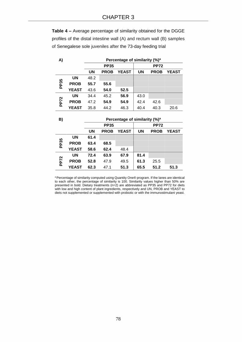
CHAPTER 3
78
Table 4 – Average percentage of similarity obtained for the DGGE
profiles of the distal intestine wall (A) and rectum wall (B) samples
of Senegalese sole juveniles after the 73-day feeding trial
A) Percentage of similarity (%)a
PP35 PP72
UN PROB YEAST UN PROB YEAST
PP
35
UN 48.2
PROB 55.7 55.6
YEAST 43.6 54.0 52.5
PP
72
UN 34.4 45.2 56.9 43.0
PROB 47.2 54.9 54.9 42.4 42.6
YEAST 35.8 44.2 46.3 40.4 40.3 20.6
B) Percentage of similarity (%)a
PP35 PP72
UN PROB YEAST UN PROB YEAST
PP
35
UN 61.4
PROB 63.4 68.5
YEAST 58.6 62.4 48.4
PP
72
UN 72.4 63.9 67.9 81.4
PROB 52.8 47.9 49.5 61.3 25.5
YEAST 62.3 47.1 51.3 65.5 51.2 51.3
a Percentage of similarity computed using Quantity One® program. If the lanes are identical to each other, the percentage of similarity is 100. Similarity values higher than 50% are presented in bold. Dietary treatments (n=2) are abbreviated as PP35 and PP72 for diets with low and high content of plant ingredients, respectively and UN, PROB and YEAST to diets not supplemented or supplemented with probiotic or with the immunostimulant yeast.

CHAPTER 3
79
Figure 2 – Heat maps showing
the normalized mRNA levels of
selected genes in the distal
intestine, rectum and head kidney
tissues of Senegalese sole
juveniles after the 73-day feeding
trial. Each block represents the
mean mRNA level of 6 fish
quantified by qPCR. Letters
indicate significant differences
(P<0.05).

CHAPTER 3
80
3.4 Discussion
It is well recognised that plant ingredients have to be increasingly employed in
aquafeeds to cater to the demand of the industry. However, the application of these
ingredients at relatively high levels in the diets of some carnivorous fish species may
cause nutritional imbalances and influence the immune response as they may
contain anti-nutritional factors (Hardy, 2010). In order to cope with the need to
depend on the plant materials in fish feeds, efforts have to be made to alleviate the
negative influence of these components may have on fish. Our approach in this
direction has been to exploit the potential of probiotic organisms to counter any
negative influence that may arise upon increasing the levels of plant components in
fish diets. Senegalese sole that received plant diets supplemented with the selected
probiotics seems to have altered immune and stress responses compared to fish
receiving plant diets that lacked the probiotics. Our findings are discussed mainly
based on the expression of the immune and stress-related genes in the intestinal
segment which is considered as an immunologically relevant region in fish
(Rombout et al., 2011) where the applied diets can have a direct impact on the
elicited responses.
In fish, the induction of heat shock proteins (hsp) is a component of the cellular
stress response against a diversity of stressors, such as osmotic stress, heat shock
or infections (Basu et al., 2002). hsp90b1 (also known as gp96 or grp94) is the
primary chaperone of the endoplasmic reticulum and has crucial immunological
functions, serving as a natural adjuvant for priming innate and adaptive immunity
(Strbo and Podack, 2008). No differences in hsp90a expression were detected
between treatments in the present study, regardless of the tissues examined. Distal
intestine hsp90b and head-kidney hsp90b1 mRNA levels were significantly affected
by diets, with lower expression in fish fed higher plant ingredients inclusion level. In
contrast, high levels of plant protein in the feeds did not affect the expression of
hsp70 and hsp90 in Atlantic cod (Gadus morhua L.) (Hansen et al., 2006).
Interestingly, considering the effect of the probiotic, Senegalese sole fed yeast
supplemented diets had higher hsp90b expression in the distal intestine, while fish
fed multispecies probiotic displayed higher hsp90b1 and hsp90a transcript levels in
distal intestine and rectum, respectively.
In addition to their well-known role in reverse cholesterol transport and lipid
metabolism, apolipoproteins display anti-inflammatory, antimicrobial and antioxidant
CHAPTER 3
81
activities (Barter et al., 2004; Tada et al., 1993). The main high density lipoprotein,
Apoa1, has antioxidant properties (Barter et al., 2004) and is involved in regulation
of fish complement (Magnadóttir, 2006).
Rectum apoa1 expression, from fish fed PP35 diet, was up-regulated by the yeast
supplementation (PP35_YEAST). However, the same gene apoa1 was down-
regulated in the distal intestine from fish fed PP72 diet compared with PP35. Borges
et al. (2013) reported that apoa1 expression in sole is not regulated by dietary lipid
levels and our data indicate that the use of different protein sources may affect
apoa1 expression.
Some probiotics are known to be effective in enhancing the natural complement
activity in fish (Choi and Yoon, 2008; Panigrahi et al., 2007; Salinas et al., 2008). In
our study, no significant differences were observed in transcript levels of two c3
paralogues in distal intestine, rectum or head-kidney, related to the dietary
treatment. Expression of the key effector caspase casp3 did not change with
treatment in distal intestine and head-kidney, but in rectum, the transcript level were
significantly lower for PP72 groups compared to PP35 groups, suggesting that plant
ingredients could be associated with a reduction in apoptotic activity. Nevertheless,
in mice, van Breda et al. (2005) observed that 7 genes involved in apoptosis were
up-regulated by consumption of a 40% plant protein diet.
Certain nutrients or immunostimulants, including probiotics, can be supplemented
in the feed to modulate serum lysozyme activity in fish (Kim and Austin, 2006a). In
our study, none of tissues analysed presented treatment-related differences for lyzc
and lyzg transcript levels. Synthesis of ferritin is repressed under conditions of iron
deprivation (Torti and Torti, 2002) and the inclusion of probiotic PROB in the diet
down-regulated ftm in Senegalese sole rectum, compared to unsupplemented diets.
It is plausible that oxidative stress in the rectum, which may have indirectly mobilised
iron (Pantopoulos and Hentze, 1995), accounting for the observed ftm down-
regulation. Further the up-regulation of gpx1 and cat transcript levels in the distal
intestine could also be indicating an antioxidative effect of dietary probiotic
supplementation. Catalases are a class of enzymes that facilitates the dismutation
of hydrogen peroxide to oxygen and water (Nicholls, 2012) and gpx1 is as an
enzyme counteracting oxidative stress due to its hydroperoxide-reducing capacity
(Brigelius-Flohe and Maiorino, 2013). Rueda-Jasso et al. (2004) suggested a
relationship between cat activity and diet composition (lipid level and starch type).

CHAPTER 3
82
Nutritional imbalances and diet composition may play a role in oxidation processes
and antioxidation defense mechanisms (Rueda-Jasso et al., 2004). In Senegalese
sole, dietary plant ingredients were associated with a decrease in cat and gpx1
transcript levels in rectum, compared to control groups, thus indicating that plant
ingredients may have an impact on antioxidation defense mechanisms.
The stimulatory effect of probiotics on the innate immune system of fish has been
reported (Nayak, 2010). However, complement, lysozyme and peroxidase activities
did not differ between probiotic and control groups in the present study. These
results suggest that the immunostimulatory effect of probiotics varies greatly among
fish species and probiotic strains, and is in agreement with previous reports in
Senegalese sole (Batista et al., 2014) and rainbow trout (Merrifield et al., 2010a).
The use of probiotics or dietary raw materials that could modulate the microbiota to
prevent pathogen colonisation has the added advantage of enhancing animal health
(Tuohy et al., 2005) but this is still poorly understood in flatfish, including Senegalese
sole. In the present study, the presence of the bacterial probiotic consortium and the
yeast Saccharomyces cerevisiae in the intestine was confirmed intestine of animals
fed PROB and YEAST treatments, respectively. It should be noted that a band for
Lactobacillus sp was found in all gut samples regardless of probiotic
supplementation, corroborating a previous report that lactobacilli are part of the
natural gut flora in Senegalese sole (Tapia-Paniagua et al., 2015).
It was observed that fish fed PP72 diets had a higher number and diversity of
bacteria in their gut compared to the PP35 diets. Microbial diversity was also
affected by soybean meal in gilthead sea bream (Sparus aurata) (Dimitroglou et al.,
2010) and Atlantic salmon (Salmo salar) (Bakke-McKellep et al., 2007) where fish
fed the soybean meal diet had higher total number as well as a more diverse
population composition of adherent bacteria in the distal intestine. Intestinal
microbiota protects against infections and actively exchanges regulatory signals
with the host to prime mucosal immunity (Gaggìa et al., 2010). A recent report on
Atlantic salmon, demonstrated the ameliorating effect of a microbial additive on
combating intestinal inflammation and establishing intestinal homeostasis (Vasanth
et al., 2015). In Senegalese sole, the distal intestine contained a higher microbiota
diversity compared to rectum, which may indicate variable immune properties
across the different parts of the intestine (Inami et al., 2009). In addition to
ascertaining microbial diversity by molecular methods, we used a conventional
CHAPTER 3
83
microbiological approach to identify culturable bacteria in the gastrointestinal tract.
Dietary probiotic supplementation caused an increase in the proportion of Bacillus
sp, concomitantly with a reduction in Vibrio sp content (Batista, et al., unpublished).
Growth performance did not differ between from PP35 and PP72 groups, showing
that Senegalese sole copes well with diets in which animal protein is replaced by
plant source, in accordance to the literature (Silva et al., 2009). It is noteworthy that
the use of dietary probiotic supplementation produced a significant decrease in
growth performance. In fact, there are contradictory reports on the effect of
probiotics and prebiotics on growth performance in fish. For example, the use of
lyophilized probiotic positively influenced the growth performance, promoting the
feed efficiency and growth performance in Atlantic cod (Lauzon et al., 2010),
rainbow trout (Merrifield et al., 2010a; Ramos et al., 2015), Japanese flounder
(Taoka et al., 2006a) and sea bream (Dawood et al., 2015). In contrast, Gunther
and Jimenez-Montealegre (2004) and García de la Banda et al. (2012) showed that
lyophilized probiotic supplementation did not improve growth performance in Nile
tilapia and in Senegalese sole, respectively.
3.5 Conclusion
Our data revealed that inclusion of probiotics and plant ingredients in the diet was
associated with differences in immune- and stress-related gene expression. Overall,
fish fed PP72 diets showed lower transcript levels than the PP35 diets. In particular,
multispecies bacteria supplementation resulted in up-regulation of genes involved
in the antioxidative stress response (gpx and cat) in distal intestine, concomitantly
with the down-regulation of ftm mRNA in rectum. Moreover, the distal intestine of S.
senegalensis, showed higher microbiota variability than rectum. Inclusion of plant
ingredients was associated with a more diverse microbiota profile with no effect on
growth performance.
3.6 Acknowledgements
S. M. G. Batista is supported by FCT – SFRH/BD/76668/2011. We would like to
thank to CIIMAR/ ICBAS (UP) and FBA (University of Nordland) for the use of the
facilities and equipment and for technical support.
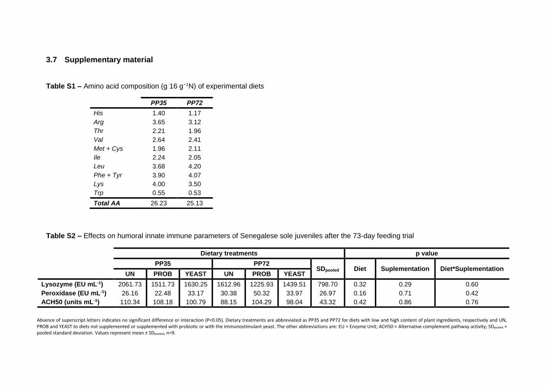
3.7 Supplementary material
Table S1 – Amino acid composition (g 16 g−1N) of experimental diets
Table S2 – Effects on humoral innate immune parameters of Senegalese sole juveniles after the 73-day feeding trial
Absence of superscript letters indicates no significant difference or interaction (P<0.05). Dietary treatments are abbreviated as PP35 and PP72 for diets with low and high content of plant ingredients, respectively and UN, PROB and YEAST to diets not supplemented or supplemented with probiotic or with the immunostimulant yeast. The other abbreviations are: EU = Enzyme Unit; ACH50 = Alternative complement pathway activity; SDpooled = pooled standard deviation. Values represent mean ± SDpooled, n=9.
PP35 PP72
His 1.40 1.17
Arg 3.65 3.12
Thr 2.21 1.96
Val 2.64 2.41
Met + Cys 1.96 2.11
Ile 2.24 2.05
Leu 3.68 4.20
Phe + Tyr 3.90 4.07
Lys 4.00 3.50
Trp 0.55 0.53
Total AA 26.23 25.13
Dietary treatments p value
PP35 PP72 SDpooled Diet Suplementation Diet*Suplementation
UN PROB YEAST UN PROB YEAST
Lysozyme (EU mL-1) 2061.73 1511.73 1630.25 1612.96 1225.93 1439.51 798.70 0.32 0.29 0.60
Peroxidase (EU mL-1) 26.16 22.48 33.17 30.38 50.32 33.97 26.97 0.16 0.71 0.42
ACH50 (units mL-1) 110.34 108.18 100.79 88.15 104.29 98.04 43.32 0.42 0.86 0.76

Table S3 – Gene relative expression in distal intestine of Senegalese sole juveniles after the 73-day feeding trial
Transcripts were quantified by qPCR and normalized using the geometric average of suitable reference genes. In each line, different superscript letters indicate significant differences (P<0.05): for a particular diet, differences caused by probiotic inclusion are indicated using x, y; for a particular probiotic inclusion, differences among the diets are shown using A, B; and among treatments (a, b). Dietary treatments are abbreviated as PP35 and PP72 for diets with low and high content of plant ingredients, respectively and UN, PROB and YEAST to diets not supplemented or supplemented with probiotic or with the immunostimulant yeast. SDpooled = pooled standard deviation. *Without significant differences after post hoc analysis. Values represent mean ± SDpooled in arbitrary units, n=6.
Dietary treatments p value
PP35 PP72 SDpooled Diet Suplementation Diet*Suplementation
UN PROB YEAST UN PROB YEAST
Lysozymes
lyzc 0.00 0.00 0.00 0.00 0.00 0.00 0.00 0.84 0.33 0.07
lyzg 1.81 2.48 2.00 2.57 1.76 2.88 1.07 0.64 0.93 0.44
Heat shock proteins
hsp90a 0.14 0.05 0.07 0.04 0.06 0.04 0.08 0.51 0.43 0.56
hsp90b 1.87A;y 3.14A;y 4.73A;x 1.11B;y 1.06B;y 1.97B;x 1.20 0.00 0.00 0.39
hsp90b1 2.05y 4.61x 2.21y 2.43y 3.68x 1.43y 1.55 0.37 0.00 0.19
Iron chelating proteins
ftm 9.45 12.10 8.98 17.35 10.66 7.05 5.74 0.44 0.08 0.08
apoa1 12.64A 16.85A 15.07A 10.77B 10.60B 7.31B 6.58 0.02 0.61 0.53
Complement components
c3-1 0.02 0.00 0.00 0.01 0.00 0.00 0.01 1.00 0.22 0.39
c3-2 0.00 0.00 0.00 0.00 0.00 0.00 0.00 0.66 0.56 0.04*
Cysteine protease
casp3 9.08 14.24 10.63 13.72 18.63 9.51 7.99 0.34 0.16 0.62
Oxidative Enzymes
gpx 0.56y 1.01x 0.72y 0.80y 1.24x 0.49y 0.33 0.49 0.00 0.15
cat 9.54ab 21.29a 12.12ab 12.28ab 9.52ab 7.02b 6.79 0.06 0.32 0.04

Table S4 – Gene relative expression in rectum of Senegalese sole juveniles after the 73-day feeding trial
Transcripts were quantified by qPCR and normalized using the geometric average of suitable reference genes. In each line, different superscript letters indicate significant differences (P<0.05): for a particular diet, differences caused by probiotic inclusion are indicated using x, y; for a particular probiotic inclusion, differences among the diets are shown using A, B; and among treatments (a, b). Dietary treatments are abbreviated as PP35 and PP72 for diets with low and high content of plant ingredients, respectively and UN, PROB and YEAST to diets not supplemented or supplemented with probiotic or with the immunostimulant yeast. SDpooled = pooled standard deviation. Values represent mean ± SDpooled in arbitrary units, n=6.
Dietary treatments p value
PP35 PP72 SDpooled Diet Suplementation Diet*Suplementation
UN PROB YEAST UN PROB YEAST
Lysozymes
lyzc 0.00 0.00 0.00 0.00 0.00 0.00 0.00 0.92 0.76 0.09
lyzg 1.36 1.34 1.47 1.53 1.04 1.04 0.36 0.16 0.25 0.15
Heat shock proteins
hsp90a 0.03xy 0.03x 0.02y 0.02xy 0.06x 0.02y 0.01 0.18 0.01 0.08
hsp90b 1.29 1.09 1.27 1.36 1.08 1.26 0.25 0.73 0.11 0.83
hsp90b1 1.05 1.41 1.12 1.24 1.28 0.89 0.37 0.64 0.10 0.36
Iron chelating proteins
ftm 3.51x 2.51y 3.01xy 3.13x 2.50y 2.33xy 0.68 0.14 0.02 0.51
apoa1 0.04b 0.14b 1.34a 0.33ab 0.56ab 0.18b 0.60 0.81 0.05 0.00
Complement components
c3-1 0.00 0.00 0.00 0.00 0.00 0.00 0.00 0.42 0.32 0.29
c3-2 0.00 0.00 0.00 0.00 0.00 0.00 0.00 0.32 0.63 0.18
Cysteine protease
casp3 2.77A 2.44A 2.63A 2.45B 1.90B 1.68B 0.60 0.01 0.13 0.42
Oxidative Enzymes
gpx 0.14A 0.16A 0.20A 0.14B 0.12B 0.11B 0.06 0.03 0.78 0.10
cat 3.01A 2.54A 3.09A 2.63B 2.21B 2.09B 0.73 0.03 0.36 0.47
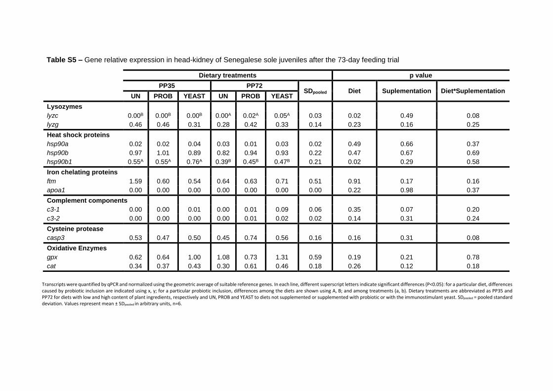
Table S5 – Gene relative expression in head-kidney of Senegalese sole juveniles after the 73-day feeding trial
Transcripts were quantified by qPCR and normalized using the geometric average of suitable reference genes. In each line, different superscript letters indicate significant differences (P<0.05): for a particular diet, differences caused by probiotic inclusion are indicated using x, y; for a particular probiotic inclusion, differences among the diets are shown using A, B; and among treatments (a, b). Dietary treatments are abbreviated as PP35 and PP72 for diets with low and high content of plant ingredients, respectively and UN, PROB and YEAST to diets not supplemented or supplemented with probiotic or with the immunostimulant yeast. SDpooled = pooled standard deviation. Values represent mean ± SDpooled in arbitrary units, n=6.
Dietary treatments p value
PP35 PP72 SDpooled Diet Suplementation Diet*Suplementation
UN PROB YEAST UN PROB YEAST
Lysozymes
lyzc 0.00B 0.00B 0.00B 0.00A 0.02A 0.05A 0.03 0.02 0.49 0.08
lyzg 0.46 0.46 0.31 0.28 0.42 0.33 0.14 0.23 0.16 0.25
Heat shock proteins
hsp90a 0.02 0.02 0.04 0.03 0.01 0.03 0.02 0.49 0.66 0.37
hsp90b 0.97 1.01 0.89 0.82 0.94 0.93 0.22 0.47 0.67 0.69
hsp90b1 0.55A 0.55A 0.76A 0.39B 0.45B 0.47B 0.21 0.02 0.29 0.58
Iron chelating proteins
ftm 1.59 0.60 0.54 0.64 0.63 0.71 0.51 0.91 0.17 0.16
apoa1 0.00 0.00 0.00 0.00 0.00 0.00 0.00 0.22 0.98 0.37
Complement components
c3-1 0.00 0.00 0.01 0.00 0.01 0.09 0.06 0.35 0.07 0.20
c3-2 0.00 0.00 0.00 0.00 0.01 0.02 0.02 0.14 0.31 0.24
Cysteine protease
casp3 0.53 0.47 0.50 0.45 0.74 0.56 0.16 0.16 0.31 0.08
Oxidative Enzymes
gpx 0.62 0.64 1.00 1.08 0.73 1.31 0.59 0.19 0.21 0.78
cat 0.34 0.37 0.43 0.30 0.61 0.46 0.18 0.26 0.12 0.18

88

89
CHAPTER 4 Innate immune response, intestinal morphology and
microbiota changes in Senegalese sole fed plant
protein diets with probiotics or autolyzed yeast
Published: Batista et al. 2016. Applied Microbiology and Biotechnology
Epub ahead of print: 1-16 (DOI: 10.1007/s00253-016-7592-7)

90

CHAPTER 4
91
Innate immune response, intestinal morphology and microbiota changes in
Senegalese sole fed plant protein diets with probiotics or autolyzed yeast
Batista S. 1,2,3, Medina, A.4, Pires M. A.5, Moriñigo M. A.4, Sansuwan K.2,6,
Fernandes J.M.O.3, Valente L.M.P.1,2, Ozório R.O.A.1,2*
1 ICBAS – Instituto de Ciências Biomédicas de Abel Salazar, Universidade de Porto, Rua Jorge Viterbo Ferreira n.º 228,
4050-313 Porto, Portugal.
2 CIMAR/CIIMAR – Centro Interdisciplinar de Investigação Marinha e Ambiental, Rua dos Bragas, 289, 4050-123 Porto,
Portugal.
3 FBA - Faculty of Biosciences and Aquaculture, Nord University, 8049 Bodø, Norway
4 Department of Microbiology, Faculty of Sciences, University of Málaga. 29071. Málaga, Spain
5 CECAV – Centro de Ciência Animal e Veterinária, UTAD, apartado 1013, 5000-801 Vila Real, Portugal
6 Department of Aquaculture, Faculty of Fisheries, Kasetsart University, Bangkok 10900, Thailand
CHAPTER 4
92
Abstract
The effects of using plant ingredients in Senegalese sole (Solea senegalensis) diet
on immune competence and intestine morphology and ecology are still
controversial. Probiotics or immunostimulants can potentially alter the intestinal
microbiota in a way that protects fish against pathogens. The current study aimed
to examine the intestine histology and microbiota, and humoral innate immune
response in juvenile sole fed diets with low (35%) or high (72%) content of plant
protein (PP) ingredients supplemented with a multispecies probiotic bacteria or
autolyzed yeast. Fish fed the probiotic diet had lower growth performance.
Lysozyme and complement activities were significantly higher in fish fed PP72 diets
than in their counterparts fed PP35 diets after 17 days and 38 days of feeding. At 2
days of feeding, fish fed unsupplemented PP72 showed larger intestine section area
and longer villus than fish fed unsupplemented PP35. At 17 days of feeding, fish fed
unsupplemented PP72 showed more goblet cells than the other dietary groups,
except the group fed yeast supplemented PP35 diet. Short-term feeding with high
dietary PP level may enhance the immune system and improve surface area for
nutrient absorption. However, this effect was reversed with long-time feeding (73
days), suggesting a habituation to dietary treatments and/or immunosuppression,
with a reduction in the number of the goblet cells. Fish fed for 38 days with diets
supplemented with autolyzed yeast showed longer intestinal villus, also suggesting
an improvement on the surface area for nutrients absorption.
The predominant bacteria found in sole intestine were Vibrio sp and dietary probiotic
supplementation caused a reduction in Vibrio content, regardless of the PP level.
CHAPTER 4
93
4.1 Introduction
Aquaculture is a growing sector of the food-production in the world (FAO) and much
effort has been done during the last years on increasing fish growth efficiency by
manipulation of dietary formulations. Senegalese sole (Solea senegalensis) is a
promising flatfish species in the European aquaculture, due to its high commercial
value and nutritional properties. The ability of Senegalese sole to efficiently use
novel plant-based diets (Silva et al., 2009) is an important asset towards the
intensification and commercialization of this species. Currently, diseases outbreaks
remain a primary constraint to aquaculture expansion. One of the newest scientific
area of high interest to improve fish growth efficiency and to prevent and/or control
fish diseases is the application of probiotics and microbial feed additives as
alternatives to antibiotics (Dimitroglou et al., 2011; Magnadottir, 2010). These feed
additives or dietary raw materials favourably affect animal performance and welfare,
particularly through the modulation of the intestinal microbiota which plays a critical
role in maintaining host health (Tuohy et al., 2005) and through the modulation of
the immune system, influencing various humoral and cellular defences (Nayak,
2010).
Among promising probiotic candidates, the Bacilli class has been widely assayed in
fish and numerous studies have demonstrated that its administration enhances
immune responses and disease resistance (Aly et al., 2008; Kumar et al., 2008;
Newaj-Fyzul et al., 2007; Salinas et al., 2005). On the other hand, the use of some
selectively fermented ingredients like yeast, allows specific changes in the
composition and/or activity of the gastrointestinal microflora that confers benefits
upon the host’s wellbeing and health (Marcel, 2008). Natural immunostimulants are
valuable for activating the fish immune system and protecting fish against adverse
conditions (Anderson, 1992; Sakai, 1999). Among them the yeast Saccharomyces
cerevisiae has been found to be a good enhancer of the trout immune system
(Siwicki et al., 1994) and it can also add nutritional value in fish diets as a possible
alternative to commonly used fish meal protein (Rumsey et al., 1992).
The microbial flora present in fish intestine plays an important role on breaking down
ingested food or inhibiting the colonization of the intestine by pathogens (Manzano
et al., 2012). The composition of bacterial communities in fish can be affected by
several factors, including the environment and their interaction with diet. A balanced
intestinal microbiota constitutes as an efficient barrier against pathogen

CHAPTER 4
94
colonization, producing metabolic substrates (e.g. vitamins and short-chain fatty
acids) and stimulating the immune system (Gaggìa et al., 2010). In this context
probiotics and some immunostimulants may play a significant role to improve the
resistance to pathogenic bacteria colonization and enhancement of host mucosa
immunity, thus reducing pathogen load and an enhancing health status of the
animals (Choct, 2009). Bacterial diversity has an important role in the function of
ecosystems (Bell et al., 2005). Biodiversity protects ecosystems against decline in
their functionality and allows for adaptation to changing conditions, as the
coexistence of many bacterial species provides an effective fail-safe measures to
help keep the pathogen bacteria from spreading (Yachi and Loreau, 1999). The
complex microbial ecology of the intestinal tract (GI) provides nutritional benefit and
protection against pathogens. Moreover, microbial ecology may play an important
role in modulating interactions between environment and the immune system
(Balcázar et al., 2007a). The potential pathogens present within the GI tract should
be reduced in order to avoid mucosal damage and improve absorptive surface area
(Merrifield et al., 2010c). Rawls et al. (2004) observed that microbiota stimulated
intestinal epithelial proliferation and influenced enterocyte morphology.
The aim of the current study was to evaluate the effects of dietary plant protein levels
intercropped with a multispecies probiotic (from the Bacilli class) or an autolysed
yeast supplementation on growth performance, innate immune response, intestine
morphology and microbiota diversity in Senegalese sole (Solea senegalensis).
4.2 Materials and methods
The feeding experiment took place at the CIIMAR facilities (University of Porto,
Portugal). The current study was conducted under the supervision of an accredited
expert in laboratory animal science by the Portuguese Veterinary Authority
(1005/92, DGV-Portugal, following FELASA category C recommendations) and
according to the guidelines on the protection of animals used for scientific purposes
from the European directive 2010/63/UE. The current study is an extension of a
submitted work by Batista et al. (2016).
4.2.1 Feed and fish
Six isoproteic (55%) and isolipidic (8%) diets with similar amino-acid composition
were formulated to contain 35% (PP35) or 72% (PP72) of plant ingredients (Table
CHAPTER 4
95
1), each supplemented with PROB and YEAST (commercial products) or
unsupplemented (UN) in a 2 × 3 factorial design. PROB (AquaStar® Growout -
Biomin® Holding GmbH, Herzogenburg, Austria) was a multispecies probiotic
(Bacillus sp, Pediococcus sp, Enterococcus sp, Lactobacillus sp) incorporated at
1.34 × 10¹⁰ CFU kg-1 diet (CFU – colony forming unit) in the extruded pellets by
means of vacuum coating (Dinnisen Pegasus vacuum mixer, PG-10VCLAB) using
fish oil as a carrier. YEAST (Levabon® Aquagrow - Biomin®) was autolyzed yeast
(Saccharomyces cerevisiae) supplemented in the mixture at 4g kg-1 diet. Diets were
prepared according to the normatives ISO (6887-1:1999, 7218:2007, 6498:2012)
for the microbiological examinations. Thereafter, the isolation and enumeration of
bacteria in the diets followed the European standards for Enterococcus spp (EN
15788:2009), Lactobacillus spp (EN 15787:2009), Pediococcus spp. (EN
15786:2009) and Bacillus spp (EN 15784:2009).
Senegalese sole were obtained from a commercial fish farm (Aquacria S.A.,
Portugal) and transported to the rearing facilities of CIIMAR. After 5-week
acclimation period, fish were distributed into 18 fibreglass tanks of 50 L each, at a
density of 5.8 kg m-2 (31 fish per tank, 33.1 ± 0.2 g each) and fed the experimental
diets in triplicate. The tanks were supplied with seawater (32.0 ± 0.1 ppm NaCl, 19.7
± 0.5 ºC, 0.33 ± 0.27 mg L-1 NH3, 0.14 ± 0.07 mg L-1 NO2) at a flow rate of 1.5 L
min−1, in a controlled semi-closed recirculation system under a constant photoperiod
(12 h light:12 h darkness). Rearing conditions were monitored during the entire trial.
The recirculation system was equipped with UV lights and ozone generator to
hamper bacteria growth in the rearing water. The trial lasted 73 days and fish were
fed to apparent satiety based on visual observation of acceptance and refusal of
feed using temporized automatic feeders.
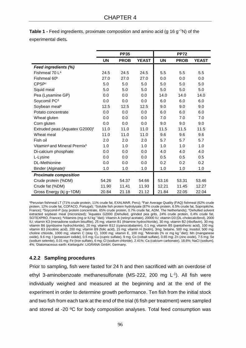
CHAPTER 4
96
Table 1 - Feed ingredients, proximate composition and amino acid (g 16 g−1N) of the
experimental diets.
PP35 PP72
UN PROB YEAST UN PROB YEAST
Feed ingredients (%)
Fishmeal 70 La 24.5 24.5 24.5 5.5 5.5 5.5
Fishmeal 60b 27.0 27.0 27.0 0.0 0.0 0.0
CPSPc 5.0 5.0 5.0 5.0 5.0 5.0
Squid meal 5.0 5.0 5.0 5.0 5.0 5.0
Pea (Lysamine GP) 0.0 0.0 0.0 14.0 14.0 14.0
Soycomil PCd 0.0 0.0 0.0 6.0 6.0 6.0
Soybean meale 12.5 12.5 12.5 9.0 9.0 9.0
Potato concentrate 0.0 0.0 0.0 6.0 6.0 6.0
Wheat gluten 0.0 0.0 0.0 7.0 7.0 7.0
Corn gluten 0.0 0.0 0.0 9.0 9.0 9.0
Extruded peas (Aquatex G2000)f 11.0 11.0 11.0 11.5 11.5 11.5
Wheat meal 11.0 11.0 11.0 9.6 9.6 9.6
Fish oil 2.0 2.0 2.0 5.7 5.7 5.7
Vitaming and Mineral Premixh 1.0 1.0 1.0 1.0 1.0 1.0
Di-calcium phosphate 0.0 0.0 0.0 4.0 4.0 4.0
L-Lysine 0.0 0.0 0.0 0.5 0.5 0.5
DL-Methionine 0.0 0.0 0.0 0.2 0.2 0.2
Binder (Alginate)i 1.0 1.0 1.0 1.0 1.0 1.0
Proximate composition
Crude protein (%DM) 54.26 54.37 54.66 53.16 53.31 53.46
Crude fat (%DM) 11.90 11.41 11.93 12.21 11.45 12.27
Gross Energy (kj g−1DM) 20.84 21.18 21.12 21.84 22.05 22.04
aPeruvian fishmeal LT (71% crude protein, 11% crude fat, EXALMAR, Peru); bFair Average Quality (FAQ) fishmeal (62% crude protein, 12% crude fat, COFACO, Portugal); cSoluble fish protein hydrolysate (87% crude protein, 6.5% crude fat, Sopropêche, France); dSoycomil-P (soy protein concentrate, 65% crude protein, 0.7% crude fat, ADM, The Netherlands); eDehulled solvent extracted soybean meal (micronized); fAquatex G2000 (Dehulled, grinded pea grits, 24% crude protein, 0.4% crude fat, SOTEXPRO, France); gVitamins (mg or IU kg−1diet): Vitamin A (retinyl acetate), 20000 IU; vitamin D3 (DL-cholecalciferol), 2000 IU; vitamin K3 (menadione sodium bisulfite), 25 mg; vitamin B1 (thiamine hydrochloride), 30 mg; vitamin B2 (riboflavin), 30 mg; vitamin B6 (pyridoxine hydrochloride), 20 mg; vitamin B12 (cyanocobalamin), 0.1 mg; vitamin B5 (pantothenic acid), 100 mg; vitamin B3 (nicotinic acid), 200 mg; vitamin B9 (folic acid), 15 mg; vitamin H (biotin), 3mg; betaine, 500 mg; inositol, 500 mg; choline chloride, 1000 mg; vitamin C (stay C), 1000 mg; vitamin E, 100 mg; hMinerals (% or mg kg−1diet): Mn (manganese oxide), 9.6 mg; I (potassium iodide), 0.5 mg; Cu (cupric sulfate), 9 mg; Co (cobalt sulfate), 0.65 mg; Zn (zinc oxide), 7.5 mg; Se (sodium selenite), 0.01 mg; Fe (iron sulfate), 6 mg; Cl (sodium chloride), 2.41%; Ca (calcium carbonate), 18,6%; NaCl (sodium), 4%; iDiatomaceous earth: Kielseguhr: LIGRANA GmbH, Germany.
4.2.2 Sampling procedures
Prior to sampling, fish were fasted for 24 h and then sacrificed with an overdose of
ethyl 3-aminobenzoate methanesulfonate (MS-222, 200 mg L-1). All fish were
individually weighed and measured at the beginning and at the end of the
experiment in order to determine growth performance. Ten fish from the initial stock
and two fish from each tank at the end of the trial (6 fish per treatment) were sampled
and stored at -20 ºC for body composition analyses. Total feed consumption was
CHAPTER 4
97
registered during the entire feeding trial. The innate immune response and intestine
histology were analysed at days 3, 17, 38 and 73 of the feeding trial.
For the evaluation of humoral innate immune parameters (lysozyme, peroxidase
and alternative complement pathway – ACH50) blood was withdrawn from the
caudal vein of 9 fish per treatment using heparinised syringes and centrifuged at
5000 × g for 10 min at 4 ºC. Liver and intestine of each fish was sampled, cut and
fixed in buffered formalin to histology analysis. To examine intestinal microbiota, the
whole intestine of other 9 fish per treatment was aseptically removed at 17, 38 and
73 days of feeding the experimental diets. The intestinal contents were sampled by
stripping and pooled by tank (3 fish per tank) and homogenized in 1 ml sterile
phosphate-buffered saline (PBS - pH 7.2).
4.2.3 Chemical analyses of diets and body composition
All chemical analyses were carried in duplicate according to AOAC (2006). Fish
were minced without thawing using a meat mincer, pooled and used to determine
their dry matter content (105 °C for 24 h). Fish were then freeze-dried and analysed
for dry matter, ash (Nabertherm L9/11/B170; Germany; 550ºC for 6 h), crude protein
(N×6.25, Leco N analyser, Model FP-528, Leco Corporation, St. Joseph, USA),
crude lipid (petroleum ether extraction, 40–60ºC, Soxtherm, Gerhardt, Germany)
and gross energy (adiabatic bomb calorimeter, Werke C2000, IKA, Germany).
4.2.4 Humoral innate immune parameters
All measurements were done in triplicate on a Power-WaveTM microplate
spectrophotometer (BioTek Synergy HT, USA). Plasma lysozyme activity (EU ml-1)
was determined using a turbidimetric assay adapted to microtitre, as described by
Hutchinson and Manning (1996). One lysozyme enzyme unit (EU) was defined as
the amount of lysozyme that caused a decrease in absorbance per min. Plasma
peroxidase activity (EU ml-1) was measured following the procedure adapted to
Solea senegalensis by Costas et al. (2011), defining that one unit of peroxidase
produces an absorbance change of 1 OD. Alternative complement pathway
(ACH50) activity was based on the lysis of rabbit red blood cells (2.8 × 10⁸ cells mL-
1), as reported by Sunyer and Tort (1995). ACH50 units were defined as the
concentration of plasma giving 50% lysis of rabbit red blood cells.

CHAPTER 4
98
4.2.5 Histological evaluation
The samples (liver and a segment with 1.5cm length from the proximal intestine)
were fixed in phosphate buffered formalin (4%, pH 7, VWR, Portugal) for 24 hours.
They were subsequently dehydrated and embedded in paraffin according to
standard histological procedures. Thereafter, 3 micra transversal sections were
obtained and stained with haematoxylin and eosin (H&E - Merck, Portugal) and
Periodic Acid-Schiff (PAS - Merck, Portugal) before examination under a light
microscope (Olympus BX51, cell^B software, GmbH, Hamburg, Germany).
Three different intestine sections of each animal were used for quantitative
measurements. The intestine section area (mm²), villus height (μm), muscular layer
thickness (from serosa to submucosa; μm) and goblet cells (number per section)
positive to PAS staining of intestinal wall were measured according Batista et al.
(2014). The villus width (µm) was measured across the base of the villus at the
luminal surface of 10 selected villi per section.
In the liver, the occurrence of possible pathological damage (presence or absence
of defined vacuoles indicator of fat degeneration) was evaluated. For each sampled
fish, the cytoplasm vacuolization degree (H&E) and glycogen content (PAS) of the
hepatocytes were analysed (Figure 1). Observations were consistently made using
a combination of low magnification (objective of 10×) for notice the general aspects
and then with higher magnification (objective of 40×) for categorizing. Ten randomly
sampled fields were analysed per section. Evaluation of the hepatocyte
vacuolization degree was made using a semi-quantitative approach, according to
the following three grades and criteria: 0 (none) – less than 1/3 of the hepatocyte
cytoplasm shows vacuoles; 1 (moderate) – between 1/3 and less than 2/3 of the
hepatocyte cytoplasm shows vacuolization; 2 (high) – more than 2/3 of the
hepatocyte cytoplasm shows vacuolization. Assessment of hepatocyte
carbohydrates (namely glycogen) content also followed a semi-quantitative
approach, with three degrees: 0 (none) – when hepatocytes in PAS sections did not
stain positively or had a very weak colour; 1 (moderate) – PAS sections presented
a median staining; 2 (high) – PAS sections present hepatocytes with a very strong
staining.

CHAPTER 4
99
Figure 1 – Liver histology of Senegalese sole for quantification of vacuoles and glycogen
content. A) high content of glycogen and reduce vacuolization B) Moderate content of
glycogen and vacuoles. C) High content in vacuoles and small amounts of glycogen (PAS
stained, Bar = 100µm).
4.2.6 Intestinal microbiota
Microbial populations were isolated from the intestinal content as described by
Merrifield et al. (2009). Intestinal content samples were serially diluted to 10-2 with
PBS and 100 μL were spread onto duplicate tryptone soy agar (TSA – 0.9% NaCl)
plates. Plates were incubated at 25 ºC for 48 hours and colony forming units (CFU)
g-1 was calculated from statistically viable plates (i.e. plates containing 30-300
colonies). One hundred colonies of one of these plates were randomly collected
from each dietary treatment assayed and cultured on TSA to obtain pure cultures.
Then the colonies were identified by the amplification and sequencing of a fragment
of 16S rDNA. Briefly, the colonies isolated were suspended in 100µL of sterile milli-
Q water. Samples were boiled at 100ºC for 15 minutes and adjusted to 1mL. These
suspensions were centrifuged at 12000 rpm for 5 minutes and 1-5μL of the
supernatant was used to carry out the PCR reactions. The fragment 16S rDNA was
amplified using the universal primers SD-Bact-0008-a-S20 (5’
AGAGTTTGATCCTGGCTCAG 3’) and SD-Bact-1492-a-A-19 (5’
GGTTACCTTGTTACGACTT 3’) (Kim and Austin, 2006a). Polymerase chain
reactions were carried out in a 50 μl reaction mixture that included 5 pmol of each
primer, 200 μM dNTPs, 1 x PCR buffer, 2 mM MgCl2, 1 U BIOTAQ DNA Polimerase
(Bioline, London, UK) and 1 μl of a boiled colony suspension. Thermal cycling
consisted of an initial step of 2 min at 95ºC and 35 cycles of 30 s at 95ºC, 30s at
52ºC and 40s at 72ºC and a final extension step 5 min at 72ºC. Polymerase chain
reaction products were electrophoresed on 1% agarose gel, visualized via ultraviolet
transillumination and then they were purified with a QiaQuick PCR purification kit

CHAPTER 4
100
(QIAGEN, Hilden, Germany). The PCR products were sequenced by cycling
sequencing using SD-Bact-0008-a-S20 and SD-Bact-1492-a-A-19 by Macrogen
(Corea). The resulting sequences (∼500 bp) were compared with the sequences
from the National Center for Biotechnology Information (NCBI) or Greengenes DNA
sequence database using the BLAST sequence algorithm (Altschul et al.,
1990).Chimeric sequences were identified by using the CHECK CHIMERA program
of the Ribosomal Database Project (Maidak et al., 1999) and the sequences
reported in this study have been deposited in the GenBank database under the
following accession numbers: KU725824 to KU725849.
4.2.7 Calculations of growth performance
Feed conversion ratio (FCR) was calculated as: dry feed intake (g) / wet weight gain
(g), and the specific growth rate (SGR, % BW day-1) as: 100 × (ln W0 – ln W1) / days,
where W0 and W1 are the initial and the final fish mean weights in grams and days
is the trial duration. Voluntary feed intake (VFI, % BW day-1) was calculated as: 100
× (dry feed intake (g) / ABW (g) / days), where ABW (average body weight) was
calculated as: (W1+W0) / 2. The net protein utilization (NPU) was calculated as (PBF
– PBI) / protein fed, where PBF is the final protein content of fish and PBI is the
initial protein content of fish.
4.2.8 Statistical analysis
Statistical analyses were performed with the software SPSS (IBM SPSS
STATISTICS, 17.0 package, IBM Corporation, New York, USA). Results are
expressed as mean ± standard deviation (SDpooled as weighted average of each
group's standard deviation) and the level of significance used was P ≤ 0.05. Data
were analysed for normality (Shapiro-Wilk test) and homogeneity of variance
(Levene’s test) and were log-transformed whenever necessary. Data were analysed
by a two-way ANOVA with diet and probiotic as main factors. When significant
differences were obtained from the ANOVA, Tukey’s post hoc tests were carried out
to identify significantly different groups. When data did not meet the ANOVA
assumptions, a non-parametric Kruskal–Wallis test was performed.

CHAPTER 4
101
4.3 Results
4.3.1 Growth performance
Fish grew from 33.1 ± 0.20 g to 50.6 ± 1.2 g after feeding the experimental diets for
73 days (Table 2). Growth performance did not differ between PP35 and PP72
groups. PROB groups had significantly lower final body weight (FBW – 45.0 ± 1.9)
and daily growth index (SGR – 0.42 ± 0.1) compared to UN groups (50.5 ± 2.0 and
0.58 ± 0.0 respectively). Additionally, UN groups had significantly better feed
conversion ratio (FCR – 1.5 ± 0.1) and net protein utilization (NPU – 19.07 ± 2.3)
than the PROB (2.2 ± 0.4 and 10.4 ± 1.9 respectively) and YEAST (2.1 ± 0.4 and
13.0 ± 4.6 respectively) supplemented groups. Voluntary feed intake (VFI) was also
lower in UN groups (0.8 ± 0.0), and differed significantly from YEAST groups (0.9 ±
0.1).
4.3.2 Humoral innate immune parameters
The values of humoral innate parameters analyzed are present in Table 3.
Peroxidase activity (EU mL-1) was not significantly different (P<0.05) among dietary
groups for any sampling time. Lysozyme activity and ACH50 were not affected by
probiotic administration, but were significantly changed by the dietary PP level at 17
and 38 days of feeding. At 17 days, fish from PP72 groups had higher lysozyme
(359 ± 201 EU mL-1) and ACH50 (204 ± 55 Units ml-1) comparing to fish from PP35
groups (177 ± 106 EU mL-1 and 169 ± 19 Units ml-1, respectively). At 38 days of
feeding, fish from PP72 groups showed higher lysozyme (908 ± 399 EU mL-1) and
ACH50 (258 ± 64 Units ml-1) activities, comparing to fish from PP35 groups (647 ±
457 EU mL-1 and 183 ± 49 Units ml-1, respectively).

Table 2 – Growth performance of Senegalese sole juveniles after the 73 days of feeding the experimental diets.
In each line, different superscript letters indicate significant differences (P<0.05): for a particular PP level, differences caused by probiotic inclusion are indicated using x, y; for a particular probiotic inclusion, differences among PP level are shown using A, B. Dietary treatments are abbreviated as PP35 and PP72 for diets with low and high content of plant ingredients, respectively and UN, PROB and YEAST to diets not supplemented or supplemented with probiotic or with the immunostimulant yeast. The other abbreviations are: PP = Pant protein level factor; S = Supplementation factor; FBW = Final body weight; SGR = Specific growth rate; FCR = Feed conversion ratio; VFI = Voluntary feed intake; NPU = Net protein utilization; SDpooled = pooled standard deviation. Values represent mean ± SDpooled, n=3.
Dietary treatments p value
PP35 PP72 SDpooled PP S PP*S
UN PROB YEAST UN PROB YEAST
Growth performance
FBW (g) 50.49x 45.73y 45.82xy 50.58x 44.21y 47.73xy 2.91 0.91 0.02 0.60
SGR 0.58x 0.44y 0.45xy 0.58x 0.40y 0.49xy 0.08 0.99 0.02 0.68
FCR 1.46y 2.04x 2.11x 1.49y 2.42x 2.02x 0.34 0.53 0.01 0.51
VFI 0.83y 0.88xy 0.90x 0.85y 0.94xy 0.94x 0.33 0.13 0.02 0.80
NPU 20.11x 10.48y 10.83y 18.03x 10.31y 15.26y 3.05 0.62 0.00 0.21

Table 3 – Effects on humoral innate immune parameters of Senegalese sole juveniles after the 2, 17, 38 and 73 days of
feeding the different experimental diets.
In each line, different superscript letters indicate significant differences (P<0.05): for a particular probiotic inclusion, differences among PP level are shown using A, B. Dietary treatments are abbreviated as PP35 and PP72 for diets with low and high content of plant ingredients, respectively and UN, PROB and YEAST to diets not supplemented or supplemented with probiotic or the immunostimulant yeast. The other abbreviations are: PP = Pant protein level factor; S = Supplementation factor; EU = Enzyme Unit; ACH50 = Alternative complement pathway activity. SDpooled = pooled standard deviation. Values represent mean ± SDpooled, n=9.
Dietary treatments p value
PP35 PP72 SDpooled PP S PP*S
UN PROB YEAST UN PROB YEAST
Lysozyme (EU mL-1)
2 days 190.98 290.74 440.12 379.32 628.70 457.64 288.07 0.07 0.06 0.43
17 days 124.21B 225.35B 175.79B 410.32A 338.09A 287.04A 156.58 0.00 0.76 0.27
38 days 612.50B 695.99B 628.39B 761.11A 1047.62A 950.69A 439.55 0.02 0.70 0.69
73 days 2061.73 1511.73 1630.25 1612.96 1225.93 1295.15 787.12 0.23 0.25 0.51
Peroxidase (EU mL-1)
2 days 122.11 97.44 101.56 105.12 59.09 92.82 48.68 0.28 0.06 0.15
17 days 141.65 111.80 110.51 100.71 71.51 122.82 61.59 0.25 0.19 0.16
38 days 59.90 80.03 48.99 54.39 78.59 31.21 45.32 0.86 0.06 0.77
73 days 26.16 22.48 33.17 30.38 50.32 33.97 26.97 0.15 0.61 0.30
ACH50 (units mL-1)
2 days 112.29 113.85 103.17 97.97 119.27 113.59 47.47 0.98 0.88 0.86
17 days 161.34B 177.01B 167.67B 221.55A 195.88A 193.10A 51.21 0.02 0.82 0.45
38 days 174.86B 197.38B 177.52B 270.95A 247.59A 253.50A 58.78 0.00 0.92 0.52
73 days 110.34 108.18 100.79 88.15 104.29 98.04 43.32 0.42 0.86 0.76
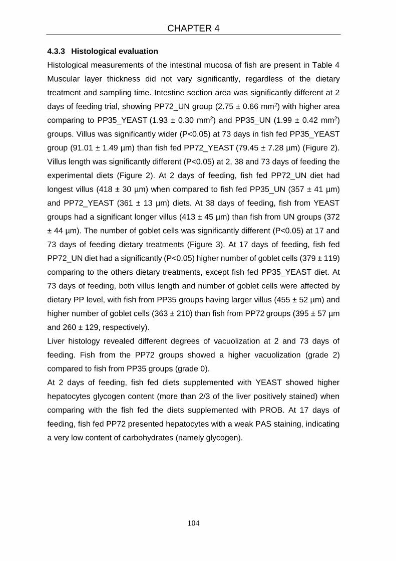
CHAPTER 4
104
4.3.3 Histological evaluation
Histological measurements of the intestinal mucosa of fish are present in Table 4
Muscular layer thickness did not vary significantly, regardless of the dietary
treatment and sampling time. Intestine section area was significantly different at 2
days of feeding trial, showing PP72_UN group (2.75 ± 0.66 mm2) with higher area
comparing to PP35_YEAST (1.93 ± 0.30 mm2) and PP35_UN (1.99 ± 0.42 mm2)
groups. Villus was significantly wider (P<0.05) at 73 days in fish fed PP35_YEAST
group (91.01 ± 1.49 µm) than fish fed PP72_YEAST (79.45 ± 7.28 µm) (Figure 2).
Villus length was significantly different (P<0.05) at 2, 38 and 73 days of feeding the
experimental diets (Figure 2). At 2 days of feeding, fish fed PP72_UN diet had
longest villus (418 ± 30 µm) when compared to fish fed PP35_UN (357 ± 41 µm)
and PP72_YEAST (361 ± 13 µm) diets. At 38 days of feeding, fish from YEAST
groups had a significant longer villus (413 ± 45 µm) than fish from UN groups (372
± 44 µm). The number of goblet cells was significantly different (P<0.05) at 17 and
73 days of feeding dietary treatments (Figure 3). At 17 days of feeding, fish fed
PP72_UN diet had a significantly (P<0.05) higher number of goblet cells (379 ± 119)
comparing to the others dietary treatments, except fish fed PP35_YEAST diet. At
73 days of feeding, both villus length and number of goblet cells were affected by
dietary PP level, with fish from PP35 groups having larger villus (455 ± 52 µm) and
higher number of goblet cells (363 ± 210) than fish from PP72 groups (395 ± 57 µm
and 260 ± 129, respectively).
Liver histology revealed different degrees of vacuolization at 2 and 73 days of
feeding. Fish from the PP72 groups showed a higher vacuolization (grade 2)
compared to fish from PP35 groups (grade 0).
At 2 days of feeding, fish fed diets supplemented with YEAST showed higher
hepatocytes glycogen content (more than 2/3 of the liver positively stained) when
comparing with the fish fed the diets supplemented with PROB. At 17 days of
feeding, fish fed PP72 presented hepatocytes with a weak PAS staining, indicating
a very low content of carbohydrates (namely glycogen).
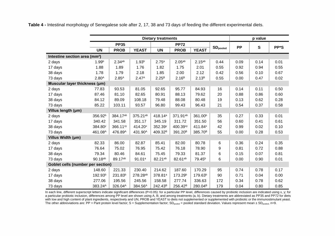
Table 4 - Intestinal morphology of Senegalese sole after 2, 17, 38 and 73 days of feeding the different experimental diets.
In each line, different superscript letters indicate significant differences (P<0.05): for a particular PP level, differences caused by probiotic inclusion are indicated using x, y; for a particular probiotic inclusion, differences among PP level are shown using A, B; and among treatments (a, b). Dietary treatments are abbreviated as PP35 and PP72 for diets with low and high content of plant ingredients, respectively and UN, PROB and YEAST to diets not supplemented or supplemented with probiotic or the immunostimulant yeast. The other abbreviations are: PP = Pant protein level factor; S = Supplementation factor; SDpooled = pooled standard deviation. Values represent mean ± SDpooled, n=9.
Dietary treatments p value
PP35 PP72 SDpooled PP S PP*S
UN PROB YEAST UN PROB YEAST
Intestine section area (mm²)
2 days 1.99b 2.34ab 1.93b 2.75a 2.05ab 2.15ab 0.44 0.09 0.14 0.01
17 days 1.88 1.89 1.76 1.82 1.75 2.01 0.55 0.92 0.94 0.55
38 days 1.78 1.79 2.18 1.85 2.00 2.12 0.42 0.56 0.10 0.67
73 days 2.80A 2.85A 2.47A 2.25B 2.16B 2.13B 0.55 0.00 0.47 0.02
Muscular layer thickness (μm)
2 days 77.83 93.53 81.05 92.65 95.77 84.93 16 0.14 0.11 0.50
17 days 87.46 81.10 82.65 80.91 88.13 79.62 20 0.88 0.86 0.60
38 days 84.12 89.09 108.18 79.48 88.08 80.48 19 0.13 0.62 0.28
73 days 85.22 103.11 93.57 96.80 99.43 96.43 21 0.54 0.37 0.58
Villus length (μm)
2 days 356.92b 384.17ab 375.21ab 418.14a 371.91ab 361.00b 35 0.27 0.33 0.01
17 days 340.42 341.58 351.17 345.19 311.72 351.50 56 0.60 0.41 0.61
38 days 384.80y 366.11xy 414.20x 352.39y 400.39xy 411.84x 42 0.99 0.02 0.10
73 days 461.08A 476.89A 431.90A 409.32B 391.20B 385.70B 55 0.00 0.28 0.53
Villus Width (μm)
2 days 82.33 86.00 82.87 85.41 82.00 80.78 6 0.36 0.24 0.35
17 days 76.64 75.02 76.95 75.42 76.18 78.90 9 0.81 0.72 0.88
38 days 79.34 80.46 84.61 75.45 79.33 81.37 6 0.15 0.07 0.81
73 days 90.18ab 89.17ab 91.01a 82.21ab 82.61ab 79.45b 6 0.00 0.90 0.01
Goblet cells (number per section)
2 days 148.60 221.33 230.40 214.62 187.60 170.29 95 0.74 0.78 0.17
17 days 192.93b 231.83b 278.28ab 378.81a 173.29b 179.63b 90 0.71 0.04 0.00
38 days 277.06 195.56 245.56 158.58 277.74 336.63 172 0.34 0.78 0.62
73 days 383.24A 326.04A 384.56A 242.43B 256.42B 280.04B 179 0.04 0.80 0.85

CHAPTER 4
106
Figure 2 - Histological sections of anterior intestines of Senegalese sole for villus width
(VW) and for villus length (VL). VW: A) PP35_YEAST diet showing higher VW than B) that
correspond to PP72_YEAST diet (H&E, Bar = 100µm); VL: A and B represents 2 days
samples time, and C and D 73 days sampling time. Note the difference between the villus
dimensions in the images A and C showing higher villus length when comparing with B and
D. A) PP35 diet and B) PP72_YEAST diet; C) PP35 diet and D) PP72_YEAST diet (H&E,
Bar = 250µm).
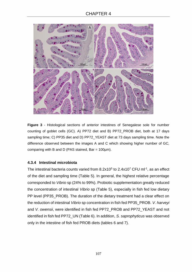
CHAPTER 4
107
Figure 3 - Histological sections of anterior intestines of Senegalese sole for number
counting of goblet cells (GC). A) PP72 diet and B) PP72_PROB diet, both at 17 days
sampling time; C) PP35 diet and D) PP72_YEAST diet at 73 days sampling time. Note the
difference observed between the images A and C which showing higher number of GC,
comparing with B and D (PAS stained, Bar = 100µm).
4.3.4 Intestinal microbiota
The intestinal bacteria counts varied from 8.2x10⁵ to 2.4x10⁷ CFU ml-1, as an effect
of the diet and sampling time (Table 5). In general, the highest relative percentage
corresponded to Vibrio sp (24% to 99%). Probiotic supplementation greatly reduced
the concentration of intestinal Vibrio sp (Table 5), especially in fish fed low dietary
PP level (PP35_PROB). The duration of the dietary treatment had a clear effect on
the reduction of intestinal Vibrio sp concentration in fish fed PP35_PROB. V. harveyi
and V. owensii, were identified in fish fed PP72_PROB and PP72_YEAST and not
identified in fish fed PP72_UN (Table 6). In addition, S. saprophyticus was observed
only in the intestine of fish fed PROB diets (tables 6 and 7).
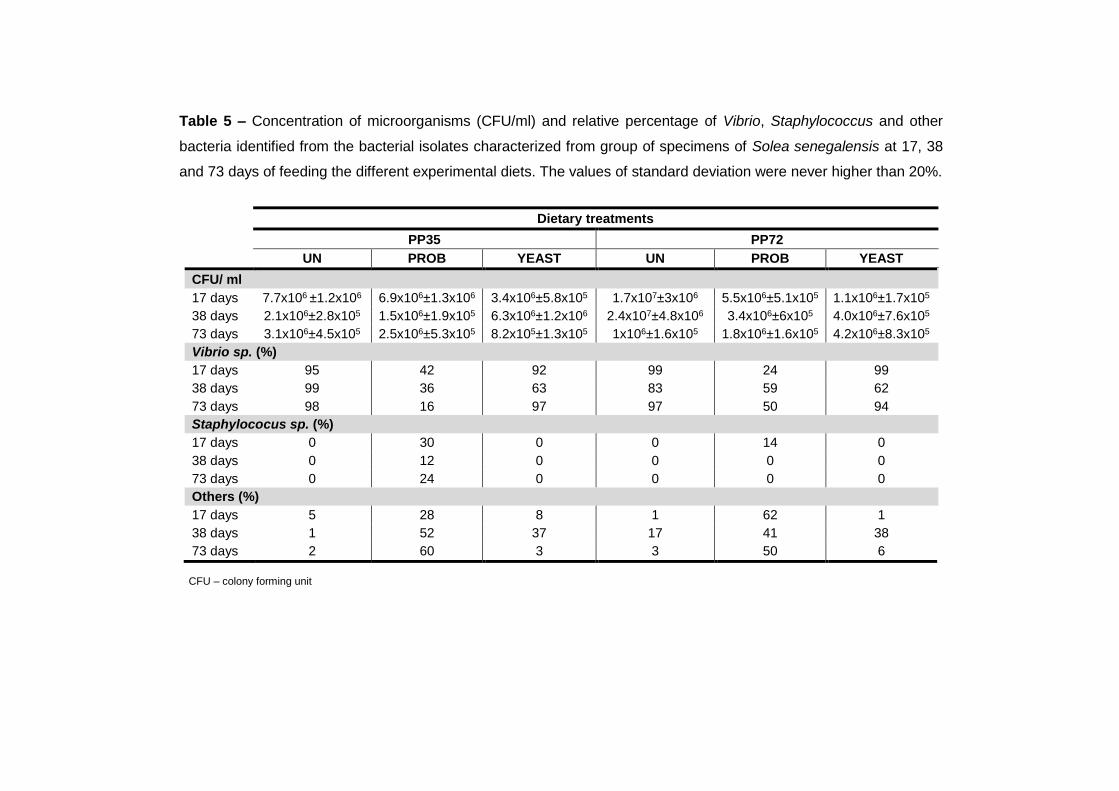
Table 5 – Concentration of microorganisms (CFU/ml) and relative percentage of Vibrio, Staphylococcus and other
bacteria identified from the bacterial isolates characterized from group of specimens of Solea senegalensis at 17, 38
and 73 days of feeding the different experimental diets. The values of standard deviation were never higher than 20%.
Dietary treatments
PP35 PP72
UN PROB YEAST UN PROB YEAST
CFU/ ml
17 days 7.7x10⁶ ±1.2x10⁶ 6.9x10⁶±1.3x10⁶ 3.4x10⁶±5.8x10⁵ 1.7x10⁷±3x10⁶ 5.5x10⁶±5.1x10⁵ 1.1x10⁶±1.7x10⁵
38 days 2.1x10⁶±2.8x10⁵ 1.5x10⁶±1.9x10⁵ 6.3x10⁶±1.2x10⁶ 2.4x10⁷±4.8x10⁶ 3.4x10⁶±6x10⁵ 4.0x10⁶±7.6x10⁵
73 days 3.1x10⁶±4.5x10⁵ 2.5x10⁶±5.3x10⁵ 8.2x10⁵±1.3x10⁵ 1x10⁶±1.6x10⁵ 1.8x10⁶±1.6x10⁵ 4.2x10⁶±8.3x10⁵
Vibrio sp. (%)
17 days 95 42 92 99 24 99
38 days 99 36 63 83 59 62
73 days 98 16 97 97 50 94
Staphylococus sp. (%)
17 days 0 30 0 0 14 0
38 days 0 12 0 0 0 0
73 days 0 24 0 0 0 0
Others (%)
17 days 5 28 8 1 62 1
38 days 1 52 37 17 41 38
73 days 2 60 3 3 50 6
CFU – colony forming unit

Table 6 - Closest matches of GenBank database sequences to isolates from intestinal samples of Solea senegelensis fed
PP72 diets for 17, 38 and 73 days.
PP72_UN PP72_PROB PP72_YEAST
Closest relative Similarity
(%) Accession
Number 17
days 38
days 73
days 17
days 38
days 73
days 17
days 38
days 73
days
γ-Proteobacteria
Vibrio alfacsensis 100 KU725824 + + + - + + + - -
Vibrio alginolyticus 99 KU725825 + - - - - - - + -
Vibrio communis 100 KU725826 - - + + - + + + +
Vibrio harveyi 98 KU725827 - - - + - + + + +
Vibrio ichthyoenteri 100 KU725828 + - + + + + - - +
Vibrio owensii 100 KU725830 - - - + - - + - -
Vibrio scophthalmi 98 KU725831 - - + + + + - + -
Vibrio tapetis 99 KU725833 + + - + - - - - -
Vibrio sp 99 KU725834 + + + + + - - - -
Vibrio sp 97 KU725837 - - - - - - - + -
Vibrio sp 98 KU725838 + - - - - - - - -
Vibrio sp 100 KU725836 + - - - - - - - -
Firmicutes
Staphylococcus saprophyticus
100 KU725839 - - - + - - - - -
Presence (+); Absence (-)

Table 7 - Closest matches of GenBank database sequences to isolates from intestinal samples of Solea senegelensis fed
PP35 diets for 17, 38 and 73 days.
PP35_UN PP35_PROB PP35_YEAST
Closest relative Similarity
(%) Accession
Number 17
days 38
days 73
days 17
days 38
days 73
days 17
days 38
days 73
days
γ-Proteobacteria
Vibrio alfacsensis 100 KU725824 + + + - + + - + -
Vibrio alginolyticus 98 KU725825 - - - - - - - - +
Vibrio communis 99 KU725826 + - - - - - - - -
Vibrio harveyi 98 KU725827 - - - - + - + - -
Vibrio ichthyoenteri 98 KU725828 - - + - - + - - +
Vibrio mytilili 100 KU725829 - - - - - - - - -
Vibrio owensii 100 KU725830 + - - - - - - - -
Vibrio scophthalmi 98 KU725831 + + - + - - - - -
Vibrio sinaloensis 100 KU725832 - - - - - - + + +
Vibrio tapetis 99 KU725833 - + - - - - - - -
Vibrio sp 99 KU725834 + + + + + - + - +
Vibrio sp 99 KU725846 - - + - - - - - -
Vibrio sp 100 KU725835 - + - - - - - + -
Vibrio sp 97 KU725836 - - + - - - - - -
Vibrio sp 98 KU725840 + - - - - - - - -
Vibrio sp 99 KU725841 - - - - - - + + -
Vibrio sp 98 KU725842 - - - - - - + - -
Vibrio sp 99 KU725843 - + - - - - - - -
Firmicutes
Staphylococcus saprophyticus
100 KU725839 - - - + + + - - -
Presence (+); Absence (-)

CHAPTER 4
111
4.4 Discussion
Probiotics confer beneficial actions to the host or to their environment through
different modes of action. Their application in aquaculture has been regarded as a
sustainable and promising strategy, not only in the context of disease control but
also in nutrition, growth and immunity (Lazado and Caipang, 2014a).
The capacity of Senegalese sole to cope with diets in which the marine-derived
protein was replaced by plant ingredients has been previously reported (Cabral et
al., 2011; Cabral et al., 2013; Silva et al., 2009). Similarly, to previous studies, the
current study demonstrated that growth performance of Senegalese sole was not
affected by high content of plant ingredients. Final body weight and FCR was,
however, negatively affected by dietary probiotic supplementation (PROB diet). In
fact, the effects of probiotics and prebiotics on fish growth performance are often
contradictory. Probiotic improved the feed efficiency and growth performance in
Senegalese sole (García de la Banda et al., 2012), Japanese flounder (Taoka et al.,
2006a) and Atlantic cod (Lauzon et al., 2010). Nevertheless, Ferguson et al. (2010)
showed no clear effect on growth performance in Nile tilapia fed with Pediococcus
acidilactici supplementation, whereas Gunther and Jimenez-Montealegre (2004)
observed a growth depression in tilapia fed Bacillus subtilis.
All humoral immune parameters analysed, plasma lysozyme, peroxidase and
alternative complement (ACH50) activities were not significantly altered by PROB
or YEAST. This trend was also observed by Batista et al. (2014) working with sole
fed the same multispecies bacteria and Díaz-Rosales et al. (2009) working with sole
fed with two different Shewanella sp. However, ACH50 was enhanced when
rainbow trout were fed for 4 weeks with diets supplemented with heat-inactivated
multispecies bacteria (Choi and Yoon, 2008) and gilthead seabream fed for 3 weeks
(Salinas et al., 2008). In addition, serum lysozyme activity increased significantly
from 438 ± 75 U ml−1 (control) to 1269 ± 134 U ml−1 in rainbow trout fed for 14 days
with diets supplemented with a autochthonous intestinal Bacillus subtilis (Newaj-
Fyzul et al., 2007). These changes in the innate immune responses were associated
to the inhibitory effect of that probiotic against the pathogenic Aeromonas sp.
Previous studies have demonstrated that oral administration of fungal
carbohydrates enhance fish innate immune response and protection against
infections (Efthimiou, 1996; Esteban et al., 2001; Siwicki et al., 1994), although this
effect seems to be temporary (reversible) and dependent on its inclusion level and
CHAPTER 4
112
feeding duration (Ortuño et al., 2002). In the current study, innate immune
parameters were affected by the increase of dietary plant ingredients content. At 17
and 38 days of feeding trial, fish from PP72 groups had higher values of lysozyme
and ACH50 activities than fish from PP35 groups, suggesting a stimulation of the
innate immune activity by dietary high PP content. Also Geay et al. (2011) observed
an increase in the ACH50, while the lysozyme was lower, in fish fed a PP diet when
compared to fish fed fishmeal based diet. On the other hand, Sitjà-Bobadilla et al.
(2005) reported that high level of fishmeal replacement by plant protein sources had
no significant effect on lysozyme activity. Jalili et al. (2013) showed that rainbow
trout fed diets with high PP levels (70 and 100%) resulted in a decrease on the
ACH50, as reported by Sitjà-Bobadilla et al. (2005) in gilthead seabream.
Hepatocytes provide the major source of complement factors (Abelseth et al., 2003)
and the progressive fat degeneration of liver with the PP inclusion could decrease
complement proteins synthesis (Sitjà-Bobadilla et al., 2005). According to our
results, this effect could be improved by a long-term feeding the diets with high plant
ingredients content. Liver from fish fed PP75 diets showed a higher level of
vacuolization comparing to fish fed PP35 diets. The use of high levels of plant
ingredients is recognised to have several disadvantages, particularly related to the
amino acid profiles and unsaturated fatty acid imbalances but especially due to their
levels of anti-nutritional factors (Geay et al., 2011). In the current study, high PP
level seems to result in an acute stimulation of lysozyme and ACH50, since at 2 and
17 days fish fed PP72 diets showed higher values than fish fed PP35 diets.
Nevertheless, at 73 days of feeding trial such effects disappeared. Food legumes
and cereals may have adverse effects on some fish species after long-term intake,
resulting in morphological problems in liver and intestine that could lead to metabolic
failure (Russell et al., 2001). PP72 diet, when compared to PP35, was formulated
to contain a low amount of soybean meal, but high content of mixed plant
ingredients, including insoluble pea protein, as protein source replacing fishmeal.
Thus, the dietary incorporation level of plant protein sources used in the current
study may have provided a high level of antinutritional factors.
Intestinal microbiota is confined within a highly specialized barrier defenses,
composed by the stratified mucous layer, an epithelium and a lamina propria with
several innate and adaptive immune cells (Maynard et al. 2012). An increase in
intestine section area, villus length and villus width are directly related to an increase
CHAPTER 4
113
of surface area, which may indicate improvement of intestinal absorptive capacity
nutrients (Caspary, 1992). At 2 days of feeding, fish fed PP72_UN showed larger
intestine section area and longer villus than fish fed PP35_UN. At 17 days of
feeding, fish fed PP72_UN showed more goblet cells than the other dietary groups,
except the group fed PP35_YEAST. At 73 days, however, villus length and width,
and goblet cells were significantly increased in fish fed low PP diets compared to
high PP diets. The latter may indicate short-term feeding fish with diets formulated
with high plant ingredients level may enhance the immune defence and improve
surface area for absorption. However, when animals are fed high PP diets for longer
periods the effects were reversed, probably due to habituation of fish and/or to a
negative effect of chronic ingestion of high PP diets and presence of antinutritional
factors. Intestinal epithelium is covered by a layer of mucus continuously produced
by goblet cells and being the first line of defense against microbes (Maynard et al.,
2012). So, the reduced number of goblet cells, after 73 days of feeding fish with the
PP72 diets, causes a decrease in mucous production, thus probably reducing
intestinal protection capacity of those fish.
After 38 days of feeding the experimental diets, villus length was significantly
increased in YEAST supplemented groups, comparing to unsupplemented (UN)
groups. Yeast cells have been reported to be a source of nucleotides, which
contribute for the intestinal maintenance in aquatic animals by improving mucosal
flora and mucosal surfaces with relative elongation of the intestinal tract (Li et al.,
2007). Also, (Abu-Elala et al., 2013) observed that fish treated with S. cerevisiae,
showed yeast colonization in intestine, accompanied by an increase in the length
and density of the intestinal villus. Such morphological alterations are often
associated with an improve in food digestion and absorption. After 2 days feeding
the YEAST diets, fish had a higher hepatocyte glycogen content comparing with fish
fed diets supplemented with PROB, showing a stimulation of the glycogen liver
storage, the first line of energy source when blood glucose concentration falls during
stress. S. cerevisiae used in YEAST diets is rich in bioactive ingredients and
nutrients such as cell wall carbohydrates. According to Wilson (1994), fish fed with
diets rich in digestible carbohydrates, showed high hepatic glycogen reserves.
However, when fish is fed YEAST for longer duration (73 days), such energy benefit
content disappeared.

CHAPTER 4
114
Fish intestinal microbiota changes with the diet and this effect is of particular interest
considering that the effects of high fishmeal replacement by plant ingredients are
still poorly understood, especially in flatfish. Molecular techniques have facilitated
culture-independent studies, becoming a valid support to traditional techniques
(Ercolini, 2004). In the current study, from all bacteria isolated from sole intestine,
approximately 20% were not identified. The intestinal tract of fish harbours a high
density of non-culturable bacteria and the composition of which has not yet been
reported, leading to differences between viable and total microbial counts (Shiina et
al., 2006). In this study, it has been possible to demonstrate the ability to modulate
the intestinal microbiota of Senegalese sole by feeding diets supplemented either
with probiotic or autolyzed Saccharomyces cerevisiae. The results obtained are in
accordance with those reported by Standen et al. (2015), who observed in tilapia
(Oreochromis niloticus) the modulation of the intestinal microbiota and morphology
caused by feeding the fish with same multispecies probiotic used in the current
study. The modulation exerted on the intestinal microbiota by viable and inactivated
cells of Saccharomyces cerevisiae have been reported in tilapia and sturgeon (Huso
huso) (Hoseinifar et al., 2011; Waché et al., 2006). In addition, the modulation of
intestinal microbiota has also been demonstrated in fish treated with S. cerevisiae
fermentation products (He et al., 2011) and glucans obtained from yeast (Kuhlwein
et al., 2013). In the current study, the predominant bacteria found in Solea
senegalensis intestine were Vibrio sp., which is in agreement with other studies
carried out with the same flatfish cultured under extensive, semi-extensive or
intensive production systems (Martin-Antonio et al., 2007). This trend was also
detected in farmed Senegalese sole fed fresh or lyophilized Shewanella
putrefaciens (Tapia-Paniagua et al., 2010; 2015).
Fish fed dietary probiotic supplementation (PP35_PROB and PP72_PROB) had
reduced intestinal bacteria related to Vibrio species. Previous studies observed that
strains of probiotic Bacillus (Liu et al., 2015; Luis-Villasenor et al., 2015; Wu et al.,
2014), Enterococcus (Lin et al., 2013) and Pediococcus and Lactobacillus (Munoz-
Atienza et al., 2013) showed an antibacterial activity against fish pathogens,
including Vibrio.
V. harveyi was detected in sole fed probiotic supplemented and especially in fish
fed yeast supplemented diets, and not detected in fish fed unsupplemented diets.
These Vibrio species are included in the Harveyi Clade and are well-known as

CHAPTER 4
115
pathogenic for marine farmed organisms (Austin and Zhang, 2006; Cano-Gomez et
al., 2011; Gomez-Gil et al., 2004), including Solea senegalensis in the case of V.
harveyi (Arijo et al., 2005a; Zorrilla et al., 2003). However, it is frequent the presence
of virulent and non-virulent strains of V. harveyi (Rico et al., 2008; Zorrilla et al.,
2003), and for this reason more studies are necessary to evaluate the potential
virulence of the strains of these species and how diet formulation may modulate
bacterial presence in the intestine. On the other hand, the dietary probiotic
supplementation in PP35 diet (PP35_PROB) induced the presence of S.
saprophyticus. S. saprophyticus is associated to microorganisms that play a critical
role in fish fermentation and are known to be resistant to different antibiotics
(Sergelidis et al., 2014; Zhang et al., 2015). In the current study, S. saprophyticus
were detected solely in fish fed probiotic diets, especially in low PP groups. Although
there are no reports that S. saprophyticus caused diseases in fish, Sun et al. (2011)
speculate that may be a potential harmful bacterium as it is often implicated in
human urinary tract infections (Kuroda et al. 2005). Its presence in the intestinal
microbiota of fish must be considered as a route for the transmission of antibiotic
resistance (Chajecka-Wierzchowska et al., 2015).
4.5 Conclusion
Senegalese sole was able to cope with high plant ingredient content in replacement
of marine-derived ingredients. The short-term feeding with high PP content may
enhance the immune defence and improve the intestinal surface area, beneficial for
increasing intestinal absorptive capacity in fish. However, it seems that this is a
temporary (acute) effect that was reversed in long-term feeding high PP diets. Fish
fed PP72 showed reduced number of the goblet cells and low hepatic glycogen
content, indicating a reduced capacity to overcome stress situations, since they
seem to have less immune competence and energy storage. Fish fed for 38 days
with YEAST diets had longer villus than unsupplemented groups.
It has been possible to demonstrate the ability to modulate the intestinal microbiota
of Senegalese sole by dietary supplementation with probiotic or autolyzed
Saccharomyces cerevisiae. The predominant bacteria present in sole intestine,
were Vibrio sp., and there was an inhibitory effect of dietary probiotic
supplementation on Vibrio sp. colonization, regardless of the dietary PP level.
CHAPTER 4
116
4.6 Acknowledgements
S. M. G. Batista is supported by FCT – SFRH/BD/76668/2011. This work was also
supported by FCT Projects PEst-OE/AGR/UI0772/2011 and PEst-
OE/AGR/UI0772/2014. We would like to thank to CIIMAR/ ICBAS (UP) and FBA
(University of Nordland) for the use of the facilities and equipment and for technical
support and Mrs. Ligia Lourenço (UTAD) for her histology technical assistance.
117
CHAPTER 5 Effects of autochthonous intestine bacteria on
growth, disease resistance, intestinal morphology
and microbiota in Senegalese sole infected with
Photobacterium damselae sp. piscicida
Batista et al. 2016. Submitted to Fish and Shellfish Immunology,
Submission ID: FSIM-D-16-00467

118

CHAPTER 5
119
Effects of autochthonous intestine bacteria on growth, disease resistance,
intestinal morphology and microbiota in Senegalese sole infected with
Photobacterium damselae sp. piscicida
Batista S. 1,2,3, Sitjà-Bobadilla A.4, Fouz B.5, Llorens A.5, Pires M. A.6, Kiron V.3,
Sousa, S.7, Manaia, C.M.7, Gomes, A.M.P.7, Barros R.8, Cristóvão B.8, Fernandes
J.M.O.3, Valente L.M.P.1,2, Ozório R.O.A.1,2*
1 ICBAS – Instituto de Ciências Biomédicas de Abel Salazar, Universidade de Porto, Rua Jorge Viterbo Ferreira nº 228,
4050-313 Porto, Portugal.
2 CIMAR/CIIMAR – Centro Interdisciplinar de Investigação Marinha e Ambiental, Rua dos Bragas, 289, 4050-123 Porto,
Portugal.
3 FBA - Faculty of Biosciences and Aquaculture, Nord University, 8049 Bodø, Norway
4 Instituto de Acuicultura de Torre de la Sal. Consejo Superior de Investigaciones Científicas, Torre de la Sal s/n, 12595
Ribera de Cabaes, Castellón, Spain.
5 Departamento de Microbiología y Ecología , Universidad de Valencia, Burjasot, 46100, Spain
6 CECAV – Centro de Ciência Animal e Veterinária, UTAD, Apartado 1013, 5000-801 Vila Real, Portugal
7 Universidade Católica Portuguesa, CBQF - Centro de Biotecnologia e Química Fina – Laboratório Associado, Escola
Superior de Biotecnologia, Rua Arquiteto Lobão Vital, Apartado 2511, 4202-401 Porto, PortugalCatólica Portuguesa/Porto,
Rua Dr. António Bernardino Almeida, 4200-072 Porto, Portugal
8 Bioinstrument - Consultoria de desenvolvimento de projetos bioquímicos S.A.

CHAPTER 5
120
Abstract
In fish farms, the control of bacterial pathogens is achieved by the administration of
chemotherapeutic agents, which could lead to the emergence of drug-resistant
microorganism and antibiotic residues. Probiotics used in aquaculture may be useful
feed additives to combat diseases, improve growth and stimulate immunity
response of fish. A growth trial followed by a bacterial challenge were carried out in
order to evaluate the benefits of using autochthonous fish bacteria as a dietary
probiotic treatment in Senegalese sole (Solea senegalensis). Enterococcus
raffinosus (PB1) and Pseudomonas protegens (PB2) were isolated from sole
intestine and incorporated in the experimental diets at 1.6 × 10¹⁰ and 1.3 × 10¹⁰ CFU
kg feed-1, respectively. A total of 264 sole (22.65 ± 0.15 g) were distributed into the
three dietary treatments: a control diet without probiotics (CTRL) and two probiotic
diets (PB1 and PB2). Fish were fed the experimental diets for 36 days during the
growth trial and then for the following 17 days of bacterial challenge. Growth
performance, disease resistance, immune responses, intestinal morphology and
changes in intestinal microbiota were evaluated. Growth performance was not
affected by dietary probiotic supplementation. Fish fed PB1 diet had an increase in
muscular layer thickness (115.3 ± 5.1 µm) when compared to PB2 diet (88.8 ± 5.2
µm) (p < 0.01), and an increase in goblet cell number (240.2 ± 35.1) when compared
to control (117.6 ± 25.8) and PB2 (110.6 ± 26.1) diets (p < 0.01). Intestinal microbiota
of posterior (66.0 ± 13.5%) and distal (62.3 ± 16.8%) intestine were similar in fish
fed probiotic diets. In contrast, fish fed CTRL diet showed low similarity with that of
fish fed PB1 (proximal: 48.1 ± 18.3%; distal: 49.3 ± 30.3%) and PB2 (proximal: 49.1
± 18.7%; distal: 45.7 ± 28.7%) diets, indicating significant differences in the microbial
populations between CTRL and the two probiotic groups. At the end of the
challenge, non-infected fish fed CTRL diet had an increase in villus length (609.3 ±
13.2 µm) compared to fish fed PB1 infected (488.1 ± 22.4 µm) or non-infected (381.5
± 88.1 µm) groups (p < 0.01). Cumulative mortality achieved 14.3%, 13.8% and
7.1% in PB2, control and PB1 groups, respectively. At 15 days post-challenge, non-
infected fish showed higher peroxidase activity (48.5 ± 21.1 EU mL-1) than infected
fish (30.3 ± 15.5 EU mL-1) (p <0.01), regardless of the diet.
In the present study, it was revealed the influence of the diet and the Photobacterium
damselae sp. piscicida infection in modulating the intestinal microbiota. Dietary
CHAPTER 5
121
supplementation with E. raffinosus had an apparent benefit on protection against
photobacteriosis.
CHAPTER 5
122
5.1. Introduction
The high number of disease outbreaks continues to compromise the intensification
of sole (Solea senegalensis) aquaculture, leading to significant mortalities and
economic losses in Mediterranean countries (Arijo et al., 2005a; Romalde, 2002;
Zorrilla et al., 1999). Photobacteriosis is caused by Photobacterium damsela ssp.
piscicida (Phdp), and is the most important disease affecting various marine species
(Bakopoulos et al., 2003; Cerezuela et al., 2012c; Mosca et al., 2014; Toranzo et
al., 2005; Xing et al., 2013), including sole (Barroso et al., 2014; Costas et al., 2013;
Toranzo et al., 2005; Zorrilla et al., 1999) at the early grow-out stages. Phdp is a
rod-shaped gram-negative bacterium (Magariños et al., 1996; Romalde, 2002)
which can induce whitish tubercles in the internal organs and haemorrhages on
head and/or fins in chronically infected fish.
Conventionally, antibiotics and chemical disinfectants have been used as the first
defence line against Phdp. However, diseased fish eat poorly the medicated food
and the emerging of drug-resistant pathogens, limit their use substantially.
Moreover, only a limited number of effective drugs/chemicals are available for
treatment in aquaculture. In addition, recent studies on the use of probiotics to
control Photobacteriosis have given encouraging results (García de la Banda et al.,
2012; Tapia-Paniagua et al., 2012).
In this context, probiotics represent alternative prophylactic tools, increasingly used
in aquaculture systems, both in water and feed, as biological control agents.
Probiotics are defined as live microorganisms which contribute to the proper
microbiological balance of host digestive tract, promoting their immune defences
and nutritional/ physiological development (Gaggìa et al., 2010). Probiotics
treatment may improve fish performance and reduce bacterial pathogen outbreaks,
by multiple ways, including pathogen inhibition via production of antagonistic
compounds, competition for attachment sites or nutrients, alteration of enzymatic
activity of pathogens, immuno-stimulatory functions, improvement of feed
digestibility and utilization. Previous studies, have reported several advantages
associated to probiotics, like the in vitro tests of inhibition of pathogens, and / or in
vivo tests that evidenced gut microflora profiles and changes on immune response
in different fish species. Roseobacter (27-4 strain) isolated from the tank walls of
healthy turbot (Scophthalmus maximus L.) was show to have a protective effect
against Vibrio anguillarum in turbot larvae (Planas et al., 2006), leading to improved

CHAPTER 5
123
survival. Queiroz and Boyd (1998) reported increased survival and growth
performance of channel catfish (Ictalurus punctatus), when a blend of Bacillus sp.
was added to the pond rearing water. Carnobacterium sp., previously isolated from
Atlantic salmon (Salmo salar) intestine, was effective in reducing infections caused
by Aeromonas salmonicida, V. ordalii, and Yersinia ruckeri in juvenile salmonids
(Robertson et al., 2000). An in vitro study carried out by Nikoskelainen et al. (2001b)
showed that Lactobacillus rhamnosus and L. bulgaricus were effective in reducing
the growth of A. salmonicida, isolated from rainbow trout (Oncorhynchus mykiss)
mucus. A reduction in V. anguillarium and V. ordalii virulence degree and an
increase in phagocytic activity of leukocytes was also reported in trout after oral
administration of Clostridium butyricum (Sakai et al., 1995), indicating
immunostimulation.
Probiotics were also shown to modulate the fish immune system in several fish
species resulting in beneficial effect to the host (Aly et al., 2008; Cerezuela et al.,
2012b; Irianto and Austin, 2002b). In addition, the effect of probiotics in intestinal
structure and gut microbiota has been reported in fish (Lazado and Caipang,
2014b). For instance, Nile tilapia (Oreochromis niloticus) fed a diet supplemented
with L. rhamnosus, showed a significant increase in villus height compared to fish
fed the control diet (Pirarat et al., 2011). By contrast, gilthead seabream (Sparus
aurata) fed B. subtillis showed a significant reduction in microvilli height compared
to fish fed the control diet (Cerezuela et al., 2012a).
Several probiotic have already been studied as feed additive in Senegalese sole,
Solea senegalensis. There are recent in vivo studies testing commercial probiotics
(Barroso et al., 2014; Batista et al., 2014) in Senegalese sole juveniles. Additional
studies were carried out testing bacteria recovered from skin mucus of gilthead sea
bream in sole diet (Lobo et al., 2014; Tapia-Paniagua et al., 2014; Tapia-Paniagua
et al., 2015). To our knowledge, this is the first in vivo study testing autochthonous
intestinal bacteria as potential dietary probiotics in Senegalese sole juveniles. Their
effectiveness to modulate growth performance, disease resistance, intestinal
morphology and ecology will be evaluated.
5.2. Materials and methods
All procedures were approved by the Ethics and Animal Welfare Committee of
Institute of Aquaculture Torre de la Sal (Spain) and supervised by the Ethics experts

CHAPTER 5
124
of the TNA selection panel of the AQUAEXCEL project. It was carried out in a
registered installation (code 36271-42-A) in accordance with the principles
published in the European animal directive (2010/63/EU) and Spanish laws (Royal
Decree RD53/2013) for the protection of animals used in scientific experiments. In
all lethal samplings, all efforts were made to minimize suffering. Fish were fasted for
24 h and then were decapitated after an over-exposure with 3-aminobenzoic acid
ethyl ester (MS-222, 100 μg mL-1).
5.2.1. Screening procedure for candidate strain probiotics
The autochthonous intestine bacteria tested in the current study as candidate
probiotics for Senegalese sole were selected using in vitro antagonism tests based
on the production of inhibitory compounds against common fish pathogens. 250
bacteria strains, isolated from the intestine microbiota of farmed healthy sole, were
tested for their antimicrobial activity against several common fish pathogens.
Thereafter, the 16 bacteria strains with the best results were tested for their stability
and viability during feed processing. Among all bacteria tested, Enterococcus
raffinosus and Pseudomonas protegens were selected due to their in vitro
antimicrobial activity (not published results) and their capacity to survive during
freeze-drying and encapsulation (not published results).
5.2.2. Experimental diets and probiotics isolation
A basal diet (57% crude protein, 9% crude lipid) formulated by SPAROS S.A.
(Olhão, Portugal) was used as the control (CTRL) without probiotic
supplementation. Two probiotic diets (PB1 and PB2) were also formulated by
supplementing the basal diet with E. raffinosus or Ps. protegens, respectively (Table
1). E. raffinosus and Ps. protegens were previously isolated from Senegalese sole
intestinal microbiota and identified by the amplification and sequencing of a
fragment of 16S rDNA gen. Bacterial strains were revived in 10 mL of LB broth (Luria
Bertani Broth, Invitrogen) and subsequently mass cultured in flasks containing 5 L
of LB broth for 36 h at room temperature (20-22 °C) with continuous shaking. Upon
incubation, cultures were harvested by centrifugation at 5000 rpm, at 4ºC for 20
minutes (Thermo Heraeus Multifuge X3 FR, Thermo Scientific), and washed three
times with sterile saline water (0.85%, w/v, NaCl, Panreac). In aquaculture, the dose
of probiotics usually varies from 106-10 CFU g feed-1 (Panigrahi et al., 2004). The
CHAPTER 5
125
harvested bacteria were suspended in 150 mL of saline solution to obtain a dose of
1010 colony forming units (CFU) mL-1. To verify the dose, appropriate dilutions of
each bacterial culture, were spread onto plates of LB agar (Luria Bertani Agar,
Invitrogen) following Miles and Misra method (Miles et al., 1938) and incubated at
25ºC for 24 h. Then, each bacteria suspension was incorporated in the extruded
pellets by means of vacuum coating (Dinnisen Pegasus vacuum mixer, PG-
10VCLAB) using fish oil as a carrier. The viability of the incorporated strains was
assessed by vortexing 10 g of each diet in 90 mL of peptone water and preparing
serial dilutions, which were spread on LB agar (Luria Bertani Agar, Invitrogen) plates
as above. The bacterial counts were able to confirm the established dietary
incorporation of each strain and their survival and viability in the diet preparations
for at least 4 weeks. E. raffinosus was incorporated in the diets at 1.6 × 10¹⁰ CFU
kg-1 feed and Ps. protegens at 1.3 × 10¹⁰ CFU kg-1 feed. During the growth and
challenge trials, diets were stored at 4ºC and new diet batches of the diets was
prepared every 4 weeks.
5.2.3. Fish and rearing conditions during growth trial
Both trials were carried out at the indoor experimental facilities of the Pathology Unit
of IATS-CSIC (Castellón, Spain). These facilities ensured the adequate water
quality and the correct disinfection of the effluent water and the material in contact
with the pathogen bacteria used during the bacterial challenge. Non vaccinated
Senegalese sole were obtained from a commercial fish farm (Aquacria S.A.,
Portugal) and acclimatized for 20 days to IATS experimental facilities. Fish were fed
during the acclimatization period with the CTRL diet in a flow-through system of flat-
bottomed tanks with aerated seawater.
After acclimation, 264 fish (22.65 ± 0.15 g body weight) were randomly distributed
(22 fish / tank, 22.6 ± 0.2 g each) into 12 90 L-fibreglass tanks, supplied with
seawater in a flow-through system. Fish were fed for 36 days the experimental diets
(n=4 tanks / treatment) to apparent satiety with automatic feeders. Visual
observation of acceptance and refusal of feed was used to adjust daily ration, as
previously described by (Borges et al., 2009). Water parameters were daily
monitored with salinity at 37.5 ± 0.1 ppm, temperature at 18.0 ± 1.1 ºC, oxygen
levels always higher than 85% saturation and unionized ammonia below toxic levels

CHAPTER 5
126
(< 0.05 mg L-1). The tanks were under natural photoperiod at our latitude (40°5´N;
0°10´E).
Table 1 - Feed ingredients and proximate composition of the experimental diet
aPeruvian fishmeal LT (71% crude protein, 11% crude fat, EXALMAR, Peru); bFair Average Quality (FAQ) fishmeal (62% crude protein, 12% crude fat, COFACO, Portugal); cSoluble fish protein hydrolysate (87% crude protein, 6.5% crude fat, Sopropêche, France); dSoycomil-P (soy protein concentrate, 65% crude protein, 0.7% crude fat, ADM, The Netherlands); eDehulled solvent extracted soybean meal (micronized); fAquatex G2000 (Dehulled, grinded pea grits, 24% crude protein, 0.4% crude fat, SOTEXPRO, France); gVitamins (mg or IU kg−1diet): Vitamin A (retinyl acetate), 20000 IU; vitamin D3 (DL-cholecalciferol), 2000 IU; vitamin K3 (menadione sodium bisulfite), 25 mg; vitamin B1 (thiamine hydrochloride), 30 mg; vitamin B2 (riboflavin), 30 mg; vitamin B6 (pyridoxine hydrochloride), 20 mg; vitamin B12 (cyanocobalamin), 0.1 mg; vitamin B5 (pantothenic acid), 100 mg; vitamin B3 (nicotinic acid), 200 mg; vitamin B9 (folic acid), 15 mg; vitamin H (biotin), 3mg; betaine, 500 mg; inositol, 500 mg; choline chloride, 1000 mg; vitamin C (stay C), 1000 mg; vitamin E, 100 mg; hMinerals (% or mg kg−1diet): Mn (manganese oxide), 9.6 mg; I (potassium iodide), 0.5 mg; Cu (cupric sulfate), 9 mg; Co (cobalt sulfate), 0.65 mg; Zn (zinc oxide), 7.5 mg; Se (sodium selenite), 0.01 mg; Fe (iron sulfate), 6 mg; Cl (sodium chloride), 2.41%; Ca (calcium carbonate), 18,6%; NaCl (sodium), 4%; iDiatomaceous earth: Kielseguhr: LIGRANA GmbH, Germany.
5.2.4. Pathogen challenge
5.2.4.1. Pathogen inoculum preparation and challenge dose validation
Phdp strain Lab 2 used in this study was kindly provided by Dr. Belén Fouz and
Skretting Spain, isolated from diseased European seabass (Dicentrarchus labrax),
was cultured in tryptic soy agar (TSA, Pronadisa, Madrid, Spain) supplemented with
NaCl at a final concentration of 1% (TSA-1) and sheep blood at a final concentration
of 5% (TSAB-1), at 22 °C for 48 hours.
DIET
Feed ingredients (%)
Fishmeal 70 La 24.5
Fishmeal 60 (COFACO)b 27.0
CPSP90c 5.0
Squid meal 5.0
Soybean meal (micro)e 12.5
Extruded peas (Aquatex G2000)f 11.0
Wheat meal 11.0
Fish oil 2.0
Vitaming and Mineral Premixh 1.0
Binder (Alginate)i 1.0
Proximate composition
Dry Matter (DM, %) 91.02
Crude protein (%DM) 57.49
Crude fat (%DM) 8.82
Ash 16.41
Gross Energy (kj g−1DM) 20.32

CHAPTER 5
127
To identify the suitable infective dose for the bacterial challenge, four different doses
were tested in a pre-challenge experiment. Juvenile sole, held in 90 L tanks at an
average temperature of 19 °C, were intracoelomically (i.c.) injected with 0.1 ml of
four different bacterial suspensions (D1, – 3.0 × 10⁷, D2 – 6.0 × 10⁶, D3 – 1.4 × 10⁶
and D4 – 7.0 × 10⁵ CFU mL-1) in phosphate-buffered saline (pH 7.4) at a ratio of 8
fish/dose, according to Azeredo et al. (2015) and to Fouz et al. (2010). Negative
control fish were i.c. injected 0.1 ml of PBS (non-infected group). Mortality was
recorded daily during 3 weeks and the cause of death was considered due to Phdp
if the inoculated bacterium was recovered in pure culture from internal organs
(kidney and liver) streaked onto TSAB-1 plates. Identification of the pathogen was
carried out by means of an agglutination test with the corresponding antiserum. The
lethal dose causing 30% of mortality (LD30) was chosen in order to follow the ethics
specifications of the AQUAEXCEL TNA selection committee that considered LD30
to be sufficient to answer the question of whether fish fed the supplemented diet
resist disease better than controls.
5.2.4.2. Bacterial challenge
At the end of the growth trial, a total of 85 fish (29 from CTRL, 28 from PB1 and 28
from PB2) were i.c. injected with the LD30 of Phdp. Infected fish were returned to the
original tanks (13-15 fish tank-1), and kept under the same rearing conditions as the
growth trial. Similarly, 84 fish from each dietary treatment (28, 27 and 29 from CTRL,
PB1 and PB2, respectively) were i.c. injected with PBS and then returned to the
original tanks (non-infected groups). Fish were fed daily the experimental diets
during the post-challenge (p.c.) period at a fixed rate of 1% body weight.
Fish mortality was monitored daily until no more mortalities were recorded for a
minimum of two consecutive days. Post-mortem examination was performed by
standard microbiological methods, based on pathogen culturing and isolation steps
as described above. Cumulative mortality (CM) was calculated per dietary treatment
as follows: CM = “number of dead fish” “initial total number of fish”-1 × 100.
5.2.5. Sampling procedures
Prior to sampling, fish were fasted for 24 h and then sacrificed with an overdose of
MS-222. Fish were individually weighed and measured at the beginning and at the
end of the growth trial, in order to determine growth performance. Eight fish from the
CHAPTER 5
128
initial stock and two fish from each tank at the end of the growth trial (8 fish per
treatment) were sampled and stored at -20 ºC for whole body composition analyses.
Total feed consumption was registered during the whole trial. Liver and viscera from
other 12 fish per treatment were sampled to calculate hepatosomatic (HSI) and
viscerosomatic (VSI) indexes. HSI was calculated as: 100 × [liver weight (g) / whole
body weight (g)] and VSI as 100 × [viscera weight (g) / whole body weight (g)].
The innate immune response was assessed at the end of the growth trial and at 2
and 15 days post-challenge (p.c.). Blood was withdrawn from the caudal vein of 12
fish (end of the growth trial) or 6 fish (2 and 15 days p.c.) per treatment using
heparinised syringes and centrifuged at 5000 × g for 10 min at 4 ºC for plasma
collection. Humoral innate immune parameters, lysozyme, peroxidase and
alternative complement pathway (ACH50) activities, were analysed in plasma.
Intestine histology and microbiota diversity were assessed at the end of the growth
trial (8 fish per treatment) and 15 days of p.c. (6 fish per treatment). The intestine of
each fish was collected, and a segment with 1.5 cm length from the proximal
intestine was cut and stored in buffered formalin for the histological analyses. PI and
DI segments were aseptically removed and stored at -80 ºC for examine intestinal
microbiota diversity (DGGE) and for PCR-based analyses for E. raffinosus and Ps.
protegens detection.
5.2.6. Proximate composition
Frozen whole fish from each tank were minced without thawing, using a meat
mincer, pooled and moisture content was determined. Fish were subsequently
freeze-dried before further analysis. Feed and whole body samples were analysed
for dry matter (105°C for 24 h), ash (Nabertherm L9/11/B170; Germany; 550ºC for
6 h), crude protein (N × 6.25, Leco N analyser, Model FP-528, Leco Corporation,
St. Joseph, USA), crude lipid (petroleum ether extraction, 40 - 60ºC, Soxtherm,
Gerhardt, Germany) and gross energy (adiabatic bomb calorimeter, Werke C2000,
IKA, Germany).
5.2.7. Detection of E. raffinosus and Ps. protegens in sole intestine
A PCR-based method was used to detect the presence / absence of E. raffinosus
and Ps. protegens in the PI and DI of fish collected at the end of the growth trial.
DNA was extracted using cetyltrimethylammoniumbromide (Zhou et al., 1996). A

CHAPTER 5
129
specific combination of primers for the detection of both bacteria was designed using
the Beacon Designer™ software. The sequences with the accession numbers
Y18296 and AJ278812 were used for E. raffinosus and Ps. protegens, respectively.
A set of primers (0.2 µM each) were then used for the amplification by PCR using
the EmeraldAmp® Max PCR Master Mix (Takara Bio Inc., Otsu, Japan) in a 50 µL
reaction. The primer pair F - GCGTGAGTGAAGAAGGT and R –
CTGGTTAGATACCGTCAA was used for E. raffinosus detection and the primer pair
F – ACCTCACGCTATTAGATG and R – ATCCTCTCAGACCAGTTA was used for
Ps. protegens detection. Samples were denatured for 5 min at 96°C and then
subjected to 45 cycles of amplification (1 min at 94ºC, 1 min at 56ºC and 2 min at
72ºC) and an extension for 10 min at 72°C. Then samples were loaded into a 1.5%
(w/v) agarose gel containing SYBR safe DNA gel stain (Invitrogen, Carlsbad, CA,
USA) for gel electrophoresis. Identity of the amplicons were confirmed by Sanger
sequencing.
5.2.8. Humoral innate immune parameters
All measurements were done in triplicate on a Power-WaveTM microplate
spectrophotometer (BioTek Synergy HT, USA). Plasma lysozyme activity (EU ml-1)
was determined using a turbidimetric assay adapted to microtitre, as described by
Hutchinson and Manning (1996). One lysozyme enzyme unit (EU) was defined as
the amount of lysozyme that caused a decrease in 0.001 absorbance units per min.
One lysozyme enzyme unit (EU) was defined as the amount of lysozyme that
caused a decrease in absorbance of levels per min. Plasma peroxidase activity (EU
ml-1) was measured following the procedure adapted to Solea senegalensis by
Costas et al. (2011), defining that one unit of peroxidase produces an absorbance
change of 1 OD. Alternative complement pathway activity (ACH50) was based on
the lysis of rabbit red blood cells (2.8 × 10⁸ cells mL-1), and measured as reported
by Sunyer and Tort (1995). ACH50 units were defined as the concentration of
plasma giving 50% lysis of rabbit red blood cells.
5.2.9. Intestine morphological evaluation
Proximal intestine segments (1.5 cm length) were fixed in phosphate buffered
formalin (4%, pH 7, VWR, Portugal) for 24 hours. Samples were subsequently
dehydrated and embedded in paraffin according to standard histological

CHAPTER 5
130
procedures. Thereafter, 3 μm-thick sections were obtained and stained with
haematoxylin and eosin (H&E - Merck, Portugal) and Periodic acid-Schiff (PAS -
Merck, Portugal) before examination under a light microscope (Olympus BX51,
cell^B software, GmbH, Hamburg, Germany). The intestine section area (mm²),
villus height (μm), villus width (µm), muscular layer thickness (μm) and PAS-positive
goblet cells (GC - number per section) were determined according described to
Batista et al. (2016).
5.2.10. Intestinal microbiota composition
The DGGE analysis of PCR-amplified 16S rRNA genes was carried out in the
proximal and distal intestine of 2 - 4 fish per treatment, to determine the similarity
indices (100% similarity, represents complete identity) of intestinal microbiota
diversity between treatments. DNA was extracted using
cetyltrimethylammoniumbromide (Zhou et al., 1996). The PCR amplification of DNA
was carried out using the primer combination of 341F with GC clamp,
CCTACGGGAGGCAGCAG and 907R, CCGTCAATTCMTTTGAGTTT (Muyzer et
al., 1995), by a modified touchdown protocol as described by Dhanasiri et al. (2011),
except for the temperature of the initial denaturation step (96oC). DGGE was
performed on the PCR products from DNA samples using a 16 × 16 cm gel on the
Bio-Rad DCode ™ system (Bio-Rad, USA), as described by Dhanasiri et al. (2011)
but using a 30 to 55% (w/v) urea and formamide denaturing gradient. PCR products
from proximal and distal intestine were loaded into separated gels and
electrophoresis was performed with 0.5% TAE buffer, at 20 V for 15 minutes and
then at a constant voltage of 75 V for 17 hours. DGGE profile analysis (Quantity
One® version 4.6.3 software, Bio-Rad) was performed in gels to determine similarity
matrices (Dhanasiri et al., 2011).
5.2.11. Calculations of growth performance
Feed conversion ratio (FCR) was calculated as: feed intake (g) / wet weight gain
(g), and the daily growth index (DGI, % BW / day) as: 100 × [(W1)1/3 − (W0)1/3] / t,
where W0 and W1 are the initial and the final body weights (g) and t is the trial
duration. Voluntary feed intake (VFI, % BW / day) was calculated as: 100 × (feed
intake (g) / ABW (g) / days), where ABW (average body weight) was calculated as:

CHAPTER 5
131
(W1+W0) / 2. The protein efficiency ratio (PER) was calculated as weight gain (g) /
protein intake (g).
5.2.12. Statistical analysis
Statistical analyses were performed with the software SPSS (IBM SPSS
STATISTICS, 17.0 package, IBM Corporation, New York, USA). Data were
analysed for normality (Shapiro-Wilk test) and homogeneity of variance (Levene’s
test) and were log-transformed whenever necessary. Data were analysed by a one-
way ANOVA (at 36-days of growth trial and in the end of the challenge) considering
diet as factor and two-way ANOVA (at 2 and 15 days p.c.) with dietary treatment
and infection as main factors. When significant differences were obtained from the
ANOVA, Tukey’s post hoc tests were carried out to identify significant differences
between groups. When data did not meet the ANOVA assumptions, a non-
parametric Kruskal–Wallis test was performed for each factor and the pairwise
multiple comparison of mean ranks were carried out to identify significant
differences between groups. In all cases, the minimum level of significance used
was set at p ≤ 0.05.
5.3. Results
5.3.1. Growth performance and body composition
Fish grew from 22.6 ± 0.2 g to 39.9 ± 1.6 g after 36 days of feeding the experimental
diets (Table 2). Daily growth index (1.53 ± 0.06% BW day-1), feed conversion ratio
(1.43 ± 0.12), voluntary feed intake (2.07 ± 0.06% BW day-1), and protein efficiency
ratio (1.23 ± 0.06) did not differ (p < 0.05) between dietary treatments. HSI (1.1 ±
0.0%) and VSI (2.7 ± 0.1 %) indexes did not vary among the dietary groups. Whole
body crude protein (17.78 ± 0.39 %), crude lipid (5.09 ± 0.33 %) and gross energy
(6.13 ± 0.16 kJ/g) were not significantly affected by the dietary treatments (p < 0.05).
5.3.2. Detection of E. raffinosus and Ps. protegens in the intestine
E. raffinosus (96 bp) and Ps. protegens (92 bp) PCR products were detected in the
distal and posterior intestine. The image gels from proximal (A) and distal (B)
intestine tissues assayed for E. raffinosus detection are depicted in Figure 1. All
proximal intestine samples (24), regardless of the dietary treatment, presented a 96

CHAPTER 5
132
bp amplification product (100% present) (Figure 1A). In the distal intestine (Figure
1B), 7 out of 8 fish (88%) fed the PB1 diet presented the 96 bp amplification product.
All fish from CTRL (8) and PB2 (8) groups had 100% of presence. Figure 2 shows
the image gels from proximal (A) and distal (B) intestine tissues assayed for Ps.
protegens detection. For both PI and DI samples, 100% of the samples from fish fed
PB2 diet presented the amplification product of 92 bp. However, for PI (Figure 2A),
Ps. protegens was detected in 75% and 88% of samples form fish fed CTRL and
PB1 diets, respectively. Also in DI (Figure 2B), only 63% and 50% of samples from
fish fed CTRL and PB1 diets, respectively, contained Ps. protegens.
Table 2 – Growth performance, nutrient utilization and whole body composition of
Senegalese sole juveniles after the 36-day growth trial.
The results were not significantly different among the dietary treatments (p > 0.05). Abbreviations are: CTRL = Diet with no supplementation; PB1 = Diet supplemented with Enterococcus raffinosus; PB1 = Diet supplemented with Pseudomonas protegens; IBW = Initial body weight; FBW = Final body weight; SGR = Specific growth rate; FCR = Feed conversion ratio; VFI = Voluntary feed intake; DGI = Daily growth index; PER = Protein efficiency ratio; HIS = Hepatosomatic index; VSI = Viscerosomatic index. Values represent mean ± SD, n = 4, except for HIS and VSI with n = 8.
Dietary treatments
CTRL PB1 PB2 p value
Growth
IBW (g) 22.62 ± 0.17 22.66 ± 0.18 22.67 ± 0.16 0.92
FBW (g) 39.9 ± 1.6 38.3 ± 1.8 38.1 ± 1.9 0.35
DGI 1.63 ± 0.14 1.50 ± 0.14 1.48 ± 0.15 0.31
FCR 1.34 ± 0.15 1.48 ± 0.11 1.50 ± 0.10 0.20
PER 1.30 ± 0.14 1.19 ± 0.08 1.16 ± 0.07 0.17
VFI 2.05 ± 0.10 2.10 ± 0.12 2.10 ± 0.19 0.84
HSI 1.11 ± 0.07 1.05 ± 0.14 1.10 ± 0.20 0.87
VSI 2.80 ± 0.38 2.65 ± 0.21 2.67 ± 0.31 0.76
Whole body composition (% WW)
Dry matter 25.08 ± 0.25 24.76 ± 0.79 24.64 ± 0.25 0.29
Ash 2.16 ± 0.08 2.16 ± 0.08 2.17 ± 0.17 0.99
Protein 17.88 ± 0.30 17.76 ± 0.58 17.70 ± 0.32 0.58
Lipid 5.32 ± 0.34 4.99 ± 0.36 4.98 ± 0.22 0.27
Energy (kj) 6.24 ± 0.12 6.09 ± 0.22 6.05 ± 0.09 0.22
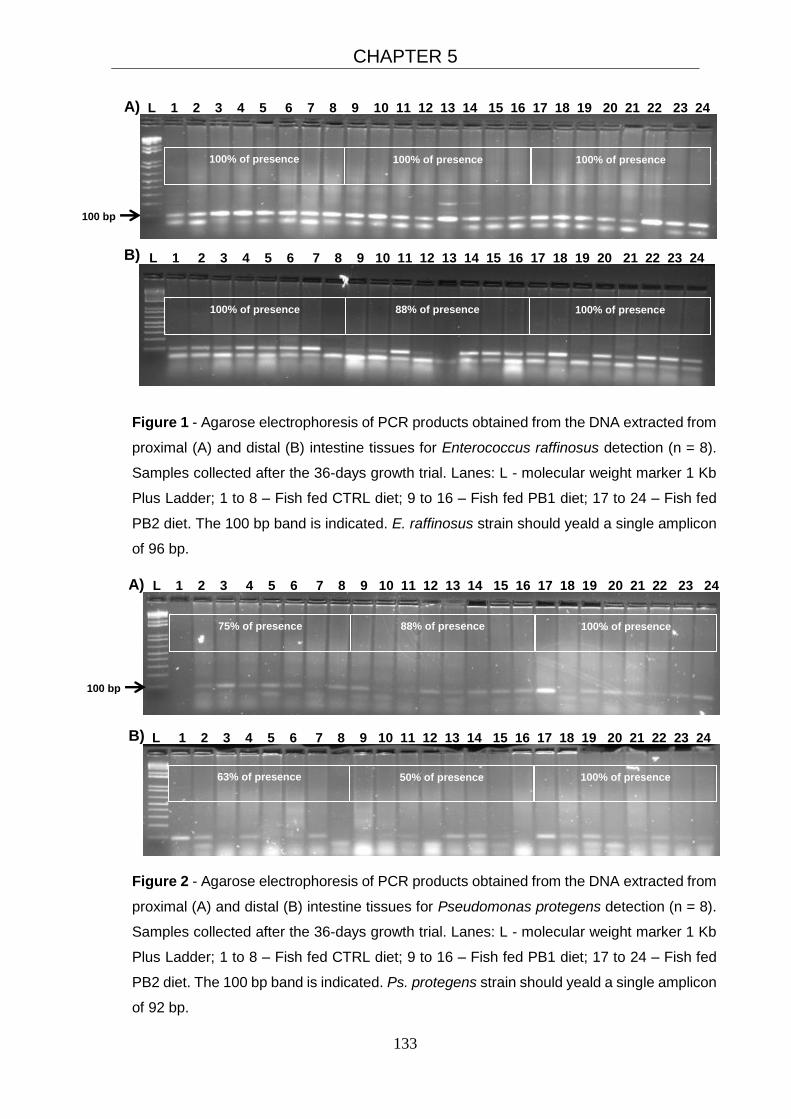
CHAPTER 5
133
Figure 1 - Agarose electrophoresis of PCR products obtained from the DNA extracted from
proximal (A) and distal (B) intestine tissues for Enterococcus raffinosus detection (n = 8).
Samples collected after the 36-days growth trial. Lanes: L - molecular weight marker 1 Kb
Plus Ladder; 1 to 8 – Fish fed CTRL diet; 9 to 16 – Fish fed PB1 diet; 17 to 24 – Fish fed
PB2 diet. The 100 bp band is indicated. E. raffinosus strain should yeald a single amplicon
of 96 bp.
Figure 2 - Agarose electrophoresis of PCR products obtained from the DNA extracted from
proximal (A) and distal (B) intestine tissues for Pseudomonas protegens detection (n = 8).
Samples collected after the 36-days growth trial. Lanes: L - molecular weight marker 1 Kb
Plus Ladder; 1 to 8 – Fish fed CTRL diet; 9 to 16 – Fish fed PB1 diet; 17 to 24 – Fish fed
PB2 diet. The 100 bp band is indicated. Ps. protegens strain should yeald a single amplicon
of 92 bp.
L 1 2 3 4 5 6 7 8 9 10 11 12 13 14 15 16 17 18 19 20 21 22 23 24 A)
100 bp
L 1 2 3 4 5 6 7 8 9 10 11 12 13 14 15 16 17 18 19 20 21 22 23 24 B)
75% of presence
88% of presence
100% of presence
63% of presence
50% of presence 100% of presence
100 bp
L 1 2 3 4 5 6 7 8 9 10 11 12 13 14 15 16 17 18 19 20 21 22 23 24 A)
B) L 1 2 3 4 5 6 7 8 9 10 11 12 13 14 15 16 17 18 19 20 21 22 23 24
100% of presence
100% of presence 100% of presence
100% of presence
88% of presence
100% of presence
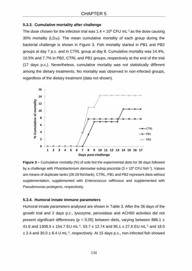
CHAPTER 5
134
0
2
4
6
8
10
12
14
16
1 2 3 4 5 6 7 8 9 10 11 12 13 14 15 16 17
% C
um
ula
tiv
e o
f m
ort
ality
Days post-challenge
CTRL
PB1
PB2
5.3.3. Cumulative mortality after challenge
The dose chosen for the infection trial was 1.4 × 10⁶ CFU mL-1 as the dose causing
30% mortality (LD30). The mean cumulative mortality of each group during the
bacterial challenge is shown in Figure 3. Fish mortality started in PB1 and PB2
groups at day 7 p.c. and in CTRL group at day 8. Cumulative mortality was 14.4%,
10.5% and 7.7% in PB2, CTRL and PB1 groups, respectively at the end of the trial
(17 days p.c.). Nevertheless, cumulative mortality was not statistically different
among the dietary treatments. No mortality was observed in non-infected groups,
regardless of the dietary treatment (data not shown).
Figure 3 – Cumulative mortality (%) of sole fed the experimental diets for 36 days followed
by a challenge with Photobacterium damselae subsp piscicida (3 × 10⁶ CFU fish-1). Values
are means of duplicate tanks (28-29 fish/tank). CTRL, PB1 and PB2 represent diets without
supplementation, supplemented with Enterococcus raffinosus and supplemented with
Pseudomonas protegens, respectively.
5.3.4. Humoral innate immune parameters
Humoral innate parameters analysed are shown in Table 3. After the 36 days of the
growth trial and 2 days p.c., lysozyme, peroxidase and ACH50 activities did not
present significant differences (p < 0.05) between diets, varying between 886.1 ±
41.6 and 1308.3 ± 154.7 EU mL-1, 53.7 ± 12.74 and 95.1 ± 27.6 EU mL-1 and 18.0
± 2.4 and 30.0 ± 8.4 U mL-1, respectively. At 15 days p.c., non-infected fish showed

CHAPTER 5
135
significantly higher peroxidase activity (48.5 ± 21.1 EU mL-1) than the infected ones
(30.3 ± 15.5 EU mL-1) (p < 0.01).
5.3.5. Intestinal morphological evaluation
Morphological evaluation results of the intestinal wall are depicted in Table 4. At the
end of the growth trial, fish fed PB1 diet had a significantly thicker muscular layer
(115.3 ± 5.1 µm) than PB2 fed fish (88.8 ± 5.2 µm) (p < 0.01), and a significantly
higher number of PAS-positive GC (240.2 ± 35.1) than control (117.6 ± 25.8) and
PB2 (110.6 ± 26.1) fed fish (p < 0.01). The analysis performed 15 days p.c. showed
that fish fed control diet had a higher villus length (609.3 ± 13.2 µm) than PB1 fed
fish, in both infected (488.1 ± 22.4 µm) and non-infected (381.5 ± 88.1 µm) groups
(p < 0.01).
5.3.6. Assessment of intestinal microbiota
Percentages of similarity obtained for the DGGE profiles of PI and DI of sole at the
end of the growth trial are shown in Figure 4A and Figure 4B, respectively. Fish fed
the PB1 and PB2 diets had high similarity values for both PI (66.0 ± 13.5 %, mean
± deviation) and DI (62.3 ± 16.8%), however only in PI that difference was statistical
significant (p < 0.05). Fish fed CTRL diet showed low similarity values with fish fed
PB1 (PI: 48.1 ± 18.3%; DI: 49.3 ± 30.3%) or PB2 (PI: 49.1 ± 18.7%; DI: 45.7 ±
28.7%) diets, indicating differences in the microbial populations between CTRL and
the two probiotic groups.
At day 15 p.c., the DGGE images generated from the PI (Figure 5) and DI (Figure
6) samples were analysed, considering both infected and non-infected animals. In
the PI samples, higher similarity values were observed within the infected fish
(Figure 5B), compared to the non-infected fish (Figure 5A). The mean similarity
value within the infected fish was more than 60%. In the DI samples, the opposite
occurred, showing in this case that non-infected fish (Figure 6A) had higher similarity
values between treatments, comparing to the infected ones (Figure 6B). However,
this similarity was less evident (> 50%) than the infected fish in the PI segment (>
60%).
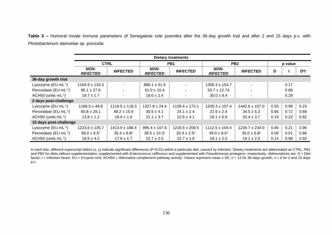
136
Table 3 – Humoral innate immune parameters of Senegalese sole juveniles after the 36-day growth trial and after 2 and 15 days p.c. with
Photobacterium damselae sp. piscicida.
In each line, different superscript letters (x, y) indicate significant differences (P<0.01) within a particular diet, caused by infection. Dietary treatments are abbreviated as CTRL, PB1 and PB2 for diets without supplementation, supplemented with Enterococcus raffinosus and supplemented with Pseudomonas protegens, respectively. Abbreviations are: D = Diet factor; I = Infection factor; EU = Enzyme Unit; ACH50 = Alternative complement pathway activity. Values represent mean ± SE, n = 12 for 36-days growth; n = 6 for 2 and 15 days p.c.
Dietary treatments
CTRL PB1 PB2 p value
NON-
INFECTED INFECTED
NON- INFECTED
INFECTED NON-
INFECTED INFECTED D I D*I
36-day growth trial
Lysozyme (EU mL-1) 1164.9 ± 133.3 - 886.1 ± 41.6 - 1308.3 ± 154.7 - 0.17
Peroxidase (EU mL-1) 95.1 ± 27.6 - 61.0 ± 10.4 - 53.7 ± 12.74 - 0.86
ACH50 (units mL-1) 19.7 ± 1.7 - 18.0 ± 2.4 - 30.0 ± 8.4 - 0.29
2 days post-challenge
Lysozyme (EU mL-1) 1168.5 ± 49.8 1118.5 ± 118.3 1327.8 ± 24.4 1139.4 ± 173.1 1209.3 ± 107.4 1442.6 ± 157.0 0.33 0.99 0.23
Peroxidase (EU mL-1) 65.8 ± 28.1 48.2 ± 15.9 30.5 ± 4.1 24.1 ± 2.4 22.9 ± 2.4 34.5 ± 5.3 0.66 0.72 0.68
ACH50 (units mL-1) 13.8 ± 1.2 18.9 ± 1.6 21.1 ± 3.7 22.6 ± 4.1 18.1 ± 0.6 20.4 ± 3.7 0.19 0.23 0.82
15 days post-challenge
Lysozyme (EU mL-1) 1223.6 ± 135.7 1413.0 ± 188.4 995.4 ± 107.6 1218.5 ± 209.5 1112.5 ± 104.4 1234.7 ± 234.0 0.46 0.21 0.96
Peroxidase (EU mL-1) 58.0 ± 6.5x 35.4 ± 8.8y 38.5 ± 10.3x 20.4 ± 2.5y 49.0 ± 8.0x 35.0 ± 6.8y 0.09 0.01 0.86
ACH50 (units mL-1) 18.5 ± 4.2 17.6 ± 1.7 22.7 ± 2.0 22.7 ± 1.8 18.1 ± 2.0 19.1 ± 2.5 0.14 0.98 0.92

137
Table 4 - Intestinal morphology of Senegalese sole juveniles after the 36-day growth trial and after 2 and 15 days p.c. with
Photobacterium damselae sp. piscicida.
In each line, different superscript letters (a, b) indicate significant differences (P<0.01) among dietary treatments. Dietary treatments are abbreviated as CTRL, PB1 and PB2 for diets without supplementation, supplemented with Enterococcus raffinosus and supplemented with Pseudomonas protegens, respectively. Abbreviations are: D = Diet factor; I = Infection factor; ISA = Intestine section area; MLT = Muscular layer thickness; VL = Villus length; VW = Villus Width; GC = Goblet cells. Values represent mean ± SE, n = 8 for 36-days growth; n = 6 for 2 and 15 days p.c.
Dietary treatments
CTRL PB1 PB2 p value
NON-
INFECTED INFECTED
NON- INFECTED
INFECTED NON-
INFECTED INFECTED D I D*I
36-day growth trial
ISA (mm²) 2.7 ± 0.3 - 2.9 ± 0.1 - 2.5 ± 0.3 - 0.57
MLT (μm) 107.7 ± 6.6ab - 115.3 ± 5.1a - 88.8 ± 5.2b - 0.01
VL (μm) 535.0 ± 37.6 - 553.0 ± 12.5 - 494.5 ± 22.4 - 0.32
VW (μm) 107.6 ± 3.8 - 106.5 ± 2.2 - 99.6 ± 4.4 - 0.26
GC (nº/ section) 117.6 ± 25.8b - 240.2 ± 35.1a - 110.6 ± 26.1b - 0.01
15 days post-challenge
ISA (mm²) 3.3 ± 0.7 3.2 ± 0.3 1.9 ± 0.6 2.6 ± 0.2 3.1 ± 0.2 3.1 ± 0.3 0.06 0.48 0.60
MLT (μm) 84.1 ± 15.6 106.8 ± 7.2 63.3 ± 15.3 108.5 ± 9.0 98.2 ± 9.1 94.0 ± 10.9 0.67 0.11 0.33
VL (μm) 609.3 ± 13.2a 587.2 ± 29.1ab 381.5 ± 88.1b 488.1 ± 22.4b 513.8 ± 31.9ab 552.2 ± 4.2ab 0.00 0.94 0.01
VW (μm) 94.1 ± 14.6 103.4 ± 5.2 71.8 ± 15.8 104.0 ± 2.2 99.8 ± 5.5 99.5 ± 3.3 0.52 0.31 0.31
GC (number/ section)
221.6 ± 45.8 279.6 ± 36.4 256.9 ± 11.6 257.5 ± 45.3 289.5 ± 67.7 250.4 ± 50.6 0.91 0.86 0.57

CHAPTER 5
138
MEAN ± STANDARD DEVIATION CTRL vs PB1 – 48.1 ± 18.3b
CTRL vs PB2 – 49.1 ± 18.7b
PB1 vs PB2 – 66.0 ± 13.5a
MEAN ± STANDARD DEVIATION CTRL vs PB1 – 49.3 ± 30.3 CTRL vs PB2 – 45.7 ± 28.7 PB1 vs PB2 – 62.3 ± 16.8
Figure 4 – DGGE images showing bands corresponding to the intestinal bacteria present
in the proximal (A) and distal intestine wall (B) samples of Senegalese sole juveniles (n =
4) fed for 36-days the dietary treatments. Dietary treatments are abbreviated as CTRL, PB1
and PB2 for diets without supplementation, supplemented with Enterococcus raffinosus and
supplemented with Pseudomonas protegens, respectively. Lanes: L – molecular weight
marker 1 Kb Plus Ladder; 1 to 4 – Fish fed CTRL diet; 5 to 8 – Fish fed PB1 diet; 9 to 12 –
Fish fed PB2 diet. Tables present percentage of similarity computed using Quantity One®
program, for the DGGE profiles. If the lanes are identical to each other, the percentage of
similarity is 100. Values higher than 50% are in bold. Different superscript letters (a, b)
indicate significant differences (P<0.05).

CHAPTER 5
139
Figure 5 – DGGE images showing bands corresponding to the intestinal bacteria present
in the proximal intestine wall samples of non-infected (A) and infected (B) of Senegalese
sole juveniles (n = 4), 15-days post-challenge. Dietary treatments are abbreviated as
CTRL, PB1 and PB2 for diets without supplementation, supplemented with Enterococcus
raffinosus and supplemented with Pseudomonas protegens, respectively. Lanes: L –
molecular weight marker 1 Kb Plus Ladder; 1 to 4 – Fish fed CTRL diet; 5 to 8 – Fish fed
PB1 diet; 9 to 12 – Fish fed PB2 diet. Tables present percentage of similarity computed
using Quantity One® program, for the DGGE profiles. If the lanes are identical to each
other, the percentage of similarity is 100. Values higher than 50% are in bold.
MEAN ± STANDARD DEVIATION CTRL vs PB1 – 62.6 ± 16.2 CTRL vs PB2 – 67.6 ± 16.7 PB1 vs PB2 – 67.4 ± 13.0
MEAN ± STANDARD DEVIATION CTRL vs PB1 – 52.5 ± 22.0 CTRL vs PB2 – 39.6 ± 28.0 PB1 vs PB2 – 42.2 ± 29.6

CHAPTER 5
140
Figure 6 – DGGE images showing bands corresponding to the intestinal bacteria present
in the distal intestine wall samples of non-infected (A) and infected (B) of Senegalese sole
juveniles (n = 4), 15-days post-challenge. Dietary treatments are abbreviated as CTRL,
PB1 and PB2 for diets without supplementation, supplemented with Enterococcus
raffinosus and supplemented with Pseudomonas protegens, respectively. Lanes: L –
molecular weight marker 1 Kb Plus Ladder; 1 to 4 – Fish fed CTRL diet; 5 to 8 – Fish fed
PB1 diet; 9 to 12 – Fish fed PB2 diet. Tables present percentage of similarity computed
using Quantity One® program, for the DGGE profiles. If the lanes are identical to each
other, the percentage of similarity is 100. Values higher than 50% are in bold. Different
superscript letters (a, b) indicate significant differences (P<0.05).
MEAN ± STANDARD DEVIATION CTRL vs PB1 – 43.4 ± 9.8ab
CTRL vs PB2 – 40.8 ± 13.8b
PB1 vs PB2 – 53.4 ± 17.6a
MEAN ± STANDARD DEVIATION CTRL vs PB1 – 55.1 ± 15.5 CTRL vs PB2 – 51.5 ± 18.9 PB1 vs PB2 – 51.3 ± 18.2

CHAPTER 5
141
5.4. Discussion
In the current study we have depicted in an integrative way the effects of dietary
supplementation with two promising probiotics in Senegalese sole. In previous
studies with probiotics, with the same fish species, the effects of probiotics on
growth performance were contradictory, as a significant increase in final body weight
and growth rate was obtained with a monospecies probiotic after 2 months
administration period (García de la Banda et al., 2012; García de La Banda et al.,
2010; Sáenz de Rodrigáñez et al., 2009). However previous studies did not confirm
such positive effect after 72 days of feeding (Barroso et al., 2014) or after 1-month
feeding (Batista et al., 2014), as in our study. This may be due to differences in the
feeding times. In our case, a 36 days growth trial could not had been enough to
demonstrate/ evidence the potential effect of probiotic in growth. In addition, we
cannot rule out the effect of intrinsic internal factors such as genotype or the
interaction among genotype and environment on the growth performance (Bagley
et al., 1994). Senegalese sole has a slow growth rate and despite growth
performance was not a main objective of our work, it is noteworthy that only with 36
days growth trial the fish were able to achieve good DGI results (> 1.5), values in
accordance with previous growth studies with Senegalese sole (Guerreiro et al.,
2012; Rema et al., 2008; Silva et al., 2009).
The probiotics used in the supplemented diets, E. raffinosus and Ps. protegens were
detected in both PI and DI wall. E. raffinosus was detected in almost all fish intestine
analysed, regardless of the dietary treatment, indicating their normal presence in
Senegalese sole intestine microbiota. Enterococcus species are not considered as
belonging to aquatic environments, but they have been found in freshwater fish and
their surrounding environment (González et al., 2000) as well as in marine fish
(Sahnouni et al., 2012). Ps. protegens was detected in 100% of fish fed PB2 diet,
but not in all fish fed CTRL or PB1 diets. This finding indicates that dietary
supplementation of Ps. protegens was effective in modulating the intestine
microbiota, leading to increased Ps. protegens colonization in the intestinal wall.
Tapia-Paniagua et al. (2010), Tapia-Paniagua et al. (2015) and Martin-Antonio et al.
(2007) have previously detected Pseudomonas sp. as part of the intestinal
microbiota of sole. Vibrio and Pseudomonas are the most common genera reported
in marine fish intestine (Toranzo et al., 1993).
CHAPTER 5
142
Usually, the innate immune system of the fish is the main target in profiling the
immunomodulatory properties of candidate probiotics (Lazado and Caipang,
2014b). The effects of probiotic on different microorganisms in the immune system
has been studied in several fish species (Aly et al., 2008; Cerezuela et al., 2012b;
Irianto and Austin, 2002b). In the present study, dietary supplementation of E.
raffinosus and Ps. protegens did not change the humoral innate immune response
of sole after 36 days of growth trial. However, at 15-days p.c. the bacterial pathogen
was associated with a significant decrease in the peroxidase activity in all the
groups, despite at 2-days p.c. that decrease was not so evident. The exposure to
an inflammatory agent triggers an immune response with the subsequent release of
antimicrobial substances found in neutrophil granules. The observed decrease
could be due to the long-term consumption of the peroxidases released by
granulocytes to combat the bacteria, or to a decrease in the number of circulating
granulocytes. At shorter time p.c. (24h), an increase in peroxidase activity was
observed in Senegalese after an LD50 intraperitoneal injection of Phdp (Costas et
al., 2013).
Yishuai et al. (2015) investigated several enzyme activities of a number of innate
immune parameters in the serum, skin mucus and skin of Atlantic salmon, after
challenge with a pathogenic strain of Aeromonas salmonicida. Superoxide
dismutase, peroxidase and catalase activities significantly decreased at days 4 and
6 p.c., in mucus and skin. According to Yishuai et al. (2015), the decreased
antioxidant capacity of the infected fish could possibly contribute to the mortality of
the challenged groups.
The effect of probiotics in the intestinal structure and gut microbiota has been
reported in fish (Lazado and Caipang, 2014b; Tapia-Paniagua et al., 2012; Tapia-
Paniagua et al., 2015). Batista et al. (2014) observed that in sole juveniles fed
multispecies probiotics, had a thicker muscular layer (MLT), but no effects on the
villus length and number of GC were detected. In the current study, fish fed PB1
diet also had a thicker muscular layer than PB2 fed fish at the end of the growth trial.
Moreover, PB1 group had more GCs than the other treatments. A higher number of
GC in the intestinal epithelium is associated to an increase in the mucus production,
leading to a thicker protective mucus layer. The epithelial surfaces of the body are
the first defences against infection. Infections occur only when the pathogen can
cross through these external protective barriers and colonize the fish tissues

CHAPTER 5
143
(Janeway et al., 2001). The main constituents of the mucus are mucins, and its
composition and thickness is key for the equilibrium between commensal and
pathogenic microorganisms dwelling in this mucus layer (Janeway et al., 2001).
Thus, the increase in MLT and in the number of GC may indicate an enhancement
in the protection against some pathogens. This could explain, at least in part, the
lower susceptibility tendency of fish fed PB1 compared to control or PB2 groups. In
terms of protection against photobacteriosis, despite the cumulative mortality was
not significantly different among the dietary treatments, fish fed PB1 diet seem to be
less susceptible to the bacteria than those fed PB2 and control diets.
The fact that fish fed PB1 diet (infected or non-infected) had shorter villus than non-
infected fish fed the CTRL diet, could suggest a reduction of the surface area for
absorption, despite no differences were detected in growth performance.
The ability to modulate intestinal microbiota has been reported in fish treated with
different probiotics, such as lactic acid bacteria (Carnevali et al., 2006; Carnevali et
al., 2004; Planas et al., 2006), Bacillus sp and Enterococcus faecium (Avella et al.,
2010a; Avella et al., 2010b; Avella et al., 2011). DGGE results in the current study
suggested that the addition of probiotics deeply influenced the intestinal microbiota
of Senegalese sole after 36 days of the growth trial. Fish fed the PB1 and PB2 diets
had similarity values higher than 60%, whereas fish fed CTRL diet had low similarity
values (<50%) with fish fed PB1 or PB2 diets. This indicate that dietary
supplementation of E. raffinosus and Ps. protegens bacteria in the basal diet
modulates the microbiota population, once that animals fed the PB1 and PB2 diets
have different profile from the ones fed CTRL diet.
The ability of probiotic bacteria to modulate Senegalese sole intestinal microbiota
has been already reported for Shewanella putrefaciens (Pdp11) and Sh. baltica
(Pdp13). García de La Banda et al. (2010) observed that intestinal microbiota was
differently influenced depending on the strain assayed, since only fish fed the Pdp13
diet showed a compact clustering, with a higher similarity index (80%). Tapia-
Paniagua et al. (2010) observed that Pdp11 caused an increase in the dominance
of Vibrio species in the intestinal microbiota. However, Tapia-Paniagua et al. (2014)
showed that both Pdp11 and Pdp13 caused an increase on the predominance of
Shewanella spp., whereas Pdp13 caused a decrease in Vibrio spp. Similarly, Tapia-
Paniagua et al. (2015), tested the use of Pdp11 associated to oxytetracycline (OTC),
and observed that a decreased intensity of the DGGE bands related to Vibrio genus

CHAPTER 5
144
and the presence of DGGE bands related to Lactobacillus and Shewanella genera
in the specimens that received OTC and Pdp11 jointly. Moreover, Pdp11 played a
role in preventing the negative effect of OTC in decreasing the richness and diversity
of the intestinal microbiota.
At day 15 p.c., intestinal microbiota from PI of infected fish showed higher similarity
index, with mean values superior to 60%. This fact reveals a very similar microbiota
among fish subjected to the Phdp infection, suggesting that Phdp was able to
successfully colonize the intestine and compete with autochthonous microbiota.
Cipriano (2011) demonstrated reduced bacterial diversity with dominance shifting to
opportunistic pathogens on the skin and mucus after Aeromonas salmonicida
infection in Atlantic salmon. Similarly, bacterial diversity decreased dramatically with
community dominance shifting to Pseudomonas and Vibrio during disease caused
by a variety of organisms in turbot Scophthalmus maximus (Toranzo et al., 1993).
As the infection proceeds, the pathogens grows, replicates and displaces other
species of the normal microbiota until becoming the dominant one. This situation
apparently enabled the pathogen to overwhelm the host defence mechanisms and
initiate a systemic infection, which resulted in clinical disease and mortality
(Cipriano, 2011).
In the DI, the opposite pattern occurred. Non-infected fish had higher similarity
values among fish, when compared to infected fish. However, this similarity was
less pronounced (> 50%) than in the PI of infected fish (> 60%). Kristiansen et al.
(2011) evaluated the bacterial community of the PI and DI segments of Atlantic
salmon fed control or prebiotic supplemented diets. They also observed that the
indigenous microbiota of the gastro-intestinal tract was modulated by the feeding
regime and varied between PI and DI segments, with a less clear prebiotic effect in
the DI. Indeed, the gastro-intestinal tract has a complex set of microbial communities
that differ between the various segments (Sartor, 2008), that can explain the
presently observed results.
5.5. Conclusion
In the present study, sole fed diets supplemented with two potential probiotic
bacteria (E. raffinosus and Ps. protegens) for 36 days, did not show significant
differences on growth performance or innate immune parameters. Fish fed E.
raffinosus probiotic had increased muscular layer thickness and higher number of
CHAPTER 5
145
GC, indicating a possible enhancement in the protection against some pathogens.
PB1 groups had lower cumulative mortality after at the end of the challenge (albeit
not significant), indicating that fish fed diet with E. raffinosus had a slight edge over
the other groups in terms of protection against photobacteriosis.
Dietary supplementation of E. raffinosus and Ps. protegens bacteria in the basal diet
have been able to modulate the microbiota, having the animals fed the PB1 and
PB2 diets different profile from the ones fed CTRL diet. Fish challenged with Phdp
had higher similarity in their intestinal microbiota, especially in the proximal area,
perhaps due to dominance of Phdp during disease. In addition, a decrease in the
peroxidase activity was observed in infected fish, revealing a lower antioxidant
capacity than their non-infected counterparts.
E. raffinosus seems to be the best candidate to be administered to sole before
stressing periods, enhancing the protection capacity of the fish leading to less
mortalities during photobacteriosis. However additional studies with challenges
would be needed to confirm this result.
Acknowledgements
S. M. G. Batista is supported by FCT – SFRH/BD/76668/2011. This work was also
supported by: PROBIOSOLEA project with the financial support of Quadro de
Referência Estratégico Nacional – QREN and Programa Operacional Regional do
Norte – ON2 (Ref. no. 13551), supported by the European fund for regional
development FEDER; and AQUAEXCEL project (2011-2015), funded by the
European Commission. We would like to thank to CIIMAR/ ICBAS (UP), IATS-CSIC
and FBA (Nord University) for the use of the facilities and equipment. Also
acknowledge Mrs. Ligia Lourenço (UTAD) for her histology technical assistance.

146
147
CHAPTER 6 General discussion, highlighted conclusions and
future perspectives

148

CHAPTER 6
149
6.1. General discussion
Probiotics confer beneficial actions to the host or to their environment through
different modes of action. Their application in aquaculture has been regarded as a
sustainable and promising strategy not only in the context of disease control but
also in nutrition, growth and immunity (Lazado and Caipang, 2014a)
Plant protein (PP) ingredients are becoming increasingly used in aquafeeds as an
affordable replacement for fishmeal. Nevertheless, the application of PP ingredients
at relatively high levels in the diets may cause nutritional imbalances in carnivorous
fish species and influence the immune response. These ingredients often contain a
wide range of anti-nutritional factors that have a negative impact on fish health as
well as feed utilization (Hardy, 2010). The current study evaluated the use of
probiotic supplementation in Senegalese sole diet at different biotic and abiotic
conditions, including intercropping their use with high levels of plant ingredients to
mitigate the anti-nutritional effects of the latter.
In the chapter 2 fish fed probiotic supplemented diets did not show changes on
growth performance. Nevertheless, fish fed high content of multispecies probiotic
(Bacillus sp., Pediococcus sp., Enterococcus sp., Lactobacillus sp.) presented
higher growth homogeneity. This finding was also observed in previous studies
(García de la Banda et al., 2012; Sáenz de Rodrigáñez et al., 2009; Varela et al.,
2010; Wang et al., 2008b). The beneficial effect of probiotics on the growth
performance and dietary nutrient utilization has been commonly reported for several
fish species (Carnevali et al., 2006; El-Haroun et al., 2006; Taoka et al., 2006a;
Wang et al., 2008b) including Senegalese sole (Díaz-Rosales et al., 2009; García
de la Banda et al., 2012; Sáenz de Rodrigáñez et al., 2009). However, in chapters
3 and 4, final body weight and feed conversion ratio were negatively affected by the
use of the same multispecies bacteria tested in chapter 2. In chapter 5, the two
autochthonous bacteria used as probiotic did not affect growth performance. In fact,
the effects of probiotics and prebiotics on the growth performance are not
consistent. Positive effects on feed efficiency and growth performance was
observed in Senegalese sole (García de la Banda et al., 2012), Japanese flounder
(Taoka et al., 2006a) and in Atlantic cod (Lauzon et al., 2010). Conversely, no
changes in growth (Ferguson et al., 2010) or growth retardation (Gunther and
Jimenez-Montealegre, 2004) were observed in Nile tilapia. The trial duration may
play a role in the outcome of the dietary probiotic treatment. In our case, a 36 days
CHAPTER 6
150
growth trial could not had been enough to demonstrate/ evidence the potential effect
of probiotic in growth. In addition, we cannot rule out the effect of intrinsic internal
factors such as genotype or the interaction among genotype and environment on
the growth performance (Bagley et al., 1994). Senegalese sole has a slow growth
rate and despite growth performance was not a main objective of our work, it is
noteworthy that only with 36 days growth trial the fish were able to achieve good
daily growth index results (> 1.5), values in accordance with previous growth studies
in Senegalese sole (Guerreiro et al., 2012; Rema et al., 2008; Silva et al., 2009).
In chapters 3 and 4 we demonstrated that growth performance of Senegalese sole
was not negatively affected by high content of plant ingredients. Similarly, previous
reports (Cabral et al., 2011; Cabral et al., 2013; Silva et al., 2009) proved the
capacity of Senegalese sole to cope with diets rich in plant ingredients as
replacement for the marine-derived ingredients.
Serum peroxidase, lysozyme and ACH50 activities, are commonly used as
indicators of non-specific immune status in fish. In chapters 2, 3, 4 and 5 the dietary
probiotic supplementation, did not affected the innate immune parameters
measured. Nevertheless, in chapter 2 fish fed dietary probiotic supplementation
showed a tendency to have increased lysozyme and ACH50 activities. This trend
was also observed by Díaz-Rosales et al. (2009) working with Senegalese sole
where probiotics induced a slightly increase in some of the studied immune
parameters. Some probiotics are known to be effective in enhancing the natural
complement activity in fish (Choi and Yoon, 2008; Panigrahi et al., 2007; Salinas et
al., 2008). Certain nutrients and immunostimulants can be supplemented in the diet
to modulate serum lysozyme activity in fish (Kim and Austin, 2006a). These effects
on the immune system seem to be temporary (reversible) and dependent on the
inclusion level and trial duration (Ortuño et al., 2002). Conversely, Balcázar et al.
(2007a), Merrifield et al. (2010b) and Merrifield et al. (2010b) did not find significant
effects of probiotics on serum lysozyme activity. In chapter 3, we addressed the
molecular basis of the innate immune response in fish fed PP diets supplemented
with a commercial multispecies probiotic and an immunostimulant (autolyzed yeast).
Dietary probiotic supplementation altered the immune and stress responses in sole.
In the distal intestine, rectum and head-kidney, the transcript level of the genes c3
paralogues, lyzc and lyzg did not show treatment-related differences, which is in
agreement with the results of the humoral innate immune indicators. In fish, the

CHAPTER 6
151
induction of heat shock proteins (hsp) is a component of the cellular stress response
against a diversity of stressors, such as osmotic stress, heat shock or infections
(Basu et al., 2002). Senegalese sole fed yeast supplemented diets had higher
hsp90b expression in the distal intestine, while fish fed multispecies probiotic
displayed higher hsp90b1 and hsp90a transcript levels in distal intestine and rectum,
respectively. In addition, hsp90b in distal intestine and hsp90b1 mRNA levels in
head-kidney were significantly affected by diets, with lower expression in fish fed
the diet with the highest PP content.
The main high density lipoprotein (Apoa1) has anti-inflammatory, antimicrobial and
antioxidant properties (Barter et al., 2004) and is involved in regulation of fish
complement (Magnadóttir, 2006). Apoa1 expression in rectum of fish fed low PP
content (PP35) was up-regulated by the dietary yeast supplementation. However,
the same gene apoa1 was down-regulated in the distal intestine of fish fed high PP
diet, suggesting that the use of different protein sources (marine-derived vs. plant
protein) may modulate the apoa1 expression.
The expression of the key effector caspase casp3 in rectum was also significantly
affected by PP content, suggesting that plant ingredients may reduce apoptotic
activity. Nevertheless, van Breda et al. (2005) observed in mice that 7 genes
involved in apoptosis were up-regulated by a consumption of 40% plant protein diet.
The biosynthesis of ferritin is repressed under conditions of iron deprivation (Torti
and Torti, 2002). The dietary multispecies probiotic supplementation down-
regulated ferritin m gene (ftm) in Senegalese sole rectum, compared to
unsupplemented diets. This ftm down-regulation occurred maybe due to an
indirectly mobilization of iron (Pantopoulos and Hentze, 1995) during an oxidative
stress response in the rectum. Further the up-regulation of gpx1 and cat transcript
levels in the distal intestine of sole may also indicate an antioxidative effect of dietary
probiotic supplementation. Catalases are a class of enzymes that facilitate the
dismutation of hydrogen peroxide to oxygen and water (Nicholls, 2012) and gpx1 is
as an enzyme counteracting oxidative stress due to its hydroperoxide-reducing
capacity (Brigelius-Flohe and Maiorino, 2013). Rueda-Jasso et al. (2004) suggested
a relationship between cat activity and diet composition (lipid level and starch type).
Nutritional imbalances and diet composition may play a role in oxidation processes
and antioxidative defense mechanisms (Rueda-Jasso et al., 2004). In Senegalese
sole, a high dietary plant protein content was associated with a decrease in cat and
CHAPTER 6
152
gpx1 transcript levels in rectum, which indicates modulatory effects of plant
ingredients on the antioxidative defense mechanisms. In chapter 4, the innate
immune parameters were affected by the increase of dietary PP content. At 17 and
38 days of the feeding trial, fish fed PP72 diets had higher lysozyme and ACH50
activities than fish fed PP35 diets, suggesting a stimulation of the innate immune
response by plant ingredients. Also Geay et al. (2011) observed an increase in the
ACH50, while the lysozyme was lower, in fish fed a PP diet when compared to fish
fed fishmeal based diet. On the other hand, Sitjà-Bobadilla et al. (2005) reported
that high level of fishmeal replacement by plant protein sources had no significant
effect on lysozyme activity. Jalili et al. (2013) showed that rainbow trout fed diets
with high PP levels (70 and 100%) resulted in a decrease on the ACH50, as reported
by Sitjà-Bobadilla et al. (2005) in gilthead seabream. Hepatocytes provide the major
source of complement factors (Abelseth et al., 2003) and the progressive fat
degeneration of liver with PP inclusion could decrease complement proteins
synthesis (Sitjà-Bobadilla et al., 2005). According to our results, this effect could be
improved by a long-term feeding diets with high plant ingredients content. Liver from
fish fed PP72 diets showed a higher level of vacuolization comparing to fish fed
PP35 diets. The use of high levels of plant ingredients is recognised to have several
disadvantages, particularly related to the amino acid profiles and unsaturated fatty
acid imbalances, but especially for their anti-nutritional factors levels (Geay et al.,
2011). High PP level seems to result in an acute stimulation of lysozyme and
ACH50, since fish fed PP72 diets for 2 and 17 days showed higher activities than
fish fed PP35 diets. Nevertheless, at 73 days of feeding trial lysozyme activity
returned to original values.
Intestinal microbiota changed with the diet and this effect is of particular interest
considering that the effects of high fishmeal replacement by plant ingredients are
still poorly understood, especially in flatfish. The intestinal tract of fish harbors a high
density of nonculturable bacteria whose composition of which has not yet been
reported, leading to differences between viable and total microbial counts (Shiina et
al., 2006). Molecular techniques have facilitated culture-independent studies,
becoming a valid support to traditional techniques (Ercolini, 2004).
In chapters 3 and 4 it has been possible to demonstrate the ability of PP diets with
probiotic or autolyzed Saccharomyces cerevisiae to modulate the intestinal
microbiota of Senegalese sole. After 73 days of feeding, fish fed PP72 diets had a

CHAPTER 6
153
higher number and diversity of bacteria in their gut compared to the PP35 diets.
Microbial diversity was also affected by soybean meal in gilthead sea bream (Sparus
aurata) (Dimitroglou et al., 2010) and Atlantic salmon (Salmo salar) (Bakke-
McKellep et al., 2007) where fish fed the soybean meal diet had higher total number
as well as a more diverse population composition of adherent bacteria in the distal
intestine. In chapter 3, it was also observed that the distal intestine showed higher
microbiota diversity, when compared to rectum segment, which may indicate
variable immune properties across the different segments of the intestine (Inami et
al., 2009). In addition to ascertaining microbial diversity by molecular methods, we
used a conventional microbiological techniques in chapter 4 to identify culturable
bacteria in the gastrointestinal tract.
The predominant bacteria found in Solea senegalensis intestine content were Vibrio
sp., which is in agreement with other studies carried out with the same flatfish
cultured under extensive, semi-extensive or intensive production systems (Martin-
Antonio et al., 2007). This trend was also detected in farmed Senegalese sole fed
fresh or lyophilized Shewanella putrefaciens (Tapia-Paniagua et al., 2010; Tapia-
Paniagua et al., 2015). The supplementation of diet with the multispecies probiotic
reduced the intestinal bacteria related to Vibrio sp. Previous studies also observed
that strains of probiotic Bacillus (Liu et al., 2015; Luis-Villasenor et al., 2015; Wu et
al., 2014), Enterococcus (Lin et al., 2013) and Pediococcus and Lactobacillus
(Munoz-Atienza et al., 2013) showed an antibacterial activity against fish pathogens,
including Vibrio species. V. harveyi was detected in sole fed probiotic supplemented
and especially in fish fed yeast supplemented diets, and not detected in fish fed non-
supplemented diets. These Vibrio species are included in the Harveyi Clade and are
well-known to cause pathogenic outbreaks in marine fish farms (Austin and Zhang,
2006; Cano-Gomez et al., 2011; Gomez-Gil et al., 2004), including Solea
senegalensis in the case of V. harveyi (Arijo et al., 2005a; Zorrilla et al., 2003).
However, the presence of virulent and non-virulent strains of V. harveyi (Rico et al.,
2008; Zorrilla et al., 2003) is frequent, and for this reason more studies are
necessary to evaluate the potential virulence of the strains of these species and how
diet formulation may modulate the presence of these bacteria in the intestine. In
addition, the modulation of intestinal microbiota has been demonstrated in fish
treated with S. cerevisiae (yeast) fermentation products (He et al., 2011) and
isolated glucans (Kuhlwein et al., 2013).
CHAPTER 6
154
On the other hand, the dietary probiotic supplementation in PP35 diet induced the
presence of S. saprophyticus, normally associated to microorganisms that play a
critical role in fish fermentation and are known to be resistant to different antibiotics
(Sergelidis et al., 2014; Zhang et al., 2015). Although there are no reports that S.
saprophyticus may cause diseases in fish, Sun et al. (2011) speculate that may be
potentially harmful as it is often implicated in human urinary tract infections (Kuroda
et al., 2005). Its presence in the intestinal microbiota of fish must be considered as
a potential route for the transmission of antibiotic resistance (Chajecka-
Wierzchowska et al., 2015).
In chapter 5, both autochthonous bacteria tested (Enterococcus raffinosus and
Pseudomonas protegens) deeply influenced the intestinal microbiota of Senegalese
sole after 36 days of the growth trial after 36 days of the growth trial. Fish fed control
diet had low similarity values (<50%) compared to fish fed diets supplemented with
E. raffinosus (PB1) or P. protegens (PB2), indicating that dietary supplementation
of both bacteria had significantly altered the microbial populations of fish intestine.
At day 15 of post-infection, proximal intestinal microbiota of infected fish showed
higher similarity index (> 60%), suggesting that Phdp was able to successfully
colonize the intestine and compete with autochthonous microbiota. Cipriano (2011)
demonstrated reduced bacterial diversity with dominance shifting to opportunistic
pathogens on the skin and mucus after Aeromonas salmonicida infection in Atlantic
salmon. Similarly, bacterial diversity decreased dramatically with community
dominance shifting to Pseudomonas and Vibrio during disease caused by a variety
of organisms in turbot, Scophthalmus maximus (Toranzo et al., 1993). Conversely,
distal intestine of non-infected groups had higher similarity values between dietary
treatments, when compared to distal intestine of infected fish. However, this
similarity was less pronounced (> 50%) than the proximal intestine of infected fish
(> 60%). The gastro-intestinal tract has a complex set of microbial communities that
differ between the various segments (Sartor, 2008).
Intestinal microbiota is confined within highly specialized barrier defenses,
composed by the stratified mucous layer, an epithelium and a lamina propria with
several innate and adaptive immune cells (Maynard et al., 2012). Dietary probiotic
supplementation may interfere with intestinal health, by altering its morphology
(length, width, surface area of the villi and muscular layer thickness), improving the
tract nutrient absorption capacity and enhancing the animal protection against

CHAPTER 6
155
pathogen outbreak (Liu et al., 2007; Peinado et al., 2012). In chapters 2, 4 and 5
the effects of experimental diets on intestinal morphology, were studied. In chapter
2, the muscular layer thickness showed a significant difference between fish fed
mono- and multi-species probiotic diets, at lower probiotic dosage. Fish fed
monospecies probiotic diet had the thinnest muscular layer among treatments,
denoting a possible influence of the type of probiotic inclusion in the intestine
morphology. Although the changes in the muscular thickness have already been
mentioned for different species (broilers, rabbits and rats) the
physiological/metabolic mechanisms behind is yet poorly understood.
In chapter 4, the same multispecies probiotic tested in chapter 2 did not influence
the intestinal morphology of sole. However, fish fed unsupplemented PP72 diets
showed larger intestine section area and longer villus than fish fed unsupplemented
PP35 diets, after a very short-term (2 days) of feeding. At 17 days of feeding, fish
fed unsupplemented PP72 diets showed more goblet cells (GC) than the other
dietary groups, except the group fed PP35 diet supplemented with the yeast. At 73
days, however, villus length and width, and GC were significantly increased in fish
PP35 diets compared to PP72 diets. The latter may indicate the immune defence
and nutrient absorption are stimulated during short-term feeding with high levels of
plant protein. In contrast, when animals are fed high plant protein levels for long-
term periods the effects described above disappeared, probably due to habituation
of fish and/or to a negative effect caused by chronic consumption of antinutritional
compounds. The intestinal epithelium is covered by a layer of mucus continuously
produced by GC enabling first line barrier against microbes (Maynard et al., 2012).
It is plausible to infer that the reduction of GC after 73 days of feeding PP72 diets
may have caused the reduction of mucous production, consequently reducing
intestinal protection capacity of those fish.
Yeast cells have been reported to be a source of nucleotides, which contribute for
the intestinal maintenance in aquatic animals by improving mucosal flora and
mucosal surfaces with relative elongation of the intestinal tract (Li et al., 2007). After
38 days of feeding YEAST diets, villus length was significantly increased, when
compared to control groups. Abu-Elala et al. (2013) observed that fish treated with
S. cerevisiae showed yeast colonization in intestine, followed by an increase in the
length and density of the intestinal villus. Such morphological alterations are often
associated with an improvement in feed digestion and absorption. After 2 days

CHAPTER 6
156
feeding the YEAST diets, fish had higher hepatocyte glycogen content comparing
with fish fed probiotic diets. A stimulation of the glycogen liver storage can be
interpreted as a defense mechanism since blood glucose concentration falls when
fish is subjected to stress, such as disease outbreaks. S. cerevisiae is rich in
bioactive ingredients and nutrients such as cell wall carbohydrates. According to
Wilson (1994), fish fed diets rich in digestible carbohydrates, showed high hepatic
glycogen reserves. However, when fish were fed YEAST for av longer duration (73
days), such trend in energy storage disappeared.
In the growth trial of chapter 5, fish fed a diet supplemented with E. raffinosus (PB1)
had a thicker intestinal muscular layer (MLT), when compared to fish fed P.
protegens (PB2) at the end of the growth trial. Moreover, PB1 group had more GC
than the other treatments. A higher number of GC in the intestinal epithelium is
associated to an increase in the mucus production, leading to a thicker protective
mucus layer. The epithelial surfaces of the body are the first defences against
infection. Infections occur only when the pathogen can cross through these external
protective barriers and colonize the fish tissues (Janeway et al., 2001). The main
constituents of the mucus are mucins, and its composition and thickness is key for
the equilibrium between commensal and pathogenic microorganisms dwelling in this
mucus layer (Janeway et al., 2001). The changes in MLT and number of GC may
indicate an enhancement in the protection against pathogens. This could explain, at
least in part, the lower susceptibility tendency of fish fed PB1 compared to control
or PB2 groups. In terms of protection against photobacteriosis, even if the
cumulative mortality was not significantly different among dietary treatments, fish
fed PB1 diet seem to be less susceptible to this bacterial pathogen than those fed
PB2 and control diets.
The fact that fish fed PB1 diet (infected or non-infected) had shorter villus than non-
infected fish fed the CTRL diet, could suggest a reduction of the surface area for
absorption, despite no differences were detected in growth performance.
The effects of dietary probiotic supplementation, following an infection with
Photobacterium damselae subsp. piscicida (Phdp) were tested in chapter 5. At 15-
days post-infection the pathogenic bacteria caused a decrease in the peroxidase
activity in fish fed control and probiotic diets. Exposure to an inflammatory agent
triggers an immune response with the subsequent release of antimicrobial
substances found in neutrophil granules. The observed decrease could be due to

CHAPTER 6
157
the long-term consumption of the peroxidases released by granulocytes to combat
the bacteria, or to a decrease in the number of circulating granulocytes. At shorter
time post-chalenge (24h), an increase in peroxidase activity was observed in
Senegalese sole after an LD50 intraperitoneal injection of Phdp (Costas et al., 2013).
Yishuai et al. (2015) investigated several enzyme activities corresponding to a
number of innate immune parameters in the serum, skin mucus and skin of Atlantic
salmon, after challenge with a pathogenic strain of Aeromonas salmonicida.
Superoxide dismutase, peroxidase and catalase activities significantly decreased at
days 4 and 6 p.c., in mucus and skin. According to Yishuai et al. (2015), the
decreased antioxidant capacity of the infected fish could possibly contribute to the
mortality of the challenged groups.
6.2. Highlighted conclusions
Growth performance
Senegalese sole was able to cope with diets in which the marine-derived protein
was replaced by plant protein.
Dietary probiotic supplementation may decrease costs associated to size
grading, being however strain- and dose-dependent.
Immune and stress response
The use of probiotics and plant ingredients in sole diet was associated with
changes in immune- and stress-related gene expression. Overall, fish fed diets
with high plant protein content showed lower transcript levels of those genes,
when compared with their counterparts fed diets with low PP content.
Multispecies bacteria supplementation may have triggered the activation of the
antioxidative stress response, with an up-regulation of gpx and cat transcript
levels in distal intestine, concomitantly with the down-regulation of ftm mRNA in
rectum.
Fish subjected to the Phdp infection had a decrease in the peroxidase activity,
suggesting a lowest antioxidant capacity.
CHAPTER 6
158
Gut morphology
Probiotics and the yeast supplementation in sole diets were able to change the
intestinal morphology, influencing some parameters like the muscular layer
thickness, number of goblet cells and villus length. The autochthonous
Enterococcus raffinosus bacteria has demonstrated some kind of enhancement
in the protection against pathogen disease, by increasing the muscular layer
thickness and the number of goblet cells in fish fed diets with that bacteria.
Short-term feeding with high PP content may improve the intestinal surface area,
which would be beneficial in increasing intestinal absorptive capacity in fish.
However, it seems that this was a temporary (acute) effect that was reversed in
long-term feeding high PP diets, leading to decreased immune competence and
energy storage in the liver.
Intestinal microbiota
The predominant bacteria present in sole intestine were Vibrio sp. Probiotics and
the inactivated yeast supplementation in sole diets had an effect on the intestinal
microbiota, with predominant species varying with the dietary formulation.
Distal intestine of Senegalese sole showed higher microbiota variability than
rectum and the inclusion of plant ingredients was associated with more diverse
microbiota profile.
Fish subjected to Phdp infection had higher similarity of intestinal microbiota,
especially in the proximal intestinal samples, showing the dominance of Phdp
during disease.
Disease resistence
Fish fed diets with the autochthonous Enterococcus raffinosus bacteria had lower
cumulative mortality after 17 days post infection, indicating that fish fed diet with
E. raffinosus had a slight edge over the other groups against photobacteriosis.

CHAPTER 6
159
6.3. Future perspectives
The beneficial effects of dietary probiotics and immunostimulants in Senegalese
sole remain to be further elucidated, especially regarding the supplementation
doses, duration, alternative candidate bacteria to be used, as well as which abiotic
and biotic conditions the probiotic treatment is beneficial to fish. In addition, the
modulatory effects of probiotic on intestinal microbiota needs to be better evaluated
using additional technologies, such as next-generation sequencing. Metagenomics
is a timely approach in the field of microbial ecology and is used in the same manner
as 16S rRNA gene fingerprinting to describe global microbial community profiles.
However, even metagenomics cannot reveal the actual activities at a specific time
and place, or how those activities change in response to environmental forces or
biotic interactions. On the other hand, metatranscriptomics is a cutting edge method
that can help us to understand how communities respond to changes in their
environment and also could be a very important tool to study the interactions
between intestinal probiotic bacteria and the fish host.
Furthermore, the analysis of the probiotic effects on the immune system would be
improved using genome-wide transcriptomic and epigenetic approaches, targeting
a high number of genes, covering both the adaptive and the innate branches of the
immune system. In fish, virtually nothing is known about how probiotics influence
DNA methylation, histone modifications and long non-coding RNAs in the host gut.
Knowledge about these epigenetic effects of probiotics will help us to better
understand the complex interplay between the gut microbiome, the host immune
system and the environment, which will improve the application of probiotics in
aquaculture.

160
BIBLIOGRAPHIC REFERENCES
161
Bibliographic references
Abelseth, T. K., Stensvag, K., Espelid, S., Nygaard, R., Ellingsen, T., Bogwald, J.,
and Dalmo, R. A. (2003). The spotted wolffish (Anarhichas minor Olafsen)
complement component C3: isolation, characterisation and tissue
distribution. Fish Shellfish Immunol 15, 13-27.
Abraham, T. J., Mondal, S., and Babu, C. S. (2008). Effect of commercial
aquaculture probiotic and fish gut antagonistic bacterial flora on the growth
and disease resistance of ornamental fishes Carassius auratus and
Xiphophorus helleri. J Fish Aquat Sci 25, 27-30.
Abu-Elala, N., Marzouk, M., and Moustafa, M. (2013). Use of different
Saccharomyces cerevisiae biotic forms as immune-modulator and growth
promoter for Oreochromis niloticus challenged with some fish pathogens. Int
J Vet Sci Med 1, 21-29.
AFNOR (1992). Viandes, préparations de viande et produits à base de viande -
Détermination de la teneur en phosphore total. NF V04-406. In "NF V04-406"
(AFNOR, ed.).
Alami-Durante, H., Olive, N., and Rouel, M. (2007). Early thermal history
significantly affects the seasonal hyperplastic process occurring in the
myotomal white muscle of Dicentrarchus labrax juveniles. Cell Tissue Res
327, 553-570.
Alberts, B., Johnson, A., Lewis, J., Raff, M., Roberts, K., and Walters, P. (2002).
Chapter 24. The Adaptive Immune System. In "Molecular Biology of the Cell".
Garland Science, New York and London.
Al-Dohail, M. A., Hashim, R., and Aliyu-Paiko, M. (2009). Effects of the probiotic,
Lactobacillus acidophilus, on the growth performance, haematology
parameters and immunoglobulin concentration in African Catfish (Clarias
gariepinus, Burchell 1822) fingerling. Aquac Res 40, 1642-1652.
Al-Dohail, M. A., Hashim, R., and Aliyu-Paiko, M. (2011). Evaluating the use of
Lactobacillus acidophilus as a biocontrol agent against common pathogenic
bacteria and the effects on the haematology parameters and histopathology
in African catfish Clarias gariepinus juveniles. Aquac Res 42, 196-209.
Alonso, M. C., Cano, I., Garcia-Rosado, E., Castro, D., Lamas, J., Barja, J. L.,
Borrego, J. J., and Bergmann, S. M. (2005). Isolation of lymphocystis disease

BIBLIOGRAPHIC REFERENCES
162
virus from sole, Solea senegalensis Kaup, and blackspot sea bream,
Pagellus bogaraveo (Brunnich). J Fish Dis 28, 221-228.
Altschul, S. F., Gish, W., Miller, W., Myers, E. W., and Lipman, D. J. (1990). Basic
local alignment search tool. J Mol Biol 215, 403-410.
Aly, S. M., Abdel-Galil Ahmed, Y., Abdel-Aziz Ghareeb, A., and Mohamed, M. F.
(2008). Studies on Bacillus subtilis and Lactobacillus acidophilus, as potential
probiotics, on the immune response and resistance of Tilapia nilotica
(Oreochromis niloticus) to challenge infections. Fish Shellfish Immunol 25,
128-136.
Anadón, A., Rosa Martínez-Larrañaga, M., and Aranzazu Martínez, M. (2006).
Probiotics for animal nutrition in the European Union. Regulation and safety
assessment. Regul Toxicol Pharm 45, 91-95.
Anderson, D. P. (1992). Immunostimulants, adjuvants, and vaccine carriers in fish:
Applications to aquaculture. Annu Rev Fish Dis 2, 281-307.
Andersson, H., Asp, N.-G., Bruce, Å., Roos, S., Wadström, T., and Wold, A. E.
(2001). Health effects of probiotics and prebiotics A literature review on
human studies. Food Nutr Res, 58-75.
AOAC (2006). "Official methods of analysis of AOAC International," 18/Ed.,
Maryland, USA.
Arijo, S., Brunt, J., Chabrillón, M., Díaz-Rosales, P., and Austin, B. (2008).
Subcellular components of Vibrio harveyi and probiotics induce immune
responses in rainbow trout, Oncorhynchus mykiss (Walbaum), against V.
harveyi. J Fish Dis 31, 579-590.
Arijo, S., Chabrillón, M., Díaz-Rosales, P., Rico, R. M., Martínez-Manzanares, E.,
Balebona, M. C., Toranzo, A. E., and Moriñigo, M. A. (2005a). Bacteria
isolated from outbreaks affecting cultured sole, Solea senegalensis (Kaup).
Bull Eur Ass Fish Pathol 25, 148 - 154.
Arijo, S., Rico, R., Chabrillón, M., Díaz-Rosales, P., Martínez-Manzanares, E.,
Balebona, M. C., Magariños, B., Toranzo, A. E., and Moriñigo, M. A. (2005b).
Effectiveness of a divalent vaccine for sole, Solea senegalensis (Kaup),
against Vibrio harveyi and Photobacterium damselae subsp. piscicida. J Fish
Dis 28, 33-38.
Arjona, F. J., Vargas-Chacoff, L., Ruiz-Jarabo, I., Gonçalves, O., Páscoa, I., Martín
del Río, M. P., and Mancera, J. M. (2009). Tertiary stress responses in
BIBLIOGRAPHIC REFERENCES
163
Senegalese sole (Solea senegalensis Kaup, 1858) to osmotic challenge:
Implications for osmoregulation, energy metabolism and growth. Aquaculture
287, 419-426.
Arjona, F., Vargas-Chacoff, L., Ruiz-Jarabo, I., Martín del Rio, M., and Mancera, J.
(2007). Osmoregulatory response of Senegalese sole (Solea senegalensis)
to changes in environmental salinity. Comp Biochem Phys A 148, 413-421.
Asli, M. M., Hosseini, S. A., Lotfollahian, H., and Shariatmadari, F. (2007). Effect of
probiotics, yeast, vitamin e and vitamin c supplements on performance and
immune response of laying hen during high environmental temperature. Int J
Poult Sci 6, 895-900.
Aureli, P., Capurso, L., Castellazzi, A. M., Clerici, M., Giovannini, M., Morelli, L.,
Poli, A., Pregliasco, F., Salvini, F., and Zuccotti, G. V. (2011). Probiotics and
health: An evidence-based review. Pharmacol Res 63, 366-376.
Austin, B., and Zhang, X. H. (2006). Vibrio harveyi: a significant pathogen of marine
vertebrates and invertebrates. Lett Appl Microbiol 43, 119-124.
Avella, M. A., Gioacchini, G., Decamp, O., Makridis, P., Bracciatelli, C., and
Carnevali, O. (2010a). Application of multi-species of Bacillus in sea bream
larviculture. Aquaculture 305, 12-19.
Avella, M. A., Olivotto, I., Silvi, S., Place, A. R., and Carnevali, O. (2010b). Effect of
dietary probiotics on clownfish: a molecular approach to define how lactic acid
bacteria modulate development in a marine fish. Am J Physiol Regul Integr
Comp Physiol 298, R359-371.
Avella, M. A., Olivotto, I., Silvi, S., Ribecco, C., Cresci, A., Palermo, F., Polzonetti,
A., and Carnevali, O. (2011). Use of Enterococcus faecium to improve
common sole (Solea solea) larviculture. Aquaculture 315, 384-393.
Azeredo, R., Perez-Sanchez, J., Sitja-Bobadilla, A., Fouz, B., Tort, L., Aragao, C.,
Oliva-Teles, A., and Costas, B. (2015). European sea bass (Dicentrarchus
labrax) immune status and disease resistance are impaired by arginine
dietary supplementation. PLoS One 10, e0139967.
Azevedo, R. V. d., and Braga, L. s. G. T. (2012). Use of Probiotics in Aquaculture.
In "Probiotic in Animals" (P. E. Rigobelo, ed.), pp. 103-118. InTech.
Bagley, M. J., Bentley, B., and Gall, G. A. E. (1994). A genetic evaluation of the
influence of stocking density on the early growth of rainbow trout
(Oncorhynchus mykiss). Aquaculture 121, 313-326.
BIBLIOGRAPHIC REFERENCES
164
Bairagi, A., Ghosh, K., Sen, S., and Ray, A. (2002). Enzyme producing bacterial
flora isolated from fish digestive tracts. Aquacult Int 10, 109-121.
Bakke-McKellep, A. M., Penn, M. H., Salas, P. M., Refstie, S., Sperstad, S.,
Landsverk, T., Ringo, E., and Krogdahl, A. (2007). Effects of dietary
soyabean meal, inulin and oxytetracycline on intestinal microbiota and
epithelial cell stress, apoptosis and proliferation in the teleost Atlantic salmon
(Salmo salar L.). Br J Nutr 97, 699-713.
Bakopoulos, V., Volpatti, D., Gusmani, L., Galeotti, M., Adams, A., and Dimitriadis,
G. J. (2003). Vaccination trials of sea bass, Dicentrarchus labrax (L.), against
Photobacterium damsela subsp. piscicida, using novel vaccine mixtures. J
Fish Dis 26, 77-90.
Balcázar, J. L., Blas, I. d., Ruiz-Zarzuela, I., Cunningham, D., Vendrell, D., and
Múzquiz, J. L. (2006). The role of probiotics in aquaculture. Vet Microbiol 114,
173-186.
Balcázar, J. L., Blas, I., Ruiz-Zarzuela, I., Vendrell, D., Calvo, A. C., Márquez, I.,
Gironés, O., and Múzquiz, J. L. (2007a). Changes in intestinal microbiota and
humoral immune response following probiotic administration in brown trout
(Salmo trutta). Br J Nutr 97, 522-527
Balcázar, J. L., de Blas, I., Ruiz-Zarzuela, I., Vendrell, D., Gironés, O., and Múzquiz,
J. L. (2007b). Enhancement of the immune response and protection induced
by probiotic lactic acid bacteria against furunculosis in rainbow trout
(Oncorhynchus mykiss). FEMS Immunol Med Microbiol 51, 185-193.
Balebona, M. C., Morinigo, M. A., Sedano, J., Martinez-Mazanares, E., Vidaurreta,
A., Borrego, J. J., and Toranzo, A. E. (1992). Isolation of Pasteurella piscicida
from Seabass in southwestern Spain. Bull Eur Assoc Fish Pathol 12, 168-
170.
Barroso, C., Ozório, R. O. A., Afonso, A., Moraes, J. R. E., and Costas, B. (2014).
Immune responses and gut morphology in Senegalese sole (Solea
senegalensis) fed dietary probiotic supplementation and following exposure
to Photobacterium damselae subsp. piscicida. Aquaculture Research 47,
951-960.
Barter, P. J., Nicholls, S., Rye, K. A., Anantharamaiah, G. M., Navab, M., and
Fogelman, A. M. (2004). Antiinflammatory properties of HDL. Circ Res 95,
764-772.

BIBLIOGRAPHIC REFERENCES
165
Basu, N., Todgham, A. E., Ackerman, P. A., Bibeau, M. R., Nakano, K., Schulte, P.
M., and Iwama, G. K. (2002). Heat shock protein genes and their functional
significance in fish. Gene 295, 173-183.
Batista, S., Ozório, R., Kollias, S., Dhanasiri, A., Lokesh, J., Kiron, V., LMP, V., and
Fernandes, J. (2016). Changes in intestinal microbiota, immune- and stress-
related transcript levels in Senegalese sole (Solea senegalensis) fed plant
ingredients diets intercropped with probiotics or imunostimulants.
Aquaculture 458, 149-157
Batista, S., Ramos, M. A., Cunha, S., Barros, R., Cristóvão, B., Rema, P., Pires, M.
A., Valente, L. M. P., and Ozório, R. O. A. (2015). Immune responses and
gut morphology of Senegalese sole (Solea senegalensis, Kaup 1858) fed
monospecies and multispecies probiotics. Aquacult Nutr 21, 625-634.
Bayarri, M., Muñoz-Cueto, J., López-Olmeda, J., Vera, L., Rol de Lama, M., Madrid,
J., and al., e. (2004). Daily locomotor activity and melatonin rhythms in
Senegal sole (Solea senegalensis). Physiol Behav 81, 577-583.
Bell, T., Newman, J. A., Silverman, B. W., Turner, S. L., and Lilley, A. K. (2005). The
contribution of species richness and composition to bacterial services. Nature
436, 1157-1160.
Bilen, A. M., and Bilen, E. (2013). Effects of diet on the fatty acids composition of
cultured sea bass (Dicentrarchus labrax) liver tissues and histology
compared with wild sea bass caught in Aegean Sea. Mar Sci Technol Bull 2,
13-19.
Blomberg, L., Henriksson, A., and Conway, P. L. (1993). Inhibition of adhesion of
Escherichia coli K88 to piglet ileal mucus by Lactobacillus spp. Appl Environ
Microb 59, 34-39.
Borges, P. A. C. (2014). Towards the Understanding of Senegalese Sole Lipid
Metabolism, Universidade do Porto, Porto.
Borges, P., Medale, F., Veron, V., Pires, M. d. A., Dias, J., and Valente, L. M. P.
(2013). Lipid digestion, absorption and uptake in Solea senegalensis. Comp
Biochem Phys A 166, 26-35.
Borges, P., Oliveira, B., Casal, S., Dias, J., Conceição, L., and Valente, L. M. P.
(2009). Dietary lipid level affects growth performance and nutrient utilisation
of Senegalese sole (Solea senegalensis) juveniles. Brit J Nutr 102, 1007-
1014.
BIBLIOGRAPHIC REFERENCES
166
Boyd, C. E., and Massaut, L. (1999). Risks associated with the use of chemicals in
pond aquaculture. Aquacult Eng 20, 113-132.
Boyle, R. J., Robins-Browne, R. M., and Tang, M. L. (2006). Probiotic use in clinical
practice: what are the risks? Am J Clin Nutr 83, 1256-1264.
Braunbeck, T. (1998). Cytological alterations in fish hepatocytes following in vivo
and in vitro sublethal exposure to xenobiotics - structural biomarkers of
environmental contamination. In "Fish Ecotoxicology" (T. Braunbeck, D.
Hinton and B. Streit, eds.), pp. 61-140, Birkhäuser Basel.
Brigelius-Flohe, R., and Maiorino, M. (2013). Glutathione peroxidases. Biochim
Biophys Acta 1830, 3289-3303.
Brunt, J., Hansen, R., Jamieson, D. J., and Austin, B. (2008). Proteomic analysis of
rainbow trout (Oncorhynchus mykiss, Walbaum) serum after administration
of probiotics in diets. Vet Immunol Immunop 121, 199-205.
Brunt, J., Newaj-Fyzul, A., and Austin, B. (2007). The development of probiotics for
the control of multiple bacterial diseases of rainbow trout, Oncorhynchus
mykiss (Walbaum). J Fish Dis 30, 573-9.
BS EN 15784:2009 (2009) Animal feeding stuffs. Isolation and enumeration of
presumptive Bacillus spp., pp. 18.
BS EN 15786:2009 (2009) Animal feeding stuffs. Isolation and enu meration of
Pediococcus spp., pp. 20.
BS EN 15787:2009 (2009) Animal feeding stuffs. Isolation and enumeration of
Lactobacillus spp., pp. 20.
BS EN 15788:2009 (2009) Animal feeding stuffs – isolation and enumeration of
Enterococcus (E. faecium) spp., pp. 18.
Buentello, J. A., and Gatlin III, D. M. (1999). Nitric oxide production in activated
macrophages from channel catfish (Ictalurus punctatus): influence of dietary
arginine and culture media. Aquaculture 179, 513-521.
Burrells, C., Williams, P. D., Southgate, P. J., and Crampton, V. O. (1999).
Immunological, physiological and pathological responses of rainbow trout
(Oncorhynchus mykiss) to increasing dietary concentrations of soybean
proteins. Vet Immunol Immunop 72, 277-288.
Busch, A., Herrmann, H.-H., Kühn, I., Simon, O., Struck, J., and Süphke, E. (2004).
"Probiotics in animal nutrition.," Agrimedia, Bonn, Germany.

BIBLIOGRAPHIC REFERENCES
167
Caballero, M. J., Izquierdo, M. S., Kjørsvik, E., Montero, D., Socorro, J., Fernández,
A. J., and Rosenlund, G. (2003). Morphological aspects of intestinal cells
from gilthead seabream (Sparus aurata) fed diets containing different lipid
sources. Aquaculture 225, 325-340.
Cabral, E. M., Bacelar, M., Batista, S., Castro-Cunha, M., Ozório, R. O. A., and
Valente, L. M. P. (2011). Replacement of fishmeal by increasing levels of
plant protein blends in diets for Senegalese sole (Solea senegalensis)
juveniles. Aquaculture 322–323, 74-81.
Cabral, E. M., Fernandes, T. J. R., Campos, S. D., Castro-Cunha, M., Oliveira, M.
B. P. P., Cunha, L. M., and Valente, L. M. P. (2013). Replacement of fish
meal by plant protein sources up to 75% induces good growth performance
without affecting flesh quality in ongrowing Senegalese sole. Aquaculture
380-383, 130-138.
Cabral, H. N. (2000). Distribution and abundance patterns of flatfishes in the Sado
Estuary, Portugal. Estuaries 23, 351-358.
Cabral, H. N. (2003). Differences in growth rates of juvenile Solea solea and Solea
senegalensis in the Tagus estuary, Portugal. J Mar Biol Assoc UK 83, 861-
868.
Cabral, H., and Costa, M. J. (1999). Differential use of nursery areas within the
Tagus estuary by sympatric soles, Solea solea and Solea senegalensis.
Environ Biol Fish 56, 389-397.
Campos, C., Valente, L. M. P., Conceição, L. E. C., Engrola, S., and Fernandes, J.
M. O. (2014). Molecular regulation of muscle development and growth in
Senegalese sole larvae exposed to temperature fluctuations. Aquaculture
432, 418-425.
Campos, C., Valente, L. M., Borges, P., Bizuayehu, T., and Fernandes, J. M. (2010).
Dietary lipid levels have a remarkable impact on the expression of growth-
related genes in Senegalese sole (Solea senegalensis Kaup). J Exp Biol 213,
200-209.
Cañavate, J. (2005). Opciones del lenguado senegales Solea senegalensis Kaup,
1858 para diversificar la acuicultura marina. Boletín del Instituto Español de
Oceanografía 21, 147-154.
Cano, I., Valverde, E. J., Lopez-Jimena, B., Alonso, M. C., Garcia-Rosado, E.,
Sarasquete, C., Borrego, J. J., and Castro, D. (2010). A new genotype of

BIBLIOGRAPHIC REFERENCES
168
Lymphocystivirus isolated from cultured gilthead seabream, Sparus aurata
L., and Senegalese sole, Solea senegalensis (Kaup). J Fish Dis 33, 695-700.
Cano-Gomez, A., Hoj, L., Owens, L., and Andreakis, N. (2011). Multilocus sequence
analysis provides basis for fast and reliable identification of Vibrio harveyi-
related species and reveals previous misidentification of important marine
pathogens. Syst Appl Microbiol 34, 561-5.
Carnevali, O., de Vivo, L., Sulpizio, R., Gioacchini, G., Olivotto, I., Silvi, S., and
Cresci, A. (2006). Growth improvement by probiotic in European sea bass
juveniles (Dicentrarchus labrax, L.), with particular attention to IGF-1,
myostatin and cortisol gene expression. Aquaculture 258, 430-438.
Carnevali, O., Zamponi, M. C., Sulpizio, R., Rollo, A., Nardi, M., Orpianesi, C., Silvi,
S., Caggiano, M., Polzonetti, A. M., and Cresci, A. (2004). Administration of
Probiotic Strain to Improve Sea Bream Wellness during Development. Aquac
Int 12, 377-386.
Caspary, W. F. (1992a). Physiology and pathophysiology of intestinal absorption.
Am J Clin Nutr 55, 299s-308s.
Caspary, W. F. (1992b). Physiology and pathophysiology of intestinal absorption.
Am J Clin Nutr 55, 299S-308S.
Castanheira, M., Martins, C., and Engrola, S. (2011). Daily oxygen consumption
rhythms of Senegalese sole Solea senegalensis (Kaup, 1858) juveniles. J
Exp Mar Biol Ecol 407, 1-5.
Castex, M., Lemaire, P., Wabete, N., and Chim, L. (2009). Effect of dietary probiotic
Pediococcus acidilactici on antioxidant defences and oxidative stress status
of shrimp Litopenaeus stylirostris. Aquaculture 294, 306-313.
Castro, N., Toranzo, A. E., Devesa, S., González, A., Núñez, S., and Magariños, B.
(2012). First description of Edwardsiella tarda in Senegalese sole, Solea
senegalensis (Kaup). J Fish Dis 35, 79-82.
Casula, G., and Cutting, S. M. (2002). Bacillus Probiotics: Spore Germination in the
Gastrointestinal Tract. Appl Environ Microb 68, 2344-2352.
Cepeda, C., and Santos, Y. (2002). First isolation of Flexibacter maritimus from
farmed Senegalese sole (Solea senegalensis, Kaup) in Spain. Bull Eur Assoc
Fish Pathol 22, 388-392.
Cerezuela, R., Fumanal, M., Tapia-Paniagua, S. T., Meseguer, J., Morinigo, M. A.,
and Esteban, M. A. (2012a). Histological alterations and microbial ecology of
BIBLIOGRAPHIC REFERENCES
169
the intestine in gilthead seabream (Sparus aurata L.) fed dietary probiotics
and microalgae. Cell Tissue Res 350, 477-489.
Cerezuela, R., Fumanal, M., Tapia-Paniagua, S. T., Meseguer, J., Moriñigo, M. Á.,
and Esteban, M. Á. (2013). Changes in intestinal morphology and microbiota
caused by dietary administration of inulin and Bacillus subtilis in gilthead sea
bream (Sparus aurata L.) specimens. Fish Shellfish Immunol 34, 1063-1070.
Cerezuela, R., Guardiola, F. A., Gonzalez, P., Meseguer, J., and Esteban, M. A.
(2012b). Effects of dietary Bacillus subtilis, Tetraselmis chuii, and
Phaeodactylum tricornutum, singularly or in combination, on the immune
response and disease resistance of sea bream (Sparus aurata L.). Fish
Shellfish Immunol 33, 342-349.
Cerezuela, R., Guardiola, F. A., Meseguer, J., and Esteban, M. Á. (2012c).
Increases in immune parameters by inulin and Bacillus subtilis dietary
administration to gilthead seabream (Sparus aurata L.) did not correlate with
disease resistance to Photobacterium damselae. Fish Shellfish Immunol 32,
1032-1040.
Chabrillón, M., Rico, R. M., Arijo, S., Díaz-Rosales, P., Balebona, M. C., and
Moriñigo, M. A. (2005a). Interactions of microorganisms isolated from
gilthead sea bream, Sparus aurata L., on Vibrio harveyi, a pathogen of
farmed Senegalese sole, Solea senegalensis (Kaup). J Fish Dis 28, 531-537.
Chabrillón, M., Rico, R. M., Balebona, M. C., and Moriñigo, M. A. (2005b). Adhesion
to sole, Solea senegalensis Kaup, mucus of microorganisms isolated from
farmed fish, and their interaction with Photobacterium damselae subsp.
piscicida. J Fish Dis 28, 229-237.
Chajecka-Wierzchowska, W., Zadernowska, A., Nalepa, B., Sierpinska, M., and
Laniewska-Trokenheim, L. (2015). Coagulase-negative staphylococci
(CoNS) isolated from ready-to-eat food of animal origin--phenotypic and
genotypic antibiotic resistance. Food Microbiol 46, 222-6.
Chang, C. I., and Liu, W. Y. (2002). An evaluation of two probiotic bacterial strains,
Enterococcus faecium SF68 and Bacillus toyoi, for reducing edwardsiellosis
in cultured European eel, Anguilla anguilla L. J Fish Dis 25, 311-315.
Chang, H. W., Nam, Y. D., Sung, Y., Kim, K. H., Roh, S. W., Yoon, J. H., An, K. G.,
and Bae, J. W. (2007). Quantitative real time PCR assays for the enumeration

BIBLIOGRAPHIC REFERENCES
170
of Saccharomyces cerevisiae and the Saccharomyces sensu stricto complex
in human feces. J Microbiol Methods 71, 191-201.
Choct, M. (2009). Managing gut health through nutrition. Brit Poultry Sci 50, 9-15.
Choi, S.-H., and Yoon, T.-J. (2008). Non-specific Immune Response of Rainbow
Trout (Oncorhynchus Mykiss) by Dietary Heat-inactivated Potential
Probiotics. Immune Netw 8, 67-74.
Cipriano, R. (2011). Far from superficial: microbial diversity associated with the
dermal mucus of fish. In "Health and Diseases of Aquatic Organisms:
Bilateral Perspectives" (R. Cipriano and I. Schelkunov, eds.), pp. 151-167.
MSU Press, East Lansing, MI.
Colen, R., Ramalho, A., Rocha, F., and Dinis, M. T. (2014). Cultured Aquatic
Species Information Programme. Solea solea. Cultured Aquatic Species
Information Programme. FAO ed. (online), Rome.
Commission Regulation (EC) 911/2009 Concerning the authorisation of a new use
of the preparation of Pediococcus acidilactici CNCM MA 18/5M as a feed
additive for salmonids and shrimps (holder of authorisation Lallemand SAS),
L 257/10 - L 257/11.
Commission Regulation (EC) 95/2013 Concerning the authorisation of a preparation
of Pediococcus acidilactici CNCM MA 18/5M as a feed additive for all fish
other than salmonids (holder of authorisation Lallemand SAS), L 33/19 - L
33/20.
Constenla, M., and Padrós, F. (2010). Histopathological and ultrastructural studies
on a novel pathological condition in Solea senegalensis. Dis Aquat Organ 90,
191-196.
Constenla, M., Padrós, F., and Palenzuela, O. (2014). Endolimax piscium sp. nov.
(Amoebozoa), causative agent of systemic granulomatous disease of
cultured sole, Solea senegalensis Kaup. J Fish Dis 37, 229-240.
Costa, P. M., Miguel, C., Caeiro, S., Lobo, J., Martins, M., Ferreira, A. M., Caetano,
M., Vale, C., DelValls, T. A., and Costa, M. H. (2011). Transcriptomic
analyses in a benthic fish exposed to contaminated estuarine sediments
through laboratory and in situ bioassays. Ecotoxicology 20, 1749-64.
Costas, B., Aragão, C., Mancera, J., Dinis, M., and Conceição, L. (2008). High
stocking density induces crowding stress and affects amino acid metabolism

BIBLIOGRAPHIC REFERENCES
171
in Senegalese sole Solea senegalensis (Kaup 1858) juveniles. Aquac Res
39, 1-9.
Costas, B., Conceição, L. E. C., Dias, J., Novoa, B., Figueras, A., and Afonso, A.
(2011). Dietary arginine and repeated handling increase disease resistance
and modulate innate immune mechanisms of Senegalese sole (Solea
senegalensis Kaup, 1858). Fish Shellfish Immun 31, 838-847.
Costas, B., Rêgo, P. C. N. P., Simões, I., Marques, J. F., Castro-Cunha, M., and
Afonso, A. (2013). Cellular and humoral immune responses of Senegalese
sole, Solea senegalensis (Kaup), following challenge with two
Photobacterium damselae subsp. piscicida strains from different
geographical origins. J Fish Dis 36, 543-553.
Couto, A., Kortner, T. M., Penn, M., Bakke, A. M., Krogdahl, Å., and Oliva-Teles, A.
(2014). Effects of dietary phytosterols and soy saponins on growth, feed
utilization efficiency and intestinal integrity of gilthead sea bream (Sparus
aurata) juveniles. Aquaculture 432, 295-303.
Coz-Rakovac, R., Strunjak-Perovic, I., Popovic, N. T., Hacmanjek, M., Simpraga,
B., and Teskeredzic, E. (2002). Health status of wild and cultured sea bass
in the northern Adriatic Sea. Vet Med Czech 47, 222-226.
Cutrín, J. M., Dopazo, C. P., Thiéry, R., Leao, P., Olveira, J. G., Barja, J. L., and
Bandín, I. (2007). Emergence of pathogenic betanodaviruses belonging to
the SJNNV genogroup in farmed fish species from the Iberian Peninsula. J
Fish Dis 30, 225-232.
Das, S., Ward, L. R., and Burke, C. (2008). Prospects of using marine actinobacteria
as probiotics in aquaculture. Appl microbiol biot 81, 419-429.
Dawood, M. A. O., Koshio, S., Ishikawa, M., and Yokoyama, S. (2015). Effects of
heat killed Lactobacillus plantarum (LP20) supplemental diets on growth
performance, stress resistance and immune response of red sea bream,
Pagrus major. Aquaculture 442, 29-36.
Dhanasiri, A. S., Brunvold, L., Brinchmann, M., Korsnes, K., Bergh, Ø., and Kiron,
V. (2011). Changes in the Intestinal Microbiota of Wild Atlantic cod Gadus
morhua L. Upon Captive Rearing. Microb Ecol 61, 20-30.
Dias, J., Rueda-Jasso, R., Panserat, S., da Conceicao, L. E. C., Gomes, E. F., and
Dinis, M. T. (2004). Effect of dietary carbohydrate-to-lipid ratios on growth,
BIBLIOGRAPHIC REFERENCES
172
lipid deposition and metabolic hepatic enzymes in juvenile Senegalese sole
(Solea senegalensis, Kaup). Aquac Res 35, 1122-1130.
Díaz-Rosales, P., Arijo, S., Chabrillón, M., Alarcón, F. J., Tapia-Paniagua, S. T.,
Martínez-Manzanares, E., Balebona, M. C., and Moriñigo, M. A. (2009).
Effects of two closely related probiotics on respiratory burst activity of
Senegalese sole (Solea senegalensis, Kaup) phagocytes, and protection
against Photobacterium damselae subsp. piscicida. Aquaculture 293, 16-21.
Díaz-Rosales, P., Rico, R., Arijo, S., Chabrillón, M., Balebona, M., Sáenz de
Rodriguañez, M., Alarcón, F., and Moriñigo, M. (2006a). Effect of two
probiotics on respiratory burst of phagocytes from sole (Solea senegalensis,
Kaup 1858). Aquaculture Europe. “Linking Tradition and Technology. Highest
Quality for the Consumer”. Florence, Italy.
Díaz-Rosales, P., Salinas, I., Rodríguez, A., Cuesta, A., Chabrillón, M., Balebona,
M. C., Moriñigo, M. Á., Esteban, M. Á., and Meseguer, J. (2006b). Gilthead
seabream (Sparus aurata L.) innate immune response after dietary
administration of heat-inactivated potential probiotics. Fish Shellfish Immunol
20, 482-492.
Dimitroglou, A., Merrifield, D. L., Carnevali, O., Picchietti, S., Avella, M., Daniels, C.,
Güroy, D., and Davies, S. J. (2011). Microbial manipulations to improve fish
health and production – A Mediterranean perspective. Fish Shellfish Immunol
30, 1-16.
Dimitroglou, A., Merrifield, D. L., Spring, P., Sweetman, J., Moate, R., and Davies,
S. J. (2010). Effects of mannan oligosaccharide (MOS) supplementation on
growth performance, feed utilisation, intestinal histology and gut microbiota
of gilthead sea bream (Sparus aurata). Aquaculture 300, 182-188.
Djouvinov, D., Stefanov, M., Boicheva, S., and Vlaikova, T. (2005). Effect of diet
formulation on basis of digestible amino acids and supplementation of broiler
chicks. Trakia J Sci 3, 61-69.
Efthimiou, S. (1996). Dietary intake of β-1,3/1,6 glucans in juvenile dentex (Dentex
dentex), Sparidae: effects on growth performance, mortalities and non-
specific defense mechanisms. J Appl Ichthyol 12, 1-7.
El-Haroun, E. R., Goda, A. M. A. S., and Kabir Chowdhury, M. A. (2006). Effect of
dietary probiotic Biogen® supplementation as a growth promoter on growth
BIBLIOGRAPHIC REFERENCES
173
performance and feed utilization of Nile tilapia Oreochromis niloticus (L.).
Aquac Res 37, 1473-1480.
Ellis, A. E. (1990). Lysozyme assays. In "Techniques in fish immunology" (F. T.
Stolen JS, Anderson DP, Roberson BS, van Muiswinkel WB editors, ed.), pp.
101-103. SOS Publications, Fair Haven.
Engstad, R. E., Robertsen, B., and Frivold, E. (1992). Yeast glucan induces increase
in lysozyme and complement-mediated haemolytic activity in Atlantic salmon
blood. Fish Shellfish Immunol 2, 287-297.
Ercolini, D. (2004). PCR-DGGE fingerprinting: novel strategies for detection of
microbes in food. J Microbiol Methods 56, 297-314.
Esteban, M. A., Cuesta, A., Ortuño, J., and Meseguer, J. (2001). Immunomodulatory
effects of dietary intake of chitin on gilthead seabream (Sparus aurata L.)
innate immune system. Fish Shellfish Immunol 11, 303-315.
FAO (2001). Food and Agriculture Organization of the United Nations (FAO). Health
and nutritional properties of probiotics in food including powder milk with
livelactic acid bacteria. In: The joint FAO/WHO expert consultation report on
evaluation of health and nutritional properties of probiotics in food including
powder milk with live lactic acid bacteria (October 2001).
Ferguson, R. M. W., Merrifield, D. L., Harper, G. M., Rawling, M. D., Mustafa, S.,
Picchietti, S., Balcázar, J. L., and Davies, S. J. (2010). The effect of
Pediococcus acidilactici on the gut microbiota and immune status of on-
growing red tilapia (Oreochromis niloticus). J Appl Microbiol 109, 851-862.
Fernandes, J. M. O., Mommens, M., Hagen, Ø., Babiak, I., and Solberg, C. (2008).
Selection of suitable reference genes for real-time PCR studies of Atlantic
halibut development. Comp Biochem Phys B 150, 23-32.
Fernández-Díaz, C., Yúfera, M., Cañavate, J. P., Moyano, F. J., Alarcón, F. J., and
Díaz, M. (2001). Growth and physiological changes during metamorphosis of
Senegal sole reared in the laboratory. J Fish Biol 58, 1086-1097.
Fouz, B., Llorens, A., Valiente, E., and Amaro, C. (2010). A comparative
epizootiologic study of the two fish-pathogenic serovars of Vibrio vulnificus
biotype 2. J Fish Dis 33, 383-390.
Francis, G., Makkar, H. P. S., and Becker, K. (2001). Antinutritional factors present
in plant-derived alternate fish feed ingredients and their effects in fish.
Aquaculture 199, 197-227.

BIBLIOGRAPHIC REFERENCES
174
Fukuda, Y., Matsuoka, S., Mizuno, Y., and Narita, K. (1996). Pasteurella piscicida
Infection in Cultured Juvenile Japanese Flounder. Fish Pathol 31, 33-38.
Fuller, R. (1989). Probiotics in man and animals. J appl bacteriol 66, 365-378.
Fulton, R. M., Nersessian, B. N., and Reed, W. M. (2002). Prevention of Salmonella
enteritidis infection in commercial ducklings by oral chicken egg-derived
antibody alone or in combination with probiotics. Poultry Sci 81, 34-40.
Gaggìa, F., Mattarelli, P., and Biavati, B. (2010). Probiotics and prebiotics in animal
feeding for safe food production. Int J Food Microbiol 141, Supplement, S15-
S28.
Galloway, T. F., Bardal, T., Kvam, S. N., Dahle, S. W., Nesse, G., Randol, M.,
Kjorsvik, E., and Andersen, O. (2006). Somite formation and expression of
MyoD, myogenin and myosin in Atlantic halibut (Hippoglossus hippoglossus
L.) embryos incubated at different temperatures: transient asymmetric
expression of MyoD. J Exp Biol 209, 2432-2441.
García de la Banda, I., Lobo, C., Chabrillón, M., León-Rubio, J. M., Arijo, S., Pazos,
G., María Lucas, L., and Moriñigo, M. Á. (2012). Influence of dietary
administration of a probiotic strain Shewanella putrefaciens on senegalese
sole (Solea senegalensis, Kaup 1858) growth, body composition and
resistance to Photobacterium damselae subsp piscicida. Aquac Res 43, 662-
669.
García de La Banda, I., Lobo, C., León-Rubio, J. M., Tapia-Paniagua, S., Balebona,
M. C., Moriñigo, M. A., Moreno-Ventas, X., Lucas, L. M., Linares, F., Arce,
F., and Arijo, S. (2010). Influence of two closely related probiotics on juvenile
Senegalese sole (Solea senegalensis, Kaup 1858) performance and
protection against Photobacterium damselae subsp. piscicida. Aquaculture
306, 281-288.
Gatesoupe, F. J. (1999). The use of probiotics in aquaculture. Aquaculture 180, 147-
165.
Gatesoupe, F. J. (2002). Probiotic and formaldehyde treatments of Artemia nauplii
as food for larval pollack, Pollachius pollachius. Aquaculture 212, 347-360.
Geay, F., Ferraresso, S., Zambonino-Infante, J., Bargelloni, L., Quentel, C.,
Vandeputte, M., Kaushik, S., Cahu, C., and Mazurais, D. (2011). Effects of
the total replacement of fish-based diet with plant-based diet on the hepatic
transcriptome of two European sea bass (Dicentrarchus labrax) half-

BIBLIOGRAPHIC REFERENCES
175
sibfamilies showing different growth rates with the plant-based diet. BMC
Genomics 12, 1-18.
Gildberg, A., Mikkelsen, H., Sandaker, E., and Ringø, E. (1997). Probiotic effect of
lactic acid bacteria in the feed on growth and survival of fry of Atlantic cod
(Gadus morhua). Hydrobiologia 352, 279-285.
Gill, H. S. (2003). Probiotics to enhance anti-infective defences in the
gastrointestinal tract. Best Pract Res Cl Ga 17, 755-773.
Gomez, G. D., and Balcazar, J. L. (2008). A review on the interactions between gut
microbiota and innate immunity of fish. FEMS Immunol Med Microbiol 52,
145-154.
Gómez-Gil, B., and Roque, A. (1998). Selection of probiotic bacteria for use in
aquaculture. In "Advances in shrimp biotechnology" (F. TW, ed.), pp. 175.
National Center for Genetic Engineering and Biotechnology, Bangkok.
Gómez-Gil, B., Roque, A., and Turnbull, J. F. (2000). The use and selection of
probiotic bacteria for use in the culture of larval aquatic organisms.
Aquaculture 191, 259-270.
Gómez-Gil, B., Roque, A., Chimetto, L., Moreira, A. P., Lang, E., and Thompson, F.
L. (2012). Vibrio alfacsensis sp. nov., isolated from marine organisms. Int J
Syst Evol Microbiol 62, 2955-2961.
Gómez-Gil, B., Soto-Rodriguez, S., Garcia-Gasca, A., Roque, A., Vazquez-Juarez,
R., Thompson, F. L., and Swings, J. (2004). Molecular identification of Vibrio
harveyi-related isolates associated with diseased aquatic organisms.
Microbiology 150, 1769-1777.
González, C. J., Encinas, J. P., Garcıa-López, M. L., and Otero, A. (2000).
Characterization and identification of lactic acid bacteria from freshwater
fishes. Food Microbiol 17, 383-391.
Gram, L., Melchiorsen, J., Spanggaard, B., Huber, I., and Nielsen, T. F. (1999).
Inhibition of Vibrio anguillarum by Pseudomonas fluorescens AH2, a Possible
Probiotic Treatment of Fish. Appl Environ Microb 65, 969-973.
Grayson, T. H., Williams, R. J., Wrathmell, A. B., Munn, C. B., and Harris, J. E.
(1987). Effects of immunopotentiating agents on the immune response of
rainbow trout, Salmo gair dneri Richardson, to ERM vaccine. J Fish Biol 31,
195-202.
BIBLIOGRAPHIC REFERENCES
176
Green, L. C., Wagner, D. A., Glogowski, J., Skipper, P. L., Wishnok, J. S., and
Tannenbaum, S. R. (1982). Analysis of nitrate, nitrite, and [15N] nitrate in
biological fluids. Anal Biochem 126, 131-138.
Guerreiro, I., Peres, H., Castro-Cunha, M., and Oliva-Teles, A. (2012). Effect of
temperature and dietary protein/lipid ratio on growth performance and
nutrient utilization of juvenile Senegalese sole (Solea senegalensis).
Aquacult Nutr 18, 98-106.
Gunther, J., and Jimenez-Montealegre, R. (2004). Effect of the probiotic Bacillus
subtilis on the growth and food utilization of tilapia (Oreochromis niloticus)
and prawn (Macrobrachium rosenbergii) under laboratory conditions. Rev
Biol Trop 52, 937-43.
Hall, T. E., and Johnston, I. A. (2003). Temperature and developmental plasticity
during embryogenesis in the Atlantic cod Gadus morhua L. Marine Biology
142, 833-840.
Hansen, A.-C., Rosenlund, G., Karlsen, Ø., Olsvik, P. A., and Hemre, G.-I. (2006).
The inclusion of plant protein in cod diets, its effects on macronutrient
digestibility, gut and liver histology and heat shock protein transcription.
Aquac Res 37, 773-784.
Hardie, L. J., Fletcher, T. C., and Secombes, C. J. (1991). The effect of dietary
vitamin C on the immune response of the Atlantic salmon (Salmo salar L.).
Aquaculture 95, 201-214.
Hardy, R. W. (2010). Utilization of plant proteins in fish diets: effects of global
demand and supplies of fishmeal. Aquac Res 41, 770-776.
He, S., Zhou, Z., Meng, K., Zhao, H., Yao, B., Ringo, E., and Yoon, I. (2011). Effects
of dietary antibiotic growth promoter and Saccharomyces cerevisiae
fermentation product on production, intestinal bacterial community, and
nonspecific immunity of hybrid tilapia (Oreochromis niloticus female x
Oreochromis aureus male). J Anim Sci 89, 84-92.
Helland, S. J., and Grisdale-Helland, B. (1998). Growth, feed utilization and body
composition of juvenile Atlantic halibut (Hippoglossus hippoglossus) fed diets
differing in the ratio between the macronutrients. Aquaculture 166, 49-56.
Hernandez, L. H. H., Barrera, T. C., Mejia, J. C., Mejia, G. C., Del Carmen, M.,
Dosta, M., De Lara Andrade, R., and Sotres, J. A. M. (2010). Effects of the
commercial probiotic Lactobacillus casei on the growth, protein content of
BIBLIOGRAPHIC REFERENCES
177
skin mucus and stress resistance of juveniles of the Porthole livebearer
Poecilopsis gracilis (Poecilidae). Aquacult Nutr 16, 407-411.
Hodneland, K., García, R., Balbuena, J. A., Zarza, C., and Fouz, B. (2011). Real-
time RT-PCR detection of betanodavirus in naturally and experimentally
infected fish from Spain. J Fish Dis 34, 189-202.
Holland, M. C. H., and Lambris, J. D. (2002). The complement system in teleosts.
Fish Shellfish Immunol 12, 399-420.
Hong, H. A., Duc le, H., and Cutting, S. M. (2005). The use of bacterial spore formers
as probiotics. FEMS Microbiol Rev 29, 813-835.
Hoseinifar, S. H., Mirvaghefi, A., and Merrifield, D. L. (2011). The effects of dietary
inactive brewer's yeast Saccharomyces cerevisiae var. ellipsoideus on the
growth, physiological responses and gut microbiota of juvenile beluga (Huso
huso). Aquaculture 318, 90-94.
Huang, J. M., La Ragione, R. M., Nunez, A., and Cutting, S. M. (2008).
Immunostimulatory activity of Bacillus spores. FEMS Immunol Med Mic 53,
195-203.
Hutchinson, T. H., and Manning, M. J. (1996). Seasonal trends in serum lysozyme
activity and total protein concentration in dab (Limanda limanda L.) sampled
from Lyme Bay, U.K. Fish Shellfish Immunol 6, 473-482.
Imsland, A., Foss, A., Conceicao, L., Dinis, M., Delbare, D., Schram, E., Kamstra,
A., Rema, P., and White, P. (2003). A review of the culture potential of Solea
solea and S. senegalensis. Rev Fish Biol Fish 13, 379 - 407.
Inami, M., Taverne-Thiele, A. J., Schroder, M. B., Kiron, V., and Rombout, J. H.
(2009). Immunological differences in intestine and rectum of Atlantic cod
(Gadus morhua L.). Fish Shellfish Immunol 26, 751-759.
Irianto, A., and Austin, B. (2002a). Probiotics in aquaculture. J Fish Dis 25, 633-642.
Irianto, A., and Austin, B. (2002b). Use of probiotics to control furunculosis in
rainbow trout, Oncorhynchus mykiss (Walbaum). J Fish Dis 25, 333-342.
Irianto, A., and Austin, B. (2003). Use of dead probiotic cells to control furunculosis
in rainbow trout, Oncorhynchus mykiss (Walbaum). J Fish Dis 26, 59-62.
ISO 6498:2012 (2012) Animal feeding stuffs – guidelines for sample preparation.,
pp. 46.
ISO 6887-1:1999 (1999) Microbiology of food and animal feeding stuffs –
preparation of test samples, initial suspension and decimal dilutions for

BIBLIOGRAPHIC REFERENCES
178
microbiological examination – Part 1: general rules for the preparation of the
initial suspension and decimal dilutions., pp. 5.
ISO-7218:2007 (2007) Microbiology of food and animal feeding stuffs – general
requirements and guidance for microbiological examinations., pp. 66.
Jalili, R., Tukmechi, A., Agh, N., Noori, F., and Ghasemi, A. (2013). Replacement of
dietary fish meal with plant sources in rainbow trout (Oncorhynchus mykiss)
effect on growth performance, immune responses, blood indices and disease
resistance. Iran J Fish Sci 12, 577-591.
Janeway, C. A., Travers, P., Walport, M., and Shlomchik, M. J. (2001).
"Immunobiology : the immune system in health and disease," 5th/Ed. Garland
Science, New York.
Johnston, I. A., Bower, N. I., and Macqueen, D. J. (2011). Growth and the regulation
of myotomal muscle mass in teleost fish. J Exp Biol 214, 1617-28.
Jørgensen, J. B., Lunde, H., and Robertsen, B. (1993). Peritoneal and head kidney
cell response to intraperitoneally injected yeast glucan in Atlantic salmon,
Salmo salar L. J Fish Dis 16, 313-325.
Kaushik, S. J. (1998). Nutritional bioenergetics and estimation of waste production
in non-salmonids. Aquat Living Resour 11, 211-217.
Kaushik, S. J., Cravedi, J. P., Lalles, J. P., Sumpter, J., Fauconneau, B., and
Laroche, M. (1995). Partial or total replacement of fish meal by soybean
protein on growth, protein utilization, potential estrogenic or antigenic effects,
cholesterolemia and flesh quality in rainbow trout, Oncorhynchus mykiss.
Aquaculture 133, 257-274.
Kesarcodi-Watson, A., Kaspar, H., Lategan, M. J., and Gibson, L. (2008). Probiotics
in aquaculture: The need, principles and mechanisms of action and screening
processes. Aquaculture 274, 1-14.
Kim, D. H., and Austin, B. (2006b). Cytokine expression in leucocytes and gut cells
of rainbow trout, Oncorhynchus mykiss Walbaum, induced by probiotics. Vet
Immunol Immunop 114, 297-304.
Kim, D.-H., and Austin, B. (2006a). Innate immune responses in rainbow trout
(Oncorhynchus mykiss, Walbaum) induced by probiotics. Fish Shellfish
Immunol 21, 513-524.
Kristiansen, M., Merrifield, D., Vecino, J., Myklebust, R., and Ringø, E. (2011).
Evaluation of Prebiotic and Probiotic Effects on the Intestinal Gut Microbiota

BIBLIOGRAPHIC REFERENCES
179
and Histology of Atlantic salmon (Salmo salar L.). J Aquac Res Development
S1:009.
Kuhlwein, H., Emery, M. J., Rawling, M. D., Harper, G. M., Merrifield, D. L., and
Davies, S. J. (2013). Effects of a dietary beta-(1,3)(1,6)-D-glucan
supplementation on intestinal microbial communities and intestinal
ultrastructure of mirror carp (Cyprinus carpio L.). J Appl Microbiol 115, 1091-
1106.
Kumar, R., Mukherjee, S. C., Ranjan, R., and Nayak, S. K. (2008). Enhanced innate
immune parameters in Labeo rohita (Ham.) following oral administration of
Bacillus subtilis. Fish Shellfish Immunol 24, 168-172.
Kuroda, M., Yamashita, A., Hirakawa, H., Kumano, M., Morikawa, K., Higashide, M.,
Maruyama, A., Inose, Y., Matoba, K., Toh, H., Kuhara, S., Hattori, M., and
Ohta, T. (2005). Whole genome sequence of Staphylococcus saprophyticus
reveals the pathogenesis of uncomplicated urinary tract infection. Proc Natl
Acad Sci U S A 102, 13272-7.
Lauzon, H. L., Gudmundsdottir, S., Steinarsson, A., Oddgeirsson, M., Martinsdottir,
E., and Gudmundsdottir, B. K. (2010). Impact of probiotic intervention on
microbial load and performance of Atlantic cod (Gadus morhua L.) juveniles.
Aquaculture 310, 139-144.
Lazado, C. C., and Caipang, C. M. A. (2014a). Atlantic cod in the dynamic probiotics
research in aquaculture. Aquaculture 424-425, 53-62.
Lazado, C. C., and Caipang, C. M. A. (2014b). Mucosal immunity and probiotics in
fish. Fish Shellfish Immunol 39, 78-89.
Li, P., Lawrence, A. L., Castille, F. L., and Gatlin, D. M. (2007). Preliminary
evaluation of a purified nucleotide mixture as a dietary supplement for Pacific
white shrimp Litopenaeus vannamei (Boone). Aquac Res 38, 887-890.
Li, Y., and Lovell, R. T. (1985). Elevated levels of dietary ascorbic acid increase
immune responses in channel catfish. J Nutr 115, 123-31.
Lin, S., Mao, S., Guan, Y., Luo, L., Luo, L., and Pan, Y. (2012). Effects of dietary
chitosan oligosaccharides and Bacillus coagulans on the growth, innate
immunity and resistance of koi (Cyprinus carpio koi). Aquaculture 342, 36-
41.
Lin, Y. H., Chen, Y. S., Wu, H. C., Pan, S. F., Yu, B., Chiang, C. M., Chiu, C. M.,
and Yanagida, F. (2013). Screening and characterization of LAB-produced

BIBLIOGRAPHIC REFERENCES
180
bacteriocin-like substances from the intestine of grey mullet (Mugil cephalus
L.) as potential biocontrol agents in aquaculture. J Appl Microbiol 114, 299-
307.
Lindsay, G. J. H. (1986). The significance of chitinolytic enzymes and lysozyme in
rainbow trout (Salmo gairdneri) defence. Aquaculture 51, 169-173.
Liu, J. R., Lai, S. F., and Yu, B. (2007). Evaluation of an intestinal Lactobacillus
reuteri strain expressing rumen fungal xylanase as a probiotic for broiler
chickens fed on a wheat-based diet. Br Poult Sci 48, 507-14.
Liu, X. F., Li, Y., Li, J. R., Cai, L. Y., Li, X. X., Chen, J. R., and Lyu, S. X. (2015).
Isolation and characterisation of Bacillus spp. antagonistic to Vibrio
parahaemolyticus for use as probiotics in aquaculture. World J Microbiol
Biotechnol 31, 795-803.
Lobo, C., Moreno-Ventas, X., Tapia-Paniagua, S., Rodriguez, C., Morinigo, M. A.,
and de La Banda, I. G. (2014). Dietary probiotic supplementation
(Shewanella putrefaciens Pdp11) modulates gut microbiota and promotes
growth and condition in Senegalese sole larviculture. Fish Physiol Biochem
40, 295-309.
Luis-Villasenor, I., Voltolina, D., Gomez-Gil, B., Ascencio, F., Campa-Cordova, A.,
Audelo-Naranjo, J., and Zamudio-Armenta, O. (2015). Probiotic modulation
of the gut bacterial community of juvenile Litopenaeus vannamei challenged
with Vibrio parahaemolyticus CAIM 170. Lat Am J Aquat Res 43, 766-775.
Macqueen, D. J., Robb, D. H. F., Olsen, T., Melstveit, L., Paxton, C. G. M., and
Johnston, I. A. (2008). Temperature until the 'eyed stage' of embryogenesis
programmes the growth trajectory and muscle phenotype of adult Atlantic
salmon. Biology Letters 4, 294-298.
Magariños, B., Devesa, S., González, A., Castro, N., and Toranzo, A. E. (2011).
Furunculosis in Senegalese sole (Solea senegalensis) cultured in a
recirculation system. Vet Rec 168, 431b.
Magariños, B., Romalde, J. L., López-Romalde, S., Moriñigo, M. A., and Toranzo,
A. E. (2003). Pathobiological characterisation of Photobacterium damselae
subsp. piscicida isolated from cultured sole (Solea senegalensis). Bull Eur
Ass Fish Pathol 23, 183-190.
BIBLIOGRAPHIC REFERENCES
181
Magariños, B., Toranzo, A. E., and Romalde, J. L. (1996). Phenotypic and
pathobiological characteristics of Pasteurella piscicida. Ann Rev Fish Dis 6,
41-64.
Magnadóttir, B. (2006). Innate immunity of fish (overview). Fish Shellfish Immunol
20, 137-151.
Magnadottir, B. (2010). Immunological Control of Fish Diseases. Mar Biotechnol 12,
361-379.
Maidak, B. L., Cole, J. R., Parker, C. T., Jr., Garrity, G. M., Larsen, N., Li, B., Lilburn,
T. G., McCaughey, M. J., Olsen, G. J., Overbeek, R., Pramanik, S., Schmidt,
T. M., Tiedje, J. M., and Woese, C. R. (1999). A new version of the RDP
(Ribosomal Database Project). Nucleic Acids Res 27, 171-173.
Makridis, P., Martins, S., Reis, J., and Dinis, M. T. (2008). Use of probiotic bacteria
in the rearing of Senegalese sole (Solea senegalensis) larvae. Aquac Res
39, 627-634.
Makridis, P., Moreira, C., Alves Costa, R., Rodrigues, P., and Dinis, M. T. (2009).
Use of microalgae bioencapsulated in Artemia during the weaning of
Senegalese sole (Solea senegalensis Kaup). Aquaculture 292, 153-157.
Manchado, M., Salas-Leiton, E., Infante, C., Ponce, M., Asensio, E., Crespo, A.,
Zuasti, E., and Cañavate, J. P. (2008). Molecular characterization, gene
expression and transcriptional regulation of cytosolic HSP90 genes in the
flatfish Senegalese sole (Solea senegalensis Kaup). Gene 416, 77-84.
Manzano, M., Iacumin, L., Giusto, C., Cecchini, F., Patthey, C., Fontanillas, R., and
Comi, G. (2012). Utilization of Denaturing Gradient Gel Electrophoresis
(DGGE) to evaluate the Intestinal Microbiota of Brown Trout Salmo trutta
fario. J Vet Sci Med Diagn 1, 1-6.
Marcel, B. R. (2008). Prebiotics. In "Handbook of Prebiotics", pp. 39-68. CRC Press.
Martin-Antonio, B., Manchado, M., Infante, C., Zerolo, R., Labella, A., Alonso, C.,
and Borrego, J. J. (2007). Intestinal microbiota variation in Senegalese sole
(Solea senegalensis) under different feeding regimes. Aquac Res 38, 1213-
1222.
Maynard, C. L., Elson, C. O., Hatton, R. D., and Weaver, C. T. (2012). Reciprocal
interactions of the intestinal microbiota and immune system. Nature 489, 231-
241.

BIBLIOGRAPHIC REFERENCES
182
McCracken, V. J., and Gaskins, H. R. (1999). Probiotics and the immune systems.
In "Probiotics: a critical review" (T. GW, ed.), pp. 85-112. Horizon Scientific
Press, Wymondham.
Mehrim, A. I. (2009). Effect of dietary supplementation of Biogen® (Commercial
probiotic) on mono-sex Nile tilapia Oreochromis niloticus under different
stocking densities. J Fisheries Aquat Sci 4, 261-273.
Merrifield, D. L., Bradley, G., Baker, R. T. M., and Davies, S. J. (2010a). Probiotic
applications for rainbow trout (Oncorhynchus mykiss Walbaum) II. Effects on
growth performance, feed utilization, intestinal microbiota and related health
criteria postantibiotic treatment. Aquacult Nutr 16, 496-503.
Merrifield, D. L., Bradley, G., Harper, G. M., Baker, R. T. M., Munn, C. B., and
Davies, S. J. (2011). Assessment of the effects of vegetative and lyophilized
Pediococcus acidilactici on growth, feed utilization, intestinal colonization and
health parameters of rainbow trout (Oncorhynchus mykiss Walbaum).
Aquacult Nutr 17, 73-79.
Merrifield, D. L., Burnard, D., Bradley, G., Davies, S. J., and Baker, R. T. M. (2009).
Microbial community diversity associated with the intestinal mucosa of
farmed rainbow trout (Oncoryhnchus mykiss Walbaum). Aquac Res 40,
1064-1072.
Merrifield, D. L., Dimitroglou, A., Bradley, G., Baker, R. T. M., and Davies, S. J.
(2010b). Probiotic applications for rainbow trout (Oncorhynchus mykiss
Walbaum) I. Effects on growth performance, feed utilization, intestinal
microbiota and related health criteria. Aquacult Nutr 16, 504-510.
Merrifield, D. L., Dimitroglou, A., Foey, A., Davies, S. J., Baker, R. T. M., Bøgwald,
J., Castex, M., and Ringø, E. (2010c). The current status and future focus of
probiotic and prebiotic applications for salmonids. Aquaculture 302, 1-18.
Merrifield, D. L., Harper, G. M., Dimitroglou, A., Ringø, E., and Davies, S. J. (2010d).
Possible influence of probiotic adhesion to intestinal mucosa on the activity
and morphology of rainbow trout (Oncorhynchus mykiss) enterocytes. Aquac
Res 41, 1268-1272.
Miles, A. A., Misra, S. S., and Irwin, J. O. (1938). The estimation of the bactericidal
power of the blood. J Hyg-Cambridge 38, 732-749.
Miranda, C. D., and Zemelman, R. (2001). Antibiotic resistant bacteria in fish from
the Concepcion Bay, Chile. Mar Pollut Bull 42, 1096-1102.

BIBLIOGRAPHIC REFERENCES
183
Moghaddam, M. M., Khodi, S., and Mirhosseini, A. (2014). Quorum Sensing in
Bacteria and a Glance on Pseudomonas aeruginosa. Clin Microbiol 3, 1-10.
Morais, S., Aragão, C., Cabrita, E., Conceição, L. E. C., Constenla, M., Costas, B.,
Dias, J., Duncan, N., Engrola, S., Estevez, A., Gisbert, E., Mañanós, E.,
Valente, L. M. P., Yúfera, M., and Dinis, M. T. (2014). New developments and
biological insights into the farming of Solea senegalensis reinforcing its
aquaculture potential. Rev. Aquaculture 6, 1-37.
Moreira, N., Soares, S., Valente, L. M. P., Castro-Cunha, M., Cunha, L. M., and
Guedes de Pinho, P. (2014). Effect of two experimental diets (protein and
lipid vegetable oil blends) on the volatile profile of Senegalese sole (Solea
senegalensis Kaup, 1858) muscle. Food Chem 153, 327-333.
Morelli, L., Zonenschain, D., Callegari, M., Grossi, E., Maisano, F., and Fusillo, M.
(2003). Assessment of a new synbiotic preparation in healthy volunteers:
survival, persistence of probiotic strains and its effect on the indigenous flora.
Nutr J 2, 11.
Moriarty, D. J. W. (1998). Control of luminous Vibrio species in penaeid aquaculture
ponds. Aquaculture 164, 351-358.
Moriarty, D. J. W. (2003). Probiotics in aquaculture. Microbiol Australia 24, 15-17.
Mosca, F., Ciulli, S., Volpatti, D., Romano, N., Volpe, E., Bulfon, C., Massimini, M.,
Caccia, E., Galeotti, M., and Tiscar, P. G. (2014). Defensive response of
European sea bass (Dicentrarchus labrax) against Listonella anguillarum or
Photobacterium damselae subsp. piscicida experimental infection. Vet
Immunol Immunopathol 162, 83-95.
Mourente, G., Good, J. E., Thompson, K. D., and Bell, J. G. (2007). Effects of partial
substitution of dietary fish oil with blends of vegetable oils, on blood leucocyte
fatty acid compositions, immune function and histology in European sea bass
(Dicentrarchus labrax L). Brit J Nutr 98, 770-779.
Munoz-Atienza, E., Gomez-Sala, B., Araujo, C., Campanero, C., del Campo, R.,
Hernandez, P., Herranz, C., and Cintas, L. (2013). Antimicrobial activity,
antibiotic susceptibility and virulence factors of Lactic Acid Bacteria of aquatic
origin intended for use as probiotics in aquaculture. BMC Microbiol 13, 15.
Muyzer, G., Teske, A., Wirsen, C. O., and Jannasch, H. W. (1995). Phylogenetic
relationships of Thiomicrospira species and their identification in deep-sea

BIBLIOGRAPHIC REFERENCES
184
hydrothermal vent samples by denaturing gradient gel electrophoresis of 16S
rDNA fragments. Arch Microbiol 164, 165-172.
Navarro, D., Rubio, V. C., Luz, R. K., Madrid, J. A., and Sanchez-Vazquez, F. J.
(2009). Daily feeding rhythms of Senegalese sole under laboratory and
farming conditions using self-feeding systems. Aquaculture 291, 130-135.
Nayak, S. K. (2010). Probiotics and immunity: A fish perspective. Fish Shellfish
Immunol 29, 2-14.
Naylor, R. L., Goldburg, R. J., Primavera, J. H., Kautsky, N., Beveridge, M. C. M.,
Clay, J., Folke, C., Lubchenco, J., Mooney, H., and Troell, M. (2000). Effect
of aquaculture on world fish supplies. Nature 405, 1017-1024.
Neumann, N. F., Fagan, D., and Belosevic, M. (1995). Macrophage activating
factor(s) secreted by mitogen stimulated goldfish kidney leukocytes
synergize with bacterial lipopolysaccharide to induce nitric oxide production
in teleost macrophages. Dev Comp Immunol 19, 473-482.
Newaj-Fyzul, A., Adesiyun, A. A., Mutani, A., Ramsubhag, A., Brunt, J., and Austin,
B. (2007). Bacillus subtilis AB1 controls Aeromonas infection in rainbow trout
(Oncorhynchus mykiss, Walbaum). J Appl Microbiol 103, 1699-1706.
Nicholls, P. (2012). Classical catalase: ancient and modern. Arch Biochem Biophys
525, 95-101.
Nikoskelainen, S., Ouwehand, A. C., Bylund, G., Salminen, S., and Lilius, E. M.
(2003). Immune enhancement in rainbow trout (Oncorhynchus mykiss) by
potential probiotic bacteria (Lactobacillus rhamnosus). Fish Shellfish
Immunol 15, 443-452.
Nikoskelainen, S., Ouwehand, A., Salminen, S., and Bylund, G. (2001a). Protection
of rainbow trout (Oncorhynchus mykiss) from furunculosis by Lactobacillus
rhamnosus. Aquaculture 198, 229-236.
Nikoskelainen, S., Salminen, S., Bylund, G., and Ouwehand, A. C. (2001b).
Characterization of the properties of human- and dairy-derived probiotics for
prevention of infectious diseases in fish. Appl Environ Microb 67, 2430-2435.
Oliva-Teles, A. (2012). Nutrition and health of aquaculture fish. J Fish Dis 35, 83-
108.
Olveira, J. G., Souto, S., Dopazo, C. P., Thiéry, R., Barja, J. L., and Bandín, I.
(2009). Comparative analysis of both genomic segments of betanodaviruses

BIBLIOGRAPHIC REFERENCES
185
isolated from epizootic outbreaks in farmed fish species provides evidence
for genetic reassortment. J Gen Virol 90, 2940-2951.
Ortuño, J., Cuesta, A., Rodrıguez, A., Esteban, M. A., and Meseguer, J. (2002). Oral
administration of yeast, Saccharomyces cerevisiae, enhances the cellular
innate immune response of gilthead seabream (Sparus aurata L.). Vet
Immunol Immunop 85, 41-50.
Osuna-Jiménez, I., Williams, T. D., Prieto-Álamo, M.-J., Abril, N., Chipman, J. K.,
and Pueyo, C. (2009). Immune- and stress-related transcriptomic responses
of Solea senegalensis stimulated with lipopolysaccharide and copper
sulphate using heterologous cDNA microarrays. Fish Shellfish Immunol 26,
699-706.
Padrós, F., Zarza, C., Estévez, A., Crespo, S., and Furones, M. D. (2003). La
patología como factor limitante para el desarrollo del cultivo del lenguado. In
"IX Congresso nacional de aquicultura - Libro de Resúmenes", pp. 343-345,
Cádiz.
Panigrahi, A., Kiron, V., Kobayashi, T., Puangkaew, J., Satoh, S., and Sugita, H.
(2004). Immune responses in rainbow trout Oncorhynchus mykiss induced
by a potential probiotic bacteria Lactobacillus rhamnosus JCM 1136. Vet
Immunol Immunop 102, 379-88.
Panigrahi, A., Kiron, V., Puangkaew, J., Kobayashi, T., Satoh, S., and Sugita, H.
(2005). The viability of probiotic bacteria as a factor influencing the immune
response in rainbow trout Oncorhynchus mykiss. Aquaculture 243, 241-254.
Panigrahi, A., Kiron, V., Satoh, S., Hirono, I., Kobayashi, T., Sugita, H., Puangkaew,
J., and Aoki, T. (2007). Immune modulation and expression of cytokine genes
in rainbow trout Oncorhynchus mykiss upon probiotic feeding. Dev Comp
Immunol 31, 372-382.
Pantopoulos, K., and Hentze, M. W. (1995). Rapid responses to oxidative stress
mediated by iron regulatory protein. EMBO J 14, 2917-2924.
Patterson, J. A., and Burkholder, K. M. (2003). Application of prebiotics and
probiotics in poultry production. Poultry sci 82, 627-631.
Paubert-Braquet, M., Gan, X.-H., Gaudichon, C., Hedef, N., Serikoff, A., Bouley, C.,
Bonavida, B., and Braquet, P. (1995). Enhancement of host resistance
against Salmonella typhimurium in mice fed a diet supplemented with yogurt

BIBLIOGRAPHIC REFERENCES
186
of milks fermented with various Lactobacillus casei strains. Int J Immunother
11, 153-161.
Peinado, M. J., Ruiz, R., Echavarri, A., and Rubio, L. A. (2012). Garlic derivative
propyl propane thiosulfonate is effective against broiler enteropathogens in
vivo. Poult Sci 91, 2148-2157.
Pieters, N., Brunt, J., Austin, B., and Lyndon, A. R. (2008). Efficacy of in-feed
probiotics against Aeromonas bestiarum and Ichthyophthirius multifiliis skin
infections in rainbow trout (Oncorhynchus mykiss, Walbaum). J Appl
Microbiol 105, 723-732.
Pineiro, M., and Stanton, C. (2007). Probiotic bacteria: legislative framework -
requirements to evidence basis. J Nutr 137, 850s-853s.
Pirarat, N., Pinpimai, K., Endo, M., Katagiri, T., Ponpornpisit, A., Chansue, N., and
Maita, M. (2011). Modulation of intestinal morphology and immunity in nile
tilapia (Oreochromis niloticus) by Lactobacillus rhamnosus GG. Res Vet Sci
91, e92-e97.
Planas, M., Pérez-Lorenzo, M., Hjelm, M., Gram, L., Uglenes Fiksdal, I., Bergh, Ø.,
and Pintado, J. (2006). Probiotic effect in vivo of Roseobacter strain 27-4
against Vibrio (Listonella) anguillarum infections in turbot (Scophthalmus
maximus L.) larvae. Aquaculture 255, 323-333.
Plumb, J. A., and Hanson, L. A. (2010). "Principles of Health Maintenance, Health
Maintenance and Principal Microbial Diseases of Cultured Fishes.
Prieto-Alamo, M. J., Abril, N., Osuna-Jimenez, I., and Pueyo, C. (2009). Solea
senegalensis genes responding to lipopolysaccharide and copper sulphate
challenges: large-scale identification by suppression subtractive hybridization
and absolute quantification of transcriptional profiles by real-time RT-PCR.
Aquat Toxicol 91, 312-319.
Quade, M. J., and Roth, J. A. (1997). A rapid, direct assay to measure degranulation
of bovine neutrophil primary granules. Vet Immunol Immunop 58, 239-248.
Queiroz, J. F., and Boyd, C. E. (1998). Effects of a Bacterial Inoculum in Channel
Catfish Ponds. J World Aquacult Soc 29, 67-73.
Radu, S., Ahmad, N., Ling, F. H., and Reezal, A. (2003). Prevalence and resistance
to antibiotics for Aeromonas species from retail fish in Malaysia. Int J Food
Microbiol 81, 261-266.

BIBLIOGRAPHIC REFERENCES
187
Raida, M. K., Larsen, J. L., Nielsen, M. E., and Buchmann, K. (2003). Enhanced
resistance of rainbow trout, Oncorhynchus mykiss (Walbaum), against
Yersinia ruckeri challenge following oral administration of Bacillus subtilis and
B. licheniformis (BioPlus2B). J Fish Dis 26, 495-498.
Ramos, M. A., Gonçalves, J. F. M., Batista, S., Costas, B., Pires, M. A., Rema, P.,
and Ozório, R. O. A. (2015). Growth, immune responses and intestinal
morphology of rainbow trout (Oncorhynchus mykiss) supplemented with
commercial probiotics. Fish Shellfish Immunol 45, 19-26.
Rana, K. J., Siriwardena, S., and Hasan, M. R. (2009). "Impact of rising feed
ingredient prices on aquafeeds and aquaculture production.," FAO, Rome.
Rawls, J. F., Mahowald, M. A., Goodman, A. L., Trent, C. M., and Gordon, J. I.
(2007). In vivo imaging and genetic analysis link bacterial motility and
symbiosis in the zebrafish gut. Proc Natl Acad Sci U.S.A. 104, 7622-7627.
Rawls, J. F., Samuel, B. S., and Gordon, J. I. (2004). Gnotobiotic zebrafish reveal
evolutionarily conserved responses to the gut microbiota. Proc Natl Acad Sci
USA 101, 4596-4601.
Reinecke, M., Björnsson, B. T., Dickhoff, W. W., McCormick, S. D., Navarro, I.,
Power, D. M., and Gutiérrez, J. (2005). Growth hormone and insulin-like
growth factors in fish: Where we are and where to go. Gen Comp Endocr
142, 20-24.
Rema, P., Conceicao, L. E. C., Evers, F., Castro-Cunha, M., Dinis, M. T., and Dias,
J. (2008). Optimal dietary protein levels in juvenile Senegalese sole (Solea
senegalensis). Aquacult Nutr 14, 263-269.
Ribeiro, L., Sarasquete, C., and Dinis, M. T. (1999). Histological and histochemical
development of the digestive system of Solea senegalensis (Kaup, 1858)
larvae. Aquaculture 171, 293-308.
Rico, R. M., Tapia-Paniagua, S., Martínez-Manzanares, E., Balebona, M. C., and
Moriñigo, M. A. (2008). Characterization of Vibrio harveyi strains recovered
from diseased farmed Senegalese sole (Solea senegalensis). J Appl
Microbiol 105, 752-760.
Ringø, E., and Gatesoupe, F.J. (1998). Lactic acid bacteria in fish: a review.
Aquaculture 160, 177-203.
Robertson, P. A. W., O'Dowd, C., Burrells, C., Williams, P., and Austin, B. (2000).
Use of Carnobacterium sp. as a probiotic for Atlantic salmon (Salmo salar L.)

BIBLIOGRAPHIC REFERENCES
188
and rainbow trout (Oncorhynchus mykiss, Walbaum). Aquaculture 185, 235-
243.
Rodríguez, S., Vilas, M. P., Gutierrez, M. C., Pérez-Prieto, S. I., Sarasquete, M. C.,
and Rodríguez, R. B. (1997). Isolation and Preliminary Characterization of a
Birnavirus from the Sole Solea Senegalensis in Southwest Spain. J Aquat
Anim Health 9, 295-300.
Romalde, J. (2002). Photobacterium damselae subsp. piscicida: an integrated view
of a bacterial fish pathogen. Int Microbiol 5, 3-9.
Romalde, J. L., Ravelo, C., Lopez-Romalde, S., Avendano-Herrera, R., Magarinos,
B., and Toranzo, A. E. (2005). Vaccination strategies to prevent emerging
diseases for Spanish aquaculture. Dev Biol 121, 85-95.
Rombout, J. H. W. M., Abelli, L., Picchietti, S., Scapigliati, G., and Kiron, V. (2011).
Teleost intestinal immunology. Fish Shellfish Immunol 31, 616-626.
Rombout, J. H., Lamers, C. H., Helfrich, M. H., Dekker, A., and Taverne-Thiele, J.
J. (1985). Uptake and transport of intact macromolecules in the intestinal
epithelium of carp (Cyprinus carpio L.) and the possible immunological
implications. Cell Tissue Res 239, 519-530.
Ronneseth, A., Wergeland, H. I., and Pettersen, E. F. (2007). Neutrophils and B-
cells in Atlantic cod (Gadus morhua L.). Fish Shellfish Immunol 23, 493-503.
Rueda-Jasso, R., Conceicao, L. E. C., Dias, J., De Coen, W., Gomes, E., Rees, J.
F., Soares, F., Dinis, M. T., and Sorgeloos, P. (2004). Effect of dietary non-
protein energy levels on condition and oxidative status of Senegalese sole
(Solea senegalensis) juveniles. Aquaculture 231, 417-433.
Rumsey, G. L., Siwicki, A. K., Anderson, D. P., and Bowser, P. R. (1994). Effect of
soybean protein on serological response, non-specific defense mechanisms,
growth, and protein utilization in rainbow trout. Vet Immunol Immunop 41,
323-339.
Rumsey, G. L., Winfree, R. A., and Hughes, S. G. (1992). Nutritional value of dietary
nucleic acids and purine bases to rainbow trout (Oncorhynchus mykiss).
Aquaculture 108, 97-110.
Russell, P. M., Davies, S. J., Gouveia, A., and Tekinay, A. A. (2001). Influence of
dietary starch source on liver morphology in juvenile cultured European sea
bass (Dicentrarchus labrax L.). Aquac Res 32, 306-314.

BIBLIOGRAPHIC REFERENCES
189
Sáenz de Rodrigáñez, M. A., Díaz-Rosales, P., Chabrillón, M., Smidt, H., Arijo, S.,
León-Rubio, J. M., Alarcón, F. J., Balebona, M. C., Moriñigo, M. A., Cara, J.
B., and Moyano, F. J. (2009). Effect of dietary administration of probiotics on
growth and intestine functionality of juvenile Senegalese sole (Solea
senegalensis, Kaup 1858). Aquacult Nutr 15, 177-185.
Sahnouni, F., Matallah-Boutiba, A., Chemlal, D., and Boutiba, Z. (2012).
Characterization of technological properties of lactic acid bacteria isolated
from intestinal microbiota of marine fish caught in the coast of Oran Algeria.
Agric J 7, 81-87.
Sakai, M. (1999). Current research status of fish immunostimulants. Aquaculture
172, 63-92.
Sakai, M., Otubo, T., Atsuta, S., and Kobayashi, M. (1993). Enhancement of
resistance to bacterial infection in rainbow trout, Oncorhynchus mykiss
(Walbaum), by oral administration of bovine lactoferrin. J Fish Dis 16, 239-
247.
Sakai, M., Yoshida, T., Atsuta, S., and Kobayashi, M. (1995). Enhancement of
resistance to vibriosis in rainbow trout, Oncorhynchus mykiss (Walbaum), by
oral administration of Clostridium butyricum bacterin. J Fish Dis 18, 187-190.
Salas-Leiton, E., Anguis, V., Manchado, M., and Cañavate, J. P. (2008). Growth,
feeding and oxygen consumption of Senegalese sole (Solea senegalensis)
juveniles stocked at different densities. Aquaculture 285, 84-89.
Salas-Leiton, E., Anguis, V., Martín-Antonio, B., Crespo, D., Planas, J. V., Infante,
C., Cañavate, J. P., and Manchado, M. (2010). Effects of stocking density
and feed ration on growth and gene expression in the Senegalese sole (Solea
senegalensis): Potential effects on the immune response. Fish Shellfish
Immunol 28, 296-302.
Salinas, I., Abelli, L., Bertoni, F., Picchietti, S., Roque, A., Furones, D., Cuesta, A.,
Meseguer, J., and Esteban, M. A. (2008). Monospecies and multispecies
probiotic formulations produce different systemic and local
immunostimulatory effects in the gilthead seabream (Sparus aurata L.). Fish
Shellfish Immunol 25, 114-123.
Salinas, I., Cuesta, A., Esteban, M. A., and Meseguer, J. (2005). Dietary
administration of Lactobacillus delbrueckii and Bacillus subtilis, single or

BIBLIOGRAPHIC REFERENCES
190
combined, on gilthead seabream cellular innate immune responses. Fish
Shellfish Immunol 19, 67-77.
Salinas, I., Díaz-Rosales, P., Cuesta, A., Meseguer, J., Chabrillón, M., Moriñigo, M.
Á., and Esteban, M. Á. (2006). Effect of heat-inactivated fish and non-fish
derived probiotics on the innate immune parameters of a teleost fish (Sparus
aurata L.). Vet Immunol Immunop 111, 279-286.
Sanders, M. E. (2003). Probiotics: considerations for human health. Nutr Rev 61,
91-99.
Santos, Y., Pazos, F., and Barja, J. (1999). Flexibacter maritimus, causal agent of
flexibacteriosis in marine fish. In "ICES Identification Leaflets for Diseases
and Parasites of Fish and Shellfish No. 55" (O. G, ed.), pp. 1-6. International
Council for the Exploration of the Sea, Copenhagen, Denmark.
Saraiva, A., Costa, J., Serrão, J., Cruz, C., and Eiras, J. C. (2015). A histology-
based fish health assessment of farmed seabass (Dicentrarchus labrax L.).
448, 375-381.
Sartor, R. B. (2008). Microbial Influences in Inflammatory Bowel Diseases.
Gastroenterology 134, 577-594.
Sealey, W. M., Barrows, F. T., Smith, C. E., Overturf, K., and LaPatra, S. E. (2009).
Soybean meal level and probiotics in first feeding fry diets alter the ability of
rainbow trout Oncorhynchus mykiss to utilize high levels of soybean meal
during grow-out. Aquaculture 293, 195-203.
Secombes, C. J. (1990). Isolation of salmonid macrophages and analysis of their
killing activity. In "Techniques in fish immunology" (J. S. Stolen, T. C.
Fletcher, D. P. Anderson, B. S. Roberson and W. B. van Muiswinkel, eds.),
pp. 137-154. SOS Publications, Fair Haven.
Secombes, C. J., Wang, T., Hong, S., Peddie, S., Crampe, M., Laing, K. J.,
Cunningham, C., and Zou, J. (2001). Cytokines and innate immunity of fish.
Dev Comp Immunol 25, 713-723.
Sergelidis, D., Abrahim, A., Papadopoulos, T., Soultos, N., Martziou, E., Koulourida,
V., Govaris, A., Pexara, A., Zdragas, A., and Papa, A. (2014). Isolation of
methicillin-resistant Staphylococcus spp. from ready-to-eat fish products. Lett
Appl Microbiol 59, 500-506.

BIBLIOGRAPHIC REFERENCES
191
Sharifuzzaman, S. M., and Austin, B. (2009). Influence of probiotic feeding duration
on disease resistance and immune parameters in rainbow trout. Fish
Shellfish Immunol 27, 440-445.
Shiina, A., Itoi, S., Washio, S., and Sugita, H. (2006). Molecular identification of
intestinal microflora in Takifugu niphobles. Comp Biochem Phys D 1, 128-
132.
Shiri Harzevili, A. R., Van Duffel, H., Dhert, P., Swings, J., and Sorgeloos, P. (1998).
Use of a potential probiotic Lactococcus lactis AR21 strain for the
enhancement of growth in the rotifer Brachionus plicatilis (Müller). Aquac Res
29, 411-417.
Silva, J. M. G., Espe, M., Conceição, L. E. C., Dias, J., and Valente, L. M. P. (2009).
Senegalese sole juveniles (Solea senegalensis Kaup, 1858) grow equally
well on diets devoid of fish meal provided the dietary amino acids are
balanced. Aquaculture 296, 309-317.
Silva, J., Valente, L., Castro-Cunha, M., Bacelar, M., and P, G. d. P. (2012). Impact
of dietary plant protein levels on the volatile composition of Senegalese sole
(Solea senegalensis Kaup, 1858) muscle. Food Chem 131, 596-602.
Sinyakov, M. S., Dror, M., Zhevelev, H. M., Margel, S., and Avtalion, R. R. (2002).
Natural antibodies and their significance in active immunization and
protection against a defined pathogen in fish. Vaccine 20, 3668-74.
Sitjà-Bobadilla, A., Peña-Llopis, S., Gómez-Requeni, P., Médale, F., Kaushik, S.,
and Pérez-Sánchez, J. (2005). Effect of fish meal replacement by plant
protein sources on non-specific defence mechanisms and oxidative stress in
gilthead sea bream (Sparus aurata). Aquaculture 249, 387-400.
Siwicki, A. K., Anderson, D. P., and Rumsey, G. L. (1994). Dietary intake of
immunostimulants by rainbow trout affects non-specific immunity and
protection against furunculosis. Vet Immunol Immunop 41, 125-139.
Souza, D. M. d., Suita, S. M., Leite, F. P. L., Romano, L. A., Wasielesky, W., and
Ballester, E. L. C. (2012). The use of probiotics during the nursery rearing of
the pink shrimp Farfantepenaeus brasiliensis (Latreille, 1817) in a zero
exchange system. Aquac Res 43, 1828-1837.
Standen, B. T., Rodiles, A., Peggs, D. L., Davies, S. J., Santos, G. A., and Merrifield,
D. L. (2015). Modulation of the intestinal microbiota and morphology of tilapia,

BIBLIOGRAPHIC REFERENCES
192
Oreochromis niloticus, following the application of a multi-species probiotic.
Appl Microbiol Biotechnol 99, 8403-17.
Strbo, N., and Podack, E. R. (2008). Secreted heat shock protein gp96-Ig: an
innovative vaccine approach. Am J Reprod Immunol 59, 407-16.
Sugita, H., Shibuya, K., Shimooka, H., and Deguchi, Y. (1996). Antibacterial abilities
of intestinal bacteria in freshwater cultured fish. Aquaculture 145, 195-203.
Sun, Y. Z., Yang, H. L., Ma, R. L., Song, K., and Lin, W. Y. (2011). Molecular
analysis of autochthonous microbiota along the digestive tract of juvenile
grouper Epinephelus coioides following probiotic Bacillus pumilus
administration. J Appl Microbiol 110, 1093-1103.
Sun, Y.-Z., Yang, H.-L., Ma, R.-L., and Zhai, S.-W. (2012). Does Dietary
Administration of Lactococcus lactis Modulate the Gut Microbiota of Grouper,
Epinephelus coioides. J World Aquacult Soc 43, 198-207.
Sunyer, J., and Tort, L. (1995). Natural hemolytic and bactericidal activities of sea
bream Sparus aurata serum are effected by the alternative complement
pathway. Vet Immunol Immunop 45, 333-345.
Swift, S., Vaughan, E. E., and de Vos, W. M. (2000). Quorum Sensing within the
Gut Ecosystem. Microb Ecol Health D 12, 81-92.
Tacon, A. G. J., and Metian, M. (2008). Global overview on the use of fish meal and
fish oil in industrially compounded aquafeeds: Trends and future prospects.
Aquaculture 285, 146-158.
Tada, N., Sakamoto, T., Kagami, A., Mochizuki, K., and Kurosaka, K. (1993).
Antimicrobial activity of lipoprotein particles containing apolipoprotein Al. Mol
Cell Biochem 119, 171-178.
Tafalla, C., and Novoa, B. (2000). Requirements for nitric oxide production by turbot
(Scophthalmus maximus) head kidney macrophages. Dev Comp Immunol
24, 623-631.
Taoka, Y., Maeda, H., Jo, J.-Y., Jeon, M.-J., Bai, S. C., Lee, W.-J., Yuge, K., and
Koshio, S. (2006a). Growth, stress tolerance and non-specific immune
response of Japanese flounder Paralichthys olivaceus to probiotics in a
closed recirculating system. Fisheries Sci 72, 310-321.
Taoka, Y., Maeda, H., Jo, J.-Y., Kim, S.-M., Park, S.-I., Yoshikawa, T., and Sakata,
T. (2006b). Use of live and dead probiotic cells in tilapia Oreochromis
niloticus. Fisheries Sci 72, 755-766.

BIBLIOGRAPHIC REFERENCES
193
Tapia-Paniagua, S. T., Díaz-Rosales, P., García de la Banda, I., Lobo, C., Clavijo,
E., Balebona, M. C., and Moriñigo, M. A. (2014). Modulation of certain liver
fatty acids in Solea senegalensis is influenced by the dietary administration
of probiotic microorganisms. Aquaculture 424-425, 234-238.
Tapia-Paniagua, S. T., Díaz-Rosales, P., León-Rubio, J. M., García de La Banda,
I., Lobo, C., Alarcón, F. J., Chabrillón, M., Rosas-Ledesma, P., Varela, J. L.,
Ruiz-Jarabo, I., Arijo, S., Esteban, M. A., Martínez-Manzanares, E., Mancera,
J. M., Balebona, M. C., and Moriñigo, M. A. (2012). Use of the probiotic
Shewanella putrefaciens Pdp11 on the culture of Senegalese sole (Solea
senegalensis, Kaup 1858) and gilthead seabream (Sparus aurata L.).
Aquacult Int 20, 1025-1039.
Tapia-Paniagua, S. T., Vidal, S., Lobo, C., García de la Banda, I., Esteban, M. A.,
Balebona, M. C., and Moriñigo, M. A. (2015). Dietary administration of the
probiotic SpPdp11: Effects on the intestinal microbiota and immune-related
gene expression of farmed Solea senegalensis treated with oxytetracycline.
Fish Shellfish Immunol 46, 449-458.
Tapia-Paniagua, S., Chabrillón, M., Díaz-Rosales, P., Banda, I. G., Lobo, C.,
Balebona, M. C., and Moriñigo, M. A. (2010). Intestinal microbiota diversity
of the flat fish Solea senegalensis (Kaup, 1858) following probiotic
administration. Microb Ecol 60, 310-319.
Thiéry, R., Cozien, J., de Boisséson, C., Kerbart-Boscher, S., and Névarez, L.
(2004). Genomic classification of new betanodavirus isolates by phylogenetic
analysis of the coat protein gene suggests a low host-fish species specificity.
J Gen Virol 85, 3079-3087.
Timmerman, H. M., Koning, C. J. M., Mulder, L., Rombouts, F. M., and Beynen, A.
C. (2004). Monostrain, multistrain and multispecies probiotics - A comparison
of functionality and efficacy. Int J Food Microbiol 96, 219-233.
Tingaud-Sequeira, A., Chauvigne, F., Lozano, J., Agulleiro, M., Asensio, E., and
Cerda, J. (2009). New insights into molecular pathways associated with
flatfish ovarian development and atresia revealed by transcriptional analysis.
BMC Genomics 10, 434.
Tinh, N. T., Dierckens, K., Sorgeloos, P., and Bossier, P. (2008). A review of the
functionality of probiotics in the larviculture food chain. Mar Biotech 10, 1-12.
BIBLIOGRAPHIC REFERENCES
194
Toranzo, A. E., Barreiro, S. n., Casal, J. F., Figueras, A., Magarinños, B., and Barja,
J. L. (1991). Pasteurellosis in cultured gilthead seabream (Sparus aurata):
first report in Spain. Aquaculture 99, 1-15.
Toranzo, A. E., Magariños, B., and Romalde, J. L. (2005). A review of the main
bacterial fish diseases in mariculture systems. Aquaculture 246, 37-61.
Toranzo, A. E., Novoa, B., Romalde, J. L., Núñez, S., Devesa, S., Mariño, E., Silva,
R., Martínez, E., Figueras, A., and Barja, J. L. (1993). Microflora associated
with healthy and diseased turbot (Scophthalmus maximus) from three farms
in northwest Spain. Aquaculture 114, 189-202.
Toranzo, A., Avendaño, R., López-Vázquez, C., Magariños, B., Dopazo, C.,
Romalde, J., and Barja, J. (2003). Principales patologías bacterianas y
víricas en lenguado cultivado: caracterización de los agentes etiológicos. In
"IX Congreso nacional de acuicultura", pp. 354-356, Cádiz.
Torti, F. M., and Torti, S. V. (2002). Regulation of ferritin genes and protein. Blood
99, 3505-3516.
Tsirtsikos, P., Fegeros, K., Balaskas, C., Kominakis, A., and Mountzouris, K. C.
(2012). Dietary probiotic inclusion level modulates intestinal mucin
composition and mucosal morphology in broilers. Poult Sci 91, 1860-1868.
Tuohy, K. M., Rouzaud, G. C., Bruck, W. M., and Gibson, G. R. (2005). Modulation
of the human gut microflora towards improved health using prebiotics-
assessment of efficacy. Curr Pharm Des 11, 75-90.
Urán, P. A., Aydin, R., Schrama, J. W., Verreth J. A. J., Rombout J. H. W. M.
(2008). Soybean meal-induced uptake block in Atlantic salmon Salmo salar
distal enterocytes. J Fish Biol 73, 2571-2579.
Valente, L. M. P., Linares, F., Villanueva, J. L. R., Silva, J. M. G., Espe, M., Escórcio,
C., Pires, M. A., Saavedra, M. J., Borges, P., Medale, F., Alvárez-Blázquez,
B., and Peleteiro, J. B. (2011). Dietary protein source or energy levels have
no major impact on growth performance, nutrient utilisation or flesh fatty acids
composition of market-sized Senegalese sole. Aquaculture 318, 128-137.
van Breda, S. G., van Agen, E., van Sanden, S., Burzykowski, T., Kleinjans, J. C.,
and Delft, J. H. (2005). Vegetables affect the expression of genes involved in
carcinogenic and anticarcinogenic processes in the lungs of female C57BL/6
mice. J Nutr 135, 2546-2552.

BIBLIOGRAPHIC REFERENCES
195
Van Loo, J. (2007). How chicory fructans contribute to zootechnical performance
and well-being in livestock and companion animals. J Nutr 137, 2594s-2597s.
Vandesompele, J., De Preter, K., Pattyn, F., Poppe, B., Van Roy, N., De Paepe, A.,
and Speleman, F. (2002). Accurate normalization of real-time quantitative
RT-PCR data by geometric averaging of multiple internal control genes.
Genome Biol 3, Research0034.1.
Varela, J. L., Ruiz-Jarabo, I., Vargas-Chacoff, L., Arijo, S., León-Rubio, J. M.,
García-Millán, I., Martín del Río, M. P., Moriñigo, M. A., and Mancera, J. M.
(2010). Dietary administration of probiotic Pdp11 promotes growth and
improves stress tolerance to high stocking density in gilthead seabream
Sparus auratus. Aquaculture 309, 265-271.
Vasanth, G. K., Kiron, V., Kulkarni, A., Dahle, D., Lokesh, J., and Kitani, Y. (2015).
A microbial feed additive abates intestinal inflammation in Atlantic salmon.
Front Immunol 6.
Verschuere, L., Rombaut, G., Sorgeloos, P., and Verstraete, W. (2000). Probiotic
bacteria as biological control agents in aquaculture. Microbiol Mol Biol Rev
64, 655-671.
Vilar, P., Failde, L. D., Bermudez, R., Vigliano, F., Riaza, A., Silva, R., Santos, Y.,
and Quiroga, M. I. (2012). Morphopathological features of a severe ulcerative
disease outbreak associated with Tenacibaculum maritimum in cultivated
sole, Solea senegalensis (L.). J Fish Dis 35, 437-445.
Vinagre, C., Fonseca, V., Cabral, H., and Costa, M. J. (2006). Habitat suitability
index models for the juvenile soles, Solea solea and Solea senegalensis, in
the Tagus estuary: Defining variables for species management. Fish Res 82,
140-149.
Vine, N. G., Leukes, W. D., Kaiser, H., Daya, S., Baxter, J., and Hecht, T. (2004).
Competition for attachment of aquaculture candidate probiotic and
pathogenic bacteria on fish intestinal mucus. J Fish Dis 27, 319-326.
Waché, Y., Auffray, F., Gatesoupe, F.-J., Zambonino, J., Gayet, V., Labbé, L., and
Quentel, C. (2006). Cross effects of the strain of dietary Saccharomyces
cerevisiae and rearing conditions on the onset of intestinal microbiota and
digestive enzymes in rainbow trout, Onchorhynchus mykiss, fry. Aquaculture
258, 470-478.
BIBLIOGRAPHIC REFERENCES
196
Walker, D. P., and Hill, B. J. (1980). Studies on the Culture, Assay of Infectivity and
Some in vitro Properties of Lymphocystis Virus. J Gen Virol 51, 385-395.
Wang, Y.-B., Li, J.-R., and Lin, J. (2008a). Probiotics in aquaculture: Challenges and
outlook. Aquaculture 281, 1-4.
Wang, Y.-B., Tian, Z.-Q., Yao, J.-T., and Li, W.-f. (2008b). Effect of probiotics,
Enteroccus faecium, on tilapia (Oreochromis niloticus) growth performance
and immune response. Aquaculture 277, 203-207.
Wilson, R. P. (1994). Utilization of dietary carbohydrate by fish. Aquaculture 124,
67-80.
Woo Jung, L., Lattimer, L. N., Stephen, S., Borum, M. L., and Doman, D. B. (2015).
Fecal Microbiota Transplantation: A Review of Emerging Indications Beyond
Relapsing Clostridium difficile Toxin Colitis. Gastroenterology & Hepatology
11, 24-32.
Wright, A. v. (2005). Regulating the Safety of Probiotics - The European Approach.
Curr Pharm Design 11, 17-23.
Wu, H. J., Sun, L. B., Li, C. B., Li, Z. Z., Zhang, Z., Wen, X. B., Hu, Z., Zhang, Y. L.,
and Li, S. K. (2014). Enhancement of the immune response and protection
against Vibrio parahaemolyticus by indigenous probiotic Bacillus strains in
mud crab (Scylla paramamosain). Fish Shellfish Immunol 41, 156-162.
Wu, S. M., Shih, M.-J., and Ho, Y.-C. (2007). Toxicological stress response and
cadmium distribution in hybrid tilapia (Oreochromis sp.) upon cadmium
exposure. Comp Biochem Phys C 145, 218-226.
Xing, C.-F., Hu, H.-H., Huang, J.-B., Fang, H.-C., Kai, Y.-H., Wu, Y.-C., and Chi, S.-
C. (2013). Diet supplementation of Pediococcus pentosaceus in cobia
(Rachycentron canadum) enhances growth rate, respiratory burst
and resistance against photobacteriosis. Fish Shellfish Immunol 35, 1122-
1128.
Yachi, S., and Loreau, M. (1999). Biodiversity and Ecosystem Productivity in a
Fluctuating Environment: The Insurance Hypothesis. Proc Natl Acad Sci 96,
1463-1468.
Yishuai, D., Mengmeng, Y., Peng, X., Lingjie, M., Xian, L., Guoxiang, S., and Ying,
L. (2015). The impact of Aeromonas salmonicida infection on innate immune
parameters of Atlantic salmon (Salmo salar L). Fish Shellfish Immunol 44,
307-315.

BIBLIOGRAPHIC REFERENCES
197
Zar, J. H. (1999). Biostatistical Analysis. Prantica Hall, London.
Zhang, H., Li, Y., Xu, K., Wu, J., and Dai, Z. (2015). Microbiological changes and
biodiversity of cultivable indigenous bacteria in Sanbao larger yellow croaker
(Pseudosciaena crocea), a Chinese salted and fermented seafood. J Food
Sci 80, M776-781.
Zhou, J., Bruns, M. A., and Tiedje, J. M. (1996). DNA recovery from soils of diverse
composition. Appl Environ Microb 62, 316-322.
Zhou, X., Tian, Z., Wang, Y., and Li, W. (2010). Effect of treatment with probiotics
as water additives on tilapia (Oreochromis niloticus) growth performance and
immune response. Fish Physiol Biochem 36, 501-509.
Zorrilla, I., Arijo, S., Chabrillon, M., Diaz, P., Martinez-Manzanares, E., Balebona,
M. C., and Moriñigo, M. A. (2003). Vibrio species isolated from diseased
farmed sole, Solea senegalensis (Kaup), and evaluation of the potential
virulence role of their extracellular products. J Fish Dis 26, 103-108.
Zorrilla, I., Balebona, M. C., Moriñigo, M. A., Sarasquete, C., and Borrego, J. J.
(1999). Isolation and characterization of the causative agent of
pasteurellosis, Photobacterium damsela ssp. piscicida, from sole, Solea
senegalensis (Kaup). J Fish Dis 22, 167-172.


![The Design of Prototype Recirculating Aquaculture System ...batrachus [17, 18], Channa striatus [20][19], Solea senegalensis , Cyprinus carpio [21] and rats [22]. Therefore the study](https://static.cupdf.com/doc/110x72/60176f40ccfe43774e1c82f7/the-design-of-prototype-recirculating-aquaculture-system-batrachus-17-18.jpg)