www.bba-direct.com
Biochimica et Biophysica Acta 1606 (2003) 137–146
Rhodamine 123 as a probe of mitochondrial membrane potential:
evaluation of proton flux through F0 during ATP synthesis
Alessandra Baraccaa,*,1, Gianluca Sgarbib,1, Giancarlo Solainib, Giorgio Lenaza
aDepartment of Biochemistry ‘‘G. Moruzzi’’ Alma Mater Studiorum-University of Bologna, Via Irnerio 48, I-40126 Bologna, ItalybScuola Superiore di Studi Universitari e di Perfezionamento ‘‘S. Anna’’, P.zza Martiri della Liberta 33, 56127 Pisa, Italy
Received 9 January 2003; received in revised form 20 May 2003; accepted 25 July 2003
Abstract
Rhodamine 123 (RH-123) was used to monitor the membrane potential of mitochondria isolated from rat liver. Mitochondrial
energization induces quenching of RH-123 fluorescence and the rate of fluorescence decay is proportional to the mitochondrial membrane
potential. Exploiting the kinetics of RH-123 fluorescence quenching in the presence of succinate and ADP, when protons are both pumped
out of the matrix driven by the respiratory chain complexes and allowed to diffuse back into the matrix through ATP synthase during ATP
synthesis, we could obtain an overall quenching rate proportional to the steady-state membrane potential under state 3 condition. We
measured the kinetics of fluorescence quenching by adding succinate and ADP in the absence and presence of oligomycin, which abolishes
the ADP-driven potential decrease due to the back-flow of protons through the ATP synthase channel, F0. As expected, the initial rate of
quenching was significantly increased in the presence of oligomycin, and conversely preincubation with subsaturating concentrations of the
uncoupler carbonyl cyanide p-trifluoro-metoxyphenilhydrazone (FCCP) induced a decreased rate of quenching. N,NV-dicyclohexylcarbo-diimide (DCCD) behaved similarly to oligomycin in increasing the rate of quenching. These findings indicate that RH-123 fluorescence
quenching kinetics give reliable and sensitive evaluation of mitochondrial membrane potential, complementing steady-state fluorescence
measurements, and provide a mean to study proton flow from the mitochondrial intermembrane space to the matrix through the F0 channel.
D 2003 Elsevier B.V. All rights reserved.
Keywords: Mitochondria; Membrane potential; ATP synthase; Proton transport; Rhodamine 123
1. Introduction
Several cationic dyes distribute electrophoretically into
the mitochondrial matrix in response to the electric potential
across the inner mitochondrial membrane [1–3]. The accu-
mulation takes place as a consequence of their charge and of
their solubility in both the inner membrane lipids and the
matrix aqueous space. For the above reason, these dyes have
been extensively employed to measure the mitochondrial
electric potential (Dwmit) exploiting their spectroscopic
properties or, alternatively, after isotopic labelling [4–6].
0005-2728/$ - see front matter D 2003 Elsevier B.V. All rights reserved.
doi:10.1016/S0005-2728(03)00110-5
Abbreviations: F1F0-ATPase, H+-translocating ATP synthase of F1F0
type; F0, membrane sector part of H+-translocating ATP synthase; Dwmit,
electric membrane potential of mitochondria; DCCD, N,NV-dicyclohexyl-carbodiimide; FCCP, carbonyl cyanide p-trifluoro-metoxyphenilhydrazone;
RH-123, Rhodamine 123
* Corresponding author. Tel.: +39-051-2091204; fax: +39-051-
2091217.
E-mail address: [email protected] (A. Baracca).1 Contributed equally to this work.
Among these dyes, Rhodamine 123 (RH-123) was first
used to measure Dwmit in intact cells both as a microscopic
stain [7,8] and by cytofluorometry by monitoring the
increase in fluorescence due to its electrophoretic accumu-
lation in mitochondria [9]. In isolated mitochondria, Emaus
et al. [10] first showed that energization induced a red shift
and extensive quenching of RH-123 fluorescence, so that
dye accumulation could be suggested as a sensitive and
specific probe of Dwmit [10,11].
Although RH-123 and similar dyes are still employed
preferentially in cellular studies, their use with isolated
mitochondrial suspensions has appeared in several inves-
tigations to measure respiration-driven membrane potential
[10,12]. Changes of Dwmit are induced, directly or indirect-
ly, by the proton movements occurring across the mitochon-
drial inner membrane during oxidative phosphorylation
[13,14]: under physiological conditions, there is active
proton extrusion by respiration and passive proton intake
through ATP synthase during ATP synthesis, besides other
possible leak pathways; likewise, membrane polarisation by

A. Baracca et al. / Biochimica et Biophysica Acta 1606 (2003) 137–146138
addition of respiratory substrates to isolated mitochondria
induces RH-123 quenching [10,15], while depolarisation by
ADP addition induces fluorescence recovery [10].
Monitoring the changes of Dwmit as a result of both
active and passive proton transports across the mitochon-
drial inner membrane, by analysing the individual steps in
which the protons are moved, can be of great value in
understanding physiological and pathological processes in
which mitochondrial function is involved at the level of
proton movements.
Exploiting Dwmit in the presence of respiratory sub-
strates and ADP, when protons translocated by the respi-
ratory chain are allowed to diffuse back into the matrix
through F1F0-ATPase during ATP synthesis, is a measure
of the steady state established between respiration-driven
Dwmit formation and ADP-driven Dwmit dissipation. The
different extent of steady-state Dwmit after addition of
succinate and ADP in the presence and absence of oligo-
mycin, which abolishes the ADP-driven proton dissipation
through the ATP synthase channel, should be a measure of
the contribution of passive proton movement through the
ATP synthase channel itself. The lipophilic nature of RH-
123 allows it to diffuse through the mitochondrial mem-
brane in response to potential and concentration gradients,
being trans-bilayer diffusion of the dye much slower than
the actual changes in Dwmit [3,15]. However, because RH-
123 is less lipophilic than other cation dyes, it perturbs
only slightly the membrane surface potential and it has
similar kinetic constants for influx and efflux from mito-
chondrial matrix [15]. Due to this reason, RH-123 results a
good candidate to measure the actual membrane potential,
but it cannot be used to directly measure kinetics of Dwmit
formation. According to Scaduto and Grotyohann [11],
RH-123 uptake is in proportion to Dwmit, therefore the
rate of fluorescence quenching has also to be a function of
Dwmit, as well as the steady-state level of fluorescence
decrease.
For this reason, in isolated rat liver mitochondria we
investigated the kinetics of fluorescence quenching induced
by succinate after addition of ADP, in the presence and
absence of oligomycin. The results indicate that measuring
the kinetics of fluorescence quenching consequent to RH-
123 uptake by isolated mitochondria is a better estimation of
Dwmit than the steady-state RH-123 fluorescence quenching
measurement; the results suggest also that the rate of RH-
123 fluorescence quenching can be used to study proton
intake into the mitochondrial matrix through the F0-ATPase
channel during ATP synthesis.
2. Materials and methods
2.1. Materials
Carbonyl cyanide p-trifluoro-metoxyphenilhydrazone
(FCCP), N,NV-dicyclohexylcarbodiimide (DCCD), cyclo-
sporin A, rotenone, antimycin A, oligomycin, and ADP
were obtained from Sigma (St. Louis, MO, USA). Lyophi-
lised yeast hexokinase, essentially salt-free, was purchased
from Fluka Chemie GmbH (CH) and Rhodamine 123 from
Molecular Probes (Eugene, OR, USA). Chemicals were
dissolved, stored, and used according to the instructions
from the manufacturers. All other reagents used were of the
highest grade available and obtained from regular commer-
cial sources.
2.2. Isolation of mitochondria
Coupled mitochondria were isolated from rat liver im-
mediately after killing anaesthetic-treated animals by decap-
itation according to a slightly modified method of Kun et al.
[16], and avoiding the digitonin treatment. Essentially,
tissue homogenate (0.22 M mannitol, 0.07 M sucrose,
0.02 M HEPES, 1 mM K-EDTA, 0.1 mM K-EGTA, pH
7.4 containing 0.4% albumin) was centrifuged at 2000 rpm
for 10 min (Sorvall SS34 rotor) to remove nuclei and plasma
membrane fragments. Then the supernatant was filtered with
gauze and centrifuged at 10000 rpm for 10 min (Sorvall
SS34 rotor) to obtain the mitochondrial pellet. Mitochondria
were washed in 0.25 M sucrose, 0.02 M HEPES, 1 mM K-
EDTA, and 0.1 mM K-EGTA, pH 7.4 and resuspended in
the same buffer at about 60 mg/ml protein.
All buffers used contained K-EGTA to avoid any calcium
contamination in mitochondrial preparations that could
interfere with membrane potential measurements.
2.3. Protein determination
Protein concentration of mitochondrial preparations was
assessed by the biuret colorimetric method [17] in the
presence of 0.3% (w/v) sodium deoxycholate. Bovine serum
albumin was used as standard.
2.4. Respiration measurements
Respiratory rates of rat liver mitochondria were mea-
sured at 30 jC using a Clark-type oxygen electrode
essentially according to Aicardi and Solaini [18]. The
standard incubation medium was composed of 0.25 M
sucrose, 0.05 M HEPES, 0.5 mM EDTA, 4 mM MgSO4,
and 5 mM KH2PO4, pH 7.4 and mitochondrial concentra-
tion was usually below 0.5 mg/ml. State 4 and state 3 (0.2
mM ADP) oxygen consumption rates (nmol O2/min/mg)
were calculated from the first derivative of the oxygraph
traces. The respiratory control index (RCI), measured using
either glutamate–malate (10 mM/10 mM) or succinate (20
mM) as substrate, was always higher than 8 and 4,
respectively.
The effect of RH-123 low concentrations (20–100 nM)
on both oxygen consumption rates (state 4 and state 3) and
RCI of the mitochondrial preparations was tested after
incubation of the mitochondria with the fluorescent cation.
Biophysica Acta 1606 (2003) 137–146 139
2.5. Spectrofluorometric measurements of Dwmit
Rhodamine was dissolved in ethanol and the concentra-
tion was assayed spectrophotometrically at 507 nm (e507 =101 mM� 1 cm� 1). The ethanol concentration in all incuba-
tion mixtures of mitochondria was kept below 0.4% (v/v).
Fluorescence measurements were made at 25 jCwith a Jasco
FP-777 spectrofluorometer using a thermostatic apparatus, to
avoid differential temperature-dependent unspecific binding
of the fluorescent cation to mitochondria [11].
As suggested by Emaus et al. [10], the experimental
work has been performed by exciting RH-123 at 503 nm
and detecting the fluorescence emission at 527 nm. During
the measurements, the reaction medium containing mito-
chondria was continuously stirred.
Mitochondrial potential (Dwmit) changes have been
evaluated by measuring RH-123 fluorescence quenching
under the following conditions: 0.15 mg rat liver mito-
chondria were added to 0.5 ml buffer (250 mM sucrose, 10
mM HEPES, 100 AM K-EGTA, 2 mM MgCl2, 4 mM
KH2PO4, pH 7.4) containing an ADP regenerating system
(10 mM glucose and 2.5 U hexokinase). Before rhodamine
(50 nM) addition, samples were incubated with 33 nM
cyclosporin A, 1 Ag/ml rotenone, and 0.1 mM ADP.
Finally, mitochondria were energized by 20 mM succinate
in the presence or absence of oligomycin (0.2 AM) to
detect membrane potential changes associated with state 4
and state 3 respiratory conditions, respectively.
2.6. Analysis of the fluorescence quenching kinetics
Time courses of RH-123 fluorescence decay were ana-
lysed by means of an exponential decay best fitting using
the GraphPad Prism 3.0 software (GraphPad Software).
Fluorescence values calculated in the time range 0–60 s
were normalised on the initial fluorescence (Fi). On the
basis of the fluorescence kinetics obtained, we performed
both dynamic and static measurements by evaluating the
fluorescence quenching initial rate and the steady-state
quenching extent, respectively. The initial rates of RH-123
fluorescence quenching were calculated as first derivative of
curves at t= 0.
2.7. Statistical analysis
Fluorescence quenching data are presented as meansFS.D. The significance of differences of the RH-123 fluo-
rescence measurements was evaluated by the unpaired t
test.
A. Baracca et al. / Biochimica et
3. Results
Respiring mitochondria generate a proton gradient across
the inner membrane, producing a pH gradient and a mem-
brane potential or Dwmit. The Dwmit represents most of the
energy of the proton gradient [19], and using the fluorescent
cation RH-123, others have measured this potential [10–
12,15]. Lipophilic dyes such as RH-123 accumulate in the
mitochondrial matrix driven by the electric gradient follow-
ing the Nerst equation. The higher the Dw, the more RH-123
is taken up into the matrix. In the aqueous phase, the
dispersed RH-123 has an emission peak at 525 nm, whereas
when it is highly concentrated within the mitochondrial
matrix, RH-123 shows a red shift and a fluorescence
quenching [10,11].
3.1. Characterisation of the mitochondrial model
The experimental work has been performed on rat liver
mitochondria prepared as detailed under the Materials and
methods section.
The degree of coupling of the mitochondrial preparations
was assayed determining the RCI by oxygen consumption
measurements. The RCI values of preparations used for
experiments were in the range 4–6 and 8–10, using
succinate and glutamate/malate as energizing substrate,
respectively (not shown). According to several authors
[10,11,20–22], RH-123 affects mitochondrial respiration
and oxidative phosphorylation, therefore assays to test
whether low concentrations, 20–100 nM, could affect either
uncoupled (state 4) or ADP-stimulated (state 3) respiratory
rates were performed; at the above concentrations, however,
the dye was unable to induce changes on the respiratory
rates (not shown).
3.2. Dwmit Assay
The electrochemical potential of the proton gradient
generated across the mitochondrial membrane was assessed
by monitoring fluorescence quenching of RH-123. Protons
were extruded from mitochondria by the respiratory com-
plexes and easily diffused in through F0. However, a
significant fluorescence quenching was maintained at steady
state as a balance between activities of respiration and
proton flow through F0. Fig. 1 shows the effect of a series
of consecutive additions on the dye fluorescence. Addition
of 0.3 mg/ml mitochondria to the medium containing 50 nM
RH-123 and an ADP-regenerating system, induced a rapid
quenching of the RH-123 fluorescence partially due to
uptake of the probe by mitochondria [10,11]. Cyclosporin
A was then added to prevent possible dissipation of the
membrane potential due to the permeability transition pore
opening [23,24], and mitochondrial respiration was stimu-
lated by saturating glutamate/malate addition. A further
decrease of fluorescence to a steady state corresponding to
apparent state 4 respiration occurred. Addition of ADP
induced an enhancement of steady-state fluorescence, which
corresponds to state 3 respiration, when the proton gradient
significantly decreased due to ADP phosphorylation. Rote-
none, a specific inhibitor of NADH dehydrogenase, caused
a further increase of fluorescence due to the membrane

Fig. 1. Time course of RH-123 fluorescence upon addition of several substrates and inhibitors of oxidative phosphorylation. Fluorescence was measured on a
spectrofluorometer, by exciting at 503 nm and collecting the emitted fluorescence at 527 nm. 0.3 mg/ml coupled mitochondria was added to a basic reaction
medium (respiratory buffer) containing 250 mM sucrose, 10 mM HEPES, 100 AM K-EGTA, 2 mM MgCl2, 4 mM KH2PO4 (pH 7.4), 10 mM glucose, 2.5 U
hexokinase, and 50 nM RH-123. Further addition, where indicated, was 33 nM cyclosporin A, 10 mM/10 mM glutamate–malate, 100 AM ADP, 1 Ag/ml
rotenone, 20 mM succinate, 0.2 AM oligomycin, and 1 Ag/ml antimycin.
A. Baracca et al. / Biochimica et Biophysica Acta 1606 (2003) 137–146140
potential dissipation. However, Dwmit could be recovered by
addition of saturating succinate and, according to the
literature [25], succinate-energized mitochondria showed a
slightly higher steady-state membrane potential than the one
induced by glutamate/malate. Finally, inhibition of ATP
synthase by oligomycin induced a further increase of the
membrane potential due to a block of proton flow through
F0, whereas addition of antimycin A, an inhibitor of
complex III of the respiratory chain allowed a recovery of
fluorescence caused by membrane potential decrease as a
consequence of DlH+ disappearance. These observations
allowed us to investigate dynamic and steady-state RH-123
redistribution across the inner mitochondrial membrane as a
consequence of membrane potential changes.
Because the purpose of the present work was mainly to
describe a well-reproducible and sensitive method to pro-
vide information concerning the contribution of the proton
translocation to the membrane potential, changes of RH-123
fluorescence were measured as a function of time in the
presence of 0.3 mg/ml coupled mitochondria, with respira-
tion induced by succinate oxidation in the presence of
cyclosporin A, rotenone, ADP, and an ADP-regenerating
system under conditions of ADP phosphorylation, therefore
under conditions of proton influx through F0. From mito-
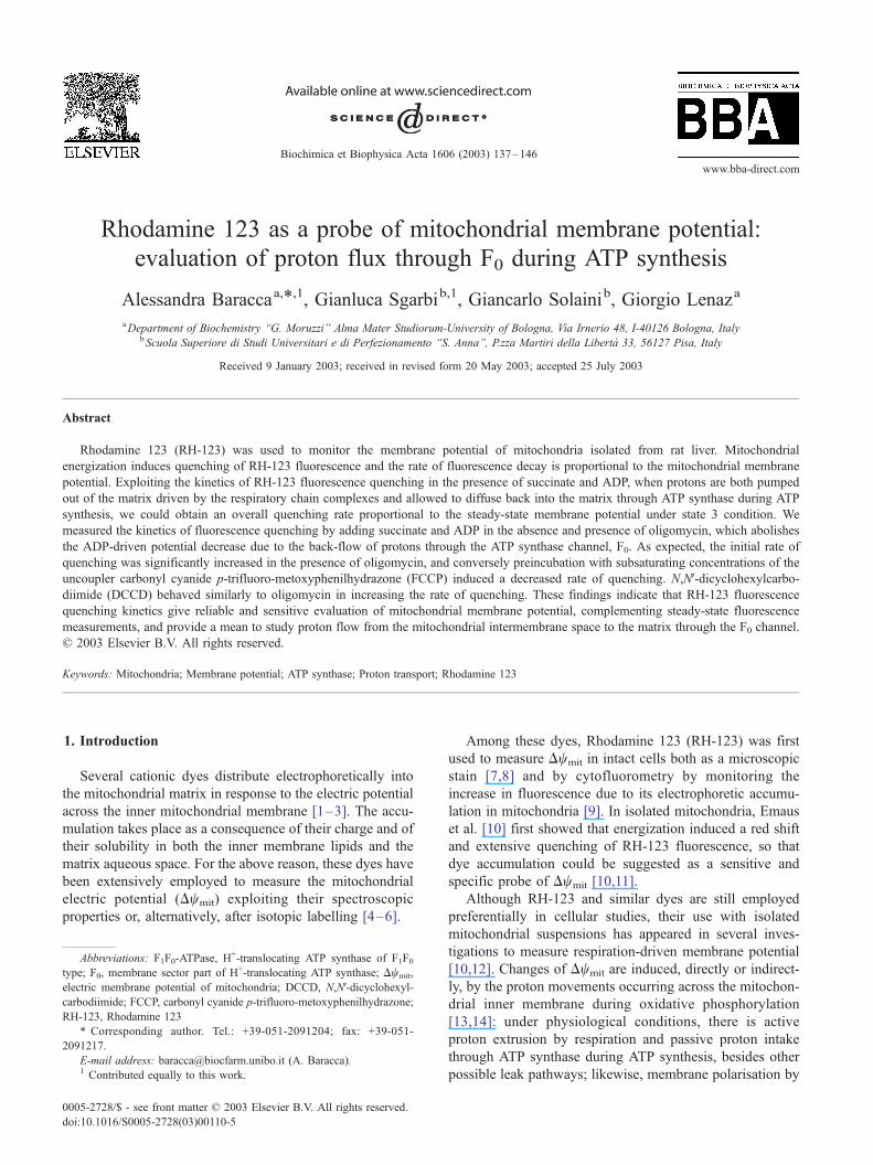
chondrial energization monitored by the dynamic fluores-
cence quenching of RH-123 reported in Fig. 2A, the time
course of F/Fi decay could be derived at different times
between 0 and 60 s (Fig. 2B). The curves represent the
exponential decay best fitting value as obtained by the
GraphPad Prism software (GraphPad Software Incorporat-
ed). Fi is the initial fluorescence as derived from the best
fitting analysis, and the initial fluorescence quenching rates
were calculated as first derivatives at time zero.
The initial rate of the fluorescence decay in the presence
of oligomycin increased from 0.20 to 0.33 (DF/Fi)/s/mg
protein and the quenching extent at steady-state level
increased from 2.67 to 2.96 (DF/Fi)/mg protein. The en-
hancement of both the fluorescence quenching rate and the
steady-state quenching induced by oligomycin was 65% and
11%, respectively. Furthermore, a comparative analysis of
the data of both fluorescence quenching extent and fluores-
cence quenching rate of 15 different mitochondrial prepa-
rations, besides supporting the above data, showed that the
mean of the fluorescence quenching rate of oligomycin-
treated mitochondria (0.32F 0.03 (DF/Fi)/s/mg proteinFS.D.) with respect to its control [0.21F 0.02 (DF/Fi)/s/mg
proteinF S.D.] (Fig. 3A), has a higher significance differ-
ence (P < 0.001) than the mean of the steady-state quenching
values, as measured in the presence or absence of oligomycin
[3.14F 0.36 and 2.81F 0.32 (DF/Fi)/mg proteinF S.D.,
respectively, with P < 0.05] (Fig. 3B).
Previous studies have shown that dye fluorescence
response to mitochondrial Dw is related to the dye/protein
ratio [6,10]; therefore, a titration of 50 nM RH-123 during
succinate-driven respiration was carried out with coupled
mitochondria in the small range from 0.15 to 0.45 mg/ml
protein. Increase of DF/Fi was observed when mitochondria
were added to the dye, as expected (Fig. 4B). However, DF/
Fi values appeared scattered with respect to the best linear
fitting, with a correlation coefficient r = 0.74. On the
contrary, plotting the rate of fluorescence quenching in-
duced by respiration, (DF/Fi)/s, as a function of protein
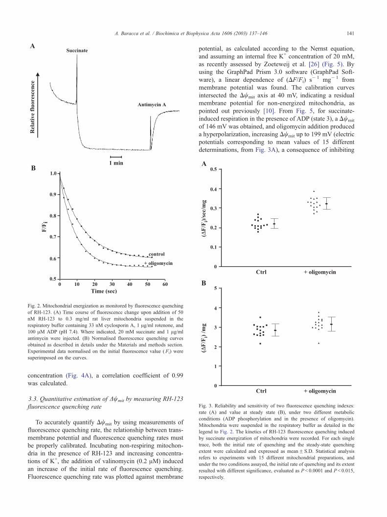
Fig. 3. Reliability and sensitivity of two fluorescence quenching indexes:
rate (A) and value at steady state (B), under two different metabolic
conditions (ADP phosphorylation and in the presence of oligomycin).
Mitochondria were suspended in the respiratory buffer as detailed in the
legend to Fig. 2. The kinetics of RH-123 fluorescence quenching induced
by succinate energization of mitochondria were recorded. For each single
trace, both the initial rate of quenching and the steady-state quenching
extent were calculated and expressed as meanF S.D. Statistical analysis
refers to experiments with 15 different mitochondrial preparations, and
under the two conditions assayed, the initial rate of quenching and its extent
resulted with different significance, evaluated as P < 0.0001 and P < 0.015,
respectively.
Fig. 2. Mitochondrial energization as monitored by fluorescence quenching
of RH-123. (A) Time course of fluorescence change upon addition of 50
nM RH-123 to 0.3 mg/ml rat liver mitochondria suspended in the
respiratory buffer containing 33 nM cyclosporin A, 1 Ag/ml rotenone, and
100 AM ADP (pH 7.4). Where indicated, 20 mM succinate and 1 Ag/ml
antimycin were injected. (B) Normalised fluorescence quenching curves
obtained as described in details under the Materials and methods section.
Experimental data normalised on the initial fluorescence value ( Fi) were
superimposed on the curves.
A. Baracca et al. / Biochimica et Biophysica Acta 1606 (2003) 137–146 141
concentration (Fig. 4A), a correlation coefficient of 0.99
was calculated.
3.3. Quantitative estimation of Dwmit by measuring RH-123
fluorescence quenching rate
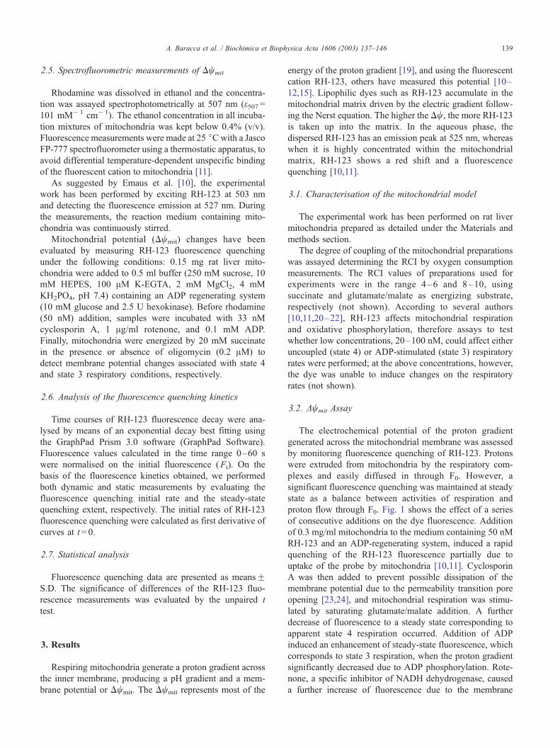
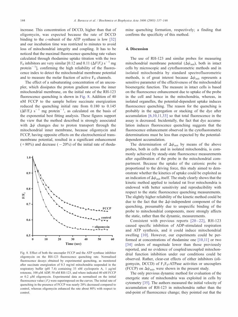
To accurately quantify Dwmit by using measurements of
fluorescence quenching rate, the relationship between trans-
membrane potential and fluorescence quenching rates must
be properly calibrated. Incubating non-respiring mitochon-
dria in the presence of RH-123 and increasing concentra-
tions of K+, the addition of valinomycin (0.2 AM) induced
an increase of the initial rate of fluorescence quenching.
Fluorescence quenching rate was plotted against membrane
potential, as calculated according to the Nernst equation,
and assuming an internal free K+ concentration of 20 mM,
as recently assessed by Zoeteweij et al. [26] (Fig. 5). By
using the GraphPad Prism 3.0 software (GraphPad Soft-
ware), a linear dependence of (DF/Fi) s� 1 mg� 1 from
membrane potential was found. The calibration curves
intersected the Dwmit axis at 40 mV, indicating a residual
membrane potential for non-energized mitochondria, as
pointed out previously [10]. From Fig. 5, for succinate-
induced respiration in the presence of ADP (state 3), a Dwmit
of 146 mV was obtained, and oligomycin addition produced
a hyperpolarization, increasing Dwmit up to 199 mV (electric
potentials corresponding to mean values of 15 different
determinations, from Fig. 3A), a consequence of inhibiting
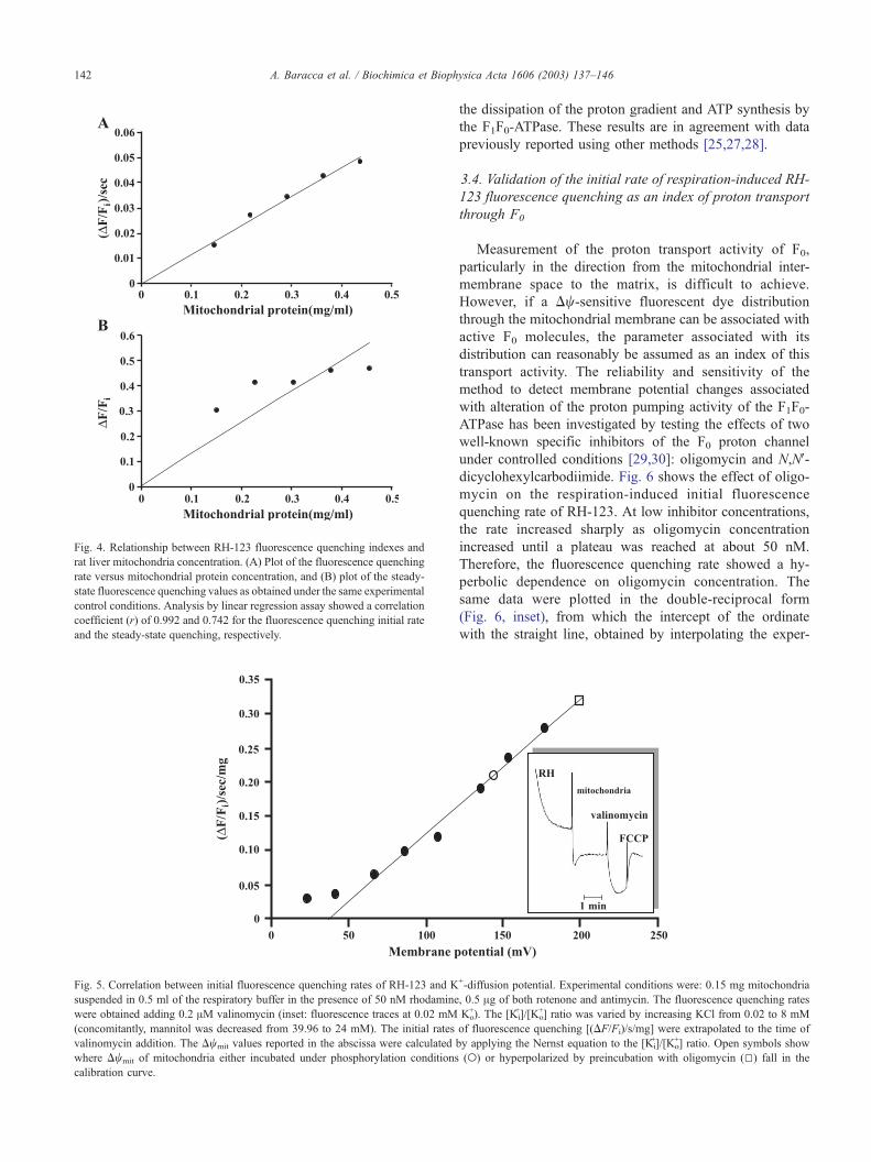
Fig. 5. Correlation between initial fluorescence quenching rates of RH-123 and K
suspended in 0.5 ml of the respiratory buffer in the presence of 50 nM rhodamine
were obtained adding 0.2 AM valinomycin (inset: fluorescence traces at 0.02 mM
(concomitantly, mannitol was decreased from 39.96 to 24 mM). The initial rates
valinomycin addition. The Dwmit values reported in the abscissa were calculated
where Dwmit of mitochondria either incubated under phosphorylation conditions
calibration curve.
Fig. 4. Relationship between RH-123 fluorescence quenching indexes and
rat liver mitochondria concentration. (A) Plot of the fluorescence quenching
rate versus mitochondrial protein concentration, and (B) plot of the steady-
state fluorescence quenching values as obtained under the same experimental
control conditions. Analysis by linear regression assay showed a correlation
coefficient (r) of 0.992 and 0.742 for the fluorescence quenching initial rate
and the steady-state quenching, respectively.
A. Baracca et al. / Biochimica et Biophysica Acta 1606 (2003) 137–146142
the dissipation of the proton gradient and ATP synthesis by
the F1F0-ATPase. These results are in agreement with data
previously reported using other methods [25,27,28].
3.4. Validation of the initial rate of respiration-induced RH-
123 fluorescence quenching as an index of proton transport
through F0
Measurement of the proton transport activity of F0,
particularly in the direction from the mitochondrial inter-
membrane space to the matrix, is difficult to achieve.
However, if a Dw-sensitive fluorescent dye distribution
through the mitochondrial membrane can be associated with
active F0 molecules, the parameter associated with its
distribution can reasonably be assumed as an index of this
transport activity. The reliability and sensitivity of the
method to detect membrane potential changes associated
with alteration of the proton pumping activity of the F1F0-
ATPase has been investigated by testing the effects of two
well-known specific inhibitors of the F0 proton channel
under controlled conditions [29,30]: oligomycin and N,NV-dicyclohexylcarbodiimide. Fig. 6 shows the effect of oligo-
mycin on the respiration-induced initial fluorescence
quenching rate of RH-123. At low inhibitor concentrations,
the rate increased sharply as oligomycin concentration
increased until a plateau was reached at about 50 nM.
Therefore, the fluorescence quenching rate showed a hy-
perbolic dependence on oligomycin concentration. The
same data were plotted in the double-reciprocal form
(Fig. 6, inset), from which the intercept of the ordinate
with the straight line, obtained by interpolating the exper-
+-diffusion potential. Experimental conditions were: 0.15 mg mitochondria
, 0.5 Ag of both rotenone and antimycin. The fluorescence quenching rates
Ko+). The [Ki
+]/[Ko+] ratio was varied by increasing KCl from 0.02 to 8 mM
of fluorescence quenching [(DF/Fi)/s/mg] were extrapolated to the time of
by applying the Nernst equation to the [Ki+]/[Ko
+] ratio. Open symbols show
(o) or hyperpolarized by preincubation with oligomycin (5) fall in the

Fig. 6. Titration of RH-123 fluorescence quenching rate with oligomycin. Initial rate values were measured after succinate energization of 0.3 mg/ml respiring
mitochondria suspended in the respiratory buffer containing 33 nM cyclosporin A, 1 Ag/ml rotenone, 100 AM ADP, 50 nM RH-123, and oligomycin at the
indicated concentration. The rate estimated in the absence of oligomycin was subtracted from each value determined at the different inhibitor concentrations.
The inset represents the double-reciprocal plot, from which a maximal rate of 0.12 (DF/Fi)/s/mg protein was calculated.
A. Baracca et al. / Biochimica et Biophysica Acta 1606 (2003) 137–146 143
imental points, gave the value of 8.33 [(DF/Fi)/s/mg]� 1,
corresponding to a maximal rate of 0.12 (DF/Fi) s� 1 mg
protein� 1. The intercept of the straight line with the
abscissa was at � 141 AM� 1 corresponding to a concen-
tration of 7.1 nM oligomycin necessary for 50% fluores-
cence quenching rate increase. Because the ATP synthase/
oligomycin stoichiometry is 1 to 1 and the binding of the
inhibitor is rapid and irreversible, the expected concentra-
tion of ATP synthase in the fluorometer cuvette should be
14.2 nM, a figure consistent with data previously reported
Fig. 7. Titration of RH-123 fluorescence quenching rate with DCCD. Initial ra
mitochondria suspended in the respiratory buffer containing 33 nM cyclosporin A,
concentrations. The rate estimated in the absence of DCCD was subtracted from the
the double-reciprocal plot, from which a maximal rate of 0.11 (DF/Fi)/s/mg prote
[31]. Incidentally, the above observation confirms that the
oligomycin concentration (0.2 AM) used in the experiments
described above was competent to completely inhibit the
ATP synthase in the cuvette. Similar results were obtained
when DCCD, which covalently binds to Glu 58 of the c-
subunit of the F0-ATPase sector, substituted for oligomycin
(Fig. 7). In the present case, the intercepts of the straight line
with the axes in the double-reciprocal plot gave a maximal
rate value of 0.11 (DF/Fi) s� 1 mg protein� 1 and 130 nM
DCCD necessary for 50% fluorescence quenching rate
te values were obtained after addition of 20 mM succinate to 0.3 mg/ml
1 Ag/ml rotenone, 100 AMADP, 50 nM RH-123, and DCCD at the indicated
values determined at different inhibitor concentrations. The inset represents
in was calculated.
A. Baracca et al. / Biochimica et Biophysica Acta 1606 (2003) 137–146144
increase. This concentration of DCCD, higher than that of
oligomycin, was expected because the rate of DCCD
binding to the c-subunit of the ATP synthase is low [32]
and our incubation time was restricted to minutes to avoid
loss of mitochondrial integrity and coupling. It has to be
noticed that the maximal fluorescence quenching rate values
calculated through rhodamine uptake titration with the two
F0 inhibitors are very similar [0.12 and 0.11 (DF/Fi) s� 1 mg
protein� 1], confirming the high reliability of the fluores-
cence index to detect the mitochondrial membrane potential
and to measure the molar fraction of active F0 channels.
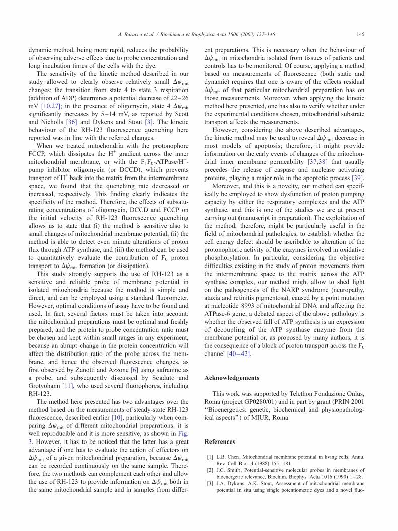
The effect of a subsaturating concentration of an uncou-
pler, which dissipates the proton gradient across the inner
mitochondrial membrane, on the initial rate of the RH-123
fluorescence quenching is shown in Fig. 8. Addition of 40
nM FCCP to the sample before succinate energization
reduced the quenching initial rate from 0.180 to 0.145
(DF/Fi) s� 1 mg protein� 1, as calculated on the basis of
the exponential best fitting analysis. These figures support
the view that the method described is strongly associated
with Dw changes due to proton transport through the
mitochondrial inner membrane, because oligomycin and
FCCP, having opposite effects on the electrochemical trans-
membrane potential, resulted in a significant enhancement
( + 80%) and decrease (� 20%) of the initial rate of rhoda-
Fig. 8. Effect of both the uncoupler FCCP and the ATP synthase inhibitor
oligomycin on the RH-123 fluorescence quenching rate. Normalised
fluorescence decays obtained by experimental quenching, as monitored
after succinate energization of 0.3 mg/ml mitochondria suspended in the
respiratory buffer (pH 7.4) containing 33 nM cyclosporin A, 1 Ag/ml
rotenone, 100 AM ADP, 50 nM RH-123, and where indicated 40 nM FCCP
or 0.2 AM oligomycin. Experimental data as normalised on the initial
fluorescence value ( Fi) were superimposed on the curves. The initial rate of
quenching in the presence of FCCP was nearly 20% decreased compared to
control, whereas oligomycin enhanced the rate about 80% with respect to
control.
mine quenching formation, respectively; a finding that
confirms the specificity of this method.
4. Discussion
The use of RH-123 and similar probes for measuring
mitochondrial membrane potential (Dwmit), both in intact
cells by microscopic and cytofluorometric methods and in
isolated mitochondria by standard spectrofluorometric
methods, is of great interest because Dwmit represents a
sensitive parameter of the effectiveness of the mitochondrial
bioenergetic function. The measure in intact cells is based
on the fluorescence enhancement due to uptake of the probe
in the cell and hence in the mitochondria, whereas, in
isolated organelles, the potential-dependent uptake induces
fluorescence quenching. The reason for the quenching is
probably in the aggregation or stacking of the dye after
accumulation [6,10,11,33] so that total fluorescence in the
assay is decreased. Incidentally, the fact that dye accumu-
lation induces fluorescence quenching suggests that the
fluorescence enhancement observed in the cytofluorometric
determinations must be less than expected by the potential-
dependent accumulation.
The determination of Dwmit by means of the above
probes, both in cells and in isolated mitochondria, is com-
monly achieved by steady-state fluorescence measurements
after equilibration of the probe in the mitochondrial com-
partment. Because the uptake of the cationic probe is
proportional to the driving force, this study aimed to dem-
onstrate whether the kinetics of uptake could be exploited as
an indication of Dwmit itself. The study clearly shows that the
kinetic method applied to isolated rat liver mitochondria is
endowed with better sensitivity and reproducibility with
respect to the static fluorescence quenching measurements.
The slightly higher reliability of the kinetic method could be
due to the fact that the Dw-independent component of the
quenching, presumably due to unspecific binding of the
probe to mitochondrial components, more strongly affects
the static, rather than the dynamic, measurements.
Consistent with previous reports [20–22], RH-123
caused specific inhibition of ADP-stimulated respiration
and ATP synthesis, and it could induce mitochondrial
swelling [10]. However, our experiments could be per-
formed at concentrations of rhodamine one [10,11] or two
[34] orders of magnitude lower than those previously
reported, and no evidence of coupled/uncoupled mitochon-
drial function inhibition under our conditions could be
observed. Rather, clear-cut effects of either inhibitors (oli-
gomycin, DCCD) of F1F0-ATPase activities or uncouplers
(FCCP) on Dwmit, were shown in the present study.
The only previous dynamic method for evaluation of the
energetic state of mitochondria was exploited in cells by
cytometry [35]. The authors measured the initial velocity of
accumulation of RH-123 in mitochondria rather than the
end-point of fluorescence change; they pointed out that the
A. Baracca et al. / Biochimica et Biophysica Acta 1606 (2003) 137–146 145
dynamic method, being more rapid, reduces the probability
of observing adverse effects due to probe concentration and
long incubation times of the cells with the dye.
The sensitivity of the kinetic method described in our
study allowed to clearly observe relatively small Dwmit
changes: the transition from state 4 to state 3 respiration
(addition of ADP) determines a potential decrease of 22–26
mV [10,27]; in the presence of oligomycin, state 4 Dwmit
significantly increases by 5–14 mV, as reported by Scott
and Nicholls [36] and Dykens and Stout [3]. The kinetic
behaviour of the RH-123 fluorescence quenching here
reported was in line with the referred changes.
When we treated mitochondria with the protonophore
FCCP, which dissipates the H+ gradient across the inner
mitochondrial membrane, or with the F1F0-ATPase/H+-
pump inhibitor oligomycin (or DCCD), which prevents
transport of H+ back into the matrix from the intermembrane
space, we found that the quenching rate decreased or
increased, respectively. This finding clearly indicates the
specificity of the method. Therefore, the effects of subsatu-
rating concentrations of oligomycin, DCCD and FCCP on
the initial velocity of RH-123 fluorescence quenching
allows us to state that (i) the method is sensitive also to
small changes of mitochondrial membrane potential, (ii) the
method is able to detect even minute alterations of proton
flux through ATP synthase, and (iii) the method can be used
to quantitatively evaluate the contribution of F0 proton
transport to Dwmit formation (or dissipation).
This study strongly supports the use of RH-123 as a
sensitive and reliable probe of membrane potential in
isolated mitochondria because the method is simple and
direct, and can be employed using a standard fluorometer.
However, optimal conditions of assay have to be found and
used. In fact, several factors must be taken into account:
the mitochondrial preparations must be optimal and freshly
prepared, and the protein to probe concentration ratio must
be chosen and kept within small ranges in any experiment,
because an abrupt change in the protein concentration will
affect the distribution ratio of the probe across the mem-
brane, and hence the observed fluorescence changes, as
first observed by Zanotti and Azzone [6] using safranine as
a probe, and subsequently discussed by Scaduto and
Grotyohann [11], who used several fluorophores, including
RH-123.
The method here presented has two advantages over the
method based on the measurements of steady-state RH-123
fluorescence, described earlier [10], particularly when com-
paring Dwmit of different mitochondrial preparations: it is
well reproducible and it is more sensitive, as shown in Fig.
3. However, it has to be noticed that the latter has a great
advantage if one has to evaluate the action of effectors on
Dwmit of a given mitochondrial preparation, because Dwmit
can be recorded continuously on the same sample. There-
fore, the two methods can complement each other and allow
the use of RH-123 to provide information on Dwmit both in
the same mitochondrial sample and in samples from differ-
ent preparations. This is necessary when the behaviour of
Dwmit in mitochondria isolated from tissues of patients and
controls has to be monitored. Of course, applying a method
based on measurements of fluorescence (both static and
dynamic) requires that one is aware of the effects residual
Dwmit of that particular mitochondrial preparation has on
those measurements. Moreover, when applying the kinetic
method here presented, one has also to verify whether under
the experimental conditions chosen, mitochondrial substrate
transport affects the measurements.
However, considering the above described advantages,
the kinetic method may be used to reveal Dwmit decrease in
most models of apoptosis; therefore, it might provide
information on the early events of changes of the mitochon-
drial inner membrane permeability [37,38] that usually
precedes the release of caspase and nuclease activating
proteins, playing a major role in the apoptotic process [39].
Moreover, and this is a novelty, our method can specif-
ically be employed to show dysfunction of proton pumping
capacity by either the respiratory complexes and the ATP
synthase, and this is one of the studies we are at present
carrying out (manuscript in preparation). The exploitation of
the method, therefore, might be particularly useful in the
field of mitochondrial pathologies, to establish whether the
cell energy defect should be ascribable to alteration of the
protonophoric activity of the enzymes involved in oxidative
phosphorylation. In particular, considering the objective
difficulties existing in the study of proton movements from
the intermembrane space to the matrix across the ATP
synthase complex, our method might allow to shed light
on the pathogenesis of the NARP syndrome (neuropathy,
ataxia and retinitis pigmentosa), caused by a point mutation
at nucleotide 8993 of mitochondrial DNA and affecting the
ATPase-6 gene; a debated aspect of the above pathology is
whether the observed fall of ATP synthesis is an expression
of decoupling of the ATP synthase enzyme from the
membrane potential or, as proposed by many authors, it is
the consequence of a block of proton transport across the F0channel [40–42].
Acknowledgements
This work was supported by Telethon Fondazione Onlus,
Roma (project GP0280/01) and in part by grant (PRIN 2001
‘‘Bioenergetics: genetic, biochemical and physiopatholog-
ical aspects’’) of MIUR, Roma.
References
[1] L.B. Chen, Mitochondrial membrane potential in living cells, Annu.
Rev. Cell Biol. 4 (1988) 155–181.
[2] J.C. Smith, Potential-sensitive molecular probes in membranes of
bioenergetic relevance, Biochim. Biophys. Acta 1016 (1990) 1–28.
[3] J.A. Dykens, A.K. Stout, Assessment of mitochondrial membrane
potential in situ using single potentiometric dyes and a novel fluo-
A. Baracca et al. / Biochimica et Biophysica Acta 1606 (2003) 137–146146
rescence resonance energy transfer technique, Methods Cell Biol. 65
(2001) 285–309.
[4] H. Rottenberg, Membrane potential and surface potential in mito-
chondria: uptake and binding of lipophilic cations, J. Membr. Biol.
81 (1984) 127–138.
[5] J.B. Jackson, D.G. Nicholls, Methods for the determination of mem-
brane potential in bioenergetic systems, Methods Enzymol. 127
(1986) 557–577.
[6] A. Zanotti, G.F. Azzone, Safranine as membrane potential probe in rat
liver mitochondria, Arch. Biochem. Biophys. 201 (1980) 255–265.
[7] L.V. Johnson, M.L. Walsh, L.B. Chen, Localization of mitochondria
in living cells with rhodamine 123, Proc. Natl. Acad. Sci U. S. A. 77
(1980) 990–994.
[8] L.V. Johnson, M.L. Walsh, B.J. Bockus, L.B. Chen, Monitoring of
relative mitochondrial membrane potential in living cells by fluo-
rescence microscopy, J. Cell Biol. 88 (1981) 526–535.
[9] X. Ronot, L. Benel, M. Adolphe, J.C. Mounolou, Mitochondrial anal-
ysis in living cells: the use of rhodamine 123 and flow cytometry,
Biol. Cell 57 (1986) 1–7.
[10] R.K. Emaus, R. Grunwald, J.J. Lemasters, Rhodamine 123 as a probe
of transmembrane potential in isolated rat liver mitochondria: spectral
and metabolic properties, Biochim. Biophys. Acta 850 (1986)
436–448.
[11] R.C. Scaduto Jr., L.W. Grotyohann, Measurement of mitochondrial
membrane potential using fluorescent rhodamine derivatives, Bio-
phys. J. 76 (1999) 469–477.
[12] J.E. O’Connor, J.L. Vargas, B.F. Kimler, J. Hernandez-Yago, S. Gri-
solia, Use of rhodamine 123 to investigate alterations in mitochondrial
activity in isolated mouse liver mitochondria, Biochem. Biophys. Res.
Commun. 151 (1988) 568–573.
[13] E.M. Fontaine, A. Devin, M. Rigoulet, X.M. Leverve, The yield of
oxidative phosphorylation is controlled both by force and flux, Bio-
chem. Biophys. Res. Commun. 232 (1997) 532–535.
[14] R.P. Hafner, G.C. Brown, M.D. Brand, Analysis of the control of
respiration rate, phosphorylation rate, proton leak rate and protonmo-
tive force in isolated mitochondria using the ‘‘top-down’’ approach of
metabolic control theory, Eur. J. Biochem. 188 (1990) 313–319.
[15] J.R. Bunting, Influx and efflux kinetics of cationic dye binding to
respiring mitochondria, Biophys. Chem. 42 (1992) 163–175.
[16] E. Kun, E. Kirsten, W.N. Piper, Stabilization of mitochondrial func-
tions with digitonin, Methods Enzymol. 55 (1979) 115–118.
[17] A.G. Gornall, C.J. Bardawill, M.M. David, Determination of serum
proteins by means of the biuret reaction, J. Biol. Chem. 177 (1949)
751–766.
[18] G. Aicardi, G. Solaini, Effects of niridazole and 5-nitroimidazoles on
heart mitochondrial respiration, Biochem. Pharmacol. 31 (1982)
3703–3705.
[19] D.G. Nicholls, S.J. Ferguson, Bioenergetics 2, Academic Press, Lon-
don, 1992.
[20] M.S. Mai, W.S. Allison, Inhibition of an oligomycin-sensitive ATPase
by cationic dyes, some of which are atypical uncouplers of intact
mitochondria, Arch. Biochem. Biophys. 221 (1983) 467–476.
[21] J.S. Modica-Napolitano, M.J. Weiss, L.B. Chen, J.R. Aprille, Rhod-
amine 123 inhibits bioenergetic function in isolated rat liver mito-
chondria, Biochem. Biophys. Res. Commun. 118 (1984) 717–723.
[22] J.S. Modica-Napolitano, J.R. Aprille, Basis for the selective cytotox-
icity of rhodamine 123, Cancer Res. 47 (1987) 4361–4365.
[23] P. Bernardi, The permeability transition pore. Control points of a
cyclosporin A-sensitive mitochondrial channel involved in cell death,
Biochim. Biophys. Acta 1275 (1996) 5–9.
[24] K.M. Broekemeier, M.E. Dempsey, D.R. Pfeiffer, Cyclosporin A is a
potent inhibitor of the inner membrane permeability transition in liver
mitochondria, J. Biol. Chem. 264 (1989) 7826–7830.
[25] K. Van Dam, A.H. Wiechmann, Respiratory control and oxidative
phosphorylation measurements in mitochondria, Methods Enzymol.
55 (1979) 225–229.
[26] J.P. Zoeteweij, B. Van de Water, H.J.G.M. De Bont, J.F. Nagelkerke,
Mitochondrial K+ as modulator of Ca2 +-dependemt cytotoxicity in
hepatocytes, Biochem. J. 299 (1994) 539–543.
[27] A. Masini, D. Ceccarelli-Stanzani, U. Moscatello, An investigation on
the effect of oligomycin on state-4 respiration in isolated rat-liver
mitochondria, Biochim. Biophys. Acta 767 (1984) 130–137.
[28] P. Mitchell, J. Moyle, Estimation of membrane potential pH differ-
ence across the cristae membrane of rat liver mitochondria, Eur. J.
Biochem. 7 (1969) 471–484.
[29] P.E. Linnett, R.B. Beechey, Inhibitors of the ATP synthethase system,
Methods Enzymol. 55 (1979) 472–518.
[30] J. Hoppe, W. Sebald, The proton conducting F0-part of bacterial ATP
synthases, Biochim. Biophys. Acta 768 (1984) 1–27.
[31] R.M. Bertina, P.I. Schrier, E.C. Slater, The binding of aurovertin to
mitochondria and its effect on mitochondrial respiration, Biochim.
Biophys. Acta 305 (1973) 503–518.
[32] H.S. Penefsky, Mechanism of inhibition of mitochondrial adenosine
triphosphatase by dicyclohexylcarbodimide and oligomycin: relation-
ship to ATP synthesis, Proc. Natl. Acad. Sci. U. S. A. 82 (1985)
1589–1593.
[33] J.R. Bunting, T.V. Phan, E. Kamali, R.M. Dowben, Fluorescent
cationic probes of mitochondria: metrics and mechanism of interac-
tion, Biophys. J. 56 (1989) 979–993.
[34] N. Zamzami, D. Metivier, G. Kroemer, Quantitation of mitochon-
drial transmembrane potential in cells and in isolated mitochondria,
Methods Enzymol. 322 (2000) 208–213.
[35] G. Juan, M. Cavazzoni, G.T. Saez, J.E. O’Connor, A fast kinetic
method for assessing mitochondrial membrane potential in isolated
hepatocytes with rhodamine 123 and flow cytometry, Cytometry 15
(1994) 335–342.
[36] I.D. Scott, D.G. Nicholls, Energy transduction in intact synaptosomes:
influence of plasma-membrane depolarization on the respiration and
membrane potential of internal mitochondria determined in situ, Bio-
chem. J. 186 (1980) 21–33.
[37] P. Bernardi, E. Basso, R. Colonna, P. Costantini, F. Di Lisa, O.
Eriksson, E. Fontane, M. Forte, F. Ichas, S. Massari, A. Nicolli,
V. Petronilli, L. Scorrano, Perspectives on the mitochondrial perme-
ability transition, Biochim. Biophys. Acta 1365 (1998) 200–206.
[38] S. Susin, N. Zamzami, G. Kroemer, Mitochondria as regulators of
apoptosis: doubt no more, Biochim. Biophys. Acta 1366 (1988)
151–165.
[39] S. Matsuyama, J. Llopis, Q.L. Deveraux, R.Y. Tsien, J.C. Reed,
Changes in intramitochondrial cytosolic pH: early events that modu-
late caspase activation during apoptosis, Nat. Cell Biol. 2 (2000)
318–325.
[40] A. Baracca, S. Barogi, V. Carelli, G. Lenaz, G. Solaini, Catalytic
activities of mitochondrial ATP synthase in patients with mitochon-
drial DNA T8993G mutation in the ATPase 6 gene encoding subunit
a, J. Biol. Chem. 275 (2000) 4177–4182.
[41] B.H. Robinson, MtDNA and nuclear mutations affecting oxidative
phosphorylation: correlating severity of clinical defect with extent
of bioenergetic compromise, J. Bioenerg. Biomembr. 26 (1994)
311–316.
[42] I. Trounce, S. Neill, D.C. Wallace, Cytoplasmic transfer of the
mtDNA nt 8993 T!G (ATP6) point mutation associated with Leigh
syndrome into mtDNA-less cells demonstrates cosegregation with a
decrease in state III respiration and ADP/O ratio, Proc. Natl. Acad.
Sci. U. S. A. 91 (1994) 8334–8338.