M E C H A N I S M S O F D E V E L O P M E N T 1 2 6 ( 2 0 0 9 ) 3 3 7 – 3 4 9
. sc iencedi rec t . com
ava i lab le a t wwwjourna l homepage: www.elsevier .com/ locate /modo
NRAGE: A potential rheostat during branching morphogenesis
George N. Nikopoulosa,b,1, Joao Ferreira Martinsa,c, Tamara L. Adamsa, Aldona Karaczyna,b,Derek Adamsa, Calvin Varya,b, Leif Oxburgha,b, Joseph M. Verdia,b,*,2
aMaine Medical Center Research Institute, Center for Molecular Medicine, 81 Research Drive, Scarborough, ME 04074, USAbThe University of Maine Orono, Department of Biochemistry, Microbiology and Molecular Biology, 5735 Hitchner Hall, Orono,
ME 04469-5735, USAcDepartment of Physiology, Faculty of Medicine, University of Porto, 4200-319 Porto, Portugal
A R T I C L E I N F O
Article history:
Received 16 September 2008
Received in revised form
23 February 2009
Accepted 24 February 2009
Available online 4 March 2009
Keywords:
Branching morphogenesis
NRAGE
p38 MAP kinase
Apoptosis
BMP
Ret
GDNF
0925-4773/$ - see front matter Published bydoi:10.1016/j.mod.2009.02.005
* Corresponding author. Address: MaineScarborough, ME 04074, USA. Tel.: +1 (207) 8
E-mail address: [email protected] (J.M. Ver1 The author was supported by a pre-docto2 The author was supported by NIH R01 NS
A B S T R A C T
Branching morphogenesis is a developmental process characteristic of many organ sys-
tems. Specifically, during renal branching morphogenesis, its been postulated that the final
number of nephrons formed is one key clinical factor in the development of hypertension
in adulthood. As it has been established that BMPs regulate, in part, renal activity of p38
MAP kinase (p38MAPK) and it has demonstrated that the cytoplasmic protein Neurotrophin
Receptor MAGE homologue (NRAGE) augments p38MAPK activation, it was hypothesized that
a decrease in the expression of NRAGE during renal branching would result in decreased
branching of the UB that correlated with changes in p38MAPK activation. To verify this,
the expression of NRAGE was reduced in ex vivo kidney explants cultures using antisense
morpholino. Morpholino treated ex vivo kidney explants expression were severely stunted
in branching, a trait that was rescued with the addition of exogenous GDNF. Renal explants
also demonstrated a precipitous drop in p38MAPK activation that too was reversed in the
presence of recombinant GDNF. RNA profiling of NRAGE diminished ex vivo kidney explants
resulted in altered expression of GDNF, Ret, BMP7 and BMPRIb mRNAs. Our results sug-
gested that in early kidney development NRAGE might have multiple roles during renal
branching morphogenesis through association with both the BMP and GDNF signaling
pathways.
Published by Elsevier Ireland Ltd.
1. Introduction essential hypertension (Brenner et al., 1988). Since reports
Essential hypertension, or hypertension with no identifi-
able cause, is unfortunately a common disease of the Western
world (Kearney et al., 2005). In the early 1970’s David Barker
proposed the ‘‘fetal origins of disease hypothesis’’, supposing
that the prevalence of many adult diseases, including hyper-
tension, is a result of abnormal fetal development (Barker
et al., 1970). Brenner later refined this hypothesis by propos-
ing that lower nephron numbers predisposed individuals to
Elsevier Ireland Ltd.
Medical Center Research85 8190; fax: +1 (207) 885di).
ral fellowship from the A
055304 and in part by N
based on the Brenner–Barker hypothesis suggest a link
between kidney development and hypertension (Langley
and Jackson, 1994; Levitt et al., 1996; Woodall et al., 1996),
elucidating the molecular mechanisms that govern kidney
development could elucidate the key factors affecting the
development of hypertension later in life.
The development of the kidney begins with renal branch-
ing morphogenesis (RBM). During RBM reciprocal inductive
interactions, between the ureteric bud (UB) and the surrounding
Institute, Center for Regenerative Medicine, 81 Research Dr.,8110.
merican Heart Association.
IH COBRE in Stem and Progenitor Biology.
338 M E C H A N I S M S O F D E V E L O P M E N T 1 2 6 ( 2 0 0 9 ) 3 3 7 – 3 4 9
metanephric mesenchyme (MM) result in the development of
the collecting ducts and the nephrons. The precise molecular
signals that control RBM are currently unknown and still ac-
tively pursued. Bone morphogenic proteins (BMPs) are mem-
bers of the transforming growth factor beta (TGFb)
superfamily of signaling molecules and have been implicated
in a diverse array of biological processes, including cell
growth, differentiation and apoptosis (Hogan, 1996). BMPs
play crucial roles during RBM, transducing their signal either
through the canonical SMAD-mediated pathway, and/or
through the non-canonical BMP signaling cascade of MAP ki-
nases, TAK1, TAB1, and p38MAPK (Nohe et al., 2004; Oxburgh
et al., 2004; Winnier et al., 1995).
We recently demonstrated that NRAGE is a potential mem-
ber of the non-canonical BMP pathway utilizing the multipo-
tential neural progenitor cells resulting in BMP instructive
apoptosis (Kendall et al., 2005). It has been suggested that
the same non-canonical BMP signaling pathway also medi-
ates branching of the UB (Hu et al., 2004) suggesting a poten-
tial role for NRAGE during embryonic renal branching
morphogenesis. It was hypothesized that a decrease in the
expression of NRAGE during RBM would result in altered
branching of the UB and potentially in cell viability.
Utilizing NRAGE morpholinos (Kendall et al., 2005), we
attenuated NRAGE protein expression in ex vivo kidney
culture explants to determine if decreased NRAGE expression
affects p38MAPK activation and consequently branching of the
UB. We also investigated the global ramifications of lowering
NRAGE expression in the developing explants in hopes of elu-
cidating other pathways and mechanisms that NRAGE may
regulate during renal development. As predicted, lowering
NRAGE expression severely retarded the growth and branch-
ing of the UB. What was surprising and unexpected was that
gene profiling revealed that lowering NRAGE levels lead to a
reduction in the expression of BMPR1b, Ret, GDNF, and
BMP7 in the developing kidney. Rescue experiments demon-
strated that exogenously applied recombinant GDNF cor-
rected the deficiency in branching in ex vivo explants
cultures, with GDNF being more robust to promote growth
and branching than BMP7. These results demonstrate the
importance of NRAGE in affecting the maximal response dur-
ing branching morphogenesis.
2. Methods
2.1. Cell culture
mIMCD-3 (ATCC, Virginia, USA) cells were cultured in
DMEM/F12 (Invitrogen, California, USA) supplemented with
10% fetal bovine serum (Hyclone, Utah, USA) in a 37 �Cand 5% CO2 humidified incubator. In branching experi-
ments, 1 · 106 cells were plated in a collagen matrix as de-
scribed by Piscione without modification (Piscione et al.,
2001). The matrix was assembled on ice and plated with var-
ious doses of GDNF (0–10 ng/ml) (R&D Systems, Minnesota,
USA), BMP7 (0–10 ng/ml) (R&D Systems, Minnesota, USA),
no supplementation, or TGFb (0–25 ng/ml) (R&D Systems,
Minnesota, USA) for 3–14 days with media refreshed every
morning.
2.2. Co-immunoprecipitation and immunoblotting
Cell lysates were generated from mIMCD-3 cells that were
treated with and without 10 ng/ml BMP7 (R&D Systems, Min-
nesota, USA) for 1 h in DMEM-F12 (Invitrogen, California,
USA). Cells were lysed in 350 ll of NPB lysis buffer consisting
of: 20 mM Tris, pH 7.5, containing 300 mM sucrose, 60 mM
KCl, 15 mM NaCl, 5% (v/v) glycerol, 2 mM EDTA, 1% (v/v) Triton
X-100, with protease inhibitor cocktail I (Sigma–Aldrich,
Missouri, USA) for 20 min on ice. Lysates were immunoprecip-
itated using 50 ll of G-sepharose beads (Amersham Biosci-
ences–GE Healthcare, USA) and 2 lg of NRAGE (1:1000) or Ret
antibody (1:2000) (Upstate-Millipore, Massacheusetts, USA;
R&D Systems, Minnesota, USA) overnight at 4 �C. The beads
were collected by centrifugation at 12,000 RPMs for 5 min
and washed three times with fresh ice-cold lysis buffer. The
samples were subjected to 12% SDS–PAGE under reducing
conditions. After transferring the resolved proteins to Hybond
C membrane (Amersham Biosciences–GE Healthcare, USA),
blots were probed for NRAGE (1:1000) (Upstate-Millipore, Mas-
sachusetts, USA), TAK1 (1:1000) (Upstate-Millipore, Massachu-
setts, USA), Tab1 (1:1000) (Pro-Sci, California, USA), XIAP
(1:1000) (Cell Signaling Technology, Massachusetts, USA) or
b-actin (1:2500) (Sigma–Aldrich, Missouri, USA) antibodies.
Blots were developed using an appropriate horseradish perox-
idase conjugated goat anti-mouse or rabbit IgG (Bio-Rad) and
the ECL detection system (Amersham Biosciences–GE Health-
care, USA).
2.3. Kidney organ culture
Kidney organ culture was performed as previously de-
scribed by Nikopoulos et al. (2008) using kidneys from E11.5
Hoxb7-GFP mice (Srinivas et al., 1999) or E11.5 ICR mouse
embryos (Taconic, New York, USA). Morpholino sequences
used in this study are as follows, NRAGE morpholino:
GGTTTCTGAGCCATAGCTCTCGTC and for the negative con-
trol morpholino: CCTCTTACCTCAGTTACAATTTATA (Gene-
Tools, Oregon, USA). BMP7 or GDNF (R&D Systems, Minnesota,
USA) was added at concentrations and time described for
each experiment. Kidneys were analyzed under a Leica ste-
reomicroscope (Leica, USA) or subjected to immunofluores-
cent staining using: TOPRO-3 (1:10000; Invitrogen, California,
USA) to identify nuclei, or Ki67 (1:1000; AbCam, Massachu-
setts, USA) to identify proliferating cells, Dolichous Bifluorous
Agglutinin to identify cells of the ureteric bud (DBA; 1:1000;
Vector Labs, California, USA) or anti-laminin to also identify
cells of the ureteric bud (Sigma–Aldrich, Missouri, USA). Kid-
ney explants stained with Ki67 were visualized using a Leica
TCS-SP confocal microscope (Leica, USA). The number of
Ki67 positive cells was determined by counting the Ki67 posi-
tive nuclei in a given field for each kidney analyzed and calcu-
lating the number of cells per lm3 using the Leica TCS
software.
2.4. TUNEL analysis
E11.5 Hoxb7-GFP kidney explants were cultured with
either NRAGE morpholino or negative control for 72 h, in
DMEM/F12 media + 10% FBS prior to being in 4% PFA over-
M E C H A N I S M S O F D E V E L O P M E N T 1 2 6 ( 2 0 0 9 ) 3 3 7 – 3 4 9 339
night at 4 �C. After consecutive washes in PBS, kidneys were
incubated with 20 lg/ml of proteinase-K (37 �C) and were sub-
jected to Terminal deoxynucleotidyl Transferase Biotin-dUTP
Nick End Labeling (TUNEL) analysis to detect apoptotic cells
using the TetraMethylRhodamine red (TMR) in situ cell death
detection kit (Roche) and counterstained with 1:10,000 dilu-
tion of TOPRO-3 (Invitrogen, California, USA) overnight. Kid-
neys were mounted on glass slides with Prolong Gold
(Invitrogen, California, USA) and analyzed using a Leica SP-
TCS confocal microscope (Leica, USA). TUNEL positive nuclei
in the metanephric mesenchyme, as delineated by the ure-
teric bud specific GFP expression, were counted and the calcu-
lated as the number of TUNEL positive cells per unit volume
(lm3) for comparison between NRAGE and Negative Control
treated kidneys.
2.5. RNA isolation and quantitative PCR of kidney organcultures and mIMCD-3 cells
E11.5 mouse (Taconic, New York, USA) kidney explants
were cultured with NRAGE morpholino, negative control mor-
pholino, or Endo-Porter only. Total RNA was extracted from
six kidney organ cultures for each treatment for each day of
culture and pooled into one RNA sample using the RNAqu-
eous-micro kit (Ambion, California, USA). RNA samples were
treated with 1 unit of DNAse I (37 �C, 15 min) then inactivated
using Ambion DNAase inactivation slurry. mIMCD-3 cells
were incubated with NRAGE morpholino, negative control
morpholino, and p38MAPK phosphorylation inhibitor SB203580,
DMSO, or Endo-Porter only, for 48 h. Total RNA was extracted
from mIMCD-3 cells using the RNAqueous�-4PCR (Ambion,
California, USA) and also treated with 1 unit of DNAse I
(37 �C, 15 min). DNAase was inactivated as above. cDNA was
synthesized from all RNA samples with the first-strand syn-
thesis reaction kit (SuperArray-SABiosceinces, Maryland,
USA). Verified quantitative real-time PCR (qPCR) primers for
all genes were obtained from SuperArray. qRT-PCR was per-
formed in triplicate for each gene for each day, and three
independent repeats of the experiment was performed using
SYBR-Green I as per manufacturers instructions using an iCy-
cler IQ (Bio-Rad, California, USA). Relative fold change in gene
expression was calculated using the means of all experiments
for each gene on each day with each treatment using the 2(�DDCT) method as previously described (Livak and Schmittgen,
2001). One sample t-test for each gene was performed using
Prism 4.0 Software (GraphPad, California, USA).
2.6. Hoxb7-NRAGECherry transgenic mice
Utilizing the same promoter sequence used by Costantini
and colleagues to generate mice that specifically expressed
GFP in the ureteric bud of the kidney; we constructed a trans-
gene where full length NRAGE was fused in frame with the
fluorescent protein mCherry (Shaner et al., 2004). mCherry
was chosen as the fluorescent marker due to its low level of
cell toxicity and high photostability. Furthermore, it has been
previously utilized in transgenic animals to track develop-
ment, where it was shown to have no effect on normal devel-
opment (Renn and Winkler, 2009; Shcherbo et al., 2009;
Winnier et al., 1995). After the digestion of the plasmid, we
performed pro-nuclear injections on 60 fertilized eggs. We
fixed and cryo-preserved the isolated kidneys from at E17
for fluorescent microscopic analysis. To identify cells of the
ureteric bud of the kidney we utilized Dolichos biflorous agglu-
tinin (1:1000, Vector Labs), a lectin that specifically stains cells
of the ureteric bud of the kidney, or subjected them to p38MAPK
staining. p38MAPK staining was performed by using a poly-
clonal antibody to p38MAPK (1:1000 dilution; Cell Signaling
Technology, Massachusetts, USA) or a polyclonal phospho-
p38MAPK antibody (1:1000; Cell Signaling Technology, Massa-
chusetts, USA), and each received a rabbit Alexa-546 (1:5000;
Invitrogen, California, USA) secondary antibody. Nuclei were
identified by either TOPRO-3 (Invitrogen, California, USA) or
DAPI (Vector Labs).
2.7. mIMCD-3 cells in collagen matrix
Utilizing rat tail collagen (Invitrogen, California, USA),
10,000 mIMCD-3 cells were embedded into three-dimen-
sional culture system as described previously (Piscione
et al., 1997). The mIMCD-3 cells were treated with 10 ng/ml
BMP7, 25 ng/ml GDNF or morpholino. When treated with
morpholino, the mIMCD3 cells were treated for 48 h in an
adherent cell culture dish to suppress NRAGE expression be-
fore the beginning of the tubulogenesis assay. We defined a
branch point as a pixel that has three or more neighboring
pixels on our phase contrast microscope. When there are
no longer two neighboring pixels, these branches are labeled
2� branches.
3. Results
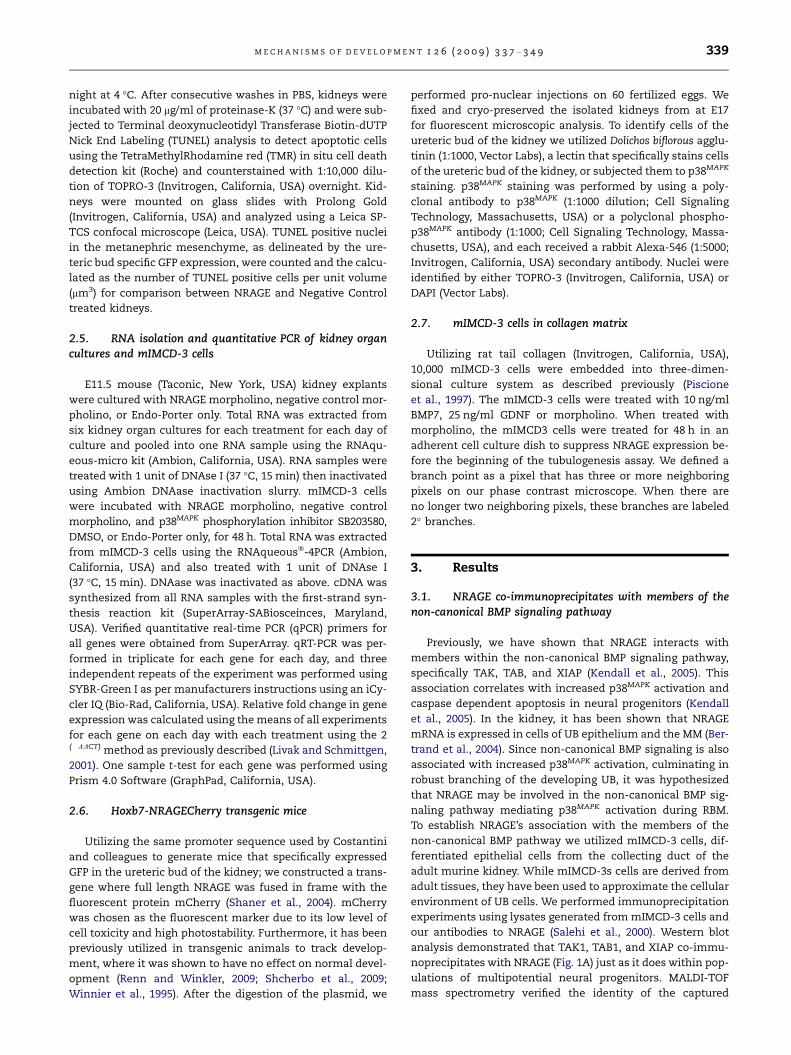
3.1. NRAGE co-immunoprecipitates with members of thenon-canonical BMP signaling pathway
Previously, we have shown that NRAGE interacts with
members within the non-canonical BMP signaling pathway,
specifically TAK, TAB, and XIAP (Kendall et al., 2005). This
association correlates with increased p38MAPK activation and
caspase dependent apoptosis in neural progenitors (Kendall
et al., 2005). In the kidney, it has been shown that NRAGE
mRNA is expressed in cells of UB epithelium and the MM (Ber-
trand et al., 2004). Since non-canonical BMP signaling is also
associated with increased p38MAPK activation, culminating in
robust branching of the developing UB, it was hypothesized
that NRAGE may be involved in the non-canonical BMP sig-
naling pathway mediating p38MAPK activation during RBM.
To establish NRAGE’s association with the members of the
non-canonical BMP pathway we utilized mIMCD-3 cells, dif-
ferentiated epithelial cells from the collecting duct of the
adult murine kidney. While mIMCD-3s cells are derived from
adult tissues, they have been used to approximate the cellular
environment of UB cells. We performed immunoprecipitation
experiments using lysates generated from mIMCD-3 cells and
our antibodies to NRAGE (Salehi et al., 2000). Western blot
analysis demonstrated that TAK1, TAB1, and XIAP co-immu-
noprecipitates with NRAGE (Fig. 1A) just as it does within pop-
ulations of multipotential neural progenitors. MALDI-TOF
mass spectrometry verified the identity of the captured
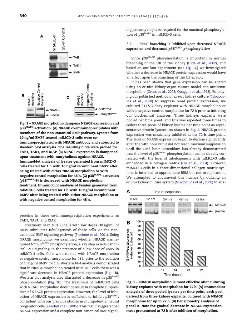
Fig. 1 – NRAGE morpholino dampens NRAGE expression and
p38MAPK activation. (A) NRAGE co-immunoprecipitates with
members of the non-canonical BMP pathway. Lysates from
10 ng/ml BMP7 treated mIMCD-3 cells were co-
immunoprecipitated with NRAGE antibody and subjected to
Western blot analysis. The resulting blots were probed for
TAB1, TAK1, and XIAP. (B) NRAGE expression is dampened
upon treatment with morpholinos against NRAGE.
Immunoblot analysis of lysates generated from mIMCD-3
cells treated for 1 h with 10 ng/ml recombinant BMP7 after
being treated with either NRAGE morpholino or with
negative control morpholino for 48 h. (C) p38MAPK activation
(p38MAPK-P) is decreased with NRAGE morpholino
treatment. Immunoblot analysis of lysates generated from
mIMCD-3 cells treated for 1 h with 10 ng/ml recombinant
BMP7 after being treated with either NRAGE morpholino or
with negative control morpholino for 48 h.
Fig. 2 – NRAGE morpholino is most effective after culturing
kidney explants with morpholino for 72 h. (A) Immunoblot
analysis of three pooled lysates per time point, each pool
derived from three kidney explants, cultured with NRAGE
morpholino for up to 72 h. (B) Densitometry analysis of
panel A. Note the gradual decrease in NRAGE expression,
most pronounced at 72 h after addition of morpholino.
340 M E C H A N I S M S O F D E V E L O P M E N T 1 2 6 ( 2 0 0 9 ) 3 3 7 – 3 4 9
proteins in these co-immunoprecipitation experiments as
TAK1, TAB1, and XIAP.
Treatment of mIMCD-3 cells with low doses (10 ng/ml) of
BMP7 stimulates tubulogenesis of these cells via the non-
canonical BMP signaling pathway (Piscione et al., 2001). Using
NRAGE morpholino, we examined whether NRAGE was re-
quired for p38MAPK phosphorylation, a key step in non-canon-
ical BMP signaling, in the presence of a low dose of BMP7 in
mIMCD-3 cells. Cells were treated with NRAGE morpholino
or negative control morpholino for 48 h prior to the addition
of 10 ng/ml BMP7 for 1 h. Western blot analysis demonstrated
that in NRAGE morpholino treated mIMCD-3 cells there was a
significant decrease in NRAGE protein expression (Fig. 1B).
Western blot analysis also illustrated a decrease in p38MAPK
phosphorylation (Fig. 1C). The treatment of mIMCD-3 cells
with NRAGE morpholino does not result in complete suppres-
sion of NRAGE protein expression. However, the level of inhi-
bition of NRAGE expression is sufficient to inhibit p38MAPK,
consistent with our previous studies in multipotential neural
progenitor cells (Kendall et al., 2005). This result suggests that
NRAGE expression and a complete non-canonical BMP signal-
ing pathway might be required for the maximal phosphoryla-
tion of p38MAPK in mIMCD-3 cells.
3.2. Renal branching is inhibited upon decreased NRAGEexpression and decreased p38MAPK phosphorylation
Since p38MAPK phosphorylation is important in normal
branching of the UB of the kidney (Hida et al., 2002), and
based on our last experiment (see Fig. 1C) we investigated
whether a decrease in NRAGE protein expression would have
an effect upon the branching of the UB ex vivo.
It has been shown that gene expression can be altered
using an ex vivo kidney organ culture model and antisense
morpholino (Gross et al., 2003; Quaggin et al., 1998). Employ-
ing our published method of ex vivo kidney culture (Nikopou-
los et al., 2008) to suppress renal protein expression, we
cultured E11.5 kidney explants with NRAGE morpholino or
with a negative control morpholino for 72 h prior to initiating
our biochemical analyses. Three kidneys explants were
pooled per time point, and this was repeated three times to
collect three pools of kidney lysates per time point as repre-
sentative protein lysates. As shown in Fig. 2, NRAGE protein
expression was maximally inhibited at the 72-h time point.
The level of NRAGE expression began to decline significantly
after the 24th hour but it did not reach maximal suppression
until the 72nd hour. Rosenblum has already demonstrated
that the level of p38MAPK phosphorylation can be directly cor-
related with the level of tubulogenesis with mIMCD-3 cells
embedded in a collagen matrix (Hu et al., 2004). However,
mIMCD-3 cells in a three-dimensional collagen matrix sys-
tem, is intended to approximate RBM but not to replicate it.
We attempted to circumvent this nuance by utilizing an
ex vivo kidney culture system (Nikopoulos et al., 2008) to ana-
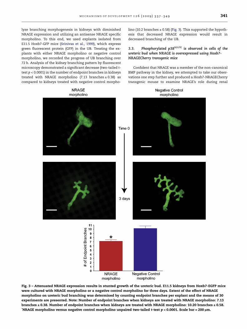
M E C H A N I S M S O F D E V E L O P M E N T 1 2 6 ( 2 0 0 9 ) 3 3 7 – 3 4 9 341
lyze branching morphogenesis in kidneys with diminished
NRAGE expression and utilizing an antisense NRAGE specific
morpholino. To this end, we used explants isolated from
E11.5 Hoxb7-GFP mice (Srinivas et al., 1999), which express
green fluorescent protein (GFP) in the UB. Treating the ex-
plants with either NRAGE morpholino or negative control
morpholino, we recorded the progress of UB branching over
72 h. Analysis of the kidney branching pattern by fluorescent
microscopy demonstrated a significant decrease (two-tailed t-
test p < 0.0001) in the number of endpoint branches in kidneys
treated with NRAGE morpholino (7.13 branches ± 0.38) as
compared to kidneys treated with negative control morpho-
Fig. 3 – Attenuated NRAGE expression results in stunted growth
were cultured with NRAGE morpholino or a negative control mo
morpholino on ureteric bud branching was determined by coun
experiments are presented. Note: Number of endpoint branches
branches ± 0.38. Number of endpoint branches when kidneys a*NRAGE morpholino versus negative control morpholino unpair
lino (10.2 branches ± 0.58) (Fig. 3). This supported the hypoth-
esis that decreased NRAGE expression would result in
decreased branching of the UB.
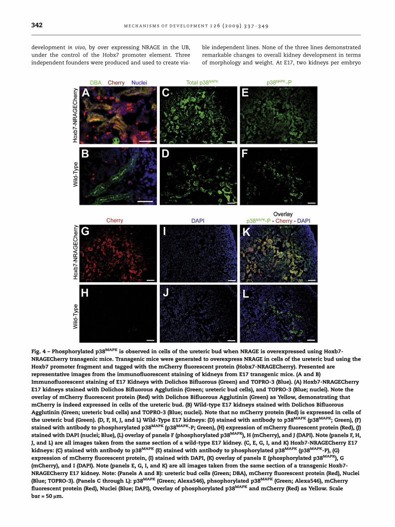
3.3. Phosphorylated p38MAPK is observed in cells of theureteric bud when NRAGE is overexpressed using Hoxb7-NRAGECherry transgenic mice
Confident that NRAGE was a member of the non-canonical
BMP pathway in the kidney, we attempted to take our obser-
vations one step further and produced a Hoxb7-NRAGECherry
transgenic mouse to examine NRAGE’s role during renal
of the ureteric bud. E11.5 kidneys from Hoxb7-EGFP mice
rpholino for three days. Extent of the effect of NRAGE
ting endpoint branches per explant and the means of 30
when kidneys are treated with NRAGE morpholino: 7.13
re treated with NRAGE morpholino: 10.20 branches ± 0.58.
ed two-tailed t-test p < 0.0001. Scale bar = 200 lm.
342 M E C H A N I S M S O F D E V E L O P M E N T 1 2 6 ( 2 0 0 9 ) 3 3 7 – 3 4 9
development in vivo, by over expressing NRAGE in the UB,
under the control of the Hobx7 promoter element. Three
independent founders were produced and used to create via-
Fig. 4 – Phosphorylated p38MAPK is observed in cells of the uret
NRAGECherry transgenic mice. Transgenic mice were generated
Hoxb7 promoter fragment and tagged with the mCherry fluores
representative images from the immunofluorescent staining of
Immunofluorescent staining of E17 Kidneys with Dolichos Biflu
E17 kidneys stained with Dolichos Bifluorous Agglutinin (Green
overlay of mCherry fluorescent protein (Red) with Dolichos Biflu
mCherry is indeed expressed in cells of the ureteric bud. (B) Wi
Agglutinin (Green; ureteric bud cells) and TOPRO-3 (Blue; nuclei
the ureteric bud (Green). (D, F, H, J, and L) Wild-Type E17 kidney
stained with antibody to phosphorylated p38MAPK (p38MAPK-P; G
stained with DAPI (nuclei; Blue), (L) overlay of panels F (phospho
J, and L) are all images taken from the same section of a wild-t
kidneys: (C) stained with antibody to p38MAPK (E) stained with a
expression of mCherry fluorescent protein, (I) stained with DAP
(mCherry), and I (DAPI). Note (panels E, G, I, and K) are all imag
NRAGECherry E17 kidney. Note: (Panels A and B): ureteric bud c
(Blue; TOPRO-3). (Panels C through L): p38MAPK (Green; Alexa546
fluorescent protein (Red), Nuclei (Blue; DAPI), Overlay of phosph
bar = 50 lm.
ble independent lines. None of the three lines demonstrated
remarkable changes to overall kidney development in terms
of morphology and weight. At E17, two kidneys per embryo
eric bud when NRAGE is overexpressed using Hoxb7-
to overexpress NRAGE in cells of the ureteric bud using the
cent protein (Hobx7-NRAGECherry). Presented are
kidneys from E17 transgenic mice. (A and B)
orous (Green) and TOPRO-3 (Blue). (A) Hoxb7-NRAGECherry
; ureteric bud cells), and TOPRO-3 (Blue; nuclei). Note the
orous Agglutinin (Green) as Yellow, demonstrating that
ld-type E17 kidneys stained with Dolichos Bifluorous
). Note that no mCherry protein (Red) is expressed in cells of
s: (D) stained with antibody to p38MAPK (p38MAPK; Green), (F)
reen), (H) expression of mCherry fluorescent protein (Red), (J)
rylated p38MAPK), H (mCherry), and J (DAPI). Note (panels F, H,
ype E17 kidney. (C, E, G, I, and K) Hoxb7-NRAGECherry E17
ntibody to phosphorylated p38MAPK (p38MAPK-P), (G)
I, (K) overlay of panels E (phosphorylated p38MAPK), G
es taken from the same section of a transgenic Hoxb7-
ells (Green; DBA), mCherry fluorescent protein (Red), Nuclei
), phsophorylated p38MAPK (Green; Alexa546), mCherry
orylated p38MAPK and mCherry (Red) as Yellow. Scale
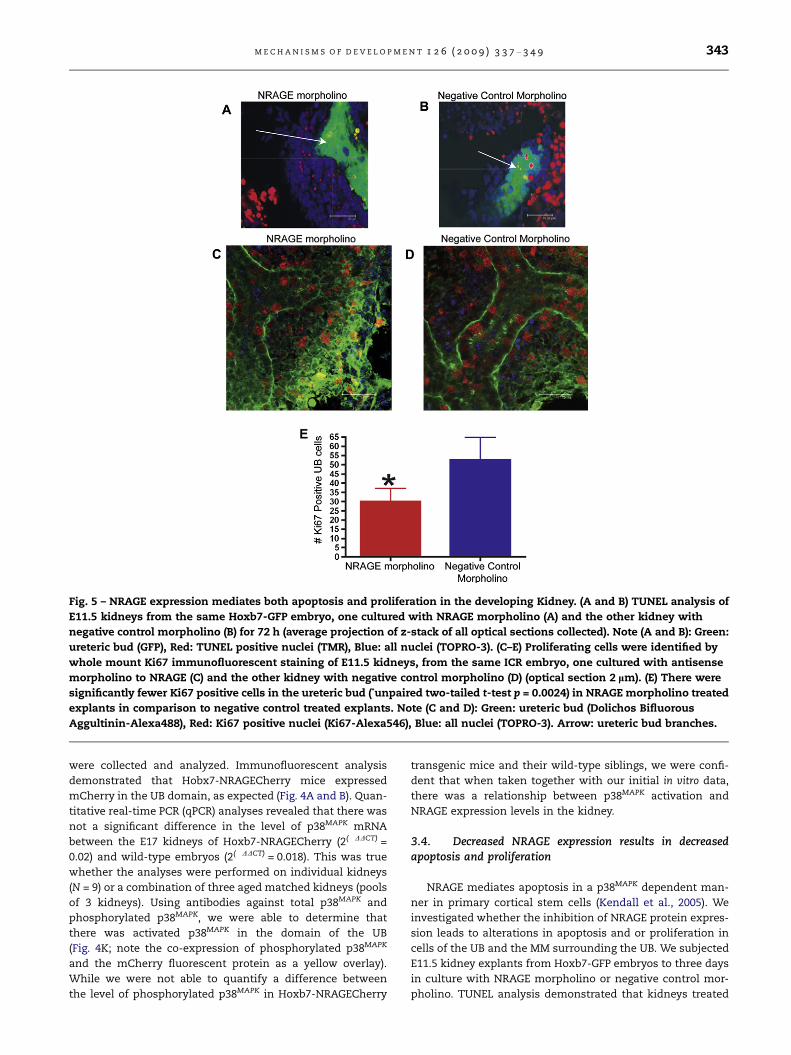
Fig. 5 – NRAGE expression mediates both apoptosis and proliferation in the developing Kidney. (A and B) TUNEL analysis of
E11.5 kidneys from the same Hoxb7-GFP embryo, one cultured with NRAGE morpholino (A) and the other kidney with
negative control morpholino (B) for 72 h (average projection of z-stack of all optical sections collected). Note (A and B): Green:
ureteric bud (GFP), Red: TUNEL positive nuclei (TMR), Blue: all nuclei (TOPRO-3). (C–E) Proliferating cells were identified by
whole mount Ki67 immunofluorescent staining of E11.5 kidneys, from the same ICR embryo, one cultured with antisense
morpholino to NRAGE (C) and the other kidney with negative control morpholino (D) (optical section 2 lm). (E) There were
significantly fewer Ki67 positive cells in the ureteric bud (*unpaired two-tailed t-test p = 0.0024) in NRAGE morpholino treated
explants in comparison to negative control treated explants. Note (C and D): Green: ureteric bud (Dolichos Bifluorous
Aggultinin-Alexa488), Red: Ki67 positive nuclei (Ki67-Alexa546), Blue: all nuclei (TOPRO-3). Arrow: ureteric bud branches.
M E C H A N I S M S O F D E V E L O P M E N T 1 2 6 ( 2 0 0 9 ) 3 3 7 – 3 4 9 343
were collected and analyzed. Immunofluorescent analysis
demonstrated that Hobx7-NRAGECherry mice expressed
mCherry in the UB domain, as expected (Fig. 4A and B). Quan-
titative real-time PCR (qPCR) analyses revealed that there was
not a significant difference in the level of p38MAPK mRNA
between the E17 kidneys of Hoxb7-NRAGECherry (2(�DDCT) =
0.02) and wild-type embryos (2(�DDCT) = 0.018). This was true
whether the analyses were performed on individual kidneys
(N = 9) or a combination of three aged matched kidneys (pools
of 3 kidneys). Using antibodies against total p38MAPK and
phosphorylated p38MAPK, we were able to determine that
there was activated p38MAPK in the domain of the UB
(Fig. 4K; note the co-expression of phosphorylated p38MAPK
and the mCherry fluorescent protein as a yellow overlay).
While we were not able to quantify a difference between
the level of phosphorylated p38MAPK in Hoxb7-NRAGECherry
transgenic mice and their wild-type siblings, we were confi-
dent that when taken together with our initial in vitro data,
there was a relationship between p38MAPK activation and
NRAGE expression levels in the kidney.
3.4. Decreased NRAGE expression results in decreasedapoptosis and proliferation
NRAGE mediates apoptosis in a p38MAPK dependent man-
ner in primary cortical stem cells (Kendall et al., 2005). We
investigated whether the inhibition of NRAGE protein expres-
sion leads to alterations in apoptosis and or proliferation in
cells of the UB and the MM surrounding the UB. We subjected
E11.5 kidney explants from Hoxb7-GFP embryos to three days
in culture with NRAGE morpholino or negative control mor-
pholino. TUNEL analysis demonstrated that kidneys treated

344 M E C H A N I S M S O F D E V E L O P M E N T 1 2 6 ( 2 0 0 9 ) 3 3 7 – 3 4 9
with NRAGE morpholino displayed a significant decrease
(unpaired two-tailed t-test, p = 0.0045) in the number of TUN-
EL positive MM cells, averaging 0.014 ± 0.006 TUNEL positive
cells per lm3 (N = 3), in comparison with kidney explants trea-
ted with negative control morpholino, which averaged 0.171 ±
0.027 TUNEL positive cells lm3 (N = 3). There was no signifi-
cant difference in TUNEL staining between the treatment
groups in cells of the UB (Fig. 5A and B).
To complement the TUNEL analysis, we utilized an anti-
body to Ki67 to quantify the number of proliferating UB and
MM cells in both NRAGE and negative control morpholino
treated kidneys. We counted the number of Ki67 positive cells
in the UB of kidneys explants treated with either NRAGE or
negative control morpholino. Fig. 5C–E illustrates a significant
decrease (unpaired two-tailed t-test, p = 0.0024) in the number
of Ki67 positive UB cells in NRAGE morpholino kidney ex-
plants (28 ± 3.6 Ki67 positive UB cells; N = 6) versus the nega-
tive control treated explants (55 ± 5.6 Ki67 positive UB cells;
N = 6). Moreover, to ensure that we were counting the cells
in equal areas, we determined the number of Ki67 positive
cells was, on average, 0.025 ± 0.001 cells per lm3 (N = 6) in
NRAGE morpholino treated kidneys and 0.050 ± 0.002 Ki67
positive cells per lm3 (N = 6) in negative control treated
kidneys. Comparing the number of Ki67 positive cells per
lm3 in each treatment group, we confirmed that there was
a significant decrease in Ki67 positive cells in NRAGE morpho-
Table 1 – List of genes analysed in qRT-PCR expression profile o
TCF7 – Transcription factor 7 T-cell specific Robo2 – Roundabout
Lef1 – Lymphoid enhancer binding factor 1 ATF – Activating tran
Ctnnb1 – Beta-Catenin BMP4 – Bone morpho
Myc – Myelocytomatosis oncogene BMP7 – Bone morpho
Wnt 4 – Wingless-related MMTV integration
site 4
SMAD1,2,3,5,7 – MAD
Wnt11 Wnt 11 – Wingless-related MMTV
integration site 11
Nfkb1 – Nuclear fact
chain gene enhancer
Pax2 – Paired box gene 2 Lhx1 – LIM homeobo
WT1 – Wilms tumor homolog BMPR1a – Bone morp
receptor, type 1A
GDNF – Glial cell line derived neurotrophic
factor
BMPR1b – Bone morp
receptor, type IB
Ret – Ret proto-oncogene BMPR2 – Bone morph
receptor, type II
Slit2 – Slit homolog 2 Nog – Noggin
Table 2 – Genes identified as having a significant change in exNRAGE morpholino. Genes identified as having significant chaculture with NRAGE morpholino, normalized to negative contrand shown is the difference of [(Fold Change Gene X) � 1].
Genes with altered expression Fold change in gene eto negative control ([F�decrease, + increase
GDNF �0.149
Ret + 0.662
BMP7 + 0.198
BMPRIb �0.159
lino treated kidneys (unpaired two-tailed t-test, p < 0.0001).
There was no significant difference in the number of Ki67
positive cells in the MM.
In summary, decreased NRAGE expression lead to de-
creased apoptosis in cells of the MM but not in cells of the
UB. In contrast, diminished NRAGE expression lead to de-
creased proliferation in cells of the UB but had no impact
upon proliferation in cells of the MM. These results suggest
that NRAGE may mediate apoptosis and proliferation in both
cells of the UB and MM during RBM. However, since there are
many pathways by which cells undergo proliferation or
apoptosis, we followed up on these results by establishing
gene expression profiles for NRAGE morpholino and negative
control morpholino, respectively.
3.5. Decreased NRAGE expression in the kidney results inaltered expression of renal genes during kidney development
A targeted gene expression profile of NRAGE depleted E11.5
ex vivo kidney explant cultures was undertaken to determine
the extent of NRAGE’s potential involvement in other signal-
ing pathways or developmental processes. Murine E11.5 ICR
kidney explants were treated with NRAGE morpholino or with
a negative control morpholino for three days. Three days of
culture was selected as our end point because, we had previ-
ously demonstrated that the maximum inhibition of NRAGE
f embryonic kidney explants and mIMCD-3 cells in culture.
homolog 2 Grem1 – Gremlin
scription factor 2 FGF7,8 – FGF – Fibroblast growth factor
genetic protein-4 Spry1 – Sprouty homolog 1 (Drosophila)
genetic protein 7 Ngfr – Nerve growth factor receptor
(TNFR superfamily)
homolog
or of kappa light
in B-cells 1
x protein 1
hogenetic protein
hogenetic protein
ogenic protein
pression after three days of kidney organ culture withnge in gene expression after three days of kidney organol morpholino treated kidneys. The normalized value is 1
xpression normalizedold Change GeneX] � 1)
p Value, number in group
p < 0.001, N = 9
p = 0.0022, N = 9
p = 0.0035, N = 9
p = 0.0454, N = 9
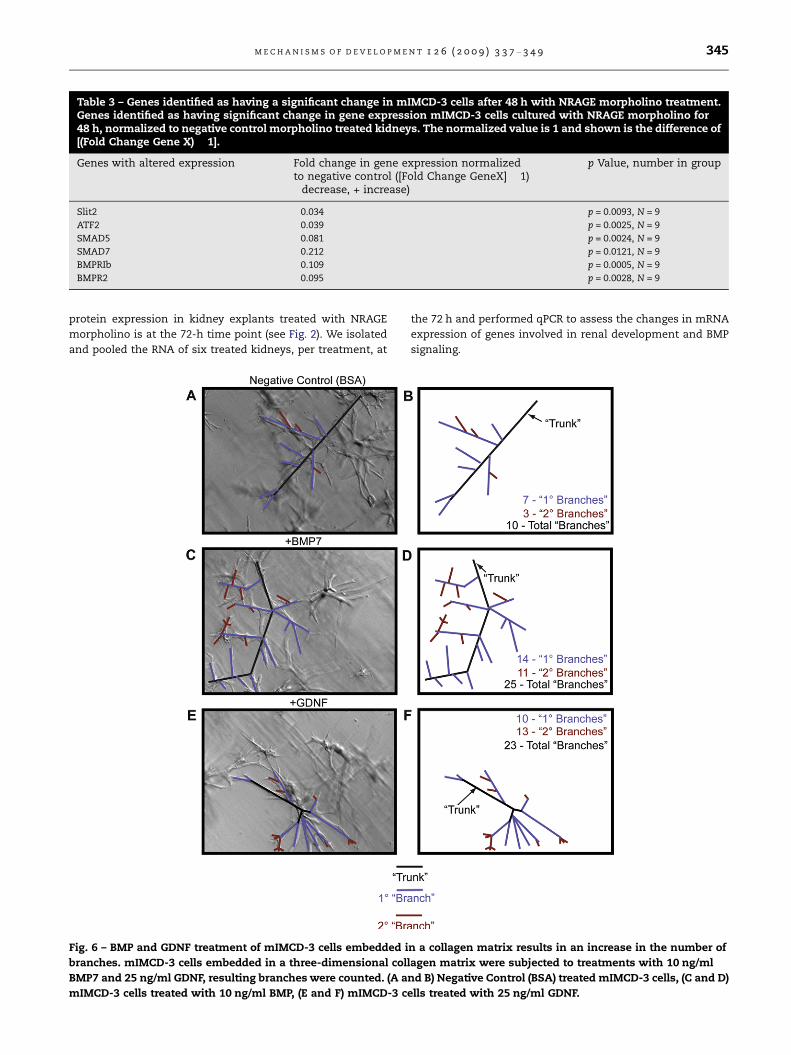
Table 3 – Genes identified as having a significant change in mIMCD-3 cells after 48 h with NRAGE morpholino treatment.Genes identified as having significant change in gene expression mIMCD-3 cells cultured with NRAGE morpholino for48 h, normalized to negative control morpholino treated kidneys. The normalized value is 1 and shown is the difference of[(Fold Change Gene X) � 1].
Genes with altered expression Fold change in gene expression normalizedto negative control ([Fold Change GeneX] � 1)�decrease, + increase)
p Value, number in group
Slit2 �0.034 p = 0.0093, N = 9
ATF2 �0.039 p = 0.0025, N = 9
SMAD5 �0.081 p = 0.0024, N = 9
SMAD7 �0.212 p = 0.0121, N = 9
BMPRIb �0.109 p = 0.0005, N = 9
BMPR2 �0.095 p = 0.0028, N = 9
M E C H A N I S M S O F D E V E L O P M E N T 1 2 6 ( 2 0 0 9 ) 3 3 7 – 3 4 9 345
protein expression in kidney explants treated with NRAGE
morpholino is at the 72-h time point (see Fig. 2). We isolated
and pooled the RNA of six treated kidneys, per treatment, at
Fig. 6 – BMP and GDNF treatment of mIMCD-3 cells embedded i
branches. mIMCD-3 cells embedded in a three-dimensional coll
BMP7 and 25 ng/ml GDNF, resulting branches were counted. (A a
mIMCD-3 cells treated with 10 ng/ml BMP, (E and F) mIMCD-3 c
the 72 h and performed qPCR to assess the changes in mRNA
expression of genes involved in renal development and BMP
signaling.
n a collagen matrix results in an increase in the number of
agen matrix were subjected to treatments with 10 ng/ml
nd B) Negative Control (BSA) treated mIMCD-3 cells, (C and D)
ells treated with 25 ng/ml GDNF.
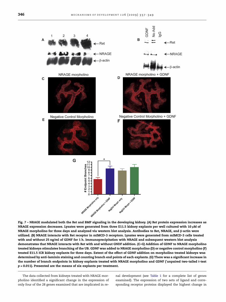
Fig. 7 – NRAGE modulated both the Ret and BMP signaling in the developing kidney. (A) Ret protein expression increases as
NRAGE expression decreases. Lysates were generated from three E11.5 kidney explants per well cultured with 10 lM of
NRAGE morpholino for three days and analyzed via western blot analysis. Antibodies to Ret, NRAGE, and b-actin were
utilized. (B) NRAGE interacts with Ret receptor in mIMCD-3 receptors. Lysates were generated from mIMCD-3 cells treated
with and without 25 ng/ml of GDNF for 1 h. Immunoprecipitation with NRAGE and subsequent western blot analysis
demonstrates that NRAGE interacts with Ret with and without GNDF addition. (C–G) Addition of GDNF to NRAGE morpholino
treated kidneys stimulates branching of the UB. GDNF was added to NRAGE morpholino (D) or negative control morpholino (F)
treated E11.5 ICR kidney explants for three days. Extent of the effect of GDNF addition on morpholino treated kidneys was
determined by anti-laminin staining and counting branch end points of each explants. (G) There was a significant increase in
the number of branch endpoints in kidney explants treated with NRAGE morpholino and GDNF (*unpaired two-tailed t-test
p = 0.011). Presented are the means of six explants per treatment.
346 M E C H A N I S M S O F D E V E L O P M E N T 1 2 6 ( 2 0 0 9 ) 3 3 7 – 3 4 9
The data collected from kidneys treated with NRAGE mor-
pholino identified a significant change in the expression of
only four of the 28 genes examined that are implicated in re-
nal development (see Table 1 for a complete list of genes
examined). The expression of two sets of ligand and corre-
sponding receptor proteins displayed the highest change in
M E C H A N I S M S O F D E V E L O P M E N T 1 2 6 ( 2 0 0 9 ) 3 3 7 – 3 4 9 347
expression: GDNF, Ret, and BMP7 and BMPRIb (Table 2). There
was no significant change in gene expression in kidneys com-
pared to those treated with negative control morpholino or
treated with Endo-Porter only (data not shown). Western blot
analysis confirmed the change in protein levels (Fig. 7A), this
was important because of GDNF–Ret signaling is critically
important to overall kidney development. To determine if
the change in gene expression in kidney organ cultures was
specific to events surrounding the development of the UB,
we repeated the analysis using NRAGE depleted mIMCD-3
cells cultured with NRAGE morpholino for 72 h. The qPCR
data from mIMCD-3 cells cultured with NRAGE morpholino
identified six genes whose expressions were significantly
changed: Slit2, ATF2, SMAD5, SMAD7, BMPRIb, and BMPR2
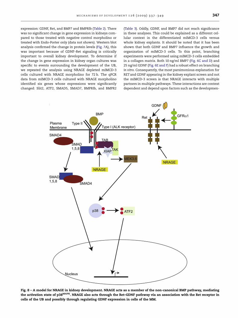
Fig. 8 – A model for NRAGE in kidney development. NRAGE acts
the activation state of p38MAPK. NRAGE also acts through the Re
cells of the UB and possibly through regulating GDNF expressio
(Table 3). Oddly, GDNF, and BMP7 did not reach significance
in these analyses. This could be explained as a different cel-
lular context in the differentiated mIMCD-3 cells versus
whole kidney explants. It should be noted that it has been
shown that both GDNF and BMP7 influence the growth and
organization of mIMCD-3 cells. To this point, branching
experiments were performed using mIMCD-3 cells embedded
in a collagen matrix. Both 10 ng/ml BMP7 (Fig. 6C and D) and
25 ng/ml GDNF (Fig. 6E and F) had a robust effect on branching
in vitro. Consequently, the most parsimonious explanation for
RET and GDNF appearing in the kidney explant screen and not
the mIMCD-3 screen is that NRAGE interacts with multiple
partners in multiple pathways. These interactions are context
dependent and depend upon factors such as the developmen-
as a member of the non-canonical BMP pathway, mediating
t–GDNF pathway via an association with the Ret receptor in
n in cells of the MM.
348 M E C H A N I S M S O F D E V E L O P M E N T 1 2 6 ( 2 0 0 9 ) 3 3 7 – 3 4 9
tal time point analyzed, for example early development, as
seen in the E11 kidney explants, versus mature differentiated
cells, as seen in the mIMCD-3 cells. It also depends on the
specific cell type considered, for example UB cells versus
MM cells.
3.6. Addition of GDNF to NRAGE morpholino treatedkidneys stimulates branching of the ureteric bud
GDNF is required for normal development of the kidney
(Sanchez et al., 1996). Therefore, a decrease in GDNF tran-
script levels, as seen in our qPCR gene expression, should re-
sult in significantly inhibited branching of the UB. We
attempted to stimulate UB branching in kidney explants trea-
ted with NRAGE morpholino by the addition of recombinant
GDNF. E11.5 ICR kidney explants were cultured with NRAGE
morpholino for 72 h, with 25 ng/ml of recombinant GDNF
added for the last 48 h. Branching of the UB was stimulated
in kidney explants with decreased NRAGE expression when
25 ng/ml GDNF was added to the culture (Fig. 7C–G). These re-
sults suggest that NRAGE may not only be involved in solely
mediating the map kinase cascade downstream of the non-
canonical BMP pathway, but could also be involved in the
GDNF–Ret signaling pathway. It is unknown if NRAGE impacts
solely upon the ligand, GDNF, or the receptor Ret, which are
expressed in the cells of the MM and UB, respectively. To ad-
dress this point, we used lysates from mIMCD-3 cells treated
with 25 ng/ml of GDNF for 1 h to co-immunoprecipitate the
Ret receptor with NRAGE. As demonstrated in Fig. 7B, NRAGE
and RET were co-immunoprecipitated. It is not hard to envi-
sion a system where NRAGE is employed as a rheostat to bol-
ster Ret signaling to enhance renal growth and depending on
the cellular environment to bolster non-canonical BMP sig-
naling to enhance branching.
4. Discussion
Our focus has been on NRAGE and its role in RBM because
the risk of developing hypertension in adult life increases
with abnormal RBM. There have been several genes that have
been shown to have abnormal RBM phenotypes in knock-out
mice for their respective genes, including WT1 (Kreidberg
et al., 1993), Pax2 (Torres et al., 1995), Eya-1 (Ding et al.,
1999), Six-1 (Xu et al., 1999), Lim-1 (Shawlot and Behringer,
1995), GDNF (Shakya et al., 2005), c-Ret (Schuchardt et al.,
1996), Gremlin (Michos et al., 2007) and we anxiously await
and believe NRAGE will join this list. Reports have shown that
BMP signaling during RBM is dependent upon the non-canon-
ical BMP signaling pathway. Rosenblum’s group has demon-
strated that p38MAPK phosphorylation is a key event in
tubulogenesis of mIMCD-3 cells (Hu et al., 2004), an observa-
tion we have replicated and confirmed using kidney explants.
Other groups have demonstrated the canonical BMP pathway
is not necessary for the normal branching of the UB (Chu
et al., 2004) using mice that were lacking SMAD4 expression
in their UB. These observations, in conjunction with our find-
ings that NRAGE mediates p38MAPK phosphorylation in neural
progenitors (Kendall et al., 2005), suggested a role for NRAGE
in RBM as part of the non-canonical BMP pathway.
The data presented herein suggests that NRAGE has a role
in RBM, but it is more complex than we originally envisioned,
with different roles in the cells of the UB versus cells of the
MM. In mIMCD-3 cells, NRAGE associates with members of
the non-canonical BMP pathway and the c-Ret receptor. In
our kidney explant cultures, NRAGE expression impacts di-
rectly upon the expression of GDNF and Ret. Since the BMP
and GDNF–Ret signaling pathways are two key signaling path-
ways in RBM, it is not hard to envision a model in which
NRAGE act as a rheostat to influence both pathways. In one
situation, NRAGE would be a part of the signaling pathway
that provides maximum phosphorylation of p38MAPK to help
facilitate UB branching. In another context, NRAGE would to
contribute to the GDNF–Ret signaling pathway to assist in
growth and maturation of the kidney. In both situations,
NRAGE’ effects would impact upon UB cells and MM cells
(see Fig. 8 for proposed model). Further analysis using trans-
genic and knock-out mice with cell specific control of expres-
sion of NRAGE in vivo will provide further clarification as to
the role of NRAGE in RBM and its impact upon hypertension
in adult life.
R E F E R E N C E S
Barker, D.M., Sutherland, L.E., Jaffe, D., Dahl, L.D., 1970. Effects ofchronic excess salt ingestion. Juxtaglomerular granulation inkidneys of rats with differing genetic susceptibilities tohypertension. Arch. Pathol. 89, 247–258.
Bertrand, M., Huijbers, I., Chomez, P., De Backer, O., 2004.Comparative expression analysis of the MAGED genes duringembryogenesis and brain development. Dev. Dyn. 230, 325–334.
Brenner, B.M., Garcia, D.L., Anderson, S., 1988. Glomeruli andblood pressure. Less of one, more the other? Am. J. Hypertens.1, 335–347.
Chu, G.C., Dunn, N.R., Anderson, D.C., Oxburgh, L., Robertson, E.J.,2004. Differential requirements for Smad4 in TGFbeta-dependent patterning of the early mouse embryo.Development 131, 3501–3512.
Ding, B., Huang, S.L., Zhang, S.Q., Li, Y.X., 1999. Inhibitory effect ofMAP kinase antisense oligonucleotide on angiotensin II-induced c-myc gene expression and proliferation of rat cardiacfibroblast. Zhongguo Yao Li Xue Bao 20, 934–940.
Gross, I., Morrison, D.J., Hyink, D.P., Georgas, K., English, M.A.,Mericskay, M., Hosono, S., Sassoon, D., Wilson, P.D., Little, M.,Licht, J.D., 2003. The receptor tyrosine kinase regulatorSprouty1 is a target of the tumor suppressor WT1 andimportant for kidney development. J. Biol. Chem. 278, 41420–41430.
Hida, M., Omori, S., Awazu, M., 2002. ERK and p38 MAP kinase arerequired for rat renal development. Kidney Int. 61, 1252–1262.
Hogan, B.L., 1996. Bone morphogenetic proteins in development.Curr. Opin. Genet. Dev. 6, 432–438.
Hu, M.C., Wasserman, D., Hartwig, S., Rosenblum, N.D., 2004.P38MAPK acts in the BMP7-dependent stimulatory pathwayduring epithelial cell morphogenesis and is regulated bySmad1. J. Biol. Chem. 279, 12051–12059.
Kearney, P.M., Whelton, M., Reynolds, K., Muntner, P., Whelton,P.K., He, J., 2005. Global burden of hypertension: analysis ofworldwide data. Lancet 365, 217–223.
Kendall, S.E., Battelli, C., Irwin, S., Mitchell, J.G., Glackin, C.A.,Verdi, J.M., 2005. NRAGE mediates p38 activation and neural
M E C H A N I S M S O F D E V E L O P M E N T 1 2 6 ( 2 0 0 9 ) 3 3 7 – 3 4 9 349
progenitor apoptosis via the bone morphogenetic proteinsignaling cascade. Mol. Cell Biol. 25, 7711–7724.
Kreidberg, J.A., Sariola, H., Loring, J.M., Maeda, M., Pelletier, J.,Housman, D., Jaenisch, R., 1993. WT-1 is required for earlykidney development. Cell 74, 679–691.
Langley, S.C., Jackson, A.A., 1994. Increased systolic bloodpressure in adult rats induced by fetal exposure to maternallow protein diets. Clin. Sci. (Lond.) 86, 217–222 (discussion 121).
Levitt, N.S., Lindsay, R.S., Holmes, M.C., Seckl, J.R., 1996.Dexamethasone in the last week of pregnancy attenuateshippocampal glucocorticoid receptor gene expression andelevates blood pressure in the adult offspring in the rat.Neuroendocrinology 64, 412–418.
Livak, K.J., Schmittgen, T.D., 2001. Analysis of relative geneexpression data using real-time quantitative PCR and the2(�Delta Delta C(T)) method. Methods 25, 402–408.
Michos, O., Goncalves, A., Lopez-Rios, J., Tiecke, E., Naillat, F.,Beier, K., Galli, A., Vainio, S., Zeller, R., 2007. Reduction of BMP4activity by gremlin 1 enables ureteric bud outgrowth andGDNF/WNT11 feedback signalling during kidney branchingmorphogenesis. Development 134, 2397–2405.
Nikopoulos, G.N., Adams, T.L., Adams, D., Oxburgh, L., Prudovsky,I., Verdi, J.M., 2008. The use of Endo-Porter to delivermorpholinos in kidney organ culture. Biotechniques 44, 547–549.
Nohe, A., Keating, E., Knaus, P., Petersen, N.O., 2004. Signaltransduction of bone morphogenetic protein receptors. CellSignal 16, 291–299.
Oxburgh, L., Chu, G.C., Michael, S.K., Robertson, E.J., 2004. TGFbetasuperfamily signals are required for morphogenesis of thekidney mesenchyme progenitor population. Development 131,4593–4605.
Piscione, T.D., Phan, T., Rosenblum, N.D., 2001. BMP7 controlscollecting tubule cell proliferation and apoptosis via Smad1-dependent and -independent pathways. Am. J. Physiol. Renal.Physiol. 280, F19–F33.
Piscione, T.D., Yager, T.D., Gupta, I.R., Grinfeld, B., Pei, Y., Attisano,L., Wrana, J.L., Rosenblum, N.D., 1997. BMP-2 and OP-1 exertdirect and opposite effects on renal branching morphogenesis.Am. J. Physiol. 273, F961–F975.
Quaggin, S.E., Vanden Heuvel, G.B., Igarashi, P., 1998. Pod-1, amesoderm-specific basic-helix-loop-helix protein expressedin mesenchymal and glomerular epithelial cells in thedeveloping kidney. Mech. Dev. 71, 37–48.
Renn, J., Winkler, C., 2009. Osterix-mCherry transgenic medakafor in vivo imaging of bone formation. Dev. Dyn. 238, 241–248.
Salehi, A.H., Roux, P.P., Kubu, C.J., Zeindler, C., Bhakar, A., Tannis,L.L., Verdi, J.M., Barker, P.A., 2000. NRAGE, a novel MAGEprotein, interacts with the p75 neurotrophin receptor andfacilitates nerve growth factor-dependent apoptosis. Neuron27, 279–288.
Sanchez, M.P., Silos-Santiago, I., Frisen, J., He, B., Lira, S.A.,Barbacid, M., 1996. Renal agenesis and the absence of entericneurons in mice lacking GDNF. Nature 382, 70–73.
Schuchardt, A., D’Agati, V., Pachnis, V., Costantini, F., 1996. Renalagenesis and hypodysplasia in ret-k-mutant mice result fromdefects in ureteric bud development. Development 122, 1919–1929.
Shakya, R., Watanabe, T., Costantini, F., 2005. The role of GDNF/Ret signaling in ureteric bud cell fate and branchingmorphogenesis. Dev. Cell 8, 65–74.
Shaner, N.C., Campbell, R.E., Steinbach, P.A., Giepmans, B.N.,Palmer, A.E., Tsien, R.Y., 2004. Improved monomeric red,orange and yellow fluorescent proteins derived fromDiscosoma sp. red fluorescent protein. Nat. Biotechnol. 22,1567–1572.
Shawlot, W., Behringer, R.R., 1995. Requirement for Lim1 in head-organizer function. Nature 374, 425–430.
Shcherbo, D., Murphy, C.S., Ermakova, G.V., Solovieva, E.A.,Chepurnykh, T.V., Shcheglov, A.S., Verkhusha, V.V., Pletnev,V.Z., Hazelwood, K.L., Roche, P.M., Lukyanov, S., Zaraisky, A.G.,Davidson, M.W., Chudakov, D.M., 2009. Far-red fluorescent tagsfor protein imaging in living tissues. Biochem. J. 418, 567–574.
Srinivas, S., Goldberg, M.R., Watanabe, T., D’Agati, V., al-Awqati,Q., Costantini, F., 1999. Expression of green fluorescent proteinin the ureteric bud of transgenic mice: a new tool for theanalysis of ureteric bud morphogenesis. Dev. Genet. 24, 241–251.
Torres, M., Gomez-Pardo, E., Dressler, G.R., Gruss, P., 1995. Pax-2controls multiple steps of urogenital development.Development 121, 4057–4065.
Winnier, G., Blessing, M., Labosky, P.A., Hogan, B.L., 1995. Bonemorphogenetic protein-4 is required for mesoderm formationand patterning in the mouse. Genes Dev. 9, 2105–2116.
Woodall, S.M., Johnston, B.M., Breier, B.H., Gluckman, P.D., 1996.Chronic maternal undernutrition in the rat leads to delayedpostnatal growth and elevated blood pressure of offspring.Pediatr. Res. 40, 438–443.
Xu, P.X., Adams, J., Peters, H., Brown, M.C., Heaney, S., Maas, R.,1999. Eya1-deficient mice lack ears and kidneys and showabnormal apoptosis of organ primordia. Nat. Genet. 23, 113–117.