1
Center for Preventive and Sports Medicine Klinikum rechts der Isar München
Influence of exercise training on cardiac remodeling after acute myocardial infarction – a
PET/MRI study
Carolin Dominique Kelso
Vollständiger Abdruck der von der Fakultät für Medizin der Technischen Universität München
zur Erlangung des akademischen Grades eines Doktors der Medizin genehmigten Dissertation.
Vorsitzender: Prof. Dr. Ernst. J. Rumenny
Prüfer der Dissertation:
1. Prof. Dr. Martin Halle
2. apl. Prof. Dr. Tareq Ibrahim
Die Dissertation wurde am 01.07.2019 bei der Technischen Universität München
eingereicht und durch die Fakultät für Medizin am 12.02.2020 angenommen.
2
Table of contents
List of tables .................................................................................................................................. 3
List of figures ................................................................................................................................. 3
Abbreviations................................................................................................................................ 4
1 Introduction .......................................................................................................................... 5
1.1 Myocardial infarction ................................................................................................... 5
1.2. Cardiac remodeling ....................................................................................................... 7
1.3. Cardiac rehabilitation ................................................................................................... 9
1.3.1 Exercise training in cardiac rehabilitation ..................................................... 11
1.4. Cardiac imaging........................................................................................................... 13
1.4.1 Principle of magnetic resonance imaging ...................................................... 14
2 Objective ............................................................................................................................. 17
3. Methods .............................................................................................................................. 18
3.1. Study population ........................................................................................................ 18
3.2. Questionnaire ............................................................................................................. 18
3.3. Imaging ........................................................................................................................ 19
3.3.1. Positron-emission-tomography/magnetic resonance imaging ....................... 20
3.3.2. Imaging analysis .......................................................................................... 20
3.4. Statistical analyses ...................................................................................................... 20
4. Results ................................................................................................................................. 23
4.1. Patient characteristics, cardiovascular profile, imaging data ................................... 23
4.2. Cardiac rehabilitation and exercise training post-myocardial infarction ................. 25
4.3. The effect of exercise training on cardiac remodeling .............................................. 26
4.4. The influence of exercise training prior to myocardial infarction on initial infarction
size and volume ...................................................................................................................... 30
5. Discussion ........................................................................................................................... 31
5.1. Participation in rehabilitation programs and exercise behavior post-myocardial
infarction ................................................................................................................................. 32
5.2. The impact of exercise training after myocardial infarction on cardiac remodeling 35
5.3. Effect of self-reported exercise training prior to myocardial infarction on infarction
size and volume ...................................................................................................................... 39
6. Study limitations ................................................................................................................. 42
7. Conclusion .......................................................................................................................... 42
8. References .......................................................................................................................... 44
9. Appendix ............................................................................................................................. 53
9.1. Questionnaire ............................................................................................................. 53
10. Acknowledgments .......................................................................................................... 56
3
List of tables Table 1. Definition of acute myocardial infarction adapted from ................................................. 5
Table 2. Patient demographics, cardiovascular profile, imaging data in overall patient
population post MI ...................................................................................................................... 23
Table 3. Patient demographics, cardiovascular profile, imaging data in exercise subgroups post
MI ................................................................................................................................................ 24
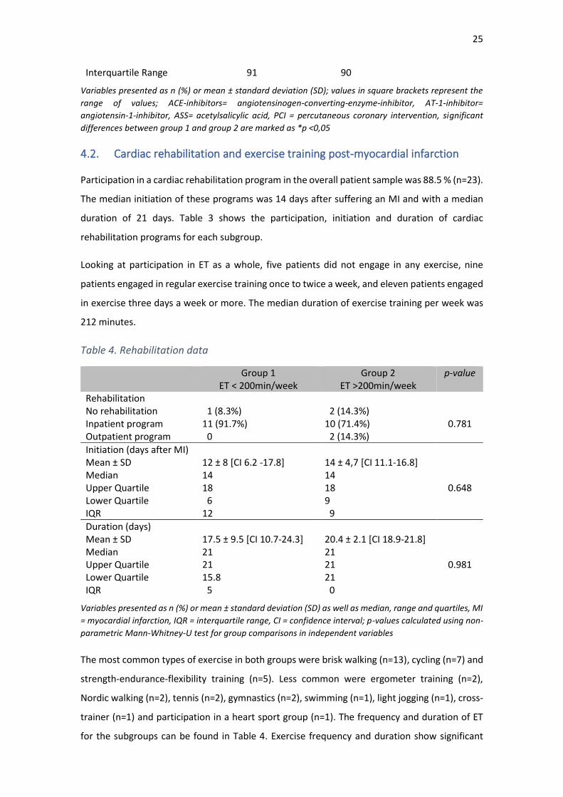
Table 4. Rehabilitation data ........................................................................................................ 25
Table 5. Exercise frequency (x/week) and duration (min/week) between the subgroups .......... 26
Table 6. Cardiac remodeling parameters at T1, T2 and difference from T1 to T2 (T2-T1) .......... 27
Table 7. Infarction parameters from T1 to T2 and change between T1 to T2 ............................ 28
Table 8. Initial infarction size between group No-ET and group ET ............................................ 31
Table 9. Initial infarction volume group No-ET and group ET ..................................................... 31
List of figures Figure 1. Phases of rehabilitation ................................................................................................ 10 Figure 2. Difference in ∆LVEDV (∆LVEDV = T2 LVEDV-T1 LVEDV) between group 1 and group 230 Figure 3. Initial infarction size between group No-ET and group ET. .......................................... 31 Figure 4. Initial infarction volume between group No-ET and group ET. .................................... 31
4
Abbreviations
AMI Acute myocardial infarction
BSA Body surface area
CAD Coronary artery disease
CCTA Coronary Computed Tomography Angiography
CHD Coronary heart disease
CR Cardiac rehabilitation
CVD Cardiovascular disease
DGPR Deutsche Gesellschaft für Prävention und Rehabilitation (German Society for
Prevention and Rehabilitation)
ECG Electrocardiography
EDV End-diastolic volume
EF Ejection fraction
ESV End-systolic volume
ET Exercise training
18F-FDG 18 F-fluorodeoxyglucose
Gd Gadolinium
HF Heart Failure
IRENA Intensivierte Reha-Nachsorge (intensive follow-up care after rehabilitation)
KARENA Kardiovaskuläres Reha-Nachsorgeprogramm (cardiovascular rehabilitation
follow-up program)
LGE Late gadolinium enhancement
LV Left ventricle
LVEDV Left-ventricular end-diastolic volume
LVESV Left-ventricular end-systolic volume
MI Myocardial infarction
MRI Magnetic Resonance Imaging
PET Positron-Emission-Tomography
PCI Percutaneous coronary intervention
RAAS Renin-angiotensinogen-aldosterone system
SPECT Single-Photon-Emissions-Computer-Tomography
STEMI ST-segment elevation Myocardial Infarction
SV Stroke volume
VO2peak Maximum oxygen uptake
5
1 Introduction
1.1 Myocardial infarction
Myocardial infarction (MI) is an acute manifestation of coronary artery disease (CAD) and
coronary heart disease (CHD), which both belong to a group of illnesses that affect the heart and
blood vessels, and are known as cardiovascular disease (CVD). (Steg et al., 2012; WHO, 2017).
CVD, including acute myocardial infarction (AMI), is not only the leading cause of death in
Germany, but also worldwide (WHO, 2017). According to the German Federal Statistical Office,
in 2014 38,9% of deaths in Germany were caused by CVD. Within CVD, CHD caused 14% of
overall deaths and AMI 5,5% of overall deaths. The majority of deaths in patients suffering from
CVD occurred in patients over the age of 65 (Statistisches Bundesamt, 2014).
The trigger for MI is ischemia, which most often is caused by a ruptured plaque leading to a
thrombotic occlusion of the coronary artery (Davies, 2000; Thygesen et al, 2007). At the cellular
level, ischemia leads to edema, inflammation and cell death in the form of coagulation necrosis.
This process begins approximately 30 minutes after the onset of ischemia. After a few hours,
myocytes that would normally be supplied with oxygen by the occluded artery, become necrotic.
The size of the infarction is influenced by several factors, such as the extent of collateral blood
vessels supplying the occluded area, ongoing or periodic vessel occlusion, myocardial
preconditioning to ischemia and individual oxygen demand (Thygesen et al., 2007).
The primary symptom of coronary ischemia is ongoing chest pain that lasts over twenty minutes
and is non-responsive to nitroglycerin (which acts as a vasodilating substance). Other non-
specific symptoms include dyspnea, nausea, syncopia, fatigue, palpitations. However, an AMI
can also present itself with atypical symptoms, especially in diabetics, women and the elderly
(Thygesen et al., 2007; Steg et al., 2012).
Successful therapy requires a quick diagnosis of an AMI. The criteria for diagnosis of MI were
defined in the “ESC guidelines for management of acute myocardial infarction in patients
presenting with ST-Segment elevation” (Steg et al., 2012), which can be found in Table 1.
Table 1. Definition of acute myocardial infarction adapted from (Steg et al., 2012)
Elevation and/or fall of cardiac biomarker values (by preference troponin) with a minimum of one biomarker value >99th percentile of the upper reference limit And at least one of the following criteria: - Symptoms of ischemia - New significant ST-T changes or new left-bundle-branch-block in the ECG - Development of pathological Q-waves in the ECG - Cardiac imaging presenting a new loss of viable myocardium or regional wall motion abnormality - Identification of an intracoronary thrombus by angiography or autopsy
6
Cardiac death with previous ischemic symptoms, presumably new ECG changes or new LBBB, death occurring before blood cardiac biomarkers values are released or before cardiac biomarker values would be increased
Stent thrombosis associated with MI when detected by coronary angiography or autopsy in the setting of myocardial ischemia and with a rise and/or fall of cardiac biomarker values with at least one value above the 99th percentile URL.
ECG = electrocardiography, LBBB = left bundle branch block, URL =upper reference limit
In the initial diagnosis of an AMI, an ECG plays an important role, as changes in the ST-segment,
left bundle branch blocks and changes in the initial Q-wave are typical indicators. During the first
hours of a MI, however, an ECG may not show these typical pathologies. Blood tests can help to
detect an increase in cardiac biomarker values during the early phase of AMI. Troponin (T and I)
are the most important biomarkers as they are highly sensitive and specific in detecting
myocardial necrosis. When the probability for MI is high, such as in patients with an ST-segment
elevation or a new left bundle branch block, reperfusion treatment should be performed
without waiting for the results of the blood tests (Steg et al., 2012). If the diagnosis is still unclear,
emergency imaging should be carried out. Coronary angiography is the imaging method of
choice as it directly allows subsequent primary percutaneous coronary intervention (PCI). If a
hospital does not have the capability to perform a coronary angiography, then a two-
dimensional echocardiography should be performed to detect wall motion abnormalities. If wall
motion abnormalities are found, the patient should be transferred to a hospital which can
perform PCI immediately (Ibanez et al., 2017).
Symptoms presented during diagnostics should be treated as well, to not only provide relief to
patients, but to also lower myocardial oxygen demand, as the myocardial workload and
vasoconstriction are increased by pain. The recommended medication for symptom relief during
the acute phase of MI include: titrated i.v.-opioids for pain-relief, tranquilizers in agitated
patients, and oxygen if patients suffer from hypoxia, breathlessness or heart failure (Ibanez et
al., 2017; Steg et al., 2012). Patients should also receive dual antithrombotic and antiplatelet
medication as well as an intravenous anticoagulant as soon as possible (Ibanez et al., 2017; Steg
et al., 2012).
The primary goal for patients suffering from AMI is the revascularization of the occluded artery.
Each patient should preferably receive a mechanical revascularization by PCI. When PCI is
contraindicated by other factors, pharmacological revascularization is an option. The standard
procedure is to initiate treatment within the first twelve hours after symptom onset and
diagnosis. However, even for a patient suffering from symptoms for more than twelve hours, if
there is clinical or electrocardiographic evidence of ischemia, revascularization therapy will still
be beneficial (Steg et al., 2012). The time between diagnosis of ST-segment elevation myocardial
7
infarction (STEMI) and the beginning of PCI should be no longer than 60 minutes. If a PCI center
cannot be reached within 120 minutes, fibrinolytic therapy should be considered as
revascularization therapy. During PCI the culprit lesion is dilated using balloon angioplasty. After
the angioplasty, the guidelines suggest implanting a stent, preferably drug-eluting rather than
bare-metal (Ibanez et al., 2017; Steg et al., 2012).
Upon successful revascularization therapy, the patient should follow a medication plan that
includes dual antiplatelet and antithrombotic therapy using aspirin and an ADP-receptor
inhibitor for 12 months. Ibanez et al. (2017) recommend initiating an early i.v. beta-blocker
treatment in hemodynamically stable patients, previously treated with PCI, followed by a long-
term oral therapy with beta-blockers. In case of contraindications for beta-blockers (e.g.
obstructive airway disease or second- and third-degree atrioventricular block), the calcium-
antagonist verapamil may be used in patients without heart failure (HF) or impaired left
ventricular (LV) function. Statins and angiotensin-converting-enzyme-inhibitors (ACE-
inhibitors)/angiotensin-receptor-blockers are also recommended. In a patient with an EF <40%
and heart failure or diabetes, aldosterone-antagonists should be used (Ibanez et al., 2017;
O'Gara et al., 2013; Steg et al., 2012).
After successful treatment of AMI an exercise-based cardiac rehabilitation (CR) program should
be initiated. Long-term non-pharmaceutical strategies for post-MI patients focus on lifestyle
changes, such as smoking cessation, diet and weight control, an increase in physical activity (in
the form of structured exercise training (ET)), blood pressure management and psychosocial
interventions such as stress reduction (Ibanez et al., 2017; O'Gara et al., 2013; Steg et al., 2012).
Follow-up care is essential to encourage a patient to adhere to lifestyle modifications and
prevent falling back into behavioral patterns that preceded the MI. Studies have shown that
long-term adherence to life-style modification, especially ET, is necessary to maintain the
positive effects on the impaired cardiovascular system following MI (Vona et al., 2004; Moholdt
et al., 2011).
1.2. Cardiac remodeling
The International Forum on Cardiac Remodeling defines remodeling as follows:
“Cardiac remodeling may be defined as genome expression, molecular, cellular
and interstitial changes that are manifested clinically as changes in size, shape
and function of the heart after cardiac injury. The process of cardiac
remodeling is influenced by hemodynamic load, neurohormonal activation and
other factors still under investigation […]” (Cohn et al., 2000, p. 570).
8
A common cause of severe cardiac injury is a MI, which triggers cardiac remodeling within a few
hours after onset (Cohn et al., 2000; Erlebacher et al., 1984; Pfeffer, 1990; Sutton, 2000). The
first hours of an MI are defined by edema, inflammation and coagulation necrosis leading to
impaired contractile function and dyskinesis in the infarcted area (Pfeffer, 1990). Due to the loss
of myocytes, the infarcted area is vulnerable to mechanical forces, such as an increased cardiac
load, resulting in a thinning of the ventricular wall in the infarcted area. This is also known as
infarction expansion. Infarction expansion not only leads to dilation of the infarcted area, but
also to dilation in the non-infarcted myocardium. The process of ventricular dilation begins early
after infarction and continues after infarction healing is completed (Pfeffer, 1990; Sutton, 2000).
The inflammatory phase of a MI is characterized by macrophages, neutrophils and monocytes
inducing complex biochemical and intracellular signaling pathways that result in (a) a
degradation in the extracellular matrix and collagen fibers, and neurohumoral activation; and
(b) a limitation in infarction expansion and an induction of scar tissue formation (Frangogiannis,
2008; Frangogiannis, 2012; Sutton, 2000). The death of myocardial cells results in hemodynamic
changes, namely hypotension and impaired systolic function, which activate the sympathetic
nervous system and the renin-angiotensinogen-aldosterone-system (RAAS) and induces the
secretion of natriuretic peptides. This complex process affects the non-infarcted myocardium,
maintains stroke volume and circulation despite the loss of myocytes. Natriuretic peptides
reduce cardiac preload and peripheral vascular resistance, and ameliorate pump function.
Cardiac remodeling can initially be seen as an adaptive process to compensate for impaired
cardiac function (Pfeffer, 1990; Sutton, 2000).
As mentioned above, infarction leads to ventricular dilation, which increases diastolic and
systolic wall stress and an increase in ventricular volumes (Cohn et al., 2000; Pfeffer, 1990;
Sutton, 2000). Ventricular dilation triggers the sympathetic, neurohumoral and RAAS system and
acts as a strong stimulant for ventricular hypertrophy of the non-infarcted myocardium (Cohn
et al., 2000; Pfeffer, 1990; Sutton, 2000). Hypertrophy of the myocardium can be seen as a buffer
to increased wall stress and cardiac load while the necrotic infarcted area is being replaced by
scar tissue, which is more resistant to mechanical forces (Sutton, 2000).
Despite the transient compensatory effect of an overactivated sympathetic nervous system and
neurohumoral response after MI, long-term activation becomes detrimental and leads to
further impaired cardiac function and heart failure, which is associated with a poor prognosis
(Cohn et al., 2000; Hobbs et al., 2007; Cowie et al., 2000).
Monitoring a patient’s cardiac remodeling is important in detecting heart failure at an early
stage and initiating adequate therapy. To measure cardiac remodeling, parameters such as end-
9
diastolic volume (EDV), end-systolic volume (ESV), ejection fraction (EF), heart size and shape
can be analyzed via echocardiography or cardiac imaging (e.g. magnetic-resonance imaging).
EDV not only provides information about the diastolic filling of the ventricle, but also about the
structural changes of the ventricle. ESV is determined by EDV and cardiac shortening (Cohn et
al., 2000).
In addition, cardiac remodeling should not only be monitored, but also attenuated to prevent
further heart failure progression. Next to optimal medication therapy, which has been shown to
attenuate cardiac remodeling (Cohn et al., 2000), the possible effects of ET on cardiac
remodeling, CAD and HF, have been an important research topic in sports medicine and
cardiology (Haykowsky et al., 2007; Haykowsky et al. 2011; Jorge et al., 2011; Laughlin et al.,
2012; Moholdt et al. 2011). Trials have shown that ET improves endothelial dysfunction on a
systemic level, which is present in many patients following MI (Hambrecht et al., 2003; Vona et
al., 2004). Moreover, ET can restore the imbalance of the autonomic nervous system, which is
one of the main factors promoting cardiac remodeling as described above (Jorge et al., 2011;
Malfatto et al., 1996; Rodrigues et al., 2014). Other trials analyzing whether ET ameliorates the
complex inflammatory processes (which are offset during myocardial infarction) found a
significant reduction in the inflammatory process which leads to inter alia decreased scar-
thinning and risk for future cardiac events (Puhl et al., 2015; Milani et al., 2004). A meta-analysis
from Haykowsky et al (2011) found that ET attenuated cardiac remodeling (Haykowsky et al.,
2011). Current research is focusing on comparisons of different training methods such as aerobic
interval training and moderate continuous training, as well as exercise duration and frequency,
to determine the best possible ET program to improve cardiac remodeling long-term.
1.3. Cardiac rehabilitation
The importance of exercise-based cardiac rehabilitation (CR) is well-known. Several meta-
analyses and reviews have shown that CR drastically reduces all-cause mortality and cardiac
mortality in patients suffering from AMI and/or CHD (Anderson & Taylor, 2014; Ades, 2001; Doll
et al., 2015; Hammill et al., 2010; Heran et al., 2011; Go et al., 2014; Lawler et al., 2011; Rauch
et al., 2014).
ET has been found to ameliorate the prognoses in patients suffering from MI by attenuating
cardiac remodeling, improving endothelial and cardiac function, enhancing exercise capacity,
inhibiting sympathetic activity and lowering cardiovascular risk factors leading, not only to a
higher long-term survival rate, but also to an increase in the quality of life (Bjarnason-Wehrens
et al., 2009; Haykowsky et al., 2011; Fletcher et al., 2001; Giannuzzi et al., 2003).
10
Participation in a comprehensive CR program which includes ET has been shown to result in less
adverse events such as reinfarction (Lawler et al., 2011) and readmission to hospitals (Heran et
al., 2011). Moreover, it improves cardiac risk factors (Lawler et al., 2011). Thus, CR plays a pivotal
role in prevention after MI.
Rehabilitation programs have several purposes. The main goal is to prevent further progression
of a disease and improve the current state of a patient’s health. Secondarily, it is helpful in
reintegrating a person back into his or her social and professional life (Dietz, 2003; Bjarnason-
Wehrens et al., 2009; Bjarnsason-Wehrens et al., 2007; Bundesarbeitsgemeinschaft für
Rehabilitation, 2011; Bundesarbeitsgemeinschaft für Rehabilitation, 2005). A term often used in
rehabilitation is “participation”, which emphasizes the right of an individual suffering from an
acute or chronic disease to be able to take part in social and professional activities.
Rehabilitation uses a multifactor approach to reinstate a person’s health and participation. It
consists of ET, prevention and lifestyle education, as well as psychological intervention, in order
to strengthen a patient’s coping mechanisms and encourage a healthy lifestyle (Dietz R, 2003;
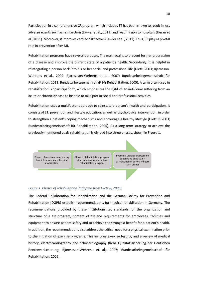
Bundesarbeitsgemeinschaft für Rehabilitation, 2005). As a long-term strategy to achieve the
previously mentioned goals rehabilitation is divided into three phases, shown in Figure 1.
Figure 1. Phases of rehabilitation (adapted from Dietz R, 2003)
The Federal Collaboration for Rehabilitation and the German Society for Prevention and
Rehabilitation (DGPR) establish recommendations for medical rehabilitation in Germany. The
recommendations provided by these institutions set standards for the organization and
structure of a CR program, content of CR and requirements for employees, facilities and
equipment to ensure patient safety and to achieve the strongest benefit for a patient’s health.
In addition, the recommendations also address the critical need for a physical examination prior
to the initiation of exercise programs. This includes exercise testing, and a review of medical
history, electrocardiography and echocardiography (Reha Qualitätssicherung der Deutschen
Rentenverischerung; Bjarnsason-Wehrens et al., 2007; Bundesarbeitsgemeinschaft für
Rehabilitation, 2005).
Phase I: Acute treatment during hospitilization+ early bedside
mobilization
Phase II: Rehabilitation program at an inpatient or outpatient
rehabilitation program
Phase III: Lifelong aftercare by supervising physician +
participation in coronary heart sport groups
11
There are several types of rehabilitation programs that patients can participate in. Most patients
visit “inpatient” and “outpatient” rehabilitation centers, however, there are also newer models,
such as home-based rehabilitation programs that have been successfully implemented ( Fischer
et al., 2012; Anderson &Taylor, 2014). The choice of a certain program type depends on several
factors such as risk profile for future cardiac events and co-morbidities. In high-risk patients with
instable cardiac function inpatient centers are recommended (Corrà et al., 2010). To date, the
superiority of an inpatient, outpatient or a home-based program has not been demonstrated.
According to a Cochrane review all programs are equally effective, at least in post-MI patients
with low risk for future cardiac events (Anderson & Taylor, 2014).
When a patient finishes the cardiac rehabilitation program, follow-up care is usually performed
by the patient’s general practitioner or cardiologist. In Germany, the German Pension Fund pays
for exercise training programs such as IRENA (intensive rehabilitation follow-up care) or KARENA
(coronary rehabilitation follow-up care) (Fischer et al., 2012; Reibis et al., 2014). In addition,
regional “heart sport groups” organized by the DGPR, offer supervised ET following a
rehabilitation program (Bundesarbeitsgemeinschaft für Rehabilitation, 2011).
1.3.1 Exercise training in cardiac rehabilitation
ET is a key component in CR. Specific recommendations for physical activity, as a part of a
comprehensive rehabilitation program, are set forth in the national guidelines, which are
provided by the DGPR.
The most recent guideline from the DGPR provides that ET in a patient with stable CAD should
consist of aerobic endurance training (evidence level IA), dynamic resistance training (evidence
level IB), and coordination and flexibility training (evidence level IIbC) (Bjarnason-Wehrens et
al., 2009).
Before engaging in an exercise program, however, an extensive physical examination is needed
to determine an individual’s risk of adverse cardiac events and current level of fitness (Fletcher
et al., 2001; Perk et al., 2012). Based on the results of exercise testing, the exercise program
should be adjusted to a patient’s current exercise capacity, exercise tolerance, risk factors and
personal motivation (Bjarnason-Wehrens B et al., 2009; Fletcher et al., 2001;
Bundesarbeitsgemeinschaft für Rehabilitation, 2005).
The current guideline from the Germany Society for Prevention and Rehabilitation suggests that
training should be at about 40-80% of VO2peak (maximum oxygen uptake), which is determined
during previous exercise testing, and that patients should exercise at a moderate continuous
intensity level (Bjarnsason-Wehrens et al., 2007; Smith et al., 2011). If training is not monitored
12
using VO2peak, heart rate measurement is another means to monitor training intensity. The
guideline recommends that training intensity should be at around 60-75% of patients’ maximum
heart rate, which can be measured using mobile heart rate monitors during training sessions
(Bjarnason-Wehrens et al., 2009). In addition, a visual scale, such as the Borg Scale, is often used
to determine exercise intensity and individual perceived exertion (Bjarnsason-Wehrens et al.,
2007; Bjarnason-Wehrens et al., 2009; Fletcher et al., 2001). The scale ranges from six to twenty,
beginning with almost imperceptible exertion to very hard exertion, and is measured in
“received perception of exertion = RPE”. A value from 11-14 is categorized as moderately
strenuous training and should be the target range during training (Bjarnsason-Wehrens et al.,
2007; Bjarnason-Wehrens et al., 2009; Borg, 1982; Borg, 1990).
Patients should engage in ET daily for at least thirty minutes during each exercise session. A
minimum of five days of training per week is a common recommendation. This
recommendation, however, only applies to patients with stable cardiac function and low-to-
medium risk of an adverse cardiac event. To ensure patient safety, patients should begin training
moderately and gradually increase the duration, frequency and intensity of the training
(Bjarnason-Wehrens et al., 2009; Fletcher et al., 2001; National Clinical Guideline Centre, 2013;
Smith et al., 2011). Fletcher et al. (2001) propose walking for 10 minutes a day and gradually
increasing duration and intensity. Patients with low-to-moderate risk, and sufficient
cardiovascular fitness following myocardial infarction may begin training with a more strenuous
exercise regimen (Fletcher et al., 2001; National Clinical Guideline Centre, 2013).
In addition to endurance training, some cardiac patients, specifically those with sufficient left
ventricular function and exercise tolerance, can also participate in dynamic resistance training.
Concerns regarding a high increase in blood pressure, arrhythmia or AMI, caused by strength
training, have been dismissed by several studies and guidelines (Bjarnason-Wehrens et al., 2004;
Pollock et al, 2000). The increase in blood pressure depends on resistance load, load intensity,
repetition count and duration of the exercise (Bjarnason-Wehrens et al., 2004). When applied
correctly in a professional and supervised setting with an adequate intensity and repetition
count, the increase in blood pressure is comparable to that found during aerobic endurance
training (Bjarnason-Wehrens B et al., 2004; Pollock et al., 2000). However, patients with a high
risk for adverse cardiac events, impaired cardiac function, multi-morbidity and low exercise
tolerance should not engage in this training method (Bjarnason-Wehrens et al., 2004; Pollock et
al., 2000). Before initiating dynamic resistance training, an extensive physical examination is
necessary and patients should first engage in aerobic ET for a few weeks (Bjarnason-Wehrens et
13
al., 2004; Pollock et al., 2000). Useful equipment for dynamic strength training can be elastic
bands or free weights (Balady et al., 2007; Fletcher et al., 2001).
Although the current guideline from the DGPR suggests integrating exercises for muscle
coordination and flexibility, there is less evidence for the benefit of these exercises compared
to aerobic endurance and dynamic strength training (IIbC). (Bjarnsason-Wehrens et al., 2007;
Bjarnason-Wehrens et al., 2009). Improving muscle coordination and flexibility, however, has
the added benefit of strengthening a patient’s capability to manage activities of every-day life
(ADL), e.g. carrying groceries, gardening, cleaning, etc.
1.4. Cardiac imaging
Non-invasive cardiac imaging has been of great importance in diagnosing CVD, including CAD
and the extent of a MI. Furthermore, cardiac imaging plays a pre-eminent role in determining
the prognosis of patients suffering from MI, CHD and HF. Non-invasive cardiac imaging includes
echocardiography, magnetic resonance imaging (MRI), single-photon-emissions-computer-
tomography (SPECT), coronary computed tomography angiography (CCTA) and positron-
emissions-tomography (PET) (Herrmann et al., 2018). Each of these imaging techniques has its
own set of indications, advantages and disadvantages. A PET-scan, for example, assesses
myocardial viability reliably, estimates the severity of CAD and determines myocardial blood
flow (Nekolla et al., 2009). PET scans have proven to be important in molecular imaging, a new
imaging field that identifies biological processes with the help of radioactive tracers. Molecular
imaging may provide novel information for future treatment of CVD. PET imaging is sensitive in
measuring inflammation, angiogenesis and sympathetic nerve function (Nekolla et al., 2009;
Pan, 2016). Furthermore, PET scans can help in diagnosing high-risk CAD patients for future
adverse events such as MI, who can then benefit from PCI (Rischpler et al., 2013; Sawada, 2006).
In contrast, a MRI, has the advantage of depicting myocardial anatomy and measuring cardiac
function, ventricular volumes and characterization of soft tissue, as well as the transmurality
and size of a MI and cardiac remodeling. A MRI is also feasible in detecting myocardial viability
(Nekolla et al., 2009; Pontone et al., 2017; Wintersperger et al., 2015; Arai, 2011b; Rischpler et
al., 2015).
In the past few years new imaging techniques have evolved in cardiovascular research. Hybrid
PET/CT imaging is common in oncology and has also gained importance in the cardiological field
(Rischpler et al., 2013). Hybrid PET/MRI is another prospective imaging method that has become
more common in research and may play a stronger role in diagnostics in patients suffering from
CAD and/or MI. A hybrid PET-MRI combines the advantages of both imaging techniques in one
14
session: a reliable assessment of cardiac function, cardiac morphology, infarction size using MRI;
and the assessment of viability, inflammation and perfusion using a PET scan (Krumm et al.,
2018; Pan, 2016).
Although both PET and MRI, and hybrid PET-MRI scans, currently do not play a role in imaging
patients with AMI, they can fulfill an important function after revascularization therapy as they
“[…] provide detailed evaluation of infarcted myocardium, edema extent, area at risk and
salvaged myocardium in acute myocardial infarction […]” (Krumm et al., 2018). Moreover, a
study, performed by Rischpler et al. (2016) using a hybrid 18F-FDG-PET/MRI system, suggests
that the uptake of 18F-FDG in the myocardium after revascularization therapy correlates with
adverse cardiac function after six months (Rischpler et al., 2016). Thus, combined PET/MRI
imaging can deliver useful and additional information for both diagnosis and prognosis in CVD.
1.4.1 Principle of magnetic resonance imaging
MRI makes use of the magnetic moment, also known as spin, that naturally occurs in the human
body. Since hydrogen nuclei (1H) (which consists out of one proton) are found in a high
concentration in water and fat, its magnetic spin is used to produce images. (Berger, 2002;
Ridgway, 2010). Normally, the hydrogen protons spin on their own axis with the axes aligned in
random directions. A magnetic resonance system produces a magnetic field, with different field
strengths, known as Tesla. When placed into a magnetic field, the hydrogen protons align
towards or against this magnetic field, producing a magnetic vector (Berger, 2002; Ridgway,
2010). When energy in the form of radiofrequency waves (pulse) are used in the direction of the
vector, the direction of the spins is altered. When the radiofrequency pulse is turned off, the
protons return to their original state and the original magnetic vector is aligned again. This
process is known as relaxation.
During the relaxation phase, a signal is transmitted that is detected by the magnetic resonance
system and computed into images. Since tissues relax at different speeds, these different
relaxation times are used to create an image:
“[…] The time taken for the protons to fully relax is measured in two ways. The
first is the time taken for the magnetic vector to return to its resting state and
the second is the time needed for the axial spin to return to its resting state.
The first is called T1 relaxation, the second is called T2 relaxation […]” (Berger,
2002, p. 35).
15
One MRI scan consists of several radiofrequency pulse sequences that create an image of the
tissue of interest. In cardiovascular imaging, MRI has a high spatial resolution and can accurately
produce a contrast between different tissues (Tseng et al., 2016). A MRI has the capability to
determine ventricular function, volumes and wall motion with the help of the cine technique,
and thus, allows an examination of a patient’s cardiac function (Tseng et al., 2016). Contrast
agents, such as gadolinium (Gd), can also be used to enhance MRI imaging. These contrast
agents are injected intravenously and distribute quickly into the extravascular space. They do
not, however, permeate intact cell membranes due to their charge and size (Arai, 2011a). The
distribution of gadolinium after about five-to-twenty minutes post-intravenous injection is
known as late gadolinium enhancement (LGE) and it produces a hyper-enhanced imaging signal
caused by the accumulated gadolinium in the depicted tissue (Gerber et al., 2000). LGE can show
the infarcted area during the acute and chronic phase of infarction: in an acute setting LGE
accurately depicts the transmurality of an infarction. After scar formation LGE can be used to
depict scar size. LGE-MRI can also be used to determine myocardial viability (Abdel-Aty et al.,
2011b; Rischpler et al., 2013).
As briefly explained above, each imaging technique has its advantages and disadvantages.
Hybrid imaging techniques such as PET/MRI, PET/CT or echocardiography/MRI-fusions aim at
combining the advantages of each technique to measure cardiac parameters, such as
morphology, structure and remodeling even more precisely. Accurate imaging may influence
patient treatment and outcome as (a) more patients can be identified who could benefit from
certain treatments and (b) identifying changes in cardiac structure, function, metabolism or
remodeling may allow for better monitoring of previous, current and future treatments.
Consequently, successful monitoring could improve a patient’s treatment by allowing
important adaptions in a patient’s individual therapy more quickly than in standard follow-up
imaging. A meta-analysis by Haykowsky et al. (2011) showed that many trials used
echocardiography to determine cardiac remodeling parameters, whereas SPECT or MRI, as
well as left ventriculography, were not as common (Haykowsky et al., 2011). Compared to an
MRI, echocardiography, however, strongly depends on sonographic conditions and is not as
accurate in measuring morphology and mechanical function (Bauer et al., 2008). Thus, MRI
may be superior in measuring cardiac remodeling parameters and impact future exercise
recommendations for patients following MI.
The benefits of providing ET after MI as a part of patient rehabilitation have thoroughly been
examined in terms of systemic effects, which include improvements in exercise capacity,
endothelial function and autonomous nervous system (Kavanagh et al., 2002; Hambrecht et
16
al., 2003; Martinez et al., 2011). Many interventional trials have examined whether structured
ET impacts cardiac remodeling. Most of these trials confirmed the positive effects of ET on
cardiac remodeling by changing the factors initiation of ET, duration, frequency and type of ET
(Giallauria et al., 2008; Giallauria et al., 2013; Giannuzzi et al., 2003). In retrospect, however, it
remains unclear whether participation in current CR programs designed by the DGPR and in
leisure time exercise behavior as recommended in CR programs attenuate cardiac remodeling
as well, especially when examining self-reported leisure time ET.
Not only ET following MI can change cardiac remodeling, ET prior to an MI could limit
infarction size and thus affect cardiac remodeling as well. In humans, prospective studies
answering this question are not available due to ethical reasons. In animal trials, however, ET
has been found to be able to limit infarction size, thus some authors presume a similar effect
could be present in humans suffering from MI (de Waard & Duncker, 2009; Frasier et al., 2011;
Freimann et al., (2005)
17
2 Objective
The primary aim of this observational study was to determine whether ET in the first six months
following MI can ameliorate cardiac remodeling, as measured by a cardiac MRI-scan. Cardiac
remodeling parameters included left-ventricular end-diastolic volume, left-ventricular end-
systolic volume, stroke volume and ejection fraction. In comparison to interventional studies,
which manipulate the parameter ET, e.g. by changing exercise duration, frequency or intensity,
to analyze the influence of ET on cardiac remodeling (Giallauria et al., 2008; Giallauria et al.,
2013; Giannuzzi et al., 2003), this retrospective study examined whether patients (a)
participated in a CR program including ET as recommended by the DGPR and (b) analyzed
exercise behavior and adherence to ET recommendations following a CR program. Cardiac
remodeling parameters were measured with the use of an MRI scan.
In addition, the effect of self-reported exercise prior to MI on initial infarction size and infarction
volume, which were measured by late-gadolinium enhancement during the MRI-scan, was
analyzed.
The following hypotheses were proposed:
• Self-reported exercise training and participation in CR programs following myocardial
infarction can attenuate cardiac remodeling parameters (∆LVEDV, ∆LVESV) measured
by an MRI scan
• Self-reported exercise training prior to myocardial infarction can lead to a smaller initial
infarction size (LGE%) and volume (ml) measured by an MRI scan
18
3. Methods
3.1. Study population
From May to October 2014, 29 patients were selected to participate in this study. The inclusion
criteria for this study were the following:
• First acute myocardial infarction (AMI), defined as: ongoing chest pain >20 min,
electrocardiographic (ECG) changes, such as ST-segment elevation or new left-bundle
block, and elevated cardiac enzyme levels.
• Treatment of AMI with percutaneous coronary intervention (PCI) upon hospital
admission.
• No contraindication for a simultaneous PET/MRI, e.g. ferromagnetic material implants,
implanted cardioverter-defibrillator, pacemakers, claustrophobia, allergies to contrast
agents, low creatinine clearance <50ml/min, hemodynamic instability or pregnancy.
• 18 years of age and capable of giving written informed consent.
A simultaneous PET/MRI scan was performed 5 ± 1.4 (mean ± SD) days after a successful PCI.
The pain to balloon time was 4,5h (median, 25th percentile 2,4h, 75th percentile 13,3h). The
second scan, MRI-only, was planned to take place after six to nine months following PCI. After
their second scan, patients received a questionnaire from the Center for Preventive and Sports
Medicine at the University Hospital Klinikum rechts der Isar in Munich, assessing the patients’
participation in exercise training (ET) before and post-MI, and their participation in a
rehabilitation program.
Written informed consent was obtained from each patient before participation in this study.
This study was approved by the ethics committee of the Technical University Munich and was
performed in accordance to the ethical standards defined in the Declaration of Helsinki.
Of the original 29 patients, 26 (24 men, 2 women), gave written informed consent to participate.
3.2. Questionnaire
The Center for Preventive and Sports Medicine developed a questionnaire consisting of 29
questions concerning demographic characteristics, cardiovascular risk factors, ET prior to an MI,
medication and rehabilitation measures post-MI, and ET during the first six months after MI. The
questionnaire included multiple-choice, closed and open-ended questions.
Data regarding age, weight, height/stature, cardiovascular risk factors and medication post-MI
were collected to assess both general and cardiovascular characteristics of the study population.
19
Information on rehabilitation measures, more specifically, participation in a rehabilitation
program post-MI was also gathered. Patients were asked whether they participated in a
rehabilitation program, whether their participation was inpatient or outpatient, and when and
how long they participated in the rehabilitation program. This information was gathered to
determine whether patients suffering from MI received the follow-up care specified in current
treatment guidelines from the European Society of Cardiology and the German Society for
Prevention and Rehabilitation (Bjarnason-Wehrens et al., 2009).
The questionnaire also asked about the patients’ participation in leisure time ET before suffering
from an MI. The questionnaire aimed to document the extent, frequency and type of exercise
in order to analyze whether participation in regular ET prior to myocardial infarction influences
the initial infarction size (LGE%) and initial infarction volume (ml).
To examine whether ET post-MI affects cardiac remodeling (measured by cardiac functional
parameters) information on ET during the first six months, including cardiac rehabilitation,
following an MI was gathered. Furthermore, the duration of ET in minutes per week and
frequency of ET per week was assessed. The questionnaire also documented the type of exercise
patients engaged in: aerobic endurance training, technical/rhythmic sports, rehabilitative sport
programs or other types of ET. Different exercise types were categorized by example. A meta-
analysis by Haykowsky et al. (2007) showed that aerobic exercise training is superior to a
combination of aerobic and strength training in attenuating cardiac remodeling of heart failure
patients (Haykowsky et al., 2007). Thus, collecting data on the type of ET performed by patients
after MI is important. This information on patients’ exercise behavior was used to analyze
whether patients adhered to exercise recommendations from current guidelines. These
guidelines suggest engaging in aerobic endurance training on at least five days a week,
preferably daily, for a minimum of thirty minutes at a moderate intensity (Bjarnason-Wehrens
B et al., 2009; Jones et al., 2013; National Clinical Guideline Centre, 2013; Smith et al., 2011).
Follow-up telephone interviews were conducted with patients who either failed to return the
questionnaire or returned it incomplete, requiring clarification. The original questionnaire can
be found in the appendix.
3.3. Imaging
PET/MRI-imaging was performed by the Department of Nuclear Medicine at the University
Hospital Klinikum Rechts der Isar in Munich. The images were acquired using a hybrid system
Biograph mMR scanner (Siemens Healthcare GmbH, Erlangen, Germany) (Rischpler et al., 2016).
The cardiac parameters in this study were the following: LVEDV, LVESV, SV, EF. Additionally,
20
infarction size, infarction volume and LV-volume were measured. Each of these parameters was
measured at the first and second scan. 18F-FDG uptake was only measured in the first scan. After
completion of the second scan, the data was sent to the Center for Preventive and Sports
Medicine at the University Hospital Klinikum rechts der Isar for further analysis.
3.3.1. Positron-emission-tomography/magnetic resonance imaging
The first scan (T1) was performed as a simultaneous PET/MRI scan using the previously
mentioned hybrid imaging system. On the day prior to the scan patients received a low-
carbohydrate diet followed by 12 hours of fasting to prevent a physiological uptake of 18F-FDG
in cardiomyocytes. On the day of the scan unfractionated heparin was injected (50 IU/kg body
weight) 30 minutes before a standardized dose of 18F-FDG was administered intravenously
(Rischpler et al., 2016). Approximately 145 ± 51 minutes after intravenous injection of 18F-FDG a
PET scan was initiated. The PET scan was performed in 3D-mode, emission data was corrected
and the collected images were reconstructed using a 3D attenuation-weighted ordered-subsets
expectation maximization iterative reconstruction algorithm (AW-OSEM 3D) (Rischpler et al.,
2016).
To obtain T1- weighted MRI images and evaluate late gadolinium enhancement (LGE%), 0.2
mmol/kg body weight Gadopentetat-Dimeglumin was injected into each patient (Magnograf®;
Marotrast GmbH, Jena, Germany) (Rischpler et al., 2016). After 10 minutes the MRI scan was
initiated. To acquire images, ECG triggering was performed, and patients were advised to hold
their breath. With the use of steady-state free precision (SSFP) cine sequences systolic function
was examined (Rischpler et al., 2016). The second MRI-only scan (T2) was performed six to nine
months post-MI to measure once-again the aforementioned parameters.
3.3.2. Imaging analysis
For regional analysis LGE and 18F-FDG images were lined up correspondingly using the
MunichHeart/m3p software and the 17-segment model of the American Heart Association (AHA)
was used on these images (Rischpler et al., 2016). Manual delineation on short axis images was
performed, with the help of the MunichHeart/MR software, to determine the degree of late
gadolinium enhancement in the left ventricle (Rischpler et al., 2016).
3.4. Statistical analyses
Data was analyzed using IBM SPSS 23 (IBM Corp, Armonk, NY, USA).
Descriptive statistics (mean values ± and standard deviations, median values + 25th and 75th
percentile) were calculated for all variables. Normal distribution was analyzed by graphical
21
analysis using histograms, P-Plots and Q-Q-Plots and cross-checked with numerical analysis
using the Kolmogorov-Smirnov test.
To determine a possible influence of ET post-MI on cardiac remodeling parameters, the study
sample (n=26) was divided into two groups based on their median duration of performed
exercise/week. The median value of ET among all patients was 212 minutes/week, thus ET of
200 minutes/week was used to separate patients into two different exercise groups. Group 1
consisted of 12 patients with ET of less than 200min/week per week vs. Group 2 consisted of 14
patients achieving more than 200min/week of ET.
As most variables were not normally distributed within the groups, non-parametric tests, such
as the Mann-Whitney-U test for independent variables, were carried out to detect significant
differences between the two exercise groups in the following variables.
Independent variables in the group comparisons were patient characteristics; including age,
weight (kg), height (cm), body-mass-index (BMI, m2/kg) and body surface area (BSA, m2), as well
as imaging data; including pain-to-balloon time (h), 1st scan (days after PCI), 2nd scan (days after
PCI).
In categorical variables, such as sex, cardiovascular risk factors and medication post-MI, a cross-
table and calculation of Chi-squared (χ2) was carried out to detect a significant difference in
these variables between the group 1 ET <200min/week and group 2 ET >200min/week. The
significance level was set to p < 0,05.
Most cardiac remodeling data were not normally distributed; therefore, the non-parametric
Mann-Whitney-U test was carried out for group comparisons of independent variables: ∆LVEDV,
∆LVESV, ∆EF, ∆SV, ∆infarction volume, ∆infarction size as well as ∆LV volume. Since the data was
not normally distributed, the Spearman’s Rho correlation coefficient was calculated in order to
assess the relationship between ET in min/week and remodeling parameters ∆LVEDV, ∆LVESV,
∆EF, ∆SV, ∆infarction volume, ∆infarction size, ∆LV volume. The significance level was set to p<
0.05.
The second aim of this study was to examine the potential influence of ET prior to an MI on
initial infarction size and initial infarction volume. After data from the questionnaire concerning
regular participation in ET prior to MI was analyzed, the study sample (n=26) was divided once
again into two new groups. Regular exercise training was seen as participating in ET at least once
to twice. The No-ET-group consisted of 14 patients, who engaged in either no ET or sporadically
at most. The ET-group consisted of 12 patients who engaged in ET on a regular basis. The patient
distribution among these groups was not the same as in the post-MI groups ET<200min/week

22
and ET>200min/week. A descriptive analysis was also performed on these data sets. Normal
distribution was tested as previously mentioned for these data sets, and non-parametric tests
were used to compare the independent variables, infarction size and volume between the two
groups.
23
4. Results
4.1. Patient characteristics, cardiovascular profile, imaging data
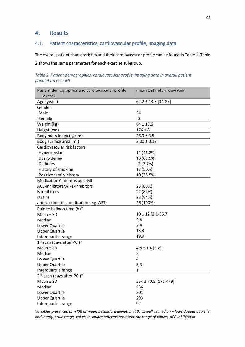
The overall patient characteristics and their cardiovascular profile can be found in Table 1. Table
2 shows the same parameters for each exercise subgroup.
Table 2. Patient demographics, cardiovascular profile, imaging data in overall patient population post MI
Patient demographics and cardiovascular profile overall
mean ± standard deviation
Age (years) 62.2 ± 13.7 [34-85]
Gender Male Female
24 2
Weight (kg) 84 ± 13.6
Height (cm) 176 ± 8
Body mass index (kg/m2) 26.9 ± 3.5
Body surface area (m2) 2.00 ± 0.18
Cardiovascular risk factors Hypertension Dyslipidemia Diabetes History of smoking Positive family history
12 (46.2%) 16 (61.5%) 2 (7.7%) 13 (50%) 10 (38.5%)
Medication 6 months post-MI ACE-inhibitors/AT-1-inhibitors ß-inhibitors statins anti-thrombotic medication (e.g. ASS)
23 (88%) 22 (84%) 22 (84%) 26 (100%)
Pain to balloon time (h)* Mean ± SD Median Lower Quartile Upper Quartile Interquartile range
10 ± 12 [2.1-55.7] 4,5 2,4 13,3 19,9
1st scan (days after PCI)* Mean ± SD Median Lower Quartile Upper Quartile Interquartile range
4.8 ± 1.4 [3-8] 5 4 5,3 1
2nd scan (days after PCI)* Mean ± SD Median Lower Quartile Upper Quartile Interquartile range
254 ± 70.5 [171-479] 236 201 293 92
Variables presented as n (%) or mean ± standard deviation (SD) as well as median + lower/upper quartile
and interquartile range, values in square brackets represent the range of values; ACE-inhibitors=
24
angiotensinogen-converting-enzyme-inhibitor, AT-1-inhibitor= angiotensin-1-inhibitor, ASS=
acetylsalicylic acid, PCI = percutaneous coronary intervention, *= values not normally distributed,
therefore median and mean values are presented.
Post-MI patients were divided into two subgroups based on their participation in ET per week.
Table 2 presents the previously shown patient demographics, cardiovascular profile and imaging
data between the two groups.
Table 3. Patient demographics, cardiovascular profile, imaging data in exercise subgroups post MI
Patient demographics and cardiovascular profile in exercise groups
Group 1 ET <200min/week
n=12
Groupe 2 ET >200min/week
n=14
p-value
Age (years) 63.7 ± 14.7 [38-85] 60.9 ± 13.2 [34-85] 0.595
Gender Male Female
10 2
14 0
0.112
Weight (kg) 87.5 ± 16 80.5 ± 9.9 0.280
Height (cm) 178 ± 9 175.4 ± 8.6 0.501
Body mass index (kg/m2) 27.8 ± 4.1 26.1 ± 2.7 0.436
Body surface area (m2) 2.04 ± 0.20 1.96 ± 0.15 0.481
Cardiovascular risk factors Hypertension Dyslipidemia Diabetes History of smoking Positive family history
6 (50%) 6 (50%) 1 (8.3%) 6 (50%) 4 (33.3%)
6 (42.9%) 10 (71.4%) 1 (7.4%) 7 (50%) 6 (42.9%)
0.408 0.595 0.674 1.000 0.628
Medication 6 months post MI ACE-inhibitors /AT-1-inhibitors ß-inhibitors statins Antithrombotic medication (e.g
ASS)
11 (91.7%) 9 (75%) 9 (75%) 12 (100%)
12 (85.7%) 13 (92.9%) 13 (92.9%) 14 (100%)
0.191 0.399 0.399
Pain to balloon time (h)* Mean ± SD Median Lower Quartile Upper Quartile Interquartile Range
12.5 ± 15.5 6.3 2.7 18.3 15.6
7.4 ± 7.4 4.5 2.2 8.2 6.1
0.347
1st scan (days after PCI) * Mean ± SD Median Lower Quartile Upper Quartile Interquartile Range
4.8 ± 1.5 5 3 5 2
4.8 ± 1.4 5 4 5 1
0.494
2nd scan (days after PCI) * Mean ± SD Median Lower Quartile Upper Quartile
25 ± 83 240 196 287
253 ± 61 234 208 298
0.940
25
Interquartile Range 91 90
Variables presented as n (%) or mean ± standard deviation (SD); values in square brackets represent the
range of values; ACE-inhibitors= angiotensinogen-converting-enzyme-inhibitor, AT-1-inhibitor=
angiotensin-1-inhibitor, ASS= acetylsalicylic acid, PCI = percutaneous coronary intervention, significant
differences between group 1 and group 2 are marked as *p <0,05
4.2. Cardiac rehabilitation and exercise training post-myocardial infarction
Participation in a cardiac rehabilitation program in the overall patient sample was 88.5 % (n=23).
The median initiation of these programs was 14 days after suffering an MI and with a median
duration of 21 days. Table 3 shows the participation, initiation and duration of cardiac
rehabilitation programs for each subgroup.
Looking at participation in ET as a whole, five patients did not engage in any exercise, nine
patients engaged in regular exercise training once to twice a week, and eleven patients engaged
in exercise three days a week or more. The median duration of exercise training per week was
212 minutes.
Table 4. Rehabilitation data
Group 1 ET < 200min/week
Group 2 ET >200min/week
p-value
Rehabilitation No rehabilitation Inpatient program Outpatient program
1 (8.3%) 11 (91.7%) 0
2 (14.3%) 10 (71.4%) 2 (14.3%)
0.781
Initiation (days after MI) Mean ± SD Median Upper Quartile Lower Quartile IQR
12 ± 8 [CI 6.2 -17.8] 14 18 6 12
14 ± 4,7 [CI 11.1-16.8] 14 18 9 9
0.648
Duration (days) Mean ± SD Median Upper Quartile Lower Quartile IQR
17.5 ± 9.5 [CI 10.7-24.3] 21 21 15.8 5
20.4 ± 2.1 [CI 18.9-21.8] 21 21 21 0
0.981
Variables presented as n (%) or mean ± standard deviation (SD) as well as median, range and quartiles, MI
= myocardial infarction, IQR = interquartile range, CI = confidence interval; p-values calculated using non-
parametric Mann-Whitney-U test for group comparisons in independent variables
The most common types of exercise in both groups were brisk walking (n=13), cycling (n=7) and
strength-endurance-flexibility training (n=5). Less common were ergometer training (n=2),
Nordic walking (n=2), tennis (n=2), gymnastics (n=2), swimming (n=1), light jogging (n=1), cross-
trainer (n=1) and participation in a heart sport group (n=1). The frequency and duration of ET
for the subgroups can be found in Table 4. Exercise frequency and duration show significant
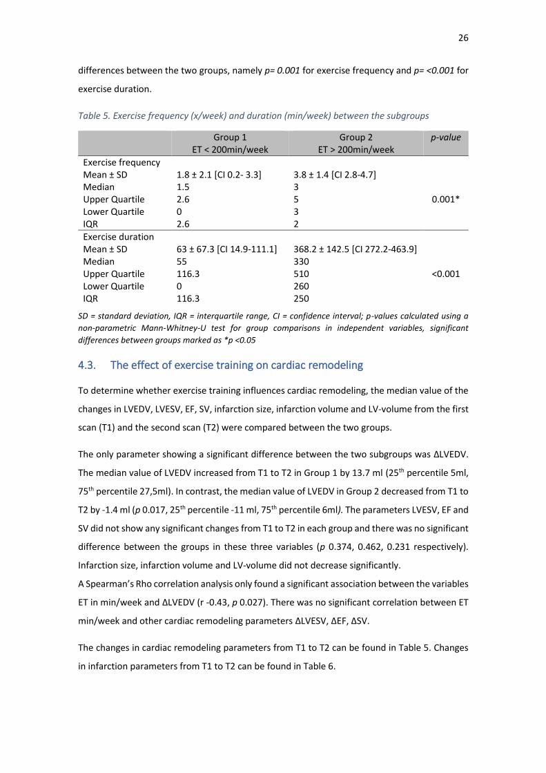
26
differences between the two groups, namely p= 0.001 for exercise frequency and p= <0.001 for
exercise duration.
Table 5. Exercise frequency (x/week) and duration (min/week) between the subgroups
Group 1 ET < 200min/week
Group 2 ET > 200min/week
p-value
Exercise frequency Mean ± SD Median Upper Quartile Lower Quartile IQR
1.8 ± 2.1 [CI 0.2- 3.3] 1.5 2.6 0 2.6
3.8 ± 1.4 [CI 2.8-4.7] 3 5 3 2
0.001*
Exercise duration Mean ± SD Median Upper Quartile Lower Quartile IQR
63 ± 67.3 [CI 14.9-111.1] 55 116.3 0 116.3
368.2 ± 142.5 [CI 272.2-463.9] 330 510 260 250
<0.001
SD = standard deviation, IQR = interquartile range, CI = confidence interval; p-values calculated using a
non-parametric Mann-Whitney-U test for group comparisons in independent variables, significant
differences between groups marked as *p <0.05
4.3. The effect of exercise training on cardiac remodeling
To determine whether exercise training influences cardiac remodeling, the median value of the
changes in LVEDV, LVESV, EF, SV, infarction size, infarction volume and LV-volume from the first
scan (T1) and the second scan (T2) were compared between the two groups.
The only parameter showing a significant difference between the two subgroups was ∆LVEDV.
The median value of LVEDV increased from T1 to T2 in Group 1 by 13.7 ml (25th percentile 5ml,
75th percentile 27,5ml). In contrast, the median value of LVEDV in Group 2 decreased from T1 to
T2 by -1.4 ml (p 0.017, 25th percentile -11 ml, 75th percentile 6ml). The parameters LVESV, EF and
SV did not show any significant changes from T1 to T2 in each group and there was no significant
difference between the groups in these three variables (p 0.374, 0.462, 0.231 respectively).
Infarction size, infarction volume and LV-volume did not decrease significantly.
A Spearman’s Rho correlation analysis only found a significant association between the variables
ET in min/week and ∆LVEDV (r -0.43, p 0.027). There was no significant correlation between ET
min/week and other cardiac remodeling parameters ∆LVESV, ∆EF, ∆SV.
The changes in cardiac remodeling parameters from T1 to T2 can be found in Table 5. Changes
in infarction parameters from T1 to T2 can be found in Table 6.
27
Table 6. Cardiac remodeling parameters at T1, T2 and difference from T1 to T2 (T2-T1)
Group 1 ET <200min/week
Group 2 ET >200min/week
p-value
LVEDV T1 Mean ± SD Median Upper Quartile Lower Quartile IQR
123± 30 [CI 104.5-142.6] 112.5 148 101 47
135 ± 40 [CI 112.8-158] 133 150 110 40
0.297
T2 Mean ± SD Median Upper Quartile Lower Quartile IQR
137 ± 30 [CI 118-155.8] 134 166 110 56
131 ± 40 [CI 108-154.5] 134.4 142 99.5 43
0.560
∆LVEDV (T2-T1) Mean ± SD Median Upper Quartile Lower Quartile IQR
13 ± 18 [CI 1.8-25] 13.7 27.5 5 22
- 4 ± 19.5 [CI -15.5 - +7] - 1.4 6 - 11 17
0.017*
LVESV (ml) T1 Mean ± SD Median Upper Quartile Lower Quartile IQR
69 ± 25 [CI 53-84.5] 64 85 47 38
71 ± 30 [CI 54-88.5] 69 87 46 40.5
0.899
T2 Mean ± SD Median Upper Quartile Lower Quartile IQR
77 ± 34 [CI 55.7-99] 66.8 104 49. 55
71 ± 31 [CI 53-89] 63.7 85 51 34
0.742
Δ LVESV (T2-T1) Mean ± SD Median Upper Quartile Lower Quartile IQR
9 ± 18 [CI -2.6 - +20] 5.7 26 -4 30
-0.25 ± 19.5 [CI -11.5 -+11] 6 14.8 -17.9 32
0.374
EF (%) T1 Mean ± SD Median Upper Quartile Lower Quartile IQR
45 ± 11 [CI 38-52] 45.5 53.7 38.8 14.8
48 ± 9 [CI 43-54] 45.2 57.8 40.8 17
0.705
T2 Mean ± SD Median Upper Quartile Lower Quartile IQR
45 ± 15 [CI 35.6-54.5] 50 58.2 29.8 28.4
47 ± 10 [CI 41-52.7] 48 54 40 14
1.000
Δ EF (T2-T1) Mean ± SD
-0.12 ± 8.6 [CI -5.6 - +5.4]
-1.7 ± 10 [CI -7.5 - +3.9]
28
Median Upper Quartile Lower Quartile IQR
3.2 5.7 -7.6 13
-4.3 3.2 -9 12.6
0.462
SV (ml) T1 Mean ± SD Median Upper Quartile Lower Quartile IQR
55 ± 14 [CI 45.7-64] 54.3 65.3 51.3 14
64 ± 14 [CI 55.6-72.5] 66 77.4 47.8 29.6
0.160
T2 Mean ± SD Median Upper Quartile Lower Quartile IQR
59 ± 19 [CI 47.6-71.4] 57.3 72.8 46.3 26.6
60 ± 16 [CI 50.5-69.5] 60.9 74.7 45.4 29.3
0.899
ΔSV (T2-T1) Mean ± SD Median Upper Quartile Lower Quartile IQR
4.6 ± 16 [CI -5.3- +14.6] 2.9 11.5 -3.9 15.4
-3 ± 12 [CI -9.7- +3.9] -1.8 7.5 -15.1 22.7
0.231
SD= standard deviation; IQR = interquartile range, CI = confidence interval, LVEDV = Left-ventricular end-
diastolic volume, LVESV = left-ventricular end-systolic volume, EF = Ejection fraction, SV = stroke volume,
significant differences between groups marked as *p <0.05
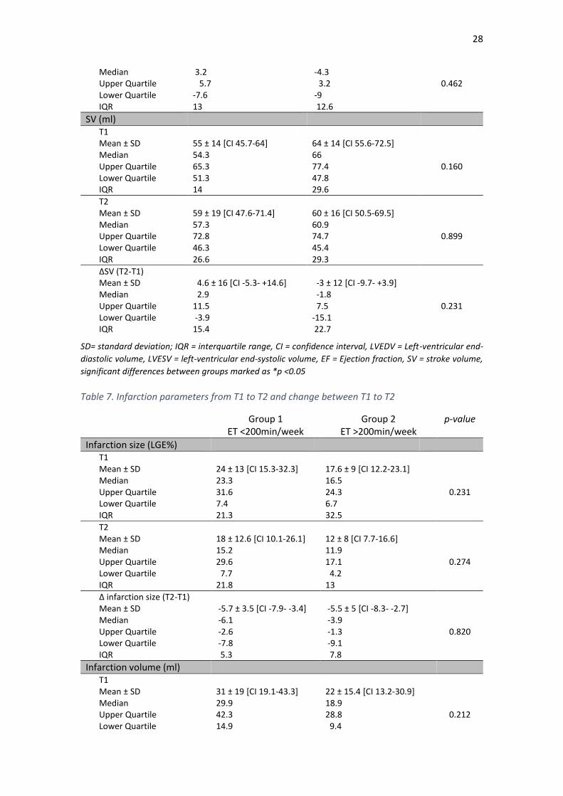
Table 7. Infarction parameters from T1 to T2 and change between T1 to T2
Group 1 ET <200min/week
Group 2 ET >200min/week
p-value
Infarction size (LGE%) T1 Mean ± SD Median Upper Quartile Lower Quartile IQR
24 ± 13 [CI 15.3-32.3] 23.3 31.6 7.4 21.3
17.6 ± 9 [CI 12.2-23.1] 16.5 24.3 6.7 32.5
0.231
T2 Mean ± SD Median Upper Quartile Lower Quartile IQR
18 ± 12.6 [CI 10.1-26.1] 15.2 29.6 7.7 21.8
12 ± 8 [CI 7.7-16.6] 11.9 17.1 4.2 13
0.274
Δ infarction size (T2-T1) Mean ± SD Median Upper Quartile Lower Quartile IQR
-5.7 ± 3.5 [CI -7.9- -3.4] -6.1 -2.6 -7.8 5.3
-5.5 ± 5 [CI -8.3- -2.7] -3.9 -1.3 -9.1 7.8
0.820
Infarction volume (ml) T1 Mean ± SD Median Upper Quartile Lower Quartile
31 ± 19 [CI 19.1-43.3] 29.9 42.3 14.9
22 ± 15.4 [CI 13.2-30.9] 18.9 28.8 9.4
0.212
29
IQR 27.4 19.4
T2 Mean ± SD Median Upper Quartile Lower Quartile IQR
17 ± 12.6 [CI 9.2-25.2] 12.9 21.8 8 13.8
12 ± 8 [CI 7-16.3] 11.9 16.2 4.5 11.8
0.322
∆ Infarction volume (T2-T1) Mean ± SD Median Upper Quartile Lower Quartile IQR
-14 ± 8 [CI -19.4- -8.7] -14.5 -6.8 -20.2 13.4
-10 ± 10 [CI -16.2- -4.5] -5.4 -2.9 -17.2 14.2
0.176
LV volume (ml) T1 Mean ± SD Median Upper Quartile Lower Quartile IQR
132 ± 34 [CI 110.7-154.8] 132.9 148.4 103.6 44.8
118 ± 22 [CI 105.6-131.6] 113.5 133.3 101.5 31.8
0.231
T2 Mean ± SD Median Upper Quartile Lower Quartile IQR
100 ± 29 [CI 81.5-118.9] 99.6 116.9 74.3 42.7
95 ± 14 [CI 87.6-103.9] 98.2 106.3 85 21.4
0.781
∆ LV volume (T2-T1) Mean ± SD Median Upper Quartile Lower Quartile IQR
-32 ± 19 [CI -44.5- -20.6] -33 -19.6 -39.5 19.8
-23 ± 18 [CI -33.3- 12.6] -18.6 -11.5 -32.2 20.7
0.145
SD= standard deviation; IQR = interquartile range, CI = confidence interval, LV = left-ventricular volume,
significant differences between groups marked as *p <0.05
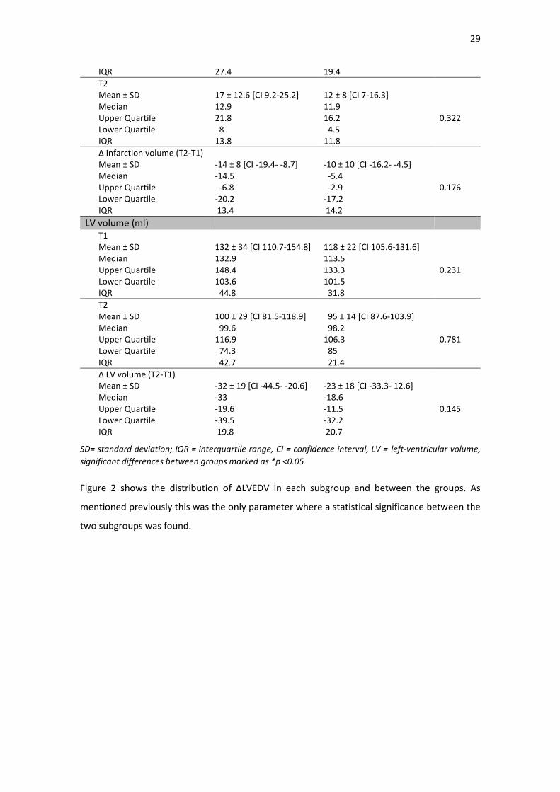
Figure 2 shows the distribution of ∆LVEDV in each subgroup and between the groups. As
mentioned previously this was the only parameter where a statistical significance between the
two subgroups was found.
30
Figure 2. Difference in ∆LVEDV (∆LVEDV = T2 LVEDV-T1 LVEDV) between group 1 and group 2 ; Variables presented as median values of ∆LVEDV, LVEDV = left ventricular end diastolic volume,
* p <0.054.4. The influence of exercise training prior to myocardial infarction on
initial infarction size and volume
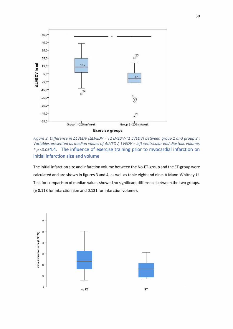
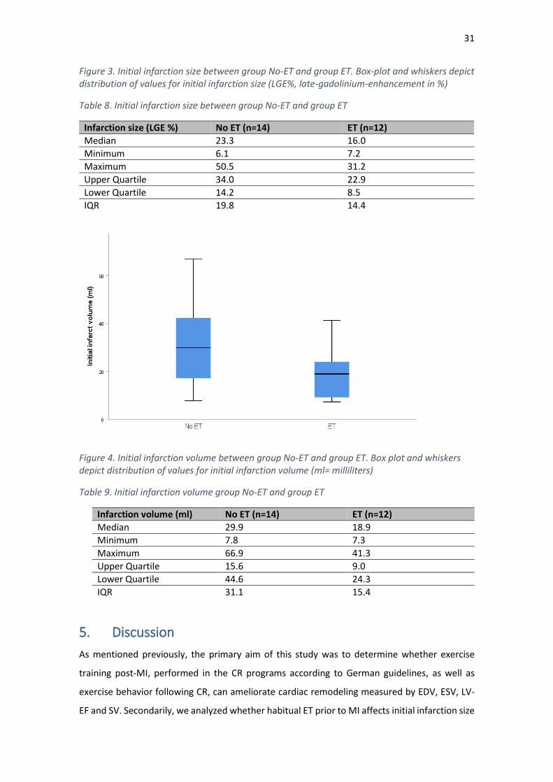
The initial infarction size and infarction volume between the No-ET-group and the ET-group were
calculated and are shown in figures 3 and 4, as well as table eight and nine. A Mann-Whitney-U-
Test for comparison of median values showed no significant difference between the two groups.
(p 0.118 for infarction size and 0.131 for infarction volume).
31
Figure 3. Initial infarction size between group No-ET and group ET. Box-plot and whiskers depict distribution of values for initial infarction size (LGE%, late-gadolinium-enhancement in %)
Table 8. Initial infarction size between group No-ET and group ET
Infarction size (LGE %) No ET (n=14) ET (n=12)
Median 23.3 16.0
Minimum 6.1 7.2
Maximum 50.5 31.2
Upper Quartile 34.0 22.9
Lower Quartile 14.2 8.5
IQR 19.8 14.4
Figure 4. Initial infarction volume between group No-ET and group ET. Box plot and whiskers depict distribution of values for initial infarction volume (ml= milliliters)
Table 9. Initial infarction volume group No-ET and group ET
Infarction volume (ml) No ET (n=14) ET (n=12)
Median 29.9 18.9
Minimum 7.8 7.3
Maximum 66.9 41.3
Upper Quartile 15.6 9.0
Lower Quartile 44.6 24.3
IQR 31.1 15.4
5. Discussion
As mentioned previously, the primary aim of this study was to determine whether exercise
training post-MI, performed in the CR programs according to German guidelines, as well as
exercise behavior following CR, can ameliorate cardiac remodeling measured by EDV, ESV, LV-
EF and SV. Secondarily, we analyzed whether habitual ET prior to MI affects initial infarction size
32
and infarction volume. A questionnaire was developed by the Center for Preventive and Sports
Medicine from the University Hospital Klinikum rechts der Isar to analyze exercise behavior
before and after MI. The Department for Nuclear Medicine from the University Hospital Klinikum
rechts der Isar performed imaging analysis to measure the cardiac remodeling parameters. To
compare the effects of ET on cardiac remodeling we divided our patients into two exercise
groups, based on the median value of ET in minutes per week, resulting in group 1 exercising
<200min/week and group 2 exercising >200min/week. For analysis of exercise behavior prior to
MI and its influence on initial infarction size and volume, we divided the patients into two
groups, namely group No-ET and group ET, based on their participation in exercise.
5.1. Participation in rehabilitation programs and exercise behavior post-
myocardial infarction
This observational study analyzed participation in rehabilitation programs post MI and exercise
behavior in the first six to nine months post MI in 26 patients suffering from AMI and treated
with PCI.
Of the 26 patients participating in this study, 23 attended a rehabilitation program. The majority
(n=21) took part in an inpatient center, only two participated in outpatient centers. The
distribution of patients in inpatient and outpatient centers in our study is in alignment with
statistical analyses from the German Society for Pension Insurance (Deutsche
Rentenversicherung Bund, 2018). In 2016, only 14% of rehabilitation services were performed
in outpatient centers. Of all outpatient rehabilitative services, only 3% (women) and 9% (men)
were used for cardiac rehabilitation (Deutsche Rentenversicherung Bund, 2018; Fischer et al.,
2012).
Despite the small sample size in this study, the majority (88.5%) participated in a CR program.
This rate is high in comparison to other European countries, as shown in the EUROSPIRE III
survey from 2009 (Kotseva et al., 2009). This survey analyzed medical records of approximately
14,000 patients and interviewed almost 9000 patients suffering from CHD throughout Europe.
Their aim was to evaluate whether the guidelines and prevention programs for cardiovascular
patients were sufficiently implemented throughout Europe. The inclusion criteria were the
following: elective or emergency CABG, elective or emergency PTCA, STEMI or NSTEMI,
troponin-negative acute myocardial ischemia, with emergency CABG and PTCA including
emergency treatment for patients with AMI.
Kotseva et al. (2009) found that less than half of eligible patients for cardiac rehabilitation
programs received recommendations to participate in a rehabilitation program; of these
33
patients, 75% attended at least half of the provided rehabilitation sessions. Thus, only 1/3 of the
eligible patients attended a minimum of half of the rehabilitation sessions (Kotseva et al., 2009).
In the most recent EUROSPIRE IV survey (2016) the number of patients who received
recommendations to attend a CR program increased to 50.7% of patients. Of these patients,
81.3% attended at least half of the sessions. Despite this slight increase in patients receiving
advice to participate in a CR program and the number of patients attending half of the sessions,
the implementation of CR in clinical practice is still insufficient and unsatisfying. In comparison,
patients in our study showed a high participation rate, with 23 out of 26 patients (88.5%), in a
CR program. It may be possible that recommendation, organization and accessibility of
rehabilitation programs in the area our patients were treated in, namely Munich, Germany, is
implemented better than in other regions across Europe. When looking into the application
process for CR programs it is also possible that insurance companies in Germany may approve
applications more quickly or more often than insurance companies in other European countries.
This study did not investigate the exact number of exercise sessions attended during CR (in
contrast to the EUROSPIRE study). Thus, it cannot be said how high the participation rate in ET
sessions were during the CR program. As most patients participated in inpatient CR programs it
may only be presumed that the likelihood of attaining most ET sessions was high due to e.g. a
higher group mentality and motivation to attend ET sessions.
Financial aspects may also play a role in the underutilization of CR programs. One of the patients
in this study, who was self-employed, did not participate in a CR program because he was not
able to cover the costs of such a program. Thus, it is possible, that when under a private health
insurance plan, the number of patients not participating in a CR program may be higher.
However, this is an assumption, that was not analyzed in this study and should be considered in
future research.
Not only is participation in CR programs lacking, adherence to recommendations concerning an
increase in physical activity, specifically exercise training, is meager.
Kotseva et al. (2009) found:
“[…] Increased physical activity after their coronary event was reported by
59.1% of patients, and 23.9% reported to have been following specific advice
from a health or exercise professional. A small minority (12.0%) attended a
fitness or leisure centre or joined a community-walking group. Just less than
half of the patients (48.0%) increased their everyday physical activity. The
majority of patients reported mild (57.8%) or no (12.1%) physical activities
outside work. Moderate (vigorous activity at least 20 min once or twice a week)
34
and intensive (vigorous activity at least 20min three or more times a week)
activity was reported by 16.4 and 13.8%, respectively. Only 33.8% of patients
reported doing some regular exercise to increase their physical fitness […] “
(Kotseva et al., 2009, p. 127)
Furthermore, only 62% of the German patients in the EUROSPIRE survey stated that they
increased their amount of physical activity after MI, which is only slightly above the average of
59,1%. Physical activity and ET are not the same:
“Physical activity is defined as any bodily movement produced by skeletal
muscles that results in energy expenditure beyond resting expenditure. Exercise
is a subset of physical activity that is planned, structured, repetitive, and
purposeful in the sense that improvement or maintenance of physical fitness is
the objective. Physical fitness includes cardiorespiratory fitness, muscle
strength, body composition, and flexibility, comprising a set of attributes that
people have or achieve that relates to the ability to perform physical activity.”
(Thompson et al., 2003, p. 3109).
Since our study did not analyze changes in physical activity described as an increase in energy
expenditure, e.g. by gardening, house work, cycling or walking as forms of transportation, etc.,
the general increase in physical activity and movement in our patient sample cannot be
compared to the participants of the EUROSPIRE trial. Thus, we cannot say if patients solely
changed their exercise behavior or the total amount of physical activity.
When looking at regular ET, only 30% of all cardiac patients in the EUROSPIRE III survey stated
that they participated in regular ET post-MI. Although the patients participating in the
EUROSPIRE III survey belonged to a geographically selected area and were treated mainly in
academic hospitals, implementation of guideline recommendations for coronary patients were,
according to the authors of the survey, likely to be even worse than reported (Kotseva et al.,
2009). The most recent EUROSPIRE IV survey (2016) showed little improvement in terms of
exercise behavior. Although the majority of patients stated they had increased their physical
activity level, only four out of ten patients exercised vigorously once a week or more for at least
twenty minutes (Kotseva et al., 2016).
In comparison, in our study group, five patients did not engage in any form of ET in the first six
to nine months post- MI, five patients exercised less than 150 minutes a week, and 16 patients
exercised more than 150 minutes a week. Thus, roughly 1/3 of our patients did not engage in
35
the recommended amount of ET per week. These results reflect the results of the EUROSPIRE
surveys III and IV from 2009 and 2016.
In secondary and tertiary prevention for cardiac patients, most guidelines recommend a
minimum of 30 minutes of moderate intensity ET on at least five days a week, preferably daily
(Bjarnason-Wehrens et al., 2009; Giannuzzi et al., 2003; Jones et al., 2013; National Clinical
Guideline Centre, 2013; Bjarnason-Wehrens et al., 2007; Eckel et al., 2014; Perk et al., 2012).
When comparing adherence to ET recommendations in current guidelines, most of our patients
(58%) met these recommendations. However, the intensity level of the patients cannot be
determined as ET was self-reported and not monitored. Overestimation of individual
participation in ET and exercise expenditure is considered likely, based on other trials (Shephard,
2003).
5.2. The impact of exercise training after myocardial infarction on cardiac
remodeling
This study hypothesizes that ET following MI attenuates cardiac remodeling. Over the past
decades many positive effects of ET initiated after MI have been demonstrated: e.g., an
improvement in cardiopulmonary fitness, exercise capacity, autonomic and endothelial
functions, and a reduction in morbidity and mortality (Fletcher et al., 2001; Hambrecht et al.,
2003; Heran et al., 2011; Keteyian et al., 2008; Leon et al., 2005; Jorge et al., 2011). The exact
impact of ET on cardiac remodeling is still unclear, especially in terms of the correct training
modality, frequency, intensity and duration.
In this retrospective analysis it was found that one parameter significantly improved between
the two exercise groups, ET<200min/week and ET >200min/week. LVEDV decreased by a
median of -1.4 ml (mean -4.3 ml ± 19.5) from the first to the second scan in the group ET
>200min/week. Whereas, patients in the ET <200min/week showed a median increase from the
first to the second scan of 13.7 ml (mean 13.4 ± 18). LVESV showed a slight increase in ET
>200min/week of median 6.1 ml (mean -0.25 ml ± 19.5) and in ET <200min/week of median 5.7
ml (mean 8.7 ml ± 18). The changes in LVESV were, however, non-significant. Other secondary
parameters measured, such as EF and SV, did not show an improvement from the first to the
second scan. In contrast, patients exercising regularly for the first six months post-MI showed a
stronger decrease in EF and SV than patients not exercising. Surprisingly, stroke volume even
increased in patients in the ET <200min/week group compared to the ET >200min/week.
The improvement in EDV is in line with a meta-analysis conducted by Haykowsky et al. (2011),
which analyzed the effect of ET on cardiac remodeling begun shortly after myocardial infarction.
36
The authors concluded that exercise training can attenuate cardiac remodeling, measured via
EDV and ESV, but the beneficial effect depends upon the time of initiation of exercise training
post-MI, as well as the duration of the exercise program: “The largest changes in LV remodeling
were obtained when programs began after around 1-week post MI hospital discharge and lasted
for 6 months […]” (Haykowsky, 2011, p. 5). Furthermore, when looking at ESV, which according
to White et al. (1987) is an important predictor for mortality post-MI, “each one-week delay in
initiation exercise training would require an additional month of training to obtain a comparable
reduction in ESV” (Haykowsky et al., 2011, p. 5;White et al., 1987,).
In this study sample most patients did not initiate CR until two weeks (or later) after MI. Thus,
the initiation of the CR program may already have been too late to have a larger impact on
cardiac remodeling. Roughly, 80% of patients in this study stated that they engaged in ET on a
regular basis in the first six months after cardiac rehabilitation. However, most patients
performed exercise without supervision or a specific program. The type of training, duration,
frequency and intensity, as well as a lack of supervision may be reasons why the beneficial
effects on cardiac remodeling were not as prominent in this patient group.
In Germany, supervised ET usually only takes place for the duration of CR programs, which last
about three to four weeks (Bjarnsason-Wehrens et al., 2007). After finishing rehabilitation,
patients have the possibility to take part in supervised heart sport programs such as IRENA,
KARENA or “Herzsport” (Fischer et al., 2012). These programs offer a minimum of one training
session per week under the care of a doctor and a professional trainer. Up to 90 sessions over a
period of 30 months are financially covered by health insurance or governmental institutions
(Bundesarbeitsgemeinschaft für Rehabilitation, 2011). Patients can continue exercising in these
groups at their own cost.
In this study, only one patient participated in a coronary heart sport group. This patient showed
a decrease in EDV by 30,1 (ml), in ESV by 37,5 (ml) and an increase in EF by 21,2 (%). Although
results of only one patient are too weak to draw any conclusions, this single case illustrates that
ET may have larger effects on cardiac remodeling when participating in longer, supervised ET
programs in comparison to those exercising without supervision. The improvement in cardiac
remodeling parameters in this patient suggests that more focus and resources need to be
applied to long-term supervised exercise training programs.
Giallauria et al. (2008) studied the effects of ET on cardiac remodeling in post-MI patients
suffering from mild left ventricular dysfunction. Sixty-one patients were divided into a training
or control group. ET was performed three times a week in the training group and consisted of a
five-minute warm-up and cool-down, and a 30-minute session on a bicycle-ergometer at 60-70%
37
of VO₂-peak, which was previously determined in an initial exercise performance test. The
program lasted for six months. At the six-month follow-up, EDV and ESV had decreased in the
training group and were much lower compared to the control group. In contrast, EDV and ESV
increased in the control group. This randomized-controlled study demonstrates the anti-
remodeling effect of a six-month exercise program in post-MI patients and the necessity to
extend the duration of exercise programs following MI (Giallauria et al., 2008).
Other studies, however, were not able to confirm a beneficial effect of ET on cardiac remodeling.
Kubo et al. (2004) examined the exercise activity at the ventilatory threshold in patients with
extended anterior myocardial infarction and reduced ejection fraction (EF <45%). These patients
were divided into an exercise group and a control group. The training program was initiated
approximately four weeks after myocardial infarction and lasted for 12 weeks. Patients engaged
in walking on a treadmill or using a bicycle ergometer for 20 minutes twice a day, three times a
week under supervision. They trained at their ventilatory threshold (anaerobic threshold), which
was determined during exercise performance testing before beginning the training program.
These results showed an increase in EDV and ESV in the exercise group, but not in the control
group, where EDV and ESV decreased. The authors concluded that training at individual
ventilatory threshold in patients with extended anterior myocardial infarction further provoked
cardiac remodeling, and that these patients should not initiate training until the healing process
of the infarction was completed (approximately two to three months) (Kubo et al., 2004).
In our study, significant group differences regarding weekly exercise frequency (p = 0.001) and
exercise duration (p <0.001) suggest that other training parameters such as type of exercise and
exercise intensity may play an important role in influencing cardiac remodeling. EDV was the
only parameter which showed a significant difference between the two groups namely that
group 2 >200min ET/week showed an improvement in EDV compared to group 1 < 200min
ET/week. It is possible that other cardiac remodeling parameters, such as EF, SV, LV-Volume or
ESV may also be improved by changing certain exercise parameters. Across both groups, many
patients engaged in the same type of exercise, e.g. brisk walking or cycling. This could indicate
that exercise duration and frequency play an important role in improving EDV, however, they
do not affect the other remodeling parameters. In addition, another training parameter has not
been considered, namely training intensity. Training intensity may not only be able to achieve
even better improvements in EDV, it may also be the pivotal parameter in improving the other
cardiac remodeling parameters.
The role of exercise intensity is currently being investigated in several studies. As described
previously, most guidelines recommend continuous training at a moderate-intensity level in
38
primary and secondary prevention (Fletcher et al., 2001; Balady et al., 2007; Bjarnsason-
Wehrens et al., 2007; Bjarnason-Wehrens et al., 2009; National Clinical Guideline Centre, 2013).
Several trials testing aerobic interval training, also known as high-intensity interval training
(HIIT), in patients suffering from MI, CHD and/or HF, however, are on-going (Ellingsen et al.,
2017; Hannan et al., 2018; Wisløff et al., 2007; Støylen et al., 2012, Rognmo et al. 2004).
A study performed by Wisløff et al. (2007) compared the effects of aerobic interval training (AIT)
with moderate-intensity continuous training (MCT) on cardiac remodeling in post-MI elderly
patients with stable HF (age 75,5 ± 11,1 years). Patients were randomly divided into either the
AIT-, MCT- or a control group and engaged in “uphill” treadmill walking. The AIT and MCT groups
each participated in two supervised exercise sessions and one unsupervised session at home.
The control group engaged in one supervised exercise session once every three weeks. In the
AIT group each supervised training lasted for 38 minutes and consisted of a 10-minute warm up
at 50-60% of VO2peak (= about 60-70% of peak heart rate), followed by four four- minute intervals
at 90-95% of peak heart rate. Between each four-minute interval patients engaged in a three-
minute active pause at 50-70% peak heart rate. Each training session ended with a three-minute
cool down at 50-70% peak heart rate. The MCT group on the other hand walked for 47 minutes
at 70-75% of peak heart rate without any pauses or increased intervals. The home-based training
consisted of outdoor uphill walking. Patients were instructed to follow the same training
protocol as on the treadmill. To monitor exercise intensity, patients carried a heart rate monitor
and the Borg scale was used during and after each training session. To achieve peak heart rate
in each session, speed and inclination of the treadmill was adjusted during each training session.
The results of this study were remarkable: “Twelve weeks of AIT induced reverse LV remodeling.
LV diastolic and systolic diameters declined by 12% and 15% and estimated LV end-diastolic and
end-systolic volumes by 18% and 25%, respectively […]” (Wisløff et al., 2007, p. 3090). The
authors also found that systolic function recovered immensely, with an increase in EF by 10%.
In contrast, there was no change in systolic function in patients in the MCT group. The same
effect was seen for diastolic function (Wisløff et al., 2007). As the study included patients being
infarction free for 12 months, it should be assessed whether these effects can also be found in
patients recently suffering from an MI, as exercise capacity seems to play a pivotal role in
attenuating cardiac remodeling.
In comparison the results of a large randomized controlled and multicenter SMART-EX Heart
Failure Study (Study of Myocardial Recovery After Exercise Training in Heart Failure, 2017) was
not able to detect a superiority of HIIT over MCT. In this trial, 231 patients suffering from stable
and optimally treated heart failure with reduced ejection fraction (NYHA II-III) were randomly
39
divided into three groups, namely HIIT, MCT and RRE (recommendation for regular exercise)
groups. The programs lasted for 12 weeks. The primary endpoint was the change in left-
ventricular end-diastolic diameter (LVEDD) as the parameter for cardiac remodeling. The HIIT
group engaged in three supervised exercise sessions per week on a treadmill or bicycle. The
training sessions consisted of four four-minute intervals aiming at 90-95% of maximum heart
rate followed to three-minute active recovery periods at moderate intensity. HIIT sessions lasted
for 38 minutes including warm- up and cool- down. Patients training in the MCT group engaged
in the same amount of supervised training sessions on a treadmill or bicycle. However, the aim
was to train at 60-70% of maximum heart rate. Training sessions lasted 47 minutes. The RRE
group received recommendations on regular home training without supervision and met once
every three weeks for a supervised training session at 50-70% of maximum heart rate. During
the follow-up period after completion of the 12-week training period, telephone contact was
made every four weeks to detect adverse clinical events and to animate physical activity. Of the
231 patients, 207 were included in the final analysis. The main result of this study was that HIIT
was not superior to MCT. However, it must be noted that 51% of patients in the HIIT group did
not meet the prescribed exercise intensity and underperformed. In addition, 80% of patients in
the MCT group exercised above their prescribed exercise intensity. Moreover, the
improvements seen in the LVEDD, as a parameter for cardiac remodeling, shown after 12 weeks
of supervised ET, were not maintained after 52 weeks, suggesting patients did not adhere to
exercise recommendations when training without supervision. The authors concluded, that
further studies are needed to test whether HIIT is superior to MCT, due to the fact, that training
intensity over 90% was not met by a sufficient number of patients (Ellingsen et al., 2017)
The use of aerobic interval training in patients suffering from recent MI needs to be investigated
further in the future, as the adequate intensity, frequency and duration of ET remains unclear.
5.3. Effect of self-reported exercise training prior to myocardial infarction on
infarction size and volume
As mentioned previously, the effect of self-reported ET prior to MI on infarction size and volume
has not been tested in prospective studies in humans. Our goal was to determine whether ET
prior to MI is able to limit the size of the infarction as well as the infarction volume.
In our study patients engaging in ET prior to MI regularly (≥ 1x/week) showed smaller infarction
sizes and lower infarction volumes compared to sedentary patients. However, a comparison of
these results between the two groups showed no statistical significance. Despite the lack of
statistical significance, a graphical analysis showed a visual tendency towards smaller infarction
40
sizes and volumes in physically active patients. It is possible that the results may be statistically
significant in a larger sample size, as this group of patients (n=26) was quite small.
Another possible reason for these non-significant results may lie in the frequency of ET. Of the
12 patients who engaged in ET, only three patients exercised three times per week or more, the
other nine patients exercised once or twice a week. Despite engaging in ET on a regular basis, a
frequency of once or twice a week may be too little to significantly affect initial infarction size
and volume.
In an animal trial performed by Brown et al (2003), female rats were randomly divided into a
sedentary and an exercise group. Over 20 weeks, the exercise group engaged in wheel running
began with two weeks of running at a 10% grade for 20 meters per minute at initially five
minutes a day increasing to 30 minutes per day. Over the following six weeks, exercise duration
and speed were increased to 35 m/min for twenty minutes. After twenty weeks the left anterior
descending artery was occluded, and ischemia lasted for one hour followed by a two- hour
reperfusion therapy. In contrast to the sedentary group, the exercise group showed smaller
infarction sizes, which suggests that the heart of trained rats is more resistant to ischemia. In
addition, Brown et al. found that the non-infarcted myocardium surrounding the ischemic area
showed better blood flow in the exercise group, suggesting that regular ET over a longer period
of time results in sustained cardiac function during the ischemic period. Upon reperfusion the
previously ischemic area showed a higher blood flow in exercising rats than in sedentary rats
(Brown et al., 2003). Thus, ET prior to MI may not only reduce infarction size, but also ensure
cardiac pump function of the non-infarcted myocardium during an ischemic period. When
comparing this animal study to the results in our study, it becomes obvious that a structured ET
regimen, with an increase in exercise duration and intensity is needed to possibly achieve similar
results in humans. As patients did not adequately answer questions concerning exercise
intensity and duration prior to myocardial infarction, it cannot be determined that they trained
at the correct intensity or duration to achieve significantly smaller infarction sizes and volumes.
Freiman et al. (2005) showed that six weeks of swim training in rats prior to MI resulted in a 40%
reduction in infarction size four weeks after MI. In their study, rats were divided into an exercise
and sedentary group. The exercise group engaged in swim training for six days a week beginning
at 15 min per session and increasing to 90 min on the sixth day and continuing this training
duration until the end of the training period. In addition, “[the] area of the viable muscle, and
thickness of the interventricular septum were both significantly larger in ExMI, by 23% and 9%,
respectively.” (Freimann et al., 2005, p. 933). The authors concluded that ET prior to MI may
influence cardiac remodeling positively after MI, namely by arteriogenesis. The authors found
41
that only the exercise group showed a higher arteriole density “suggesting that prior exercise
training conditioned the myocardium for enhanced arteriogenesis in response to MI […]”
(Freimann et al., 2005, p. 936) and that this pre-conditioning may limit infarction-size.
The frequency and duration of ET may play an eminent role on infarction size and infarction
volume. Median values for infarction size in the ET group was 16% LGE and 23% in the No-ET
group. Although these results were non-significant in statistical analyses, they do show a trend
towards smaller infarction sizes. The same goes for infarction volume with a median value of 18
ml in the ET-group and 29ml in the No-ET group. As only three patients exercised three times a
week or more and the majority of patients in the ET-group exercised once or twice a week, it
may be possible that with the use of a structured ET regimen on a regular basis, a relevant pre-
conditioning limiting infarction size and volume may also be possible in humans. However, trials
with larger patient numbers are needed to further analyze this hypothesis.
In the study performed by de Waard et al. (2009), a running-wheel was used as the training
method instead of swimming. Mice were divided into an exercise group and a sedentary group.
The exercise group engaged in two weeks of voluntary wheel-running. After inducing MI, the
mice were once again split up into an exercise group, which engaged in wheel-running for up to
8 weeks, and a sedentary group, resulting in four groups: exercise-infarction-exercise, exercise-
infarction-sedentary, sedentary-infarction-exercise and sedentary-infarction-sedentary. Unlike
Freiman et al. (2005), de Waard et al. did not find an influence of prior ET on infarction size.
However, mice engaging in prior wheel-running showed a decrease in post-infarction mortality
from 40% to 20% and improved LV-function. The authors proposed that the decrease in
mortality may have been because “the increased infarct thickness acted to reduce systolic wall
stress and thereby prevented cardiac rupture, thus enhancing post-MI survival in mice that had
been subjected to prior exercise […]” (de Waard & Duncker, 2009, p. 934). The authors further
concluded, that although exercise may not be able to prevent acute infarction in patients with
a high risk of MI, it may have an impact on post-myocardial cardiac function and survival (de
Waard & Duncker, 2009).
These animal studies demonstrate that ET prior to MI, in other words lifestyle modifications
prior to cardiac injury, may be important non-pharmacological means of protection against the
negative outcomes of a MI. In addition, more detailed analysis of ET prior to MI in terms of
exercise intensity, type, frequency and duration is necessary. As many patients did not answer
these questions sufficiently in this retrospective analysis, a deeper insight to each of these
parameters and their possible influence on infarction size and volume was not achieved.
42
6. Study limitations
The most important limitation in this study was the use of a self-constructed questionnaire as it
lacked validation: questionnaires represent subjective evaluations, and participants tend to
overestimate themselves, especially when it comes to physical activity (Shephard, 2003). This
overestimation can influence data immensely and lead to significance in data analysis, when
there is none and vice versa. In analyzing the effects of exercise training, questionnaires often
look at frequency, duration, intensity and modality. Intensity itself is difficult to measure using
a questionnaire, unlike in interventional studies where intensity is often measured by heart rate,
peak oxygen intake or as an absolute value. In a questionnaire, intensity is often stated in
absolute terms and then transformed into a metabolic equivalent. As Shephard (2003)
explained, patients tend to overestimate themselves and conversion calculations are based on
young individuals, not the elderly, which limits the validity of these calculations (Shephard,
2003). The flaws of questionnaires limit their validity and their significance and show the
necessity to develop new research methods to accurately answer these questions.
Further limitations in this study were the small sample size and the heterogeneity in age and
sex. The more heterogenous the sample, the more difficult it is to extrapolate results to a larger
population, especially when questionnaires are the main method of collecting data. In addition,
data generated from small sample sizes is often not normally distributed, which can influence
statistical analysis, unlike larger sample sizes, where data tends to be more normally distributed.
To gain more insight into participation in exercise training during the duration of cardiac
rehabilitation programs, it would have been useful to contact the attended rehabilitation
centers as well and include this data, so that not only effects of exercise training before and after
an MI can be analyzed, but also the effects of training implemented during the healing process
following an MI.
Finally, it must be mentioned that this study was an observational study, thus there was no
active intervention. An intervention study may be able to show more reliable and controlled
effects of exercise training on cardiac remodeling as training mode, duration, frequency and can
be defined precisely during the intervention program and performed uniformly among patients.
7. Conclusion
Despite its limitations, the results of this retrospective analysis suggest that exercise training
may become an important factor in ameliorating cardiac remodeling in patients suffering from
MI. There is no doubt that timely percutaneous intervention and revascularization therapy, as
43
well as optimal pharmacological therapy, are the most important treatment strategies in AMI.
In the future, long-term cardiac remodeling may be ameliorated further by ET. However, the
frequency, duration, modality and intensity of ET remain to be determined in future research.
Many patients often lead a sedentary lifestyle before myocardial infarction, which is why long-
term programs must aim to ensure that ET, with appropriate intensity and frequency, becomes
a habit and is not neglected after a short time.
Research focusing on exercise prior MI in a larger study population is needed to further confirm
effects in humans. The slight, although non-significant, trend in this study sample towards
smaller infarction sizes suggests that even if myocardial infarction cannot be prevented in all
patients, these patients will still benefit from exercise training prior to a MI. Thus, it is important
to promote lifestyle modifications, especially exercise behavior.
The prevention of MI and its secondary diseases is a major task not only for the medical
community, but also for governments. The consequences of physical inactivity as well as the
socioeconomic and medical costs are well-known. Underutilization of cardiac rehabilitation
programs and a lack of information about disease-prevention are common in industrialized
countries. The results of this study show that participation in ET before and after MI is not as
high as it should be. Despite the high participation in a cardiac rehabilitation program in this
study, it is surprising that the number of patients complying with the recommendations of
current guidelines for regular exercise is not very large. Long-term follow-up care in Germany
needs to aim at making exercise a habit and not a short-term activity. Increasing the duration
and number of supervised ET programs may be an important step in adherence to exercise
recommendations.
44
8. References Abdel-Aty H, T. C. (2010). The Use of Cardiovascular Magnetic Resonance in Acute Myocardial
Infarction. Current Cardiology Reports, 12(1), 76–81. https://doi.org/10.1007/s11886-009-
0076-y
Ades P (2001). Cardiac Rehabilitation and Secondary Prevention of Coronary Heart Disease.
New England Journal of Medicine, 345(12), 892–902.
https://doi.org/10.1056/NEJMra001529
Arai AE (2011a). Magnetic Resonance Imaging for Area at Risk, Myocardial Infarction, and
Myocardial Salvage. Journal of Cardiovascular Pharmacology and Therapeutics, 16(3-4),
313–320. https://doi.org/10.1177/1074248411412378
Arai AE (2011b). The cardiac magnetic resonance (CMR) approach to assessing myocardial
viability. Journal of Nuclear Cardiology, 18(6), 1095–1102. https://doi.org/10.1007/s12350-
011-9441-5
Bauer et al. (2008). Magnetresonanztomographie (MRT): Stellenwert in der kardialen
Diagnostik [Magnetic resonance imaging: its place in cardiac diagnosis]. Dtsch med
Wochenschr, 133(13), 654–657. https://doi.org/10.1055/s-2008-1067302
Berger A (2002). Magnetic resonance imaging: How does it work. BMJ, 35(324).
https://doi.org/10.1136/bmj.324.7328.35
Bjarnason-Wehrens B, Mayer-Berger W, Meister ER, Baum K, Hambrecht R, Gielen S (2004).
Einsatz von Kraftausdauertraining und Muskelaufbautraining in der kardiologischen
Rehabilitation. Zeitschrift für Kardiologie, 93(5), 357–370. https://doi.org/10.1007/s00392-
004-0063-7
Bjarnason-Wehrens B, Schulz O, Gielen S, Halle M, Dürsch M, Hambrecht R, Lowis H,
Kindermann W, Schulze R, Rauch B (2009). Leitlinie körperliche Aktivität zur
Sekundärprävention und Therapie kardiovaskulärer Erkrankungen. Clinical Research in
Cardiology Supplements, 4(S3), 1–44. https://doi.org/10.1007/s11789-009-0078-8
Bjarnsason-Wehrens B Held K, Hoberg E, Karoff M, Rauch B (2007). Deutsche Leitlinie zur
Rehabilitation von Patienten mit Herz-Kreislauferkrankungen (DLL-KardReha). Clinical
Research in Cardiology Supplements, 2(3), 1–54. https://doi.org/10.1007/s11789-007-0001-
0
Borg G (1982). Pyschophysical bases of perceived exertion. Medicine and Science in Sports and
Exercise, 14(5), 377-281.
Borg G (1990). Psychophysical scaling with applications in physical work and the perception of
exertion. Scandinavian Journal of Work, Environment & Health, 16(1), 55–58.
https://doi.org/10.5271/sjweh.1815
Brown DA, Jew KN, Sparagna GC, Musch TI, Moore RL (2003). Exercise training preserves
coronary flow and reduces infarct size after ischemia-reperfusion in rat heart. Journal of
Applied Physiology (Bethesda, Md. : 1985), 95(6), 2510–2518.
https://doi.org/10.1152/japplphysiol.00487.2003
Bundesarbeitsgemeinschaft für Rehabilitation (2005). Rahmenempfehlung zur ambulanten
kardiologischen Rehabilitation. Rahmenempfehlungen zur ambulanten kardiologischen
Rehabilitation, 1–58.
Bundesarbeitsgemeinschaft für Rehabilitation (Ed.) (2011). Rahmenvereinbarung über den
Rehabilitationssport und das Funktionstraining: Vom 1. Januar 2011. Frankfurt/Main:
Bundesarbeitsgemeinschaft für Rehabilitation.
45
Cohn JN, Ferrari R, Sharpe N (2000). Cardiac Remodeling—Concepts and Clinical Implications: A
Consensus Paper From an International Forum on Cardiac Remodeling. Journal of the
American College of Cardiology, 35(3), 569–582.
Corrà U, Piepoli MF, Carré F, Heuschmann P, Hoffmann U, Verschuren M, Halcox J, &
Document Reviewers, Giannuzzi P, Saner H, Wood D, Piepoli MF, Corrà U, Benzer W,
Bjarnason-Wehrens B, Dendale P, Gaita D, McGee H, Mendes M, Niebauer J, Zwisler AD,
Schmid JP (2010). Secondary prevention through cardiac rehabilitation: Physical activity
counselling and exercise training: key components of the position paper from the Cardiac
Rehabilitation Section of the European Association of Cardiovascular Prevention and
Rehabilitation. European Heart Journal, 31(16), 1967–1974.
https://doi.org/10.1093/eurheartj/ehq236
Davies MJ (2000). Coronary disease - The pathophysiology of acute coronary syndromes.
Heart, 83(3), 361–366. https://doi.org/10.1136/heart.83.3.361
De Waard MC1, Duncker DJ (2009). Prior exercise improves survival, infarct healing, and left
ventricular function after myocardial infarction. Journal of Applied Physiology (Bethesda,
Md. : 1985), 107(3), 928–936. https://doi.org/10.1152/japplphysiol.91281.2008
Deutsche Rentenversicherung Bund (2018). Reha-Bericht 2018: Die medizinische und
berufliche Rehabilitation der Rentenversicherung im Licht der Statistik. Reha-Bericht, 1–94.
Dietz R, R. B. (2003). Leitlinie zur Diagnose und Behandlung der chronischen koronaren
Herzerkrankung der Deutschen Gesellschaft für Kardiologie – Herz- und Kreislaufforschung
(DGK). Zeitschrift für Kardiologie, 92(6), 501–522. https://doi.org/10.1007/s00392-003-
0960-1
Doll JA, Hellkamp A, Thomas L, Ho PM, Kontos MC, Whooley MA, Boyden TF, Peterson ED,
Wang TY. (2015). Effectiveness of cardiac rehabilitation among older patients after acute
myocardial infarction. American Heart Journal, 170(5), 855–864.
https://doi.org/10.1016/j.ahj.2015.08.001
Eckel RH, Jakicic JM, Ard JD, de Jesus JM, Houston Miller N, Hubbard VS, Lee IM, Lichtenstein
AH, Loria CM, Millen BE, Nonas CA, Sacks FM, Smith SC Jr, Svetkey LP, Wadden TA, Yanovski
SZ (2014). 2013 AHA/ACC guideline on lifestyle management to reduce cardiovascular risk:
a report of the American College of Cardiology/American Heart Association Task Force on
Practice Guidelines. Circulation, 129(25 Suppl 2), 99.
https://doi.org/10.1161/01.cir.0000437740.48606.d1
Ellingsen Ø, Halle M, Conraads V, Støylen A, Dalen H, Delagardelle C, Larsen AI, Hole T, Mezzani
A, Van Craenenbroeck EM, Videm V, Beckers P, Christle JW, Winzer E, Mangner N, Woitek F,
Höllriegel R, Pressler A, Monk-Hansen T, Snoer M, Feiereisen P, Valborgland T, Kjekshus J,
Hambrecht R, Gielen S, Karlsen T, Prescott E, Linke A (2017). High-Intensity Interval Training
in Patients With Heart Failure With Reduced Ejection Fraction. Circulation, 135(9), 839–849.
https://doi.org/10.1161/CIRCULATIONAHA.116.022924
Erlebacher JA, Weiss JL, Weisfeldt ML, Bulkley BH (1984). Early dilation of the infarcted
segment in acute transmural myocardial infarction: Role of infarct expansion in acute left
ventricular enlargement. Journal of the American College of Cardiology, 4(2), 201–208.
Fletcher GF, Ades PA, Kligfield P, Arena R, Balady GJ, Bittner VA, Coke LA, Fleg JL, Forman DE,
Gerber TC, Gulati M, Madan K, Rhodes J, Thompson PD, Williams MA (2001). Exercise
Standards for Testing and Training: A Statement for Healthcare Professionals From the
American Heart Association. Circulation, 104(14), 1694–1740.
https://doi.org/10.1161/hc3901.095960
46
Francesco Giallauria, Wanda Acampa, Francesca Ricci, Alessandra Vitelli, Giorgio Torella, Rosa
Lucci, Giuseppina Del Prete, Emilia Zampella, Roberta Assante, Giuseppe Rengo, Dario
Leosco, Alberto Cuocolo, Carlo Vigorito (2013). Exercise training early after acute
myocardial infarction reduces stress-induced hypoperfusion and improves left ventricular
function. European Journal of Nuclear Medicine and Molecular Imaging, 40(3), 315–324.
https://doi.org/10.1007/s00259-012-2302-x
Frangogiannis NG (2008). The immune system and cardiac repair. Pharmacological Research,
58(2), 88–111. https://doi.org/10.1016/j.phrs.2008.06.007
Frangogiannis NG (2012). Regulation of the inflammatory response in cardiac repair.
Circulation Research, 110(1), 159–173. https://doi.org/10.1161/CIRCRESAHA.111.243162
Frasier CR, Moore RL, Brown DA (2011). Exercise-induced cardiac preconditioning: How
exercise protects your achy-breaky heart. Journal of Applied Physiology (Bethesda, Md. :
1985), 111(3), 905–915. https://doi.org/10.1152/japplphysiol.00004.2011
Freimann et al. (2005). Prior exercise training improves the outcome of acute myocardial
infarction in the rat. Heart structure, function, and gene expression. Journal of the
American College of Cardiology, 45(6), 931–938. https://doi.org/10.1016/j.jacc.2004.11.052
G. Malfatto, M. Facchini, R. Bragato, G. Branzi, L. Sala and G. Leonetti (1996). Short and long
term effects of exercise training on the tonic autonomic modulation of heart rate variability
after myocardial infarction. European heart journal, 17(4), 532–538.
https://doi.org/10.1093/oxfordjournals.eurheartj.a014905
Gary J. Balady, MD, FAHA, Chair, Mark A. Williams, PhD, Co-Chair, Philip A. Ades, M. D., Vera
Bittner, MD, FAHA, Patricia Comoss, R. N., JoAnne M. Foody, MD, FAHA, . . . Douglas
Southard, PhD, MPH, PA-C (2007). Core components of cardiac rehabilitation/secondary
prevention programs: 2007 update: a scientific statement from the American Heart
Association Exercise, Cardiac Rehabilitation, and Prevention Committee, the Council on
Clinical Cardiology; the Councils on Cardiovascular Nursing, Epidemiology and Prevention,
and Nutrition, Physical Activity, and Metabolism; and the American Association of
Cardiovascular and Pulmonary Rehabilitation. Circulation, 115(20), 2675–2682.
https://doi.org/10.1161/CIRCULATIONAHA.106.180945
Gerber BL, Lima JA, Garot J, Bluemke DA (2000). Magnetic Resonance Imaging of Myocardial
Infarct. Topics in Magnetic Resonance Imaging, 11(6), 372–382.
https://doi.org/10.1097/00002142-200012000-00006
Giallauria F, Cirillo P, Lucci R, Pacileo M, De Lorenzo A, D'Agostino M, Moschella S, Psaroudaki
M, Del Forno D, Orio F, Vitale DF, Chiariello M, Vigorito C (2008). Left ventricular
remodelling in patients with moderate systolic dysfunction after myocardial infarction:
favourable effets of exercise training and predictive role of N-Terminal pro-brain natriuretic
peptide. European Journal of Cardiovascular Prevention & Rehabilitation, 15(1), 113–118.
https://doi.org/10.1097/HJR.0b013e3282f00990
Giannuzzi P, Mezzani A, Saner H, Björnstad H, Fioretti P, Mendes M, Cohen-Solal A, Dugmore L,
Hambrecht R, Hellemans I, McGee H, Perk J, Vanhees L, Veress G (2003). Physical activity
for primary and secondary prevention. Position paper of the Working Group on Cardiac
Rehabilitation and Exercise Physiology of the European Society of Cardiology. European
Journal of Cardiovascular Prevention and Rehabilitation : Official Journal of the European
Society of Cardiology, Working Groups on Epidemiology & Prevention and Cardiac
Rehabilitation and Exercise Physiology, 10(5), 319–327.
https://doi.org/10.1097/01.hjr.0000086303.28200.50
47
Giannuzzi P, Temporelli PL, Corrà U, Tavazzi L (2003). Antiremodeling effect of long-term
exercise training in patients with stable chronic heart failure: results of the Exercise in Left
Ventricular Dysfunction and Chronic Heart Failure (ELVD-CHF) Trial. Circulation, 108(5),
554–559. https://doi.org/10.1161/01.CIR.0000081780.38477.FA
Go AS, Mozaffarian D, Roger VL, Benjamin EJ, Berry JD, Blaha MJ, Dai S, Ford ES, Fox CS, Franco
S, Fullerton HJ, Gillespie C, Hailpern SM, Heit JA, Howard VJ, Huffman MD, Judd SE, Kissela
BM, Kittner SJ, Lackland DT, Lichtman JH, Lisabeth LD, Mackey RH, Magid DJ, Marcus GM,
Marelli A, Matchar DB, McGuire DK, Mohler ER 3rd, Moy CS, Mussolino ME, Neumar RW,
Nichol G, Pandey DK, Paynter NP, Reeves MJ, Sorlie PD, Stein J, Towfighi A, Turan TN, Virani
SS, Wong ND, Woo D, Turner MB (2014). Executive summary:Heart disease and stroke
statistics-2014 update: a report from the American Heart Association. Circulation, 129(3),
399–410. https://doi.org/10.1161/01.cir.0000442015.53336.12
H.M. Fischer, A. Charrier, R. Dörr, S. G. Spitzer (2012). Ambulante Rehabilitation nach
Herzinfarkt und bei Herzinsuffizienz [Outpatient rehabilitation after myocardial infarction or
for heart failure]. Herz, 37(1), 30–37. https://doi.org/10.1007/s00059-011-3557-x
Hambrecht R1, Adams V, Erbs S, Linke A, Kränkel N, Shu Y, Baither Y, Gielen S, Thiele H,
Gummert JF, Mohr FW, Schuler G (2003). Regular Physical Activity Improves Endothelial
Function in Patients With Coronary Artery Disease by Increasing Phosphorylation of
Endothelial Nitric Oxide Synthase. Circulation, 107(25), 3152–3158.
https://doi.org/10.1161/01.CIR.0000074229.93804.5C
Hammill BG, Curtis LH, Schulman KA, Whellan DJ (2010). Relationship between cardiac
rehabilitation and long-term risks of death and myocardial infarction among elderly
Medicare beneficiaries. Circulation, 121(1), 63–70.
https://doi.org/10.1161/CIRCULATIONAHA.109.876383
Hannan AL, Hing W, Simas V, Climstein M, Coombes JS, Jayasinghe R, Byrnes J, Furness J
(2018). High-intensity interval training versus moderate-intensity continuous training within
cardiac rehabilitation: A systematic review and meta-analysis. Open Access Journal of Sports
Medicine, 9, 1–17. https://doi.org/10.2147/OAJSM.S150596
Haykowsky M1, Scott J, Esch B, Schopflocher D, Myers J, Paterson I, Warburton D, Jones L,
Clark AM (2011). A meta-analysis of the effects of exercise training on left ventricular
remodeling following myocardial infarction: start early and go longer for greatest exercise
benefits on remodeling. Trials, 12(92), 1–8. https://doi.org/10.1186/1745-6215-12-92
Haykowsky MJ, Liang Y, Pechter D, Jones LW, McAlister FA, Clark AM. (2007). A meta-analysis
of the effect of exercise training on left ventricular remodeling in heart failure patients: The
benefit depends on the type of training performed. Journal of the American College of
Cardiology, 49(24), 2329–2336. https://doi.org/10.1016/j.jacc.2007.02.055
Heran BS, Chen JM, Ebrahim S, Moxham T, Oldridge N, Rees K, Thompson DR, Taylor RS (2011).
Exercise-based cardiac rehabilitation for coronary heart disease. The Cochrane Database of
Systematic Reviews. (7), 1–73. https://doi.org/10.1002/14651858.CD001800.pub2
Herrmann S, Kraus NA, Frantz S (2018). Kardiologische Funktionsdiagnostik [Cardiological
functional diagnostics]. Der Internist, 59(1), 3–14. https://doi.org/10.1007/s00108-017-
0356-3
Hobbs FD, Roalfe AK, Davis RC, Davies MK, Hare R (2007). Prognosis of all-cause heart failure
and borderline left ventricular systolic dysfunction: 5 year mortality follow-up of the
Echocardiographic Heart of England Screening Study (ECHOES). European Heart Journal,
28(9), 1128–1134. https://doi.org/10.1093/eurheartj/ehm102
48
Ibanez B, James S, Agewall S, Antunes MJ, Bucciarelli-Ducci C, Bueno H, Caforio ALP, Crea F,
Goudevenos JA, Halvorsen S, Hindricks G, Kastrati A, Lenzen MJ, Prescott E, Roffi M,
Valgimigli M, Varenhorst C, Vranckx P, Widimský P (2017). 2017 ESC Guidelines for the
management of acute myocardial infarction in patients presenting with ST-segment
elevation: The Task Force for the management of acute myocardial infarction in patients
presenting with ST-segment elevation of the European Society of Cardiology (ESC).
European Heart Journal, 39(2), 119–177. https://doi.org/10.1093/eurheartj/ehx393
Jones et al. (2013). MI- secondary prevention: Secondary prevention in primary and scondary
care for patients following a mycoardial infarction.
Keteyian SJ, Brawner CA, Savage PD, Ehrman JK, Schairer J, Divine G, Aldred H, Ophaug K, Ades
PA (2008). Peak aerobic capacity predicts prognosis in patients with coronary heart disease.
American Heart Journal, 156(2), 292–300. https://doi.org/10.1016/j.ahj.2008.03.017
Kotseva K, Wood D, De Backer G, De Bacquer D, Pyörälä K, Keil U (2009). EUROASPIRE III:A
survey on the lifestyle, risk factors and use of cardioprotective drug therapies in coronary
patients from 22 European countries: A survey on the lifestyle, risk factors and use of
cardioprotective drug therapies in coronary patients from 22 European countries. European
Journal of Cardiovascular Prevention and Rehabilitation : Official Journal of the European
Society of Cardiology, Working Groups on Epidemiology & Prevention and Cardiac
Rehabilitation and Exercise Physiology, 16(2), 121–137.
https://doi.org/10.1097/HJR.0b013e3283294b1d
Kotseva K, Wood D, De Bacquer D, De Backer G, Rydén L, Jennings C, Gyberg V, Amouyel P,
Bruthans J, Castro Conde A, Cífková R, Deckers JW, De Sutter J, Dilic M, Dolzhenko M, Erglis
A, Fras Z, Gaita D, Gotcheva N, Goudevenos J, Heuschmann P, Laucevicius A, Lehto S, Lovic
D, Miličić D, Moore D, Nicolaides E, Oganov R, Pajak A, Pogosova N, Reiner Z, Stagmo M,
Störk S, Tokgözoğlu L, Vulic D (2016). EUROASPIRE IV: A European Society of Cardiology
survey on the lifestyle, risk factor and therapeutic management of coronary patients from
24 European countries. European Journal of Preventive Cardiology, 23(6), 636–648.
https://doi.org/10.1177/2047487315569401
Kristian Thygesen, Joseph S. Alpert and Harvey D. White (2007). Universal definition of
myocardial infarction. European Heart Journal, 28(20), 2525–2538.
https://doi.org/10.1093/eurheartj/ehm355
Krumm P, Mangold S, Gatidis S, Nikolaou K, Nensa F, Bamberg F, la Fougère C (2018). Clinical
use of cardiac PET/MRI: Current state-of-the-art and potential future applications. Japanese
Journal of Radiology, 36(5), 313–323. https://doi.org/10.1007/s11604-018-0727-2
Kubo N, Ohmura N, Nakada I, Yasu T, Katsuki T, Fujii M, Saito M (2004). Exercise at ventilatory
threshold aggravates left ventricular remodeling in patients with extensive anterior acute
myocardial infarction. American heart journal, 147(1), 113–120.
https://doi.org/10.1016/S0002-8703(03)00521-0
Laughlin MH, Bowles DK, Duncker DJ (2012). The coronary circulation in exercise training.
American Journal of Physiology. Heart and Circulatory Physiology, 302(1), H10-23.
https://doi.org/10.1152/ajpheart.00574.2011
Lawler PR, Filion KB, Eisenberg MJ (2011). Efficacy of exercise-based cardiac rehabilitation
post–myocardial infarction: A systematic review and meta-analysis of randomized
controlled trials. American heart journal, 162(4), 571.
https://doi.org/10.1016/j.ahj.2011.07.017
Leon AS, Franklin BA, Costa F, Balady GJ, Berra KA, Stewart KJ, Thompson PD, Williams MA,
Lauer MS (2005). Cardiac Rehabilitation and Secondary Prevention of Coronary Heart
49
Disease: An American Heart Association Scientific Statement From the Council on Clinical
Cardiology (Subcommittee on Exercise, Cardiac Rehabilitation, and Prevention) and the
Council on Nutrition, Physical Activity, and Metabolism (Subcommittee on Physical Activity),
in Collaboration With the American Association of Cardiovascular and Pulmonary
Rehabilitation. Circulation, 111(3), 369–376.
https://doi.org/10.1161/01.CIR.0000151788.08740.5C
Lindsey Anderson, Rod S Taylor (2014). Cardiac rehabilitation for people with heart disease: an
overview of Cochrane systematic reviews. The Cochrane Database of Systematic Reviews,
12, 1–49. https://doi.org/10.1002/14651858.CD011273.pub2
Luciana Jorge, Bruno Rodrigues, Kaleizu Teodoro Rosa, Christiane Malfitano, Tatiana Carolina
Alba Loureiro, Alessandra Medeiros, Rui Curi, Patricia Chakur Brum, Silvia Lacchini, Nicola
Montano,Ka´tia De Angelis and Maria-Cla´udia Irigoyen (2011). Cardiac and peripheral
adjustments induced by early exercise training intervention were associated with
autonomic improvement in infarcted rats: role in functional capacity and mortality.
European Heart Journal, 32(7), 904–912. https://doi.org/10.1093/eurheartj/ehq244
M R Cowie, D A Wood, A J S Coats, S G Thompson, V Suresh, P A Poole-Wilson, G C Sutton
(2000). Survival of patients with a new diagnosis of heart failure: a population based study.
Heart, 83(5), 505–510. https://doi.org/10.1136/heart.83.5.505
M. Vona, MD, A. Rossi, MD, P. Capodaglio, S. Rizzo, P. Servi, SE, M. De Marchi, MD, and F.
Cobelli, MD (2004). Impact of physical training and detraining on endothelium-dependent
vasodilation in patients with recent acute myocardial infarction. American Heart Journal,
147(6), 1039–1046. https://doi.org/10.1016/j.ahj.2003.12.023
Martinez DG1, Nicolau JC, Lage RL, Toschi-Dias E, de Matos LD, Alves MJ, Trombetta IC, Dias da
Silva VJ, Middlekauff HR, Negrão CE, Rondon MU (2011). Effects of long-term exercise
training on autonomic control in myocardial infarction patients. Hypertension (Dallas, Tex. :
1979), 58(6), 1049–1056. https://doi.org/10.1161/HYPERTENSIONAHA.111.176644
Milani RV, Lavie CJ, Mehra MR (2004). Reduction in C-reactive protein through cardiac
rehabilitation and exercise training. Journal of the American College of Cardiology, 43(6),
1056–1061. https://doi.org/10.1016/j.jacc.2003.10.041
Moholdt T, Aamot IL, Granøien I, Gjerde L, Myklebust G, Walderhaug L, Hole T, Graven T,
Stølen T, Mølmen-Hansen HE, Støylen A, Skogvoll E, Slørdahl SA, Wisløff U (2011). Long-
term follow-up after cardiac rehabilitation: A randomized study of usual care exercise
training versus aerobic interval training after myocardial infarction. International journal of
cardiology, 152(3), 388–390. https://doi.org/10.1016/j.ijcard.2011.08.025
National Clinical Guideline Centre (Ed.) (2013). MI-secondary prevention: Secondary prevention
in primary and secondary care for patients followig a myocardial infarction (172nd ed.):
NICE Clinical Guidelines.
Nekolla SG, Martinez-Moeller A, Saraste A. (2009). PET and MRI in cardiac imaging: From
validation studies to integrated applications. European Journal of Nuclear Medicine and
Molecular Imaging, 36(Suppl 1), 121-130. https://doi.org/10.1007/s00259-008-0980-1
O'Gara PT, Kushner FG, Ascheim DD, Casey DE Jr, Chung MK, de Lemos JA, Ettinger SM, Fang
JC, Fesmire FM, Franklin BA, Granger CB, Krumholz HM, Linderbaum JA, Morrow DA, Newby
LK, Ornato JP, Ou N, Radford MJ, Tamis-Holland JE, Tommaso CL, Tracy CM, Woo YJ, Zhao
DX (2013). 2013 ACCF/AHA guideline for the management of ST-elevation myocardial
infarction: A report of the American College of Cardiology Foundation/American Heart
Association Task Force on Practice Guidelines. Journal of the American College of
Cardiology, 61(4), e78-e140. https://doi.org/10.1016/j.jacc.2012.11.019
50
Pan JA, S. M. (2016). Clinical Utility and Future Applications of PET/CT and PET/CMR in
Cardiology. Diagnostics (Basel, Switzerland), 6(3), 1–25.
https://doi.org/10.3390/diagnostics6030032
Perk J, De Backer G, Gohlke H, Graham I, Reiner Z, Verschuren M, Albus C, Benlian P, Boysen G,
Cifkova R, Deaton C, Ebrahim S, Fisher M, Germano G, Hobbs R, Hoes A, Karadeniz S,
Mezzani A, Prescott E, Ryden L, Scherer M, Syvänne M, Scholte op Reimer WJ, Vrints C,
Wood D, Zamorano JL, Zannad F (2012). European Guidelines on cardiovascular disease
prevention in clinical practice (version 2012). The Fifth Joint Task Force of the European
Society of Cardiology and Other Societies on Cardiovascular Disease Prevention in Clinical
Practice (constituted by representatives of nine societies and by invited experts). European
Heart Journal, 33(13), 1635–1701. https://doi.org/10.1093/eurheartj/ehs092
Pfeffer MA, B. E. (1990). Ventricular Remodeling After Myocardial Infarction: Experimentsl
observations and clinical implications. Circulation, 81(4), 1161–1172.
https://doi.org/10.1161/01.CIR.81.4.1161
Pollock ML, Franklin BA, Balady GJ, Chaitman BL, Fleg JL, Fletcher B, Limacher M, Piña IL, Stein
RA, Williams M, Bazzarre T. (2000). Resistance Exercise in Individuals With and Without
Cardiovascular Disease: Benefits, Rationale, Safety, and Prescription An Advisory From the
Committee on Exercise, Rehabilitation, and Prevention, Council on Clinical Cardiology,
American Heart Association. Circulation, 101(7), 828–833.
https://doi.org/10.1161/01.CIR.101.7.828
Pontone G, Carità P, Rabbat MG, Guglielmo M, Baggiano A, Muscogiuri G, Guaricci AI (2017).
Role of Cardiac Magnetic Resonance Imaging in Myocardial Infarction. Current Cardiology
Reports, 19(101), 1–10. https://doi.org/10.1007/s11886-017-0907-1
Puhl SL, Müller A, Wagner M, Devaux Y, Böhm M, Wagner DR, Maack C (2015). Exercise
attenuates inflammation and limits scar thinning after myocardial infarction in mice.
American Journal of Physiology. Heart and Circulatory Physiology, 309(2), H345-H359.
https://doi.org/10.1152/ajpheart.00683.2014
Rauch B, Riemer T, Schwaab B, Schneider S, Diller F, Gohlke H, Schiele R, Katus H, Gitt A,
Senges J (2014). Short-term comprehensive cardiac rehabilitation after AMI is associated
with reduced 1-year mortality: results from the OMEGA study. European Journal of
Preventive Cardiology, 21(9), 1060–1069. https://doi.org/10.1177/2047487313486040
Reha Qualitätssicherung der Deutschen Rentenverischerung. Reha-Therapiestandards KHK,
2010.
Reibis RK, Gaede-Illig C, Völler H (2014). Rehabilitation nach akutem Myokardinfarkt:
Rehabilitation after Acute Myocardial Infarction [Rehabilitation after acute myocardial
infarction]. Die Rehabilitation, 53(3), 191–201. https://doi.org/10.1055/s-0034-1370119
Ridgway JP (2010). Cardiovascular magnetic resonance physics for clinicians: Part I. Journal of
Cardiovascular Magnetic Resonance : Official Journal of the Society for Cardiovascular
Magnetic Resonance, 12(71), 1–28. https://doi.org/10.1186/1532-429X-12-71
Rischpler C, Dirschinger RJ, Nekolla SG, Kossmann H, Nicolosi S, Hanus F, van Marwick S, Kunze
KP, Meinicke A, Götze K, Kastrati A, Langwieser N, Ibrahim T, Nahrendorf M, Schwaiger M,
Laugwitz KL (2016). Prospective Evaluation of 18F-Fluorodeoxyglucose Uptake in
Postischemic Myocardium by Simultaneous Positron Emission Tomography/Magnetic
Resonance Imaging as a Prognostic Marker of Functional Outcome. Circulation:
Cardiovascular Imaging, 9(4), 1–13.
Rischpler C, Langwieser N, Souvatzoglou M, Batrice A, van Marwick S, Snajberk J, Ibrahim T,
Laugwitz KL, Nekolla SG, Schwaiger M (2015). PET/MRI early after myocardial infarction:
51
evaluation of viability with late gadolinium enhancement transmurality vs. 18F-FDG uptake.
European Heart Journal Cardiovascular Imaging, 16(6), 661–669.
https://doi.org/10.1093/ehjci/jeu317
Rischpler C, Nekolla SG, Dregely I, Schwaiger M (2013). Hybrid PET/MR imaging of the heart:
Potential, initial experiences, and future prospects. Journal of Nuclear Medicine : Official
Publication, Society of Nuclear Medicine, 54(3), 402–415.
https://doi.org/10.2967/jnumed.112.105353
Rodrigues B, Lira FS, Consolim-Colombo FM, Rocha JA, Caperuto EC, De Angelis K, Irigoyen MC
(2014). Role of exercise training on autonomic changes and inflammatory profile induced by
myocardial infarction. Mediators of Inflammation, 2014(702473), 1–11.
https://doi.org/10.1155/2014/702473
Rognmo Ø, Hetland E, Helgerud J, Hoff J, Slørdahl SA (2004). High intensity aerobic interval
exercise is superior to moderate intensity exercise for increasing aerobic capacity in
patients with coronary artery disease. European Journal of Cardiovascular Prevention &
Rehabilitation, 11(3), 216–222. https://doi.org/10.1097/01.hjr.0000131677.96762.0c
Sawada SG (2006). Positron emission tomography for assessment of viability. Current Opinion
in Cardiology, 21(5), 464–468. https://doi.org/10.1097/01.hco.0000240583.91591.a8
Shephard RJ (2003). Limits to the measurement of habitual physical activity by questionnaires.
British Journal of Sports Medicine, 37(3), 197–206. https://doi.org/10.1136/bjsm.37.3.197
Smith SC Jr, Benjamin EJ, Bonow RO, Braun LT, Creager MA, Franklin BA, Gibbons RJ, Grundy
SM, Hiratzka LF, Jones DW, Lloyd-Jones DM, Minissian M, Mosca L, Peterson ED, Sacco RL,
Spertus J, Stein JH, Taubert KA (2011). AHA/ACCF Secondary Prevention and Risk Reduction
Therapy for Patients with Coronary and other Atherosclerotic Vascular Disease: 2011
update: a guideline from the American Heart Association and American College of
Cardiology Foundation: A Guideline From the American Heart Association and American
College of Cardiology Foundation. Circulation, 124(22), 2458–2473.
https://doi.org/10.1161/CIR.0b013e318235eb4d
Statistisches Bundesamt (2014). Todesursachen in Deutschland: Fachserie 12 Reihe 4 - 2014.
Retrieved from
https://www.destatis.de/DE/Publikationen/Thematisch/Gesundheit/Todesursachen/Todes
ursachen2120400147004?__blob=publicationFile#SEARCH=%22Anzahl%20der%20Gestorbe
nen%20nach%20ICD%2010%22
Steg PG, James SK, Atar D, Badano LP, Blömstrom-Lundqvist C, Borger MA, Di Mario C,
Dickstein K, Ducrocq G, Fernandez-Aviles F, Gershlick AH, Giannuzzi P, Halvorsen S, Huber K,
Juni P, Kastrati A, Knuuti J, Lenzen MJ, Mahaffey KW, Valgimigli M, van 't Hof A, Widimsky P,
Zahger D (2012). ESC Guidelines for the management of acute myocardial infarction in
patients presenting with ST-segment elevation. European Heart Journal, 33(20), 2569–2619.
https://doi.org/10.1093/eurheartj/ehs215
Støylen et al. (2012). Controlled study of myocardial recovery after interval training in heart
failure: SMARTEX-HF--rationale and design. European Journal of Preventive Cardiology,
19(4), 813–821. https://doi.org/10.1177/1741826711403252
Sutton MG1, S. N. (2000). Left Ventricular Remodeling After Myocardial Infarction:
Pathophysiology and Therapy. Circulation, 101(25), 2981–2988.
https://doi.org/10.1161/01.CIR.101.25.2981
Terence Kavanagh, MD, FRCP (C), Donald J. Mertens, MD, MSc, & Larry F. Hamm, PhD
Kavanagh T, Mertens DJ, Hamm LF, Beyene J, Kennedy J, Corey P, Shephard RJ (2002).
52
Prediction of Long-Term Prognosis in 12 169 Men Referred for Cardiac Rehabilitation.
Circulation, 106(6), 666–671. https://doi.org/10.1161/01.CIR.0000024413.15949.ED
Thompson PD, Buchner D, Pina IL, Balady GJ, Williams MA, Marcus BH, Berra K, Blair SN, Costa
F, Franklin B, Fletcher GF, Gordon NF, Pate RR, Rodriguez BL, Yancey AK, Wenger NK (2003).
Exercise and physical activity in the prevention and treatment of atherosclerotic
cardiovascular disease: A statement from the Council on Clinical Cardiology (Subcommittee
on Exercise, Rehabilitation, and Prevention) and the Council on Nutrition, Physical Activity,
and Metabolism (Subcommittee on Physical Activity). Circulation, 107(24), 3109–3116.
https://doi.org/10.1161/01.CIR.0000075572.40158.77
Tseng WY, Su MY, Tseng YH (2016). Introduction to Cardiovascular Magnetic Resonance:
Technical Principles and Clinical Applications. Acta Cardiologica Sinica, 32(2), 129–144.
https://doi.org/10.6515/ACS20150616A
White HD, Norris RM, Brown MA, Brandt PW, Whitlock RM, Wild CJ (1987). Left ventricular
end-systolic volume as the major determinant of survival after recovery from myocardial
infarction. Circulation, 76(1), 44–51. https://doi.org/10.1161/01.CIR.76.1.44
WHO (2017). Cardiovascular diseases (CVDs). Retrieved from
http://www.who.int/mediacentre/factsheets/fs317/en/
Wintersperger BJ, Bamberg F, De Cecco CN (2015). Cardiovascular Imaging: The Past and the
Future, Perspectives in Computed Tomography and Magnetic Resonance Imaging.
Investigative Radiology, 50(9), 557–570. https://doi.org/10.1097/RLI.0000000000000164
Wisløff U, Støylen A, Loennechen JP, Bruvold M, Rognmo Ø, Haram PM, Tjønna AE, Helgerud J,
Slørdahl SA, Lee SJ, Videm V, Bye A, Smith GL, Najjar SM, Ellingsen Ø, Skjaerpe T (2007).
Superior cardiovascular effect of aerobic interval training versus moderate continuous
training in heart failure patients: a randomized study. Circulation, 115(24), 3086–3094.
https://doi.org/10.1161/CIRCULATIONAHA.106.675041
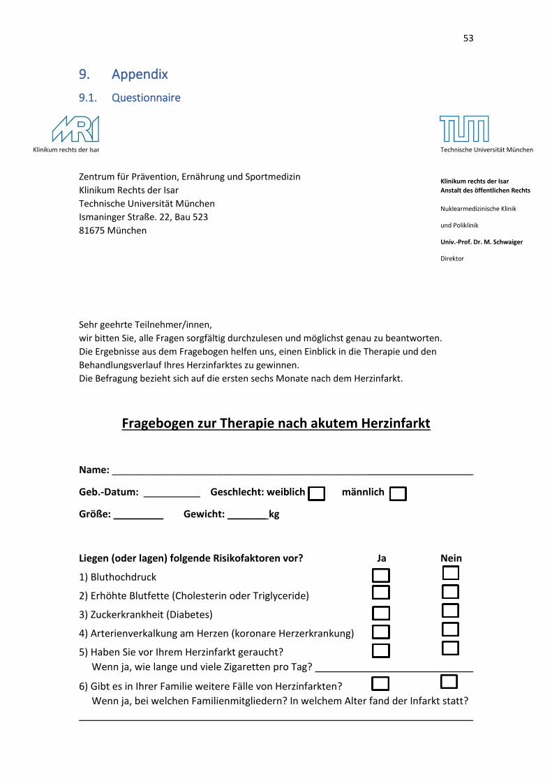
53
9. Appendix
9.1. Questionnaire
Zentrum für Prävention, Ernährung und Sportmedizin
Klinikum Rechts der Isar
Technische Universität München
Ismaninger Straße. 22, Bau 523
81675 München
Sehr geehrte Teilnehmer/innen,
wir bitten Sie, alle Fragen sorgfältig durchzulesen und möglichst genau zu beantworten.
Die Ergebnisse aus dem Fragebogen helfen uns, einen Einblick in die Therapie und den
Behandlungsverlauf Ihres Herzinfarktes zu gewinnen.
Die Befragung bezieht sich auf die ersten sechs Monate nach dem Herzinfarkt.
Fragebogen zur Therapie nach akutem Herzinfarkt
Name: ______________________________________________
Geb.-Datum: Geschlecht: weiblich männlich
Größe: _________ Gewicht: _______ kg
Liegen (oder lagen) folgende Risikofaktoren vor? Ja Nein
1) Bluthochdruck
2) Erhöhte Blutfette (Cholesterin oder Triglyceride)
3) Zuckerkrankheit (Diabetes)
4) Arterienverkalkung am Herzen (koronare Herzerkrankung)
5) Haben Sie vor Ihrem Herzinfarkt geraucht?
Wenn ja, wie lange und viele Zigaretten pro Tag?
6) Gibt es in Ihrer Familie weitere Fälle von Herzinfarkten?
Wenn ja, bei welchen Familienmitgliedern? In welchem Alter fand der Infarkt statt?
Technische Universität München Klinikum rechts der Isar
Klinikum rechts der Isar
Anstalt des öffentlichen Rechts
Nuklearmedizinische Klinik
und Poliklinik
Univ.-Prof. Dr. M. Schwaiger
Direktor

54
7) Waren Sie vor Ihrem Herzinfarkt sportlich aktiv? Wenn ja, welchen Sportarten sind
Sie nachgegangen? Wie oft haben Sie pro Woche trainiert und wie lange (in
Minuten/Woche)? Mit etwa welcher Intensität haben Sie
trainiert?___________________________________
_______________________________________________________________________
___
_______________________________________________________________________
___
_______________________________________________________________________
___
Welche der Medikamente nehmen Sie ein? Ja Nein
1) ACE-Hemmer (z.B. Ramipril)
2) Betablocker (z.B. Bisoprolol, Metoprolol)
3) AT1-Blocker (z.B. Losartan)
4) Ca2+-Kanal-Blocker (z.B. Verapamil)
5) Statine (z.B. Simvastatin)
6) Andere
Fragen zu Rehabilitations-Maßnahmen
1) Welche Versorgungsformen haben Sie in Anspruch genommen?
Ambulant Stationär
2) In welcher Reha-Klinik/ambulanten Versorgung waren Sie?
3) Wie viele Tage nach dem Herzinfarkt begann Ihre Therapie in der Reha-Einrichtung?
4) Wie lange nahmen Sie an der Reha-Maßnahme teil? (Angabe in Wochen)
5) Wie oft haben Sie in den ersten 6 Monaten nach der Reha durchschnittlich in der
Woche Sport getrieben?
6) Welchen körperlichen Aktivitäten sind Sie nachgegangen?

55
Körperliche Aktivität Welche Sportart? Ungefähr wie schnell/intensiv?
Durchschnittliche Dauer/Woche (Angabe in Minuten)
MET (vom Arzt ausgefüllt)
Ausdauersport (z.B. spazieren gehen, laufen, schwimmen, Rad fahren, Fitnesskurse, Ergometertraining)
Technische oder rhythmisch orientierte Sportartarten (z.B. Tanzen, Reiten, Golf)
Rehabilitationssport (z.B. Herzsportgruppe)
andere Sportarten
MET Total:
Vielen Dank für Ihre Teilnahme!

56
10. Acknowledgments
First and foremost, I would like to thank Professor Dr. med. Martin Halle, director of the Center
for Preventive and Sports Medicine at the University Hospital Klinikum rechts der Isar, for giving
me the opportunity to conduct my doctoral thesis in his department.
Second, I would like to thank Dr. Alessandra Boscheri and Dr. med. Katrin Esefeld for their
support during the primary phase of this thesis. She supported me in medical and statistical
questions and had a large impact on my motivation for this thesis.
Many thanks to Privat-Dozent Dr. med. Christoph Rischpler from the Department for Nuclear
Medicine at the University Hospital in Essen who gave me relentless support in technical
questions concerning cardiac imaging.
During the final phase of this thesis, I am very grateful for the support from Professor Dr. med.
Jürgen Scharhag who offered me important statistical and medical advice.
Last, but not least, I would like to express my gratitude to my family. Without their support I
would not have been able to finish this thesis.