Homeostatic Interplay between Bacterial Cell-CellSignaling and Iron in VirulenceRonen Hazan1,2, Jianxin He1,2, Gaoping Xiao1,2, Valerie Dekimpe3, Yiorgos Apidianakis1,2, Biliana
Lesic1,2, Christos Astrakas1,2, Eric Deziel3, Francois Lepine3, Laurence G. Rahme1,2*
1 Department of Surgery, Harvard Medical School and Massachusetts General Hospital, Boston, Massachusetts, United States of America, 2 Department of Microbiology
and Molecular Genetics, Harvard Medical School, Boston, Massachusetts, United States of America, 3 INRS-Institut Armand-Frappier, Laval, Quebec, Canada
Abstract
Pathogenic bacteria use interconnected multi-layered regulatory networks, such as quorum sensing (QS) networks to senseand respond to environmental cues and external and internal bacterial cell signals, and thereby adapt to and exploit targethosts. Despite the many advances that have been made in understanding QS regulation, little is known regarding howthese inputs are integrated and processed in the context of multi-layered QS regulatory networks. Here we report theexamination of the Pseudomonas aeruginosa QS 4-hydroxy-2-alkylquinolines (HAQs) MvfR regulatory network anddetermination of its interaction with the QS acyl-homoserine-lactone (AHL) RhlR network. The aim of this work was toelucidate paradigmatically the complex relationships between multi-layered regulatory QS circuitries, their signalingmolecules, and the environmental cues to which they respond. Our findings revealed positive and negative homeostaticregulatory loops that fine-tune the MvfR regulon via a multi-layered dependent homeostatic regulation of the cell-cellsignaling molecules PQS and HHQ, and interplay between these molecules and iron. We discovered that the MvfR reguloncomponent PqsE is a key mediator in orchestrating this homeostatic regulation, and in establishing a connection to the QSrhlR system in cooperation with RhlR. Our results show that P. aeruginosa modulates the intensity of its virulence response,at least in part, through this multi-layered interplay. Our findings underscore the importance of the homeostatic interplaythat balances competition within and between QS systems via cell-cell signaling molecules and environmental cues in thecontrol of virulence gene expression. Elucidation of the fine-tuning of this complex relationship offers novel insights into theregulation of these systems and may inform strategies designed to limit infections caused by P. aeruginosa and relatedhuman pathogens.
Citation: Hazan R, He J, Xiao G, Dekimpe V, Apidianakis Y, et al. (2010) Homeostatic Interplay between Bacterial Cell-Cell Signaling and Iron in Virulence. PLoSPathog 6(3): e1000810. doi:10.1371/journal.ppat.1000810
Editor: Marvin Whiteley, The University of Texas at Austin, United States of America
Received August 18, 2009; Accepted February 5, 2010; Published March 12, 2010
Copyright: � 2010 Hazan et al. This is an open-access article distributed under the terms of the Creative Commons Attribution License, which permitsunrestricted use, distribution, and reproduction in any medium, provided the original author and source are credited.
Funding: This work was supported by The Claflin Distinguished Scholar Award, Shriners Research grants #8770 and #8850 and National Institute of Health grantAI063433 to LGR. RH was supported by a Shriners Hospitals Research Fellowship (#8494). The funding agencies had no role in study design, data collection andanalysis, decision to publish, or preparation of the manuscript.
Competing Interests: The authors have declared that no competing interests exist.
* E-mail: [email protected]
Introduction
Microbes translate environmental cues to coordinate and
modulate gene expression such that they can adapt to different
niches and overcome hostile environments. Adaptation and
coordination of gene expression is particularly important for
pathogenic microorganisms that need to colonize dynamic host
environments since their ability to sense and respond to host
environmental cues is critical for their survival. In bacteria,
modulation and coordination of gene expression are also influenced
by population density via the regulated production of small
molecules that serve as intricate signals impacting the expression
of virulence factor genes. Many studies have addressed the role of
quorum sensing (QS) communication networks in virulence where
by diffusible intercellular auto-inducers factor and environmental
signals bacterial cultures mediate pathogenicity by coordinating the
expression of a large array of genes [1,2]. Nevertheless, less is known
regarding how environmental cues are translated in the context of
QS signaling and how environmental cues and QS are integrated to
promote the ability of a pathogen to survive and colonize particular
niches within their host environments. The processing and
integration of environmental inputs in QS becomes even more
complex when a pathogen is able to occupy more than one niche.
Pseudomonas aeruginosa is a ubiquitous and an extremely versatile
Gram-negative bacterium with an astounding ability to survive in
many different environments and to infect multiple hosts ranging
from amoebas to humans [3]. This pathogen has an extensively
studied complex QS communication network that facilitates cross-
talk between organisms and impacts many P. aeruginosa group-
related behaviors including virulence [4, 5, 6, 7, 8, and 9]. There
are at least three known QS systems in P. aeruginosa: two are
dependent on the acyl-homoserine-lactone (AHL) QS transcrip-
tion factors LasR and RhlR [10] and a third is dependent on the
4-hydroxy-2-alkylquinolines (HAQs) LysR-type transcription fac-
tor MvfR [11,12]. MvfR activation is mediated by the cell-cell
signaling molecules 4-hydroxy-2-heptylquinoline (HHQ) and 3,4-
dihydroxy-2-heptylquinoline (PQS), and leads to the positive
regulation of many virulence-related factors, a large number of
which are also controlled by the QS signal acyl-homoserine-
lactone (AHL)-mediated RhlR and LasR circuitry.
The MvfR pathway is a critical virulence component essential
for the full virulence of P. aeruginosa in multiple hosts [13,14,15]
PLoS Pathogens | www.plospathogens.org 1 March 2010 | Volume 6 | Issue 3 | e1000810
and is connected to LasR and RhlR by: (i) the dependence of mvfR
expression at the early growth stages as a result of positive control
by LasR [16], (ii) the conversion of HHQ into PQS controlled by
PqsH [17,18] whose expression is mediated by LasR [19,20], and
(iii) the negative effects of RhlR on the pqs operon [16,21], which is
responsible for the synthesis of all HAQs [11,14,19,22,23]
including the MvfR ligands HHQ and PQS [12,17,21].
The QS regulons MvfR, LasR and RhlR respond not only to
QS signal molecules but also to environmental signals [24],
including host factors [25,26,27,28] and other environmental cues
such as phosphate [29], magnesium [30] and iron
[31,32,33,34,35]. Iron acquisition is controlled by a large set of
P. aeruginosa genes activated in response to iron starvation
[36,37,38], including two siderophore complexes, pyoverdine
and pyochelin [39,40], and several ferric uptake regulators,
among them are the general iron uptake regulator Fur, Fur-
regulated pyoverdine siderophore-specific extracytoplasmic sigma
factor PvdS, several ECF sigma factors, and the AraC regulator
PchR, which regulates pyochelin uptake [40]. In low iron
conditions, PvdS binds to iron-starvation (IS) boxes to induce
the transcription of many genes involved in the iron starvation
response [41]. The intricate relationship between QS and iron is
exemplified by a series of findings demonstrating that iron
starvation induced QS systems [26,32,34] and that the QS
regulators MvfR [11], LasR/RhlR [42] and VqsR [31,43,44]
were found to be responsible for the induction of many iron
response genes. Moreover, MvfR contains an IS box in its
promoter [36], and PQS production is positively-affected by two
Fur-regulated small RNAs, Prrf 1 and 2 [45]. Adding to the
complexity of how environmental cues such as iron levels affect
QS and how iron is integrated into QS to modulate virulence gene
expression is the ability of PQS to bind iron [46], to act as an iron
trap molecule [47], and to form a toxic complex against the host
[48].
MvfR activation by HHQ and PQS leads to the upregulation of
the anthranilic acid (AA)- biosynthetic encoding genes phnAB, and
pqsA-E operon [11,12,14] that have a conserved genomic
organization in P. aeruginosa and in HAQs-producing Burkholderia
species [49], to produce more HAQs leading to the upregulation
of the MvfR-regulon in a positive feedback loop. Although the fifth
gene of the pqs operon pqsE (PA14_51380), which encodes a
predicted GloB, Zn-dependent hydrolase [50] and member of the
metallo-beta-lactamase super family (Pfam PF00753), is not
required for HAQ synthesis [12,19], it is co-regulated together
with the pqsA-D genes. We have shown that PqsE is essential for
complete P. aeruginosa virulence in mice because it controls the
expression of a number of MvfR regulon-dependent genes [11].
Although PqsE was previously implicated as the PQS response
gene [19,20], it was recently shown to act independently of MvfR
and PQS [51]. Thus, the PqsE functions associated with the
integration and translation of the QS cell-cell signals has yet to be
resolved.
Here we examine the interplay between environmental cues and
cell-cell signaling molecules and assess how they are integrated in
the modulation of MvfR regulon gene expression. To elucidate the
QS multi-layered regulation, we also examine the functional
dependency of the MvfR regulon components, especially PqsE,
and PQS and HHQ, on the Rhl regulon. The findings presented
offer new insights into the highly complex P. aeruginosa virulence-
associated regulatory loops that may aid in understanding and
controlling its pathogenicity.
Results
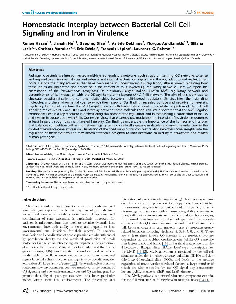
Dissection of the QS MvfR regulon reveals a keycomponent functioning independently of the cell-cellsignaling molecules PQS and HHQ
To elucidate how multi-layered regulatory networks sense and
respond to external and internal cell signals to modulate gene
expression, we studied the role of MvfR pathway components in
integrating and translating signals from PQS and HHQ in the
activation of the MvfR regulon genes. To this end, we measured
pyocyanin production as an index. This secreted P. aeruginosa
phenazine was chosen since its production is dependent on the
MvfR pathway components, including the cell-cell signaling
molecules, PQS and HHQ, and their corresponding biosynthetic
enzymes PqsA-D, their AA precursor, PqsE, and on its Phz
biosynthetic operons (Figure 1A and [11]). Here we found that
overexpression of PqsE under a constitutive promoter
(pDN19pqsE) in pqsA2 and mvfR2 mutant cells not producing
HAQs restored pyocyanin production (Figure 1A). In contrast,
overexpression of mvfR under a constitutive promoter in a pqsE2
background did not restore pyocyanin production (Figure 1A)
even when HHQ, PQS, or PA14 cell-free supernatants were
added (data not shown). These results highlight the crucial role of
PqsE in the regulation of MvfR regulon-dependent factors and
demonstrate that PqsE possesses activation properties that are
independent of HAQ-mediated signals (Table S1). To assess PqsE
mode of action on pyocyanin production, we co-cultured pqsE2
cells constitutively expressing the phenazine biosynthetic operon
phzA2-G2 with pqsE2 cells harboring the phzM and phzS genes
essential to pyocyanin synthesis [52] and assessed pyocyanin
production. As shown in Figure 1B, approximately 60% of the
pyocyanin production was restored, indicating that PqsE partic-
ipated in pyocyanin production regulation rather than in its
synthesis.
Second, we tested whether the precursor of all HAQs, AA was
required for PqsE function instead. To this end we used a triple
mutant strain deficient in phnAB, trpE and kynBU (AA2 mutant)
unable to produce any AA since all three AA synthesis pathways
were knocked out [53]. Expression of PqsE in this triple mutant
also resulted in high levels of pyocyanin production (Figure 1A)
corroborating with the above results and demonstrating that PqsE
function did not require AA or any of its derivatives to promote
production of the MvfR regulon-dependent factor pyocyanin.
Third, since PqsE controlled the regulation of one of the key
MvfR-regulated factors, pyocyanin, we sought to define the impact
Author Summary
Bacterial cells can communicate with one another abouttheir surrounding environment. This information can be inthe form of small self-secreted molecules acting as signalsto activate or inhibit the expression of genes. Pseudomo-nas aeruginosa is an environmental bacterium that infectsdiverse organisms from plants to humans. Our resultsshow that this pathogen uses two highly sensitivenetworks, namely MvfR and LasR/RhlR pathways, tomodulate its virulence functions by titrating the concen-tration of the small molecules HHQ and PQS in a mannerthat depends upon the presence or absence of iron. Vianegative and positive feedback loops, this bacteriumprocesses the signaled information to regulate its viru-lence functions and homeostatically balance the produc-tion of the small molecules required for the activation ofthe MvfR virulence network. Our study sheds light onparadigmatic complex networks that maintain a homeo-static bacterial virulence response.
Environmental and QS Cooperation
PLoS Pathogens | www.plospathogens.org 2 March 2010 | Volume 6 | Issue 3 | e1000810

of this factor in the regulation of all MvfR-dependent virulence
genes. We carried out whole genome expression studies and
compared the expression profiles of a pqsE2 mutant to those of the
PA14 parental strain, an mvfR2 mutant and to those of PA14 and
an mvfR2 over-expressing pqsE strain (NCBI GEO, accession
number #GSE17147). These results showed that PqsE profoundly
affected the expression of 90% of the MvfR-regulated genes,
including at least thirty-six known and predicted transcription
factors (Tables S1B and S2). Of the PqsE-dependent genes, 241
were found to be negatively regulated and 384 positively regulated
Pyoc
yani
n (m
g/L)
A
0
20
40
60
pqsA-
mvfR-
AA-
pqsE- PA14
Pyoc
yani
n (m
g/L)
B
phzC1-::phzC2- pqsE-
8.0
6.0
4.0
2.0
0.0
PA14
+ PqsE + PhZ + MvfR
D
PA14
mvfR-
mvfR- +
PqsE
+ PqsE
StrainStrain
**
*
- -
PA14 pqsE-
C
mvfR- +
PqsE
mvfR-
20
60
100
80
40
020 60 10080400 120
PA14pqsE-
pqsA-
pqsA- + PqsE
Time (h)
E
Fly
Surv
ival
(%)
Figure 1. PqsE, a key mediator of the MvfR regulon activation, functions independently of AA and its derivatives. (A and B) Pyocyaninproduction was measured from PA14 and mutants with and without constitutive expression of PqsE or MvfR as a consequence of the presence ofpDN19pqsE or pDN18mvfR plasmids, respectively. (A) AA2 is a triple mutant with non-functional phnAB, trpE and kynBU that does not produceanthranilate. Production of pyocyanin (+ Phz) was achieved by co-culturing two sets of cells one constitutively expressing phzA2-G2, and the otherphzM and phzS genes encoding the phenazines and pyocyanin biosynthetic genes respectively. Asterisks in A show strains harboring the plasmidpDN19pqsE that are significantly different (P value ,0.01) from PA14 harboring that plasmid. (C–D) PqsE is essential for the virulence of P. aeruginosaagainst Cryptococcus neoformans independently of HAQs. PqsE was constitutively expressed in mvfR2 mutant cells. An empty vector served as acontrol (2). (C) 1 mL of bacterial culture was spotted onto YPD top-agar where yeast cells were plated. Yeast killing zones were formed only aroundthe PA14 and mutant cells expressing PqsE. (D) The death of yeast cells within the killing zone was demonstrated by assessing their viability on YPDplates. (E) PqsE causes fly mortality in absence of HAQs. Survival kinetics of Drosophila melanogaster was assessed using a fly feeding assay. Thesurvival kinetics of pqsA2 and pqsE2 infected flies was significant different (P value ,0.005) form that of PA14-infected flies. However, the kinetics ofpqsA2 + PqsE- infected flies did not differ significantly from that of the PA14-infected flies(P value = 0.27).doi:10.1371/journal.ppat.1000810.g001
Environmental and QS Cooperation
PLoS Pathogens | www.plospathogens.org 3 March 2010 | Volume 6 | Issue 3 | e1000810
by PqsE (Table S1). At least 75 positively-regulated genes encoded
for putative or known virulence factors (Table S1) [11,42].
Importantly, included among the positively-regulated virulence
transcriptional factors was the QS AHL regulator rhlR [38] and
iron response genes, including the iron starvation sigma factor pvdS
and genes involved in the synthesis of the siderophore complex
pyochelin (Table S3A).
To confirm that PqsE overexpression also restores virulence
functions apart from restoring their expression independently of
the signaling molecules PQS and HHQ, we used two assays. The
first is based on the observation that virulent P. aeruginosa strains;
including PA14 kill yeast [54,55,56]; and the second is based on
that P. aeruginosa can infect and kill Drosophila melanogaster
[57,58,59], and that mvfR mutant cells exhibit attenuated virulence
in flies [57]. As illustrated in Figure 1C–D, a zone of yeast growth
inhibition was observed around PA14, but not around the mvfR2,
or pqsE2 mutants following plating of C. neoformans KN99a 5 mm
from the bacterial colony on a YPD plate (Figure 1D). The killing
zone was restored following PqsE overexpression in mvfR2
backgrounds (Figure 1C–D). In agreement flies infected with
pqsA2 or pqsE2 mutants cells exhibited significant delayed in
mortality compared to that caused by the WT or the pqsA2 cells
expressing pqsE (Figure 1E) demonstrating again that PqsE is
crucial for P. aeruginosa pathogenicity and independent of PQS and
HHQ.
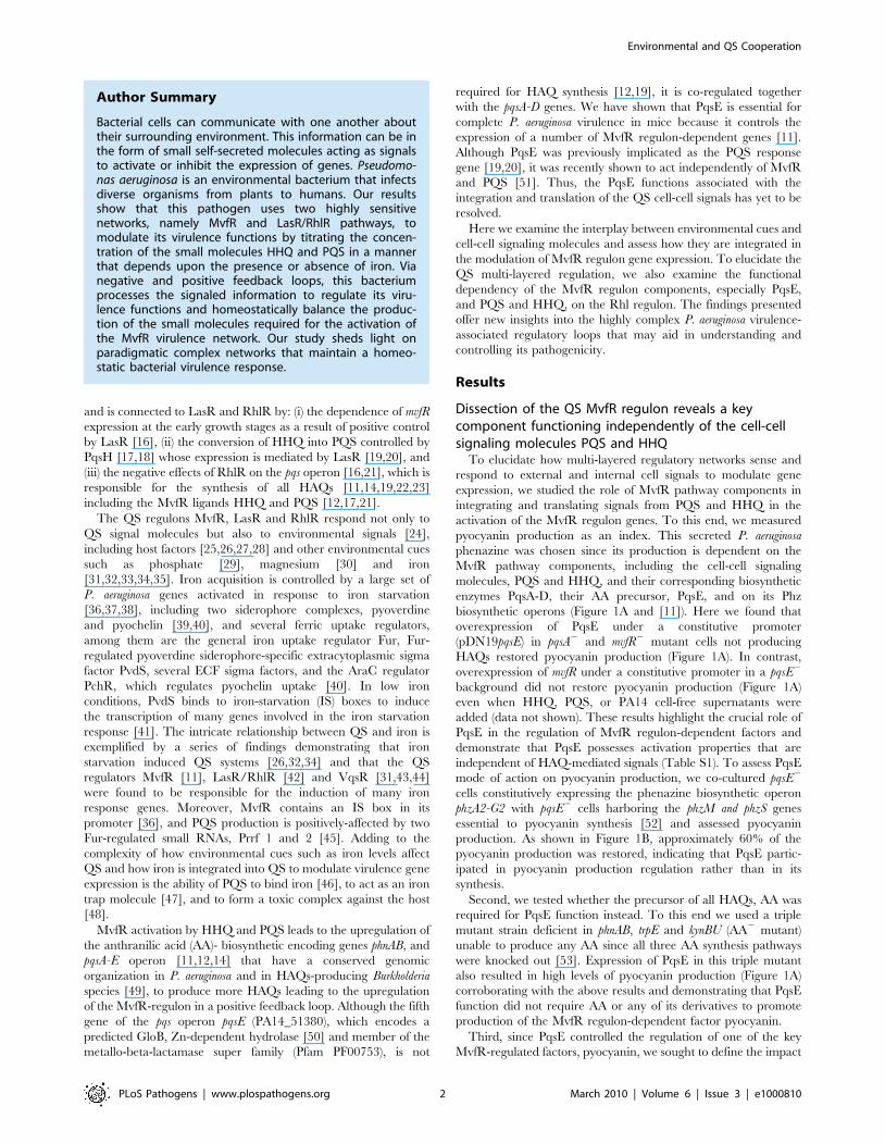
MvfR-dependent gene regulation relies on the functionalcooperation between RhlR and PqsE
Comparison of the pqsE transcriptome (Table S1) to lasR/rhlR
[42] revealed that almost half (46%) of the genes regulated by
LasR/RhlR were also regulated by PqsE (Figure S3A) indicating a
relationship between AHL- and MvfR-mediated QS regulons.
This relationship is also extended to the negative effects that both
components have on the transcription of the pqs operon ([16] and
Table S1 and Figure 2A). A green fluorescent protein (GFP)
reporter gene [32] fused to the pqs operon promoter (Figures 2B),
quantitative PCR analysis (Figure S2D) and quantification of
HHQ and PQS levels (Figure 2C) further validated the above
finding. Moreover, in agreement, Figure 2D shows that HAQ
synthesis down-regulation paralleled the accumulation of AA
(HAQ precursor) followed by an increase in antABC gene
expression that encodes enzymes for AA degradation (Table S1).
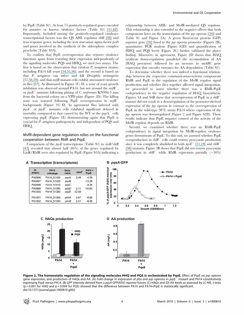
To determine whether there was indeed a functional relation-
ship between the respective communication-systems components
RhlR and PqsE in the regulation of the MvfR regulon signal
production and whether they together affected signal integration,
we proceeded to assess whether there was a RhlR-PqsE
codependency in the negative regulation of HAQ biosynthesis.
Figures 3A and S4B show that overexpression of PqsE in a rhlR2
mutant did not result in a downregulation of the promoter-derived
expression of the pqs operon in contrast to the overexpression of
PqsE in the wild-type (WT) strain PA14 where expression of the
pqs operon was downregulated (Figure 2 and Figure S2D). These
results indicate that PqsE negative control of the activity of the
MvfR regulon depends on RhlR.
Second, we examined whether there was an RhlR-PqsE
codependency in signal integration by MvfR-regulon virulence
genes downstream of PqsE. To this end, we assessed whether PqsE
overproduction in rhlR2 cells could restore pyocyanin production
since it was completely abolished in both pqsE2 [11,19] and rhlR2
[38] mutants. Figure 3B shows that PqsE did not restore pyocyanin
production in rhlR2 while RhlR expression partially (,30%)
Figure 2. The homeostatic regulation of the signaling molecules HHQ and PQS is orchestrated by PqsE. Effect of PqsE on pqs operongene expression, and production of HAQs and AA. (A) Fold change in expression of phn and pqs operons in pqsE2 mutant and PA14 constitutivelyexpressing PqsE versus PA14. (B) GFP intensity derived from a pqsA-GFP(ASV) reporter fusion; (C) HAQs and (D) AA levels as assessed by LC-MS. t-tests(p = 0.001 for HHQ and p = 0.004 for PQS) showed that the difference between PA14 and PA14+PqsE is statistically significant.doi:10.1371/journal.ppat.1000810.g002
Environmental and QS Cooperation
PLoS Pathogens | www.plospathogens.org 4 March 2010 | Volume 6 | Issue 3 | e1000810

restored pyocyanin production in pqsE2 mutant cells. This finding
suggests that PqsE also depends on RhlR in the positive regulation
of pyocyanin production and that RhlR acts downstream of PqsE.
Interestingly, Figure S5 shows that pyoverdine levels are higher in
rhlR2 than in PA14 but not in pqsE2 mutant cells. Moreover, PqsE
or RhlR overproduction in rhlR2 or pqsE2 mutant cells respectively
did not fully downregulated pyoverdine production, while PqsE or
RhlR overproduction in the corresponding mutant cells did (Figure
S5). This finding suggests RhlR-PqsE codependency in the
homeostatic regulation of pyoverdine.
Based on the above findings, it is likely that the PqsE-RhlR
activities were not limited to controlling downstream genes
associated only with pyocyanin or pyoverdine production if the
high number of genes co-regulated by PqsE and the Las/Rhl
system are considered (Figure S3A).
Signal integration studies reveal a homeostatic negativefeedback regulation by HHQ and PQS on cell-cellsignaling and PqsE-controlled genes, respectively
The pyocyanin levels produced by the non-HAQs producing
mutants pqsA2, mvfR2 and AA2 [12,19,22,53] overexpressing pqsE
were higher than the levels produced by the HAQs-producing
PA14 parental strain carrying the same plasmid (Figure 1A). This
difference raised the question regarding whether the presence
and/or levels of HAQs had dose-dependent negative effects on
pyocyanin levels. To this end we assessed the effect of exogenously-
added HAQs on pyocyanin levels by using 20 mg/L of PQS or
HHQ, a concentration corresponding to the approximate
maximal physiologic levels reached by PA14 or pqsH2 strains
respectively at stationary phase ([17] and Figure S4A). Figure 4A
shows that the pyocyanin levels in either pqsA::pqsH2 or mvfR2
mutants overexpressing pqsE were significantly lower in the
presence of either HHQ or PQS. Figure 4B shows that PQS
concentrations (up to 1 mg/L) induced pyocyanin production in
both pqsH2 and pqsA2::pqsH2 cells but concentrations .1 mg/L
decreased pyocyanin production in a dose-dependent manner in
all strains tested (Figure 4B) without significantly affecting cell
growth (data not shown). This concentration-dependent decrease
in pyocyanin levels was independent of PqsE function and phz
operon regulation since it was also observed in pqsE2 cells
constitutively expressing phz genes (Figure 4C). The PQS-
mediated down-regulation was not specific to PA14 cells as it
was also observed in the PA01 P. aeruginosa strain (Figure 4C).
To determine whether high physiological levels of PQS and/or
HHQ negatively-impact pqs operon gene expression, we conduct-
ed experiments using pqsA2::pqsH2 cells harboring the pqsA-GFP
(ASV) reporter gene. Figure 4D shows that 20 mg/L HHQ
negatively-impacted pqsA gene expression compared to 10 mg/L.
PqsA gene expression was not affected by any of the PQS
concentrations tested. Interestingly, a negative effect on pqsA gene
expression, similar to that observed following treatment with
20 mg/L HHQ, was also observed when the two HAQs were
added together in sub-inhibitory concentrations (1 mg/L PQS
+10 mg/L of HHQ). This result is indicating that together HHQ
and PQS have synergistic inhibitory effect and implying also that
high activation of the pqs operon led to its down-regulation.
To further elucidate the role of PQS on PqsE-dependent gene
regulation, we compared the transcriptional profiles of mvfR2 mutant
cells overexpressing PqsE in the absence or presence of 20 mg/L
PQS (Table S1). High PQS concentrations negatively affected the
expression of 191 of 625 (31%) PqsE-regulated genes (Figure 4E and
Table S1). This effect was more apparent among the known and
putative virulence factors where the expression of 64% of the PqsE-
regulated genes, (including chitinase, halovibrin, cellulase, pyocins,
lectin, and elastase genes) was significantly reduced by more than 2-
fold upon PQS addition (Table S1). The addition of PQS further
increased the expression of only 7 genes: fpvA, the major pyoverdine
receptor; gatC, a Glu-tRNA amidotransferase subunit C; sucA, a 2-
oxoglutarate dehydrogenase; bkdA1, a 2-oxoisovalerate dehydroge-
nase and of three hypothetical proteins; PA4642, PA1343 and
PA2405 (Table S1). Interestingly, transcription of phz operon genes
was not modified by the addition of PQS although pyocyanin
0
40
60
0 10 20 30
rhlR rhlR + PqsE
% o
f pqs
A-G
FP (A
SV) m
axim
al
exp
ress
ion
over
tim
e
Time (h)
A pqsA-GFP(ASV)
40
100
80
20
--
B Induction of pyocyanin
Pyoc
yani
n (m
g/L)
+ RhlR + PqsE
-
-
pqsE -
pqsA -
mvfR
rhlR
-
PA140.0
2.0
4.0
6.0
8.0
10.0
35.0
37.0
Figure 3. MvfR network regulation requires finely tuned cooperation between the MvfR component PqsE and the AHL QS regulatorRhlR. (A) The expression of pqsA was determined by measuring GFP emission. A pqsA-GFP (ASV) fusion in the rhlR mutant harboring pDN19pqsE wasused to determine pqsA expression levels. (B) Pyocyanin levels were measured from various PA14 mutants harboring either pDN19pqsE orpUCP20rhlR plasmids. Empty vector served as control.doi:10.1371/journal.ppat.1000810.g003
Environmental and QS Cooperation
PLoS Pathogens | www.plospathogens.org 5 March 2010 | Volume 6 | Issue 3 | e1000810

production was affected (Figure 4A), suggesting that PQS may be
acting post-transcriptionally in this case.
Homeostatic feedback modulation of the MvfR regulon isfine-tuned by an iron starvation response
As shown in Table S3A, PqsE positively-affected the expression
of 43 iron starvation-related genes [36,37] including the iron
starvation sigma factor PvdS [41,60], the pyochelin regulator
PchR [61], vqsR [31,62] and PA2384 [63]. Interestingly, PqsE
negatively regulated only 6 iron related genes, bfrB and the
siderophore pyoverdine associated genes pvdA pvdF, pvdJ, pvdN and
pvdQ (Table S3A) reflected also in the pyoverdine levels (Figure
S5). It is noteworthy that PqsE acted differentially on the
siderophores, serving as a positive regulator of pyochelin and a
negative regulator of pyoverdine (Figure S5). In addition, Table
Figure 4. Negative homeostatic feedback regulation on MvfR regulon products and activity is mediated via cell-cell signaling moleculeconcentration. (A) Pyocyanin levels were assessed in PA14 and mutants cells harboring the plasmid pDN19pqsE with or without the addition of PQS orHHQ (20 mg/L). t-tests (p,0.05) showed that the difference between untreated and PQS/HHQ treated cells was statistically significant (B–C) Pyocyaninlevels were determined following the addition of PQS over a broad-range of concentrations using a PQS non-producing strain (B) or using a narrow range ofPQS concentrations in PQS-producing strains (C). PqsE was constitutively expressed (+PqsE). The empty vector was used as a control. phz genes wereexpressed following co-culture of pqsE2 cells constitutively expressing phzA2-G2 with pqsE2 cells constitutively expressing the phzM and phzS genes. Thecells were grown in the presence of exogenously added PQS and pyocyanin production measured by measuring the OD600 nm. (D) The expression of pqsAwas determined using a pqsA-GFP (ASV) fusion in a pqsA-::pqsH2 double mutant in the presence of various concentrations of HHQ and PQS. (E) A Venndiagram showing the number of PqsE-regulated genes counterbalanced by PQS. The data was adapted from Table S1.doi:10.1371/journal.ppat.1000810.g004
Environmental and QS Cooperation
PLoS Pathogens | www.plospathogens.org 6 March 2010 | Volume 6 | Issue 3 | e1000810

S3A reveal that HAQs are also involved in the control of iron-
related genes by PqsE since constitutive expression of pqsE
triggered this effect in the mvfR2 background cells lacking HAQs
but not in PA14 cells.
To examine how iron starvation is translated in the context of
MvfR signaling, we first examined whether there is a relationship
between iron starvation and the regulation of PQS and MvfR
regulon genes. We compared pqsA transcription using a pqsA-GFP
(ASV) reporter in PA14 cells grown in the absence (D-TSB
medium) or presence of high iron levels. Figure 5A demonstrates
that iron significantly reduced pqsA transcription. Subsequently,
we examined the effect of iron directly on the induction of pqs
operon transcription in presence only of PQS and not of other
HAQs in pqsA2::pqsH2 mutant cells. Using 1 mg/L PQS, an
amount sufficient to fully induce pqs operon transcription and
increasing concentrations of FeCl3 Figure 5B shows an iron
concentration-dependent effect on pqsA gene expression.
We next examined if iron could also counterbalance the
downstream effects of PQS on PqsE-dependent genes by assessing
the effect of HAQs and iron on pyocyanin production. Figure 5C
shows that the addition of iron abolished the reduction in
pyocyanin production conferred by PQS (20 mg/L) and restored
pyocyanin production to that observed in the presence of 1 mg/L
PQS. A similar effect was observed in PA14 cells and pqsA2::pqsH2
cells overexpressing PqsE (Figure S6A) where the addition of
20 mg/L PQS decreased pyocyanin levels which were restored in
the presence of iron. Since iron alone did not affect pyocyanin
production in the experimental conditions used, it suggested that
pyocyanin production was affected due to direct effect of iron on
PQS. No significant difference in growth was observed between
PA14 cells grown in absence or presence of various concentrations
of iron (up to 250 mM, Figure S6B). Collectively, these findings
indicate that iron counterbalanced PQS-dependent regulation by
‘fine-tuning’ its activity, possibly by reducing PQS activity when it
is in a complex with it.
Discussion
In this work, we delineated paradigmatically the complex
relationships between bacterial multi-layered regulatory QS
circuitries, their signaling molecules, and the environmental cues
to which they respond.
Figure 5. Homeostatic interplay between PQS and iron: Iron fine-tunes PQS activities. The effect of iron on MvfR induction was testedusing the pqsA-GFP reporter in PA14 (A) and PA14 pqsA2::pqsH cells treated with PQS (1 mg/L) (B). The effect of iron on pyocyanin production wastested when PQS was supplied at 1 mg/L or 20 mg/L (C). The cells were grown in low iron medium D-TSB or in media supplemented with iron (FeCl3or FeSO4, 200 mM). Asterisks show samples that are statistically significant different (P value,0.05) from the PQS 1 mg/L treated sample.doi:10.1371/journal.ppat.1000810.g005
Environmental and QS Cooperation
PLoS Pathogens | www.plospathogens.org 7 March 2010 | Volume 6 | Issue 3 | e1000810
The intracellular communication system of P. aeruginosa
possesses complex signal transduction systems allowing this
versatile pathogen to regulate and coordinate virulence functions
in the context of multiple hosts, environments, and competition
from other microorganisms [7,64,65,66]. Here we showed that
one of these complex signal transduction systems, MvfR, responds
to both positive and negative feedback loops that are intercon-
nected with the RhlR QS complex system and that these
interactions fine tune the production and concentration of secreted
output signals that in turn serve as inputs to preserve a homeostatic
regulation. Moreover, our experiments demonstrated that via the
finely tuned cooperation and homeostatic interplay between the
MvfR circuitry components PqsE, and PQS and HHQ with RhlR
and iron, this pathogen governs and balances the intensity of its
virulence response.
Although HHQ and PQS principally serve as MvfR ligands
[17,18], our results show that once maximal in vitro physiological
levels are reached, they negatively impact their own production
and the downstream PqsE regulated genes. PqsE, HHQ and PQS
are essential molecules in the negative feedback auto-regulatory
loops that contribute to this homeostatic regulation. Although the
HHQ concentrations shown here are not attained in vitro because
HHQ is fully converted into PQS, this effect is most likely relevant
in vivo where we have shown that HHQ levels are higher than
those of PQS [17]. In addition, in lasR2 mutants that accumulated
during chronic infections HHQ levels are also higher than PQS
since PqsH responsible for the conversion of HHQ to PQS is
under the control of LasR [67]. Nevertheless, we show that HHQ
and PQS have together synergistic effect as a negative auto-
regulators that down-regulated pqs operon transcription, reducing
their own production and that of the other HAQs. Thus, jointly
with PqsE, PQS and HHQ most probably contributed to the
down-regulation of the pqs and phn operons observed during the
late growth phase of P. aeruginosa (Figure S1).
In addition to being activator and auto-down-regulator PQS
acted also as a homeostatic agent at high physiological
concentrations by down-regulating most PqsE-dependent, down-
stream genes. Consistently, maximum pyocyanin production
occurred only at low PQS concentrations that were sufficient to
maximally activate the pqs operon. The homeostatic effect of PQS
downstream of the PqsE genes was clearly independent of MvfR,
PqsE and of other HAQs given that its effects were still apparent in
pqsA2 and mvfR2 backgrounds. Interestingly, this effect appeared
also to be post-transcriptional since PQS did not significantly
impact phz operon transcription but affected pyocyanin production
even when the phz operon was constitutively expressed. The
mechanism behind this effect remains to be discovered. One
intriguing possibility may be that PQS exerts its effect via RsmA
and/or on small RNAs like rsmZ or prrF.
Previous studies have suggested that while PqsE is the PQS
response protein [19,20], it does not influence PQS production
[11,12]. Here we show that PqsE is a crucial player in
orchestrating the homeostatic regulation of the signaling molecules
HHQ and PQS as well as establishing a connection to the QS
RhlR system, underscoring it as a key mediator of MvfR regulon
activation and cooperation with the AHL QS system. Our findings
also provide initial answers as to why PqsE, although not involved
in the synthesis of HAQs in vivo or in vitro [11,19,20]), is tightly
regulated together with the other pqs operon genes. Although our
findings are primarily based on trans-regulatory studies, the
overexpression of PqsE demonstrated for the first time that PqsE
can impact HAQs concentrations by down-regulating their
production. In corroboration, are both the AA accumulation
and the transcriptional induction of the antABC genes responsible
for AA degradation [68,69] and shown to be regulated by prrF1
and prrF2 [45]. Since pqsE is co-transcribed by MvfR together with
pqsA-D genes, the reduced production of HAQs mediated by PqsE
indicates that pqsE gene transcription itself is also downregulated in
a negative feedback mechanism that finely balances the regulatory
loop.
Although PQS and HHQ signal molecules are critical to MvfR-
dependent gene expression, their addition has failed to rescue
pqsE- mutant cells to activate expression of many MvfR-regulated
genes or to produce of pyocyanin [11,17,19,20]. Here we found
that overexpression of PqsE induced pyocyanin production and
transcription of an additional approximately 600 MvfR-regulated
genes independently of MvfR, HAQs and AA, demonstrating the
crucial role of PqsE in activating MvfR regulon genes indepen-
dently of the HAQs. Ultimately, expression of PqsE in an mvfR2 or
pqsA2 strain restored P. aeruginosa virulence as determined by
growth inhibition of yeast and flies feeding assay, indicating that
PqsE did not need HAQs to confer virulence in these systems.
Corroboratory results were reported by Farrow et al. [51] who
showed in a qualitative manner that expression of PqsE in an
mvfR2 mutant restored pyocyanin production. These results
together indicate that, at least with regard to the genes listed in
Table S1, PQS and HHQ only act as inducers of MvfR to express
PqsE that once expressed induces the P. aeruginosa virulence
response without HAQs or MvfR. Thus, PqsE cannot be
designated as the ‘‘quinolone signal response protein’’. Neverthe-
less, it is not yet known how PqsE, a protein that belongs to the
metallo-beta-lactamase super family without any known DNA
binding motifs, regulates the transcription of so many genes. Its
predicted hydrolase activity suggests that it may cleave or
participate in the synthesis of small molecules. Due to the location
of the pqsE gene in the pqs operon, the immediate candidates likely
targeted by PqsE are HAQs. However following extensive LC/MS
analyses, we were unable to detect any molecule that accumulated
or diminished in concentration in pqsE2 cultures compared to WT
cultures (data not shown). In addition we were unable to
complement pyocyanin production in a pqsE2 culture by
exogenously adding HAQs, AHLs or whole PA14 supernatants
([11] and data not shown). Nonetheless, collectively, our results
indicate that PqsE is involved in a negative feedback loop that
affects the regulation and integration of HAQs-mediated cell-cell
signaling molecules and that is functionally dependent on RhlR.
The exact nature of the co-dependency between PqsE and RhlR
remains unclear. The downregulation of rhlR expression by ,2
fold in a pqsE mutant is not sufficient to explain the striking
transcriptional and phenotypic effects mediated by PqsE. Since
PqsE is not predicted to be a transcriptional factor [50] it is highly
likely that it may exert its effect on RhlR post-transcriptionally,
and this effect may be perhaps extended to other transcriptional
factors.
The MvfR affected gene list has a substantial overlap [11] with
the previously published list of Rhl/Las-controlled genes [42], and
the expression of almost all MvfR-regulated genes controlled by
PqsE. Both PqsE activities (i.e., fine-tuning HAQs production by
down-regulating the pqs operon, induction of pyocyanin produc-
tion and downregulation of pyoverdine production) were depen-
dent on RhlR apparently acting downstream but in a tight
collaboration with PqsE. Recently, Farrow and colleagues showed
that the addition of AHL C4-HSL (a RhlR inducer) to PAO1
pqsE2 isogenic mutants also restored pyocyanin production [51].
These findings, although we did not reproduce them in PA14 cells,
are in agreement with our findings that PqsE and RhlR functions
are linked. However, the exact relationship between PqsE and
RhlR, that is when or how they cooperate, remains elusive since
Environmental and QS Cooperation
PLoS Pathogens | www.plospathogens.org 8 March 2010 | Volume 6 | Issue 3 | e1000810
RhlR in some cases functions in the absence of PqsE; for example,
the RhlR-dependent C4-HSL levels in a pqsE2 mutant strain were
identical to the parental strain (data not shown) as also was
previously shown for the mvfR2 mutant [11].
The relationship between iron, QS regulation, and P. aeruginosa
virulence is multifaceted [31,32,34,36,45,63] and extremely
complex. Data presented in this report demonstrate that the
MvfR regulon represents a striking paradigm of the interplay
between environmental signals and bacterial secreted cell-cell
signal molecules that participate in positive and negative
homeostatic regulatory loops. QS MvfR components control the
transcription of many iron related genes, while iron related
regulators control the expression of QS genes (see Table S3B) in
addition to iron related genes. The relationship between iron and
QS regulation is further strengthened through the iron-related
regulators VqsR [43] and the PA2384 product [63] that were
found to control the expression of phnAB and pqsA-E operons.
Furthermore, the iron starvation sigma factor PvdS was shown to
positively control the expression of mvfR via its IS box [36], iron
was shown to control the pqs operon during biofilm formation
[32], and the two small Fur-regulated RNAs Prrf 1 and 2
positively-regulated PQS production [45]. Our results showing
that iron levels affected HAQs activities both as inducers of MvfR
and as fine-balancers provide corroboration for the view that the
MvfR regulon is closely linked with iron regulation. The
complexity of the interplay between the MvfR regulon and iron
control is further increased by: a. the ability of PQS but not HHQ
to trap iron [47], which likely reduces available iron within the cell
and promotes iron starvation, thereby affecting PqsE-mediated
control of bacterial iron response genes, including the siderophores
pyochelin and pyoverdine; and b. iron, especially in high
concentrations, induces oxidative stress that was shown to affect
and being affected by PQS [70]. Thus, it is possible that some of
the phenotypic effects of PQS and iron shown here could be
attributed to oxidative stress. Thus, it would be of importance to
further investigate the contribution of iron, as a nutrient, a signal
molecule, and an oxidative stress inducer in QS and P. aeruginosa
virulence.
The existence of a tight interconnection between iron
concentrations, QS, and virulence in P. aeruginosa is likely due to
iron conditions encountered in vivo [71,72] serving as a signal
indicating a hostile environment requiring expression of virulence
or fitness-related genes. When host tissues become damaged as a
consequence of virulence factor production, the resulting increase
in iron concentrations should down-regulate virulence factor
concentrations, thereby reducing bacterial virulence that may
favor host survival and potentially chronic infection.
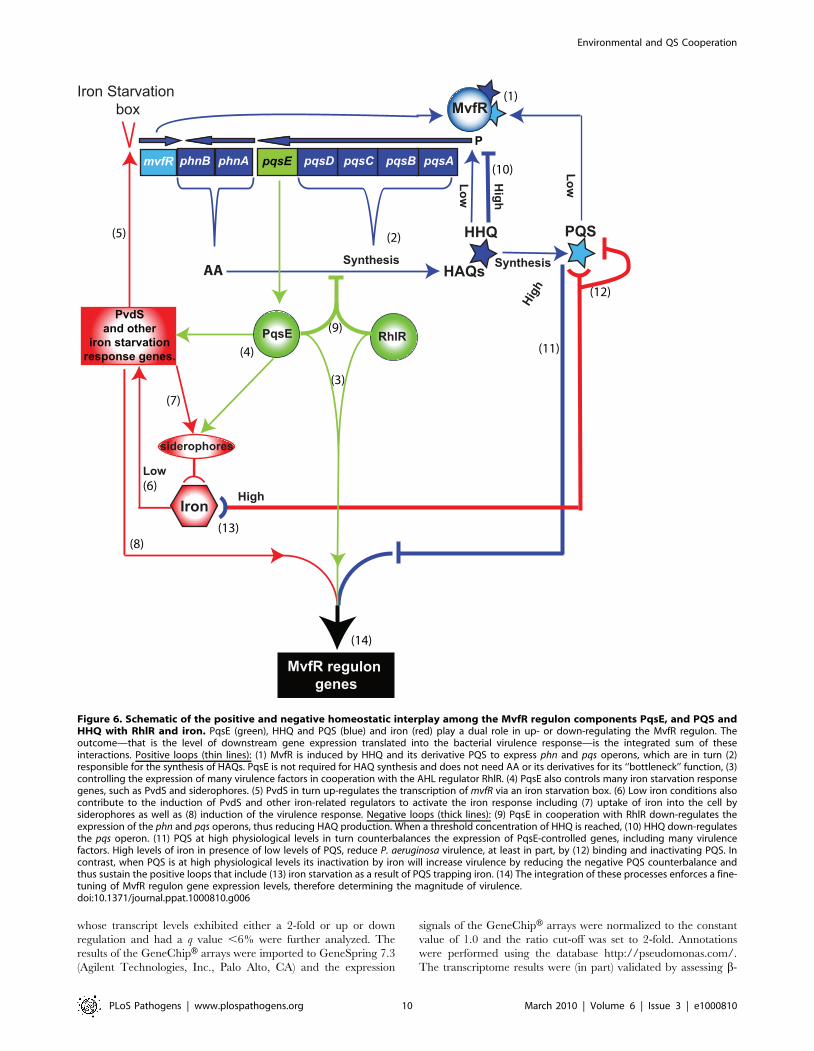
A complete understanding of the regulation of the multiple P.
aeruginosa virulence networks, in particular the mechanisms of the
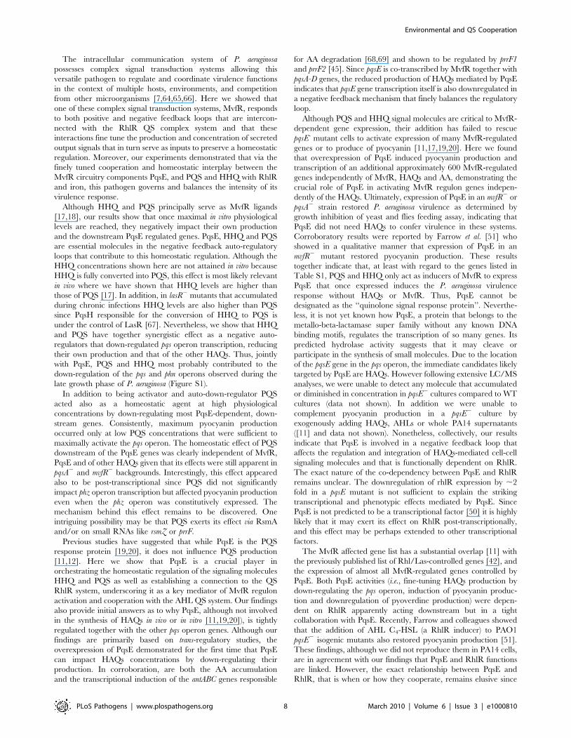
homeostatic and down-regulation processes (Figure 6), will be
essential for the development of drugs targeting QS inhibition
[73,74]. The findings presented in this study may aid in the design
of anti-infective therapies tailored to interfere with virulence
pathways and provide a paradigm for understanding the complex
QS networks of other bacterial pathogens besides that of P.
aeruginosa.
Materials and Methods
Bacterial strains, growth conditions, and plasmidsTable S4 lists bacterial strains and plasmids used in this study. P.
aeruginosa were routinely grown in Luria Bertani (LB) broth at 37uCfor 18 h, and diluted to OD600 nm 0.05 and grown to the desired
OD600 nm. For low iron media the bacteria were grown in D-TSB
medium [36] that was treated with Chelex 100 beads (Bio-Rad,
Hercules, CA) and for high iron FeCl3 or FeSO4 were added at
concentrations of 200 mM. The E. coli JM109 strain was used for
sub-cloning and plasmid propagation. The E. coli S17-1 strain was
used for conjugation between E. coli and P. aeruginosa by the
pEX18Ap-derivative allelic replacement method [75]. Antibiotics
used included ampicillin (Amp) (100 mg/ml), carbenicillin (Crb)
(300 mg/ml), gentamycin (Gnt) (15/60 mg/ml), kanamycin ((Kan),
50/200), tetracycline (Tet) (15/200 mg/ml) and chloramphenicol
(Cam) (15/50 mg/ml) for E. coli and P. aeruginosa respectively.
DNA manipulationsThe plasmid overexpressing PqsE was generated by PCR
amplification of the pqsE gene from PA14 genomic DNA using
primer pairs GX119 and GX120 (Table S4). The PCR product
was digested with HindIII/XbaI and sub-cloned into the pDN19
plasmid vector under plac promoter to generate pDN19pqsE that
constitutively expresses pqsE. Construct integrity was confirmed by
DNA sequencing. Plasmids were introduced into E. coli or P.
aeruginosa PA14 by electroporation. Non polar deletions were
generated by pEX18AP allelic replacement using sucrose
selection. Fragments with the size of about 1 kb flanking the
desired genes were cloned into the pEX18Ap plasmid vector and
introduced into E. coli by electroporation followed by conjugation
to P. aeruginosa. Alternatively, the l-Red recombinase method was
used to generate chromosomal deletions or insertions [53].
Reporter genesTwo kinds of reporter genes were used: 1) translational and
transcriptional fusions to lacZ where the b-galactosidase activity
assay was performed in triplicate as described [76] and; 2) a pqsA-
GFP (ASV) fusion consisting of a pqsA promoter upstream to a
short-lived GFP that allows for the detection of pqs operon up or
down regulation carried on the plasmid pAC37 [32]. Overnight
cultures were diluted to an OD600 nm of 0.05 in black, clear bottom
sterile 96-well assay plates (Corning Inc., Corning, NY). The plates
were incubated for 50 h at 37uC in an Infinite F200 plate reader
(Tecan Group Ltd, Mannedorf, Switzerland). Every 30 min the
plates were shaken for 2 min and read at 600 nm and fluorescence
detected by excitation at 485 nm and emission at 535 nm. The
results are expressed as an average of 3–6 observations that were
normalized to a strain that did not carry the plasmid pAC37.
RNA isolation, generation and analysis of transcriptomedata
Bacteria were respectively grown overnight at 37uC, diluted to
an OD600 nm of 0.05 in 25 ml LB with the corresponding
antibiotics at 37uC until the OD600 nm reached 3.0. The total
RNA was isolated with the RNeasy Mini kit (QIAGEN Inc.,
Valencia, CA) and cDNA synthesis and labeling performed
according to the manufacturer’s instructions (Affymetrix, Santa
Clara, CA). The P. aeruginosa PAO1 GeneChipH Genome array
(Affymetrix) was used for hybridization, staining, washing and
scanning according to the manufacturer’s instructions. Experi-
ments were independently performed in triplicate. Affymetrix
DAT files were processed using the Affymetrix Gene Chip
Operating System (GCOS) to create .cel files. The raw intensity
.cel files were normalized by robust multi-chip analysis (RMA)
(Bioconductor release 1.7) with PM-only models. Array quality
control metrics generated by the Affymetrix Microarray Suite 5.0
were used to assess hybridization quality. Normalized expression
values were analyzed with SAM (Significance Analysis of
Microarray) using the permuted unpaired two-class test. Genes
Environmental and QS Cooperation
PLoS Pathogens | www.plospathogens.org 9 March 2010 | Volume 6 | Issue 3 | e1000810
whose transcript levels exhibited either a 2-fold or up or down
regulation and had a q value ,6% were further analyzed. The
results of the GeneChipH arrays were imported to GeneSpring 7.3
(Agilent Technologies, Inc., Palo Alto, CA) and the expression
signals of the GeneChipH arrays were normalized to the constant
value of 1.0 and the ratio cut-off was set to 2-fold. Annotations
were performed using the database http://pseudomonas.com/.
The transcriptome results were (in part) validated by assessing b-
HAQs
Iron Starvation box
MvfR regulon genes
siderophores
AASynthesis
PQS
Low
PvdSand other
iron starvationresponse genes.
High
(1)
(3)
(4)
(5)
(6)
(7)
High
HHQ
High Iron
Low
Synthesis
(8)
(9)
(10)
(11)
(12)
Low
pqsApqsBpqsCpqsDpqsEphnAphnBmvfR
MvfR
(2)
RhlR
P
PqsE
(13)
(14)
Figure 6. Schematic of the positive and negative homeostatic interplay among the MvfR regulon components PqsE, and PQS andHHQ with RhlR and iron. PqsE (green), HHQ and PQS (blue) and iron (red) play a dual role in up- or down-regulating the MvfR regulon. Theoutcome—that is the level of downstream gene expression translated into the bacterial virulence response—is the integrated sum of theseinteractions. Positive loops (thin lines): (1) MvfR is induced by HHQ and its derivative PQS to express phn and pqs operons, which are in turn (2)responsible for the synthesis of HAQs. PqsE is not required for HAQ synthesis and does not need AA or its derivatives for its ‘‘bottleneck’’ function, (3)controlling the expression of many virulence factors in cooperation with the AHL regulator RhlR. (4) PqsE also controls many iron starvation responsegenes, such as PvdS and siderophores. (5) PvdS in turn up-regulates the transcription of mvfR via an iron starvation box. (6) Low iron conditions alsocontribute to the induction of PvdS and other iron-related regulators to activate the iron response including (7) uptake of iron into the cell bysiderophores as well as (8) induction of the virulence response. Negative loops (thick lines): (9) PqsE in cooperation with RhlR down-regulates theexpression of the phn and pqs operons, thus reducing HAQ production. When a threshold concentration of HHQ is reached, (10) HHQ down-regulatesthe pqs operon. (11) PQS at high physiological levels in turn counterbalances the expression of PqsE-controlled genes, including many virulencefactors. High levels of iron in presence of low levels of PQS, reduce P. aeruginosa virulence, at least in part, by (12) binding and inactivating PQS. Incontrast, when PQS is at high physiological levels its inactivation by iron will increase virulence by reducing the negative PQS counterbalance andthus sustain the positive loops that include (13) iron starvation as a result of PQS trapping iron. (14) The integration of these processes enforces a fine-tuning of MvfR regulon gene expression levels, therefore determining the magnitude of virulence.doi:10.1371/journal.ppat.1000810.g006
Environmental and QS Cooperation
PLoS Pathogens | www.plospathogens.org 10 March 2010 | Volume 6 | Issue 3 | e1000810
galactosidase expression and RT-PCR of selected genes (Figure
S2). The data are deposited in NCBI GEO with accession number
#GSE17147.
Quantitative real-time RT- PCRCells from each triplicate experiment were harvested at an
OD600 nm of 2, 3 and 4. Total RNA was subsequently isolated using
the RiboPure-Bacteria RNA Isolation kit (Ambion, Austin, TX)
according to the manufacturer’s instructions. cDNAs were synthe-
sized with random reverse primers using the Reverse Transcription
RETROscript kit (Ambion) according to the manufacturer’s
instructions. Specific primers (Table S4) for the amplification of
products of approximately 200 base pairs were designed using the
Primer3 algorithm (http://frodo.wi.mit.edu/primer3/) and ana-
lyzed by In Silico simulation of PCR amplifications (http://insilico.
ehu.es/) and by the Primer Analysis Software NetPrimer (Premier
Biosoft International, http://www.premierbiosoft.com/netprimer/
index.html) for the detection of expressed pqsA, pqsE and rpoD that
served as the normalizer genes [77]. Quantitative RT-PCR was
carried out using the Brilliant II SYBR Green QPCR Master Mix
(Stratagene) with a RT Fluorescence Detection System MX3005P
(Stratagene, La Jolla, CA) in a 25 ml final volume. The efficiency of
each pair of primers was determined by a standard curve of 8
dilutions of 1:4 of PA14 genomic DNA. The relative expression
ratios were calculated and analyzed using MXPro analysis software,
version 4.01 (Stratagene) using a mathematical model that included
an efficiency correction. The fold induction of mRNA was
determined from the threshold values that were first normalized
for rpoD gene expression that served as a normalizer and then for the
threshold value of the WT strain harboring the pDN19 plasmid at
an OD600 nm of 2 that served as the calibrator. The data are
expressed as the average of triplicate samples.
HAQs detectionThe quantification of HAQs concentration in bacterial culture
supernatants and in vivo from rectus adominus muscle of burned
and infected mice was performed by LC/MS as described
previously [17,78]. The HAQs were separated on a C18
reverse-phase column connected to a mass spectrometer using a
water/acetonitrile gradient [78]. Positive electrospray in the
MRM mode with 261023 mTorr argon and 30 V as the collision
gas were employed to quantify HAQs using the ion transitions
HHQ 244.159, HHQ-D4 248.163, HQNO 260.159, PQS
260.175, and PQS-D4 264.179. The pseudomolecular ions of
each compound were monitored in full scan mode using the
unsaturated PA14 HAQs response factors.
Pyocyanin production assaySamples of 5 ml were spun down and the supernatants mixed
with equal volumes of chloroform. The lower blue organic phase
was collected and mixed with 5 ml of HCl (0.2 N). The upper
reddish phase was collected and its OD52 onm was measured. The
concentration of pyocyanin was determined by the formula: mg/L
= OD52 onm617.072 normalized to cell counts and the statistical
significance was assessed using the Student’s 2 tailed t-test
assuming equal variance [79]. In order to assess the production
of pyocyanin by expression of the phz genes we used a co-culture of
cells harboring the pUCP-A2G2 and pUCP-MS plasmids [80]. All
experiments were performed in triplicate.
Pyoverdine production detectionD-TSB medium was used to grow 200 ml of bacterial cells in 96
wells plate. Production of pyoverdine was assessed using a plate
reader (Infinite F200, Tecan Group Ltd, Mannedorf, Switzerland).
Pyoverdine levels were determined every 30 minutes using
excitation at 400 nm and emission at 460 nm and the values
obtained were normalized to cell growth (OD600 nm). Pyoverdine
concentrations were calculated using a calibration curve of
fluorescence of a range of concentrations of pyoverdine (Sigma
Aldrich, US).
Yeast killing assayYeast (Cryptococcus neoformans KN99 a, Candida albicans ATCC
#90028 DAY185 strain or Saccharomyces cerevisiae YJM310 strain)
were plated for 2 days on YPD agar (Difco) plates at 30uC. A
colony was picked and grown for 18 h in liquid YPD media (Difco)
at 30uC with shaking (200 rpm). The yeast was diluted 1:100 in
4 ml soft YPD agar (0.6% agar) and poured onto an YPD plate
that was dried for 30 min in a laminar flow hood. A 1 ml drop of
an overnight culture of the desired bacterial strain was put on top
of the yeast lawn and the plate incubated for 2–3 days at 30uC. A
dead yeast zone was formed around a by PA14 bacterial colony
bun not around mutants such e.g., mvfR 2, pqsA 2 and pqsE 2. The
viability of yeast in these zones was tested by plating yeast from
distance of 5 mm from the bacterial colonies on YPD plates.
Fly infectionFly infection feeding assay was performed as previously
described in [58,59]. Briefly, 45 female Oregon-R flies per group,
5–7 days old, were fed with a mixture of 4 ml of LB bacterial
culture at OD600 nm 3.0 with 1 ml of 20% sucrose. Thus, feeding
mix contained a final concentration of 80% LB containing
,26109 bacterial cells per ml and 4% sucrose. An autoclaved
cotton ball was placed at the bottom of each fly vial and was
impregnated with 5 ml of the feeding mix. The 45 flies per
treatment group were sub-divided in three fly vials (15 flies in
each), sealed with a clean cotton ball, and incubated at 25uC. Fly
survival was recorded twice a day until all flies succumbed to the
infection. Statistical analysis of the survival curves was preformed
using the log-rank test (Mantel-Haenszel) of the Kaplan-Meier
estimate of survival using the software MedCalc (http://www.
medcalc.be/). Two independent experiments gave similar results.
Supporting Information
Figure S1 Transcription profile of mvfR and pqsA-E. The
transcription profile was determined from the transcriptome
analysis of PA14 cultures along the growth curve in LB at 37uC.
Found at: doi:10.1371/journal.ppat.1000810.s001 (0.85 MB EPS)
Figure S2 Microarray data validation. The effect of PqsE on the
expression of various differentially-expressed genes in the tran-
scriptome (Table S1) was further confirmed by b-galactosidase
assays derived from transcriptional fusions of the tested genes with
lacZ (A–C) and by quantitative PCR (D). The levels of pqsA and
pqsE gene expression by PCR were determined from PA14
cultures harboring pDN19pqsE (+PqsE) or the control vector
pDN19. The PA14 sample at OD600 nm of 2 served as the
calibrator.
Found at: doi:10.1371/journal.ppat.1000810.s002 (1.12 MB EPS)
Figure S3 PqsE and RhlR cooperate in the regulation of the pqs
operon and of PqsE downstream genes. (A) A Venn diagram
showing the number of genes co-regulated by PqsE (Table S1) and
by the Las/Rhl system [42]. (B) Constitutively-expressed PqsE
does not reduce the expression of pqsA in a rhlR2 mutant. The
expression of the pqsA gene in an rhlR2 mutant constitutively
expressing PqsE or harboring the empty vector was assessed by
Environmental and QS Cooperation
PLoS Pathogens | www.plospathogens.org 11 March 2010 | Volume 6 | Issue 3 | e1000810

quantitative PCR reaction. An OD600 nm reading of a sample from
rhlR2 served as the calibrator.
Found at: doi:10.1371/journal.ppat.1000810.s003 (0.82 MB EPS)
Figure S4 pqsE is not required for HAQs production. The levels
of HHQ, PQS and HQNO were assessed by LC/MS from PA14
(circles) and pqsE2 mutant (squares) cultures at various growth
stages in LB at 37uC.
Found at: doi:10.1371/journal.ppat.1000810.s004 (0.68 MB EPS)
Figure S5 PqsE downregulates pyoverdine production in a
RhlR dependent manner. The effect of PqsE and RhlR on
pyoverdine production was assessed by measuring the pyoverdine
production in PA14 and mutants harboring pDN19pqsE (+PqsE)
or the empty vector pDN19 cells as control. Cells were grown in
D-TSB medium in 96 wells plate and were incubated at 37uC with
shaking for 1 minute every 30 minutes. The results shown are
averages of 6 wells.
Found at: doi:10.1371/journal.ppat.1000810.s005 (4.36 MB EPS)
Figure S6 Iron counteracts PQS-mediated activity. (A) The
effect of iron and PQS was assessed by measuring pyocyanin
production in PA14 and a PA14 pqsA::2pqsH2 double-mutant
constitutively expressing PqsE. PQS was added at 20 mg/L and
iron at 200 mM. Asterisks show samples that are statistical
significantly different (P value,0.01) from the untreated sample
of PA14 (*) and pqsA::2pqsH2 (**). (B) The effect of iron is not a
consequence of growth impairment. Growth curves were per-
formed with PA14 cells in D-TSB media supplied with various
concentrations of FeCl3 in 96 wells plate incubated at 37uC with
shaking for 1 minute every 30 minutes. The results shown are
averages of 6 wells.
Found at: doi:10.1371/journal.ppat.1000810.s006 (0.93 MB EPS)
Table S1 The PqsE controlled genes list. A list of genes
comprising the PqsE regulated genes was generated from our
transcriptional data (NCBI GEO accession number #GSE17147).
The values represent ratios of differential expression between the
pqsE2 mutant vs. PA14 (pqsE2), mvfR2 vs. PA14 (mvfR2), mvfR2
harboring pDN19pqsE vs. mvfR2 with pDN19 (mvfR2 + PqsE),
mvfR2 + pDN19pqsE treated with PQS (20 mg/L) vs. untreated
(mvfR2 + PqsE + PQS) and PA14 harboring pDN19pqsE vs. PA14
harboring the empty vector pDN19 (PA14 + PqsE). The
expression results were validated using reporter genes and
quantitative PCR (Figure S2).
Found at: doi:10.1371/journal.ppat.1000810.s007 (0.08 MB PDF)
Table S2 Transcriptional regulators controlled by the MvfR
pathway. The data on the differential expression of transcription
regulators was adapted from Table S1.
Found at: doi:10.1371/journal.ppat.1000810.s008 (0.07 MB XLS)
Table S3 The interplay between the pqs operon and iron. The
mvfR regulon components controlling (A) or controlled by (B) iron
related regulators. (A) The data was adapted from Table S1. The
values represent fold changes in the pqsE2 mutant vs. PA14
(pqsE2), mvfR2 vs. PA14 (mvfR2), mvfR2 harboring pDN19pqsE vs.
mvfR2 + pDN19 (mvfR2 + PqsE), mvfR2 + PqsE treated with PQS
(20 mg/L) vs. untreated (mvfR2 + PqsE + PQS) and PA14
harboring pDN19pqsE vs. PA14 with pDN19 (PA14 + PqsE). (B)
Iron related regulators controlling the MvfR regulon component.
Fold change in expression of mvfR, pqsA-E and phnAB were
retrieved from previously published studies of iron-related
conditions and regulators 1 [37], 2 [36], 3 [62], 4 [63].
Found at: doi:10.1371/journal.ppat.1000810.s009 (0.07 MB XLS)
Table S4 Strains, plasmids and primers. The P. aeruginosa strains,
plasmids and primers that were used in this study.
Found at: doi:10.1371/journal.ppat.1000810.s010 (0.07 MB
DOC)
Acknowledgments
We thank Linda S. Thomashow for the pUCP-A2G2 and pUCP-MS, Tim
Tolker-Nielsen for pAC37 and Gloria Soberon-Chavez for pGYMCrhlR
Plasmids.
Author Contributions
Conceived and designed the experiments: RH GX LGR. Performed the
experiments: RH JH GX VD YA BL CA FL. Analyzed the data: RH VD
YA ED FL LGR. Contributed reagents/materials/analysis tools: ED FL
LGR. Wrote the paper: RH LGR.
References
1. Fuqua C, Parsek MR, Greenberg EP (2001) Regulation of gene expression by
cell-to-cell communication: acyl-homoserine lactone quorum sensing. Annu Rev
Genet 35: 439–468.
2. Joint I, Allan Downie J, Williams P (2007) Bacterial conversations: talking,
listening and eavesdropping. An introduction. Philos Trans R Soc Lond B Biol
Sci 362: 1115–1117.
3. Cornelis P (2008) Pseudomonas: Genomics and Molecular Biology; In: Cornelis P,
ed;Horizon Scientific Press, 244.
4. Kirisits MJ, Parsek MR (2006) Does Pseudomonas aeruginosa use intercellular
signalling to build biofilm communities? Cell Microbiol 8: 1841–1849.
5. Schuster M, Greenberg EP (2006) A network of networks: quorum-sensing gene
regulation in Pseudomonas aeruginosa. Int J Med Microbiol 296: 73–81.
6. Venturi V (2006) Regulation of quorum sensing in Pseudomonas. FEMS Microbiol
Rev 30: 274–291.
7. Heurlier K, Denervaud V, Haas D (2006) Impact of quorum sensing on fitness of
Pseudomonas aeruginosa. Int J Med Microbiol 296: 93–102.
8. Smith RS, Iglewski BH (2003) P. aeruginosa quorum-sensing systems and
virulence. Curr Opin Microbiol 6: 56–60.
9. Dubern JF, Diggle SP (2008) Quorum sensing by 2-alkyl-4-quinolones in
Pseudomonas aeruginosa and other bacterial species. Mol Biosyst 4: 882–888.
10. Shiner EK, Rumbaugh KP, Williams SC (2005) Inter-kingdom signaling: deciphering
the language of acyl homoserine lactones. FEMS Microbiol Rev 29: 935–947.
11. Deziel E, Gopalan S, Tampakaki AP, Lepine F, Padfield KE, et al. (2005) The
contribution of MvfR to Pseudomonas aeruginosa pathogenesis and quorum sensing
circuitry regulation: multiple quorum sensing-regulated genes are modulated
without affecting lasRI, rhlRI or the production of N-acyl-L-homoserine lactones.
Mol Microbiol 55: 998–1014.
12. Deziel E, Lepine F, Milot S, He J, Mindrinos MN, et al. (2004) Analysis of
Pseudomonas aeruginosa 4-hydroxy-2-alkylquinolines (HAQs) reveals a role for 4-
hydroxy-2-heptylquinoline in cell-to-cell communication. Proc Natl Acad
Sci U S A 101: 1339–1344.
13. Rahme LG, Tan MW, Le L, Wong SM, Tompkins RG, et al. (1997) Use of
model plant hosts to identify Pseudomonas aeruginosa virulence factors. Proc Natl
Acad Sci U S A 94: 13245–13250.
14. Cao H, Krishnan G, Goumnerov B, Tsongalis J, Tompkins R, et al. (2001) A
quorum sensing-associated virulence gene of Pseudomonas aeruginosa encodes a
LysR-like transcription regulator with a unique self-regulatory mechanism. Proc
Natl Acad Sci U S A 98: 14613–14618.
15. Rahme LG, Ausubel FM, Cao H, Drenkard E, Goumnerov BC, et al. (2000)
Plants and animals share functionally common bacterial virulence factors. Proc
Natl Acad Sci U S A 97: 8815–8821.
16. Xiao G, He J, Rahme LG (2006) Mutation analysis of the Pseudomonas aeruginosa
mvfR and pqsABCDE gene promoters demonstrates complex quorum-sensing
circuitry. Microbiology 152: 1679–1686.
17. Xiao G, Deziel E, He J, Lepine F, Lesic B, et al. (2006) MvfR, a key Pseudomonas
aeruginosa pathogenicity LTTR-class regulatory protein, has dual ligands. Mol
Microbiol 62: 1689–1699.
18. Wade DS, Calfee MW, Rocha ER, Ling EA, Engstrom E, et al. (2005)
Regulation of Pseudomonas quinolone signal synthesis in Pseudomonas aeruginosa.
J Bacteriol 187: 4372–4380.
19. Gallagher LA, McKnight SL, Kuznetsova MS, Pesci EC, Manoil C (2002)
Functions required for extracellular quinolone signaling by Pseudomonas aeruginosa.
J Bacteriol 184: 6472–6480.
20. Diggle SP, Winzer K, Chhabra SR, Worrall KE, Camara M, et al. (2003) The
Pseudomonas aeruginosa quinolone signal molecule overcomes the cell density-
dependency of the quorum sensing hierarchy, regulates rhl-dependent genes at
the onset of stationary phase and can be produced in the absence of LasR. Mol
Microbiol 50: 29–43.
Environmental and QS Cooperation
PLoS Pathogens | www.plospathogens.org 12 March 2010 | Volume 6 | Issue 3 | e1000810

21. McGrath S, Wade DS, Pesci EC (2004) Dueling quorum sensing systems in
Pseudomonas aeruginosa control the production of the Pseudomonas quinolone signal
(PQS). FEMS Microbiol Lett 230: 27–34.
22. Lepine F, Milot S, Deziel E, He J, Rahme LG (2004) Electrospray/mass
spectrometric identification and analysis of 4-hydroxy-2-alkylquinolines (HAQs)
produced by Pseudomonas aeruginosa. J Am Soc Mass Spectrom 15: 862–869.
23. Lepine F, Dekimpe V, Lesic B, Milot S, Lesimple A, et al. (2007) PqsA is
required for the biosynthesis of 2,4-dihydroxyquinoline (DHQ), a newly
identified metabolite produced by Pseudomonas aeruginosa and Burkholderia
thailandensis. Biol Chem 388: 839–845.
24. Soberon-Chavez G, Aguirre-Ramirez M, Ordonez L (2005) Is Pseudomonas
aeruginosa only ‘‘sensing quorum’’? Crit Rev Microbiol 31: 171–182.
25. Zaborina O, Lepine F, Xiao G, Valuckaite V, Chen Y, et al. (2007) Dynorphin
Activates Quorum Sensing Quinolone Signaling in Pseudomonas aeruginosa. PLoS
Pathog 3: e35. doi:10.1371/journal.ppat.0030035.
26. Duan K, Surette MG (2007) Environmental regulation of Pseudomonas aeruginosa
PAO1 Las and Rhl quorum-sensing systems. J Bacteriol 189: 4827–4836.
27. Wagner VE, Frelinger JG, Barth RK, Iglewski BH (2006) Quorum sensing:
dynamic response of Pseudomonas aeruginosa to external signals. Trends Microbiol
14: 55–58.
28. Wu L, Estrada O, Zaborina O, Bains M, Shen L, et al. (2005) Recognition of
host immune activation by Pseudomonas aeruginosa. Science 309: 774–777.
29. Jensen V, Lons D, Zaoui C, Bredenbruch F, Meissner A, et al. (2006) RhlR
expression in Pseudomonas aeruginosa is modulated by the Pseudomonas quinolone
signal via PhoB-dependent and -independent pathways. J Bacteriol 188:
8601–8606.
30. Guina T, Wu M, Miller SI, Purvine SO, Yi EC, et al. (2003) Proteomic analysis
of Pseudomonas aeruginosa grown under magnesium limitation. J Am Soc Mass
Spectrom 14: 742–751.
31. Cornelis P, Aendekerk S (2004) A new regulator linking quorum sensing and
iron uptake in Pseudomonas aeruginosa. Microbiology 150: 752–756.
32. Yang L, Barken KB, Skindersoe ME, Christensen AB, Givskov M, et al. (2007)
Effects of iron on DNA release and biofilm development by Pseudomonas
aeruginosa. Microbiology 153: 1318–1328.
33. Bollinger N, Hassett DJ, Iglewski BH, Costerton JW, McDermott TR (2001)
Gene expression in Pseudomonas aeruginosa: evidence of iron override effects on
quorum sensing and biofilm-specific gene regulation. J Bacteriol 183:
1990–1996.
34. Kim EJ, Wang W, Deckwer WD, Zeng AP (2005) Expression of the quorum-
sensing regulatory protein LasR is strongly affected by iron and oxygen
concentrations in cultures of Pseudomonas aeruginosa irrespective of cell density.
Microbiology 151: 1127–1138.
35. Masse E, Arguin M (2005) Ironing out the problem: new mechanisms of iron
homeostasis. Trends Biochem Sci 30: 462–468.
36. Ochsner UA, Wilderman PJ, Vasil AI, Vasil ML (2002) GeneChip expression
analysis of the iron starvation response in Pseudomonas aeruginosa: identification of
novel pyoverdine biosynthesis genes. Mol Microbiol 45: 1277–1287.
37. Palma M, Worgall S, Quadri LE (2003) Transcriptome analysis of the
Pseudomonas aeruginosa response to iron. Arch Microbiol 180: 374–379.
38. Ochsner UA, Koch AK, Fiechter A, Reiser J (1994) Isolation and
characterization of a regulatory gene affecting rhamnolipid biosurfactant
synthesis in Pseudomonas aeruginosa. J Bacteriol 176: 2044–2054.
39. Vasil ML (2007) How we learnt about iron acquisition in Pseudomonas aeruginosa: a
series of very fortunate events. Biometals 20: 587–601.
40. Cornelis P, Matthijs S, Van Oeffelen L (2009) Iron uptake regulation in
Pseudomonas aeruginosa. Biometals 22: 15–22.
41. Visca P, Leoni L, Wilson MJ, Lamont IL (2002) Iron transport and regulation,
cell signalling and genomics: lessons from Escherichia coli and Pseudomonas. Mol
Microbiol 45: 1177–1190.
42. Schuster M, Lostroh CP, Ogi T, Greenberg EP (2003) Identification, timing,
and signal specificity of Pseudomonas aeruginosa quorum-controlled genes: a
transcriptome analysis. J Bacteriol 185: 2066–2079.
43. Juhas M, Wiehlmann L, Huber B, Jordan D, Lauber J, et al. (2004) Global
regulation of quorum sensing and virulence by VqsR in Pseudomonas aeruginosa.
Microbiology 150: 831–841.
44. Juhas M, Wiehlmann L, Salunkhe P, Lauber J, Buer J, et al. (2005) GeneChip
expression analysis of the VqsR regulon of Pseudomonas aeruginosa TB. FEMS
Microbiol Lett 242: 287–295.
45. Oglesby AG, Farrow JM, 3rd, Lee JH, Tomaras AP, Greenberg EP, et al. (2008)
The influence of iron on Pseudomonas aeruginosa physiology: a regulatory link
between iron and quorum sensing. J Biol Chem 283: 15558–15567.
46. Bredenbruch F, Geffers R, Nimtz M, Buer J, Haussler S (2006) The Pseudomonas
aeruginosa quinolone signal (PQS) has an iron-chelating activity. Environ
Microbiol 8: 1318–1329.
47. Diggle SP, Matthijs S, Wright VJ, Fletcher MP, Chhabra SR, et al. (2007) The
Pseudomonas aeruginosa 4-quinolone signal molecules HHQ and PQS play
multifunctional roles in quorum sensing and iron entrapment. Chem Biol 14:
87–96.
48. Zaborin A, Romanowski K, Gerdes S, Holbrook C, Lepine F, et al. (2009) Red
death in Caenorhabditis elegans caused by Pseudomonas aeruginosa PAO1. Proc
Natl Acad Sci U S A 106: 6327–6332.
49. Vial L, Lepine F, Milot S, Groleau MC, Dekimpe V, et al. (2008) Burkholderia
pseudomallei, B. thailandensis, and B. ambifaria produce 4-hydroxy-2-alkylquinoline
analogues with a methyl group at the 3 position that is required for quorum-
sensing regulation. J Bacteriol 190: 5339–5352.
50. Yu S, Jensen V, Seeliger J, Feldmann I, Weber S, et al. (2009) Structure
elucidation and preliminary assessment of hydrolase activity of PqsE, the
Pseudomonas quinolone signal (PQS) response protein. Biochemistry 48:
10298–10307.
51. Farrow JM, 3rd, Sund ZM, Ellison ML, Wade DS, Coleman JP, et al. (2008)
PqsE functions independently of PqsR-Pseudomonas quinolone signal and
enhances the rhl quorum-sensing system. J Bacteriol 190: 7043–7051.
52. Mavrodi DV, Bonsall RF, Delaney SM, Soule MJ, Phillips G, et al. (2001)
Functional analysis of genes for biosynthesis of pyocyanin and phenazine-1-
carboxamide from Pseudomonas aeruginosa PAO1. J Bacteriol 183: 6454–6465.
53. Lesic B, Rahme LG (2008) Use of the lambda Red recombinase system to rapidly
generate mutants in Pseudomonas aeruginosa. BMC Mol Biol 9: 20.
54. Hogan DA, Kolter R (2002) Pseudomonas-Candida interactions: an ecological role
for virulence factors. Science 296: 2229–2232.
55. Kaleli I, Cevahir N, Demir M, Yildirim U, Sahin R (2007) Anticandidal activity
of Pseudomonas aeruginosa strains isolated from clinical specimens. Mycoses 50:
74–78.
56. Kerr JR (1994) Suppression of fungal growth exhibited by Pseudomonas aeruginosa.
J Clin Microbiol 32: 525–527.
57. Lau GW, Goumnerov BC, Walendziewicz CL, Hewitson J, Xiao W, et al. (2003)
The Drosophila melanogaster toll pathway participates in resistance to infection
by the gram-negative human pathogen Pseudomonas aeruginosa. Infect Immun 71:
4059–4066.
58. Apidianakis Y, Pitsouli C, Perrimon N, Rahme L (2009) Synergy between
bacterial infection and genetic predisposition in intestinal dysplasia. Proc Natl
Acad Sci U S A.
59. Apidianakis Y, Rahme LG (2009) Drosophila melanogaster as a model host for
studying Pseudomonas aeruginosa infection. Nat Protoc 4: 1285–1294.
60. Tiburzi F, Imperi F, Visca P (2008) Intracellular levels and activity of PvdS, the
major iron starvation sigma factor of Pseudomonas aeruginosa. Mol Microbiol 67:
213–227.
61. Heinrichs DE, Poole K (1996) PchR, a regulator of ferripyochelin receptor gene
(fptA) expression in Pseudomonas aeruginosa, functions both as an activator and as a
repressor. J Bacteriol 178: 2586–2592.
62. Juhas M, Eberl L, Tummler B (2005) Quorum sensing: the power of cooperation
in the world of Pseudomonas. Environ Microbiol 7: 459–471.
63. Zheng P, Sun J, Geffers R, Zeng AP (2007) Functional characterization of the
gene PA2384 in large-scale gene regulation in response to iron starvation in
Pseudomonas aeruginosa. J Biotechnol 132: 342–352.
64. Girard G, Bloemberg GV (2008) Central role of quorum sensing in regulating
the production of pathogenicity factors in Pseudomonas aeruginosa. Future
Microbiol 3: 97–106.
65. Bjarnsholt T, Givskov M (2007) The role of quorum sensing in the pathogenicity
of the cunning aggressor Pseudomonas aeruginosa. Anal Bioanal Chem 387:
409–414.
66. Winstanley C, Fothergill JL (2009) The role of quorum sensing in chronic cystic
fibrosis Pseudomonas aeruginosa infections. FEMS Microbiol Lett 290: 1–9.
67. D’Argenio DA, Wu M, Hoffman LR, Kulasekara HD, Deziel E, et al. (2007)
Growth phenotypes of Pseudomonas aeruginosa lasR mutants adapted to the airways
of cystic fibrosis patients. Mol Microbiol 64: 512–533.
68. Bundy BM, Campbell AL, Neidle EL (1998) Similarities between the antABC-
encoded anthranilate dioxygenase and the benABC-encoded benzoate dioxygen-
ase of Acinetobacter sp. strain ADP1. J Bacteriol 180: 4466–4474.
69. Urata M, Miyakoshi M, Kai S, Maeda K, Habe H, et al. (2004) Transcriptional
regulation of the ant operon, encoding two-component anthranilate 1,2-
dioxygenase, on the carbazole-degradative plasmid pCAR1 of Pseudomonas
resinovorans strain CA10. J Bacteriol 186: 6815–6823.
70. Haussler S, Becker T (2008) The pseudomonas quinolone signal (PQS) balances life
and death in Pseudomonas aeruginosa populations. PLoS Pathog 4: e1000166.
10.1371/journal.ppat.1000166.
71. Haas B, Kraut J, Marks J, Zanker SC, Castignetti D (1991) Siderophore
presence in sputa of cystic fibrosis patients. Infect Immun 59: 3997–4000.
72. Ratledge C, Dover LG (2000) Iron metabolism in pathogenic bacteria. Annu
Rev Microbiol 54: 881–941.
73. Lesic B, Lepine F, Deziel E, Zhang J, Zhang Q, et al. (2007) Inhibitors of
pathogen intercellular signals as selective anti-infective compounds. PLoS
Pathog 3: e126. doi:10.1371/journal.ppat.0030126.
74. Rasmussen TB, Givskov M (2006) Quorum-sensing inhibitors as anti-pathogenic
drugs. Int J Med Microbiol 296: 149–161.
75. Schweizer HP (1992) Allelic exchange in Pseudomonas aeruginosa using novel
ColE1-type vectors and a family of cassettes containing a portable oriT and the
counter-selectable Bacillus subtilis sacB marker. Mol Microbiol 6: 1195–1204.
76. Miller JH (1972) Experiments in molecular genetics. Cold Spring Harbor, N.Y.:
Cold Spring Harbor Laboratory. pp 352–355.
77. Savli H, Karadenizli A, Kolayli F, Gundes S, Ozbek U, et al. (2003) Expression
stability of six housekeeping genes: A proposal for resistance gene quantification
studies of Pseudomonas aeruginosa by real-time quantitative RT-PCR. J Med
Microbiol 52: 403–408.
78. Lepine F, Deziel E, Milot S, Rahme LG (2003) A stable isotope dilution assay for
the quantification of the Pseudomonas quinolone signal in Pseudomonas aeruginosa
cultures. Biochim Biophys Acta 1622: 36–41.
Environmental and QS Cooperation
PLoS Pathogens | www.plospathogens.org 13 March 2010 | Volume 6 | Issue 3 | e1000810

79. Essar DW, Eberly L, Hadero A, Crawford IP (1990) Identification and
characterization of genes for a second anthranilate synthase in Pseudomonas
aeruginosa: interchangeability of the two anthranilate synthases and evolutionary
implications. J Bacteriol 172: 884–900.
80. Mavrodi DV, Blankenfeldt W, Thomashow LS (2006) Phenazine compounds in
fluorescent Pseudomonas spp. biosynthesis and regulation. Annu Rev Phytopathol
44: 417–445.
Environmental and QS Cooperation
PLoS Pathogens | www.plospathogens.org 14 March 2010 | Volume 6 | Issue 3 | e1000810