BioMed Central Page 1 of 16 (page number not for citation purposes) BMC Microbiology Open Access Research article Acceleration of epithelial cell syndecan-1 shedding by anthrax hemolytic virulence factors Taissia G Popova, Bryan Millis, Chris Bradburne, Svetlana Nazarenko, Charles Bailey, Vikas Chandhoke and Serguei G Popov* Address: National Center for Biodefense and Infectious Diseases, George Mason University, Manassas, VA 20110, USA Email: Taissia G Popova - [email protected]; Bryan Millis - [email protected]; Chris Bradburne - [email protected]; Svetlana Nazarenko - [email protected]; Charles Bailey - [email protected]; Vikas Chandhoke - [email protected]; Serguei G Popov* - [email protected] * Corresponding author Abstract Background: It has been recently reported that major pathogens Staphylococcus aureus and Pseudomonas aeruginosa accelerate a normal process of cell surface syndecan-1 (Synd1) ectodomain shedding as a mechanism of host damage due to the production of shedding-inducing virulence factors. We tested if acceleration of Synd1 shedding takes place in vitro upon treatment of epithelial cells with B. anthracis hemolysins, as well as in vivo during anthrax infection in mice. Results: The isolated anthrax hemolytic proteins AnlB (sphingomyelinase) and AnlO (cholesterol- binding pore-forming factor), as well as ClnA (B. cereus homolog of B. anthracis phosphatidyl choline-preferring phospholipase C) cause accelerated shedding of Synd1 and E-cadherin from epithelial cells and compromise epithelial barrier integrity within a few hours. In comparison with hemolysins in a similar range of concentrations, anthrax lethal toxin (LT) also accelerates shedding albeit at slower rate. Individual components of LT, lethal factor and protective antigen are inactive with regard to shedding. Inhibition experiments favor a hypothesis that activities of tested bacterial shedding inducers converge on the stimulation of cytoplasmic tyrosine kinases of the Syk family, ultimately leading to activation of cellular sheddase. Both LT and AnlO modulate ERK1/2 and p38 MAPK signaling pathways, while JNK pathway seems to be irrelevant to accelerated shedding. Accelerated shedding of Synd1 also takes place in DBA/2 mice challenged with Bacillus anthracis (Sterne) spores. Elevated levels of shed ectodomain are readily detectable in circulation after 24 h. Conclusion: The concerted acceleration of shedding by several virulence factors could represent a new pathogenic mechanism contributing to disruption of epithelial or endothelial integrity, hemorrhage, edema and abnormal cell signaling during anthrax infection. Background It has been recently reported that major pathogens Staphy- lococcus aureus and Pseudomonas aeruginosa exploit acceler- ation of syndecan (Synd) ectodomain shedding as a mechanism of host damage and thus increase their viru- lence [1-3]. Approximately 1% of cell surface proteins are thought to be shed into the extracellular environment by host cells as a process of normal cell surface turnover [4], Published: 07 February 2006 BMC Microbiology 2006, 6:8 doi:10.1186/1471-2180-6-8 Received: 23 September 2005 Accepted: 07 February 2006 This article is available from: http://www.biomedcentral.com/1471-2180/6/8 © 2006 Popova et al; licensee BioMed Central Ltd. This is an Open Access article distributed under the terms of the Creative Commons Attribution License (http://creativecommons.org/licenses/by/2.0 ), which permits unrestricted use, distribution, and reproduction in any medium, provided the original work is properly cited.

Welcome message from author
This document is posted to help you gain knowledge. Please leave a comment to let me know what you think about it! Share it to your friends and learn new things together.
Transcript

BioMed CentralBMC Microbiology
ss
Open AcceResearch articleAcceleration of epithelial cell syndecan-1 shedding by anthrax hemolytic virulence factorsTaissia G Popova, Bryan Millis, Chris Bradburne, Svetlana Nazarenko, Charles Bailey, Vikas Chandhoke and Serguei G Popov*Address: National Center for Biodefense and Infectious Diseases, George Mason University, Manassas, VA 20110, USA
Email: Taissia G Popova - [email protected]; Bryan Millis - [email protected]; Chris Bradburne - [email protected]; Svetlana Nazarenko - [email protected]; Charles Bailey - [email protected]; Vikas Chandhoke - [email protected]; Serguei G Popov* - [email protected]
* Corresponding author
AbstractBackground: It has been recently reported that major pathogens Staphylococcus aureus andPseudomonas aeruginosa accelerate a normal process of cell surface syndecan-1 (Synd1) ectodomainshedding as a mechanism of host damage due to the production of shedding-inducing virulencefactors. We tested if acceleration of Synd1 shedding takes place in vitro upon treatment of epithelialcells with B. anthracis hemolysins, as well as in vivo during anthrax infection in mice.
Results: The isolated anthrax hemolytic proteins AnlB (sphingomyelinase) and AnlO (cholesterol-binding pore-forming factor), as well as ClnA (B. cereus homolog of B. anthracis phosphatidylcholine-preferring phospholipase C) cause accelerated shedding of Synd1 and E-cadherin fromepithelial cells and compromise epithelial barrier integrity within a few hours. In comparison withhemolysins in a similar range of concentrations, anthrax lethal toxin (LT) also accelerates sheddingalbeit at slower rate. Individual components of LT, lethal factor and protective antigen are inactivewith regard to shedding. Inhibition experiments favor a hypothesis that activities of tested bacterialshedding inducers converge on the stimulation of cytoplasmic tyrosine kinases of the Syk family,ultimately leading to activation of cellular sheddase. Both LT and AnlO modulate ERK1/2 and p38MAPK signaling pathways, while JNK pathway seems to be irrelevant to accelerated shedding.Accelerated shedding of Synd1 also takes place in DBA/2 mice challenged with Bacillus anthracis(Sterne) spores. Elevated levels of shed ectodomain are readily detectable in circulation after 24 h.
Conclusion: The concerted acceleration of shedding by several virulence factors could representa new pathogenic mechanism contributing to disruption of epithelial or endothelial integrity,hemorrhage, edema and abnormal cell signaling during anthrax infection.
BackgroundIt has been recently reported that major pathogens Staphy-lococcus aureus and Pseudomonas aeruginosa exploit acceler-ation of syndecan (Synd) ectodomain shedding as a
mechanism of host damage and thus increase their viru-lence [1-3]. Approximately 1% of cell surface proteins arethought to be shed into the extracellular environment byhost cells as a process of normal cell surface turnover [4],
Published: 07 February 2006
BMC Microbiology 2006, 6:8 doi:10.1186/1471-2180-6-8
Received: 23 September 2005Accepted: 07 February 2006
This article is available from: http://www.biomedcentral.com/1471-2180/6/8
© 2006 Popova et al; licensee BioMed Central Ltd. This is an Open Access article distributed under the terms of the Creative Commons Attribution License (http://creativecommons.org/licenses/by/2.0), which permits unrestricted use, distribution, and reproduction in any medium, provided the original work is properly cited.
Page 1 of 16(page number not for citation purposes)
BMC Microbiology 2006, 6:8 http://www.biomedcentral.com/1471-2180/6/8
however during the infectious process shedding can reachpathological proportions. The diverse list of proteins,which are normally shed from the cell surface includes butis not limited to cytokines, growth factors, and cell adhe-sion molecules, such as tumor necrosis factor (TNFα),transforming growth factor α (TGFα), epidermal growthfactors (EGFs), L-selectin, CD44, and Synds [[5] and cita-tions within].
Synds are a group of four distinct proteoglycans (PGs),containing transmembrane core proteins modified withseveral heparan sulfate (HS) and chondroitin sulfatechains. Synd1 is the major HS PG of epithelial cells, whichbinds and regulates a wide variety of biological moleculesthrough its HS chains [6]. Syndecans act as adhesion mol-ecules, modulators of growth factor function, and co-receptors in processes as diverse as morphogenesis, tissuerepair, host defense, tumor development, and energymetabolism. In mammary epithelial cells Synd1 co-local-izes with actin filaments thus anchoring the cytoskeletonto extracellular matrix. Synd4 can be found in the focal
adhesion junctions between cells [7]. Therefore, Synds areinvolved in modulation of cell spreading, adhesion,motility and maintenance of intercellular contacts. Ecto-domain shedding rapidly changes the surface phenotypeof affected cells and generates soluble, biologically activeectodomains that can function as paracrine or autocrineeffectors. A growing body of evidence indicates that thesemolecular and cellular features enable ectodomain shed-ding to regulate many pathophysiological processes, suchas microbial pathogenesis, inflammation, and tissuerepair [1,8].
Shedding of syndecans can be abnormally increased inthe case of the infectious process. The P. aeruginosa shed-ding enhancer was identified as LasA, a known metallo-protease virulence factor [1]. Studies in vivo indicate thatP. aeruginosa activates Synd1 shedding to enhance its vir-ulence in a murine model of lung infection [2]. Sheddingenhancers of S. aureus are represented by pore-forming α-toxin and sphingomyelinase β-toxin [3]. During the infec-tious process, proteolytic removal of ectodomain in a sol-
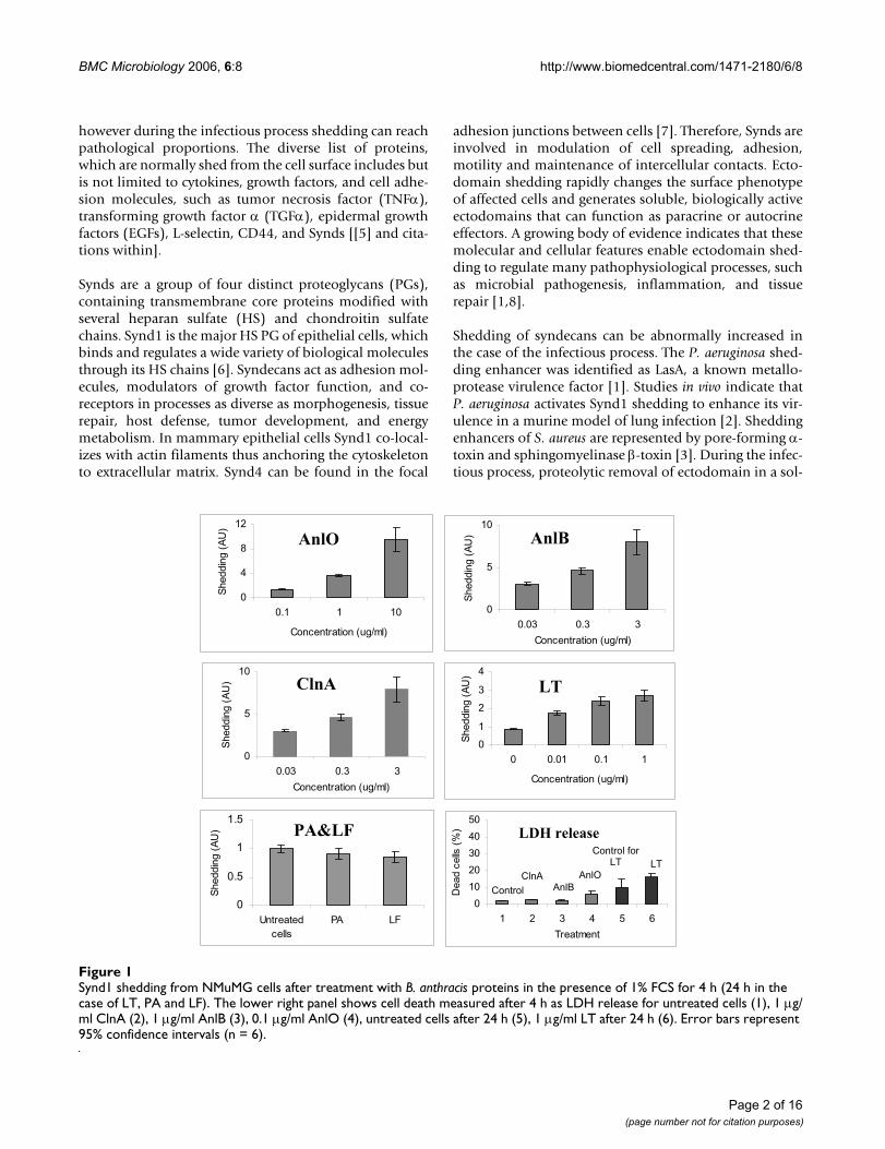
Synd1 shedding from NMuMG cells after treatment with B. anthracis proteins in the presence of 1% FCS for 4 h (24 h in the case of LT, PA and LF)Figure 1Synd1 shedding from NMuMG cells after treatment with B. anthracis proteins in the presence of 1% FCS for 4 h (24 h in the case of LT, PA and LF). The lower right panel shows cell death measured after 4 h as LDH release for untreated cells (1), 1 µg/ml ClnA (2), 1 µg/ml AnlB (3), 0.1 µg/ml AnlO (4), untreated cells after 24 h (5), 1 µg/ml LT after 24 h (6). Error bars represent 95% confidence intervals (n = 6).
0
0.5
1
1.5
Untreated
cells
PA LF
Sheddin
g (
AU
)
0
1
2
3
4
0 0.01 0.1 1
Concentration (ug/ml)
Sheddin
g (
AU
)
0
4
8
12
0.1 1 10
Concentration (ug/ml)
Sheddin
g (
AU
)
0
5
10
0.03 0.3 3
Concentration (ug/ml)
Sheddin
g (
AU
)
0
5
10
0.03 0.3 3
Concentration (ug/ml)
Sheddin
g (
AU
)
0
10
20
30
40
50
1 2 3 4 5 6
Treatment
Dead c
ells
(%)
LT
Control for LT
AnlOClnAAnlBControl
LDH releasePA&LF
LT
AnlO AnlB
ClnA
Page 2 of 16(page number not for citation purposes)
BMC Microbiology 2006, 6:8 http://www.biomedcentral.com/1471-2180/6/8
uble form by secreted microbial factors could enhancehost colonization by altering the morphology and com-promising the integrity of protective barriers formed bypolarized epithelial cells of the skin, the surfaces of bodycavities and internal organs, as well as endothelial cellslining blood vessel walls. The initial pathology can be fur-ther aggravated by exposing intercellular, basolateral, andsubepithelial adhesive components to bacterial factors[9]. Structural damage to the host cell surface with result-ing insult to protective barriers caused by ectodomainshedding along with pathological signaling can initiate amechanism ultimately leading to the malfunction andfailure of life-critical organs and systems.
Inhalation anthrax is a systemic disease characterized bysevere damage to epithelia residing in major internalorgans such as the liver, lung, intestines, spleen, and kid-neys. Disruption of vasculature resulting in massive hem-orrhages and pleural edema is a hallmark of systemicanthrax [10-12]. The B. anthracis genome contains genesfor several proteolytic and hemolytic factors, which arestructurally similar to the shedding inducers from P. aeru-ginosa and S. aureus, including among others the S. aureusα- and β-toxin homologues: anthralysin O (AnlO, pore-forming cholesterol-dependent hemolysin) and anthra-lysin B (AnlB, sphingomyelienase), respectively [13,14].Another anthrax hemolytic factor of interest regarding itspotential activity in ectodomain shedding is anthralysin A
(AnlA, phosphatidyl choline-preferring phospholipase C,PC-PLC), which is 99% homologous to its B. cereus coun-terpart, cereolysin A (ClnA) [13]. Johansen et al. [15]reported that NIH 3T3 cells stably transfected with thegene encoding ClnA displayed a transformed phenotype.Exogenously applied ClnA decreased cell-cell contacts andincreased cell migration [16]. Despite these observationsthe ectodomain shedding has never been studied withregard to infections caused by B. anthracis or B. cereus.Therefore, the main goal of this study was to test ourhypothesis regarding the shedding activity of B. anthracishemolytic proteins and to demonstrate that Synd ectodo-main shedding takes place in response to anthrax infec-tion.
In addition to the hemolysins our attention was attractedto the lethal toxin (LT), a major anthrax virulence factor[17]. The mediator(s) of its toxicity remains unknown. Ithas been shown that LT abrogates intracellular signalingthrough proteolytic cleavage of mitogen-activated proteinkinase kinases (MAPKK) [18]. Administration of lethaldoses of a purified LT to mice and rats causes pleuraledema [19] indicating that LT compromises the integrityof intercellular contacts maintaining the fluid homeosta-sis in the lung. It has been previously suggested that LT iscapable of increasing vascular permeability [20], andrecent experiments with endothelial cells in culture areconsistent with this conclusion [21]. Therefore, we
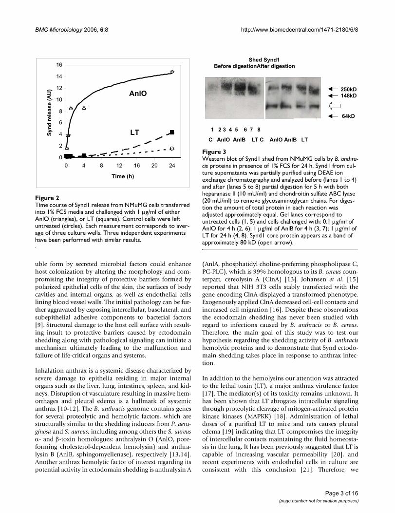
Western blot of Synd1 shed from NMuMG cells by B. anthra-cis proteins in presence of 1% FCS for 24 hFigure 3Western blot of Synd1 shed from NMuMG cells by B. anthra-cis proteins in presence of 1% FCS for 24 h. Synd1 from cul-ture supernatants was partially purified using DEAE ion exchange chromatography and analyzed before (lanes 1 to 4) and after (lanes 5 to 8) partial digestion for 5 h with both heparanase II (10 mU/ml) and chondroitin sulfate ABC lyase (20 mU/ml) to remove glycosaminoglycan chains. For diges-tion the amount of total protein in each reaction was adjusted approximately equal. Gel lanes correspond to untreated cells (1, 5) and cells challenged with: 0.1 µg/ml of AnlO for 4 h (2, 6); 1 µg/ml of AnlB for 4 h (3, 7); 1 µg/ml of LT for 24 h (4, 8). Synd1 core protein appears as a band of approximately 80 kD (open arrow).
C AnlO AnlB LT C AnlO AnlB LT
Shed Synd1Before digestionAfter digestion
1 2 3 4 5 6 7 8
64kD
250kD148kD
Time course of Synd1 release from NMuMG cells transferred into 1% FCS media and challenged with 1 µg/ml of either AnlO (triangles), or LT (squares)Figure 2Time course of Synd1 release from NMuMG cells transferred into 1% FCS media and challenged with 1 µg/ml of either AnlO (triangles), or LT (squares). Control cells were left untreated (circles). Each measurement corresponds to aver-age of three culture wells. Three independent experiments have been performed with similar results.
0
2
4
6
8
10
12
14
16
0 4 8 12 16 20 24
Time (h)
Synd
rele
ase
(AU
)
LT
AnlO
Page 3 of 16(page number not for citation purposes)
BMC Microbiology 2006, 6:8 http://www.biomedcentral.com/1471-2180/6/8
wanted to test whether LT can function as a sheddinginducer, and whether the inhibition of MAPKKs by LTmodulates cell signaling relevant to shedding.
In the current study, we report characterization of the iso-lated anthrax hemolytic proteins AnlO, AnlB, as well asthe AnlA homolog ClnA from B. cereus, regarding their
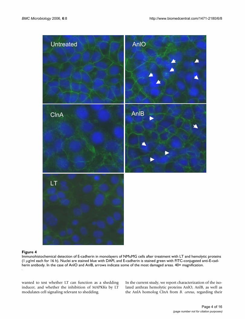
Immunohistochemical detection of E-cadherin in monolayers of NMuMG cells after treatment with LT and hemolytic proteins (1 µg/ml each for 16 h)Figure 4Immunohistochemical detection of E-cadherin in monolayers of NMuMG cells after treatment with LT and hemolytic proteins (1 µg/ml each for 16 h). Nuclei are stained blue with DAPI, and E-cadherin is stained green with FITC-conjugated anti-E-cad-herin antibody. In the case of AnlO and AnlB, arrows indicate some of the most damaged areas. 40× magnification.
LT
AnlBClnA
AnlOUntreated
Page 4 of 16(page number not for citation purposes)

BMC Microbiology 2006, 6:8 http://www.biomedcentral.com/1471-2180/6/8
Synd shedding and barrier permeability-enhancing activi-ties. We found that LT also stimulates Synd1 shedding(although to a less extent compared to hemolysins) by themechanism which involves the MAPKK signaling path-ways. Overall, our observations identify a novel strategy ofanthrax virulence factors, which among their diverse bio-chemical activities stimulate the host cell stress responsesultimately leading to activation of the ectodomain shed-ding. In connection with these observations we also dem-onstrate that Synd1 shedding occurs during the infectiousprocess in the B. anthracis (Sterne) spore-challenged mice.The elevated levels of shed Synd1 are readily detectable inthe circulation just 24 h post challenge. This new featureof the anthrax infectious process could be of high patho-genic significance. We suggest that a cumulative effect ofseveral virulence factors on ectodomain shedding couldcompromise the integrity of host protective barriers, causemalfunction of major organs, and ultimately contribute tothe pathological systemic response typical in anthraxpatients.
ResultsHemolytic factors and LT increase ectodomain shedding from normal murine mammary gland (NMuMG) epithelial cellsShedding activity of isolated recombinant proteins identi-fied by us as candidate shedding inducers was tested usingNMuMG epithelial cells. This murine cell type representsa commonly used model in similar studies [3]. Synd1 isabundant on the epithelial cells and can be assayed usingantibodies specific to the corresponding core proteins orthe HS chains. Using immunodetection with antibodiesspecific for Synd1 core protein we tested NMuMG cell cul-ture supernatants for the presence of soluble Synd1 in theassay conditions when highly charged HS chains are selec-tively retained by the blotting membrane. We found thatall tested hemolytic proteins, namely AnlO, AnlB andClnA, as well as LT cause a concentration-dependentacceleration of Synd1 release into culture supernatantsalbeit at significantly different amounts (Fig. 1). The timecourse of Synd1 release in the case of AnlO shows a fastincrease in the amount of shed Synd which continues forseveral hours (Fig. 2). The LT causes increase in Synd1shedding, although the effect of LT is of lower intensityand develops slower compared to AnlO. The individualcomponents of LT, the protective antigen (PA) and thelethal factor (LF) do not induce shedding (Fig. 1). Thissuggests that neither the extracellular enzymatic activity ofLF nor the sole pore-forming capacity of PA is directlyresponsible for shedding.
Analysis of cell viability in the shedding experimentsreveals a marginal to small degree of the lactate dehydro-genase (LDH) release from treated cells compared tountreated ones. There is no obvious correlation of cell
death with the amount of shed Synd1, which allows us toconclude that the processes of shedding and cell death arenot directly related to each other but rather take place con-comitantly, depending on the nature of the pathogenicfactor and other treatment conditions. For example, treat-ments with either ClnA or AnlB presented in Fig. 1 are notcytolytic, while the amounts of shed Synd1 in both casesincrease 8-fold. AnlO increases cell death 3-fold (from 2%to 6%), while there is an 11-fold increase in Synd1 shed-ding. In the case of LT, incubation for 24 h leads to a 1.6-fold increase in cell death (from 10% to 16%), while theamount of shed Synd1 increases almost 3-fold. The aboveconclusion agrees with direct microscopic observation oftreated monolayers displaying live cells with partially orcompletely shed Synd1 detected by fluorescently-labeledanti-Synd1 antibodies (see below). We did not explorethis topic further, however available data show that the E-cadherin shedding preceded apoptosis in enterocytes[22], while in endothelial cells LT compromised the bar-rier integrity (presumably through ectodomain shedding)independently of apoptosis or necrosis [21]. Stimulationwith the phorbol ester, PMA, a known inducer of shed-ding [5], prolonged viability of epithelial cells [23] andpredisposed monocytes to apoptotic death caused by LT[24].
In order to confirm the biochemical identity of shedSynd1, the following experiment has been carried out.Western blot of NMuMG cell supernatants after treatmentwith either AnlO, AnlB or LT using anti-mouse Synd1antibody demonstrates a high molecular mass smearband that can be attributed to the presence of heterogene-ous heparan sulfate glucosaminoglycan chains in shedSynd1 (Fig. 3). Indeed, digestion of the supernatants withheparanase II and chondrotin sulfate ABC lyase leads tothe appearance of a single band corresponding in gelmobility to the core Synd1 protein, which typicallymigrates in a gel as an approximately 80 KD band [3].
Immunostaining of the NMuMG monolayers with fluorescently-labeled antibodies against E-cadherin and Synd1We examined the NMuMG cells grown on glass slidesusing immuno-fluorescence microscopy. After challengewith proteins causing Synd1 shedding the cells were fluo-rescently stained using E-cadherin- and Synd1-specificFITC-conjugated monoclonal antibodies. E-cadherin is amajor transmembrane component of the apical junc-tional complex. In a simple polarized epithelium thejunctional complex consists of tight junctions and under-lying adherens junctions playing a key role in the forma-tion and maintenance of epithelial barriers [25]. Wetherefore wanted to see if the process of Synd ectodomainshedding observed in our experiments is accompanied bythe loss of intercellular contacts reflected in the dissocia-
Page 5 of 16(page number not for citation purposes)
BMC Microbiology 2006, 6:8 http://www.biomedcentral.com/1471-2180/6/8
Page 6 of 16(page number not for citation purposes)
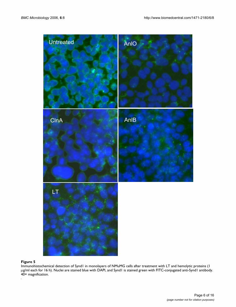
Immunohistochemical detection of Synd1 in monolayers of NMuMG cells after treatment with LT and hemolytic proteins (1 µg/ml each for 16 h)Figure 5Immunohistochemical detection of Synd1 in monolayers of NMuMG cells after treatment with LT and hemolytic proteins (1 µg/ml each for 16 h). Nuclei are stained blue with DAPI, and Synd1 is stained green with FITC-conjugated anti-Synd1 antibody. 40× magnification.
AnlO
ClnA AnlB
LT
Untreated
BMC Microbiology 2006, 6:8 http://www.biomedcentral.com/1471-2180/6/8
tion of the junctional complex. Our analyses revealedobvious cytopathogenic changes in treated NMuMG cells.In all cases, a network of E-cadherin visible in untreatedconfluent cells becomes either disorganized, damaged ordisappears from intercellular contacts upon treatment(Fig. 4), similar to what has been reported for VE-cadherinin endothelial cells treated with LT [21]. Partially conflu-ent NMuMG cells demonstrate intensive Synd1 stainingalong the perimeter of cells, which partially or completelydisappears from cell surfaces after incubation with theshedding-inducing proteins, while remnants of the ecto-domain remain visible in the intercellular space (Fig. 5).Notably, the treated cells retain a high intensity of DAPIblue fluorescence typical for undamaged nuclei, indicat-ing that the loss of E-cadherin and Synd1 takes place fromviable cells.
Inhibition of Synd1 releaseThe ectodomain shedding of cell surface molecules is typ-ically mediated by host metalloproteinase sheddases[5,26]. Both constitutive and accelerated shedding areinhibited by a variety of substances active in a number ofreceptor- and stress-activated signaling pathways, whichinvolve protein tyrosine kinases (PTKs), protein kinase C(PKC), and mitogen-activated protein kinases (MAPKs)[3,5,26]. The activity of LT in macrophages and epithelialcells has been previously reported to involve down regu-lation of MAPK kinase cascades [18,23].
The results of inhibition experiments are presented in theTable. It shows that piceatannol, a specific inhibitor of theSyk family of PTKs [27] is active in both spontaneous andinduced Synd1 shedding for all tested proteins. In the caseof AnlO and LT at low concentration of 0.5 µM the inhib-itor shows some stimulatory effect on both constitutiveand induced shedding, but it strongly inhibits Synd1release in concentrations typical for its activity range of 5to 50 µM [28]. The effect of piceatannol suggests that allfour factors stimulate signaling pathways, which mostprobably involve cytoplasmic Syk, however piceatannolhas also been reported to inhibit other tyrosine kinases ina similar concentration range. In agreement with theabove suggestion the inhibitor of Src PTK family PP2 iscompletely inactive (data not shown). A general PTKinhibitor tyrphostin A25 (at 0.5 to 5 µM) shows only aweak activity. The phosphatidylinositol-3-kinase inhibi-tor LY294002 is inactive with all tested proteins, and weconclude that the cell survival pathway mediated by thiskinase is irrelevant to shedding.
Suramin is a multi-potent therapeutic [29], which amongother activities displays an antitumoral effect by blockingthe growth factors binding to several receptors, includingthe ones for epidermal growth factor (EGF), plateletderived growth factor (PGDF), insulin growth factor II,
and transforming growth factor-β (TGF-β). These growthfactors bind to heparan sulfate-containing proteoglycans,which can be shed in various pathophysiological proc-esses, such as wound repair, and microbial infections[26,30]. Most importantly, suramin modulates activity ofprotein tyrosine phosphatases (PTPs) involved in celladhesion, integrin signaling and cell cycle progression[31,32]. Among these, PTP1B and Cdc25A are inhibitedby suramin in the low µM range. The drug activates PTPαand PTP LAR at higher concentrations. Because of low bio-availability, the suramin concentration above 50 µM hasto be used for the activation effect [33]. The Table showsthat similar to piceatannol, suramin stimulates sheddingat 20 µM. At higher concentration, suramin effectivelyinhibits Synd1 shedding in NMuMG cells induced by LTand AnlO. This effect is consistent with the inhibition-activation pattern of suramin activity toward PTPs, but themulti-potency of suramin excludes its clear interpretationwithout additional studies. Shedding activities of lipasesClnA and AnlB are insensitive to suramin at all concentra-tions tested.
In order to understand which signaling pathways amongp38, ERK and JNK are involved in LT-mediated accelera-tion of Synd shedding, we tested SB202190, an inhibitorof p38; PD98059, an inhibitor of MEK1/2 (ERK pathway);and the JNK inhibitor II. While both AnlO and LT induceSynd1 shedding, LT on itself is a known inhibitor ofMAPK signaling. In contrast, AnlO was reported to stimu-late p38 in macrophages [34]. We found that the p38inhibitor in the range of 1 to 10 µM decreased Synd1shedding in NMuMG cells induced by either AnlO or LTto the level of spontaneous shedding observed in cultureswithout treatment, but it is inactive with ClnA and AnlB.The inhibitor of ERK pathways PD 98059 (at 5 to 50 µM)behaves similar to SB202190 with AnlO, but it is lesseffective in the LT-induced shedding. The only statisticallyreliable effect of the JNK inhibitor is the slight increase inspontaneous shedding from untreated cells. None of thetested inhibitors is toxic to cells in the conditions of theinhibitor experiments (viability of inhibitor-treated con-trol cells remains at the level above 90%, data not shown)indicating that activity of tested inhibitors is not depend-ent on their cytotoxic effect.
Collectively, the inhibition experiments demonstrate thatB. anthracis pathogenic factors induce Synd1 sheddingthrough different signaling pathways, which seem to con-verge on activation of cytoplasmic PTKs. Among testedproteins, one can preliminarily identify two groups ofshedding inducers. The first one includes LT and pore-forming AnlO influencing MAPK pathways, which arecommonly activated in response to receptor stimulationand stress, while the other consists of membranolyticlipases ClnA and AnlB.
Page 7 of 16(page number not for citation purposes)

BMC Microbiology 2006, 6:8 http://www.biomedcentral.com/1471-2180/6/8
Page 8 of 16(page number not for citation purposes)
AnlO and LT cause transient phosphorylation of ERK1/2 and p38 in NMuMG epithelial cellsFigure 6AnlO and LT cause transient phosphorylation of ERK1/2 and p38 in NMuMG epithelial cells. Cells were transferred into growth media with 1% FCS, treated with indicated concentrations of either AnlO or LT for different periods of time and lysed. Western blots were probed with phosphorylation-specific antibodies against ERK1/2 and p38. Results from control untreated cells are shown in the AnlO panel.
AnlO
0
0
total p38 p-p38
1
0.1
total ERK1/2 p-ERK1/2
1
0.1
LT
total-p38p-p38
1
0.1
total-ERK1/2p-ERK1/2
1
0.1
(µg/ml)
Treatment (min)
0 10 30 60 240
Treatment (min)
0 10 30 60 240
BMC Microbiology 2006, 6:8 http://www.biomedcentral.com/1471-2180/6/8
ERK1/2 and p38 phosphorylation patterns in NMuMGs treated with AnlO or LTSince both PD98059 and SB202190 influence the AnlO-and LT-induced Synd1 shedding, ERK1/2 and p38 phos-phorylation patterns were studied in more detail. Fig. 6shows that, compared with untreated cells, within severalminutes the AnlO causes a strong transient ERK1/2 activa-tion lasting for more than 4 h in the presence of 1 µg/mlAnlO. A lower concentration of AnlO causes a shorterperiod of activation followed by a slight increase in sign-aling after 4 h. The latter is present in both control andtreated cells and probably reflects a distress caused byincubation in low FCS media. The p38 phosphorylationreaches maximum intensity after the ERK1/2. In the sameconditions, LT also causes transient activation signals butits effect on the amounts of activated ERK1/2 and p38 isdifferent from that of AnlO: the up-regulation of ERK1/2phosphorylation is detectable at the 10 min time pointand then completely disappears within 30 min. While thetransient peak of p38 activation is detectable at 0.1 µg/mlof LT, its level remains lower compared to AnlO. As a con-trol, LT (1 µg/ml) boiled for 15 min is inactive in MAPKactivation (not shown). It seems that the enzymatic cleav-age of MAPKKs by LT is an important factor reducing theintensity of signaling.
Anthrax LT and hemolytic factors compromise epithelial barrier permeabilityAfter our findings demonstrated the fact of acceleratedSynd shedding, it was important to test if the latter isaccompanied by changes in barrier permeability. We useda primary culture of human small airway epithelial cells(HSAECs) grown on collagen-coated membranes with thepores permeable to Dextran Blue 2000, which was used as
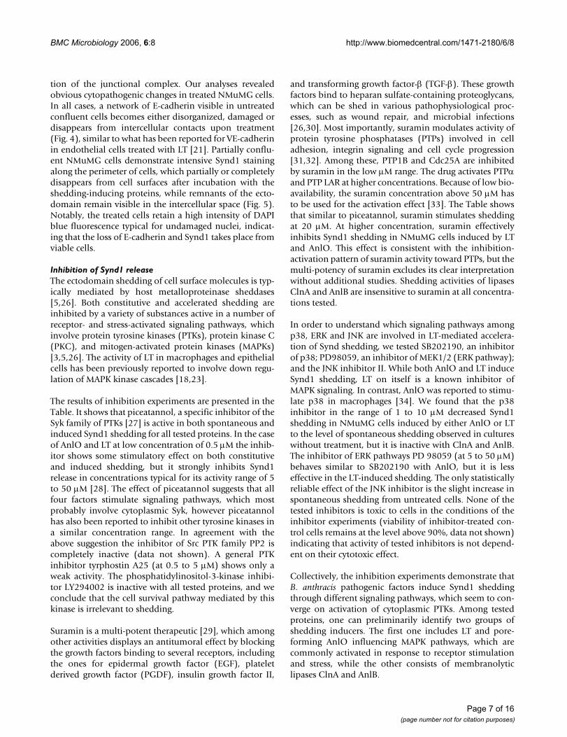
an indicator of barrier integrity [35]. The membranes sep-arated the lower and the upper chambers of the culturewells, thus mimicking the barrier represented in the lowerairways of the lung. The cells were challenged by addingAnlO, ClnA or LT in the upper chambers for 4 h. Aftertreatment, Blue Dextran 2000 was added to the upperchambers for 2 h. The changes in barrier permeabilitywere evaluated by measuring the optical absorbance ofBlue Dextran 2000 in the lower chambers, in comparisonwith untreated cells. Fig. 7 shows that the AnlO and ClnAcause intensive shedding of Synd1. In these experimentalconditions the LT challenge does not induce shedding andtherefore serves as a negative control. The effect of LTbecomes detectable only after the 24 h (data not shown).In general, the HSAECs display the pattern of sensitivity toAnlO, ClnA and LT similar to that of the NMuMG cells. Asexpected, the treatment with AnlO and ClnA causes astrong increase in barrier permeability (Fig. 7B) correlat-ing with shedding. This suggests a possible causal rela-tionship between the two processes, which needs to beelaborated in further studies.
Anthrax infection in mice is accompanied by acceleration of Synd1 sheddingThe experiments with recombinant proteins describedabove suggested that Synd shedding could also take placeduring anthrax systemic infectious process. To confirmthis hypothesis the DBA/2 mice were challenged with B.anthracis spores of the toxigenic Sterne strain intraperito-neally as described before [36]. Blood samples weredrawn, and serum was tested similar to culture superna-tants of protein-treated cells using immunoblot with theantibody against Synd1 core protein. The assay shows aseveral-fold increase in the amount of shed Synd1 on the
Shedding of Synd1 into culture supernatants (A) and changes in barrier permeability (B) for HSAECs treated with the hemolytic proteins and LTFigure 7Shedding of Synd1 into culture supernatants (A) and changes in barrier permeability (B) for HSAECs treated with the hemolytic proteins and LT. The cells initially confluent on a 12 mm insert (Costar, 3.0 µm pores) were treated with 1 µg/ml of each pro-tein for 4 h in 1% FCS media. After treatment the change in barrier permeability was tested as increase in the absorbance of Blue Dextran 2000 added for 2 h to the culture media in the top chamber and detected in the lower chamber, in comparison with untreated cells. Error bars represent 95% confidence intervals; 3 culture plate wells were used for each measurement.
0 1 2 3 4 5
Control
ClnA
AnlO
LT
Relative absorbance(595 mn)
0 1 0 2 0 3 0 4 0
Control
CnlA
AnlO
LT
Shedding (AU)
A B
Page 9 of 16(page number not for citation purposes)

BMC Microbiology 2006, 6:8 http://www.biomedcentral.com/1471-2180/6/8
next day post challenge (Fig. 8A). ELISA protocol with thesame antibody demonstrates similar results, when dilutedserum samples are used to coat the assay plate wells (Fig.8B). A high level of circulating ectodomain is sustaineduntil two days post-infection. A calibration curveobtained with a control serum spiked with different con-centrations of recombinant Synd1 allows estimate thataverage blood Synd1 concentration at day 1 post chal-lenge is increased by 18 µg/ml.
In the conditions of the experiment, about 50% of ani-mals die at day 3, and all animals die by day 6 post chal-lenge. The onset of death on day 3 is accompanied by adecrease in the amount of released Synd1, which could beexplained by a number of mechanisms, such as degrada-tion of syndecan core protein resulting in the loss of itsimmunoreactivity with antibodies and a reduced reten-tion of the protein on the surface of the assay membraneor plate. In any case, the abnormal release of Synd1 intocirculation of infected mice directly indicates that thepathological acceleration of Synd1 shedding takes place invivo at systemic level and is accompanied by the processesof its biochemical turnover.
DiscussionAcceleration of ectodomain shedding represents a part ofan adaptive response of the host cells to different stressfactors and injury such as G protein-coupled receptor ago-nists, growth factors, cytokines, osmotic stress, woundingand phorbol ester activation [37-39]. However the func-tional significance of the ectodomain shedding in micro-bial pathology is uncertain: it could either promotepathogenesis, cellular defenses or both. Microbial mem-brane-damaging factors and other toxins can disturb cellhomeostasis and serve as strong inducers of stress pro-
ceeding through activation of signaling pathways ulti-mately resulting in cytoskeletal rearrangements andincrease in barrier permeability [40]. Although thecytoskeletal rearrangements and Synd1 ectodomain shed-ding are closely interconnected [6,41], a direct linkbetween stress response, Synd1 ectodomain shedding andbarrier dysfunction has never been demonstrated for bac-terial toxins.
Initial evidence that B. anthracis toxins can disrupt hostepithelial and endothelial barriers is available from earlyanthrax publications. For example, Smith et al. [42] usingLT produced in vivo identified vascular damage and renalfailure as a consequence of its activity, while Smith andStoner [20] demonstrated that LT induced an increase invascular permeability. These observations agree with thefact that the most damaged organs in the infectious proc-ess are the ones with high epithelial and endothelial cellcontent such as spleen, lungs, liver, renal system, vascula-ture of blood and lymphatic vessels. A recent report byWarfel et al. [21] confirmed that LT can increase theendothelial barrier dysfunction independent of necrosisor apoptosis. The LT preparations used in early studieswere crude, so we took into account a possibility thatpathogenic factors other than LT could have played role inthe observed effects. A spectrum of these factors includes(but may not be limited to) cytolytic lipases and pore-forming toxins [13,43], and proteases of different specifi-city [14,36].
Our experiments demonstrate that bacterial secreted fac-tors, such as pore-forming toxin AnlO, and cytolyticlipases ClnA and AnlB accelerate the normal process ofhost cell Synd1 and E-cadherin ectodomain shedding,which is as a proteolytic mechanism of releasing them in
Synd1 content in sera of mice challenged with 30 LD50 of Sterne strain spores intraperitoneallyFigure 8Synd1 content in sera of mice challenged with 30 LD50 of Sterne strain spores intraperitoneally. The serum samples were either dot-blotted in duplicates onto membrane (A) or were used to cover wells of the ELISA plate (B) for immunodetection of Synd1 with antibody 281-2. Graphs represent average fold increases over control. In (A), two mice were used at the day of challenge, three mice at each day 1 and 2, and two mice at day 3. In (B), seven mice were used at each of the days 0, 1 and 2, and three mice at day 3. A calibration concentration-response curve obtained by triplicate measurements of control serum spiked with recombinant Synd1 is shown in (C). The bars indicate 95% confidence intervals. *P < 0.05, **P < 0.01.
0
1
2
3
4
5
6
7
0 1 2 3
Time post challenge (days)
Sheddin
g (
AU
)
0
1
2
3
0 1 2 3
Time post challenge (days)
Rela
tive a
bsorb
ance
1
1.5
2
2.5
3
0 10 20
Synd1 spike (ug/ml)
Rela
tive a
bsorb
ance
C**A B*
Page 10 of 16(page number not for citation purposes)

BMC Microbiology 2006, 6:8 http://www.biomedcentral.com/1471-2180/6/8
a soluble form [4]. The effects of AnlO and AnlB are simi-lar to what has been previously reported for their bio-chemical analogs, staphylococcal α- and β-toxins,respectively, in a similar concentration range [3]. We fur-ther show that the process of Synd1 shedding is accompa-nied by loss of epithelial barrier integrity of HSAECs inculture. The abnormal level of shed Synd1 in the blood ofspore-challenged mice suggests that anthrax secreted fac-tors could compromise epithelial barrier integrity at theearly stages of the disease.
The full spectrum of proteins shed in anthrax requires fur-ther studies. Our preliminary data (not shown) indicatethat different combinations of shedding factors could pro-duce synergictic effects. Biologically relevant concentra-tions of anthrax shedding inducers in tissues, organs andbody fluids are unknown, and therefore the assessment ofeach protein's contribution to ectodomain shedding invivo is currently impossible, but the capacity of B. anthracisto produce hemolytic proteins, in addition to LT, has beendemonstrated in both aerobic and anaerobic culture con-ditions [13,43,44]. The antibodies against these proteinsare also detectable in serum of mice challenged with B.anthracis (Sterne) spores (data not shown). In our experi-mental conditions the release of Synd1 and E-cadherintakes place within several hours, and high levels of shedSynds are detectable after 24 hours post infection withspores (we have not tested earlier time points). This obser-vation opens a possibility of using shed ectodomainrelease into circulation for early detection of the anthraxinfectious process. Maximal Synd release coincides intime with the appearance of bacteria in the spleens ofchallenged animals tested in our previous experiments[45]. Bacteria became detectable in the spleens at approx-imately 16 h post infection, reached maximum numbersat about 24 h and then declined before death.
Normally, ES is mediated by metalloproteinases, whichare collectively called sheddases or secretases. Our dataagree with the host sheddase modulation mechanismdemonstrated by others [3,5,38], because metalloprotein-ase inhibitors such as galardin and phosphoramidonreduce shedding induced by AnlO (data not shown),which has no enzymatic activity on its own. Other anthraxproteins, ClnA and AnlB are not proteases, and thereforecannot shed Synd1 by direct proteolysis on the cell sur-face. LT is a metalloprotease but induction of Synd1 shed-ding requires LT delivery into the host cell, in agreementwith the extracellular cleavage of the Synd1 core proteinby cellular sheddase. The fact that proteins of an abso-lutely different nature, such as proteases and lipases of dis-tinct enzymatic specificities along with pore-formingtoxins possessing no catalytic activity, display similareffects with regard to Synd1 shedding indicates activationof a common intracellular mechanism by diverse extracel-
lular signals. Indeed, the activity of piceatannol againstshedding by all tested inducers suggests that cytoplasmicSyk PTK serves as the common point of convergence. Thismechanism however retains a certain level of specificityjudging by the fact that neither PA nor LF induces shed-ding.
The MAPK-mediated pathways have been previouslyimplicated in receptor-induced ectodomain shedding [5].It has also been reported that AnlO stimulated the p38 sig-naling in macrophages [34]. Our data show that theinhibitors of ERK1/2 (PD98059) and p38 (SB202190)decrease the AnlO- and LT-induced shedding. This effectagrees with the mechanism recently discovered for thehydrogen peroxide-stimulated cytoskeletal reorganizationin endothelial cells [46]. It has been shown that both ofthe above inhibitors attenuated MAPK-mediated activa-tion of the small heat shock protein Hsp27 downstreamfrom ERK1/2 and p38. This protein is responsible for theactin stress fiber polymerization, which accompaniesSynd ectodomain shedding [41,47,48]. We however can-not conclude which of the MAPK pathways plays a pre-dominant role in shedding.
Currently available data suggest that the p38 pathwaydefends against bacterial pore-forming toxins in vivo andin vitro [49], therefore the p38-mediated shedding couldrepresent a protective cell response to stress (unless itreaches a pathological proportion). In agreement withthis, Kevil et al. [50] reported that p38 inhibitor attenu-ated the oxidant stress fiber formation and prevented gen-eration of gaps between endothelial cells. Warfel et al. [21]found that p38 inhibitor increased the endothelial barrierresistance, although concluded that this effect was some-what paradoxical given the ability of LT, as known inhib-itor of MAPK signaling, to decrease barrier resistance. Wesuggest that LT plays a dual role upon interaction with thehost cells by inducing both the cellular stress and the inhi-bition of MAPK activation. The intricate combination ofboth processes results in the reduction of the transientstress signal, which nevertheless remains sufficient for theinduction of shedding but reduces the potency of LT asshedding inducer. In support of this, our data show thatLT induces small but detectable activation of p38 even athigh LT concentration. In contrast to LT the pretreatmentof cells with the effective p38 inhibitor is expected to com-pletely block the stress signal and the consequent shed-ding. Transient p38 activation has never been reported inconnection with LT activity although LT-induced ERK1/2activation has recently been described as toxin-induced LTresistance in macrophages [51] in a lower concentrationrange compared to our experiments.
Page 11 of 16(page number not for citation purposes)

BMC Microbiology 2006, 6:8 http://www.biomedcentral.com/1471-2180/6/8
ConclusionOur findings provide additional insights into the studiesof B. anthracis virulence factors, such as LT and hemolyticproteins, at cellular and organism levels, where sheddinghas to be taken into account as a new anthrax pathogenicmechanism. It could rapidly change cell surface propertiesand increase barrier permeability by the concerted effectof several pathogenic factors contributing to dissemina-tion of infection, hemorrhages and edema. Shedding gen-erates biologically active ectodomains that can function asparacrine or autocrine effectors [8]. The shed soluble pro-teoglycans are highly hydrated and this effect is expectedto exacerbate edema by causing influx of water into theintercellular space. It has already been shown that shedSynd1 is toxic to mice [52], and that inoculation of Synd1ectodomain restored sensitivity of Synd-/- mice to P. aeru-ginosa [2]. These findings suggest high pathological signif-icance of Synd shedding in the anthrax infectious process,which needs to be further addressed in animal experi-ments.
MethodsInhibitors, reagents and isolated proteinsNMuMG epithelial cells (CRL-1636) were from ATCC(Manassas, VA), human lung epithelial cells (HSAEC)were from Cambrex, Inc. (Walkersville, MD). DMEMmedia was from ATCC (Manassas, VA), other cell culturereagents were from Cellgro (Herndon, VA). Galardin, tyr-phostin A25, piceatannol, suramin, SB202190, PD98059,JNK inhibitor II and PP2 were from Calbiochem (Darm-stadt, Germany). Anti-human Synd1 antibody clone Mi15and rat anti-mouse Synd1 (clone 281-2) were from BDBiosciences (San Diego, CA). Antibody against total anddouble phosphorylated p38 (Thr180/Tyr182), ERK(Thr202/Tyr204) and JNK (Thr183/Tyr185) were fromCell Signaling Technology (Beverly, MA). Hemolytic B.anthracis proteins were expressed in E. coli, purified andcharacterized as described before [13]. They are at least95% homogeneous based on the results of SDS-PAGEanalyses. The phospatidyl choline-preferring phospholi-pase C from B. cereus (cereolysin A) was purchased fromSigma (MA) and was used without further purification.Recombinant protective antigen (PA) and lethal factor(LF) were purchased from List Biological Laboratories(Campbell, CA). The endotoxin content of all proteinswas determined by Quantitative Chromatogenic LAL kit(Cambrex, MD). Recombinant murine Synd1 expressedin E. coli as a His6-tagged protein (>95% purity) was a giftfrom Prof. Myung-Chul Chung (George Mason Univer-sity, VA). The protein concentration was determined usingBradford assay with BSA as a standard.
Activation of shedding in cultured cellsHuman Small Airway Epithelial Cells, or HSAECs (Cam-brex, Inc., Walkersville, MD), were grown in DMEM/F12
complete medium with 10% fetal calf serum (FCS,Gibco). Before challenge the FCS content was reduced to1%. NMuMG cells were grown up in Dulbecco's modifiedEagle's medium with 4.5 g/l glucose, 10 µg/ml insulin,and 10% FCS. HSAECs were grown up in Ham's F12media supplemented with non-essential aminoacids,pyruvate, β-mercaptoethanol and 10% FCS. Cells wereseeded in 96-well plates, cultured to 1 day post conflu-ence, then stimulated with indicated proteins usingserum-free media from Cellgro (Herndon, VA) supple-mented with 1% FCS. LT was used as a mixture of equalamounts of PA and LF at total concentration of 1.0 µg/ml,0.1 µg/ml, and 0.01 µg/ml. After stimulation, the plateswere spun down at 1,100 × g for 10 min and supernatantwas frozen at -20°C for further analyses. In control exper-iments, a known inducer of Synd1 shedding, PMA (4α-phorbol-12,13 didecanoate) in concentration 10 µM in1% FCS media induced 4-fold increase in shed Synd-1from NMuMG cells after 24 h incubation. In the sameconditions, endotoxin in concentration 10 ng/ml inducedneither significant shedding nor activation of p38 andERK1/2. Pre-treatment of AlnO (1 µg/ml) with polymyxin(50 µg/ml) had no effect on the amount of shed Synd1and the p38 or ERK1/2 phosphorylation. Endotoxin con-tamination from added proteins in shedding experimentsdid not exceed 5 ng per ml of culture medium.
Dot Blot AssaysSupernatant from treated cells (100 µl) was added to 1 mlof acidification buffer (150 mM NaCl, 50 mM NaOAc,0.1% Tween-20, pH 4.5). Immobilon NY+ membrane (9× 12 cm, Millipore) was prepared by first soaking it inacidification buffer. Bio-Dot microfiltration apparatus(Bio-Rad, CA) was used for all syndecan dot blots. Thesample wells were re-saturated with 100 µl of acidificationto prevent drying of the membrane. 400 µl of sample solu-tion (in acidification buffer) was used per well on theapparatus. First the sample was kept in the wells on top ofthe membrane for 3 min and then it was drawn throughthe filter/membrane for another 3 min. The wells wererinsed twice with 400 µl of acidification buffer. The mem-brane was taken out, rinsed twice for 5 min in acidifica-tion buffer and blocked with 3% powdered milk in buffer#2 (150 mM NaCl, 10 mM Tris-HCl, pH 7.4) for 1 h. Themembrane was then incubated with rat anti-mouse Synd1antibody at a dilution of 1:1000 in 3% milk in buffer #3(150 mM NaCl, 10 mM Tris-HCl, 0.3% Tween-20, pH7.4) for 2 h on a platform shaker at room temperature. Forthe cells of human origin the antibody against humanSynd1 has been used in a dilution 1:2000. The membranewas then washed with buffer #3 three times for 5 min andincubated with goat anti-rat HRP-conjugated secondaryantibody at a dilution of 1:7500 for murine Synd1 blot, orwith goat anti-mouse HRP-conjugated polyclonal anti-body at 1:1000 for the heparan sulfate or human Synd1
Page 12 of 16(page number not for citation purposes)

BMC Microbiology 2006, 6:8 http://www.biomedcentral.com/1471-2180/6/8
blots for 1 h at room temperature. The membranes werethen washed again three times for 5 min with buffer #3.All membranes were developed using ECL Plus WesternBlotting Detection kit (Amersham Biosciences, NJ) andKodak BioMax Light Film (Sigma, MO). The results werequantified by scanning the exposed film, and evaluatingthe intensity of exposed dots by software AlphaEase FC(Alpha Innotech, San Leandro, CA). Results wereexpressed as amount of Synd shed in relative absorbanceunits (AU) using a calibration curve generated by two-folddilutions of culture supernatants from mouse or humanepithelial cells treated with AnlO. The absolute amountsof shed ectodomain varied between experiments, presum-ably because of the sensitivity of shedding to small varia-tions in treatment conditions noticed previously [5]. EachAU measurement represents the mean and the 95% confi-dence intervals calculated using the Student t-test.
Lactate dehydrogenase (LDH) release from treated cellsThe LDH activity in the culture medium was measured asindex of cytolysis using a spectrophotometric assay withpyruvate and NADH as substrates according to the manu-facturer's protocol (Roche Applied Science, Germany)using 10 µl samples of culture medium from treated oruntreated cells. To determine total intracellular LDH activ-ity the untreated cells were lysed with 1% Triton X-100,and the assay was performed simultaneously with the testculture medium samples. The fraction of lysed cells in par-ticular treatment conditions was calculated as the LDHrelease into the incubation medium relative to the totalLDH activity present in the epithelial cells before treat-ment. Each measurement was done in triplicate. Themean and the 95% confidence intervals were calculatedusing the Student t-test.
Western blot of Synd1 after heparanase and chondroitinase digestionConfluent NMuMG cells grown as described above wereput in 1% FCS media with either ClnA, Anl B, Anl O or LTfor 24 h. After 24 h, the floating cells were removed fromsupernatant by centrifugation, and proteoglycans in con-ditioned media were precipitated twice with 4 volumes of95% ethanol containing 3% potassium acetate. Pellet wasresuspended in 100 µM Tris-HCl, pH 8.0 containing 0.1%Triton X-100, 5 µM EDTA, and 1 mM phenylmethylsulfo-nyl fluoride. Half of each sample was incubated with both10 mU/ml of heparan sulfate lyase and 25 mU/ml chon-droitin sulfate lyase ABC (Seikagaku America Inc., Rock-ville, MD) for 5 h at 37°C. A fresh portion of enzymes wasadded after 2.5 h of incubation. Enzyme-treated samples(equivalent to 0.5 ml of conditioned media) were sub-jected to SDS-PAGE (3.5 to 20% gradient gel) and electro-transferred to Immobilon-N+ membranes (Bio-Rad, CA),which were processed as described above for the dot-blotimmunoassay.
Syndecan-1 and E-cadherin immunostainingNMuMG cells were grown to confluence on glass slides(BD Falcon and BD BioCoat) for 5 days and then chal-lenged with indicated components. Cells were fixed for 10min with methanol, washed 3 times with PBS, and thenblocked for 20 min with 1% BSA in PBS. After washingwith PBS, FITC-labeled murine monoclonal anti-mouseE-cadherin or anti-mouse Synd1 monoclonal antibodies(both from BD Transduction Laboratories) were used for1 h staining in a dark, after which the slides were washedwith PBS, mounted and examined under fluorescencemicroscope with appropriate filters. Vectashield mount-ing medium included diamidino phenyl indole (DAPI)for nuclear staining. According to the manufacturer, theanti-mouse E-cadherin antibody cross-reacts with humanE-cadherin.
Analysis of mouse sera after challenge with B. anthracis sporesThe 9 week old mice (DBA/2 from Taconic, Germantown,NY) were challenged intraperitoneally with 1 × 10⁷ sporesof B. anthracis non-encapsulated Sterne strain 34F2[pXO1+, pXO2-] obtained from the Colorado SerumCompany (Boulder, CO). The 50% lethaldose of 3 × 10⁶
spores (LD50) by the inraperitoneal (i.p.) route was estab-lished earlier [22]. Mice were anesthetized by intraperito-neal injection of Avertin (2,2,2 tribromethanol, Aldrich)at 24 h time points and were bled by cardiac puncture.Serum sample (20 µl) from each mouse was analyzed sep-arately in triplicate with dot blot as described above forcell culture supernatants.
For the ELISA assay of Synd1, serum from each mice (2.5µl) was diluted in 200 µl of phosphate buffered saline(PBS), 0.5 mM EDTA, 0.1 mM PMSF, 0.1% NP-40 (Pierce)and used to coat wells of the Nunc Maxisorp™ plates (eBi-osciences) overnight at 4°C. After incubation, the plateswere washed 3 times with 200 µl per well of PBS, 0.1%Tween-20 and blocked for 1 h at 4°C with 200 µl per wellof PBS plus 1% BSA (Sigma). Plates were incubated in 100µl per well of fresh blocking solution plus 1:1000 dilutionof rat anti-mouse Synd1 antibody 281-2 (BD Biosciences)for 2 h at 4°C, washed 5 times with 200 µl per well of PBS,0.1% Tween-20, and finally incubated at room tempera-ture for 1 h with 100 µl per well of goat anti-rat HRP-con-jugated secondary antibody (Jackson Immunoresearch)diluted 1:7500 with blocking solution. After incubation,plates were washed 5 times with 300 µl per well of PBS,0.3% Tween-20 and developed using tetramethyl benzi-dine reagent (Beckman Coulter) added to all wells andincubated at room temperature for 30 min. Absorbancewas detected at 450/570 nm using µQuant plate reader(Bio-Tek Instruments). The average absorbance calculatedfor each treatment group was corrected for the averageabsorbance of the control wells without serum, and the
Page 13 of 16(page number not for citation purposes)
BMC Microbiology 2006, 6:8 http://www.biomedcentral.com/1471-2180/6/8
results were presented as a fold change relative to theuntreated group value. The concentration-response curvewas obtained using naïve mouse serum from the unchal-lenged control group spiked with serial dilutions ofrecombinant murine Synd1. The curve shows a linearslope (R2 = 0.998) in the tested interval of spiked Synd1concentrations (0 to 22 µg/ml). It allows estimate theSynd1 release due to infection, however the amount ofSynd1 present in normal serum cannot be determined.
Epithelial barrier permeability studiesHSAECs were grown on commercially available Costarinserts (Corning, NY) consisting of 12 mm permeablePTFE membranes with a pore size of 3.0 µm, and coatedby the manufacturer with equal mixtures of Type I andType III Collagen. Cells were seeded at concentrations of1 × 105 cell/ml and allowed to grow till confluence inDMEM/F12 complete medium with 10% FCS. Beforechallenge the FCS content was reduced to 1%. Epithelialbarrier permeability to small molecules was evaluatedafter the cells were treated with shedding inducers by theaddition of Blue Dextran 2000 (Sigma, MO) to the upperchamber of the inserts above the epithelial cells, incuba-tion for 2 h, and then measuring the absorbance ofmedium in the lower chamber at 600 nm. The absorbance
values were normalized relative to the untreated cells con-trol.
Statistical analysesData are presented as means ± confidence intervals forreplicate experiments (n ≥ 3) unless indicated otherwise.Unpaired two-tailed Student's t test was used for statisticalanalyses, and confidence intervals were calculated for P =0.05.
AbbreviationsAnthralysin A, AnlA (phospatidyl choline-preferringphospholipase C from Bacillus. anthracis); anthralysin B,AnlB (sphingomyelinase from B. anthracis); anthralysinO, AnlO (cholesterol-binding pore-forming factor from B.anthracis); cereolysin A, ClnA (B. cereus homolog of AnlA);heparan sulfate, HS; human small airway epithelial cells,HSAECs; lactate dehydrogenase, LDH; lethal toxin, LT;normal murine mammary gland epithelial cells, NMuMGcells; proteoglycan, PG; syndecan, Synd.
Authors' contributionsSP conceived the study, developed the research plan,directed experimental work and was principal writer ofthe manuscript. TP, BM, CBr and SN carried out the exper-iments and participated in the manuscript writing. CB and
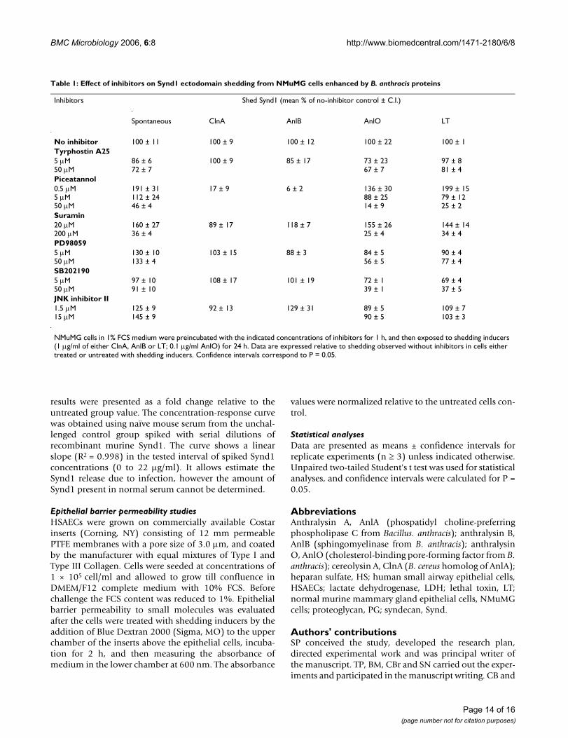
Table 1: Effect of inhibitors on Synd1 ectodomain shedding from NMuMG cells enhanced by B. anthracis proteins
Inhibitors Shed Synd1 (mean % of no-inhibitor control ± C.I.)
Spontaneous ClnA AnlB AnlO LT
No inhibitor 100 ± 11 100 ± 9 100 ± 12 100 ± 22 100 ± 1Tyrphostin A255 µM50 µM
86 ± 672 ± 7
100 ± 9 85 ± 17 73 ± 2367 ± 7
97 ± 881 ± 4
Piceatannol0.5 µM5 µM50 µM
191 ± 31112 ± 2446 ± 4
17 ± 9 6 ± 2 136 ± 3088 ± 2514 ± 9
199 ± 1579 ± 1225 ± 2
Suramin20 µM200 µM
160 ± 2736 ± 4
89 ± 17 118 ± 7 155 ± 2625 ± 4
144 ± 1434 ± 4
PD980595 µM50 µM
130 ± 10133 ± 4
103 ± 15 88 ± 3 84 ± 556 ± 5
90 ± 477 ± 4
SB2021905 µM50 µM
97 ± 1091 ± 10
108 ± 17 101 ± 19 72 ± 139 ± 1
69 ± 437 ± 5
JNK inhibitor II1.5 µM15 µM
125 ± 9145 ± 9
92 ± 13 129 ± 31 89 ± 590 ± 5
109 ± 7103 ± 3
NMuMG cells in 1% FCS medium were preincubated with the indicated concentrations of inhibitors for 1 h, and then exposed to shedding inducers (1 µg/ml of either ClnA, AnlB or LT; 0.1 µg/ml AnlO) for 24 h. Data are expressed relative to shedding observed without inhibitors in cells either treated or untreated with shedding inducers. Confidence intervals correspond to P = 0.05.
Page 14 of 16(page number not for citation purposes)

BMC Microbiology 2006, 6:8 http://www.biomedcentral.com/1471-2180/6/8
VC participated in research coordination and helped todraft the manuscript. All authors read and approved thefinal manuscript.
AcknowledgementsThis work was supported by the U.S. Department of Defense grant DAMD17-03-C-0122.
References1. Park PW, Pier GB, Preston MJ, Goldberger O, Fitzgerald ML, Bern-
field M: Syndecan-1 shedding is enhanced by LasA, a secretedvirulence factor of Pseudomonas aeruginosa. J Biol Chem 2000,275:3057-3064.
2. Park PW, Pier GB, Hinkes MT, Bernfield M: Exploitation of synde-can-1 shedding by Pseudomonas aeruginosa enhances viru-lence. Nature 2001, 411:98-102.
3. Park PW, Foster TJ, Nishi E, Duncan SJ, Klagsbrun M, Chen Y: Acti-vation of syndecan-1 ectodomain shedding by Staphylococ-cus aureus alpha-toxin and beta-toxin. J Biol Chem 2004,279:251-258.
4. Hooper NM, Turner AJ: Membrane protein secretases. BiochemSoc Trans 1999, 27:211-257.
5. Fitzgerald ML, Wang Z, Park PW, Murphy G, Bernfield M: Sheddingof syndecan-1 and -4 ectodomains is regulated by multiplesignaling pathways and mediated by a TIMP-3-sensitive met-alloproteinase. J Cell Biol 2000, 148:811-824.
6. Beauvais DM, Rapraeger AC: Syndecans in tumor cell adhesionand signaling. Reprod Biol Endocrinol 2004, 2:3-15.
7. Wilcox-Adelman SA, Denhez F, Iwabuchi T, Saoncella S, Calautti E,Goetinck PF: Syndecan-4: dispensable or indispensable? Glyco-conj J 2002, 19:305-13.
8. Bernfield M, Gotte M, Park PW, Reizes O, Fitzgerald ML, Lincecum J,Zako M: Functions of cell surface heparan sulfate proteogly-cans. Annu Rev Biochem 1999, 68:729-777.
9. Mun-Bryce S, Rosenberg GA: Matrix metalloproteinases in cer-ebrovascular disease. J Cereb Blood Flow Metab 1998,18:1163-1172.
10. Abramova FA, Grinberg LM, Yampolskaya OV, Walker DH: Pathol-ogy of inhalational anthrax in 42 cases from the Sverdlovskoutbreak of 1979. Proc Natl Acad Sci U S A 1993, 90:2291-2294.
11. Grinberg LM, Abramova FA, Yampolskaya OV, Walker DH, Smith JH:Quantitative pathology of inhalational anthrax I: quantita-tive microscopic findings. Mod Pathol 2001, 14:482-495.
12. Vasconcelos D, Barnewall R, Babin M, Hunt R, Estep J, Nielsen C,Carnes R, Carney J: Pathology of inhalation anthrax incynomolgus monkeys (Macaca fascicularis). Lab Invest 2003,83:1201-1209.
13. Klichko VI, Miller J, Wu A, Popov SG, Alibek K: Anaerobic induc-tion of Bacillus anthracis hemolytic activity. Biochem BiophysRes Commun 2003, 303:855-862.
14. Read TD, Peterson SN, Tourasse N, Baillie LW, Paulsen IT, NelsonKE, Tettelin H, Fouts DE, Eisen JA, Gill SR, Holtzapple EK, OkstadOA, Helgason E, Rilstone J, Wu M, Kolonay JF, Beanan MJ, Dodson RJ,Brinkac LM, Gwinn M, DeBoy RT, Madpu R, Daugherty SC, DurkinAS, Haft DH, Nelson WC, Peterson JD, Pop M, Khouri HM, RaduneD, Benton JL, Mahamoud Y, Jiang L, Hance IR, Weidman JF, Berry KJ,Plaut RD, Wolf AM, Watkins KL, Nierman WC, Hazen A, Cline R,Redmond C, Thwaite JE, White O, Salzberg SL, Thomason B, Fried-lander AM, Koehler TM, Hanna PC, Kolsto AB, Fraser CM: Thegenome sequence of Bacillus anthracis Ames and compari-son to closely related bacteria. Nature 2003, 423:81-86.
15. Johansen T, Bjorkoy G, Overvatn A, Diaz-Meco MT, Traavik T,Moscat J: NIH 3T3 cells stably transfected with the geneencoding phosphatidylcholine-hydrolyzing phospholipase Cfrom Bacillus cereus acquire a transformed phenotype. J MolCell Biol 1994, 14:646-654.
16. Firth JD, Putnins EE, Larjava H, Uitto VJ: Exogenous phospholipaseC stimulates epithelial cell migration and integrin expres-sion in vitro. Wound Repair Regen 2001, 9:86-94.
17. Moayeri M, Leppla SH: The roles of anthrax toxin in pathogen-esis. Curr Opin Microbiol 2004, 7:19-24.
18. Duesbery NS, Webb CP, Leppla SH, Gordon VM, Klimpel KR, Cope-land TD, Ahn NG, Oskarsson MK, Fukasawa K, Paull KD, Vande
Woude GF: Proteolytic inactivation of MAP-kinase-kinase byanthrax lethal factor. Science 1998, 280:734-737.
19. Moayeri M, Haines D, Young HA, Leppla SH: Bacillus anthracislethal toxin induces TNF-alpha-independent hypoxia-medi-ated toxicity in mice. J Clin Invest 2003, 112:670-682.
20. Smith H, Stoner HB: Anthrax toxic complex. Fed Proc 1967,26:1554-1557.
21. Warfel JM, Steele AD, D'Agnillo F: Anthrax lethal toxin inducesendothelial barrier dysfunction. Am J Pathol 2005,166:1871-1881.
22. Fouquet S, Lugo-Martinez VH, Faussat AM, Renaud F, Cardot P,Chambaz J, Pincon-Raymond M, Thenet S: Early loss of E-cadherinfrom cell-cell contacts is involved in the onset of Anoikis inenterocytes. J Biol Chem 2004, 279:43061-43069.
23. Kirby JE: Anthrax lethal toxin induces human endothelial cellapoptosis. Infect Immun 2004, 72:430-439.
24. Kassam A, Der SD, Mogridge J: Differentiation of human mono-cytic cell lines confers susceptibility to Bacillus anthracislethal toxin. Cell Microbiol 2005, 7:281-292.
25. Ivanov AI, Nusrat A, Parkos CA: Endocytosis of the apical junc-tional complex: mechanisms and possible roles in regulationof epithelial barriers. Bioessays 2005, 27:356-365.
26. Arribas J, Borroto A: Protein ectodomain shedding. Chem Rev2002, 102:4627-4638.
27. Wong WS, Leong KP: Tyrosine kinase inhibitors: a newapproach for asthma. Biochim Biophys Acta 2004, 1697:53-69.
28. Mahabeleshwar GH, Kundu GC: Syk, a protein-tyrosine kinase,suppresses the cell motility and nuclear factor kappa B-mediated secretion of urokinase type plasminogen activatorby inhibiting the phosphatidylinositol 3'-kinase activity inbreast cancer cells. J Biol Chem 2003, 278:6209-6221.
29. Ralevic V, Burnstock G: Receptors for purines and pyrimidines.Pharmacol Rev 1998, 50:413-92.
30. Keradmand F, Werb Z: Shedding light on sheddases: role ingrowth and development. BioEssays 2002, 24:8-12.
31. Zhang YL, Keng YF, Zhao Y, Wu L, Zhang ZY: Suramin is an activesite-directed, reversible, and tight-binding inhibitor of pro-tein-tyrosine phosphatases. J Biol Chem 1998, 15:12281-12287.
32. Stoker AW: Protein tyrosine phosphatases and signalling. JEndocrinol 2005, 185:19-33.
33. McCain DF, Wu L, Nicke P, Kassack MU, Kreimeyer A, Gagliardi A,Collins DC, Zhang ZY: Suramin derivatives as inhibitors andactivators of protein-tyrosine phosphatases. J Biol Chem 2004,279:14713-14725.
34. Park JM, Ng VH, Maeda S, Rest RF, Karin M: Anthrolysin O andother Gram-positive cytolysins are Toll-like receptor ago-nists. J Exp Med 2004, 200:1647-1655.
35. Sawai T, Usui N, Dwaihy J, Drongowski RA, Abe A, Coran AG, Har-mon CM: The effect of phospholipase A2 on bacterial translo-cation in a cell culture model. Pediatr Surg Int 2002, 16:262-266.
36. Popov SG, Popova TG, Hopkins S, Weinstein RS, MacAfee R, FryxellKJ, Chandhoke V, Bailey C, Alibek K: Effective antiprotease-anti-biotic treatment of experimental anthrax. BMC Infect Dis 2005,5:25-39.
37. Li L, Chaikof EL: Mechanical stress regulates syndecan-4expression and redistribution in vascular smooth musclecells. Arterioscler Thromb Vasc Biol 2002, 22:61-68.
38. Higashiyama S, Nanba D: ADAM-mediated ectodomain shed-ding of HB-EGF in receptor cross-talk. Biochim Biophys Acta2005, 1751:110-117.
39. Timmermann M, Hogger P: Oxidative stress and 8-iso-prostag-landin F(2alpha) induce ectodomain shedding of CD163 andrelease of tumor necrosis factor-alpha from human mono-cytes. Free Radic Biol Med 2005, 39:98-107.
40. Aktories K, Barbieri JT: Bacterial cytotoxins: targeting eukary-otic switches. Nat Rev Microbiol 2005, 3:397-410.
41. Kato M, Saunders S, Nguyen H, Bernfield M: Loss of cell surfacesyndecan-1 causes epithelia to transform into anchorage-independent mesenchyme-like cells. Mol Biol Cell 1995,6:559-576.
42. Smith H, Keppie J, Stanley JL: The chemical basis of the virulenceof Bacillus anthracis. V. The specific toxin produced by B.anthracis in vivo. Br J Exp Pathol 1955, 36:460-472.
43. Mikshis NI, Eremin SA, Bolotnikova MF: Correlation of the viru-lence of Bacillus anthracis with expression of signs, coded forby chromosomal genes. Mol Gen Mikrobiol Virusol 1999, 4:25-28.
Page 15 of 16(page number not for citation purposes)
http://www.ncbi.nlm.nih.gov/entrez/query.fcgi?cmd=Retrieve&db=PubMed&dopt=Abstract&list_uids=9809504
http://www.ncbi.nlm.nih.gov/entrez/query.fcgi?cmd=Retrieve&db=PubMed&dopt=Abstract&list_uids=9809504
http://www.ncbi.nlm.nih.gov/entrez/query.fcgi?cmd=Retrieve&db=PubMed&dopt=Abstract&list_uids=8460135
http://www.ncbi.nlm.nih.gov/entrez/query.fcgi?cmd=Retrieve&db=PubMed&dopt=Abstract&list_uids=8460135
http://www.ncbi.nlm.nih.gov/entrez/query.fcgi?cmd=Retrieve&db=PubMed&dopt=Abstract&list_uids=8460135
http://www.ncbi.nlm.nih.gov/entrez/query.fcgi?cmd=Retrieve&db=PubMed&dopt=Abstract&list_uids=9563949
http://www.ncbi.nlm.nih.gov/entrez/query.fcgi?cmd=Retrieve&db=PubMed&dopt=Abstract&list_uids=9563949
http://www.ncbi.nlm.nih.gov/entrez/query.fcgi?cmd=Retrieve&db=PubMed&dopt=Abstract&list_uids=6051334
http://www.ncbi.nlm.nih.gov/entrez/query.fcgi?cmd=Retrieve&db=PubMed&dopt=Abstract&list_uids=9755289
http://www.ncbi.nlm.nih.gov/entrez/query.fcgi?cmd=Retrieve&db=PubMed&dopt=Abstract&list_uids=7545031
http://www.ncbi.nlm.nih.gov/entrez/query.fcgi?cmd=Retrieve&db=PubMed&dopt=Abstract&list_uids=7545031
BMC Microbiology 2006, 6:8 http://www.biomedcentral.com/1471-2180/6/8
Publish with BioMed Central and every scientist can read your work free of charge
"BioMed Central will be the most significant development for disseminating the results of biomedical research in our lifetime."
Sir Paul Nurse, Cancer Research UK
Your research papers will be:
available free of charge to the entire biomedical community
peer reviewed and published immediately upon acceptance
cited in PubMed and archived on PubMed Central
yours — you keep the copyright
Submit your manuscript here:http://www.biomedcentral.com/info/publishing_adv.asp
BioMedcentral
44. Shannon JG, Ross CL, Koehler TM, Rest RF: Characterization ofanthrolysin O, the Bacillus anthracis cholesterol-dependentcytolysin. Infect Immun 2003, 71:3183-3189.
45. Popov SG, Popova TG, Grene E, Klotz F, Cardwell J, Bradburne C,Jama Y, Maland M, Wells J, Nalca A, Voss T, Bailey C, Alibek K: Sys-temic cytokine response in murine anthrax. Cell Microbiol 2004,6:225-233.
46. Nguyen A, Chen P, Cai H: Role of CaMKII in hydrogen peroxideactivation of ERK1/2, p38 MAPK, HSP27 and actin reorgani-zation in endothelial cells. FEBS Lett 2004, 572:307-313.
47. An SS, Fabry B, Mellema M, Bursac P, Gerthoffer WT, Kayyali US,Gaestel M, Shore SA, Fredberg JJ: Role of heat shock protein 27in cytoskeletal remodeling of the airway smooth muscle cell.J Appl Physiol 2004, 96:1701-1713.
48. McMullen ME, Bryant PW, Glembotski CC, Vincent PA, Pumiglia KM:Activation of p38 has opposing effects on the proliferationand migration of endothelial cells. J Biol Chem 2005,280:20995-21003.
49. Huffman DL, Abrami L, Sasik R, Corbeil J, Goot F, Aroian R:Mitogen-activated protein kinase pathways defend againstbacterial pore-forming toxins. Proc Nat Acad Sci USA 2004,101:10995-11000.
50. Kevil CG, Oshima T, Alexander JS: The role of p38 MAP kinasein hydrogen peroxide mediated endothelial solute permea-bility. Endothelium 2001, 8:107-116.
51. Salles II, Tucker AE, Voth DE, Ballard JD: Toxin-induced resist-ance in Bacillus anthracis lethal toxin-treated macrophages.Proc Nat Acad Sci USA 2003, 100:12426-12431.
52. Johnson GB, Brunn GJ, Platt JL: An endogenous pathway to sys-temic inflammatory response syndrome (SIRS)-like reac-tions through toll-like receptor 4. J Immun 2004, 172:20-24.
Page 16 of 16(page number not for citation purposes)
Related Documents