Thanh-Nga Tran, HMS IV
Gillian Lieberman, MD
Fat Embolism Syndrome
A case presentation
Thanh Nga Tran, Harvard Medical School, Year IV
Gillian Lieberman, MD
March 21, 2005
Thanh-Nga Tran, HMS IV
Gillian Lieberman, MD
Our Patient
H.K is a 58 year-old woman with a history of afib on amiodarone, HTN, CAD, DM, high chol, s/p gastric bypass presented 2/3/2005 after a mechanical fall. Pt slipped on hardwood floor. Had right distal femur comminuted fracture. She was admitted to the orthopedics service,
OR on 2/4/2005 for ORIF of her right femur. Pre-operatively, pt had intact mental status and moved all extremities well.
Thanh-Nga Tran, HMS IV
Gillian Lieberman, MD
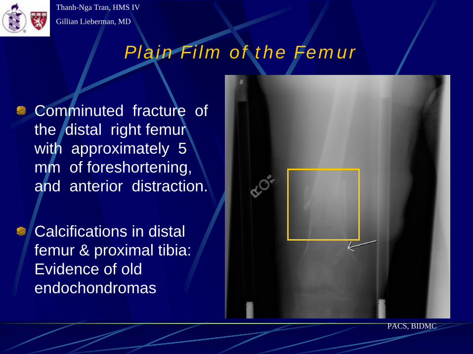
Plain Film of the Femur
Comminuted fracture of the distal right femur with approximately 5 mm of foreshortening, and anterior distraction.
Calcifications in distal femur & proximal tibia: Evidence of old endochondromas
PACS, BIDMC
Thanh-Nga Tran, HMS IV
Gillian Lieberman, MD
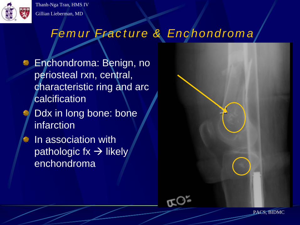
Femur Fracture & Enchondroma
Enchondroma: Benign, no periosteal rxn, central, characteristic ring and arc calcificationDdx in long bone: bone infarctionIn association with pathologic fx likely enchondroma
PACS, BIDMC
Thanh-Nga Tran, HMS IV
Gillian Lieberman, MD
Intra-operative Course
Partway through, our patient acutely decompensated with SBP 60s-70s, hypoxia to 80s, end tidal CO2 37. She was started on pressors, first dopamine, then neosynephrine, then epinephrine.
Thanh-Nga Tran, HMS IV
Gillian Lieberman, MD
Ddx of Acute Hypoxia
Pulmonary embolism from:ClotFatAir
PneumothoraxAcute MIMechanical: ET tube, machine malfunction
Thanh-Nga Tran, HMS IV
Gillian Lieberman, MD
Menu of Tests Available for Work-up
Concern for massive PE usual work-up may include:
CXRABGsHemodynamic monitoringEchocardiogramV/Q scanPulmonary angiogram – gold standardHelical CT –PE protocolMRA
But patient was in the O.R.
Thanh-Nga Tran, HMS IV
Gillian Lieberman, MD
Intra-Operative TEE
Emergent intra-op Transesophageal Echocardiogram (TEE) revealed dilated RV cavity size with RV free wall hypokinesis, mild pulmonary hypertension, new PFO concerning for massive PE.Pt was given herparin initially.
Emergent intra-op pulmonary arteriogram was performed.
Thanh-Nga Tran, HMS IV
Gillian Lieberman, MD
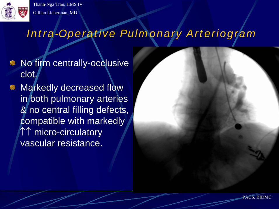
Intra-Operative Pulmonary Arteriogram
No firm centrally-occlusive clot. Markedly decreased flow in both pulmonary arteries & no central filling defects, compatible with markedly ↑↑
micro-circulatory
vascular resistance.
PACS, BIDMC
Thanh-Nga Tran, HMS IV
Gillian Lieberman, MD
Pulmonary Arteriogram
Ddx includes fat embolism, air embolism, possibly extremely soft clot. Pt received Nitric Oxide (NO) for ?vasospasm with some improvement to flow
Faintuch, Lang et al - Inhaled nitric oxide as an adjunct to suction thrombectomy for pulmonary embolism - J Vasc Interv Radiol. 2004 Nov;15(11):1311-5.Two pts w massive PE: condition only improved after administration of NO
Pt received intra-arterial tPA to both arteries for clot preventionIVC filter placed
Thanh-Nga Tran, HMS IV
Gillian Lieberman, MD
Post-Operative Course
Pt noted to have flaccid paresis post OR and next day onset of L hemiparesisStat head CT to r/o intracranial hemorrhage, brain infarction
Thanh-Nga Tran, HMS IV
Gillian Lieberman, MD
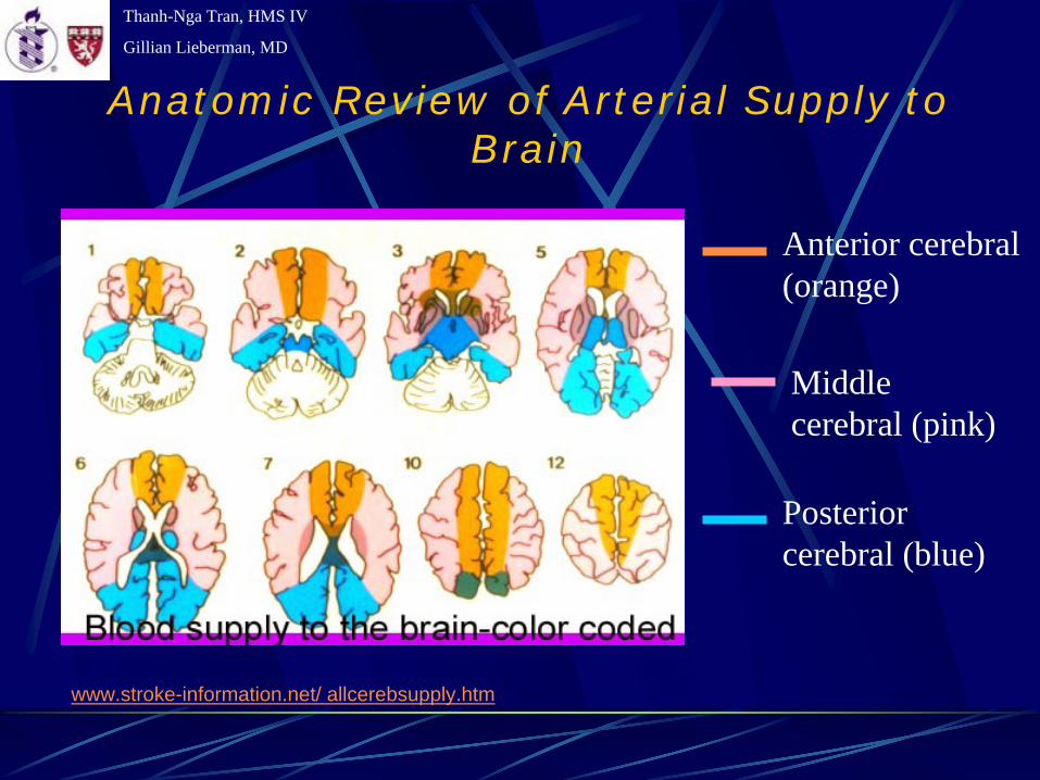
Anatomic Review of Arterial Supply to Brain
Anterior cerebral (orange)
Middle cerebral (pink)
Posterior cerebral (blue)
www.stroke-information.net/ allcerebsupply.htm
Thanh-Nga Tran, HMS IV
Gillian Lieberman, MD
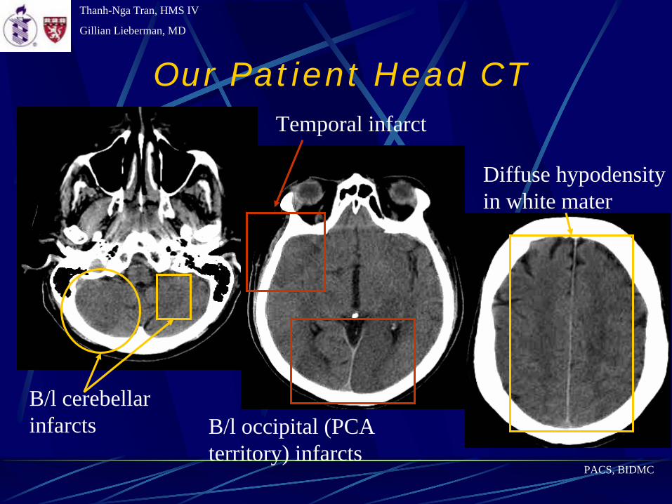
Our Patient Head CT
B/l cerebellar infarcts B/l occipital (PCA
territory) infarcts
Temporal infarct
PACS, BIDMC
Diffuse hypodensity in white mater
Thanh-Nga Tran, HMS IV
Gillian Lieberman, MD
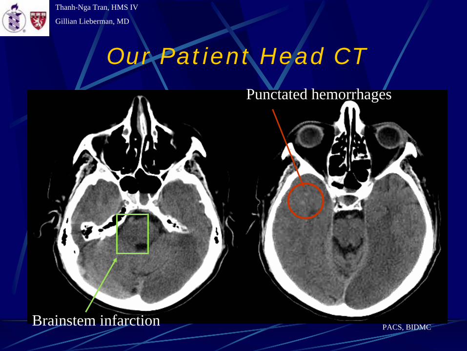
Our Patient Head CT
Brainstem infarction
Punctated hemorrhages
PACS, BIDMC
Thanh-Nga Tran, HMS IV
Gillian Lieberman, MD
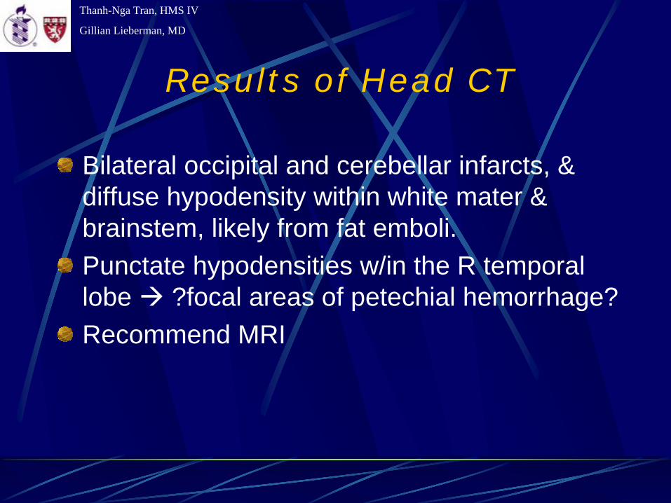
Results of Head CT
Bilateral occipital and cerebellar infarcts, & diffuse hypodensity within white mater & brainstem, likely from fat emboli.Punctate hypodensities w/in the R temporal lobe ?focal areas of petechial hemorrhage?Recommend MRI
Thanh-Nga Tran, HMS IV
Gillian Lieberman, MD
Our Patient MRI
T1 – Image anatomy – fluid darkT2 – Image pathology – fluid bright FLAIR (Fluid-Attenuated Inversion Recovery)– Image brain pathology
Edema, inflammation T2 w fluid suppressed
Diffusion – sensitive for acute stroke (~ <2hrs)
Thanh-Nga Tran, HMS IV
Gillian Lieberman, MD
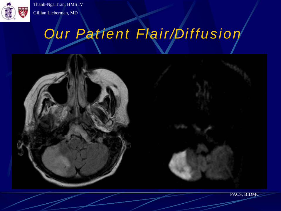
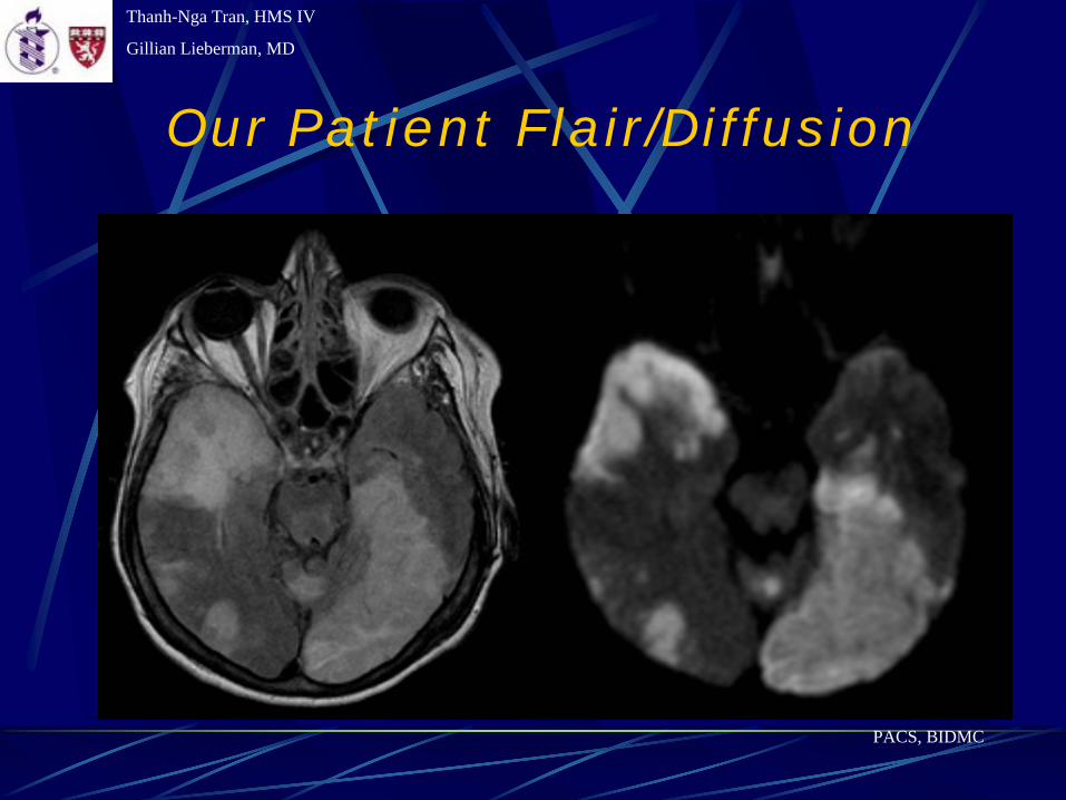
Our Patient Flair/Diffusion
PACS, BIDMC
Thanh-Nga Tran, HMS IV
Gillian Lieberman, MD
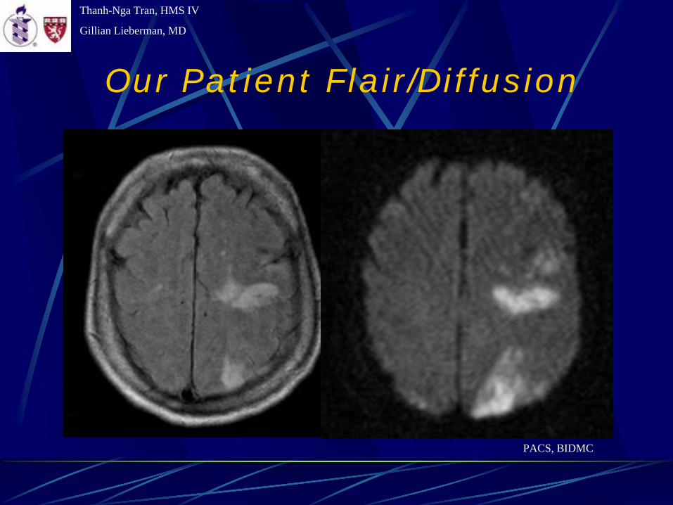
Our Patient Flair/Diffusion
PACS, BIDMC
Thanh-Nga Tran, HMS IV
Gillian Lieberman, MD
Our Patient Flair/Diffusion
PACS, BIDMC
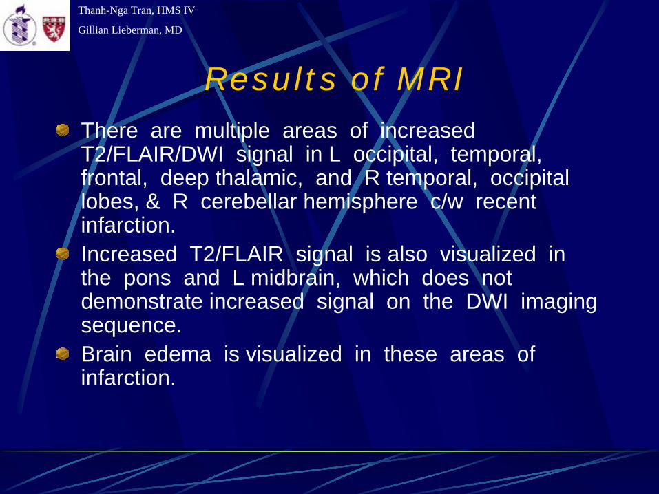
Thanh-Nga Tran, HMS IV
Gillian Lieberman, MD
Results of MRIThere are multiple areas of increased T2/FLAIR/DWI signal in L occipital, temporal, frontal, deep thalamic, and R temporal, occipital lobes, & R cerebellar hemisphere c/w recent infarction. Increased T2/FLAIR signal is also visualized in the pons and L midbrain, which does not demonstrate increased signal on the DWI imaging sequence. Brain edema is visualized in these areas of infarction.
Thanh-Nga Tran, HMS IV
Gillian Lieberman, MD
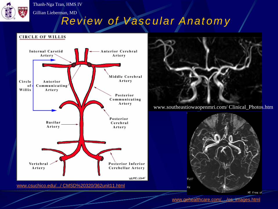
Review of Vascular Anatomy
www.csuchico.edu/.../ CMSD%20320/362unit11.html
www.gehealthcare.com/. ../os_images.html
www.southeastiowaopenmri.com/ Clinical_Photos.htm
Thanh-Nga Tran, HMS IV
Gillian Lieberman, MD
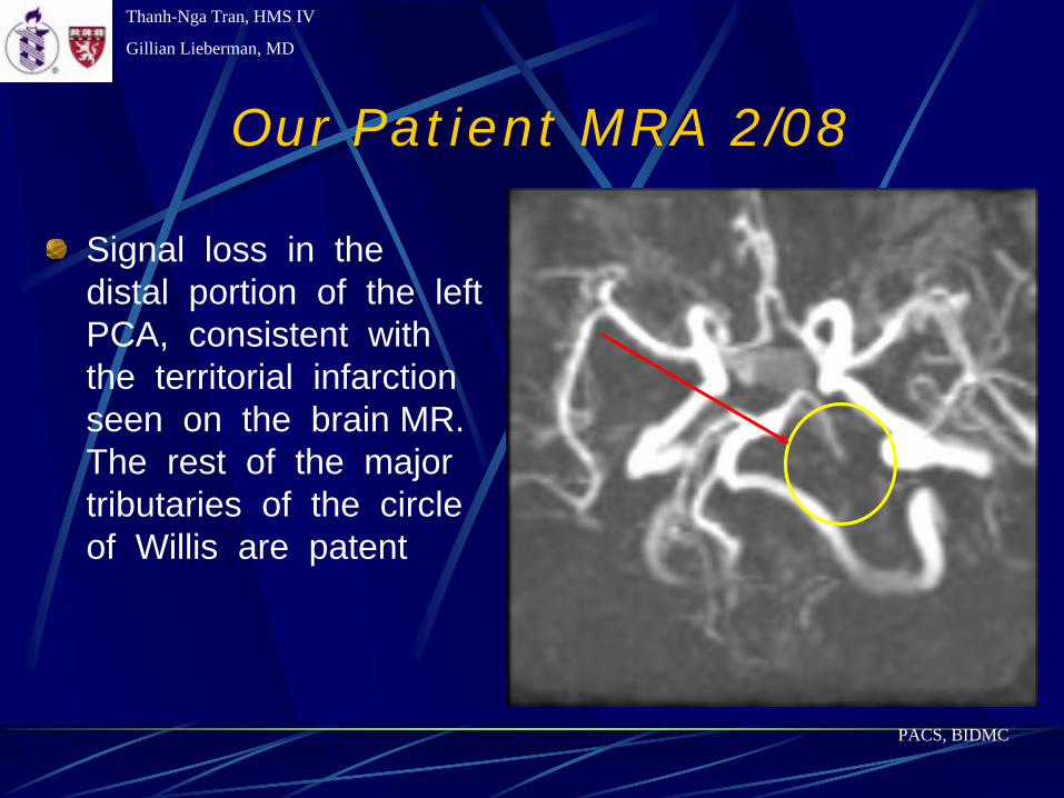
Our Patient MRA 2/08
Signal loss in the distal portion of the left PCA, consistent with the territorial infarction seen on the brain MR. The rest of the major tributaries of the circle of Willis are patent
PACS, BIDMC
Thanh-Nga Tran, HMS IV
Gillian Lieberman, MD
Our Patient Course
EEG on 2/20/05 2/2 to R-sided weakness and inability to tolerate head CT: non-convulsive status epilepticusRepeat head CT
Thanh-Nga Tran, HMS IV
Gillian Lieberman, MD
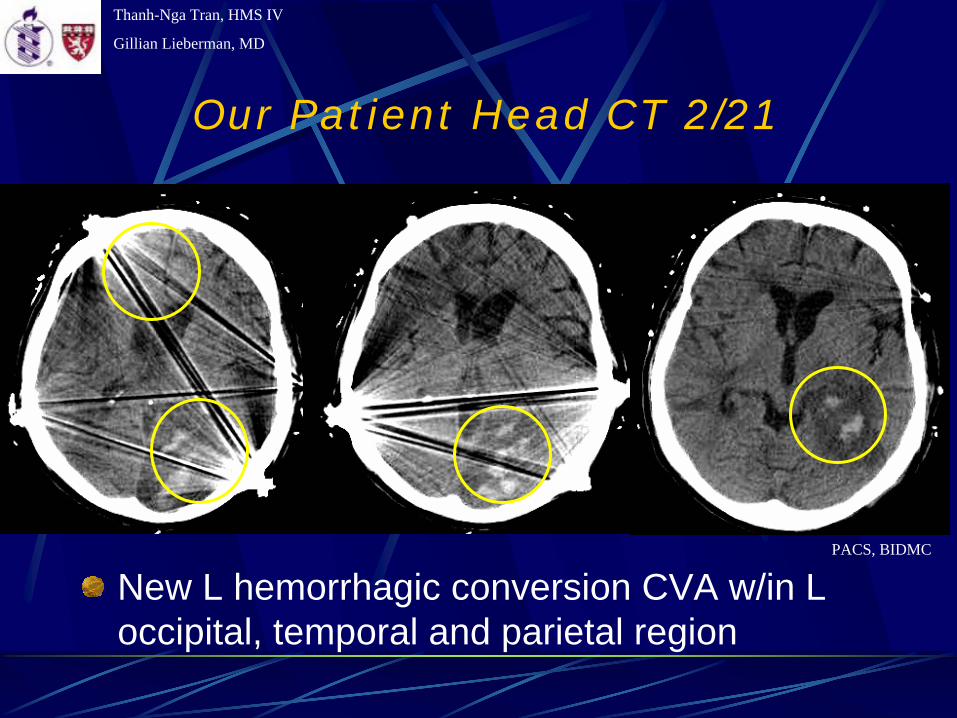
Our Patient Head CT 2/21
New L hemorrhagic conversion CVA w/in L occipital, temporal and parietal region
PACS, BIDMC
Thanh-Nga Tran, HMS IV
Gillian Lieberman, MD
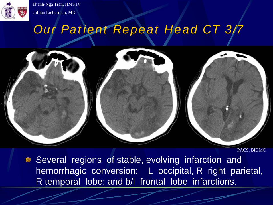
Our Patient Repeat Head CT 3/7
Several regions of stable, evolving infarction and hemorrhagic conversion: L occipital, R right parietal, R temporal lobe; and b/l frontal lobe infarctions.
PACS, BIDMC
Thanh-Nga Tran, HMS IV
Gillian Lieberman, MD
Our Patient Course
Pt is s/p ORIF w fat emboli to lungs, brain through PFO, hemorrhagic conversion of CVA and subsequent non-convulsive status epilepticus (treated).Pt also had NSTEMI, line sepsis, respiratory failure and hypotension (could be 2/2 to aspiration pneumonitis or PNA), and acute renal failure.Pt is non-responsive, occasional grimace.Pt currently still in MICU. Supportive care given to family for the tragic complication.
Thanh-Nga Tran, HMS IV
Gillian Lieberman, MD
Fat Embolism Syndrome (FES)
First described by Zenker and later Von Bergmann in mid 1800sMortality 5-15%Frequently assoc with long bone/pelvic fractures, more frequent w closed fx; orthopedics surgeries, liposuction, burns, soft tissue injuriesNon-trauma: pancreatitis, DM, osteomyelitis, bone tumor lysis, steroids, fatty liver, cyclosporin, lipid infusionFES typically manifests 12 to 72 hours after the initial insult or as late as two weeks
Thanh-Nga Tran, HMS IV
Gillian Lieberman, MD
FES
Classic triad: hypoxemia; neurologic abnormalities; and a petechial rash (pathognomonic)Early findings: dyspnea, tachypnea, and hypoxemiaNeurological abnl often followed: confusional state, delta MS, occasional seizures and focal deficitsLate: possible petechial rash of head, neck, anterior thorax, subconjunctiva, and axillae – resolved in 5-7 daysMinor: lipiduria, scotomata (Purtscher's retinopathy), fever, coag abnl, myocardial depression: 2/2 to toxic mediators or dysfunctional lipid metabolism
Thanh-Nga Tran, HMS IV
Gillian Lieberman, MD
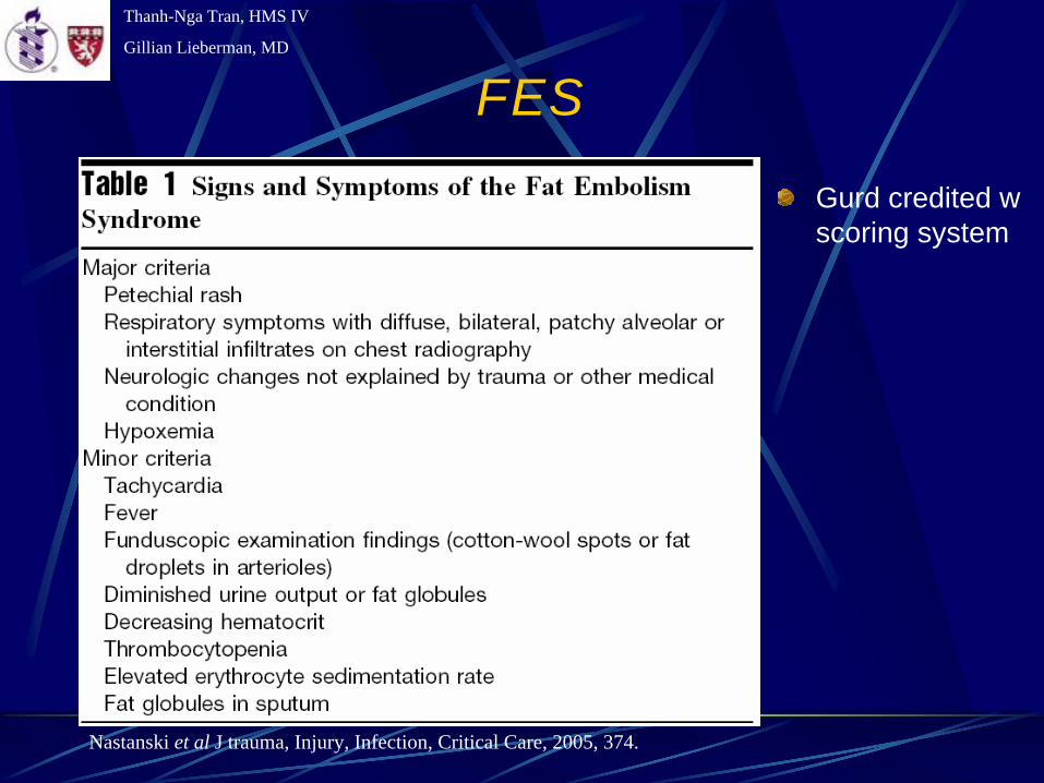
FES
Gurd credited w scoring system
Nastanski et al J trauma, Injury, Infection, Critical Care, 2005, 374.
Thanh-Nga Tran, HMS IV
Gillian Lieberman, MD
FES Etiology
Mechanical: Occlusion of blood vessels by fat globules from trauma (marrow, adipose)- With continued embolization, pulmonary artery and right heart pressures rise PFO systemic circulation paradoxical embolism.
Biochemical: Via toxic intermediates of plasma-derived fat such as chylomicrons or infused lipids: embolized fat
free fatty acids (FFA) directly toxic to pneumocytes and capillary endothelium in the lung, causing interstitial hemorrhage, edema and chemical pneumonitis; also cardiac contractile dysfx.
Thanh-Nga Tran, HMS IV
Gillian Lieberman, MD
Diagnosis
Clinical presentationRadiologic:
CXR: nl in most, minority w evenly distributed, fleck-like pulmonary shadows (Snow Storm appearance), increased pulmmarkings and R heart dilatation V/Q: mottled pattern of subsegmental perfusion defects with a normal ventilatory patternChest CT: Focal areas of ground glass opacification with interloblar septal thickening Brain MRI: may reveal high intensity T2 signal, mult scattered lesions in white mater & ischemia on DWI & FLAIRPET scanning: cerebral blood flow alterations & correction
No reliable lab tests
Thanh-Nga Tran, HMS IV
Gillian Lieberman, MD
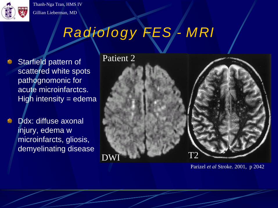
Radiology FES - MRI
Starfield pattern of scattered white spots pathognomonic for acute microinfarctcs. High intensity = edema
Ddx: diffuse axonal injury, edema w microinfarcts, gliosis, demyelinating disease
DWI T2Parizel et al Stroke. 2001, p 2042
Patient 2
Thanh-Nga Tran, HMS IV
Gillian Lieberman, MD
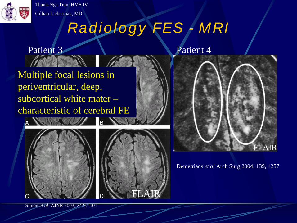
Radiology FES - MRI
Demetriads et al Arch Surg 2004; 139, 1257
FLAIRSimon et al AJNR 2003; 24:97-101
Multiple focal lesions in periventricular, deep, subcortical white mater – characteristic of cerebral FE
Patient 3 Patient 4
FLAIR
Thanh-Nga Tran, HMS IV
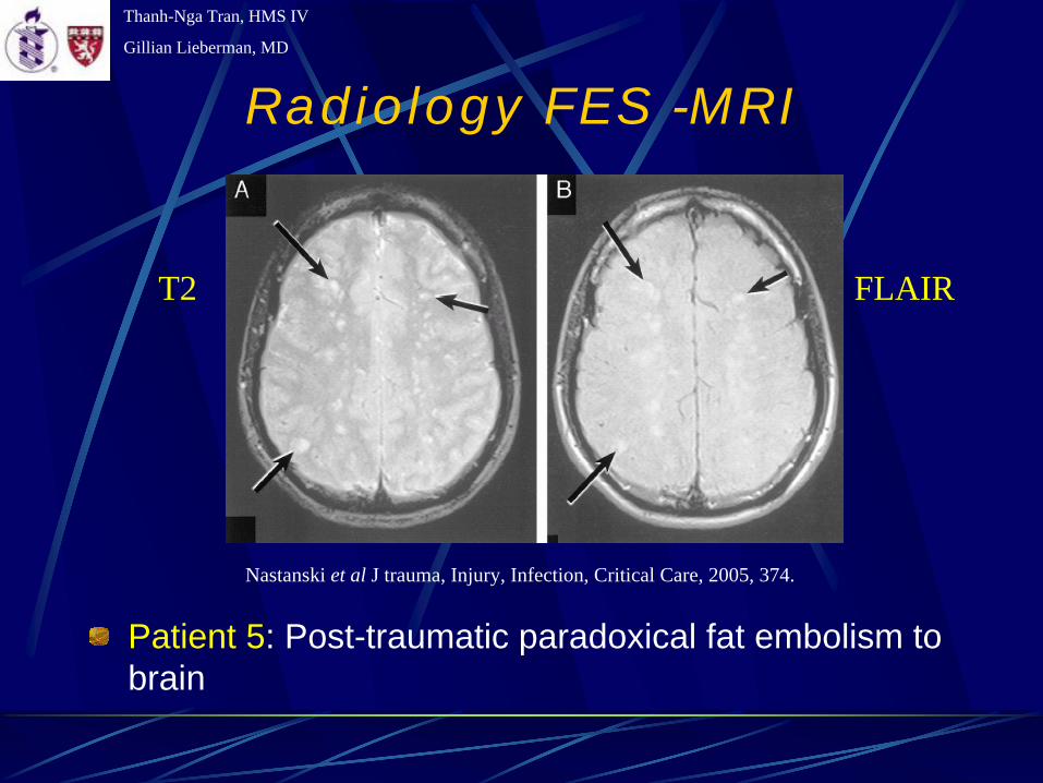
Gillian Lieberman, MD
Radiology FES -MRI
Patient 5: Post-traumatic paradoxical fat embolism to brain
Nastanski et al J trauma, Injury, Infection, Critical Care, 2005, 374.
T2 FLAIR
Thanh-Nga Tran, HMS IV
Gillian Lieberman, MD
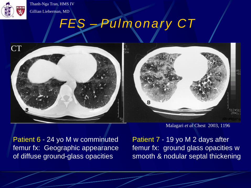
FES – Pulmonary CT
Patient 7 - 19 yo M 2 days after femur fx: ground glass opacities w smooth & nodular septal thickening
Patient 6 - 24 yo M w comminuted femur fx: Geographic appearance of diffuse ground-glass opacities
Malagari et al Chest 2003, 1196
CT
Thanh-Nga Tran, HMS IV
Gillian Lieberman, MD
Treatment
Early immobilization of fracturesOperative correctionSupportive care
Maintain of intravascular volume as shock can exacerbate the lung injury. Recs albumin binds fatty acids, and may decrease the extent of lung injury
Mechanical ventilation and PEEP may be required to maintain PaO2. High dose corticosteroids have been effective in preventing development of FES in several trials, but controversy still persists
Thanh-Nga Tran, HMS IV
Gillian Lieberman, MD
Other Unusual Embolisms

Thanh-Nga Tran, HMS IV
Gillian Lieberman, MD
Patient 8: 29 yo G1P1 – still birth 3mo prior, p/w R thorax dullness. CXR c/wcystic lung massCT – homogeneous, w air, hyperdense border, density similar to pleural effusions
Kaptanoguu et al Scand Cardiovasc 1999, 117
Amniotic Fluid
Thanh-Nga Tran, HMS IV
Gillian Lieberman, MD
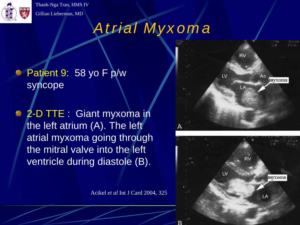
Atrial Myxoma
Patient 9: 58 yo F p/w syncope
2-D TTE : Giant myxoma in the left atrium (A). The left atrial myxoma going through the mitral valve into the left ventricle during diastole (B).
Acikel et al Int J Card 2004, 325
Thanh-Nga Tran, HMS IV
Gillian Lieberman, MD
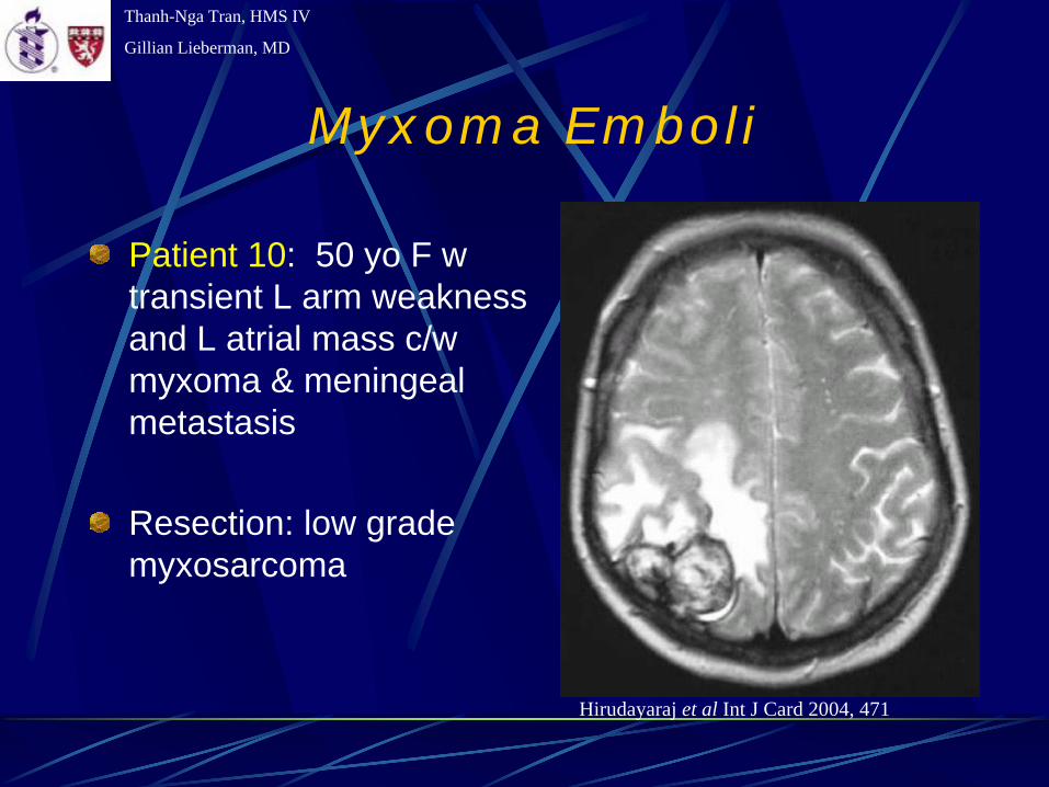
Myxoma Emboli
Patient 10: 50 yo F w transient L arm weakness and L atrial mass c/w myxoma & meningeal metastasis
Resection: low grade myxosarcoma
Hirudayaraj et al Int J Card 2004, 471
Thanh-Nga Tran, HMS IV
Gillian Lieberman, MD
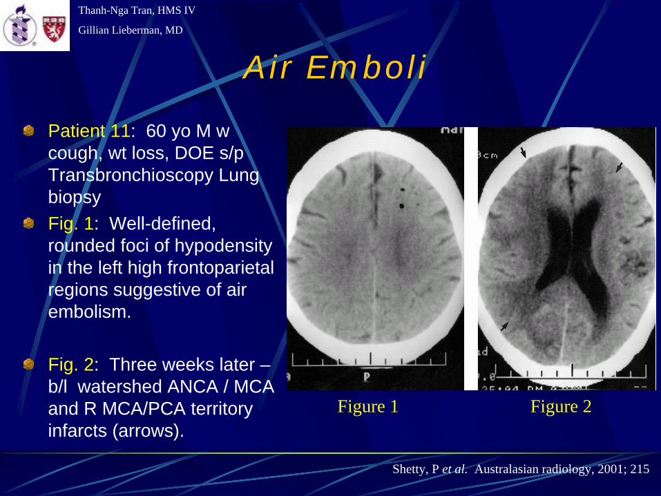
Air Emboli
Patient 11: 60 yo M w cough, wt loss, DOE s/p Transbronchioscopy Lung biopsyFig. 1: Well-defined, rounded foci of hypodensity in the left high frontoparietal regions suggestive of air embolism.
Fig. 2: Three weeks later – b/l watershed ANCA / MCA and R MCA/PCA territory infarcts (arrows).
Shetty, P et al. Australasian radiology, 2001; 215
Figure 1 Figure 2
Thanh-Nga Tran, HMS IV
Gillian Lieberman, MD
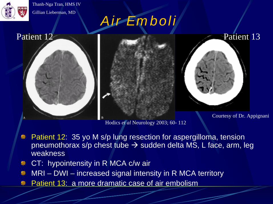
Air Emboli
Patient 12: 35 yo M s/p lung resection for aspergilloma, tension pneumothorax s/p chest tube sudden delta MS, L face, arm, leg weaknessCT: hypointensity in R MCA c/w airMRI – DWI – increased signal intensity in R MCA territoryPatient 13: a more dramatic case of air embolism
Hodics et al Neurology 2003; 60- 112Courtesy of Dr. Appignani
Patient 12 Patient 13
Thanh-Nga Tran, HMS IV
Gillian Lieberman, MD
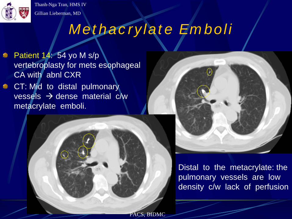
Methacrylate Emboli
Patient 14: 54 yo M s/p vertebroplasty for mets esophageal CA with abnl CXRCT: Mid to distal pulmonary vessels dense material c/wmetacrylate emboli.
Distal to the metacrylate: the pulmonary vessels are low density c/w lack of perfusion
PACS, BIDMC
Thanh-Nga Tran, HMS IV
Gillian Lieberman, MD
ReferencesUp-to-date: http://www.uptodate.orgOdegard, K. Fat Embolism: Diagnosis and Treatment. http://www.orthoteers.co.uk/Nrujp~ij33lm/Orthfatembolism.htmwww.stroke-information.net/ allcerebsupply.htmParizel, PM et al. Early diagnosis of cerebral fat embolism syndrome by diffusion-weighted MRI (Starfield pattern). Stroke 2003; 2942.Demetriades, D et al. Image of the month. Arch Surg 2004; 139, 1257.Prologo, JD et al. CT diagnosis of cerebral fat embolism. Am J Emergency Medicine 2004; 22(7), 605.Simon, AD et al. Contrast-enhanced MR imaging of cerebral fat embolism: Case report and review of literature. Am J Neuroradiol, 2003; 24:97-101.Nastanski, F et al. Posttraumatic paradoxical fat embolism to the brain: A case report. J of Trauma Injury, Infection, Critical Care, 2005; 58(2), 372Malagari, K et al. High resolution CT findings in mild pulmonary fat embolism. Chest 2003; 123(4): 1196.
Thanh-Nga Tran, HMS IV
Gillian Lieberman, MD
References
Kaptanoguu et al. Lung mass due to amniotic fluid embolism. Scand Cardiovasc J 1999; 33, 117.Acikel M et al. A giant left atrial myxoma: an unusual cause of syncope and cerebral emboli. Int J of Card 2004; 94, 325.Hirudayaraj, P et al. Myxomatous meningeal tumor: a case of “metastaic” cardiac myxoma. Int J Card 2004; 96: 471.Shetty, PG et al. Fatal cerebral air embolism as a complication of transbronchoscopic lung biopsy: a case report. Australasian Radiology 2001; 45: 215.Hodics et al. Cerebral air embolism. Neurology 2003;60: 112-145.
Thanh-Nga Tran, HMS IV
Gillian Lieberman, MD
Acknowledgements
Barbara Appignani, MDLarry BarbarasDaniel Cornfield, MDElvira Lang, MDPamela LepkowskiGillian Lieberman, MDPhoebe Olhava, MDJesse Wei, MD