ARTICLE Visceral fat inflammation and fat embolism are associated with lung’s lipidic hyaline membranes in subjects with COVID-19 Georgia Colleluori 1,7 , Laura Graciotti 2,3,7 , Mauro Pesaresi 4 , Angelica Di Vincenzo 1 , Jessica Perugini 1 , Eleonora Di Mercurio 1 , Sara Caucci 3 , Patrizia Bagnarelli 3 , Cristina M. Zingaretti 1 , Enzo Nisoli 5 , Stefano Menzo 3 , Adriano Tagliabracci 4 , Annie Ladoux 6 , Christian Dani 6 , Antonio Giordano 1 and Saverio Cinti 1 ✉ © The Author(s), under exclusive licence to Springer Nature Limited 2022 BACKGROUND: Preliminary data suggested that fat embolism could explain the importance of visceral obesity as a critical determinant of coronavirus disease-2019 (COVID-19). METHODS: We performed a comprehensive histomorphologic analysis of autoptic visceral adipose tissue (VAT), lungs and livers of 19 subjects with COVID-19 (COVID-19+), and 23 people without COVID-19 (controls). Human adipocytes (hMADS) infected with SARS-CoV-2 were also studied. RESULTS: Although there were no between-group differences in body-mass-index and adipocytes size, a higher prevalence of CD68+ macrophages among COVID-19+ VAT was detected (p = 0.005) and accompanied by crown-like structures presence, signs of adipocytes stress and death. Consistently, human adipocytes were successfully infected by SARS-CoV-2 in vitro and displayed lower cell viability. Being VAT inflammation associated with lipids spill-over from dead adipocytes, we studied lipids distribution by ORO. Lipids were observed within lungs and livers interstitial spaces, macrophages, endothelial cells, and vessels lumen, features suggestive of fat embolism syndrome, more prevalent among COVID-19+ (p < 0.001). Notably, signs of fat embolism were more prevalent among people with obesity (p = 0.03) independently of COVID-19 diagnosis, suggesting that such condition may be an obesity complication exacerbated by SARS-CoV-2 infection. Importantly, all infected subjects’ lungs presented lipids-rich (ORO+) hyaline membranes, formations associated with COVID-19-related pneumonia, present only in one control patient with non-COVID- 19-related pneumonia. Importantly, transition aspects between embolic fat and hyaline membranes were also observed. CONCLUSIONS: This study confirms the lung fat embolism in COVID-19+ patients and describes for the first time novel COVID-19- related features possibly underlying the unfavorable prognosis in people with COVID-19 and obesity. International Journal of Obesity (2022) 46:1009–1017; https://doi.org/10.1038/s41366-022-01071-w INTRODUCTION Since December 2019, the severe acute respiratory syndrome coronavirus 2 (SARS-CoV-2), responsible for the development of coronavirus disease 2019 (COVID-19), has spread globally, result- ing in a worldwide health crisis that caused over five million deaths [1]. The lung is a crucial target organ not only due to the severe bilateral pneumonia observed in 15–30% of hospitalized patients [2, 3] but also because it is the site from which the infection spreads to blood vessels, heart, gut, brain, and kidneys [4]. Published data support interstitial fibrosis with alveolar hyaline membrane (HM) formation as the main underlying histopatholo- gic event responsible for pneumonia and acute respiratory syndrome distress [5, 6]. The reasons for HM bilateral expression, histogenesis, and sudden clinical appearance during COVID-19 early stages are not completely understood [7]. The severity of COVID-19 is strictly associated with the presence of comorbidities [8]; while obesity alone is responsible for 20% of COVID-19 hospitalizations, obesity in combination with type 2 diabetes and hypertension accounts for 58% [9]. Obesity and impaired metabolic health are in fact strongly associated with COVID-19 unfavorable prognosis and pose also young patients at higher risks [10, 11]. Significantly, visceral obesity increases the risk of COVID-19-related complications, independently of age, gender, body mass index [12], total and subcutaneous adipose tissue areas [13–16]. Visceral obesity is, in fact, strongly associated with chronic low-grade inflammation, blood hypercoagulability, impaired metabolic health, and higher risk of cardiovascular events, all risk factors for COVID-19 severity [8, 11, 16–18]. Visceral adipose tissue (VAT) excessive expansion is paralleled by adipocytes hypertrophy, death, and lipids spill-over, phenomena resulting in macrophages Received: 26 October 2021 Revised: 5 January 2022 Accepted: 12 January 2022 Published online: 26 January 2022 1 Center for the Study of Obesity, Department of Experimental and Clinical Medicine, Marche Polytechnic University, Via Tronto 10A, Ancona, Italy. 2 Section of Experimental Pathology, Department of Clinical and Molecular Sciences, Marche Polytechnic University, Via Tronto 10A, Ancona, Italy. 3 Section of Microbiology, Department of Bioscience and Public Health, Marche Polytechnic University, Via Tronto 10A, Ancona, Italy. 4 Section of Legal Medicine, Department of Bioscience and Public Health, Marche Polytechnic University, Via Tronto 10A, Ancona, Italy. 5 Center for Study and Research on Obesity, Department of Medical Biotechnology and Translational Medicine, University of Milan, Via Vanvitelli 32, Milan, Italy. 6 Université Côte d’Azur, CNRS, Inserm, iBV, Faculté de Médecine, 06107 Cedex 2 Nice, France. 7 These authors contributed equally: Georgia Colleluori, Laura Graciotti. ✉ email: [email protected] www.nature.com/ijo International Journal of Obesity 1234567890();,:

Welcome message from author
This document is posted to help you gain knowledge. Please leave a comment to let me know what you think about it! Share it to your friends and learn new things together.
Transcript
ARTICLE
Visceral fat inflammation and fat embolism are associated withlung’s lipidic hyaline membranes in subjects with COVID-19Georgia Colleluori 1,7, Laura Graciotti2,3,7, Mauro Pesaresi4, Angelica Di Vincenzo1, Jessica Perugini1, Eleonora Di Mercurio1,Sara Caucci3, Patrizia Bagnarelli3, Cristina M. Zingaretti1, Enzo Nisoli 5, Stefano Menzo3, Adriano Tagliabracci4, Annie Ladoux6,Christian Dani6, Antonio Giordano1 and Saverio Cinti 1✉
© The Author(s), under exclusive licence to Springer Nature Limited 2022
BACKGROUND: Preliminary data suggested that fat embolism could explain the importance of visceral obesity as a criticaldeterminant of coronavirus disease-2019 (COVID-19).METHODS: We performed a comprehensive histomorphologic analysis of autoptic visceral adipose tissue (VAT), lungs and livers of19 subjects with COVID-19 (COVID-19+), and 23 people without COVID-19 (controls). Human adipocytes (hMADS) infected withSARS-CoV-2 were also studied.RESULTS: Although there were no between-group differences in body-mass-index and adipocytes size, a higher prevalence ofCD68+ macrophages among COVID-19+ VAT was detected (p = 0.005) and accompanied by crown-like structures presence, signsof adipocytes stress and death. Consistently, human adipocytes were successfully infected by SARS-CoV-2 in vitro and displayedlower cell viability. Being VAT inflammation associated with lipids spill-over from dead adipocytes, we studied lipids distribution byORO. Lipids were observed within lungs and livers interstitial spaces, macrophages, endothelial cells, and vessels lumen, featuressuggestive of fat embolism syndrome, more prevalent among COVID-19+ (p < 0.001). Notably, signs of fat embolism were moreprevalent among people with obesity (p = 0.03) independently of COVID-19 diagnosis, suggesting that such condition may be anobesity complication exacerbated by SARS-CoV-2 infection. Importantly, all infected subjects’ lungs presented lipids-rich (ORO+)hyaline membranes, formations associated with COVID-19-related pneumonia, present only in one control patient with non-COVID-19-related pneumonia. Importantly, transition aspects between embolic fat and hyaline membranes were also observed.CONCLUSIONS: This study confirms the lung fat embolism in COVID-19+ patients and describes for the first time novel COVID-19-related features possibly underlying the unfavorable prognosis in people with COVID-19 and obesity.
International Journal of Obesity (2022) 46:1009–1017; https://doi.org/10.1038/s41366-022-01071-w
INTRODUCTIONSince December 2019, the severe acute respiratory syndromecoronavirus 2 (SARS-CoV-2), responsible for the development ofcoronavirus disease 2019 (COVID-19), has spread globally, result-ing in a worldwide health crisis that caused over five milliondeaths [1]. The lung is a crucial target organ not only due to thesevere bilateral pneumonia observed in 15–30% of hospitalizedpatients [2, 3] but also because it is the site from which theinfection spreads to blood vessels, heart, gut, brain, and kidneys[4]. Published data support interstitial fibrosis with alveolar hyalinemembrane (HM) formation as the main underlying histopatholo-gic event responsible for pneumonia and acute respiratorysyndrome distress [5, 6]. The reasons for HM bilateral expression,histogenesis, and sudden clinical appearance during COVID-19early stages are not completely understood [7].
The severity of COVID-19 is strictly associated with the presenceof comorbidities [8]; while obesity alone is responsible for 20% ofCOVID-19 hospitalizations, obesity in combination with type 2diabetes and hypertension accounts for 58% [9]. Obesity andimpaired metabolic health are in fact strongly associated withCOVID-19 unfavorable prognosis and pose also young patients athigher risks [10, 11]. Significantly, visceral obesity increases the riskof COVID-19-related complications, independently of age, gender,body mass index [12], total and subcutaneous adipose tissue areas[13–16]. Visceral obesity is, in fact, strongly associated with chroniclow-grade inflammation, blood hypercoagulability, impairedmetabolic health, and higher risk of cardiovascular events, all riskfactors for COVID-19 severity [8, 11, 16–18]. Visceral adipose tissue(VAT) excessive expansion is paralleled by adipocytes hypertrophy,death, and lipids spill-over, phenomena resulting in macrophages
Received: 26 October 2021 Revised: 5 January 2022 Accepted: 12 January 2022Published online: 26 January 2022
1Center for the Study of Obesity, Department of Experimental and Clinical Medicine, Marche Polytechnic University, Via Tronto 10A, Ancona, Italy. 2Section of ExperimentalPathology, Department of Clinical and Molecular Sciences, Marche Polytechnic University, Via Tronto 10A, Ancona, Italy. 3Section of Microbiology, Department of Bioscience andPublic Health, Marche Polytechnic University, Via Tronto 10A, Ancona, Italy. 4Section of Legal Medicine, Department of Bioscience and Public Health, Marche PolytechnicUniversity, Via Tronto 10A, Ancona, Italy. 5Center for Study and Research on Obesity, Department of Medical Biotechnology and Translational Medicine, University of Milan, ViaVanvitelli 32, Milan, Italy. 6Université Côte d’Azur, CNRS, Inserm, iBV, Faculté de Médecine, 06107 Cedex 2 Nice, France. 7These authors contributed equally: Georgia Colleluori,Laura Graciotti. ✉email: [email protected]
www.nature.com/ijoInternational Journal of Obesity
1234567890();,:
infiltration, crown-like structures (CLS) development and inflam-mation, in turn contributing to the obesity-related complications[19–21]. The elevated adipocytes ACE2 expression in obesity [22],receptor exploited by SARS-CoV-2 for cell entry, has been oftenspeculated as a possible pathophysiological mechanism respon-sible for obesity-related COVID-19 severity [8, 23, 24]. However,although obesity has been strongly associated with COVID-19severity, original articles comprehensively analyzing adipose tissuesamples belonging to subjects with COVID-19 and providing directevidence of SARS-CoV-2 infection are lacking [16]. In ourpreliminary study, we described the presence of embolic fat inthe lung of patients with obesity died from COVID-19 and wehypothesized that such histopathologic hallmark could be due toadipocytes stress induced by SARS-CoV-2 infection [23]. In thepresent study, we performed for the first time a comprehensivehistomorphological assessment of VAT, lung, and liver autopticsamples belonging to subjects with (COVID-19+) and without(controls) COVID-19, and specifically, we focused on tissue lipiddistribution. We observed novel SARS-CoV-2-related histopatho-logical features, i.e., VAT inflammation, signs of fat embolism indifferent organs, and HM of lipidic nature in the lung, possiblycontributing to the severity of COVID-19 among subjects withvisceral obesity.
RESULTSAutoptic VAT, lung and liver samples belonging to 49 subjects werecollected and screened to be included in the study. Forty-twosubjects were considered suitable for the study (i.e., good-preservation for the histomorphological analyses), 19 of which died
due to COVID-19-related bilateral pneumonia (COVID-19+ group),while the remaining 23 died for different reasons (control group).Subject characteristics, including gender, age, BMI, comorbidities,and cause of death, are reported in Supplementary Tables 1 and 2.SARS-CoV-2 infection was assessed by RT-qPCR performed on nasalpharyngeal or pharyngeal swab samples. Study population meanage was 65.0 ± 14.3 years old, BMI was 29.0 ± 5.4 kg/m2 with 35.7%of patients suffering from obesity (BMI ≥ 30.0 kg/m2), and 45.2%being overweight (BMI ≥ 25.0 kg/m2). In total, 35% of the populationwas composed of women (n= 15). There were no significantdifferences in mean age (COVID-19+: 69.5 ± 11.0 vs controls: 61.0 ±16.0 years old; p= 0.09) and BMI (COVID-19+: 30.0 ± 5.0 vs controls:28.1 ± 5.6 kg/m2; p= 0.62) between our study groups.Unequivocal signs of chronic, low-grade inflammation in both
COVID-19+ and control subjects with a BMI ≥ 25.0 kg/m2 wereobserved in VAT samples (Fig. 1A). However, although there were nobetween-group differences in BMI and VAT adipocytes size (Fig. 1B),higher prevalence of CD68+ macrophages (Fig. 1C) and a trend forhigher presence of CLS (Fig. 1D) were evidenced in COVID-19+compared to controls, suggesting higher SARS-CoV-2-induced VATinflammation. Other inflammatory cells were represented mainly bylymphocytes, but their number was negligible in all investigatedcases.We then assessed whether the higher VAT inflammation in
COVID-19+ was associated with adipocyte death. Perilipin 1(PLIN1) immunohistochemistry is a reliable method for identifyingand quantifying dead adipocytes [19, 25]. However, in the presentstudy, all samples display PLIN1 negative adipocytes, probably dueto the autoptic nature of the specimens. We thus performed amorphologic and ultrastructural study to assess VAT adipocyte
Fig. 1 Visceral adipose tissue inflammation and fat embolism in subjects with COVID-19. A Light microscopy (LM): representativeimmunohistochemistry of visceral adipose tissue infiltrated by CD68+ macrophages (in brown); inset shows an enlargement of the squaredarea. B Visceral adipose tissue adipocytes area, C number of CD68+ macrophages per 104 adipocytes, and D number of crown-like structures(CLS) per 104 adipocytes in COVID-19+ vs control subjects. Asterisk (*) indicates p < 0.05. E Transmission electron microscopy (TEM): normaladipocyte adjacent to a stressed adipocyte showing dilated endoplasmic reticulum (arrows). F TEM: dead adipocytes and interstitial free lipiddroplets (*); arrows indicate adipocytes remnants. G TEM: free lipid droplets of variable size were frequently found in COVID-19 subjects(asterisks). H Enlargement of the squared area in G showing lipid droplets inside endothelial cells (arrows). I TEM: enlargement of a capillaryfrom a COVID-19+ subject showing a lipid droplet extruding into the capillary lumen (arrow), note the abundant Weibel-Palade bodiesdenoting increased blood hypercoagulability (arrowheads). J TEM: a capillary filled with embolic fat near a stressed adipocyte. K LM:mesenteric fat sample showing lipid-rich embolic material in a vein (squared area, enlarged in inset). Morphometric data are expressed asmeans ± SE. Scale bar: A= 100 μm, E= 0.8 μm, F= 2.5 μm, G= 10 μm, H= 3 μm, I= 1.5 μm, J= 0.8 μm, K= 35 μm.
G. Colleluori et al.
1010
International Journal of Obesity (2022) 46:1009 – 1017
stress and death. Electron microscopy showed frequent signs ofadipocytes death in the proximity of CLS in both COVID-19+ andcontrol individuals with a BMI ≥ 25 kg/m2, a finding consistentwith previous studies documenting obesity-related adipocytedeath [26]. Furthermore, the VAT of COVID-19+ was rich instressed and dead adipocytes (75% in ~120 mature adipocytes;Fig. 1E, F) also in areas lacking CLS and seemingly normal at lightmicroscopy (LM). In line with the observed widespread death, cellremnants were evident in closed proximity of dying adipocytes,while free lipid droplets were often found in fat interstitial spaces(Fig. 1F, G). Notably, large lipid vacuoles were frequently observed:(1) inside endothelial cells belonging to capillaries adjacent to freelipid droplets (Fig. 1H, I); (2) extruding from endothelial cells intothe capillary lumen (Fig. 1I); (3) in the lumen of VAT capillaries(Fig. 1J); (4) in macrophages near interstitial free lipid droplets(data not shown). In addition, several clusters of lipid-richstructures were found in the lumen of ~11% of the venulesobserved in the mesenteric fat samples (total venules examined175; Fig. 1K). In summary, the in-depth ultrastructural analyses ofCOVID-19+ VAT autoptic samples revealed the widespreadpresence of free lipid droplets inside the capillary lumen, likelyderived from the dead adipocytes; all these features underline acondition able to generate fat embolism syndrome [27].
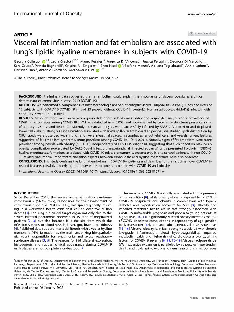
We then aimed at assessing whether the observed VATalterations were associated with SARS-CoV-2 presence in thetissue or if they were a consequence of the systemic inflammation.Although SARS-CoV-2 ability to infect human adipose tissue hasbeen frequently speculated [8, 13, 18, 23], direct evidence of suchphenomenon are scarce in the literature [16, 28, 29]. While SARS-CoV-2 genomic RNA, nucleocapsid and spike proteins were notdetectable in VAT samples of patients affected by COVID-19, virus-like structures with morphology and size resembling thosepresent in SARS-CoV-2-infected VeroE6 cells (Fig. 2A) were quiteoften found in the cytoplasm of stressed adipocytes (Fig. 2B).Furthermore, the presence of ribosome-like clusters described invirus-infected cells [30] was evident in visceral adipocytes ofindividuals with COVID-19, although rarely, (Fig. 2C, D) andfrequently in SARS-CoV-2-infected VeroE6 cells (24–48 h post-infection) (Fig. 2E). In addition typical sign of several pathologicalconditions of viral infection, such as confronting cisternae,ribosome lamella complex and annulate lamellae [31], wereobserved in ~25% of VAT cells belonging to subjects with COVID-19 (Supplementary Fig. 1A–D) and in ~45% of SARS-CoV-2-infected VeroE6 cells (Supplementary Fig. 1E), but not in theiruninfected respective controls. Next, to provide direct evidence ofSARS-CoV-2 ability to infect human adipocytes, leading to cell
Fig. 2 SARS-CoV-2 in visceral adipose tissue and hMADS. A Transmission electron microscopy (TEM): VeroE6 infected cell showing severalvirions into the rough endoplasmic reticulum (RER), some indicated by arrows. Inset: enlargement of squared area. B TEM: adipocyte from thevisceral adipose tissue (VAT) depot of a COVID-19+ subject showing several virions into RER (arrows). Inset: enlargement of squared area.C TEM: VAT of a COVID-19+ subject showing an adipocyte (Ad) with two large ribosome-like clusters (dotted lines) in the cytoplasm.D Enlargement of squared area in C shows a ribosome-like cluster and a virion-like structure into the dilated RER (arrow). E TEM: SARS-CoV-2-infected VeroE6 cells showing a ribosome-like cluster (squared area), enlarged in the inset. F Kinetic of SARS-CoV-2 infection inundifferentiated and differentiated hMADS. SARS-CoV-2 genomic RNA detected in the supernatant at different time points expressed ascopies (cps)/ml. G SARS-CoV-2 quantification in supernatant and cell pellets of hMADS infected cells. H The MTT viability assay in hMADSadipocytes shows lower cell viability in the infected compared to the uninfected cells at 24- and 96-h post-infection (p < 0.05). I Hoechstnuclear staining showing pyknotic nuclei (arrows). J Percentage of pyknotic nuclei in hMADS at 96 h post-infection compared to uninfectedcontrols (p < 0.05). K Lipid droplets average area (μm2) in differentiated hMADS 96 h post-infection compared to uninfected controls (p <0.0001). Expression of putative SARS-CoV-2 receptors L or proteases M assessed by RT-qPCR and normalized for the expression of 36B4 mRNA.Expressions were measured in cells that received (red bars) or did not receive (blue bars) the differentiation cocktail for the indicated numberof days. The means ± SEM were calculated from three independent experiments (ACE2, BSG, NRP1, CSTL) or four independent experiments(FURIN, DPP4), with determinations performed in duplicate (*p < 0.05, **p < 0.01). Scale bar: A, B= 200 nm, C= 500 nm, D= 100 nm,E= 180 nm, F= 120 μm, G= 70 μm, H= 5 μm.
G. Colleluori et al.
1011
International Journal of Obesity (2022) 46:1009 – 1017
stress and death, we infected differentiated human multipotentadipocytes (hMADS) (Fig. 2F–H), and evaluated SARS-CoV-2kinetics in vitro. The growth kinetics of SARS-CoV-2 wasdetermined as viral load (copies/ml) in the supernatants collectedafter 24-, 48-, 72- and 96-h post-infection (Fig. 2F). Though SARS-CoV-2 genomic RNA was detectable in both differentiated andundifferentiated hMADS at the first time points post-infection (24and 48 h), it could be detected only in mature adipocytes at latertime points (72 and 96 h) (Fig. 2F). Consistently, SARS-CoV-2genomic RNA was also detected in the hMADS adipocytes pelletafter 96-h of infection (Fig. 2G). Importantly, the infected hMADSadipocytes displayed lower cell viability (Fig. 2H), higherprevalence of pyknotic nuclei (Fig. 2I–L), and smaller lipid dropletsize—suggestive of cell delipidation and stress—compared to theuninfected controls (Fig. 2K). Furthermore, in line with these data,evidence of increased material extrusion from infected cellscompared to uninfected controls was observed by LM (p < 0.05),strongly suggesting massive cell delipidation induced by SARS-CoV-2 (Supplementary Fig. 1F–H). We hence performed a time-course analysis of hMADS expression of putative SARS-CoV-2receptors (Fig. 2L) and proteases (Fig. 2M) in the presence orabsence of the adipogenic differentiation cocktail (at 4, 7, 14, and18 days). ACE2 receptor was expressed at very low levels in bothdifferentiated and undifferentiated hMADS, even though we usedspecifically designed primers holding a 100.92% efficiency. On theother side, the BASIGIN receptor was preferentially detected indifferentiated hMADS which displayed an increased expressionafter 14 days. The receptor NEUROPILIN 1 was expressed byundifferentiated cells. Concerning proteases expression, whiledifferentiated hMADS expressed the protease FURIN, the undiffer-entiated ones preferentially expressed DPPIV. The expression ofCATHEPSIN L did not differs between the two conditions, while wedid not detect TMPRSS2 in both differentiated and undifferen-tiated hMADS (data not shown).
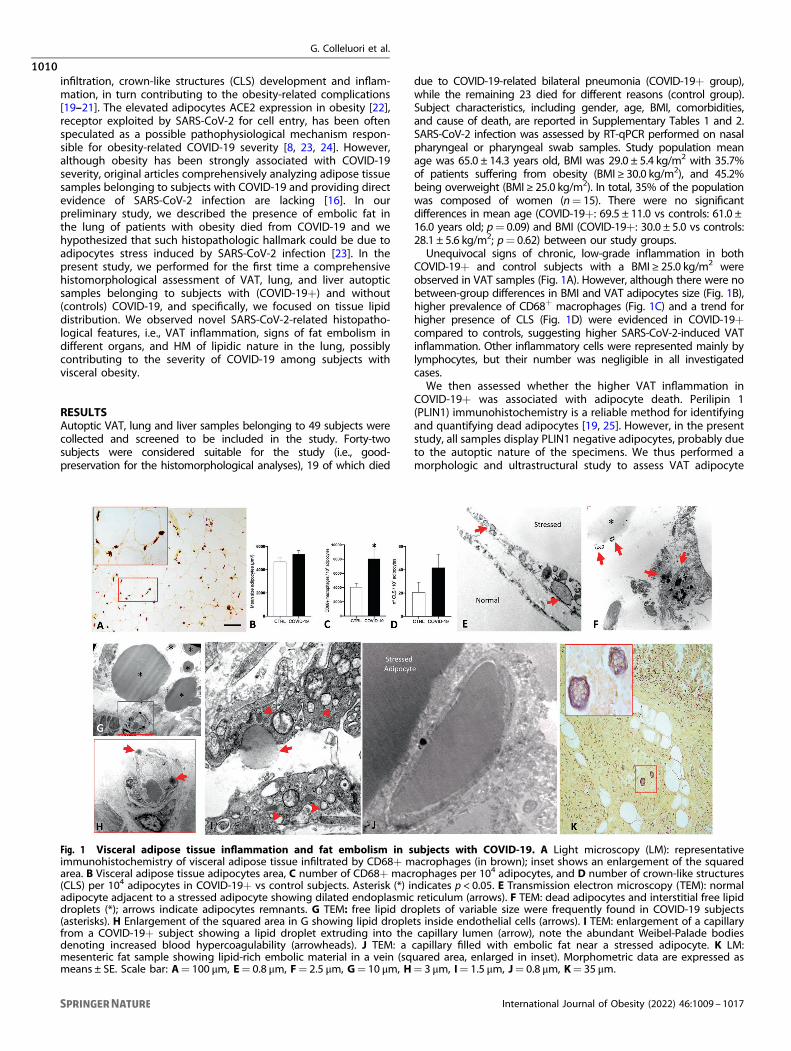
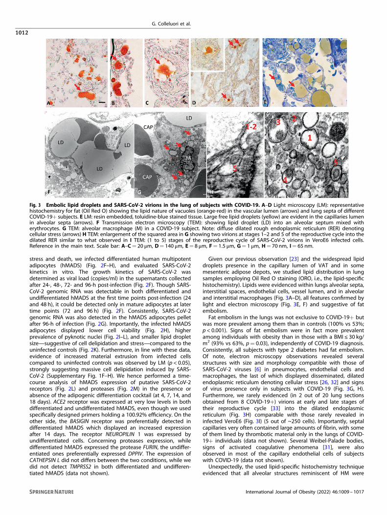
Given our previous observation [23] and the widespread lipiddroplets presence in the capillary lumen of VAT and in somemesenteric adipose depots, we studied lipid distribution in lungsamples employing Oil Red O staining (ORO, i.e., the lipid-specifichistochemistry). Lipids were evidenced within lungs alveolar septa,interstitial spaces, endothelial cells, vessel lumen, and in alveolarand interstitial macrophages (Fig. 3A–D), all features confirmed bylight and electron microscopy (Fig. 3E, F) and suggestive of fatembolism.Fat embolism in the lungs was not exclusive to COVID-19+ but
was more prevalent among them than in controls (100% vs 53%;p < 0.001). Signs of fat embolism were in fact more prevalentamong individuals with obesity than in those with a BMI ≤ 30 kg/m2 (93% vs 63%, p= 0.03), independently of COVID-19 diagnosis.Consistently, all subjects with type 2 diabetes had fat embolism.Of note, electron microscopy observations revealed severalstructures with size and morphology compatible with those ofSARS-CoV-2 viruses [6] in pneumocytes, endothelial cells andmacrophages, the last of which displayed disseminated, dilatedendoplasmic reticulum denoting cellular stress [26, 32] and signsof virus presence only in subjects with COVID-19 (Fig. 3G, H).Furthermore, we rarely evidenced (in 2 out of 20 lung sectionsobtained from 8 COVID-19+) virions at early and late stages oftheir reproductive cycle [33] into the dilated endoplasmicreticulum (Fig. 3H) comparable with those rarely revealed ininfected VeroE6 (Fig. 3I) (5 out of ~250 cells). Importantly, septalcapillaries very often contained large amounts of fibrin, with someof them lined by thrombotic material only in the lungs of COVID-19+ individuals (data not shown). Several Weibel-Palade bodies,signs of activated coagulative phenomena [31], were alsoobserved in most of the capillary endothelial cells of subjectswith COVID-19 (data not shown).Unexpectedly, the used lipid-specific histochemistry technique
evidenced that all alveolar structures reminiscent of HM were
Fig. 3 Embolic lipid droplets and SARS-CoV-2 virions in the lung of subjects with COVID-19. A–D Light microscopy (LM): representativehistochemistry for fat (Oil Red O) showing the lipid nature of vacuoles (orange-red) in the vascular lumen (arrows) and lung septa of differentCOVID-19+ subjects. E LM: resin embedded, toluidine-blue stained tissue. Large free lipid droplets (yellow) are evident in the capillaries lumenin alveolar septa (arrows). F Transmission electron microscopy (TEM): showing lipid droplet (LD) into an alveolar septum mixed witherythrocytes. G TEM: alveolar macrophage (M) in a COVID-19 subject. Note: diffuse dilated rough endoplasmic reticulum (RER) denotingcellular stress (arrows) H TEM: enlargement of the squared area in G showing two virions at stages 1–2 and 5 of the reproductive cycle into thedilated RER similar to what observed in I TEM: (1 to 5) stages of the reproductive cycle of SARS-CoV-2 virions in VeroE6 infected cells.Reference in the main text. Scale bar: A–C= 20 μm, D= 140 μm, E= 8 μm, F= 1.5 μm, G= 1 μm, H= 70 nm, I= 65 nm.
G. Colleluori et al.
1012
International Journal of Obesity (2022) 46:1009 – 1017
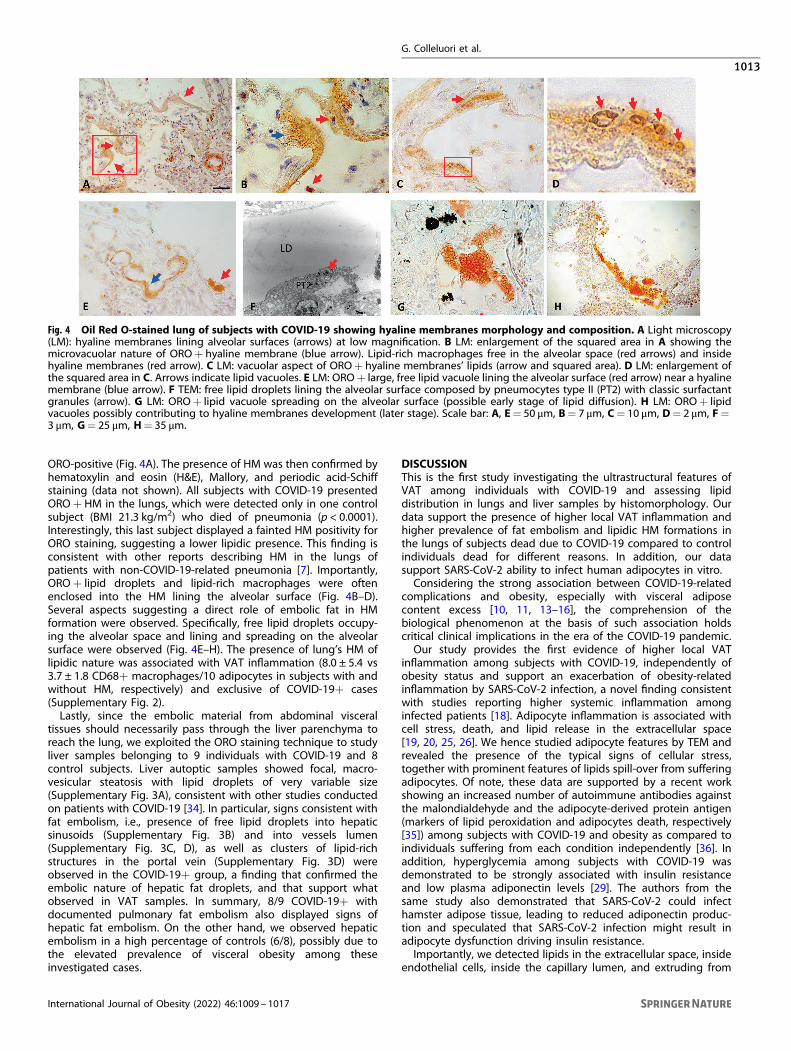
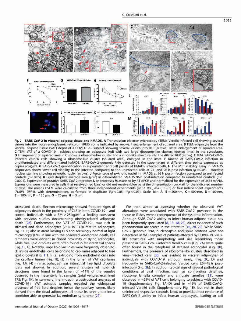
ORO-positive (Fig. 4A). The presence of HM was then confirmed byhematoxylin and eosin (H&E), Mallory, and periodic acid-Schiffstaining (data not shown). All subjects with COVID-19 presentedORO+ HM in the lungs, which were detected only in one controlsubject (BMI 21.3 kg/m2) who died of pneumonia (p < 0.0001).Interestingly, this last subject displayed a fainted HM positivity forORO staining, suggesting a lower lipidic presence. This finding isconsistent with other reports describing HM in the lungs ofpatients with non-COVID-19-related pneumonia [7]. Importantly,ORO+ lipid droplets and lipid-rich macrophages were oftenenclosed into the HM lining the alveolar surface (Fig. 4B–D).Several aspects suggesting a direct role of embolic fat in HMformation were observed. Specifically, free lipid droplets occupy-ing the alveolar space and lining and spreading on the alveolarsurface were observed (Fig. 4E–H). The presence of lung’s HM oflipidic nature was associated with VAT inflammation (8.0 ± 5.4 vs3.7 ± 1.8 CD68+ macrophages/10 adipocytes in subjects with andwithout HM, respectively) and exclusive of COVID-19+ cases(Supplementary Fig. 2).Lastly, since the embolic material from abdominal visceral
tissues should necessarily pass through the liver parenchyma toreach the lung, we exploited the ORO staining technique to studyliver samples belonging to 9 individuals with COVID-19 and 8control subjects. Liver autoptic samples showed focal, macro-vesicular steatosis with lipid droplets of very variable size(Supplementary Fig. 3A), consistent with other studies conductedon patients with COVID-19 [34]. In particular, signs consistent withfat embolism, i.e., presence of free lipid droplets into hepaticsinusoids (Supplementary Fig. 3B) and into vessels lumen(Supplementary Fig. 3C, D), as well as clusters of lipid-richstructures in the portal vein (Supplementary Fig. 3D) wereobserved in the COVID-19+ group, a finding that confirmed theembolic nature of hepatic fat droplets, and that support whatobserved in VAT samples. In summary, 8/9 COVID-19+ withdocumented pulmonary fat embolism also displayed signs ofhepatic fat embolism. On the other hand, we observed hepaticembolism in a high percentage of controls (6/8), possibly due tothe elevated prevalence of visceral obesity among theseinvestigated cases.
DISCUSSIONThis is the first study investigating the ultrastructural features ofVAT among individuals with COVID-19 and assessing lipiddistribution in lungs and liver samples by histomorphology. Ourdata support the presence of higher local VAT inflammation andhigher prevalence of fat embolism and lipidic HM formations inthe lungs of subjects dead due to COVID-19 compared to controlindividuals dead for different reasons. In addition, our datasupport SARS-CoV-2 ability to infect human adipocytes in vitro.Considering the strong association between COVID-19-related
complications and obesity, especially with visceral adiposecontent excess [10, 11, 13–16], the comprehension of thebiological phenomenon at the basis of such association holdscritical clinical implications in the era of the COVID-19 pandemic.Our study provides the first evidence of higher local VAT
inflammation among subjects with COVID-19, independently ofobesity status and support an exacerbation of obesity-relatedinflammation by SARS-CoV-2 infection, a novel finding consistentwith studies reporting higher systemic inflammation amonginfected patients [18]. Adipocyte inflammation is associated withcell stress, death, and lipid release in the extracellular space[19, 20, 25, 26]. We hence studied adipocyte features by TEM andrevealed the presence of the typical signs of cellular stress,together with prominent features of lipids spill-over from sufferingadipocytes. Of note, these data are supported by a recent workshowing an increased number of autoimmune antibodies againstthe malondialdehyde and the adipocyte-derived protein antigen(markers of lipid peroxidation and adipocytes death, respectively[35]) among subjects with COVID-19 and obesity as compared toindividuals suffering from each condition independently [36]. Inaddition, hyperglycemia among subjects with COVID-19 wasdemonstrated to be strongly associated with insulin resistanceand low plasma adiponectin levels [29]. The authors from thesame study also demonstrated that SARS-CoV-2 could infecthamster adipose tissue, leading to reduced adiponectin produc-tion and speculated that SARS-CoV-2 infection might result inadipocyte dysfunction driving insulin resistance.Importantly, we detected lipids in the extracellular space, inside
endothelial cells, inside the capillary lumen, and extruding from
Fig. 4 Oil Red O-stained lung of subjects with COVID-19 showing hyaline membranes morphology and composition. A Light microscopy(LM): hyaline membranes lining alveolar surfaces (arrows) at low magnification. B LM: enlargement of the squared area in A showing themicrovacuolar nature of ORO+ hyaline membrane (blue arrow). Lipid-rich macrophages free in the alveolar space (red arrows) and insidehyaline membranes (red arrow). C LM: vacuolar aspect of ORO+ hyaline membranes’ lipids (arrow and squared area). D LM: enlargement ofthe squared area in C. Arrows indicate lipid vacuoles. E LM: ORO+ large, free lipid vacuole lining the alveolar surface (red arrow) near a hyalinemembrane (blue arrow). F TEM: free lipid droplets lining the alveolar surface composed by pneumocytes type II (PT2) with classic surfactantgranules (arrow). G LM: ORO+ lipid vacuole spreading on the alveolar surface (possible early stage of lipid diffusion). H LM: ORO+ lipidvacuoles possibly contributing to hyaline membranes development (later stage). Scale bar: A, E= 50 μm, B= 7 μm, C= 10 μm, D= 2 μm, F=3 μm, G= 25 μm, H= 35 μm.
G. Colleluori et al.
1013
International Journal of Obesity (2022) 46:1009 – 1017
endothelial cells into the capillary lumen, all features indicative offat embolism. Although virus-like structures were evidenced byTEM in the same VAT depots, the lack of SARS-CoV-2 detection byqPCR did not allow us to conclude that such inflammation, cellularstress, and death were all related to the presence of the virus. It is,in fact, possible that the described VAT features were secondary tothe systemic inflammation induced by COVID-19 or due to thepresence of different viruses within the depot. On the other side,we demonstrated that SARS-CoV-2 could infect human adipocyteseven though neither adipocytes nor adipocyte progenitorsgathered all of the known molecular requirements for the virusentry (e.g., expression of all known virus proteases and receptors).This set of data is in part consistent with other findings andsuggests that additional, not yet characterized, receptors andproteases may be exploited for this purpose [16, 37]. Puray-Chavezet al. in fact indicated that human H522 lung adenocarcinomacells are permissive to SARS-CoV-2 infection despite completeACE2 absence and that virus entry is dependent on heparansulfate in this cell line [37]. Importantly, despite being the firstSARS-CoV-2 targets, olfactory and respiratory epithelial cellsexpress low ACE2 protein levels [38]. For these reasons, additionalco-factors facilitating the virus-host cell interaction (e.g.,NEUROPILIN-1 [39, 40]) may be critical for the infection of tissuesexpressing low ACE2 levels. In our study, BASIGIN receptor andFURIN protease were highly expressed in differentiated hMADSand could be exploited for SARS-CoV-2 infection. However, itshould be noted that, although FURIN critical role in mediatingSARS-CoV-2 infection is widely accepted and seem to be ofrelevance in patients with type 2 diabetes where the protease ishighly expressed [41], the role of BASIGIN has been recentlyquestioned [42].Given the widespread presence of lipid droplets in the capillary
lumen of VAT and our preliminary data [23], we studied lipiddistribution in lung and liver samples and confirmed the presenceof fat embolism. Interestingly, we noticed similar lipid-likestructures also in lung’s images from other reports on patientswith COVID-19 [5, 6, 43], which is why we believe it is worthperforming further in-depth analyses on available samples.Fat embolism in the lungs was not exclusive to, but more
prevalent among subjects with COVID-19; it was in fact alsodetected among subjects with obesity independently of SARS-CoV-2 infection. These data are not surprising given that adipocytedeath and release of lipids are both phenomena occurring inobesity [19, 25, 26]. This finding provides the first evidencepointing out fat embolism as a complication of obesity (andobesity plus type 2 diabetes), determined by adipocyte death andpossibly exacerbated by the COVID-19-induced inflammatorystatus. Importantly, studying lung’s lipid accumulation, weunexpectedly revealed the presence of lipidic HM, a formationstrongly contributing/associated to COVID-19-related interstitialfibrosis and pneumonia [6]. HM were present in all patients withCOVID-19 and in only one control who died for pneumonia, afinding consistent with other reports describing HM presence inthis latter disease [7]. Our histomorphologic assessment revealedseveral aspects indicative of a direct role of embolic fat in HMformation. Consistently, the presence of lung HM of lipidic nature(in the lungs) was associated with VAT inflammation.Our findings on intestinal and liver fat embolism strongly
support the embolic nature of lipid droplets in the lungs. As theportal system drains venous blood from most abdominal fatdepots to the liver, the embolic fat originated in the VATnecessarily pass through the liver to reach other organs. Theunequivocal presence of lipid droplets into sinusoids and liverveins supports the fat embolic production by abdominal fat.In summary, in our case series, although fat embolism may be
present in obesity and type 2 diabetes independently of COVID-19, the embolic lipidic material could contribute to the formationof HM only in the case of COVID-19-related pneumonia. This novel
finding holds critical clinical implications and deserves furtherinvestigation. Furthermore, these data provide insights into HMnature, as their formation process has not been characterized yet[44]. Additional studies investigating the HM nature of non-COVID-19-related pneumonia are required to detail such histopathologi-cal features.Collectively our data reveal higher local VAT inflammation in
subjects with COVID-19 and SARS-CoV-2 ability to infect humanadipocytes. In addition, we provide the first evidence thatsupports the fat embolism as a complication of obesity, likelydetermined by adipocyte death and exacerbated by the COVID-19-induced inflammatory status. Lastly, we reveal for the first timethe presence of lung’s lipidic HM among all infected subjects, anovel COVID-19-related histopathological feature associated withVAT inflammation and fat embolism. Consistently, fat embolismdisplays similar signs and symptoms as observed in COVID-19, inline with a recently published case report [45]. When fat embolismand COVID-19 are suspected, differential diagnosis is critical forproper patient care. Based on our findings, the assessment of fatembolism symptoms is mandatory in the context of the COVID-19pandemic, especially among patients with pulmonary symptoms,obesity, and high waist circumference, last two of which arerecognized as signs of high visceral adipose accumulation. Suchcomplex clinical status should be therefore adequately assessedand properly addressed. Our data hold critical clinical implicationsin the context of obesity and COVID-19 pandemics and need to beconfirmed by additional studies with larger sample size.
MATERIALS AND METHODSStudy approvalWe followed the report “Research ethics during COVID-19 pandemic:observational, and in particular, epidemiological studies” published by theItalian Istituto Superiore di Sanità on May 2020 (Rapporto ISS COVID-19, n.47/2020) [46]. Given that our study was observational (cross-sectional,case-control) in nature, and it was conducted on autoptic specimens anddid not entail neither an intervention, nor the collection of subject’ssensitive information, we have not obtained an informed consent. Ourstudy did not entail any physical risk for the subjects. In Italy, theevaluation of non-pharmacological observational studies is not governedby the same normative references provided for the evaluation of clinicaltrials and observational studies concerning drugs. Furthermore, asreported in the above report [46] in the section dedicated to our type ofstudy in conditions of pandemic and therefore of high risk for thecommunities, some administrative steps may be abolished. Therefore, ourInstitutional Review Board does not require ethical approval for studiesconducted on autoptic specimens and not collecting personal orsensitive data.
Study subjects and tissue samplingAutoptic lung, liver, and VAT samples of 49 subjects were collected at theDepartment of Legal Medicine of the Ospedali Riuniti of Ancona betweenMarch 2020 and May 2021. Twenty-four subjects were affected by COVID-19,while the remaining 25 were not and died for different reasons. SARS-CoV-2infection was assessed in all subjects by RT-PCR tests on a nasopharyngealswab. Subjects were included in the analyses only if their lung’s sampleswere well preserved such that a high-quality histological assessment couldbe performed. We hence analyzed 19 COVID-19+ cases and 23 controls.Among the studied subjects, 15 had documented respiratory conditions, i.e.,pneumonia, dyspnoea, respiratory distress (10 COVID-19+ and 5 controls),15 had documented hypertension (7 COVID-19+ and 8 controls), 11 hadtype 2 diabetes (6 COVID-19+ and 5 controls) and 10 suffered fromcardiovascular diseases (2 COVID-19+ and 8 controls). VAT was sampledfrom the omentum and mesentery region. Lungs were extensively sampledacross central and peripheral regions of each lobe bilaterally. A median ofseven tissue blocks (range five to nine) were taken from each lung. Liversamples were collected from the right and left lobes.Samples were sliced into different pieces to be studied by LM and
transmission electron microscopy (TEM). A comprehensive methodo-logical description for such methodologies has been describedelsewhere [47].
G. Colleluori et al.
1014
International Journal of Obesity (2022) 46:1009 – 1017
Immunohistochemistry and morphometric analysesThe collected visceral (omental) adipose tissue, lung and liver autopsieswere fixed overnight at 4 °C in 4% paraformaldehyde. Samples were thenembedded in paraffin to be studied by LM and to perform immunohis-tochemistry and morphometric analyses. For each sample, 3 µm paraffinsections were obtained and used for immunohistochemical analyses. Acomprehensive description of the protocol has been described elsewhere[47]. To detect the presence of CD68+ macrophages in VAT samples, weused CD68 (Dako #M0814; dilution 1:200; antigen retrieval method bycitrate buffer pH6) antibody. To study SARS-CoV-2 presence in VAT, weused the SARS-CoV-2 nucleocapsid (Invitrogen #MA-17404) and spikeprotein (Sino Biological #40150-T62) antibodies at different dilutions. Thesame antibodies were used to detect the virus on infected VeroE6 atdilution: 1:1000 for nucleocapsid protein and 1:100 for the spike protein.Negative control in which primary antibody was omitted were alwaysincluded in each set of reactions to assess antibody specificity. Tissuesections were observed with a Nikon Eclipse E800 light microscope. Formorphometric purposes, for each paraffin section, ten digital images wereacquired at ×20 magnification with a Nikon DXM 1220 camera. CD68+macrophages widespread in VAT parenchyma and organized to form CLSwere counted in all images. For each subject, the number of totalmacrophages and the density of CLS/104 adipocytes were counted withthe ImageJ morphometric program (RRID:SCR_003070). Adipocytes’ areawas measured in all patients by counting 100 adipocytes for each paraffintissue section using ImageJ.
Histochemical stainingFor ORO staining, samples were cryoprotected in 30% sucrose overnight,embedded in the optimal cutting temperature compound medium, andthen sliced to obtain 7 μm thick cryosections by Leica CM1900 cryostat(Vienna, Austria). ORO staining was then performed on lungs [43] and liver(n= 9) cryosections. In brief, dried cryosections were first placed in 60%isopropanol, then in filtrated ORO working solution (15min at roomtemperature) and briefly washed again in 60% isopropanol and lastly inH2O. Tissue slices were then counterstained with hematoxylin and coveredwith a cover-slip using Vectashield mounting medium (Vector Labora-tories). Lung and liver tissues organization and morphology were alsostudied by H&E staining on paraffin sections. Lung’s HM presence andcharacterization were performed on paraffin sections by H&E, periodicacid-Schiff and Masson trichome staining.
Transmission electron microscopyFor ultrastructural analyses, 3-mm thick VAT (n= 4), lung (n= 8), and liver(n= 1) samples were further fixed in 2% glutaraldehyde-2% paraformal-dehyde in 0.1 M phosphate buffer (pH 7.4) and post-fixed in OsmiumTetroxide 1% then embedded in epoxy resin for TEM studies as describedelsewhere [47]. Cell pellets from the in vitro studies were similarly fixed in2% glutaraldehyde-2% paraformaldehyde in 0.1 M phosphate buffer (pH7.4) for 1 h at room temperature and then embedded in epoxy resin. AnMT-X ultratome (RMC; Tucson) was used to obtain ultrathin sections(~70 nm). Ultrastructural characterization was performed on all samplesusing a CM10 Philips transmission electron microscope (Philips, Eindhoven,The Netherlands, http://www.usa.philips.com).
SARS-CoV-2 infection in VeroE6VeroE6 cells were cultured in Dulbecco’s modified Eagle medium(Euroclone, Milano, Italy), supplemented with 10% fetal calf serum (FCSEuroclone) and antibiotics/antimycotic (100 U/ml penicillin, 100 µg/mlstreptomycin, 0.25 µg/ml amphotericin B) at 37 °C, 5% CO2 in a humidifiedatmosphere (90%), as described previously [48]. Cells were maintained in75 cm2 tissue culture flasks. The day before infection, a confluentmonolayer was trypsinized, and 1.5 × 10⁶ cells were seeded in every eightflasks (25 cm2). Confluent monolayers were infected with SARS-CoV-2(78952 isolates, accession no. MT483867) [49] at a multiplicity of infectionof 3.29 ∙ 105. After 2 h of incubation, the medium containing the inoculumwas removed, the cells were washed twice, and fresh medium was added,which was collected after 6, 12, 24, and 48 h for viral genomequantification and replaced with 2 ml of fresh culture medium to allowscraping of the infected monolayer. Uninfected cell monolayer controlswere treated as infected ones. Cell suspensions (2 ml) were subsequentlycentrifuged at 800 rpm for 5min. Aliquots of infected supernatants,collected as above, were analyzed using RT-qPCR assay as describedelsewhere [49]. Briefly, 5 µl of RNA extracted from 140 µl of infectedsupernatants were run together with a calibration curve, obtained from
ten-fold dilutions of a standard plasmid certified and quantified by asupplier (2019-nCoV Positive Control, nCoVPC, 85 IDT) and negativecontrol, applying a protocol described by CDC (https://www.fda.gov/media/134922/download).
In vitro studies on hMADSEthical approval. Human adipocytes progenitors -Aps- (hMADS cells) wereisolated from adipose tissue, as surgical scraps from a surgical specimen ofvarious surgeries of young donors, with the informed consent of theparents. All methods were approved and performed following theguidelines and regulations of the Centre Hospitalier Universitaire de NiceReview Board.
Cell differentiation. hMADS cells were maintained and differentiated aspreviously described [50]. They will be further referred to as hMADSadipocytes. They were routinely tested for the absence of mycoplasma.Treatments and biological assays were carried out in duplicates on controlor differentiated hMADS cells from days 4 to 18.
Gene expression analysis. Total RNA was extracted using the TRI-Reagentkit (Euromedex, Soufflweyersheim, France) and reverse transcription (RT)was performed using MMLV reverse transcriptase (Promega, Charbon-nieres, France), as recommended by the manufacturers. All primersequences are described in the Supplementary section. Real-time PCRassays were run on an ABI Prism One-step real-time PCR machine (AppliedBiosystems, Courtaboeuf, France). Normalization was performed using36B4 as a reference gene. Quantification was performed using thecomparative Ct method. The results are shown as mean ± standard errorof the mean (SEM), with the number of experiments indicated. Statisticalsignificance was determined by t-tests BiostaTGV (INSERM and SorbonneUniversity, PARIS, France). Probability values < 0.05 were consideredstatistically significant and are marked with a single asterisk, <0.01 withdouble asterisks and <0.001 with triple asterisks. Sequences for the primersused in this study ACE2 (FW 5′- AGAACCCTGGACCCTAGCAT -3′; REV 5′- AGTCGGTACTCCATCCCACA -3′); BASIGIN (FW: 5′- CAGAGTGAAGGCCGTGAAGT -3′; REV: 5′- ACTCTGACTTGCAGACCAGC -3′); NRP1 (FW: 5′- GGGGCTCTCACAAGACCTTC -3′; REV: 5′- GATCCTGAATGGGTCCCGTC -3′); CSTL (FW: 5′- CTGGTGGTTGGCTACGGATT -3′; REV: 5′- CTCCGGTCTTTGGCCATCTT -3′); FURIN(FW: 5′-CTACAGCAGTGGCAACCAGA-3′; REV: 5′- TGTGAGACTCCGTGCACTTC-3′); 36B4 (FW: 5′- CTACAACCCTGAAGAAGTGCTTG -3′; REV: 5′- CAATCTGCAGACAGACACTGG -3′); DPP4 (SINO biologicals Inc. #HP100-649(Eschborn, Germany)
hMADS SARS-CoV-2 infection. hMADS and hMADS adipocytes cells wereinfected with a viral stock of SARS-CoV-2 (EPI_ISL_417491), at a 50% TissueCulture Infectious Dose (TCID50) of 2000 TCID50/ml for 2 h at a temperatureof 37 °C. Following incubation, the medium containing the inoculum wasremoved, the cells were washed twice, and the medium was supplemen-ted with different specific compounds. Supernatants were collected at 24,48, 72, 96 h for viral genome quantification, and medium renewal wasperformed at each sampling time. Uninfected cell monolayer controls weretreated as the infected ones. Supernatants, collected as above, and cellpellets, collected at 96 h post-infection, were analyzed using RT-qPCR asdescribed in the VeroE6 cell section.
Cell viability assay (MTT assay). The effect of SARS-CoV-2 infection on cellviability of hMADS adipocytes was measured using the metabolic dye [4,5-dimethylthiazol-2-yl]−2,5-diphenyl tetrazolium (MTT) (Sigma, St. Louis, MO,USA). Briefly, hMADS cells were seeded in 96-well plates at a density of4500 cells/cm2, differentiated and then infected with the viral stock ofSARS-CoV-2 for 2 h at 37 °C. Following the incubation with the virus, cellswere placed in supplemented medium. Time-course analyses of cellsurvival were determined at 24, 48, 72, and 96 h. After the incubationperiod, the media were replaced with 100 µl MTT (0.5 mg/ml), dissolved inPBS and incubated for 3 h. MTT-containing medium was removed, and100 μl of dimethyl sulfoxide was added to dissolve formazan crystalsformed by live cells. Absorbance was subsequently measured at 570 nmusing a BioTek Synergy HTX microplate reader (BioTek, Winooski, VT, USA).Results were expressed as percentages of viable cells relative to uninfectedcontrols.
Nuclear morphology analyses. Alterations in nuclear morphology weredetermined by assessment of nuclear staining using fluorescent stains andfluorescent microscopy [51].
G. Colleluori et al.
1015
International Journal of Obesity (2022) 46:1009 – 1017
For these experiments, hMADS adipocytes were differentiated in 2-wellLab-Tek Chamber Slides (Nalge Nunc International, Naperville, IL, USA),washed with PBS pH 7.4 and fixed with 10% paraformaldehyde in PBS for10min at RT. After washing with PBS, nuclear staining was performed withHoechst. Finally, cells were airdried and cover-slipped using Vectashieldmounting medium (Vector Laboratories, Burlingame, CA, USA) and analyzedby fluorescent microscopy. The number of altered nuclei were counted (in thefield displaying nuclear fragmentation and nuclear condensation) and dividedby the total number of nuclei multiplied by 100. Observations were carriedout by Lucia IMAGE 4.82, Laboratory Investigations Morphometric Analyses.Lipid droplet size (μm2) was measured in SARS-CoV-2-infected hMADS
adipocytes and untreated controls. For this purpose, we used a drawing tabletand a morphometric program (Nikon LUCIA IMAGE, Laboratory Imaging,version 4.61; Praha, Czech Republic). hMADS adipocytes were examined witha Nikon Eclipse Ti-S inverted light microscope (Nikon Instruments S.p.A,Calenzano, Italy), and digital images were captured at ×20 with a Nikon DS-L2camera (Nikon Instruments S.p.A, Calenzano, Italy). Five random fields wereanalyzed, at least 1700 lipid droplets were measured for each sample, and thedifference between infected and non-infected cells was assessed by unpairedt-test. Similarly, the quantitative assessment of the material extruded from thehMADS was calculated using the same microscope and software andexpressed as the number of vacuoles extruded from the cells on the total cellamount.
Statistical analysisBetween-group comparisons for linear and categorical variables weredetermined by unpaired two-tailed Student’s t test and Chi-square test,respectively. Group differences were considered significant when p < 0.05.Data in graphs are expressed as mean ± SEM. Statistical analyses wereperformed with Prism 6.0 (GraphPad Software Inc., La Jolla, CA) and IBMSPSS Statistics Data Editor (v.24).
REFERENCES1. Medicine JHU. Coronavirus resource center. 2021. https://coronavirus.jhu.edu/
map.html.2. Mahendra M, Nuchin A, Kumar R, Shreedhar S, Mahesh PA. Predictors of mortality
in patients with severe COVID-19 pneumonia—a retrospective study. Adv RespirMed. 2021;89:135–44.
3. Huang C, Wang Y, Li X, Ren L, Zhao J, Hu Y, et al. Clinical features of patientsinfected with 2019 novel coronavirus in Wuhan, China. Lancet. 2020;395:497–506.
4. Wadman M, Couzin-Frankel J, Kaiser J, Matacic C. A rampage through the body.Science. 2020;368:356–60.
5. Xu Z, Shi L, Wang Y, Zhang J, Huang L, Zhang C, et al. Pathological findings ofCOVID-19 associated with acute respiratory distress syndrome. Lancet RespirMed. 2020;8:420–2.
6. Bradley BT, Maioli H, Johnston R, Chaudhry I, Fink SL, Xu H, et al. Histopathologyand ultrastructural findings of fatal COVID-19 infections in Washington State: acase series. Lancet. 2020;396:320–32.
7. Schneider JL, Rowe JH, Garcia-de-Alba C, Kim CF, Sharpe AH, Haigis MC. Theaging lung: physiology, disease, and immunity. Cell. 2021;184:1990–2019.
8. Stefan N, Birkenfeld AL, Schulze MB. Global pandemics interconnected—obesity,impaired metabolic health and COVID-19. Nat Rev Endocrinol. 2021;17:135–49.
9. O’Hearn M, Liu J, Cudhea F, Micha R, Mozaffarian D. Coronavirus Disease 2019hospitalizations attributable to cardiometabolic conditions in the United States: acomparative risk assessment analysis. J Am Heart Assoc. 2021;10:e019259.
10. Williamson EJ, Walker AJ, Bhaskaran K, Bacon S, Bates C, Morton CE, et al. Factorsassociated with COVID-19-related death using OpenSAFELY. Nature. 2020;584:430–6.
11. Onder G, Palmieri L, Vanacore N, Giuliano M, Brusaferro S,Italian National Instituteof Health C-MG. Nonrespiratory complications and obesity in patients dying withCOVID-19 in Italy. Obesity. 2021;29:20–3.
12. Locke AE, Kahali B, Berndt SI, Justice AE, Pers TH, Day FR, et al. Genetic studies ofbody mass index yield new insights for obesity biology. Nature. 2015;518:197–206.
13. Battisti S, Pedone C, Napoli N, Russo E, Agnoletti V, Nigra SG, et al. Computedtomography highlights increased visceral adiposity associated with critical illnessin COVID-19. Diabetes Care. 2020;43:e129–30.
14. Watanabe M, Caruso D, Tuccinardi D, Risi R, Zerunian M, Polici M, et al. Visceral fatshows the strongest association with the need of intensive care in patients withCOVID-19. Metabolism. 2020;111:154319.
15. Petersen A, Bressem K, Albrecht J, Thiess HM, Vahldiek J, Hamm B, et al. The roleof visceral adiposity in the severity of COVID-19: Highlights from a unicentercross-sectional pilot study in Germany. Metabolism. 2020;110:154317.
16. Drucker DJ. Diabetes, obesity, metabolism, and SARS-CoV-2 infection: the end ofthe beginning. Cell Metab. 2021;33:479–98.
17. Kompaniyets L, Goodman AB, Belay B, Freedman DS, Sucosky MS, Lange SJ, et al.Body mass index and risk for COVID-19-related hospitalization, intensive care unitadmission, invasive mechanical ventilation, and death—United States, March-December 2020. Morb Mortal Wkly Rep. 2021;70:355–61.
18. Morys F, Dagher A. Poor metabolic health increases COVID-19-related mortality inthe UK biobank sample. Front Endocrinol. 2021;12:652765.
19. Cinti S, Mitchell G, Barbatelli G, Murano I, Ceresi E, Faloia E, et al. Adipocyte deathdefines macrophage localization and function in adipose tissue of obese miceand humans. J Lipid Res. 2005;46:2347–55.
20. Hotamisligil GS. Inflammation, metaflammation and immunometabolic disorders.Nature. 2017;542:177–85.
21. Colleluori G, Villareal DT. Aging, obesity, sarcopenia and the effect of diet andexercise intervention. Exp Gerontol. 2021;155:111561.
22. Gupte M, Boustany-Kari CM, Bharadwaj K, Police S, Thatcher S, Gong MC, et al.ACE2 is expressed in mouse adipocytes and regulated by a high-fat diet. Am JPhysiol Regul Integr Comp Physiol. 2008;295:R781–8.
23. Cinti S, Graciotti L, Giordano A, Valerio A, Nisoli E. COVID-19 and fat embolism: ahypothesis to explain the severe clinical outcome in people with obesity. Int JObes. 2020;44:1800–2.
24. Shin J, Toyoda S, Nishitani S, Fukuhara A, Kita S, Otsuki M, et al. Possible invol-vement of adipose tissue in patients with older age, obesity, and diabetes withCoronavirus SARS-CoV-2 infection (COVID-19) via GRP78 (BIP/HSPA5): significanceof hyperinsulinemia management in COVID-19. Diabetes. 2021;70:2745–2755.
25. Murano I, Barbatelli G, Parisani V, Latini C, Muzzonigro G, Castellucci M, et al. Deadadipocytes, detected as crown-like structures, are prevalent in visceral fat depotsof genetically obese mice. J Lipid Res. 2008;49:1562–8.
26. Camastra S, Vitali A, Anselmino M, Gastaldelli A, Bellini R, Berta R, et al. Muscle andadipose tissue morphology, insulin sensitivity and beta-cell function in diabetic andnondiabetic obese patients: effects of bariatric surgery. Sci Rep. 2017;7:9007.
27. Meng Y, Zhang M, Ling H, Huang S, Miao Q, Yu Y, et al. Nontraumatic multiple-organ fat embolism: an autopsy case and review of literature. Am J Forensic MedPathol. 2020;41:131–4.
28. Hirschbuhl K, Dintner S, Beer M, Wylezich C, Schlegel J, Delbridge C, et al. Viralmapping in COVID-19 deceased in the Augsburg autopsy series of the first wave:a multiorgan and multimethodological approach. PLoS ONE. 2021;16:e0254872.
29. Reiterer M, Rajan M, Gomez-Banoy N, Lau JD, Gomez-Escobar LG, Ma L, et al.Hyperglycemia in acute COVID-19 is characterized by insulin resistance andadipose tissue infectivity by SARS-CoV-2. Cell Metab. 2021;33:2174–88 e5.
30. Goldsmith CS, Tatti KM, Ksiazek TG, Rollin PE, Comer JA, Lee WW, et al. Ultra-structural characterization of SARS coronavirus. Emerg Infect Dis. 2004;10:320–6.
31. Ghadially F. Ultrastructural pathology of the cell and matrix. 1997 (Butterworth-Heinemann, Boston).
32. Giordano A, Murano I, Mondini E, Perugini J, Smorlesi A, Severi I, et al. Obeseadipocytes show ultrastructural features of stressed cells and die of pyroptosis. JLipid Res. 2013;54:2423–36.
33. Perlman S, Netland J. Coronaviruses post-SARS: update on replication andpathogenesis. Nat Rev Microbiol. 2009;7:439–50.
34. Lagana SM, Kudose S, Iuga AC, Lee MJ, Fazlollahi L, Remotti HE, et al. Hepaticpathology in patients dying of COVID-19: a series of 40 cases including clinical,histologic, and virologic data. Mod Pathol. 2020;33:2147–55.
35. Frasca D, Diaz A, Romero M, Garcia D, Jayram D, Thaller S, et al. Identification andcharacterization of adipose tissue-derived human antibodies with “Anti-self”specificity. Front Immunol. 2020;11:392.
36. Frasca D, Reidy L, Romero M, Diaz A, Cray C, Kahl K, et al. The majority of SARS-CoV-2-specific antibodies in COVID-19 patients with obesity are autoimmune and notneutralizing. Int J Obes. 2021;1–6. https://doi.org/10.1038/s41366-021-01016-9.
37. Puray-Chavez M, LaPak KM, Schrank TP, Elliott JL, Bhatt DP, Agajanian MJ, et al.Systematic analysis of SARS-CoV-2 infection of an ACE2-negative human airwaycell. Cell Rep. 2021;36:109364.
38. Hikmet F, Mear L, Edvinsson A, Micke P, Uhlen M, Lindskog C. The proteinexpression profile of ACE2 in human tissues. Mol Syst Biol. 2020;16:e9610.
39. Cantuti-Castelvetri L, Ojha R, Pedro LD, Djannatian M, Franz J, Kuivanen S, et al.Neuropilin-1 facilitates SARS-CoV-2 cell entry and infectivity. Science. 2020;370:856–60.
40. Giordano A, Cesari P, Capparuccia L, Castellucci M, Cinti S. Sema3A andneuropilin-1 expression and distribution in rat white adipose tissue. J Neurocytol.2003;32:345–52.
41. Ganesan SK, Venkatratnam P, Mahendra J, Devarajan N. Increased mortality of COVID-19 infected diabetes patients: role of furin proteases. Int J Obes. 2020;44:2486–8.
42. Ragotte RJ, Pulido D, Donnellan FR, Hill ML, Gorini G, Davies H, et al. Humanbasigin (CD147) does not directly interact with SARS-CoV-2 spike glycoprotein.mSphere. 2021;6:e0064721.
43. Konopka KE, Nguyen T, Jentzen JM, Rayes O, Schmidt CJ, Wilson AM, et al. Diffusealveolar damage (DAD) resulting from coronavirus disease 2019 Infection isMorphologically Indistinguishable from Other Causes of DAD. Histopathology.2020;77:570–8.
G. Colleluori et al.
1016
International Journal of Obesity (2022) 46:1009 – 1017
44. Wellman TJ, de Prost N, Tucci M, Winkler T, Baron RM, Filipczak P, et al. Lungmetabolic activation as an early biomarker of acute respiratory distress syndromeand local gene expression heterogeneity. Anesthesiology. 2016;125:992–1004.
45. Alexa AL, Onutu AH. Fat embolism syndrome mimicking a COVID-19 infection.Case Rep Crit Care. 2021;2021:5519812.
46. ISS. Istituto Superiore di Sanità-Research ethics during the COVID-19 pandemic:observational and, in particular, epidemiological studies. 2020.
47. Cinti S, Zingaretti MC, Cancello R, Ceresi E, Ferrara P. Morphologic techniques forthe study of brown adipose tissue and white adipose tissue. Methods Mol Biol.2001;155:21–51.
48. de Wilde AH, Raj VS, Oudshoorn D, Bestebroer TM, van Nieuwkoop S, Limpens R,et al. MERS-coronavirus replication induces severe in vitro cytopathology and isstrongly inhibited by cyclosporin A or interferon-alpha treatment. J Gen Virol.2013;94:1749–60.
49. Alessandrini F, Caucci S, Onofri V, Melchionda F, Tagliabracci A, Bagnarelli P, et al.Evaluation of the ion AmpliSeq SARS-CoV-2 research panel by massive parallelsequencing. Genes. 2020;11:929–941.
50. Rodriguez AM, Elabd C, Amri EZ, Ailhaud G, Dani C. The human adipose tissue is asource of multipotent stem cells. Biochimie. 2005;87:125–8.
51. Cummings BS, Schnellmann RG. Measurement of cell death in mammalian cells.Curr Protoc Pharmacol. 2004.
52. Colleluori G, Graciotti L, Pesaresi M, Di Vincenzo A, Perugini J, Di Mercurio E, et al.Visceral fat inflammation and fat embolism are associated with lung’s lipidichyaline membranes in COVID-19 patients. bioRxiv. 2021. https://doi.org/10.1101/2021.10.30.466586.
ACKNOWLEDGEMENTSThis study was funded by Fondo Integrativo Speciale per la Ricerca from the ItalianMinistry of University and Research; grant number: FISR2020IP_05217, and supported
by Progetti di Rilevante Interesse Nazionale (PRIN 2017, #2017L8Z2) and by CariploFoundation to EN (grant 2016-1006). Results from this study were made available onNovember 1st 2021 on bioRxiv [52].
AUTHOR CONTRIBUTIONSGC, LG, MP, AG, and SC: study conceptualization. GC, LG, and SC: study coordination.MP and AT: collected autoptic samples and clinical data. GC, MP, and ADV histologicalstudies on autoptic samples and cell cultures. CMZ, LG, and SC: electron microscopystudies. JP, EDM, AL, and CD: in vitro studies on hMADS. SC, PB, and SM: SARS-CoV-2infection for the in vitro studies. GC, LG, MP, JP, EN, SM, AG, and SC: data analyses andinterpretation. All authors approved the final version of the manuscript and takeresponsibility for its content.
COMPETING INTERESTSThe authors declare no competing interests.
ADDITIONAL INFORMATIONSupplementary information The online version contains supplementary materialavailable at https://doi.org/10.1038/s41366-022-01071-w.
Correspondence and requests for materials should be addressed to Saverio Cinti.
Reprints and permission information is available at http://www.nature.com/reprints
Publisher’s note Springer Nature remains neutral with regard to jurisdictional claimsin published maps and institutional affiliations.
G. Colleluori et al.
1017
International Journal of Obesity (2022) 46:1009 – 1017
Related Documents