Cover Page
The handle http://hdl.handle.net/1887/29602 holds various files of this Leiden University dissertation Author: Groeneweg, Femke Lokke Title: Corticosteroid receptor dynamics : analysis by advanced fluorescence microscopy Issue Date: 2014-11-06

Chapter2
Chapter 2
Rapid non-genomic effects ofcorticosteroids through the
membrane-associated MR and GRand their role in the central stress
response
Femke L Groeneweg1, Henk Karst2, E Ron de Kloet1, Marian Joëls2
Parts of this chapter have been published as:
i Rapid non-genomic effects of corticosteroids and their role in the central stress response.(2011)J Endocrinol. (2):153–167
ii Mineralocorticoid and glucocorticoid receptors at the neuronal membrane, regulators ofnongenomic corticosteroid signaling. (2012)Mol. Cell. Endocrinol. (2): 299–309
1 Department of Medical Pharmacology, Leiden University / LUMC, Leiden, The Netherlands.2 Department of Neuroscience and Pharmacology, University Medical Center,
Utrecht, The Netherlands
21

R -
C affect brain functioning through both delayed,genomic and rapid, non-genomic mechanisms. The latter mode of ac-tion was long known but only in recent years the physiological basisin the brain is beginning to be unravelled. We now know that cortico-steroids exert rapid, non-genomic effects on the excitability and acti-vation of neurons in (amongst others) the hypothalamus, hippocam-pus, amygdala and prefrontal cortex. In addition, corticosteroids af-fect cognition, adaptive behaviour and neuroendocrine output withinminutes. Knowledge on the identity of the receptors and secondarypathways mediating the non-genomic effects of corticosteroids on acellular level is accumulating. Interestingly, in many cases an essen-tial role for the ‘classical’ MR and GR in a novel membrane-associatedmechanism is found.
Here, we systematically review the recent literature on non-genomic actions of corticosteroids on neuronal activity and function-ing in selected limbic brain targets. We will discuss the relevance ofthese permissive effects for cognition and neuroendocrine control,and the integration of this novel mode of action into the complexbalanced pattern of stress effects in the brain. Subsequently, we willreview the knowledge regarding the underlying molecular pathwaysaddressing the following questions: How do the MR and GR translo-cate to the membrane and what are their signalling partners?
22
2.1. R
Chapter2
Corticosteroids play a major role in the response of the brain to stress. For manyyears, they were believed to be only responsible for the delayed and prolonged ef-fects of stress, as opposed tomonoamines and neuropeptideswhichwere thought toestablish rapid effects (de Kloet et al., 2005).While this is generally true, the pictureis actually more complex. For instance, corticosteroids influence a wide range of be-haviors and endocrine outputs within minutes, a timeframe that is too rapid to beexplained by genomic effects (de Kloet et al., 1999; Haller et al., 2008). In agreement,we and others recently established that corticosteroids rapidly alter neuronal activ-ity and excitability in a number of brain areas, providing a physiological basis forthe rapid effects on behavior (Tasker et al., 2006; de Kloet et al., 2008). Many rapideffects are still mediated by the classical corticosteroid receptors, the MR and theGR, but by a subpopulation of these receptors, anchored at the membrane (Karstet al., 2005, 2010). The existence of such a rapid mode of action raises many newquestions. Where in the brain do these rapid effects take place? Which receptorsand pathways are involved in these effects? What are the functional consequencesfor cognition and neuroendocrine control? How are these rapid corticosteroid ac-tions integrated with other components of the stress response? Equally importantare the remainingmolecular questions. How strong is the evidence for amembrane-localization of the MR and GR and for other types of (novel) membrane receptors.Also, as steroid receptors do not have a transmembrane domain, how doMR andGRassociate with the plasma membrane? And finally, are there common downstreampathways. In this chapter we discuss our current understanding of rapid actions ofcorticosterone, with emphasis on their function within the brain.
2.1 Rapid effects of corticosterone in the brain
The rapid effects of corticosterone on brain and cognition have been subject of sev-eral recent reviews (Dallman, 2005; Tasker et al., 2006; de Kloet et al., 2008; Halleret al., 2008; Prager and Johnson, 2009; Evanson et al., 2010a). However, over thelast two years a number of new studies have emerged that extend and challengethe existing views on the function and nature of these rapid effects. Here, we focuson the integration of these new findings in the existing theories on rapid cortico-steroid signalling. The findings are discussed per brain area; i.e. the hypothalamus,pituitary, hippocampus, amygdala and frontal cortical areas. In the following sec-tions, the major findings in these four different brain areas are summarized (see foroverview Table 2.1). In this review we restrict ourselves to the non-genomic roles oftheMR and GRwithin neurons. Both receptors have vital functions in the peripheryand also here many non-genomic actions have been observed. However, these arebeyond the scope of this review and have been described elsewhere (Boldyreff andWehling, 2003; Grossmann and Gekle, 2009; Funder, 2010).
23

R -
Hypothalamus
The PVN is one of the core structures in theHPA-axis. PVNneurons express high lev-els of GR, but virtually noMR. Indeed, throughGR activation in the PVN corticoster-one negatively feeds back on theHPA-axis in a delayed, genomic fashion (de Kloet etal., 1998). However, corticosterone also regulates HPA-axis activity in a more rapidtime frame, through non-genomic actions (Jones et al., 1972; Dallman, 2005). Im-portantly, a recent study showed that this rapid inhibition can be induced by localinfusion of dexamethasone or a membrane-impermeable conjugate of dexametha-sone with bovine serum albumine (dex-BSA) into the PVN (Evanson et al., 2010b).This effect can be prevented by co-administration of an antagonist of the cannabi-noid receptor type 1 (CB1) (Evanson et al., 2010b). Thus, at the level of the PVN,corticosterone rapidly reduces HPA-axis activation in a non-genomic, membrane-associated manner, involving endocannabinoid signalling.
Insight in the neurobiological substrate of these fast effects was provided byTasker and colleagues. This group was the first to carry out detailed studies on thefrequency of miniature excitatory postsynaptic currents (mEPSCs) in the PVN andthe nearby supraoptic nucleus (SON) (Di et al., 2003). An mEPSC reflects the post-synaptic current resulting from the spontaneous release of a single glutamatergicvesicle from a presynaptic terminal (Bekkers and Stevens, 1989). Importantly, thefrequency of these events (particularly in the absence of changes in mEPSC ampli-tude) is considered to be determined by presynaptic properties, reflecting changesin either release probability of the vesicles or changes in the number of synaptic con-tacts. Tasker and colleagues established that a high dose of corticosterone (between100n and 1 µ ) or its synthetic analogue dexamethasone reduces the frequency ofmEPSCs in PVNneurons (Di et al., 2003;Malcher-Lopes et al., 2006). This effect wasdetectable within 5 minutes and did not reverse when corticosterone was washedout. Effectively, the excitability of PVN neurons was reduced by application of cor-ticosteroids in a rapid but prolonged manner. Rapid changes in mEPSC frequencyinduced by corticosterone could not be blocked by MR or GR antagonists (Di et al.,2003, 2009). In contrast, preliminary data shows that they are prevented by condi-tional knockout of the GR gene within the hypothalamus and thus will involve the(membrane-associated) GR (Haam et al., 2010; Tasker and Herman, 2011). How thisnew finding should be integrated with the lack of effect of antagonists remains un-clear and awaits further clarification in a full study. The effects within the PVNwerefurther shown to be non-genomic, membrane-initiated and to involve G-proteincoupled signalling. Interestingly, rapid corticosteroid actions required retrogradeendocannabinoid signalling and the CB1 receptor. The presumed cellular signallingpathway is visualized in Figure 2.1 . Since the CB1 receptor is also required for rapidinhibition of theHPA-axis (Evanson et al., 2010b), the rapid inhibition ofmEPSC fre-quency (and thus excitability) of PVN neurons could provide the cellular substratefor this phenomenon.
24

2.1. R
Chapter2
2-AG / AEA
Inhibition of transmission
PKA-cAMP K+
K+
Facilitation of transmission
ERK
A B
GPCR
GRG
GG
MR
MRCB1
Figure 2.1: Schematic representation of the synaptic pathways of corticosterone-induced rapideffects on glutamatergic transmission(A) Inhibition of glutamatergic transmission is initiated by postsynaptically located receptors; this can beeither G-protein coupled receptors (hypothalamus) or membrane-localized GRs (amygdala). Activationof these receptors by corticosterone induces activation of G-proteins and the cAMP-protein kinase A(PKA) pathway, which eventually induces synthesis of the retrograde messengers anandamide (AEA)and 2-arachidonoylglycerol (2-AG). In a retrograde mode of action at the presynaptic terminal 2-AG andAEA activate the cannabinoid receptor type 1 (CB1), which in turn inhibits the release probability ofglutamatergic vesicles. (B) Facilitation of glutamatergic transmission is initiated by both pre- and post-synaptically located membrane-MRs. Presynaptically, activation of the MR by corticosterone activatesan extracellular signal-regulated kinase (ERK) pathway resulting in stimulation of the release probabilityof glutamate vesicles. At the same time, postsynaptic activation of a membrane-associated MR inhibitspotassium IA-currents, and stimulates membrane diffusion of AMPA receptors. All three effects togetherresult in a facilitation of glutamatergic transmission.
However, rapid inhibitory effects of corticosterone in the PVN are not restrictedto vasopressin- and CRH-containing parvocellular neurons, but they are seen in allneuronal populations (parvocellular and magnocellular) in the PVN (Di et al., 2003,2005; Tasker et al., 2006). In the magnocellular neurons in the PVN and SON, a sec-ond effect was observed on the spontaneous release of gamma-aminobutyric acid(GABA), the main inhibitory neurotransmitter. The frequency of mIPSCs (minia-ture inhibitory postsynaptic currents) was rapidly increased by dexamethasone, butthis required even higher concentrations (1 µ ormore) (Di et al., 2005, 2009). Func-tionally, this suggests a more general coordinative role for the non-genomic effectsof corticosterone in the hypothalamus, which requires further specification (Taskeret al., 2006).
Pituitary
Fast and delayed effects of corticosteroids have also been observed at the level of theanterior pituitary gland, where GR is abundantly expressed and MR levels are quitelow (Reul et al., 1990). Already in the 1970’s and 80’s both rapid and delayed actions
25

R -
of corticosteroids on pituitary ACTH release were reported (Jones et al., 1972; Wid-maier and Dallman, 1984). Inhibition of ACTH release was seen as early as 1 minuteand as late as 2 hours after corticosteroid administration. The latter is a genomicaction mediated by GR-driven gene transcription, while the former action was in-sensitive to protein synthesis inhibitors and thus mediated by non-genomic path-ways (Keller-Wood and Dallman, 1984). Interestingly, the rapid inhibition of ACTHrelease was only seen when corticosterone levels were rapidly rising and not whenthey were already high, suggesting that this feedback is rate-sensitive (Jones et al.,1972; Kaneko and Hiroshige, 1978).
The cellular basis of the rapid effects is not well established and controversyremains about the receptor mediating the effects. On the one hand, pretreatmentwith a GR antagonist did not prevent the rapid effects of corticosterone on CRH-inducedACTH secretion in vivo (Hinz andHirschelmann, 2000). Also, in a pituitary-derived cell line a membrane binding place for dexamethasone and corticosteronewas identified that did not have any affinity for the GR-antagonist RU486 (Maier etal., 2005). However, another line of evidence does suggest a role for the classical GRinmediating rapid feedback at the pituitary. Thus, a rapid and non-genomic translo-cation of annexin-I by dexamethasone was prevented by GR-antagonist treatmentin a pituitary derived cell line (Solito et al., 2003). This translocation of annexin-Iwas required for rapid inhibition of ACTH release (Buckingham et al., 2003; Tierneyet al., 2003). Thus, corticosterone rapidly inhibits ACTH release from the pituitary,but whether this is due to a novel receptor or to the classical GR is still controver-sial. This rapid inhibition is also seen in control human subjects, while it is absentin depressed patients, suggesting that the rapid negative feedback is somehow as-sociated with disease (Young et al., 1991).
Hippocampus
Adaptation to a stressful situation is a coordinated effort mediated by the limbicsystem —the hippocampus and amygdala— in coordination with the prefrontalcortex (see Figure 2.2). This, among other things, involves projections of these ar-eas to and hence control over the PVN (Ulrich-Lai and Herman, 2009). Collectively,these areas also facilitate the formation of a memory trace of a stressful emotionalevent. Processing of contextual information depends predominantly on hippocam-pal function. The hippocampus expresses high levels of both MR and GR in all sub-fields (except its cornu ammonus-3 (CA3) region that mainly expresses MR) (Reuland de Kloet, 1985). Corticosterone exerts strong genomic effects on the activityand plasticity of all hippocampal subfields as well as on hippocampus-dependentmemory (McEwen, 2001; Kim and Diamond, 2002; Mirescu and Gould, 2006; Joëls,2008). Low levels of corticosterone, through MR activation, facilitate plasticity andhippocampus-dependent memory (Diamond et al., 1992). By contrast, absence orvery high levels of corticosterone inhibit plasticity; the latter is mediated throughthe GR (Alfarez et al., 2002; Kim et al., 2004).
26

2.1. R
Chapter2
Amygdala Hippocampus
Prefrontal cortex
PVN
Contextual aspects Emotional aspects
Planning and control
Adaptation to stress
Memory
Pituitary
Adrenals
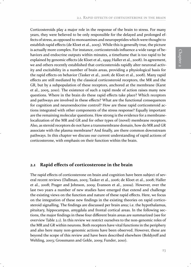
Figure 2.2: Brain circuitry of stressThe limbic system is implicated in adapta-tion, learning & memory processes, mood,and control of the HPA-axis. The hormonesof theHPA-axis coordinate information pro-cessing and promote connectivity betweenamygdala, prefrontal cortex and hippocam-pus to facilitate behavioral adaptation. Pro-jections from the limbic structures inner-vate the PVN network and regulate trans-synaptically the activity of the HPA-axis.
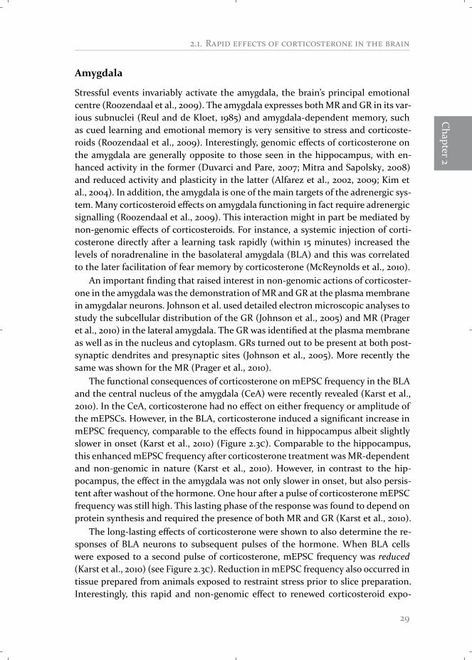
Similar to neurons in the hypothalamus, hippocampal neurons spontaneouslyshow mEPSCs. In a first study (Karst et al., 2005), the effect of corticosterone wasexamined on mEPSC frequency in the CA1 region of the hippocampus. It appearedthat within 5 minutes of corticosterone administration, the frequency of mEPSCs issignificantly enhanced, i.e. changed in a direction opposite to that observed in thePVN. The amplitude was unaffected (Karst et al., 2005; Olijslagers et al., 2008) (seeFigure 2.3 , ). This effect was recently reproduced by other investigators (Qiu et al.,2010) and granule neurons in the dentate gyrus respond similarly to corticosteroneas CA1 neurons (Pasricha et al., 2011). Similar to the corticosteroid effect in the hy-pothalamus, the rapid effect in the hippocampus does not depend on gene transcrip-tion and involves a membrane-located receptor (Karst et al., 2005). However, fur-ther studies established profound differences between rapid responses to corticos-terone in the hippocampus compared to the PVN. The increased mEPSC frequencyin hippocampus rapidly reversed when corticosterone was washed out. Also, in theCA1 the effect occurred at a 10-fold lower dose of corticosterone (10n or higher)than in the hypothalamus. Importantly, corticosterone efficiently enhancedmEPSCfrequency in the hippocampus of wild type and GR knockout mice, but the effectswere completely abolished in MR knockout mice, supporting that rapid effects inthe hippocampus aremediated byMRs (Karst et al., 2005). This was confirmed withspecific MR and GR antagonists. Importantly, the membrane-located MR appearsto have a lower affinity than the cytosolic form (Karst et al., 2005), so that it po-tentially could play an important role when corticosteroid levels rise, shortly afterstress (Joëls et al., 2008). Follow-up studies suggested that rapid corticosteroid ef-fects involve MRs inserted into the presynaptic membrane (Olijslagers et al., 2008)(see Figure 2.1 ). This is backed up by preliminary evidence that shows localizationof the MR in the plasma membrane of hippocampal neurons, co-localized with thepresynaptic marker synapsin I (Qiu et al., 2010).
27

R -
A*
***
AmygdalaBLA
HippocampusCA12
0
1
frequ
ency
(Hz)baseline
cort
B C
pulse 1 pulse 2 pulse 1 pulse 2
baselinecorticosterone
Figure 2.3: Effect of two pulses of corticosterone onmEPSC frequency in the CA1 region and thebasolateral amygdala (BLA)(A) Typical traces of mEPSC pulses recorded from hippocampal neurons before (white bars) and after(black bars) treatment with 100n corticosterone. (B) In hippocampal CA1 neurons exposure to twoconsecutive pulses of 100n corticosterone (1hour apart) both induce a reversible increase in mEPSCfrequency. (C) In amygdalar BLA neurons, the first pulse of corticosterone induces an increase in mEPSCfrequency, this increase is not reversible. For the second pulse of corticosterone the basal mEPSC fre-quency is already elevated and the second pulse induces an irreversible decrease instead. mEPSC, minia-ture excitatory postsynaptic current. ∗ p < 0.05 compared with baseline (paired t test). Figure reprintedwith permission from Karst et al. (2010).
Corticosterone also affects two postsynaptic features of CA1 neurons through theMR. Firstly, corticosterone was found to inhibit postsynaptic IA-currents, an effectthat could be blocked with an MR-antagonist (Olijslagers et al., 2008). IA-currentsare potassium currents that are negative regulators of neuronal excitability and plas-ticity (Hoffman et al., 1997; Yuan et al., 2002). Consequently, the inhibition of thesecurrents by corticosterone is expected to stimulate excitability and plasticity of hip-pocampal neurons. Secondly, corticosterone stimulated, within 5 minutes, lateraldiffusion of α-amino-3-hydroxyl-5-methyl-4-isoxazole-propionic acid (AMPA) recep-tors in cultured hippocampal neurons (Groc et al., 2008). This effect also turnedout to depend on membrane-localized MRs (Groc et al., 2008). Potentially, a moremobile pool of AMPA receptors facilitates the induction of synaptic plasticity. Allof these studies support a membrane-localised form of the MR as main mediator ofrapid corticosteroid signalling in the hippocampus. Overall, corticosterone seemsto rapidly potentiate the excitability of hippocampal neurons via membrane-MRslocated on both pre- and postsynaptic sites, thus priming the hippocampal circuitfor subsequent stimulation by context-dependent factors.
However, not all rapid effects in the hippocampus involve the MR. First, a non-genomic increase in spine density of hippocampal neurons was found to dependon GRs rather than MRs (Komatsuzaki et al., 2005). Yet other rapid corticoster-one effects occur independent of MR or GR and therefore could be mediated bya novel (so far not identified) membrane-localized receptor. This applies to rapidstimulatory corticosterone effects on inhibitory transmission (Hu et al., 2010), onlevels of extracellular excitatory amino acid (Venero and Borrell, 1999), long-termpotentiation (LTP) induction (Wiegert et al., 2006) and N-methyl-D-aspartatic acid(NMDA)-dependent neurotoxicity (Xiao et al., 2010). Some studies also reported in-hibitory actions of corticosterone on NMDA signalling (Sato et al., 2004; Liu et al.,2007). Apparently, corticosterone affects hippocampal signalling in multiple ways,involving membrane-located MRs, GRs and other, still unknown receptors.
28
2.1. R
Chapter2
Amygdala
Stressful events invariably activate the amygdala, the brain’s principal emotionalcentre (Roozendaal et al., 2009). The amygdala expresses bothMR and GR in its var-ious subnuclei (Reul and de Kloet, 1985) and amygdala-dependent memory, suchas cued learning and emotional memory is very sensitive to stress and corticoste-roids (Roozendaal et al., 2009). Interestingly, genomic effects of corticosterone onthe amygdala are generally opposite to those seen in the hippocampus, with en-hanced activity in the former (Duvarci and Pare, 2007; Mitra and Sapolsky, 2008)and reduced activity and plasticity in the latter (Alfarez et al., 2002, 2009; Kim etal., 2004). In addition, the amygdala is one of the main targets of the adrenergic sys-tem.Many corticosteroid effects on amygdala functioning in fact require adrenergicsignalling (Roozendaal et al., 2009). This interaction might in part be mediated bynon-genomic effects of corticosteroids. For instance, a systemic injection of corti-costerone directly after a learning task rapidly (within 15 minutes) increased thelevels of noradrenaline in the basolateral amygdala (BLA) and this was correlatedto the later facilitation of fear memory by corticosterone (McReynolds et al., 2010).
An important finding that raised interest in non-genomic actions of corticoster-one in the amygdala was the demonstration ofMR and GR at the plasmamembranein amygdalar neurons. Johnson et al. used detailed electron microscopic analyses tostudy the subcellular distribution of the GR (Johnson et al., 2005) and MR (Prageret al., 2010) in the lateral amygdala. The GR was identified at the plasma membraneas well as in the nucleus and cytoplasm. GRs turned out to be present at both post-synaptic dendrites and presynaptic sites (Johnson et al., 2005). More recently thesame was shown for the MR (Prager et al., 2010).
The functional consequences of corticosterone on mEPSC frequency in the BLAand the central nucleus of the amygdala (CeA) were recently revealed (Karst et al.,2010). In the CeA, corticosterone had no effect on either frequency or amplitude ofthe mEPSCs. However, in the BLA, corticosterone induced a significant increase inmEPSC frequency, comparable to the effects found in hippocampus albeit slightlyslower in onset (Karst et al., 2010) (Figure 2.3 ). Comparable to the hippocampus,this enhancedmEPSC frequency after corticosterone treatment wasMR-dependentand non-genomic in nature (Karst et al., 2010). However, in contrast to the hip-pocampus, the effect in the amygdala was not only slower in onset, but also persis-tent after washout of the hormone. One hour after a pulse of corticosterone mEPSCfrequency was still high. This lasting phase of the response was found to depend onprotein synthesis and required the presence of both MR and GR (Karst et al., 2010).
The long-lasting effects of corticosterone were shown to also determine the re-sponses of BLA neurons to subsequent pulses of the hormone. When BLA cellswere exposed to a second pulse of corticosterone, mEPSC frequency was reduced(Karst et al., 2010) (see Figure 2.3 ). Reduction inmEPSC frequency also occurred intissue prepared from animals exposed to restraint stress prior to slice preparation.Interestingly, this rapid and non-genomic effect to renewed corticosteroid expo-
29

R -
sure depended on the GR rather than the MR. Similar to the hypothalamus (but incontrast to the hippocampus), it was shown to involve a postsynaptically localizedGR and subsequent retrograde endocannabinoid signalling (see Figure 2.1 ). Thus,in a non-stressed animal corticosterone seems to have a stimulatory effect in the(basolateral) amygdala. However, due to the long-lasting nature of these effects, asecond exposure to corticosterone induces opposite effects, suggesting metaplastic-ity of corticosteroid responses. These data suggests that the amygdala will responddifferently to a stressor depending on the recent stress history of the organism.
Prefrontal cortex
The prefrontal cortex (PFC) is critically involved in complex behavioural control,such as behavioural inhibition, decision-making and working memory. It is exten-sively connected to the amygdala and receives afferents originating in the hippocam-pus (Arnsten, 2009). Despite its important function, the PFC is underrepresentedconcerning studies on the effects of corticosteroids and stress. A number of stud-ies have examined the effect of chronic stress or corticosterone exposure on thePFC. Under these conditions, LTP, dendritic complexity and PFC-dependent work-ing memory were reduced in a genomic fashion (Arnsten, 2009; Holmes and Well-man, 2009). On the contrary, exposure to acute stress or corticosterone increasedglutamatergic transmission and improved working memory performance (Yuen etal., 2009, 2010). These effects occurred with a delay of several hours and were shownto require gene transcription (Yuen et al., 2010). Thus acute and chronic stress affectPFC plasticity and functionality in an opposite manner.
The only studies so far in the PFC that focused on rapid, non-genomic effectswere performed in synaptosomes. In this preparation, corticosterone induced arapid enhancement of glutamate uptake and of calcium-dependent calmodulin sta-bilization (Sze and Iqbal, 1994; Zhu et al., 1998). Unfortunately, the receptors orpathways involved were not examined. In a recent study, Roozendaal and colleaguesreported a putative membrane-GR mediated effect of corticosterone in the insularcortex that is involved in memory acquisition. In this elegant study, administra-tion of either corticosterone or cort-BSA directly into the insular cortex facilitatedthe acquisition of object recognition memory (Roozendaal et al., 2010). Althoughthere are some concerns about the stability of cort-BSA in vivo, this is still indica-tive of a membrane-initiated effect. The effect was prevented by co-administrationof a GR antagonist. The authors further proved that the facilitation of memory bymembrane-GR activationwas established through protein kinase A (PKA), cAMP re-sponse element-binding (CREB) and histone acetylation (Roozendaal et al., 2010).Taken together, rapid non-genomic actions of corticosterone are found in (some)prefrontal areas; so far they seem to be mostly excitatory (as are the sub-acute ge-nomic effects) and could have implications for higher-order learning in complextasks. However, the data is still very sparse.
30
2.2. F
Chapter2
2.2 Functional implications of rapid corticosteroid effects inthe brain
Taking all results into account, we can distinguish some interesting general featuresof the rapid effects of corticosteroids in the brain.
i) It is important to notice that all non-genomic effects are permissive or condi-tional effects. In none of the studies corticosteroids induced any activity on theirown, instead they facilitate or inhibit signalling of ion channels, receptors and neu-rotransmitters. In short, they increase or decrease the threshold for activation ofthese neurons by context-dependent factors. Therefore, it will depend on the con-text which effects (in which brain areas) will be most pronounced during a stressfulencounter.ii) We see a distinctive pattern with a general increase in excitability for some
areas (hippocampus, amygdala and potentially the prefrontal cortex) and a decreasein others (the hypothalamus).iii) While some responses are transient (mostly in the hippocampus), other ef-fects are prolonged (hypothalamus, pituitary and amygdala). The brain circuitryactivated by stress will thus be different depending on the delay after the stressor.iv) In general, the inhibitory effects on hypothalamic functioning seem to requirea higher dose of corticosterone than most effects in other brain areas. If so, the setof responses seen after a mild stressor may be different from that of a more severestressor, the latter having an additional negative effect on PVN-related responses(Prager and Johnson, 2009).v) Finally, a number of rapid corticosteroid effects require the presence of classical
MR and GR inserted in or attached to the plasma membrane, while other effectsare mediated through yet unknown (G-protein coupled) receptors. In general, MR-mediated effects tend to stimulate excitation, while GR-mediated effects can alsobe inhibitory (see Figure 2.1).
We will refer to these five general points when we next consider the potentialfunctional consequences of rapid corticosteroid actions in the brain for HPA-axisregulation and cognition, also taking the ultradian release pattern into considera-tion. Finally we will address the integration of these rapid effects with the rest ofthe brain’s response to stress.
Regulation of the HPA-axis
Corticosteroids exert rapid, as well as delayed, inhibitory feedback at the core struc-tures of the HPA-axis; the PVN of the hypothalamus (Evanson et al., 2010b) and thepituitary gland (Jones et al., 1972; Hinz and Hirschelmann, 2000). In the pituitarythis seems to be caused by both GR-dependent (Buckingham et al., 2003) and GR-independent (Hinz andHirschelmann, 2000) rapid signalling pathways. In the PVN,the rapid suppression of glutamatergic transmission by corticosterone could wellunderlie (amongst others) fast suppression of the HPA-axis in a GR-independent
31

R -
manner (Tasker, 2006). As mentioned earlier, this hypothesis is backed up by theeffectiveness of intra-PVN infusions of dexamethasone or dex-BSA on HPA-axis ac-tivity in a rapid time frame (Evanson et al., 2010b).
In addition, extra-hypothalamic structures also control the activity of the HPA-axis. For instance, the hippocampus and prefrontal cortex exert negative feedbackon the HPA-axis through (indirect) projections to the PVN, while the amygdala hasa stimulatory influence on the PVN and thus HPA-axis (Ulrich-Lai and Herman,2009). Rapid non-genomic corticosteroid actions in these areas may affect this lim-bic control over the HPA-axis. This also enables a role for the MR, absent from thehypothalamus, in the regulation of HPA-axis activation. Indeed, MRs in the hip-pocampus are important to determine the threshold of the stress response (Reulet al., 2000; Joëls et al., 2008). In agreement, treatment of rats with MR agonistsinduced a rapid suppression of both ACTH and corticosterone release (Atkinsonet al., 2008). Thus, not only can corticosterone inhibit HPA-axis activation directlythrough its genomic and non-genomic effects at core structures of the axis, it alsoprovides a second layer of control at limbic areas that enables a subtler and context-dependent rapid trans-synaptic regulation of the HPA-axis.
Adaptation of behaviour and cognition
In addition to regulation of the HPA-axis through (trans-synaptic) connections tothe PVN, the limbic circuitry is vital for adaptation to stressful events and the for-mation of memory of these events (Figure 2.2). Many actions of corticosteroids, forexample facilitation of memory consolidation, are dependent on gene transcrip-tion, through activation of the genomic GR (and MR) (Oitzl et al., 2001). However,corticosteroids also affect behaviour and memory in a rapid and presumably non-genomic manner. Thus, rapid effects of corticosteroids have been described for anumber of adaptive behaviours, including rapid facilitation of novelty-induced lo-comotion (Sandi et al., 1996a,b), context-dependent aggression (Mikics et al., 2004)and risk assessment behaviour (Mikics et al., 2005). These effects were all observedwithin 7 minutes and the latter two were proven to be independent of gene tran-scription, see also Table 2.1. In all cases, an injection with corticosterone rapidlyincreased a specific type of behaviour that is seen as adaptive in that context (i.e.aggression towards an intruder, or locomotion and risk assessment in a novel en-vironment). Interestingly, the MR has been repeatedly reported to be involved inthese types of behaviour, involving novelty reactivity, coping strategies and aggres-sion (Oitzl and de Kloet, 1992; Sandi and Rose, 1994; Berger et al., 2006; Joëls et al.,2008; Brinks et al., 2009; Kruk et al., 2013). As these behavioural effects are rapidlyinduced and by stress-doses of corticosterone, they always seemed incompatiblewith the constitutively active genomic MR. The lower affinity membrane-MR couldprove to be the logical substrate for these effects. Unfortunately, this role of themembrane-MR has not been studied directly yet. There is circumstantial evidencefor involvement of MRs in novelty behaviour. This comes from a study using knock-
32

2.2. F
Chapter2
out mice for the limbic system-associated membrane protein (LSAMP). These miceshowed increased novelty reactivity and impaired learning (Catania et al., 2008; Qiuet al., 2010), and associated with this, a reduction in non-genomic MR function inthe hippocampus (Qiu et al., 2010).
In behavioural studies on the regulation of memory, the GR is reported to havea predominant function in memory consolidation, while the MR is mostly involvedin memory retrieval and learning strategies (Oitzl and de Kloet, 1992; de Kloet etal., 1999). A similar convergence of functions is seen in the rapid domain. Firstly,a rapid facilitation of memory consolidation by corticosterone was shown to de-pend on the (presumably membrane localized) GR in the cortex (Roozendaal et al.,2010). Secondly, application of antagonists for endocannabinoid signalling in theamygdala was reported to block corticosterone-induced effects on memory consol-idation (Campolongo et al., 2009). Together, this suggests that the membrane-GRmediated and endocannabinoid-dependent inhibition of neuronal excitability (seeFigure 2.1 and Karst et al. (2010)) might be implicated in memory consolidation.In contrast, corticosterone effects on memory retrieval seem to be MR-mediated.Administration of corticosterone 30 minutes before a memory retrieval task im-paired retrieval of information in a non-genomic, hippocampal-dependent andMR-mediated manner (Khaksari et al., 2007; Sajadi et al., 2007). Finally, acute stress orcort-BSA infusion into the hippocampus induced a shift in memory retrieval tested5 or 15 minutes later, although this study did not investigate the receptor involved(Chauveau et al., 2010). Rapid —in addition to delayed— corticosteroid effects thusseem to be involved in all phases of the memory process, i.e. acquisition, consoli-dation and retrieval. In general, the GR seems to potentiate consolidation via bothrapid and delayed (genomic) pathways. Conversely, the MR seems to have a specific(non-genomic) role during memory retrieval, possibly as a mechanism to focus at-tention to a new stressor. Taken together, in its role as rapid corticosteroid sensor,the MR facilitates adaptive behaviour in the context of the stressor while inhibitingbehaviours that are no longer relevant.
Implication of ultradian pulses
Corticosteroids do not only reach the brain in high amounts during a stressful sit-uation, but also during ultradian peaks (Droste et al., 2008). Rapid non-genomiccorticosteroid actions might have an additional function in translating these pulsesinto ultradian alterations in brain function. Indeed, both rapid feedback on theHPA-axis (Windle et al., 1998), aggressive behaviour (Haller et al., 2000) and novelty re-activity (Sarabdjitsingh et al., 2010) depend on the phase of an ultradian pulse theanimal is in. In a recent study by Sarabdjitsingh et al., ultradian pulses weremanipu-lated experimentally. Exposure to noise stress induced a stronger ACTH release andhigher behavioural reactivity when animals were stressed during the rising phase ofan ultradian corticosterone pulse compared to animals exposed to the same stressorduring the falling phase (Sarabdjitsingh et al., 2010).
33

R -
Effect
Receptor
Con
cArea
Delay
Preparation
Sign
alling
pathways
Refer-
ence
mEP
SCfreq
↓ot
hero
rmGR
100n
PVN
&SO
N5m
inra
t,ex
vivo
Gαs
,cAM
P-PK
A,
ECB,
CB1
[1–5
]
mIP
SCfreq
↑ot
her
1µPV
N&
SON
5min
rat,ex
vivo
Gβγ
,NO
releas
e[2
,4]
eEPS
Cfreq
↓eIPS
Cam
plitud
e↑
unkn
own
1µSO
N7m
inra
t,ex
vivo
[4]
2-AG
andAEA
leve
ls↑
unkn
own
1µhy
poth
10m
inra
t,ex
vivo
PKA
[3–4
]Va
sopr
essin
releas
e↓
mGR
100n
hypo
th20
min
rat,ex
vivo
Ca²
⁺,PL
C[6
–7]
HPA
-axis
↓un
know
n10
nglo
cal
PVN
15m
inra
t,in
vivo
CB1
[8]
mEP
SCfreq
↑m
MR
10n
CA1&
DG
5min
mou
se,exvivo
ERK1/2
[9–12]
I Acu
rren
t↓
MR
100n
CA1
5min
mou
se,exvivo
G-p
rotein
s[10]
AM
PAR
mob
ility
↑m
MR
50n
CA1
2min
rat,
cultu
re[13]
mIP
SCfreq
↑M
R30
nVe
ntra
lCA1
notk
nown
rat,ex
vivo
[14]
MR
atm
embr
ane
mM
Rhi
ppoc
mou
se,c
ultu
re[11]
spin
ede
nsity
↑m
GR
100n
CA1
60m
inra
t,ex
vivo
[15]
GR
atm
embr
ane
mGR
hipp
ocm
ouse
,exvivo
[15]
aspa
rtatean
dglut
amateleve
ls↑
othe
r60
0ng/
mlloc
alhi
ppoc
20m
inra
t,in
vivo
[16]
NM
DA-d
epen
dent
neur
otox
icity
↑ot
her
10n
hipp
oc15
min
(+24
h)ra
t,cu
lture
ERK1/2,
NR2
A[17]
LTP
indu
ctio
n↑
othe
r10
0nCA1
10m
inm
ouse
,exvivo
[18]
sIPS
Cfreq
↑ot
her
25n
CA1
5min
rat,ex
vivo
G-p
rotein
s,NO
[19]
JNK
&p3
8ph
osph
orylat
ion
↑ot
her
1nM
hipp
o5m
inra
t,cu
lture
G-p
rotein
s,PK
C[2
0]NM
DA-d
epen
dent
curren
t↓
notG
R10
0nhi
ppoc
seco
nds
rat,
cultu
recA
MP-
PKA
[21]
NM
DA-d
epen
dent
curren
tpr
olon
ged
notG
R1µ
hipp
ocse
cond
sra
t,cu
lture
[22]
AEA
leve
ls↑
unkn
own
3mg/
kgsc
hipp
oc10
min
rat,in
vivo
[23]
NM
DA-d
epen
dent
curren
t↓
unkn
own
400n
CA1
seco
nds
mou
se,exvivo
[24]
Ca²
⁺-cu
rren
ts↓
unkn
own
10p
CA1
4min
guin
eapi
g,ex
vivo
G-p
rotein
s,PK
C[25]
Mem
oryre
trieva
lalte
red
unkn
own
0.3n
mol
loca
ldo
rsal
hipp
oc10
min
mou
se,invivo
[26]
34
2.2. F
Chapter2
GR
inm
embr
ane
mGR
BLA
mou
se[27]
MR
inm
embr
ane
mM
RBL
Am
ouse
[28]
mEP
SCfreq
↑m
MR
100n
BLA
15m
inm
ouse
,exvivo
[29]
mEP
SCfreq
↓m
GR
100n
BLA
15m
inm
ouse
,exvivo
CB1
[29]
AEA
leve
ls↑
unkn
own
3mg/
kgsc
amyg
dala
10m
inra
t,in
vivo
[23]
glut
amateup
take
↑un
know
n10
nfron
talc
ortex
5min
rat,
syna
ptos
omes
G-p
rotein
s[30]
calm
odul
indy
nam
ics
↑un
know
n30
nco
rtex
15m
inra
t,sy
napt
osom
es[31]
Mem
oryco
nsol
idat
ion
↑m
GR
3nglo
cal
insu
larc
ortex
/m
PFC
24h
rat,in
vivo
βAC,c
AM
P,PK
A,
CRE
B[32–
33]
Wor
king
mem
ory
↓m
GR
3nglo
cal
mPF
C60
min
rat,in
vivo
βAC,c
AM
P,PK
A[33]
ATP-
indu
cedcu
rren
ts↓
GR
10n
DRG
seco
nds
rat,
cultu
rePK
A[34]
mem
oryre
trieva
l↓
MR
1mg/
kg30
min
rat,in
vivo
opio
ids
[35–
36]
risk
asse
ssm
ent
↑un
know
n0.
5mg/
kg7m
inra
t,in
vivo
[37]
aggr
essive
beha
viou
r↑
unkn
own
0.5m
g/kg
7min
rat,in
vivo
[38]
loco
mot
ion
↑ot
her
2.5m
g/kg
7min
rat,in
vivo
NO
[39–
40]
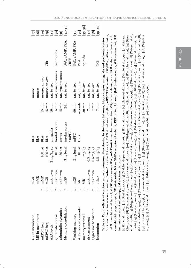
Table2.1:Rapid
effectsof
corticosterone
onne
uron
alfunction
ingin
thehypo
thalam
us,h
ippo
campu
s,am
ygda
laan
dprefrontalcortex
‘unk
nown’
rece
ptor
was
notex
amin
ed,‘othe
r’no
tth
eM
Ror
GR,
DRG
dorsal
root
gang
lion,
eIPSC
/EPSC
evok
edIP
SC/E
PSC,A
EAan
andi
mid
e,2-AG
2-ar
achi
dono
ylglyc
erol
,sIPSC
spon
tane
ousIP
SC,m
MR/m
GR
mem
bran
e-as
sociated
MR/
GR,
scsu
bcut
aneo
us,E
CB
endo
cann
abin
oids
,CB1
cann
abin
oid
rece
ptor
type
1,NO
nitric
oxid
e,NR2A
NM
DA
rece
ptor
2Asu
buni
t,PK
Cpr
otein
kina
seC,βAC
β-ad
reno
cept
or,W
Bwes
tern
blot
,IFM
imm
unofl
uore
scen
tmicro
scop
y,EM
elec
tron
micro
scop
y.[1](
Die
tal.,
2003
),[2
](Die
tal.,
2005
),[3](
Malch
er-L
opes
etal.,20
06),
[4](
Die
tal.,
2009
),[5
](Haa
met
al.,20
10),
[6](
Liuet
al.,19
95),
[7],
(Liu
and
Che
n,19
95),
[8](
Evan
son
etal.,20
10b)
,[9]
(Kar
stet
al.,20
05),
[10]
(Olij
slag
erset
al.,20
08),
[11]
(Qiu
etal.,20
10),
[12]
(Pas
rich
aet
al.,20
11),[
13](
Gro
cet
al.,20
08),
[14]
(Mag
gioan
dSe
gal,20
09),
[15]
(Kom
atsu
zaki
etal.,20
05),
[16]
(Ven
eroan
dBo
rrell,19
99),
[17]
(Xiaoet
al.,20
10),
[18]
(Wiege
rtet
al.,
2006
),[19]
(Huet
al.,20
10),
[20]
(Qie
tal.,
2005
),[2
1](L
iuet
al.,20
07),
[22]
(Tak
ahas
hiet
al.,20
02),
[23]
(Hill
etal.,20
10),
[24]
(Satoet
al.,20
04),
[25]
(Ffren
ch-M
ullen,
1995
),[2
6](C
hauv
eauet
al.,20
10),
[27]
(Joh
nson
etal.,20
05),
[28]
(Pra
gere
tal.,
2010
),[29]
(Kar
stet
al.,20
10),
[30]
(Zhu
etal.,19
98),
[31]
(Sze
and
Iqba
l,19
94),
[32]
(Roo
zend
aale
tal.,
2010
),[33]
(Bar
segy
anet
al.,20
10),
[34]
,(Li
uet
al.,20
08),
[35]
(Kha
ksar
ieta
l.,20
07),
[36]
(Sajad
iet
al.,20
07),
[37]
(Mikicset
al.,20
05),
[38]
(Mikicset
al.,20
04),
[39]
(San
diet
al.,19
96b)
,[40
](Sa
ndie
tal.,
1996
a)
35

R -
These responses were seen within minutes, so that non-genomic mechanismsmust have been involved. In the brain, these effects were associated with increasedactivity of the amygdala and decreased activity of the PVN (recorded by c-fos ex-pression) during the rising compared to the falling phase (Sarabdjitsingh et al.,2010), reminiscent of the corticosteroid effects seen for mEPSC frequency in PVNand amygdala. Hypothetically, during the rising phase of an ultradian pulse, non-genomic pathways are activated in limbic areas, which in turn could affect stress-related behaviour.
Integration of non-genomic and genomic effects
In several cases, rapid non-genomic corticosteroid actions were shown to transgressinto more lasting effects, integrating two temporal domains (rapid and delayed)which up till recently were each linked to different classes of stress hormones, i.e.monoamines (and to some extent neuropeptides) on the one hand and corticoste-roids on the other hand. For example, rapid effects in the hypothalamus are longlasting (Di et al., 2003) and thus HPA-axis feedback will be inhibited over a long pe-riod of time. Indeed, dexamethasone infusions in the PVN exert both rapid and de-layed negative feedback actions on the HPA-axis activity (Dallman et al., 1994; Dall-man, 2005). Similarly, the increased excitability in the BLA starts as a non-genomicMR-dependent phenomenon and eventually evolves into a genomic phenomenonthat also requires the GR (Karst et al., 2010). At a cognitive level, the facilitation ofmemory consolidation by cort-BSA injections in the insular cortex is evoked by amembrane-associated effect that evolves into a genomic effect through activation ofthe transcription factor CREB (Roozendaal et al., 2010). Finally, rapid corticosteroneeffects on aggressive and risk assessment behaviour are independent of gene tran-scription immediately after corticosterone injection but develop into transcription-dependent effects later on (Mikics et al., 2004, 2005). Thus, many non-genomiceffects of corticosterone are tightly linked to later genomic actions. At least in onecase (Karst et al., 2010), the initial non-genomic action is required for the subse-quent genomic phase, suggesting that both phases work in coordination.
However, non-genomic and genomic actions can also be integrated if they occurindependent from each other. In the hippocampus, the initial enhancedmEPSC fre-quency is quickly reversed: when corticosteroid levels drop, the effects are imme-diately lost (Karst et al., 2005). Supposedly, a brief period of enhanced excitabilityis followed by a refractory period with an increased threshold for the induction ofnew signals, the latter depends on genomic GR signalling (Alfarez et al., 2002, 2009;Krugers et al., 2010). A similar dichotomy was seen with respect to LTP inductionin the hippocampus. Corticosterone given immediately before LTP induction stim-ulated LTP induction (Wiegert et al., 2006), while corticosterone applied hours ear-lier inhibited the induction of the same type of LTP (Diamond et al., 1992; Pavlideset al., 1993). The initial rapid facilitation of signalling might help the organism to
36
2.3. M -
Chapter2
appraise the novel situation; gradually the genomic phase will take over and restorethe activity of the circuits to regain homeostasis (Joëls et al., 2006).
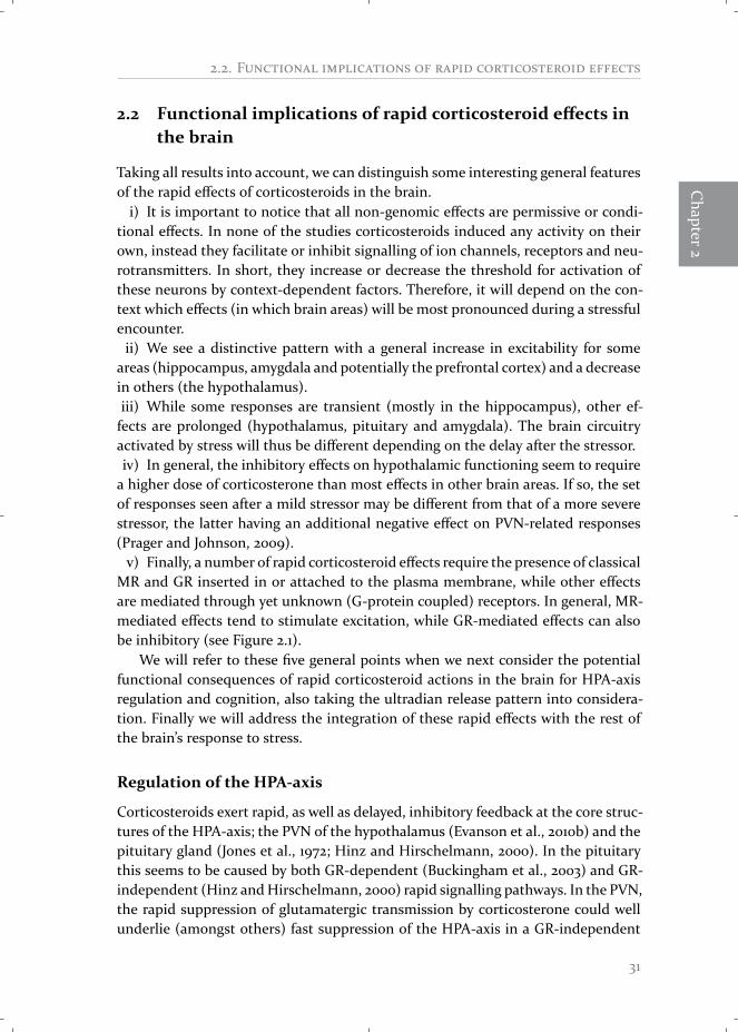
Overall, this implies that the temporal pattern of activation by corticosterone isdifferent for the various areas. As summarized in Figure 2.2, both the hippocampusand amygdala, are more sensitive for incoming signals during stress or corticoster-one exposure, while activity in the PVN is rapidly inhibited. In a delayed fashion,the hippocampus will switch to a state where the threshold for activation is ele-vated, while activation thresholds in the amygdala and hypothalamus do not differbetween the two time-domains. Hypothetically, this can have consequences for thecognitive functions associated with these brain areas. For example, as the amygdalais involved in emotional memory formation, the prolonged activation in this areamight support efficient encoding of emotional aspects of a stressful event, whichcould explain the preferential memory of emotional over neutral, hippocampal-dependent information (Buchanan and Lovallo, 2001; Karst et al., 2010). Finally, itseems that a second exposure of corticosterone switches amygdalar excitability backto its pre-stress state (Karst et al., 2010). This mechanism could protect the amyg-dala from inappropriately prolonged activation (McEwen, 2001; Karst et al., 2010).For the PFC, the limited data so far, suggest that its sensitivity is elevated by corti-costerone in both an acute and more prolonged manner. However, as the data forthe PFC is still sparse, we have not included it in Figure 2.2.
2.3 Molecular aspects of non-genomic corticosterone actions
The quest for a better understanding of the role of non-genomic corticosteroid sig-nalling is paralleled by another quest: that for a better understanding of the cellularbasis of these non-genomic effects. Here we will summarize the current state of un-derstanding of the membrane localization, and translocation, of the MR and GR aswell as that of their downstream signalling partners. We will, again, focus mostlyon corticosteroid signalling in neural tissues but we will also use knowledge fromthe periphery and of related steroids and their receptors where necessary.
Presence of MR and GR at the plasma membrane, critical evaluationof the evidence
Formany years themembrane localization of theMR andGRhas been controversial,however, over the last years evidence of their membrane presence has culminated.(i) Intracellular applied corticosterone cannot induce rapid non-genomic effects;therefore it is unlikely that the receptors are located inside the cells. (ii) Membraneimpermeable corticosterone-BSA (cort-BSA) and dex-BSA conjugates induce thesame rapid effects as free corticosterone or dexamethasone. Moreover, they do sowith equal (Xiao et al., 2010) to slightly reduced (Karst et al., 2005; Qi et al., 2005)
37

R -
minutes hours
AppraisalContextuallearning
Consolidation
mMR
gGR
stress
minutes hours
FearEmotionallearning
Consolidation
mMR gGR
stress
minutes hours
Inhibition of HPA-axisand other endocrine systems
??
gGR
stress
mGR
Exc
itabi
lity
+
- Exc
itabi
lity
+
-
Exc
itabi
lity
+
-
Hippocampus Amygdala (BLA)
Hypothalamus Figure 2.4: A putative model of the temporaldynamics of excitability in the hippocampus,amygdala and hypothalamusA stressor or corticosterone injection induces atemporal diverse set of responses in the three dif-ferent brain areas. Denoted are the receptors thatare (mainly) responsible for the effects in the dif-ferent areas. Importantly, the temporal patternof excitability in hippocampus, amygdala and hy-pothalamus determines the actions of stress andcorticosterone on neuroendocrine regulation, be-haviour and cognition. mMR/mGR (membrane-associated MR/GR), gGR (genomic GR), ?? (recep-tor unclear).
efficacy. (iii) Most convincingly, the presence of MR and GR has been shown insynaptosome extracts (Komatsuzaki et al., 2005; Wang and Wang, 2009; Qiu et al.,2010) and at neuronal membranes using electron microscopy (Johnson et al., 2005;Prager et al., 2010). (iv) Finally, the MR and GR are by no means unique in their as-sociation with the plasma membrane. Membrane localization has been shown formost, if not all, steroid receptors including the ER α and β, AR and PR (Hammesand Levin, 2007).
Not all rapid corticosteroid effects can be attributed to the MR or GR though.Multiple non-genomic actions of corticosteroids on neurotransmission (Wiegertet al., 2006; Di et al., 2009), HPA-axis regulation (Evanson et al., 2010b) and be-haviour (Sandi et al., 1996b) remain in the presence of MR and GR antagonistsand are thus postulated to require a novel membrane-associated receptor. However,the identity of this receptor has proved very difficult to resolve; as yet, none havebeen cloned. The most likely candidates are G-protein coupled receptors (GPCR),because inhibitors of G-proteins can preventmany—though not all (Orchinik et al.,1997)— MR/GR independent corticosteroid effects (Di et al., 2003, 2005). Multiplenon-MR/GR corticosteroid binding sites have been identified in the membrane of
38

2.3. M -
Chapter2
neuronal substrates in a number of species (Orchinik et al., 1991, 1992, 1997, 2000;Guo et al., 1995; Maier et al., 2005; Breuner andOrchinik, 2009; Schmidt et al., 2010).However, the affinity and selectivity of these binding sites is very variable, makingit unlikely that they all stem from a single type of evolutionary conserved receptor.
The association of steroid receptors at the plasma membrane
How is the membrane association of receptors mediating rapid corticosteroid ac-tions accomplished and how is this process regulated? Unfortunately, there is littleknown about this subject regarding MR and GR. However, much more results havebeen obtained on themembrane translocation of ERα. Since ERα and corticosteroidreceptors may share some of the pathways involved in membrane localization, wewill first evaluate the available insights in the ERα and next compare this with whatis presently known about corticosteroid receptors.
The estrogen receptors ERα and ERβ can both be targeted to the cell membrane(Gorosito et al., 2008;Micevych andDominguez, 2009)where they primarily exist incaveolae (Razandi et al., 2002). Caveolae are invaginations of the plasmamembraneformed by caveolins, scaffolding proteins that bind and bring together a large num-ber of signalling molecules including GPCRs, G-proteins, c-Src and other kinases;this facilitates rapid signal transduction (Anderson, 1998; Cohen et al., 2004). Themost ubiquitously expressed caveolin is caveolin-1. Ablation of caveolin-1 severelydiminished ERα membrane localization (Sud et al., 2010). Moreover, mutation ofa single amino acid (S522A) in the ligand binding domain of the ERα resulted in a60% reduction of caveolin-1 binding, membrane localization and rapid signallingof ERα (Razandi et al., 2003). Caveolin-1 binding is also required for membranetranslocation of the ERβ, AR and PR (Lu et al., 2001; Salatino et al., 2006; Gilad andSchwartz, 2007). Mutation of another amino acid, cysteine477 (C477A), resultedin an almost complete reduction of ERα membrane localization, while its genomicfunctions were left undisturbed (Acconcia et al., 2005). This mutation was shown tobe essential for palmitoylation of the receptor. Palmitoylation is a post-translationalmodification where a lipid tail is attached to the receptor, thus enabling insertioninto the plasma membrane. ERα palmitoylation is essential for caveolin-1 binding,membrane translocation and rapid signalling (Acconcia et al., 2005; Pedram et al.,2007). A final component of the ERα membrane translocation pathway was identi-fied recently: disruption of heat shock protein (HSP) 27 prevented palmitoylation,caveolin-1 binding, membrane localization and rapid signalling of ERα (Razandi etal., 2010). Together this leads to a model where ERα associates with HSP27, thisinteraction enables ERα to get palmitoylated, and due to the palmitoylation the re-ceptor can bind caveolin-1 which facilitates transport to the plasma membrane (seeFigure 2.5 ).
Importantly, this membrane translocation process seems to be a common path-way for all steroid receptors. The group of Levin (Pedram et al., 2007) identifieda conserved sequence surrounding the palmitoylation site of ERα and this same
39

R -
CAV
1
C
HS
P27
(2)
(4)
(1) SR (3)
CAV
1
SR
BA
Raf
PKA
cAMP
ERK1/2
MEK
RafGβγ
Gα SR
GPCR
Figure 2.5: Steroid receptor membrane association and downstream signalling(A) The putative common pathway for membrane translocation of steroid receptors is shown with theERα as example. Translocation of the ERα requires the association of heat shock protein 27 (HSP27)(step 1), subsequently the receptor is palmitoylated at cysteine 477 (step 2), this facilitates association ofthe adaptor protein caveolin-1 (CAV1) (step 3). Finally, the ERα is transported to the plasma membrane,where it is localized in caveolae (step 4). (B) Model of the downstream signalling pathways implied innon-genomic corticosteroid signalling in neurons.
sequence was identified in the AR, PR, ERβ and other receptors. Mutation of keyamino acids in this sequence abolished membrane localization and rapid signallingfor all steroid receptors tested (Pedram et al., 2007). Similarly, association of HSP27is required for membrane translocation of ERα, PR and AR (Razandi et al., 2010).Thus, so far the data suggest that there is a common membrane translocationpathway for all (or most) steroid receptors involving caveolin-1, palmitoylation andHSP27.
Membrane translocation of MR and GR
Now the question remains whether the MR and GR are transported to the mem-brane in a similar way. For these receptors only a few studies have been reportedand none in brain cells. In peripheral models an association between both MRand GR to caveolin-1 has been demonstrated. In epithelial cells, dexamethasone in-duced rapid binding of GR to c-Src and subsequent activation of the PI3K-Akt path-way (Matthews et al., 2008). Transfection of a double-negative form of caveolin-1disrupted all aspects of this signalling cascade, as did disruption of caveolae. Inaddition, a direct interaction between the GR and caveolin-1 was seen with co-immunoprecipitation (Matthews et al., 2008). In contrast, in hepatic cells no colo-calization of membrane-associated GR and caveolin-1 could be found with conven-tional confocal microscopy (Spies et al., 2006).
40
2.3. M -
Chapter2
For MR, a similar association was studied in caveolin-1 knockout (cav1-/-) mice(Pojoga et al., 2010a,b). First of all, a direct association between the MR andcaveolin-1 (but not caveolin-2) was shown with co-immunoprecipitation in hearthomogenates from both rat and mouse as well as in cultured human endothelialcells (Pojoga et al., 2010a). As expected, this association was lost in cav1-/- mice.Secondly, these mice showed heightened vascular responses to treatment with theMR antagonist eplerenone (as compared to wild type mice) and a reduced sensitiv-ity to aldosterone treatment on myocardial damage (Pojoga et al., 2010b). Thus, notonly is the MR associated with caveolin-1 in vascular tissues, but a loss of caveolin-1also alters the vascular responses to MR agonists and antagonists. The precise con-sequences of the loss of caveolin-1 for MR-associated functioning seem to dependstrongly on the context of the response.
Additional supporting evidence for themembrane localization of theMR comesfrom the group of Grossmann and Gekle (2008, 2010). In an initial study, theyshowed that transfection of only the ligand binding domain of the MR was suffi-cient for aldosterone to rapidly activate the ERK1/2 pathway in Chinese hamsterovary cells (Grossmann et al., 2008). This is similar to the ERα, where the ligandbinding domain suffices for membrane translocation and signalling (Razandi et al.,2002). More recently, they studied the colocalization between the MR and the EGFreceptor. This colocalization was lost when lipid rafts (including caveolae) were dis-rupted (Grossmann et al., 2010). This strongly suggests that the MR is localized incaveolae, since the EGF receptor is known to be associated with caveolae.
Finally, regarding the conserved palmitoylation motif, an interesting pictureemerges. The palmitoylationmotif of theGR contains all essential groups andwouldbe predicted to be a palmitoylation site (although the GR was not tested in theoriginal study) (Pedram et al., 2007). The MR, by contrast, lacks the essential cys-teine residue. As this cysteine provides the thiol group to which the palmitate tailis transferred, the MR cannot be palmitoylated at this sequence. The MR could bepalmitoylated at another motif or could translocate to the membrane through analternative pathway.
Regulation of membrane translocation and place in the membrane
Why does only part of the receptor population translocate to the membrane whilethe bulk remains in the cytoplasmandnucleus, andwhat determines the proportionof these pools? For the ERα, most studies estimate that approximately 5–10% ofthe receptor population is localized at or in the membrane, which leaves 90–95%of the population in the cytoplasm and nucleus (Chambliss et al., 2000). Caveolin-1overexpression was found to elevate the proportion of membrane ERα (Sud et al.,2010), suggesting that this protein has a regulatory effect.
It is known that ligand binding affects membrane translocation. Most studiesshow that treatment with (high concentration of) ligands reduces palmitoylation,association with caveolin-1 and membrane expression (Razandi et al., 2002; Ac-
41

R -
concia et al., 2005; Micevych and Dominguez, 2009). In contrast, other studies re-port an increased membrane translocation with steroid treatment (Razandi et al.,2002; Gorosito et al., 2008; Bondar et al., 2009). Clearly, the timing, concentrationand duration of ligand exposure will influence these effects. GR expression in hip-pocampal synaptosomes was slightly decreased after 3 weeks of daily corticosteroneinjections and increased by adrenalectomy (which abolishes endogenous corticos-terone) (Wang and Wang, 2009), suggesting that the GR also traffics from the mem-brane by ligand treatment. Interestingly though, in amygdalar neurons acute stressor corticosterone treatment abolishedMR-mediated non-genomic signalling, whileit actually allowed GR-mediated actions to take place (Karst et al., 2010).
It is still unclear how steroid receptors are integrated into the plasmamembrane.The effectiveness of impermeable hormone conjugates (such as estradiol-BSA orcort-BSA) suggests that the receptors are accessible from the outside of the plasmamembrane. In addition, biotinylation studies (for ERα) provide evidence for an ex-tracellular recognition site of the receptors (Bondar et al., 2009). This would sug-gest that the receptors are integrated in the outer sheet of the membrane with theirpalmitate tail. However, this seems in contradiction with studies showing a directinteraction of steroid receptors with caveolin-1 (Razandi et al., 2002; Sud et al., 2010)and secondmessenger molecules such as c-Src and G-proteins (Sanchez et al., 2011),which suggest that receptors are inserted into the inner sheet of the membrane,where they are able to interact with the cytoplasmic molecules. Possibly the steroidreceptor shuttles to the inside of the membrane upon activation, but at present thisis mere speculation.
A general model of steroid downstream signalling
As a final point we will evaluate the secondary pathways of steroid receptors. Sur-prisingly, although the physiological functions of steroids are very diverse (rangingfrom sexual differentiation to electrolyte balance) the non-genomic signal pathwaysshow a large overlap.We will discuss the very basics of steroid receptor downstreamsignalling in order to come to general characteristics.
As steroids are lipophilic and easily penetrate the plasma membrane, their re-ceptors do not need to be located at the plasma membrane. More likely, membrane-association of steroid receptors is required for binding to signalling partners thatare present only at the membrane. In fact, caveolae are well known signalosomesthat bring receptors, adaptor molecules and kinases together (Anderson, 1998). In-deed, the ERα was shown to assemble a multi-protein complex consisting of othermembrane-spanning receptors (most often growth factor receptors) and multiplesmall adaptor molecules like G-proteins (both Gα and Gβγ subtypes) (Kumar et al.,2007), c-Src (Sanchez et al., 2011) and PI3K (Simoncini et al., 2000). Through thissignalosome a variety of kinase pathways are activated (Hammes and Levin, 2007;Vasudevan and Pfaff, 2007; Micevych and Dominguez, 2009). Most commonly, ac-tivation of the phospholipase C - protein kinase C (PLC-PKC), cAMP-PKA (protein
42

2.3. M -
Chapter2
kinase A), PI3K-Akt and Ras-ERK pathways have been found (Figure 2.1). Impor-tantly, activation of components of these three general pathways has been reportedfor the ERα, ERβ, AR, PR, MR and GR. For example, ERK1/2 phosphorylation can beseen within minutes of stimulation with aldosterone, corticosterone, estradiol, an-drogens or vitamin D (Qiu et al., 2001; Pedram et al., 2007; Grossmann et al., 2008)and reviewed in Hammes and Levin (2007) and Grossmann et al. (2010).
The initial event, i.e. the composition of the signalosome, seems to determinewhich downstream pathway is recruited. For example, in hippocampal neuronsestradiol can activate two distinctive pathways in a single cell; on the one hand acti-vation of ERK1/2 leads to subsequent genomic effects through activation of the tran-scription factor cAMP response element binding (CREB), on the other hand inhibi-tion of PKA induces a decrease in Ca²⁺-currents (Boulware et al., 2005). These twoeffects originate from two separate pathways; one involves ERα bound to caveolin-1and attracts the metabotrophic glutamate receptor GluR1A and Gq resulting in theactivation of ERK1/2 and CREB, while the other effect originates from an ERα/β het-erodimer bound to caveolin-3, GluR2/3 and Gio, this pathway results in the inhibi-tion of PKA and Ca²⁺-currents (Boulware et al., 2007). Also interesting in this regardis the role of the coreceptors in the signalosomes; multiple studies showed that inhi-bition of growth factor signalling prevented the non-genomic effects of steroids. Forexample, phosphorylation of ERK1/2 by either aldosterone (Grossmann et al., 2005)or estrogen (Razandi et al., 2003) could be prevented by inhibitors of the EGF recep-tor. Direct interactions between the MR (Grossmann et al., 2010) and ERα (Song etal., 2010) with growth factor receptors were also shown. In fact, some people opt fora GPCR hypothesis for rapid steroid signalling; the activation of amembrane steroidreceptor activates a growth factor receptor and this enables further signalling (seeMicevych and Dominguez, 2009). Whether this is just one mechanism of action orthe mechanism of action remains to be determined.
Ultimately, activation of the cellular pathways affects the physiology of cells andtissues. Depending on the precise composition of the signalosome and the cellularcontext a wide variety of effects are obtained. These are too diverse to discuss infull here, but we will give a few examples for rapid aldosterone signalling in the pe-riphery. In kidney cells, aldosterone rapidly enhances sodium transport through anERK pathway, this results in a rapid effect on sodium absorption in these cells whicheventually also regulates blood pressure (Gekle et al., 2001). In the vascular system,activation of the enzyme nitric oxide (NO) synthase by aldosterone (through a PI3K-Akt pathway) results in an increased release of NO which attracts immune cells andaffects constriction of vascular smooth muscle cells (Hafezi-Moghadam et al., 2002;Mutoh et al., 2008).
The signal partners of central non-genomic corticosteroid signalling
The cellular pathways involved in neuronal non-genomic corticosteroid actionshave not been studied in detail yet, however, many studies did examine the involve-
43
R -
ment of some signal partners (see Table 2.1) and we can fit these within the generalmodel of non-genomic steroid signalling. As for ERα and other steroid receptors, themost obvious effectors of the rapid effects are G-proteins. Inhibition of G-proteinactivation abolished the rapid effects of corticosterone on (i) inhibition of mEPSCsin the hypothalamus (Di et al., 2003), (ii) facilitation of mIPSCs in the hypothala-mus (Di et al., 2005), (iii) facilitation of mEPSC’s in the hippocampus (Olijslagers etal., 2008) (iv) inhibition of potassium currents in the hippocampus (Olijslagers etal., 2008), (v) inhibition of calcium currents in the hippocampus (Ffrench-Mullen,1995) and (vi) activation of glutamate uptake in frontal neurons (Zhu et al., 1998)(see also Table 2.1). Interestingly, as for the estradiol effects in the hypothalamus,corticosterone can activate two different signalling pathways in single neurons inthe hypothalamus. Through activation of Gαs, corticosterone induces the releaseof endocannabinoids and an inhibition of glutamate release, while Gβγ activationleads to the release of NO and the facilitation of GABA release in the same neuron(Di et al., 2009). It remains to be investigated whether different GPCRs or caveolinsubtypes are also involved.
More downstream, corticosterone rapidly activates both the cAMP-PKA path-ways and the ERK1/2 pathway in neurons. cAMP-PKA signalling is required in thehypothalamus (Malcher-Lopes et al., 2006) and for one effect in the hippocampus(Liu et al., 2007). Activation of the ERK1/2 pathway is seen after corticosteroid ex-posure in some cases (Xiao et al., 2005, 2010; Roozendaal et al., 2010) and is re-quired for other effects (Olijslagers et al., 2008). Evidence for the involvement of thePI3K pathway has not yet been studied in the brain. Thus, although still very lim-ited, non-genomic corticosteroid signalling in neurons follows similar kinase path-ways as their peripheral counterparts and as that of other steroid receptors. Likely,the regional variation in the precise signalling cascades activated will prove to becrucial for understanding the more subtle difference between the actions in differ-ent neurons and under changing conditions. As examples from related fields show,this variation could well arise from the recruitment of different proximal adaptormolecules and interactions with signalling of other (neurotransmitter) receptors.In Figure 2.5 we show a very general model of the downstream signalling partnersin central non-genomic corticosteroid signalling.
2.4 Concluding remarks
The existence of rapid effects of corticosterone has been known for over 50 years;however, it is only in the last 10 years that these effects have been studied in moredetail. Yet, there are still many unanswered questions.
First, we cannot appreciate the consequences of non-genomic effects of cortico-steroids when they are studied in isolation, instead we must view these effects inthe context of the complete stress response. Exactly how rapid non-genomic and
44
2.4. C
Chapter2
genomic actions are integrated to collectively accomplish the behavioural responseto stress awaits further investigation, as discussed in the previous section.
Secondly, through its non-genomic effects corticosterone acts in the sametime-domain as other transmitters and hormones released after stress, e.g. cate-cholamines or CRH. This gives ample opportunities for cross-talk between the vari-ous stress hormones (Alfarez et al., 2009). For example, activation of the noradren-ergic system in the amygdala is required for effects of corticosterone to take place(Roozendaal et al., 2002, 2006). However, at this time relatively little is known aboutthe mechanism by which corticosteroids alter responsiveness to other stress factorsand if non-genomic corticosteroid signalling is involved.
Thirdly, only a few signalling partners for rapid effects have been discovered. Acomparison of the available data (see Table 2.1) suggests that many pathways areshared across brain areas. For example, multiple studies have proven involvementof G-proteins and the ERK-CREB pathway. Importantly, these same pathways arealso activated by rapid signalling of other steroid receptors (Hammes and Levin,2007; Vasudevan and Pfaff, 2007; Levin, 2008). Information gathered in these re-lated fields could serve as an important guideline for investigation of the signallingpartners of corticosteroids in the brain. For instance, both rapid corticosterone (Diet al., 2009) and estradiol signalling (Boulware et al., 2007) in neurons suggeststhat the specific type of G-protein that is engaged in the hormonal actions is animportant determinant of the subsequent signalling cascade and the physiologicaloutcome.
Fourthly, regulation of membrane translocation of the MR and GR in neurons isstill undiscovered. Caveolin-1 is required for membrane translocation of all steroidreceptors including the MR and GR (Matthews et al., 2008; Pojoga et al., 2010b).However, this has yet to be shown for the MR and GR in neurons. All three types ofcaveolins are expressed in the brain and they are known to be required for ERα andERβ non-genomic signalling (Boulware et al., 2007). Interestingly, neurons do nothave caveolae (Head and Insel, 2007), instead caveolins seem to be associated withsynaptic markers and interact with multiple types of glutamate receptors. Thus, itis likely that caveolin association enables the enrichment of MR and GR at synap-tic sites in the membrane (Johnson et al., 2005; Prager et al., 2010) and places thereceptors well in reach to regulation of synaptic transmission.
Finally, the conserved palmitoylation motif found in many steroid receptors, in-cluding the ERα and the GR, is presumably ineffective in the MR. This motif is ab-solutely required for palmitoylation and membrane expression of ERα, ERβ, PR andAR (Pedram et al., 2007) and it thus remains unclear if and how the MR could bepalmitoylated, possibly at another sequence. Consensus palmitoylation sequenceswere identified in the MR with the online CSS-Palm tool (Ren et al., 2008), how-ever, this still needs conformation in vivo. Alternatively, the MR could use anotherpathway for translocation to the membrane.
45