Turner’s Syndrome in Adulthood M. ELSHEIKH, D. B. DUNGER, G. S. CONWAY, AND J. A. H. WASS Department of Endocrinology (M.E., J.A.H.W.), Radcliffe Infirmary, Oxford, OX2 6HE, United Kingdom; Department of Paediatrics (D.B.D.), Addenbrooke’s Hospital, Cambridge, CB2 2QQ, United Kingdom; and Department of Endocrinology (G.S.C.), Middlesex Hospital, London, W1N 8AA, United Kingdom Turner’s syndrome is the most common chromosomal abnor- mality in females, affecting 1:2,500 live female births. It is a result of absence of an X chromosome or the presence of a structurally abnormal X chromosome. Its most consistent clin- ical features are short stature and ovarian failure. However, it is becoming increasingly evident that adults with Turner’s syndrome are also susceptible to a range of disorders, includ- ing osteoporosis, hypothyroidism, and renal and gastrointes- tinal disease. Women with Turner’s syndrome have a reduced life expectancy, and recent evidence suggests that this is due to an increased risk of aortic dissection and ischemic heart disease. Up until recently, women with Turner’s syndrome did not have access to focused health care, and thus quality of life was reduced in a significant number of women. All adults with Turner’s syndrome should therefore be followed up by a mul- tidisciplinary team to improve life expectancy and reduce morbidity. (Endocrine Reviews 23: 120 –140, 2002) I. Introduction II. History III. Epidemiology IV. Genetics A. Cytogenetics B. X inactivation and haploinsufficiency C. Candidate “Turner genes” V. Clinical Features of Turner’s Syndrome A. Diagnosis B. Short stature C. Bone abnormalities D. Osteoporosis E. Lymphedema VI. Gonadal Function A. Sex hormone replacement therapy B. Options for fertility VII. Cardiovascular Disease A. Congenital heart disease B. Aortic dissection C. Hypertension D. Ischemic heart disease VIII. Hypothyroidism IX. Renal Disorders X. Gastrointestinal Disease A. Inflammatory bowel disease B. Hepatic disease XI. Malignancy XII. Otological Disorders XIII. Ophthalmic Disorders XIV. Skin Disorders XV. Anorexia Nervosa XVI. Psychosocial Development XVII. Conclusions I. Introduction T URNER’S SYNDROME (TS) IS the result of complete or partial X chromosome monosomy in a phenotypic fe- male, associated with characteristic clinical features, the most consistent being short stature and gonadal dysgenesis. Subjects with TS usually receive intensive medical care during childhood, but the majority are discharged from spe- cialist clinics after the induction of puberty and attainment of final height. Until recently, adult women with TS had little access to focused health care, and, as a result, the medical profession has been slow to realize the extent of morbidity, particularly the extent of cardiovascular risk and the long- term consequences of estrogen deficiency. Affected women are susceptible to a number of medical problems, including cardiovascular disease, osteoporosis, and other endocrine, gastrointestinal, and renal disorders. Women with TS need long-term follow-up so that early medical intervention may reduce morbidity and improve life expectancy. This review highlights the medical disorders associated with TS and con- centrates on the particular problems pertaining to adults with TS. II. History TS was first described in 1768 by the anatomist Giovanni Morgagni; he reported the postmortem findings of a short woman who showed renal malformations and gonadal dys- genesis. In 1902, Funke described a 15-yr-old girl with go- nadal dysgenesis, short stature, absent puberty, congenital lymphedema, and a webbed neck (1). In 1930, Ullrich (2) gave the definitive description of the clinical features character- istic of TS, and the diagnosis was confirmed by karyotyping 57 yr later. However, the syndrome is named after Henry Turner (3), an American endocrinologist from Oklahoma, who in 1938 described seven women with characteristic phe- notypic features of the syndrome. He emphasized the pres- ence of gonadal dysgenesis and was the first to initiate es- trogen replacement therapy. Abbreviations: DM, Diabetes mellitus; GBY, gonadoblastoma locus on the Y chromosome; HRT, hormone replacement therapy; IBD, in- flammatory bowel disease; MRI, magnetic resonance imaging; PHOG, pseudoautosomal homeobox-containing gene; SHOX, short stature ho- meobox-containing gene; TS, Turner’s syndrome. 0163-769X/02/$20.00/0 Endocrine Reviews 23(1):120 –140 Printed in U.S.A. Copyright © 2002 by The Endocrine Society 120 Downloaded from https://academic.oup.com/edrv/article/23/1/120/2424315 by guest on 02 June 2022

Welcome message from author
This document is posted to help you gain knowledge. Please leave a comment to let me know what you think about it! Share it to your friends and learn new things together.
Transcript
Turner’s Syndrome in Adulthood
M. ELSHEIKH, D. B. DUNGER, G. S. CONWAY, AND J. A. H. WASS
Department of Endocrinology (M.E., J.A.H.W.), Radcliffe Infirmary, Oxford, OX2 6HE, United Kingdom; Department ofPaediatrics (D.B.D.), Addenbrooke’s Hospital, Cambridge, CB2 2QQ, United Kingdom; and Department of Endocrinology(G.S.C.), Middlesex Hospital, London, W1N 8AA, United Kingdom
Turner’s syndrome is the most common chromosomal abnor-mality in females, affecting 1:2,500 live female births. It is aresult of absence of an X chromosome or the presence of astructurally abnormal X chromosome. Its most consistent clin-ical features are short stature and ovarian failure. However,it is becoming increasingly evident that adults with Turner’ssyndrome are also susceptible to a range of disorders, includ-ing osteoporosis, hypothyroidism, and renal and gastrointes-tinal disease. Women with Turner’s syndrome have a reduced
life expectancy, and recent evidence suggests that this is dueto an increased risk of aortic dissection and ischemic heartdisease. Up until recently, women with Turner’s syndrome didnot have access to focused health care, and thus quality of lifewas reduced in a significant number of women. All adults withTurner’s syndrome should therefore be followed up by a mul-tidisciplinary team to improve life expectancy and reducemorbidity. (Endocrine Reviews 23: 120–140, 2002)
I. IntroductionII. History
III. EpidemiologyIV. Genetics
A. CytogeneticsB. X inactivation and haploinsufficiencyC. Candidate “Turner genes”
V. Clinical Features of Turner’s SyndromeA. DiagnosisB. Short statureC. Bone abnormalitiesD. OsteoporosisE. Lymphedema
VI. Gonadal FunctionA. Sex hormone replacement therapyB. Options for fertility
VII. Cardiovascular DiseaseA. Congenital heart diseaseB. Aortic dissectionC. HypertensionD. Ischemic heart disease
VIII. HypothyroidismIX. Renal DisordersX. Gastrointestinal Disease
A. Inflammatory bowel diseaseB. Hepatic disease
XI. MalignancyXII. Otological Disorders
XIII. Ophthalmic DisordersXIV. Skin DisordersXV. Anorexia Nervosa
XVI. Psychosocial DevelopmentXVII. Conclusions
I. Introduction
TURNER’S SYNDROME (TS) IS the result of complete orpartial X chromosome monosomy in a phenotypic fe-
male, associated with characteristic clinical features, the mostconsistent being short stature and gonadal dysgenesis.
Subjects with TS usually receive intensive medical careduring childhood, but the majority are discharged from spe-cialist clinics after the induction of puberty and attainmentof final height. Until recently, adult women with TS had littleaccess to focused health care, and, as a result, the medicalprofession has been slow to realize the extent of morbidity,particularly the extent of cardiovascular risk and the long-term consequences of estrogen deficiency. Affected womenare susceptible to a number of medical problems, includingcardiovascular disease, osteoporosis, and other endocrine,gastrointestinal, and renal disorders. Women with TS needlong-term follow-up so that early medical intervention mayreduce morbidity and improve life expectancy. This reviewhighlights the medical disorders associated with TS and con-centrates on the particular problems pertaining to adultswith TS.
II. History
TS was first described in 1768 by the anatomist GiovanniMorgagni; he reported the postmortem findings of a shortwoman who showed renal malformations and gonadal dys-genesis. In 1902, Funke described a 15-yr-old girl with go-nadal dysgenesis, short stature, absent puberty, congenitallymphedema, and a webbed neck (1). In 1930, Ullrich (2) gavethe definitive description of the clinical features character-istic of TS, and the diagnosis was confirmed by karyotyping57 yr later. However, the syndrome is named after HenryTurner (3), an American endocrinologist from Oklahoma,who in 1938 described seven women with characteristic phe-notypic features of the syndrome. He emphasized the pres-ence of gonadal dysgenesis and was the first to initiate es-trogen replacement therapy.
Abbreviations: DM, Diabetes mellitus; GBY, gonadoblastoma locuson the Y chromosome; HRT, hormone replacement therapy; IBD, in-flammatory bowel disease; MRI, magnetic resonance imaging; PHOG,pseudoautosomal homeobox-containing gene; SHOX, short stature ho-meobox-containing gene; TS, Turner’s syndrome.
0163-769X/02/$20.00/0 Endocrine Reviews 23(1):120–140Printed in U.S.A. Copyright © 2002 by The Endocrine Society
120
Dow
nloaded from https://academ
ic.oup.com/edrv/article/23/1/120/2424315 by guest on 02 June 2022

III. Epidemiology
TS is one of the most common chromosomal abnormalities,estimated to affect approximately 3% of all female fetuses.However, there appears to be a high fetal wastage with only1% of these embryos surviving to term (4). Thus, TS is re-sponsible for 7–10% of all spontaneous abortions. TS affectsapproximately 1 in 2,500 live female births (5, 6), correspond-ing to approximately 1.5 million women worldwide. Envi-ronmental risk factors for conceiving a child with TS areunknown. TS is not, in general, associated with advancingparental age (7).
TS is associated with a 3-fold increase in overall mortalityand a life expectancy that is reduced by up to 13 yr (8, 9). Evenafter exclusion of deaths from congenital heart disease, themortality rates remain excessive, particularly in women with45,X monosomy. Cardiovascular disease is the most commoncause of death in adults with TS (8, 9).
IV. Genetics
A. Cytogenetics
TS is characterized cytogenetically by X chromosomemonosomy, the presence of an abnormal X chromosome, ormosaicism of a 45X cell line with another cell line, whichmight be 46XX, 46XY or have an abnormal sex chromosomerearrangement. There is a correlation between the exact cy-togenetic appearance and the phenotype in TS (Table 1). Pure45,X monosomy is the most common karyotype and is as-sociated with the most abnormal phenotype. In about twothirds of women with TS, the normal X chromosome is ma-ternal in origin (4, 7, 10). Monosomy X results from nondis-junction as a result of failure of the sex chromatids to separateduring meiosis in the parental gamete or in the early em-bryonic divisions. The latter usually results in mosaicism.Turner mosaics usually have a less severe phenotype and upto 40% enter puberty spontaneously before developing go-nadal failure (11). Women with 45X/46,XY mosaicism havean increased risk of developing gonadoblastoma, and a mi-nority of these women are masculinized. Structural X chro-mosome abnormalities are thought to occur as a result ofbreakages in the X chromosome with subsequent reunion ofX chromosome sequences. Isochromosome Xq is the mostcommon structural abnormality and is associated with au-toimmune disorders and deafness, but congenital abnormal-
ities are conspicuously absent (4). Women with the ring Xchromosome are more likely to have psychological sequelaebut are less likely to have structural congenital abnormalities,and spontaneous menses occur in about a third.
B. X inactivation and haploinsufficiency
In women with normal karyotype, early in embryogenesis,there is inactivation of one X chromosome in each cell. Thisis a random process, but faulty genes on the X chromosomeare preferentially inactivated. An increasing number ofgenes, however, that escape inactivation and remain activeon both X chromosomes have been identified (12–15). Somegenes that escape X inactivation have homologs on the Ychromosome, so that their presence on both sex chromo-somes is essential for normal development. The TS pheno-type is considered to be the result of haploinsufficiency ofgenes that escape inactivation. Consistent with this notion isthe finding that 31 of 34 genes that escape X inactivation mapto the short arm of the X chromosome, deletion of which isknown to account for most of the Turner phenotype (14).
C. Candidate “Turner genes”
Stature. Genes responsible for short stature have been local-ized to the distal part of short arm of the X (Xp11–22) and Y(Yp11) chromosomes, the pseudoautosomal regions thathave been shown to escape X inactivation. Two groups ofinvestigators simultaneously identified a strong candidategene for short stature within pseudoautosomal regions thatencodes for proteins found predominantly in bone fibro-blasts (16–18). This gene, known as SHOX (short staturehomeobox-containing gene), or PHOG (pseudoautosomalhomeobox containing osteogenic gene), is expressed on boththe inactive and active X and Y chromosomes. SHOX/PHOGpoint mutations have been shown to be associated with shortstature (16, 17, 19). SHOX/PHOG mutations may also beresponsible for some of the skeletal abnormalities associatedwith TS, such as the Madelung deformity of the wrist (18, 19).Clement-Jones and colleagues (18) showed that SHOX isstrongly expressed during human embryonic limb develop-ment, which would be consistent with its role in the devel-opment of Madelung deformity and possibly cubitus valgus,high arched palate, and micrognathia. Interestingly, SHOXmutations have also been linked to Leri-Weill syndrome, arare disorder characterized by short stature and skeletal ab-
TABLE 1. Cytogenetics of 196 women attending the Adult Turner clinics at the Middlesex and Oxford Radcliffe hospitals, UK, andcorrelation with phenotype
Karyotype No. % Phenotype
45,X 95 48 Most severe phenotype. Highest incidence of structural cardiac and renal abnormalities.46,Xi(Xq) 36 18 Structural abnormalities uncommon. Increased risk of autoimmunity, particularly
thyroiditis and IBD, and deafness.45,X/46,XX 21 11 Least severe phenotype. Increased mean height. Spontaneous puberty and menses in up
to 40%.46,Xr(X) 19 10 Spontaneous menses in 33%. Congenital abnormalities uncommon. Cognitive
dysfunction in those with a small ring chromosome.45,X/46,XY 11 6 Increased risk of gonadoblastoma.45,X/46,X,idic(Y) 2 1 Increased risk of gonadoblastoma.46,XXp- 3 1.5 Similar phenotype to 45,X monosomy.46,XXq- 6 3 Variable phenotype.other 3 1.5
Elsheikh et al. • Turner’s Syndrome Endocrine Reviews, February 2002, 23(1):120–140 121
Dow
nloaded from https://academ
ic.oup.com/edrv/article/23/1/120/2424315 by guest on 02 June 2022
normalities similar to those found in TS (20). Additionally,nonspecific aneuploidy per se may contribute to short stature,as growth failure is seen in other syndromes associated withchromosomal imbalance (e.g., Down’s syndrome).
Ovarian function. Ovarian failure in TS could also be due tohaploinsufficiency of a gene on the X chromosome that es-capes inactivation. The regions of the X chromosome vital fornormal ovarian development have been described as “criticalregions” comprising the short arm distal to Xp11, Xq13–25,and Xq26–28 (21, 22). Several groups have sought genesdisrupted at various X chromosome breakpoints, whichcould be candidates for ovarian determining genes (23–25).Other ovarian determining genes on the X chromosome havebeen identified as possible candidates because of the phe-notypes described in knockout mice. For instance, the ZFX(zinc finger) gene on the distal Xp (15, 26), when ablated inmice, results in accelerated ovarian failure. A second candi-date gene for gonadal failure in TS is the DFFRX (Drosophilafat facets related X), which maps to Xp11.4 (27). Certainlymutations of the Y homolog (DFFRY) have been associatedwith defective spermatogenesis (28). However, an alterna-tive, and more likely, explanation for the cause of acceleratedoocyte atresia in TS is that sex chromosome imbalance resultsin impaired meiosis and subsequent germ cell apoptosis(29, 30).
Lymphedema. Identification of genes responsible for otherphenotypic features of TS has proven difficult, but severalcandidate genes on Xp are currently being investigated (21,27, 30). A putative lymphedema gene that escapes X inacti-vation is thought to be located on the sex chromosomes, andhaploinsufficiency of this may be responsible for lymphed-ema and the visceral abnormalities so common in TS. It isthought that this may lie on Xp; however, it has not yet beencharacterized.
Gonadoblastoma. Approximately 6% of women with TS have45,X/46,XY mosaicism (31) and are at increased risk of de-veloping a gonadoblastoma. This is a rare neoplasm thatdevelops almost exclusively in the dysgenetic gonads ofwomen with Y chromosome mosaicism. One or more geneson the Y chromosome, the GBY (gonadoblastoma locus onthe Y chromosome) critical region, have been postulated topredispose dysgenetic gonads to develop gonadoblastoma.The GBY locus is thought to lie in a small region near thecentromere of the Y chromosome, but its specific site remainselusive. A strong candidate gene is TSPY (testis-specific pro-tein Y encoded) within the GBY locus (32). There has beenrecent interest in the significance of low-level Y mosaicism(mosaic level of �5%) and the risk of gonadoblastoma. Suchmosaicism can be missed using conventional cytogenetictechniques, and some investigators therefore advocate rou-tine screening for Y chromosome material using moleculargenetic techniques such as PCR. However, in recent studiesscreening women with TS for low-level Y mosaicism, only 5%were found to have Y chromosome material that had notpreviously been picked up using conventional cytogeneticmethods (33–35). Additionally, there have been no reportedcases of gonadoblastoma developing in women with TS andlow-level Y chromosome mosaicism (36). Thus, the routine
screening for Y chromosome material by PCR is currently notrecommended.
V. Clinical Features of Turner’s Syndrome
There is a wide variation of clinical features seen in femaleswith TS, ranging from the severe phenotype with short stat-ure, gonadal dysgenesis, lymphedema, and characteristicdysmorphic features, to women with only a mild reductionin final height, or premature ovarian failure (Table 2).
A. Diagnosis
The diagnosis of TS may be delayed until adulthood in upto 10% of women. This is especially likely in females whoenter puberty spontaneously and subsequently present withamenorrhea (primary or secondary) or infertility. The diag-nosis is made on the basis of a chromosomal analysis. Aperipheral lymphocyte karyotype is routinely analyzed andis diagnostic in the majority of cases. In rare instances, thiskaryotype is normal in females with TS mosaicism; however,if TS is suspected on clinical grounds, karyotyping of othertissue samples, such as skin fibroblasts, may be necessary(37).
B. Short stature
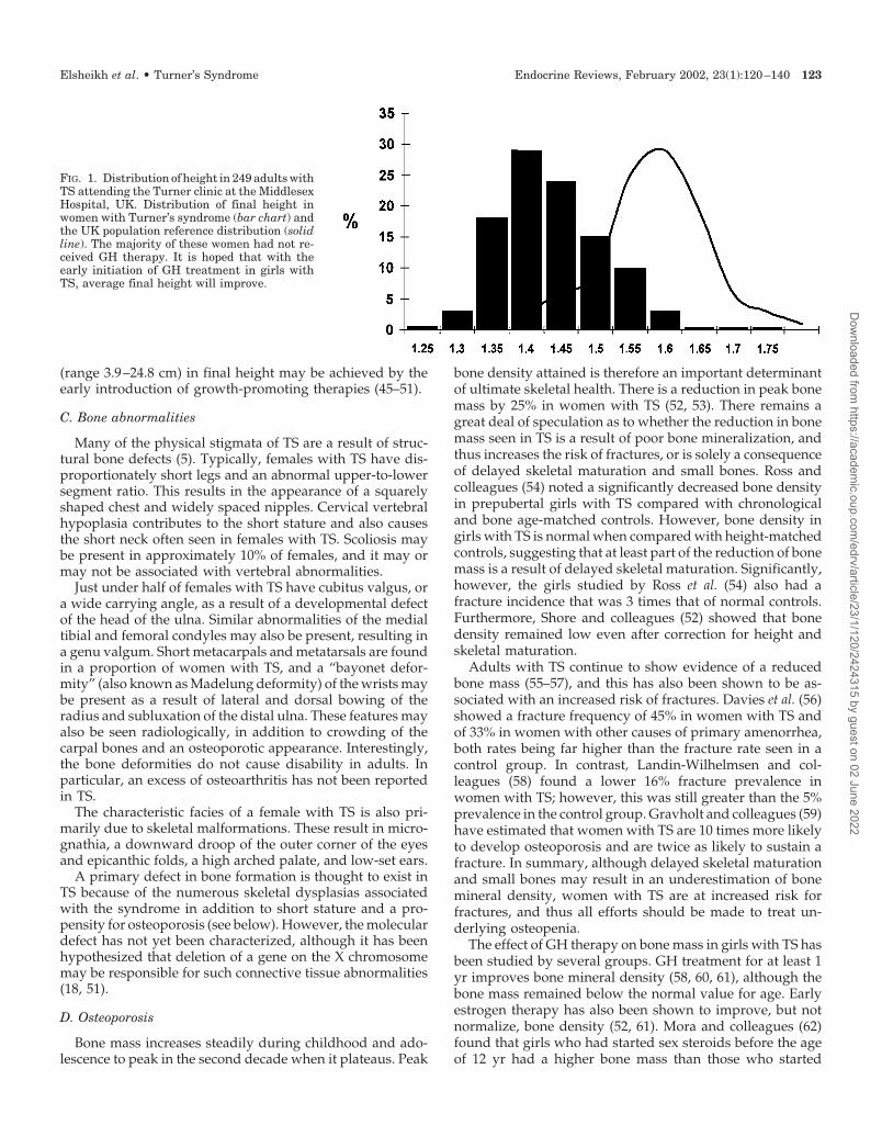
Short stature is an almost invariable finding in womenwith TS, present in all with monosomy X and in more than96% of mosaic females or those with a structurally abnormalX chromosome. The mean final adult height is between 143and 147 cm in untreated Caucasian females with TS (Fig. 1)(38–41).
The cause of growth failure in TS is currently unknown,but it is thought to be due to a primary bone defect. The genesresponsible for growth, as previously stated (Section IV.C),are thought to lie in the pseudoautosomal region of Xp. Thereis no evidence that GH deficiency is a cause of short staturein TS although partial GH insensitivity may be a factor (42–44). Treatment during childhood consists of early GH ther-apy in supraphysiological doses and estrogen replacementaround the normal age of puberty. An average gain of 10 cm
TABLE 2. TS: clinical features
Feature Frequency (%)
Short stature 98Gonadal failure 95Micrognathia 60Cubitus valgus 47Low posterior hairline 42Short neck 40High arched palate 38Short fourth metacarpal 37Multiple naevi 25Webbed neck 25Lymphedema of hands and feet 22Nail dysplasia 13Scoliosis 11Madelung deformity 7
[Adapted with permission from B. Lippe: Endocrinol Metab ClinNorth Am 20:121–152, 1991 (5).]
122 Endocrine Reviews, February 2002, 23(1):120–140 Elsheikh et al. • Turner’s Syndrome
Dow
nloaded from https://academ
ic.oup.com/edrv/article/23/1/120/2424315 by guest on 02 June 2022
(range 3.9–24.8 cm) in final height may be achieved by theearly introduction of growth-promoting therapies (45–51).
C. Bone abnormalities
Many of the physical stigmata of TS are a result of struc-tural bone defects (5). Typically, females with TS have dis-proportionately short legs and an abnormal upper-to-lowersegment ratio. This results in the appearance of a squarelyshaped chest and widely spaced nipples. Cervical vertebralhypoplasia contributes to the short stature and also causesthe short neck often seen in females with TS. Scoliosis maybe present in approximately 10% of females, and it may ormay not be associated with vertebral abnormalities.
Just under half of females with TS have cubitus valgus, ora wide carrying angle, as a result of a developmental defectof the head of the ulna. Similar abnormalities of the medialtibial and femoral condyles may also be present, resulting ina genu valgum. Short metacarpals and metatarsals are foundin a proportion of women with TS, and a “bayonet defor-mity” (also known as Madelung deformity) of the wrists maybe present as a result of lateral and dorsal bowing of theradius and subluxation of the distal ulna. These features mayalso be seen radiologically, in addition to crowding of thecarpal bones and an osteoporotic appearance. Interestingly,the bone deformities do not cause disability in adults. Inparticular, an excess of osteoarthritis has not been reportedin TS.
The characteristic facies of a female with TS is also pri-marily due to skeletal malformations. These result in micro-gnathia, a downward droop of the outer corner of the eyesand epicanthic folds, a high arched palate, and low-set ears.
A primary defect in bone formation is thought to exist inTS because of the numerous skeletal dysplasias associatedwith the syndrome in addition to short stature and a pro-pensity for osteoporosis (see below). However, the moleculardefect has not yet been characterized, although it has beenhypothesized that deletion of a gene on the X chromosomemay be responsible for such connective tissue abnormalities(18, 51).
D. Osteoporosis
Bone mass increases steadily during childhood and ado-lescence to peak in the second decade when it plateaus. Peak
bone density attained is therefore an important determinantof ultimate skeletal health. There is a reduction in peak bonemass by 25% in women with TS (52, 53). There remains agreat deal of speculation as to whether the reduction in bonemass seen in TS is a result of poor bone mineralization, andthus increases the risk of fractures, or is solely a consequenceof delayed skeletal maturation and small bones. Ross andcolleagues (54) noted a significantly decreased bone densityin prepubertal girls with TS compared with chronologicaland bone age-matched controls. However, bone density ingirls with TS is normal when compared with height-matchedcontrols, suggesting that at least part of the reduction of bonemass is a result of delayed skeletal maturation. Significantly,however, the girls studied by Ross et al. (54) also had afracture incidence that was 3 times that of normal controls.Furthermore, Shore and colleagues (52) showed that bonedensity remained low even after correction for height andskeletal maturation.
Adults with TS continue to show evidence of a reducedbone mass (55–57), and this has also been shown to be as-sociated with an increased risk of fractures. Davies et al. (56)showed a fracture frequency of 45% in women with TS andof 33% in women with other causes of primary amenorrhea,both rates being far higher than the fracture rate seen in acontrol group. In contrast, Landin-Wilhelmsen and col-leagues (58) found a lower 16% fracture prevalence inwomen with TS; however, this was still greater than the 5%prevalence in the control group. Gravholt and colleagues (59)have estimated that women with TS are 10 times more likelyto develop osteoporosis and are twice as likely to sustain afracture. In summary, although delayed skeletal maturationand small bones may result in an underestimation of bonemineral density, women with TS are at increased risk forfractures, and thus all efforts should be made to treat un-derlying osteopenia.
The effect of GH therapy on bone mass in girls with TS hasbeen studied by several groups. GH treatment for at least 1yr improves bone mineral density (58, 60, 61), although thebone mass remained below the normal value for age. Earlyestrogen therapy has also been shown to improve, but notnormalize, bone density (52, 61). Mora and colleagues (62)found that girls who had started sex steroids before the ageof 12 yr had a higher bone mass than those who started
FIG. 1. Distribution of height in 249 adults withTS attending the Turner clinic at the MiddlesexHospital, UK. Distribution of final height inwomen with Turner’s syndrome (bar chart) andthe UK population reference distribution (solidline). The majority of these women had not re-ceived GH therapy. It is hoped that with theearly initiation of GH treatment in girls withTS, average final height will improve.
Elsheikh et al. • Turner’s Syndrome Endocrine Reviews, February 2002, 23(1):120–140 123
Dow
nloaded from https://academ
ic.oup.com/edrv/article/23/1/120/2424315 by guest on 02 June 2022
treatment after the age of 12 yr. The combination of estrogenreplacement therapy and GH treatment results in a greatergain in bone mass (60, 63). In girls with TS who enter pubertyspontaneously, bone mass has been found to be within nor-mal limits (64).
After adolescence, estrogen replacement therapy seems tobe the single most important factor in maintaining peak bonemass. Sylven and colleagues (57) looked at bone mineraldensity in 47 middle-aged women with TS and found thatwomen with TS had a bone mass less than age-matchednormal values. They also found that the duration of hormonereplacement therapy (HRT) was the significant factor inmaintaining bone mass. Stepan and co-workers (55) alsodemonstrated that women with TS have a lower bone masscompared with age and sex-matched normal values, butthose women receiving HRT had a higher mean bone mineraldensity compared with those who were untreated (�2.3 sdvs. �4.5 sd). Finally, Davies et al. (56) did not show a sig-nificant difference in bone mineral density or fracture risk inwomen with TS compared with women with other causes ofprimary amenorrhea, suggesting that estrogen deficiency isan important factor in the pathogenesis of osteopenia in TS.
Since bone mass is improved but not normalized afterhormonal therapy (65), an intrinsic bone defect is likely. Thecause of osteopenia in TS is probably a result of the combi-nation of an intrinsic bone defect in addition to estrogendeficiency. However, if this were the case, one would expectbone density to be higher in women with a mosaic karyotypecompared with 45,X monosomy, but there are no data cor-relating bone density in TS with karyotype (52, 53, 56, 65).Further research is required to determine whether loss ofgenes on the X chromosome may predispose women with TSto osteopenia. Additionally, further longitudinal studies arerequired to assess the prevalence of clinically significantosteoporosis and osteoporotic fractures in TS.
Estrogen replacement should be optimized and lifestyleadvice given with regards to exercise and adequate calciumintake. Weight-bearing exercise has been shown to improvebone mass in women with TS (66). All women with TS shouldhave a bone densitometry performed on transfer to an adultclinic, and bone mass should then be monitored, although thefrequency of monitoring remains controversial. The role ofbisphosphonates in the treatment of osteoporosis in womenwith TS has yet to be clarified.
E. Lymphedema
Lymphedema, the result of obstruction at the level of thelymphatojugular connection, is a major defect in TS, whichis present in affected fetuses, particularly those with 45,Xmonosomy. When severe, this results in fetal wastage as aconsequence of severe generalized lymphedema, but spon-taneous resolution can occur with subsequent live birth.Milder lymphedema is responsible for some of the typicalfeatures of TS, such as a webbed neck, ptosis, and a lowposterior hairline. Lymphedema of the hands and feet maybe present at birth but often resolves. It may recur, partic-ularly after the initiation of estrogen therapy. In mostwomen, lymphedema may be controlled using supportstockings and/or diuretics. Nail dysplasia (pitting nails and
lateral hyperconvexity) pathognomonic of TS is also thoughtto be secondary to lymphedema. Fetal lymphedema may alsobe responsible for the development of aortic coarctation (seeSection VII.A).
VI. Gonadal Function
In a karyotypically normal fetus, the number of germ cellsrises progressively to 600,000 by 2 months post conception toa maximum of 7,000,000 at about 5 months gestation. Thenumber of germ cells then decreases so that at full gestationonly 50% of the germ cells remain. There is then a progressivegerm cell degeneration up until the age of menopause (67).
The gonads in TS differentiate normally until the thirdmonth of gestation. After this period, the absence of part, orthe whole, X chromosome in the germ cells results in anaccelerated degeneration of oocytes and an increase in ovar-ian stromal fibrosis (68). Ovarian failure occurs within thefirst few months or years of life. Ultrasonic assessment of thepelvis in females with TS reveal the majority to have streakovaries and, in many, the ovaries are too small to be iden-tified (69). In a recent study of 93 females with TS, the ovarieswere not identified in 44% of females (70). In females with TSwho have not had estrogen treatment, the uterus is hypo-plastic and remains prepubertal in size.
Most females with TS do not enter puberty spontaneouslybecause of the early gonadal failure and subsequent estrogendeficiency. Spontaneous breast development is either mini-mal or does not occur, and primary amenorrhea is usual.However, this is not inevitable, as indicated by a recent studyfrom Italy in which the incidence of spontaneous puberty inwomen with TS was found to be as high as 16% (11). Thefrequency was significantly higher in women with Turnermosaicism (40% vs. 9%). This is in keeping with our ownobservations in which the overall incidence of spontaneousmenarche and puberty was 12%. Only 8% of those with 45,Xkaryotype and 10% of those with a structural X chromosomeabnormality entered puberty spontaneously, as opposed to47% of females with 45,X/46,XX mosaicism.
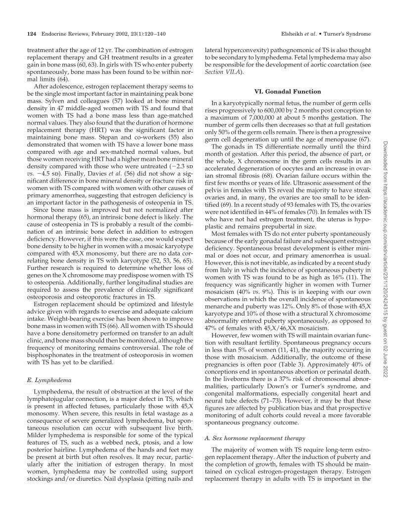
However, few women with TS will maintain ovarian func-tion with resultant fertility. Spontaneous pregnancy occursin less than 5% of women (11, 41), the majority occurring inthose with mosaicism. Additionally, the outcome of thesepregnancies is often poor (Table 3). Approximately 40% ofconceptions end in spontaneous abortion or perinatal death.In the liveborns there is a 37% risk of chromosomal abnor-malities, particularly Down’s or Turner’s syndrome, andcongenital malformations, especially congenital heart andneural tube defects (71–73). However, it may be that thesefigures are affected by publication bias and that prospectivemonitoring of adult cohorts could reveal a more favorablespontaneous pregnancy outcome.
A. Sex hormone replacement therapy
The majority of women with TS require long-term estro-gen replacement therapy. After the induction of puberty andthe completion of growth, females with TS should be main-tained on cyclical estrogen-progestagen therapy. Estrogenreplacement therapy in adults with TS is important in the
124 Endocrine Reviews, February 2002, 23(1):120–140 Elsheikh et al. • Turner’s Syndrome
Dow
nloaded from https://academ
ic.oup.com/edrv/article/23/1/120/2424315 by guest on 02 June 2022
prevention of osteoporosis (57) and in reducing risk factorsfor atherosclerosis (106, 107). In addition, it has been shownrecently that estrogen replacement may improve aspects ofcognitive function in women with TS (108). The role of es-trogens in the protection against colonic cancer remains ten-tative, but studies have shown that rates of colon cancer canbe halved in postmenopausal women by estrogen replace-ment therapy (109, 110). This protection is extremely relevantin females with TS, as they have been shown to have anincreased risk of colorectal neoplasms (59, 111).
The estrogen dose and route of administration should beindividualized taking into account patient preference andcoexisting illness (Table 4). The oral route is the most pop-ular, but transdermal estrogen patches avoid first-pass he-patic metabolism and are therefore ideal in women with liverdisease or hypertriglyceridemia. However, 10% of womendevelop skin reactions to transdermal preparations. Naturalestrogen preparations such as conjugated estrogens or E2valerate have a benefit over oral contraceptive preparationsin that they nearly all offer continuous estrogen for 28 dwithout a pill-free week. A proportion of women with TSusing the oral contraceptive will be symptomatic of estrogenin the pill-free week. In addition, the presence of ethinyl E2in the oral contraceptive pill has been associated with anincreased incidence of liver disease in women with normalkaryotypes (112) as well as in women with TS (113). It mayalso have an adverse effect on lipid metabolism through itseffect on hepatic metabolism, and it may exacerbate hyper-tension, a common problem in TS.
The progestagen should be given for a minimum of 10d/month to prevent the development of endometrial carci-noma (Table 4). Alternatively, a continuous combined reg-imen, with the advantage of no menstrual bleeding after thefirst year of therapy, might be considered, although the long-term outcome of its use in young women is lacking. The riskof breast cancer in women with TS is not thought to be higherthan in the general population (59, 111), and the use of
physiological doses of estrogens should not contribute to anexcess risk. The duration of HRT after the age of 50 yr shouldbe made on an individual basis, weighing the risks andbenefits for each woman.
B. Options for fertility
Spontaneous pregnancies occur in less than 5% of womenwith TS and are associated with a high risk of fetal loss.Chromosomal and congenital abnormalities are present inalmost half of surviving babies. Fertile women with TSshould therefore be counseled with regard to these increasedrisks and be offered prenatal diagnostic testing. Moreover,they should be advised to consider the high risk of earlyovarian failure when planning pregnancies.
The majority of women with TS, however, will be infertile.Pregnancy may be achieved by oocyte donation and in vitrofertilization. Oocyte donation has been available over thepast 15 yr to treat women with premature ovarian failure ofany cause (114). High-dose estrogens (E2 valerate, 4–8 mg/dby mouth) and progestagens (300–600 mg micronized pro-gesterone transvaginally) are used to prepare the endome-trium for implantation. Best results are achieved once theendometrial thickness is greater than 6.5 mm (115, 116). Thepregnancy rate in women with TS who have had adequateendometrial preparation is approximately 40% per treatmentcycle, a result similar to that achieved in other forms ofovarian failure (117, 118), with the highest results attainedusing fresh embryos. However, the risk of miscarriage inwomen with TS is high, with only 50% of pregnancies re-sulting in a live birth (116, 117). This is thought to be due touterine hypoplasia in TS and possibly relative uterine isch-emia during pregnancy (116). Most women with TS requirecesarean section for delivery because of cephalopelvic dis-proportion resulting from their body habitus.
Women with TS who do become pregnant may be at in-creased risk from cardiovascular complications, particularly
TABLE 3. Spontaneous pregnancy in TS
Karyotype Subjectsa
(n)Pregnanciesa
(n)Healthy
pregnanciesAbortions orstillbirthsa
Congenital orchromosomal
abnormalitiesa
45,X 16 32 14 15 3Mosaic 49 104 39 39 26Ring chromosome 7 12 5 1 646,Xdel(Xp) 3 6 1 2 3
Total 80b 167b 64 (38%)b 65 (40%)b 38 (23%)a Data taken from the following references: Refs. 11, 41, 71–105.b Includes five in which details of karyotype were not given.
TABLE 4. Suggested HRT in women with TS
Estrogen preparation Dose
Conjugated estrogens (oral) 0.625–1.25 mg dailyE2 valerate (oral) 2 mg dailyE2 transdermal patch 50–100 �g/24 h. New patch twice a week.
Progestagen Cyclical dose (d 1–12) Continuous daily dose
Medroxyprogesterone acetate 10 mg 2.5–5 mgNorethisterone 0.7–1 mg 0.35–1 mg
Elsheikh et al. • Turner’s Syndrome Endocrine Reviews, February 2002, 23(1):120–140 125
Dow
nloaded from https://academ
ic.oup.com/edrv/article/23/1/120/2424315 by guest on 02 June 2022
aortic root dissection (Section VII.B). At least three deathshave been reported in pregnant women with TS from aorticdissection (119, 120). All women should therefore undergo afull cardiological assessment before seeking to become preg-nant, including echocardiography or magnetic resonance im-aging (MRI) of the aortic root, cardiac valves, and left ven-tricular function. Hypertension is more common in youngwomen with TS, and this should be monitored and treatedaggressively. However, preeclampsia does not seem to occurwith increased frequency in TS.
There is currently much research into the removal of func-tioning ovarian tissue followed by its cryopreservation withthe aim of reimplantation at a later stage when fertility isrequired (121, 122). Its clinical application has centeredmainly on women who are scheduled to begin cancer che-motherapy; however, it may also be suitable for the fewwomen with TS who exhibit early evidence of ovarian func-tion but are at high risk of later ovarian failure. Research intothis technique is still in its infancy. There have been no casesto date of successful human ovarian autotransplantation. Ifthis technique is considered, subjects and their families mustbe counseled about the uncertainty of future success ratesassociated with this ovarian cryopreservation. The optimumage for ovarian biopsy in females with TS with normal go-nadotropins is unknown and should be individualized, pos-sibly based on serum inihibin levels, a more sensitive indi-cator of ovarian function. However, it is likely that ovariancryopreservation should be considered in childhood or earlyadolescence. Furthermore, because of the high risk of fetalabnormalities associated with spontaneous pregnancies infemales with TS, donor oocyte donation may be moreappropriate.
VII. Cardiovascular Disease
The increased mortality in TS is primarily a result of itscardiovascular complications (8, 9). It has long been knownthat left-sided congenital cardiac abnormalities are moreprevalent in women with TS, but recently, other cardiovas-cular risk factors have come to light, particularly the in-creased risk of aortic dissection and ischemic heart disease.
A. Congenital heart disease
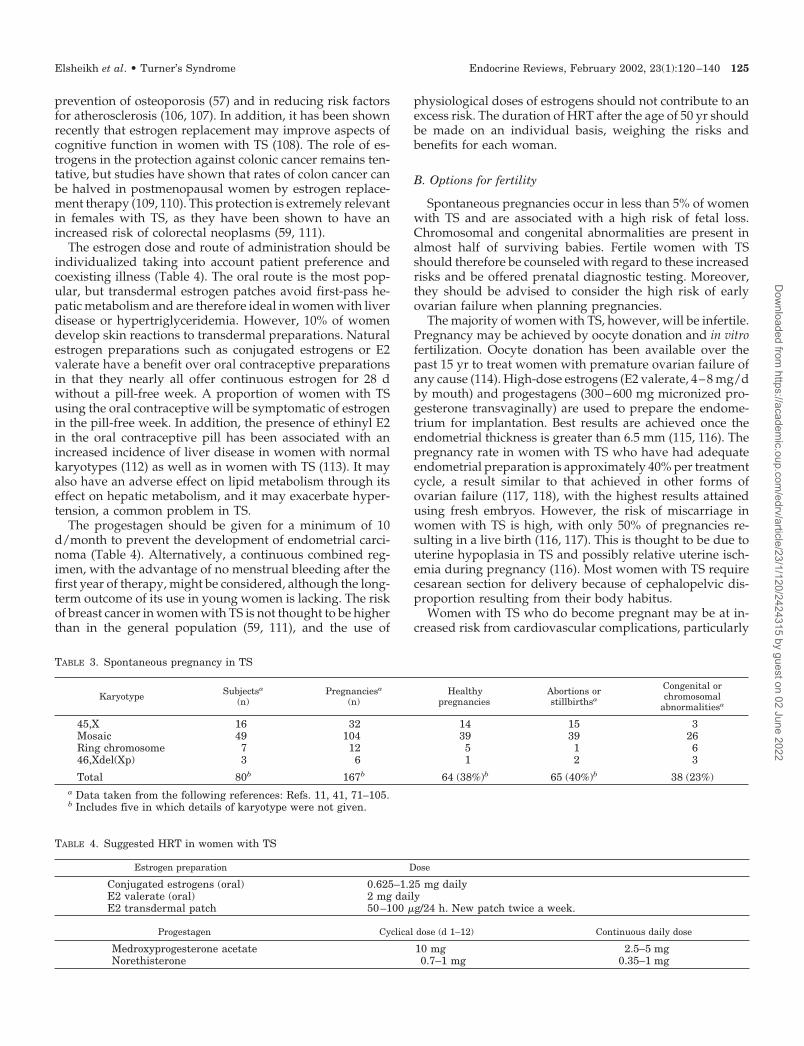
Congenital cardiac anomalies are common in females withTS, with a prevalence estimated to be between 23 and 40%(Table 5 and Refs. 123–125). These studies also indicate that
structural cardiac anomalies are most prevalent in womenwith pure 45,X monosomy and tend to be less common inthose with an isochromosome Xq karyotype.
Bicuspid aortic valve is the most common congenital mal-formation affecting the heart (123–126). It is usually an iso-lated abnormality. However, it may occur in combinationwith other anomalies, particularly aortic coarctation, and, asit calcifies, may result in progressive valvular dysfunction, asevidenced by aortic stenosis or regurgitation. The cause ofthe abnormal aortic valve is unknown.
Coarctation of the aorta affects approximately 10% ofwomen with TS and is an important cause of hypertension.It is particularly common in females with webbing of theneck (5, 127). This association, in addition to the high inci-dence of aortic coarctation in 45,X aborted fetuses with severelymphedema (128), has led to the theory that aortic coarc-tation is a result of the abnormal lymphatic flow in TS al-tering intracardiac blood flow by compressing the ascendingaorta (5, 127, 129). Aortic coarctation, providing it is the onlycardiac abnormality, is usually surgically corrected in child-hood with excellent results. Untreated, it may be complicatedby aortic rupture, congestive cardiac failure, and persistenthypertension. Seirafi and colleagues (130), who studied theoutcome of surgery in 176 patients with aortic coarctation,found that surgical repair before the first year of life was lesslikely to be associated with persistent hypertension (4.2% vs.27%).
Other cardiac anomalies, such as partial anomalous ve-nous drainage and mitral valve prolapse, are also more com-mon in TS compared with the general population (131–133).
All left-sided cardiac anomalies associated with TS resultin an increased susceptibility to infective endocarditis, andtherefore prophylactic antibiotics before dental or surgicalprocedures are essential. Patients with cardiovascular anom-alies require long-term cardiological follow-up.
B. Aortic dissection
In the last decade the association of aortic dissection andTS has been increasingly recognized (Table 6), with several
TABLE 5. Structural cardiac anomalies in TS
No. (%)a
Total no. of patients assessed 1,126Structural abnormalities
Bicuspid aortic valve 132 (12)Coarctation of aorta 103 (9)Aortic stenosis/regurgitation 38 (3.4)Partial anomalous venous drainage of the
pulmonary veins26 (2.3)
Other 17 (1.5)Total 316 (28)
a Data taken from the following references: Refs. 123, 124, 125,131, 132, 134.
TABLE 6. Aortic dilatation and dissection in TS: a review of theliterature
No. (%)
Total no. of patients 70No. of patients below 21 yr 33 (47)No. of patients above 21 yr 37 (53)45,X karyotype (data available for 44
cases)36 (82)
No. with structural cardiac abnormality 50 (71)No. with hypertension 32 (46)No. with no risk factor 6 (8.6)No. presenting with aortic dissection 44 (63)Site of aortic dilatation/dissection
Ascending aorta 29 (49)Descending aorta 9 (15)Both 21 (36)Unspecified 11
Evidence of cystic medial necrosis(histology available for 25 patients)
18 (72)
Deaths 25 (36)
[Reproduced with permission from M. Elsheikh et al.: Clin Endo-crinol (Oxf) 54:69–73, 2001 (134).]
126 Endocrine Reviews, February 2002, 23(1):120–140 Elsheikh et al. • Turner’s Syndrome
Dow
nloaded from https://academ
ic.oup.com/edrv/article/23/1/120/2424315 by guest on 02 June 2022
reports of sudden death. Aortic root dilatation is thought tooccur with a prevalence estimated to be between 8 and 42%(134–137), although not every aortic root dilation necessarilygoes on to dissect. In our series, 42% of adult women with TSattending a dedicated clinic had echocardiographic evidenceof significant aortic root dilatation (134). Hypertension, anabnormal aortic valve, and other left-sided cardiac malfor-mations have been shown to predispose to aortic dissectionin Marfan’s syndrome and the general population (138, 139),and in TS these risk factors are present in more than 90% offemales who develop aortic dilatation (136). As with otherstructural cardiac anomalies, it is most commonly associatedwith 45,X karyotype.
Aortic dilatation or dissection may occur at any age, andin the published literature just under half the patients werediagnosed before the age of 21 yr (134). Aortic root dilatationis thought to be due to a mesenchymal defect (136), as patho-logical evidence of cystic medial necrosis has been found byseveral investigators. In the 25 case reports in which histo-logical data were available, 71% had evidence of cystic me-dial necrosis. Additionally, there is evidence of other con-nective tissue abnormalities in TS, particularly in the skeletalsystem (140), and there are several features suggestive of amesenchymal defect that are common to both TS andMarfan’s syndrome. Additional research is needed to deter-mine whether an abnormality of the X chromosome mayaffect collagen synthesis and be responsible for the connec-tive tissue abnormalities seen in TS.
The influence of estrogens on the natural history of aorticdilatation is unknown. Aortic root dilatation often develops
before the administration of exogenous estrogens, and therehave been no reports to suggest any deterioration after HRT.However, pregnancy certainly increases the risk of progres-sion to aortic dissection (118, 119, 141), although this may bedue to the increased hemodynamic load rather than the highestrogen state.
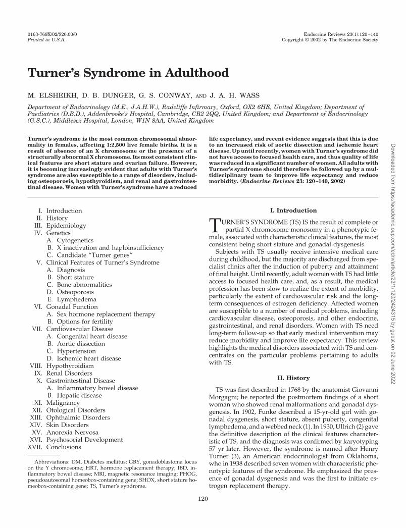
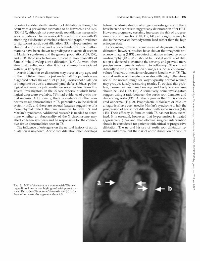
Echocardiography is the mainstay of diagnosis of aorticdilatation; however, studies have shown that magnetic res-onance imaging (MRI) can detect dilatation missed on echo-cardiography (133). MRI should be used if aortic root dila-tation is detected to examine the severity and provide moreprecise measurements relevant to follow-up. The currentdifficulty in the interpretation of images is the lack of normalvalues for aortic dimensions relevant to females with TS. Thenormal aortic root diameter correlates with height; therefore,use of the normal range for karyotypically normal womenmay produce falsely reassuring results. To obviate this prob-lem, normal ranges based on age and body surface areashould be used (142, 143). Alternatively, some investigatorssuggest using a ratio between the aortic root diameter anddescending aorta (136). A ratio of greater than 1.5 is consid-ered abnormal (Fig. 2). Prophylactic �-blockers or calciumantagonists have been used in Marfan’s syndrome to halt theprogression of aortic root dilatation with some success (144,145). Their efficacy in females with TS has not been exam-ined. It is essential, however, that hypertension is treatedaggressively (134) and that elective surgical interventionshould be considered for patients with critical or progressivedilatation. The natural history of aortic root dilatation re-mains unknown, but the risk of aortic dissection or rupture
FIG. 2. MRI of the aorta in a woman with TS show-ing a dilated aortic root highlighted with paired ar-rows. The ratio of diameter of the aortic root (a) to thedescending aorta (b) is greater than 1.5.
Elsheikh et al. • Turner’s Syndrome Endocrine Reviews, February 2002, 23(1):120–140 127
Dow
nloaded from https://academ
ic.oup.com/edrv/article/23/1/120/2424315 by guest on 02 June 2022
may be as high as 60%; therefore, regular surveillance isrecommended. Until more is known about the natural his-tory of aortic root dilatation, it is currently recommendedthat females with normal echocardiograms are imaged every5 yr and those with abnormal echocardiograms are followedup annually by a cardiologist (146).
C. Hypertension
The risk of hypertension is increased 3-fold in women withTS (59). It is estimated to occur in 7–17% of children and24–40% of adults with TS (5, 41, 147). Furthermore, even girlswith TS who are normotensive have been shown to have anabnormal circadian blood pressure rhythm, with loss of noc-turnal reduction in blood pressure, increasing the risk ofend-organ hypertensive damage (148, 149). Failure to rec-ognize hypertension may contribute to the excess cardiovas-cular mortality in women with TS. In our clinic population,24% (36/150) of women were hypertensive, and this wasattributable to renal disease or aortic coarctation in only 20%(7/36). Thus, the majority of women have no obvious sec-ondary cause for hypertension despite the young age ofonset. Further studies are needed to determine the exactetiology of hypertension in Turner’s patients without sig-nificant cardiac or renal disease. There does not appear to bean association with karyotype, and it is hypothesized thathypertension is secondary to small vessel renovascular dis-ease (5), as elevated renin activity has been shown in hy-pertensive girls with TS before estrogen therapy is initiated(150). The use of ethinyl E2 as estrogen replacement therapymay exacerbate hypertension, but the use of natural estro-gens and GH therapy does not seem to influence bloodpressure (106, 107, 151).
D. Ischemic heart disease
The incidence of ischemic heart disease in adults with TSis unknown, but a recent epidemiological study from Den-mark (59) indicates that women with TS may be twice aslikely to develop coronary artery disease compared with thegeneral population. Women with TS have several risk factorsfor ischemic heart disease and atherosclerosis that include, inaddition to hypertension, insulin resistance and hyper-lipidemia.
Insulin resistance. Type 2 diabetes mellitus (DM) is 2–4 timesmore common in women with TS compared with the generalpopulation (59, 152) and tends to develop at a younger age.Impaired glucose tolerance is even more prevalent, affecting10–34% of females with TS (153–155). Karyotype does notseem to influence glucose tolerance. Cicognani et al. (153)have demonstrated impaired glucose tolerance in girls withTS as young as 5 yr old, well before the use of sex steroidsor GH. Caprio and colleagues (156) have demonstrated anearly metabolic defect in glucose uptake resulting in reducedinsulin sensitivity and hyperinsulinemia, and this may ex-plain the high incidence of carbohydrate intolerance in TS.They also showed that this was independent of body massindex, although obesity, a common problem in TS (155, 157–159), will aggravate the insulin resistance.
The cause of obesity in females with TS is unknown, but
may be related, in part, to estrogen deficiency. Certainly theintroduction of HRT improves fat-free mass and waist-hipratio in women with TS without affecting body mass index(106, 107). Gravholt and colleagues (106) also showed thatwomen with TS had reduced physical fitness as evidenced bya reduction in maximal oxygen uptake, which is only par-tially improved by sex hormone replacement. Further re-search into the pathogenesis of obesity in females with TS iswarranted.
Up to 50% of women with TS may be insulin resistant (5,106). Hyperinsulinemia may be present in childhood, beforehormone treatment. GH therapy has been shown to furtheraggravate hyperinsulinemia (157, 160, 161), but not glucoseintolerance, as the effects are reversed 6–12 months aftertherapy is discontinued. The use of oxandrolone therapy inrelatively high doses to increase final height may furtherexacerbate insulin resistance (162), resulting in glucose in-tolerance in 44% of oxandrolone-treated females with TS(147). Whether sex hormone replacement therapy aggravatesglucose intolerance in women with TS remains unclear.Gravholt and colleagues (106) demonstrated a deteriorationin glucose tolerance as assessed by an oral glucose tolerancetest after 6 months of estrogen therapy, with an impairedinsulin response. In contrast, we have demonstrated an im-provement in insulin sensitivity in women with TS by theadministration of HRT (107), which is consistent with severalrandomized studies in karyotypically normal postmeno-pausal women, showing a reduction in insulin levels withHRT (163, 164).
Gravholt and colleagues (59) also showed an 11-fold in-crease in the frequency of type 1 DM in their study popu-lation. This may, however, be due to misclassification ofinsulin requiring type 2 DM, as this has not been the expe-rience of other groups (5, 41). Additionally, islet cell anti-bodies are not present to excess in females with TS (165, 166).
Hyperlipidemia. Hypercholesterolemia has been demonstratedin Turner girls as young as 11 yr and does not seem to beinfluenced by karyotype. Ross and colleagues (158) studied137 girls with TS, all under 14 yr of age, and showed that girlsabove the age of 11 yr had significantly higher total, high-density lipoprotein, and low-density lipoprotein cholesterolscompared with karyotypically normal controls, irrespectiveof body mass index. A recent study of 28 women with TS(167) reported that up to 50% of women, median age 21 yr,may have hypercholesterolemia. However, other investiga-tors have not confirmed that cholesterol values differ fromthose of karyotypically normal women (106, 161, 168, 169).Hypertriglyceridemia occurs with increased frequency in TSand may be a direct consequence of obesity and hyperinsu-linemia (159).
GH therapy has been shown to reduce total and low-density lipoprotein cholesterol and increase high-density li-poprotein cholesterol in adolescent girls with TS (161), al-though it is unlikely that this effect is sustained oncetreatment is discontinued. Short-term studies looking at theeffect of estrogen replacement therapy on metabolic param-eters in TS have failed to show an effect on lipids (106, 107,170). HRT has been shown to have a favorable effect oncholesterol concentrations in postmenopausal women (163,
128 Endocrine Reviews, February 2002, 23(1):120–140 Elsheikh et al. • Turner’s Syndrome
Dow
nloaded from https://academ
ic.oup.com/edrv/article/23/1/120/2424315 by guest on 02 June 2022
171), and it may be that longer term trials are required todefinitively assess the effect of HRT on lipids in femaleswith TS.
Estrogen deficiency. Premature ovarian failure, as discussedearlier, is present in more than 90% of patients and results inthe loss of the cardioprotective effect of estrogens seen inpremenopausal women. Premature ovarian failure of anycause has been demonstrated to increase cardiovascular mor-tality (172), and the use of HRT in postmenopausal womenis likely to reduce the risk of ischemic heart disease (163, 173).However, up to 24% of adult women with TS do not maintainestrogen replacement therapy after induction of puberty(174).
Women with TS should therefore have an annual cardio-vascular evaluation. Endocarditis prophylaxis should begiven to those with known cardiac anomalies. All adults withTS should have at least annual blood pressure measure-ments. Hypertension should be treated aggressively with anaim to keep the blood pressure below 140/80. There are nocomparative trials published assessing antihypertensiveagents in TS, but �-blockers and diuretics are currently theantihypertensives of choice as vasodilators may exacerbateankle edema. However, more than one drug may be requiredto control blood pressure.
Women with TS should have an annual fasting bloodglucose and lipid profile, and weight loss in obese patientsshould be encouraged. The effect of aggressive lipid lower-ing on the risk of ischemic heart disease in women with TShas not been studied. Estrogen replacement should beindividualized.
Those women with no cardiovascular anomalies diag-nosed in childhood should be echoed regularly to monitoraortic root diameter. It is still unclear as to how often thisshould be done, but the current recommendation is every 5yr. If there is any doubt, MRI may be a useful adjunctin assessing aortic dilatation. Those women with a knowncardiovascular anomaly should be followed up by acardiologist.
VIII. Hypothyroidism
Radetti et al. (175) studied 478 females with TS, mean ageof 15.5 yr, and found that 22.2% had positive thyroid auto-antibodies, of which 27% (29/106) were hypothyroid and 3%were thyrotoxic. The incidence of autoimmune thyroid dis-ease in females with TS increases with age. Chiovato andcolleagues (176) demonstrated a doubling in the prevalenceof autoimmune thyroid disease from the first to the thirddecade of life. There does not seem to be an increased fre-quency of positive thyroid autoantibodies or of hypothy-roidism before the age of 10 yr (177–179). Germain and Plot-nick (177) demonstrated a peak incidence of thyroid
dysfunction at the age of 15 yr and that the ability of positivethyroid autoantibodies to predict the development of thyroiddysfunction increases from the age of 13 yr. Twenty-five to30% of adults with TS are hypothyroid (41, 168) comparedwith 1.5% of adult women in the general population (180).Up to 50% have positive thyroid autoantibodies (155, 169,181). In our series, approximately 41% of women had positiveantimicrosomal and/or antithyroglobulin antibodies, butonly 16% were hypothyroid (182). The incidence of Graves’disease is not increased in TS. This is perhaps surprisingconsidering the similar pathogenetic mechanisms of auto-immune hypo- and hyperthyroidism.
Autoimmune thyroid disease has been found to be par-ticularly prevalent in women with the isochromosome[46,Xi(Xq)] karyotype compared with other karyotypes (179,183). Our series confirms this with 83% of females with46,Xi(Xq), with or without mosaicism, having positive thy-roid autoantibodies compared with 41% of 45,X females and14% of females with other karyotypes (182) (Table 7). Otherautoimmune diseases are also associated with the isochro-mosome X, suggesting that an abnormality of the X chro-mosome may be linked with autoimmunity. The exact patho-genesis of thyroid and other autoimmune diseases in TS isunknown.
The effect of GH therapy on the occurrence of positivethyroid autoantibodies has been studied by two groups withconflicting results. Nienhuis and associates (184) suggestedthat GH therapy increased the risk of developing thyroidautoantibodies but not the risk of developing clinical disease.However, this has been refuted by Ivarsson and colleagues(181), who found an increased prevalence of thyroid auto-immunity in 89 girls with TS compared with controls, but theprevalence did not rise after up to 5 yr of GH therapy. It maybe that the rise seen in thyroid autoantibodies in the firststudy was related to the increase in autoimmune thyroiddisease with age, but further data are required.
We suggest that all females with TS should have thyroidautoantibodies and TSH checked annually beginning at theage of 10 yr. If the thyroid autoantibodies become positive,then repeat measurements are not required and the patientshould be followed up with an annual TSH measurement.Hypothyroidism should be treated promptly to avoidassociated morbidity, particularly obesity and hyper-cholesterolemia.
IX. Renal Disorders
Congenital renal anomalies are approximately 9 timesmore common in females with TS compared with the generalpopulation (59). Lippe and colleagues (185) evaluated 141unselected girls with TS by either an iv pyelogram or ultra-sound and found that 47 girls (33%) had evidence of a struc-tural renal malformation. Of 113 adults with TS attending
TABLE 7. The prevalence of autoimmune thyroiditis in 145 females with TS
45,X X isochromosome Other karyotypes Total P value
No. positive thyroid autoantibodies (%) 35/86 (41) 20/24 (83) 5/35 (14) 60 (41) �0.001No. autoimmune thyroid disease (%) 12/86 (14) 9/24 (37.5) 2/35 (6) 23 (16) 0.0034
[Reproduced with permission from M. Elsheikh et al.: Clin Endocrinol (Oxf) 55:223–226, 2001 (182).]
Elsheikh et al. • Turner’s Syndrome Endocrine Reviews, February 2002, 23(1):120–140 129
Dow
nloaded from https://academ
ic.oup.com/edrv/article/23/1/120/2424315 by guest on 02 June 2022
another Turner clinic, 49 subjects (43%) had a congenitalrenal anomaly (41). Reports in the literature give a prevalenceof structural renal abnormalities in TS between 25% and 43%(186–188). Renal abnormalities (Table 8) occur through anumber of mechanisms: an early defect in ureteric buddingmay give rise to a double collecting system or absent kidney,and an abnormality in migration of the kidney from thepelvis may result in a pelvic or a horseshoe kidney. Theseabnormalities do not usually result in significant morbidity.However, the risk of pyelonephritis and pelvoureteric ob-struction is increased, thus increasing the risk of chronicrenal impairment. Surgical intervention was required in 4 ofthe 49 girls (8.5%) with renal tract abnormalities studied byLippe and colleagues and in 8 of the 49 patients (16%) fol-lowed up by Sybert (41). The frequency of renal malforma-tions is higher in females with 45,X monosomy but the as-sociation is weak and renal disease may occur with anykaryotype (185, 188).
Renovascular abnormalities are also more prevalent in TSand may be at least partially responsible for the increasedincidence of hypertension in TS.
All females with TS should therefore have a renal ultra-sound performed at the time of diagnosis. If significant ab-normalities are detected, appropriate evaluation and therapyshould be instituted. Urinary tract infections should betreated vigorously and renal obstruction relieved. Subse-quent monitoring for progressive renal impairment, either asa consequence of obstructive uropathy or recurrent infec-tions, and hypertension is essential.
X. Gastrointestinal Disease
A. Inflammatory bowel disease
TS is associated with a greatly increased risk of developingulcerative colitis and Crohn’s disease (Table 9). The preva-lence of inflammatory bowel disease (IBD) in the generalpopulation is estimated at 150–250 per 100,000 population,ulcerative colitis being twice as common as Crohn’s disease(189). Gravholt and colleagues (59) calculated a 2-fold in-crease in risk of developing IBD in women with TS. However,other studies found the risk to be even greater. In one study,the IBD in TS was estimated at 3% (190). We looked at theprevalence of IBD in 270 women with TS whose medicaldetails are held on the adult Turner register database andfound a similar prevalence of IBD of 2.6%. In contrast to thegeneral population, Crohn’s disease in TS, which usuallyinvolves the colon, appears to be at least twice as commonas ulcerative colitis. The reason for this is unclear. Gastro-
intestinal symptoms often develop at a young age, the me-dian age of onset being 16 yr (range 9–40 yr). Inflammatorybowel disease is often severe in women with TS and has beenfatal in at least three cases. Colectomy has been necessary in40% of the reported cases, and complications such as fistulaformation and sepsis are common.
Women with the isochromosome Xq karyotype are par-ticularly susceptible, accounting for 52% of the reportedcases of IBD in women with TS. The causes of Crohn’s diseaseand ulcerative colitis are not known, but immunologicaldysfunction is thought to play an important role. IBD, likemost immune-mediated diseases, is more common inwomen. Additionally, the increased risk of IBD in TS, par-ticularly in the presence of an X isochromosome, wouldsuggest that perhaps a gene on the long arm of the X chro-mosome (Xq) may be associated with immune dysfunction.
Ulcerative colitis and Crohn’s disease should be ruled outin women with TS and unexplained diarrhea or gastrointes-tinal bleeding. Additionally, TS should be considered in ad-olescents with IBD and growth failure.
There have been several reports suggesting that womenwith TS are also at increased risk of gastrointestinal bleedingfrom intestinal telangiectasia (5, 201, 202). This is thought tobe a developmental defect, but does not seem to be associatedwith vascular malformations elsewhere. An iron deficiencyanemia is a common presentation, in addition to intermittentgastrointestinal hemorrhage. Fortunately, severe hemor-rhage is rare and most cases respond to conservativemanagement.
There have been reports of celiac disease developing inwomen with TS (194, 203, 204), but it is unlikely that it occurswith an increased frequency.
TABLE 8. Renal abnormalities in TS
Abnormality Incidence in TS
Double collecting system 5–11%Horseshoe kidney 10–16%Rotational abnormalities 6–8%Ectopic kidney (including pelvic kidney) 2.5–3.5%Absent kidney 2–5%Ureteropelvic obstruction 3–5%Aberrant blood supply 2%
[Adapted from Refs. 185, 187, 188.]
TABLE 9. IBD in TS
Diagnosis Karyotype Age of onset of IBD (yr)
UC 45,X/46,Xi(Xq) 40a
UC 46,Xi(Xq) 16UC 46,Xi(Xq) 31UC 46,Xdel(Xp) 16UC 46,Xdel(Xp) 20UC 45,X/46,XX 13CD 45,X/46Xi(Xq) 17CD 45,X/46Xi(Xq) 28a
CD 45,X/46Xi(Xq) 16a
CD 45,X/46Xi(Xq) 21CD 46,Xi(Xq) 13CD 46,Xi(Xq) 15CD 45,X/46,Xi(Xq)/47,Xi(Xq),i(Xq) 9CD 46,Xi(Xq) 24CD 46,Xi(Xq) 10CD 45,X/46,Xi(Xq) 13CD 45,X 18CD 45,X 17CD 45,X 40CD 45,X 14CD 45,X 15CD 45,X 14CD 45,X/46,XX 15CD 45,X/46,Xr(X) 22CD 45,X/46,XY 29
a Death from IBD. CD, Crohn’s disease; UC, ulcerative colitis.[Data taken from the following references: Refs. 190–200, in additionto own series.]
130 Endocrine Reviews, February 2002, 23(1):120–140 Elsheikh et al. • Turner’s Syndrome
Dow
nloaded from https://academ
ic.oup.com/edrv/article/23/1/120/2424315 by guest on 02 June 2022
B. Hepatic disease
Recent evidence suggests that women with TS have anincreased risk of developing chronic liver disease. Gravholtand colleagues (59) have shown the prevalence of liver cir-rhosis in TS to be 5 times that of the general population. Inanother study of 49 women with TS over the age of 35 yr, 80%had evidence of abnormal liver function (168). There havebeen several cases reported in the literature associating TSwith portal hypertension and either hepatic cirrhosis or fi-brosis (205–208). Elevated liver enzymes, particularly �-glutamyl transferase, have previously been noted by severalinvestigators (168, 209–211). We looked at liver function testsin our clinic population (212). Of 80 women for whom datawere available, 35 (44%) had elevated serum liver enzymeconcentrations. Data for �-glutamyl transferase were avail-able for 36 women and it was elevated in 47% of subjects.
The cause of abnormal liver function in TS is unclear, butit does not seem to be related to alcohol excess or infectioushepatitis. We did not show a correlation between abnormalliver function and karyotype, body mass index, or type orduration of HRT (212). It remains unclear as to whether theabnormal liver function is related to an autoimmune process,and the risk of progression to hepatic cirrhosis is unknown.Liver biopsies have revealed a variety of abnormalities rang-ing from fatty infiltration to hepatic fibrosis and vascularabnormalities. Some authors found liver morphology inwomen with TS to resemble that of a newborn liver and thushypothesize that hepatic abnormalities in TS may be due tofailure of normal development as a result of lifelong estrogendeficiency (206, 208). We noted a significant improvement inliver function after E2 valerate therapy (212). However, thishas not been the experience of other investigators. Wemmeand colleagues (209) showed a rise of serum liver enzymeconcentrations after therapy with conjugated estrogens, andMasters noted a similar deterioration in liver function inwomen taking ethinyl E2 (113). Certainly, the use of thecombined oral contraceptive pill has been associated with anincreased incidence of liver disease (112) but the use of nat-ural estrogens such as E2 valerate has not been shown toadversely affect liver function (213, 214) in women withnormal karyotypes and hepatic function. In TS, however, theeffect of estrogens on liver function remains controversial.Until this issue is resolved, transdermal estrogens are rec-ommended in those women with elevated liver enzymes, asthese have less deleterious effects on hepatic metabolism(170).
XI. Malignancy
The incidence of breast, ovarian, and endometrial cancersdoes not seem to differ from that of the general population(57, 111). There have been some case reports linking TS withendometrial carcinoma (215–217); however, these were as-sociated with unopposed estrogen therapy. The risk of de-veloping breast or endometrial cancer may increase with theincreasing use of sex HRT in women with TS. HRT hasobvious benefits to women with TS, which far outweigh therisks, but to reduce the risk of breast or endometrial cancer
it is very important that natural estrogens are prescribed inphysiological doses and in combination with a progestin.
Gonadoblastoma, an often malignant neoplasm of the dys-genetic gonads composed of cells of stromal and germ cellorigin, may develop in females with TS and 45,X/45,XYmosaicism (Section IV.C). The risk of developing gonado-blastoma increases with age and is estimated to be 2% at age10 yr and 27.5% at age 30 yr (218, 219). Malignant transfor-mation occurs in 60% of these tumors, with 50% developinginto dysgerminomas and 10% into other malignant germ celltumors. Gonadoblastomas vary widely in size, but the largertumors are more likely to harbor a malignancy (220). Theymay produce androgens or estrogens, resulting in viriliza-tion or feminization, respectively. Early prophylactic exci-sion of the gonads is thus recommended in all Turner mo-saics with Y chromosome material detected on routinekaryotyping. The exact pathogenesis of gonadoblastoma isunknown. The sharp rise in its incidence after puberty (218)suggests that sex hormones may play a facilitative role in thedevelopment of these tumors. Certainly, gonadectomyshould ideally be performed before estrogen replacementtherapy. It may be postulated that gonadotropins play astimulatory role in the pathogenesis of gonadoblastoma;however, this is unlikely as other disorders associated withelevated gonadotropins, such as 45,X monosomy andKlinefelter’s syndrome, are not associated with an excessiverisk of gonadal neoplasia.
Gravholt and colleagues (59) found that women with TSare 5 times more likely to develop cancer of the colon. Thisis in keeping with an earlier report by Hasle et al. (111), whofound the relative risk of colonic cancer in TS to be as highas 6.9. However, there has been a paucity of reports of colonicneoplasia in association with TS in the literature (221, 222).It therefore remains unclear as to whether the associationbetween colonic malignancy and TS is coincidental orwhether a real association exists. Although women with TSare at increased risk of IBD, which itself predisposes to coloncancer, IBD did not precede the development of colon cancerin any of the reported cases. The underlying mechanism foran increased risk of carcinoma is therefore speculative. Long-standing estrogen deficiency may play a role in the patho-genesis of colon cancer as ERs are normally found on thecolonic mucosa, and epidemiological studies in postmeno-pausal women suggest that HRT reduces the risk of cancerof the colon (109, 223).
There have been a few cases reported in the literature offemales with TS developing leukemia (224–227). However,the incidence of leukemia does not seem to be higher infemales with TS. Women with TS do not seem to be atincreased risk of developing other malignancies.
XII. Otological Disorders
The congenital craniofacial malformations and consequentdistortion of the eustachian tubes and impaired ventilationof the middle ear predispose girls with TS to middle earinfections. Anderson and colleagues (228) were the first todemonstrate this, with 68% of TS girls having had significantotitis media, which was recurrent in more than half of these
Elsheikh et al. • Turner’s Syndrome Endocrine Reviews, February 2002, 23(1):120–140 131
Dow
nloaded from https://academ
ic.oup.com/edrv/article/23/1/120/2424315 by guest on 02 June 2022
cases, frequently necessitating surgery. Lippe (5) found that75% of her patients had recurrent middle ear infections re-quiring tube insertion for drainage of middle ear fluidand/or an adenoidectomy. Sybert (41) showed that 78%(125/161) of her patients with TS had significant middle earinfections, including 7% who had developed a cholestea-toma. There was a relationship with karyotype: ear problemswere least prevalent in women with mosaicism. Similar find-ings were demonstrated by other investigators (229, 230).Conductive hearing loss consequent to recurrent otitis mediais thus a problem in a significant proportion of women.Stenberg and colleagues (230) studied 56 girls with TS aged4–15 yr and also noted a progressive sensorineural loss in58% of girls, the youngest being 6 yr old. This was again leastfrequent in girls with mosaicism. Hultcrantz, Sylven, andcolleagues (231, 232) showed a similar abnormality in adultswith TS. Deafness has been demonstrated to be progressivewith age, with up to 61% of women with TS over the age of35 yr suffering from significant hearing loss and more thana quarter requiring a hearing aid (168). The cause of senso-rineural deafness is unknown, but it is postulated that it maybe due to a premature ageing process. Hearing loss due toboth sensorineural and conductive deafness appears to bemost severe in subjects who lack a short arm of an X chro-mosome, such as women with 45,X monosomy or isochro-mosome X, compared with those with mosaicism or partialdeletions of Xp (233, 234). Regular audiological evaluationshould therefore be routine in view of the high risk of bothsensorineural and conductive hearing loss, as deafness canbe quite profound before the symptom is volunteered.
XIII. Ophthalmic Disorders
Ophthalmic problems are seen in 63% of women with TS(41). Strabismus is the most common abnormality, present inone third of women (235, 236). Ptosis is present in 16–29% ofsubjects (235, 236). Other defects that occur with increasedfrequency in TS include amblyopia and reduced color vision.These abnormalities, thought to occur as a result of fetallymphedema, often require surgical correction.
XIV. Skin Disorders
Multiple pigmented naevi are very common, affecting 27%of females with TS (5, 237). GH therapy may cause a revers-ible increase in naevi growth (238), but the clinical signifi-cance of this is unclear. The etiology of naevi in TS is unclearbut may be related to a neural cell crest defect. In contrast topigmented naevi developing in karyotypically normalwomen, sun exposure is not a major determinant of naevigrowth in TS (239). There have been two reports of malignantmelanoma developing in women with TS (240, 241), but therisk of malignant transformation of melanocytic naevi is notincreased in TS (239). Naevi should therefore be removedonly if they are located in an area where they are likely to berubbed by clothing or if malignant transformation issuspected.
Immune-related dermatological conditions such as psori-asis, alopecia, and vitiligo also occur with slightly increased
frequency in TS. The prevalence of psoriasis in females withTS is twice that of the general population. Additionally,alopecia areata is 3 times more common in women with TS(5, 41). No obvious correlation with karyotype has beennoted, but the number of reported cases is small.
Long-standing estrogen deficiency may result in fine facialwrinkling in untreated women with TS. Finally, women withTS are at increased risk of developing keloid scars aftersurgery, although the evidence remains anecdotal. Theyshould therefore be counseled about the risk of keloid scarsbefore surgery.
XV. Anorexia Nervosa
Anorexia nervosa affects up to 1% of the general popula-tion (242). There have been several reports in the literaturesuggesting that it occurs with greater frequency in femaleswith TS (90, 243–250). The onset of symptoms often coincideswith the onset of estrogen therapy (250–252). No correlationwith karyotype has been observed.
XVI. Psychosocial Development
Females with TS have, in general, normal intelligence, theexception being in females with a mosaic karyotype thatincludes a small ring X chromosome. Migeon and colleagues(253) demonstrated the absence or an abnormality of theX-chromosome inactivation center in tiny ring X chromo-somes in females with TS and severe intellectual impairment.Van Dyke et al. (254) postulated that the failure of geneticinactivation of the abnormal X chromosome may be respon-sible for the severity of the phenotype. Zenger Hain andassociates (255) showed that in females with a similar karyo-type, but in whom the r(X) chromosome was inactivated,intelligence was normal. These results have also been dem-onstrated by other investigators (256, 257).
A significant number of females with TS have deficits inspecific areas of intellectual performance. They usually havenormal verbal abilities but impaired nonverbal skills such asvisual-spatial processing (258, 259), motor coordination(260–262), and perceptual abilities (263). This may be re-flected by poor arithmetic skills, difficulty with construc-tional tasks, poor sense of direction, and difficulty in learningto drive. They may also have a reduction in short-term mem-ory and attention span (259, 264). Additionally, executivefunction (the ability to plan and execute multistep tasks) maybe impaired in some women with TS (262, 265).
The severity of the cognitive impairment has been shownto be related to the karyotype. Murphy and colleagues (259)demonstrated that females with 45,X monosomy had signif-icantly lower performance scores compared with femaleswith a mosaic karyotype. They also demonstrated differ-ences in brain metabolism between females with TS andcontrols that correspond to the specific cognitive disabilities(266). Anatomical differences in brain development have alsobeen shown in females with TS independent of karyotype.Murphy et al. (267) demonstrated lower hippocampal andright parietal lobe volumes in women with TS and Reiss etal. (265) showed a reduction in parietal and occipital lobe
132 Endocrine Reviews, February 2002, 23(1):120–140 Elsheikh et al. • Turner’s Syndrome
Dow
nloaded from https://academ
ic.oup.com/edrv/article/23/1/120/2424315 by guest on 02 June 2022
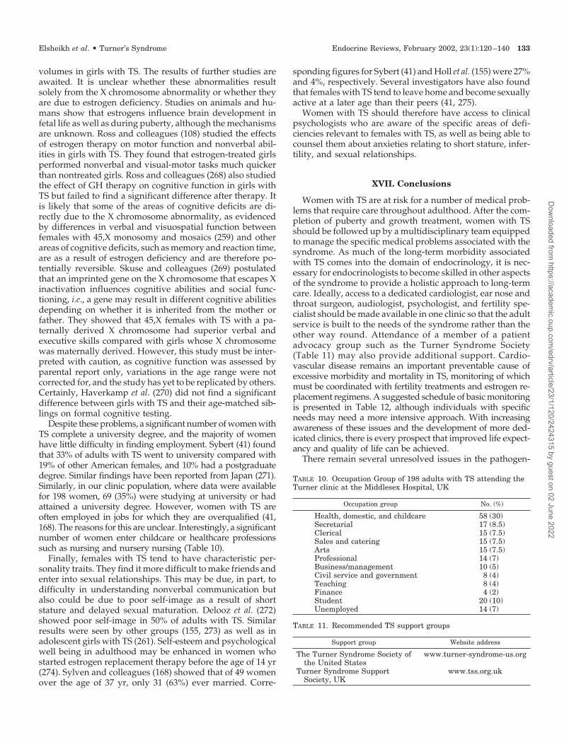
volumes in girls with TS. The results of further studies areawaited. It is unclear whether these abnormalities resultsolely from the X chromosome abnormality or whether theyare due to estrogen deficiency. Studies on animals and hu-mans show that estrogens influence brain development infetal life as well as during puberty, although the mechanismsare unknown. Ross and colleagues (108) studied the effectsof estrogen therapy on motor function and nonverbal abil-ities in girls with TS. They found that estrogen-treated girlsperformed nonverbal and visual-motor tasks much quickerthan nontreated girls. Ross and colleagues (268) also studiedthe effect of GH therapy on cognitive function in girls withTS but failed to find a significant difference after therapy. Itis likely that some of the areas of cognitive deficits are di-rectly due to the X chromosome abnormality, as evidencedby differences in verbal and visuospatial function betweenfemales with 45,X monosomy and mosaics (259) and otherareas of cognitive deficits, such as memory and reaction time,are as a result of estrogen deficiency and are therefore po-tentially reversible. Skuse and colleagues (269) postulatedthat an imprinted gene on the X chromosome that escapes Xinactivation influences cognitive abilities and social func-tioning, i.e., a gene may result in different cognitive abilitiesdepending on whether it is inherited from the mother orfather. They showed that 45,X females with TS with a pa-ternally derived X chromosome had superior verbal andexecutive skills compared with girls whose X chromosomewas maternally derived. However, this study must be inter-preted with caution, as cognitive function was assessed byparental report only, variations in the age range were notcorrected for, and the study has yet to be replicated by others.Certainly, Haverkamp et al. (270) did not find a significantdifference between girls with TS and their age-matched sib-lings on formal cognitive testing.
Despite these problems, a significant number of women withTS complete a university degree, and the majority of womenhave little difficulty in finding employment. Sybert (41) foundthat 33% of adults with TS went to university compared with19% of other American females, and 10% had a postgraduatedegree. Similar findings have been reported from Japan (271).Similarly, in our clinic population, where data were availablefor 198 women, 69 (35%) were studying at university or hadattained a university degree. However, women with TS areoften employed in jobs for which they are overqualified (41,168). The reasons for this are unclear. Interestingly, a significantnumber of women enter childcare or healthcare professionssuch as nursing and nursery nursing (Table 10).
Finally, females with TS tend to have characteristic per-sonality traits. They find it more difficult to make friends andenter into sexual relationships. This may be due, in part, todifficulty in understanding nonverbal communication butalso could be due to poor self-image as a result of shortstature and delayed sexual maturation. Delooz et al. (272)showed poor self-image in 50% of adults with TS. Similarresults were seen by other groups (155, 273) as well as inadolescent girls with TS (261). Self-esteem and psychologicalwell being in adulthood may be enhanced in women whostarted estrogen replacement therapy before the age of 14 yr(274). Sylven and colleagues (168) showed that of 49 womenover the age of 37 yr, only 31 (63%) ever married. Corre-
sponding figures for Sybert (41) and Holl et al. (155) were 27%and 4%, respectively. Several investigators have also foundthat females with TS tend to leave home and become sexuallyactive at a later age than their peers (41, 275).
Women with TS should therefore have access to clinicalpsychologists who are aware of the specific areas of defi-ciencies relevant to females with TS, as well as being able tocounsel them about anxieties relating to short stature, infer-tility, and sexual relationships.
XVII. Conclusions
Women with TS are at risk for a number of medical prob-lems that require care throughout adulthood. After the com-pletion of puberty and growth treatment, women with TSshould be followed up by a multidisciplinary team equippedto manage the specific medical problems associated with thesyndrome. As much of the long-term morbidity associatedwith TS comes into the domain of endocrinology, it is nec-essary for endocrinologists to become skilled in other aspectsof the syndrome to provide a holistic approach to long-termcare. Ideally, access to a dedicated cardiologist, ear nose andthroat surgeon, audiologist, psychologist, and fertility spe-cialist should be made available in one clinic so that the adultservice is built to the needs of the syndrome rather than theother way round. Attendance of a member of a patientadvocacy group such as the Turner Syndrome Society(Table 11) may also provide additional support. Cardio-vascular disease remains an important preventable cause ofexcessive morbidity and mortality in TS, monitoring of whichmust be coordinated with fertility treatments and estrogen re-placement regimens. A suggested schedule of basic monitoringis presented in Table 12, although individuals with specificneeds may need a more intensive approach. With increasingawareness of these issues and the development of more ded-icated clinics, there is every prospect that improved life expect-ancy and quality of life can be achieved.
There remain several unresolved issues in the pathogen-
TABLE 10. Occupation Group of 198 adults with TS attending theTurner clinic at the Middlesex Hospital, UK
Occupation group No. (%)
Health, domestic, and childcare 58 (30)Secretarial 17 (8.5)Clerical 15 (7.5)Sales and catering 15 (7.5)Arts 15 (7.5)Professional 14 (7)Business/management 10 (5)Civil service and government 8 (4)Teaching 8 (4)Finance 4 (2)Student 20 (10)Unemployed 14 (7)
TABLE 11. Recommended TS support groups
Support group Website address
The Turner Syndrome Society ofthe United States
www.turner-syndrome-us.org
Turner Syndrome SupportSociety, UK
www.tss.org.uk
Elsheikh et al. • Turner’s Syndrome Endocrine Reviews, February 2002, 23(1):120–140 133
Dow
nloaded from https://academ
ic.oup.com/edrv/article/23/1/120/2424315 by guest on 02 June 2022
esis and management of TS. The gene(s) responsible for thebone disorders, premature ovarian failure, and other featuresof TS have yet to be elucidated. Preliminary studies on theeffects of estrogen replacement therapy on colonic cancer andcognitive function have yielded tantalizing results, but fur-ther work is necessary. Long-term data looking at the naturalhistory of aortic root dilatation are required. Finally, theassociation of the isochromosome X karyotype with auto-immune thyroid disease and inflammatory bowel diseaseprovides an exciting model from which to study the geneticsof autoimmune diseases.
Acknowledgments
Address all correspondence and requests for reprints to: John A. H.Wass, M.D., Department of Endocrinology, Radcliffe Infirmary, Wood-stock Road, Oxford, OX2 6HE, United Kingdom.
References
1. Tesch LG, Rosenfeld RG 1995 Morgagni, Ullrich and Turner: thediscovery of gonadal dysgenesis. Endocrinologist 5:327–328
2. Ullrich O 1930 Uber typische Kombinationsbilder multipler Abar-tungen. Z Kinderheilk 49:271–276
3. Turner HH 1938 A syndrome of infantilism, congenital webbedneck and cubitus valgus. Endocrinology 23:566–574
4. Stratakis CA, Rennert OM 1994 Turner syndrome: molecular andcytogenetics, dysmorphology, endocrine, and other clinical man-ifestations and their management. Endocrinologist 4:442–453
5. Lippe B 1991 Turner syndrome. Endocrinol Metab Clin North Am20:121–152
6. Nielsen J, Wohlert M 1991 Chromosome abnormalities foundamong 34,910 newborn children: results from a 13-year incidencestudy in Arhus, Denmark. Hum Genet 87:81–83
7. Jacobs P, Dalton P, James R, Mosse K, Power M, Robinson D,Skuse D 1997 Turner syndrome: a cytogenetic and molecularstudy. Ann Hum Genet 61:471–483
8. Price WH, Clayton JF, Collyer S, De Mey R, Wilson J 1986 Mor-tality ratios, life expectancy, and causes of death in patients withTurner’s syndrome. J Epidemiol Community Health 40:97–102
9. Naeraa RW, Gravholt CH, Hansen J, Nielsen J, Juul S 1995 Mor-tality in Turner syndrome. In: Albertsson-Wikland K, Ranke MB,eds. Turner syndrome in a lifespan perspective: research and clin-ical aspects. Amsterdam: Elsevier; 323
10. Hassold T, Benham F, Leppert M 1988 Cytogenetic and molecular
analysis of sex chromosome monosomy. Am J Hum Genet 42:534–541
11. Pasquino AM, Passeri F, Pucarelli I, Segni M, Municchi G 1997Spontaneous pubertal development in Turner’s syndrome. ItalianStudy Group for Turner’s Syndrome. J Clin Endocrinol Metab82:1810–1813
12. Avner P, Heard E 2001 X-chromosome inactivation: counting,choice and initiation. Annu Rev Genet 2:59–67
13. Bailey JA, Carrel L, Chakravarti A, Eichler EE 2000 From the cover:molecular evidence for a relationship between LINE-1 elementsand X chromosome inactivation: the Lyon repeat hypothesis. ProcNatl Acad Sci USA 97:6634–6639
14. Carrel L, Cottle AA, Goglin KC, Willard HF 1999 A first-gener-ation X-inactivation profile of the human X chromosome. Proc NatlAcad Sci USA 96:14440–14444
15. Zinn AR, Ross JL 1998 Turner syndrome and haploinsufficiency.Curr Opin Genet Dev 8:322–327
16. Ellison JW, Wardak Z, Young MF, Gehron-Robey P, Laig-Webster M, Chiong W 1997 PHOG, a candidate gene for involve-ment in the short stature of Turner syndrome. Hum Mol Genet6:1341–1347
17. Rao E, Weiss B, Fukami M, Rump A, Niesler B, Mertz A, MuroyaK, Binder G, Kirsch S, Winkelmann M, Nordsiek G, Heinrich U,Breuning MH, Ranke MB, Rosenthal A, Ogata T, Rappold GA1997 Pseudoautosomal deletions encompassing a novel homeoboxgene cause growth failure in idiopathic short stature and Turnersyndrome. Nat Genet 16:54–63
18. Clement-Jones M, Schiller S, Rao E, Blaschke RJ, Zuniga A, ZellerR, Robson SC, Binder G, Glass I, Strachan T, Lindsay S, RappoldGA 2000 The short stature homeobox gene SHOX is involved inskeletal abnormalities in Turner syndrome. Hum Mol Genet 9:695–702
19. Kosho T, Muroya K, Nagai T, Fujimoto M, Yokoya S, SakamotoH, Hirano T, Terasaki H, Ohashi H, Nishimura G, Sato S, MatsuoN, Ogata T 1999 Skeletal features and growth patterns in 14 patientswith haploinsufficiency of SHOX: implications for the develop-ment of Turner syndrome. J Clin Endocrinol Metab 84:4613–4621
20. Shears DJ, Vassal HJ, Goodman FR, Palmer RW, Reardon W,Superti-Furga A, Scambler PJ, Winter RM 1998 Mutation anddeletion of the pseudoautosomal gene SHOX cause Leri-Weill dys-chondrosteosis. Nat Genet 19:70–73
21. Zinn AR, Tonk VS, Chen Z, Flejter WL, Gardner HA, Guerra R,Kushner H, Schwartz S, Sybert VP, Van-Dyke DL, Ross JL 1998Evidence for a Turner syndrome locus or loci at Xp11.2-p22.1. Am JHum Genet 63:1757–1766
22. Davison RM, Davis CJ, Conway GS 1999 The X chromosome andovarian failure. Clin Endocrinol (Oxf) 51:673–679
23. Prueitt RL, Ross JL, Zinn AR 2000 Physical mapping of nine Xqtranslocation breakpoints and identification of XPNPEP2 as a pre-mature ovarian failure candidate gene. Cytogenet Cell Genet. 89:44–50
24. Davison RM, Fox M, Conway GS 2000 Mapping of the POF1 locusand identification of putative genes for premature ovarian failure.Mol Hum Reprod 6:314–318
25. Bione S, Sala C, Manzini C, Arrigo G, Zuffardi O, Banfi S, BorsaniG, Jonveaux P, Philippe C, Zuccotti M, Ballabio A, Toniolo D 1998A human homologue of the Drosophila melanogaster diaphanousgene is disrupted in a patient with premature ovarian failure:evidence for conserved function in oogenesis and implications forhuman sterility. Am J Hum Genet 62:533–541
26. Luoh SW, Bain PA, Polakiewicz RD, Goodheart ML, Gardner H,Jaenisch R, Page DC 1997 Zfx mutation results in small animal sizeand reduced germ cell number in male and female mice. Devel-opment 124:2275–2284
27. Ogata T, Tyler-Smith C, Purvis-Smith S, Turner G 1993 Chro-mosomal localisation of a gene(s) for Turner stigmata on Yp. J MedGenet 30:918–922
28. Sun C, Skaletsky H, Birren B, Devon K, Tang Z, Silber S, OatesR, Page DC 1999 An azoospermic man with a de novo point mu-tation in the Y-chromosomal gene USP9Y. Nat Genet 23:429–432
29. Burgoyne PS, Baker TG 1985 Perinatal oocyte loss in XO mice andits implications for the aetiology of gonadal dysgenesis in XOwomen. J Reprod Fertil 75:633–645
TABLE 12. Suggested follow-up of adults with TS
BaselineKaryotypeRenal and pelvic ultrasoundEchocardiographyThyroid autoantibodiesGonadotropins
AnnualPhysical examination (BMI, blood pressure, CVS, etc.)Thyroid functionFasting lipidsFasting blood glucoseLiver functionRenal function
3–5 YearlyEchocardiographyBone densitometryAudiogram
BMI, Body mass index; CVS, cardiovascular system.
134 Endocrine Reviews, February 2002, 23(1):120–140 Elsheikh et al. • Turner’s Syndrome
Dow
nloaded from https://academ
ic.oup.com/edrv/article/23/1/120/2424315 by guest on 02 June 2022
30. Ogata T, Matsuo N 1995 Turner syndrome and female sex chro-mosome aberrations: deduction of the principal factors involved inthe development of clinical features. Hum Genet 95:607–629
31. Ostrer H, Clayton CM 1989 Y chromosome mosaicism in 45,XTurner syndrome. Am J Med Genet 34:294–296
32. Lau C 1999 Gonadoblastoma, testicular and prostate cancers, andthe TSPY gene. Am J Hum Genet 64:921–927
33. Binder G, Koch A, Wajs E, Ranke MB 1995 Nested polymerasechain reaction study of 53 cases with Turner’s syndrome: is cyto-genetically undetected Y mosaicism common? J Clin EndocrinolMetab 80:3532–3536
34. Quilter CR, Taylor K, Conway GS, Nathwani N, Delhanty JD1998 Cytogenetic and molecular investigations of Y chromosomesequences and their role in Turner syndrome. Ann Hum Genet62:99–106
35. Mendes JR, Strufaldi MW, Delcelo R, Moises RC, Vieira JG,Kasamatsu TS, Galera MF, Andrade JA, Verreschi IT 1999 Y-chromosome identification by PCR and gonadal histopathology inTurner’s syndrome without overt Y-mosaicism. Clin Endocrinol(Oxf) 50:19–26
36. Gravholt CH, Fedder J, Naeraa RW, Muller J 2000 Occurrence ofgonadoblastoma in females with Turner syndrome and Y chromo-some material: a population study. J Clin Endocrinol Metab 85:3199–3202
37. Azcona C, Bareille P, Stanhope R 1999 Lesson of the week: Turn-er’s syndrome mosaicism in patients with a normal blood lym-phocyte karyotype. Br Med J 318:856–857
38. Ranke MB, Pfluger H, Rosendahl W, Stubbe P, Enders H, BierichJR, Majewski F 1983 Turner syndrome: spontaneous growth in 150cases and review of the literature. Eur J Pediatr 141:81–88
39. Ranke MB, Grauer ML 1994 Adult height in Turner syndrome:results of a multinational survey. Horm Res 42:90–94
40. Lyon AJ, Preece MA, Grant DB 1985 Growth curve for girls withTurner syndrome. Arch Dis Child 60:932–935
41. Sybert VP 1995 The adult patient with Turner syndrome. In: Al-bertsson-Wikland K, Ranke MB, eds. Turner syndrome in a lifespan perspective: research and clinical aspects. Amsterdam:Elsevier; 205–218
42. Ross JL, Long LM, Loriaux DL, Cutler Jr GB 1985 Growth hor-mone secretory dynamics in Turner syndrome. J Pediatr 106:202–206
43. Ranke MB, Blum WF, Haug F, Rosendahl W, Attanasio A, EndersH, Gupta D, Bierich JR 1987 Growth hormone, somatomedin levelsand growth regulation in Turner’s syndrome. Acta Endocrinol(Copenh) 116:305–313
44. Reiter JC, Craen M, Van-Vliet G 1991 Decreased growth hormoneresponse to growth hormone-releasing hormone in Turner’s syn-drome: relation to body weight and adiposity. Acta Endocrinol(Copenh) 125:38–42
45. Saenger P 1999 Growth-promoting strategies in Turner’s syn-drome. J Clin Endocrinol Metab 84:4345–4348
46. Carel JC, Mathivon L, Gendrel C, Ducret JP, Chaussain JL 1998Near normalization of final height with adapted doses of growthhormone in Turner’s syndrome. J Clin Endocrinol Metab 83:1462–1466
47. Rosenfeld RG, Attie KM, Frane J, Brasel JA, Burstein S, Cara JF,Chernausek S, Gotlin RW, Kuntze J, Lippe BM, Mahoney CP,Moore WV, Saenger P, Johanson AJ 1998 Growth hormone ther-apy of Turner’s syndrome: beneficial effect on adult height. J Pe-diatr 132:319–324
48. Sas TC, de-Muinck-Keizer-Schrama SM, Stijnen T, Jansen M,Otten BJ, Hoorweg Nijman JJ, Vulsma T, Massa GG, Rouwe CW,Reeser HM, Gerver WJ, Gosen JJ, Rongen-Westerlaken C, DropSL 1999 Normalization of height in girls with Turner syndromeafter long-term growth hormone treatment: results of a random-ized dose-response trial. J Clin Endocrinol Metab 84:4607–4612
49. Betts PR, Butler GE, Donaldson MD, Dunger DB, Johnston DI,Kelnar CJ, Kirk J, Price DA, Wilton P 1999 A decade of growthhormone treatment in girls with Turner syndrome in the UK. UKKIGS Executive Group. Arch Dis Child 80:221–225
50. Cacciari E, Mazzanti L 1999 Final height of patients with Turner’ssyndrome treated with growth hormone (GH): indications for GHtherapy alone at high doses and late estrogen therapy. Italian Study
Group for Turner Syndrome. J Clin Endocrinol Metab 84:4510–4515
51. Bachrach LK 1993 Bone mineralization in childhood and adoles-cence. Curr Opin Pediatr 5:467–473
52. Shore RM, Chesney RW, Mazess RB, Rose PG, Bargman GJ 1982Skeletal demineralization in Turner’s syndrome. Calcif Tissue Int34:519–522
53. Smith MA, Wilson J, Price WH 1982 Bone demineralisation inpatients with Turner’s syndrome. J Med Genet 19:100–103
54. Ross JL, Long LM, Feuillan P, Cassorla F, Cutler Jr GB 1991Normal bone density of the wrist and spine and increased wristfractures in girls with Turner’s syndrome. J Clin Endocrinol Metab73:355–359
55. Stepan JJ, Musilova J, Pacovsky V 1989 Bone demineralization,biochemical indices of bone remodeling, and estrogen replacementtherapy in adults with Turner’s syndrome. J Bone Miner Res 4:193–198
56. Davies MC, Gulekli B, Jacobs HS 1995 Osteoporosis in Turner’ssyndrome and other forms of primary amenorrhoea. Clin Endo-crinol (Oxf) 43:741–746
57. Sylven L, Hagenfeldt K, Ringertz H 1995 Bone mineral density inmiddle-aged women with Turner’s syndrome. Eur J Endocrinol132:47–52
58. Landin-Wilhelmsen K, Bryman I, Windh M, Wilhelmsen L 1999Osteoporosis and fractures in Turner syndrome—importance ofgrowth promoting and oestrogen therapy. Clin Endocrinol (Oxf)51:497–502
59. Gravholt CH, Juul S, Naeraa RW, Hansen J 1998 Morbidity inTurner syndrome. J Clin Epidemiol 51:147–158
60. Bergmann P, Valsamis J, Van Perborgh J, De Schepper J, VanVliet G 1990 Comparative study of the changes in insulin-likegrowth factor-1, procollagen-III N-terminal extension peptide,bone gla-protein and bone mineral content in children with Turnersyndrome treated with recombinant growth hormone. J Clin En-docrinol Metab 71:1461–1467
61. Sass TC, De Muinck Keizer Schrama SM, Stijnen T, Asarfi A, VanLeeuwen WJ, Van Teunenbroek A, Van Rijn RR, Drop SL 2000A longitudinal study on bone mineral density until adulthood ingirls with Turner’s syndrome participating in a growth hormoneinjection frequency-response trial. Clin Endocrinol (Oxf) 52:531–536
62. Mora S, Weber G, Guarneri MP, Nizzoli G, Pasolini D, ChiumelloG 1992 Effect of estrogen replacement therapy on bone mineralcontent in girls with Turner syndrome. Obstet Gynecol 79:747–751
63. Neely EK, Marcus R, Rosenfeld RG, Bachrach LK 1993 Turnersyndrome adolescents receiving growth hormone are not os-teopenic. J Clin Endocrinol Metab 76:861–866
64. Emans SJ, Grace E, Hoffer FA, Gundberg C, Ravnikar V, WoodsER 1990 Estrogen deficiency in adolescents and young adults:impact on bone mineral content and effects of estrogen replacementtherapy. Obstet Gynecol 76:585–592
65. Lanes R, Gunczler P, Esaa S, Martinis R, Villaroel O, WeisingerJR 1999 Decreased bone mass despite long-term estrogen replace-ment therapy in young women with Turner’s syndrome and pre-viously normal bone density. Fertil Steril 72:896–899
66. Naeraa RW, Brixen K, Hansen RM, Hasling C, Mosekilde L,Andresen JH, Charles P, Nielsen J 1991 Skeletal size and bonemineral content in Turner’s syndrome: relation to karyotype, es-trogen treatment, physical fitness, and bone turnover. Calcif TissueInt 49:77–83
67. Conway GS 1997 Premature ovarian failure. Curr Opin ObstetGynecol 9:202–206
68. Weiss L 1971 Additional evidence of gradual loss of germ cells inthe pathogenesis of streak ovaries in Turner’s syndrome. J MedGenet 8:540–544
69. Massarano AA, Adams JA, Preece MA, Brook CG 1989 Ovarianultrasound appearances in Turner syndrome. J Pediatr 114:568–573
70. Haber HP, Ranke MB 1999 Pelvic ultrasonography in Turner syn-drome: standards for uterine and ovarian volume. J UltrasoundMed 18:271–276
71. Dewhurst J 1978 Fertility in 47,XXX and 45,X patients. J Med Genet15:132–135
72. Kaneko N, Kawagoe S, Hiroi M 1990 Turner’s syndrome—review
Elsheikh et al. • Turner’s Syndrome Endocrine Reviews, February 2002, 23(1):120–140 135
Dow
nloaded from https://academ
ic.oup.com/edrv/article/23/1/120/2424315 by guest on 02 June 2022
of the literature with reference to a successful pregnancy outcome.Gynecol Obstet Invest 29:81–87
73. Tarani L, Lampariello S, Raguso G, Colloridi F, Pucarelli I, Pas-quino AM, Bruni LA 1998 Pregnancy in patients with Turner’ssyndrome: six new cases and review of literature. Gynecol Endo-crinol 12:83–87
74. Bishun NP, Mannion PL, Rashad MN 1964 Chromosomal mosa-icism in a case of repeated abortion. Lancet 1:936 (Letter)
75. Kava HW, Klinger HP 1968 Secondary infertility in a phenotyp-ically normal 45,X/46,XX female. Fertil Steril 19:835–838
76. Arakaki DT, Waxman SH 1970 Chromosome abnormalities inearly spontaneous abortions. J Med Genet 7:118–124
77. Mackay EV, Adam GS, Khoo SK 1971 A successful pregnancyassociated with sex chromosomal mosaicism of the X0/XX type.Aust NZ J Obstet Gynaecol 11:259–261
78. Nakashima I, Robinson A 1971 Fertility in a 45,X female. Pediatrics47:770–773
79. Grace HJ, Quinlan DK, Edge WE 1973 45,X lymphocyte karyotypein a fertile woman. Am J Obstet Gynecol 115:279–282
80. Lieber E, Berger J 1973 Fertility in a 45X/46XX patient. Lancet 1:199(Letter)
81. Groll M, Cooper M 1976 Menstrual function in Turner’s syndrome.Obstet Gynecol 47:225–226
82. Philip J, Sele V 1976 45,XO Turner’s syndrome without evidenceof mosaicism in a patient with two pregnancies. Acta Obstet Gy-necol Scand 55:283–286
83. Lajborek-Czyz I 1976 A 45,X woman with a 47,XY, G� son. ClinGenet 9:113–116
84. Otto PA, Kasahara S, Nunesmaia HG, Frota-Pessoa O 1977 Riskof 45,X karyotype in offspring of Turner’s syndrome patients. Lan-cet 2:257 (Letter)
85. King CR, Magenis E, Bennett S 1978 Pregnancy and the Turnersyndrome. Obstet Gynecol 52:617–624
86. Shokeir MH 1978 Pregnancy in five women with 45,X/46,XX and45,X/47,XXX gonadal dysgenesis. Birth Defects 14:171–184
87. Nielsen J, Sillesen I, Hansen KB 1979 Fertility in women withTurner’s syndrome. Case report and review of literature. Br J ObstetGynaecol 86:833–835
88. Kohn G, Yarkoni S, Cohen MM 1980 Two conceptions in a 45,Xwoman. Am J Med Genet 5:339–343
89. Wray HL, Freeman MV, Ming PM 1981 Pregnancy in the Turnersyndrome with only 45,X chromosomal constitution. Fertil Steril35:509–514
90. Abraham SF, Beumont PJ, Booth A, Smith A 1981 Anorexia ner-vosa, pregnancy and XO/XX mosaicism. Med J Aust 1:582–583
91. Badawy SZ, Sunderji SG, Lanman JT 1981 Pregnancy outcome in45,X/46,XX mosaicism. Fertil Steril 35:88–90
92. Kable WT, Yussman MA 1981 Pregnancy in mosaic Turner pa-tients: case report and a guide to reproductive counseling. FertilSteril 35:477–478
93. Muasher S, Baramki TA, Diggs ES 1980 Turner phenotype inmother and daughter. Obstet Gynecol 56:752–756
94. Muram D, Jolly EE 1982 Pregnancy and gonadal dysgenesis. JObstet Gynecol 3:87–88
95. Ayuso MC, Bello MJ, Benitez J, Sanchez-Cascos A, Mendoza G1984 Two fertile Turner women in a family. Clin Genet 26:591–596
96. Baudier MM, Chihal HJ, Dickey RP 1985 Pregnancy and repro-ductive function in a patient with non-mosaic Turner syndrome.Obstet Gynecol 65:60S–64S
97. Swapp GH, Johnston AW, Watt JL, Couzin DA, Stephen GS 1989A fertile woman with non-mosaic Turner’s syndrome. Case reportand review of the literature. Br J Obstet Gynaecol 96:876–880
98. Varela M, Shapira E, Hyman DB 1991 Ullrich-Turner syndrome inmother and daughter: prenatal diagnosis of a 46,X, del(X)(p21)offspring from a 45,X mother with low-level mosaicism for thedel(X)(p21) in one ovary. Am J Med Genet 39:411–412
99. Massa G, Vanderschueren-Lodeweyckx M, Fryns JP 1992 Dele-tion of the short arm of the X chromosome: a hereditary form ofTurner syndrome. Eur J Pediatr 151:893–894
100. Palka G, Calabrese G, Stuppia L, Guanciali-Franchi P, Morizio E,Peila R, Antonucci A 1994 A woman with an apparent non-mosaic45,X delivered a 46,X, der(X) liveborn female. Clin Genet 45:93–96
101. Ditkoff EC, Vidali A, Sauer MV 1996 Pregnancy in a woman with
Turner mosaicism following ovarian stimulation and in vitro fer-tilization. J Assist Reprod Genet 13:447–448
102. Taga M, Minaguchi H, Saotome K 1996 Two pregnancies in a45,X/46,Xr(X)/46,XX Turner mosaic patient. A case report. Gy-necol Obstet Invest 42:206–208
103. Blumenthal AL, Allanson JE 1997 Turner syndrome in a motherand daughter: r(X) and fertility. Clin Genet 52:187–191
104. Uehara S, Nata M, Obara Y, Niinuma T, Funato T, Yajima A 1997A Turner syndrome woman with a ring X chromosome [45,X/46,X,r(X)(p22.3q27)] whose child also had a ring X chromosome. FertilSteril 67:576–579
105. Magee AC, Nevin NC, Armstrong MJ, McGibbon D, Nevin J 1998Ullrich-Turner syndrome: seven pregnancies in an apparent 45,Xwoman. Am J Med Genet 75:1–3
106. Gravholt CH, Naeraa RW, Nyholm B, Gerdes LU, Christiansen E,Schmitz O, Christiansen JS 1998 Glucose metabolism, lipid me-tabolism, and cardiovascular risk factors in adult Turner’s syn-drome. The impact of sex hormone replacement. Diabetes Care21:1062–1070
107. Elsheikh M, Bird R, Casadei B, Conway GS, Wass JA 2000 Theeffect of hormone replacement therapy on cardiovascular hemo-dynamics in women with Turner’s syndrome. J Clin EndocrinolMetab 85:614–618
108. Ross JL, Roeltgen D, Feuillan P, Kushner H, Cutler Jr GB 1998Effects of estrogen on nonverbal processing speed and motor func-tion in girls with Turner’s syndrome. J Clin Endocrinol Metab83:3198–3204
109. Calle EE, Miracle-McMahill HL, Thun MJ, Heath Jr CW 1995Estrogen replacement therapy and risk of fatal colon cancer in aprospective cohort of postmenopausal women. J Natl Cancer Inst87:517–523
110. Chute CG, Willett WC, Colditz GA, Stampfer MJ, Rosner B,Speizer EE 1991 A prospective study of reproductive history andexogenous estrogens on the risk of colorectal cancer in women.Epidemiology 2:201–207
111. Hasle H, Olsen JH, Nielsen J, Hansen J, Friedrich U, TommerupN 1996 Occurrence of cancer in women with Turner syndrome. Br JCancer 73:1156–1159
112. Hannaford PC, Kay CR, Vessey MP, Painter R, Mant J 1997Combined oral contraceptives and liver disease. Contraception55:145–151
113. Masters KW 1996 Treatment of Turner’s syndrome—a concern.Lancet 348:681–682
114. Lutjen P, Trounson A, Leeton J, Findlay J, Wood C, Renou P 1984The establishment and maintenance of pregnancy using in vitrofertilization and embryo donation in a patient with primary ovarianfailure. Nature 307:174–175
115. Hovatta O 1999 Pregnancies in women with Turner’s syndrome.Ann Med 31:106–110
116. Khastgir G, Abdalla H, Thomas A, Korea L, Latarche L, Studd J1997 Oocyte donation in Turner’s syndrome: an analysis of thefactors affecting the outcome. Hum Reprod 12:279–285
117. Foudila T, Soderstrom-Anttila V, Hovatta O 1999 Turner’s syn-drome and pregnancies after oocyte donation. Hum Reprod 14:532–535
118. Press F, Shapiro HM, Cowell CA, Oliver GD 1995 Outcome ofovum donation in Turner’s syndrome patients. Fertil Steril 64:995–998
119. Nagel TC, Tesch LG 1997 ART and high risk patients! Fertil Steril68:748–749
120. Garvey P, Elovitz M, Landsberger EJ 1998 Aortic dissection andmyocardial infarction in a pregnant patient with Turner syndrome.Obstet Gynecol 91:864
121. Hovatta O, Silye R, Krausz T, Abir R, Margara R, Trew G, LassA, Winston RM 1996 Cryopreservation of human ovarian tissueusing dimethylsulphoxide and propanediol-sucrose as cryopro-tectants. Hum Reprod 11:1268–1272
122. Newton H, Aubard Y, Rutherford A, Sharma V, Gosden R 1996Low temperature storage and grafting of human ovarian tissue.Hum Reprod 11:1487–1491
123. Gotzsche CO, Krag-Olsen B, Nielsen J, Sorensen KE, KristensenBO 1994 Prevalence of cardiovascular malformations and associ-
136 Endocrine Reviews, February 2002, 23(1):120–140 Elsheikh et al. • Turner’s Syndrome
Dow
nloaded from https://academ
ic.oup.com/edrv/article/23/1/120/2424315 by guest on 02 June 2022
ation with karyotypes in Turner’s syndrome. Arch Dis Child 71:433–436
124. Mazzanti L, Cacciari E 1998 Congenital heart disease in patientswith Turner’s syndrome. Italian Study Group for Turner Syndrome(ISGTS). J Pediatr 133:688–692
125. Sybert VP 1998 Cardiovascular malformations and complicationsin Turner syndrome. Pediatrics 101:E11
126. Miller MJ, Geffner ME, Lippe BM, Itami RM, Kaplan SA,DiSessa TG, Isabel Jones JB, Friedman WF 1983 Echocardiogra-phy reveals a high incidence of bicuspid aortic valve in Turnersyndrome. J Pediatr 102:47–50
127. Berdahl LD, Wenstrom KD, Hanson JW 1995 Web neck anomalyand its association with congenital heart disease. Am J Med Genet56:304–307
128. Lacro RV, Jones KL, Benirschke K 1988 Coarctation of the aortain Turner syndrome: a pathologic study of fetuses with nuchalcystic hygromas, hydrops fetalis, and female genitalia. Pediatrics81:445–451
129. Clark EB 1984 Neck web and congenital heart defects: a pathogenicassociation in 45 X-O Turner syndrome? Teratology 29:355–361
130. Seirafi PA, Warner KG, Geggel RL, Payne DD, Cleveland RJ 1998Repair of coarctation of the aorta during infancy minimizes the riskof late hypertension. Ann Thorac Surg 66:1378–1382
131. van Egmond H, Orye E, Praet M, Coppens M, Devloo-Blanc-quaert A 1988 Hypoplastic left heart syndrome and 45X karyotype.Br Heart J 60:69–71
132. Hou JW, Hwu WL, Tsai WY, Lee JS, Wang TR, Lue HC 1993Cardiovascular disorders in Turner’s syndrome and its correlationto karyotype. J Formos Med Assoc 92:188–189
133. Dawson-Falk KL, Wright AM, Bakker B, Pitlick PT, Wilson DM,Rosenfeld RG 1992 Cardiovascular evaluation in Turner syn-drome: utility of MR imaging. Australas Radiol 36:204–209
134. Elsheikh M, Casadei B, Conway GS, Wass JAH 2001 Hyperten-sion is a major risk factor for aortic root dilatation in women withTurner’s syndrome. Clin Endocrinol (Oxf) 54:69–73
135. Allen DB, Hendricks SA, Levy JM 1986 Aortic dilatation in Turnersyndrome. J Pediatr 109:302–305
136. Lin AE, Lippe BM, Geffner ME, Gomes A, Lois JF, Barton CW,Rosenthal A, Friedman WF 1986 Aortic dilation, dissection, andrupture in patients with Turner syndrome. J Pediatr 109:820–826
137. Lin AE, Lippe B, Rosenfeld RG 1998 Further delineation of aorticdilation, dissection, and rupture in patients with Turner syndrome.Pediatrics 102:E12
138. Roberts WC 1975 The hypertensive diseases. Evidence that sys-temic hypertension is a greater risk factor to the development ofother cardiovascular diseases than previously suspected. Am J Med59:523–532
139. Larson EW, Edwards WD 1984 Risk factors for aortic dissection:necropsy study of 161 cases. Am J Cardiol 53:849–855
140. Lubin MB, Gruber HE, Rimoin DL, Lachman RS 1990 Skeletalabnormalities in the Turner syndrome. In: Rosenfeld RG, Grum-bach MM, eds. Turner syndrome. New York: Marcel Dekker;281–300
141. Birdsall M, Kennedy S 1996 The risk of aortic dissection in womenwith Turner syndrome. Hum Reprod 11:1587
142. Roge CLL, Silverman NH, Hart PA, Ray RM 1978 Cardiac growthpattern determined by echocardiography. Circulation 57:285–290
143. Henry LW, Gardin JM, Ware JH 1990 Echocardiographic mea-surements in normal subjects from infancy to old age. Circulation62:1054–1061
144. Pyeritz RE 1983 Cardiovascular manifestations of heritable disor-ders of connective tissue. Prog Med Genet 5:191–302
145. Rossi-Foulkes R, Roman MJ, Rosen SE, Kramer-Fox R, Ehlers KH,O’Loughlin JE, Davis JG, Devereux RB 1999 Phenotypic featuresand impact of � blocker or calcium antagonist therapy on aorticlumen size in the Marfan syndrome. Am J Cardiol 83:1364–1368
146. Rosenfeld RG, Tesch LG, Rodriguez-Rigau LJ, McCauley E, AschR, Cara J, Conte F, Hall JG, Lippe B, Nagel TC, Neely K, Page DC,Ranke M, Saenger P, Watkins JM, Wilson DM 1994 Recommen-dations for diagnosis, treatment, and management of individualswith Turner syndrome. Endocrinologist 4:351–358
147. Saenger P 1993 Clinical review 48: The current status of diagnosis
and therapeutic intervention in Turner’s syndrome. J Clin Endo-crinol Metab 7:297–301
148. Nathwani NC, Unwin R, Brook CG, Hindmarsh PC 2000 Theinfluence of renal and cardiovascular abnormalities on blood pres-sure in Turner syndrome. Clin Endocrinol (Oxf) 52:371–377
149. Nathwani NC, Unwin R, Brook CG, Hindmarsh PC 2000 Bloodpressure and Turner syndrome. Clin Endocrinol (Oxf) 52:363–370
150. Virdis R, Cantu MC, Ghizzoni L, Ammenti A, Nori G, Volta C,Cravidi C, Vanelli M, Balestrazzi P, Bernasconi S 1986 Bloodpressure behaviour and control in Turner syndrome. Clin ExpHypertens A 8:787–791
151. Price DA, Clayton PE, Crowne EH, Roberts CR 1993 Safety andefficacy of human growth hormone treatment in girls with Turnersyndrome. Horm Res 39:44–48
152. Nielsen J, Johansen K, Yale H 1969 The frequency of diabetesmellitus in patients with Turner’s syndrome and pure gonadaldysgenesis. Acta Endocrinol (Copenh) 62:251–258
153. Cicognani A, Mazzanti L, Tassinari D, Pellacani A, Forabosco A,Landi L, Pifferi C, Cacciari E 1988 Differences in carbohydratetolerance in Turner syndrome depending on age and karyotype.Eur J Pediatr 148:64–68
154. Weise M, James D, Leitner CH, Hartmann KK, Bohles HJ, Atta-nasio A 1993 Glucose metabolism in Ullrich Turner syndrome:long-term effects of therapy with human growth hormone. GermanLilly UTS Study Group. Horm Res 39:36–41
155. Holl RW, Kunze D, Etzrodt H, Teller W, Heinze E 1994 Turnersyndrome: final height, glucose tolerance, bone density and psy-chosocial status in 25 adult patients. Eur J Pediatr 153:11–16
156. Caprio S, Boulware S, Diamond M, Sherwin RS, Carpenter TO,Rubin K, Amiel S, Press M, Tamborlane WV 1991 Insulin resis-tance: an early metabolic defect of Turner’s syndrome. J Clin En-docrinol Metab 72:832–836
157. AvRuskin TW, Crigler Jr JF, Soeldner JS 1979 Turner’s syndromeand carbohydrate metabolism. I. Impaired insulin secretion aftertolbutamide and glucagon stimulation tests: evidence of insulindeficiency. Am J Med Sci 277:145–152
158. Ross JL, Feuillan P, Long LM, Kowal K, Kushner H, Cutler Jr GB1995 Lipid abnormalities in Turner syndrome. J Pediatr 126:242–245
159. Elsheikh M, Conway GS 1998 The impact of obesity on cardio-vascular risk factors in Turner’s syndrome. Clin Endocrinol (Oxf)49:447–450
160. Filler G, Amendt P, Kohnert KD, Devaux S, Ehrich JH 1998Glucose tolerance and insulin secretion in children before andduring recombinant growth hormone treatment. Horm Res 50:32–37
161. van Teunenbroek A, de Muinck Keizer Schrama SM, AanstootHJ, Stijnen T, Hoogerbrugge N, Drop SL 1999 Carbohydrate andlipid metabolism during various growth hormone dosing regimensin girls with Turner syndrome. Dutch Working Group on GrowthHormone. Metabolism 48:7–14
162. Wilson DM, Frane JW, Sherman B, Johanson AJ, Hintz RL,Rosenfeld RG 1988 Carbohydrate and lipid metabolism in Turnersyndrome: effect of therapy with growth hormone, oxandrolone,and a combination of both. J Pediatr 112:210–217
163. The Writing Group for the PEPI Trial Effects of Estrogen orEstrogen/Progestin Regimens on Heart Disease Risk Factors inPostmenopausal Women 1995 The Postmenopausal Estrogen/Progestin Interventions (PEPI) Trial. JAMA 273:199–208
164. Brussaard HE, Gevers-Leuven JA, Frolich M, Kluft C, Krans HM1997 Short-term oestrogen replacement therapy improves insulinresistance, lipids and fibrinolysis in postmenopausal women withNIDDM. Diabetologia 40:843–849
165. Bright GM, Blizzard RM, Kaiser DL, Clarke WL 1982 Organ-specific autoantibodies in children with common endocrine dis-eases. J Pediatr 100:8–14
166. Marner B, Bille G, Christy M, Damsgaard EM, Garne S, HeinzeE, Larsen S, Lernmark A, Mandrup-Poulsen T, Nerup J 1991 Isletcell cytoplasmic antibodies (ICA) in diabetes and disorders of glu-cose tolerance. Diabet Med 8:812–816
167. Garden AS, Diver MJ, Fraser WD 1996 Undiagnosed morbidity inadult women with Turner’s syndrome. Clin Endocrinol (Oxf) 45:589–594
168. Sylven L, Hagenfeldt K, Brondum-Nielsen K, von-Schoultz B
Elsheikh et al. • Turner’s Syndrome Endocrine Reviews, February 2002, 23(1):120–140 137
Dow
nloaded from https://academ
ic.oup.com/edrv/article/23/1/120/2424315 by guest on 02 June 2022
1991 Middle-aged women with Turner’s syndrome. Medical status,hormonal treatment, and social life. Acta Endocrinol (Copenh)125:359–365
169. Lanes R, Gunczler P, Palacios A, Villaroel O 1997 Serum lipids,lipoprotein lp(a), and plasminogen activator inhibitor-1 in patientswith Turner’s syndrome before and during growth hormone andestrogen therapy. Fertil Steril 68:473–477
170. Jospe N, Orlowski CC, Furlanetto RW 1995 Comparison of trans-dermal and oral estrogen therapy in girls with Turner’s syndrome.J Pediatr Endocrinol Metab 8:111–116
171. Plushner SL 1997 Lipoprotein disorders in women: which womenare the best candidates for hormone replacement therapy? AnnPharmacother 31:98–107
172. van der Schouw YT, van der Graaf Y, Steyerberg EW, EijkemansJC, Banga JD 1996 Age at menopause as a risk factor for cardio-vascular mortality. Lancet 347:714–718
173. Stampfer MJ, Colditz GA 1991 Estrogen replacement therapy andcoronary heart disease: a quantitative assessment of the epidemi-ologic evidence. Prev Med 20:47–63
174. Conway GS, Davies M, Merry A 1996 Treatment of Turner’s syn-drome (letter). Lancet 348:1590–1591
175. Radetti G, Mazzanti L, Paganini C, Bernasconi S, Russo G, RigonF, Cacciari E 1995 Frequency, clinical and laboratory features ofthyroiditis in girls with Turner’s syndrome. The Italian StudyGroup for Turner’s Syndrome. Acta Paediatr 84:909–912
176. Chiovato L, Larizza D, Bendinelli G, Tonacchera M, Marino M,Mammoli C, Lorini R, Severi F, Pinchera A 1996 Autoimmunehypothyroidism and hyperthyroidism in patients with Turner’ssyndrome. Eur J Endocrinol 134:568–575
177. Germain EL, Plotnick LP 1986 Age-related anti-thyroid antibodiesand thyroid abnormalities in Turner syndrome. Acta PediatricaScand 75:750–755
178. Gluck M, Attanasio A, Speer U, Butenandt O, Tietze HU,Scherbaum WA 1992 Prevalence of autoantibodies to endocrineorgans in girls with Ullrich-Turner syndrome aged 5–14 years.Horm Res 38:114–119
179. de Kerdanet M, Lucas J, Lemee F, Lecornu M 1994 Turner’s syn-drome with X-isochromosome and Hashimoto’s thyroiditis. ClinEndocrinol (Oxf) 41:673–676
180. Tunbridge WM, Evered DC, Hall R, Appleton D, Brewis M, ClarkF, Evans JG, Young E, Bird T, Smith PA 1977 The spectrum ofthyroid disease in a community: the Whickham survey. Clin En-docrinol (Oxf) 7:481–493
181. Ivarsson SA, Ericsson UB, Nilsson KO, Gustafsson J, Hagenas L,Hager A, Moell C, Tuvemo T, Westphal O, Albertsson-WiklandK 1995 Thyroid autoantibodies, Turner’s syndrome and growthhormone therapy. Acta Paediatr 84:63–65
182. Elsheikh M, Wass JAH, Conway GS 2001 Autoimmune thyroiddisease in women with Turner’s syndrome—the association withkaryotype. Clin Endocrinol (Oxf) 55:223–226
183. Gruneiro de Papendieck L, Jorcansky S, Coco R, Rivarola MA,Brgada C 1987 High incidence of thyroid disturbances in 49 chil-dren with Turner syndrome. J Paediatr 111:258–261
184. Nienhuis HE, Rongen-Westerlaken C, Geertzen HG, Rijkers GT,Zegers BJ, Wit JM 1993 Long-term effect of human growth hor-mone therapy on the prevalence of autoantibodies in Turner syn-drome. The Dutch Growth Hormone Working Group. Horm Res39:49–53
185. Lippe B, Geffner ME, Dietrich RB, Boechat MN, Kangarloo H1988 Renal malformations in patients with Turner syndrome: im-aging in 141 patients. Pediatrics 82:852–856
186. Matthies F, Macdiarmid WD, Rallison ML, Tyler FH 1971 Renalanomalies in Turner’s syndrome: types and suggested embryo-genesis. Clin Pediatr 10:561–565
187. Litvak AS, Rousseau TG, Wrede LD, Mabry CC, McRoberts JW1978 The association of significant renal anomalies with Turner’ssyndrome. J Urol 120:671–672
188. Flynn MT, Ekstrom L, De Arce M, Costigan C, Hoey HM 1996Prevalence of renal malformation in Turner syndrome. PediatrNephrol 10:498–500
189. Thompson NP, Montgomery SM, Wadsworth ME, Pounder RE,Wakefield AJ 2000 Early determinants of inflammatory bowel
disease: use of two national longitudinal birth cohorts. Eur J Gas-troenterol Hepatol 12:25–30
190. Price WH 1979 A high incidence of chronic inflammatory boweldisease in patients with Turner’s syndrome. J Med Genet16:263–266
191. Williams ED, Engel E, Taft PD, Forbes AP 1966 Gonadal dysgen-esis and ulcerative colitis. A case report with clinical, cytogenetic,and post-mortem studies. J Med Genet 3:51–55
192. Weinrieb IJ, Fineman RM, Spiro HM 1976 Turner syndrome andinflammatory bowel disease. N Engl J Med 294:1221–1222
193. Keating JP, Ternberg JL, Packman R 1978 Association of Crohndisease and Turner syndrome. J Pediatr 92:160–161
194. Scobie BA 1979 Co-existing coeliac and inflammatory bowel dis-ease in a patient with Turner’s syndrome. Aust NZ J Med 9:316–317
195. Arulanantham K, Kramer MS, Gryboski JD 1980 The associationof inflammatory bowel disease and X chromosomal abnormality.Pediatrics 66:63–67
196. Kohler JA, Grant DB 1981 Crohn’s disease in Turner’s syndrome.Br Med J Clin Res Ed 282:950
197. Nishimura H, Kino M, Kubo S, Kawamura K 1985 Hashimoto’sthyroiditis and ulcerative colitis in a patient with Turner’s syn-drome. JAMA 254:357
198. Knudtzon J, Svane S 1988 Turner’s syndrome associated withchronic inflammatory bowel disease. A case report and review ofthe literature. Acta Med Scand 223:375–378
199. Manzione NC, Kram M, Kram E, Das KM 1988 Turner’s syndromeand inflammatory bowel disease: a case report with immunologicstudies. Am J Gastroenterol 83:1294–1297
200. Hayward PA, Satsangi J, Jewell DP 1996 Inflammatory boweldisease and the X chromosome. QJM 89:713–718
201. Rutlin E, Wisloff F, Myren J, Serck-Hanssen A 1981 Intestinaltelangiectasis in Turner’s syndrome. Endoscopy 13:86–87
202. Reinhart WH, Mordasini C, Staubli M, Scheurer U 1983 Abnor-malities of gut vessels in Turner’s syndrome. Postgrad Med J 59:122–124
203. Thatcher N, Stephens AD, Besser GM 1973 Turner’s syndromewith coeliac disease, thin bones and abnormal liver function tests.Postgrad Med J 49:738–741
204. Bonamico M, Bottaro G, Pasquino AM, Caruso-Nicoletti M, Mari-ani P, Gemme G, Paradiso E, Ragusa MC, Spina M 1998 Celiacdisease and Turner syndrome. J Pediatr Gastroenterol Nutr 26:496–499
205. Gardner LI 1974 Intrahepatic bile stasis in 45,X Turner’s syndrome(letter). N Engl J Med 290:406
206. Andrade RJ, Alcantara R, Fraile JM, Lazo MD, Llamas A, Car-mona C, Franquelo E 1995 Chronic asymptomatic intrahepaticcholestasis associated with Turner’s syndrome. GastroenterolHepatol 18:375–378
207. Floreani A, Molaro M, Baragiotta A, Naccarato R 1999 Chroniccholestasis associated with Turner’s syndrome. Digestion 60:587–589
208. Garavelli L, Donadio A, Banchini G, Fornaciari G, Plancher AC,Franchi F, Gardini G 1998 Liver abnormalities and portal hyper-tension in Ullrich-Turner syndrome. Am J Med Genet 80:180–182
209. Wemme H, Pohlenz J, Schonberger W 1995 Effect of oestrogen/gestagen replacement therapy on liver enzymes in patients withUllrich-Turner syndrome. Eur J Pediatr 154:807–810
210. Albareda MM, Gallego A, Enriquez J, Rodriguez JL, Webb SM1999 Biochemical liver abnormalities in Turner’s syndrome. Eur JGastroenterol Hepatol 11:1037–1039
211. Larizza D, Locatelli M, Vitali L, Vigano C, Calcaterra V, TinelliC, Sommaruga MG, Bozzini A, Campani R, Severi F 2000 Serumliver enzymes in Turner syndrome. Eur J Pediatr 159:143–148
212. Elsheikh M, Hodgson H, Wass JAH, Conway GS 2001 Hormonereplacement therapy may improve hepatic function in women withTurner’s syndrome. Clin Endocrinol (Oxf) 55:227–231
213. Moore B, Paterson M, Sturdee D 1987 Effect of oral hormonereplacement therapy on liver function tests. Maturitas 9:7–15
214. Darj E, Axelsson O, Carlstrom K, Nilsson S, von Schoultz B 1993Liver metabolism during treatment with estradiol and natural pro-gesterone. Gynecol Endocrinol 7:111–114
215. Cutler BS, Forbes AP, Ingersoll FM, Scully RE 1972 Endometrial
138 Endocrine Reviews, February 2002, 23(1):120–140 Elsheikh et al. • Turner’s Syndrome
Dow
nloaded from https://academ
ic.oup.com/edrv/article/23/1/120/2424315 by guest on 02 June 2022
carcinoma after stilbestrol therapy in gonadal dysgenesis. N EnglJ Med 287:628–631
216. McCarty Jr KS, Barton TK, Peete Jr CH, Creasman WT 1978Gonadal dysgenesis with adenocarcinoma of the endometrium: anelectron microscopic and steroid receptor analyses with a reviewof the literature. Cancer 42:512–520
217. Louka MH, Ross RD, Lee Jr JH, Lewis Jr GC 1978 Endometrialcarcinoma in Turner’s syndrome. Gynecol Oncol 6:294–304
218. Manuel M, Katayama KP, Jones HW 1976 The age of occurrenceof gonadal tumours in intersex patients with a Y chromosome. Am JObstet Gynecol 3:293–300
219. Verp MS, Simpson JL 1987 Abnormal sexual differentiation andneoplasia. Cancer Genet Cytogenet 25:191–218
220. Savage MO, Lowe DG 1990 Gonadal neoplasia and abnormalsexual differentiation. Clin Endocrinol (Oxf) 32:519–533
221. Ochi H, Takeuchi J, Sandberg AA 1985 Multiple cancers in aTurner’s syndrome with 45,X/46,XXp-/46,XX/47,XXX karyotype.Cancer Genet Cytogenet 16:335–339
222. Cheng HM, Tsai MC 1993 Turner’s syndrome associated withsigmoid polyp and colon cancer: report of a case. J Formos MedAssoc 92:580–582
223. Hebert-Croteau N 1998 A meta-analysis of hormone replacementtherapy and colon cancer in women. Cancer Epidemiol BiomarkersPrev 7:653–659
224. Pawliger DF, Barrow M, Noyes WD 1970 Acute leukaemia andTurner’s syndrome. Lancet 760:1345
225. Wertelecki W, Shapiro JR 1970 45,XO Turner’s syndrome andleukaemia. Lancet 1:789–790
226. Chaganti RS, Bailey RB, Jhanwar SC, Arlin ZA, Clarkson BD 1982Chronic myelogenous leukemia in the monosomic cell line of afertile Turner syndrome mosaic (45,X/46,XX). Cancer Genet Cy-togenet 5:215–221
227. Dube ID, Arlin ZA, Kalousek DK, Eaves CJ, Eaves AC 1984Nonclonal hemopoietic progenitor cells detected in long-term mar-row cultures from a Turner syndrome mosaic with chronic myeloidleukemia. Blood 64:1284–1287
228. Anderson H, Filipsson R, Fluur E, Koch B, Lindsten J, WedenbergE 1969 Hearing impairment in Turner’s syndrome. Acta Otolar-yngol 247:1–26
229. Sculerati N, Oddoux C, Clayton CM, Lim JW, Oster H 1996 Hear-ing loss in Turner syndrome. Laryngoscope 106:992–997
230. Stenberg AE, Nylen O, Windh M, Hultcrantz M 1998 Otologicalproblems in children with Turner’s syndrome. Hear Res 124:85–90
231. Hultcrantz M, Sylven L 1997 Turner’s syndrome and hearing dis-orders in women aged 16–34. Hear Res 103:69–74
232. Hultcrantz M, Sylven L, Borg E 1994 Ear and hearing problems in44 middle-aged women with Turner’s syndrome. Hear Res76:127–132
233. Barrenasa ML, Nylen O, Hanson C 1999 The influence of karyo-type on the auricle, otitis media and hearing in Turner syndrome.Hear Res 138:163–710
234. Barrenasa M, Landin-Wilhelmsenb K, Hansonc C 2000 Ear andhearing in relation to genotype and growth in Turner syndrome.Hear Res 144:21–28
235. Adhikary HP 1981 Ocular manifestations of Turner’s syndrome.Trans Ophthalmol Soc UK 101:395–396
236. Chrousos GA, Ross JL, Chrousos G, Chu FC, Kenigsberg D,Cutler Jr G, Loriaux DL 1984 Ocular findings in Turner syndrome.A prospective study. Ophthalmology 91:926–928
237. Becker B, Jospe N, Goldsmith LA 1994 Melanocytic nevi in Turnersyndrome. Pediatr Dermatol 11:120–124
238. Bourguignon JP, Pierard GE, Ernould C, Heinrichs C, Craen M,Rochiccioli P, Arrese JE, Franchimont C 1993 Effects of humangrowth hormone therapy on melanocytic naevi. Lancet 341:1505–1506
239. Zvulunov A, Wyatt DT, Laud PW, Esterly NB 1998 Influence ofgenetic and environmental factors on melanocytic naevi: a lessonfrom Turner’s syndrome. Br J Dermatol 138:993–997
240. Gare M, Ilan Y, Sherman Y, Ben-Chetrit E 1993 Malignant mel-anoma in Turner’s syndrome. Int J Dermatol 32:743–744
241. Zvulunov A 1995 Malignant melanoma in Turner’s syndrome. IntJ Dermatol 34:823 (Letter)
242. Hobbs WL, Johnson CA 1996 Anorexia nervosa: an overview. AmFam Physician 54:1273–1286
243. Forssman H, Mellbin G, Walinder J 1970 Concurrence of Turner’ssyndrome and anorexia nervosa. Br J Psychiatry 116:221–223
244. Dickens JA 1970 Concurrence of Turner’s syndrome and anorexianervosa. Br J Psychiatry 117:237
245. Liston EH, Shershow LW 1973 Concurrence of anorexia nervosaand gonadal dysgenesis. A critical review with practical consid-erations. Arch Gen Psychiatry 29:834–836
246. Kron L, Katz JL, Gorzynski G, Weiner H 1977 Anorexia nervosaand gonadal dysgenesis. Further evidence of a relationship. ArchGen Psychiatry 34:332–335
247. Margo JL, Hawton KE 1978 Anorexia nervosa and Turner’s syn-drome. Br Med J 2:15–16
248. Darby PL, Garfinkel PE, Vale JM, Kirwan PJ, Brown GM 1981Anorexia nervosa and ’Turner syndrome’: cause or coincidence?Psychol Med 11:141–145
249. Fieldsend B 1988 Anorexia nervosa and Turner’s syndrome. Br JPsychiatry 152:270–271
250. Ohzeki T, Igarashi Y, Egi S, Kagawa J, Higurashi M 1988 Turner’ssyndrome with anorexia nervosa. Am J Med 84:792–793
251. Dougherty Jr GG, Rockwell WJ, Sutton G, Ellinwood Jr EH 1983Anorexia nervosa in treated gonadal dysgenesis: case report andreview. J Clin Psychiatry 44:219–221
252. Muhs A, Lieberz K 1993 Anorexia nervosa and Turner’s syndrome.Psychopathology 26:29–40
253. Migeon BR, Luo S, Jani M, Jeppesen P 1994 The severe phenotypeof females with tiny ring X chromosomes is associated with in-ability of these chromosomes to undergo X inactivation. Am J HumGenet 55:497–504
254. Van Dyke DL, Wiktor A, Roberson JR, Weiss L 1991 Mentalretardation in Turner syndrome. J Pediatr 118:415–417
255. Zenger-Hain JL, Wiktor A, Goldman J, Van-Dyke DL, Weiss L1993 X-inactivation pattern in an Ullrich-Turner syndrome patientwith a small ring X and normal intelligence. Am J Med Genet47:490–493
256. Cole H, Huang B, Salbert BA, Brown J, Howard-Peebles PN,Black SH, Dorfmann A, Febles OR, Stevens CA, Jackson-Cook C1994 Mental retardation and Ullrich-Turner syndrome in cases with45,X/46X,�mar: additional support for the loss of the X-inactiva-tion center hypothesis. Am J Med Genet 52:136–145
257. Jani MM, Torchia BS, Pai GS, Migeon BR 1995 Molecular char-acterization of tiny ring X chromosomes from females with func-tional X chromosome disomy and lack of cis X inactivation. Genom-ics 27:182–188
258. Pennington BF, Heaton RK, Karzmark P, Pendleton MG, LehmanR, Shucard DW 1985 The neuropsychological phenotype in Turnersyndrome. Cortex 21:391–404
259. Murphy DG, Allen G, Haxby JV, Largay KA, Daly E, White BJ,Powell CM, Schapiro MB 1994 The effects of sex steroids, and theX chromosome, on female brain function: a study of the neuro-psychology of adult Turner syndrome. Neuropsychologia 32:1309–1323
260. Salbenblatt JA, Meyers DC, Bender BG, Linden MG, RobinsonA 1989 Gross and fine motor development in 45,X and 47,XXX girls.Pediatrics 84:678–682
261. Ross JL, Kushner H, Roeltgen DP 1996 Developmental changes inmotor function in girls with Turner syndrome. Pediatr Neurol15:317–332
262. Romans SM, Stefanatos G, Roeltgen DP, Kushner H, Ross JL 1998Transition to young adulthood in Ullrich-Turner syndrome: neu-rodevelopmental changes. Am J Med Genet 79:140–147
263. Bender B, Puck M, Salbenblatt J, Robinson A 1984 Cognitivedevelopment of unselected girls with complete and partial X mono-somy. Pediatrics 73:175–182
264. McCauley E, Kay T, Ito J, Treder R 1987 The Turner syndrome:cognitive deficits, affective discrimination, and behavior problems.Child Dev 58:464–473
265. Reiss AL, Mazzocco MM, Greenlaw R, Freund LS, Ross JL 1995Neurodevelopmental effects of X monosomy: a volumetric imagingstudy. Ann Neurol 38:731–738
266. Murphy DG, Mentis MJ, Pietrini P, Grady C, Daly E, Haxby JV,De-La-Granja M, Allen G, Largay K, White BJ, Powell CM, Hor-
Elsheikh et al. • Turner’s Syndrome Endocrine Reviews, February 2002, 23(1):120–140 139
Dow
nloaded from https://academ
ic.oup.com/edrv/article/23/1/120/2424315 by guest on 02 June 2022

witz B, Rapoport SI, Schapiro MB 1997 A PET study of Turner’ssyndrome: effects of sex steroids and the X chromosome on brain.Biol Psychiatry 41:285–298
267. Murphy DG, DeCarli C, Daly E, Haxby JV, Allen G, White BJ,McIntosh AR, Powell CM, Horwitz B, Rapoport SI 1993 X-chro-mosome effects on female brain: a magnetic resonance imagingstudy of Turner’s syndrome. Lancet 342:1197–1200
268. Ross JL, Feuillan P, Kushner H, Roeltgen D, Cutler Jr GB 1997Absence of growth hormone effects on cognitive function in girlswith Turner syndrome. J Clin Endocrinol Metab 82:1814–1817
269. Skuse DH, James RS, Bishop DV, Coppin B, Dalton P, Aamodt-Leeper G, Bacarese-Hamilton M, Creswell C, McGurk R, JacobsPA 1997 Evidence from Turner’s syndrome of an imprinted X-linked locus affecting cognitive function. Nature 387:705–708
270. Haverkamp F, Keuker T, Kaiser G, Noeker M, Zerres K, Reitz C2000 Social cognition in relation to visuospatial cognitive styles inUllrich-Turner syndrome: evidence for a selective deficit in socialcontext dependent visual integration. In: Saenger P, Pasquino AM,
eds. Optimizing health care for Turner patients in the 21st century.Amsterdam: Elsevier Science; 97–103
271. Okada Y 1994 The quality of life of Turner women in comparisonwith grown-up GH-deficient women. Endocr J 41:345–354
272. Delooz J, Van-den-Berghe H, Swillen A, Kleczkowska A, FrynsJP 1993 Turner syndrome patients as adults: a study of their cog-nitive profile, psychosocial functioning and psychopathologicalfindings. Genet Couns 4:169–179
273. Pavlidis K, McCauley E, Sybert VP 1995 Psychosocial and sex-ual functioning in women with Turner syndrome. Clin Genet47:85– 89
274. Ross JL, McCauley E, Roeltgen D, Long L, Kushner H, FeuillanP, Cutler Jr GB 1996 Self-concept and behavior in adolescent girlswith Turner syndrome: potential estrogen effects. J Clin EndocrinolMetab 81:926–931
275. Toublanc JE, Thibaud F, Lecointre C 1997 Psychosocial and sexualoutcome in women with Turner syndrome. Contracept Fertil Sex25:633–638
International Symposium“Autoimmune Thyroiditis and Insulitis, Early Detection, and Possibilities for
Immune Intervention”Netherlands Architecture Institute
March 14–16, 2002Rotterdam, The Netherlands
Low thyroid reserve due to autoimmune thyroiditis is increasingly recognized as a serious health problem.In the field of type 1 diabetes mellitus it has already been a goal for several years to try and attenuate thedestructive autoimmune—insulitis—process. Because of recent developments in basic and clinical immu-nology, the organizers of the meeting found the time appropriate to bring together experts in the fields ofthyroid and islet autoimmunity, early detection of endocrine autoimmune diseases, and tolerance induction.We hope for a cross-fertilization during the discussions of above-described topics. There will be ample time forsuch discussions during oral and poster presentations and plenary sessions.
Lectures (approximately 10–12) are by invitation of the Organizing Committee. Abstracts must be sent tothe Organizing Committee; eight abstracts will be selected for oral presentation.
For further information and submission of abstracts please contact: Prof. Hemmo A. Drexhage, De-partment of Immunology, Erasmus University Medical Center Rotterdam, P.O. Box 1738, 3000 DRRotterdam, The Netherlands, Phone 31-10-408.8188, Fax 31-10-408.9456, E-mail: [email protected]; or one of the other members of the organizing committee: Dr. Alex F. Muller (Departmentof Internal Medicine, Diakonessenhuis, Utrecht, The Netherlands, E-mail: [email protected]),Dr. Arie Berghout (Department of Internal Medicine, MCRZ, Rotterdam, The Netherlands, E-mail:[email protected]), Prof. Theo J. Visser (Department of Internal Medicine, EMCR, Rotterdam, TheNetherlands, E-mail: [email protected]).
Second International Symposium onProgestins, Progesterone Receptor Modulators, and
Progesterone AntagonistsSiena (Italy) 20–23 November, 2002
www.unisi.it/eventi/progestins
Topics to be reviewed include: Mechanism of Action, Progesterone Receptors and their Isoforms, Pharma-cology, Target Tissue Effects, Clinical Applications, Contraception, and Controversial Issues Related toProgestin Treatment.
Contact person: Tzina Lindenberg, E-mail: [email protected]; Fax: �972-2-6522018; Tel: �972-2-6555188.
140 Endocrine Reviews, February 2002, 23(1):120–140 Elsheikh et al. • Turner’s Syndrome
Dow
nloaded from https://academ
ic.oup.com/edrv/article/23/1/120/2424315 by guest on 02 June 2022
Related Documents