1 Staphylococcus aureus–induced endothelial permeability and inflammation are mediated by microtubule destabilization 1 Pratap Karki, 2 Yunbo Ke, 3 Yufeng Tian, 3 Tomomi Ohmura, 3 Albert Sitikov, 3 Nicolene Sarich, 3,4 Christopher P. Montgomery, 1 Anna A. Birukova 1 Division of Pulmonary and Critical Care Medicine, Department of Medicine, University of Maryland School of Medicine, Baltimore, MD 21201 2 Department of Anesthesiology, University of Maryland School of Medicine, Baltimore, MD 21201 3 Section of Pulmonary and Critical Care Medicine, Department of Medicine, University of Chicago, Chicago, Illinois 60637 4 Department of Critical Care Medicine, Nationwide Children’s Hospital, Columbus, OH 43205 Running title: Microtubule-associated signaling in S au-induced lung injury Keywords: lung injury, endothelium, microtubules, HDAC6, CLASP2, GEF-H1, S. aureus, ROS, barrier disruption, inflammation Corresponding author address: Anna A. Birukova, MD Pulmonary and Critical Care Medicine University of Maryland School of Medicine 20 Penn Street, HSF-2, Room S143 Baltimore, MD 21201 Phone: (410) 706-2545 Fax: (410) 706-6952 Email: [email protected] Abstract Staphylococcus aureus is a major etiological agent of sepsis and induces endothelial cell (EC) barrier dysfunction and inflammation, two major hallmarks of acute lung injury. However, the molecular mechanisms of bacterial pathogen– induced EC barrier disruption are incompletely understood. Here, we investigated the role of microtubules (MT) in the mechanisms of EC barrier compromise caused by heat-killed S. aureus (HKSA). Using a customized monolayer permeability assay in human pulmonary ECs and MT fractionation, we observed that HKSA- induced barrier disruption is accompanied by MT destabilization and increased histone deacetylase-6 (HDAC6) activity resulting from elevated reactive oxygen species (ROS) production. Molecular or pharmacological HDAC6 inhibition rescued barrier function in HKSA-challenged vascular endothelium. The HKSA-induced EC permeability was associated with impaired MT-mediated delivery of cytoplasmic linker–associated protein 2 (CLASP2) to the cell periphery, limiting its interaction with adherens junction proteins. HKSA-induced EC barrier dysfunction was also associated with increased Rho GTPase activity via activation of MT-bound Rho-specific guanine nucleotide exchange factor-H1 (GEF-H1) and was abolished by HDAC6 down-regulation. HKSA activated the NF-κB proinflammatory pathway and increased the expression of intercellular and vascular cell adhesion molecules in ECs, an effect that was also HDAC6-dependent and mediated, at least in part, by a GEF-H1/Rho-dependent mechanism. Of note, HDAC6-knockout mice or HDAC6 inhibitor–treated wild type mice were partially protected from vascular leakage and inflammation caused by both HKSA or methicillin-resistant S. aureus. Our results indicate that S. aureus–induced, ROS-dependent up- regulation of HDAC6 activity destabilizes MT and thereby activates the GEF-H1/Rho pathway, increasing both EC permeability and inflammation. Introduction http://www.jbc.org/cgi/doi/10.1074/jbc.RA118.004030 The latest version is at JBC Papers in Press. Published on January 8, 2019 as Manuscript RA118.004030 by guest on February 25, 2020 http://www.jbc.org/ Downloaded from

Welcome message from author
This document is posted to help you gain knowledge. Please leave a comment to let me know what you think about it! Share it to your friends and learn new things together.
Transcript
1
Staphylococcus aureus–induced endothelial permeability and inflammation are mediated by
microtubule destabilization
1Pratap Karki,
2Yunbo Ke,
3Yufeng Tian,
3Tomomi Ohmura,
3Albert Sitikov,
3Nicolene Sarich,
3,4Christopher P. Montgomery,
1Anna A. Birukova
1Division of Pulmonary and Critical Care Medicine, Department of Medicine, University of Maryland
School of Medicine, Baltimore, MD 21201 2Department of Anesthesiology, University of Maryland School of Medicine, Baltimore, MD 21201
3Section of Pulmonary and Critical Care Medicine, Department of Medicine, University of Chicago,
Chicago, Illinois 60637 4Department of Critical Care Medicine, Nationwide Children’s Hospital, Columbus, OH 43205
Running title: Microtubule-associated signaling in S au-induced lung injury
Keywords: lung injury, endothelium, microtubules, HDAC6, CLASP2, GEF-H1, S. aureus, ROS, barrier
disruption, inflammation
Corresponding author address:
Anna A. Birukova, MD
Pulmonary and Critical Care Medicine
University of Maryland School of Medicine
20 Penn Street, HSF-2, Room S143
Baltimore, MD 21201
Phone: (410) 706-2545
Fax: (410) 706-6952
Email: [email protected]
Abstract
Staphylococcus aureus is a major etiological agent
of sepsis and induces endothelial cell (EC) barrier
dysfunction and inflammation, two major
hallmarks of acute lung injury. However, the
molecular mechanisms of bacterial pathogen–
induced EC barrier disruption are incompletely
understood. Here, we investigated the role of
microtubules (MT) in the mechanisms of EC
barrier compromise caused by heat-killed S.
aureus (HKSA). Using a customized monolayer
permeability assay in human pulmonary ECs and
MT fractionation, we observed that HKSA-
induced barrier disruption is accompanied by MT
destabilization and increased histone deacetylase-6
(HDAC6) activity resulting from elevated reactive
oxygen species (ROS) production. Molecular or
pharmacological HDAC6 inhibition rescued
barrier function in HKSA-challenged vascular
endothelium. The HKSA-induced EC permeability
was associated with impaired MT-mediated
delivery of cytoplasmic linker–associated protein
2 (CLASP2) to the cell periphery, limiting its
interaction with adherens junction proteins.
HKSA-induced EC barrier dysfunction was also
associated with increased Rho GTPase activity via
activation of MT-bound Rho-specific guanine
nucleotide exchange factor-H1 (GEF-H1) and was
abolished by HDAC6 down-regulation. HKSA
activated the NF-κB proinflammatory pathway
and increased the expression of intercellular and
vascular cell adhesion molecules in ECs, an effect
that was also HDAC6-dependent and mediated, at
least in part, by a GEF-H1/Rho-dependent
mechanism. Of note, HDAC6-knockout mice or
HDAC6 inhibitor–treated wild type mice were
partially protected from vascular leakage and
inflammation caused by both HKSA or
methicillin-resistant S. aureus. Our results indicate
that S. aureus–induced, ROS-dependent up-
regulation of HDAC6 activity destabilizes MT and
thereby activates the GEF-H1/Rho pathway,
increasing both EC permeability and
inflammation.
Introduction
http://www.jbc.org/cgi/doi/10.1074/jbc.RA118.004030The latest version is at JBC Papers in Press. Published on January 8, 2019 as Manuscript RA118.004030
by guest on February 25, 2020http://w
ww
.jbc.org/D
ownloaded from
2
Staphylococcus aureus (SA) infections are the
predominant cause of sepsis, which is still a 12th
leading cause of death in US population. Severe
sepsis is the most common cause of mortality
among critically ill patients in non-coronary
intensive care units (ICU) (1,2). Both sepsis and
SA-induced pneumonia are the major contributors
in the development of acute lung injury (ALI) and
its life-threatening complication, acute respiratory
distress syndrome (ARDS) (3). Antibiotic therapy
is provided to treat SA infections, but the
pathogenesis associated with killed bacterium and
widespread emergence of drug-resistant species
such as methicillin-resistant SA (MRSA) remain a
daunting challenge. MRSA infection is a major
cause (38%) of ventilator-associated pneumonia in
surgical ICU, and a large population of MRSA
pneumonia develop severe sepsis and septic shock
(4,5).
Cellular wall components of SA,
peptidoglycan G and lipoteichoic acid are potent
activators of endothelial permeability and
inflammation driving ARDS (6,7). We have
previously demonstrated that heat killed SA
(HKSA) increases permeability in cultured human
pulmonary endothelial cells (HPAECs) and
induces vascular leak and inflammation in mice
(8). Detrimental effects of SA on pulmonary
endothelium were triggered by the activation of
p38 and Erk1/2 MAP kinases, NFB
inflammatory cascade and small GTPase RhoA
(6). Our recent report suggests an important role of
microtubule (MT) dynamics in regulation of SA-
induced endothelial dysfunction and inflammation
(9).
Small GTPases RhoA, Rac1 and Rap1
play an active role in vascular endothelial
cytoskeletal remodeling and regulation of
endothelial barrier integrity (10). These GTPases
act as a molecular switch by cycling between
GTP-bound active and GDP-bound inactive states.
Activation of RhoA triggers endothelial cell (EC)
barrier disruption and promotes inflammation. Rho
pathway of EC permeability involves the
phosphorylation and inactivation of myosin
phosphatase by Rho associated kinase resulting in
increased levels of phosphorylated regulatory
myosin light chains and actomyosin contractility
(Reviewed in (11)). On the other hand, Rho
signaling further activates the NF-B signaling
cascade leading to increased expression of EC
adhesion molecules including intercellular
adhesion molecule-1 (ICAM-1), EC-specific
vascular cell adhesion molecule-1 (VCAM-1),
inflammatory cytokines IL-6, IL-1β, IL-8,
ultimately resulting in augmented inflammation
(12-14).
MT cytoskeleton plays a critical role in
control of cell division and intracellular trafficking
of organelles and proteins. Increasing evidences
suggests that MT also play an active role in
regulation of endothelial permeability via cross-
talk with actin cytoskeleton (15,16). MT cycle
between polymerized and depolymerized states,
which is determined by the post-translational
modifications such as acetylation/deacetylation of
tubulin and by numerous MT-associated proteins
(17). A plethora of studies have documented that
MT destabilization induced by various agonists
impair endothelial function by the activation of
Rho pathway (18-23). Indeed, MT may directly
regulate Rho activity via guanine nucleotide
exchange factor H1 (GEF-H1), whose activity is
suppressed if GEF-H1 is in MT-bound state. Once
released from the MT during MT disassembly,
GEF-H1 activates Rho (24,25).
Furthermore, histone deacetylase 6
(HDAC6), a member of class II HDACs, has
emerged as a key regulator of MT dynamics
(26,27). A role of HDAC6 in MT-dependent
endothelial dysfunction was initially suggested by
the findings that HDAC6 activity promoted the
destabilization of MT by deacetylating α-tubulin,
while inhibition of HDAC6 attenuated lung
dysfunction caused by thrombin or
lipopolysaccharide (LPS) (28-30). A similar
pattern of endothelial barrier protection has also
been achieved using MT stabilizing agent, taxol
(31,32). However, the precise role of MT and
molecular/cellular mechanisms involved in
bacterial pathogen-induced endothelial barrier
dysfunction and inflammation remains to be
investigated.
Cytoplasmic linker-associated protein 2
(CLASP2) is a MT plus-end tracking protein that
promotes the stabilization of dynamic
microtubules (33). CLASP2 is directly involved in
MT - actin cytoskeleton interactions (34) and MT
stabilization at the cell cortex (35). Regional
immobilization of CLASP2 allows MT
stabilization and promotes directionally persistent
fibroblast and epithelial cell motility (35).
by guest on February 25, 2020http://w
ww
.jbc.org/D
ownloaded from
3
CLASP2 interaction with adherens junction
protein p120-catenin observed in keratinocytes
(36,37) suggests its additional role in linking MT
with cell junction complexes. These features
suggest CLASP2 as an important potential
regulator of MT-actin cytoskeleton crosstalk and
EC barrier regulation.
In this study, we explored the role of MT
dynamics in HKSA-induced endothelial barrier
dysfunction and lung inflammation. By employing
molecular and pharmacological inhibitors, we
elucidated the mechanisms of HKSA-induced
activation of HDAC6 that causes MT
destabilization and MT-dependent signaling,
leading to increased endothelial permeability and
vascular inflammation. We also evaluated the
protective effects of HDAC6 inhibition as a
potential therapeutics for HKSA or MRSA-
induced vascular leak and inflammation in vivo.
Results HKSA-induced EC barrier disruption is
accompanied by MT destabilization. Our earlier
study has shown that the destabilization of MT
mediates EC dysfunction caused by Gram-positive
bacteria-derived peptidoglycan G (PepG) (9). We
further investigated the MT-dependent mechanism
of EC permeability and inflammation caused by
HKSA. HKSA robustly increased endothelial
permeability in a dose-dependent fashion, as
reflected by decreased transendothelial electrical
resistance (TER) (Fig. 1A). We further analyzed
effect of HKSA on EC monolayer macromolecular
permeability using permeability assay developed
by our group (38) and described in Methods.
HKSA increased in a dose-dependent manner the
permeability of human pulmonary EC monolayer
for FITC-dextran tracer (Fig. 1B). To evaluate
effects of HKSA challenge on MT dynamics, we
performed MT fractionation assay to investigate
HKSA-induced changes in the pools of
polymerized and depolymerized tubulin. The
results showed that HKSA treatment markedly
decreased the pool of polymerized MT suggesting
increased MT disassembly (Fig. 1C).
HKSA increases HDAC6 activity in redox-
dependent fashion. We further investigated the
mechanism of HKSA-induced MT destabilization
mediating HKSA-induced EC permeability.
Acetylation of tubulin confers increased MT
stability (39). HKSA treatment markedly
decreased the pool of acetylated tubulin-positive
MT in pulmonary EC detected by
immunofluorescence staining with acetylated
tubulin-specific antibody (Fig. 2A). Combined
visualization of acetylated tubulin and VE-
cadherin-positive intercellular junctions shows
coordinated decline in acetylated tubulin and VE-
cadherin immunoreactivity in HKSA-challenged
EC (Fig. 2B). Imaging data were further
confirmed by Western blotting analysis of total
cell lysates from of control and HKSA-challenged
EC. The results show sustained decrease of
acetylated tubulin in the total tubulin pool (Fig.
2C).
Histone deacetylase 6 (HDAC6) is a
member of HDAC family which is localized in
cytosol and shown to deacetylate MT (30). We
examined whether HDAC6 was involved in
HKSA-induced EC dysfunction. Using
biochemical HDAC6 activation assay we found
that HKSA indeed increased HDAC6 activity
(Fig. 2D). HKSA-induced HDAC6 activation was
attenuated by reactive oxygen species (ROS)
scavengers N-acetylcysteine and amifostine, a
FDA-approved compound with reported
antioxidant properties (40) (Fig. 2D).
Inhibition of HDAC6 attenuates HKSA-induced
EC barrier disruption. To further evaluate the role
of HDAC6 in HKSA-induced endothelial barrier
disruption, we employed pharmacological and
molecular inhibitors of HDAC6. EC pretreatment
with HDAC6-specific inhibitor Tubastatin A
(TubA) completely abolished HKSA-induced
decrease in the pool of acetylated tubulin as
determined by Western blot analysis (Fig. 3A).
Immunofluorescence staining of microtubule
cytoskeleton showed that HKSA induced partial
disassembly of peripheral MT, and this effect was
abrogated by pretreatment with TubA (Fig. 3B).
TubA also strongly attenuated HKSA-induced
actin cytoskeletal remodeling by inhibiting the
formation of paracellular gaps (Fig. 3C, marked
by arrows). The bar graph depicts the results of
quantitative image analysis of paracellular gap
formation. The efficacy of TubA in attenuating
HKSA-induced EC barrier dysfunction was further
demonstrated by monitoring TER changes in
HKSA-stimulated EC monolayers. HKSA-induced
drop in resistance was attenuated by EC pre-
by guest on February 25, 2020http://w
ww
.jbc.org/D
ownloaded from

4
treatment with TubA (Fig. 3D, left panel) or
siRNA-mediated HDAC6 knockdown (Fig. 3D,
right panel).
HKSA causes AJ disassembly and impairs
interactions between MT and AJ proteins. To
investigate the mechanism of HKSA-induced
breakdown of cell junctions, we performed
immunofluorescence staining of pulmonary EC
with adherens junction protein VE-cadherin.
HKSA treatment dramatically decreased VE-
cadherin localization at cell-cell junctions and
caused intercellular gap formation (Fig. 4A,
shown by arrows). HKSA-induced disappearance
of VE-cadherin from cell-cell contacts was
rescued by HDAC6 inhibitor TubA (Fig. 4A).
HKSA induced disassembly of VE-cadherin -
p120-catenin complex and decrease in the VE-
cadherin presence at the cell surface, as
demonstrated by a surface protein biotinylation
assay. After in situ biotinylation of cell surface
proteins in control and stimulated EC monolayers,
the level of biotinylated VE-cadherin was assessed
by Western blot. HKSA-induced decrease of VE-
cadherin presence at the cell surface was
attenuated by HDAC6 inhibitor (Fig. 4B).
Reciprocal co-immunoprecipitation assays using
VE-cadherin and p120-catenin antibodies showed
that HKSA-induced dissociation of VE-cadherin –
p120-catenin protein complex was attenuated by
TubA (Fig. 4C). The disappearance of VE-
cadherin from the cell surface upon HKSA
challenge may be caused by enhanced
internalization, reduced recycling of VE-cadherin
back to the cell surface or both, and ultimately
leads to AJ disassembly and EC barrier
compromise.
The interaction between MT plus-end
binding protein CLASP2 and p120-catenin
regulates MT dynamics at the AJ in keratinocytes
(36). Using surface protein biotinylation assay, we
found that siRNA-specific knockdown of CLASP2
decreased the pool of biotinylated VE-cadherin
reflecting increased VE-cadherin internalization
and AJ disassembly in pulmonary endothelial cells
(Fig. 5A). These data suggest functional
interactions between CLASP2 and EC adherens
junction complex. Interestingly, HKSA
stimulation caused CLASP2 redistribution from
EC membrane/cytoskeletal fraction suggesting
CLASP2 disappearance from the EC cortical
compartment (Fig. 5B). Western blot panels (Fig.
5B, right) confirm the expression of cell
membrane (VEGFR2, Na+/K
+-ATPase) and
cytosolic (HSP90, GAPDH) protein markers in the
corresponding fractions.
Co-immunoprecipitation assays using
p120-catenin antibody showed the presence of
CLASP2 in p120-catenin immunocomplexes,
which was significantly decreased in the HKSA-
treated group. Importantly, pharmacological
inhibition of HDAC6 activity preserved CLASP2 -
p120-catenin interactions in HKSA-stimulated EC
(Fig. 5C). Bar graphs depict the results of
quantitative densitometry of western blot data.
HKSA-induced EC inflammation is mediated by
HDAC6. We next sought to determine whether
HDAC6 plays a role in the HKSA-induced EC
inflammation. Activation of the NFB pathway is
a central mechanism of HKSA-induced EC
inflammatory response reflected by increased
NFkB phosphorylation and nuclear translocation
(41). We monitored phospho-NFkB levels as a
measure of NFB activation in HKSA-treated
pulmonary EC. HKSA increased phospho-NFkB
levels, and this effect was attenuated by
pharmacological inhibition of HDAC6 activity
using TubA (Fig. 6A). Along with NFkB
phosphorylation, the degradation of NFkB
inhibitory subunit, IkBα, is another indicator of
NFkB activation. The results showed that HKSA
treatment decreased cellular levels of IkBα, and
this effect was also blocked by HDAC inhibitor
(Fig. 6A). NFkB activation leads to its nuclear
translocation. Immunofluorescence analysis of
NFkB intracellular localization showed HKSA-
induced NFkB localization in the nuclei of
stimulated cells, which was attenuated by TubA
(Fig. 6B). EC adhesion molecules: intercellular
adhesion molecule-1 (ICAM-1) and vascular cell
adhesion molecule-1 (VCAM-1) are key
regulators of the neutrophil recruitment at the sites
of inflammation, and their expression is triggered
by NFkB pathway (42). HKSA caused potent
upregulation of ICAM-1 and VCAM-1 expression
in pulmonary EC. This effect was suppressed by
HDAC6 inhibitor TubA (Fig. 6C), which also
abolished HKSA-induced decrease in acetylated
tubulin (Fig. 6C, bottom panel). Similarly,
HKSA-induced ICAM-1 and VCAM-1 expression
was attenuated by molecular inhibition of HDAC6
by guest on February 25, 2020http://w
ww
.jbc.org/D
ownloaded from
5
using gene-specific siRNA (Fig. 6D). HDAC6
inhibition also suppressed HKSA-induced
production of inflammatory cytokines IL-6 and IL-
8 and soluble ICAM1 by HKSA-stimulated
pulmonary EC (Fig. 6E).
To test directly the role of HDAC6
activity in the HSKA-induced EC inflammation,
we overexpressed the wild type HDAC6 and
catalytic deficient (CD) HDAC6 mutant and
determined the levels of phospho-NFkB, ICAM-1,
VCAM-1 in HKSA-challenged pulmonary EC.
While HKSA caused pronounced NFkB
phosphorylation in EC with ectopic expression of
wild type HDAC6, the NFkB phosphorylation
response was abrogated in pulmonary EC with
ectopic expression of CD-HDAC6 mutant (Fig.
6F). In line with effects on NFkB phosphorylation,
ectopic expression of CD-HDAC6 mutant
suppressed HKSA-induced expression of ICAM-1
and VCAM-1 (Fig. 6F).
HKSA-induced EC inflammation is mediated by
MT-bound GEF-H1 and Rho activation. The
activation of RhoA GTPase signaling plays an
essential role in mediating both EC permeability
and inflammation (10). Furthermore, MT may
directly modulate Rho pathway via MT-bound
RhoA-specific guanine nucleotide exchange factor
GEF-H1 (25). In this context, we investigated
whether HKSA induces Rho activation via MT -
GEF-H1 mechanism. HKSA treatment increased
GEF-H1 activity evaluated by GEF-H1 activation
pulldown assay described in Methods. GEF-H1
activation was attenuated by siRNA-induced
HDAC6 knockdown (Fig. 7A). Activation of
GEF-H1-RhoA pathway in HKSA-stimulated EC
was further reflected by increased Rho kinase-
specific phosphorylation of myosin phosphatase
and increased phosphorylation of regulatory
myosin light chain (Fig. 7B). These effects were
attenuated by HDAC6 knockdown.
To verify the role of MT-bound GEF-H1
in mediating HKSA-induced inflammation, we
employed GEF-H1 gene-specific siRNA to deplete
GEF-H1 endogenous expression. The results show
that depletion of GEF-H1 suppressed the HKSA-
induced NFkB phosphorylation. Likewise, GEF-
H1 knockdown attenuated the HKSA-induced
expression of VCAM-1 (Fig. 7C).
HDAC6 inhibition prevents HKSA-induced lung
inflammation in vivo. Protective effects of HDAC6
inhibitor TubA were further tested in the murine
model of HKSA-induced lung injury. Mice
challenged with HKSA developed prominent acute
lung injury with increased total cell counts and
protein content in bronchoalveolar lavage (BAL)
fluid (Fig. 8A). Pathologic effects of HKSA were
significantly attenuated in TubA-treated groups.
The lung vascular leak evaluated by Evans
blue extravasation into the lung parenchyma
demonstrated pronounced accumulation of the dye
in lungs of HKSA-challenged mice, which was
markedly attenuated by i.v. injection of HDAC6
inhibitor (Fig. 8B). We also employed non-
invasive fluorescence imaging tool to monitor
HKSA-induced vascular leak in the same animals
at different days. The accumulation of
intravenously injected Angiosense 680 EX tracer
in mice lungs reflects lung vascular leak and
clearance of the tracer from the lungs corresponds
to the vascular barrier recovery. HKSA-challenged
mice showed the accumulation of the tracer with
maximal accumulation at day 1 and gradual
decline at days 2 and 3 with recovery by day 6.
Intravenous administration of TubA markedly
decreased the initial accumulation of the tracer
leading to accelerated recovery indicated by faster
clearance of the tracer from the lungs (Fig. 8C).
Role of HDAC6 in septic ALI was further
evaluated using genetic model of HDAC6 KO
mice. HKSA-induced lung dysfunction HDAC6
KO mice and matching controls was monitored by
analysis of protein content and cell counts in BAL
samples, and extravasation of Evans Blue tracer
into lung parenchyma. The results show that BAL
parameters of HKSA-induced ALI: cell counts and
protein content (Fig. 9A), as well as HKSA-
induced Evans blue accumulation in the lung
parenchyma (Fig. 9B) reflecting lung vascular
leak were significantly attenuated in HDAC6 KO
mice, as compared to matching wild type controls.
Consistently, non-invasive optical imaging of
HKSA-injured lungs over 6 days post-challenge
showed that HKSA induced lung accumulation of
intravenously injected fluorescent probe
(Angiosense) in wild type mice reflecting lung
injury and barrier dysfunction. Lung injury was
significantly attenuated in HDAC6 KO mice (Fig.
9C).
by guest on February 25, 2020http://w
ww
.jbc.org/D
ownloaded from
6
Considering the emerging severity of
antibiotic-resistant infections, we also investigated
whether HDAC6 inhibition could prevent lung
injury caused by inoculation of live methicillin-
resistant Staphylococcus aureus (MRSA) strain.
Mice were pretreated with TubA (40 mg/kg, i.v.)
prior to MRSA inoculation (CR-MRSA USA 300
clinical strain 923) followed by analysis of lung
injury parameters 16 hrs after MRSA injection.
The results show that pretreatment with HDAC6
inhibitor attenuated increase in BAL total cell
counts and protein content in MRSA treated mice
(Fig. 10A), and reduced Evans blue extravasation
(Fig. 10B). BAL from MRSA treated mice
showed elevated levels of soluble ICAM-1 and
inflammatory cytokine KC, which were reduced in
HDAC6 inhibitor-treated groups (Fig. 10C).
Discussion SA infections represent emerging and serious
global concern, with their role in a wide range of
pathologies from pneumonia to sepsis. The
widespread occurrence of antibiotics resistant SA
isolates such as MRSA seeks an urgent need for
alternative approaches to combat these infections.
In this study, we unraveled an important role of
MT-associated signaling in SA-induced
endothelial permeability and inflammation.
As the role of actin cytoskeletal
remodeling in regulation of endothelial
permeability is well recognized, yet growing
evidence suggests that MT dynamics also have a
major impact on both, endothelial barrier function
and inflammatory activation (16,19,43,44). Studies
by different groups including our own have shown
that barrier disruptive agonists thrombin, TNFα,
LPS or HKSA cause MT destabilization leading to
endothelial barrier dysfunction (9,18,20,45).
Accordingly, MT stabilizing drugs such as taxol
reversed these pathologic responses. This study
shows that HKSA-induced pulmonary EC
permeability and inflammation is associated with
destabilization of the MT cytoskeleton caused by
HKSA-induced activation of HDAC6 enzymatic
activity, which has not been shown before.
Activation of ROS production in vascular
endothelium triggered by Gram-positive and
Gram-negative bacterial compounds has been
previously linked to EC dysfunction (46,47),
although specific signaling mechanisms were not
completely understood. Importantly, our data
show that HKSA-induced HDAC6 activation was
blunted in the presence of ROS inhibitors, NAC
and amifostine. This novel finding supports the
redox-dependent mechanism of HDAC6-mediated
regulation of EC permeability and inflammation
via destabilization of MT cytoskeleton in the
HKSA model of ALI. Redox-dependent
mechanism of HDAC6 activation may involve
additional intermediate steps, such as HDAC6
phosphorylation. For example, a recent study
demonstrated that cigarette smoking-induced
endothelial dysfunction and priming to ALI was
mediated by oxidative stress-activated glycogen
synthase kinase (GSK)-3β which then
activated HDAC6 via phosphorylation of serine-
22, leading to α-tubulin deacetylation (48).
Redox-dependent MT disassembly has been also
described in the settings of Gram-negative ALI
caused by LPS (19). Taken together, these
findings suggest a universal mechanism of MT-
dependent EC dysfunction caused by bacterial
pathogens. These findings also contribute to the
current knowledge of the role of redox signaling in
epigenetic regulation. Other study demonstrated
ROS-mediated activation of another member of
HDAC family, HDAC3, during LPS challenge in
cardiomyocytes (49).
The results of this study reveal the two
separate mechanisms of EC barrier disruption and
inflammation caused by HKSA-induced MT
destabilization. As a first mechanism, partial
disassembly of peripheral MT impairs delivery of
CLASP2 to the cell periphery, where it interacts
with AJ proteins to enhance barrier integrity. The
compromised interaction between CLASP2 and
AJ proteins: VE-cadherin and p120-catenin
suppresses AJ assembly thereby increasing EC
permeability. As a second mechanism, HKSA-
HDAC6-induced MT disassembly leads to release
of MT bound GEF-H1, which activates Rho
pathway of EC barrier dysfunction and additional
activation of the NF-kB inflammatory cascade.
MT-associated proteins play a role in the
agonist-induced regulation of MT dynamics and
endothelial barrier, as shown in our previous
reports (19,51,52). CLASPs represent a group of
MT plus-end binding proteins which provide
structural links and maintain a dynamic interaction
between MT and actin cytoskeleton (53).
CLASP2, via direct interaction with p120-catenin
by guest on February 25, 2020http://w
ww
.jbc.org/D
ownloaded from
7
may also regulate AJ functions and increase MT
stability at the cell cortical compartment (36).
Published studies demonstrate a rapid
disassembly of AJ and MT destabilization in the
vascular EC following inflammatory insult.
Disassembly of AJ complexes and accordingly
CLASP2 dissociation from AJ may be initiated by
different stimuli, including ROS-mediated
phosphorylation of VE-cadherin which occurs in a
minute time frame (50). On the other hand,
CLASP2 is targeted by growing MT to the AJ,
where it participates in MT anchoring at the cell
periphery, thus contributing to stabilization of the
peripheral MT. Interestingly, MT stabilization by
taxol partially attenuated both, the EC barrier
dysfunction and disassembly of AJ complexes
caused by LPS (19). Our results show that loss of
peripheral CLASP2 exerted pronounced negative
effect on EC barrier function, but was partially
restored by inhibition of HDAC6, which suggests
that HDAC6 activation-led MT disruption affects
AJ assembly that ultimately results in EC barrier
disruption. Taken together, these data suggest a
positive feedback regulation of AJ integrity by
MT. We speculate that eventually both events
contribute to vascular endothelium dysfunction
under inflammatory conditions. However, further
studies needed to determine whether AJ
disassembly precedes the HKSA-induced
CLASP2-mediated MT destabilization, or
disruption of the MT network is a primary event
leading to AJ disassembly due to loss of the
membrane-bound CLASP2.
As mentioned above, the second pathway
of EC permeability and inflammation caused by
HKSA is a release and activation of GEF-H1
following agonist-induced MT depolymerization.
Studies by our and other groups have shown that
release of MT-bound GEF-H1 leads to activation
of RhoA and its downstream signaling pathways
(24,25,54,55). Here, we show that HKSA-induced
GEF-H1 release and activation is dependent on
HDAC6 activity. Activation of RhoA signaling
augmented HKSA-induced EC inflammatory
activation and increased permeability. In turn,
HDAC6 inhibition partially stabilized MT,
attenuated RhoA signaling and suppressed HKSA-
induced expression of adhesion molecules ICAM-
1, VCAM-1, and proinflammatory cytokines. The
requirement of HDAC6 activation in this process
was further supported by the findings that EC with
ectopic expression of wild type HDAC6 responded
to HKSA challenge by higher levels of NFkB
activation and ICAM-1 and VCAM-1 expression,
than cells with ectopic expression of catalytic
deficient HDAC6 mutant. The "MT-GEF-H1-
inflammation" axis was further confirmed by
experiments with siRNA-mediated depletion of
GEF-H1, which inhibited HKSA-caused activation
of NFkB and subsequent elevation of VCAM-1.
We also cannot exclude other mechanisms of
HDAC6-induced RhoA pathway activation. For
example, inhibition of HDAC6 or HDAC3 has
been shown to protect LPS-induced endothelial
dysfunction and ALI by suppressing heat shock
protein 90 (HSP90)-mediated Rho activation (56).
It is also important to note that treatment
with the HDAC6 inhibitor TubA resulted in
complete rescue of acetylated tubulin in HKSA-
challenged cells, but only partially attenuated
HKSA-induced endothelial cell permeability,
activation of NFkB signaling, and expression of
inflammatory cytokines and adhesion molecules.
How can these apparent discrepancies be
reconciled? The primary mechanism of EC
inflammation caused by microbial components
involves HKSA interaction with Toll-like
receptors-2 and -4 (TLR2,4) resulting in the
recruitment of TIR domain-containing adaptors
MyD88, TIRAP, and TRIF to the cytoplasmic
TLR domains called TIR domains. MyD88 is
essential for the induction of inflammatory
cytokines triggered by all TLRs and TIRAP is
specifically involved in the MyD88-dependent
pathway via TLR2 and TLR4 (57). Downstream,
TLR activation triggers phosphorylation/activation
of mitogen-activated protein kinases p42/p44,
JNK1/2, and p38, and activation of NFkB cascade,
following NFkB translocation to the nucleus (58,
59). However, NFkB signaling is additionally
regulated by RhoA activity leading to
amplification of inflammatory response (60). The
results of the current study show that MT
destabilization by activated HDAC6 in HKSA-
challenged pulmonary EC stimulates RhoA
signaling via GEF-H1-dependent mechanism. This
auxiliary stimulation of NFkB cascade by GEF-
H1-RhoA axis was abolished by MT stabilization
with HDAC6 inhibitor TubA, while the priming
TLR-MyD88-NFkB inflammatory mechanism was
not affected. Collectively, these findings
demonstrate existence of both, MT-dependent and
by guest on February 25, 2020http://w
ww
.jbc.org/D
ownloaded from
8
MT-independent mechanisms of inflammation
triggered by HKSA.
The role of HDAC6-regulated MT
dynamics in SA-induced lung barrier dysfunction
and inflammation was further evaluated using
animal models of HKSA- and MRSA-induced
ALI. Some HDAC inhibitors have also been
successfully employed to alleviate various animal
models of lung injury (29,61,62). The data showed
protective effects of TubA in both models of ALI
caused by intratracheal instillation of heat-
inactivated SA and more clinically relevant MRSA
isolate. The role of HDAC6 mechanism in
propagation of HKSA-induced lung dysfunction
was further supported by marked attenuation of
HKSA-induced lung injury and inflammation in
HDAC6 knockout mice.
In summary, this study described a
HDAC6-MT-associated signaling axis promoting
HKSA/MRSA-induced lung injury and
inflammation. Based on our results, we propose a
mechanistic model of HKSA-induced and
HDAC6-MT-Rho mediated lung injury where SA
infections trigger the generation of ROS leading to
HDAC6 activation, which results in destabilization
of MT by tubulin deacetylation (Fig. 11). The
destabilized MT ultimately cause EC permeability
by activating RhoA signaling, weakening AJ
assembly and impaired delivery of barrier-
protective signaling molecules. HKSA-induced
MT destabilization mediated by HDAC6 also
promotes inflammation via GEF-H1-RhoA-
induced augmentation of NFkB inflammatory
cascade. The in vivo efficacy of tubastatin A in
protecting MRSA-induced lung injury underscores
the potential of HDAC6 inhibitors as future
alternative therapeutic approaches to treat
antibiotics resistant SA isolates-induced
infections. Thus, MT stabilization or prevention of
increased MT instability under pathologic
conditions may represent an important therapeutic
option aimed at restoration of the healthy
“signaling homeostasis” of cytoskeleton-
associated networks.
Experimental procedures
Cell culture and reagents. Human pulmonary
artery endothelial cells (HPAEC), culture medium
and growth supplements were obtained from
Lonza (Allendale, NJ). Cells were cultured
following the manufacturer’s instructions and used
at passages 5-8. HKSA was purchased from
InvivoGen (San Diego, CA). ICAM1 and VCAM1
antibodies were obtained from Santa Cruz
Biotechnology (Santa Cruz, CA); β-actin, α, β and
acetylated tubulin antibodies were from Sigma (St.
Louis, MO); diphospho-MLC, phospho-NFB,
IBα and HDAC6 antibodies were obtained from
Cell Signaling (Beverly, MA); CLASP2, VE-
cadherin and p120-catenin antibodies were from
BD Transduction Laboratories (San Diego, CA).
Texas Red phalloidin and Alexa Flour 488
conjugated secondary antibodies were purchased
form Molecular Probes (Eugene, OR). Unless
specified, all other biochemical reagents were
obtained from Sigma (St. Louis, MO).
Measurement of endothelial permeability. The
barrier integrity across HPAEC monolayer was
determined by measuring transendothelial
electrical resistance using an electrical cell-
substrate impedance sensing system ECIS Z
(Applied Biophysics, Troy, NY) as described
earlier (54). Experiments were conducted only on
wells that achieved >1200 Ω (10
microelectrodes/well) of steady state resistance.
Resistance was expressed by the in-phase voltage
(proportional to the resistance), which was
normalized to the initial voltage and expressed as a
fraction of the normalized resistance value. In the
current studies, we did not observe significant
effects of nonspecific RNA or specific siRNA on
cell viability and monolayer integrity. Initial
testing of non-transfected and siRNA-transfected
EC monolayers did not reveal significant
differences in basal TER levels. Endothelial
permeability to macromolecules was monitored by
express permeability visualization assay (Millipore
cat. #17-10398) described elsewhere (38). After
washing unbound FITC-avidin, the fluorescence of
matrix-bound FITC-avidin in control and agonist-
treated EC monolayers was measured on Victor
X5 Multilabel Plate Reader (Perkin Elmer,
Waltham, MA).
Immunocytochemistry. HPAEC monolayers were
plated on glass cover slips for
immunofluorescence staining. Cells were fixed in
3.7% formaldehyde and permeabilized with 0.1%
Triton X-100 in PBS-Tween (PBST) for 30 min at
room temperature followed by blocking with 2%
by guest on February 25, 2020http://w
ww
.jbc.org/D
ownloaded from
9
BSA in PBST for 30 min. Incubations with
antibodies of interest were performed in blocking
solution (2% BSA in PBST) for 1 hour at room
temperature followed by staining with Alexa-488
or Alexa-544 conjugated secondary antibodies.
After immunostaining, slides were analyzed using
an EVOS FL Auto 2 Cell Imaging System
(Thermo Fisher Scientific, Waltham MA).
Quantitative analysis of paracellular gap formation
was performed as previously described (18,22).
Western blot and co-immunoprecipitation.
Proteins were separated by SDS-PAGE and
transferred to polyvinylidene difluoride (PVDF)
membranes, which were incubated with desired
primary antibodies at 4°C overnight. After
incubating with HRP-conjugated secondary
antibodies at room temperature for 1 hour,
membranes were developed using enhanced
chemiluminescence substrate (Thermo Fisher,
Waltham, MA). Equal protein loading was verified
by probing the membranes with α-tubulin or β-
tubulin antibody. For co-immunoprecipitation
studies cells were lysed on ice with cold TBS-
NP40 lysis buffer (20 mM Tris pH 7.4, 150 mM
NaCl, 1% NP40) supplemented with protease and
phosphatase inhibitor cocktails (Roche,
Indianapolis, IN). Clarified lysates were then
incubated with specific antibodies of interest
overnight at 4°C, followed by 1 hour incubation
with Protein G agarose beads. After washing 3-4
times with TBS-NP40 lysis buffer, the complexes
were analyzed by Western blotting using
appropriate antibodies. The relative intensities of
the protein bands were quantified by scanning
densitometry using Image Quant software
(Molecular Dynamics, Sunnyvale, CA).
Subcellular fractionation. For isolation of
microtubule-enriched fraction, human pulmonary
EC monolayers were stimulated with HKSA and
microtubule-enriched fractions were isolated as
previously described (44). The levels of tubulin in
microtubule-enriched fractions from control and
stimulated EC were evaluated by immunoblotting
with α-tubulin antibodies. In fractionation studies,
cytosolic (soluble) and membrane/cytoskeletal
(particulate) fractions were isolated as described
previously (63). Protein extracts were separated by
SDS-PAGE, transferred to polyvinylidene fluoride
(PVDF) membrane, and probed with specific
antibodies. In all experiments, the total cell lysates
were normalized by loading equal protein amounts
per lane, as monitored by protein measurements in
SDS samples. Reprobing total lysate samples with
antibody to α-tubulin or particular protein of
interest served as additional normalization
controls.
RhoA and GEF-H1 activity assays. Active (GTP-
bound) RhoA was captured using GST-rhotekin
beads as previously reported (64). Briefly, HPAEC
grown to confluence on 60 mm dishes and
stimulated with HKSA were lysed with ice-cold
Rho assay buffer containing 100 mM NaCl, 50
mM Tris-HCl (pH 7.6), 20 mM NaF, 10 mM
MgCl2, 1% Triton X-100, 0.5% deoxycholic acid,
0.1% SDS, 1 mM Na3VO4, and protease inhibitors.
After centrifugation step, the supernatants were
incubated at 4°C for 45 min with 20–25 μg of
GST-rhotekin beads. Total cell lysates and the
rhotekin-captured proteins after beads washing
step were analyzed by Western blotting using
RhoA antibody. Active GEF-H1 was affinity
precipitated from cell lysates as previously
described (65) using a RhoA(G17A) mutant
which cannot bind nucleotide and therefore has
high affinity for activated GEFs (66). Activated
GEF-H1 in RhoA (G17A) pulldowns was detected
by immunoblotting and normalized to total GEF-
H1 in cell lysates for each sample.
Surface protein biotinylation. Control and agonist-
treated cells were washed with PBS at 37 ºC and
incubated for 10 min with 5 mM Sulfo-NHS-SS-
Biotin (Pierce Biotechnology, Rockford, IL) at
RT. After two-time wash with ice-cold PBS
containing 100mM glycine, cells were lysed for 30
min on ice in 1% Triton-100 PBS and centrifuged
at 10,000 × g for 10 min at 4°C. Equal amounts of
cell lysates were incubated with 60 µL of
Streptavidin-agarose (Pierce Biotechnology,
Rockford, IL) for 1 hr at 4°C. Beads were washed
three times with ice-cold PBS and boiled in SDS
sample buffer with 5% 2-mercaptoethanol.
Samples were centrifuged for 1 min at 1,000 × g
and supernatants were subjected to western blot
analysis with VE-cadherin antibody.
Measurement of cytokines and HDAC6 activity.
The cytokines levels in conditioned media from
cells or bronchoalveolar lavage (BAL) fluid from
by guest on February 25, 2020http://w
ww
.jbc.org/D
ownloaded from
10
mouse lungs were determined using ELISA kits
available from R&D Systems (Minneapolis, MN)
according to manufacturer’s instructions. HDAC6
activity was measured using a fluorogenic HDAC6
assay kit available from BPS Bioscience (San
Diego, CA) following the manufacturer’s
instructions.
siRNA and DNA transfections. For knockdown of
HDAC6, GEF-H1 or CLASP2 in human
pulmonary EC cultures, pre-designed human
siRNAs were ordered from Ambion (Austin, TX)
in purified, desalted, deprotected and annealed
double strand form. Transfection of EC with
siRNA was performed as previously described
(67). After 72 hrs of transfection cells were used
for experiments or harvested for western blot
verification of specific protein depletion. Non-
specific RNA (Dharmacon, Lafayette, CO) was
used as a control treatment. Plasmids encoding
human wild type and catalytically inactive (CD-
HDAC6) HDAC6 proteins bearing GFP-tag were
obtained from Addgene (Cambridge, MA) and
used for transient transfections according to
protocols described previously (18,55). Control
transfections were performed with empty vectors.
After 24-48 hrs of transfection cells were treated
with agonist of interest and used for permeability
measurements or used for immunostaining.
Animal studies. All experimental protocols
involving the use of animals were approved by the
Institutional Animal Care & Use Committee of
University of Chicago and University of
Maryland. Briefly, 8-10 week old C57BL/6J mice
were anesthetized with an intraperitoneal injection
of ketamine (75 mg/kg) and xylazine (7.5 mg/kg).
Then, sterile saline solution, 2 x 10⁸ bacterial
cells/mouse of HKSA, or 1 x 10⁸ MRSA (CR-
MRSA USA 300 clinical strain 923) were injected
intratracheally using a 20-guage catheter.
Immediately after HKSA or MRSA injections, 10
mg/kg of tubastatin A was injected intravenously
and after 24 hr animals were sacrificed by
exsanguination under anesthesia. HDAC6 null
mice were kindly provided by Timothy Mckinsey
(University of Colorado, Denver CO) (68).
Evaluation of lung injury parameters. After
intratracheal injection of 1 ml of sterile Hanks
balanced salt buffer, BAL was carried out, and
total protein and cells content were measured as
described previously (69). To analyze vascular
leak, Evans blue dye (30 mg/kg) was injected into
the external jugular vein 2 hr before the end of the
experiment as described elsewhere (69).
In vivo optical imaging: at specified times
after challenge with HKSA or vehicle, mice were
injected via tail vein with 100 µl of 2 nmol
Angiosense 680 EX imaging agent (PerkinElmer,
Boston, MA; cat# NEV10054EX). After 24 hours,
fluorescent optical imaging was performed using
Xenogen IVIS 200 Spectrum (Caliper Life
Sciences. Alameda, CA). Mice were exposed to
isoflurane anesthesia with O2 through the gas
anesthesia manifold and placed on the imaging
stage. Acquisition and image analysis were
performed with Living Image 4.3.1 Software, as
we previously described (70).
Statistical analysis. Results are expressed as
means ± SD of three to six independent
experiments. The comparison between controls
and stimulated groups was done by unpaired
Student's t-test or a one-way ANOVA, followed
by the post hoc Tukey test with P < 0.05
considered statistically significant.
Acknowledgements This work was supported by the grants HL107920, HL130431 from the National Heart, Lung, and Blood
Institute, and GM114171 from the National Institute of General Medical Sciences.
Conflict of Interest The authors declare that they have no conflicts of interest with the contents of this article.
Author Contributions AAB: designed the study and wrote the paper; PK: wrote the paper, performed biochemical studies, was
involved in discussion of the results; YK: performed animal studies and ELISA assays, was involved in
discussion of the results; YT: performed pulldown and immunoprecipitation studies, and western blot
by guest on February 25, 2020http://w
ww
.jbc.org/D
ownloaded from
11
analysis of phosphorylated proteins; TO: performed imaging studies; AS: performed permeability
measurements by XPerT and enzymatic assays; NS: performed TER measurements; CPM: optimized and
performed in vivo experiments with MRSA. All authors reviewed the results and approved the final
version of the manuscript.
References
1. Mayr, F. B., Yende, S., and Angus, D. C. (2014) Epidemiology of severe sepsis. Virulence 5, 4-
11
2. Heron, M. (2016) Deaths: Leading Causes for 2014. Natl Vital Stat Rep 65, 1-96
3. Johnson, E. R., and Matthay, M. A. (2010) Acute lung injury: epidemiology, pathogenesis, and
treatment. J Aerosol Med Pulm Drug Deliv 23, 243-252
4. Woske, H. J., Roding, T., Schulz, I., and Lode, H. (2001) Ventilator-associated pneumonia in a
surgical intensive care unit: epidemiology, etiology and comparison of three bronchoscopic
methods for microbiological specimen sampling. Crit Care 5, 167-173
5. Paulsen, J., Mehl, A., Askim, A., Solligard, E., Asvold, B. O., and Damas, J. K. (2015)
Epidemiology and outcome of Staphylococcus aureus bloodstream infection and sepsis in a
Norwegian county 1996-2011: an observational study. BMC Infect Dis 15, 116
6. Wu, T., Xing, J., and Birukova, A. A. (2013) Cell-type-specific crosstalk between p38 MAPK
and Rho signaling in lung micro- and macrovascular barrier dysfunction induced by
Staphylococcus aureus-derived pathogens. Transl Res 162, 45-55
7. Xing, J., Moldobaeva, N., and Birukova, A. A. (2011) Atrial natriuretic peptide protects against
Staphylococcus aureus-induced lung injury and endothelial barrier dysfunction. J Appl Physiol
(1985) 110, 213-224
8. Meliton, A. Y., Meng, F., Tian, Y., Sarich, N., Mutlu, G. M., Birukova, A. A., and Birukov, K. G.
(2015) Oxidized phospholipids protect against lung injury and endothelial barrier dysfunction
caused by heat-inactivated Staphylococcus aureus. Am J Physiol Lung Cell Mol Physiol 308,
L550-562
9. Tian, Y., Mambetsariev, I., Sarich, N., Meng, F., and Birukova, A. A. (2015) Role of
microtubules in attenuation of PepG-induced vascular endothelial dysfunction by atrial natriuretic
peptide. Biochim Biophys Acta 1852, 104-119
10. Spindler, V., Schlegel, N., and Waschke, J. (2010) Role of GTPases in control of microvascular
permeability. Cardiovasc Res 87, 243-253
11. Vandenbroucke, E., Mehta, D., Minshall, R., and Malik, A. B. (2008) Regulation of endothelial
junctional permeability. Ann N Y Acad Sci 1123, 134-145
12. Anwar, K. N., Fazal, F., Malik, A. B., and Rahman, A. (2004) RhoA/Rho-associated kinase
pathway selectively regulates thrombin-induced intercellular adhesion molecule-1 expression in
endothelial cells via activation of I kappa B kinase beta and phosphorylation of RelA/p65. J
Immunol 173, 6965-6972
13. Guo, F., Tang, J., Zhou, Z., Dou, Y., Van Lonkhuyzen, D., Gao, C., and Huan, J. (2012) GEF-
H1-RhoA signaling pathway mediates LPS-induced NF-kappaB transactivation and IL-8
synthesis in endothelial cells. Mol Immunol 50, 98-107
14. Shimada, H., and Rajagopalan, L. E. (2010) Rho kinase-2 activation in human endothelial cells
drives lysophosphatidic acid-mediated expression of cell adhesion molecules via NF-kappaB p65.
J Biol Chem 285, 12536-12542
15. Akhshi, T. K., Wernike, D., and Piekny, A. (2014) Microtubules and actin crosstalk in cell
migration and division. Cytoskeleton (Hoboken) 71, 1-23
16. Alieva, I. B., Zemskov, E. A., Smurova, K. M., Kaverina, I. N., and Verin, A. D. (2013) The
leading role of microtubules in endothelial barrier dysfunction: disassembly of peripheral
microtubules leaves behind the cytoskeletal reorganization. J Cell Biochem 114, 2258-2272
17. Nogales, E. (2000) Structural insights into microtubule function. Annu Rev Biochem 69, 277-302
by guest on February 25, 2020http://w
ww
.jbc.org/D
ownloaded from
12
18. Birukova, A. A., Birukov, K. G., Smurova, K., Adyshev, D., Kaibuchi, K., Alieva, I., Garcia, J.
G., and Verin, A. D. (2004) Novel role of microtubules in thrombin-induced endothelial barrier
dysfunction. FASEB J 18, 1879-1890
19. Kratzer, E., Tian, Y., Sarich, N., Wu, T., Meliton, A., Leff, A., and Birukova, A. A. (2012)
Oxidative stress contributes to lung injury and barrier dysfunction via microtubule destabilization.
Am J Respir Cell Mol Biol 47, 688-697
20. Petrache, I., Birukova, A., Ramirez, S. I., Garcia, J. G., and Verin, A. D. (2003) The role of the
microtubules in tumor necrosis factor-alpha-induced endothelial cell permeability. Am J Respir
Cell Mol Biol 28, 574-581
21. Birukova, A. A., Birukov, K. G., Adyshev, D., Usatyuk, P., Natarajan, V., Garcia, J. G., and
Verin, A. D. (2005) Involvement of microtubules and Rho pathway in TGF-beta1-induced lung
vascular barrier dysfunction. J Cell Physiol 204, 934-947
22. Birukova, A. A., Smurova, K., Birukov, K. G., Usatyuk, P., Liu, F., Kaibuchi, K., Ricks-Cord,
A., Natarajan, V., Alieva, I., Garcia, J. G., and Verin, A. D. (2004) Microtubule disassembly
induces cytoskeletal remodeling and lung vascular barrier dysfunction: role of Rho-dependent
mechanisms. J Cell Physiol 201, 55-70
23. Verin, A. D., Birukova, A., Wang, P., Liu, F., Becker, P., Birukov, K., and Garcia, J. G. (2001)
Microtubule disassembly increases endothelial cell barrier dysfunction: role of MLC
phosphorylation. Am J Physiol Lung Cell Mol Physiol 281, L565-574
24. Krendel, M., Zenke, F. T., and Bokoch, G. M. (2002) Nucleotide exchange factor GEF-H1
mediates cross-talk between microtubules and the actin cytoskeleton. Nat Cell Biol 4, 294-301
25. Chang, Y. C., Nalbant, P., Birkenfeld, J., Chang, Z. F., and Bokoch, G. M. (2008) GEF-H1
couples nocodazole-induced microtubule disassembly to cell contractility via RhoA. Mol Biol
Cell 19, 2147-2153
26. Matsuyama, A., Shimazu, T., Sumida, Y., Saito, A., Yoshimatsu, Y., Seigneurin-Berny, D.,
Osada, H., Komatsu, Y., Nishino, N., Khochbin, S., Horinouchi, S., and Yoshida, M. (2002) In
vivo destabilization of dynamic microtubules by HDAC6-mediated deacetylation. EMBO J 21,
6820-6831
27. Zilberman, Y., Ballestrem, C., Carramusa, L., Mazitschek, R., Khochbin, S., and Bershadsky, A.
(2009) Regulation of microtubule dynamics by inhibition of the tubulin deacetylase HDAC6. J
Cell Sci 122, 3531-3541
28. Saito, S., Lasky, J. A., Guo, W., Nguyen, H., Mai, A., Danchuk, S., Sullivan, D. E., and Shan, B.
(2011) Pharmacological inhibition of HDAC6 attenuates endothelial barrier dysfunction induced
by thrombin. Biochem Biophys Res Commun 408, 630-634
29. Ni, Y. F., Wang, J., Yan, X. L., Tian, F., Zhao, J. B., Wang, Y. J., and Jiang, T. (2010) Histone
deacetylase inhibitor, butyrate, attenuates lipopolysaccharide-induced acute lung injury in mice.
Respir Res 11, 33
30. Hubbert, C., Guardiola, A., Shao, R., Kawaguchi, Y., Ito, A., Nixon, A., Yoshida, M., Wang, X.
F., and Yao, T. P. (2002) HDAC6 is a microtubule-associated deacetylase. Nature 417, 455-458
31. Gorshkov, B. A., Zemskova, M. A., Verin, A. D., and Bogatcheva, N. V. (2012) Taxol alleviates
2-methoxyestradiol-induced endothelial permeability. Vascul Pharmacol 56, 56-63
32. Bogatcheva, N. V., Adyshev, D., Mambetsariev, B., Moldobaeva, N., and Verin, A. D. (2007)
Involvement of microtubules, p38, and Rho kinases pathway in 2-methoxyestradiol-induced lung
vascular barrier dysfunction. Am J Physiol Lung Cell Mol Physiol 292, L487-499
33. Maki, T., Grimaldi, A. D., Fuchigami, S., Kaverina, I., and Hayashi, I. (2015) CLASP2 Has Two
Distinct TOG Domains That Contribute Differently to Microtubule Dynamics. J Mol Biol 427,
2379-2395
34. Engel, U., Zhan, Y., Long, J. B., Boyle, S. N., Ballif, B. A., Dorey, K., Gygi, S. P., Koleske, A.
J., and Vanvactor, D. (2014) Abelson phosphorylation of CLASP2 modulates its association with
microtubules and actin. Cytoskeleton (Hoboken) 71, 195-209
by guest on February 25, 2020http://w
ww
.jbc.org/D
ownloaded from

13
35. Lansbergen, G., Grigoriev, I., Mimori-Kiyosue, Y., Ohtsuka, T., Higa, S., Kitajima, I., Demmers,
J., Galjart, N., Houtsmuller, A. B., Grosveld, F., and Akhmanova, A. (2006) CLASPs attach
microtubule plus ends to the cell cortex through a complex with LL5beta. Developmental cell 11,
21-32
36. Shahbazi, M. N., Megias, D., Epifano, C., Akhmanova, A., Gundersen, G. G., Fuchs, E., and
Perez-Moreno, M. (2013) CLASP2 interacts with p120-catenin and governs microtubule
dynamics at adherens junctions. J Cell Biol 203, 1043-1061
37. Shahbazi, M. N., and Perez-Moreno, M. (2014) Microtubules CLASP to Adherens Junctions in
epidermal progenitor cells. Bioarchitecture 4, 25-30
38. Dubrovskyi, O., Birukova, A. A., and Birukov, K. G. (2013) Measurement of local permeability
at subcellular level in cell models of agonist- and ventilator-induced lung injury. Lab Invest 93,
254-263
39. Piperno, G., LeDizet, M., and Chang, X. J. (1987) Microtubules containing acetylated alpha-
tubulin in mammalian cells in culture. J Cell Biol 104, 289-302
40. Fu, P., Murley, J. S., Grdina, D. J., Birukova, A. A., and Birukov, K. G. (2011) Induction of
cellular antioxidant defense by amifostine improves ventilator-induced lung injury. Crit Care
Med 39, 2711-2721
41. Kempe, S., Kestler, H., Lasar, A., and Wirth, T. (2005) NF-kappaB controls the global pro-
inflammatory response in endothelial cells: evidence for the regulation of a pro-atherogenic
program. Nucleic Acids Res 33, 5308-5319
42. Kolaczkowska, E., and Kubes, P. (2013) Neutrophil recruitment and function in health and
inflammation. Nat Rev Immunol 13, 159-175
43. Smurova, K. M., Biriukova, A. A., Verin, A. D., and Alieva, I. B. (2008) [The microtubule
system in endothelial barrier dysfunction: disassembly of peripheral microtubules and
microtubules reorganization in internal cytoplasm]. Tsitologiia 50, 49-55
44. Birukova, A. A., Birukov, K. G., Gorshkov, B., Liu, F., Garcia, J. G., and Verin, A. D. (2005)
MAP kinases in lung endothelial permeability induced by microtubule disassembly. Am J Physiol
Lung Cell Mol Physiol 289, L75-84
45. Li, L., Hu, J., He, T., Zhang, Q., Yang, X., Lan, X., Zhang, D., Mei, H., Chen, B., and Huang, Y.
(2015) P38/MAPK contributes to endothelial barrier dysfunction via MAP4 phosphorylation-
dependent microtubule disassembly in inflammation-induced acute lung injury. Sci Rep 5, 8895
46. McLoughlin, A., Rochfort, K. D., McDonnell, C. J., Kerrigan, S. W., and Cummins, P. M. (2017)
Staphylococcus aureus-mediated blood-brain barrier injury: an in vitro human brain
microvascular endothelial cell model. Cell Microbiol 19
47. Pai, A. B., Patel, H., Prokopienko, A. J., Alsaffar, H., Gertzberg, N., Neumann, P., Punjabi, A.,
and Johnson, A. (2012) Lipoteichoic acid from Staphylococcus aureus induces lung endothelial
cell barrier dysfunction: role of reactive oxygen and nitrogen species. PLoS One 7, e49209
48. Borgas, D., Chambers, E., Newton, J., Ko, J., Rivera, S., Rounds, S., and Lu, Q. (2016) Cigarette
Smoke Disrupted Lung Endothelial Barrier Integrity and Increased Susceptibility to Acute Lung
Injury via Histone Deacetylase 6. Am J Respir Cell Mol Biol 54, 683-696
49. Zhu, H., Shan, L., Schiller, P. W., Mai, A., and Peng, T. (2010) Histone deacetylase-3 activation
promotes tumor necrosis factor-alpha (TNF-alpha) expression in cardiomyocytes during
lipopolysaccharide stimulation. J Biol Chem 285, 9429-9436
50. Birukova, A. A., Starosta, V., Tian, X., Higginbotham, K., Koroniak, L., Berliner, J. A., and
Birukov, K. G. (2013) Fragmented oxidation products define barrier disruptive endothelial cell
response to OxPAPC. Transl Res 161, 495-504
51. Tian, X., Tian, Y., Sarich, N., Wu, T., and Birukova, A. A. (2012) Novel role of stathmin in
microtubule-dependent control of endothelial permeability. FASEB J 26, 3862-3874
52. Tian, Y., Tian, X., Gawlak, G., O'Donnell, J. J., 3rd, Sacks, D. B., and Birukova, A. A. (2014)
IQGAP1 regulates endothelial barrier function via EB1-cortactin cross talk. Mol Cell Biol 34,
3546-3558
by guest on February 25, 2020http://w
ww
.jbc.org/D
ownloaded from
14
53. Tsvetkov, A. S., Samsonov, A., Akhmanova, A., Galjart, N., and Popov, S. V. (2007)
Microtubule-binding proteins CLASP1 and CLASP2 interact with actin filaments. Cell Motil
Cytoskeleton 64, 519-530
54. Birukova, A. A., Adyshev, D., Gorshkov, B., Bokoch, G. M., Birukov, K. G., and Verin, A. D.
(2006) GEF-H1 is involved in agonist-induced human pulmonary endothelial barrier dysfunction.
Am J Physiol Lung Cell Mol Physiol 290, L540-548
55. Birukova, A. A., Fu, P., Xing, J., Yakubov, B., Cokic, I., and Birukov, K. G. (2010)
Mechanotransduction by GEF-H1 as a novel mechanism of ventilator-induced vascular
endothelial permeability. Am J Physiol Lung Cell Mol Physiol 298, L837-848
56. Joshi, A. D., Barabutis, N., Birmpas, C., Dimitropoulou, C., Thangjam, G., Cherian-Shaw, M.,
Dennison, J., and Catravas, J. D. (2015) Histone deacetylase inhibitors prevent pulmonary
endothelial hyperpermeability and acute lung injury by regulating heat shock protein 90 function.
Am J Physiol Lung Cell Mol Physiol 309, L1410-1419
57. Takeda, K., and Akira, S. (2004) TLR signaling pathways. Semin Immunol 16, 3-9
58. Arbibe, L., Mira, J. P., Teusch, N., Kline, L., Guha, M., Mackman, N., Godowski, P. J., Ulevitch,
R. J., and Knaus, U. G. (2000) Toll-like receptor 2-mediated NF-kappa B activation requires a
Rac1-dependent pathway. Nat Immunol 1, 533-540
59. Dauphinee, S. M., and Karsan, A. (2006) Lipopolysaccharide signaling in endothelial cells. Lab
Invest 86, 9-22
60. Perez-Moreno, M., Davis, M. A., Wong, E., Pasolli, H. A., Reynolds, A. B., and Fuchs, E. (2006)
p120-catenin mediates inflammatory responses in the skin. Cell 124, 631-644
61. Cetinkaya, M., Cansev, M., Cekmez, F., Tayman, C., Canpolat, F. E., Kafa, I. M., Yaylagul, E.
O., Kramer, B. W., and Sarici, S. U. (2015) Protective Effects of Valproic Acid, a Histone
Deacetylase Inhibitor, against Hyperoxic Lung Injury in a Neonatal Rat Model. PLoS One 10,
e0126028
62. Zhang, L., Jin, S., Wang, C., Jiang, R., and Wan, J. (2010) Histone deacetylase inhibitors
attenuate acute lung injury during cecal ligation and puncture-induced polymicrobial sepsis.
World J Surg 34, 1676-1683
63. Birukova, A. A., Malyukova, I., Poroyko, V., and Birukov, K. G. (2007) Paxillin-beta-catenin
interactions are involved in Rac/Cdc42-mediated endothelial barrier-protective response to
oxidized phospholipids. Am J Physiol Lung Cell Mol Physiol 293, L199-211
64. Birukova, A. A., Smurova, K., Birukov, K. G., Kaibuchi, K., Garcia, J. G., and Verin, A. D.
(2004) Role of Rho GTPases in thrombin-induced lung vascular endothelial cells barrier
dysfunction. Microvasc Res 67, 64-77
65. Kakiashvili, E., Speight, P., Waheed, F., Seth, R., Lodyga, M., Tanimura, S., Kohno, M.,
Rotstein, O. D., Kapus, A., and Szaszi, K. (2009) GEF-H1 mediates tumor necrosis factor-alpha-
induced Rho activation and myosin phosphorylation: role in the regulation of tubular paracellular
permeability. J Biol Chem 284, 11454-11466
66. Garcia-Mata, R., Wennerberg, K., Arthur, W. T., Noren, N. K., Ellerbroek, S. M., and Burridge,
K. (2006) Analysis of activated GAPs and GEFs in cell lysates. Methods Enzymol 406, 425-437
67. Birukova, A. A., Burdette, D., Moldobaeva, N., Xing, J., Fu, P., and Birukov, K. G. (2010) Rac
GTPase is a hub for protein kinase A and Epac signaling in endothelial barrier protection by
cAMP. Microvasc Res 79, 128-138
68. Demos-Davies, K. M., Ferguson, B. S., Cavasin, M. A., Mahaffey, J. H., Williams, S. M.,
Spiltoir, J. I., Schuetze, K. B., Horn, T. R., Chen, B., Ferrara, C., Scellini, B., Piroddi, N., Tesi,
C., Poggesi, C., Jeong, M. Y., and McKinsey, T. A. (2014) HDAC6 contributes to pathological
responses of heart and skeletal muscle to chronic angiotensin-II signaling. Am J Physiol Heart
Circ Physiol 307, H252-258
69. Fu, P., Birukova, A. A., Xing, J., Sammani, S., Murley, J. S., Garcia, J. G., Grdina, D. J., and
Birukov, K. G. (2009) Amifostine reduces lung vascular permeability via suppression of
inflammatory signalling. Eur Respir J 33, 612-624
by guest on February 25, 2020http://w
ww
.jbc.org/D
ownloaded from
15
70. Birukova, A. A., Meng, F., Tian, Y., Meliton, A., Sarich, N., Quilliam, L. A., and Birukov, K. G.
(2015) Prostacyclin post-treatment improves LPS-induced acute lung injury and endothelial
barrier recovery via Rap1. Biochim Biophys Acta 1852, 778-791
Figure Legends
Figure 1. HKSA-induced EC permeability is accompanied by MT destabilization. (A) HPAEC
monolayers grown on gold microelectrodes were challenged with indicated concentrations of HKSA, and
TER was monitored for 20 hrs. (B) Cells grown on 96-well plates with immobilized biotinylated gelatin
were exposed to HKSA for 2 hrs. After completion of stimulation, FITC-avidin (25 µg/mL) was added to
the cells and incubated for 3 min followed by wash with PBS and measurement of FITC fluorescence in
Victor X5 plate reader. Normalized values are expressed as mean±SD; n=6, *p<0.05. (C) MT
fractionation from control and HKSA-treated (2x10⁸ particles/ml, 6 hrs or 24 hrs) EC was performed by
separation of soluble depolymerized tubulin and insoluble tubulin polymers assembled into MT by
centrifugation as described in Methods. Results of densitometry shown as mean + SD; n=5, * p<0.05.
Figure 2. HKSA decreases the pool of acetylated microtubules and increases HDAC activity in
redox-dependent manner. (A) The levels of acetylated MT in control and HKSA treated (2x10⁸
particles/ml, 6 hrs) cells were determined by immunofluorescence staining with acetylated tubulin
antibody; bar=5 µm. (B) Co-staining of control and HKSA-stimulated (6 hrs) EC monolayers with
antibodies to acetylated tubulin and VE-cadherin. VE-cadherin-positive adherens junctions outline the
cell borders. Shown are representative results of three independent experiments; bar=10 µm. (C) The total
cell lysates collected from cells were treated with HKSA for indicated times, and acetylated tubulin
levels in total cell lysates were analyzed by Western blot. Bottom panel shows total a-tubulin levels.
Shown are representative results of three independent experiments. (D) ECs were stimulated with HKSA
(3 hrs) followed by the HDAC6 activity assay. Where indicated, cells were pretreated for 30 min with
ROS scavengers NAC (1 mM) or amifostine (amifostine trihydrate, WR2721, 4 mM). The results are
presented after normalizing to background controls, *p<0.05; n=5.
Figure 3. HDAC6 inhibitor attenuates HKSA-induced EC barrier dysfunction. HPAECs were
treated with vehicle or HDAC6 inhibitor Tubastatin A (TubA, 10 µM, 30 min) followed by HKSA
challenge (2x10⁸ particles/ml, 6 hrs), and MT integrity, actin cytoskeleton remodeling and paracellular
gap formation were monitored. (A) Acetylated tubulin levels were determined by Western blotting.
Bottom panel depicts total α-tubulin levels in samples (B) Microtubule organization was monitored by
immunofluorescence staining with α-tubulin antibody. The insert depicts the higher magnification images
revealing details of peripheral MT organization; bar=5 µm. (C) Cells were stained with Texas Red
phalloidin to visualize F-actin. Cell nuclei were visualized by DAPI counterstaining. Paracellular gaps
shown by arrows; bar=10 µm. Shown are representative results of three independent experiments. (D)
Cells were stimulated with HKSA (shown by arrow) in the presence or absence of TubA (left panel); or
they were transfected with HDAC6-specific or non-specific siRNA prior to HKSA challenge (right
panel). TER was monitored over 20-hr time period.
Figure 4. HDAC6 inhibitor rescues HKSA-induced cell junction breakdown by reversing
compromised membrane targeting and interactions of adherens junction proteins. (A) Immunofluorescence staining of HPAECs with VE-Cadherin antibody after HKSA challenge (2x10
8
particles/ml, 6 hrs) was performed to monitor cell junction remodeling. Cell nuclei were visualized by
DAPI counterstaining; bar=10 µm. Shown are representative results of three independent experiments.
(B) EC monolayers were treated with HKSA alone (4 hrs) or in combination with TubA, and surface
biotinylation assay was performed. (C) Reciprocal co-immunoprecipitation assays in similar experiments
were performed with VE-Cadherin antibody (left panel) or p120 antibody (right panel). Precipitated
immunocomplexes were analyzed by Western blotting with indicated antibodies. Bar graphs depict a
quantitative analysis of western blot densitometry data. Results shown as mean + SD; n=5, * p<0.05.
by guest on February 25, 2020http://w
ww
.jbc.org/D
ownloaded from

16
Figure 5. Role of MT end-binding protein CLASP2 and microtubules in modulation of HKSA-
induced cell junction dysfunction. (A) HPAECs were transfected with CLASP2-specific (siCLASP2) or
non-specific siRNA (nsRNA) for 72 hrs, and surface protein biotinylation assay was performed for VE-
cadherin. The efficiency of endogenous CLASP2 knockdown was confirmed by membrane reprobing
with CLASP2 antibody. (B) Cells were treated with HKSA (4 hrs), and membrane/cytoskeletal fractions
were analyzed by Western blotting to monitor redistribution of CLASP2. Equal protein amounts of
corresponding total cell lysates were loaded and additionally probed for CLASP2 to ensure equal input
protein. Results of Western blot analysis (Fig. 5B, right panel) confirm the expression of cell membrane
(VEGFR2 and Na+/K
+-ATPase) and cytosolic (HSP90, GAPDH) protein markers in the corresponding
fractions. (C) Cells were challenged with HKSA (4 hrs) with or without pre-treatment with TubA, and co-
immunoprecipitation was performed with p120 antibody followed by immunoblotting with CLASP2. The
membrane was reprobed with p120 antibody to ensure even pulldown among the groups. Bar graphs
depict a quantitative analysis of western blot densitometry data. Results shown as mean + SD; n=5, *
p<0.05.
Figure 6. HDAC6 inhibition prevents HKSA-induced EC inflammation. (A) HPAEC were treated
with HKSA in the presence or absence of TubA, and the levels of phospho-NFB, total NFB and IBα
in cell lysates were detected by Western blot analysis. Equal total protein amounts were loaded on each
lane; probing with α-tubulin was used as a confirmatory normalization control. Bar graphs depict
quantitative densitometry analysis of phospho-NFB and IBα levels. Results shown as mean + SD; n=3,
* p<0.05. (B) HKSA-induced nuclear translocation of NFkB (6 hrs) was evaluated by
immunofluorescence staining with NFB antibody; bar=10 µm. Shown are representative images of three
independent experiments. (C) VCAM-1 and ICAM-1 expression in total cell lysates from HKSA-
stimulated (24 hrs) HPAEC monolayers were monitored by Western blotting. Equal total protein amounts
were loaded on each lane; membrane reprobing with α-tubulin antibody was used an additional
normalization control. Reprobing with antibody to acetylated-tubulin was used to evaluate the efficacy of
TubA to inhibit α-tubulin deacetylation. (D) Cells were transfected with HDAC6-specific or non-specific
siRNA followed by stimulation with HKSA for indicated times and detection of VCAM-1 and ICAM-1
expression by Western blot. Bar graphs depict a quantitative analysis of western blot densitometry data.
Results shown as mean + SD; n=5, * p<0.05. (E) Cells were exposed to HKSA (24 hrs) with or without
pre-treatment with TubA, and levels of soluble ICAM (sICAM), IL-8 and IL-6 in conditioned medium
were analyzed by ELISA assays; *p<0.05; n=4. (F) HPAECs were transfected with plasmids encoding
GFP-tagged wild type (WT) or catalytic deficient HDAC6 mutant (CD) followed by stimulation with
HKSA 24 hrs post-transfection. Phospho-NFkB (top panels) or VCAM-1 and ICAM-1 (bottom panels)
levels were determined by Western blotting. Equal total protein amounts were loaded on each lane;
probing with α-tubulin was used as a confirmatory normalization control. Ectopic expression of HDAC6
constructs was verified by probing with GFP antibody. Bar graphs depict a quantitative analysis of
western blot densitometry data and shown as mean + SD; n=5, * p<0.05.
Figure 7. MT-bound GEF-H1 dependent Rho activation mediates HKSA-induced EC barrier
disruption and inflammation. HPAEC transfected with HDAC6-specific or non-specific siRNA
(nsRNA) were exposed to HKSA for indicated times. (A) GEF-H1 activation assay was performed with
RhoG17A beads, and active GEF-H1 captured by the beads were detected by Western blotting. The
efficiency of HDAC6 knockdown was confirmed by western blotting of total cell lysates with HDAC6
antibody. (B) Phosphorylation of myosin light chain (MLC) and myosin phosphatase (MYPT1) in total
cell lysates was detected by Western blotting with corresponding phosphospecific antibodies. Membranes
were then reprobed with pan-MYPT and pan-MLC antibodies. (C) HPAEC were transfected with GEF-
H1-specific or non-specific siRNA and challenged with HKSA 72 hrs after transfection. The levels of
phospho-NFkB (HKSA 6 hrs) and VCAM-1 (HKSA 24 hrs) were evaluated by Western blotting. Equal
total protein amounts were loaded on each lane; probing with α-tubulin was used as a confirmatory
by guest on February 25, 2020http://w
ww
.jbc.org/D
ownloaded from
17
normalization control. Bar graphs depict a quantitative analysis of western blot densitometry data and
shown as mean + SD; n=5, * p<0.05.
Figure 8. HDAC6 inhibition prevents HKSA-induced vascular leak and lung inflammation in vivo. Anesthetized mice were injected with HKSA (intratracheally, 2 x 10
8 bacterial cells/mouse) followed by
i.v. injection of TubA. (A) Total cells and protein content in BAL was determined 24 hrs after HKSA
administration; n=8, *p<0.05. (B) Analysis of Evans blue-labeled albumin extravasation into the lung
parenchyma reflecting vascular leak. Lungs were excised from the chest, perfused with PBS and imaged.
Shown are representative single animal images from three independent experiments. Total 9 animals per
condition were analyzed. (C) Live imaging analysis of lung vascular barrier dysfunction after HKSA
intratracheal injection with and without intravenous administration of indicated doses of TubA. HKSA-
induced accumulation of fluorescent Angiosense 680 EX imaging agent in the lungs of same animals was
detected by Xenogen IVIS 200 Spectrum imaging system 1, 2, 3 and 6 days after HKSA challenge and
presented in arbitrary colors. Shown are representative single animal images from three independent
experiments. Total 9 animals per condition were analyzed.
Figure 9. HKSA-induced lung inflammation is attenuated in HDAC6 knockout mice. Wild type
C57BL/6 (WT) or HDAC6 knockout (KO) mice were exposed to HKSA (2 x 10⁸ bacterial cells/mouse,
i.t.) for 24 h. (A) BAL was collected and total cells and protein content was determined 24 hrs after
HKSA administration. HDAC6 KO mice showed significant decrease in protein content and cell counts in
BAL; n=8, *p<0.05. (B) Analysis of Evans blue-labeled albumin extravasation into the lung parenchyma.
Lungs from HKSA-challenged (24 hrs) wt and HDAC6 KO mice were excised from the chest, perfused
with PBS and imaged. Shown are representative single animal images from three independent
experiments. Total 6 animals per condition were analyzed. (C) Live imaging analysis of lung vascular
barrier dysfunction after HKSA intratracheal injection in WT and HDAC6 KO mice. HKSA-induced
accumulation of fluorescent Angiosense 680 EX imaging agent in the lungs of same animals was detected
by Xenogen IVIS 200 Spectrum imaging system 1, 2, 3 and 6 days after HKSA challenge and presented
in arbitrary colors. Shown are representative single animal images from three independent experiments.
Total 6 animals per condition were analyzed.
Figure 10. HDAC6 inhibitor TubA ameliorates MRSA-induced lung inflammation in mice. MRSA
(CR-MRSA USA 300 clinical strain 923) was injected in mice (1 x 10⁸ cells/mouse, i.t.) with or without
intravenous administration of TubA (40 mg/kg). Control groups received saline, and mice were sacrificed
after 24 h. (A) BAL was collected from mice followed by the determination of total cells and protein
content. n=11, *p<0.05. (B) Analysis of Evans blue-labeled albumin extravasation into the lung
parenchyma. Lungs from MRSA-challenged (24 hrs) wt and HDAC6 KO mice with and without TubA
cotreatment were excised from the chest, perfused with PBS and imaged. Shown are representative single
animal images from three independent experiments. Total 8 animals per condition were analyzed. (C) The
levels of soluble ICAM-1 and KC in BAL samples were determined by ELISA. n=4 per condition,
*p<0.05.
Figure 11. Proposed model HDAC6-dependent regulation of HKSA-induced EC permeability and
inflammation. S. aureus infections generate ROS that activates HDAC6 causing MT destabilization.
Disassembly of peripheral MT structure and inhibition of MT growth to the cell periphery compromises
delivery of MT-associated protein CLASP2 and its interaction with AJ proteins leading to decreased AJ
assembly and increased EC permeability. Concurrently, GEF-H1 released from destabilized MT evokes
inflammation via the RhoA-dependent augmentation of NFkB inflammatory cascade. RhoA also directly
increases endothelial permeability via activation of actin cytoskeletal remodeling and actomyosin
contractile mechanisms. ROS scavengers or pharmacological or genetic inhibitors of HDAC6 rescue
HKSA/MRSA-induced EC permeability and inflammation by enhancing stability of MT cytoskeleton and
promoting MT-dependent mechanisms of barrier protection.
by guest on February 25, 2020http://w
ww
.jbc.org/D
ownloaded from
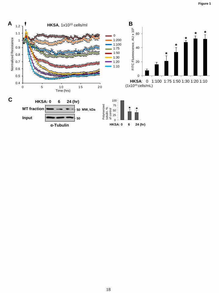
0.4
0.5
0.6
0.7
0.8
0.9
1
1.1
1.2
0 5 1 0 1 5 2 0
A
C
Figure 1
B
HKSA: 0 1:100 1:75 1:50 1:30 1:20 1:10 0
20
40
60
FITC
Flu
ores
cenc
e, A
U x
103
(1x10¹⁰ cells/mL)
* *
* * *
1:200 0
1:100 1:75
1:30 1:50
1:20 1:10
Time (hrs)
Nor
mal
ized
Res
ista
nce
HKSA, 1x10¹⁰ cells/ml
50
50
α-Tubulin
MT fraction
Input
HKSA: 0 6 24 (hr)
MW, kDa
0 6 24 (hr)
*
0
25 50
100
75
HKSA:
* P
olym
eriz
ed
tubu
lin, %
of
con
trol
18
by guest on February 25, 2020http://w
ww
.jbc.org/D
ownloaded from
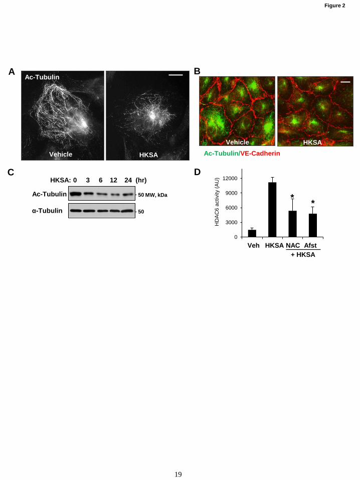
A B
Figure 2
C
50
50
MW, kDa
HKSA: 0 3 6 12 24 (hr)
Ac-Tubulin
α-Tubulin
0
3000
6000
9000
12000
H
DA
C6
activ
ity (A
U)
Veh HKSA NAC Afst + HKSA
* *
Vehicle HKSA
Ac-Tubulin
Vehicle HKSA Ac-Tubulin/VE-Cadherin
D
19
by guest on February 25, 2020http://w
ww
.jbc.org/D
ownloaded from
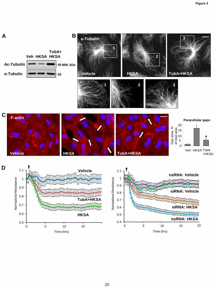
HKSA Vehicle TubA+HKSA
F-actin
A
D
Figure 3
B
C
50
50
MW, kDa
TubA+ Veh HKSA HKSA
Ac-Tubulin
α-Tubulin
Nor
mal
ized
Res
ista
nce
Time (hrs)
0.5
0.6
0.7
0.8
0.9
1
1.1
0 5 10 15
Vehicle TubA+HKSA
1
2
3
1 2 3
HKSA
α-Tubulin
0.4
0.5
0.6
0.7
0.8
0.9
1
1.1
0 5 10 15
Nor
mal
ized
Res
ista
nce
Time (hrs)
Vehicle
HKSA
TubA+HKSA
nsRNA: Vehicle
nsRNA: HKSA
siRNA: Vehicle
siRNA: HKSA
20
Paracellular gaps
Veh 0 5
10
15 20
TubA +HKSA
Gap
are
a, %
of
tota
l are
a
HKSA
*
20
by guest on February 25, 2020http://w
ww
.jbc.org/D
ownloaded from

Figure 4
A
B C
Vehicle HKSA TubA+HKSA
VE-cadherin
HKSA TubA+ HKSA
Veh
*
0
25
50
100 75
Sur
face
VE
C,
% o
f con
trol
0
25
50
100 75
VE
-cad
herin
, %
of c
ontro
l
HKSA TubA+ HKSA
Veh
*
p120
-cat
enin
, %
of c
ontro
l HKSA TubA+
HKSA Veh
*
0
25
50
100 75
100
100
MW, kDa
TubA+ Veh HKSA HKSA
Biotin- bound Input
VE-cadherin 100
MW, kDa 100
p120-ctn
VE-cad
IP: p120-catenin
TubA+ Veh HKSA HKSA
100
MW, kDa p120-ctn
VE-cad
IP: VE-cadherin
TubA+ Veh HKSA HKSA
100
21
by guest on February 25, 2020http://w
ww
.jbc.org/D
ownloaded from
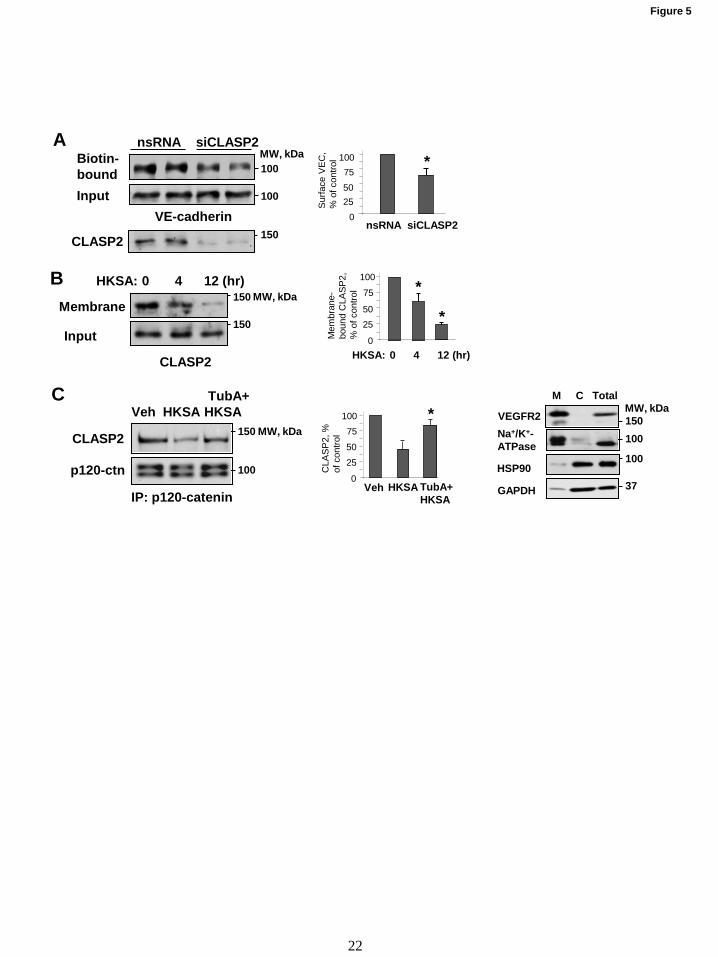
Figure 5
A
B
150
100
100
MW, kDa
CLASP2
nsRNA siCLASP2 Biotin- bound Input
VE-cadherin
C
0 4 12 (hr)
*
0
25 50
100
75
HKSA:
* M
embr
ane-
bo
und
CLA
SP
2,
% o
f con
trol
HKSA TubA+ HKSA
Veh
*
0
25
50
100 75
CLA
SP
2, %
of
con
trol
0 25 50
100 75
*
siCLASP2 nsRNA
Sur
face
VE
C,
% o
f con
trol
Membrane
Input 150
150 HKSA: 0 4 12 (hr)
CLASP2
MW, kDa
100
150
p120-ctn
CLASP2
IP: p120-catenin
TubA+ Veh HKSA HKSA
MW, kDa
37
100
100
150 MW, kDa
HSP90
M C Total
Na+/K+- ATPase
VEGFR2
GAPDH
22
by guest on February 25, 2020http://w
ww
.jbc.org/D
ownloaded from
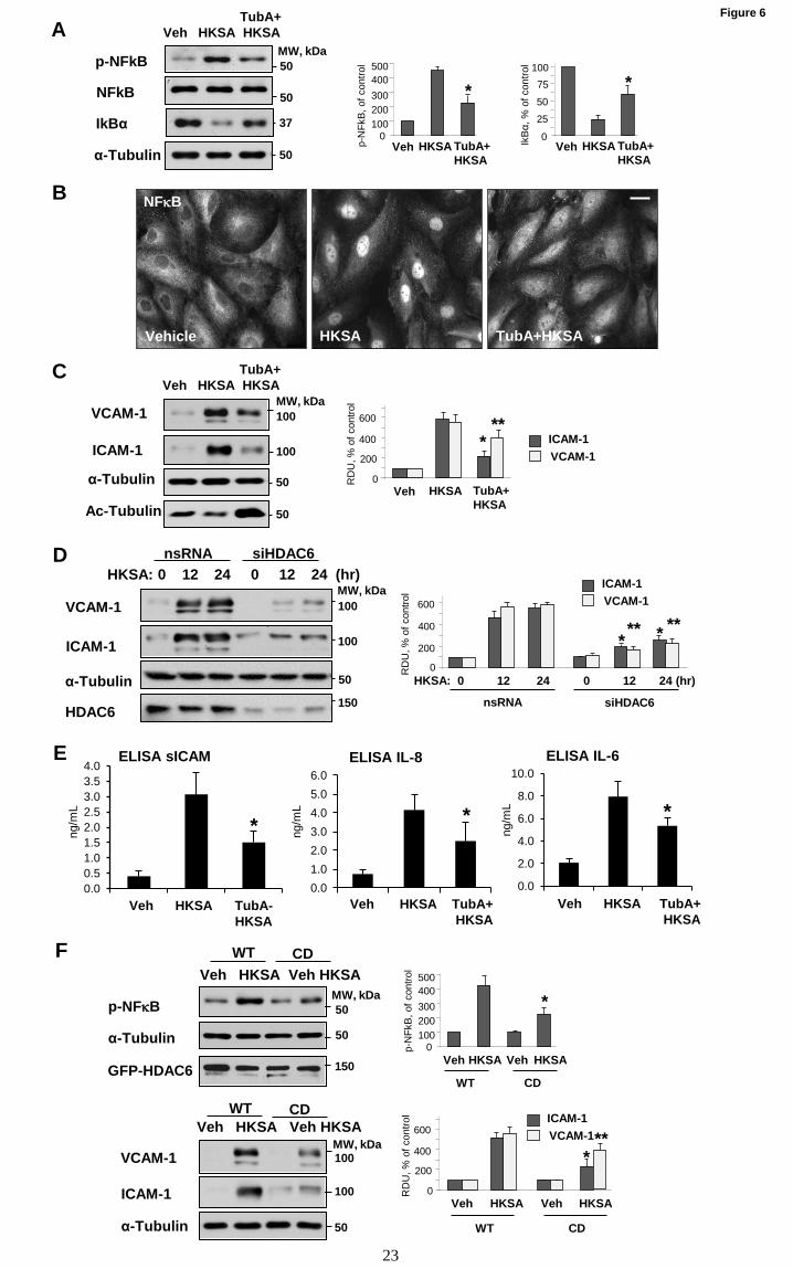
A Figure 6
E
B
0.0 0.5 1.0 1.5 2.0 2.5 3.0 3.5 4.0
ELISA sICAM
Veh HKSA TubA- HKSA
* ng/m
L
0.0 1.0 2.0 3.0 4.0 5.0 6.0
ELISA IL-8
*
Veh HKSA TubA+ HKSA
ng/m
L
0.0
2.0
4.0
6.0
8.0
10.0 ELISA IL-6
*
Veh HKSA TubA+ HKSA
ng/m
L
NFκB
Vehicle HKSA TubA+HKSA
C
D
100
100
50
50
α-Tubulin
ICAM-1
VCAM-1
Ac-Tubulin
TubA+ Veh HKSA HKSA
MW, kDa
100
100
MW, kDa
50
150
HKSA: 0 12 24 0 12 24 (hr)
VCAM-1
ICAM-1
α-Tubulin
HDAC6
siHDAC6 nsRNA
50
50
MW, kDa
37
50 α-Tubulin
p-NFkB
IkBα
NFkB
TubA+ Veh HKSA HKSA
IkBα,
% o
f con
trol
HKSA TubA+ HKSA
Veh
*
0
25
50
100 75 *
p-N
FkB
, of c
ontro
l
0 100 200 300 400 500
HKSA TubA+ HKSA
Veh
RD
U, %
of c
ontro
l
0
200
600
400 **
*
HKSA TubA+ HKSA
Veh
VCAM-1 ICAM-1
0
200
400
600
RD
U, %
of c
ontro
l
0 12 24 0 12 24 (hr) HKSA:
nsRNA siHDAC6
* * ** **
VCAM-1 ICAM-1
23
F
100
100
MW, kDa
50
VCAM-1
ICAM-1
α-Tubulin
Veh HKSA Veh HKSA CD WT
50
50
150 GFP-HDAC6
p-NFκB
α-Tubulin
Veh HKSA Veh HKSA CD WT
MW, kDa *
p-N
FkB
, of c
ontro
l
0 100 200 300 400 500
Veh HKSA Veh HKSA
WT CD
VCAM-1 ICAM-1
Veh HKSA Veh HKSA
RD
U, %
of c
ontro
l
WT CD
0
200
600
400 **
*
by guest on February 25, 2020http://w
ww
.jbc.org/D
ownloaded from
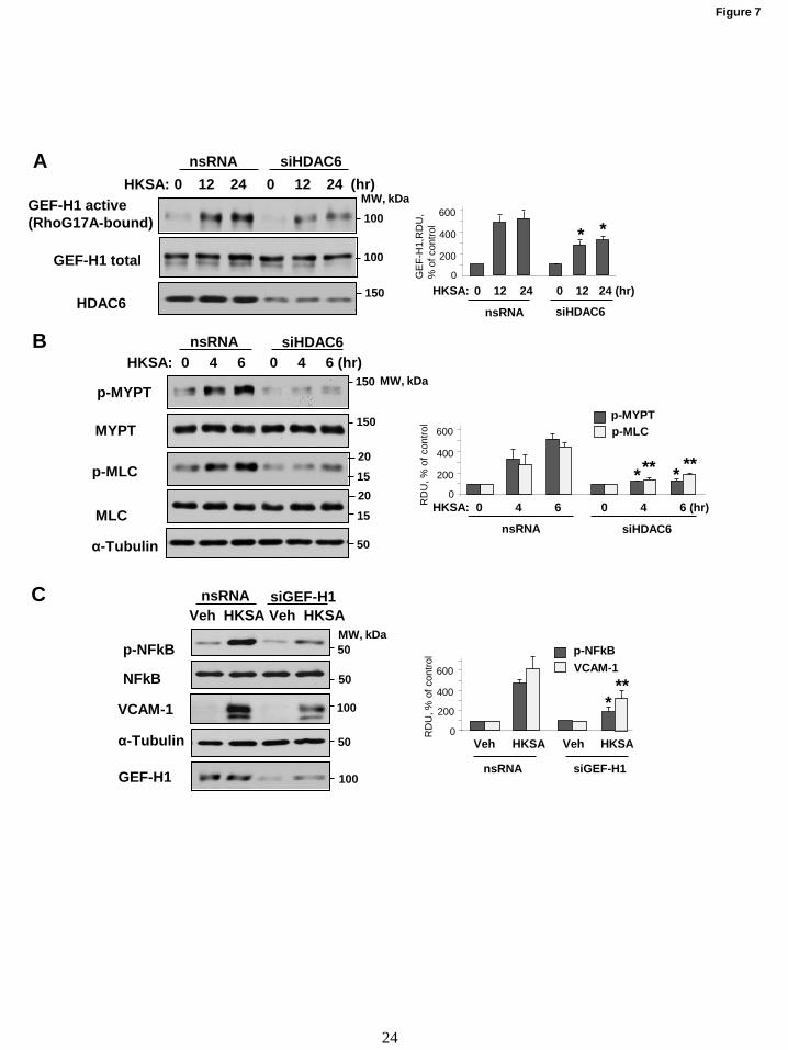
A
B
C
50
15
15
20
20
150
150
MW, kDa p-MYPT
HKSA: 0 4 6 0 4 6 (hr) siHDAC6 nsRNA
p-MLC
α-Tubulin
MYPT
MLC
100
100
150
GEF-H1 active (RhoG17A-bound)
GEF-H1 total
HKSA: 0 12 24 0 12 24 (hr) siHDAC6 nsRNA
HDAC6
MW, kDa
100
50
50
MW, kDa
50
Veh HKSA Veh HKSA
VCAM-1
α-Tubulin
GEF-H1
nsRNA siGEF-H1
p-NFkB
NFkB
100
0
200
400
600
0 12 24 0 12 24 (hr) HKSA:
nsRNA siHDAC6
* *
GE
F-H
1,R
DU
, %
of c
ontro
l
Veh HKSA Veh HKSA
RD
U, %
of c
ontro
l
VCAM-1 p-NFkB
nsRNA siGEF-H1
0
200
600
400 ** *
0
200
400
600 R
DU
, % o
f con
trol
0 4 6 0 4 6 (hr) HKSA:
nsRNA siHDAC6
* * ** **
p-MYPT p-MLC
Figure 7
24
by guest on February 25, 2020http://w
ww
.jbc.org/D
ownloaded from
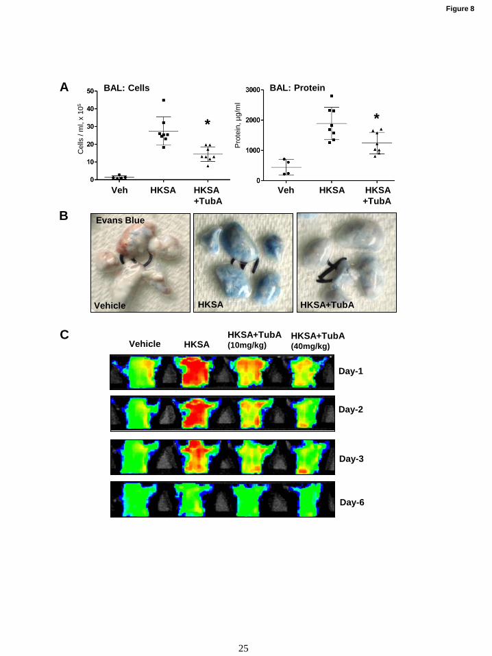
A
Figure 8
B
C
Cel
ls /
ml,
x 10
5
Veh HKSA HKSA +TubA
Pro
tein
, µg/
ml
Day-1
Day-2
Day-3
Day-6
Vehicle HKSA HKSA+TubA (40mg/kg)
HKSA+TubA (10mg/kg)
Vehicle HKSA HKSA+TubA
Evans Blue
BAL: Cells BAL: Protein
* *
Veh HKSA HKSA +TubA
25
by guest on February 25, 2020http://w
ww
.jbc.org/D
ownloaded from
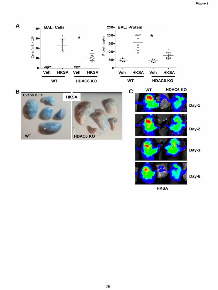
Cel
ls /
ml,
x 10
5
Pro
tein
, µg/
ml
BAL: Cells BAL: Protein A
C
Figure 9
B
Veh HKSA Veh HKSA
WT HDAC6 KO
Veh HKSA Veh HKSA
WT HDAC6 KO
HKSA Evans Blue
Day-1
Day-2
Day-3
Day-6
WT HDAC6 KO
HKSA
* *
WT HDAC6 KO
26
by guest on February 25, 2020http://w
ww
.jbc.org/D
ownloaded from
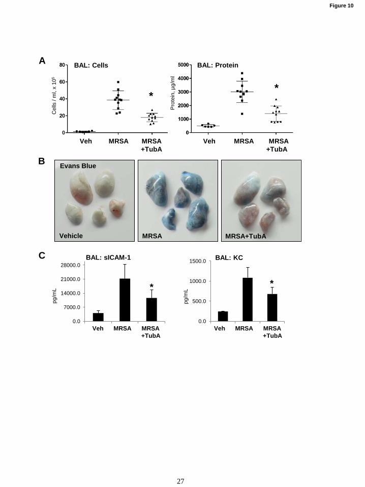
A
Figure 10
Vehicle MRSA MRSA+TubA
B Evans Blue
C
0.0
7000.0
14000.0
21000.0
28000.0 BAL: sICAM-1
Veh MRSA MRSA +TubA
*
pg/m
L
0.0
500.0
1000.0
1500.0 BAL: KC
Veh MRSA MRSA +TubA
*
pg/m
L
Cel
ls /
ml,
x 10
5
Veh MRSA MRSA +TubA
Pro
tein
, µg/
ml
BAL: Cells BAL: Protein
* *
Veh MRSA MRSA +TubA
27
by guest on February 25, 2020http://w
ww
.jbc.org/D
ownloaded from
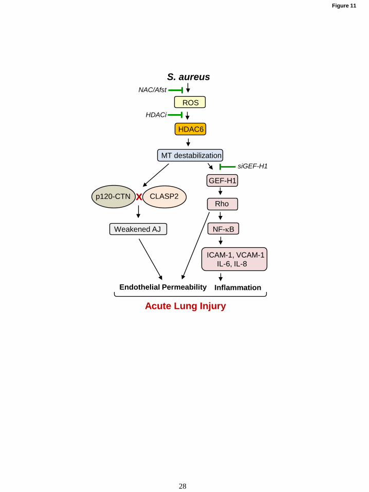
Figure 11
HDAC6
S. aureus
ROS
MT destabilization
p120-CTN CLASP2 x
Weakened AJ
Endothelial Permeability
GEF-H1
Rho
NF-κB
ICAM-1, VCAM-1 IL-6, IL-8
Inflammation
Acute Lung Injury
NAC/Afst
HDACi
siGEF-H1
28
by guest on February 25, 2020http://w
ww
.jbc.org/D
ownloaded from
Christopher P. Montgomery and Anna A. BirukovaPratap Karki, Yunbo Ke, Yufeng Tian, Tomomi Ohmura, Albert Sitikov, Nicolene Sarich,
mediated by microtubule destabilizationinduced endothelial permeability and inflammation are−Staphylococcus aureus
published online January 8, 2019J. Biol. Chem.
10.1074/jbc.RA118.004030Access the most updated version of this article at doi:
Alerts:
When a correction for this article is posted•
When this article is cited•
to choose from all of JBC's e-mail alertsClick here
by guest on February 25, 2020http://w
ww
.jbc.org/D
ownloaded from
Related Documents