MOLECULAR AND CELLULAR BIOLOGY, Mar. 1994, p. 2201-2212 Vol. 14, No. 3 0270-7306/94/$04.00+0 Copyright © 1994, American Society for Microbiology Human GATA-3 trans-Activation, DNA-Binding, and Nuclear Localization Activities Are Organized into Distinct Structural Domains ZHUOYING YANG,' LIN GU,' PAUL-HENRI ROMEO,2 DOMINIQUE BORIES,2 HOZUMI MOTOHASHI,3 MASAYUKI YAMAMOTO,3 AND JAMES DOUGLAS ENGELI* Department of Biochemistry, Molecular Biology and Cell Biology, Northwestern University, Evanston, Illinois 60208-3500'; Department of Molecular Genetics and Hematology, INSERM U91, Hdpital Henri Mondor, 94010 Creteil, France2; and Department of Biochemistry, Tohoku University School of Medicine, Sendai 980, Japan3 Received 15 October 1993/Returned for modification 19 November 1993/Accepted 3 December 1993 GATA-3 is a zinc finger transcription factor which is expressed in a highly restricted and strongly conserved tissue distribution pattern in vertebrate organisms, specifically, in a subset of hematopoietic cells, in cells within the central and peripheral nervous systems, in the kidney, and in placental trophoblasts. Tissue-specific cellular genes regulated by GATA-3 have been identified in T lymphocytes and the placenta, while GATA-3-regulated genes in the nervous system and kidney have not yet been defined. We prepared monoclonal antibodies with which we could dissect the biochemical and functional properties of human GATA-3. The results of these experiments show some anticipated phenotypes, for example, the definition of discrete domains required for specific DNA-binding site recognition (amino acids 303 to 348) and trans activation (amino acids 30 to 74). The signaling sequence for nuclear localization of human GATA-3 is a property conferred by sequences within and surrounding the amino finger (amino acids 249 to 311) of the protein, thereby assigning a function to this domain and thus explaining the curious observation that this zinc finger is dispensable for DNA binding by the GATA family of transcription factors. Human GATA-3 (hGATA-3) is a 444-amino-acid vertebrate transcription factor protein which binds with high affinity to related consensus DNA sites (GATA, GATTA, and GATCT) through a two-C4-zinc-finger DNA-binding domain (23, 24, 31, 52, 54). GATA-1, the founding member of this multigene family, was originally detected in and cloned from erythroid lineage cells (1, 7, 8, 30, 36, 37, 49). Cloning of the related factors, GATA-2 and GATA-3, was reported shortly thereafter (52), and newer family members have since been identified (2, 22). The basis for classification of these proteins arises from the highly conserved tissue-restricted distribution patterns of indi- vidual GATA family members in different vertebrate organ- isms. The amino acid sequence of the DNA-binding domain is over 85% identical among different GATA family members and is also highly conserved among vertebrate species (35), while regions outside the DNA-binding domains vary from extremely high similarity between species (e.g., 92% for GATA-3 [24]) to low identity (e.g., about 40% for chicken GATA-1 [cGATA-1], mouse GATA-1 [mGATA-1], and hGATA-1 [7, 47, 49, 57]). GATA-3 was first shown to be abundantly expressed in T lymphocytes and in the embryonic brain (16, 20, 24, 52). A number of target genes for GATA-3-directed transcriptional activity have been identified in T lymphocytes, which only express GATA-3 among the members of this family (24, 26, 52). Both the human and murine T-cell receptor (TCR) 8 and a gene enhancers were shown to bind GATA-3, and muta- tional analysis showed that the GATA-binding sites were required for tissue-specific TCR 8 enhancer activity (16, 20, 24, * Corresponding author. Mailing address: Department of Biochem- istry, Molecular Biology and Cell Biology, Northwestern University, Evanston, IL 60208-3500. Phone: (708) 491-5139. Fax: (708) 467-2152. Electronic mail address: [email protected]. 38, 39). hGATA-3 also binds to multiple sites within the human immunodeficiency virus type 1 long terminal repeat and is required for maximal stimulation of human immunodefi- ciency virus long terminal repeat-directed transcription in T cells (54). GATA-3 is abundantly expressed during T-lympho- cyte differentiation both in mature CD4+ CD8- and CD4- CD8+ and in less mature CD4+ CD8+ T cells (26). Given that GATA-binding sites have been identified in the regulatory regions of a growing number of T-cell-specific genes, we infer that GATA-3 may be one of the transcription factors respon- sible for T-cell identity and/or determination. More recently, GATA-2 and GATA-3 have also been shown to be abundantly expressed in placental trophoblasts, in which the human gonadotropin a-subunit promoter (30a) and the mouse placental lactogen I gene promoter (27a) are regulated by a GATA factor. GATA-3 is expressed at the earliest stages of Xenopus, chicken, and murine embryonic development (24, 26, 56) and in the developing vertebrate central nervous system (25), peripheral nervous system (llb), and kidney (25a). Homozygous mutant mice lacking mGATA-3 fail to survive gestation, consistent with the expectation that GATA-3 fulfills a vital regulatory function during vertebrate embryogenesis (27). Structural and functional studies of GATA-1 have shown that the amino-terminal segment of the polypeptide contrib- utes to the transcriptional activation function of the protein, while two related zinc fingers, lying somewhat closer to the carboxyl terminus of the protein, determine the DNA-binding affinity and specificity (29, 53). The two fingers of GATA-1 can be functionally subdivided; the amino finger (which we refer to here as finger I) contributes to the specificity and stability of DNA binding, while the carboxyl finger (finger II) is absolutely required for recognition of the GATA consensus motif (29, 34, 53). Since the amino acid sequence of the entire family of factors is conserved throughout the finger domain, we would 2201

Welcome message from author
This document is posted to help you gain knowledge. Please leave a comment to let me know what you think about it! Share it to your friends and learn new things together.
Transcript
MOLECULAR AND CELLULAR BIOLOGY, Mar. 1994, p. 2201-2212 Vol. 14, No. 30270-7306/94/$04.00+0Copyright © 1994, American Society for Microbiology
Human GATA-3 trans-Activation, DNA-Binding, and NuclearLocalization Activities Are Organized into Distinct
Structural DomainsZHUOYING YANG,' LIN GU,' PAUL-HENRI ROMEO,2 DOMINIQUE BORIES,2 HOZUMI MOTOHASHI,3
MASAYUKI YAMAMOTO,3 AND JAMES DOUGLAS ENGELI*Department of Biochemistry, Molecular Biology and Cell Biology, Northwestern University, Evanston, Illinois 60208-3500';
Department of Molecular Genetics and Hematology, INSERM U91, Hdpital Henri Mondor, 94010 Creteil, France2;and Department of Biochemistry, Tohoku University School of Medicine, Sendai 980, Japan3
Received 15 October 1993/Returned for modification 19 November 1993/Accepted 3 December 1993
GATA-3 is a zinc finger transcription factor which is expressed in a highly restricted and strongly conservedtissue distribution pattern in vertebrate organisms, specifically, in a subset of hematopoietic cells, in cellswithin the central and peripheral nervous systems, in the kidney, and in placental trophoblasts. Tissue-specificcellular genes regulated by GATA-3 have been identified in T lymphocytes and the placenta, whileGATA-3-regulated genes in the nervous system and kidney have not yet been defined. We prepared monoclonalantibodies with which we could dissect the biochemical and functional properties of human GATA-3. Theresults of these experiments show some anticipated phenotypes, for example, the definition of discrete domainsrequired for specific DNA-binding site recognition (amino acids 303 to 348) and trans activation (amino acids30 to 74). The signaling sequence for nuclear localization of human GATA-3 is a property conferred bysequences within and surrounding the amino finger (amino acids 249 to 311) of the protein, thereby assigninga function to this domain and thus explaining the curious observation that this zinc finger is dispensable forDNA binding by the GATA family of transcription factors.
Human GATA-3 (hGATA-3) is a 444-amino-acid vertebratetranscription factor protein which binds with high affinity torelated consensus DNA sites (GATA, GATTA, and GATCT)through a two-C4-zinc-finger DNA-binding domain (23, 24, 31,52, 54). GATA-1, the founding member of this multigenefamily, was originally detected in and cloned from erythroidlineage cells (1, 7, 8, 30, 36, 37, 49). Cloning of the relatedfactors, GATA-2 and GATA-3, was reported shortly thereafter(52), and newer family members have since been identified (2,22).The basis for classification of these proteins arises from the
highly conserved tissue-restricted distribution patterns of indi-vidual GATA family members in different vertebrate organ-isms. The amino acid sequence of the DNA-binding domain isover 85% identical among different GATA family membersand is also highly conserved among vertebrate species (35),while regions outside the DNA-binding domains vary fromextremely high similarity between species (e.g., 92% forGATA-3 [24]) to low identity (e.g., about 40% for chickenGATA-1 [cGATA-1], mouse GATA-1 [mGATA-1], andhGATA-1 [7, 47, 49, 57]).GATA-3 was first shown to be abundantly expressed in T
lymphocytes and in the embryonic brain (16, 20, 24, 52). Anumber of target genes for GATA-3-directed transcriptionalactivity have been identified in T lymphocytes, which onlyexpress GATA-3 among the members of this family (24, 26,52). Both the human and murine T-cell receptor (TCR) 8 anda gene enhancers were shown to bind GATA-3, and muta-tional analysis showed that the GATA-binding sites wererequired for tissue-specific TCR 8 enhancer activity (16, 20, 24,
* Corresponding author. Mailing address: Department of Biochem-istry, Molecular Biology and Cell Biology, Northwestern University,Evanston, IL 60208-3500. Phone: (708) 491-5139. Fax: (708) 467-2152.Electronic mail address: [email protected].
38, 39). hGATA-3 also binds to multiple sites within thehuman immunodeficiency virus type 1 long terminal repeat andis required for maximal stimulation of human immunodefi-ciency virus long terminal repeat-directed transcription in Tcells (54). GATA-3 is abundantly expressed during T-lympho-cyte differentiation both in mature CD4+ CD8- and CD4-CD8+ and in less mature CD4+ CD8+ T cells (26). Given thatGATA-binding sites have been identified in the regulatoryregions of a growing number of T-cell-specific genes, we inferthat GATA-3 may be one of the transcription factors respon-sible for T-cell identity and/or determination.More recently, GATA-2 and GATA-3 have also been shown
to be abundantly expressed in placental trophoblasts, in whichthe human gonadotropin a-subunit promoter (30a) and themouse placental lactogen I gene promoter (27a) are regulatedby a GATA factor. GATA-3 is expressed at the earliest stagesof Xenopus, chicken, and murine embryonic development (24,26, 56) and in the developing vertebrate central nervous system(25), peripheral nervous system (llb), and kidney (25a).Homozygous mutant mice lacking mGATA-3 fail to survivegestation, consistent with the expectation that GATA-3 fulfillsa vital regulatory function during vertebrate embryogenesis(27).
Structural and functional studies of GATA-1 have shownthat the amino-terminal segment of the polypeptide contrib-utes to the transcriptional activation function of the protein,while two related zinc fingers, lying somewhat closer to thecarboxyl terminus of the protein, determine the DNA-bindingaffinity and specificity (29, 53). The two fingers of GATA-1 canbe functionally subdivided; the amino finger (which we refer tohere as finger I) contributes to the specificity and stability ofDNA binding, while the carboxyl finger (finger II) is absolutelyrequired for recognition of the GATA consensus motif (29, 34,53). Since the amino acid sequence of the entire family offactors is conserved throughout the finger domain, we would
2201
2202 YANG ET AL.
anticipate similar results for hGATA-3 DNA-binding specific-ity and affinity.To elicit their functions, transcription factors must encode
nuclear targeting signals to allow the protein to traverse thenuclear membrane. The essence of the simplest nuclear local-ization sequence appears to be a short string of basic aminoacids (4), and the control of nuclear localization has beenshown to play a critical role in the activity of several transcrip-tion factors. The GATA proteins have not been analyzed withregard to whether or not nuclear localization plays a functionalrole in the transcriptional regulation elicited by these proteins.
Since previous studies have usually assayed the biochemicalactivity of different members of the GATA factor family byindirect DNA binding assays (electrophoretic gel mobility shiftassays [EGMSA]), we wished to examine the biological prop-erties of GATA-3 by using a direct assay for the activitiesand/or cellular distribution of the protein. To address therelationship between the primary sequence of hGATA-3 andits functional properties, we first prepared monoclonal anti-bodies (MAbs) recognizing the bacterially expressed protein.We then undertook a systematic mutational analysis ofhGATA-3, examining in-frame deletions within the hGATA-3coding region by expressing each of the mutated proteins ineucaryotic cells. The effects of the mutations were assessedwith the anti-hGATA-3 MAbs by separately examining theability of various mutated hGATA-3 proteins to bind to DNA,to activate transcription, and to direct localization of the factorto the nucleus. We find that the trans-activating domain islocalized within the amino-terminal portion of the proteinwhile the DNA-binding domain resides within the carboxylfinger. Using immunolocalization assays, we show that thehGATA-3 protein is found within the nuclei, both in cells inwhich it is normally expressed and in transfected cells. Finally,we showed that amino acid (aa) residues 249 to 311 arerequired for nuclear localization of the hGATA-3 protein.
MATERIALS AND METHODS
Cell lines. Human T-lymphocyte cell lines Jurkat and HuT78and murine T-lymphocyte cell line BW5147.3 were purchasedfrom the American Type Culture Collection. YN79 (a sublineof the Y79 human retinoblastoma line), NB-1 (neuroblasto-ma), and HEL (erythroleukemia) lines were generously pro-vided by S. Tsuchiya. MEL (murine erythroleukemia) andC1300 (neuroblastoma) cell lines were provided by VikramPatel and Bernard Mirkin, respectively. Transformed quailfibroblast (QT6) cells, which do not express GATA factors,were used as previously described (24, 52).
Generation of MAbs recognizing GATA-3. The first 785nucleotides encoding the amino-terminal portion of thehGATA-3 cDNA, ending at aa 264 (the first cysteine encoun-tered in the amino finger [20, 24]), were amplified by PCRusing oligonucleotide primers containing synthetic restrictionsites for convenient BamHI-EcoRI insertion into the bacterialexpression plasmid pGEX-A (a gift of Jurgen Kun, Institut furGenetik der Universitat Koln). The PCR product was verifiedby DNA sequencing. Cells containing the bacterial expressionplasmid were grown to mid-log phase, and the glutathioneS-transferase (GST)/hGATA-3 fusion peptide was induced bythe addition of isopropylthiogalactopyranoside (IPTG) to 0.5mM for 3 h at 37°C. Cells were then lysed, and the fusionprotein was bound to and recovered from a glutathione columnas previously described (46).Approximately 100 pLg of GST/hGATA-3 was injected intra-
peritoneally into each mouse, using Hunter's Titermax adju-vant (CytRx Corp., Norcross, Ga.). After three consecutive
injections with 100,ug of fusion protein every 3 weeks, animalswere assayed by enzyme-linked immunosorbent assay (ELISA)for the ability of the serum to recognize GST/hGATA-3. Fourweeks after the final injection, spleen cells were fused toNSO-1 myeloma cells as described previously (10). Positivehybridoma wells were initially identified by ELISA using thefusion protein as the fixed antigen and subsequently by bio-chemical assays using native hGATA-3 protein (see below).hGATA-3 mutations. All deletions were prepared either by
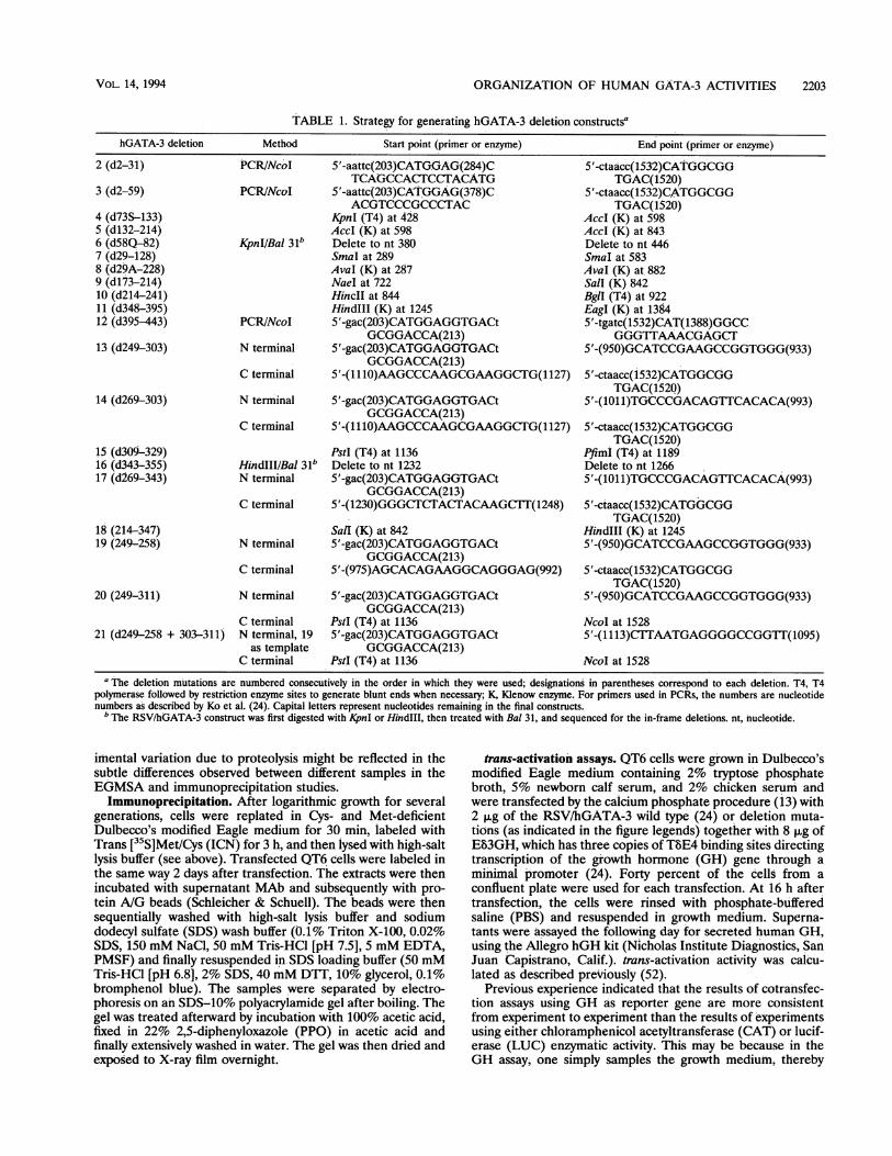
using convenient restriction enzyme sites to excise specificfragments or by PCR, as shown in Table 1, starting with thebase plasmid Rous sarcoma virus (RSV)/hGATA-3 describedpreviously (24). In either case, mutations were chosen to resultin in-frame deletions, and the integrity of each mutation wasconfirmed by DNA sequencing. All of the deletion mutationsexamined are described in Table 1 and are numbered consec-utively in the order in which they are described here.To prepare the GAL4/hGATA-3 fusion proteins, either
PCR products or restriction fragments of the hGATA-3 codingregion were subcloned in frame into the multiple cloning sites3' to the GAL4 DNA-binding and dimerization domain (aa 1to 147) of plasmid pSG424 (28). These mutations were alsoconfirmed to be in frame by DNA sequencing. The hGATA-3mutations examined as GAL4 fusion chimeras are described inTable 2.EGMSA and supershift assays. For EGMSA (9, 11) of
overexpressed hGATA-3 protein, whole-cell extracts wereprepared from hGATA-3-transfected QT6 cells disrupted withhigh-salt lysis buffer (500 mM NaCl, 1% Triton X-100, 50 mMTris-HCl [pH 7.5], phenylmethylsulfonyl fluoride [PMSF]) andpartially purified by double-stranded calf thymus DNA-cellu-lose chromatography (10). The extracts were then incubatedwith radiolabeled oligonucleotide TbE4, which corresponds tothe GATA sequences within the human TCR 8 gene enhancerfootprint 4 (24, 38). Gel shift assays were performed asdescribed previously in a final buffer consisting of 80 mMNaCl, 25 mM N-2-hydroxyethylpiperazine-N'-2-ethanesulfonicacid (HEPES; pH 7.7), 10 mM dithiothreitol [DTT], 5 mMMgCl2, 100 mg of bovine serum albumin per ml, and 2.5%Ficoll (52). For the supershift assay, 2 ,ul of supernatant fromeach ELISA-positive hybridoma was added to the gel shiftreaction 20 min after initiation of the GATA-3/T8E4 DNAbinding reaction at 4°C, and then the assay was performed asdescribed above.The mouse thymus cell, Jurkat cell, and NB-1 cell nuclear
extracts were prepared first by addition of lysis buffer (20 mMHEPES [pH 7.7], 20% glycerol, 10 mM NaCl, 1.5 mM MgCl2,0.2 mM EDTA, 0.1% Triton X-100, 1 mM DTT, 1 mM PMSF,leupeptin, and pepstatin). The nuclear pellets were thentreated with nuclear extraction buffer (lysis buffer plus 400 mMNaCl). The nuclear extracts were then dialyzed against 20 mMHEPES (pH 7.9)-20% glycerol-100 mM KC1-0.2 mMEDTA-1 mM DTT-1 mM PMSF. The probe used was aGATA site found within the vasoactive intestinal peptide(VIP) gene promoter (5'-GATCCAAAATGTAAGATAAGAGGAAATTTT-3'); the mutated VIP GATA site substituted thesequence 5'-ACCTAA-3' within the GATA consensus nucle-otides shown in boldface in the wild-type sequence. ForEGMSA using the GAL4/hGATA-3 fusion protein, a probecontaining five copies of the GAL4-binding site excised frompSG5CAT (28) by HindIll and KpnI was radiolabeled by usingpolynucleotide kinase.The protein extracts prepared here are somewhat unstable
over time even when maintained at - 70°C, and therefore thegel shift and immunoprecipitation experiments were usuallycarried out immediately after lysis. Nonetheless, slight exper-
MOL. CELL. BIOL.
ORGANIZATION OF HUMAN GATA-3 ACTIVITIES 2203
TABLE 1. Strategy for generating hGATA-3 deletion constructsa
hGATA-3 deletion Method Start point (primer or enzyme) End point (primer or enzyme)
2 (d2-31) PCRINcoI 5'-aattc(203)CATG.GAG(284)C 5'-ctaacc(1532)CATGGCGGTCAGCCACTCCTACATG TGAC(1520)
3 (d2-59) PCR/NcoI 5'-aattc(203)CAT,GGAG(378)C 5'-ctaacc(1532)CATGGCGGACGTCCCGCCCTAC TGAC(1520)
4 (d73S-133) KpnI (T4) at 428 AccI (K) at 5985 (d132-214) AccI (K) at 598 AccI (K) at 8436 (d58Q-82) KpnIIBal 31b Delete to nt 380 Delete to nt 4467 (d29-128) SmaI at 289 SmaI at 5838 (d29A-228) AvaI (K) at 287 AvaI (K) at. 8829 (d173-214) NaeI at 722 Sall (K) 84210 (d214-241) HincII at 844 BgI (T4) at 92211 (d348-395) HindIlI (K) at 1245 EagI (K) at 138412 (d395-443) PCR/NcoI 5'-gac(203)CATGGAGGTGACt 5'-tgatc(1532)CAT(1388)GGCC
GCGGACCA(213) GGGTTAAACGAGCT13 (d249-303) N terminal 5'-gac(203)CATGGAGGTGACt 5'-(950)GCATCCGAAGCCGGTGGG(933)
GCGGACCA(213)C terminal 5'-(1110)AAGCCCAAGCGAAGGCTG(1127) 5'-ctaacc(1532)CATGGCGG
TGAC(1520)14 (d269-303) N terminal 5'-gac(203)CATGGAGGTGACt 5'-(101l)TGCCCGACAGTTCACACA(993)
GCGGACCA(213)C terminal 5'-(1110)AAGCCCAAGCGAAGGCTG(1127) 5'-ctaacc(1532)CATGGCGG
TGAC(1520)15 (d309-329) PstI (T4) at 1136 PfimI (T4) at 118916 (d343-355) HindIII/Bal 31b Delete to nt 1232 Delete to nt 126617 (d269-343) N terminal 5'-gac(203)CATGGAGGTGACt 5'-(1011)TGCCCGACAGTTCACACA(993)
GCGGACCA(213)C terminal 5'-(1230)GGGCTCTAC(TACAAGCT1(1248) 5'-ctaacc(1532)CATGGCGG
TGAC(1520)18 (214-347) SalI (K) at 842 HindIII (K) at 124519 (249-258) N terminal 5'-gac(203)CATGGAGGTGACt 5'-(950)GCATCCGAAGCCGGTGGG(933)
GCGGACCA(213)C terminal 5'-(975)AGCACAGAAGGCAGGGAG(992) 5'-ctaacc(1532)CATGGCGG
TGAC(1520)20 (249-311) N terminal 5'-gac(203)CATGGAGGTGACt 5'-(950)GCATCCGAAGCCGGTGGG(933)
GCGGACCA(213)C terminal PstI (T4) at 1136 NcoI at 1528
21 (d249-258 + 303-311) N terminal, 19 5'-gac(203)CATGGAGGTGACt 5'-(1113)CTTAATGAGGGGCCGGIT(1095)as template GCGGACCA(213)
C terminal PstI (T4) at 1136 NcoI at 1528a The deletion mutations are numbered consecutively in the order in which they were used; designations in parentheses correspond to each deletion. T4, T4
polymerase followed by restriction enzyme sites to generate blunt ends when necessary; K, Klenow enzyme. For primers used in PCRs, the numbers are nucleotidenumbers as described by Ko et al. (24). Capital letters represent nucleotides remaining in the final constructs.
b The RSV/hGATA-3 construct was first digested with KpnI or HindIII, then treated with Bal 31, and sequenced for the in-frame deletions. nt, nucleotide.
imental variation due to proteolysis might be reflected in thesubtle differences observed between different samples in theEGMSA and immunoprecipitation studies.
Immunoprecipitation. After logarithmic growth for severalgenerations, cells were replated in Cys- and Met-deficientDulbecco's modified Eagle medium for 30 min, labeled withTrans [35S]Met/Cys (ICN) for 3 h, and then lysed with high-saltlysis buffer (see above). Transfected QT6 cells were labeled inthe same way 2 days after transfection. The extracts were thenincubated with supernatant MAb and subsequently with pro-tein A/G beads (Schleicher & Schuell). The beads were thensequentially washed with high-salt lysis buffer and sodiumdodecyl sulfate (SDS) wash buffer (0.1% Triton X-100, 0.02%SDS, 150 mM NaCl, 50 mM Tris-HCl [pH 7.5], 5 mM EDTA,PMSF) and finally resuspended in SDS loading buffer (50 mMTris-HCl [pH 6.8], 2% SDS, 40 mM DTT, 10% glycerol, 0.1%bromphenol blue). The samples were separated by electro-phoresis on an SDS-10% polyacrylamide gel after boiling. Thegel was treated afterward by incubation with 100% acetic acid,fixed in 22% 2,5-diphenyloxazole (PPO) in acetic acid andfinally extensively washed in water. The gel was then dried andexposed to X-ray film overnight.
trans-activation assays. QT6 cells were grown in Dulbecco'smodified Eagle medium containing 2% tryptose phosphatebroth, 5% newborn calf serum, and 2% chicken serum andwere transfected by the calcium phosphate procedure (13) with2 ,ug of the RSV/hGATA-3 wild type (24) or deletion muta-tions (as indicated in the figure legends) together with 8 ,ug ofE83GH, which has three copies of TSE4 binding sites directingtranscription of the growth hormone (GH) gene through aminimal promoter (24). Forty percent of the cells from aconfluent plate were used for each transfection. At 16 h aftertransfection, the cells were rinsed with phosphate-bufferedsaline (PBS) and resuspended in growth medium. Superna-tants were assayed the following day for secreted human GH,using the Allegro hGH kit (Nicholas Institute Diagnostics, SanJuan Capistrano, Calif.). trans-activation activity was calcu-lated as described previously (52).
Previous experience indicated that the results of cotransfec-tion assays using GH as reporter gene are more consistentfrom experiment to experiment than the results of experimentsusing either chloramphenicol acetyltransferase (CAT) or lucif-erase (LUC) enzymatic activity. This may be because in theGH assay, one simply samples the growth mediumi, thereby
VOL. 14, 1994
2204 YANG ET AL.
TABLE 2. Strategy for generating GAL4/hGATA-3 fusion constructsa
Construct Cloning site Start point of hGATA-3 End point of hGATA-3in pSG424
2 SmaI NcoI (K) at 202 SmaI at 2893 SmaI NcoI (K) at 202 KpnI (T4) at 4284 SmaI NcoI (K) at 202 Sall (K) at 8425" SmaI 5'-gac(203)CATGGAGGTGACtG 5'(933)TGTGTGAACTGTGGGGCA
CGGACCA(213)6 SmaI NcoI (K) at 202 HindlIl (K) at 12487 SmaI NcoI (K) at 202 NcoI (K) at 15288` SmaI NcoI (K) at 202 NcoI (K) at 15289 SmaI SmaI at 289 KpnI (T4) at 42810 SmaI SmaI at 289 BamHI (K) at 47711 SmaI SmaI at 289 SmaI at 58312 BamHI (K) KpnI (T4) at 428 Sall (K) at 84213 SmaI AccI (K) at 598 AccI (K) at 843
hGATA-3 fragments were cloned into the restriction enzyme sites in pSG424 as indicated. Abbreviations are as for Table 1."The hGATA-3 fragment was generated by PCR.'The parental plasmid for the hGATA-3 portion is construct 18 of hGATA-3 from Fig. 4A.
avoiding cell lysis and thus minimizing potential complicationsfrom differential protein extraction or degradation. On theother hand, it is relatively difficult to internally control for GHtransfection efficiency, since one may encounter significantvariability in the activity of secreted (GH) and endogenous(CAT or LUC) proteins when analyzing different transfectionsamples. We therefore repeated each GH reporter transfectionexperiment on at least three occasions for the data reportedhere.
In other experiments, a CAT instead of GH reporter genewas used. At 48 h after transfection, cell lysates were preparedby freeze-thaw (40). A LUC assay (internal transfection effi-ciency control) was carried out first, and CAT assays wereperformed with aliquots of extract containing equal quantitiesof LUC activity. The CAT assay results were quantified bydetermining the amount of acetylated ['4C]chloramphenicolproduced in the enzymatic assay (12); the degree of conversionwas quantitated on a Molecular Dynamics Phosphorlmager.
Subcellular immunolocalization. For immunohistochemicalstaining, human HuT78, NB-1, YN79, or HEL cells weredisplayed on glass slides by using a Cytospin III (Shandon),fixed in 1% paraformaldehyde for 5 min, and then fixed in coldacetone for 5 min. After incubation in 2% sheep serum,specimens were reacted with MAb 31 or 35 overnight at 4°C.After being washed in PBS, the samples were incubated withhorseradish peroxidase-conjugated F(ab')2 fragment of anti-mouse immunoglobulin G (1:100 dilution; Amersham) over-night at 4°C. Diaminobenzidine was used as the chromogen.Nuclei were counterstained with methyl green for 1 h.For immunofluorescence staining, QT6 cells transfected
with hGATA-3 or hGATA-3 deletion constructs were treatedwith trypsin and replated onto coverslips 24 h prior to staining.The coverslips were fixed in 1% paraformaldehyde, permeabi-lized in - 20°C acetone, and then incubated with anti-hGATA-3 MAb or a control MAb (which binds to GST) for 1h. The coverslips were then washed with PBS before additionof secondary fluorescein isothiocyanate-labeled mouse immu-noglobulin G (Jackson Laboratory). Cell staining was visual-ized and photographed on a Bio-Rad MRC 600 confocalfluorescence microscope.
RESULTS
MAbs specifically recognize GATA-3. To independentlyassess the biochemical properties of hGATA-3, we generated
MAbs recognizing the transcription factor by injecting micewith bacterially expressed, affinity-purified GST/hGATA-3 fu-sion protein. This fusion protein contained the amino-terminalportion of the hGATA-3 protein, ending (at the carboxylterminus) at the first cysteine residue defining finger I of theprotein (24) (Materials and Methods).Hybridoma clones which initially tested positive in ELISA
were subsequently examined by supershift EGMSA, in whichpartially purified hGATA-3 protein recovered from trans-fected QT6 quail fibroblast cells (which express no endogenousGATA proteins [24, 52]) was mixed with radiolabeled oligo-nucleotide containing the TbE4 binding site (24) as well assupernatant antibody taken from each ELISA-positive hybri-doma culture (Materials and Methods). Four positive hybri-doma wells were identified, and two of these (clones 31 and 35)were subcloned to generate monoclonal lines.MAb 31 was tested in immunoprecipitation experiments
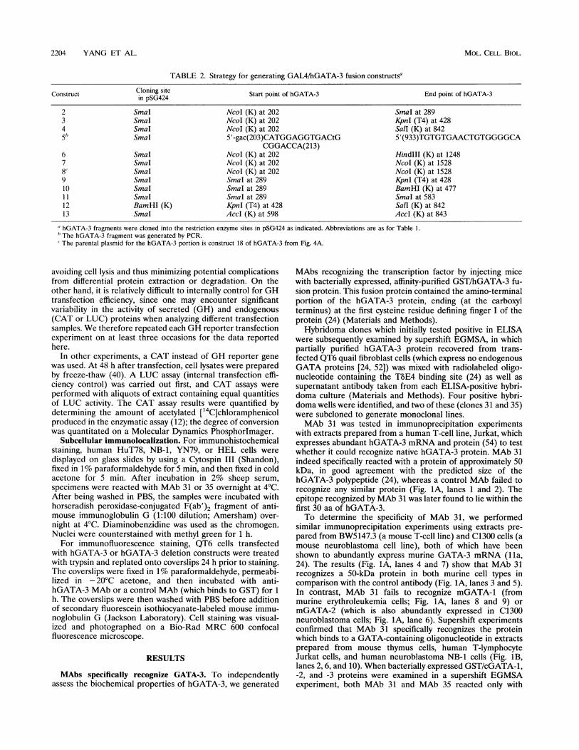
with extracts prepared from a human T-cell line, Jurkat, whichexpresses abundant hGATA-3 mRNA and protein (54) to testwhether it could recognize native hGATA-3 protein. MAb 31indeed specifically reacted with a protein of approximately 50kDa, in good agreement with the predicted size of thehGATA-3 polypeptide (24), whereas a control MAb failed torecognize any similar protein (Fig. 1A, lanes 1 and 2). Theepitope recognized by MAb 31 was later found to lie within thefirst 30 aa of hGATA-3.To determine the specificity of MAb 31, we performed
similar immunoprecipitation experiments using extracts pre-pared from BW5147.3 (a mouse T-cell line) and C1300 cells (amouse neuroblastoma cell line), both of which have beenshown to abundantly express murine GATA-3 mRNA (lla,24). The results (Fig. 1A, lanes 4 and 7) show that MAb 31recognizes a 50-kDa protein in both murine cell types incomparison with the control antibody (Fig. IA, lanes 3 and 5).In contrast, MAb 31 fails to recognize mGATA-1 (frommurine erythroleukemia cells; Fig. 1A, lanes 8 and 9) ormGATA-2 (which is also abundantly expressed in C1300neuroblastoma cells; Fig. IA, lane 6). Supershift experimentsconfirmed that MAb 31 specifically recognizes the proteinwhich binds to a GATA-containing oligonucleotide in extractsprepared from mouse thymus cells, human T-lymphocyteJurkat cells, and human neuroblastoma NB-1 cells (Fig. 1B,lanes 2,6, and 10). When bacterially expressed GST/cGATA-1,-2, and -3 proteins were examined in a supershift EGMSAexperiment, both MAb 31 and MAb 35 reacted only with
MOL. CELL. BIOL.
ORGANIZATION OF HUMAN GATA-3 ACTIVITIES 2205
1 2 3 4 5 6 7 8 9
B.1 2 3 4 5 6 7 8 9 10 11 12 13 14 15 16
C.
2.3 -_
I _-W
1 2 3 4 5 6 7 8 9 10 11 12
_3'
FIG. 1. Anti-hGATA-3 MAbs specifically recognize human, mouse, and chicken GATA-3 protein. (A) Immunoprecipitation of cell extracts.
A MAb recognizing GST (lanes 1, 3, and 5), murine GATA-2 (14a) (lane 6), or murine GATA-1 (19) (lane 8) or anti-hGATA-3 MAb 31 (lanes2, 4, 7, and 9) was used in immunoprecipitation reactions. The cells examined were from human (Jurkat; lanes 1 and 2) or mouse (BW5147.3, lanes3 and 4) T-lymphocyte cell lines, the C1300 mouse neuroblastoma cell line (lanes 5 to 7), or the MEL (mouse erythroleukemia; lanes 8 and 9) cellline. Arrows on the left depict the gel mobility positions of the protein size markers. (B) EGMSA supershift of T-cell and neuroblastoma cellextracts. Extracts were prepared from mouse thymus cells (lanes 1 to 4), human T-lymphocyte Jurkat cells (lanes 5 to 8), and human neuroblastomaNB-1 cells (lane 9 to 16) as described in Materials and Methods. The binding reactions were carried out with a radiolabeled GATA-binding sitefrom the VIP gene promoter (Materials and Methods) as a probe together with no antibody (lanes 1, 5, and 9), MAb 31 (lanes 2, 6, and 10),negative control mouse serum (lanes 3, 7, and 15), double-stranded, unlabeled GATA-binding site as a specific competitor (lanes 4, 8, and 11),or sense-strand (lane 12), antisense-strand (lane 13), or mutated (lane 14) VIP-1 GATA site as a cold competitor. Lane 16 is probe only. The lowerarrow points to the probe/mGATA-3 protein complex, and the upper arrow represents the ternary complex of the probe, mGATA-3 protein, andthe antibody. (C) EGMSA supershift of chicken GST/GATA-1, -2, and -3 proteins. Bacterially expressed, affinity-purified GST/cGATA-1 (lanes2 to 4), GST/cGATA-2 (lanes 6 to 8), or GST/cGATA-3 (lanes 10 to 12) fusion proteins (23) were used in conjunction with oligonucleotide MaP(49) in this supershift experiment. Supematants from MAb 31 (lanes 3, 7, and 11), MAb 35 (lanes 4, 8, and 12), or growth medium alone (lanes2, 6, and 10) were added to each of the EGMSA reactions. Lanes 1, 5, and 9 contain no added protein. The arrows and numbers on the left indicatethe positions of GST/cGATA-1, -2, or -3 and MaP probe complex in the EGMSA assay, and the arrow labeled 3* on the right represents theternaiy complex of DNA/GATA-3 protein which has reacted with the antibody.
cGATA-3 (Fig. 1B, lanes 11 and 12), not with cGATA-1 orcGATA-2 (Fig. 1B, lanes 2 to 4 and 6 to 8). Taken together, theEGMSA supershift and immunoprecipitation experimentsshow that both anti-hGATA-3 MAbs 31 and 35 recognizehuman, mouse, and chicken GATA-3 proteins but fail to bindto chicken or mouse GATA-1 or GATA-2. Given the highdegree of sequence identity between the murine and humanGATA-1 and GATA-2 proteins (47, 57), we anticipate thatthese antibodies will not recognize hGATA-1 or hGATA-2.
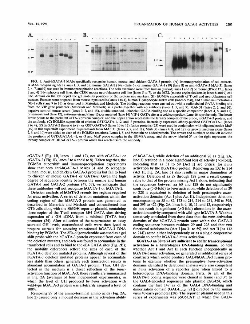
Deletion analysis of hGATA-3 reveals two regions requiredfor trans activation. A series of deletion mutations within thecoding region of the hGATA-3 protein was generated asdescribed in Materials and Methods and cotransfected intoQT6 cells along with the E83GH reporter plasmid, containingthree copies of the T-cell receptor BE4 GATA sites drivingexpression of a GH cDNA from a minimal (TATA box)promoter (24). After collection of the supernatants to assaysecreted GH levels, cotransfected cells were then lysed toprepare extracts for assessing transfected hGATA-3 DNAbinding by EGMSA. The BE4 oligonucleotide was used as a gelshift probe with the hGATA-3 protein expressed from each ofthe deletion mutants, and each was found to accumulate in thetransfected cells and to bind to the BE4 GATA sites (Fig. 2B);the mobility differences reflect the sizes of each of thehGATA-3 deletion mutated proteins. Although several of thehGATA-3 deletion mutated proteins appear to accumulateless stably than others, generally each transfection results inabundant accumulation of GATA-3 protein. Thus, GH de-tected in the medium is a direct reflection of the trans-activation function of hGATA-3; these results are summarizedin Fig. 2A (averages of four independent experiments), inwhich the level of GH produced by trans activation withwild-type hGATA-3 protein was arbitrarily assigned a level of100%.Removing 29 of the amino-terminal amino acids (Fig. 2A,
line 2) caused only a modest decrease in the activation ability
of hGATA-3, while deletion of an additional 28 aa (Fig. 2A,line 3) resulted in a more significant loss of activity (>5-fold),indicating that aa 31 to 59 (Act I) are critical for transactivation by the hGATA-3 protein. Removing aa 132 to 214(Act II; Fig. 2A, line 5) also results in major diminution ofactivity. Deletion of aa 29 through 128 gives a result compa-rable to that of the protein missing Act I alone, indicating thatthe sequences between aa 60 and 128 do not significantlycontribute (>3-fold) to trans activation, while deletion of aa 29to 228 is equivalent to deletion of both presumptive trans-activation domains (Act I plus Act II). Independent deletionsencompassing aa 58 to 82, 173 to 214, 214 to 241, 348 to 395,and 395 to 422 (Fig. 2A, lines 6, 9, 10, 11, and 12, respectively)do not result in significant (>3-fold) alterations in the trans-activation activity compared with wild-type hGATA-3. We thustentatively concluded from these data that the trans-activationdomains of the hGATA-3 protein were within the GATA-3coding sequence between residues 29 and 228 and that twofunctional subdomains (Act I [aa 31 to 59] and Act II [aa 132to 214]) acted either independently or as a single cooperativedomain to confer hGATA-3 trans activation.hGATA-3 aa 30 to 74 are sufficient to confer transcriptional
activation to a heterologous DNA-binding domain. To testwhether Act I and Act II each function independently inhGATA-3 trans activation, we generated an additional series ofconstructs which would produce GAL4/hGATA-3 fusion pro-teins to examine whether the presumptive trans-activationdomains identified by deletional analysis were also competentin trans activation of a reporter gene when linked to a
heterologous DNA-binding domain. Parts, or all, of thehGATA-3 coding sequence were cloned in frame (and 3') tothe GAL4-encoded sequences into plasmid pSG424, whichcontains the first 147 aa of the GAL4 DNA-binding anddimerization domain (GAL41-147 [21]) directed by the simianvirus 40 early promoter (28). The reporter plasmid used in thisseries of experiments was pSGSCAT, in which five GAL4-
A.
69 -1
46 _
30 _-
_ t _ _
~~~~- fo +Iw .:i; W..ESu_.
--,,h
i ]
I 1
VOL. 14, 1994
2206 YANG ET AL.
hGATA-3 mutants:444
2 31
2 59
73+S 133
132 214
58+Q 82
29 128
29+A 228
173 214
214 241
348 395
395 443
GATA sitetrans-activation
100%
68%
12%
36%
17%
65%
7%
1%
78%
37%
60%
50%
1 2 3 4 5 6 7 8 9 10 11 12 13 14
.'k~~~~~Abi_rft
FIG. 2. Deletion mutations identify two domains required for transactivation by hGATA-3. (A) Schematic structure of hGATA-3 proteindeletions and cotransfection trans-activation results. The top diagramrepresents the wild-type hGATA-3 protein, beginning at aa 1 (at the Nterminus) and extending to aa 444 (at the C terminus) (20, 24). The twostippled regions represent the individual C4 zinc fingers (24). Subse-quent diagrams represent various hGATA-3 deletion mutations, whoseconstruction details are described in Table 1 and are identified bynumbers at the left. Numbers above each construct indicate either thefirst or the last amino acid of each deletion. On this and all subsequentdiagrams, capital letters indicate extra amino acids generated during thevarious cloning steps which are not encoded in the wild-type hGATA-3protein. On the right, the numbers represent the relative trans-activationability (averages of four independent cotransfection experiments) ofeach of the mutated proteins in comparison with wild-type hGATA-3(line 1). The calculated standard deviations of transfections for theconstructs in percentages are as follows: 1, 15.1; 2, 16.1; 3, 3.0; 4, 7.8; 5,14.7; 6, 19.7; 7, 3.1; 8, 0.6; 9, 7.6; 10, 8.5; 11, 17.8; and 12, 10.1. (B) DNAbinding of the hGATA-3 mutated proteins. QT6 cells cotransfected withRSV/hGATA-3 or each of the constructs encoding the mutated proteinswere used to prepare cellular extracts for EGMSA (Materials andMethods). The probe used in this case was radiolabeled TbE4 oligonu-cleotide, containing two GATA-binding sites (24). The hGATA-3/GATA-binding site complexes are indicated on the left (arrows). Lanes:1, labeled oligonucleotide alone; 2, a reaction with extracts preparedfrom QT6 cells transfected with antisense hGATA-3; 3 to 14, gel shiftresults with extracts prepared from QT6 cells transfected with each ofthe RSV/hGATA-3 mutations corresponding to lines 1 to 12 of panel A.The different positions of the shifted bands reflect changes in the sizeand/or charge of the mutated hGATA-3 proteins.
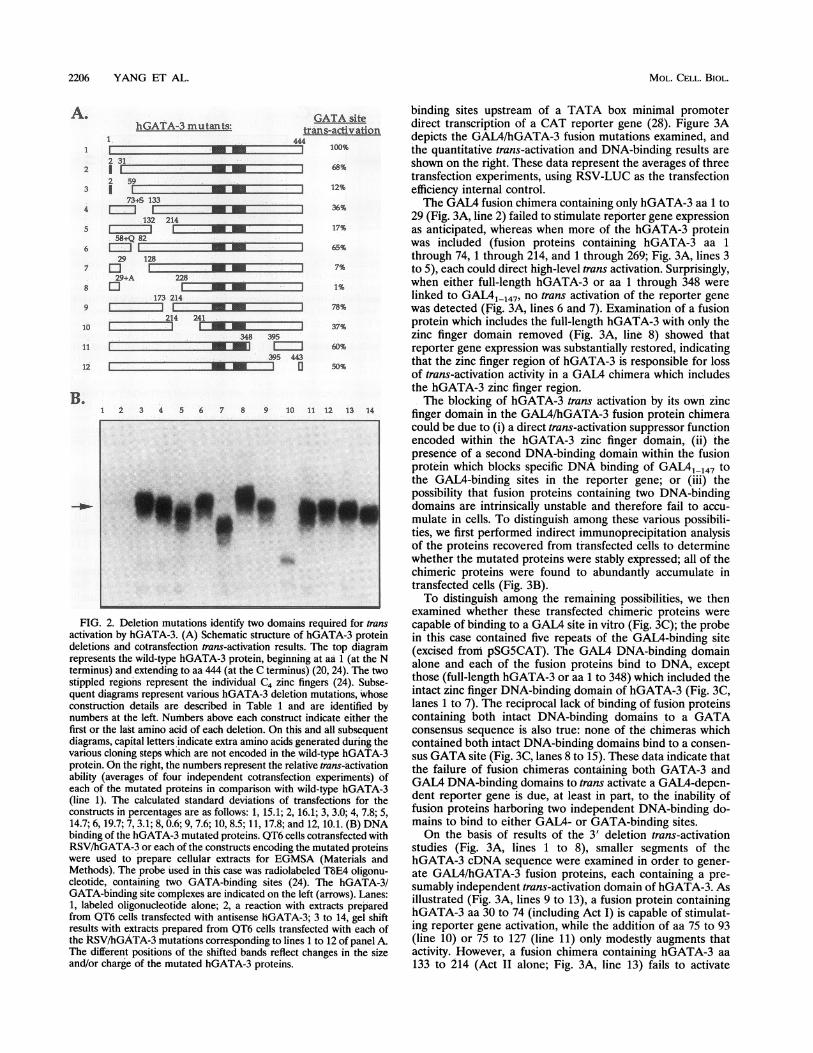
binding sites upstream of a TATA box minimal promoterdirect transcription of a CAT reporter gene (28). Figure 3Adepicts the GAL4/hGATA-3 fusion mutations examined, andthe quantitative trans-activation and DNA-binding results areshown on the right. These data represent the averages of threetransfection experiments, using RSV-LUC as the transfectionefficiency internal control.The GAL4 fusion chimera containing only hGATA-3 aa 1 to
29 (Fig. 3A, line 2) failed to stimulate reporter gene expressionas anticipated, whereas when more of the hGATA-3 proteinwas included (fusion proteins containing hGATA-3 aa 1through 74, 1 through 214, and 1 through 269; Fig. 3A, lines 3to 5), each could direct high-level trans activation. Surprisingly,when either full-length hGATA-3 or aa 1 through 348 werelinked to GAIAM_147, no trans activation of the reporter genewas detected (Fig. 3A, lines 6 and 7). Examination of a fusionprotein which includes the full-length hGATA-3 with only thezinc finger domain removed (Fig. 3A, line 8) showed thatreporter gene expression was substantially restored, indicatingthat the zinc finger region of hGATA-3 is responsible for lossof trans-activation activity in a GAL4 chimera which includesthe hGATA-3 zinc finger region.The blocking of hGATA-3 trans activation by its own zinc
finger domain in the GAL4/hGATA-3 fusion protein chimeracould be due to (i) a direct trans-activation suppressor functionencoded within the hGATA-3 zinc finger domain, (ii) thepresence of a second DNA-binding domain within the fusionprotein which blocks specific DNA binding of GALM147 to
the GAL4-binding sites in the reporter gene; or (iii) thepossibility that fusion proteins containing two DNA-bindingdomains are intrinsically unstable and therefore fail to accu-mulate in cells. To distinguish among these various possibili-ties, we first performed indirect immunoprecipitation analysisof the proteins recovered from transfected cells to determinewhether the mutated proteins were stably expressed; all of thechimeric proteins were found to abundantly accumulate intransfected cells (Fig. 3B).To distinguish among the remaining possibilities, we then
examined whether these transfected chimeric proteins werecapable of binding to a GAL4 site in vitro (Fig. 3C); the probein this case contained five repeats of the GAL4-binding site(excised from pSG5CAT). The GAIA DNA-binding domainalone and each of the fusion proteins bind to DNA, exceptthose (full-length hGATA-3 or aa 1 to 348) which included theintact zinc finger DNA-binding domain of hGATA-3 (Fig. 3C,lanes 1 to 7). The reciprocal lack of binding of fusion proteinscontaining both intact DNA-binding domains to a GATAconsensus sequence is also true: none of the chimeras whichcontained both intact DNA-binding domains bind to a consen-sus GATA site (Fig. 3C, lanes 8 to 15). These data indicate thatthe failure of fusion chimeras containing both GATA-3 andGAL4 DNA-binding domains to trans activate a GAL4-depen-dent reporter gene is due, at least in part, to the inability offusion proteins harboring two independent DNA-binding do-mains to bind to either GAL4- or GATA-binding sites.On the basis of results of the 3' deletion trans-activation
studies (Fig. 3A, lines 1 to 8), smaller segments of thehGATA-3 cDNA sequence were examined in order to gener-ate GAL4/hGATA-3 fusion proteins, each containing a pre-sumably independent trans-activation domain of hGATA-3. Asillustrated (Fig. 3A, lines 9 to 13), a fusion protein containinghGATA-3 aa 30 to 74 (including Act I) is capable of stimulat-ing reporter gene activation, while the addition of aa 75 to 93(line 10) or 75 to 127 (line 11) only modestly augments thatactivity. However, a fusion chimera containing hGATA-3 aa133 to 214 (Act II alone; Fig. 3A, line 13) fails to activate
A.
2
3
4
5
6
7
8
9
10
11
12
B.
MOL. CELL. BIOL.
ORGANIZATION OF HUMAN GATA-3 ACTIVITIES 2207
]264 3 444 GAL4 site GAL4-dependent reporter gene transcription. Similarly, aa 76[Transadvation DNAhnchng, to 215 (line 12), which appeared to contain distinct trans-
EEMEMEMIPEFPCIRRRYPCEFEL I + activating activities from deletion analysis (Fig. 2A), fail top6 FR24 29 independently provide a trans-activation function when linkedS§PUMMIEFP O GNS65Rt I +74 400 to the heterologous DNA-binding domain. Taken together,
214 these data indicate that only aa 30 to 74, or other hGATA-3RGIRALDK 100 +
269 coding regions containing Act I, can function to direct tran-° FP I .... ..CEFEL scriptional trans activation when fused to a heterologousGN___S_R_4____DNA-binding domain.
E________________ E_ E I Finger II of hGATA-3 is necessary and sufficient for se--xPEFPI_=EFEL20 . quence-specific DNA binding. As with all members of this
ESE§-PE -PFPo CGN&%R 80 multigene family in vertebrate organisms, the DNA-binding-PEFP-= GNS%SR 4 domain of hGATA-3 is composed of two zinc fingers, both of
30 12776PEFSSSR5 + which are highly conserved within the GATA factor family. InPEFI
6 1 STVPRCIRALDK mouse and chicken GATA-1, only finger II is required for00s_P PE" IC L24 + DNA binding, while finger I appears to be largely dispensable
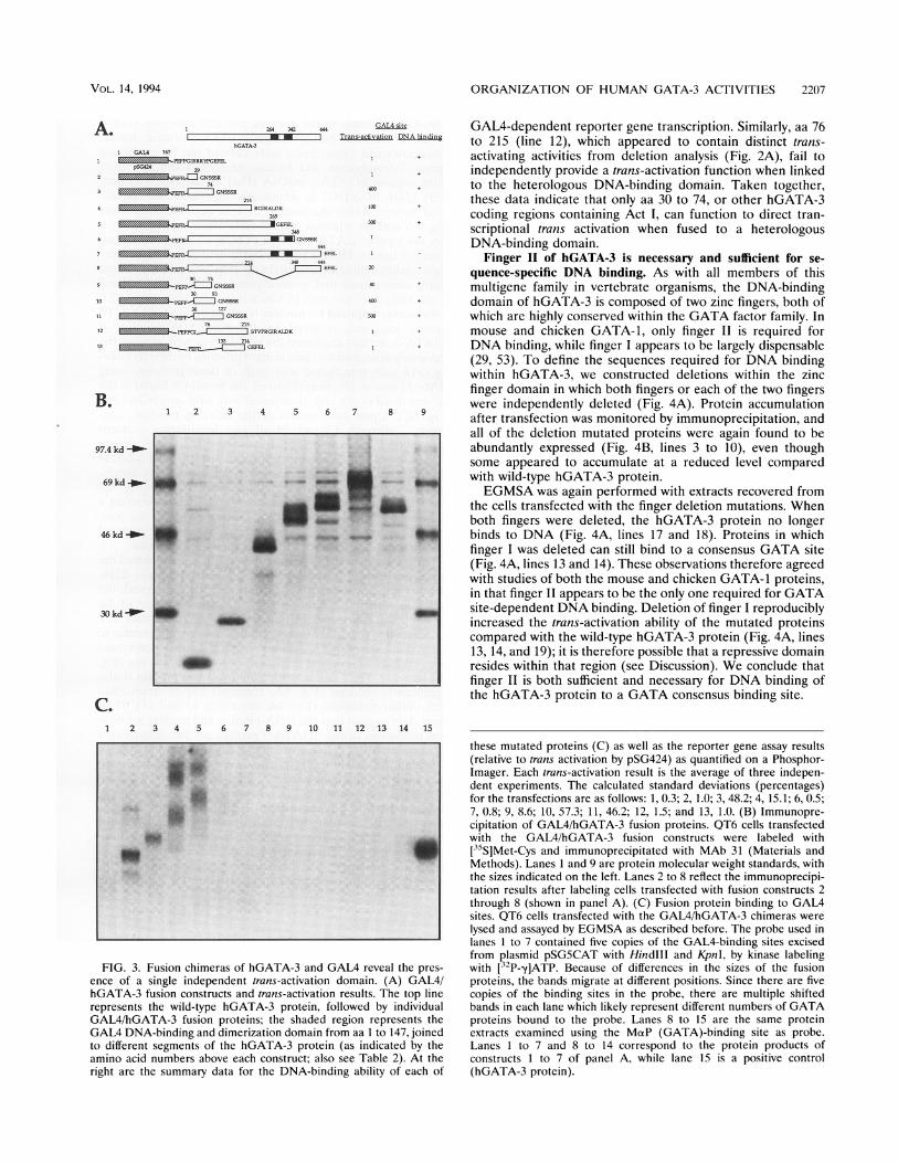
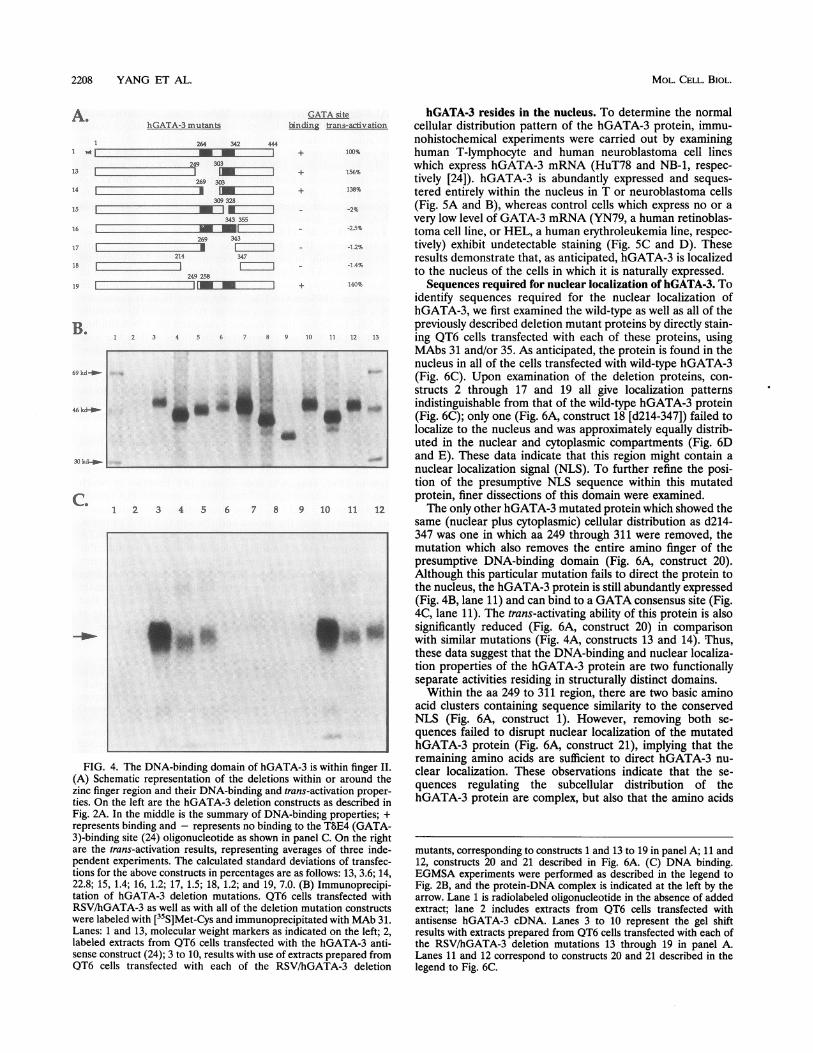
(29, 53). To define the sequences required for DNA bindingwithin hGATA-3, we constructed deletions within the zincfinger domain in which both fingers or each of the two fingerswere independently deleted (Fig. 4A). Protein accumulation
1 2 3 4 5 6 7 8 9 . .after transfection was monitored by immunoprecipitation, andall of the deletion mutated proteins were again found to be
cd_.4 ... *abundantly expressed (Fig. 4B, lines 3 to 10), even thoughsome appeared to accumulate at a reduced level comparedkd__ + ~ _ S +with wild-type hGATA-3 protein.EGMSA was again performed with extracts recovered from
the cells transfected with the finger deletion mutations. Whenboth fingers were deleted, the hGATA-3 protein no longer
:d_ w _6 .. t *_, _ binds to DNA (Fig. 4A, lines 17 and 18). Proteins in whichfinger I was deleted can still bind to a consensus GATA site(Fig. 4A, lines 13 and 14). These observations therefore agreedwith studies of both the mouse and chicken GATA-1 proteins,in that finger II appears to be the only one required for GATA
d _P~ _ _ site-dependent DNA binding. Deletion of finger I reproducibly-_ _ increased the trans-activation ability of the mutated proteins
compared with the wild-type hGATA-3 protein (Fig. 4A, lines13, 14, and 19); it is therefore possible that a repressive domainresides within that region (see Discussion). We conclude thatfinger II is both sufficient and necessary for DNA binding ofthe hGATA-3 protein to a GATA consensus binding site.
1 2 3 4 5 6 7 8 9 10 11 12 13 14 15
S
II
._'_
FIG. 3. Fusion chimeras of hGATA-3 and GAL4 reveal the pres-ence of a single independent trans-activation domain. (A) GAL4/hGATA-3 fusion constructs and trans-activation results. The top linerepresents the wild-type hGATA-3 protein, followed by individualGAL4/hGATA-3 fusion proteins; the shaded region represents theGAL4 DNA-binding and dimerization domain from aa I to 147, joinedto different segments of the hGATA-3 protein (as indicated by theamino acid numbers above each construct; also see Table 2). At theright are the summary data for the DNA-binding ability of each of
these mutated proteins (C) as well as the reporter gene assay results(relative to trans activation by pSG424) as quantified on a Phosphor-Imager. Each trans-activation result is the average of three indepen-dent experiments. The calculated standard deviations (percentages)for the transfections are as follows: 1, 0.3; 2, 1.0; 3, 48.2; 4, 15.1; 6, 0.5;7, 0.8; 9, 8.6; 10, 57.3; 11, 46.2; 12, 1.5; and 13, 1.0. (B) Immunopre-cipitation of GAL4/hGATA-3 fusion proteins. QT6 cells transfectedwith the GAL4/hGATA-3 fusion constructs were labeled with[35S]Met-Cys and immunoprecipitated with MAb 31 (Materials andMethods). Lanes 1 and 9 are protein molecular weight standards, withthe sizes indicated on the left. Lanes 2 to 8 reflect the immunoprecipi-tation results after labeling cells transfected with fusion constructs 2through 8 (shown in panel A). (C) Fusion protein binding to GAL4sites. QT6 cells transfected with the GAL4/hGATA-3 chimeras were
lysed and assayed by EGMSA as described before. The probe used inlanes 1 to 7 contained five copies of the GAL4-binding sites excisedfrom plasmid pSG5CAT with HindIll and KpnI, by kinase labelingwith [32P-yIATP. Because of differences in the sizes of the fusionproteins, the bands migrate at different positions. Since there are fivecopies of the binding sites in the probe, there are multiple shiftedbands in each lane which likely represent different numbers of GATAproteins bound to the probe. Lanes 8 to 15 are the same proteinextracts examined using the MaP (GATA)-binding site as probe.Lanes 1 to 7 and 8 to 14 correspond to the protein products ofconstructs 1 to 7 of panel A, while lane 15 is a positive control(hGATA-3 protein).
A.
2
6
8
9
10
11
12
B.
97.4 k
69 k
46 k
30 k
C.
VOL. 14, 1994
2208 YANG ET AL.
hGATA-3 mutants
264 342A I
249 303
269 303
309 328
34316r[
269 34317 [E
214 318 1 -- -
249 2819 1.
1 2 3 4 5 6
... ..... -a
1 2 3 4 5 6
P.:
t~~~~~~.tFIG. 4. The DNA-binding domaii
(A) Schematic representation of thezinc finger region and their DNA-binties. On the left are the hGATA-3 d
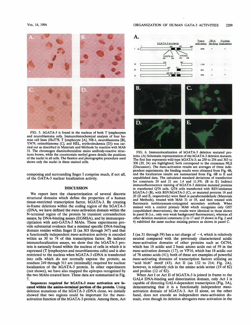
GATA site hGATA-3 resides in the nucleus. To determine the normalbind trans-activation cellular distribution pattern of the hGATA-3 protein, immu-
! 444 nohistochemical experiments were carried out by examining_--I + 100 human T-lymphocyte and human neuroblastoma cell lineslizz + 16%% which express hGATA-3 mRNA (HuT78 and NB-1, respec-
tively [24]). hGATA-3 is abundantly expressed and seques-+ 138% tered entirely within the nucleus in T or neuroblastoma cells=_ -2% (Fig. 5A and B), whereas control cells which express no or a
355 very low level of GATA-3 mRNA (YN79, a human retinoblas-ZN - -~25% toma cell line, or HEL, a human erythroleukemia line, respec-
Izzz - -1.2 tively) exhibit undetectable staining (Fig. 5C and D). These_7 results demonstrate that, as anticipated, hGATA-3 is localized____--1.4% to the nucleus of the cells in which it is naturally expressed.
1112 + 140% Sequences required for nuclear localization of hGATA-3. Toidentify sequences required for the nuclear localization ofhGATA-3, we first examined the wild-type as well as all of thepreviously described deletion mutant proteins by directly stain-
7 8 9 10 11 12 13 ing QT6 cells transfected with each of these proteins, usingMAbs 31 and/or 35. As anticipated, the protein is found in thenucleus in all of the cells transfected with wild-type hGATA-3(Fig. 6C). Upon examination of the deletion proteins, con-structs 2 through 17 and 19 all give localization patternsI* * * indistinguishable from that of the wild-type hGATA-3 protein
^ (Fig. 6C); only one (Fig. 6A, construct 18 [d214-347]) failed tolocalize to the nucleus and was approximately equally distrib-uted in the nuclear and cytoplasmic compartments (Fig. 6Dand E). These data indicate that this region might contain anuclear localization signal (NLS). To further refine the posi-tion of the presumptive NLS sequence within this mutatedprotein, finer dissections of this domain were examined.
7 8 9 10 11 12 The only other hGATA-3 mutated protein which showed thesame (nuclear plus cytoplasmic) cellular distribution as d214-347 was one in which aa 249 through 311 were removed, themutation which also removes the entire amino finger of thepresumptive DNA-binding domain (Fig. 6A, construct 20).Although this particular mutation fails to direct the protein tothe nucleus, the hGATA-3 protein is still abundantly expressed(Fig. 4B, lane 11) and can bind to a GATA consensus site (Fig.4C, lane 11). The trans-activating ability of this protein is also
a .* significantly reduced (Fig. 6A, construct 20) in comparison* with similar mutations (Fig. 4A, constructs 13 and 14). Thus,these data suggest that the DNA-binding and nuclear localiza-tion properties of the hGATA-3 protein are two functionallyseparate activities residing in structurally distinct domains.
Within the aa 249 to 311 region, there are two basic aminoacid clusters containing sequence similarity to the conservedNLS (Fig. 6A, construct 1). However, removing both se-quences failed to disrupt nuclear localization of the mutatedhGATA-3 protein (Fig. 6A, construct 21), implying that the
ofhGTiihiigeLremaining amino acids are sufficient to direct hGATA-3 nu-
n ofhGATA-3iiswthitin fingern clear localization. These observations indicate that the se-
iding and trans-activation proper- quences regulating the subcellular distribution of theeletion constructs as described in hGATA-3 protein are complex, but also that the amino acids
Fig. 2A. In the middle is the summary of DNA-binding properties; +
represents binding and - represents no binding to the T8E4 (GATA-3)-binding site (24) oligonucleotide as shown in panel C. On the rightare the trans-activation results, representing averages of three inde-pendent experiments. The calculated standard deviations of transfec-tions for the above constructs in percentages are as follows: 13, 3.6; 14,22.8; 15, 1.4; 16, 1.2; 17, 1.5; 18, 1.2; and 19, 7.0. (B) Immunoprecipi-tation of hGATA-3 deletion mutations. QT6 cells transfected withRSV/hGATA-3 as well as with all of the deletion mutation constructswere labeled with [35S]Met-Cys and immunoprecipitated with MAb 31.Lanes: 1 and 13, molecular weight markers as indicated on the left; 2,labeled extracts from QT6 cells transfected with the hGATA-3 anti-sense construct (24); 3 to 10, results with use of extracts prepared from
QT6 cells transfected with each of the RSV/hGATA-3 deletion
mutants, corresponding to constructs 1 and 13 to 19 in panel A; 11 and12, constructs 20 and 21 described in Fig. 6A. (C) DNA binding.EGMSA experiments were performed as described in the legend toFig. 2B, and the protein-DNA complex is indicated at the left by thearrow. Lane 1 is radiolabeled oligonucleotide in the absence of addedextract; lane 2 includes extracts from QT6 cells transfected withantisense hGATA-3 cDNA. Lanes 3 to 10 represent the gel shiftresults with extracts prepared from QT6 cells transfected with each ofthe RSV/hGATA-3 deletion mutations 13 through 19 in panel A.Lanes 11 and 12 correspond to constructs 20 and 21 described in thelegend to Fig. 6C.
A.
13
14
15
B.
69 kd-_-
46 kd-_-
30 k&-O-
C.
MOL. CELL. BIOL.
ORGANIZATION OF HUMAN GATA-3 ACTIVITIES 2209
b - @~~~
_ _
A.hGATA-3 mutants:
26-4 342
_\
KSRPKAR KPKRRL250 256 303 308
214 34718 L249 311
20
249258 30331121 _
Trans- DNA Nudearactivation binding locaization
44471 100% + +
-1.4%
34%
93%
+
+
*~~~~~~~~~~~~~~~~~~~~~~~~~~~~~~~~~~~~~~~~~~~~~......z# W
FIG. 5. hGATA-3 is found in the nucleus of both T lymphocytesand neuroblastoma cells. Immunohistochemical analysis of four hu-man cell lines (HuT78, T lymphocyte [A], NB-1, neuroblastoma [B],YN79, retinoblastoma [C], and HEL, erythroleukemia [D]) was car-ried out as described in Materials and Methods by reaction with MAb31. The chromogen diaminobenzidine stains antibody-reactive struc-tures brown, while the counterstain methyl green details the positionsof the nuclei in all cells. The fixation and photographic procedure usedshows only the nuclei in these stained cells.
composing and surrounding finger I comprise much, if not all,of the GATA-3 nuclear localization activity.
DISCUSSIONWe report here the characterization of several discrete
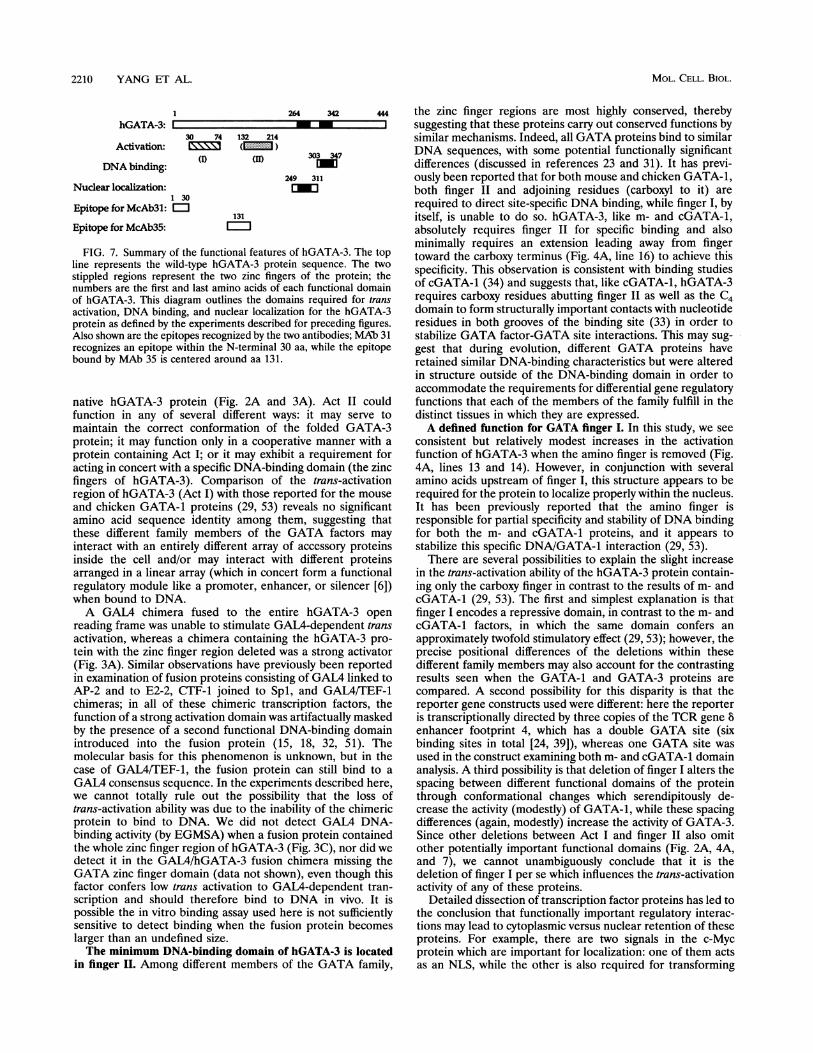
structural domains which define the properties of a humantissue-restricted transcription factor, hGATA-3. By creatingin-frame deletions within the coding region of the hGATA-3cDNA, we have defined the trans-activation domain within theN-terminal region of the protein by transient cotransfectionassays, by DNA-binding assays (EGMSA), and by immunopre-cipitation with anti-GATA-3 MAbs. These experiments pro-vide substantial evidence that a minimal specific DNA-bindingdomain resides within finger II (aa 303 through 347) and thata functionally independent trans-activation activity is encodedwithin aa 30 to 74 of this transcription factor. By indirectimmunolocalization assays, we show that the hGATA-3 pro-tein is naturally found within the nucleus of cells in which it isexpressed (T lymphocytes and neuroblastoma cells) and is alsorestricted to the nucleus when hGATA-3 cDNA is transfectedinto cells which do not normally express the protein; aaresidues 249 through 311 are (minimally) required for nuclearlocalization of the hGATA-3 protein. In other experiments(not shown), we have also mapped the epitopes recognized bythe two MAbs created here. These data are summarized in Fig.7.
Sequences required for hGATA-3 trans activation are lo-cated within the amino-terminal portion of the protein. Usingdeletion mutations of the hGATA-3 cDNA clone, we initiallyshowed that two regions could be important for the trans-activation function of the hGATA-3 protein. Among them, Act
FIG. 6. Immunolocalization of hGATA-3 deletion mutated pro-teins. (A) Schematic representation of the hGATA-3 deletion mutants.The first line represents wild-type hGATA-3; aa 250 to 256 and 303 to308 (20, 24) are highlighted; both correspond to the consensus NLS(Discussion). The trans-activation results are averages of three inde-pendent experiments, the binding results were obtained from Fig. 4B,and the localization results are summarized from Fig. 6B to E andunpublished data. The calculated standard deviations of transfectionfor constructs 20 and 21 are 1.4 and 11.3%. (B to E) Indirectimmunofluorescence staining of hGATA-3 deletion mutated proteinsin transfected QT6 cells. QT6 cells transfected with RSV/antisensehGATA-3 (B), with RSV/hGATA-3 (C), or mutated proteins 18 and20 (D and E, respectively) were fixed in paraformaldehyde (Materialsand Methods), treated with MAb 31 or 35, and then treated withfluorescein isothiocyanate-conjugated secondary antibody. Whenstained with a control primary MAb which recognizes only GST(unpublished observations), the results were identical to those shownin panel B (i.e., only very weak background fluorescence), whereas allother deletion mutation constructs (2 to 17 and 19 shown in Fig. 2 and4) exhibited the same nuclear staining pattern as shown in panel C.
I (aa 31 through 59) has a net charge of - 4, which is relativelyneutral compared with the previously characterized acidictrans-activation domains of other proteins such as GCN4,which has 18 acidic and 2 basic amino acids out of 59 in thetrans-activation domain (17), or VP16, which has 18 acidic outof 78 amino acids (41); both of these are examples of powerfultrans-activating domains of transcription factors utilizing an"acid blob" motif (43). Act II (aa 132 to 214; Fig. 2A),however, is relatively rich in the amino acids serine (19 of 82)and proline (12 of 82).When Act I or Act II of hGATA-3 is joined in frame to the
GAL4 DNA-binding and dimerization domain, only Act I iscapable of directing GAL4-dependent transcription (Fig. 3A),demonstrating that it is a functionally independent trans-activation domain of hGATA-3. Act II alone, on the otherhand, does not encode an independent trans-activation do-main, even though its deletion abrogates trans activation in the
<1
I
VOL. 14, 1994
2210 YANG ET AL.
264 342
hGATA-3:
30 74 132 214Activation: ( )
DNA binding:
Nuclear localization:1 30
Epitope for McAb3l: E
Epitope for McAb35:
303 347
249 311
[mu
131
FIG. 7. Summary of the functional features of hGATA-3. The topline represents the wild-type hGATA-3 protein sequence. The twostippled regions represent the two zinc fingers of the protein; thenumbers are the first and last amino acids of each functional domainof hGATA-3. This diagram outlines the domains required for transactivation, DNA binding, and nuclear localization for the hGATA-3protein as defined by the experiments described for preceding figures.Also shown are the epitopes recognized by the two antibodies; MAb 31recognizes an epitope within the N-terminal 30 aa, while the epitopebound by MAb 35 is centered around aa 131.
native hGATA-3 protein (Fig. 2A and 3A). Act II couldfunction in any of several different ways: it may serve tomaintain the correct conformation of the folded GATA-3protein; it may function only in a cooperative manner with aprotein containing Act I; or it may exhibit a requirement foracting in concert with a specific DNA-binding domain (the zincfingers of hGATA-3). Comparison of the trans-activationregion of hGATA-3 (Act I) with those reported for the mouseand chicken GATA-1 proteins (29, 53) reveals no significantamino acid sequence identity among them, suggesting thatthese different family members of the GATA factors may
interact with an entirely different array of accessory proteinsinside the cell and/or may interact with different proteinsarranged in a linear array (which in concert form a functionalregulatory module like a promoter, enhancer, or silencer [6])when bound to DNA.A GAL4 chimera fused to the entire hGATA-3 open
reading frame was unable to stimulate GAL4-dependent transactivation, whereas a chimera containing the hGATA-3 pro-
tein with the zinc finger region deleted was a strong activator(Fig. 3A). Similar observations have previously been reportedin examination of fusion proteins consisting of GAL4 linked toAP-2 and to E2-2, CTF-1 joined to Spl, and GAL4/TEF-1chimeras; in all of these chimeric transcription factors, thefunction of a strong activation domain was artifactually maskedby the presence of a second functional DNA-binding domainintroduced into the fusion protein (15, 18, 32, 51). Themolecular basis for this phenomenon is unknown, but in thecase of GAL4/TEF-1, the fusion protein can still bind to a
GAL4 consensus sequence. In the experiments described here,we cannot totally rule out the possibility that the loss oftrans-activation ability was due to the inability of the chimericprotein to bind to DNA. We did not detect GAL4 DNA-binding activity (by EGMSA) when a fusion protein containedthe whole zinc finger region of hGATA-3 (Fig. 3C), nor did wedetect it in the GAL4/hGATA-3 fusion chimera missing theGATA zinc finger domain (data not shown), even though thisfactor confers low trans activation to GAL4-dependent tran-scription and should therefore bind to DNA in vivo. It ispossible the in vitro binding assay used here is not sufficientlysensitive to detect binding when the fusion protein becomeslarger than an undefined size.The minimum DNA-binding domain of hGATA-3 is located
in finger II. Among different members of the GATA family,
the zinc finger regions are most highly conserved, therebysuggesting that these proteins carry out conserved functions bysimilar mechanisms. Indeed, all GATA proteins bind to similarDNA sequences, with some potential functionally significantdifferences (discussed in references 23 and 31). It has previ-ously been reported that for both mouse and chicken GATA-1,both finger II and adjoining residues (carboxyl to it) arerequired to direct site-specific DNA binding, while finger I, byitself, is unable to do so. hGATA-3, like m- and cGATA-1,absolutely requires finger II for specific binding and alsominimally requires an extension leading away from fingertoward the carboxy terminus (Fig. 4A, line 16) to achieve thisspecificity. This observation is consistent with binding studiesof cGATA-1 (34) and suggests that, like cGATA-1, hGATA-3requires carboxy residues abutting finger II as well as the C4domain to form structurally important contacts with nucleotideresidues in both grooves of the binding site (33) in order tostabilize GATA factor-GATA site interactions. This may sug-gest that during evolution, different GATA proteins haveretained similar DNA-binding characteristics but were alteredin structure outside of the DNA-binding domain in order toaccommodate the requirements for differential gene regulatoryfunctions that each of the members of the family fulfill in thedistinct tissues in which they are expressed.A defined function for GATA finger I. In this study, we see
consistent but relatively modest increases in the activationfunction of hGATA-3 when the amino finger is removed (Fig.4A, lines 13 and 14). However, in conjunction with severalamino acids upstream of finger I, this structure appears to berequired for the protein to localize properly within the nucleus.It has been previously reported that the amino finger isresponsible for partial specificity and stability of DNA bindingfor both the m- and cGATA-1 proteins, and it appears tostabilize this specific DNA/GATA-1 interaction (29, 53).There are several possibilities to explain the slight increase
in the trans-activation ability of the hGATA-3 protein contain-ing only the carboxy finger in contrast to the results of m- andcGATA-1 (29, 53). The first and simplest explanation is thatfinger I encodes a repressive domain, in contrast to the m- andcGATA-1 factors, in which the same domain confers anapproximately twofold stimulatory effect (29, 53); however, theprecise positional differences of the deletions within thesedifferent family members may also account for the contrastingresults seen when the GATA-1 and GATA-3 proteins arecompared. A second possibility for this disparity is that thereporter gene constructs used were different: here the reporteris transcriptionally directed by three copies of the TCR gene 8enhancer footprint 4, which has a double GATA site (sixbinding sites in total [24, 39]), whereas one GATA site wasused in the construct examining both m- and cGATA-1 domainanalysis. A third possibility is that deletion of finger I alters thespacing between different functional domains of the proteinthrough conformational changes which serendipitously de-crease the activity (modestly) of GATA-1, while these spacingdifferences (again, modestly) increase the activity of GATA-3.Since other deletions between Act I and finger II also omitother potentially important functional domains (Fig. 2A, 4A,and 7), we cannot unambiguously conclude that it is thedeletion of finger I per se which influences the trans-activationactivity of any of these proteins.
Detailed dissection of transcription factor proteins has led tothe conclusion that functionally important regulatory interac-tions may lead to cytoplasmic versus nuclear retention of theseproteins. For example, there are two signals in the c-Mycprotein which are important for localization: one of them actsas an NLS, while the other is also required for transforming
MOL. CELL. BIOL.
ORGANIZATION OF HUMAN GATA-3 ACTIVITIES 2211
activity (5). Nuclear accumulation of p53 is mediated byseveral NLSs, each sharing responsibility for nuclear accumu-lation (42). In another well-characterized example, the NLS isboth necessary and sufficient for the nuclear uptake of NF-KB,unless the IKB protein binds to it, masking the NLS, andNF-KB remains in the cytoplasm (3). In other cases, nuclearlocalization correlates with the cell cycle (50).We show here that in T lymphocytes and neuroblastoma
cells, in which hGATA-3 is normally expressed, or in QT6 cellsafter transfection of hGATA-3 under the control of a strongconstitutive promoter, the hGATA-3 protein is localized en-tirely within the nucleus. By deletion analysis, we have mappeda region between aa 249 and 311, overlapping finger I, which isrequired for the nuclear localization of the transfectedhGATA-3 protein. Within this aa 249 to 311 region containingand surrounding finger I, there are two short stretches of basicamino acids, KSRPKAR and KPKRRL, both of which are verysimilar to the consensus NLS (underlined residues) as de-scribed previously (4). However, precise excision of either orboth segments fails to prevent hGATA-3 transport into thenucleus (Fig. 6A, construct 21, and data not shown). Further-more, immunofluorescence staining of hGATA-3 harboringsmaller deletions (e.g., d269-303 or d249-258; Fig. 4A, con-structs 14 and 15, and data not shown) within this 58-aa regionshowed that each of these also accumulated only in thenucleus, suggesting that multiple signals in the hGATA-3protein direct nuclear accumulation and that they functionindependently.There are other examples of multiple NLSs in a single
constitutively expressed nuclear protein which act indepen-dently in an additive fashion, where only disruption of all NLSsites abolishes localization (reviewed in reference 44). This hasbeen demonstrated specifically with NS1 of influenza A virus,which has two independent NLSs; only if both are deleted willthe protein fail to localize to the nucleus (14). For the tumorsuppressor p53, there are three NLSs; deleting any of themresults in only partial cytoplasmic localization (42).
It is noteworthy that for both d214-347 and d249-311hGATA-3 mutated proteins, the immunofluorescence datashowed mixed cytoplasmic and nuclear distribution of theprotein. This failure in complete compartmentalization couldbe due to several possibilities, the simplest being that smallerproteins created by deletion can freely diffuse between nucleusand cytoplasm. However, protein size alone cannot be a majordeterminant for localization of hGATA-3, since proteinssmaller than hGATA-3 mutation d249-311 (Fig. 2A, 4A, and6A) are entirely sequestered within the nucleus (data notshown). A second possibility is that the hGATA-3 protein canenter the nucleus by interacting with other proteins, as is thecase for tumor suppressor protein pRB (55). The domainsresponsible for pRB nuclear localization and protein interac-tion are both important for successful nuclear translocation,and interestingly, mutations affecting the NLS of pRB retainpartial nuclear localization activity due to the ability of mu-tated pRB to enter the nucleus by associating with otherproteins through an intact protein interaction domain. A thirdpossibility is that there are additional NLSs outside aa 249 to311 of hGATA-3, whose function is at least partially redundantwith the activity identified here and which can function in theabsence of aa 249 to 311.
It has been reported that the NLSs are incorporated into theDNA-binding domains for a number of nuclear proteins (e.g.,Fos [48] and GAL4 [45]). In the case of hGATA-3, the NLS islocated within the zinc finger region (finger I) but is separatefrom the DNA-binding function of the protein (located infinger II). The most conservative interpretation of the data
shown here is that the NLS for hGATA-3 is buried around andwithin the sequence of the amino finger, thereby identifying anew and distinct function for finger I of the GATA proteins.
ACKNOWLEDGMENTS
We appreciate constructive criticisms of the manuscript and provi-sion of the purified GST/cGATA-1, -2, and -3 proteins by ourcolleague L. J. Ko and provision of the C1300 cells by K. George, andwe thank B. Wu for assistance and for providing some of the reagentsused in this study.
This work was supported by NIH research grant GM 28896 (J.D.E.).
REFERENCES1. Antoniou, M., E. deBoer, G. Habets, and F. Grosveld. 1988. The
human ,B-globin gene contains multiple regulatory regions: iden-tification of one promoter and two downstream enhancers. EMBOJ. 7:377-384.
2. Arceci, R. J., A. A. J. King, M. C. Simon, S. H. Orkin, and D. B.Wilson. 1993. Mouse GATA-4: a retinoic acid-inducible GATA-binding transcription factor expressed in endodermally derivedtissues and heart. Mol. Cell. Biol. 13:2235-2246.
3. Beg, A. A., S. M. Ruben, R. I. Scheinman, S. Haskill, C. A. Rosen,and A. S. Baldwin, Jr. 1992. IKB interacts with the nuclearlocalization sequences of the subunits of NF-KB: a mechanism forcytoplasmic retention. Genes Dev. 6:1899-1913.
4. Cheisky, D., R. Ralph, and G. Jonak 1989. Sequence requirementsfor synthetic peptide-mediated translocation to the nucleus. Mol.Cell. Biol. 9:2487-2492.
5. Dang, C. V., and W. M. F. Lee. 1988. Identification of the humanc-Myc protein nuclear translocation signal. Mol. Cell. Biol.8:4048-4054.
6. Engel, J. D., K. M. George, L. J. Ko, J. M. Kornhauser, M. W.Leonard, P. Ting, and M. Yamamoto. 1991. Transcription factorregulation of hematopoietic lineage cells. Semin. Hematol. 28:158-169.
7. Evans, T., and G. Felsenfeld. 1989. The erythroid-specific tran-scription factor Eryfl: a new finger protein. Cell 58:877-885.
8. Evans, T., M. Reitman, and G. Felsenfeld. 1988. An erythrocyte-specific DNA-binding factor recognizes a regulatory sequencecommon to all chicken globin genes. Proc. Natl. Acad. Sci. USA85:5976-5980.
9. Fried, M. G., and D. M. Crothers. 1981. Equilibria and kinetics oflac repressor-operator interactions by polyacrylamide gel electro-phoresis. Nucleic Acids Res. 9:6505-6525.
10. Gallarda, J. L., K. P. Foley, Z. Yang, and J. ID. Engel. 1989. TheP-globin stage selector element factor is erythroid-specific promot-er/enhancer binding protein NF-E4. Genes Dev. 3:1845-1859.
11. Garner, M. M., and A. Revzin. 1981. A gel electrophoresis methodfor quantifying the binding of proteins to specific DNA regions:application to components of the Escherichia coli lactose operonregulatory system. Nucleic Acids Res. 9:3047-3060.
11a.George, K., and J. D. Engel. Unpublished data.11b.George, K., M. Roth, M. W. Leonard, K. Lieuw, F. Grosveld, and
J. D. Engel. Unpublished data.12. Gorman, C. M., L. F. Moffat, and B. H. Howard. 1982. Recombi-
nant genomes which express chloramphenicol acetyltransferase inmammalian cells. Mol. Cell. Biol. 2:1044-1051.
13. Graham, F. L., and A. J. van der Eb. 1973. A new technique for theassay of infectivity of human adenovirus 5 DNA. Virology 52:456-467.
14. Greenspan, D., P. Palese, and M. Krystal. 1988. Two nuclearlocalization signals in the influenza virus NS1 nonstructural pro-tein. J. Virol. 62:3020-3026.
14a.Gu, L., B. Andres, L. Ko, Z. Yang, and J. D. Engel. Unpublisheddata.
15. Henthorn, P., M. Kiledjian, and T. Kadesch. 1990. Two distincttranscription factors that bind the immunoglobulin enhancer11E5/K2 motif. Science 247:467-470.
16. Ho, I. C., P. Vorhees, N. Marin, B. K. Oakley, S.-F. Tsai, S. H.Orkin, and J. M. Leiden. 1991. Human GATA-3: a lineage-restricted transcription factor that regulates the expression of theT cell receptor ot gene. EMBO J. 10:1187-1192.
VOL. 14, 1994
2212 YANG ET AL.
17. Hope, I. A., and K. Struhl. 1986. Functional dissection of an
eukaryotic transcriptional activator protein, GCN4 of yeast. Cell46:885-894.
18. Hwang, J.-J., P. Chambon, and I. Davidson. 1993. Characteriza-tion of the transcriptional activation function and the DNAbinding domain of transcriptional enhancer factor-1. EMBO J.12:2337-2348.
19. Ito, E., T. Toki, H. Ishihara, H. Ohtani, L. Gu, M. Yokoyama, J. D.Engel, and M. Yamamoto. 1993. Erythroid transcription factorGATA-1 is abundantly transcribed in mouse testis. Nature (Lon-don) 362:466-469.
20. Joulin, V., D. Bories, J.-F. Eleouet, M.-C. Labastie, S. Chretien,M.-G. Mattei, and P.-H. Romeo. 1991. A T-cell specific TCR 8DNA binding protein is a member of the human GATA family.EMBO J. 10:1809-1816.
21. Keegan, L., G. Gill, and M. Ptashne. 1986. Separation of DNAbinding from the transcription-activating function of a regulatoryprotein. Science 231:699-704.
22. Kelley, C., H. Blumberg, L. I. Zon, and T. Evans. 1993. GATA-4:a novel transcription factor expressed in endocardium of thedeveloping heart. Development 118:817-827.
23. Ko, L. J., and J. D. Engel. 1993. DNA-binding specificities of theGATA transcription factor family. Mol. Cell. Biol. 13:4011-4022.
24. Ko, L. J., M. Yamamoto, M. W. Leonard, K. M. George, and J. D.Engel. 1991. Murine and human GATA-3 factors mediate tran-scription through a cis-regulatory element within the human T-cellreceptor 8 gene enhancer. Mol. Cell. Biol. 11:2778-2784.
25. Kornhauser, J. M., M. W. Leonard, M. Yamamoto, J. H. LaVail,K. E. Mayo, and J. D. Engel. GATA transcription factor expres-sion parallels development of the embryonic chicken optic tectum.Mol. Brain Res., in press.
25a.Labastie, P., and P.-H. Romeo. Unpublished data.26. Leonard, M. W., K.-C. Lim, and J. D. Engel. 1993. Expression of
the GATA transcription factor family during early erythroiddevelopment and differentiation. Development 119:519-531.
27. Lindenbaum, M., M. W. Leonard, F. G. Grosveld, and J. D. Engel.Unpublished data.
27a.Linzer, D. I. H. Personal communication.28. Lum, S. S. Y., S. Hsu, M. Vaewhongs, and B. Wu. 1992. The hsp70
gene CCAAT-binding factor mediates transcriptional activationby the adenovirus Ela protein. Mol. Cell. Biol. 12:2599-2605.
29. Martin, D. I. K., and S. H. Orkin. 1990. Transcriptional activationand DNA-binding by the erythroid factor GF-1/NF-E1/Eryfl.Genes Dev. 4:1886-1898.
30. Martin, D. I. K., S.-F. Tsai, and S. H. Orkin. 1989. Increased-y-globin expression in HPFH mediated by an erythroid-specificDNA-binding factor. Nature (London) 338:435-438.
30a.Mellon, P. Personal communication.31. Merika, M., and S. H. Orkin. 1993. DNA-binding specificity of
GATA family transcription factors. Mol. Cell. Biol. 13:3999-4010.32. Mermod, N., E. A. O'Neill, T. J. Kelly, and R. Tjian. 1989. The
proline-rich transcriptional activator of CTF/NF-1 is distinct fromthe replication and DNA binding domain. Cell 58:741-753.
33. Omichinski, J. G., G. M. Clore, 0. Schaad, G. Felsenfeld, C.Trainor, E. Appella, S. J. Stahl, and A. M. Gronenborn. 1993.NMR structure of a specific DNA complex of Zn-containing DNAbinding domain of GATA-1. Science 261:438-446.
34. Omichinski, J. G., C. Trainor, T. Evans, A. M. Gronenborn, G. M.Clore, and G. Felsenfeld. 1993. A small single-"finger" peptidefrom the erythroid transcription factor GATA-1 binds specificallyto DNA as a zinc or iron complex. Proc. Natl. Acad. Sci. USA90:1676-1680.
35. Orkin, S. H. 1992. GATA-binding transcription factors in hema-topoietic cells. Blood 80:575-581.
36. Perkins, N. D., R. H. Nicolas, M. A. Plumb, and G. H. Goodwin.1989. The purification of an erythroid protein which binds toenhancer and promoter elemnents of haemoglobin genes. NucleicAcids Res. 17:1299-1314.
37. Plumb, M., J. Frampton, H. Wainwright, M. Walker, K. Macleod,G. Goodwin, and P. Harrison. 1989. GATAAG; a cis-controlregion binding an erythroid-specific nuclear factor with a role inglobin and non-globin gene expression. Nucleic Acids Res. 17:73-92.
38. Redondo, J. M., S. Hata, C. Brocklehurst, and M. S. Krangel.1990. A T cell-specific enhancer within the human T cell receptor8 locus. Science 247:1225-1229.
39. Redondo, J. M., J. L. Pfohl, and M. S. Krangel. 1991. Identifica-tion of an essential site for transcriptional activation within thehuman T-cell receptor 8 enhancer. Mol. Cell. Biol. 11:5671-5680.
40. Rosenthal, N. 1987. Identification of regulatory elements of clonedgenes with functional assays. Methods Enzymol. 152:704-724.
41. Sadowski, I., J. Ma, S. Triezenberg, and M. Ptashne. 1988.GAL4-VP16 is an unusually potent transcriptional activator. Na-ture (London) 335:563-564.
42. Shaulsky, G., N. Goldfinger, A. Ben-Se'ev, and V. Rotter. 1990.Nuclear accumulation of p53 protein is mediated by severalnuclear localization signals and plays a role in tumorigenesis. Mol.Cell. Biol. 10:6565-6577.
43. Sigler, P. B. 1988. Acid blobs and negative noodles. Nature(London) 333:210-212.
44. Silver, P. A. 1991. How proteins enter the nucleus. Cell 64:489-497.
45. Silver, P. A., L. P. Keegan, and M. Ptashne. 1984. Amino terminusof the yeast GAL4 gene product is sufficient for nuclear localiza-tion. Proc. Natl. Acad. Sci. USA 81:5951-5955.
46. Smith, D. B., and K. S. Johnson. 1988. Single-step purification ofpolypeptides in Escherichia coli as fusions with glutathione S-transferase. Gene 67:31-40.
47. Trainor, C. D., T. Evans, G. Felsenfeld, and M. S. Boguski. 1990.Structure and evolution of a human erythroid transcription factor.Nature (London) 343:92-96.
48. Tratner, I., and I. M. Verma. 1991. Identification of a nucleartargeting sequence in the Fos protein. Oncogene 6:2049-2053.
49. Tsai, S.-F., D. I. K. Martin, L. I. Zon, A. D. D'Andrea, G. G. Wong,and S. H. Orkin. 1989. Cloning of cDNA for the major DNA-binding protein of the erythroid lineage through expression inmammalian cells. Nature (London) 339:446-451.
50. Vris, S., J.-M. Lemaitre, M. Libovici, N. Thierry, and M. Mechali.1992. Comparative analysis of the intracellular localization ofc-Myc, c-Fos, and replicative proteins during cell cycle progres-sion. Mol. Cell. Biol. 12:3548-3555.
51. Williams, T., and R. Tjian. 1991. Analysis of the DNA-binding andactivation properties of the human transcription factor AP-2.Genes Dev. 5:670-682.
52. Yamamoto, M., L. J. Ko, M. W. Leonard, H. Beug, S. H. Orkin,and J. D. Engel. 1990. Activity and tissue-specific expression of thetranscription factor NF-E1 multigene family. Genes Dev. 4:1650-1662.
53. Yang, H.-Y., and T. Evans. 1992. Distinct roles for the twocGATA-1 finger domains. Mol. Cell. Biol. 12:4562-4570.
54. Yang, Z., and J. D. Engel. 1993. T lymphocyte transcription factorhGATA-3 regulates HIV-1 expression. Nucleic Acids Res. 21:2831-2836.
55. Zacksenhaus, E., R. Bremner, R. A. Phillips, and B. A. Gallie.1993. A bipartite nuclear localization signal in the retinoblastomagene product and its importance for biological activity. Mol. Cell.Biol. 13:4588-4599.
56. Zon, L. I., C. Mather, S. Burgess, M. E. Bolce, R. M. Harland, andS. H. Orkin. 1991. Expression of GATA-binding proteins duringembryonic development in Xenopus laevis. Proc. Natl. Acad. Sci.USA 88:10642-10646.
57. Zon, L. I., S.-F. Tsai, S. Burgess, P. Matsudaira, G. A. P. Bruns,and S. H. Orkin. 1990. The major human erythroid DNA-bindingprotein (GF-1): primary sequence and localization of the gene tothe X chromosome. Proc. Natl. Acad. Sci. USA 87:668-672.
MOL. CELL. BIOL.
Related Documents