Int. J. Computational Biology and Drug Design, Vol. 5, No. 2, 2012 89 Copyright © 2012 Inderscience Enterprises Ltd. Phylogenomic analysis of polyketide synthase genes in actinomycetes: structural analysis of KS domains and modules of polyketide synthases Samreen Sarwar, Mehboob Ahmed and Shahida Hasnain* Department of Microbiology and Molecular Genetics, Quaid-e-Azam Campus, University of the Punjab, Lahore 54590, Pakistan Fax: +92 42 9230481 E-mail: [email protected] E-mail: [email protected] E-mail: [email protected] *Corresponding author Abstract: Polyketides are complex and diverse secondary metabolites, synthesised by large multifunctional enzymes, Polyketide Synthases (PKS). The phylogenomic analysis of β-ketosynthase (KS) domains and PKSs within actinomycetes suggests the contribution of point mutations, gene duplications, horizontal gene transfer and homologous recombination in the evolution of PKSs. PKS genealogy suggested the ancestral module structure with KS-AT-ACP domain composition. KS domains showed similar core and highly variable loop regions at the dimer interface, which seems to affect the selectivity of the primer unit. In PKS modules, the linker regions comprise a significant fraction of the module. The reducing domains (ketoreductase and dehydrogenase) protrude out from the central axis of the module and also responsible for extreme variability in the final products. Thus, phylogenomic and structural analysis of PKSs can assist in the artificial reprogramming of PKSs. Keywords: PKS; β-ketosynthase; combinatorial biosynthesis; catalytic triad; primer unit; ketoreductase; dehydrogenase. Reference to this paper should be made as follows Sarwar, S., Ahmed, M. and Hasnain, S. (2012) ‘Phylogenomic analysis of polyketide synthase genes in actinomycetes: structural analysis of KS domains and modules of polyketide synthases’, Int. J. Computational Biology and Drug Design, Vol. 5, No. 2, pp.89–110. Biographical notes: Samreen Sarwar completed her Masters from the Department of Microbiology and Molecular Genetics, University of the Punjab, as a gold medallist. During her Master’s studies, her specialisation was in bioinformatics. Her main area of interest was to observe the differences and similarities in the PKS genes of actinomycetes at various levels. Mehboob Ahmed did his PhD in the field of cyanobacterial secondary metabolites and their genetics. Now he is working as Assistant Professor in the University of the Punjab, teaching and guiding research regarding different aspects of bioinformatics.

Welcome message from author
This document is posted to help you gain knowledge. Please leave a comment to let me know what you think about it! Share it to your friends and learn new things together.
Transcript

Int. J. Computational Biology and Drug Design, Vol. 5, No. 2, 2012 89
Copyright © 2012 Inderscience Enterprises Ltd.
Phylogenomic analysis of polyketide synthase genes in actinomycetes: structural analysis of KS domains and modules of polyketide synthases
Samreen Sarwar, Mehboob Ahmed and Shahida Hasnain* Department of Microbiology and Molecular Genetics, Quaid-e-Azam Campus, University of the Punjab, Lahore 54590, PakistanFax: +92 42 9230481E-mail: [email protected]: [email protected]: [email protected]*Corresponding author
Abstract: Polyketides are complex and diverse secondary metabolites, synthesised by large multifunctional enzymes, Polyketide Synthases (PKS). The phylogenomic analysis of β-ketosynthase (KS) domains and PKSs within actinomycetes suggests the contribution of point mutations, gene duplications, horizontal gene transfer and homologous recombination in the evolution of PKSs. PKS genealogy suggested the ancestral module structure with KS-AT-ACP domain composition. KS domains showed similar core and highly variable loop regions at the dimer interface, which seems to affect the selectivity of the primer unit. In PKS modules, the linker regions comprise a signifi cant fraction of the module. The reducing domains (ketoreductase and dehydrogenase) protrude out from the central axis of the module and also responsible for extreme variability in the final products. Thus, phylogenomic and structural analysis of PKSs can assist in the artifi cial reprogramming of PKSs.
Keywords: PKS; β-ketosynthase; combinatorial biosynthesis; catalytic triad; primer unit; ketoreductase; dehydrogenase.
Reference to this paper should be made as follows Sarwar, S., Ahmed, M. and Hasnain, S. (2012) ‘Phylogenomic analysis of polyketide synthase genes in actinomycetes: structural analysis of KS domains and modules of polyketide synthases’, Int. J. Computational Biology and Drug Design, Vol. 5, No. 2, pp.89–110.
Biographical notes: Samreen Sarwar completed her Masters from the Department of Microbiology and Molecular Genetics, University of the Punjab, as a gold medallist. During her Master’s studies, her specialisation was in bioinformatics. Her main area of interest was to observe the differences and similarities in the PKS genes of actinomycetes at various levels.
Mehboob Ahmed did his PhD in the fi eld of cyanobacterial secondary metabolites and their genetics. Now he is working as Assistant Professor in the University of the Punjab, teaching and guiding research regarding different aspects of bioinformatics.

90 S. Sarwar et al.
Shahida Hasnain has more than 35 years of teaching and research experience in the fi eld of botany/microbiology/genetics. Currently, she is working as Director, School of Biological Sciences, University of the Punjab, Lahore, Pakistan. Beside this, she is also Chairperson of the Doctoral Program Coordination Committee of the University. She has published more than 260 research papers in journals of international repute. She has guided 29 PhDs (25 awarded, 4 submitted), while 8 more are currently under work. Under her supervision, 15 MS/MPhil and 93 MSc/ BS students completed their research. On the basis of her research performance, she was awarded with title National Distinguished Professor by HEC in 2010. Her main areas of research are microbial genetics, human genetics and drug discovery.
1 Introduction
Actinomycetes are well-known for their secondary metabolites exhibiting antagonistic properties. A wide variety of bioactive compounds had been isolated from them (Waksman et al., 2010). Polyketides, a family of complex natural products produced by actinomycetes and fungi, show remarkable diversity and irregular distribution between species (Jenke-Kodama and Dittmann, 2009). Polyketide Synthases (PKS), the set of key enzymes responsible for the biosynthesis of polyketides, are multifunctional in nature, due to their subunits called modules. Type I and type II polyketide synthases are multi-modular and mono-modular, respectively. Each module is composed of three essential domains, namely, Ketoacyl Synthase (KS), Acyl Transferase (AT) and Acyl Carrier Protein (ACP), and three additional non-essential reductive domains, specifi cally, Ketoereductase (KR), Dehydratase (DH) and Enoylreductase (ER). The reductive domains are responsible for the production of partially reduced or fully oxidised polyketides (Kroken et al., 2003). Biosynthesis of polyketides is an assembly line process. In type I multimodular PKSs, the ketoacyl synthase domain catalyses carbon-carbon bond formation between acyl-ketosynthase and the extender unit selected by acyl transferase to give β-keto-acyl-ACP. Then reductive domains modify the oxygen bearing centres. The polyketide chain is further extended by downstream modules, and fi nally, a Thiesterase domain (TE) carries out ring cyclisation to form the fi nal product (Menzella et al., 2005).
Microbial genome sequencing provides possibilities for understanding of microbial polyketides, independent of their medicinal and therapeutic values. Bioinformatic approaches have been extensively used for the analysis of sequenced genomes. With the rapid spread of multidrug resistance among pathogenic micro-organisms, there is an urgent need to seek novel bio-active compounds. A phylogenomic analysis of PKS sequences can help us understand new evolutionary links, which can be exploited in combinatorial bio-synthesis and can also help us to explain the ecology of bacteria (Jenke-Kodama et al., 2008).
Structural analysis of individual domains and the modules of PKSs can introduce new targets for metabolic engineering to improve the properties of drugs. By manipulating the structural and functional relationships, sets of genes from different biosynthetic pathways can be combined in unique orders to generate libraries of novel polyketides (Floss and Yu, 2005).
The aim of this study is to reveal the phylogenomic relation between KS domain and modules of PKSs from different groups of actinomycetes. Another aim is to perform structural analysis and comparison of KS domains and complete modules of PKSs, and
Phylogenomic and structural analysis of PKS genes 91
to study the organisation of domains within the module. Further, the conformation of the active site of KS domain and the interaction of the KS domain with the substrate are also elucidated.
2 Materials and methods
2.1 DNA and protein data retrieval from genomic databasesTo retrieve nucleotide sequences of the KS domains and PKS modules from the sequenced genomes of actinomycetes, the consensus nucleotide sequence of the most highly conserved domain in type I PKSs, i.e., the KS domain (Genbank accession No. AAF43113) was used as a query in the blastn tool of NCBI Blast (www.ncbi.nlm.nih.gov/blast). This KS domain sequence was taken from the genome of Streptomyces coelicolor A3 (2) (Taxonomy ID: 100226), which is the model actinomycete. The nucleotide sequences and the corresponding protein sequences of KS domains and complete modules of PKSs were retrieved from the protein and nucleotide databases of NCBI (www.ncbi.nlm.nih.gov/nucleotide). Most of the sequences retrieved were of type-I polyketide synthases, with some sequences of type II polyketide synthases.
2.2 Sequence alignmentThe length of KS domain ranges between 1000–2000 nucleotides and the length of a single module of PKS ranges between 3000–6000 nucleotides (Menzella et al., 2005). Out of a total of 440 retrieved nucleotide sequences, 104 were selected and sorted into two groups, i.e., from 1–2 kb and 4–6 kb to get KS domain and single module sequences of PKSs. The fi rst group contained 51 KS domain sequences, while the second group contained 53 PKS module sequences. Similarly, corresponding protein sequences of 59 KS domains and 62 PKS modules were selected for analysis. Selection of sequences was on the basis of the percentage of sequence similarity, because the MEGA 4.0 software does not construct the phylogenetic tree for sequences that differ by more than 99%. Another criterion for the selection was the length of sequences for KS domains and PKS modules to get isolated KS domain and PKS module sequences out of multimodular PKS gene sequences.
For these groups of nucleotide and corresponding protein sequences, sequence alignment and comparison were performed using the multiple sequence alignment program CLUSTALW implemented in Molecular Evolutionary Genetics Analysis (MEGA) software (Tamura et al., 2007). This sequence comparison was to explore and analyse the DNA and protein sequence variation.
2.3 Genealogy construction and evaluationThe nucleotide sequences and predicted protein sequences of KS domains and PKS modules were phylogenetically analysed with the neighbour joining method with a bootstrapping test of 1,000 replications in MEGA 4 (Tamura et al., 2007). The resulting genealogies were evaluated to rank major clades and sub-clades. The criterion used to rank nucleotide sequences in major clades and sub-clades was moderate bootstrap support, while the criteria used to categorise proteins in major clades and sub-clades were moderate bootstrap support, similar domain architecture and similar domain composition.
92 S. Sarwar et al.
2.4 Structural analysisFor the structural analysis of the KS domains and PKS modules under study, their 3-D structure prediction is the initial step. Phyre (www.sbg.bio.ic.ac.uk/~phyre) was used as a computational tool for the 3-D structure prediction, which was applied using standard parameters described by Kelley and Sternberg (2009). For proteins having more than 1,200 amino acids, another automated homology modelling (construction of an atomic resolution model of protein from the query sequence) programme, ESyPred3D (http://www.fundp.ac.be/sciences/biologie/urbm/bioinfo/esypred/) was used according to the set parameters (Lambert et al., 2002). Then the predicted structures of all KS domains were downloaded and saved in the Brookhaven Protein Databank (PDB) fi le format. The SEARCHPKS program (www.nii.res.in/searchpks.html), a tool for detection of domain composition, was used for domain recognition in protein sequences of PKS modules (Yadav et al., 2003). Further, the Model-3D-PKS of SBSPKS (structure-based sequence analysis of polyketide synthases) was used for modelling, visualisation and analysis of 3-D models of biologically active dimeric conformations of complete PKS modules with KS-AT-ACP, KS-AT-KR-ACP and KS-AT-DH-KR-ACP domain composition (http://www.nii.ac.in/~pksdb/sbspks/master.html). For the visualisation of 3D structures of KS domains and modules of PKSs, a molecular graphic program RASMOL was used (http://openrasmol.org/). Another molecular visualisation program VMD was used for analysing the predicted 3-D structures of the KS domains and PKS modules. The interactions of receptor (KS domain) and ligand (acetyl CoA) were evaluated using Hex 6.1 protein docking software (http://hex.loria.fr/).
2.5 Analysis of structural homologyTo test the precise relationship between the KS domains of different modules within the same or different organisms, SuperPose version 1.0 (http://wishart.biology.ualberta.ca/SuperPose/) was used. For structural comparison, another tool ALADYN (dynamic-based alignment of proteins) was used (http://aladyn.escience-lab.org/). It compares two proteins by aligning them after matching their internal dynamics.
2.6 Gene length conservationThe protein sequence lengths of the KS domains and PKS modules with KS-AT-ACP, KS-AT-KR-ACP and KS-AT-DH-KR-ACP domain compositions were compared to fi nd the reduction in the size of the gene family, where afterwards, any subsequent deletion is not possible.
3 Results and discussion
3.1 Phylogenomic analysis of protein sequences of KS domains The complex evolutionary history of PKSs has been well explored by the earlier phlyogenomic studies (Jenke-Kodama and Dittmann, 2009; Kroken et al., 2003). However, with the genome sequencing of new species and strains of actinomycetes, phylogenomic analysis of PKS genes can reveal new evolutionary links that can be exploited by biosynthetic engineers. Our focus in this study was to explore evolutionary connections between the KS domains and modules of PKSs within actinomycetes. The initial source and the location for isolation of these organisms, whose data have been published, are shown in Table 1.

Phylogenomic and structural analysis of PKS genes 93
Table 1 Initial source and location for the isolation of the organisms whose KS domain and PKS gene sequences are used in phylogenomic analysis
OrganismGenbank
accession No. Source Location of isolation
S. coelicolor A3 (2) AF202898 Soil United KingdomS. lasaliensis FM173265 Soil Hyde park, London, EnglandS. fl aveolus FJ809786 Soil Shanghai, JapanS. avermitilis BA000030 Soil Ito city, Shizuoka Pref,
JapanS. venezuelae AF079138 Soil Caracus, VenezuleaActinosynnema mirum CP001630 Grass blade New Jersy, USAS. fradiae U78289 Soil New JersyAmycolatopsis mediterranei AJ223012 Soil FranceS. bingchenggensis CP002047 Soil Horbin ChinaS. halstedii AB086653 Soil JordanS. hygroscopicus AF007101 Soil Rapa NuiS. nodosus AF357202 Marine New York, USAS. neyagawaensis DQ149987 Soil Neyagawa, Osaka, JapanS. orinoci AM778535 Soil GermanyS. scabiei FN554889 Aquatic Lake Juam (eutrophic),
KoreaS. aculeolatus DQ272520 Soil Palau IslandsS. rimosus DQ174320 Soil ColombiaAmycolatopsis mediterranei AF040570 Soil Southern FranceA. mediterranei CP002000 Soil FranceSaccharopolyspora erythraea AY330485 Soil PhilippinesS. erythraea AM420293 Soil PhilippinesS. thioluteus AJ575648 Soil Mitaka city, Near Tokyo,
JapanS. venezuelae AF079138 Soil Caracus, VenezuleaActinomadura kijaniata EU301739 Soil KenyaPseudonocardia autotrophica EU108007 Soil Tokamiya, Shiga, JapanS. griseus AP009493 soil Russia
The sequence alignment showed the presence of highly conserved regions throughout the KS domain. The similarity was highest at the centre of the domain, whereas both ends of the domain linked to the linkers, varying signifi cantly in their nucleotide sequences. At these sites, homology was ambiguous. Literature related to the selected organisms was reviewed, and data about their polyketide products was collected. The common polyketides produced by the organisms under study are listed in Table 2. The genome sizes of organisms encoding polyketides were considerably larger than the genome sizes of bacteria other than actinomycetes, indicating that there can be some correlation between the presence of PKS genes and the genome size of actinomycetes (Donadio et al., 2005). At least 5% of the genome of actinomycetes can be occupied by the PKS genes (Weissman and Leadlay, 2005). The genome of a single organism can have more than 30 genes of PKSs. In S. avermitilis, 30 different gene clusters for secondary metabolites with a total length of 594 kb have been reported (Omura et al., 2001). The genes for the secondary metabolites occupy a large portion of the genome of actinomycetes, making the genome size considerably larger as compared to other bacteria.

94 S. Sarwar et al.
Table 2 Secondary metabolites, which can be related to PKS genes, encoded by complete bacterial genomes of actinomycetes and their activities
Organism Polyketide Activity Reference
S. coelicolor A3(2) Actinorhodin Antibacterial Doull and Vining (1990)
S. lasaliensis Quinomycin A Antibiotic Steinerova et al. (1987)S. fl aveolus Sanglifehrin Immunosuppressant Qu et al. (2011)S. avermitilis Avermectin Antiparasitic Zucko et al. (2011)S. venezuelae Chloramphenicol Antibacterial Ahmed and Vining
(1983)Actinosynnema mirum Nocardicin Antibiotic Land et al. (2009)S. fradiae Tylosin Antibacterial Cundliffe et al. (2001)S. griseus Streptomycin Antibacterial Waksman et al. (1946)S. bingchenggensis Milbemycin Antiparasitic Wang et al. (2010)S. hygroscopicus Rapamycin
AscomycinImmunosuppressant, Antitumor antifungal
Aparicio et al. (1996), Komaki and Harayama (2006)
S. purpureus Bottromycin Antibiotic Komaki and Harayama (2006)
S. roseochromogenus Albocycline Antibiotic Komaki and Harayama (2006)
S. nodosus Amphotericin Antifungal Zucko et al. (2011)S. neyagawaensis Concanamycin A Antiviral Zucko et al. (2011)Saccharothrix Enediyne Antitumor –S. orinoci Neoaureothin Antifungal Traitcheva et al. (2007)S. aculeolatus SF-2415 Antibiotic Komaki and Harayama
(2006)S. diastatochromogenes Oligomycin Antibiotic Panek and Jain (1998)S. rimosus Rimocidin Antibiotic Komaki et al. (2009)Amycolatopsis mediterranei Rifamycin Antibacterial Schupp et al. (1998)Actinomadura kijaniata Kijanimicin Antibacterial Horan and Brodsky
(1982)Pseudonocardia autotrophica Nystatin Antifungal Kim et al. (2009)Saccharopolyspora erythraea Erythromycin Antibacterial Zucko et al. (2011)S. thioluteus Aureothin Antitumor,
AntimicrobialTraitcheva et al. (2007)
S. olivochromogenes Oleandomycin Antibiotic Komaki and Harayama (2006)
S. blastmyceticus Blastmycin Antibiotic Perlmutter (1996)S. aureofaciens Auricin Antibiotic Novakova et al. (2002)
S. fi lipinensis Filipin Antifungal Komaki and Harayama (2006)
S. hiroshimensis Phenomycin Antitumor Nakamura et al. (1967)S. violaceoruber Granaticin Anticancer Ichinose et al. (1998)A. orientalis Quartromicin Antiviral Tsunakawa et al. (1992)
Phylogenomic and structural analysis of PKS genes 95
Organism Polyketide Activity Reference
Micromonospora aurantiaca Tetracycline Antibacterial –Frankia sp. EAN1pec Urdamycin Antibiotic –S. abikoensis Candimycin Antibacterial Komaki and Harayama
(2006)S. scabies Concanamycin A
orthologAntiviral Zucko et al. (2011)
The amino acid sequence is much more conserved than the nucleotide sequence, and can provide a more comprehensive phylogenomic analysis. Thus, the phylogenomic analysis of PKSs can help to explain the potential of PKSs that can be targeted and modifi ed to increase the activity and diversity of polyketide products. This can also lead to the discovery of new evolutionary links (McGuffi n et al., 2000). The analysis of protein sequences of 59 KS domains revealed separation of studied organisms, into ten clades (Figure 1). In some cases, KS domains of different modules of the same or different PKS enzymes present in a single organism varied signifi cantly, and were clustered in separate clades. For example, KS domains of S. hiroshimensis were scattered in four different clades, showing gene diversifi cation within a single organism. Similarly, KS domain sequences of S. hygroscopicus were scattered in three different clades. This distribution in separate clades can be due to different types of polykeides being synthesised by these organisms. For example, S. hygroscopicus is known to produce two different types of polyketides: rapamycin and milbemycin (Wu et al., 2000). The grouping of KS domains of PKSs from a single organism in different clades supports the possibility of horizontal gene transfer (Ginolhac et al., 2005).
Clade I, II and VIII contained KS domain sequences of type II PKSs. Clade IV, VII and X contained KS domain sequences of type I PKSs. These fi ndings support the possibilities of gene duplication in the evolution of KS domain in PKSs. In the remaining clades, there was mixed clustering of KS domains of type I and II PKSs. For example, the KS domains of S. fradiae were clustered within clade III with the KS domains of S. purpureus, S. rimosus, S. abikoensis and S. hiroshimensis. These organisms synthesise bottromycin, rimocidin, candimycin and phenomycin respectively. These imperfect clustering patterns or mixed clusters of KS domains of different PKSs should not necessarily be explained by horizontal gene transfer events and gene duplication. Instead there is possibility that PKS multi-gene family existed before the speciation processes, resulting in the recent diversity (Nei and Rooney, 2005). The bootstrap values in this phylogenetic tree were low at certain branches, owing to the high level of sequence similarity, presence of highly conserved regions and nearly equal lengths of the KS domain sequences. The KS domains of S. diastatochromogenes, Saccharopolyspora erythraea and S. bingchenggensis formed distinct branches in the tree. In clade III, the KS domains of type II PKSs were clustered together in a sub-cluster. The basal clade in this phylogenomic tree contained KS domains of type I PKSs. This phylogenomic tree included 27 cryptic KS domain sequences, in which the function of the pks gene has not yet been defi ned. Gene duplications leading to functional specialisation toward novel substrates are responsible for the clustering of KS domains in different clades of the phylogenomic tree. The phylogenomic distribution of the KS domains of different actinomycetes showed that all the KS domains and thus, PKSs, share a common
Table 2 Secondary metabolites, which can be related to PKS genes, encoded by complete bacterial genomes of actinomycetes and their activities (continued)

96 S. Sarwar et al.
ancestor (Jenke-Kodama et al., 2006). Phylogenetic analysis of KS domains can be used to make markers for the preliminary screening of novel PKS gene clusters in bacterial genomes, as the KS domain is the highly conserved domain of PKSs (Pang et al., 2008).
3.2 Phylogenomic analysis of protein sequences of PKS modulesThe complex evolutionary history of PKSs reflects the impact of gene duplication, homologous recombination, horizontal gene transfer (HGT) and point mutation on the evolution of bacterial type I PKSs (Jenke-Kodama et al., 2008). In the phylogenomic
Figure 1 Genealogy of amino acid sequences of KS domains was inferred using the Neighbour-Joining method. The percentage of replicate trees in which the associated taxa clustered together in the bootstrap test (1000 replicates) is shown. The shaded area highlights the actinomycetes synthesising type I polyketides

Phylogenomic and structural analysis of PKS genes 97
analysis of protein sequences of PKSs modules, the bacterial PKSs were grouped in 15 clades (Figure 2). PKS genealogy suggested that the ancestral domain structure of PKSs was KS-AT-ACP, owing to the basal position of the clade having minimal modules. The ER domain was not observed in the PKS modules under study, whereas the TE domain was observed in the terminal modules of PKSs. The results of phylogenetic analysis suggested that gene duplication is a common evolutionary scenario that is responsible for the modularisation of polyketide synthases. The retrieval of sequences of multiple modules from each species and their subsequent sequence comparison supported the hypothesis of gene duplication in the evolution
Figure 2 Genealogy of protein sequences of type I PKSs. Major clades are indicated by vertical bars (those in parentheses are domains that may or may not be present). Gains and losses of domains are indicated by arrows. Bold vertical bars represent subclades within a clade. AT, acyltransferase; KS, ketosynthase; ACP, acyl carrier protein; KR, ketoreductase; DH, dehydrogenase; TE, thioesterase
98 S. Sarwar et al.
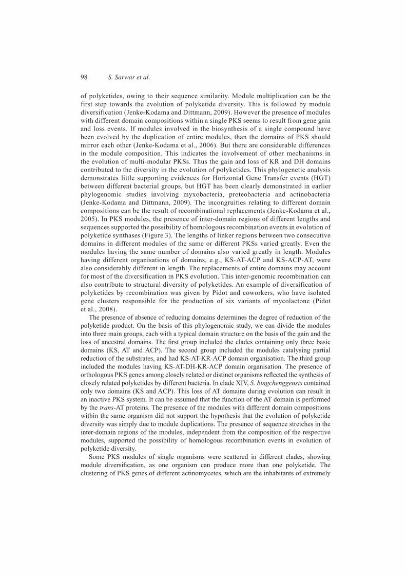
of polyketides, owing to their sequence similarity. Module multiplication can be the first step towards the evolution of polyketide diversity. This is followed by module diversification (Jenke-Kodama and Dittmann, 2009). However the presence of modules with different domain compositions within a single PKS seems to result from gene gain and loss events. If modules involved in the biosynthesis of a single compound have been evolved by the duplication of entire modules, than the domains of PKS should mirror each other (Jenke-Kodama et al., 2006). But there are considerable differences in the module composition. This indicates the involvement of other mechanisms in the evolution of multi-modular PKSs. Thus the gain and loss of KR and DH domains contributed to the diversity in the evolution of polyketides. This phylogenetic analysis demonstrates little supporting evidences for Horizontal Gene Transfer events (HGT) between different bacterial groups, but HGT has been clearly demonstrated in earlier phylogenomic studies involving myxobacteria, proteobacteria and actinobacteria (Jenke-Kodama and Dittmann, 2009). The incongruities relating to different domain compositions can be the result of recombinational replacements (Jenke-Kodama et al., 2005). In PKS modules, the presence of inter-domain regions of different lengths and sequences supported the possibility of homologous recombination events in evolution of polyketide synthases (Figure 3). The lengths of linker regions between two consecutive domains in different modules of the same or different PKSs varied greatly. Even the modules having the same number of domains also varied greatly in length. Modules having different organisations of domains, e.g., KS-AT-ACP and KS-ACP-AT, were also considerably different in length. The replacements of entire domains may account for most of the diversification in PKS evolution. This inter-genomic recombination can also contribute to structural diversity of polyketides. An example of diversification of polyketides by recombination was given by Pidot and coworkers, who have isolated gene clusters responsible for the production of six variants of mycolactone (Pidot et al., 2008).
The presence of absence of reducing domains determines the degree of reduction of the polyketide product. On the basis of this phylogenomic study, we can divide the modules into three main groups, each with a typical domain structure on the basis of the gain and the loss of ancestral domains. The fi rst group included the clades containing only three basic domains (KS, AT and ACP). The second group included the modules catalysing partial reduction of the substrates, and had KS-AT-KR-ACP domain organisation. The third group included the modules having KS-AT-DH-KR-ACP domain organisation. The presence of orthologous PKS genes among closely related or distinct organisms refl ected the synthesis of closely related polyketides by different bacteria. In clade XIV, S. bingchenggensis contained only two domains (KS and ACP). This loss of AT domains during evolution can result in an inactive PKS system. It can be assumed that the function of the AT domain is performed by the trans-AT proteins. The presence of the modules with different domain compositions within the same organism did not support the hypothesis that the evolution of polyketide diversity was simply due to module duplications. The presence of sequence stretches in the inter-domain regions of the modules, independent from the composition of the respective modules, supported the possibility of homologous recombination events in evolution of polyketide diversity.
Some PKS modules of single organisms were scattered in different clades, showing module diversifi cation, as one organism can produce more than one polyketide. The clustering of PKS genes of different actinomycetes, which are the inhabitants of extremely
Phylogenomic and structural analysis of PKS genes 99
different environments together, suggested that these genes might have been evolved from a common ancestral gene, and the evolution of polyketide synthases is as older as the evolution of different actinomycetes adopting new and unique environments.
3.3 Structural analysis
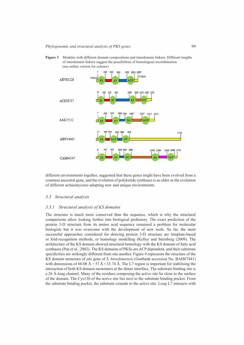
3.3.1 Structural analysis of KS domainsThe structure is much more conserved than the sequence, which is why the structural comparisons allow looking further into biological prehistory. The exact prediction of the protein 3-D structure from its amino acid sequence remained a problem for molecular biologists but it was overcome with the development of new tools. So far, the most successful approaches considered for drawing protein 3-D structure are template-based or fold-recognition methods, or homology modelling (Kelley and Sternberg (2009). The architecture of the KS domain showed structural homology with the KS domain of fatty acid synthases (Pan et al., 2002). The KS domains of PKSs are ACP dependent, and their substrate specifi cities are strikingly different from one another. Figure 4 represents the structure of the KS domain monomer of pks gene of S. hiroshimensis (Genbank accession No. BAH67441) with dimensions of 60.08 Å × 57 Å × 33.74 Å. The L7 region is important for stabilising the interaction of both KS domain monomers at the dimer interface. The substrate binding site is a 26 Å-long channel. Many of the residues composing the active site lie close to the surface of the domain. The Cys130 of the active site lies next to the substrate binding pocket. From the substrate binding pocket, the substrate extends to the active site. Loop L7 interacts with
Figure 3 Modules with different domain compositions and interdomain linkers. Different lengths of interdomain linkers suggest the possibilities of homologous recombination (see online version for colours)
100 S. Sarwar et al.
the L7 while loop L3 interacts with the L3 of the KS domains at the dimer interface in KS dimers. L3 also forms the base of the substrate binding pocket. The catalytic triad is formed by Cys130, His265 and Asn237.
Figure 4 The structure of an active KS domain of S. hiroshimensis (Genbank accession No. BAH67441). The 3D structure was predicted using the phyre (protein fold recognition server) tool and visualised in VMD 1.9 software (see online version for colours)
The amino acids constituting the active site of the KS domain are located around the cysteine residue. In most actinomycetes, the active site of KS domain has the sequence VDTACSSS (Figure 5). The highly conserved amino acids in this active site were C (Cysteine), S (Serine) and D (aspartic acid). However other amino acids often replace V (Valine) in the active site. In the present study, V (Valine) residue was mostly replaced by I (Isoleucine) and L (Leucine). In 14% of the cases, A (Alanine) replaced the T residue, and thus, the active site had VDAACSSS sequence. Within the same organism, the sequence of the active site varied between different KS domains. The KS domain of Streptomyces sp. ID05-A0450 (Genbank accession No. BAH68155), in which the cysteine residue has been replaced with W (tryptophan), is considered inactive because the presence of cysteine residue within the active site is necessary for the condensation activity of the KS domain. The sequence of the active site of this domain is VDTAWPSS. Amino acid substitution, leading to the loss of activity within the active site, gave the evidence of birth-death evolution. This process of birth-to-death evolution has also contributed to the diversity of polyketides. Similarly, certain point mutations in the substrate binding pockets alter substrate specifi cities, and thus, contribute to diversity and evolution of polyketides.

Phylogenomic and structural analysis of PKS genes 101
Figure 5 ClustalW multiple sequence alignment of amino acid sequences of KS domains. Only the parts of the sequences containing active site (100–174 residues) of a selected number of KS domains are shown.
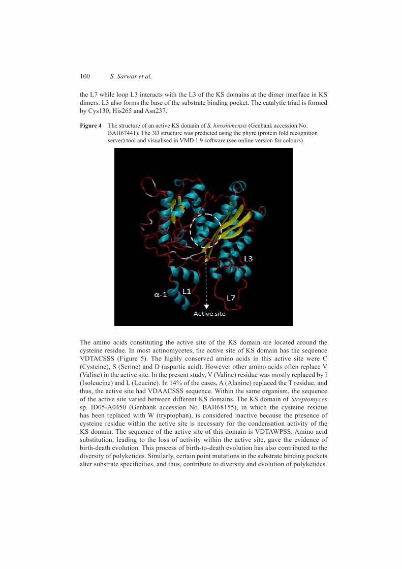
The structure of the KS domain monomer of S. hiroshimensis (Genbank accession No. BAH67441) was superimposed with the KS domain monomer of S. roseochromogenus (Genbank accession No. BAH67135) (Figure 6).The substrate binding pocket of the KS domain of S. roseochromogenus was 27 Å long, and its active site had LDTACSSS as the sequence. The substrate binding pockets of both monomers were aligned in the same line. The core regions were similar in both domains, while the loops of both domains were strikingly different. Extensive connecting loop regions were present around the core. These loop regions are important in maintaining the fl exibility of the substrate binding pocket. In the case of S. hiroshimensis, smaller than average loop regions infl uence the size of the substrate binding pocket, and KS domains with small L7 and L3 regions have small substrate binding pockets, and thus favour small priming substrates like acetyl CoA. KS domains with large substrate binding pockets favour large substrates for the synthesis of polyketides. So the overall architecture is important in the selection of the substrate.
Thus, the changes in the conformation of substrate binding pockets of KS domains of PKSs can also lead to a change in substrate specifi city and in turn, to the product being synthesised. Throughout the evolution of the PKSs, the diversity of the products being synthesised has grown signifi cantly, with a little change in the structure of the KS domain. This considerable structural similarity supports the hypothesis of gene duplication in the evolution of polyketide synthases, while only some of them are members of early multi-gene families before speciation, whereas others are deleted eventually. Point mutations in the sequence of biosynthetic enzymes can alter the chemistry of the metabolite being synthesised, contributing to gene diversifi cation.
The structure of α-helix at the N terminal end of the KS domain is important for the inter-modular interactions of PKSs. The modules or multi-enzyme subunits associate with
102 S. Sarwar et al.
one another to form overall PKS complexes. This region is responsible for interaction with the ACP domain of the next module in an assembly line. This long α-helix interacts with 3 α-helices of the ACP domain. These regions are known as docking domains. The interaction between these helices is responsible for protein-protein recognition. The amino acid sequences of these α-helices are highly conserved. These conserved residues are responsible for inter-domain interactions. A small number of PKS docking domains showed lower sequence similarity.
3.3.2 Structural analysis of PKS modulesTotal 62 PKS modules were grouped into four major categories, depending upon the module composition. Some modules contained the terminal Thioesterase (TE) domain.
• KS-ACP: 1 module (loading module)
• KS-AT-ACP: 15 modules
• KS-AT-KR-ACP: 19 modules
• KS-AT-DH-KR-ACP: 27 modules.
Figure 6 (A) Structural comparison of KS domains of S. hiroshimensis (Genbank accession No. BAH67441) (red) and S. roseochromogenus (Genbank accession No.BAH67135) (blue). Red and blue colours indicate the regions having similar structures. The regions in cyan and salmon colours differ from one another in structure. (B) KS domain structure of S. hiroshimensis (Genbank accession No. BAH67441). (C) KS domain structure of S. roseochromogenus (Genbank accession No. BAH67135). (see online version for colours)
Phylogenomic and structural analysis of PKS genes 103
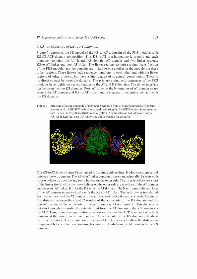
3.3.3 Architecture of KS-to-AT didomainFigure 7 represents the 3D model of the KS-to-AT didomain of the PKS module, with KS-AT-ACP domain composition. This KS-to-AT is a homodimeric protein, and each monomer contains the full length KS domain, AT domain and two linker regions: KS-to-AT linker and post-AT linker. The linker regions comprise a signifi cant fraction of the PKS module, and the domains are linked to one another in the module via these linker regions. These linkers lack sequence homology to each other and with the linker regions of other proteins, but have a high degree of structural conservation. There is no direct contact between the domains. The primary amino acid sequences of the PKS modules have highly conserved regions in the AT and KS domains. The dimer interface lies between the two KS domains. Post_AT linker at the N terminus of AT domain wraps around the AT domain and KS-to-AT linker, and is engaged in extensive contacts with the KS domains.
Figure 7 Structure of a single module of polyketide synthase from S. bingchenggensis, (Genbank accession No. ADI09173) which was predicted using the SBSPKS online bioinformatics tool. Green, Ketosynthase (KS) domain; yellow, Acyltransferase (AT) domain; purple, KS_AT linker; red, post_AT linker (see online version for colours)
The KS-to AT linker (Figure 8) contained 110 amino acid residues. It adopts a compact fold between the two domains. The KS-to AT linker contains three stranded parallel β sheets with three α helices on one side and two α helices on the other side. The three α helices are a part of the linker itself, while the two α helices on the other side are α helices of the AT domain and the post_AT linker. It links the KS with the AT domain. The N terminus helix and loop of the AT domain interact closely with the KS-to-AT linker. The substrate is transferred from the active site of the AT domain to the active site of the KS domain via the ACP domain. The distance between the Cys-207 residue of the active site of the KS domain and the Ser-645 residue of the active site of the AT domain is 53 Å (Figure 9). This distance is not short enough to transfer the extender unit from the AT domain to the KS domain via the ACP. Thus, domain reorganisation is necessary to allow the ACP to interact with both domains at the same time in one module. The active site of the KS domain extends to the dimer interface. The orientation of the post-AT linker seems to allow the distance to be spanned between the two domains, because it extends from the AT domain to the KS domain.

104 S. Sarwar et al.
Figure 8 Structure of KS-to-AT linker in the PKS module (Genbank accession No. ADI09173). A central three stranded β-sheet surrounded by three α-helices (green) on one side and two α-helices (red) on the other side (see online version for colours)
Figure 9 KS and AT domains with their active site residues (Cys-207 residue of KS domain and Ser-645 residue of AT domain). Analysis was done with the RASMOL visualisation tool (see online version for colours)
These linker regions play important structural roles in fi xing the relative positions of the KS and AT domains (Tang et al., 2006). The compact and rigid arrangement of KS domain, KS-to-AT linker, the AT domain and the post-AT linker peptide precludes the movement of the KS and AT domains relative to one another. The organisation of the domains in an assembly line in the modules of PKSs provides a more effi cient platform for combinatorial biosynthesis. By the combinatorial manipulation of PKS modules and subunits, it is possible to generate libraries of novel polyketides (Pfeifer and Khosla, 2001).
3.4 Architecture of PKS module with KR and DH domainThe second category includes PKS modules with KS-AT-KR-ACP domain composition. The interactions between these domains are mediated by inter-domain linkers and the mobile ACP domain. The KR domain lacks considerable internal sequence homology. It forms a compact

Phylogenomic and structural analysis of PKS genes 105
monomeric structure, and consists of two tightly packed subdomains. The 293 amino acid AT-KR linker region forms the structural KR subdomain (Figure 10 lavender) while the 70 amino acids constitute the conventional catalytic KR domain along with the KR-ACP linker (Figure 10 purple). Such organisation of the KR domain is found in all the modules with similar domain composition. The cleft between the KS, AT and KR domains can be the possible location for the ACP. However, the large distance between these domains supports the possibility of a mobile ACP domain. The KS and AT domains form the centre of the module. The KR domain does not make direct contact with other domains across the 2-fold axis of the module. At the C terminal end of the structural domain there is a β-strand (β-1) which forms the bridge between the two subdomains. In modules lacking the DH and ER domains, the KR domain is located close to the central core of the module.
In the third category, the PKS module with the KR and DH domains, three domains (KS, AT and ACP) form the core of the homodimer, while the reductive domains (KR and DH) protrude out from the axis of the central core. In the dimeric structure of the module, the KS domain forms contact with the DH domain (Figure 10). The ACP domain forms functional contacts with the AT, KS and DH domains. The two halves of the dimer do not adopt a similar conformation. The monomers are showing pseudo-symmetry about an axis running through the body of the dimer at a right angle. There are two hinges on either side of the structure. One is located between the KS and AT domains, while the other hinge is located between the KR domain and the central body. A third small hinge is located between the DH and the KS domain. In the central core, the dimeric KS domain is fl anked by a pair of monomeric AT domains. The DH domain makes contact with the catalytic subdomain of KR. The boundaries between the domains lie in linker regions. These linker regions varied considerably in length between different modules.
Figure 10 Structure of a single module of polyketide synthase with KS-AT-DH-KR-ACP domain composition from Actinosynnema mirum (Genbank accession No. ACU38680). Green, Ketosynthase (KS) domain; yellow, Acyltransferase (AT) domain; red, post_AT linker; slate blue, KS_AT linker; purple, catalytic subdomain of Ketoreductase (KR); lavender, structural subdomain of KR; sky blue, Dehydrogenase (DH). (see online version for colours)

106 S. Sarwar et al.
3.5 Substrate bindingKetosynthases (KS) have different substrate specifi cities. Common primer units for the KS domains include acetyl CoA, propionyl CoA and butyryl CoA (Khosla et al., 1999). In this study, the binding of the primer unit, acetyl CoA, to the substrate binding pocket of the KS domain monomer of pks gene of S. hygroscopicus (Genbank accession no. BAH67161) is studied. The catalytic triad is formed by Cys130, His265 and Asn237. The CoA moiety of acetyl CoA extends towards the catalytic triad, while the acetyl group extends towards the KS dimer interface because the acetyl binding region is located at the end of the substrate binding channel (Figure 11). The substrate binding pocket is lined with conserved residues, and the CoA moiety of acetyl CoA is engaged in extensive interactions with the conserved residues of the substrate binding pocket. The binding of CoA moiety of acetyl CoA is known to stabilise the shape of the substrate binding pocket (Pan et al., 2002). The Cys130 residue lies in close proximity to the basic histidine residue (His265). The L3 region is present at the lower side of the acetyl group binding region, and is important in determining the substrate specifi city of the priming ketosynthases. On the other hand, the L1 regions take part in the formation of the CoA binding region. The size of the acyl group binding region determines the substrate specifi city for acyl chains of varying lengths. Understanding the molecular basis of substrate specifi cities of the KS domains can be exploited in the combinatorial biosynthesis of polyketides.
Figure 11 (A) KS domain monomer of Streptomyces hygroscopicus with acetyl CoA in the substrate binding pocket. (B) Acetyl CoA binding channel with conserved residues, which interact with the CoA and acetyl groups of the primer unit. The catalytic triad is formed by the Cys130, His265 and Asn237 (see online version for colours)
Phylogenomic and structural analysis of PKS genes 107
3.6 Gene length conservationThe maximum and minimum lengths of modules (under study) with KS-AT-ACP domain composition were 1733 and 957 amino acid residues, respectively. The Frankia sp. EAN1pec(ABW14965), with 1733 amino acid residues in the module, was present in the clade which was at the basal position within the phylogenetic tree earlier given in Figure 1. Thus, the length of modules went on decreasing during evolution, and the module with 957 amino acid residues was present at a distal position in the phylogenetic tree. For KS-AT-KR-ACP modules, the maximum and minimum lengths were 1738 and 1097 amino acid residues respectively.
Similarly, S. griseus (BAG22905), with 1097 amino acid residues, was present in the clade at the distal position, while Saccharopolyspora erythraea (CAM04719), with 1738 amino acid residues was present at basal position. Likewise, for modules with KS-AT-DH-KR-ACP domain composition, the maximum and minimum lengths were 1924 and 1611 amino acid residues, respectively. Both modules clustered within a single clade, while Frankia sp. EAN1pec (ABW14203), with 1924 amino acid residues, was present at the basal position and Amycolatopsis mediterranei (CAA11037), with 1611 amino acid residues, was present at the distal position within the clade.
From the present study, it can be concluded that polyketide diversity can be greatly increased in the future by the genetic manipulation of PKS biosynthetic pathways. Various mechanisms are responsible for alterations in the architecture of the substrate binding pockets of the domains to accept novel starter and extender units, by the addition of the reducing domains to the minimal modules to change the reduction status of the product by changing the number of modules in an assembly line and by combining domains or modules form different organisms to make novel PKSs. McDaniel et al. (1997) introduced a number of changes in the domain architecture and generated 61 different 6-DEB analogues. Other examples of structural modifi cations have also been reported to improve their utility as clinical drugs (Floss, 2006). For example, structural modifi cation of rifamycin B to rifampicin made oral use of this drug possible. This approach also improved its pharmacodynamic properties, including its potency (Floss and Yu, 2005). Thus, the structural insights from polyketide synthases provide an understanding of the biosynthetic pathways that can be modifi ed.
4 Conclusion
Natural products are not produced in nature as clinical agents. They need to be modifi ed, as semisynthetic components have less toxicity than the natural ones. Thus, the application of combinatorial biosynthesis approaches to polyketides can optimise their properties to be used as clinical drugs. Even the structure of a less or ineffective drug can be used for drug development. In conclusion, the understanding of the evolutionary processes in the development of natural products can help to generate novel polyketides. Combinatorial biosynthesis and metabolic engineering can complement the screening for polyketide libraries. However, much more work needs to be done to provide practical applications to these recombinant approaches.
Acknowledgements
The University of the Punjab, Lahore , is highly acknowledged for funding and supporting this research work.

108 S. Sarwar et al.
ReferencesAhmed, Z.U. and Vining, L.C. (1983) ‘Evidence for a chromosomal location of the genes coding
for chloramphenicol production in Streptomyces venezuelae’, Journal of Bacteriology, Vol. 154, No. 1, pp.239–244.
Anand, S., Prasad, M.V.R., Yadav, G., Kumar, N., Shehara, J., Ansari, M.Z. and Mohant,V. (2010) ‘SBSPKS: structure based sequence analysis of polyketide synthases’, Nucleic Acids Research, Vol. 38,No. suppl 2, p.W487.
Aparicio, J.F., Molnar, I., Schwecke, T., Konig, A., Haydock, S.F., EeKhaw, L., Staunton, J. and Leadlay, P.F. (1996) ‘Organization of the biosynthetic gene cluster for rapamycin in Streptomyces hygroscopicus: analysis of the enzymatic domains in the modular polyketide synthase’, Gene, Vol. 169, No. 1, pp.9–16.
Cundliffe, E., Bate, N., Butler, A., Fish, S., Gandecha, A. and Merson-Davies, L. (2001) ‘The tylosin-biosynthetic genes of Streptomyces fradiae’, Antonie Van Leeuwenhoek, Vol. 79, No. 3, pp.229–234.
Donadio, S., Busti, E., Monciardini, P., Bamonte, R., Mazza,Wohlleben, W., Spellig, T. and Müller-Tiemann, B. (Eds.): P., Sosio, M. and Cavaletti, L. (2005) ‘Sources of polyketides and non-ribosomal peptides’, in Biocombinatorial Approaches for Drug Finding, Springer-Verlag, Berlin. pp.19–41.
Doull, J.L. and Vining, L.C. (1990) ‘Nutritional control of actinorhodin production by Streptomyces coelicolor A3 (2): suppressive effects of nitrogen and phosphate’, Applied Microbiology and Biotechnology, Vol. 32, No. 4, pp.449–454.
Floss, H.G. (2006) ‘Combinatorial biosynthesis–potential and problems’, Journal of Biotechnology, Vol. 124, No. 1, pp.242–257.
Floss, H.G. and Yu, T.W. (2005) ‘Rifamycin mode of action, resistance, and biosynthesis’, Chemical Reviews, Vol. 105, No. 2, pp.621–632.
Ginolhac, A., Jarrin, C., Robe, P., Perriere, G., Vogel, T.M., Simonet, P. and Nalin, R. (2005) ‘Type I polyketide synthases may have evolved through horizontal gene transfer’, Journal of Molecular Evolution, Vol. 60, No. 6, pp.716–725.
Horan, A.N.N.C. and Brodsky, B.C. (1982) ‘A novel antibiotic-producing actinomadura, Actinomadurakijaniata sp. nov.’, International Journal of Systematic Bacteriology, Vol. 32, No. 2, pp.195–200.
Ichinose, K., Bedford, D.J., Tornus, D., Bechthold, A., Bibb, M.J., Peter, W., Revill, H. Floss, G. and Hopwood, D.A. (1998) ‘The granaticin biosynthetic gene cluster of Streptomyces violaceoruberTu22: sequence analysis and expression in a heterologous host’, Chemistry & Biology, Vol. 5, No. 11, pp.647–659.
Jenke-Kodama, H. and Dittmann, E. (2009) ‘Evolution of metabolic diversity: insights from microbial polyketide synthases’, Phytochemistry, Vol. 70, Nos. 15–16, pp.1858–1866.
Jenke-Kodama, H., Borner, T. and Dittmann, E. (2006) ‘Natural biocombinatorics in the polyketide synthase genes of the actinobacterium Streptomyces avermitilis, PLoS Computational Biology, Vol. 2, No. 10, p.e132.
Jenke-Kodama, H., Muller, R. and Dittmann, E. (2008) ‘Evolutionary mechanisms underlying secondary metabolite diversity’, Natural Compounds as Drugs, Vol. I, pp.119–140.
Jenke-Kodama, H., Sandmann, A., Muller, R. and Dittmann, E. (2005) ‘Evolutionary implications of bacterial polyketide synthases’, Molecular Biology and Evolution, Vol. 22, No. 10, pp.2027–2039.
Kelley, L.A. and Sternberg, M.J.E. (2009) ‘Protein structure prediction on the Web: a case study using the Phyre server’, Nature Protocols, Vol. 4, pp.363–371.
Khosla, C., Gokhale, R.S., Jacobsen, J.R. and Cane, D.E. (1999) ‘Tolerance and specifi city of polyketide synthases’, Annual Review of Biochemistry, Vol. 68, No. 1, pp.219–253.
Kim, B.G., Lee, M.J., Seo, J., Hwang, Y.B., Lee, M.Y., Han, K., Sherman, D.H. and Kim, E.S. (2009) ‘Identifi cation of functionally clustered nystatin-like biosynthetic genes in a rare actinomycetes, Pseudonocardiaautotrophica’, Journal of Industrial Microbiology & Biotechnology, Vol. 36, No. 11, pp.1425–1434.

Phylogenomic and structural analysis of PKS genes 109
Komaki, H. and Harayama, S. (2006) ‘Sequence diversity of type-II polyketide synthase genes in streptomyces’, Actinomycetologica, Vol. 20, No. 2, pp.42–48.
Komaki, H., Izumikawa, M., Ueda, J., Nakashima, T., Khan, S.T., Takagi, M. and Shin-ya, K. (2009) ‘Discovery of a pimaricin analog JBIR-13, from Streptomyces bicolor NBRC 12746 as predicted by sequence analysis of type I polyketide synthase gene’, Applied Microbiology and Biotechnology, Vol. 83, No. 1, pp.127–133.
Kroken, S., Glass, N.L., Taylor, J.W., Yoder, O.C. and Turgeon, B.G. (2003) ‘Phylogenomic analysis of type I polyketide synthase genes in pathogenic and saprobicascomycetes’, Proceedings of the National Academy of Sciences, Vol. 100, No. 26, p.15670.
Lambert, C., Léonard, N., De Bolle, X. and Depiereux, E. (2002) ‘ESyPred3D: prediction of proteins 3D structures’, Bioinformatics, Vol. 18, pp.1250–1256.
Land, M., Lapidus, A., Mayilraj, S., Chen, F., Copeland, A., Del Rio, T.G., Nolan, M., Lucas, S., Tice, H. and Cheng, J.F. (2009) ‘Complete genome sequence of Actinosynnemamirum type strain (101T)’, Standards in Genomic Sciences, Vol. 1, No. 1, p.46.
McDaniel, R., Kao, C.M., Hwang, S.J. and Khosla, C. (1997) ‘Engineered intermodular and intramodularpolyketide synthase fusions’, Chemistry & Biology, Vol. 4, No. 9, pp.667–674.
McGuffi n, L.J., Bryson, K. and Jones, D.T. (2000) ‘The PSIPRED protein structure prediction server’, Bioinformatics, Vol. 16, No. 4, p.404.
Menzella, H.G., Reid, R., Carney, J.R., Chandran, S.S., Reisinger, S.J., Patel, K.G., Hopwood, D.A. and Santi, D.V. (2005) ‘Combinatorial polyketide biosynthesis by de novo design and rearrangement of modular polyketide synthase genes’, Nature Biotechnology, Vol. 23, No. 9, pp.1171–1176.
Nakamura, S., Yajima, T., Hamada, M., Nishimura, T. and Ishizuka, M. (1967) ‘A new antitumor antibiotic, phenomycin’, The Journal of Antibiotics, Vol. 20, No. 4, p.210.
Nei, M. and Rooney, A.P. (2005) ‘Concerted and birth-and-death evolution of multigene families’, Annual Review of Genetics, Vol. 39, p.121.
Novakova, R., Bistakova, J., Homerova, D., Rezuchova, B. and Kormanec, J. (2002) ‘Cloning and characterization of a polyketide synthase gene cluster involved in biosynthesis of a proposed angucycline-like polyketideauricin in Streptomyces aureofaciens CCM 3239’, Gene, Vol. 297, Nos. 1–2, pp.197–208.
Omura, S., Ikeda, H., Ishikawa, J., Hanamoto, A., Takahashi, C., Shinose, M., Takahashi, Y., Horikawa, H., Nakazawa, H. and Osonoe, T. (2001) ‘Genome sequence of an industrial microorganism Streptomyces avermitilis: deducing the ability of producing secondary metabolites’, Proceedings of the National Academy of Sciences, Vol. 98, No. 21, p.12215.
Pan, H., Tsai, S., Meadows, E.S., Miercke, L.J.W., Keatinge-Clay, A.T., O’Connell, J., Khosla, C. and Stroud, R.M. (2002) ‘Crystal structure of the priming [beta]-ketosynthase from the R1128 polyketide biosynthetic pathway’, Structure, Vol. 10, No. 11, pp.1559–1568.
Panek, J.S. and Jain, N.F. (1998) ‘Total synthesis of oligomycin C’, The Journal of Organic Chemistry, Vol. 63, No. 14, pp.4572–4573.
Pang, M., Tan, G.Y.A., Abdullah, N., Lee, C.W. and Ng, C.C. (2008) ‘Phylogenetic analysis of type I and type II polyketide synthase from tropical forest soil’, Biotechnology, Vol. 7, No. 4, pp.660–668.
Perlmutter, P. (1996) ‘The nucleophilic addition/electrophilic ring closure route to bio-active heterocycles’, Current Medicinal Chemistry, Vol. 3, No. 2, p.139.
Pfeifer, B.A. and Khosla, C. (2001) ‘Biosynthesis of polyketides in heterologous hosts’, Microbiology and Molecular Biology Reviews, Vol. 65, No. 1, pp.106–118.
Pidot, S., Hong, H., Seemann, T., Porter, J., Yip, M., Men, A., Johnson, M., Wilson, P., Davies, J. and Leadlay, P. (2008) ‘Deciphering the genetic basis for polyketide variation among mycobacteria producing mycolactones’, BMC Genomics, Vol. 9, No.1, p.462.
Qu, X., Jiang, N., Xu, F., Shao, L., Tang, G., Wilkinson, B. and Liu, W. (2011) ‘Cloning, sequencing and characterization of the biosynthetic gene cluster of sanglifehrin A, a potent cyclophilin inhibitor’, Molecular BioSystems, Vol. 7, pp.852–861.

110 S. Sarwar et al.
Schupp, T., Toupet, C., Engel, N. and Goff, S. (1998) ‘Cloning and sequence analysis of the putative rifamycinpolyketide synthase gene cluster from Amycolatopsismediterranei’, FEMS Microbiology Letters, Vol. 159, No. 2, pp.201–207.
Steinerova, N., Lipavska, H., Stajner, K., Caslavska, J., Blumauerova, M., Cudlin, J. and Vank, Z. (1987) ‘Production of quinomycinA in streptomyces lasaliensis’, Folia Microbiologica, Vol. 32, No. 1, pp.1–5.
Tamura, K., Dudley, J., Nei, M. and Kumar, S. (2007) ‘MEGA4: molecular evolutionary genetics analysis (MEGA) software version 4.0’, Molecular Biology and Evolution, Vol. 24, No. 8, pp.1596–1599.
Tang, Y., Kim, C.Y., Mathews, I.I., Cane, D.E. and Khosla, C. (2006) ‘The 2.7-Å crystal structure of a 194-kDa homodimeric fragment of the 6-deoxyerythronolide B synthase’, Proceedings of the National Academy of Sciences, Vol. 103, No. 30, p.11124.
Traitcheva, N., Jenke-Kodama, H., He, J., Dittmann, E. and Hertweck, C. (2007) ‘Non-colinear polyketide biosynthesis in the aureothin and neoaureothin pathways: an evolutionary perspective’, Chem. Bio Chem., Vol. 8, No. 15, pp.1841–1849.
Tsunakawa, M., Tenmyo, O., Tomita, K., Naruse, N., Kotake, C., Miyaki, T., Konishi, M. and Oki, T. (1992) ‘Quartromicin, a complex of novel antiviral antibiotics. I. Production, isolation, physico-chemical properties and antiviral activity’, The Journal of Antibiotics, Vol. 45, No. 2, p.180.
Waksman, S.A., Reilly, H.C. and Johnstone, D.B. (1946) ‘Isolation of streptomycin-producing strains of streptomyces griseus’, Journal of Bacteriology, Vol. 52, No. 3, p.393.
Waksman, S.A., Schatz, A. and Reynolds, D.M. (2010) ‘Production of antibiotic substances by actinomycetes’, Annals of the New York Academy of Sciences, Vol. 1213, No. 1, pp.112–124.
Wang, X.J., Yan, Y.J., Zhang, B., An, J., Wang, J.J., Tian, J., Jiang, L., Chen, Y.H., Huang, S.X. and Yin, M. (2010) ‘Genome sequence of the milbemycin-producing bacterium streptomyces bingchenggensis’, Journal of Bacteriology, Vol. 192, No. 17, p.4526.
Weissman, K.J. and Leadlay, P.F. (2005) ‘Combinatorial biosynthesis of reduced polyketides’, Nature Reviews Microbiology, Vol. 3, No. 12, pp.925–936.
Wu, K., Chung, L., Revill, W.P., Katz, L. and Reeves, C.D. (2000) ‘The FK520 gene cluster of Streptomyces hygroscopicus var. ascomyceticus (ATCC 14891) contains genes for biosynthesis of unusual polyketide extender units’, Gene, Vol. 251, No. 1, pp.81–90.
Yadav, G., Gokhale, R.S. and Mohanty, D. (2003) ‘SEARCHPKS: a program for detection and analysis of polyketide synthase domains’, Nucleic Acids Research, Vol. 31, pp.3654–3658.
Zucko, J., Cullum, J., Hranueli, D. and Long, P.F. (2011) ‘Evolutionary dynamics of modular polyketide synthases, with implications for protein design and engineering’, The Journal of Antibiotics, Vol. 64, No. 1, pp.89–92.
Related Documents