Structure Article Structural Basis for the Versatile Interactions of Smad7 with Regulator WW Domains in TGF- b Pathways Eric Arago ´ n, 1 Nina Goerner, 1,5 Qiaoran Xi, 2 Tiago Gomes, 1 Sheng Gao, 2,6 Joan Massague ´, 2,3, * and Maria J. Macias 1,4, * 1 Structural and Computational Biology Programme, Institute for Research in Biomedicine, Baldiri Reixac 10-12, 08028 Barcelona, Spain 2 Cancer Biology and Genetics Program, Memorial Sloan-Kettering Cancer Center, 1275 York Avenue, New York, NY 10065, USA 3 Howard Hughes Medical Institute (HHMI), Chevy Chase, MD 20185, USA 4 Institucio ´ Catalana de Recerca i Estudis Avanc ¸ ats (ICREA), Passeig Lluı ´s Companys 23, 08010-Barcelona, Spain 5 Present address: Biocrates Life Sciences AG, Innrain 66, 6020 Innsbruck, Austria 6 Present address: Cardiovascular Diseases, Merck Research Laboratory, Merck & Co., 126 E. Lincoln Avenue, Rahway, NJ 07065, USA *Correspondence: [email protected] (J.M.), [email protected] (M.J.M.) http://dx.doi.org/10.1016/j.str.2012.07.014 SUMMARY Transforming growth factor (TGF)-b and BMP signaling is mediated by Smads 1–5 (R-Smads and Co-Smads) and inhibited by Smad7, a major hub of regulation of TGF-b and BMP receptors by negative feedback and antagonistic signals. The transcription coactivator YAP and the E3 ubiquitin ligases Smurf1/ 2 and Nedd4L target R-Smads for activation or degradation, respectively. Pairs of WW domain in these regulators bind PY motifs and adjacent CDK/ MAPK and GSK3 phosphorylation sites in R-Smads in a selective and regulated manner. In contrast, here we show that Smad7 binds YAP, Smurf1, Smurf2, and Nedd4L constitutively, the binding in- volving a PY motif in Smad7 and no phosphorylation. We also provide a structural basis for how regulators that use WW domain pairs for selective interactions with R-Smads, resort to one single versatile WW domain for binding Smad7 to centralize regulation in the TGF-b and BMP pathways. INTRODUCTION Smad transcription factors are key mediators of the transforming growth factor beta (TGF-b) and bone morphogenetic proteins (BMP) pathways in the control of stem cell pluripotency and differentiation, embryo development, tissue regeneration, and differentiated tissue homeostasis (Massague ´ , 1998). According to their function, Smad proteins are classified as receptor regu- lated Smads (R-Smads), which include Smads 1, 5, and 8 in the BMP-driven version of the SMAD pathway, and Smads 2 and 3 in the TGF-b/Nodal/Activin pathway. R-Smads form complexes with the common coactivator Smad (Co-Smad) Smad4. Two inhibitory Smads (I-Smads), Smad6 and Smad7, provide critical negative regulation to these powerful and ubiquitous pathways. R-Smads and Smad4 consist of two Mad Homology domains MH1 and MH2 connected by a linker region. This linker contains a cluster of phosphorylation sites adjacent to a proline rich PY motif. MH1 domains of R-Smads and Smad4 bind to DNA, whereas the MH2 domain and the linker function as scaffolds for receptors, regulator proteins, and transcription cofactors to interact and determine the outcome of the signal (Shi and Mas- sague ´ , 2003). Several key phosphorylation steps regulate the activation and turnover of R-Smads during the TGF-b and BMP signaling cycles. Binding of TGF-b or BMPs to their receptors triggers the receptor-mediated phosphorylation of R-Smads at their C termini. This phosphorylation generates a docking site for Smad4 for the assembly of a heterotrimeric transcriptional com- plex. Once the complex is in the nucleus, a second round of phosphorylations occurs in the linker region of R-Smads, creat- ing binding sites that interact with the WW domains of activators such as YAP and Pin1, as well as with the WW domains of the HECT-type E3 ubiquitin ligases Nedd4L, Smurf1, and Smurf2 that prime R-Smads for degradation (Alarco ´ n et al., 2009; Fan et al., 2009; Fuentealba et al., 2007; Gao et al., 2009; Kuratomi et al., 2005). These binding interactions depend on WW domain contacts with the PY motif and the phosphorylated sites, and with the exception of Pin1 always involve two WW domains (Ara- go ´ n et al., 2011). Compared to R-Smads and Co-Smads, the I-Smads have low sequence similarity in the MH1 domain but conserve an MH2 domain and a linker region with a characteristic PY motif (Figure 1A). I-Smads are expressed in response to TGF-b or BMPs to provide negative feedback in the pathway (Bai and Cao, 2002; Hata et al., 1998; He et al., 2002; Kavsak et al., 2000; Nakao et al., 1997; Yan et al., 2009) and in response to other pathways such as STAT to oppose TGF-b signaling (Ulloa et al., 1999). Smad6 interferes with the formation of Smad1- Smad4 complexes (Hata et al., 1998), whereas Smad7 inhibits TGF-b and BMP receptors (Hayashi et al., 1997; Topper et al., 1998). Work in recent years has revealed Smad7 as a central hub for negative regulation of activated TGF-b or BMP receptors (Yan and Chen, 2011). Receptor-bound Smad7 recruits ubiquitin ligases Nedd4L, Smurf1, and Smurf2 to mediate receptor poly- ubiquitination and route the receptor to degradative endocytosis (Ebisawa et al., 2001; Kavsak et al., 2000; Kuratomi et al., 2005). 1726 Structure 20, 1726–1736, October 10, 2012 ª2012 Elsevier Ltd All rights reserved

Welcome message from author
This document is posted to help you gain knowledge. Please leave a comment to let me know what you think about it! Share it to your friends and learn new things together.
Transcript
Structure
Article
Structural Basis for the Versatile Interactionsof Smad7 with Regulator WW Domainsin TGF-b PathwaysEric Aragon,1 Nina Goerner,1,5 Qiaoran Xi,2 Tiago Gomes,1 Sheng Gao,2,6 Joan Massague,2,3,* and Maria J. Macias1,4,*1Structural and Computational Biology Programme, Institute for Research in Biomedicine, Baldiri Reixac 10-12, 08028 Barcelona, Spain2Cancer Biology and Genetics Program, Memorial Sloan-Kettering Cancer Center, 1275 York Avenue, New York, NY 10065, USA3Howard Hughes Medical Institute (HHMI), Chevy Chase, MD 20185, USA4Institucio Catalana de Recerca i Estudis Avancats (ICREA), Passeig Lluıs Companys 23, 08010-Barcelona, Spain5Present address: Biocrates Life Sciences AG, Innrain 66, 6020 Innsbruck, Austria6Present address: Cardiovascular Diseases, Merck Research Laboratory, Merck & Co., 126 E. Lincoln Avenue, Rahway, NJ 07065, USA*Correspondence: [email protected] (J.M.), [email protected] (M.J.M.)http://dx.doi.org/10.1016/j.str.2012.07.014
SUMMARY
Transforming growth factor (TGF)-b and BMPsignaling is mediated by Smads 1–5 (R-Smads andCo-Smads) and inhibited by Smad7, a major hub ofregulation of TGF-b and BMP receptors by negativefeedback and antagonistic signals. The transcriptioncoactivator YAP and the E3 ubiquitin ligases Smurf1/2 and Nedd4L target R-Smads for activation ordegradation, respectively. Pairs of WW domain inthese regulators bind PY motifs and adjacent CDK/MAPK and GSK3 phosphorylation sites in R-Smadsin a selective and regulated manner. In contrast,here we show that Smad7 binds YAP, Smurf1,Smurf2, and Nedd4L constitutively, the binding in-volving a PYmotif in Smad7 and no phosphorylation.We also provide a structural basis for how regulatorsthat use WW domain pairs for selective interactionswith R-Smads, resort to one single versatile WWdomain for binding Smad7 to centralize regulationin the TGF-b and BMP pathways.
INTRODUCTION
Smad transcription factors are keymediators of the transforminggrowth factor beta (TGF-b) and bone morphogenetic proteins(BMP) pathways in the control of stem cell pluripotency anddifferentiation, embryo development, tissue regeneration, anddifferentiated tissue homeostasis (Massague, 1998). Accordingto their function, Smad proteins are classified as receptor regu-lated Smads (R-Smads), which include Smads 1, 5, and 8 in theBMP-driven version of the SMADpathway, and Smads 2 and 3 inthe TGF-b/Nodal/Activin pathway. R-Smads form complexeswith the common coactivator Smad (Co-Smad) Smad4. Twoinhibitory Smads (I-Smads), Smad6 and Smad7, provide criticalnegative regulation to these powerful and ubiquitous pathways.
R-Smads and Smad4 consist of two Mad Homology domainsMH1 and MH2 connected by a linker region. This linker contains
a cluster of phosphorylation sites adjacent to a proline rich PYmotif. MH1 domains of R-Smads and Smad4 bind to DNA,whereas the MH2 domain and the linker function as scaffoldsfor receptors, regulator proteins, and transcription cofactors tointeract and determine the outcome of the signal (Shi and Mas-sague, 2003).Several key phosphorylation steps regulate the activation
and turnover of R-Smads during the TGF-b and BMP signalingcycles. Binding of TGF-b or BMPs to their receptors triggersthe receptor-mediated phosphorylation of R-Smads at theirC termini. This phosphorylation generates a docking site forSmad4 for the assembly of a heterotrimeric transcriptional com-plex. Once the complex is in the nucleus, a second round ofphosphorylations occurs in the linker region of R-Smads, creat-ing binding sites that interact with the WW domains of activatorssuch as YAP and Pin1, as well as with the WW domains of theHECT-type E3 ubiquitin ligases Nedd4L, Smurf1, and Smurf2that prime R-Smads for degradation (Alarcon et al., 2009; Fanet al., 2009; Fuentealba et al., 2007; Gao et al., 2009; Kuratomiet al., 2005). These binding interactions depend on WW domaincontacts with the PY motif and the phosphorylated sites, andwith the exception of Pin1 always involve twoWWdomains (Ara-gon et al., 2011).Compared to R-Smads and Co-Smads, the I-Smads have
low sequence similarity in the MH1 domain but conserve anMH2 domain and a linker region with a characteristic PY motif(Figure 1A). I-Smads are expressed in response to TGF-b orBMPs to provide negative feedback in the pathway (Bai andCao, 2002; Hata et al., 1998; He et al., 2002; Kavsak et al.,2000; Nakao et al., 1997; Yan et al., 2009) and in response toother pathways such as STAT to oppose TGF-b signaling (Ulloaet al., 1999). Smad6 interferes with the formation of Smad1-Smad4 complexes (Hata et al., 1998), whereas Smad7 inhibitsTGF-b and BMP receptors (Hayashi et al., 1997; Topper et al.,1998).Work in recent years has revealed Smad7 as a central hub
for negative regulation of activated TGF-b or BMP receptors(Yan and Chen, 2011). Receptor-bound Smad7 recruits ubiquitinligases Nedd4L, Smurf1, and Smurf2 to mediate receptor poly-ubiquitination and route the receptor to degradative endocytosis(Ebisawa et al., 2001; Kavsak et al., 2000; Kuratomi et al., 2005).
1726 Structure 20, 1726–1736, October 10, 2012 ª2012 Elsevier Ltd All rights reserved
Moreover, Smad7 can simultaneously bind Smurf2 and theprotein deubiquitinase USP15, recruiting both enzymes to theTGF-b receptor complex for an integrated control of receptorpolyubiquitination as a function of ligand concentration (Eich-horn et al., 2012). Smad7 also binds YAP (Ferrigno et al., 2002)providing a mechanism for sequestration of this mediator ofHippo and BMP signaling (Alarcon et al., 2009). These proteininteractions involve the linker region of Smad7 and the WWdomain region of Nedd4L, Smurf1/2, and YAP.In order to advance our understanding of the functional capac-
ities of Smad7 as a hub for TGF-b or BMP pathway regulation,we investigated the interactions between the Smad7 PY motifregion and the WW domains of its client proteins, using NMRand ITC, ion mobility mass spectrometry, and protein interac-tion analysis in mammalian cells. Our data reveal a surprisinglyabsence of selectivity and independence from phosphorylationin the interactions of Smad7 with these proteins, illuminatingthe versatility of different WW domains as mediators of con-vergent interactions with a common Smad7 target, in additionto their discriminating interactions with different R-Smadproteins.
RESULTS
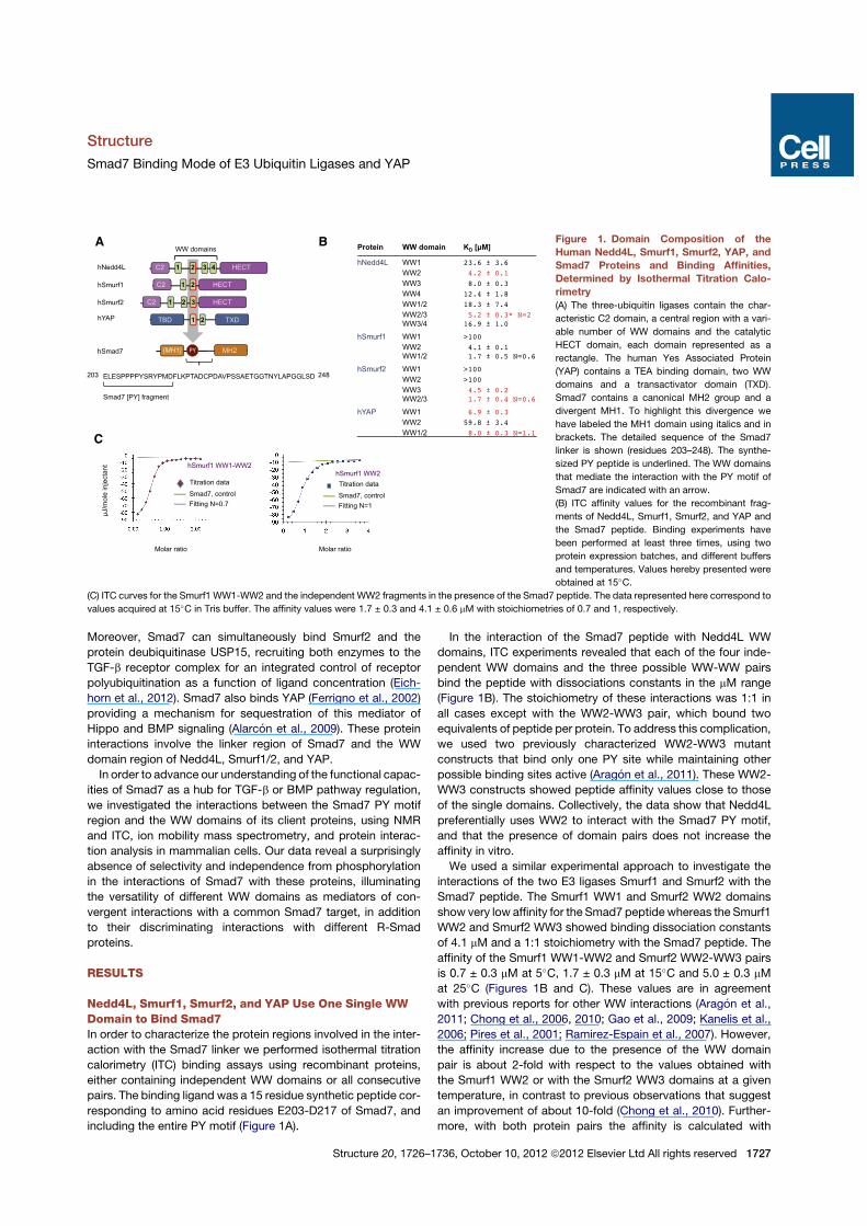
Nedd4L, Smurf1, Smurf2, and YAP Use One Single WWDomain to Bind Smad7In order to characterize the protein regions involved in the inter-action with the Smad7 linker we performed isothermal titrationcalorimetry (ITC) binding assays using recombinant proteins,either containing independent WW domains or all consecutivepairs. The binding ligand was a 15 residue synthetic peptide cor-responding to amino acid residues E203-D217 of Smad7, andincluding the entire PY motif (Figure 1A).
In the interaction of the Smad7 peptide with Nedd4L WWdomains, ITC experiments revealed that each of the four inde-pendent WW domains and the three possible WW-WW pairsbind the peptide with dissociations constants in the mM range(Figure 1B). The stoichiometry of these interactions was 1:1 inall cases except with the WW2-WW3 pair, which bound twoequivalents of peptide per protein. To address this complication,we used two previously characterized WW2-WW3 mutantconstructs that bind only one PY site while maintaining otherpossible binding sites active (Aragon et al., 2011). These WW2-WW3 constructs showed peptide affinity values close to thoseof the single domains. Collectively, the data show that Nedd4Lpreferentially uses WW2 to interact with the Smad7 PY motif,and that the presence of domain pairs does not increase theaffinity in vitro.We used a similar experimental approach to investigate the
interactions of the two E3 ligases Smurf1 and Smurf2 with theSmad7 peptide. The Smurf1 WW1 and Smurf2 WW2 domainsshow very low affinity for the Smad7 peptide whereas the Smurf1WW2 and Smurf2 WW3 showed binding dissociation constantsof 4.1 mM and a 1:1 stoichiometry with the Smad7 peptide. Theaffinity of the Smurf1 WW1-WW2 and Smurf2 WW2-WW3 pairsis 0.7 ± 0.3 mM at 5!C, 1.7 ± 0.3 mM at 15!C and 5.0 ± 0.3 mMat 25!C (Figures 1B and C). These values are in agreementwith previous reports for other WW interactions (Aragon et al.,2011; Chong et al., 2006, 2010; Gao et al., 2009; Kanelis et al.,2006; Pires et al., 2001; Ramirez-Espain et al., 2007). However,the affinity increase due to the presence of the WW domainpair is about 2-fold with respect to the values obtained withthe Smurf1 WW2 or with the Smurf2 WW3 domains at a giventemperature, in contrast to previous observations that suggestan improvement of about 10-fold (Chong et al., 2010). Further-more, with both protein pairs the affinity is calculated with
A B
C
Figure 1. Domain Composition of theHuman Nedd4L, Smurf1, Smurf2, YAP, andSmad7 Proteins and Binding Affinities,Determined by Isothermal Titration Calo-rimetry(A) The three-ubiquitin ligases contain the char-
acteristic C2 domain, a central region with a vari-
able number of WW domains and the catalytic
HECT domain, each domain represented as a
rectangle. The human Yes Associated Protein
(YAP) contains a TEA binding domain, two WW
domains and a transactivator domain (TXD).
Smad7 contains a canonical MH2 group and a
divergent MH1. To highlight this divergence we
have labeled the MH1 domain using italics and in
brackets. The detailed sequence of the Smad7
linker is shown (residues 203–248). The synthe-
sized PY peptide is underlined. The WW domains
that mediate the interaction with the PY motif of
Smad7 are indicated with an arrow.
(B) ITC affinity values for the recombinant frag-
ments of Nedd4L, Smurf1, Smurf2, and YAP and
the Smad7 peptide. Binding experiments have
been performed at least three times, using two
protein expression batches, and different buffers
and temperatures. Values hereby presented were
obtained at 15!C.
(C) ITC curves for the Smurf1 WW1-WW2 and the independent WW2 fragments in the presence of the Smad7 peptide. The data represented here correspond to
values acquired at 15!C in Tris buffer. The affinity values were 1.7 ± 0.3 and 4.1 ± 0.6 mM with stoichiometries of 0.7 and 1, respectively.
Structure
Smad7 Binding Mode of E3 Ubiquitin Ligases and YAP
Structure 20, 1726–1736, October 10, 2012 ª2012 Elsevier Ltd All rights reserved 1727
a stoichiometry below 1 at all temperatures (Figure 1C) (0.6–0.8range). ITC experiments measured in two different buffer solu-tions (tris and ammonium acetate [pH 7]) yielded similar valuesand stoichiometries.
We considered the possibility that formation of WW-WW pro-tein aggregates could affect the interpretation of the stoichi-ometry and affinity of Smad7 binding. Indeed, using nuclearmagnetic resonance (NMR), we observed nuclear Overhausereffects (NOEs) in Smurf1 samples containing either the WW1independent domain or the WW1-WW2 pair that define a dimervia WW1-WW1 contacts (Figures 2A and 2B). Since NMR andITC experiments were carried out at different concentration
ranges (milimolar versus micromolar), we made use of ionmobility-mass spectrometry (IM-MS) to investigate the potentialpresence of aggregates at the protein concentrations used inITC experiments (30–50 mM). Using this technique we identifiedthe presence of monomeric complexes, protein dimers boundto only one ligand, dimers bound to two ligands and higherorder species (Figures 2C and 2D; Figure S1 available online).The dimers, trimers, and other higher order species were identi-fied based on their different specific ionization masses and/or ontheir characteristic drift-times. Dimers were also observed byIM-MS analysis performed with samples containing either theWW1or theWW2domains of Smurf1 (Figure 2E). In the presence
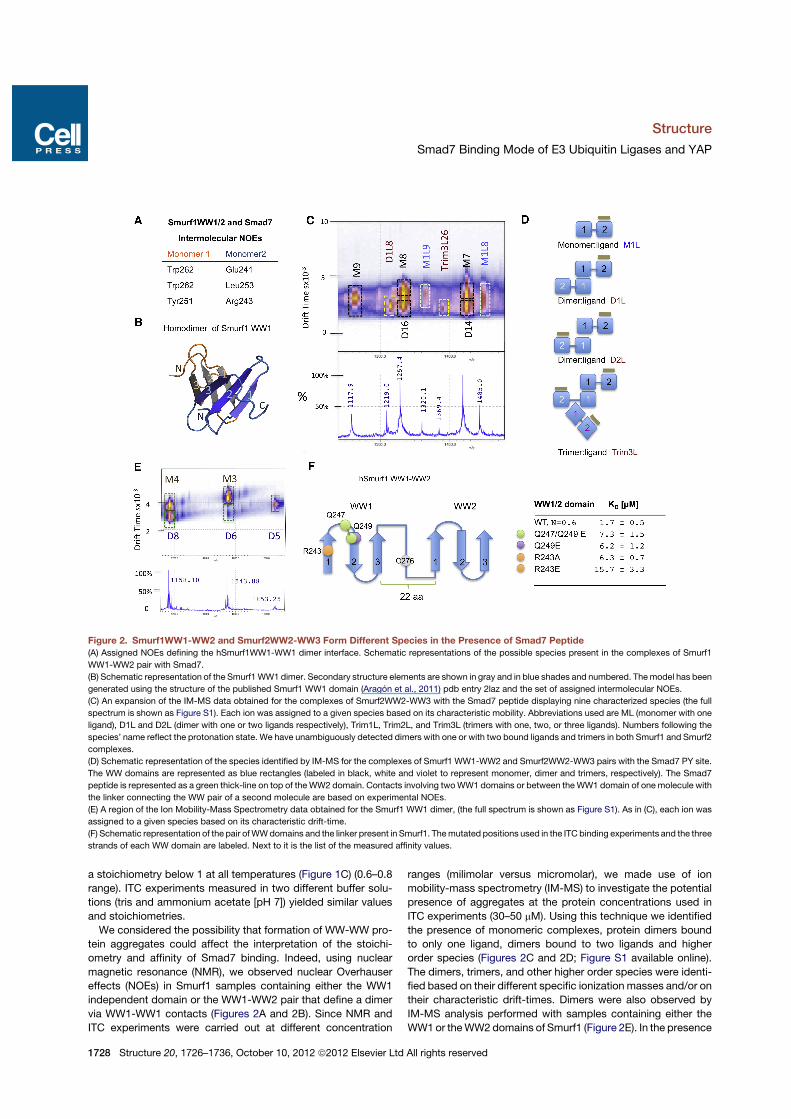
Figure 2. Smurf1WW1-WW2 and Smurf2WW2-WW3 Form Different Species in the Presence of Smad7 Peptide(A) Assigned NOEs defining the hSmurf1WW1-WW1 dimer interface. Schematic representations of the possible species present in the complexes of Smurf1
WW1-WW2 pair with Smad7.
(B) Schematic representation of the Smurf1WW1 dimer. Secondary structure elements are shown in gray and in blue shades and numbered. Themodel has been
generated using the structure of the published Smurf1 WW1 domain (Aragon et al., 2011) pdb entry 2laz and the set of assigned intermolecular NOEs.
(C) An expansion of the IM-MS data obtained for the complexes of Smurf2WW2-WW3 with the Smad7 peptide displaying nine characterized species (the full
spectrum is shown as Figure S1). Each ion was assigned to a given species based on its characteristic mobility. Abbreviations used are ML (monomer with one
ligand), D1L and D2L (dimer with one or two ligands respectively), Trim1L, Trim2L, and Trim3L (trimers with one, two, or three ligands). Numbers following the
species’ name reflect the protonation state. We have unambiguously detected dimers with one or with two bound ligands and trimers in both Smurf1 and Smurf2
complexes.
(D) Schematic representation of the species identified by IM-MS for the complexes of Smurf1 WW1-WW2 and Smurf2WW2-WW3 pairs with the Smad7 PY site.
The WW domains are represented as blue rectangles (labeled in black, white and violet to represent monomer, dimer and trimers, respectively). The Smad7
peptide is represented as a green thick-line on top of theWW2 domain. Contacts involving twoWW1 domains or between theWW1 domain of one molecule with
the linker connecting the WW pair of a second molecule are based on experimental NOEs.
(E) A region of the Ion Mobility-Mass Spectrometry data obtained for the Smurf1 WW1 dimer, (the full spectrum is shown as Figure S1). As in (C), each ion was
assigned to a given species based on its characteristic drift-time.
(F) Schematic representation of the pair ofWWdomains and the linker present in Smurf1. Themutated positions used in the ITC binding experiments and the three
strands of each WW domain are labeled. Next to it is the list of the measured affinity values.
Structure
Smad7 Binding Mode of E3 Ubiquitin Ligases and YAP
1728 Structure 20, 1726–1736, October 10, 2012 ª2012 Elsevier Ltd All rights reserved
of the Smad7 peptide the dimer population was reduced in thesample containing the WW2 domain, while that of the WW1domain was unaffected (Figure S1). Thus, the <1 stoichiometryobserved with the pairs under these experimental conditionsmay result from the coexistence of protein monomers, dimers,trimers, and tetramers each binding one equivalent of Smad7peptide, plus the presence of other species bound to two, three,or four Smad7 equivalents (Figures 2C and 2D). Since the pres-ence of the ligand does not prevent the formation of the higherorder species, we interpret that Smurf1 WW1 and Smurf2WW2 domains have a minor role in binding to short PY contain-ing sequences, but an important role in protein oligomerizationand aggregation. This observation differs from a previously re-ported interpretation of Smurf2 WW2-WW3 bound to Smad7(Chong et al., 2010), where the WW2 domain also participatesin hydrophobic and electrostatic interactions with the Smad7peptide. To characterize further the role of Smurf1WW1 domain,we introduced mutations in the WW1-WW2 pair domain inequivalent positions to those that in Smurf2 were proposed tocontact Smad7, and also two additional control mutations ina position that did not participate in the interaction of Smurf2and Smad7 but in the dimer formation. In all cases, single anddouble mutations (Arg243Ala, Gln249Glu, Gln247, and Gln249to Glu) reduced the affinity by 3-fold while the Arg243Glu muta-tion reduced the affinity by 10-fold (Figure 2F). The resultssuggest that these residues do not play a key role in bindingbut that theymay participate in protein homodimerization. Basedon this interpretation of the binding data, we conclude thatSmurf1 and Smurf2 use their WW2 domain and WW3 domain,respectively, as their primary binding sites for the Smad7 PYpeptide.ITC titrations performed at 15!C with the YAP WW domains
revealed that the YAP WW1 domain preferentially binds tothe Smad7 peptide, with a dissociation constant of 6.90 ±0.28 mM, while WW2 binds with a dissociation constant 9-foldweaker and the WW1-WW2 pair binds slightly worse than theisolatedWW1 (9.8 ± 0.9 mMandN = 1; Figure 1B). Thus, the inter-action with the Smad7 peptide mainly involves the YAP WW1domain.All together, these results suggest that in each protein a
specific, single WW domain is sufficient for high-affinity recogni-tion of the Smad7 PY site. The WW domains that mediate theinteraction with the PY motif of Smad7 are indicated with anarrow in the Figure 1A.
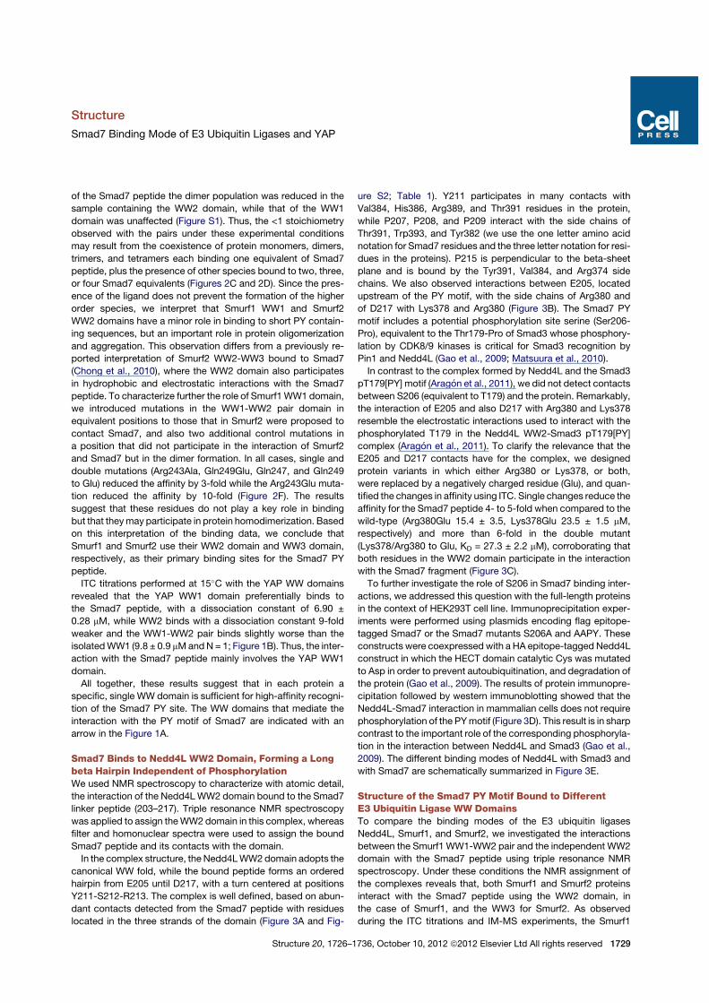
Smad7 Binds to Nedd4L WW2 Domain, Forming a Longbeta Hairpin Independent of PhosphorylationWe used NMR spectroscopy to characterize with atomic detail,the interaction of the Nedd4LWW2 domain bound to the Smad7linker peptide (203–217). Triple resonance NMR spectroscopywas applied to assign theWW2 domain in this complex, whereasfilter and homonuclear spectra were used to assign the boundSmad7 peptide and its contacts with the domain.In the complex structure, the Nedd4LWW2domain adopts the
canonical WW fold, while the bound peptide forms an orderedhairpin from E205 until D217, with a turn centered at positionsY211-S212-R213. The complex is well defined, based on abun-dant contacts detected from the Smad7 peptide with residueslocated in the three strands of the domain (Figure 3A and Fig-
ure S2; Table 1). Y211 participates in many contacts withVal384, His386, Arg389, and Thr391 residues in the protein,while P207, P208, and P209 interact with the side chains ofThr391, Trp393, and Tyr382 (we use the one letter amino acidnotation for Smad7 residues and the three letter notation for resi-dues in the proteins). P215 is perpendicular to the beta-sheetplane and is bound by the Tyr391, Val384, and Arg374 sidechains. We also observed interactions between E205, locatedupstream of the PY motif, with the side chains of Arg380 andof D217 with Lys378 and Arg380 (Figure 3B). The Smad7 PYmotif includes a potential phosphorylation site serine (Ser206-Pro), equivalent to the Thr179-Pro of Smad3 whose phosphory-lation by CDK8/9 kinases is critical for Smad3 recognition byPin1 and Nedd4L (Gao et al., 2009; Matsuura et al., 2010).In contrast to the complex formed by Nedd4L and the Smad3
pT179[PY] motif (Aragon et al., 2011), we did not detect contactsbetween S206 (equivalent to T179) and the protein. Remarkably,the interaction of E205 and also D217 with Arg380 and Lys378resemble the electrostatic interactions used to interact with thephosphorylated T179 in the Nedd4L WW2-Smad3 pT179[PY]complex (Aragon et al., 2011). To clarify the relevance that theE205 and D217 contacts have for the complex, we designedprotein variants in which either Arg380 or Lys378, or both,were replaced by a negatively charged residue (Glu), and quan-tified the changes in affinity using ITC. Single changes reduce theaffinity for the Smad7 peptide 4- to 5-fold when compared to thewild-type (Arg380Glu 15.4 ± 3.5, Lys378Glu 23.5 ± 1.5 mM,respectively) and more than 6-fold in the double mutant(Lys378/Arg380 to Glu, KD = 27.3 ± 2.2 mM), corroborating thatboth residues in the WW2 domain participate in the interactionwith the Smad7 fragment (Figure 3C).To further investigate the role of S206 in Smad7 binding inter-
actions, we addressed this question with the full-length proteinsin the context of HEK293T cell line. Immunoprecipitation exper-iments were performed using plasmids encoding flag epitope-tagged Smad7 or the Smad7 mutants S206A and AAPY. Theseconstructs were coexpressed with a HA epitope-tagged Nedd4Lconstruct in which the HECT domain catalytic Cys was mutatedto Asp in order to prevent autoubiquitination, and degradation ofthe protein (Gao et al., 2009). The results of protein immunopre-cipitation followed by western immunoblotting showed that theNedd4L-Smad7 interaction in mammalian cells does not requirephosphorylation of the PYmotif (Figure 3D). This result is in sharpcontrast to the important role of the corresponding phosphoryla-tion in the interaction between Nedd4L and Smad3 (Gao et al.,2009). The different binding modes of Nedd4L with Smad3 andwith Smad7 are schematically summarized in Figure 3E.
Structure of the Smad7 PY Motif Bound to DifferentE3 Ubiquitin Ligase WW DomainsTo compare the binding modes of the E3 ubiquitin ligasesNedd4L, Smurf1, and Smurf2, we investigated the interactionsbetween the Smurf1 WW1-WW2 pair and the independent WW2domain with the Smad7 peptide using triple resonance NMRspectroscopy. Under these conditions the NMR assignment ofthe complexes reveals that, both Smurf1 and Smurf2 proteinsinteract with the Smad7 peptide using the WW2 domain, inthe case of Smurf1, and the WW3 for Smurf2. As observedduring the ITC titrations and IM-MS experiments, the Smurf1
Structure
Smad7 Binding Mode of E3 Ubiquitin Ligases and YAP
Structure 20, 1726–1736, October 10, 2012 ª2012 Elsevier Ltd All rights reserved 1729
WW1-WW2 pair displays a high tendency to form dimers andother higher order aggregates.
Using 3D- and 2D-NOESY experiments we characterized apopulation of dimers formed via interactions between the WW1domains of two molecules according to intermolecular NOEsthat fit as a beta-clam. To illustrate these interactions, we gener-ated a model using the structure of the Smurf1 WW1 domain(pdb entry: 2laz) and the unambiguously assigned NOEs de-tected between monomers (Figures 2A and 4A). We have alsodetected a minor population of dimers formed by interactionsbetween the WW2 domain of one molecule and the pair ofprolines present in the linker connecting the WW1-WW2 pair ofa second molecule, that can explain the trimeric and tetramericspecies identified by IM-MS (Figures 2C and 2D). We observedas well the dimerizing tendency with the WW2 domain of Smurf2
in the WW2-WW3 pair. The beta-clam arrangement in the dimeris similar to that described for the WW2 domain of the mouseSalvador homolog 1 protein (Ohnishi et al., 2007). With the WWdomain pairs we detected a broadening of the intermolecularNOEs that defined the peptide in the bound conformation andtwo sets of NOEs for the Y211 with residues in the WW2/WW3domains. We interpreted the broadening and the presence ofthe second set of signals for the Y211 aromatic ring as the resultof the peptide bound in several complexes, for instance, themain conformations that correspond to themonomer in complexwith one ligand and the symmetric dimer with two bound ligandsin equilibriumwith an asymmetric dimer bound to a single ligand,schematically represented in Figure 2D. At 298K and in the pres-ence of 10% DMSO both sets of NOEs corresponding to theY211 collapse to one set that we interpret it as the bound
Figure 3. Structure of the Nedd4L WW2 Domain Bound to the Smad7 Linker(A) Detailed view of the refined structure of the Nedd4LWW2 domain (364–403) in complex with the Smad7 synthesized PY fragment (203–217). The elements of
secondary structure (graphite) are numbered and key residues of the Nedd4L WW2 domain that participate in the interaction with the peptide are labeled and
highlighted in orange.
(B) Electrostatic potential surface of the Nedd4LWW2 domain with the bound Smad7 peptide (stick representation) colored in green. The lowest energy structure
displayed in (A) and (B) and the family of 25-calculated structures are shown as Figures S2A and S2B. Positively charged sites are colored in blue and negatively
charged sites in red. Key residues in Smad7 and Nedd4L are indicated (violet and black, respectively). The N- and C-terminal sites of the Smad7 peptide are
indicated with an arrow.
(C) Two residues that recognize the N-terminal part of Smad7 were both independently and jointly mutated to glutamic acid, and the effect of the change in the
interaction affinity was determined by ITC. The binding affinity decreased by approximately 7-fold with respect to the wild-type Nedd4L WW2 domain.
(D) Human HEK293T cells transduced with vectors encoding the indicated epitope-tagged proteins were subjected to immunoprecipitation (IP) with anti-Flag
antibodies and western immunoblotting with antibodies against the indicated tags (upper panels). Aliquots of cell lysates were directly subjected to immuno-
blotting as loading controls (bottom panels).
(E)Schematic representationof thedifferent bindingmodesdisplayedbyNedd4LwithSmad3andSmad7.Nedd4Lbindspreferentially to thePYsiteofSmad7using
its WW2 domain (this work) while binding to Smad3 region including the diphosphorylated site and pT[PY] motif requires theWW2-WW3 pair (Aragon et al., 2011).
Structure
Smad7 Binding Mode of E3 Ubiquitin Ligases and YAP
1730 Structure 20, 1726–1736, October 10, 2012 ª2012 Elsevier Ltd All rights reserved
monomer. Under these experimental conditions, we did notobserve, however, contacts between the domains in the Smurf1WW1-WW2 pair, or between the WW1 and the Smad7 peptideas described for the Smurf2 WW2-WW3 complex with Smad7(Chong et al., 2010), or as we previously observed in the complexof Smurf1 WW1-WW2 pair with Smad1 (Aragon et al., 2011).Based on these observations, we focused the structural
work in the WW2 domain of Smurf1 and in the WW3 of Smurf2.As both Smurf complexes are very similar, we will describethem in parallel, with the corresponding residues separated bya slash. The Smurf1WW2- and Smurf2WW3-Smad7 complexesare well defined, based on numerous contacts detected fromE205-P215 Smad7 residues with the domains (Figures 4B and4C and Figure S3; Table 1). In both complexes the Smad7 frag-ment also forms a turn, centered at positions Y211-S212-R213,but it does not form a long hairpin as in the case of the Nedd4Lcomplex, especially in the Smurf1 complex. A comparison ofthese two complexes with that of Nedd4L revealed some addi-tional differences; for instance, E205 is interacting with Arg295/Arg312 in the second strand, but no contacts with the peptide
are observed for the residues located in loop 1 of the WWdomains, which in these cases are Ser293/Thr310 and not aLys as in the Nedd4L WW2 domain. As a consequence, E205and D217 are less defined in the Smurf1 WW2 and Smurf2WW3 complexes.Amutation introduced in the Smurf1WW2domain (Arg295Glu)
reduces the affinity to 48.6 ± 6.4 mM, suggesting an active impli-cation of Arg295 in the peptide interaction. The equivalent muta-tion introduced in Smurf2WW3 also reduces the affinity to 34.2 ±1.6 mM. The complexes here described are similar to the pre-viously characterized Smurf2WW3 and Smad7 (Chong et al.,2006), with the main differences involving the contacts with theN-terminal site of Smad7 (E205), the position of P215, and theabsence of intra peptide contacts from residues M216-D217.On the other hand, they differ from the complex between theSmurf1WW1-WW2 pair and Smad1, where both WW domainshave a direct role in ligand recognition (Figure 4D) (Alarconet al., 2009; Aragon et al., 2011) and from the complex betweenSmurf2WW2-WW3 and Smad7, where the contacts with theC-terminal part of Smad7 that we observe to occur with the
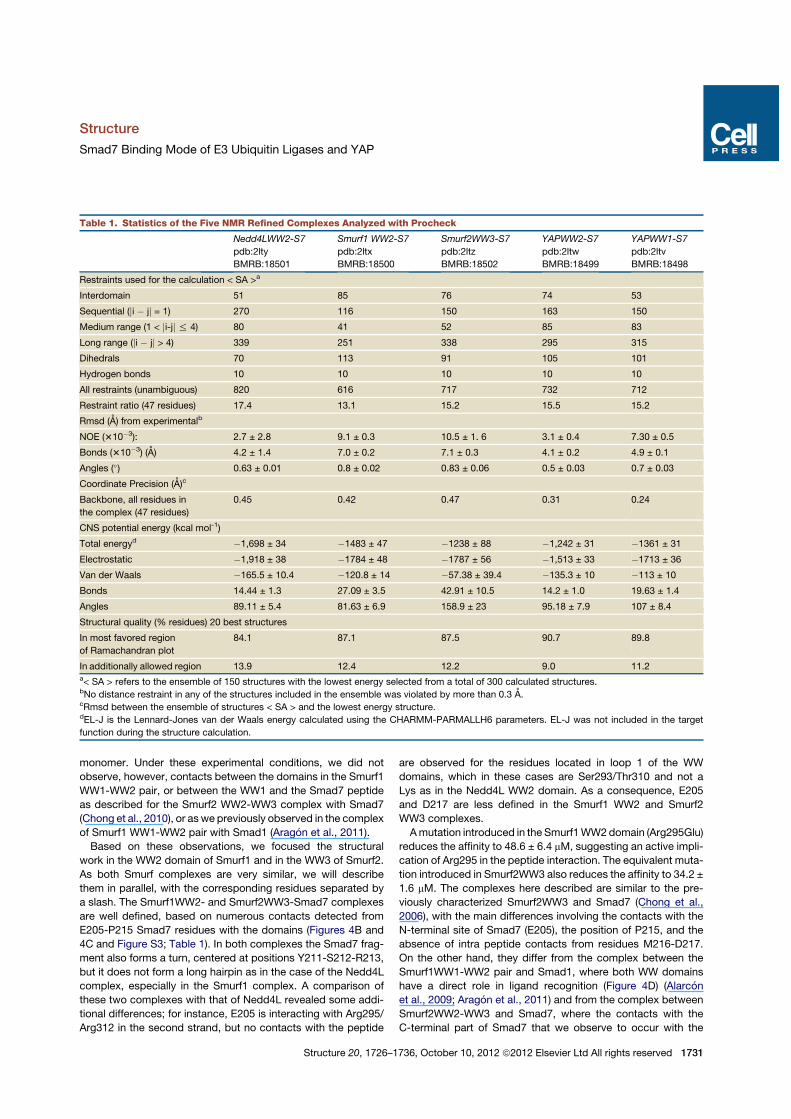
Table 1. Statistics of the Five NMR Refined Complexes Analyzed with Procheck
Nedd4LWW2-S7
pdb:2lty
BMRB:18501
Smurf1 WW2-S7
pdb:2ltx
BMRB:18500
Smurf2WW3-S7
pdb:2ltz
BMRB:18502
YAPWW2-S7
pdb:2ltw
BMRB:18499
YAPWW1-S7
pdb:2ltv
BMRB:18498
Restraints used for the calculation < SA >a
Interdomain 51 85 76 74 53
Sequential (ji ! jj = 1) 270 116 150 163 150
Medium range (1 < ji-jj % 4) 80 41 52 85 83
Long range (ji ! jj > 4) 339 251 338 295 315
Dihedrals 70 113 91 105 101
Hydrogen bonds 10 10 10 10 10
All restraints (unambiguous) 820 616 717 732 712
Restraint ratio (47 residues) 17.4 13.1 15.2 15.5 15.2
Rmsd (A) from experimentalb
NOE (310!3): 2.7 ± 2.8 9.1 ± 0.3 10.5 ± 1. 6 3.1 ± 0.4 7.30 ± 0.5
Bonds (310!3) (A) 4.2 ± 1.4 7.0 ± 0.2 7.1 ± 0.3 4.1 ± 0.2 4.9 ± 0.1
Angles (") 0.63 ± 0.01 0.8 ± 0.02 0.83 ± 0.06 0.5 ± 0.03 0.7 ± 0.03
Coordinate Precision (A)c
Backbone, all residues in
the complex (47 residues)
0.45 0.42 0.47 0.31 0.24
CNS potential energy (kcal mol-1)
Total energyd !1,698 ± 34 !1483 ± 47 !1238 ± 88 !1,242 ± 31 !1361 ± 31
Electrostatic !1,918 ± 38 !1784 ± 48 !1787 ± 56 !1,513 ± 33 !1713 ± 36
Van der Waals !165.5 ± 10.4 !120.8 ± 14 !57.38 ± 39.4 !135.3 ± 10 !113 ± 10
Bonds 14.44 ± 1.3 27.09 ± 3.5 42.91 ± 10.5 14.2 ± 1.0 19.63 ± 1.4
Angles 89.11 ± 5.4 81.63 ± 6.9 158.9 ± 23 95.18 ± 7.9 107 ± 8.4
Structural quality (% residues) 20 best structures
In most favored region
of Ramachandran plot
84.1 87.1 87.5 90.7 89.8
In additionally allowed region 13.9 12.4 12.2 9.0 11.2a< SA > refers to the ensemble of 150 structures with the lowest energy selected from a total of 300 calculated structures.bNo distance restraint in any of the structures included in the ensemble was violated by more than 0.3 A.cRmsd between the ensemble of structures < SA > and the lowest energy structure.dEL-J is the Lennard-Jones van der Waals energy calculated using the CHARMM-PARMALLH6 parameters. EL-J was not included in the target
function during the structure calculation.
Structure
Smad7 Binding Mode of E3 Ubiquitin Ligases and YAP
Structure 20, 1726–1736, October 10, 2012 ª2012 Elsevier Ltd All rights reserved 1731
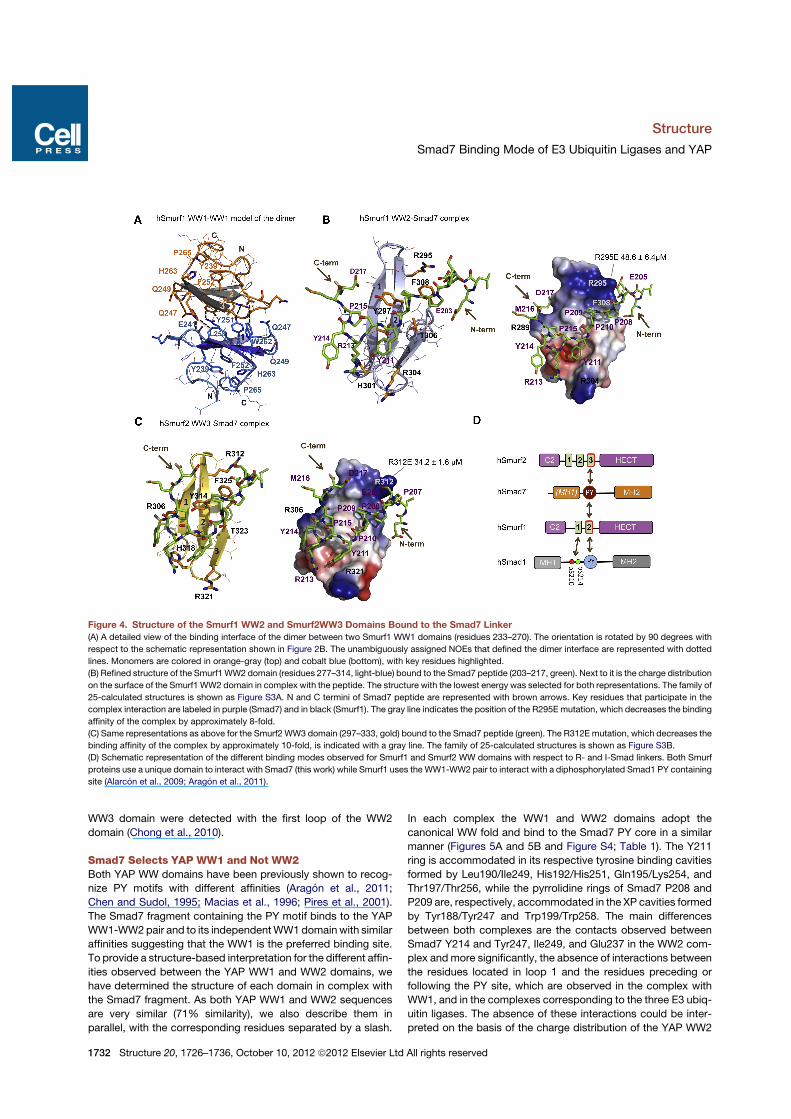
WW3 domain were detected with the first loop of the WW2domain (Chong et al., 2010).
Smad7 Selects YAP WW1 and Not WW2Both YAP WW domains have been previously shown to recog-nize PY motifs with different affinities (Aragon et al., 2011;Chen and Sudol, 1995; Macias et al., 1996; Pires et al., 2001).The Smad7 fragment containing the PY motif binds to the YAPWW1-WW2pair and to its independentWW1 domain with similaraffinities suggesting that the WW1 is the preferred binding site.To provide a structure-based interpretation for the different affin-ities observed between the YAP WW1 and WW2 domains, wehave determined the structure of each domain in complex withthe Smad7 fragment. As both YAP WW1 and WW2 sequencesare very similar (71% similarity), we also describe them inparallel, with the corresponding residues separated by a slash.
In each complex the WW1 and WW2 domains adopt thecanonical WW fold and bind to the Smad7 PY core in a similarmanner (Figures 5A and 5B and Figure S4; Table 1). The Y211ring is accommodated in its respective tyrosine binding cavitiesformed by Leu190/Ile249, His192/His251, Gln195/Lys254, andThr197/Thr256, while the pyrrolidine rings of Smad7 P208 andP209 are, respectively, accommodated in the XP cavities formedby Tyr188/Tyr247 and Trp199/Trp258. The main differencesbetween both complexes are the contacts observed betweenSmad7 Y214 and Tyr247, Ile249, and Glu237 in the WW2 com-plex and more significantly, the absence of interactions betweenthe residues located in loop 1 and the residues preceding orfollowing the PY site, which are observed in the complex withWW1, and in the complexes corresponding to the three E3 ubiq-uitin ligases. The absence of these interactions could be inter-preted on the basis of the charge distribution of the YAP WW2
Figure 4. Structure of the Smurf1 WW2 and Smurf2WW3 Domains Bound to the Smad7 Linker(A) A detailed view of the binding interface of the dimer between two Smurf1 WW1 domains (residues 233–270). The orientation is rotated by 90 degrees with
respect to the schematic representation shown in Figure 2B. The unambiguously assigned NOEs that defined the dimer interface are represented with dotted
lines. Monomers are colored in orange-gray (top) and cobalt blue (bottom), with key residues highlighted.
(B) Refined structure of the Smurf1 WW2 domain (residues 277–314, light-blue) bound to the Smad7 peptide (203–217, green). Next to it is the charge distribution
on the surface of the Smurf1 WW2 domain in complex with the peptide. The structure with the lowest energy was selected for both representations. The family of
25-calculated structures is shown as Figure S3A. N and C termini of Smad7 peptide are represented with brown arrows. Key residues that participate in the
complex interaction are labeled in purple (Smad7) and in black (Smurf1). The gray line indicates the position of the R295E mutation, which decreases the binding
affinity of the complex by approximately 8-fold.
(C) Same representations as above for the Smurf2 WW3 domain (297–333, gold) bound to the Smad7 peptide (green). The R312E mutation, which decreases the
binding affinity of the complex by approximately 10-fold, is indicated with a gray line. The family of 25-calculated structures is shown as Figure S3B.
(D) Schematic representation of the different binding modes observed for Smurf1 and Smurf2 WW domains with respect to R- and I-Smad linkers. Both Smurf
proteins use a unique domain to interact with Smad7 (this work) while Smurf1 uses the WW1-WW2 pair to interact with a diphosphorylated Smad1 PY containing
site (Alarcon et al., 2009; Aragon et al., 2011).
Structure
Smad7 Binding Mode of E3 Ubiquitin Ligases and YAP
1732 Structure 20, 1726–1736, October 10, 2012 ª2012 Elsevier Ltd All rights reserved
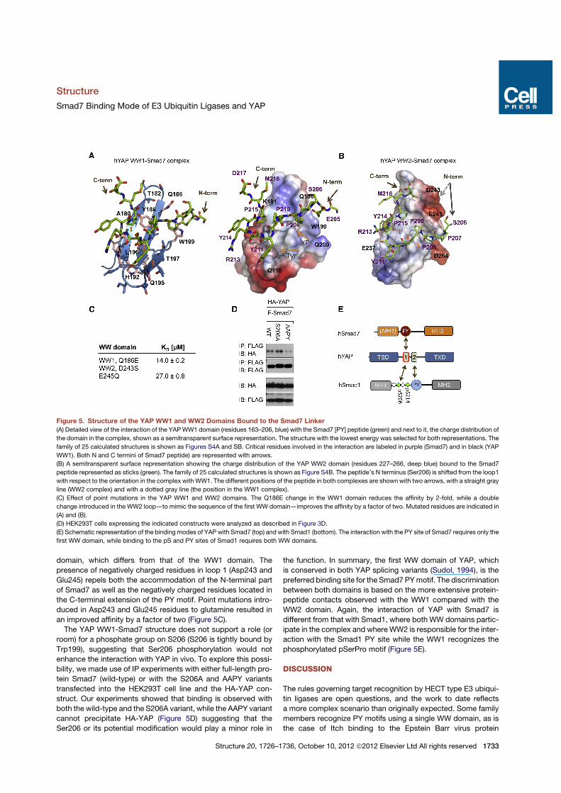
domain, which differs from that of the WW1 domain. Thepresence of negatively charged residues in loop 1 (Asp243 andGlu245) repels both the accommodation of the N-terminal partof Smad7 as well as the negatively charged residues located inthe C-terminal extension of the PY motif. Point mutations intro-duced in Asp243 and Glu245 residues to glutamine resulted inan improved affinity by a factor of two (Figure 5C).The YAP WW1-Smad7 structure does not support a role (or
room) for a phosphate group on S206 (S206 is tightly bound byTrp199), suggesting that Ser206 phosphorylation would notenhance the interaction with YAP in vivo. To explore this possi-bility, we made use of IP experiments with either full-length pro-tein Smad7 (wild-type) or with the S206A and AAPY variantstransfected into the HEK293T cell line and the HA-YAP con-struct. Our experiments showed that binding is observed withboth the wild-type and the S206A variant, while the AAPY variantcannot precipitate HA-YAP (Figure 5D) suggesting that theSer206 or its potential modification would play a minor role in
the function. In summary, the first WW domain of YAP, whichis conserved in both YAP splicing variants (Sudol, 1994), is thepreferred binding site for the Smad7 PYmotif. The discriminationbetween both domains is based on the more extensive protein-peptide contacts observed with the WW1 compared with theWW2 domain. Again, the interaction of YAP with Smad7 isdifferent from that with Smad1, where both WW domains partic-ipate in the complex and whereWW2 is responsible for the inter-action with the Smad1 PY site while the WW1 recognizes thephosphorylated pSerPro motif (Figure 5E).
DISCUSSION
The rules governing target recognition by HECT type E3 ubiqui-tin ligases are open questions, and the work to date reflectsa more complex scenario than originally expected. Some familymembers recognize PY motifs using a single WW domain, as isthe case of Itch binding to the Epstein Barr virus protein
Figure 5. Structure of the YAP WW1 and WW2 Domains Bound to the Smad7 Linker(A) Detailed view of the interaction of the YAPWW1 domain (residues 163–206, blue) with the Smad7 [PY] peptide (green) and next to it, the charge distribution of
the domain in the complex, shown as a semitransparent surface representation. The structure with the lowest energy was selected for both representations. The
family of 25 calculated structures is shown as Figures S4A and SB. Critical residues involved in the interaction are labeled in purple (Smad7) and in black (YAP
WW1). Both N and C termini of Smad7 peptide) are represented with arrows.
(B) A semitransparent surface representation showing the charge distribution of the YAP WW2 domain (residues 227–266, deep blue) bound to the Smad7
peptide represented as sticks (green). The family of 25 calculated structures is shown as Figure S4B. The peptide’s N terminus (Ser206) is shifted from the loop1
with respect to the orientation in the complex with WW1. The different positions of the peptide in both complexes are shown with two arrows, with a straight gray
line (WW2 complex) and with a dotted gray line (the position in the WW1 complex).
(C) Effect of point mutations in the YAP WW1 and WW2 domains. The Q186E change in the WW1 domain reduces the affinity by 2-fold, while a double
change introduced in the WW2 loop—to mimic the sequence of the first WW domain—improves the affinity by a factor of two. Mutated residues are indicated in
(A) and (B).
(D) HEK293T cells expressing the indicated constructs were analyzed as described in Figure 3D.
(E) Schematic representation of the binding modes of YAP with Smad7 (top) and with Smad1 (bottom). The interaction with the PY site of Smad7 requires only the
first WW domain, while binding to the pS and PY sites of Smad1 requires both WW domains.
Structure
Smad7 Binding Mode of E3 Ubiquitin Ligases and YAP
Structure 20, 1726–1736, October 10, 2012 ª2012 Elsevier Ltd All rights reserved 1733
LMP2A (Morales et al., 2007) and Nedd4 binding to the voltagegated sodium channel (Kanelis et al., 2001) and to Commissure-less (Kanelis et al., 2006). In other cases, such as the binding ofSmurf1 and Nedd4L to R-Smads, the proteins use a pair of WWdomains to expand the binding interface with a composite bidingsite that includes pSer/pThr-Pro elements in addition to a canon-ical PY motif, a combination that allows regulation of the interac-tion by input-driven protein kinases (Alarcon et al., 2009; Aragonet al., 2011; Gao et al., 2009).
The data here presented show a further versatility of WWdomains depending on the target protein in that a given HECTE3 ubiquitin ligase can use WW domains singly (this work) or ina combinatorial manner (Alarcon et al., 2009; Aragon et al.,2011; Gao et al., 2009), depending on the target. In the case ofNedd4L, the interaction with Smad7 involves a single WWdomain (WW2 preferentially). The high affinity of the Nedd4LWW2 domain for the Smad7 PY region is based on aggregatecontacts with a canonical PY motif, a C-terminal extension ofthis motif that aligns on the first strand of the WW2 domain,and an electrostatic balance between a glutamic and an asparticacids (E205 andD217) in Smad7 and two positively charged resi-dues located in loop 1 of the WW2 domain. In Smad2/3 a phos-phothreonine N-terminal to the PY motif (pT179) makes a criticalcontribution in the binding to a Nedd4L WW domain (Gao et al.,2009). In the case of Smad7 a corresponding serine residue(S206) does not need to be phosphorylated for high affinitybinding to Nedd4L; instead, an acidic residue, E205, plays thepart of pT179 in Smad3. Furthermore, Nedd4L binding toSmad2/3 involves a second WW domain, WW3, for contactswith a separate, diphosphorylated region downstream of thePY motif (Aragon et al., 2011). Thus Nedd4L binds Smad7 usinga single unregulatedWWdomain interaction even though it bindsSmad2/3 using two WW domains that require multiple phos-phorylations of the target region by different protein kinases.
Similar principles govern the interactions of Smurf1 andSmurf2 ubiquitin ligases with Smad7. Binding involves a uniqueWW domain, whereas Smurf1 binds Smad1 using two WWdomains to recognize a PY motif and a phosphorylated motif inthe linker region. We observed neither the contacts betweenthe two WW domains nor the contacts between the first WWdomain and Smad7 that were described in a recent report(Chong et al., 2010). We observed instead that the Smurf1 andSmurf2 WW-WW pairs have a high tendency to form homo-dimers via the WW1 domain in case of Smurf1 and via theWW2 domain in Smurf2 (Figure 4A). The presence of Smad7peptide did not prevent these dimerizations since the WW-WWdomain pairs bind the Smad7 peptide mainly through contactswith the WW2 domain of Smurf1 and with the WW3 domain ofSmurf2. It has recently been reported that full-length Smurf1forms homodimers and oligomers in vitro and in vivo throughintermolecular contacts mapped to a fragment containing theC2 and the WW domains of one molecule and the HECT domainof the partner (Wan et al., 2011). Furthermore, intramolecularcontacts between the C2 domain and the HECT domain ofSmurf2 have also been characterized (Wiesner et al., 2007). Inboth Smurf1/2 ligases the close conformation inhibits the mech-anism of protein self-ubiquitination (Wan et al., 2011). It ispossible that in addition to these reported interactions betweenthe C2 and HECT domains, the inter WW-WWcontacts detected
in our work could also contribute to the formation of the dimersand oligomers in vivo, and to the stabilization of the close confor-mation of Smurf1 and 2. In the presence of two Smad7 equiva-lents, a reverse reaction may occur with the MH1 and PY sitesof the Smad7 protein competing for the Smurf1 HECT and theWW2 domains, respectively, pulling apart theWW1-WW1 dimer.The result of this reaction would be the generation of two acti-vated Smurf1-Smad7 complexes, starting from the close andinactive full-length dimer. A similar mechanism may occur withthe Smurf1 and Smad1 interaction. These possibilities notwith-standing, the WW-WW homodimers here detected could haveresulted from the use of recombinant protein fragments.Our work also shows that YAP requires only the WW1 domain
for binding to the Smad7 PY region. Notably, in the interactionwith Smad1, YAP uses its WW1 domain for binding a phospho-serine motif, and instead uses the WW2 domain for binding theSmad1 PY motif (Aragon et al., 2011). By solving the structuresof both YAP WW1 and WW2 complexes with Smad7 we dis-cerned the reasons for these differences with respect to theinteraction with Smad1. The WW2 domain contains negativelycharged residues in the area where the E205 side chain is nor-mally accommodated, destabilizing the interaction. The affinityof the YAP WW1-WW2 pair for the composed pSP-PY site ofSmad1 is 8x higher than that of the WW1 domain for the PYsite of Smad7. However, the concentration of Smad7 in thenucleus is high and it could compete in vivo with Smad1 forYAP binding, providing a scenario for the inhibitory role ofSmad7.We propose thatWW-WWpairs in these Smad regulators form
functional units that evolved to recognize PY containing regionsof variable length and complexity, including composite PY/phos-pho-Ser/Thr motifs in R-Smads and simple PY motifs in Smad7.These features expand the functional versatility of E3 ubiquitinligases by optimizing the interacting surface depending on theneeds.With Smad7, Nedd4L and Smurf1/2 act as partners in tar-geting TGF-b receptors for ubiquitination. Smad7 may also actas a constitutive YAP sequestration or reservoir protein. In con-trast, R-Smads are direct targets of the ubiquitin ligases andfunctional partners of YAP only in specific stages of the Smadsignaling cycle (Aragon et al., 2011). The absence of a require-ment for phosphorylation in the interaction with Smad7 arguesthat YAP, Nedd4L and Smurf1/2 are constitutive partners ofSmad7 whereas they are conditional, phosphorylation depen-dent regulators of R-Smads in TGF-b and BMP signal transduc-tion. The features of Smad7 defined here provide a structuralbasis for its central role as a hub for negative feedback andcrosstalk regulation in TGF-b signaling.
EXPERIMENTAL PROCEDURES
CloningThe four independent hNedd4L WW domains, the three Nedd4L WW domain
pairs were prepared as described previously (Aragon et al., 2011; Chong et al.,
2010). Point mutations were introduced using the QuickChange site directed
mutagenesis Kit (Stratagene) with the appropriate complementary mutagenic
primers. All wild-type and variants were confirmed by DNA sequencing.
Protein Expression and PurificationUnlabeled, 15N-labeled, 13C, 15N and 2H, 13C, 15N-labeled proteins were ex-
pressed in Escherichia coli BL21 (DE3), in LB or in minimal medium (M9),
Structure
Smad7 Binding Mode of E3 Ubiquitin Ligases and YAP
1734 Structure 20, 1726–1736, October 10, 2012 ª2012 Elsevier Ltd All rights reserved
prepared either in H2O or in D2O (99.89%, CortecNet) enriched with 15NH4Cl
and/or D-[13C] glucose as sole sources of carbon and nitrogen, respectively
(Marley et al., 2001). Proteins were purified as described (Aragon et al.,
2011). For the NMR experiments with Smurf1/2 WW domains we have mini-
mized the aggregation tendency by concentrating the proteins (single WW2
or theWW1-WW2 pair andWW3 orWW2-WW3 for Smurf1 and 2, respectively)
in the presence of the ligand.
Transfection, Immunoprecipitation, and ImmunoblottingTransfection of the indicated plasmids was performed as described previously
(Gao et al., 2009) HEK293T cells were incubated as indicated with TGF-b1 (100
pM; R&D Systems). Immunoprecipitation and western immunoblotting were
done as described (Sapkota et al., 2007).
Smad7 Peptide Synthesis and PurificationThe peptide (Ac-ELESPPPPYSRYPMD-NH2 (203–217) was synthesized using
Fmoc-solid phase peptide synthesis with a rink amide resin (Merck Chemi-
cals), in a CEM Liberty1 microwave synthesizer (0.1 mmol scale). The acety-
lated peptide was purified by RP-HPLC using a SunFire C18 Sephasil prepar-
ative column (Waters) with an AKTApurifier10 (GE Healthcare Life Sciences),
using a linear gradient of 10%–40% acetonitrile and 0.05% TFA and an elution
time of 20 min. The peptide was analyzed by MALDI-TOF mass spectrometry
and 2D homonuclear NMR spectroscopy.
NMR AssignmentNMR data were acquired at 285 K/298 K on a Bruker Avance III 600-MHz
spectrometer equipped with a z-pulse field gradient unit. Backbone 1H, 15N,
and 13C resonance assignments were obtained by analyzing 3D CBCA(CO)
NH and HNCBCA experiments. Side-chain resonance assignments were ob-
tained by analyzing HCCC(CO)NH, 15N-TOCSY, HCCH-TOCSY and 15N-,13C NOESY spectra (Sattler et al., 1999). Inter- and intra molecular proton
distance restraints were obtained from peaks assigned in 2D-NOESY experi-
ments. All spectra were processed with NMRPipe/NMRDraw software (Dela-
glio et al., 1995) and were analyzed with CARA (Bartels et al., 1995). Spectra
used for the calculation were integrated using the batch integration method
of the XEASY package.
NMR Titration Experiments15N-HSQC spectra were acquired using 300 mM 15N-labeled protein samples
to which the unlabeled peptide was added stepwise until saturation was
achieved.
Structure Determination and RefinementStructures were calculated with CNS 1.1 (Brunger et al., 1998), using only
unambiguously assigned restraints derived from NOESY experiments,
coupling constants 3J(HN, HA from HNHA spectra) and hydrogen bonds
measured from D2O exchange experiments. The protocol for the calculation
consists of two iterations of 1 and 200 structures, respectively, using
100,000 cooling steps. All calculated structures were water refined and ranked
based on minimum values of energy and violations. The water refinement
protocol is a modification of the original protocol provided with Aria (Nilges
et al., 1997), which uses all experimental restraints during the refinement
process. Analysis of the quality of the lowest energy structures was performed
using PROCHECK-NMR (Laskowski et al., 1996) and the statistics are shown
in Table 1. Images were generated with PyMOL (DeLano, 2002).
Isothermal Titration CalorimetryITC experiments were performed using a low volume nano ITC calorimeter
(TA instruments) and five different temperatures 5, 15, 20, 25, and 30!C.
Details of the experiments are given in the Supplemental Experimental
Procedures.
Ion Mobility-Mass SpectrometryTraveling wave ion mobility mass spectrometry experiments were performed
on a Synapt G1 HDMS mass spectrometer (Waters, Manchester, UK). Exper-
imental details are given in the Supplemental Experimental Procedures.
ACCESSION NUMBERS
For the five complexes, the corresponding Protein Data Bank and
BioMagResBank codes are, respectively, 2ltv/18498 (YAPWW1_S7),
2ltw/18499 (YAPWW2_S7), 2ltx/18500 (Smurf1WW2_S7), 2lty/18501
(Nedd4LWW2_S7), and 2ltz/18502 (Smurf2WW3_S7). Protein short names
are given as in Table 1.
SUPPLEMENTAL INFORMATION
Supplemental Information includes four figures and Supplemental Experi-
mental Procedures and can be found with this article online at http://dx.doi.
org/10.1016/j.str.2012.07.014.
ACKNOWLEDGMENTS
We thank the Mass Spectrometry Core Facility at the IRB Barcelona (Dr. M.
Vilaseca) for support with the IM-MS experiments. N.G. had an IRBPhD fellow-
ship, and E.A was financed in part by a Consolider RNAREG (CSD2009-00080)
grant. This work was supported by Grant SAF2011-25119 (M.J.M.) and by NIH
Grant R37-CA34610 (J.M.). J.M. is an Investigator of the Howard Hughes
Medical Institute, and M.J.M. is an ICREA Programme Investigator.
Received: June 15, 2012
Revised: July 27, 2012
Accepted: July 27, 2012
Published online: August 23, 2012
REFERENCES
Alarcon, C., Zaromytidou, A.I., Xi, Q., Gao, S., Yu, J., Fujisawa, S., Barlas, A.,
Miller, A.N., Manova-Todorova, K., Macias, M.J., et al. (2009). Nuclear CDKs
drive Smad transcriptional activation and turnover in BMP and TGF-beta path-
ways. Cell 139, 757–769.
Aragon, E., Goerner, N., Zaromytidou, A.I., Xi, Q., Escobedo, A., Massague, J.,
and Macias, M.J. (2011). A Smad action turnover switch operated by WW
domain readers of a phosphoserine code. Genes Dev. 25, 1275–1288.
Bai, S., and Cao, X. (2002). A nuclear antagonistic mechanism of inhibitory
Smads in transforming growth factor-beta signaling. J. Biol. Chem. 277,
4176–4182.
Bartels, C., Xia, T.H., Billeter, M., Guntert, P., and Wuthrich, K. (1995). The
program XEASY for computer-supported NMR spectral analysis of biological
macromolecules. J. Biomol. NMR 5, 1–10.
Brunger, A.T., Adams, P.D., Clore, G.M., DeLano, W.L., Gros, P., Grosse-
Kunstleve, R.W., Jiang, J.S., Kuszewski, J., Nilges, M., Pannu, N.S., et al.
(1998). Crystallography & NMR system: A new software suite for macromolec-
ular structure determination. Acta Crystallogr. D Biol. Crystallogr. 54, 905–921.
Chen, H.I., and Sudol, M. (1995). The WW domain of Yes-associated protein
binds a proline-rich ligand that differs from the consensus established for
Src homology 3-binding modules. Proc. Natl. Acad. Sci. USA 92, 7819–7823.
Chong, P.A., Lin, H., Wrana, J.L., and Forman-Kay, J.D. (2006). An expanded
WWdomain recognition motif revealed by the interaction between Smad7 and
the E3 ubiquitin ligase Smurf2. J. Biol. Chem. 281, 17069–17075.
Chong, P.A., Lin, H., Wrana, J.L., and Forman-Kay, J.D. (2010). Coupling of
tandemSmad ubiquitination regulatory factor (Smurf) WWdomainsmodulates
target specificity. Proc. Natl. Acad. Sci. USA 107, 18404–18409.
Delaglio, F., Grzesiek, S., Vuister, G.W., Zhu, G., Pfeifer, J., and Bax, A. (1995).
NMRPipe: a multidimensional spectral processing system based on UNIX
pipes. J. Biomol. NMR 6, 277–293.
DeLano, W.L. (2002). The PyMOL Molecular Graphics System. http://www.
pymol.org/.
Ebisawa, T., Fukuchi, M., Murakami, G., Chiba, T., Tanaka, K., Imamura, T.,
and Miyazono, K. (2001). Smurf1 interacts with transforming growth factor-
beta type I receptor through Smad7 and induces receptor degradation.
J. Biol. Chem. 276, 12477–12480.
Structure
Smad7 Binding Mode of E3 Ubiquitin Ligases and YAP
Structure 20, 1726–1736, October 10, 2012 ª2012 Elsevier Ltd All rights reserved 1735
Eichhorn, P.J., Rodon, L., Gonzalez-Junca, A., Dirac, A., Gili, M., Martınez-
Saez, E., Aura, C., Barba, I., Peg, V., Prat, A., et al. (2012). USP15 stabilizes
TGF-b receptor I and promotes oncogenesis through the activation of TGF-b
signaling in glioblastoma. Nat. Med. 18, 429–435.
Fan, G., Fan, Y., Gupta, N., Matsuura, I., Liu, F., Zhou, X.Z., Lu, K.P., and
Gelinas, C. (2009). Peptidyl-prolyl isomerase Pin1 markedly enhances the
oncogenic activity of the rel proteins in the nuclear factor-kappaB family.
Cancer Res. 69, 4589–4597.
Ferrigno, O., Lallemand, F., Verrecchia, F., L’Hoste, S., Camonis, J., Atfi, A.,
and Mauviel, A. (2002). Yes-associated protein (YAP65) interacts with Smad7
and potentiates its inhibitory activity against TGF-beta/Smad signaling.
Oncogene 21, 4879–4884.
Fuentealba, L.C., Eivers, E., Ikeda, A., Hurtado, C., Kuroda, H., Pera, E.M., and
De Robertis, E.M. (2007). Integrating patterning signals: Wnt/GSK3 regulates
the duration of the BMP/Smad1 signal. Cell 131, 980–993.
Gao, S., Alarcon, C., Sapkota, G., Rahman, S., Chen, P.Y., Goerner, N.,
Macias, M.J., Erdjument-Bromage, H., Tempst, P., and Massague, J. (2009).
Ubiquitin ligase Nedd4L targets activated Smad2/3 to limit TGF-beta sig-
naling. Mol. Cell 36, 457–468.
Hata, A., Lagna, G., Massague, J., and Hemmati-Brivanlou, A. (1998). Smad6
inhibits BMP/Smad1 signaling by specifically competing with the Smad4
tumor suppressor. Genes Dev. 12, 186–197.
Hayashi, H., Abdollah, S., Qiu, Y., Cai, J., Xu, Y.Y., Grinnell, B.W., Richardson,
M.A., Topper, J.N., Gimbrone, M.A., Jr., Wrana, J.L., and Falb, D. (1997). The
MAD-related protein Smad7 associates with the TGFbeta receptor and func-
tions as an antagonist of TGFbeta signaling. Cell 89, 1165–1173.
He, W., Li, A.G., Wang, D., Han, S., Zheng, B., Goumans, M.J., Ten Dijke, P.,
and Wang, X.J. (2002). Overexpression of Smad7 results in severe patholog-
ical alterations in multiple epithelial tissues. EMBO J. 21, 2580–2590.
Kanelis, V., Rotin, D., and Forman-Kay, J.D. (2001). Solution structure of
a Nedd4 WW domain-ENaC peptide complex. Nat. Struct. Biol. 8, 407–412.
Kanelis, V., Bruce, M.C., Skrynnikov, N.R., Rotin, D., and Forman-Kay, J.D.
(2006). Structural determinants for high-affinity binding in a Nedd4 WW3*
domain-Comm PY motif complex. Structure 14, 543–553.
Kavsak, P., Rasmussen, R.K., Causing, C.G., Bonni, S., Zhu, H., Thomsen,
G.H., and Wrana, J.L. (2000). Smad7 binds to Smurf2 to form an E3 ubiquitin
ligase that targets the TGF beta receptor for degradation. Mol. Cell 6, 1365–
1375.
Kuratomi, G., Komuro, A., Goto, K., Shinozaki, M.,Miyazawa, K., Miyazono, K.,
and Imamura, T. (2005). NEDD4-2 negatively regulates TGF-beta signaling by
inducing ubiquitin-mediated degradation of Smad2 and TGF-beta type I
receptor. Biochem. J. 386, 461–470.
Laskowski, R.A., Rullmannn, J.A., MacArthur, M.W., Kaptein, R., and
Thornton, J.M. (1996). AQUA and PROCHECK-NMR: programs for checking
the quality of protein structures solved by NMR. J. Biomol. NMR 8, 477–486.
Macias, M.J., Hyvonen, M., Baraldi, E., Schultz, J., Sudol, M., Saraste, M., and
Oschkinat, H. (1996). Structure of the WW domain of a kinase-associated
protein complexed with a proline-rich peptide. Nature 382, 646–649.
Marley, J., Lu, M., and Bracken, C. (2001). A method for efficient isotopic
labeling of recombinant proteins. J. Biomol. NMR 20, 71–75.
Massague, J. (1998). TGF-beta signal transduction. Annu. Rev. Biochem. 67,
753–791.
Matsuura, I., Chiang, K.N., Lai, C.Y., He, D., Wang, G., Ramkumar, R., Uchida,
T., Ryo, A., Lu, K., and Liu, F. (2010). Pin1 promotes transforming growth
factor-beta-induced migration and invasion. J. Biol. Chem. 285, 1754–1764.
Morales, B., Ramirez-Espain, X., Shaw, A.Z., Martin-Malpartida, P., Yraola, F.,
Sanchez-Tillo, E., Farrera, C., Celada, A., Royo, M., and Macias, M.J. (2007).
NMR structural studies of the ItchWW3 domain reveal that phosphorylation
at T30 inhibits the interaction with PPxY-containing ligands. Structure 15,
473–483.
Nakao, A., Afrakhte, M., Moren, A., Nakayama, T., Christian, J.L., Heuchel, R.,
Itoh, S., Kawabata, M., Heldin, N.E., Heldin, C.H., and ten Dijke, P. (1997).
Identification of Smad7, a TGFbeta-inducible antagonist of TGF-beta signal-
ling. Nature 389, 631–635.
Nilges, M., Macias, M.J., O’Donoghue, S.I., and Oschkinat, H. (1997).
Automated NOESY interpretation with ambiguous distance restraints: the
refined NMR solution structure of the pleckstrin homology domain from
beta-spectrin. J. Mol. Biol. 269, 408–422.
Ohnishi, S., Guntert, P., Koshiba, S., Tomizawa, T., Akasaka, R., Tochio, N.,
Sato, M., Inoue, M., Harada, T., Watanabe, S., et al. (2007). Solution structure
of an atypical WW domain in a novel beta-clam-like dimeric form. FEBS Lett.
581, 462–468.
Pires, J.R., Taha-Nejad, F., Toepert, F., Ast, T., Hoffmuller, U., Schneider-
Mergener, J., Kuhne, R., Macias, M.J., and Oschkinat, H. (2001). Solution
structures of the YAP65 WW domain and the variant L30 K in complex with
the peptides GTPPPPYTVG, N-(n-octyl)-GPPPY and PLPPY and the applica-
tion of peptide libraries reveal a minimal binding epitope. J. Mol. Biol. 314,
1147–1156.
Ramirez-Espain, X., Ruiz, L., Martin-Malpartida, P., Oschkinat, H., andMacias,
M.J. (2007). Structural characterization of a new binding motif and a novel
binding mode in group 2 WW domains. J. Mol. Biol. 373, 1255–1268.
Sapkota, G., Alarcon, C., Spagnoli, F.M., Brivanlou, A.H., and Massague, J.
(2007). Balancing BMP signaling through integrated inputs into the Smad1
linker. Mol. Cell 25, 441–454.
Sattler, M., Schleucher, J., and Griesinger, C. (1999). Heteronuclear multidi-
mensional NMR experiments for the structure determination of proteins
in solution employing pulsed field gradients. Prog. Nucl. Magn. Reson.
Spectrosc. 34, 93–158.
Shi, Y., and Massague, J. (2003). Mechanisms of TGF-beta signaling from cell
membrane to the nucleus. Cell 113, 685–700.
Sudol, M. (1994). Yes-associated protein (YAP65) is a proline-rich phospho-
protein that binds to the SH3 domain of the Yes proto-oncogene product.
Oncogene 9, 2145–2152.
Topper, J.N., DiChiara, M.R., Brown, J.D., Williams, A.J., Falb, D., Collins, T.,
and Gimbrone, M.A., Jr. (1998). CREB binding protein is a required coacti-
vator for Smad-dependent, transforming growth factor beta transcriptional
responses in endothelial cells. Proc. Natl. Acad. Sci. USA 95, 9506–9511.
Ulloa, L., Doody, J., andMassague, J. (1999). Inhibition of transforming growth
factor-beta/SMAD signalling by the interferon-gamma/STAT pathway. Nature
397, 710–713.
Wan, L., Zou, W., Gao, D., Inuzuka, H., Fukushima, H., Berg, A.H., Drapp, R.,
Shaik, S., Hu, D., Lester, C., et al. (2011). Cdh1 regulates osteoblast function
through an APC/C-independent modulation of Smurf1. Mol. Cell 44, 721–733.
Wiesner, S., Ogunjimi, A.A., Wang, H.-R., Rotin, D., Sicheri, F., Wrana, J.L.,
and Forman-Kay, J.D. (2007). Autoinhibition of the HECT-type ubiquitin ligase
Smurf2 through its C2 domain. Cell 130, 651–662.
Yan, X., and Chen, Y.G. (2011). Smad7: not only a regulator, but also a cross-
talk mediator of TGF-b signalling. Biochem. J. 434, 1–10.
Yan, X., Lin, Z., Chen, F., Zhao, X., Chen, H., Ning, Y., and Chen, Y.G. (2009).
Human BAMBI cooperates with Smad7 to inhibit transforming growth factor-
beta signaling. J. Biol. Chem. 284, 30097–30104.
Structure
Smad7 Binding Mode of E3 Ubiquitin Ligases and YAP
1736 Structure 20, 1726–1736, October 10, 2012 ª2012 Elsevier Ltd All rights reserved
Related Documents