Rom J Morphol Embryol 2013, 54(1):13–16 ISSN (print) 1220–0522 ISSN (on-line) 2066–8279 REVIEW The anatomical relations of the sphenoid sinus and their implications in sphenoid endoscopic surgery V. BUDU 1) , CARMEN AURELIA MOGOANTĂ 2) , B. FĂNUŢĂ 3) , I. BULESCU 4) 1) ENT Department, “Carol Davila” University of Medicine and Pharmacy, Bucharest 2) ENT Department, University of Medicine and Pharmacy of Craiova 3) ENT Surgery Clinic, Emergency County Hospital, Craiova 4) “Prof. Dr. Dorin Hociotă” Institute for Phono-Audiology and Functional ENT Surgery, Bucharest Abstract Alongside the past development of endoscopic sinus surgery, knowledge about the anatomy of the sinuses has become crucial for surgeons. The sphenoid sinus is one of the most variable of all sinuses. Its relations to vital vascular and nervous elements make its approach a challenge for endoscopic surgeons. These relations include the internal carotid artery, the optic nerve, the pituitary gland, the Vidian nerve, and other vascular and nervous elements depending on the size of the sinus. This paper will make a review of these anatomical relations and their importance in endoscopic sphenoid surgery. Keywords: sphenoid sinus, endoscopic sinus surgery, internal carotid artery, optic nerve. Development of the sphenoid sinus Current theories consider that the skull base originates predominately from cartilaginous precursors with a small contribution from membranous bone [1, 2]. The sphenoid bone consists of the body, the lesser and greater wings and the pterygoid plates, all of them with different and complex ossification centers. The body of the sphenoid develops from the presphenoid and postsphenoid centers, with a contribution from the medial crus of the orbitosphenoid [3–5]. The lesser wings develop from the orbitosphenoid and the greater wings from the larger alisphenoid. The pterygoid plates follow a complex development being formed by both intramembranous ossification (the lateral pterygoid plate) and endochondral ossification (the medial pterygoid plate from the lateral cartilage of the embryonic skull) [1, 7]. Adjacent to the vomer, two paired ossification centers appear called bones of Bertin. They enclose the unossified rostrum of the basisphenoid. This will be the first site of sphenoid pneumatization [1, 6]. The relation of vascular and nervous structures such as the optic nerve, maxillary nerve, Vidian nerve and the internal carotid artery to the ossification centers of the sphenoid body explains their close relations to the sphenoid sinus. The sphenoid sinus is formed in the body of the sphenoid bone, and it is situated at the limit between the anterior and middle cranial fossa. The sphenoid sinus develops differently from the other paranasal sinuses. At birth, it arises as a recess between the sphenoid concha and the presphenoid body. Then it starts to develop inferiorly and posteriorly. In the second or third year of life, part of the sphenoid concha fusses with the presphenoid thus forming the sphenoid sinus cavity. The presphenoid recess becomes the sphenoethmoidal recess. After this stage, pneumatization occurs in the presphenoid and the basisphenoid of the sphenoid bone. By the age of 8–10 years, the real sinus cavity is visible, although the definitive cavity will be formed at puberty. The origin of the sphenoid sinus on the posterior nasal wall can be clearly identified from the location of its ostium [8]. Most authors consider three main types of sphenoid pneumatization: conchal type, presellar type, and the sellar type. The conchal type or the fetal-type represents a small sinus, separated from the sella turcica by about 10 mm of trabecular bone. The presellar type or juvenile type is pneumatized to the level of the sella turcica. The sellar type (Figure 1) or adult type is pneumatization of the sphenoid below the sella or further posteriorly [8, 9]. The most frequent type of pneumatization is the sellar type, which appears in 75–86% of cases, followed by the presellar type in 10–25%. The conchal or fetal type is usually found in 2% of cases [8, 10]. Pneumatization of the sphenoid can extend in all its components, like the greater and lesser wings, the pterygoid plates, and the basiocciput. These extensions of the sinus bring it in close relations to vessels and nerves of the skull base. R J M E Romanian Journal of Morphology & Embryology http://www.rjme.ro/

Welcome message from author
This document is posted to help you gain knowledge. Please leave a comment to let me know what you think about it! Share it to your friends and learn new things together.
Transcript
Rom J Morphol Embryol 2013, 54(1):13–16
ISSN (print) 1220–0522 ISSN (on-line) 2066–8279
RREEVVIIEEWW
The anatomical relations of the sphenoid sinus and their implications in sphenoid
endoscopic surgery
V. BUDU1), CARMEN AURELIA MOGOANTĂ2), B. FĂNUŢĂ3), I. BULESCU4)
1)ENT Department, “Carol Davila” University of Medicine and Pharmacy, Bucharest
2)ENT Department, University of Medicine and Pharmacy of Craiova
3)ENT Surgery Clinic, Emergency County Hospital, Craiova
4)“Prof. Dr. Dorin Hociotă” Institute for Phono-Audiology and Functional ENT Surgery, Bucharest
Abstract Alongside the past development of endoscopic sinus surgery, knowledge about the anatomy of the sinuses has become crucial for surgeons. The sphenoid sinus is one of the most variable of all sinuses. Its relations to vital vascular and nervous elements make its approach a challenge for endoscopic surgeons. These relations include the internal carotid artery, the optic nerve, the pituitary gland, the Vidian nerve, and other vascular and nervous elements depending on the size of the sinus. This paper will make a review of these anatomical relations and their importance in endoscopic sphenoid surgery.
Keywords: sphenoid sinus, endoscopic sinus surgery, internal carotid artery, optic nerve.
Development of the sphenoid sinus
Current theories consider that the skull base originates predominately from cartilaginous precursors with a small contribution from membranous bone [1, 2]. The sphenoid bone consists of the body, the lesser and greater wings and the pterygoid plates, all of them with different and complex ossification centers. The body of the sphenoid develops from the presphenoid and postsphenoid centers, with a contribution from the medial crus of the orbitosphenoid [3–5]. The lesser wings develop from the orbitosphenoid and the greater wings from the larger alisphenoid. The pterygoid plates follow a complex development being formed by both intramembranous ossification (the lateral pterygoid plate) and endochondral ossification (the medial pterygoid plate from the lateral cartilage of the embryonic skull) [1, 7]. Adjacent to the vomer, two paired ossification centers appear called bones of Bertin. They enclose the unossified rostrum of the basisphenoid. This will be the first site of sphenoid pneumatization [1, 6]. The relation of vascular and nervous structures such as the optic nerve, maxillary nerve, Vidian nerve and the internal carotid artery to the ossification centers of the sphenoid body explains their close relations to the sphenoid sinus.
The sphenoid sinus is formed in the body of the sphenoid bone, and it is situated at the limit between the anterior and middle cranial fossa. The sphenoid sinus develops differently from the other paranasal sinuses.
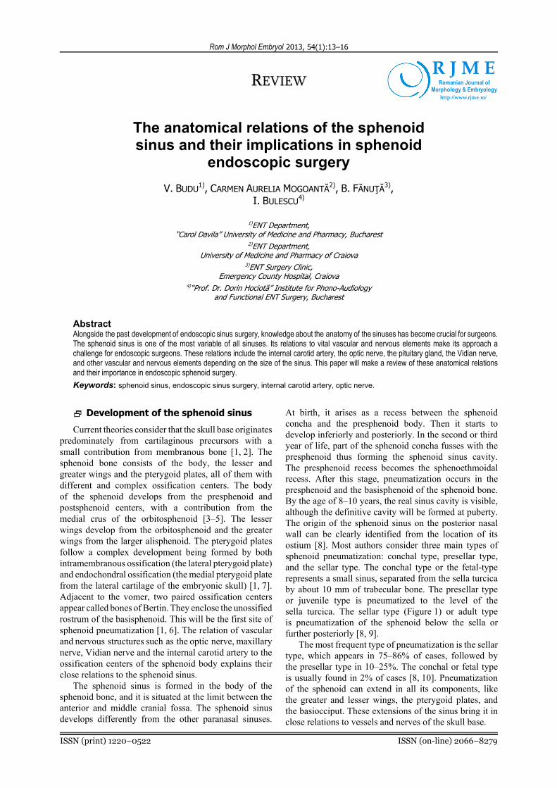
At birth, it arises as a recess between the sphenoid concha and the presphenoid body. Then it starts to develop inferiorly and posteriorly. In the second or third year of life, part of the sphenoid concha fusses with the presphenoid thus forming the sphenoid sinus cavity. The presphenoid recess becomes the sphenoethmoidal recess. After this stage, pneumatization occurs in the presphenoid and the basisphenoid of the sphenoid bone. By the age of 8–10 years, the real sinus cavity is visible, although the definitive cavity will be formed at puberty. The origin of the sphenoid sinus on the posterior nasal wall can be clearly identified from the location of its ostium [8]. Most authors consider three main types of sphenoid pneumatization: conchal type, presellar type, and the sellar type. The conchal type or the fetal-type represents a small sinus, separated from the sella turcica by about 10 mm of trabecular bone. The presellar type or juvenile type is pneumatized to the level of the sella turcica. The sellar type (Figure 1) or adult type is pneumatization of the sphenoid below the sella or further posteriorly [8, 9].
The most frequent type of pneumatization is the sellar type, which appears in 75–86% of cases, followed by the presellar type in 10–25%. The conchal or fetal type is usually found in 2% of cases [8, 10]. Pneumatization of the sphenoid can extend in all its components, like the greater and lesser wings, the pterygoid plates, and the basiocciput. These extensions of the sinus bring it in close relations to vessels and nerves of the skull base.
R J M ERomanian Journal of
Morphology & Embryologyhttp://www.rjme.ro/
V. Budu et al.
14
Figure 1 – Sagittal CT section showing a sellar type pneumatization of the sphenoid sinus.
Anatomy and variations of the sphenoid sinus. Relations of the sphenoid sinus
The sphenoid bone is one of the most complex structures of the cranium. It is a part of the floor of the middle cranial fossa, the orbital apex and a part of the lateral wall of the skull. It is composed by the body, the lesser and greater wings and the pterygoid plates. The sphenoid sinuses develop in the body of the sphenoid bone. Of the sinus’s eight sides, six sides are disposed toward the endocranium and the other two sides toward the nasopharynx and nasal cavity. The superior wall of the sinus is in continuity with the roof of the ethmoid sinus and it is part of the anterior and middle floors of the skull base, being in direct contact to the olfactory nerves, optic chiasm and hypophysis. The anterior wall is connected to the perpendicular plate of the ethmoid and vomer in the midline, as well as the lateral masses of the ethmoid on each side [8, 9, 11]. This wall can be displaced by highly-developed Onodi cells or ethmoido-sphenoidal cells. The anterior wall is the place where the sinus ostium is often found. The floor of the sinus forms the dome of the choanae and of the nasopharynx. The lateral walls are thin bony layers and can be divided into two areas: an anterior orbital area and the posterior cranial area. This wall has immediately adjacent to it highly important structures such as the internal carotid artery, optic nerve and the cavernous sinus [8, 9, 12–14].
The relations of the sphenoid sinus with structures around are close when the sinus is well pneumatized. When this happens, the surrounding vessels and nerves are seen in the sinus cavity as irregularities or ridges [8, 15]. Pneumatization of the sphenoid sinus can extend to further from its body, and into all of its parts, such as the clinoid processes, greater wings and pterygoid planes. According to Climelli D [16], cited by [17], the sinus spreads more often anteriorly and laterally than posteriorly and inferiorly.
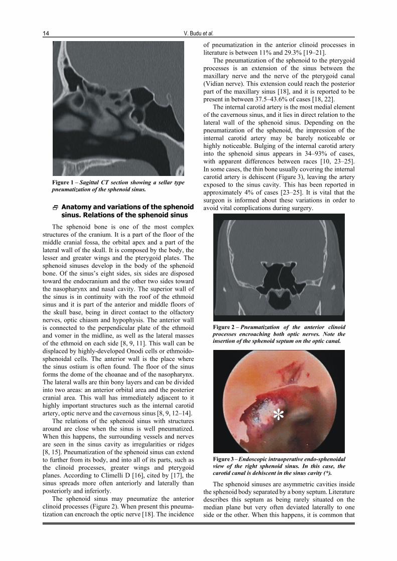
The sphenoid sinus may pneumatize the anterior clinoid processes (Figure 2). When present this pneuma-tization can encroach the optic nerve [18]. The incidence
of pneumatization in the anterior clinoid processes in literature is between 11% and 29.3% [19–21].
The pneumatization of the sphenoid to the pterygoid processes is an extension of the sinus between the maxillary nerve and the nerve of the pterygoid canal (Vidian nerve). This extension could reach the posterior part of the maxillary sinus [18], and it is reported to be present in between 37.5–43.6% of cases [18, 22].
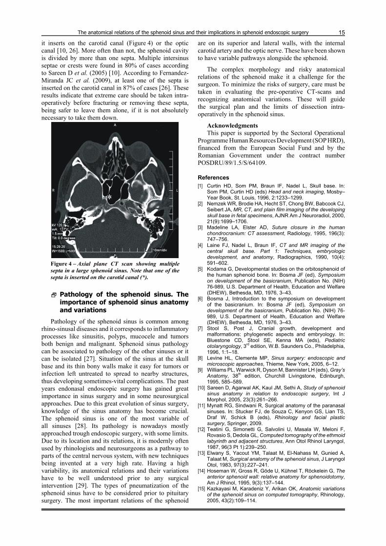
The internal carotid artery is the most medial element of the cavernous sinus, and it lies in direct relation to the lateral wall of the sphenoid sinus. Depending on the pneumatization of the sphenoid, the impression of the internal carotid artery may be barely noticeable or highly noticeable. Bulging of the internal carotid artery into the sphenoid sinus appears in 34–93% of cases, with apparent differences between races [10, 23–25]. In some cases, the thin bone usually covering the internal carotid artery is dehiscent (Figure 3), leaving the artery exposed to the sinus cavity. This has been reported in approximately 4% of cases [23–25]. It is vital that the surgeon is informed about these variations in order to avoid vital complications during surgery.
Figure 2 – Pneumatization of the anterior clinoid processes encroaching both optic nerves. Note the insertion of the sphenoid septum on the optic canal.
Figure 3 – Endoscopic intraoperative endo-sphenoidal view of the right sphenoid sinus. In this case, the carotid canal is dehiscent in the sinus cavity (*).
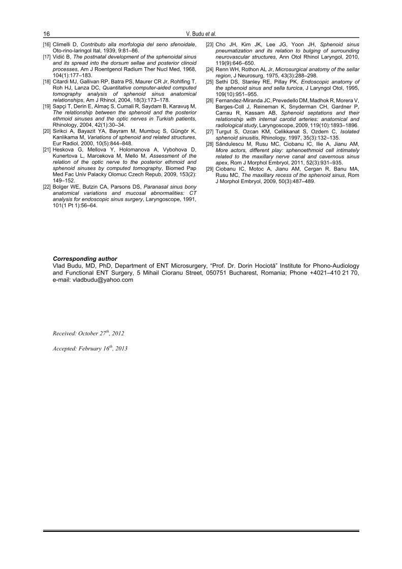
The sphenoid sinuses are asymmetric cavities inside the sphenoid body separated by a bony septum. Literature describes this septum as being rarely situated on the median plane but very often deviated laterally to one side or the other. When this happens, it is common that
The anatomical relations of the sphenoid sinus and their implications in sphenoid endoscopic surgery
15
it inserts on the carotid canal (Figure 4) or the optic canal [10, 26]. More often than not, the sphenoid cavity is divided by more than one septa. Multiple intersinus septae or crests were found in 80% of cases according to Sareen D et al. (2005) [10]. According to Fernandez-Miranda JC et al. (2009), at least one of the septa is inserted on the carotid canal in 87% of cases [26]. These results indicate that extreme care should be taken intra-operatively before fracturing or removing these septa, being safer to leave them alone, if it is not absolutely necessary to take them down.
Figure 4 – Axial plane CT scan showing multiple septa in a large sphenoid sinus. Note that one of the septa is inserted on the carotid canal (*).
Pathology of the sphenoid sinus. The importance of sphenoid sinus anatomy and variations
Pathology of the sphenoid sinus is common among rhino-sinusal diseases and it corresponds to inflammatory processes like sinusitis, polyps, mucocele and tumors both benign and malignant. Sphenoid sinus pathology can be associated to pathology of the other sinuses or it can be isolated [27]. Situation of the sinus at the skull base and its thin bony walls make it easy for tumors or infection left untreated to spread to nearby structures, thus developing sometimes-vital complications. The past years endonasal endoscopic surgery has gained great importance in sinus surgery and in some neurosurgical approaches. Due to this great evolution of sinus surgery, knowledge of the sinus anatomy has become crucial. The sphenoid sinus is one of the most variable of all sinuses [28]. Its pathology is nowadays mostly approached trough endoscopic surgery, with some limits. Due to its location and its relations, it is modernly often used by rhinologists and neurosurgeons as a pathway to parts of the central nervous system, with new techniques being invented at a very high rate. Having a high variability, its anatomical relations and their variations have to be well understood prior to any surgical intervention [29]. The types of pneumatization of the sphenoid sinus have to be considered prior to pituitary surgery. The most important relations of the sphenoid
are on its superior and lateral walls, with the internal carotid artery and the optic nerve. These have been shown to have variable pathways alongside the sphenoid.
The complex morphology and risky anatomical relations of the sphenoid make it a challenge for the surgeon. To minimize the risks of surgery, care must be taken in evaluating the pre-operative CT-scans and recognizing anatomical variations. These will guide the surgical plan and the limits of dissection intra-operatively in the sphenoid sinus.
Acknowledgments This paper is supported by the Sectoral Operational
Programme Human Resources Development (SOP HRD),
financed from the European Social Fund and by the Romanian Government under the contract number POSDRU/89/1.5/S/64109.
References [1] Curtin HD, Som PM, Braun IF, Nadel L, Skull base. In:
Som PM, Curtin HD (eds) Head and neck imaging, Mosby–Year Book, St. Louis, 1996, 2:1233–1299.
[2] Nemzek WR, Brodie HA, Hecht ST, Chong BW, Babcook CJ, Seibert JA, MR, CT, and plain film imaging of the developing skull base in fetal specimens, AJNR Am J Neuroradiol, 2000, 21(9):1699–1706.
[3] Madeline LA, Elster AD, Suture closure in the human chondrocranium: CT assessment, Radiology, 1995, 196(3): 747–756.
[4] Laine FJ, Nadel L, Braun IF, CT and MR imaging of the central skull base. Part 1: Techniques, embryologic development, and anatomy, Radiographics, 1990, 10(4): 591–602.
[5] Kodama G, Developmental studies on the orbitosphenoid of the human sphenoid bone. In: Bosma JF (ed), Symposium on development of the basicranium, Publication No. (NIH) 76-989, U.S. Department of Health, Education and Welfare (DHEW), Bethesda, MD, 1976, 3–43.
[6] Bosma J, Introduction to the symposium on development of the basicranium. In: Bosma JF (ed), Symposium on development of the basicranium, Publication No. (NIH) 76-989, U.S. Department of Health, Education and Welfare (DHEW), Bethesda, MD, 1976, 3–43.
[7] Stool S, Post J, Cranial growth, development and malformations: phylogenetic aspects and embryology. In: Bluestone CD, Stool SE, Kenna MA (eds), Pediatric otolaryngology, 3rd
edition, W.B. Saunders Co., Philadelphia, 1996, 1:1–18.
[8] Levine HL, Clemente MP, Sinus surgery: endoscopic and microscopic approaches, Thieme, New York, 2005, 6–12.
[9] Williams PL, Warwick R, Dyson M, Bannister LH (eds), Gray’s Anatomy, 38th edition, Churchill Livingstone, Edinburgh, 1995, 585–589.
[10] Sareen D, Agarwal AK, Kaul JM, Sethi A, Study of sphenoid sinus anatomy in relation to endoscopic surgery, Int J Morphol, 2005, 23(3):261–266.
[11] Mynatt RG, Sindwani R, Surgical anatomy of the paranasal sinuses. In: Stucker FJ, de Souza C, Kenyon GS, Lian TS, Draf W, Schick B (eds), Rhinology and facial plastic surgery, Springer, 2009.
[12] Teatini G, Simonetti G, Salvolini U, Masala W, Meloni F, Rovasio S, Dedola GL, Computed tomography of the ethmoid labyrinth and adjacent structures, Ann Otol Rhinol Laryngol, 1987, 96(3 Pt 1):239–250.
[13] Elwany S, Yacout YM, Talaat M, El-Nahass M, Gunied A, Talaat M, Surgical anatomy of the sphenoid sinus, J Laryngol Otol, 1983, 97(3):227–241.
[14] Hoseman W, Gross R, Göde U, Kühnel T, Röckelein G, The anterior sphenoid wall: relative anatomy for sphenoidotomy, Am J Rhinol, 1995, 9(3):137–144.
[15] Kazkayasi M, Karadeniz Y, Arikan OK, Anatomic variations of the sphenoid sinus on computed tomography, Rhinology, 2005, 43(2):109–114.
V. Budu et al.
16
[16] Climelli D, Contributo alla morfologia del seno sfenoidale, Oto-rino-laringol Ital, 1939, 9:81–86.
[17] Vidić B, The postnatal development of the sphenoidal sinus and its spread into the dorsum sellae and posterior clinoid processes, Am J Roentgenol Radium Ther Nucl Med, 1968, 104(1):177–183.
[18] Citardi MJ, Gallivan RP, Batra PS, Maurer CR Jr, Rohlfing T, Roh HJ, Lanza DC, Quantitative computer-aided computed tomography analysis of sphenoid sinus anatomical relationships, Am J Rhinol, 2004, 18(3):173–178.
[19] Sapçi T, Derin E, Almaç S, Cumali R, Saydam B, Karavuş M, The relationship between the sphenoid and the posterior ethmoid sinuses and the optic nerves in Turkish patients, Rhinology, 2004, 42(1):30–34.
[20] Sirikci A, Bayazit YA, Bayram M, Mumbuç S, Güngör K, Kanlikama M, Variations of sphenoid and related structures, Eur Radiol, 2000, 10(5):844–848.
[21] Heskova G, Mellova Y, Holomanova A, Vybohova D, Kunertova L, Marcekova M, Mello M, Assessment of the relation of the optic nerve to the posterior ethmoid and sphenoid sinuses by computed tomography, Biomed Pap Med Fac Univ Palacky Olomuc Czech Repub, 2009, 153(2): 149–152.
[22] Bolger WE, Butzin CA, Parsons DS, Paranasal sinus bony anatomical variations and mucosal abnormalities: CT analysis for endoscopic sinus surgery, Laryngoscope, 1991, 101(1 Pt 1):56–64.
[23] Cho JH, Kim JK, Lee JG, Yoon JH, Sphenoid sinus pneumatization and its relation to bulging of surrounding neurovascular structures, Ann Otol Rhinol Laryngol, 2010, 119(9):646–650.
[24] Renn WH, Rothon AL Jr, Microsurgical anatomy of the sellar region, J Neurosurg, 1975, 43(3):288–298.
[25] Sethi DS, Stanley RE, Pillay PK, Endoscopic anatomy of the sphenoid sinus and sella turcica, J Laryngol Otol, 1995, 109(10):951–955.
[26] Fernandez-Miranda JC, Prevedello DM, Madhok R, Morera V, Barges-Coll J, Reineman K, Snyderman CH, Gardner P, Carrau R, Kassam AB, Sphenoid septations and their relationship with internal carotid arteries: anatomical and radiological study, Laryngoscope, 2009, 119(10):1893–1896.
[27] Turgut S, Ozcan KM, Celikkanat S, Ozdem C, Isolated sphenoid sinusitis, Rhinology, 1997, 35(3):132–135.
[28] Săndulescu M, Rusu MC, Ciobanu IC, Ilie A, Jianu AM, More actors, different play: sphenoethmoid cell intimately related to the maxillary nerve canal and cavernous sinus apex, Rom J Morphol Embryol, 2011, 52(3):931–935.
[29] Ciobanu IC, Motoc A, Jianu AM, Cergan R, Banu MA, Rusu MC, The maxillary recess of the sphenoid sinus, Rom J Morphol Embryol, 2009, 50(3):487–489.
Corresponding author Vlad Budu, MD, PhD, Department of ENT Microsurgery, “Prof. Dr. Dorin Hociotă” Institute for Phono-Audiology and Functional ENT Surgery, 5 Mihail Cioranu Street, 050751 Bucharest, Romania; Phone +4021–410 21 70, e-mail: [email protected] Received: October 27th, 2012
Accepted: February 16th, 2013
Related Documents

![Rom J Morphol Embryol R J M E ORIGINAL … the pneumatization of the middle turbinate has often been described, pneumatized superior turbinates [1, 2], supreme turbinates, uncinate](https://static.cupdf.com/doc/110x72/5ca9f2dc88c993c9218d71be/rom-j-morphol-embryol-r-j-m-e-original-the-pneumatization-of-the-middle-turbinate.jpg)

![Rom J Morphol Embryol 2013, 54(1):205–210 R J M E CASE ... · PDF fileRom J Morphol Embryol 2013, 54(1):205–210 ISSN ... project report CEEX 68/2006 [3] shows that 650 000 ...](https://static.cupdf.com/doc/110x72/5aae038b7f8b9a07498b87b2/rom-j-morphol-embryol-2013-541205210-r-j-m-e-case-j-morphol-embryol-2013.jpg)


