Rom J Morphol Embryol 2011, 52(3):915–918 CASE REPORT Sinusoidal hemangioma of the arm: case report and review of literature M. CIUREA 1) , RALUCA CIUREA 2) , D. POPA 1) , H. PÂRVĂNESCU 1) , DANIELA MARINESCU 3) , MARIA VRABETE 4) 1) Department of Plastic and Reconstructive Surgery 2) Department of Pathology 3) Department of General Surgery Clinic 1 4) Department of Anesthesia and Intensive Care Faculty of Medicine, University of Medicine and Pharmacy of Craiova Abstract Sinusoidal hemangioma is a rare variant of cavernous hemangioma that develops more frequently subcutaneous, often in the extremities. We report the case of a 19-year-old patient three times operated for a recurrent soft tissue tumor, located in the distal third of the antero- medial face of the right arm. Treatment was surgical and consisted of microsurgical excision of a tumor with dermal and intramuscular location. Histopathological analysis showed a nodular proliferation of interconnected thin-walled blood vessels, sinusoidal growth pattern and the presence of pseudopapillary structures. Immunohistochemical investigations were positive for CD31, CD34 and Ki67 index decreased, confirming the benign nature of vascular proliferation. Keywords: sinusoidal hemangioma, intramuscular, diagnostic, treatment. Introduction Hemagiomas are benign vascular whose exact cause is not known. They are a group of hamartomatous entities of neoplastic or reactive nature, which have a common morphological feature represented by the existence of well-differentiated blood vessels and limited proliferative capacity. It can develop on skin, subcutaneous tissue and muscle, bone or internal organs [1]. Intramuscular location of hemangiomas is very rare [2], representing less than 1% of all hemangiomas [1]. In the past few years, a number of rare vascular tumors have been characterized, among which sinusoidal hemangioma, considered a distinct subtype of cavernous hemangiomas [3]. They may have many clinical aspects, and microscopically some cases can mimic well- differentiated angiosarcoma [3, 4]. Although sinusoidal hemangiomas are very rare, their clinico-pathological characteristics are important to be recognized in order to avoid diagnostic pitfalls. As a result, we consider useful to report the particularities of such a case, along with similar data from the literature reviews. Materials and Methods This paper report the case of 19-year-old female patient who was three times operated for recurrent soft tissue tumor, located in the distal third of the anterior- medial face of the right arm. For the last relapse, the patient was admitted and operated in the Plastic Surgery and Reconstructive Microsurgery Department of Emergency County Hospital of Craiova. Surgical excision piece was fixed in 10% formalin, processed by usual techniques for inclusion in paraffin and Hematoxylin–Eosin stained in the Pathology Department of the same hospital. Subsequently, 4 μm sections were made and they were subjected to immunohistochemical processing technique LSAB/HPR for Ki67 (MIB1), CD31 (JC70A) and CD34. Sections were incubated one hour with primary antibody (mouse anti-human Ki67; clone MIB1, dilution 1:100) and we used DAB for visualization followed by counterstaining with Hematoxylin. Negative external control was used by omitting the primary antibody. To quantify Ki67 a cell proliferation index was achieved by comparing marked cell counts to the total number of counted cells at the objective 40×. Count was performed on 10 microscopic fields each involving over 1000 cells. To assess CD31 and CD34 expression we considered cytoplasm marker. Results We present the case of a female patient diagnosed in childhood with hemangioma of the arm, injury that has fared aggressive local, marked by enlargement and three recurrences within 12 years. History has shown that the onset of disease occurred at 7-year-old, when the parents noticed a skin tumor located in the lower third of the right arm. The tumor was surgically excised and then the diagnosis was hemangioma. Three months after surgery the tumor recurred, rapidly returning to its original size. At 16-year-old patient accused local pain on exertion, which was R J M E Romanian Journal of Morphology & Embryology http://www.rjme.ro/

Welcome message from author
This document is posted to help you gain knowledge. Please leave a comment to let me know what you think about it! Share it to your friends and learn new things together.
Transcript
Rom J Morphol Embryol 2011, 52(3):915–918
CCAASSEE RREEPPOORRTT
Sinusoidal hemangioma of the arm: case report and review of literature
M. CIUREA1), RALUCA CIUREA2), D. POPA1), H. PÂRVĂNESCU1), DANIELA MARINESCU3), MARIA VRABETE4)
1)Department of Plastic and Reconstructive Surgery 2)Department of Pathology
3)Department of General Surgery Clinic 1 4)Department of Anesthesia and Intensive Care
Faculty of Medicine, University of Medicine and Pharmacy of Craiova
Abstract Sinusoidal hemangioma is a rare variant of cavernous hemangioma that develops more frequently subcutaneous, often in the extremities. We report the case of a 19-year-old patient three times operated for a recurrent soft tissue tumor, located in the distal third of the antero-medial face of the right arm. Treatment was surgical and consisted of microsurgical excision of a tumor with dermal and intramuscular location. Histopathological analysis showed a nodular proliferation of interconnected thin-walled blood vessels, sinusoidal growth pattern and the presence of pseudopapillary structures. Immunohistochemical investigations were positive for CD31, CD34 and Ki67 index decreased, confirming the benign nature of vascular proliferation. Keywords: sinusoidal hemangioma, intramuscular, diagnostic, treatment.
Introduction
Hemagiomas are benign vascular whose exact cause is not known. They are a group of hamartomatous entities of neoplastic or reactive nature, which have a common morphological feature represented by the existence of well-differentiated blood vessels and limited proliferative capacity. It can develop on skin, subcutaneous tissue and muscle, bone or internal organs [1]. Intramuscular location of hemangiomas is very rare [2], representing less than 1% of all hemangiomas [1].
In the past few years, a number of rare vascular tumors have been characterized, among which sinusoidal hemangioma, considered a distinct subtype of cavernous hemangiomas [3]. They may have many clinical aspects, and microscopically some cases can mimic well-differentiated angiosarcoma [3, 4].
Although sinusoidal hemangiomas are very rare, their clinico-pathological characteristics are important to be recognized in order to avoid diagnostic pitfalls. As a result, we consider useful to report the particularities of such a case, along with similar data from the literature reviews.
Materials and Methods
This paper report the case of 19-year-old female patient who was three times operated for recurrent soft tissue tumor, located in the distal third of the anterior-medial face of the right arm. For the last relapse, the patient was admitted and operated in the Plastic Surgery and Reconstructive Microsurgery Department of Emergency County Hospital of Craiova. Surgical
excision piece was fixed in 10% formalin, processed by usual techniques for inclusion in paraffin and Hematoxylin–Eosin stained in the Pathology Department of the same hospital.
Subsequently, 4 µm sections were made and they were subjected to immunohistochemical processing technique LSAB/HPR for Ki67 (MIB1), CD31 (JC70A) and CD34. Sections were incubated one hour with primary antibody (mouse anti-human Ki67; clone MIB1, dilution 1:100) and we used DAB for visualization followed by counterstaining with Hematoxylin. Negative external control was used by omitting the primary antibody. To quantify Ki67 a cell proliferation index was achieved by comparing marked cell counts to the total number of counted cells at the objective 40×. Count was performed on 10 microscopic fields each involving over 1000 cells. To assess CD31 and CD34 expression we considered cytoplasm marker.
Results
We present the case of a female patient diagnosed in childhood with hemangioma of the arm, injury that has fared aggressive local, marked by enlargement and three recurrences within 12 years. History has shown that the onset of disease occurred at 7-year-old, when the parents noticed a skin tumor located in the lower third of the right arm. The tumor was surgically excised and then the diagnosis was hemangioma.
Three months after surgery the tumor recurred, rapidly returning to its original size. At 16-year-old patient accused local pain on exertion, which was
R J M ERomanian Journal of
Morphology & Embryologyhttp://www.rjme.ro/

M. Ciurea et al.
916
subjected to another surgery for tumor excision. Post-operative evolution was favorable, but the tumor recurred again after three months, with the appearance of a subcutaneous formations, without changes in skin coverings. In evolution, the tumor grew in size to about 5–6 cm and became slightly painful to moderate effort. At age 18 years, the patient underwent a new surgery and the tumor was again excised with a layer of muscle biceps brachial muscle tissue due to adhesions in muscle fibers.
Three months after surgery, the tumor recurred again. After about a year after the last surgery, the patient presented with moderate local pain exacerbated by exercise and the increase in tumor size during exercise. Physical examination revealed the presence of local dehiscence postoperative scar in the longitudinal axis, upon which there was a soft tissue tumor, measuring
6/4 cm, without changes in the covering skin. On palpation, the tumor consistency was elastic and mobile on superficial anatomical plans, painless, with slightly irregular surface. Exercise tests showed a rapid increase in tumor size and moderate local pain. Also, the patient accused the numbness in small efforts on right ulnae dermatomas projections.
Ultrasound for soft tissue was inconclusive. Angio-MRI showed a round-oval tumor mass in the anterior-medial muscles of the right arm, with dimensions of 2.5/1.42 cm, with heterogeneous signal, with rich vascularization and calcification. Tumor vascular structures were represented by the branches of brachial artery, with drainage in the homonymous vein (Figure 1a).
Lesion was treated by wide surgical excision. The strategy included surgical exploration of the tumor and brachial vascular-nervous axis. During surgery, we observed a vascular tumor located in the lower third of the right arm, infiltrating into the dermis. Tumor
adhesions were present between brachial biceps muscle fibers, which made it very difficult excision. The dissection and exploration of brachial vascular–nervous axis found high dilatation of basilica and cephalic veins, which allowed the hypothesis of a venous vascular malformation (Figure 1, b and c). Interestingly, venous dilatation were located only in the arm veins, the veins of the forearm were normal. A possible explanation could be the pressure on these dilated veins caused by tumor. Communication of the vascular tumor with brachial artery was reduced.
Surgical enlarged excision piece, with 5.5/3.5/2 cm in size, showed on section a unique, nodular, well circumscribed formation, measuring 1.5/1/1.2 cm. The tumor had an elastic consistency and reddish-blackish color.
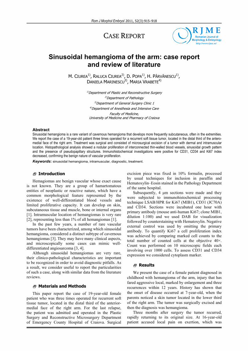
Histopathological examination of surgical sample showed a nodular proliferation, well defined but unencapsulated, the interconnected blood vessels making the whole look spongy, located in deep dermis and muscle. Vascular spaces were thin-walled, variable in size and a sinusoidal growth pattern, often with back-to-back arrangement. Inside there were numerous vascular spaces pseudopapillary structures. Both the papillae and the vascular spaces were wallpapered by a single row of endothelial cells with focal hyperchromatic nuclei, but mitosis was absent (Figure 2, a–c). Some vascular spaces were filled with red blood cells, others were empty or had recent and old, organized thrombi. Stroma of the vascular spaces was reduced and fibrous.
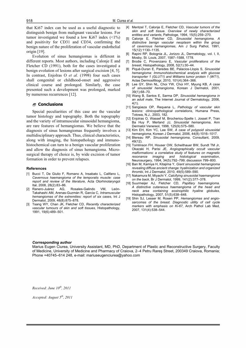
CD31 and CD34 immunostaining indicated positivity in lining cells of the vascular spaces and papillary proliferation, confirming their vascular endothelial origin (Figure 3, a and b). Immunohistochemical investigations allowed Ki67 index assessment, which showed low values, with less than 1% of tumor vessel endothelial positive cells (Figure 3c).
Figure 1 – (a) Aspect of angio-MRI. (b) Brachial artery (the small size vessel in the center of the picture). (c) Cephalic vein dilation.

Sinusoidal hemangioma of the arm: case report and review of literature
917
Figure 2 – Sinusoidal cavernous intramusculary hemangioma: (a) collage image, HE stain; (b) papillary projections, HE stain, ×40; (c) endothelia with minimal atypia, HE stain, ×400.
Figure 3 – Sinusoidal cavernous intramusculary hemangioma: (a) CD31 immunostain, ×100; (b) CD34 immunostain, ×100; (c) Ki67 immunostain, ×400.
Discussion
Sinusoidal hemangioma is a rare variant entity, recently described by Calonje E and Fletcher CD (1991), and considered a subset of cavernous hemangiomas. Cavernous hemangiomas pathogenesis is for the most part unknown, but is speculated that may involve abnormalities of the vasculogenesis and angiogenesis [5].
Tumors may develop in the extremities and trunk, including mammary gland [5–7]. In most studies, the distribution by sex indicates predominance of lesions in women [3, 6, 8–10]. Injuries most often develop in adulthood, but have been described in young children [5, 11]. The case we report new overlaps largely upon these characteristics, the tumor being diagnosed in a young female and developed in the arm.
Clinically, lesions are often located subcutaneously in the deep dermis, where they have a quite characteristic nodular appearance [5, 6, 9, 10, 12], or more rare like some plurinodular polypoid formations [13] or even cystic [14], covered by normal skin. Unlike the literature reporting the development of lesions in the deep dermis, in our case the tumor was located in the deep dermis and extended into the muscle, between the brachial biceps muscle fibers.
Symptoms of tumor were generally low, the lesions being often painless or slightly painful on effort [5, 10, 14]. In our case, tumor enlargement during exercise is explained by filling it with blood while pain was produced by the strong adhesions of the tumor with brachial biceps muscle. Paresthesia of the hand can be explained by the compressive phenomena from a large venous dilatation in the cephalic vein on the ulnar nerve. Brachial artery reduced diameter can be due to an anomaly of development or because of “theft of blood” by the tumor. Venous dilations so numerous and large can be explained through a development abnormality,
which is unlikely, given the fact that distal to the tumor, the vessels are of normal appearance.
Imaging for sinusoidal hemangiomas indicates a nodular appearance [12, 15]. Similarly to the literature reports, angio-MRI for the investigated case has been shown a single nodular mass, located in the anterior-medial muscles of the right arm, with dimensions of 2.5/1.42 cm, with heterogeneous signal and rich vascularization.
Surgery involved wide surgical excision of the lesion. According to the surgical findings, the location of the tumor was clearly intramuscular – biceps brachialis muscle. Most authors agree with the surgical treatment of the lesion, considering sufficiently a large enough surgical excision of the tumors [10, 15]. Some authors consider that the tumors can be treated by cryotherapy, electrocoagulation or laser therapy [14]. In this case, the high relapse rate may be explained by insufficient tumor excision.
Histopathologically tumors and lobular architecture is characterized by proliferation of morphologically sinusoidal vessels [5, 6, 10, 12]. The old forms of damage are listed and organized hyalinised thrombi [16]. Tendency to thrombosis and calcification present in the center of the lesion [17]. The presence of stroma with or without fibrocollagen hyalinisations between vascular type sinusoidal spaces, with focal atypia of the endothelial lining may lead to diagnostic confusion with some benign lesions (vascular malformations, lymph-angioma) or a well differentiated angiosarcoma [3, 5, 18].
Suurmeijer AJ et al. (2007) communicate positivity for CD31 and CD34 of the endothelial cells and negativity for D2-40, confirming the endothelial vascular origin of these tumors. Useful for differentiating for an angiosarcoma are the immunohistochemical tests to assess Ki67 index [18]. Shin SJ et al. (2007) considers
M. Ciurea et al.
918
that Ki67 index can be used as a useful diagnostic to distinguish benign from malignant vascular lesions. For tumor investigated we found a low Ki67 index (<1%) and positivity for CD31 and CD34, confirming the benign nature of the proliferation of vascular endothelial origin [19].
Evolution of sinus hemangiomas is different in different reports. Most authors, including Calonje E and Fletcher CD (1991), both for the cases investigated a benign evolution of lesions after surgical excision [4, 5]. In contrast, Enjolras O et al. (1998) four such cases shall congenital or childhood-onset and aggressive clinical course and prolonged. Similarly, the case presented such a development was prolonged, marked by numerous recurrences [12].
Conclusions
Special peculiarities of this case are the vascular tumor histology and topography. Both the topography and the variety of intramuscular sinusoidal hemangioma, are rare features of hemangiomas. We believe that the diagnosis of sinus hemangiomas frequently involves a multidisciplinary approach. Thus, clinical characteristics, along with imaging, the histopathology and immuno-histochemical can turn to a benign vascular proliferation and allow the diagnosis of sinus hemangioma. Micro-surgical therapy of choice is, by wide excision of tumor formation in order to prevent relapses.
References [1] Bucci T, De Giulio F, Romano A, Insabato L, Califano L,
Cavernous haemangioma of the temporalis muscle: case report and review of the literature, Acta Otorhinolaryngol Ital, 2008, 28(2):83–86.
[2] Ranero-Juárez AG, Rosales-Galindo VM, León- Takahashi AM, Arenas-Guzmán R, García C, Intramuscular hemangiomas of the extremities: report of six cases, Int J Dermatol, 2009, 48(8):875–878.
[3] Tsang WY, Chan JK, Fletcher CD, Recently characterized vascular tumours of skin and soft tissues, Histopathology, 1991, 19(6):489–501.
[4] Mentzel T, Calonje E, Fletcher CD, Vascular tumors of the skin and soft tissue. Overview of newly characterized entities and variants, Pathologe, 1994, 15(5):259–270.
[5] Calonje E, Fletcher CD, Sinusoidal hemangioma. A distinctive benign vascular neoplasm within the group of cavernous hemangiomas, Am J Surg Pathol, 1991, 15(12):1130–1135.
[6] Rapini RP, Bolognia JL, Jorizzo JL, Dermatology, vol. I, II, Mosby, St. Louis, 2007, 1587–1588, 1778.
[7] Brodie C, Provenzano E, Vascular proliferations of the breast, Histopathology, 2008, 52(1):30–44.
[8] Piqué-Duran E, Paredes BE, Palacios-Llopis S, Sinusoidal hemangioma: Immunohistochemical analysis with glucose transporter 1 (GLUT1) and Williams tumor protein 1 (WT1), Actas Dermosifiliogr, 2010, 101(4):364–366.
[9] Lee GY, Shin NL, Choi YW, Choi HY, Myung KB, A case of sinusoidal hemangioma, Korean J Dermatol, 2001, 39(1):68–70.
[10] Wang B, Santos E, Sarma DP, Sinusoidal hemangioma in an adult male, The Internet Journal of Dermatology, 2006, 4(1).
[11] Sangüeza OP, Requena L, Pathology of vascular skin lesions: clinicopathological correlations, Humana Press, Totowa, N.J., 2003, 182.
[12] Enjolras O, Wassef M, Brocheriou-Spelle I, Josset P, Tran Ba Huy P, Merland JJ, Sinusoidal hemangioma, Ann Dermatol Venereol, 1998, 125(9):575–580.
[13] Kim EH, Kim YC, Lee SW, A case of polypoid sinusoidal hemangioma, Korean J Dermatol, 2006, 44(8):1016–1017.
[14] Blereau RP, Sinusoidal hemangioma, Consultant, 2009, 49(12).
[15] Tomlinson FH, Houser OW, Scheithauer BW, Sundt TM Jr, Okazaki H, Parisi JE, Angiographically occult vascular malformations: a correlative study of features on magnetic resonance imaging and histological examination, Neurosurgery, 1994, 34(5):792–799; discussion 799–800.
[16] Ban M, Kamiya H, Kitajima Y, Giant sinusoidal hemangioma revealing diffuse ancient change: hyalinization and organized
thrombi, Int J Dermatol, 2010, 49(5):589–590. [17] Nakamura M, Miyachi Y, Calcifying sinusoidal haemangioma
on the back, Br J Dermatol, 1999, 141(2):377–378. [18] Suurmeijer AJ, Fletcher CD, Papillary haemangioma.
A distinctive cutaneous haemangioma of the head and neck area containing eosinophilic hyaline globules, Histopathology, 2007, 51(5):638–648.
[19] Shin SJ, Lesser M, Rosen PP, Hemangiomas and angio-sarcomas of the breast. Diagnostic utility of cell cycle markers with emphasis on Ki-67, Arch Pathol Lab Med, 2007, 131(4):538–544.
Corresponding author Marius Eugen Ciurea, University Assistant, MD, PhD, Department of Plastic and Reconstructive Surgery, Faculty of Medicine, University of Medicine and Pharmacy of Craiova, 2–4 Petru Rareş Street, 200349 Craiova, Romania; Phone +40745–614 248, e-mail: [email protected] Received: June 10th, 2011
Accepted: August 5th, 2011
Related Documents