Rom J Morphol Embryol 2014, 55(3):977–981 ISSN (print) 1220–0522 ISSN (on-line) 2066–8279 CASE REPORT Aneurysmal bone cyst – clinical and morphological aspects RADU RĂDULESCU 1,2) , ADRIAN BĂDILĂ 2) , ROBERT MANOLESCU 2) , MARIA SAJIN 1,3) , ION JAPIE 2) 1) “Carol Davila” University of Medicine and Pharmacy, Bucharest, Romania 2) Department of Orthopedic Surgery, Bucharest University Hospital, Romania 3) Department of Pathology, Bucharest University Hospital, Romania Abstract Aim: To assess the results of surgical treatment in aneurysmal bone cysts. Materials and Methods: 31 patients with aneurysmal bone cysts underwent surgical treatment in our department. In almost half of cases, the lesion was located in the femur. In 12 cases, a pathological bone fracture was the first clinical sign. The treatment consisted in curettage, abrasion of the cavity inner walls using a motorized burr and filling with morsellized bone grafts (autografts ± allografts) or bone substitutes (four cases). Microscopically, the diagnosis relies on cystic spaces filled with blood, divided by fibrous septae consisting in immature bone trabeculae, hemosiderin filled macrophages and fibroblasts. We performed multiple bioptic probes from different levels of the lesion. Results: Macroscopically, the osseous lesion appeared as a multi- loculated blood-filled cavity (cavities separated by septa) in 30 (96.77%) cases and as a solid tumor in one (3.23%) case. At 12 months after surgery, grafts osteointegration was present in 24 cases. At an average follow-up time of six years and four months, refilling with bone grafts was necessary in two cases and no local recurrence was observed. Conclusions: Aneurysmal bone cyst is most frequent in the second decade of age. Its prevalence in female gender is double compared to the male gender. The positive diagnosis relies on the histopathological examination. Because of the strong relationship with a number of precursor lesions (giant cell tumor, fibrous dysplasia, non-ossifying fibroma, chondroblastoma, osteoblastoma) multiple bioptic probings are mandatory, in order to diagnose, if possible, a primary lesion, which may modify the therapeutic attitude. Treatment by curettage, abrasion of the cavity inner walls and filling with morsellized grafts has very good results. The risk of recurrence is very low. Keywords: aneurysmal bone cyst, curettage, bone grafting. Introduction Aneurysmal bone cyst is an expansive benign bone lesion, which appears like a group of intraosseous cavities lacking endothelial membrane and containing blood. Peak incidence is recorded in the second decade of life, 80% of them occurring in patients less than 20 years [1]. Gender distribution is males/females 1:1.3, and the incidence in the general population is 0.14/ 100 000 [2], representing about 1% of total bone tumors. Most frequently, it is found at proximal and distal metaphysis of the femur and the proximal metaphysis of tibia, but can affect almost any bone segment in the body. Other frequent locations are the humerus, the vertebrae and the pelvic bones. 50–60% of cases involve long bones of the limbs (lower limb 40% and upper limb 20%, respectively) and in 20–30% of cases the lesion is located in the spine. Although it was described as early as 1942 by Jaffe and Lichtenstein [3], its etiopathogenesis is still uncertain, with vascular, trauma and genetic factors being the involved. This condition may be primitive or secondary. The occurrence of the primary aneurysmal cyst is classically explained because of a local circulatory disorder leading to increased venous pressure with subsequent dilation and rupture of the local vascular network. Recent genetic research conducted by Panoutsakopoulos et al. [4] and Oliveira et al. [5, 6] have demonstrated a genetic compo- nent in the etiology of primitive aneurysmal cyst. Thus, Panoutsakopoulos et al. [4] showed an association between primitive aneurysmal cyst and a chromosomal translocation t(16; 17)(q22;p13), while Oliveira et al. [5, 6] showed that this cyst can be considered as a clonal neoplastic disorder, the genes involved being USP6 and CDH11. Cytogenetic studies have shown that many aneurysmal bone cysts are clonal proliferations due to activation of an oncogene located on chromosome 17. Oncogene activation can be achieved through several mechanisms involving the rearrangement of genetic material located on chromosome 17. Most often, balanced translocations are found between the short arm of chromosome 17 and the long arm of chromosome 16, but these translocations can also occur between chromosome 17 and other chromosomes. The aneurysmal bone cyst can be secondary to benign tumors: giant cell tumor (the most frequent association), chondroblastoma, chondromyxoid fibroma, osteoblastoma or fibrous dysplasia or malignant tumors: osteosarcoma, chondrosarcoma and hemangioendothelioma. The aim of our study is to assess the medium term results of surgical treatment in aneurysmal bone cysts. Patients and Methods Between 2001 and 2010, 31 patients with aneurysmal bone cysts underwent surgical treatment in our department. Gender ratio was 21 females/10 males. The mean age of patients was 23 years (extremes: 15–41 years). In almost half of cases (15), the lesion was located in the femur (eight in the proximal metaphysis and seven in the distal one). Other recorded sites in our series were: distal radius metaphysis (four cases), proximal humeral metaphysis (three cases), proximal end of the tibia (eight cases), R J M E Romanian Journal of Morphology & Embryology http://www.rjme.ro/

Welcome message from author
This document is posted to help you gain knowledge. Please leave a comment to let me know what you think about it! Share it to your friends and learn new things together.
Transcript

Rom J Morphol Embryol 2014, 55(3):977–981
ISSN (print) 1220–0522 ISSN (on-line) 2066–8279
CCAASSEE RREEPPOORRTT
Aneurysmal bone cyst – clinical and morphological aspects
RADU RĂDULESCU1,2), ADRIAN BĂDILĂ2), ROBERT MANOLESCU2), MARIA SAJIN1,3), ION JAPIE2)
1)“Carol Davila” University of Medicine and Pharmacy, Bucharest, Romania 2)Department of Orthopedic Surgery, Bucharest University Hospital, Romania 3)Department of Pathology, Bucharest University Hospital, Romania
Abstract Aim: To assess the results of surgical treatment in aneurysmal bone cysts. Materials and Methods: 31 patients with aneurysmal bone cysts underwent surgical treatment in our department. In almost half of cases, the lesion was located in the femur. In 12 cases, a pathological bone fracture was the first clinical sign. The treatment consisted in curettage, abrasion of the cavity inner walls using a motorized burr and filling with morsellized bone grafts (autografts ± allografts) or bone substitutes (four cases). Microscopically, the diagnosis relies on cystic spaces filled with blood, divided by fibrous septae consisting in immature bone trabeculae, hemosiderin filled macrophages and fibroblasts. We performed multiple bioptic probes from different levels of the lesion. Results: Macroscopically, the osseous lesion appeared as a multi-loculated blood-filled cavity (cavities separated by septa) in 30 (96.77%) cases and as a solid tumor in one (3.23%) case. At 12 months after surgery, grafts osteointegration was present in 24 cases. At an average follow-up time of six years and four months, refilling with bone grafts was necessary in two cases and no local recurrence was observed. Conclusions: Aneurysmal bone cyst is most frequent in the second decade of age. Its prevalence in female gender is double compared to the male gender. The positive diagnosis relies on the histopathological examination. Because of the strong relationship with a number of precursor lesions (giant cell tumor, fibrous dysplasia, non-ossifying fibroma, chondroblastoma, osteoblastoma) multiple bioptic probings are mandatory, in order to diagnose, if possible, a primary lesion, which may modify the therapeutic attitude. Treatment by curettage, abrasion of the cavity inner walls and filling with morsellized grafts has very good results. The risk of recurrence is very low.
Keywords: aneurysmal bone cyst, curettage, bone grafting.
Introduction
Aneurysmal bone cyst is an expansive benign bone lesion, which appears like a group of intraosseous cavities lacking endothelial membrane and containing blood. Peak incidence is recorded in the second decade of life, 80% of them occurring in patients less than 20 years [1]. Gender distribution is males/females 1:1.3, and the incidence in the general population is 0.14/ 100 000 [2], representing about 1% of total bone tumors. Most frequently, it is found at proximal and distal metaphysis of the femur and the proximal metaphysis of tibia, but can affect almost any bone segment in the body. Other frequent locations are the humerus, the vertebrae and the pelvic bones. 50–60% of cases involve long bones of the limbs (lower limb 40% and upper limb 20%, respectively) and in 20–30% of cases the lesion is located in the spine.
Although it was described as early as 1942 by Jaffe and Lichtenstein [3], its etiopathogenesis is still uncertain, with vascular, trauma and genetic factors being the involved. This condition may be primitive or secondary. The occurrence of the primary aneurysmal cyst is classically explained because of a local circulatory disorder leading to increased venous pressure with subsequent dilation and rupture of the local vascular network. Recent genetic research conducted by Panoutsakopoulos et al. [4] and Oliveira et al. [5, 6] have demonstrated a genetic compo-nent in the etiology of primitive aneurysmal cyst. Thus, Panoutsakopoulos et al. [4] showed an association between primitive aneurysmal cyst and a chromosomal translocation
t(16; 17)(q22;p13), while Oliveira et al. [5, 6] showed that this cyst can be considered as a clonal neoplastic disorder, the genes involved being USP6 and CDH11. Cytogenetic studies have shown that many aneurysmal bone cysts are clonal proliferations due to activation of an oncogene located on chromosome 17. Oncogene activation can be achieved through several mechanisms involving the rearrangement of genetic material located on chromosome 17. Most often, balanced translocations are found between the short arm of chromosome 17 and the long arm of chromosome 16, but these translocations can also occur between chromosome 17 and other chromosomes.
The aneurysmal bone cyst can be secondary to benign tumors: giant cell tumor (the most frequent association), chondroblastoma, chondromyxoid fibroma, osteoblastoma or fibrous dysplasia or malignant tumors: osteosarcoma, chondrosarcoma and hemangioendothelioma.
The aim of our study is to assess the medium term results of surgical treatment in aneurysmal bone cysts.
Patients and Methods
Between 2001 and 2010, 31 patients with aneurysmal bone cysts underwent surgical treatment in our department. Gender ratio was 21 females/10 males. The mean age of patients was 23 years (extremes: 15–41 years). In almost half of cases (15), the lesion was located in the femur (eight in the proximal metaphysis and seven in the distal one). Other recorded sites in our series were: distal radius metaphysis (four cases), proximal humeral metaphysis (three cases), proximal end of the tibia (eight cases),
R J M ERomanian Journal of
Morphology & Embryologyhttp://www.rjme.ro/
Radu Rădulescu et al.
978
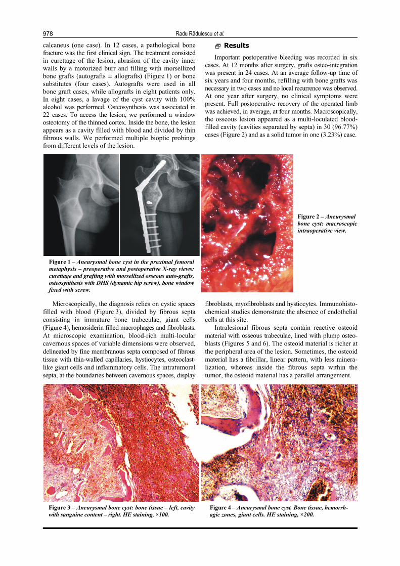
calcaneus (one case). In 12 cases, a pathological bone fracture was the first clinical sign. The treatment consisted in curettage of the lesion, abrasion of the cavity inner walls by a motorized burr and filling with morsellized bone grafts (autografts ± allografts) (Figure 1) or bone substitutes (four cases). Autografts were used in all bone graft cases, while allografts in eight patients only. In eight cases, a lavage of the cyst cavity with 100% alcohol was performed. Osteosynthesis was associated in 22 cases. To access the lesion, we performed a window osteotomy of the thinned cortex. Inside the bone, the lesion appears as a cavity filled with blood and divided by thin fibrous walls. We performed multiple bioptic probings from different levels of the lesion.
Results
Important postoperative bleeding was recorded in six cases. At 12 months after surgery, grafts osteo-integration was present in 24 cases. At an average follow-up time of six years and four months, refilling with bone grafts was necessary in two cases and no local recurrence was observed. At one year after surgery, no clinical symptoms were present. Full postoperative recovery of the operated limb was achieved, in average, at four months. Macroscopically, the osseous lesion appeared as a multi-loculated blood-filled cavity (cavities separated by septa) in 30 (96.77%) cases (Figure 2) and as a solid tumor in one (3.23%) case.
Figure 1 – Aneurysmal bone cyst in the proximal femoral metaphysis – preoperative and postoperative X-ray views: curettage and grafting with morsellized osseous auto-grafts, osteosynthesis with DHS (dynamic hip screw), bone window fixed with screw.
Figure 2 – Aneurysmal bone cyst: macroscopic intraoperative view.
Microscopically, the diagnosis relies on cystic spaces filled with blood (Figure 3), divided by fibrous septa consisting in immature bone trabeculae, giant cells (Figure 4), hemosiderin filled macrophages and fibroblasts. At microscopic examination, blood-rich multi-locular cavernous spaces of variable dimensions were observed, delineated by fine membranous septa composed of fibrous tissue with thin-walled capillaries, hystiocytes, osteoclast-like giant cells and inflammatory cells. The intratumoral septa, at the boundaries between cavernous spaces, display
fibroblasts, myofibroblasts and hystiocytes. Immunohisto-chemical studies demonstrate the absence of endothelial cells at this site.
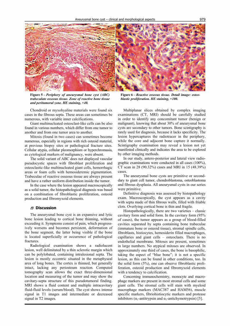
Intralesional fibrous septa contain reactive osteoid material with osseous trabeculae, lined with plump osteo-blasts (Figures 5 and 6). The osteoid material is richer at the peripheral area of the lesion. Sometimes, the osteoid material has a fibrillar, linear pattern, with less minera-lization, whereas inside the fibrous septa within the tumor, the osteoid material has a parallel arrangement.
Figure 3 – Aneurysmal bone cyst: bone tissue – left, cavity with sanguine content – right. HE staining, ×100.
Figure 4 – Aneurysmal bone cyst. Bone tissue, hemorrh-agic zones, giant cells. HE staining, ×200.
Aneurysmal bone cyst – clinical and morphological aspects
979
Figure 5 – Periphery of aneurysmal bone cyst (ABC) trabeculate osseous tissue. Zone of reactive bone tissue and peritumoral zone. HE staining, ×40.
Figure 6 – Reactive osseous tissue. Detail image: osteo-blastic proliferation. HE staining, ×100.
Chondroid or myxohyaline materials were found six cases in the fibrous septa. These areas can sometimes be numerous, with variable inner calcifications.
Giant multinucleated osteoclast-like cells can be also found in various numbers, which differ from one tumor to another and from one tumor area to another.
Mitosis (found in two cases) can sometimes become numerous, especially in regions with rich osteoid material, at previous biopsy sites or pathological fracture sites. Cellular atypia, cellular pleomorphism or hyperchromasia, as cytological markers of malignancy, were absent.
The solid variant of ABC does not displayed vascular pseudocystic spaces with fibroblast proliferation and osteoclastic-like multinucleated giant cells, hemorrhagic areas or foam cells with hemosideremic pigmentation. Trabeculae of reactive osseous tissue are always present and have a rather uniform distribution inside the tumor.
In the case where the lesion appeared macroscopically as a solid tumor, the histopathological diagnosis was based on a combination of fibroblastic proliferation, osteoid production and fibromyxoid elements.
Discussion
The aneurysmal bone cyst is an expansive and lytic bone lesion leading to cortical bone thinning, without exceeding it. Symptoms consist of pain, which progress-ively worsens and becomes persistent, deformation of the bone segment, the latter being visible if the bone is located superficially or occurrence of pathological fractures.
Radiological examination shows a radiolucent lesion, well delimitated by a thin sclerotic margin which can be polylobated, containing intralesional septa. The lesion is mostly eccentric situated in the metaphyseal area of long bones. The cortex is thinned, but generally intact, lacking any periosteum reaction. Computed tomography scan allows the exact three-dimensional location and measuring of the tumor and may show the cavitary-septa structure of this pseudotumoral finding. MRI shows a fluid content and multiple intracavitary fluid-fluid levels (serum/blood). The cyst shows intense signal in T1 images and intermediate or decreased signal in T2 images.
Multiplanar slices obtained by complex imaging examinations (CT, MRI) should be carefully studied in order to identify any concomitant tumor (benign or malignant), knowing that about 30% of aneurysmal bone cysts are secondary to other tumors. Bone scintigraphy is rarely used for diagnosis, because it lacks specificity. The lesion hypercaptures the radiotracer in the periphery, while the core and adjacent bone capture it normally. Scintigraphy examination may reveal a lesion not yet manifested clinically and indicates the area to be explored by other imaging methods.
In our study, antero-posterior and lateral view radio-graphic examinations were conducted in all cases (100%), CT scan in 28 (90.32%) cases and MRI in 15 (48.39%) cases.
The aneurysmal bone cysts are primitive or second-dary to giant cell tumor, chondroblastoma, osteoblastoma and fibrous dysplasia. All aneurysmal cysts in our series were primitive.
Definitive diagnosis was assessed by histopathology exam. Macroscopically, the cyst appears as a cavity with septa made of thin fibrous walls, filled with friable clots. Overlying cortical bone is thin and fragile.
Histopathologically, there are two varieties – classic cavitary form and solid form. In the cavitary form (95% of cases), the tumor appears as a group of blood-filled cavities separated by septa containing bone trabeculae (immature bone or osteoid tissue), stromal spindle cells, fibroblasts, histiocytes, hemosiderin filled macrophages, capillaries and giant cells – osteoclasts. There is no endothelial membrane. Mitoses are present, sometimes in large numbers. No atypical mitoses are observed. In approximately one third of cases, the bone is basophilic, taking the aspect of “blue bone”; it is not a specific lesion, as this can be found in other conditions, too. In the solid form (5%), one can observe fibroblastic proli-feration, osteoid production and fibromyxoid elements with a tendency to calcification.
Concerning immunochemistry, monocyte and macro-phage markers are present in most stromal cells and some giant cells. The stromal cells will stain with myeloid macrophage markers (MAC387 and HAM56), muscle specific markers, fibriohistiocytic markers and protease inhibitors (α1-antitrypsin and α1-antichymotrypsin) [5].

Radu Rădulescu et al.
980
The aneurysmal bone cyst can be secondary to a number of bone tumors: giant cell tumors, chondro-blastoma, fibrous histiocytomas, chondromyxoid fibromas, fibrous dysplasia or osteosarcoma. The pathologist must know the radiological appearance of these tumors in order to determine if the aneurysmal bone cyst is a primary or secondary lesion.
The most difficult differential diagnosis of the aneurysmal bone cyst is with the telangiectatic osteo-sarcoma, which has many clinical, radiological and even histological similarities. The distinction between the two is made by histopathological examination, the telangiectatic osteosarcoma having the characteristics of a malignant tumor within the hemorrhagic membranes (pleomorphic cells producing osseous or osteoid tissue and atypical mitoses) under microscopic examination. In the aneurysmal bone cyst, atypical mitoses are absent. However, cellular atypia, pleomorphism and hyperchromasia are found only with osteosarcoma; in addition, the tumor margins are ill defined, unlike in the case of an ABC where the tumor has an eggshell-like, thin cortical.
The giant cell tumor (GCT) displays large multi-nucleated cells. In the case of ABC however, nuclei are more numerous than in the GCT and they are more centrally situated. Moreover, ABC has a much more fibrous/fibroblastic stroma.
Giant cell reparative granuloma can be mistaken for a solid form of ABC. Anyway, giant cell reparative granuloma has a specific localization (jawbone), with limited areas of osteoid tissue.
The unicameral osseous cyst consists on a clear serosanguinolent lesion, without intratumoral fibrous septa. However, its differential diagnosis following a fracture can be difficult due to hemorrhage, the presence of reactive osseous tissue and multinucleate giant cells.
The osseous hemangioma is a lesion in which vascular spaces contain endothelial cells, and the osseous trabe-culae are mature, without displaying a reactive pattern.
Oliveira et al. [6] identified an association between the aneurysmal bone cyst and clonal chromosome band 17p13 translocations that place the USP6 (TRE2 or TRE17) oncogene under the regulatory influence of the highly active CDH11 promoter. They found CDH11 and/or USP6 rearrangements in 69% of cases with primitive aneurysmal bone cyst. These USP6 and CDH11 rearran-gements were present in the spindle cells and were absent in multinucleated giant cells, inflammatory cells, endothelial cells or osteoblasts. The translocations were also absent in the entire secondary aneurysmal bone cyst. In his opinion, the primary aneurysmal bone cysts are mesenchymal neoplasms, while the secondary ones are non-aneurysmal bone cysts neoplasms that mimic the morphology of the aneurysmal bone cyst. In another study [7], the same authors demonstrated the implication of the USP6 oncogene in the pathogenesis of the aneu-rysmal bone cyst, by showing how aneurysmal bone cyst variant translocations upregulate USP6 transcription by promoter swapping with the ZNF9, COL1A1, TRAP150, and OMD genes.
Leithner et al. [8], using immunohistochemistry and in situ hybridization, showed that insulin-like growth factor-I (IGF-1)or specific mRNA sequences encoding for
insulin-like growth factor-I were detectable in aneurysmal bone cysts and were mainly localized in multinucleate giant cells. The levels of insulin-like growth factor-I in normal human bone tissue are negligible.
In an immunohistochemistry study of the vascular markers in aneurysmal bone cyst, Shinde et al. [9] demonstrated that factor 8 is present at the edge of ABC cavities in almost all cases, while VEGF-C, GLUT-1 and smooth muscle actin are present in approximately half the cases. D2-40 and CD34 are present at the edge of the cavities in some cases. All this facts, suggest that the vascular factor and vascular trauma are involved in the pathogenesis of the aneurysmal bone cyst.
In our series, the treatment of the lesion consisted of creating an approximately rectangular cortical lid, harvesting intracavitary tissue from several areas, thorough cavity curettage, filling it with morsellised bone grafts, closing the lid to maintain bone grafts within the cavity, and fixation, in those cases where we considered that mechanical strength of the segment might be compromised and needs reinforcement. Intraoperative macroscopic inspection revealed cavities containing friable blood clots. We inspected the bone limits of the cyst and the adjacent bone to detect a possible bone tumor, which could lead to development of a secondary aneurysmal bone cyst.
In order to achieve a correct curettage of the bone cavity, especially towards its margins, we used a variable speed burr with heads of various sizes [10]. The remaining cells are removed by local application of pure alcohol (eight cases in our group), phenols or liquid nitrogen, which also decreases the risk of relapse [11].
Cavity filling was achieved with morsellized auto-grafts. This avoids the risk of contamination with viral germs from the allografts donor, providing faster osteo-integration. When the residual cavity was large, additional morsellized allografts were necessary (eight cases). During follow-up, there were no statistically significant differ-ences between autografts and allografts osteointegration, although allografts have been used exclusively in larger defects, but mixed with autografts [12].
Another method of filling the residual cavity is by using either bone substitutes (four patients in our group), either acrylic cement. The persistence of bone substitutes was detected in all postoperative radiological controls performed in those cases.
Other treatment options include lesion nutritional vessels embolization, percutaneous injection of acrylic cements or agents capable of inducing fibrosis, segmental resection with reconstruction [12], percutaneous sclero-therapy with Polidocanol [13], percutaneous Doxycycline treatment [14] and Denosumab administration [15]. These methods are used mainly in cysts located in the pelvis or vertebrae, which are surgically less accessible or inaccessible at all.
Complete functional rehabilitation of the segment was achieved on average at four months postoperatively, while one year postoperatively patients were asymptomatic and fully reintegrated socially and professionally.
We did not record any local recurrence, but in two cases with large lesions, cavity refilling using bone allo-grafts was required, due to partial resorption and poor
Aneurysmal bone cyst – clinical and morphological aspects
981
osteointegration of the initial grafts. Further evolution was favorable.
The natural evolution of the cavitary lesion can follow two directions. In some cases, meaning the inactive form, the lesion undergoes spontaneous involution, while in the active forms, the lesion increases in size, deforms the bone, which becomes more fragile (leading to pathological fractures subsequent to minimal trauma) and symptomatic (pain is the main revealing symptom). Buraczewski and Dabska [16] have proposed three evolutive stages: initial phase – osteolysis without peculiar findings; growth phase – rapid increase in size of osseous erosion and enlargement of involved bone and stabilization phase – standard radiological pattern.
Conclusions
Aneurysmal bone cyst is most frequent in the second decade of age. Its prevalence in female gender is double compared to the male gender. Macroscopically, the great majority of aneurysmal bone cyst appears as a multi-loculated blood-filled cavity, while the form of solid tumor is very rare. The positive diagnosis relies on the histopathological examination. Because of the strong relationship with a number of precursor lesions (giant cell tumor, fibrous dysplasia, non-ossifying fibroma, chondroblastoma, osteoblastoma) multiple bioptic probings are mandatory, in order to diagnose, if possible, a primary lesion that may modify the therapeutic attitude. Treatment by curettage, abrasion of the cavity inner walls and filling with morsellized grafts has very good results. The risk of recurrence is very low.
References [1] Rapp TB, Ward JP, Alaia MJ, Aneurysmal bone cyst, J Am
Acad Orthop Surg, 2012, 20(4):233–241. [2] Wheeless CR III, Aneurysmal bone cyst. In: Wheeless CR III,
Wheeless’ textbook of orthopaedics, Last updated May 24, 2013, http://www.wheelessonline.com/ortho/aneurysmal_bone _cyst.
[3] Jaffe HL, Lichtenstein L, Solitary unicameral bone cyst: with emphasis on the Roentgen picture, the pathologic appearance and the pathogenesis, Arch Surg, 1942, 44(6):1004–1025.
[4] Panoutsakopoulos G, Pandis N, Kyriazoglou I, Gustafson P, Mertens F, Mandahl N, Recurrent t(16;17)(q22;p13) in aneurysmal bone cysts, Genes Chromosomes Cancer, 1999, 26(3):265–266.
[5] Vigorita VJ, Orthopaedic pathology, 2nd edition, Lippincott Williams & Wilkins, 2008.
[6] Oliveira AM, Perez-Atayde AR, Inwards CY, Medeiros F, Derr V, Hsi BL, Gebhardt MC, Rosenberg AE, Fletcher JA, USP6 and CDH11 oncogenes identify the neoplastic cell in primary aneurysmal bone cysts and are absent in so-called secondary aneurysmal bone cysts, Am J Pathol, 2004, 165(5): 1773–1780.
[7] Oliveira AM, Perez-Atayde AR, Dal Cin P, Gebhardt MC, Chen CJ, Neff JR, Demetri GD, Rosenberg AE, Bridge JA, Fletcher JA, Aneurysmal bone cyst variant translocations upregulate USP6 transcription by promoter swapping with the ZNF9, COL1A1, TRAP150, and OMD genes, Oncogene, 2005, 24(21):3419–3426.
[8] Leithner A, Lang S, Windhager R, Leithner K, Karlic H, Kotz R, Haas OA, Expression of insulin-like growth factor-I (IGF-I) in aneurysmal bone cyst, Mod Pathol, 2011, 14(11):1100–1104.
[9] Shinde A, Mehlman CT, Collins MH, Aneurysmal bone cysts express vascular markers, Pediatr Dev Pathol, 2006, 9(1): 38–43.
[10] Wang EH, Marfori ML, Serrano MV, Rubio DA, Is curettage and high-speed burring sufficient treatment for aneurysmal bone cysts? Clin Orthop Relat Res, 2014, Jul 22.
[11] Reddy KI, Sinnaeve F, Gaston CL, Grimer RJ, Carter SR, Aneurysmal bone cysts: do simple treatments work? Clin Orthop Relat Res, 2014, 472(6):1901–1910.
[12] Flont P, Kolacinska-Flont M, Niedzielski K, A comparison of cyst wall curettage and en bloc excision in the treatment of aneurysmal bone cysts, World J Surg Oncol, 2013, 11:109.
[13] Brosjö O, Pechon P, Hesla A, Tsagozis P, Bauer H, Sclero-therapy with polidocanol for treatment of aneurysmal bone cysts, Acta Orthop, 2013, 84(5):502–505.
[14] Shiels WE 2nd, Mayerson JL, Percutaneous doxycycline treatment of aneurysmal bone cysts with low recurrence rate: a preliminary report, Clin Orthop Relat Res, 2013, 471(8): 2675–2683.
[15] Lange T, Stehling C, Fröhlich B, Klingenhöfer M, Kunkel P, Schneppenheim R, Escherich G, Gosheger G, Hardes J, Jürgens H, Schulte TL, Denosumab: a potential new and innovative treatment option for aneurysmal bone cysts, Eur Spine J, 2013, 22(6):1417–1422.
[16] Buraczewski J, Dabska M, Pathogenesis of aneurysmal bone cyst: relationship between the aneurysmal bone cyst and fibrous dysplasia of bone, Cancer, 1971, 28(3):597–604.
Corresponding author Adrian Bădilă, MD, Department of Orthopedic Surgery, Bucharest University Hospital, 169 Independenţei Avenue, Sector 5, 050098 Bucharest, Romania; Phone +40722–516 470, e-mail: [email protected] Received: December 15, 2013
Accepted: September 1, 2014
Related Documents