Rom J Morphol Embryol 2012, 53(4):1069–1072 ISSN (print) 1220–0522 ISSN (on-line) 2066–8279 CASE REPORT Spinal involvement with spinal cord compression syndrome in hematological diseases M. POPESCU 1) , VIOLA POPOV 2) , G. POPESCU 3) , CAMELIA DOBREA 4) , AURELIA SANDU 5) , V. T. GRIGOREAN 6) , V. STRÂMBU 6) 1) Department of Neurosurgery 2) Department of Hematology Emergency County Hospital, Pitesti 3) Department of Neurosurgery, Emergency County Hospital, Timisoara 4) Department of Hematology, “Carol Davila” University of Medicine and Pharmacy, Bucharest 5) Department of Neurosurgery, “Bagdasar–Arseni” Emergency Clinical Hospital, Bucharest 6) Department of Surgery, “Carol Davila” University of Medicine and Pharmacy, Bucharest Abstract Primary spinal involvement in hematological diseases is rare. Purpose: The purpose of this article is studying diagnostic and treatment strategies in patients with spinal cord primary hematologic tumors causing spinal cord compression syndrome. Patients and Methods: We report two cases with spinal cord primary hematologic tumors causing spinal cord compression syndrome. One patient had a diffuse large B-cell non-Hodgkin’s lymphoma located in the thoracic spine and the second patient had a plasmocytic plasmacytoma located in the thoraco-lumbar spine. Results: Both patients underwent surgery, with resection of the intracanalar tumor and spinal cord decompression and adjuvant systemic and intrathecal chemotherapy. Neurological outcome was favorable with partial remission of spinal cord compression syndrome. Finally, patients developed secondary dissemination and succumbed due to progression of the hematological disease. Conclusions: Clinical onset and radiographic evaluation is uncharacteristic in early stages. Spinal MRI is mandatory in cases with rebel pain, unresponsive to conservative treatment. Surgery is indicated in all patients with spinal cord compression syndrome. Early diagnosis is associated with better prognosis. Recommended treatment is surgical resection and systemic and intrathecal chemotherapy adapted to histological form of each tumor. In selected cases, if indicated radiotherapy can also be associated. Keywords: non-Hodgkin’s lymphoma, plasmocytic plasmacytoma, spinal cord compression, spinal tumors. Introduction Spinal tumors are classified as intramedullary, intra- dural extramedullary, extradural or secondary tumor extension to the vertebral column from other primary sites. Primary spinal involvement in hematological diseases is rare. Malignant non-Hodgkin’s lymphomas with extra-lymph-nodes locations occur in 15–25% adult patients in USA and in 40–50% in Europe. Most frequent extra-lymph-nodes disseminations of malignant non-Hodgkin’s lymphomas are: gastrointestinal tract (44%), upper airways (19%), central nervous system (5%), bone (8%), thyroid, breast, lung, testicle or skin (<3%) [1]. Extra-lymph-nodes malignant non-Hodgkin’s lymphomas with primary nervous location are infrequent, the incidence being higher in immunocompromised states (AIDS). They can have different locations: brain, spinal cord, leptomeninges and eyes. Primary location into the epidural space represents 3% from all non-Hodgkin’s lymphomas [2]. Objective The purpose of this article is studying diagnostic and treatment strategies in patients with spinal cord primary hematologic tumors that develops relatively quickly spinal cord compression syndrome, with rapidly complete motor, sensory, autonomic and sphincter loss distal to compression site. Patients, Methods and Results We report two cases with spinal cord primary hematologic tumors admitted into the Neurosurgery Department of the Emergency County Hospital, Pitesti, Romania. Case No. 1 The first patient is a woman, 71-year-old, repeatedly admitted for thoracic pain with intercostal radiating neuralgia, which develop during hospitalization, urinary retention and hypo and later anesthesia with upper neurologic level T7–T9. R J M E Romanian Journal of Morphology & Embryology http://www.rjme.ro/

Welcome message from author
This document is posted to help you gain knowledge. Please leave a comment to let me know what you think about it! Share it to your friends and learn new things together.
Transcript

Rom J Morphol Embryol 2012, 53(4):1069–1072
ISSN (print) 1220–0522 ISSN (on-line) 2066–8279
CCAASSEE RREEPPOORRTT
Spinal involvement with spinal cord compression syndrome in hematological
diseases
M. POPESCU1), VIOLA POPOV2), G. POPESCU3), CAMELIA DOBREA4), AURELIA SANDU5), V. T. GRIGOREAN6), V. STRÂMBU6)
1)Department of Neurosurgery 2)Department of Hematology
Emergency County Hospital, Pitesti 3)Department of Neurosurgery,
Emergency County Hospital, Timisoara 4)Department of Hematology,
“Carol Davila” University of Medicine and Pharmacy, Bucharest 5)Department of Neurosurgery,
“Bagdasar–Arseni” Emergency Clinical Hospital, Bucharest 6)Department of Surgery,
“Carol Davila” University of Medicine and Pharmacy, Bucharest
Abstract Primary spinal involvement in hematological diseases is rare. Purpose: The purpose of this article is studying diagnostic and treatment strategies in patients with spinal cord primary hematologic tumors causing spinal cord compression syndrome. Patients and Methods: We report two cases with spinal cord primary hematologic tumors causing spinal cord compression syndrome. One patient had a diffuse large B-cell non-Hodgkin’s lymphoma located in the thoracic spine and the second patient had a plasmocytic plasmacytoma located in the thoraco-lumbar spine. Results: Both patients underwent surgery, with resection of the intracanalar tumor and spinal cord decompression and adjuvant systemic and intrathecal chemotherapy. Neurological outcome was favorable with partial remission of spinal cord compression syndrome. Finally, patients developed secondary dissemination and succumbed due to progression of the hematological disease. Conclusions: Clinical onset and radiographic evaluation is uncharacteristic in early stages. Spinal MRI is mandatory in cases with rebel pain, unresponsive to conservative treatment. Surgery is indicated in all patients with spinal cord compression syndrome. Early diagnosis is associated with better prognosis. Recommended treatment is surgical resection and systemic and intrathecal chemotherapy adapted to histological form of each tumor. In selected cases, if indicated radiotherapy can also be associated.
Keywords: non-Hodgkin’s lymphoma, plasmocytic plasmacytoma, spinal cord compression, spinal tumors.
Introduction
Spinal tumors are classified as intramedullary, intra-dural extramedullary, extradural or secondary tumor extension to the vertebral column from other primary sites. Primary spinal involvement in hematological diseases is rare. Malignant non-Hodgkin’s lymphomas with extra-lymph-nodes locations occur in 15–25% adult patients in USA and in 40–50% in Europe. Most frequent extra-lymph-nodes disseminations of malignant non-Hodgkin’s lymphomas are: gastrointestinal tract (44%), upper airways (19%), central nervous system (5%), bone (8%), thyroid, breast, lung, testicle or skin (<3%) [1]. Extra-lymph-nodes malignant non-Hodgkin’s lymphomas with primary nervous location are infrequent, the incidence being higher in immunocompromised states (AIDS). They can have different locations: brain, spinal cord, leptomeninges and eyes. Primary location into the epidural space represents 3% from all non-Hodgkin’s lymphomas [2].
Objective
The purpose of this article is studying diagnostic and treatment strategies in patients with spinal cord primary hematologic tumors that develops relatively quickly spinal cord compression syndrome, with rapidly complete
motor, sensory, autonomic and sphincter loss distal to compression site.
Patients, Methods and Results
We report two cases with spinal cord primary hematologic tumors admitted into the Neurosurgery Department of the Emergency County Hospital, Pitesti, Romania.
Case No. 1
The first patient is a woman, 71-year-old, repeatedly admitted for thoracic pain with intercostal radiating neuralgia, which develop during hospitalization, urinary retention and hypo and later anesthesia with upper neurologic level T7–T9.
R J M ERomanian Journal of
Morphology & Embryologyhttp://www.rjme.ro/
M. Popescu et al.
1070
Until spinal MRI can be done, the patient developed paraplegia. Thoracic spine MRI showed a left epidural thoracic tumor T7–T9, infiltrating the left pedicle, most part of the vertebral body with anterior growing (Figure 1).
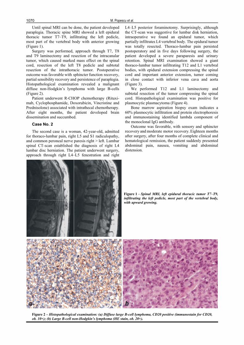
Surgery was performed, approach through T7, T8 and T9 laminectomy and resection of the intracanalar tumor, which caused marked mass effect on the spinal cord, resection of the left T8 pedicle and subtotal resection of the intrathoracic tumor. Postoperative outcome was favorable with sphincter function recovery, partial sensibility recovery and persistence of paraplegia. Histopathological examination revealed a malignant diffuse non-Hodgkin’s lymphoma with large B-cells (Figure 2).
Patient underwent R-CHOP chemotherapy (Rituxi-mab, Cyclophosphamide, Doxorubicin, Vincristine and Prednisolone) associated with intrathecal chemotherapy. After eight months, the patient developed brain dissemination and succumbed.
Case No. 2
The second case is a woman, 42-year-old, admitted for thoraco-lumbar pain, right L5 and S1 radiculopathy, and common peroneal nerve paresis right > left. Lumbar spinal CT-scan established the diagnosis of right L4 lumbar disc herniation. The patient underwent surgery, approach through right L4–L5 fenestration and right
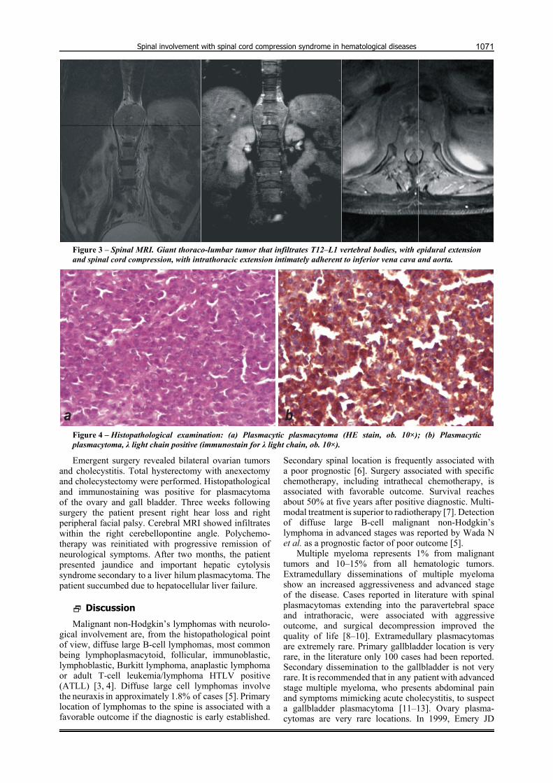
L4–L5 posterior foraminotomy. Surprisingly, although the CT-scan was suggestive for lumbar disk herniation, intraoperative we found an epidural tumor, which partially infiltrates L4 vertebral body. The epidural tumor was totally resected. Thoraco-lumbar pain persisted postoperatory and in five days following surgery, the patient developed a severe paraparesis and urinary retention. Spinal MRI examination showed a giant thoraco-lumbar tumor infiltrating T12 and L1 vertebral bodies, with epidural extension compressing the spinal cord and important anterior extension, tumor coming in close contact with inferior vena cava and aorta (Figure 3).
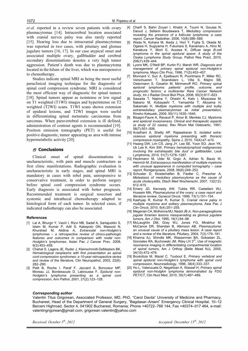
We performed T12 and L1 laminectomy and subtotal resection of the tumor compressing the spinal cord. Histopathological examination was positive for plasmocytic plasmacytoma (Figure 4).
Bone marrow aspiration biopsy exam indicates a 60% plasmocytic infiltration and protein electrophoresis and immunostaining identified lambda component of the monoclonal IgG antibody.
Outcome was favorable, with sensory and sphincter recovery and moderate motor recovery. Eighteen months after surgery, after four months of complete clinical and hematological remission, the patient suddenly presented abdominal pain, nausea, vomiting and abdominal distension.
Figure 1 – Spinal MRI, left epidural thoracic tumor T7–T9, infiltrating the left pedicle, most part of the vertebral body, with upward growing.
Figure 2 – Histopathological examination: (a) Diffuse large B-cell lymphoma, CD20 positive (immunostain for CD20, ob. 10×); (b) Large B-cell non-Hodgkin’s lymphoma (HE stain, ob. 20×).
Spinal involvement with spinal cord compression syndrome in hematological diseases
1071
Figure 3 – Spinal MRI. Giant thoraco-lumbar tumor that infiltrates T12–L1 vertebral bodies, with epidural extension and spinal cord compression, with intrathoracic extension intimately adherent to inferior vena cava and aorta.
Figure 4 – Histopathological examination: (a) Plasmacytic plasmacytoma (HE stain, ob. 10×); (b) Plasmacytic plasmacytoma, λ light chain positive (immunostain for λ light chain, ob. 10×).
Emergent surgery revealed bilateral ovarian tumors and cholecystitis. Total hysterectomy with anexectomy and cholecystectomy were performed. Histopathological and immunostaining was positive for plasmacytoma of the ovary and gall bladder. Three weeks following surgery the patient present right hear loss and right peripheral facial palsy. Cerebral MRI showed infiltrates within the right cerebellopontine angle. Polychemo-therapy was reinitiated with progressive remission of neurological symptoms. After two months, the patient presented jaundice and important hepatic cytolysis syndrome secondary to a liver hilum plasmacytoma. The patient succumbed due to hepatocellular liver failure.
Discussion
Malignant non-Hodgkin’s lymphomas with neurolo-gical involvement are, from the histopathological point of view, diffuse large B-cell lymphomas, most common being lymphoplasmacytoid, follicular, immunoblastic, lymphoblastic, Burkitt lymphoma, anaplastic lymphoma or adult T-cell leukemia/lymphoma HTLV positive (ATLL) [3, 4]. Diffuse large cell lymphomas involve the neuraxis in approximately 1.8% of cases [5]. Primary location of lymphomas to the spine is associated with a favorable outcome if the diagnostic is early established.
Secondary spinal location is frequently associated with a poor prognostic [6]. Surgery associated with specific chemotherapy, including intrathecal chemotherapy, is associated with favorable outcome. Survival reaches about 50% at five years after positive diagnostic. Multi-modal treatment is superior to radiotherapy [7]. Detection of diffuse large B-cell malignant non-Hodgkin’s lymphoma in advanced stages was reported by Wada N et al. as a prognostic factor of poor outcome [5].
Multiple myeloma represents 1% from malignant tumors and 10–15% from all hematologic tumors. Extramedullary disseminations of multiple myeloma show an increased aggressiveness and advanced stage of the disease. Cases reported in literature with spinal plasmacytomas extending into the paravertebral space and intrathoracic, were associated with aggressive outcome, and surgical decompression improved the quality of life [8–10]. Extramedullary plasmacytomas are extremely rare. Primary gallbladder location is very rare, in the literature only 100 cases had been reported. Secondary dissemination to the gallbladder is not very rare. It is recommended that in any patient with advanced stage multiple myeloma, who presents abdominal pain and symptoms mimicking acute cholecystitis, to suspect a gallbladder plasmacytoma [11–13]. Ovary plasma-cytomas are very rare locations. In 1999, Emery JD
M. Popescu et al.
1072
et al. reported in a review seven patients with ovary plasmacytomas [14]. Intracerebral location associated with cranial nerves palsy was also rarely reported [15]. Hearing loss due to intracranial plasmacytomas was reported in two cases, with pituitary and glomus jugulare tumors [16, 17]. In our case atypical onset and associated multiple ovary, gallbladder and cerebral secondary disseminations denotes a very high tumor aggression. Patient’s death was due to plasmacytoma located in the hilum of the liver, which was unresponsive to chemotherapy.
Studies indicate spinal MRI as being the most useful paraclinical imaging technique for the diagnostic of spinal cord compression syndrome. MRI is considered the most efficient way of diagnostic for spinal tumors [18]. Spinal tumors appear as isointense or low signal on T1 weighted (T1WI) images and hyperintense on T2 weighted (T2WI) scans. T1WI scans shown extension of epidural lesions, and T2WI images were useful in differentiating spinal metastatic carcinomas from sarcomas. When paravertebral extension is ill defined, administration of contrast agents is recommended [19]. Positron emission tomography (PET) is useful for positive diagnostic, tumor appearing as area with intense hypermetabolic activity [20].
Conclusions
Clinical onset of spinal disseminations is uncharacteristic, with pain and muscle contracture as first clinic manifestations. Radiographic evaluation is uncharacteristic in early stages, and spinal MRI is mandatory in cases with rebel pain, unresponsive to conservative treatment, in order to perform surgery before spinal cord compression syndrome occurs. Early diagnosis is associated with better prognosis. Recommended treatment is surgical resection and systemic and intrathecal chemotherapy adapted to histological form of each tumor. In selected cases, if indicated radiotherapy can also be associated.
References [1] Lal A, Bhurgri Y, Vaziri I, Rizvi NB, Sadaf A, Sartajuddin S,
Islam M, Kumar P, Adil S, Kakepoto GN, Masood N, Khurshed M, Alidina A, Extranodal non-Hodgkin’s lymphomas – a retrospective review of clinico-pathologic features and outcomes in comparison with nodal non-Hodgkin’s lymphomas, Asian Pac J Cancer Prev, 2008, 9(3):453–458.
[2] Chahal S, Lagera JE, Ryder J, Kleinschmidt-DeMasters BK, Hematological neoplasms with first presentation as spinal cord compression syndromes: a 10-year retrospective series and review of the literature, Clin Neuropathol, 2003, 22(6): 282–290.
[3] Petit B, Roche I, Paraf F, Jaccard A, Boncoeur MP, Moreau JJ, Bordessoule D, Labrousse F, Epidural non-Hodgkin’s lymphoma presenting as a spinal cord compression, Ann Pathol, 2001, 21(2):123–128.
[4] Charfi S, Bahri Zouari I, Khabir A, Toumi N, Gouiaa N, Daoud J, Sellami Boudawara T, Medullary compression revealing the presence of a follicular lymphoma: a case report, Cancer Radiother, 2006, 10(8):586–589.
[5] Wada N, Kohara M, Ikeda J, Hori Y, Fujita S, Okada M, Ogawa H, Sugiyama H, Fukuhara S, Kanamaru A, Hino M, Kanakura Y, Morii E, Aozasa K, Diffuse large B-cell lymphoma in the spinal epidural space: A study of the Osaka Lymphoma Study Group, Pathol Res Pract, 2010, 206(7):439–444.
[6] Lyons MK, O’Neill BP, Kurtin PJ, Marsh WR, Diagnosis and management of primary spinal epidural non-Hodgkin’s lymphoma, Mayo Clin Proc, 1996, 71(5):453–457.
[7] Monnard V, Sun A, Epelbaum R, Poortmans P, Miller RC, Verschueren T, Scandolaro L, Villa S, Majno SB, Ostermann S, Ozsahin M, Mirimanoff RO, Primary spinal epidural lymphoma: patients’ profile, outcome, and prognostic factors: a multicenter Rare Cancer Network study, Int J Radiat Oncol Biol Phys, 2006, 65(3):817–823.
[8] Sawada T, Najima Y, Ohashi K, Kato I, Miyazawa M, Nakano M, Kobayashi T, Yamashita T, Akiyama H, Sakamaki H, Multiple myeloma with multiple and bulky extramedullary plasmacytomas as initial presentation, Rinsho Ketsueki, 2009, 50(11):1635–1640.
[9] Bisagni-Faure A, Ravaud P, Amor B, Menkès CJ, Myeloma and epidural invasiveness. Clinical and therapeutic aspects (a study of 22 cases), Rev Rhum Mal Osteoartic, 1991, 58(7):501–506.
[10] Avadhani A, Shetty AP, Rajasekaran S, Isolated extra-osseous epidural myeloma presenting with thoracic compressive myelopathy, Spine J, 2010, 10(4):e7–e10.
[11] Hwang DW, Lim CS, Jang JY, Lee SE, Yoon SO, Jeon YK, Uk Lee K, Kim SW, Primary hematolymphoid malignancies involving the extrahepatic bile duct or gallbladder, Leuk Lymphoma, 2010, 51(7):1278–1287.
[12] Heckmann M, Uder M, Grgic A, Adrian N, Bautz W, Heinrich M, Extraosseous manifestation of multiple myeloma with unusual appearance in computed tomography – case report, Rontgenpraxis, 2008, 56(6):249–253.
[13] Schuster D, Klosterhalfen B, Fiedler C, Prescher A, Metastasis of medullary plasmocytoma as the cause of acute cholecystitis, Dtsch Med Wochenschr, 2007, 132(12): 612–615.
[14] Emery JD, Kennedy AW, Tubbs RR, Castellani WJ, Hussein MA, Plasmacytoma of the ovary: a case report and literature review, Gynecol Oncol, 1999, 73(1):151–154.
[15] Kashyap R, Kumar R, Kumar S, Cranial nerve palsy in multiple myeloma and solitary plasmacytoma, Asia Pac J Clin Oncol, 2010, 6(4):251–255.
[16] Megerian CA, McKenna MJ, Nadol JB Jr, Non-paraganglioma
jugular foramen lesions masquerading as glomus jugulare tumors, Am J Otol, 1995, 16(1):94–98.
[17] McLaughlin DM, Gray WJ, Jones FG, Mirakhur M, McCance DR, Sheridan B, Atkinson AB, Plasmacytoma: an unusual cause of a pituitary mass lesion. A case report and a review of the literature, Pituitary, 2004, 7(3):179–181.
[18] Khanna AJ, Shindle MK, Wasserman BA, Gokaslan ZL, Gonzales RA, Buchowski JM, Riley LH 3rd, Use of magnetic resonance imaging in differentiating compartmental location of spinal tumors, Am J Orthop (Belle Mead NJ), 2005, 34(10):472–476.
[19] Boukobza M, Mazel C, Touboul E, Primary vertebral and spinal epidural non-Hodgkin’s lymphoma with spinal cord compression, Neuroradiology, 1996, 38(4):333–337.
[20] Ho L, Valenzuela D, Negahban A, Wassef H, Primary spinal epidural non-Hodgkin lymphoma demonstrated by FDG PET/CT, Clin Nucl Med, 2010, 35(7):487–489.
Corresponding author Valentin Titus Grigorean, Associated Professor, MD, PhD, “Carol Davila” University of Medicine and Pharmacy, Bucharest, Head of the Department of General Surgery, “Bagdasar–Arseni” Emergency Clinical Hospital, 10–12 Berceni Highroad, Sector 4, 041902 Bucharest, Romania; Phone +40722–766 144, Fax +40374–017 464, e-mail: [email protected], [email protected]
Received: October 5th, 2012 Accepted: December 15th, 2012
Related Documents