Rom J Morphol Embryol 2017, 58(1):99–106 ISSN (print) 1220–0522 ISSN (online) 2066–8279 ORIGINAL PAPER Optical coherence tomography applications in tooth wear diagnosis VERONICA MERCUŢ 1) , SANDA MIHAELA POPESCU 1) , MONICA SCRIECIU 1) , MARINA OLIMPIA AMĂRĂSCU 1) , MIHAELA VĂTU 1) , OANA ANDREEA DIACONU 2) , EUGEN OSIAC 4) , ŞTEFAN MUGUREL GHELASE 4) 1) Department of Prosthetics and Oral Rehabilitation, Faculty of Dental Medicine, University of Medicine and Pharmacy of Craiova, Romania 2) Department of Endodontics, Faculty of Dental Medicine, University of Medicine and Pharmacy of Craiova, Romania 3) Department of Biophysics, Faculty of Medicine, University of Medicine and Pharmacy of Craiova, Romania 4) Department of Public Health and Management, Faculty of Medicine, University of Medicine and Pharmacy of Craiova, Romania Abstract Dental wear represents an irreversible loss of dental hard tissue under the action of physical, chemical and mechanical factors, excluding dental caries and acute trauma. Four clinical forms of dental wear are described: erosion, attrition, abfraction, and abrasion. Most experts agree that in each clinical form multiple etiological factors are involved, one being predominant. Optical coherence tomography (OCT) is a non- invasive optical method characterized by a micronic resolution. The applications of this type of investigation are multiple in the medical field in recent years, and OCT is gaining a growing importance in dentistry. The study pointed out through OCT imaging for the erosive tooth wear lesion the existence of chemical aggression, with strong demineralization of enamel and dentin. For attrition lesion, OCT images showed the contribution of excessive force and friction movements specific to bruxism. In abfraction, OCT image revealed the importance of the mechanic factor in producing this form of tooth wear and abrasion damage studied may be considered physiological, according to patient age. OCT examination may reveal existing lesions in hard dental tissues for each clinical form and could bring evidence on the mechanisms involved. Keywords: tooth wear, erosion, attrition, abfraction, abrasion, optical coherence tomography. Introduction Tooth wear is a term referring to different processes, which, either individually or in association, lead to the irreversible loss of hard dental tissue [1]. Currently, the tooth wear is considered as the result of three processes: erosion (dissolution of hard tissue by acidic substances), attrition (wear through tooth–tooth contact), and abrasion (wear produced by interaction between teeth and other materials) [2]. In addition, some authors appreciate that a further process (abfraction) might potentiate wear by abrasion and/or erosion [1–3]. However, clinically it is difficult (if not impossible) to isolate a single etiological factor when a patient presents with tooth wear. Tooth wear is an important dental lesion since it has a high prevalence, it involves also decidual and permanent teeth, sometimes in childhood, producing consequences that affect the individual for the rest of his life, and forcing the clinician to perform difficult and costly treatments [4– 6]. Therapeutically difficulties are drifting from difficult identification of the clinical subform of tooth wear and from the burden of the restoration of the hard tissue loss, with a high rate of failure [7, 8]. In order to specify the etiological factors and subforms of tooth wear, different techniques are developed to analyze dental wear in vitro: microhardness, surface profilometry, surface roughness, microradiography, atomic force micro- scopy (AFM), AFM nanoindentation, scanning electron microscopy (SEM), white light interferometry (WLI), confocal laser scanning microscopy (CLSM) [9–11]. Optical coherence tomography (OCT) is a new high- resolution optical technique that permits minimally invasive imaging of near-surface abnormalities in complex tissues, provides real-time structural imaging, and is based on low coherence interferometry using broadband light. Appli- cation of OCT in dentistry was used for hard tissue but also for soft tissue, oral mucosa and periodontal tissue. Today, OCT could be used to detect qualitative and quantitative morphological changes of oral tissues in vivo, and to diagnose incipient tooth lesions as caries and tooth wear, progression of periodontal disease and tooth wear, and detecting oral cancer [12]. The aim of the study was to highlight the morpho- logical changes in tooth wear and microstructural tooth changes of the wear surfaces by OCT. Materials and Methods Tooth samples One hundred and forty-seven teeth were extracted from 98 patients in Prosthetic Clinic of the Faculty of Dental Medicine, according to complex treatment plan for which informed consent was recorded. The study was approved by the Ethical Committee of the University of Medicine and Pharmacy of Craiova, Romania. Twenty- five teeth were selected from the extracted teeth. Selected teeth were divided in five groups, four groups, one group R J M E Romanian Journal of Morphology & Embryology http://www.rjme.ro/

Welcome message from author
This document is posted to help you gain knowledge. Please leave a comment to let me know what you think about it! Share it to your friends and learn new things together.
Transcript

Rom J Morphol Embryol 2017, 58(1):99–106
ISSN (print) 1220–0522 ISSN (online) 2066–8279
OORRIIGGIINNAALL PPAAPPEERR
Optical coherence tomography applications in tooth wear diagnosis
VERONICA MERCUŢ1), SANDA MIHAELA POPESCU1), MONICA SCRIECIU1), MARINA OLIMPIA AMĂRĂSCU1), MIHAELA VĂTU1), OANA ANDREEA DIACONU2), EUGEN OSIAC4), ŞTEFAN MUGUREL GHELASE4)
1)Department of Prosthetics and Oral Rehabilitation, Faculty of Dental Medicine, University of Medicine and Pharmacy of Craiova, Romania
2)Department of Endodontics, Faculty of Dental Medicine, University of Medicine and Pharmacy of Craiova, Romania 3)Department of Biophysics, Faculty of Medicine, University of Medicine and Pharmacy of Craiova, Romania 4)Department of Public Health and Management, Faculty of Medicine, University of Medicine and Pharmacy of Craiova, Romania
Abstract Dental wear represents an irreversible loss of dental hard tissue under the action of physical, chemical and mechanical factors, excluding dental caries and acute trauma. Four clinical forms of dental wear are described: erosion, attrition, abfraction, and abrasion. Most experts agree that in each clinical form multiple etiological factors are involved, one being predominant. Optical coherence tomography (OCT) is a non-invasive optical method characterized by a micronic resolution. The applications of this type of investigation are multiple in the medical field in recent years, and OCT is gaining a growing importance in dentistry. The study pointed out through OCT imaging for the erosive tooth wear lesion the existence of chemical aggression, with strong demineralization of enamel and dentin. For attrition lesion, OCT images showed the contribution of excessive force and friction movements specific to bruxism. In abfraction, OCT image revealed the importance of the mechanic factor in producing this form of tooth wear and abrasion damage studied may be considered physiological, according to patient age. OCT examination may reveal existing lesions in hard dental tissues for each clinical form and could bring evidence on the mechanisms involved.
Keywords: tooth wear, erosion, attrition, abfraction, abrasion, optical coherence tomography.
Introduction
Tooth wear is a term referring to different processes, which, either individually or in association, lead to the irreversible loss of hard dental tissue [1]. Currently, the tooth wear is considered as the result of three processes: erosion (dissolution of hard tissue by acidic substances), attrition (wear through tooth–tooth contact), and abrasion (wear produced by interaction between teeth and other materials) [2]. In addition, some authors appreciate that a further process (abfraction) might potentiate wear by abrasion and/or erosion [1–3]. However, clinically it is difficult (if not impossible) to isolate a single etiological factor when a patient presents with tooth wear.
Tooth wear is an important dental lesion since it has a high prevalence, it involves also decidual and permanent teeth, sometimes in childhood, producing consequences that affect the individual for the rest of his life, and forcing the clinician to perform difficult and costly treatments [4–6]. Therapeutically difficulties are drifting from difficult identification of the clinical subform of tooth wear and from the burden of the restoration of the hard tissue loss, with a high rate of failure [7, 8].
In order to specify the etiological factors and subforms of tooth wear, different techniques are developed to analyze dental wear in vitro: microhardness, surface profilometry, surface roughness, microradiography, atomic force micro-scopy (AFM), AFM nanoindentation, scanning electron microscopy (SEM), white light interferometry (WLI),
confocal laser scanning microscopy (CLSM) [9–11]. Optical coherence tomography (OCT) is a new high-resolution optical technique that permits minimally invasive imaging of near-surface abnormalities in complex tissues, provides real-time structural imaging, and is based on low coherence interferometry using broadband light. Appli-cation of OCT in dentistry was used for hard tissue but also for soft tissue, oral mucosa and periodontal tissue. Today, OCT could be used to detect qualitative and quantitative morphological changes of oral tissues in vivo, and to diagnose incipient tooth lesions as caries and tooth wear, progression of periodontal disease and tooth wear, and detecting oral cancer [12].
The aim of the study was to highlight the morpho-logical changes in tooth wear and microstructural tooth changes of the wear surfaces by OCT.
Materials and Methods
Tooth samples
One hundred and forty-seven teeth were extracted from 98 patients in Prosthetic Clinic of the Faculty of Dental Medicine, according to complex treatment plan for which informed consent was recorded. The study was approved by the Ethical Committee of the University of Medicine and Pharmacy of Craiova, Romania. Twenty-five teeth were selected from the extracted teeth. Selected teeth were divided in five groups, four groups, one group
R J M ERomanian Journal of
Morphology & Embryologyhttp://www.rjme.ro/
Veronica Mercuţ et al.
100
for each form of tooth wear: abrasion, attrition, erosion, abfraction, and one group as control, with intact teeth (impacted teeth or teeth extracted for orthodontic purposes).
Tooth were framed in clinical subforms using data from anamnesis, of clinical exam, considering the patterns established by Verrett [13], Abrahamsen [14], and Consensus of The European Federation of Conservative Dentistry [15]. The extracted teeth used in the study were disinfected with 10% H2O2 for 10 minutes after rinsing with water. Afterwards, each probe was scaled and polished for removing gingival tissue. To avoid desiccation, teeth were maintained in sterile deionized water until optical measurements. Teeth positioning: teeth were fixed in dental silicone and positioned so the light beam would fall perpendicularly on the examined wear surfaces.
For tooth wear lesions identification, we used photo-graphs made with Canon DSLR 600EOS. For each type of lesion, we selected teeth with the most significant characteristics of the lesion.
Optical measurements
For the OCT imaging, first the teeth were removed from the solution and dried using tissue paper. For the experiment, we used an OCT system manufactured by Thorlabs (OCS1300SS) powered by a swept laser source with central wavelength of 1310 nm, a spectral bandwidth of 100 nm, and an average power of 12 mW. The device was used for 2D and 3D scanning of teeth surfaces. The resolutions for air of this system were 12 μm for axial and respectively 15 μm for lateral resolutions. The system has allowed us to investigate a sample of 10×10×3 mm (length, width, depth) or 1024×1024×512 pixels in about 30 seconds using a charge-coupled device (CCD)-type detector [16, 17].
For the experiment, we used a design that permits to view tooth surface at different angles, and we examined different regions of the teeth, like incisal/occlusal region, buccal and lingual region, cervical region and radicular region. We included healthy teeth as controls. We sampled teeth surfaces pictured in width of 10 mm on a distance of 10 mm and a depth of 3 mm.
Images processing
The obtained images were processed using Image J, an open access program.
Results
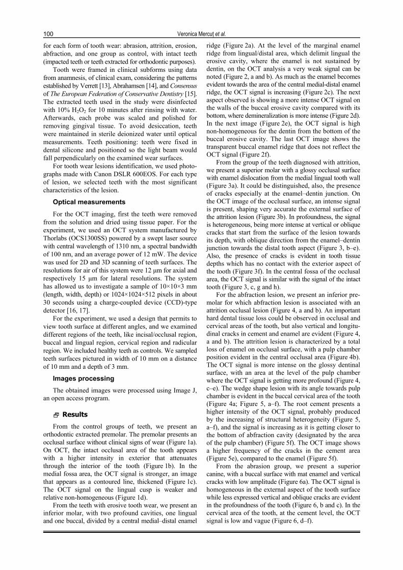
From the control groups of teeth, we present an orthodontic extracted premolar. The premolar presents an occlusal surface without clinical signs of wear (Figure 1a). On OCT, the intact occlusal area of the tooth appears with a higher intensity in exterior that attenuates through the interior of the tooth (Figure 1b). In the medial fossa area, the OCT signal is stronger, an image that appears as a contoured line, thickened (Figure 1c). The OCT signal on the lingual cusp is weaker and relative non-homogeneous (Figure 1d).
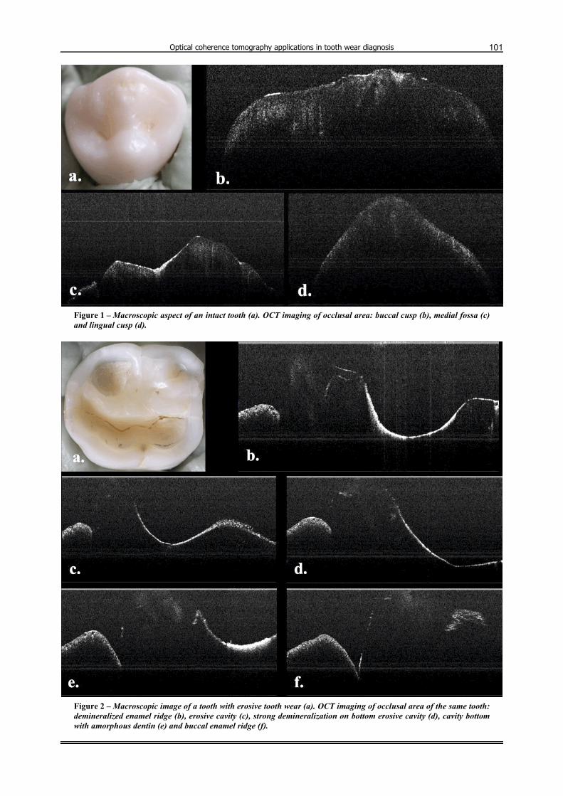
From the teeth with erosive tooth wear, we present an inferior molar, with two profound cavities, one lingual and one buccal, divided by a central medial–distal enamel
ridge (Figure 2a). At the level of the marginal enamel ridge from lingual/distal area, which delimit lingual the erosive cavity, where the enamel is not sustained by dentin, on the OCT analysis a very weak signal can be noted (Figure 2, a and b). As much as the enamel becomes evident towards the area of the central medial-distal enamel ridge, the OCT signal is increasing (Figure 2c). The next aspect observed is showing a more intense OCT signal on the walls of the buccal erosive cavity compared with its bottom, where demineralization is more intense (Figure 2d). In the next image (Figure 2e), the OCT signal is high non-homogeneous for the dentin from the bottom of the buccal erosive cavity. The last OCT image shows the transparent buccal enamel ridge that does not reflect the OCT signal (Figure 2f).
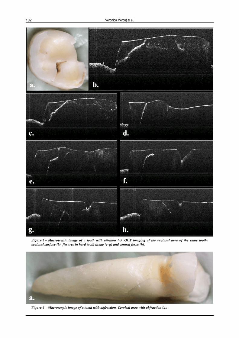
From the group of the teeth diagnosed with attrition, we present a superior molar with a glossy occlusal surface with enamel dislocation from the medial lingual tooth wall (Figure 3a). It could be distinguished, also, the presence of cracks especially at the enamel–dentin junction. On the OCT image of the occlusal surface, an intense signal is present, shaping very accurate the external surface of the attrition lesion (Figure 3b). In profoundness, the signal is heterogeneous, being more intense at vertical or oblique cracks that start from the surface of the lesion towards its depth, with oblique direction from the enamel–dentin junction towards the distal tooth aspect (Figure 3, b–e). Also, the presence of cracks is evident in tooth tissue depths which has no contact with the exterior aspect of the tooth (Figure 3f). In the central fossa of the occlusal area, the OCT signal is similar with the signal of the intact tooth (Figure 3, c, g and h).
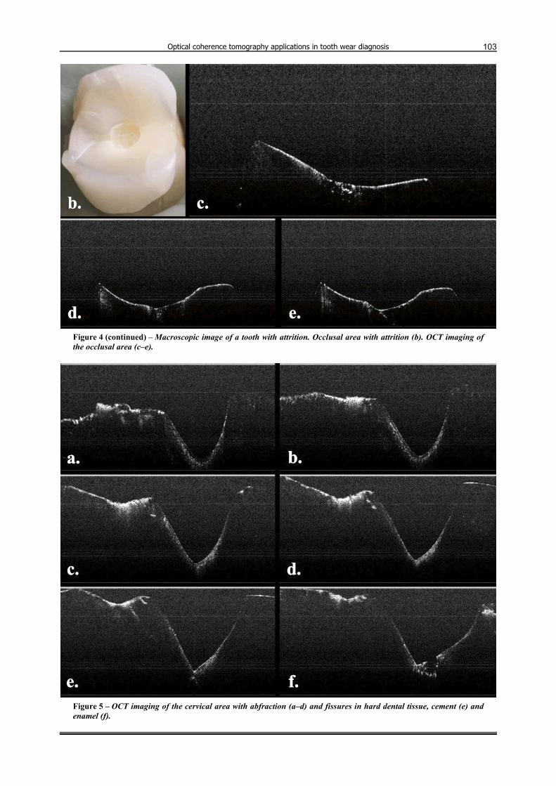
For the abfraction lesion, we present an inferior pre-molar for which abfraction lesion is associated with an attrition occlusal lesion (Figure 4, a and b). An important hard dental tissue loss could be observed in occlusal and cervical areas of the tooth, but also vertical and longitu-dinal cracks in cement and enamel are evident (Figure 4, a and b). The attrition lesion is characterized by a total loss of enamel on occlusal surface, with a pulp chamber position evident in the central occlusal area (Figure 4b). The OCT signal is more intense on the glossy dentinal surface, with an area at the level of the pulp chamber where the OCT signal is getting more profound (Figure 4, c–e). The wedge shape lesion with its angle towards pulp chamber is evident in the buccal cervical area of the tooth (Figure 4a; Figure 5, a–f). The root cement presents a higher intensity of the OCT signal, probably produced by the increasing of structural heterogeneity (Figure 5, a–f), and the signal is increasing as it is getting closer to the bottom of abfraction cavity (designated by the area of the pulp chamber) (Figure 5f). The OCT image shows a higher frequency of the cracks in the cement area (Figure 5e), compared to the enamel (Figure 5f).
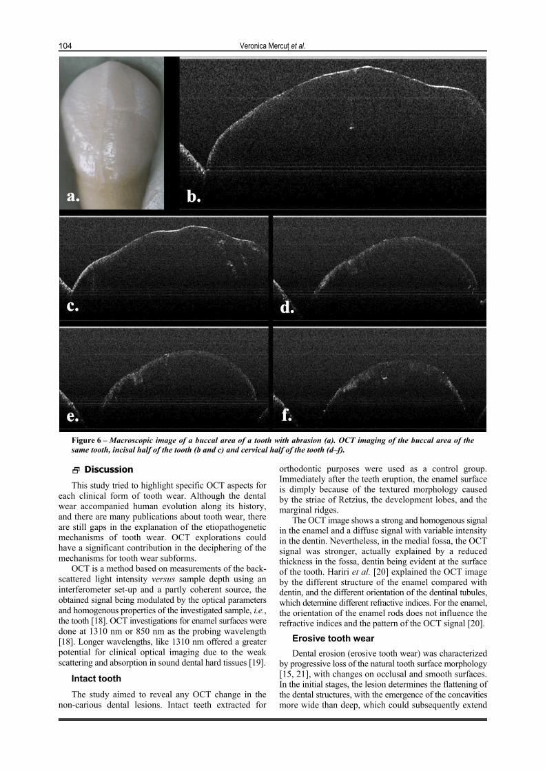
From the abrasion group, we present a superior canine, with a buccal surface with mat enamel and vertical cracks with low amplitude (Figure 6a). The OCT signal is homogeneous in the external aspect of the tooth surface while less expressed vertical and oblique cracks are evident in the profoundness of the tooth (Figure 6, b and c). In the cervical area of the tooth, at the cement level, the OCT signal is low and vague (Figure 6, d–f).
Optical coherence tomography applications in tooth wear diagnosis
101
Figure 1 – Macroscopic aspect of an intact tooth (a). OCT imaging of occlusal area: buccal cusp (b), medial fossa (c) and lingual cusp (d).
Figure 2 – Macroscopic image of a tooth with erosive tooth wear (a). OCT imaging of occlusal area of the same tooth: demineralized enamel ridge (b), erosive cavity (c), strong demineralization on bottom erosive cavity (d), cavity bottom with amorphous dentin (e) and buccal enamel ridge (f).
Veronica Mercuţ et al.
102
Figure 3 – Macroscopic image of a tooth with attrition (a). OCT imaging of the occlusal area of the same tooth: occlusal surface (b), fissures in hard tooth tissue (c–g) and central fossa (h).
Figure 4 – Macroscopic image of a tooth with abfraction. Cervical area with abfraction (a).
Optical coherence tomography applications in tooth wear diagnosis
103
Figure 4 (continued) – Macroscopic image of a tooth with attrition. Occlusal area with attrition (b). OCT imaging of the occlusal area (c–e).
Figure 5 – OCT imaging of the cervical area with abfraction (a–d) and fissures in hard dental tissue, cement (e) and enamel (f).
Veronica Mercuţ et al.
104
Figure 6 – Macroscopic image of a buccal area of a tooth with abrasion (a). OCT imaging of the buccal area of the same tooth, incisal half of the tooth (b and c) and cervical half of the tooth (d–f).
Discussion
This study tried to highlight specific OCT aspects for each clinical form of tooth wear. Although the dental wear accompanied human evolution along its history, and there are many publications about tooth wear, there are still gaps in the explanation of the etiopathogenetic mechanisms of tooth wear. OCT explorations could have a significant contribution in the deciphering of the mechanisms for tooth wear subforms.
OCT is a method based on measurements of the back-scattered light intensity versus sample depth using an interferometer set-up and a partly coherent source, the obtained signal being modulated by the optical parameters and homogenous properties of the investigated sample, i.e., the tooth [18]. OCT investigations for enamel surfaces were done at 1310 nm or 850 nm as the probing wavelength [18]. Longer wavelengths, like 1310 nm offered a greater potential for clinical optical imaging due to the weak scattering and absorption in sound dental hard tissues [19].
Intact tooth
The study aimed to reveal any OCT change in the non-carious dental lesions. Intact teeth extracted for
orthodontic purposes were used as a control group. Immediately after the teeth eruption, the enamel surface is dimply because of the textured morphology caused by the striae of Retzius, the development lobes, and the marginal ridges.
The OCT image shows a strong and homogenous signal in the enamel and a diffuse signal with variable intensity in the dentin. Nevertheless, in the medial fossa, the OCT signal was stronger, actually explained by a reduced thickness in the fossa, dentin being evident at the surface of the tooth. Hariri et al. [20] explained the OCT image by the different structure of the enamel compared with dentin, and the different orientation of the dentinal tubules, which determine different refractive indices. For the enamel, the orientation of the enamel rods does not influence the refractive indices and the pattern of the OCT signal [20].
Erosive tooth wear
Dental erosion (erosive tooth wear) was characterized by progressive loss of the natural tooth surface morphology [15, 21], with changes on occlusal and smooth surfaces. In the initial stages, the lesion determines the flattening of the dental structures, with the emergence of the concavities more wide than deep, which could subsequently extend
Optical coherence tomography applications in tooth wear diagnosis
105
in the dentin. Many characterizations were made over time for the etiopathogenetic mechanisms of the dental erosion, including the one made of Grippo et al. [22], which summarized in the best way the action of the etiological factors. Grippo et al. [22] described the dental erosion lesion appearance by the implication in the first place of the chemical demineralization with physical factors participation (friction and sometimes-even stress). The selected tooth for this subform of dental wear has presented two profound erosive cavities delimited at their border by flattened enamel ridges. Based on the conclusions from the clinical exam, it could be stated that mecha-nisms involved in this case include chemical erosion and friction movement.
The OCT signal of the eroded tooth is almost inexistent at the marginal enamel ridge from the linguo-distal area, formed because of the erosion, where the enamel is not sustained by dentin. A possible explanation for this could be the strong enamel demineralization, which impedes the reflection of the projected light beam. The OCT signal is more intense at the bottom of the eroded cavity because of the increased heterogeneity of the dentin in the eroded area, which could be correlated with the regeneration capacity of the dentin. Still, in the maximum depth point of the cavity, the OCT signal of the dentin is almost inexistent, because of the massive demineralization.
Chan et al. [23] induced in vitro hard dental tissue acidic demineralization, obtaining a decreasing of the OCT signal similar with the one in our experiment. With a clinical OCT device, Wilder-Smith et al. [24] distinguished the enamel demineralization, and measured enamel thickness. In our study, we could not measure the enamel thickness on the OCT image because the external surface of the enamel was rough and the enamel–dentin junction was difficult to delimitate, a fact expressed also by Chan et al. [23].
Dental attrition
The selected tooth for this subform of tooth wear had a glossy, smooth occlusal surface, delimitated by well-differentiated enamel ridges, with a crack of the medial–lingual enamel wall. According to the mechanisms of the dental attrition after Grippo et al. [25], the attrition lesion is a form of tooth wear produced by friction and cyclic stress, as in bruxism. From the anamnesis, the tooth belonged to a patient diagnosed with bruxism. The OCT images revealed numerous vertical and oblique cracks with high amplitude, unobservable on clinical exam. The cracks from the enamel–dentin junction will dislocate the enamel layer from the tooth surface, as could be seen in the dis-location of the enamel fragment from the medial ridge of the tooth. Compared with the erosion lesion, in attrition, the occlusal surface of the tooth (no matter enamel or dentin) releases a stronger OCT signal. This phenomenon may be because, in attrition, tooth surfaces are represented by a thin enamel layer sustained by the strong hetero-geneous dentin as a result of the adaptive processes. We note also the presence of more cracks towards the lingual area (support cusps), due to excessive forces transmitted in bruxism. The fatigue wear form, with vertical fissures that confluence in profoundness and determine enamel layer peeling is similar with the wear of the ceramic fused to metal frame [26, 27]. Mandurah et al. [28] described on the OCT image the aspect of the exterior enamel
surface in attrition similar with this study. Until now, the vertical fissures in attrition teeth were not described on the OCT images. Their presence is justified by the etiopathogenetic mechanism of the dental attrition in bruxism (excessive vertical forces of very long duration and horizontal friction forces).
Abfraction
The abfraction lesion in the presented study has coexisted with an attrition lesion localized on the occlusal area. Grippo [29] and Sarode & Sarode [30] described a mechanism of abfraction lesion similar with the mecha-nism in attrition (excessive forces which determine teeth flection movements with enamel and dentin fracture in the cervical area). The erosion and the abrasion may overlap over this first lesion, according to Grippo et al. [25]. However, Senna et al. [31] supported the inconclusiveness of the studies done to prove the role of the occlusal forces in the abfraction etiology. The selected images are signi-ficant for this type of lesion, also clinical and OCT, and Sugita et al. [32] has described them in a similar way. Compared with Sugita et al. [32] study, our study highlight the presence of the numerous fissures in hard dental tissues, with oblique direction, parallel with the lesion, a fact that confirms the involvement of the excessive occlusal forces in the mechanism of this type of tooth wear lesion.
Abrasion
Many authors [33–35] considered the abrasion as a natural phenomenon, which could become excessive in the conditions of an intense friction associated with erosion. On the studied tooth, the tooth loss is minimal, although the tooth belonged of an aged individual. The reduced destructive potential of the dental abrasion com-pared with other clinical subform has to be highlighted. The OCT signal is similar with the signal of the intact tooth for the incisal tooth half, but in the cervical tooth half the signal is becoming diffuse and heterogeneous, a fact that may be correlated with the irregular enamel surface resulted due to dental abrasion by tooth brushing. Until now, the OCT aspect for this subform of tooth wear has not been described in the literature.
Conclusions
The study highlighted each clinical subform of tooth wear through OCT signal characteristics, and in corre-lation with clinical aspects, the study pointed out the mechanism through the main etiological factor has acted in lesion producing. The erosive lesion produced by chemical aggression has the most rapid evolution, exceeding the reaction ability of the human body. The attrition lesion had grinding and clenching movements (bruxism) as etiological factors with fissure emergence in the hard dental tissues. Abfraction is produced by a complex of multifactorial causes (chemical, abrasive, and mechanical), but this study confirms excessive forces contribution. Abrasion is a physiological phenomenon, which may become pathological only through association with other factors (friction, and erosion). OCT analysis may have clinical usefulness in highlighting implicated factors in each clinical subform as well as in their detection and monitoring.
Veronica Mercuţ et al.
106
Conflict of interests The authors declare no conflict of interests.
References [1] Paesani DA. Tooth wear. In: Paesani DA (ed). Bruxism: theory
and practice. Quintessence Publishing Co., Ltd., 2010, 123–147. [2] Shellis RP, Addy M. The interactions between attrition, abrasion
and erosion in tooth wear. In: Lussi A, Ganns C (eds). Erosive tooth wear. From diagnosis to therapy. Vol. 25, Monographs in Oral Science, Karger AG, Basel, 2014, 32–45.
[3] Mehta SB, Banerji S, Millar BJ, Suarez-Feito JM. Current concepts on the management of tooth wear: part 1. Assess-ment, treatment planning and strategies for the prevention and the passive management of tooth wear. Br Dent J, 2012, 212(1):17–27.
[4] Strużycka I, Lussi A, Bogusławska-Kapała A, Rusyan E. Prevalence of erosive lesions with respect to risk factors in a young adult population in Poland – a cross-sectional study. Clin Oral Investig, 2016, Dec 15.
[5] Mulic A, Fredriksen Ø, Jacobsen ID, Tveit AB, Espelid I, Crossner CG. Dental erosion: prevalence and severity among 16-year-old adolescents in Troms, Norway. Eur J Paediatr Dent, 2016, 17(3):197–201.
[6] Wei Z, Du Y, Zhang J, Tai B, Du M, Jiang H. Prevalence and indicators of tooth wear among Chinese adults. PLoS One, 2016, 11(9):e0162181.
[7] Katsoulis J, Nikitovic SG, Spreng S, Neuhaus K, Mericske-Stern R. Prosthetic rehabilitation and treatment outcome of partially edentulous patients with severe tooth wear: 3-years results. J Dent, 2011, 39(10):662–671.
[8] Johansson A, Johansson AK, Omar R, Carlsson GE. Resto-ration of the worn dentition. In: Paesani DA (ed). Bruxism: theory and practice. Quintessence Publishing Co., Ltd., 2010, 415–438.
[9] Passos VF, Santiago SL. Methodologies to analyze the micro-morphological alterations of enamel subjected to abrasion/ erosion. Dentistry, 2014, 4(9):255.
[10] Izatt JA, Kulkarni MD, Kobayashi K, Sivak MV, Barton JK, Welch AJ. Optical coherence tomography for biodiagnostics. Opt Photon News, 1997, 8(5):41–47.
[11] Attin T, Wegehaupt FJ. Methods for assessment of dental erosion. In: Lussi A, Ganns C (eds). Erosive tooth wear. From diagnosis to therapy. Vol. 25, Monographs in Oral Science, Karger AG, Basel, 2014, 123–142.
[12] Hsieh YS, Ho YC, Lee SY, Chuang CC, Tsai JC, Lin KF, Sun CW. Dental optical coherence tomography. Sensors (Basel), 2013, 13(7):8928–8949.
[13] Verrett RG. Analyzing the etiology of an extremely worn dentition. J Prosthodont, 2001, 10(4):224–233.
[14] Abrahamsen TC. The worn dentition – pathognomonic patterns of abrasion and erosion. Int Dent J, 2005, 55(4 Suppl 1): 268–276.
[15] Carvalho TS, Colon P, Ganss C, Huysmans MC, Lussi A, Schlueter N, Schmalz G, Shellis PR, Björg Tveit A, Wiegand A. Consensus Report of the European Federation of Conser-vative Dentistry: erosive tooth wear diagnosis and manage-ment. Swiss Dent J, 2016, 126(4):342–346.
[16] Osiac E, Bălşeanu TA, Mogoantă L, Gheonea DI, Pirici I, Iancău M, Mitran SI, Albu CV, Cătălin B, Sfredel V. Optical coherence tomography investigation of ischemic stroke inside a rodent model. Rom J Morphol Embryol, 2014, 55(3):767–772.
[17] Osiac E, Săftoiu A, Gheonea DI, Mandrila I, Angelescu R. Optical coherence tomography and Doppler optical coherence
tomography in the gastrointestinal tract. World J Gastroenterol, 2011, 17(1):15–20.
[18] Sowa MG, Popescu DP, Friesen JR, Hewko MD, Choo-Smith LP. A comparison of methods using optical coherence tomography to detect demineralized regions in teeth. J Biophotonics, 2011, 4(11–12):814–823.
[19] Fried D, Staninec M, Darling CL, Lee C, Kang H, Chan KH. In vivo near-IR imaging of occlusal lesions at 1310-nm. Proc SPIE Int Soc Opt Eng, 2011, 7884(78840B), pii:78840B_1.
[20] Hariri I, Sadr A, Shimada Y, Tagami J, Sumi Y. Effects of structural orientation of enamel and dentine on light attenuation and local refractive index: an optical coherence tomography study. J Dent, 2012, 40(5):387–396.
[21] Ganns C, Lussi A. Diagnosis of erosive tooth wear. In: Lussi A, Ganns C (eds). Erosive tooth wear. From diagnosis to therapy. Vol. 25, Monographs in Oral Science, Karger AG, Basel, 2014, 22–31.
[22] Grippo JO, Simring M, Schreiner S. Attrition, abrasion, corrosion and abfraction revisited: a new perspective on tooth surface lesions. J Am Dent Assoc, 2004, 135(8):1109–1118; quiz 1163–1165.
[23] Chan KH, Chan AC, Darling CL, Fried D. Methods for moni-toring erosion using optical coherence tomography. Proc SPIE Int Soc Opt Eng, 2013, 8566:856606.
[24] Wilder-Smith CH, Wilder-Smith P, Kawakami-Wong H, Voronets J, Osann K, Lussi A. Quantification of dental erosions in patients with GERD using optical coherence tomography before and after double-blind, randomized treatment with esomeprazole or placebo. Am J Gastroenterol, 2009, 104(11):2788–2795.
[25] Grippo JO, Simring M, Coleman TA. Abfraction, abrasion, biocorrosion, and the enigma of noncarious cervical lesions: a 20-year perspective. J Esthet Restor Dent, 2012, 24(1): 10–23.
[26] Yu HY, Cai ZB, Ren PD, Zhu MH, Zhou ZR. Friction and wear behavior of dental feldspathic porcelain. Wear, 2006, 261(5–6): 611–621.
[27] Ha UTK. Interaction between enamel, porcelain and a gold alloy: an in vitro wear study. PhD Thesis, School of Dentistry, University of Adelaide, 2011, http://hdl.handle.net/2440/67246.
[28] Mandurah MM, Sadr A, Bakhsh TA, Shimada Y, Sumi Y, Tagami J. Characterization of transparent dentin in attrited teeth using optical coherence tomography. Lasers Med Sci, 2015, 30(4):1189–1196.
[29] Grippo JO. Noncarious cervical lesion the decision to ignore or restore. J Esthet Dent, 1992, 4(Suppl):55–64.
[30] Sarode GS, Sarode SC. Abfraction: a review. J Oral Maxillofac Pathol, 2013, 17(2):222–227.
[31] Senna P, Del Bel Cury A, Rösing C. Non-carious cervical lesions and occlusion: a systematic review of clinical studies. J Oral Rehabil, 2012, 39(6):450–462.
[32] Sugita I, Nakashima S, Ikeda A, Burrow MF, Nikaido T, Kubo S, Tagami J, Sumi Y. A pilot study to assess the morphology and progression of non-carious cervical lesions. J Dent, 2017, 57:51–56.
[33] Aschcroft AT, Joiner A. Tooth cleaning and tooth wear: a review. Proc Inst Mech Eng Part J J Eng Tribol, 2010, 224(6):539–549.
[34] Barnes CM. Etiology of noncarious cervical lesions. Paper 15, Faculty Publications, College of Dentistry, University of Nebraska Medical Center, 2012, http://digitalcommons.unl. edu/dentistryfacpub/15.
[35] Wood I, Jawad Z, Paisley C, Brunton P. Non-carious cervical tooth surface loss: a literature review. J Dent, 2008, 36(10): 759–766.
Corresponding author Sanda Mihaela Popescu, Professor, DMD, PhD, Department of Prosthetics and Oral Rehabilitation, Faculty of Dental Medicine, University of Medicine and Pharmacy of Craiova, 2 Petru Rareş Street, 200349 Craiova, Romania; Phone +40722–248 030, e-mail: [email protected] Received: March 18, 2016 Accepted: March 15, 2017
Related Documents


![Rom J Morphol Embryol 2013, 54(1):205–210 R J M E CASE ... · PDF fileRom J Morphol Embryol 2013, 54(1):205–210 ISSN ... project report CEEX 68/2006 [3] shows that 650 000 ...](https://static.cupdf.com/doc/110x72/5aae038b7f8b9a07498b87b2/rom-j-morphol-embryol-2013-541205210-r-j-m-e-case-j-morphol-embryol-2013.jpg)

