Role of Aspergillus niger acrA in Arsenic Resistance and Its Use as the Basis for an Arsenic Biosensor Se-In Choe, a Fabrice N. Gravelat, a Qusai Al Abdallah, a Mark J. Lee, a Bernard F. Gibbs, b and Donald C. Sheppard a Department of Microbiology and Immunology, McGill University, Montréal, Québec, Canada, a and Endocrine Laboratory, McGill University, Montréal, Québec, Canada b Arsenic contamination of groundwater sources is a major issue worldwide, since exposure to high levels of arsenic has been linked to a variety of health problems. Effective methods of detection are thus greatly needed as preventive measures. In an effort to develop a fungal biosensor for arsenic, we first identified seven putative arsenic metabolism and transport genes in Aspergillus niger, a widely used industrial organism that is generally regarded as safe (GRAS). Among the genes tested for RNA expression in response to arsenate, acrA, encoding a putative plasma membrane arsenite efflux pump, displayed an over 200-fold increase in gene expression in response to arsenate. We characterized the function of this A. niger protein in arsenic efflux by gene knockout and confirmed that AcrA was located at the cell membrane using an enhanced green fluorescent protein (eGFP) fusion construct. Based on our observations, we developed a putative biosensor strain containing a construct of the native promoter of acrA fused with egfp. We analyzed the fluorescence of this biosensor strain in the presence of arsenic using confocal microscopy and spec- trofluorimetry. The biosensor strain reliably detected both arsenite and arsenate in the range of 1.8 to 180 g/liter, which en- compasses the threshold concentrations for drinking water set by the World Health Organization (10 and 50 g/liter). A rsenic is a ubiquitous toxic metalloid that contaminates both groundwater sources (38, 43, 45) and soils (14) worldwide. Strikingly, more than 100 million people in the world are at risk from consuming water contaminated with arsenic (20), and strat- egies to detect and prevent this global problem are urgently re- quired. Arsenic has numerous valence states, but its two predominant forms in nature are arsenate(V), present in more oxidized condi- tions, and arsenite(III), found in reducing environments (40, 44, 46). Arsenate mimics phosphate and inhibits the production of ATP (25). Moreover, arsenite binds to cellular enzymes contain- ing SH groups and thus disrupts their function (25). The long- term exposure to unsafe levels of arsenic leads to arsenicosis, which is characterized by skin lesions, such as melanosis, leu- komelanosis, and hyperkeratosis, and ultimately death (32, 60). Arsenic was also one of the first elements to be recognized as a carcinogen (40, 52), and chronic exposure has been associated with bladder, lung, and skin cancer (World Health Organization [WHO], http://www.who.int/topics/arsenic/en/). The WHO has proposed guidelines for acceptable concentra- tions in drinking water. Although these guidelines suggest a max- imum exposure concentration of arsenic of 10 g/liter for drink- ing water, numerous countries continue to use a cutoff of 50 g/ liter (20). In many parts of the world, arsenic concentrations far exceed both of these limits, particularly in West Bengal and Ban- gladesh (5). There are currently two widely used methods of arse- nic detection in drinking water: laboratory-based analytical meth- ods and field-based testing methods (5). The laboratory-based analytical methods require highly trained personnel and expen- sive analytical machinery, such as inductively coupled plasma mass spectroscopy (ICPMS) and atomic absorption spectroscopy. Further, the delay in turnaround time between specimen collec- tion and result availability limits their day-to-day use. Field-based testing methods are largely chemical colorimetric assays, such as the Gutzeit method, which generate toxic arsine gas by reducing arsenic with a strong acid (20). These tests require the use of haz- ardous chemicals and can generate toxic by-products. Biosensors and bioreporters are beginning to emerge as safe, alternate meth- ods to detect environmental pollutants such as arsenic (22, 29, 50). Although several arsenic biosensors have been reported, these tra- ditionally rely on bacterial reporter systems (7), which have a rel- atively narrow tolerance to variation in culture systems, transpor- tation, and storage. No commercial biosensors are currently in use in affected countries. One approach to developing a field-use- friendly biosensor is the use of a more robust host strain, such as fungi. Filamentous fungi are highly adaptable organisms that can grow in extreme environmental conditions, such as in contami- nated areas with potentially toxic chemical compounds and ele- ments (10). This intrinsic resistance, combined with the long- term stability of resting fungal spores, suggests that they may serve as useful organisms for the development of biodetection or biore- mediation strategies. Arsenic-hyperresistant filamentous fungi, such as Aspergillus sp. P37, have been isolated from nature, and some biochemical pathways of arsenic detoxification have been characterized in these strains (14–17, 54, 58). However, the genet- ics of arsenic metabolism remain unknown in filamentous fungi (14, 59). Aspergillus niger is a filamentous fungus commonly associated with wood and is routinely used for the industrial production of enzymes and organic acids (6, 19). This organism has been ob- served to survive and propagate under concentrations of arsenic as high as 300 mg/liter (18). A. niger is efficient in bioleaching of several heavy metals (19), possibly due to its ability to produce large amounts of oxalic acids in culture. This organism also pro- Received 4 December 2011 Accepted 7 March 2012 Published ahead of print 30 March 2012 Address correspondence to Donald C. Sheppard, [email protected]. Supplemental material for this article may be found at http://aem.asm.org/. Copyright © 2012, American Society for Microbiology. All Rights Reserved. doi:10.1128/AEM.07771-11 June 2012 Volume 78 Number 11 Applied and Environmental Microbiology p. 3855–3863 aem.asm.org 3855 on August 24, 2019 by guest http://aem.asm.org/ Downloaded from

Welcome message from author
This document is posted to help you gain knowledge. Please leave a comment to let me know what you think about it! Share it to your friends and learn new things together.
Transcript
Role of Aspergillus niger acrA in Arsenic Resistance and Its Use as theBasis for an Arsenic Biosensor
Se-In Choe,a Fabrice N. Gravelat,a Qusai Al Abdallah,a Mark J. Lee,a Bernard F. Gibbs,b and Donald C. Shepparda
Department of Microbiology and Immunology, McGill University, Montréal, Québec, Canada,a and Endocrine Laboratory, McGill University, Montréal, Québec, Canadab
Arsenic contamination of groundwater sources is a major issue worldwide, since exposure to high levels of arsenic has beenlinked to a variety of health problems. Effective methods of detection are thus greatly needed as preventive measures. In an effortto develop a fungal biosensor for arsenic, we first identified seven putative arsenic metabolism and transport genes in Aspergillusniger, a widely used industrial organism that is generally regarded as safe (GRAS). Among the genes tested for RNA expression inresponse to arsenate, acrA, encoding a putative plasma membrane arsenite efflux pump, displayed an over 200-fold increase ingene expression in response to arsenate. We characterized the function of this A. niger protein in arsenic efflux by gene knockoutand confirmed that AcrA was located at the cell membrane using an enhanced green fluorescent protein (eGFP) fusion construct.Based on our observations, we developed a putative biosensor strain containing a construct of the native promoter of acrA fusedwith egfp. We analyzed the fluorescence of this biosensor strain in the presence of arsenic using confocal microscopy and spec-trofluorimetry. The biosensor strain reliably detected both arsenite and arsenate in the range of 1.8 to 180 �g/liter, which en-compasses the threshold concentrations for drinking water set by the World Health Organization (10 and 50 �g/liter).
Arsenic is a ubiquitous toxic metalloid that contaminates bothgroundwater sources (38, 43, 45) and soils (14) worldwide.
Strikingly, more than 100 million people in the world are at riskfrom consuming water contaminated with arsenic (20), and strat-egies to detect and prevent this global problem are urgently re-quired.
Arsenic has numerous valence states, but its two predominantforms in nature are arsenate(V), present in more oxidized condi-tions, and arsenite(III), found in reducing environments (40, 44,46). Arsenate mimics phosphate and inhibits the production ofATP (25). Moreover, arsenite binds to cellular enzymes contain-ing �SH groups and thus disrupts their function (25). The long-term exposure to unsafe levels of arsenic leads to arsenicosis,which is characterized by skin lesions, such as melanosis, leu-komelanosis, and hyperkeratosis, and ultimately death (32, 60).Arsenic was also one of the first elements to be recognized as acarcinogen (40, 52), and chronic exposure has been associatedwith bladder, lung, and skin cancer (World Health Organization[WHO], http://www.who.int/topics/arsenic/en/).
The WHO has proposed guidelines for acceptable concentra-tions in drinking water. Although these guidelines suggest a max-imum exposure concentration of arsenic of 10 �g/liter for drink-ing water, numerous countries continue to use a cutoff of 50 �g/liter (20). In many parts of the world, arsenic concentrations farexceed both of these limits, particularly in West Bengal and Ban-gladesh (5). There are currently two widely used methods of arse-nic detection in drinking water: laboratory-based analytical meth-ods and field-based testing methods (5). The laboratory-basedanalytical methods require highly trained personnel and expen-sive analytical machinery, such as inductively coupled plasmamass spectroscopy (ICPMS) and atomic absorption spectroscopy.Further, the delay in turnaround time between specimen collec-tion and result availability limits their day-to-day use. Field-basedtesting methods are largely chemical colorimetric assays, such asthe Gutzeit method, which generate toxic arsine gas by reducingarsenic with a strong acid (20). These tests require the use of haz-ardous chemicals and can generate toxic by-products. Biosensors
and bioreporters are beginning to emerge as safe, alternate meth-ods to detect environmental pollutants such as arsenic (22, 29, 50).Although several arsenic biosensors have been reported, these tra-ditionally rely on bacterial reporter systems (7), which have a rel-atively narrow tolerance to variation in culture systems, transpor-tation, and storage. No commercial biosensors are currently in usein affected countries. One approach to developing a field-use-friendly biosensor is the use of a more robust host strain, such asfungi.
Filamentous fungi are highly adaptable organisms that cangrow in extreme environmental conditions, such as in contami-nated areas with potentially toxic chemical compounds and ele-ments (10). This intrinsic resistance, combined with the long-term stability of resting fungal spores, suggests that they may serveas useful organisms for the development of biodetection or biore-mediation strategies. Arsenic-hyperresistant filamentous fungi,such as Aspergillus sp. P37, have been isolated from nature, andsome biochemical pathways of arsenic detoxification have beencharacterized in these strains (14–17, 54, 58). However, the genet-ics of arsenic metabolism remain unknown in filamentous fungi(14, 59).
Aspergillus niger is a filamentous fungus commonly associatedwith wood and is routinely used for the industrial production ofenzymes and organic acids (6, 19). This organism has been ob-served to survive and propagate under concentrations of arsenic ashigh as 300 mg/liter (18). A. niger is efficient in bioleaching ofseveral heavy metals (19), possibly due to its ability to producelarge amounts of oxalic acids in culture. This organism also pro-
Received 4 December 2011 Accepted 7 March 2012
Published ahead of print 30 March 2012
Address correspondence to Donald C. Sheppard, [email protected].
Supplemental material for this article may be found at http://aem.asm.org/.
Copyright © 2012, American Society for Microbiology. All Rights Reserved.
doi:10.1128/AEM.07771-11
June 2012 Volume 78 Number 11 Applied and Environmental Microbiology p. 3855–3863 aem.asm.org 3855
on August 24, 2019 by guest
http://aem.asm
.org/D
ownloaded from

duces catalase, which allows the fungus to protect itself againstenvironmental stress, including arsenic (11). The intrinsic arsenicresistance of A. niger coupled with the availability of genomic se-quence data led us to select this organism as a model system toexamine arsenic resistance and develop an arsenic biosensor sys-tem in filamentous fungi.
Although the mechanisms of arsenic detoxification remain un-studied in filamentous fungi, several pathways mediating detoxi-fication of arsenic have been elucidated in the yeast Saccharomycescerevisiae. One mechanism of resistance is mediated by the prod-ucts of three contiguous genes, ACR1, ACR2, and ACR3 (9, 15,37). ACR1 encodes a transcriptional regulator, ACR2 encodes anarsenate reductase that converts arsenate into arsenite, and ACR3encodes a plasma membrane arsenite efflux transporter thatpumps arsenite out of the cell, allowing resistance (9, 15, 26, 37).In addition, conjugation of arsenite with glutathione (GSH) toform As(GS)3 and subsequent transport into vacuoles by Ycf1p fordetoxification purposes have been proposed to increase arsenicresistance in S. cerevisiae (14, 26).
We therefore used bioinformatics to identify putative genesinvolved in the arsenic homeostasis of A. niger and developed ahypothetical model of A. niger arsenic metabolism and transportbased on S. cerevisiae. To begin to validate this model, we charac-terized the function of one of these genes, acrA, in detail. This geneis the putative orthologue of the S. cerevisiae ACR3 gene, whichencodes an arsenite efflux pump (66). Consistent with its role asan efflux pump, an AcrA-enhanced green fluorescent protein(eGFP) fusion localized to the plasma membrane in A. niger, anddisruption of A. niger acrA was associated with increased accumu-lation of and sensitivity to arsenic species. Moreover, we discov-ered that the expression of the acrA gene was rapidly and highlyinduced in the presence of very low concentrations of arsenate.Based on this observation, we tested a strain containing the con-struct of the native promoter of acrA fused with egfp as a potentialbiosensor for arsenic and confirmed the ability of this strain todetect levels of arsenic at and below the WHO thresholds for ar-senic levels in drinking water in the developed and developingworlds (24).
MATERIALS AND METHODSFungal strain culture conditions. Aspergillus niger strains were propa-gated on potato dextrose agar plates (PDA; 0.4% potato starch, 2% dex-trose, 1.5% agar) and incubated at 37°C. Other media used were yeastpeptone dextrose (YPD; 1% yeast extract, 2% peptone, 2% glucose) andSabouraud (1% enzymatic digest of casein, 2% dextrose). All media werepurchased from GIBCO (Invitrogen).
Nucleic and proteic sequence comparison. The A. niger genomic se-quences were obtained from the Central Aspergillus Data Repository(CADRE) (www.cadre-genomes.org.uk/Aspergillus_niger/). The puta-tive arsenic homeostasis genes in Aspergillus niger were found by compar-ing gene and protein sequences with those of S. cerevisiae (12, 35, 39, 49,61, 62, 66) using nucleotide-BLAST and protein-BLAST from the Na-tional Center for Biotechnology Information (http://blast.ncbi.nlm.nih.gov/Blast.cgi).
Identification and phylogenetic analysis of A. niger arsenic trans-porter. Select arsenic transporter amino acid sequences found in litera-ture (1, 13) and highly homologous sequences to A. niger AcrA identifiedthrough BLAST searches (3) were aligned with A. niger AcrA using theClustalW multiple sequence alignment (MSA) tool (34). The Gonnet sub-stitution matrix and neighbor-joining clustering model were used inClustalW MSA, and results were exported and formatted to analyze phy-logenetic relationships and build a cladogram using TreeVector (42). To
ensure that only sequences within the functional domain related to arse-nic transport were analyzed, each candidate sequence was queried to iden-tify conserved domain regions using the NCBI Conserved Domain Data-base (CDD) query protocol (36). The CDD domain cluster, TIGRFAMs,and InterPro matches were further drilled and cross-referenced to assessand verify classification of A. niger AcrA (4, 28).
Isolation of DNA and RNA. Conidia of A. niger were inoculated into20 ml of YPD broth and incubated with shaking (200 rpm) at 37°C for 16to 18 h. Subsequently, mycelia were harvested by filtration through a P5Whatman paper (GE Healthcare) and then ground to fine powder underliquid nitrogen in order to break the fungal cell wall. Samples were thenprocessed for the extraction of RNA or DNA. DNA extraction was con-ducted by resuspending the ground hyphae in 500 �l of DNA extractionbuffer [0.7 M NaCl, 0.1 M Na2(SO3), 0.05 M EDTA, 1% SDS, 0.1 MTris-HCl (pH 7.5)]. Following resuspension, DNA was extracted withphenol-chloroform and precipitated with ethanol. RNA extraction wasconducted according to the manufacturer’s instructions of the Nucleos-pin RNA plant minikit (Macherey-Nagel, Germany).
Stimulation and quantification of expression of putative genes in-volved in arsenic homeostasis. Conidia were inoculated into 20 ml ofYPD broth and subsequently incubated (200 rpm, 37°C) for 16 h. Theresulting mycelia were then stimulated by incubation in YPD containingdifferent concentrations of arsenate (potassium arsenate, monobasic;KH2AsO4; Sigma-Aldrich). RNA was harvested and extracted at differenttime points of culture and then analyzed using real-time reverse transcrip-tion (RT)-PCR. The priming oligonucleotides for the genes of interestwere designed using Primer-BLAST from the National Center for Bio-technology Information (http://www.ncbi.nlm.nih.gov/tools/primer-blast/). The primers used for each gene are shown in Table S1 in thesupplemental material. The synthesis of cDNA from 1 �g of RNA wasperformed in 20 �l using Quantitect reverse transcriptase (Qiagen, Mis-sissauga, Canada) with random primers according to the manufacturer’srecommendations. Subsequently, 0.2 �l of the synthesized cDNA wasanalyzed in real-time RT-PCR with SYBR green Quantitect System (Fer-mentas, Canada), using an ABI 7000 thermocycler (Applied Biosystems,Streetsville, Canada). The fungal gene expression was normalized toAspergillus niger TEF1 (transcription elongation factor 1) expression,which was used as the endogenous control.
Disruption of the acrA gene. All PCRs were prepared using KAPAHiFi HotStart ReadyMix according to the manufacturer’s instructions(KAPA Biosystems; Boston, MA). The Aspergillus fumigatus split markertransformation protocol previously developed in our laboratory for thegeneration of homologous integrants was adapted for use in A. niger (55).This technique relies on cotransformation of A. niger protoplasts withDNA fragments, each containing half of the marker (hph encoding hygro-mycin phosphotransferase) fused to one flanking sequence of the gene todelete. Successful homologous integration of these DNA fragments regen-erates the hygromycin marker to permit selection. Fragments were gener-ated using a modified GATEWAY system cloning approach. Briefly, theamplification of the upstream and downstream flanking sequences wasperformed using the priming oligonucleotides AcrA gates 1 and 2 and 3and 4, respectively (see Table S1 in the supplemental material). Theseprimers were created using Primer3 (53). Subsequently, the amplifiedflanking sequences were cloned into pENTR/D-TOPO vector (Invitro-gen, Canada) and transformed into DH5� Escherichia coli cells. The plas-mids were then isolated from putative transformants and analyzed usingrestriction enzymes to verify if the fragments were inserted in the correctorientation. The flanking sequence was then transferred from the pENTR/D-TOPO plasmid to the attR-ccdB-modified pAN7.1, which contains thehph resistance cassette (27), using LR clonase (Invitrogen, Canada). Con-sequently, E. coli DH5� cells were transformed and selected on ampicillinplates. The insertions in the plasmids containing the correct productsallowed amplification by PCR with the long-range PCR enzyme (Fermen-tas, Canada) of one flanking sequence fused with half of the marker cas-sette. Aspergillus niger protoplasts were then produced from young hy-
Choe et al.
3856 aem.asm.org Applied and Environmental Microbiology
on August 24, 2019 by guest
http://aem.asm
.org/D
ownloaded from
phae after digesting the cell wall for 3 h at 30°C (60 rpm) withprotoplasting solution containing Driselase from Basidiomycetes sp. andlysing enzymes from Trichoderma harzianum (Sigma-Aldrich, Canada).Subsequently, these protoplasts were cotransformed with 5 �g of eachDNA fragment (63). Transformants were selected on PDA plates with 250�g/ml of hygromycin. The deletion of the acrA open reading frame (ORF)was confirmed by long-range PCR with primers AcrA gate 1, AcrA gate 4,AcrA-RT sense, and AcrA-RT antisense, as well as by real-time RT-PCRusing primers AcrA-RT sense and AcrA-RT antisense (see Table S1). Theisolation of a pure clone was done by single-spore purification.
Construction of the acrA-complemented strain. To verify that anyphenotypes observed in the acrA-null mutant strain were due to specificdeletion of acrA, we complemented the acrA-null mutant with a wild-typeallele of acrA. We used the same GATEWAY cloning technique as the oneused for the gene deletion in order to replace the hph cassette by the acrAORF fused to the ble cassette (coding for resistance to phleomycin). Vari-ations were as follows. Priming oligonucleotides AcrA gates 1 and 5 wereused to amplify the upstream flanking sequence and the ORF, while thedownstream flanking sequence was amplified by AcrA gate 3 and gate 4(see Table S1 in the supplemental material). Cloned sequences were trans-ferred from pENTR/D-TOPO to a modified p402 vector, which containsthe ble resistance cassette (27). Transformants were selected on PDAplates with 150 �g/ml of phleomycin. The reinsertion of the acrA gene wasconfirmed by long-range PCR and real-time RT-PCR.
Construction of the AcrA-GFP overexpression strain. The GFP-en-coding gene (egfp) was amplified by PCR using the primers GFP-F andGFP-R (see Table S1 in the supplemental material) and using the plasmidp123 (57) as a template. After PCR amplification, the PCR product wascloned into the plasmid pEYFPC (31) using NcoI and NotI. The resultingplasmid was designated pGFP and contains egfp under the expression ofthe A. nidulans gpdA promoter. Afterwards, the 1,278-bp AcrA-encodinggene (acrA) was amplified by PCR using the primers acrA-F and acrA-R(see Table S1). The PCR product was cloned into pGFP using EcoRV andNotI. The resulting plasmid contains acrA tagged with egfp at the N ter-minus and under the expression of the A. nidulans gpdA promoter. Fi-nally, the 1,423-bp phleomycin cassette, which contains the A. nidulansgpdA promoter, phleomycin resistance gene (ble), and the cyc1 termina-tor, was amplified by PCR using the primers Phleo-EcoRI and Phleo-SacI(see Table S1) and p402 as a template and cloned into the acrA-egfp over-expression plasmid using EcoRI and SacI. The resulting plasmid was des-ignated pAcrA-GFP. This plasmid was then used for the transformation ofthe wild-type A. niger strain.
GFP localization of AcrA. The strain containing the AcrA-eGFP fu-sion construct was examined under confocal microscopy to identify thesubcellular localization and trafficking of AcrA. A total of 105 conidia/mlwas inoculated in 1 ml of YPD broth and incubated at 37°C (200 rpm).After 6 h of incubation, samples were taken for observation of the germ-lings under an Olympus Fluoview confocal microscope at 100� oil im-mersion using a fluorescein isothiocyanate (FITC) filter with excitation at488 nm and emission at 507 nm.
We also observed protoplasts at 100� oil immersion under confocalmicroscopy after the cell wall was digested for 3 h at 30°C (60 rpm) withprotoplasting solution used in the fungal transformation protocol.
Growth inhibition assay. Arsenic sensitivity of the different strainswas determined by growth of conidia in Sabouraud broth supplementedwith arsenate in a gradient of increasing concentration (0 to 2,048 mg/literarsenate). In a 96-well plate containing Sabouraud liquid medium, a totalof 2 � 105 conidia/ml was incubated in a final volume of 200 �l at 37°C.For a total volume of 200 �l per well, 100 �l of conidia suspended in liquidmedium was added to each well containing 100 �l of arsenate dilution.After 24 h of incubation, fungal growth was determined by the absorbancetaken at 630 nm using a spectrophotometer. The optical density at 630 nm(OD630) in each well was compared to the same strain growth control inthe absence of arsenate and expressed as percent normal growth.
Arsenic content. To determine the intracellular concentration of in-dividual strains, A. niger strains were grown in YPD liquid broth overnightat 37°C (200 rpm) at 5 � 105 conidia/ml. The culture medium was re-moved by filtration through P5 Whatman paper (GE Healthcare). Themycelial biomass was cut into 1-cm2 squares and weighed. Half of the cutsquares were exposed to chloroform vapor for 20 min to kill them. Sub-sequently, all the squares were inoculated into dishes containing YPDliquid broth with 180 mg/liter of arsenate. The plates were incubated for24 h at 37°C. The cultures were then harvested, and the mycelia werewashed with 5 ml of 0.1 N HCl for 30 min (23, 47). The mycelia were thendried by vacuum and inactivated in liquid nitrogen. Samples were dried atroom temperature and weighed. The dry samples were subsequently di-gested using 1 ml of trace metal-grade Aqua Regia (3 parts HCl:1 partHNO3) in a closed Teflon container at 95°C for 30 min. The HCl wash wasalso kept for arsenic analysis. The quantification of total arsenic species forboth killed and live biomass was determined by using a Perkin ElmerAAnalyst800 atomic absorption spectrometer with a graphite furnace atthe Trace Element Analysis Lab, Department of Earth & Planetary Sci-ences of McGill University (Montréal, Québec, Canada).
Construction of the �acrA::PacrA-GFP strain. The 1-kb upstream se-quence of acrA was amplified by PCR with priming oligonucleotidesAcrA-Bios sense and AcrA-Bios antisense (see Table S1 in the supplemen-tal material). Using the Infusion HD cloning kit (Clontech, USA), thePCR fragment was cloned into the pGFP plasmid previously digested withAgeI and EcoRV. The resulting plasmid was designated pPacrA-GFP. Sub-sequently, the acrA deletion strain was transformed ectopically with thepPacrA-GFP plasmid.
Fluorescence detection. Fluorescence was determined by growth ofconidia in YPD broth or RPMI 1640 with MOPS (morpholinepropane-sulfonic acid; Sigma-Aldrich, Canada), supplemented with arsenate in agradient of increasing concentration (0 to 180 �g/liter). Other metals andmetalloids, including sodium arsenite, cadmium, antimony(III) chloride,iron(III) phosphate, cobalt(II) sulfate, and copper(II) sulfate, were usedto test the specificity of the biosensor (Fisher Scientific, Canada). In a96-well black plate with a clear bottom (Corning, USA), a total of 2 � 105
conidia/ml was incubated in each well at 37°C. At different time points,fungal growth was determined by the absorbance taken at 630 nm, and theGFP signal was measured at excitation of 485 nm and emission of 535 nmusing the SpectraMax microplate reader. For visualization, conidia wereincubated in YPD broth supplemented with arsenate (0, 18, 90 �g/liter)on glass coverslips at 37°C for 24 h. Subsequently, coverslips were placedon microscope slides and pictures were taken using confocal microscopyat �40 magnification using a FITC filter. The �acrA strain was used as acontrol for autofluorescence. Hyphae of the biosensor strain and the�acrA strain were also pregrown for 24 h at 37°C in a 96-well black platewith a clear bottom and then exposed to a gradient of increasing concen-trations of arsenate (0 to 180 �g/liter). At the indicated time points, GFPsignal was measured using a SpectraMax microplate reader as describedabove.
RESULTSPutative arsenic transport and metabolism genes are present inA. niger. Using the recently completed whole-genome sequenceof A. niger, we performed a homology sequence alignment of pu-tative arsenic transport and metabolism genes in A. niger by com-paring these sequences with elements of known arsenic resistancepathways in well-characterized organisms, including E. coli and S.cerevisiae, using BLAST. Using this approach, we identified sevencandidate A. niger genes predicted to encode proteins involved inmediating arsenic metabolism and transport (Table 1).
Expression levels of the putative arsenic transport and me-tabolism genes are affected by arsenate exposure. To examinethe roles of these candidate genes in arsenic homeostasis, we de-termined the effects of arsenate on the expression levels of a subset
Aspergillus niger Arsenic Biosensor
June 2012 Volume 78 Number 11 aem.asm.org 3857
on August 24, 2019 by guest
http://aem.asm
.org/D
ownloaded from
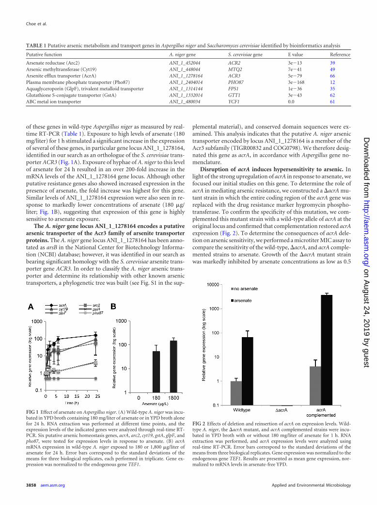
of these genes in wild-type Aspergillus niger as measured by real-time RT-PCR (Table 1). Exposure to high levels of arsenate (180mg/liter) for 1 h stimulated a significant increase in the expressionof several of these genes, in particular gene locus ANI_1_1278164,identified in our search as an orthologue of the S. cerevisiae trans-porter ACR3 (Fig. 1A). Exposure of hyphae of A. niger to this levelof arsenate for 24 h resulted in an over 200-fold increase in themRNA levels of the ANI_1_1278164 gene locus. Although otherputative resistance genes also showed increased expression in thepresence of arsenate, the fold increase was highest for this gene.Similar levels of ANI_1_1278164 expression were also seen in re-sponse to markedly lower concentrations of arsenate (180 �g/liter; Fig. 1B), suggesting that expression of this gene is highlysensitive to arsenate exposure.
The A. niger gene locus ANI_1_1278164 encodes a putativearsenic transporter of the Acr3 family of arsenite transporterproteins. The A. niger gene locus ANI_1_1278164 has been anno-tated as arsB in the National Center for Biotechnology Informa-tion (NCBI) database; however, it was identified in our search asbearing significant homology with the S. cerevisiae arsenite trans-porter gene ACR3. In order to classify the A. niger arsenic trans-porter and determine its relationship with other known arsenictransporters, a phylogenetic tree was built (see Fig. S1 in the sup-
plemental material), and conserved domain sequences were ex-amined. This analysis indicates that the putative A. niger arsenictransporter encoded by locus ANI_1_1278164 is a member of theAcr3 subfamily (TIGR00832 and COG0798). We therefore desig-nated this gene as acrA, in accordance with Aspergillus gene no-menclature.
Disruption of acrA induces hypersensitivity to arsenic. Inlight of the strong upregulation of acrA in response to arsenate, wefocused our initial studies on this gene. To determine the role ofacrA in mediating arsenic resistance, we constructed a �acrA mu-tant strain in which the entire coding region of the acrA gene wasreplaced with the drug resistance marker hygromycin phospho-transferase. To confirm the specificity of this mutation, we com-plemented this mutant strain with a wild-type allele of acrA at theoriginal locus and confirmed that complementation restored acrAexpression (Fig. 2). To determine the consequences of acrA dele-tion on arsenic sensitivity, we performed a microtiter MIC assay tocompare the sensitivity of the wild-type, �acrA, and acrA comple-mented strains to arsenate. Growth of the �acrA mutant strainwas markedly inhibited by arsenate concentrations as low as 0.5
TABLE 1 Putative arsenic metabolism and transport genes in Aspergillus niger and Saccharomyces cerevisiae identified by bioinformatics analysis
Putative function A. niger gene S. cerevisiae gene E value Reference
Arsenate reductase (Arc2) ANI_1_452044 ACR2 3e�13 39Arsenic methyltransferase (Cyt19) ANI_1_448044 MTQ2 7e�41 49Arsenite efflux transporter (AcrA) ANI_1_1278164 ACR3 5e�79 66Plasma membrane phosphate transporter (Pho87) ANI_1_2404014 PHO87 3e�168 12Aquaglyceroporin (GlpF), trivalent metalloid transporter ANI_1_1314144 FPS1 1e�36 35Glutathione S-conjugate transporter (GstA) ANI_1_1332014 GTT1 3e�43 62ABC metal ion transporter ANI_1_480034 YCF1 0.0 61
FIG 1 Effect of arsenate on Aspergillus niger. (A) Wild-type A. niger was incu-bated in YPD broth containing 180 mg/liter of arsenate or in YPD broth alonefor 24 h. RNA extraction was performed at different time points, and theexpression levels of the indicated genes were analyzed through real-time RT-PCR. Six putative arsenic homeostasis genes, acrA, arc2, cyt19, gstA, glpF, andpho87, were tested for expression levels in response to arsenate. (B) acrAmRNA expression in wild-type A. niger exposed to 180 or 1,800 �g/liter ofarsenate for 24 h. Error bars correspond to the standard deviations of themeans for three biological replicates, each performed in triplicate. Gene ex-pression was normalized to the endogenous gene TEF1.
FIG 2 Effects of deletion and reinsertion of acrA on expression levels. Wild-type A. niger, the �acrA mutant, and acrA complemented strains were incu-bated in YPD broth with or without 180 mg/liter of arsenate for 1 h. RNAextraction was performed, and acrA expression levels were analyzed usingreal-time RT-PCR. Error bars correspond to the standard deviations of themeans from three biological replicates. Gene expression was normalized to theendogenous gene TEF1. Results are presented as mean gene expression, nor-malized to mRNA levels in arsenate-free YPD.
Choe et al.
3858 aem.asm.org Applied and Environmental Microbiology
on August 24, 2019 by guest
http://aem.asm
.org/D
ownloaded from
mg/liter (Fig. 3). In contrast, the wild-type strain produced signif-icant growth at concentrations as high as 64 mg/liter. Comple-mentation of the �acrA mutant strain with a wild-type allele ofacrA restored arsenic resistance to wild-type levels, confirmingthat this phenotype was due to the absence of acrA and not anundetected secondary mutation. Collectively, these data stronglysuggest that the disruption of acrA results in hypersensitivity toarsenic and that the AcrA protein is important in arsenic detoxi-fication of Aspergillus niger.
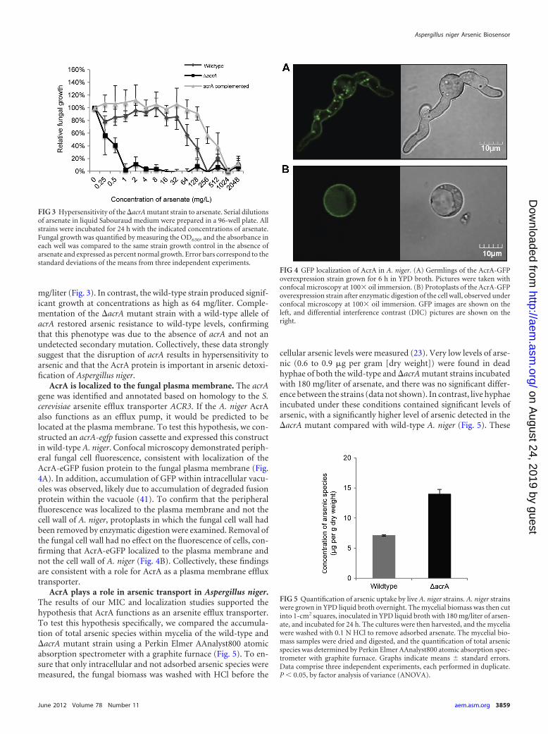
AcrA is localized to the fungal plasma membrane. The acrAgene was identified and annotated based on homology to the S.cerevisiae arsenite efflux transporter ACR3. If the A. niger AcrAalso functions as an efflux pump, it would be predicted to belocated at the plasma membrane. To test this hypothesis, we con-structed an acrA-egfp fusion cassette and expressed this constructin wild-type A. niger. Confocal microscopy demonstrated periph-eral fungal cell fluorescence, consistent with localization of theAcrA-eGFP fusion protein to the fungal plasma membrane (Fig.4A). In addition, accumulation of GFP within intracellular vacu-oles was observed, likely due to accumulation of degraded fusionprotein within the vacuole (41). To confirm that the peripheralfluorescence was localized to the plasma membrane and not thecell wall of A. niger, protoplasts in which the fungal cell wall hadbeen removed by enzymatic digestion were examined. Removal ofthe fungal cell wall had no effect on the fluorescence of cells, con-firming that AcrA-eGFP localized to the plasma membrane andnot the cell wall of A. niger (Fig. 4B). Collectively, these findingsare consistent with a role for AcrA as a plasma membrane effluxtransporter.
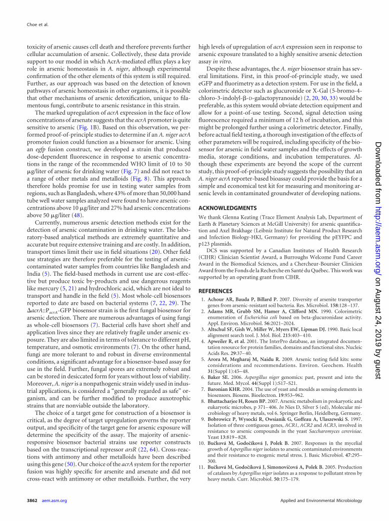
AcrA plays a role in arsenic transport in Aspergillus niger.The results of our MIC and localization studies supported thehypothesis that AcrA functions as an arsenite efflux transporter.To test this hypothesis specifically, we compared the accumula-tion of total arsenic species within mycelia of the wild-type and�acrA mutant strain using a Perkin Elmer AAnalyst800 atomicabsorption spectrometer with a graphite furnace (Fig. 5). To en-sure that only intracellular and not adsorbed arsenic species weremeasured, the fungal biomass was washed with HCl before the
cellular arsenic levels were measured (23). Very low levels of arse-nic (0.6 to 0.9 �g per gram [dry weight]) were found in deadhyphae of both the wild-type and �acrA mutant strains incubatedwith 180 mg/liter of arsenate, and there was no significant differ-ence between the strains (data not shown). In contrast, live hyphaeincubated under these conditions contained significant levels ofarsenic, with a significantly higher level of arsenic detected in the�acrA mutant compared with wild-type A. niger (Fig. 5). These
FIG 3 Hypersensitivity of the �acrA mutant strain to arsenate. Serial dilutionsof arsenate in liquid Sabouraud medium were prepared in a 96-well plate. Allstrains were incubated for 24 h with the indicated concentrations of arsenate.Fungal growth was quantified by measuring the OD630, and the absorbance ineach well was compared to the same strain growth control in the absence ofarsenate and expressed as percent normal growth. Error bars correspond to thestandard deviations of the means from three independent experiments.
FIG 4 GFP localization of AcrA in A. niger. (A) Germlings of the AcrA-GFPoverexpression strain grown for 6 h in YPD broth. Pictures were taken withconfocal microscopy at 100� oil immersion. (B) Protoplasts of the AcrA-GFPoverexpression strain after enzymatic digestion of the cell wall, observed underconfocal microscopy at 100� oil immersion. GFP images are shown on theleft, and differential interference contrast (DIC) pictures are shown on theright.
FIG 5 Quantification of arsenic uptake by live A. niger strains. A. niger strainswere grown in YPD liquid broth overnight. The mycelial biomass was then cutinto 1-cm2 squares, inoculated in YPD liquid broth with 180 mg/liter of arsen-ate, and incubated for 24 h. The cultures were then harvested, and the myceliawere washed with 0.1 N HCl to remove adsorbed arsenate. The mycelial bio-mass samples were dried and digested, and the quantification of total arsenicspecies was determined by Perkin Elmer AAnalyst800 atomic absorption spec-trometer with graphite furnace. Graphs indicate means � standard errors.Data comprise three independent experiments, each performed in duplicate.P � 0.05, by factor analysis of variance (ANOVA).
Aspergillus niger Arsenic Biosensor
June 2012 Volume 78 Number 11 aem.asm.org 3859
on August 24, 2019 by guest
http://aem.asm
.org/D
ownloaded from

results are consistent with the hypothesis that AcrA functions inthe efflux of arsenic in A. niger.
Use of a �acrA::PacrA-GFP strain as a potential biosensor ofarsenic. The dramatic upregulation of acrA expression in re-sponse to low levels of arsenate suggested that this gene might beuseful as a potential biosensor for arsenic. To test this hypothesis,we created a fusion construct in which egfp was fused to the acrApromoter. This plasmid was then expressed in the �acrA strain.Through confocal microscopy, we observed that the strain fluo-resced even at concentrations of arsenate as low as 18 �g/liter (Fig.6). To determine the sensitivity and kinetics of the biosensor strainin response to arsenate, we tested the ability of conidia of thebiosensor strain to germinate and fluoresce in the presence ofarsenate. Exposure of conidia of this strain to a gradient of arsen-ate produced a dose-dependent fluorescence that was detectableas early as 12 h after incubation and increased dramatically by 48 hof arsenate exposure (Fig. 7). At this time point, reproducibledose-dependent fluorescence was observed throughout the rangeof the maximum safe limit of arsenic detection (10 to 50 �g/liter)suggested by the World Health Organization (48, 56). In parallel,we also tested the effects of exposing pregrown hyphae of the bio-sensor strain to the same range of arsenate concentrations. Al-though greater overall fluorescence was seen with pregrown hy-phae, the background autofluorescence of hyphae alone increasedto the same degree, and no significant difference in the fluorescentsignal was found compared with exposure of conidia (data not
shown). In addition, we tested the specificity of the biosensor strainfor arsenate by testing several other metals and metalloids, includingarsenite, antimony(III), copper(II), cobalt(II), iron(III), and cad-mium (Fig. 8). Interestingly, the biosensor strain was specific to botharsenate and arsenite species and did not produce significant fluores-cence with any other metals tested. Collectively, these data suggestthat a biosensor strategy based on the A. niger acrA promoter se-quence may be a useful approach for the detection of low-level arseniccontamination of water, although further studies to examine the re-producibility and specificity of this biosensor in field specimensshould be performed.
DISCUSSION
Homology searches based on known bacterial and fungal arsenicresistance pathways identified seven putative arsenic metabolismand transport genes in Aspergillus niger (Table 1). Interestingly,although an A. niger orthologue of S. cerevisiae ACR3 was found, itwas not located in a gene cluster as is the case in S. cerevisiae.Indeed, no orthologue for the S. cerevisiae ACR1 regulatory genecould be found in A. niger, and an orthologue of ACR2 encoding aputative arsenate reductase was found clustered with genes encod-ing a putative arsenic methyltransferase and putative arsenic re-sistance protein gene, arsH. Interestingly, the arsH gene is notfound in S. cerevisiae, although it is found in different bacterialspecies, most notably in Shigella flexneri (65). This cluster found inA. niger contrasts with Aspergillus fumigatus, in which the putative
FIG 6 Fluorescence of the biosensor strain in response to arsenate. Conidia of the �acrA and �acrA::PacrA-GFP strains were incubated in YPD broth witharsenate at the indicated concentrations on glass coverslips at 37°C. After 24 h of incubation, confocal images of fungi on coverslips were obtained using confocalmicroscopy. The �acrA strain was used as a control for autofluorescence.
Choe et al.
3860 aem.asm.org Applied and Environmental Microbiology
on August 24, 2019 by guest
http://aem.asm
.org/D
ownloaded from

arsenate reductase, arsenite efflux transporter, and arsenic meth-yltransferase genes are found together in a duplicated cluster onchromosomes 1 and 5, highlighting the genomic diversity of thisgenus.
By extrapolating the function of the products of these A. nigergenes from the role of their orthologues in yeast arsenic metabo-lism and transport, we propose a hypothetical model of arsenichomeostasis in A. niger (8). In this predicted model based on ho-mology of A. niger genes with their yeast orthologues, arsenic fromthe environment is transported into the cell either as arsenitethrough GlpF or as arsenate through Pho87. Intracellular arsenateis reduced to arsenite by Arc2 and then detoxified by one of thethree methods: glutathionation by GstA and sequestration intovacuoles by an ABC metal ion transporter, biovolatilisation viamethylation by Cyt19, or direct export from the cell by the AcrAefflux transporter. To begin to test this model, we selected acrA,the A. niger homologue of S. cerevisiae ACR3, for further study (9).
In S. cerevisiae, the ACR3 gene encodes an arsenite transporterwhose expression is strongly induced by the presence of arseniteand arsenate (51). Multiple lines of evidence suggest that thisfunction is preserved in A. niger: acrA mRNA expression wasmarkedly upregulated in the presence of arsenate, an AcrA-eGFPfusion construct localized to the fungal plasma membrane, anddeletion of acrA resulted in arsenate hypersensitivity and accumu-lation of intracellular arsenic species. Although the loss of acrAresulted in marked hypersensitivity to arsenate, this phenotypewas associated with only a modest increase in intracellular arseniclevels. These data suggest a low tolerance for increased intracellu-lar arsenic levels in the A. niger �acrA strain, beyond which the
FIG 8 Specificity of the A. niger biosensor strain for arsenic species. (A)Conidia of the �acrA::PacrA-GFP strain was grown in YPD broth supplementedwith different metals and metalloids, including arsenate, arsenite, cadmium,antimony(III), cobalt(II), copper(II), and iron(III) at the indicated concentra-tions. At 48 h, GFP expression was measured by determining the raw fluores-cence intensity using a Spectramax fluorometer. (B) Conidia of the �acrAstrain were used as a control for autofluorescence. Results are shown asmeans � standard errors. Data comprise two independent experiments, eachperformed in triplicate.
FIG 7 Arsenate detection using conidia of the A. niger biosensor strain. Conidia of the �acrA and �acrA::PacrA-GFP strains were grown in YPD brothsupplemented with arsenate at the indicated concentrations. At 12 h, 24 h, 36 h, and 48 h, GFP expression was measured by determining the rawfluorescence intensity using a Spectramax fluorometer. Dashed lines indicate the arsenic threshold concentrations set by the WHO for drinking water indeveloped and developing countries (10 �g/liter and 50 �g/liter, respectively). Results are shown as means � standard errors. Data comprise twoindependent experiments, each performed in triplicate. � indicates �acrA::PacrA-GFP, and indicates �acrA. Note that the scale of the y axis increasesover time.
Aspergillus niger Arsenic Biosensor
June 2012 Volume 78 Number 11 aem.asm.org 3861
on August 24, 2019 by guest
http://aem.asm
.org/D
ownloaded from
toxicity of arsenic causes cell death and therefore prevents furthercellular accumulation of arsenic. Collectively, these data providesupport to our model in which AcrA-mediated efflux plays a keyrole in arsenic homeostasis in A. niger, although experimentalconfirmation of the other elements of this system is still required.Further, as our approach was based on the detection of knownpathways of arsenic homeostasis in other organisms, it is possiblethat other mechanisms of arsenic detoxification, unique to fila-mentous fungi, contribute to arsenic resistance in this strain.
The marked upregulation of acrA expression in the face of lowconcentrations of arsenate suggests that the acrA promoter is quitesensitive to arsenic (Fig. 1B). Based on this observation, we per-formed proof-of-principle studies to determine if an A. niger acrApromoter fusion could function as a biosensor for arsenic. Usingan egfp fusion construct, we developed a strain that produceddose-dependent fluorescence in response to arsenic concentra-tions in the range of the recommended WHO limit of 10 to 50�g/liter of arsenic for drinking water (Fig. 7) and did not react toa range of other metals and metalloids (Fig. 8). This approachtherefore holds promise for use in testing water samples fromregions, such as Bangladesh, where 43% of more than 50,000 handtube well water samples analyzed were found to have arsenic con-centrations above 10 �g/liter and 27% had arsenic concentrationsabove 50 �g/liter (48).
Currently, numerous arsenic detection methods exist for thedetection of arsenic contamination in drinking water. The labo-ratory-based analytical methods are extremely quantitative andaccurate but require extensive training and are costly. In addition,transport times limit their use in field situations (20). Other fielduse strategies are therefore preferable for the testing of arsenic-contaminated water samples from countries like Bangladesh andIndia (5). The field-based methods in current use are cost-effec-tive but produce toxic by-products and use dangerous reagentslike mercury (5, 21) and hydrochloric acid, which are not ideal totransport and handle in the field (5). Most whole-cell biosensorsreported to date are based on bacterial systems (7, 22, 29). The�acrA::PacrA-GFP biosensor strain is the first fungal biosensor forarsenic detection. There are numerous advantages of using fungias whole-cell biosensors (7). Bacterial cells have short shelf andapplication lives since they are relatively fragile under arsenic ex-posure. They are also limited in terms of tolerance to different pH,temperature, and osmotic environments (7). On the other hand,fungi are more tolerant to and robust in diverse environmentalconditions, a significant advantage for a biosensor-based assay foruse in the field. Further, fungal spores are extremely robust andcan be stored in desiccated form for years without loss of viability.Moreover, A. niger is a nonpathogenic strain widely used in indus-trial applications, is considered a “generally regarded as safe” or-ganism, and can be further modified to produce auxotrophicstrains that are nonviable outside the laboratory.
The choice of a target gene for construction of a biosensor iscritical, as the degree of target upregulation governs the reporteroutput, and specificity of the target gene for arsenic exposure willdetermine the specificity of the assay. The majority of arsenic-responsive biosensor bacterial strains use reporter constructsbased on the transcriptional repressor arsR (22, 64). Cross-reac-tions with antimony and other metalloids have been describedusing this gene (50). Our choice of the acrA system for the reporterfusion was highly specific for arsenite and arsenate and did notcross-react with antimony or other metalloids. Further, the very
high levels of upregulation of acrA expression seen in response toarsenic exposure translated to a highly sensitive arsenic detectionassay in vitro.
Despite these advantages, the A. niger biosensor strain has sev-eral limitations. First, in this proof-of-principle study, we usedeGFP and fluorimetry as a detection system. For use in the field, acolorimetric detector such as glucuronide or X-Gal (5-bromo-4-chloro-3-indolyl-�-D-galactopyranoside) (2, 20, 30, 33) would bepreferable, as this system would obviate detection equipment andallow for a point-of-use testing. Second, signal detection usingfluorescence required a minimum of 12 h of incubation, and thismight be prolonged further using a colorimetric detector. Finally,before actual field testing, a thorough investigation of the effects ofother parameters will be required, including specificity of the bio-sensor for arsenic in field water samples and the effects of growthmedia, storage conditions, and incubation temperatures. Al-though these experiments are beyond the scope of the currentstudy, this proof-of-principle study suggests the possibility that anA. niger acrA reporter-based bioassay could provide the basis for asimple and economical test kit for measuring and monitoring ar-senic levels in contaminated groundwater of developing nations.
ACKNOWLEDGMENTS
We thank Glenna Keating (Trace Element Analysis Lab, Department ofEarth & Planetary Sciences at McGill University) for arsenic quantifica-tion and Axel Brakhage (Leibniz Institute for Natural Product Researchand Infection Biology-HKI, Germany) for providing the pEYFPC andp123 plasmids.
DCS was supported by a Canadian Institutes of Health Research(CIHR) Clinician Scientist Award, a Burroughs Welcome Fund CareerAward in the Biomedical Sciences, and a Chercheur-Boursier ClinicienAward from the Fonds de la Recherche en Santé du Québec. This work wassupported by an operating grant from CIHR.
REFERENCES1. Achour AR, Bauda P, Billard P. 2007. Diversity of arsenite transporter
genes from arsenic-resistant soil bacteria. Res. Microbiol. 158:128 –137.2. Adams MR, Grubb SM, Hamer A, Clifford MN. 1990. Colorimetric
enumeration of Escherichia coli based on beta-glucuronidase activity.Appl. Environ. Microbiol. 56:2021–2024.
3. Altschul SF, Gish W, Miller W, Myers EW, Lipman DJ. 1990. Basic localalignment search tool. J. Mol. Biol. 215:403– 410.
4. Apweiler R, et al. 2001. The InterPro database, an integrated documen-tation resource for protein families, domains and functional sites. NucleicAcids Res. 29:37– 40.
5. Arora M, Megharaj M, Naidu R. 2009. Arsenic testing field kits: someconsiderations and recommendations. Environ. Geochem. Health31(Suppl 1):45– 48.
6. Baker SE. 2006. Aspergillus niger genomics: past, present and into thefuture. Med. Mycol. 44(Suppl 1):S17–S21.
7. Baronian KHR. 2004. The use of yeast and moulds as sensing elements inbiosensors. Biosens. Bioelectron. 19:953–962.
8. Bhattacharjee H, Rosen BP. 2007. Arsenic metabolism in prokaryotic andeukaryotic microbes, p 371– 406. In Nies D, Silver S (ed), Molecular mi-crobiology of heavy metals, vol 6. Springer Berlin, Heidelberg, Germany.
9. Bobrowicz P, Wysocki R, Owsianik G, Goffeau A, Ulaszewski S. 1997.Isolation of three contiguous genes, ACR1, ACR2 and ACR3, involved inresistance to arsenic compounds in the yeast Saccharomyces cerevisiae.Yeast 13:819 – 828.
10. Bucková M, Godocíková J, Polek B. 2007. Responses in the mycelialgrowth of Aspergillus niger isolates to arsenic contaminated environmentsand their resistance to exogenic metal stress. J. Basic Microbiol. 47:295–300.
11. Bucková M, Godocíková J, Simonovicová A, Polek B. 2005. Productionof catalases by Aspergillus niger isolates as a response to pollutant stress byheavy metals. Curr. Microbiol. 50:175–179.
Choe et al.
3862 aem.asm.org Applied and Environmental Microbiology
on August 24, 2019 by guest
http://aem.asm
.org/D
ownloaded from

12. Bun-ya M, et al.1996.Twonewgenes,PHO86andPHO87, involvedin inorganicphosphate uptake in Saccharomyces cerevisiae. Curr. Genet. 29:344–351.
13. Cai L, Liu G, Rensing C, Wang G. 2009. Genes involved in arsenictransformation and resistance associated with different levels of arsenic-contaminated soils. BMC Microbiol. 9:4.
14. Cánovas D, de Lorenzo V. 2007. Osmotic stress limits arsenic hypertol-erance in Aspergillus sp. P37. FEMS Microbiol. Ecol. 61:258 –263.
15. Cánovas D, Durán C, Rodríguez N, Amils R, de Lorenzo V. 2003.Testing the limits of biological tolerance to arsenic in a fungus isolatedfrom the River Tinto. Environ. Microbiol. 5:133–138.
16. Cánovas D, Mukhopadhyay R, Rosen BP, de Lorenzo V. 2003. Arsenatetransport and reduction in the hyper-tolerant fungus Aspergillus sp. P37.Environ. Microbiol. 5:1087–1093.
17. Cánovas D, Vooijs R, Schat H, de Lorenzo V. 2004. The role of thiolspecies in the hypertolerance of Aspergillus sp. P37 to arsenic. J. Biol.Chem. 279:51234 –51240.
18. Cernanský S, Kolencík M, Sevc J, Urík M, Hiller E. 2009. Fungalvolatilization of trivalent and pentavalent arsenic under laboratory condi-tions. Bioresour. Technol. 100:1037–1040.
19. Clausen C. 2004. Improving the two-step remediation process for CCA-treated wood: part I. Evaluating oxalic acid extraction. Waste Manag. 24:401– 405.
20. de Mora K, et al. 2011. A pH-based biosensor for detection of arsenic indrinking water. Anal. Bioanal. Chem. 400:1031–1039.
21. Dhar RK, Zheng Y, Rubenstone J, van Geen A. 2004. A rapid colori-metric method for measuring arsenic concentrations in groundwater.Anal. Chim. Acta 526:203–209.
22. Diesel E, Schreiber M, van der Meer JR. 2009. Development of bacteria-based bioassays for arsenic detection in natural waters. Anal. Bioanal.Chem. 394:687– 693.
23. Gadd GM. 1993. Interactions of fungi with toxic metals. New Phytol.124:25– 60.
24. Galal-Gorchev H, Ozolins G, Bonnefoy X. 1993. Revision of the WHOguidelines for drinking water quality. Ann. Ist. Super. Sanita 29:335–345.
25. Geng C, Zhu Y, Hu Y, Williams P, Meharg A. 2006. Arsenate causesdifferential acute toxicity to two P-deprived genotypes of rice seedlings(Oryza sativa L.). Plant Soil 279:297–306.
26. Ghosh M, Shen J, Rosen BP. 1999. Pathways of As(III) detoxification inSaccharomyces cerevisiae. Proc. Natl. Acad. Sci. U. S. A. 96:5001–5006.
27. Gravelat FN, et al. 2010. Aspergillus fumigatus MedA governs adherence,host cell interactions and virulence. Cell. Microbiol. 12:473– 488.
28. Haft DH, Selengut JD, White O. 2003. The TIGRFAMs database ofprotein families. Nucleic Acids Res. 31:371–373.
29. Hansen LH, Sørensen SJ. 2001. The use of whole-cell biosensors to detectand quantify compounds or conditions affecting biological systems. Mi-crob. Ecol. 42:483– 494.
30. Hirt H. 1991. A novel method for in situ screening of yeast colonies withthe beta-glucuronidase reporter gene. Curr. Genet. 20:437– 439.
31. Hoff B, Kück U. 2005. Use of bimolecular fluorescence complementation todemonstrate transcription factor interaction in nuclei of living cells from thefilamentous fungus Acremonium chrysogenum. Curr. Genet. 47:132–138.
32. Jain CK, Ali I. 2000. Arsenic: occurrence, toxicity and speciation tech-niques. Water Res. 34:4304 – 4312.
33. Joshi N, Wang X, Montgomery L, Elfick A, French CE. 2009. Novelapproaches to biosensors for detection of arsenic in drinking water. De-salination 248:517–523.
34. Larkin MA, et al. 2007. Clustal W and Clustal X version 2.0. Bioinfor-matics 23:2947–2948.
35. Maciaszczyk-Dziubinska E, Migdal I, Migocka M, Bocer T, Wysocki R.2010. The yeast aquaglyceroporin Fps1p is a bidirectional arsenite chan-nel. FEBS Lett. 584:726 –732.
36. Marchler-Bauer A, et al. 2011. CDD: a conserved domain database for thefunctional annotation of proteins. Nucleic Acids Res. 39:D225–D229.
37. Messens J, Silver S. 2006. Arsenate reduction: thiol cascade chemistrywith convergent evolution. J. Mol. Biol. 362:1–17.
38. Mukherjee A, et al. 2010. Tolerance of arsenate-induced stress in Asper-gillus niger, a possible candidate for bioremediation. Ecotoxicol. Environ.Saf. 73:172–182.
39. Mukhopadhyay R, Shi J, Rosen BP. 2000. Purification and characteriza-tion of ACR2p, the Saccharomyces cerevisiae arsenate reductase. J. Biol.Chem. 275:21149 –21157.
40. Páez-Espino D, Tamames J, de Lorenzo V, Cánovas D. 2009. Microbialresponses to environmental arsenic. Biometals 22:117–130.
41. Petersson J, Pattison J, Kruckeberg AL, Berden JA, Persson BL. 1999.Intracellular localization of an active green fluorescent protein-tagged Pho84phosphate permease in Saccharomyces cerevisiae. FEBS Lett. 462:37–42.
42. Pethica R, Barker G, Kovacs T, Gough J. 2010. TreeVector: scalable,interactive, phylogenetic trees for the web. PLoS One 5:e8934.
43. Pokhrel D, Viraraghavan T. 2006. Arsenic removal from an aqueoussolution by a modified fungal biomass. Water Res. 40:549 –552.
44. Pokhrel D, Viraraghavan T. 2008. Arsenic removal from an aqueoussolution by modified A. niger biomass: batch kinetic and isotherm studies.J. Hazard. Mater. 150:818 – 825.
45. Pokhrel D, Viraraghavan T. 2006. Arsenic removal from aqueous solu-tion by iron oxide-coated fungal biomass: a factorial design analysis. Wa-ter Air Soil Pollut. 173:195–208.
46. Pokhrel D, Viraraghavan T. 2008. Arsenic removal in an iron oxide-coated fungal biomass column: analysis of breakthrough curves. Biore-sour. Technol. 99:2067–2071.
47. Price MS, Classen JJ, Payne GA. 2001. Aspergillus niger absorbs copperand zinc from swine wastewater. Bioresour. Technol. 77:41– 49.
48. Rahman MM, Naidu R, Bhattacharya P. 2009. Arsenic contamination ingroundwater in the Southeast Asia region. Environ. Geochem. Health 31:9–21.
49. Ren X, et al. 2011. Involvement of N-6 adenine-specific DNA methyl-transferase 1 (N6AMT1) in arsenic biomethylation and its role in arsenic-induced toxicity. Environ. Health Perspect. 119:771–777.
50. Roberto FF, Barnes JM, Bruhn DF. 2002. Evaluation of a GFP reportergene construct for environmental arsenic detection. Talanta 58:181–188.
51. Rosen BP, Tamás MJ. 2010. Arsenic transport in prokaryotes and eukary-otic microbes. Adv. Exp. Med. Biol. 679:47–55.
52. Rosen P. 1971. Theoretical significance of arsenic as a carcinogen. J.Theor. Biol. 32:425– 426.
53. Rozen S, Skaletsky HJ. 2000. Primer3 on the WWW for general users andfor biologist programmers, p 365–386. In Krawetz S, Misenser S (ed),Bioinformatics methods and protocols: methods in molecular biology.Humana Press, Totowa, NJ.
54. Sharples JM, Meharg AA, Chambers SM, Cairney JW. 2000. Mechanismof arsenate resistance in the ericoid mycorrhizal fungus Hymenoscyphusericae. Plant Physiol. 124:1327–1334.
55. Sheppard DC, et al. 2005. The Aspergillus fumigatus StuA protein governsthe up-regulation of a discrete transcriptional program during the acqui-sition of developmental competence. Mol. Biol. Cell 16:5866 –5879.
56. Smith AH, Lingas EO, Rahman M. 2000. Contamination of drinking-water by arsenic in Bangladesh: a public health emergency. Bull. WorldHealth Organ. 78:1093–1103.
57. Spellig T, Bottin A, Kahmann R. 1996. Green fluorescent protein (GFP)as a new vital marker in the phytopathogenic fungus Ustilago maydis. Mol.Gen. Genet. 252:503–509.
58. Su S, Zeng X, Bai L, Jiang X, Li L. 2010. Bioaccumulation andbiovolatilisation of pentavalent arsenic by Penicillin janthinellum,Fusarium oxysporum and Trichoderma asperellum under laboratoryconditions. Curr. Microbiol. 61:261–266.
59. Tamás MJ, Wysocki R. 2001. Mechanisms involved in metalloid trans-port and tolerance acquisition. Curr. Genet. 40:2–12.
60. Thakur JK, Thakur RK, Ramanathan A, Kumar M, Singh SK. 2011. Arseniccontamination of groundwater in Nepal—an overview. Water 3:1–20.
61. Thorsen M, et al. 2009. Genetic basis of arsenite and cadmium tolerancein Saccharomyces cerevisiae. BMC Genomics 10:10 –15.
62. Todorova T, Vuilleumier S, Kujumdzieva A. 2007. Role of glutathiones-transferases and glutathione in arsenic and peroxide resistance in Sac-charomyces cerevisiae: a reverse genetic analysis approach. Biotechnol. Bio-technol. Eq. 21:348 –352.
63. Twumasi-Boateng K, et al. 2009. Transcriptional profiling identifies arole for BrlA in the response to nitrogen depletion and for StuA in theregulation of secondary metabolite clusters in Aspergillus fumigatus. Eu-karyot. Cell 8:104 –115.
64. van der Meer JR, Belkin S. 2010. Where microbiology meets microengi-neering: design and applications of reporter bacteria. Nat. Rev. Microbiol.8:511–522.
65. Vorontsov II, et al. 2007. Crystal structure of an apo form of Shigellaflexneri ArsH protein with an NADPH-dependent FMN reductase activ-ity. Protein Sci. 16:2483–2490.
66. Wysocki R, Bobrowicz P, Ulaszewski S. 1997. The Saccharomyces cerevi-siae ACR3 gene encodes a putative membrane protein involved in arsenitetransport. J. Biol. Chem. 272:30061–30066.
Aspergillus niger Arsenic Biosensor
June 2012 Volume 78 Number 11 aem.asm.org 3863
on August 24, 2019 by guest
http://aem.asm
.org/D
ownloaded from
Related Documents