Journal of Personalized Medicine Article Prognostic Implications of Right Ventricular Function and Pulmonary Pressures Assessed by Echocardiography in Hospitalized Patients with COVID-19 Maria Vincenza Polito 1 , Angelo Silverio 2, * , Marco Di Maio 2 , Michele Bellino 2 , Fernando Scudiero 3 , Vincenzo Russo 4 , Barbara Rasile 2 , Carmine Alfano 2 , Rodolfo Citro 1 , Guido Parodi 5 , Carmine Vecchione 2 and Gennaro Galasso 2 Citation: Polito, M.V.; Silverio, A.; Di Maio, M.; Bellino, M.; Scudiero, F.; Russo, V.; Rasile, B.; Alfano, C.; Citro, R.; Parodi, G.; et al. Prognostic Implications of Right Ventricular Function and Pulmonary Pressures Assessed by Echocardiography in Hospitalized Patients with COVID-19. J. Pers. Med. 2021, 11, 1245. https://doi.org/10.3390/ jpm11121245 Academic Editor: Elizabeth Vafiadaki Received: 3 November 2021 Accepted: 19 November 2021 Published: 24 November 2021 Publisher’s Note: MDPI stays neutral with regard to jurisdictional claims in published maps and institutional affil- iations. Copyright: © 2021 by the authors. Licensee MDPI, Basel, Switzerland. This article is an open access article distributed under the terms and conditions of the Creative Commons Attribution (CC BY) license (https:// creativecommons.org/licenses/by/ 4.0/). 1 Division of Cardiology, Cardiovascular and Thoracic Department, San Giovanni di Dio e Ruggi d’Aragona University Hospital, 84125 Salerno, Italy; [email protected] (M.V.P.); [email protected] (R.C.) 2 Department of Medicine, Surgery and Dentistry, University of Salerno, 84084 Salerno, Italy; [email protected] (M.D.M.); [email protected] (M.B.); [email protected] (B.R.); [email protected] (C.A.); [email protected] (C.V.); [email protected] (G.G.) 3 Division of Cardiology, “Bolognini” Hospital, ASST Bergamo Est, 24068 Seriate, Italy; [email protected] 4 Department of Translational Medical Sciences, University of Campania “Luigi Vanvitelli”–Monaldi and Cotugno Hospital, 80131 Naples, Italy; [email protected] 5 Division of Interventional Cardiology, University Hospital of Sassari, 07100 Sassari, Italy; [email protected] * Correspondence: [email protected]; Tel.: +39-089-67-3182 or +39-32-8692-4815; Fax: +39-089-67-3314 Abstract: Aims: Pulmonary involvement in Coronavirus disease 2019 (COVID-19) may affect right ventricular (RV) function and pulmonary pressures. The prognostic value of tricuspid annular plane systolic excursion (TAPSE), systolic pulmonary artery pressure (PAPS), and TAPSE/PAPS ratios have been poorly investigated in this clinical setting. Methods and results: This is a multicenter Italian study, including consecutive patients hospitalized for COVID-19. In-hospital mortality and pulmonary embolism (PE) were identified as the primary and secondary outcome measures, respec- tively. The study included 227 (16.1%) subjects (mean age 68 ± 13 years); intensive care unit (ICU) admission was reported in 32.2%. At competing risk analysis, after stratifying the population into tertiles, according to TAPSE, PAPS, and TAPSE/PAPS ratio values, patients in the lower TAPSE and TAPSE/PAPS tertiles, as well as those in the higher PAPS tertiles, showed a significantly higher inci- dence of death vs. the probability to be discharged during the hospitalization. At univariable logistic regression analysis, TAPSE, PAPS, and TAPSE/PAPS were significantly associated with a higher risk of death and PE, both in patients who were and were not admitted to ICU. At adjusted multivariable regression analysis, TAPSE, PAPS, and TAPSE/PAPS resulted in independently associated risk of in-hospital death (TAPSE: OR 0.85, CI 0.74–0.97; PAPS: OR 1.08, CI 1.03–1.13; TAPSE/PAPS: OR 0.02, CI 0.02 × 10 -1 –0.2) and PE (TAPSE: OR 0.7, CI 0.6–0.82; PAPS: OR 1.1, CI 1.05–1.14; TAPSE/PAPS: OR 0.02 × 10 -1 , CI 0.01 × 10 -2 –0.04). Conclusions: Echocardiographic evidence of RV systolic dysfunction, increased PAPS, and poor RV-arterial coupling may help to identify COVID-19 patients at higher risk of mortality and PE during hospitalization. Keywords: COVID-19; coronavirus; right ventricular dysfunction; TAPSE; pulmonary hypertension; RV–arterial coupling; outcome 1. Introduction Coronavirus disease 2019 (COVID-19) sparked in Wuhan (China) and spread to other countries, rapidly reaching the dimensions of pandemic [1]. COVID-19 has been associ- ated with cardiovascular complications, including myocardial injury, arrhythmias, acute coronary syndromes, myocarditis, pericarditis, and heart failure (HF) [2,3]. The potential mechanisms involved include direct viral damage, cytokine storm, thrombocytosis, micro and macro thromboembolic events, diffuse intravascular coagulation, and hypoxemic J. Pers. Med. 2021, 11, 1245. https://doi.org/10.3390/jpm11121245 https://www.mdpi.com/journal/jpm

Welcome message from author
This document is posted to help you gain knowledge. Please leave a comment to let me know what you think about it! Share it to your friends and learn new things together.
Transcript
Journal of
Personalized
Medicine
Article
Prognostic Implications of Right Ventricular Function andPulmonary Pressures Assessed by Echocardiography inHospitalized Patients with COVID-19
Maria Vincenza Polito 1, Angelo Silverio 2,* , Marco Di Maio 2, Michele Bellino 2, Fernando Scudiero 3,Vincenzo Russo 4 , Barbara Rasile 2, Carmine Alfano 2, Rodolfo Citro 1, Guido Parodi 5 , Carmine Vecchione 2
and Gennaro Galasso 2
�����������������
Citation: Polito, M.V.; Silverio, A.;
Di Maio, M.; Bellino, M.; Scudiero, F.;
Russo, V.; Rasile, B.; Alfano, C.;
Citro, R.; Parodi, G.; et al. Prognostic
Implications of Right Ventricular
Function and Pulmonary Pressures
Assessed by Echocardiography in
Hospitalized Patients with COVID-19.
J. Pers. Med. 2021, 11, 1245.
https://doi.org/10.3390/
jpm11121245
Academic Editor: Elizabeth Vafiadaki
Received: 3 November 2021
Accepted: 19 November 2021
Published: 24 November 2021
Publisher’s Note: MDPI stays neutral
with regard to jurisdictional claims in
published maps and institutional affil-
iations.
Copyright: © 2021 by the authors.
Licensee MDPI, Basel, Switzerland.
This article is an open access article
distributed under the terms and
conditions of the Creative Commons
Attribution (CC BY) license (https://
creativecommons.org/licenses/by/
4.0/).
1 Division of Cardiology, Cardiovascular and Thoracic Department, San Giovanni di Dio e Ruggi d’AragonaUniversity Hospital, 84125 Salerno, Italy; [email protected] (M.V.P.); [email protected] (R.C.)
2 Department of Medicine, Surgery and Dentistry, University of Salerno, 84084 Salerno, Italy;[email protected] (M.D.M.); [email protected] (M.B.); [email protected] (B.R.);[email protected] (C.A.); [email protected] (C.V.); [email protected] (G.G.)
3 Division of Cardiology, “Bolognini” Hospital, ASST Bergamo Est, 24068 Seriate, Italy; [email protected] Department of Translational Medical Sciences, University of Campania “Luigi Vanvitelli”–Monaldi and
Cotugno Hospital, 80131 Naples, Italy; [email protected] Division of Interventional Cardiology, University Hospital of Sassari, 07100 Sassari, Italy; [email protected]* Correspondence: [email protected]; Tel.: +39-089-67-3182 or +39-32-8692-4815; Fax: +39-089-67-3314
Abstract: Aims: Pulmonary involvement in Coronavirus disease 2019 (COVID-19) may affect rightventricular (RV) function and pulmonary pressures. The prognostic value of tricuspid annular planesystolic excursion (TAPSE), systolic pulmonary artery pressure (PAPS), and TAPSE/PAPS ratioshave been poorly investigated in this clinical setting. Methods and results: This is a multicenterItalian study, including consecutive patients hospitalized for COVID-19. In-hospital mortality andpulmonary embolism (PE) were identified as the primary and secondary outcome measures, respec-tively. The study included 227 (16.1%) subjects (mean age 68 ± 13 years); intensive care unit (ICU)admission was reported in 32.2%. At competing risk analysis, after stratifying the population intotertiles, according to TAPSE, PAPS, and TAPSE/PAPS ratio values, patients in the lower TAPSE andTAPSE/PAPS tertiles, as well as those in the higher PAPS tertiles, showed a significantly higher inci-dence of death vs. the probability to be discharged during the hospitalization. At univariable logisticregression analysis, TAPSE, PAPS, and TAPSE/PAPS were significantly associated with a higher riskof death and PE, both in patients who were and were not admitted to ICU. At adjusted multivariableregression analysis, TAPSE, PAPS, and TAPSE/PAPS resulted in independently associated risk ofin-hospital death (TAPSE: OR 0.85, CI 0.74–0.97; PAPS: OR 1.08, CI 1.03–1.13; TAPSE/PAPS: OR 0.02,CI 0.02 × 10⁻¹–0.2) and PE (TAPSE: OR 0.7, CI 0.6–0.82; PAPS: OR 1.1, CI 1.05–1.14; TAPSE/PAPS:OR 0.02 × 10⁻¹, CI 0.01 × 10⁻²–0.04). Conclusions: Echocardiographic evidence of RV systolicdysfunction, increased PAPS, and poor RV-arterial coupling may help to identify COVID-19 patientsat higher risk of mortality and PE during hospitalization.
Keywords: COVID-19; coronavirus; right ventricular dysfunction; TAPSE; pulmonary hypertension;RV–arterial coupling; outcome
1. Introduction
Coronavirus disease 2019 (COVID-19) sparked in Wuhan (China) and spread to othercountries, rapidly reaching the dimensions of pandemic [1]. COVID-19 has been associ-ated with cardiovascular complications, including myocardial injury, arrhythmias, acutecoronary syndromes, myocarditis, pericarditis, and heart failure (HF) [2,3]. The potentialmechanisms involved include direct viral damage, cytokine storm, thrombocytosis, microand macro thromboembolic events, diffuse intravascular coagulation, and hypoxemic
J. Pers. Med. 2021, 11, 1245. https://doi.org/10.3390/jpm11121245 https://www.mdpi.com/journal/jpm
J. Pers. Med. 2021, 11, 1245 2 of 13
vasoconstriction of the pulmonary circulation [4]. Given that COVID-19 involves the respi-ratory tract and may precipitate interstitial pneumonia, acute respiratory distress syndrome(ARDS), and pulmonary embolism (PE) [5], the effect on right ventricular (RV) functionand pulmonary pressures are currently being investigated for the potential implicationson patients treatment and outcome. Previous studies have shown that RV dysfunction [6]and pulmonary hypertension occur very frequently in patients with COVID-19, beingreported in up to one-third of cases [7]. The RV, in contrast to the left ventricle (LV), is moresusceptible to the increased afterload, related to pulmonary diseases [8]. Furthermore,vasopressors administration and mechanical ventilation may further contribute to thedeterioration of RV function and pulmonary pressures in intensive care setting.
To date, few studies have investigated the effect of RV involvement and pulmonaryhypertension in hospitalized patients with COVID-19. Thus, we aimed at evaluatingroutine echocardiographic assessment of RV function, pulmonary pressures, and RV-arterial coupling, Ref. [9] as well as their association with the occurrence of death and PEin patients hospitalized with COVID-19.
2. Methods2.1. Study Design
This was a multicenter, retrospective observational study, including consecutive pa-tients with confirmed diagnosis of COVID-19, admitted to seven Italian Hospitals (Bergamo,Naples, Sassari, and Salerno provinces) from 1 March to 22 April 2020. All cases wereconfirmed by real-time, reverse transcriptase—polymerase chain reaction analysis of throatswab specimens, performed in all patients at admission independently by symptoms;COVID-19 diagnosis was based on the World Health Organization criteria. At admission,all patients underwent medical history collection, physical examination, and laboratoryevaluation. Chest X-ray and/or computed tomography (CT) scans were also performed torule out pneumonia [10]. All patients included in the study were evaluated by the hospitalcardiology service and underwent transthoracic echocardiography (TTE) within 48 h fromthe admission. This study was conducted according to the Declaration of Helsinki andapproved by the institutional ethics committees. The need for individual informed consentwas waived, due to the observational, retrospective design of the study.
2.2. Measures and Outcome
Baseline demographic, clinical, laboratory, and TTE data were collected and recordedon an electronic dedicated datasheet. In all patients, demographic (age, gender), clinical(comorbidities, symptoms at presentation, pharmacological therapy before and duringhospitalization), and serum biomarkers (high-sensitivity troponin, D-dimer) at admissionand echocardiographic data were collected, as well as information on patient clinicalcourse (admission in intensive care unit (ICU) and necessity for respiratory support) andin-hospital complications (ARDS, acute myocardial injury, PE, acute HF), were registered.ARDS diagnosis was defined according to the Berlin definition [11].
Acute myocardial injury was defined as elevated cardiac troponin levels, with atleast one value above the 99th percentile upper reference limit [12]. The diagnosis of PEwas performed, according to the latest edition of ESC guidelines [13], and confirmed bycomputed tomography pulmonary angiography (CTPA). Acute HF was confirmed afterclinical and echocardiographic evaluation according to the current guidelines [14].
At the time of the analysis, no patient was still hospitalized. The number of patientswho died in the hospital, have recovered, and hospitalization length were also collected. In-hospital mortality was identified as the primary outcome of this study; PE was consideredas the secondary outcome.
2.3. Transthoracic Echocardiography
TTE was performed, in accordance with the current recommendations [15]. Echocar-diographic exam included the evaluation of left ventricular (LV), end-diastolic (EDV), and
J. Pers. Med. 2021, 11, 1245 3 of 13
end-systolic volumes (ESV). LV ejection fraction (LVEF) was assessed using the modifiedSimpson’s rule in the apical two- and four-chamber view. Once optimized, RV visualizationby probe adjustment, tricuspid annular plane systolic excursion (TAPSE) was calculatedby aligning an M-mode cursor parallel with the RV free wall and entangling the tricuspidannulus. Pulmonary artery systolic pressure (PASP) was obtained through the tricuspidregurgitant jet velocity, using systolic trans-tricuspid pressure gradient calculated by themodified Bernoulli equation and adding the value of right atrial pressure, derived fromthe inferior vena cava diameter and degree of respiratory collapse [16]. RV dysfunctionwas defined, in accordance with the current guidelines [15], and PH through echocardio-graphic assessment, according to European Society of Cardiology (ESC) guidelines [17].TAPSE/PASP ratio was calculated as a non-invasive index of RV-arterial coupling.
Mitral (MR) and tricuspid regurgitation (TR) were assessed using by the color dopplermethod [16]. Only patients with adequate echocardiographic windows and good qualityechocardiographic images were included in this study.
2.4. Statistical Analysis
Categorical variables were reported as numbers and percentages. Distribution ofcontinuous data were tested with the Kolmogorov–Smirnov and the Shapiro–Wilk test.Normally distributed variables were expressed as mean ± standard deviation (SD), whereasnon-normal ones were expressed as median and interquartile range (IQR). The studypopulation was divided into two groups, according to the clinical setting (ICU vs. non-ICU), and in tertiles, according to TAPSE, PASP, and TAPSE/PASP ratio values. Categoricalvariables between two groups (ICU vs. Non-ICU) were compared with chi-squared test orthe Fisher exact test, when appropriate. Categorical variables between three groups (TTEtertiles) were compared with chi-squared tests. Continuous normally-distributed variableswere compared between two groups by using the Student t-test and between three groupsby using the one-way analysis of variance (ANOVA). Continuous non-normally-distributedvariables were compared between two groups with the Mann–Withney test, and betweenthree groups with the Kruskal–Wallis test.
The crude association between TAPSE, PASP, and TAPSE/PASP ratio values for the riskof the outcomes of interest was tested by using logistic regression models and presented asodds ratio (OR), with their 95% confidence intervals (CI). Receiver operating characteristic(ROC) curve analyses were performed to evaluate the discriminative performance ofTAPSE, PASP, and TAPSE/PASP ratio for death and PE during the hospitalization.
We used the propensity score weighting technique to account for potential selectionbias among patients with different TAPSE, PASP, and TAPSE/PASP ratio values. Thepropensity score model was developed by incorporating the clinical covariates potentiallyrelated to the exposure and/or outcome, regardless of their statistical significance orcollinearity with other variables included in the model (non-parsimonious approach).The following baseline covariates were included in the propensity score model: male,age, hypertension, diabetes, dyslipidemia, smoke, coronary artery disease (CAD), priormyocardial infarction (MI), prior percutaneous coronary intervention (PCI), prior coronaryartery bypass graft (CABG), pacemaker and/or implantable cardioverter-defibrillator(ICD) and/or cardiac resynchronization therapy (CRT), heart failure, history of atrialfibrillation (AF), previous stroke, chronic obstructive pulmonary disease (COPD), chronickidney disease (CKD), and cancer. After propensity score weighting, standardized meandifferences were calculated to assess the balance for all covariates included in the propensityscore model. Values higher than 0.10 were considered statistically significant for differencesamong groups. Further multivariable adjustment for LVEF value was performed to adjuston the base of LV systolic dysfunction at the time of TTE examination.
A competing risk analysis for discharge free from death was performed and dis-played by using Kaplan–Meier survival curves, stratified according to TAPSE, PASP, andTAPSE/PASP tertiles. The risk of the study outcome vs. the probability to be dischargedwas assessed by using the Log-Rank test.
J. Pers. Med. 2021, 11, 1245 4 of 13
For all tests, p value < 0.05 was considered statistically significant. Statistical analysiswas performed by using SPSS software version 23.0 (SPSS Inc., Chicago, IL, USA) andR version 3.5.1 (R Foundation for Statistical Computing, Vienna, Austria).
3. Results3.1. Study Population
A total of 1401 patients, with a confirmed diagnosis of COVID-19, were admitted tothe participating centres; in-hospital mortality was reported of 12.9%. Out of the entirepopulation, 227 (16.1%) subjects underwent TTE within 48 h from admission and wereincluded in this analysis.
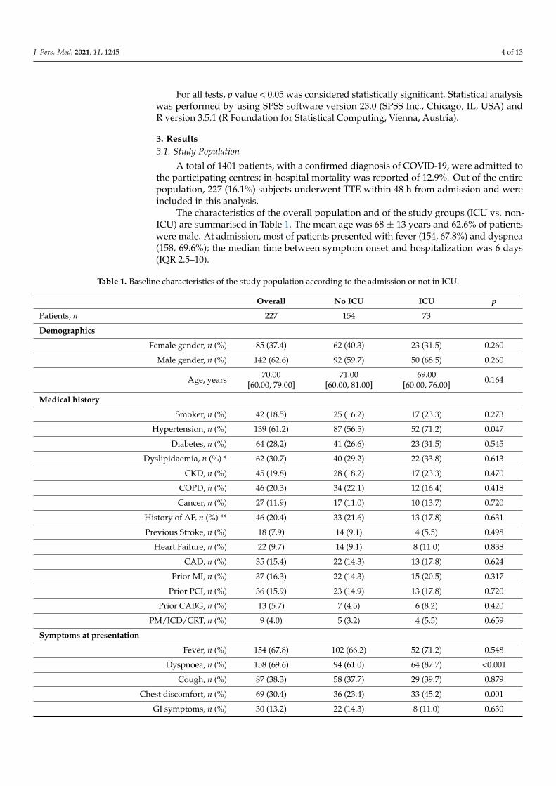
The characteristics of the overall population and of the study groups (ICU vs. non-ICU) are summarised in Table 1. The mean age was 68 ± 13 years and 62.6% of patientswere male. At admission, most of patients presented with fever (154, 67.8%) and dyspnea(158, 69.6%); the median time between symptom onset and hospitalization was 6 days(IQR 2.5–10).
Table 1. Baseline characteristics of the study population according to the admission or not in ICU.
Overall No ICU ICU p
Patients, n 227 154 73
Demographics
Female gender, n (%) 85 (37.4) 62 (40.3) 23 (31.5) 0.260
Male gender, n (%) 142 (62.6) 92 (59.7) 50 (68.5) 0.260
Age, years 70.00[60.00, 79.00]
71.00[60.00, 81.00]
69.00[60.00, 76.00] 0.164
Medical history
Smoker, n (%) 42 (18.5) 25 (16.2) 17 (23.3) 0.273
Hypertension, n (%) 139 (61.2) 87 (56.5) 52 (71.2) 0.047
Diabetes, n (%) 64 (28.2) 41 (26.6) 23 (31.5) 0.545
Dyslipidaemia, n (%) * 62 (30.7) 40 (29.2) 22 (33.8) 0.613
CKD, n (%) 45 (19.8) 28 (18.2) 17 (23.3) 0.470
COPD, n (%) 46 (20.3) 34 (22.1) 12 (16.4) 0.418
Cancer, n (%) 27 (11.9) 17 (11.0) 10 (13.7) 0.720
History of AF, n (%) ** 46 (20.4) 33 (21.6) 13 (17.8) 0.631
Previous Stroke, n (%) 18 (7.9) 14 (9.1) 4 (5.5) 0.498
Heart Failure, n (%) 22 (9.7) 14 (9.1) 8 (11.0) 0.838
CAD, n (%) 35 (15.4) 22 (14.3) 13 (17.8) 0.624
Prior MI, n (%) 37 (16.3) 22 (14.3) 15 (20.5) 0.317
Prior PCI, n (%) 36 (15.9) 23 (14.9) 13 (17.8) 0.720
Prior CABG, n (%) 13 (5.7) 7 (4.5) 6 (8.2) 0.420
PM/ICD/CRT, n (%) 9 (4.0) 5 (3.2) 4 (5.5) 0.659
Symptoms at presentation
Fever, n (%) 154 (67.8) 102 (66.2) 52 (71.2) 0.548
Dyspnoea, n (%) 158 (69.6) 94 (61.0) 64 (87.7) <0.001
Cough, n (%) 87 (38.3) 58 (37.7) 29 (39.7) 0.879
Chest discomfort, n (%) 69 (30.4) 36 (23.4) 33 (45.2) 0.001
GI symptoms, n (%) 30 (13.2) 22 (14.3) 8 (11.0) 0.630
J. Pers. Med. 2021, 11, 1245 5 of 13
Table 1. Cont.
Overall No ICU ICU p
Symptoms onset tohospitalization, days 6.00[2.50, 10.00]
6.00[3.00, 10.00]
4.00[1.00, 7.00] 0.006
Pharmacological therapy at admission
ACEi or ARB, n (%) 99 (43.6) 56 (36.4) 43 (58.9) 0.002
Betablocker, n (%) 59 (26.0) 41 (26.6) 18 (24.7) 0.878
Diuretic, n (%) 47 (20.7) 26 (16.9) 21 (28.8) 0.059
P2Y12 inhibitor, n (%) 21 (9.3) 14 (9.1) 7 (9.6) 1.000
ASA, n (%) 67 (29.5) 44 (28.6) 23 (31.5) 0.766
Statin, n (%) 71 (31.3) 43 (27.9) 28 (38.4) 0.153
Insulin, n (%) 32 (14.1) 19 (12.3) 13 (17.8) 0.367
VKA or NOAC, n (%) 42 (18.5) 29 (18.8) 13 (17.8) 0.998
Serum biomarkers
Troponin hs, n 99thpercentile; peak ∞ 24.40[2.78, 225.00]
22.10[5.15, 215.50]
43.70[1.80, 248.00] 0.718
D-dimer, peak; ng/mL ¥ 625.00[100.75, 1994.00]
564.00[176.00, 1397.50]
1363.50[13.13, 2735.00] 0.173
Echocardiographic data
LVEF, % 55.00[50.00, 59.00]
56.00[50.00, 60.00]
51.00[45.00, 55.00] <0.001
LVEDV, mL 103.00[89.00, 120.00]
101.00[88.00, 119.00]
103.00[90.50, 130.25] 0.271
LVESV, mL 47.00[39.00, 58.10]
46.00[38.00, 53.00]
50.00[43.00, 60.60] 0.007
TAPSE, mm 21.00[18.00, 23.00]
21.00[19.00, 24.00]
20.00[16.00, 21.00] <0.001
PASP, mmHg 33.00[30.00, 40.00]
32.00[29.00, 40.00]
36.00[30.00, 45.00] 0.002
Moderate or severe MR, n (%) 36 (15.9) 23 (14.9) 13 (17.8) 0.720
Moderate or severe TR, n (%) 48 (21.1) 23 (14.9) 25 (34.2) 0.002
SARS-COV 2 therapies
Glucocorticoid, n (%) 102 (44.9) 63 (40.9) 39 (53.4) 0.104
Antiviral, n (%) 119 (52.4) 66 (42.9) 53 (72.6) <0.001
Antibiotics, n (%) 167 (73.6) 103 (66.9) 64 (87.7) 0.002
Tocilizumab, n (%) # 1 (1.0) 1 (1.4) 0 (0.0) 1.000
Hydroxychloroquine, n (%) 181 (79.7) 116 (75.3) 65 (89.0) 0.026
UFH or LMWH, n (%) § 184 (81.8) 117 (77.0) 67 (91.8) 0.012
In hospital data and complications
IMV, n (%) 68 (30.0) 7 (4.5) 61 (83.6) <0.001
NIV, n (%) 100 (44.1) 55 (35.7) 45 (61.6) <0.001
ARDS, n (%) 107 (47.1) 47 (30.5) 60 (82.2) <0.001
Acute cardiac injury, n (%) 69 (30.4) 35 (22.7) 34 (46.6) <0.001
Pulmonary embolism, n (%) 32 (14.1) 21 (13.6) 11 (15.1) 0.932
J. Pers. Med. 2021, 11, 1245 6 of 13
Table 1. Cont.
Overall No ICU ICU p
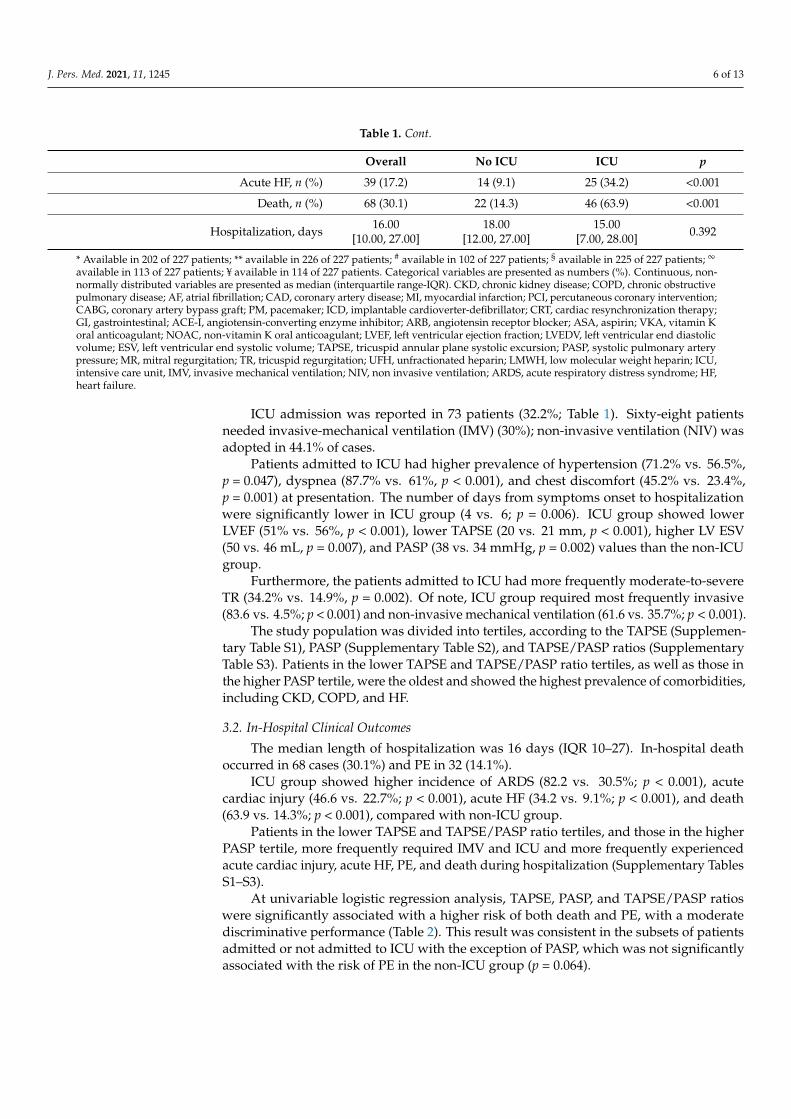
Acute HF, n (%) 39 (17.2) 14 (9.1) 25 (34.2) <0.001
Death, n (%) 68 (30.1) 22 (14.3) 46 (63.9) <0.001
Hospitalization, days 16.00[10.00, 27.00]
18.00[12.00, 27.00]
15.00[7.00, 28.00] 0.392
* Available in 202 of 227 patients; ** available in 226 of 227 patients; # available in 102 of 227 patients; § available in 225 of 227 patients; ∞
available in 113 of 227 patients; ¥ available in 114 of 227 patients. Categorical variables are presented as numbers (%). Continuous, non-normally distributed variables are presented as median (interquartile range-IQR). CKD, chronic kidney disease; COPD, chronic obstructivepulmonary disease; AF, atrial fibrillation; CAD, coronary artery disease; MI, myocardial infarction; PCI, percutaneous coronary intervention;CABG, coronary artery bypass graft; PM, pacemaker; ICD, implantable cardioverter-defibrillator; CRT, cardiac resynchronization therapy;GI, gastrointestinal; ACE-I, angiotensin-converting enzyme inhibitor; ARB, angiotensin receptor blocker; ASA, aspirin; VKA, vitamin Koral anticoagulant; NOAC, non-vitamin K oral anticoagulant; LVEF, left ventricular ejection fraction; LVEDV, left ventricular end diastolicvolume; ESV, left ventricular end systolic volume; TAPSE, tricuspid annular plane systolic excursion; PASP, systolic pulmonary arterypressure; MR, mitral regurgitation; TR, tricuspid regurgitation; UFH, unfractionated heparin; LMWH, low molecular weight heparin; ICU,intensive care unit, IMV, invasive mechanical ventilation; NIV, non invasive ventilation; ARDS, acute respiratory distress syndrome; HF,heart failure.
ICU admission was reported in 73 patients (32.2%; Table 1). Sixty-eight patientsneeded invasive-mechanical ventilation (IMV) (30%); non-invasive ventilation (NIV) wasadopted in 44.1% of cases.
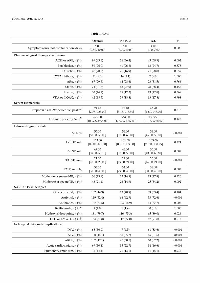
Patients admitted to ICU had higher prevalence of hypertension (71.2% vs. 56.5%,p = 0.047), dyspnea (87.7% vs. 61%, p < 0.001), and chest discomfort (45.2% vs. 23.4%,p = 0.001) at presentation. The number of days from symptoms onset to hospitalizationwere significantly lower in ICU group (4 vs. 6; p = 0.006). ICU group showed lowerLVEF (51% vs. 56%, p < 0.001), lower TAPSE (20 vs. 21 mm, p < 0.001), higher LV ESV(50 vs. 46 mL, p = 0.007), and PASP (38 vs. 34 mmHg, p = 0.002) values than the non-ICUgroup.
Furthermore, the patients admitted to ICU had more frequently moderate-to-severeTR (34.2% vs. 14.9%, p = 0.002). Of note, ICU group required most frequently invasive(83.6 vs. 4.5%; p < 0.001) and non-invasive mechanical ventilation (61.6 vs. 35.7%; p < 0.001).
The study population was divided into tertiles, according to the TAPSE (Supplemen-tary Table S1), PASP (Supplementary Table S2), and TAPSE/PASP ratios (SupplementaryTable S3). Patients in the lower TAPSE and TAPSE/PASP ratio tertiles, as well as those inthe higher PASP tertile, were the oldest and showed the highest prevalence of comorbidities,including CKD, COPD, and HF.
3.2. In-Hospital Clinical Outcomes
The median length of hospitalization was 16 days (IQR 10–27). In-hospital deathoccurred in 68 cases (30.1%) and PE in 32 (14.1%).
ICU group showed higher incidence of ARDS (82.2 vs. 30.5%; p < 0.001), acutecardiac injury (46.6 vs. 22.7%; p < 0.001), acute HF (34.2 vs. 9.1%; p < 0.001), and death(63.9 vs. 14.3%; p < 0.001), compared with non-ICU group.
Patients in the lower TAPSE and TAPSE/PASP ratio tertiles, and those in the higherPASP tertile, more frequently required IMV and ICU and more frequently experiencedacute cardiac injury, acute HF, PE, and death during hospitalization (Supplementary TablesS1–S3).
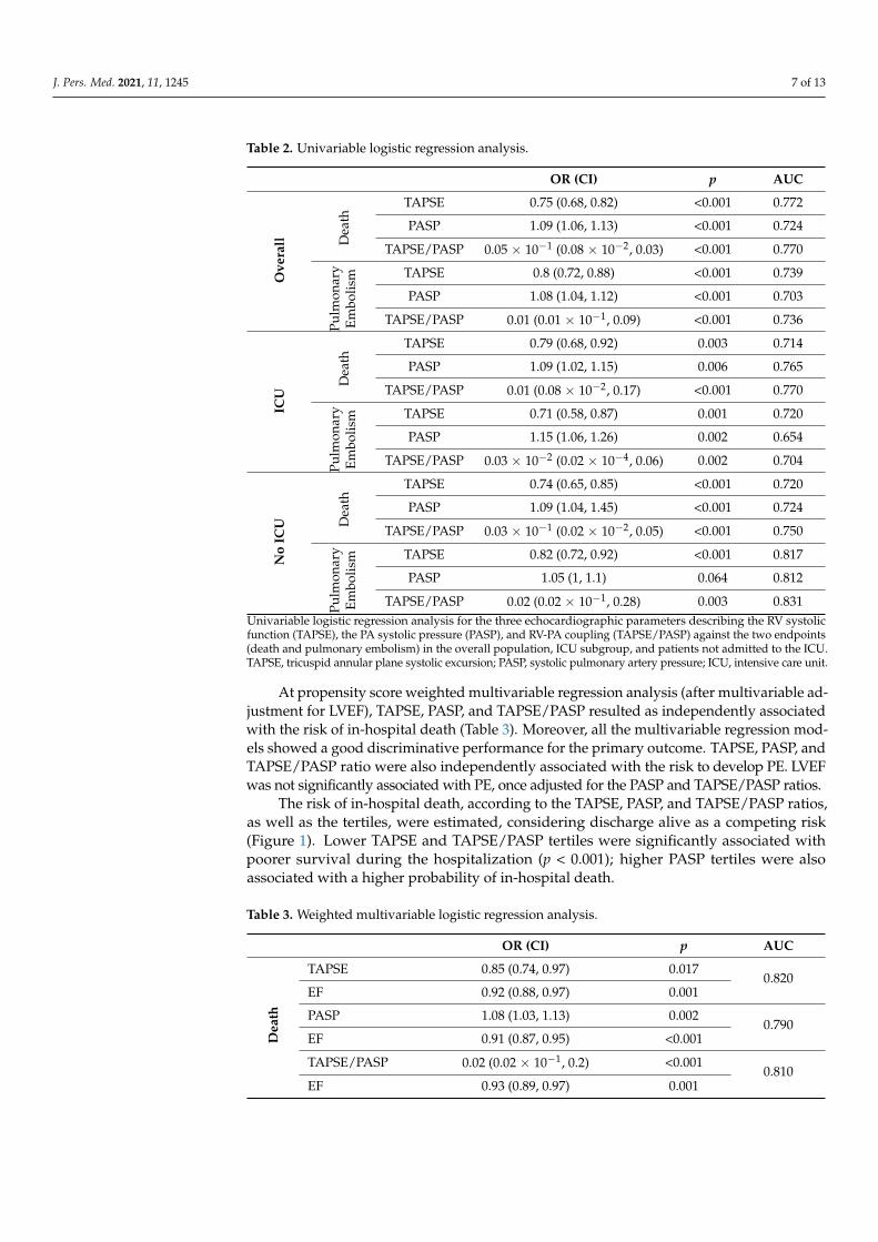
At univariable logistic regression analysis, TAPSE, PASP, and TAPSE/PASP ratioswere significantly associated with a higher risk of both death and PE, with a moderatediscriminative performance (Table 2). This result was consistent in the subsets of patientsadmitted or not admitted to ICU with the exception of PASP, which was not significantlyassociated with the risk of PE in the non-ICU group (p = 0.064).
J. Pers. Med. 2021, 11, 1245 7 of 13
Table 2. Univariable logistic regression analysis.
OR (CI) p AUC
Ove
rall D
eath
TAPSE 0.75 (0.68, 0.82) <0.001 0.772
PASP 1.09 (1.06, 1.13) <0.001 0.724
TAPSE/PASP 0.05 × 10⁻¹ (0.08 × 10⁻², 0.03) <0.001 0.770
Pulm
onar
yEm
bolis
m TAPSE 0.8 (0.72, 0.88) <0.001 0.739
PASP 1.08 (1.04, 1.12) <0.001 0.703
TAPSE/PASP 0.01 (0.01 × 10⁻¹, 0.09) <0.001 0.736
ICU
Dea
thTAPSE 0.79 (0.68, 0.92) 0.003 0.714
PASP 1.09 (1.02, 1.15) 0.006 0.765
TAPSE/PASP 0.01 (0.08 × 10⁻², 0.17) <0.001 0.770
Pulm
onar
yEm
bolis
m TAPSE 0.71 (0.58, 0.87) 0.001 0.720
PASP 1.15 (1.06, 1.26) 0.002 0.654
TAPSE/PASP 0.03 × 10⁻² (0.02 × 10⁻⁴, 0.06) 0.002 0.704
No
ICU D
eath
TAPSE 0.74 (0.65, 0.85) <0.001 0.720
PASP 1.09 (1.04, 1.45) <0.001 0.724
TAPSE/PASP 0.03 × 10⁻¹ (0.02 × 10⁻², 0.05) <0.001 0.750
Pulm
onar
yEm
bolis
m TAPSE 0.82 (0.72, 0.92) <0.001 0.817
PASP 1.05 (1, 1.1) 0.064 0.812
TAPSE/PASP 0.02 (0.02 × 10⁻¹, 0.28) 0.003 0.831Univariable logistic regression analysis for the three echocardiographic parameters describing the RV systolicfunction (TAPSE), the PA systolic pressure (PASP), and RV-PA coupling (TAPSE/PASP) against the two endpoints(death and pulmonary embolism) in the overall population, ICU subgroup, and patients not admitted to the ICU.TAPSE, tricuspid annular plane systolic excursion; PASP, systolic pulmonary artery pressure; ICU, intensive care unit.
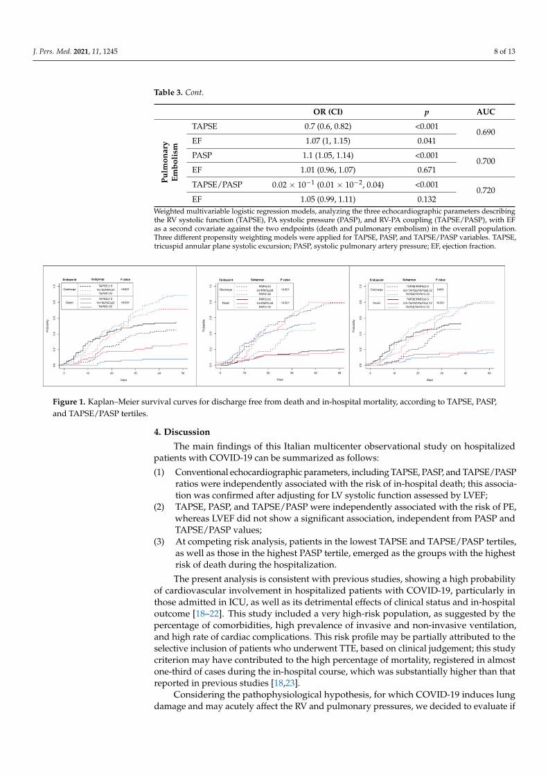
At propensity score weighted multivariable regression analysis (after multivariable ad-justment for LVEF), TAPSE, PASP, and TAPSE/PASP resulted as independently associatedwith the risk of in-hospital death (Table 3). Moreover, all the multivariable regression mod-els showed a good discriminative performance for the primary outcome. TAPSE, PASP, andTAPSE/PASP ratio were also independently associated with the risk to develop PE. LVEFwas not significantly associated with PE, once adjusted for the PASP and TAPSE/PASP ratios.
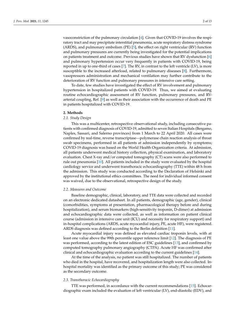
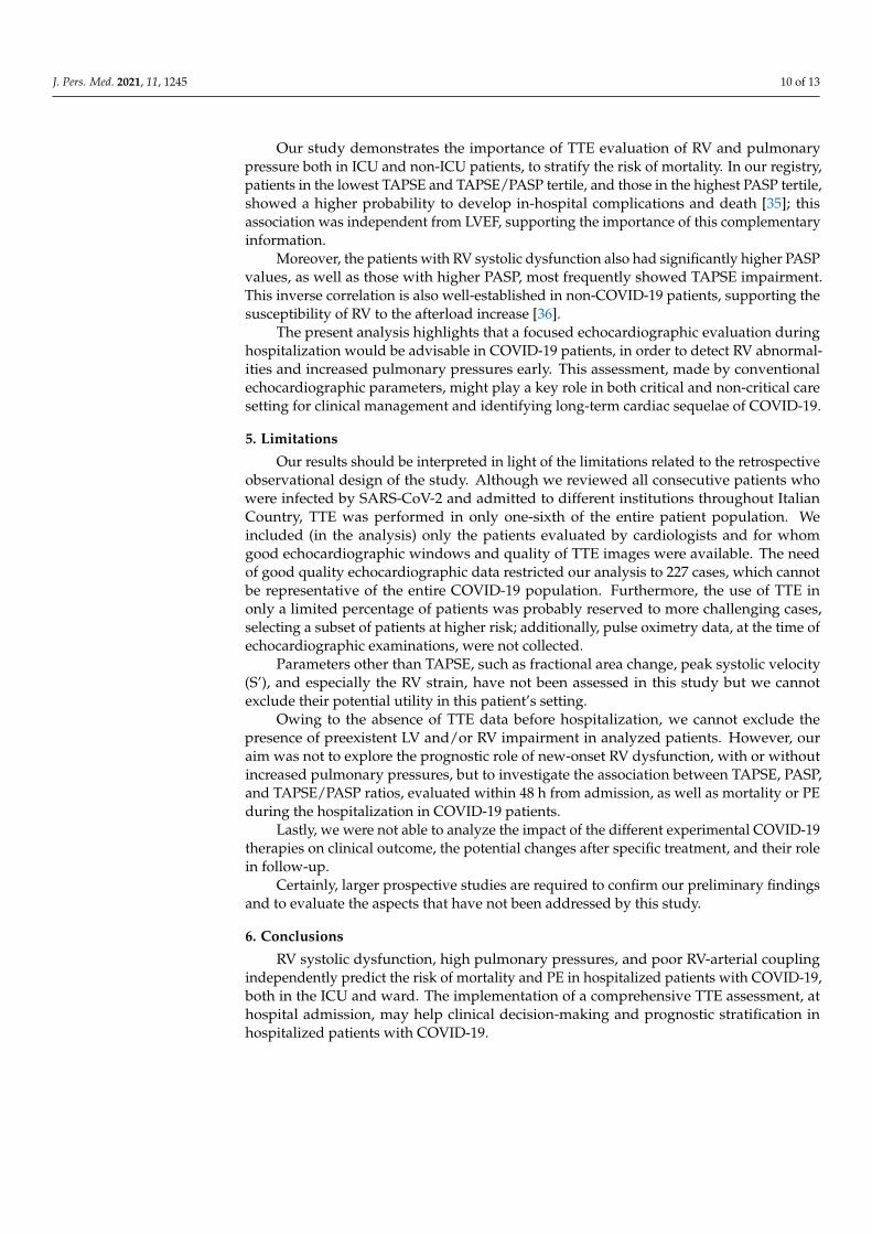
The risk of in-hospital death, according to the TAPSE, PASP, and TAPSE/PASP ratios,as well as the tertiles, were estimated, considering discharge alive as a competing risk(Figure 1). Lower TAPSE and TAPSE/PASP tertiles were significantly associated withpoorer survival during the hospitalization (p < 0.001); higher PASP tertiles were alsoassociated with a higher probability of in-hospital death.
Table 3. Weighted multivariable logistic regression analysis.
OR (CI) p AUC
Dea
th
TAPSE 0.85 (0.74, 0.97) 0.0170.820
EF 0.92 (0.88, 0.97) 0.001
PASP 1.08 (1.03, 1.13) 0.0020.790
EF 0.91 (0.87, 0.95) <0.001
TAPSE/PASP 0.02 (0.02 × 10⁻¹, 0.2) <0.0010.810
EF 0.93 (0.89, 0.97) 0.001
J. Pers. Med. 2021, 11, 1245 8 of 13
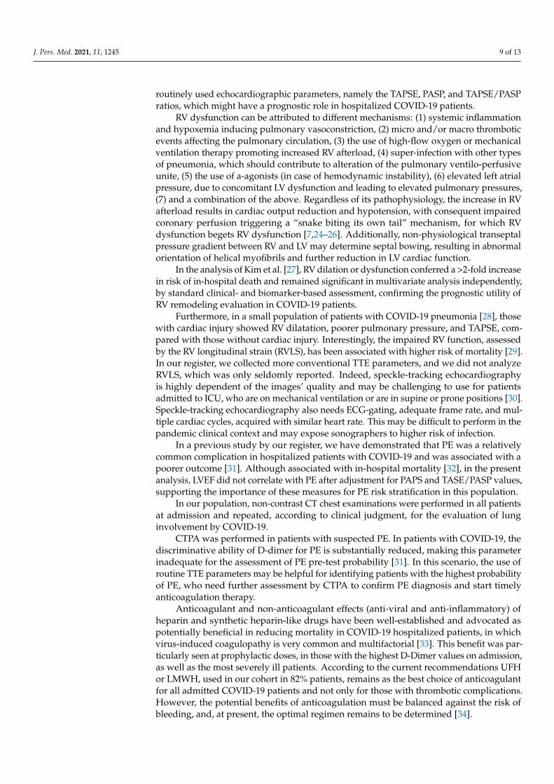
Table 3. Cont.
OR (CI) p AUC
Pulm
onar
yEm
boli
sm
TAPSE 0.7 (0.6, 0.82) <0.0010.690
EF 1.07 (1, 1.15) 0.041
PASP 1.1 (1.05, 1.14) <0.0010.700
EF 1.01 (0.96, 1.07) 0.671
TAPSE/PASP 0.02 × 10⁻¹ (0.01 × 10⁻², 0.04) <0.0010.720
EF 1.05 (0.99, 1.11) 0.132Weighted multivariable logistic regression models, analyzing the three echocardiographic parameters describingthe RV systolic function (TAPSE), PA systolic pressure (PASP), and RV-PA coupling (TAPSE/PASP), with EFas a second covariate against the two endpoints (death and pulmonary embolism) in the overall population.Three different propensity weighting models were applied for TAPSE, PASP, and TAPSE/PASP variables. TAPSE,tricuspid annular plane systolic excursion; PASP, systolic pulmonary artery pressure; EF, ejection fraction.
J. Pers. Med. 2021, 11, x FOR PEER REVIEW 8 of 12
The risk of in‐hospital death, according to the TAPSE, PASP, and TAPSE/PASP ratios,
as well as the tertiles, were estimated, considering discharge alive as a competing risk
(Figure 1). Lower TAPSE and TAPSE/PASP tertiles were significantly associated with
poorer survival during the hospitalization (p < 0.001); higher PASP tertiles were also as‐
sociated with a higher probability of in‐hospital death.
Figure 1. Kaplan–Meier survival curves for discharge free from death and in‐hospital mortality, according to TAPSE,
PASP, and TAPSE/PASP tertiles.
4. Discussion
The main findings of this Italian multicenter observational study on hospitalized pa‐
tients with COVID‐19 can be summarized as follows:
(1) Conventional echocardiographic parameters, including TAPSE, PASP, and
TAPSE/PASP ratios were independently associated with the risk of in‐hospital death;
this association was confirmed after adjusting for LV systolic function assessed by
LVEF;
(2) TAPSE, PASP, and TAPSE/PASP were independently associated with the risk of PE,
whereas LVEF did not show a significant association, independent from PASP and
TAPSE/PASP values;
(3) At competing risk analysis, patients in the lowest TAPSE and TAPSE/PASP tertiles,
as well as those in the highest PASP tertile, emerged as the groups with the highest
risk of death during the hospitalization.
The present analysis is consistent with previous studies, showing a high probability
of cardiovascular involvement in hospitalized patients with COVID‐19, particularly in
those admitted in ICU, as well as its detrimental effects of clinical status and in‐hospital
outcome [18–22]. This study included a very high‐risk population, as suggested by the
percentage of comorbidities, high prevalence of invasive and non‐invasive ventilation,
and high rate of cardiac complications. This risk profile may be partially attributed to the
selective inclusion of patients who underwent TTE, based on clinical judgement; this
study criterion may have contributed to the high percentage of mortality, registered in
almost one‐third of cases during the in‐hospital course, which was substantially higher
than that reported in previous studies [18,23].
Considering the pathophysiological hypothesis, for which COVID‐19 induces lung
damage and may acutely affect the RV and pulmonary pressures, we decided to evaluate
if routinely used echocardiographic parameters, namely the TAPSE, PASP, and
TAPSE/PASP ratios, which might have a prognostic role in hospitalized COVID‐19 pa‐
tients.
RV dysfunction can be attributed to different mechanisms: (1) systemic inflammation
and hypoxemia inducing pulmonary vasoconstriction, (2) micro and/or macro thrombotic
events affecting the pulmonary circulation, (3) the use of high‐flow oxygen or mechanical
ventilation therapy promoting increased RV afterload, (4) super‐infection with other types
of pneumonia, which should contribute to alteration of the pulmonary ventilo‐perfusive
Figure 1. Kaplan–Meier survival curves for discharge free from death and in-hospital mortality, according to TAPSE, PASP,and TAPSE/PASP tertiles.
4. Discussion
The main findings of this Italian multicenter observational study on hospitalizedpatients with COVID-19 can be summarized as follows:
(1) Conventional echocardiographic parameters, including TAPSE, PASP, and TAPSE/PASPratios were independently associated with the risk of in-hospital death; this associa-tion was confirmed after adjusting for LV systolic function assessed by LVEF;
(2) TAPSE, PASP, and TAPSE/PASP were independently associated with the risk of PE,whereas LVEF did not show a significant association, independent from PASP andTAPSE/PASP values;
(3) At competing risk analysis, patients in the lowest TAPSE and TAPSE/PASP tertiles,as well as those in the highest PASP tertile, emerged as the groups with the highestrisk of death during the hospitalization.
The present analysis is consistent with previous studies, showing a high probabilityof cardiovascular involvement in hospitalized patients with COVID-19, particularly inthose admitted in ICU, as well as its detrimental effects of clinical status and in-hospitaloutcome [18–22]. This study included a very high-risk population, as suggested by thepercentage of comorbidities, high prevalence of invasive and non-invasive ventilation,and high rate of cardiac complications. This risk profile may be partially attributed to theselective inclusion of patients who underwent TTE, based on clinical judgement; this studycriterion may have contributed to the high percentage of mortality, registered in almostone-third of cases during the in-hospital course, which was substantially higher than thatreported in previous studies [18,23].
Considering the pathophysiological hypothesis, for which COVID-19 induces lungdamage and may acutely affect the RV and pulmonary pressures, we decided to evaluate if
J. Pers. Med. 2021, 11, 1245 9 of 13
routinely used echocardiographic parameters, namely the TAPSE, PASP, and TAPSE/PASPratios, which might have a prognostic role in hospitalized COVID-19 patients.
RV dysfunction can be attributed to different mechanisms: (1) systemic inflammationand hypoxemia inducing pulmonary vasoconstriction, (2) micro and/or macro thromboticevents affecting the pulmonary circulation, (3) the use of high-flow oxygen or mechanicalventilation therapy promoting increased RV afterload, (4) super-infection with other typesof pneumonia, which should contribute to alteration of the pulmonary ventilo-perfusiveunite, (5) the use of a-agonists (in case of hemodynamic instability), (6) elevated left atrialpressure, due to concomitant LV dysfunction and leading to elevated pulmonary pressures,(7) and a combination of the above. Regardless of its pathophysiology, the increase in RVafterload results in cardiac output reduction and hypotension, with consequent impairedcoronary perfusion triggering a “snake biting its own tail” mechanism, for which RVdysfunction begets RV dysfunction [7,24–26]. Additionally, non-physiological transeptalpressure gradient between RV and LV may determine septal bowing, resulting in abnormalorientation of helical myofibrils and further reduction in LV cardiac function.
In the analysis of Kim et al. [27], RV dilation or dysfunction conferred a >2-fold increasein risk of in-hospital death and remained significant in multivariate analysis independently,by standard clinical- and biomarker-based assessment, confirming the prognostic utility ofRV remodeling evaluation in COVID-19 patients.
Furthermore, in a small population of patients with COVID-19 pneumonia [28], thosewith cardiac injury showed RV dilatation, poorer pulmonary pressure, and TAPSE, com-pared with those without cardiac injury. Interestingly, the impaired RV function, assessedby the RV longitudinal strain (RVLS), has been associated with higher risk of mortality [29].In our register, we collected more conventional TTE parameters, and we did not analyzeRVLS, which was only seldomly reported. Indeed, speckle-tracking echocardiographyis highly dependent of the images’ quality and may be challenging to use for patientsadmitted to ICU, who are on mechanical ventilation or are in supine or prone positions [30].Speckle-tracking echocardiography also needs ECG-gating, adequate frame rate, and mul-tiple cardiac cycles, acquired with similar heart rate. This may be difficult to perform in thepandemic clinical context and may expose sonographers to higher risk of infection.
In a previous study by our register, we have demonstrated that PE was a relativelycommon complication in hospitalized patients with COVID-19 and was associated with apoorer outcome [31]. Although associated with in-hospital mortality [32], in the presentanalysis, LVEF did not correlate with PE after adjustment for PAPS and TASE/PASP values,supporting the importance of these measures for PE risk stratification in this population.
In our population, non-contrast CT chest examinations were performed in all patientsat admission and repeated, according to clinical judgment, for the evaluation of lunginvolvement by COVID-19.
CTPA was performed in patients with suspected PE. In patients with COVID-19, thediscriminative ability of D-dimer for PE is substantially reduced, making this parameterinadequate for the assessment of PE pre-test probability [31]. In this scenario, the use ofroutine TTE parameters may be helpful for identifying patients with the highest probabilityof PE, who need further assessment by CTPA to confirm PE diagnosis and start timelyanticoagulation therapy.
Anticoagulant and non-anticoagulant effects (anti-viral and anti-inflammatory) ofheparin and synthetic heparin-like drugs have been well-established and advocated aspotentially beneficial in reducing mortality in COVID-19 hospitalized patients, in whichvirus-induced coagulopathy is very common and multifactorial [33]. This benefit was par-ticularly seen at prophylactic doses, in those with the highest D-Dimer values on admission,as well as the most severely ill patients. According to the current recommendations UFHor LMWH, used in our cohort in 82% patients, remains as the best choice of anticoagulantfor all admitted COVID-19 patients and not only for those with thrombotic complications.However, the potential benefits of anticoagulation must be balanced against the risk ofbleeding, and, at present, the optimal regimen remains to be determined [34].
J. Pers. Med. 2021, 11, 1245 10 of 13
Our study demonstrates the importance of TTE evaluation of RV and pulmonarypressure both in ICU and non-ICU patients, to stratify the risk of mortality. In our registry,patients in the lowest TAPSE and TAPSE/PASP tertile, and those in the highest PASP tertile,showed a higher probability to develop in-hospital complications and death [35]; thisassociation was independent from LVEF, supporting the importance of this complementaryinformation.
Moreover, the patients with RV systolic dysfunction also had significantly higher PASPvalues, as well as those with higher PASP, most frequently showed TAPSE impairment.This inverse correlation is also well-established in non-COVID-19 patients, supporting thesusceptibility of RV to the afterload increase [36].
The present analysis highlights that a focused echocardiographic evaluation duringhospitalization would be advisable in COVID-19 patients, in order to detect RV abnormal-ities and increased pulmonary pressures early. This assessment, made by conventionalechocardiographic parameters, might play a key role in both critical and non-critical caresetting for clinical management and identifying long-term cardiac sequelae of COVID-19.
5. Limitations
Our results should be interpreted in light of the limitations related to the retrospectiveobservational design of the study. Although we reviewed all consecutive patients whowere infected by SARS-CoV-2 and admitted to different institutions throughout ItalianCountry, TTE was performed in only one-sixth of the entire patient population. Weincluded (in the analysis) only the patients evaluated by cardiologists and for whomgood echocardiographic windows and quality of TTE images were available. The needof good quality echocardiographic data restricted our analysis to 227 cases, which cannotbe representative of the entire COVID-19 population. Furthermore, the use of TTE inonly a limited percentage of patients was probably reserved to more challenging cases,selecting a subset of patients at higher risk; additionally, pulse oximetry data, at the time ofechocardiographic examinations, were not collected.
Parameters other than TAPSE, such as fractional area change, peak systolic velocity(S’), and especially the RV strain, have not been assessed in this study but we cannotexclude their potential utility in this patient’s setting.
Owing to the absence of TTE data before hospitalization, we cannot exclude thepresence of preexistent LV and/or RV impairment in analyzed patients. However, ouraim was not to explore the prognostic role of new-onset RV dysfunction, with or withoutincreased pulmonary pressures, but to investigate the association between TAPSE, PASP,and TAPSE/PASP ratios, evaluated within 48 h from admission, as well as mortality or PEduring the hospitalization in COVID-19 patients.
Lastly, we were not able to analyze the impact of the different experimental COVID-19therapies on clinical outcome, the potential changes after specific treatment, and their rolein follow-up.
Certainly, larger prospective studies are required to confirm our preliminary findingsand to evaluate the aspects that have not been addressed by this study.
6. Conclusions
RV systolic dysfunction, high pulmonary pressures, and poor RV-arterial couplingindependently predict the risk of mortality and PE in hospitalized patients with COVID-19,both in the ICU and ward. The implementation of a comprehensive TTE assessment, athospital admission, may help clinical decision-making and prognostic stratification inhospitalized patients with COVID-19.
J. Pers. Med. 2021, 11, 1245 11 of 13
Supplementary Materials: The following are available online at https://www.mdpi.com/article/10.3390/jpm11121245/s1, Table S1: Characteristics of the study population stratified by TAPSE tertiles,Table S2: Characteristics of the study population stratified by PASP tertiles, Table S3: Characteristicsof the study population stratified by TAPSE/PASP tertiles.
Author Contributions: Conceptualization, M.V.P. and A.S.; methodology, A.S.; software, M.D.M.;validation, G.G., V.R. and F.S.; formal analysis, M.D.M.; investigation, M.B.; resources, F.S.; datacuration, F.S. and R.C.; writing—original draft preparation, M.V.P.; writing—review and editing,M.B.; visualization, B.R. and C.A.; supervision, G.G., G.P. and C.V. All authors have read and agreedto the published version of the manuscript.
Funding: This research received no external funding.
Institutional Review Board Statement: Ethical review and approval were waived for this study, dueto observational design of the study.
Informed Consent Statement: Informed consent was obtained from all subjects involved in thestudy.
Data Availability Statement: Not applicable.
Conflicts of Interest: No conflict of interest or any financial support to declare.
References1. Hui, D.S.; Azhar, E.I.; Madani, T.A.; Ntoumi, F.; Kock, R.; Dar, O.; Ippolito, G.; McHugh, T.D.; Memish, Z.A.; Drosten, C.; et al.
The continuing 2019-nCoV epidemic threat of novel coronaviruses to global health—The latest 2019 novel coronavirus outbreakin Wuhan, China. Int. J. Infect. Dis. 2020, 91, 264–266. [CrossRef] [PubMed]
2. Inciardi, R.M.; Lupi, L.; Zaccone, G.; Italia, L.; Raffo, M.; Tomasoni, D.; Cani, D.S.; Cerini, M.; Farina, D.; Gavazzi, E.; et al. CardiacInvolvement in a Patient With Coronavirus Disease 2019 (COVID-19). JAMA Cardiol. 2020, 5, 819–824. [CrossRef] [PubMed]
3. Esposito, L.; Cancro, F.P.; Silverio, A.; Di Maio, M.; Iannece, P.; Damato, A.; Alfano, C.; De Luca, G.; Vecchione, C.; Galasso, G.COVID-19 and Acute Coronary Syndromes: From Pathophysiology to Clinical Perspectives. Oxid. Med. Cell. Longev. 2021, 2021,4936571. [CrossRef] [PubMed]
4. Madjid, M.; Safavi-Naeini, P.; Solomon, S.D.; Vardeny, O. Potential Effects of Coronaviruses on the Cardiovascular System: AReview. JAMA Cardiol. 2020, 5, 831–840. [CrossRef] [PubMed]
5. Yang, X.; Yu, Y.; Xu, J.; Shu, H.; Xia, J.; Liu, H.; Wu, Y.; Zhang, L.; Yu, Z.; Fang, M.; et al. Clinical course and outcomes of criticallyill patients with SARS-CoV-2 pneumonia in Wuhan, China: A single-centered, retrospective, observational study. Lancet Respir.Med. 2020, 8, 475–481. [CrossRef]
6. Argulian, E.; Sud, K.; Vogel, B.; Bohra, C.; Garg, V.P.; Talebi, S.; Lerakis, S.; Narula, J. Right Ventricular Dilation in HospitalizedPatients With COVID-19 Infection. JACC Cardiovasc. Imaging 2020, 13, 2459–2461. [CrossRef] [PubMed]
7. Szekely, Y.; Lichter, Y.; Taieb, P.; Banai, A.; Hochstadt, A.; Merdler, I.; Gal Oz, A.; Rothschild, E.; Baruch, G.; Peri, Y.; et al. Spectrumof Cardiac Manifestations in COVID-19: A Systematic Echocardiographic Study. Circulation 2020, 142, 342–353. [CrossRef][PubMed]
8. Ullah, W.; Saeed, R.; Sarwar, U.; Patel, R.; Fischman, D.L. COVID-19 Complicated by Acute Pulmonary Embolism and Right-SidedHeart Failure. JACC Case Rep. 2020, 2, 1379–1382. [CrossRef]
9. Tello, K.; Wan, J.; Dalmer, A.; Vanderpool, R.; Ghofrani, H.A.; Naeije, R.; Roller, F.; Mohajerani, E.; Seeger, W.; Herberg, U.; et al.Validation of the Tricuspid Annular Plane Systolic Excursion/Systolic Pulmonary Artery Pressure Ratio for the Assessment ofRight Ventricular-Arterial Coupling in Severe Pulmonary Hypertension. Circ. Cardiovasc. Imaging 2019, 12, e009047. [CrossRef]
10. Citro, R.; Pontone, G.; Bellino, M.; Silverio, A.; Iuliano, G.; Baggiano, A.; Manka, R.; Iesu, S.; Vecchione, C.; Asch, F.M.; et al. Roleof multimodality imaging in evaluation of cardiovascular involvement in COVID-19. Trends Cardiovasc. Med. 2021, 31, 8–16.[CrossRef] [PubMed]
11. Ranieri, V.M.; Rubenfeld, G.D.; Thompson, B.T.; Ferguson, N.D.; Caldwell, E.; Fan, E.; Camporota, L.; Slutsky, A.S. Acuterespiratory distress syndrome: The Berlin Definition. JAMA 2012, 307, 2526–2533. [CrossRef] [PubMed]
12. Thygesen, K.; Alpert, J.S.; Jaffe, A.S.; Chaitman, B.R.; Bax, J.J.; Morrow, D.A.; White, H.D. Fourth Universal Definition ofMyocardial Infarction (2018). J. Am. Coll. Cardiol. 2018, 72, 2231–2264. [CrossRef]
13. Konstantinides, S.V.; Meyer, G.; Becattini, C.; Bueno, H.; Geersing, G.J.; Harjola, V.P.; Huisman, M.V.; Humbert, M.; Jennings,C.S.; Jiménez, D.; et al. 2019 ESC Guidelines for the diagnosis and management of acute pulmonary embolism developed incollaboration with the European Respiratory Society (ERS). Eur. Heart J. 2020, 41, 543–603. [CrossRef] [PubMed]
14. McDonagh, T.A.; Metra, M.; Adamo, M.; Gardner, R.S.; Baumbach, A.; Böhm, M.; Burri, H.; Butler, J.; Celutkiene, J.; Chioncel, O.;et al. 2021 ESC Guidelines for the diagnosis and treatment of acute and chronic heart failure. Eur. Heart J. 2021, 42, 3599–3726.[CrossRef] [PubMed]
J. Pers. Med. 2021, 11, 1245 12 of 13
15. Lang, R.M.; Badano, L.P.; Mor-Avi, V.; Afilalo, J.; Armstrong, A.; Ernande, L.; Flachskampf, F.A.; Foster, E.; Goldstein, S.A.;Kuznetsova, T.; et al. Recommendations for cardiac chamber quantification by echocardiography in adults: An update fromthe American Society of Echocardiography and the European Association of Cardiovascular Imaging. Eur. Heart J. Cardiovasc.Imaging 2015, 16, 233–270. [CrossRef] [PubMed]
16. Zoghbi, W.A.; Adams, D.; Bonow, R.O.; Enriquez-Sarano, M.; Foster, E.; Grayburn, P.A.; Hahn, R.T.; Han, Y.; Hung, J.; Lang, R.M.;et al. Recommendations for Noninvasive Evaluation of Native Valvular Regurgitation: A Report from the American Society ofEchocardiography Developed in Collaboration with the Society for Cardiovascular Magnetic Resonance. J. Am. Soc. Echocardiogr.2017, 30, 303–371. [CrossRef] [PubMed]
17. Galiè, N.; Humbert, M.; Vachiery, J.L.; Gibbs, S.; Lang, I.; Torbicki, A.; Simonneau, G.; Peacock, A.; Vonk Noordegraaf, A.; Beghetti,M.; et al. 2015 ESC/ERS Guidelines for the diagnosis and treatment of pulmonary hypertension: The Joint Task Force for theDiagnosis and Treatment of Pulmonary Hypertension of the European Society of Cardiology (ESC) and the European RespiratorySociety (ERS): Endorsed by: Association for European Paediatric and Congenital Cardiology (AEPC), International Society forHeart and Lung Transplantation (ISHLT). Eur. Heart J. 2016, 37, 67–119. [CrossRef] [PubMed]
18. Richardson, S.; Hirsch, J.S.; Narasimhan, M.; Crawford, J.M.; McGinn, T.; Davidson, K.W.; Barnaby, D.P.; Becker, L.B.; Chelico, J.D.;Cohen, S.L.; et al. Presenting Characteristics, Comorbidities, and Outcomes Among 5700 Patients Hospitalized With COVID-19in the New York City Area. JAMA 2020, 323, 2052–2059. [CrossRef]
19. Silverio, A.; Di Maio, M.; Citro, R.; Esposito, L.; Iuliano, G.; Bellino, M.; Baldi, C.; De Luca, G.; Ciccarelli, M.; Vecchione, C.; et al.Cardiovascular risk factors and mortality in hospitalized patients with COVID-19: Systematic review and meta-analysis of 45studies and 18,300 patients. BMC Cardiovasc. Disord. 2021, 21, 23. [CrossRef]
20. Russo, V.; Di Maio, M.; Mottola, F.F.; Pagnano, G.; Attena, E.; Verde, N.; Di Micco, P.; Silverio, A.; Scudiero, F.; Nunziata, L.;et al. Clinical characteristics and prognosis of hospitalized COVID-19 patients with incident sustained tachyarrhythmias: Amulticenter observational study. Eur. J. Clin. Investig. 2020, 50, e13387. [CrossRef] [PubMed]
21. Russo, V.; Silverio, A.; Scudiero, F.; Attena, E.; D’Andrea, A.; Nunziata, L.; Parodi, G.; Celentani, D.; Varbella, F.; Albani, S.;et al. Preadmission Statin Therapy and Clinical Outcome in Hospitalized Patients With COVID-19: An Italian MulticenterObservational Study. J. Cardiovasc. Pharmacol. 2021, 78, e94–e100. [CrossRef] [PubMed]
22. Russo, V.; Silverio, A.; Scudiero, F.; Micco, P.D.; Maio, M.D. Pre-admission atrial fibrillation in COVID-19 patients: Prevalence andclinical impact. Eur. J. Intern. Med. 2021, 88, 133–135. [CrossRef] [PubMed]
23. Wang, D.; Hu, B.; Hu, C.; Zhu, F.; Liu, X.; Zhang, J.; Wang, B.; Xiang, H.; Cheng, Z.; Xiong, Y.; et al. Clinical Characteristicsof 138 Hospitalized Patients With 2019 Novel Coronavirus-Infected Pneumonia in Wuhan, China. JAMA 2020, 323, 1061–1069.[CrossRef]
24. Russo, V.; Di Maio, M.; Attena, E.; Silverio, A.; Scudiero, F.; Celentani, D.; Lodigiani, C.; Di Micco, P. Clinical impact of pre-admission antithrombotic therapy in hospitalized patients with COVID-19: A multicenter observational study. Pharmacol. Res.2020, 159, 104965. [CrossRef] [PubMed]
25. Russo, V.; Bottino, R.; D’Andrea, A.; Silverio, A.; Di Maio, M.; Golino, P.; Nigro, G.; Valsecchi, O.; Attena, E.; Canonico, M.E.;et al. Chronic Oral Anticoagulation and Clinical Outcome in Hospitalized COVID-19 Patients. Cardiovasc. Drugs Ther. 2021, 1–8.[CrossRef]
26. Polito, M.V.; Silverio, A.; Bellino, M.; Iuliano, G.; Di Maio, M.; Alfano, C.; Iannece, P.; Esposito, N.; Galasso, G. CardiovascularInvolvement in COVID-19: What Sequelae Should We Expect? Cardiol. Ther. 2021, 10, 377–396. [CrossRef] [PubMed]
27. Kim, J.; Volodarskiy, A.; Sultana, R.; Pollie, M.P.; Yum, B.; Nambiar, L.; Tafreshi, R.; Mitlak, H.W.; RoyChoudhury, A.; Horn, E.M.;et al. Prognostic Utility of Right Ventricular Remodeling Over Conventional Risk Stratification in Patients With COVID-19. J. Am.Coll. Cardiol. 2020, 76, 1965–1977. [CrossRef]
28. D’Andrea, A.; Scarafile, R.; Riegler, L.; Liccardo, B.; Crescibene, F.; Cocchia, R.; Bossone, E. Right Ventricular Function andPulmonary Pressures as Independent Predictors of Survival in Patients With COVID-19 Pneumonia. JACC Cardiovasc. Imaging2020, 13, 2467–2468. [CrossRef]
29. Li, Y.; Li, H.; Zhu, S.; Xie, Y.; Wang, B.; He, L.; Zhang, D.; Zhang, Y.; Yuan, H.; Wu, C.; et al. Prognostic Value of Right VentricularLongitudinal Strain in Patients With COVID-19. JACC Cardiovasc. Imaging 2020, 13, 2287–2299. [CrossRef] [PubMed]
30. Skulstad, H.; Cosyns, B.; Popescu, B.A.; Galderisi, M.; Salvo, G.D.; Donal, E.; Petersen, S.; Gimelli, A.; Haugaa, K.H.; Muraru,D.; et al. COVID-19 pandemic and cardiac imaging: EACVI recommendations on precautions, indications, prioritization, andprotection for patients and healthcare personnel. Eur. Heart J. Cardiovasc. Imaging 2020, 21, 592–598. [CrossRef]
31. Scudiero, F.; Silverio, A.; Di Maio, M.; Russo, V.; Citro, R.; Personeni, D.; Cafro, A.; D’Andrea, A.; Attena, E.; Pezzullo, S.;et al. Pulmonary embolism in COVID-19 patients: Prevalence, predictors and clinical outcome. Thromb. Res. 2021, 198, 34–39.[CrossRef] [PubMed]
32. Silverio, A.; Di Maio, M.; Scudiero, F.; Russo, V.; Esposito, L.; Attena, E.; Pezzullo, S.; Parodi, G.; D’Andrea, A.; Damato, A.; et al.Clinical conditions and echocardiographic parameters associated with mortality in COVID-19. Eur. J. Clin. Investig. 2021, 51,e13638. [CrossRef] [PubMed]
33. Thachil, J.; Tang, N.; Gando, S.; Falanga, A.; Cattaneo, M.; Levi, M.; Clark, C.; Iba, T. ISTH interim guidance on recognition andmanagement of coagulopathy in COVID-19. J. Thromb. Haemost. 2020, 18, 1023–1026. [CrossRef] [PubMed]

J. Pers. Med. 2021, 11, 1245 13 of 13
34. Talasaz, A.H.; Sadeghipour, P.; Kakavand, H.; Aghakouchakzadeh, M.; Kordzadeh-Kermani, E.; Van Tassell, B.W.; Gheymati,A.; Ariannejad, H.; Hosseini, S.H.; Jamalkhani, S.; et al. Recent randomized trials of antithrombotic therapy for patients withCOVID-19: JACC state-of-the-art review. J. Am. Coll. Cardiol. 2021, 77, 1903–1921. [CrossRef]
35. Revercomb, L.; Hanmandlu, A.; Wareing, N.; Akkanti, B.; Karmouty-Quintana, H. Mechanisms of Pulmonary Hypertension inAcute Respiratory Distress Syndrome (ARDS). Front. Mol. Biosci. 2020, 7, 624093. [CrossRef] [PubMed]
36. Guazzi, M.; Bandera, F.; Pelissero, G.; Castelvecchio, S.; Menicanti, L.; Ghio, S.; Temporelli, P.L.; Arena, R. Tricuspid annular planesystolic excursion and pulmonary arterial systolic pressure relationship in heart failure: An index of right ventricular contractilefunction and prognosis. Am. J. Physiol. Heart Circ. Physiol. 2013, 305, H1373–H1381. [CrossRef] [PubMed]
Related Documents