Citation: Jîtc˘ a, G.; ˝ Osz, B.E.; Tero-Vescan, A.; Miklos, A.P.; Rusz, C.-M.; B ˘ atrînu, M.-G.; Vari, C.E. Positive Aspects of Oxidative Stress at Different Levels of the Human Body: A Review. Antioxidants 2022, 11, 572. https://doi.org/10.3390/ antiox11030572 Academic Editor: Alessandra Napolitano Received: 9 February 2022 Accepted: 14 March 2022 Published: 17 March 2022 Publisher’s Note: MDPI stays neutral with regard to jurisdictional claims in published maps and institutional affil- iations. Copyright: © 2022 by the authors. Licensee MDPI, Basel, Switzerland. This article is an open access article distributed under the terms and conditions of the Creative Commons Attribution (CC BY) license (https:// creativecommons.org/licenses/by/ 4.0/). antioxidants Review Positive Aspects of Oxidative Stress at Different Levels of the Human Body: A Review George Jîtcă 1 , Bianca E. ˝ Osz 1, *, Amelia Tero-Vescan 2 , Amalia Pus , cas , Miklos 2 , Carmen-Maria Rusz 3 , Mădălina-Georgiana Bătrînu 3 and Camil E. Vari 1 1 Department of Pharmacology and Clinical Pharmacy, Faculty of Pharmacy, George Emil Palade University of Medicine, Pharmacy, Science and Technology of Târgu Mures , , 540139 Târgu Mures , , Romania; [email protected] (G.J.); [email protected] (C.E.V.) 2 Department of Biochemistry, Faculty of Pharmacy, George Emil Palade University of Medicine, Pharmacy, Science and Technology of Târgu Mures , , 540139 Târgu Mures , , Romania; [email protected] (A.T.-V.); [email protected] (A.P.M.) 3 Doctoral School of Medicine and Pharmacy, I.O.S.U.D, George Emil Palade University of Medicine, Pharmacy, Science and Technology of Târgu Mures , , 540139 Târgu Mures , , Romania; [email protected] (C.-M.R.); [email protected] (M.-G.B.) * Correspondence: [email protected] Abstract: Oxidative stress is the subject of numerous studies, most of them focusing on the negative effects exerted at both molecular and cellular levels, ignoring the possible benefits of free radicals. More and more people admit to having heard of the term “oxidative stress”, but few of them understand the meaning of it. We summarized and analyzed the published literature data in order to emphasize the importance and adaptation mechanisms of basal oxidative stress. This review aims to provide an overview of the mechanisms underlying the positive effects of oxidative stress, highlighting these effects, as well as the risks for the population consuming higher doses than the recommended daily intake of antioxidants. The biological dose–response curve in oxidative stress is unpredictable as reactive species are clearly responsible for cellular degradation, whereas antioxidant therapies can alleviate senescence by maintaining redox balance; nevertheless, excessive doses of the latter can modify the redox balance of the cell, leading to a negative outcome. It can be stated that the presence of oxidative status or oxidative stress is a physiological condition with well-defined roles, yet these have been insufficiently researched and explored. The involvement of reactive oxygen species in the pathophysiology of some associated diseases is well-known and the involvement of antioxidant therapies in the processes of senescence, apoptosis, autophagy, and the maintenance of cellular homeostasis cannot be denied. All data in this review support the idea that oxidative stress is an undesirable phenomenon in high and long-term concentrations, but regular exposure is consistent with the hormetic theory. Keywords: oxidative stress; hormesis; neurodegeneration; physical exercise; antioxidants 1. Introduction Contemporary living standards and advances in the medical sciences promote the idea of acquiring long and healthy life by any means possible. A considerable percentage of the population is familiar with the term “oxidative stress” in association with various pathological conditions, but few people understand its beneficial role. Because of a de- sire to lower their oxidative status, many individuals use excessive amounts of dietary supplements containing antioxidant compounds. Therefore, the consumption of vitamins considered to be free from side effects and, at the same time, ignoring the existence of reductive stress, is one of the problems affecting the modern world. Some of the positive effects of oxidative stress and, respectively, the negative effects of reductive stress will be discussed in the following paragraphs. Antioxidants 2022, 11, 572. https://doi.org/10.3390/antiox11030572 https://www.mdpi.com/journal/antioxidants

Welcome message from author
This document is posted to help you gain knowledge. Please leave a comment to let me know what you think about it! Share it to your friends and learn new things together.
Transcript
�����������������
Citation: Jîtca, G.; Osz, B.E.;
Tero-Vescan, A.; Miklos, A.P.; Rusz,
C.-M.; Batrînu, M.-G.; Vari, C.E.
Positive Aspects of Oxidative Stress
at Different Levels of the Human
Body: A Review. Antioxidants 2022,
11, 572. https://doi.org/10.3390/
antiox11030572
Academic Editor: Alessandra
Napolitano
Received: 9 February 2022
Accepted: 14 March 2022
Published: 17 March 2022
Publisher’s Note: MDPI stays neutral
with regard to jurisdictional claims in
published maps and institutional affil-
iations.
Copyright: © 2022 by the authors.
Licensee MDPI, Basel, Switzerland.
This article is an open access article
distributed under the terms and
conditions of the Creative Commons
Attribution (CC BY) license (https://
creativecommons.org/licenses/by/
4.0/).
antioxidants
Review
Positive Aspects of Oxidative Stress at Different Levels of theHuman Body: A ReviewGeorge Jîtcă 1, Bianca E. Osz 1,*, Amelia Tero-Vescan 2, Amalia Pus, cas, Miklos 2, Carmen-Maria Rusz 3 ,Mădălina-Georgiana Bătrînu 3 and Camil E. Vari 1
1 Department of Pharmacology and Clinical Pharmacy, Faculty of Pharmacy, George Emil PaladeUniversity of Medicine, Pharmacy, Science and Technology of Târgu Mures, , 540139 Târgu Mures, , Romania;[email protected] (G.J.); [email protected] (C.E.V.)
2 Department of Biochemistry, Faculty of Pharmacy, George Emil Palade University of Medicine, Pharmacy,Science and Technology of Târgu Mures, , 540139 Târgu Mures, , Romania;[email protected] (A.T.-V.); [email protected] (A.P.M.)
3 Doctoral School of Medicine and Pharmacy, I.O.S.U.D, George Emil Palade University of Medicine, Pharmacy,Science and Technology of Târgu Mures, , 540139 Târgu Mures, , Romania; [email protected] (C.-M.R.);[email protected] (M.-G.B.)
* Correspondence: [email protected]
Abstract: Oxidative stress is the subject of numerous studies, most of them focusing on the negativeeffects exerted at both molecular and cellular levels, ignoring the possible benefits of free radicals.More and more people admit to having heard of the term “oxidative stress”, but few of themunderstand the meaning of it. We summarized and analyzed the published literature data in orderto emphasize the importance and adaptation mechanisms of basal oxidative stress. This reviewaims to provide an overview of the mechanisms underlying the positive effects of oxidative stress,highlighting these effects, as well as the risks for the population consuming higher doses than therecommended daily intake of antioxidants. The biological dose–response curve in oxidative stress isunpredictable as reactive species are clearly responsible for cellular degradation, whereas antioxidanttherapies can alleviate senescence by maintaining redox balance; nevertheless, excessive doses ofthe latter can modify the redox balance of the cell, leading to a negative outcome. It can be statedthat the presence of oxidative status or oxidative stress is a physiological condition with well-definedroles, yet these have been insufficiently researched and explored. The involvement of reactive oxygenspecies in the pathophysiology of some associated diseases is well-known and the involvement ofantioxidant therapies in the processes of senescence, apoptosis, autophagy, and the maintenance ofcellular homeostasis cannot be denied. All data in this review support the idea that oxidative stress isan undesirable phenomenon in high and long-term concentrations, but regular exposure is consistentwith the hormetic theory.
Keywords: oxidative stress; hormesis; neurodegeneration; physical exercise; antioxidants
1. Introduction
Contemporary living standards and advances in the medical sciences promote theidea of acquiring long and healthy life by any means possible. A considerable percentageof the population is familiar with the term “oxidative stress” in association with variouspathological conditions, but few people understand its beneficial role. Because of a de-sire to lower their oxidative status, many individuals use excessive amounts of dietarysupplements containing antioxidant compounds. Therefore, the consumption of vitaminsconsidered to be free from side effects and, at the same time, ignoring the existence ofreductive stress, is one of the problems affecting the modern world. Some of the positiveeffects of oxidative stress and, respectively, the negative effects of reductive stress will bediscussed in the following paragraphs.
Antioxidants 2022, 11, 572. https://doi.org/10.3390/antiox11030572 https://www.mdpi.com/journal/antioxidants
Antioxidants 2022, 11, 572 2 of 30
In the human body there are two main types of sources for reactive oxygen species(ROS), specifically the mitochondria and nicotinamide adenine dinucleotide phosphateoxidase (NADPH oxidase/NOX). Thus, ROS generation is oxidative phosphorylation-dependent, which is a process that ensures electron flow, and which takes place in themitochondria involving four protein complexes (I–IV) [1,2]. This process does not occurperfectly, as electron leakage can also occurs, in which electrons attach to oxygen molecules(O2) and thus ROS are obtained [3]. Consequently, during aerobic metabolism, the com-plexes that form the electron transport chain (ETC) transfer the electrons from NADH anddihydroflavin adenine dinucleotide (FADH2) towards mitochondrial complex IV. This com-plex then donates these electrons to O2 in order to obtain water (H2O). As stated previously,this process is not perfect, causing a slight percentage (<0.5%) of the electrons to react via anon-enzymatic pathway with the O2, forming the superoxide anion O∗−2 . Finally, throughcomplexes I and III, O∗−2 is released into the mitochondrial matrix and into the cytoplasm,respectively, mediated by voltage-dependent anion channels (VDACs). Concomitantly, theproduction rate of ROS of mitochondrial origin depends on kinetic and thermodynamicfactors, on O2 availability, on electron transporters and, most importantly, on the membranepotential of the mitochondria [4,5]. An interesting fact is that, due to their dynamics, themitochondria can travel across the cell interior and create areas of increased concentrationsof ROS, which are made available for the transduction of cellular signaling [6].
The second source of ROS is represented by the NADPH oxidase, belonging to theNOX enzyme family. These proteins transfer the electrons from NADPH to O2, generatingO∗−2 . ROS production, mediated by NOX. This transfer is regulated by flavin adeninedinucleotide (FAD), by phosphorylated proteins, and by the Ca2+ ion. The factors thatcan activate the NOX protein complex are insulin, growth factors, angiotensin, and tumornecrosis factor (TNF) [7].
Regarding the mechanisms that are supposed to limit the excess of reactive species,the human body possesses a set of proteins with an antioxidant role. Therefore, in the caseof the accumulation of increased levels of O∗−2 , superoxid dismutase (SOD) can convertO∗−2 into the peroxide H2O2. Subsequently, this set of antioxidant proteins, which caninvolve the peroxiredoxin (PRX)/thioredoxin (TRX) system and the glutathione peroxidase(GPX)/glutathione (GSH), system can act to reduce the previously generated H2O2 to H2O.This process includes the inactivation of PRX at the moment that H2O2 is reduced, theoxidation of cysteine (Cys) residues from the TRX structure, and consequently the reduc-tion and reactivation of PRX. The oxidized, inactive TRX is then reduced by thioredoxinreductase, which has NADPH as a co-factor. The second system, which is based on glu-tathione, functions similarly to the one described above, except that the reduction of H2O2is performed by GSH, which becomes oxidized glutathione (GSSG). In both systems, TRXand GSSG are reduced by NADPH, which is the result of the enzymatic activity of isocitratedehydrogenase and malic enzymes from the pentose phosphate pathway. An importantaspect is the large distribution of these antioxidant systems, granting the human organismantioxidant protection [8]. Along with these systems, nuclear factor erythroid 2-relatedfactor (Nrf2) plays a key role, being one of the main regulators of antioxidant systems.Moreover, Nrf2 is involved in GSH regeneration by increasing glutathione reductase (GR)expression. Its molecular mechanism is discussed further in this paper [9,10].
The homeostasis of the body, tissue trophicity, and cellular signaling depend on theorganic presence of ROS; therefore, the balance between oxidative stress, caused by themassive generation of reactive species, and reductive stress, caused by the excessive pres-ence of antioxidants, must be maintained [11,12]. Cellular stress conditions, which generatesufficient ROS, promote cellular apoptosis [13] through external mitochondrial membranepermeabilization, membrane potential, and the release of cytochrome c. Cytochrome c thenbonds to apoptotic protease activating factor 1 (APAF1) and forms an apoptosome, whichinitiates the intrinsic pathway of apoptosis via caspase 9. The intrinsic pathway involves theattachment of the cellular-death receptor Fas/CD95 and the activation of caspase-8 [14–16].In certain situations, apoptosis is a beneficial process, because the abnormal cells which can
Antioxidants 2022, 11, 572 3 of 30
no longer fulfill their function are destroyed, reducing the risk of these cells surviving andproliferating [16].
Oxidative metabolism of the cell generates reactive species, with very high energy,that can interact with susceptible endogenous molecules, such as proteins, unsaturatedfatty acids, and DNA [17–20]. Overall, oxidative stress is described as a redox ratio be-tween antioxidants and oxidizing agents, favoring the latter, which leads to the assumptionthat reactive oxygen and nitrogen species (RONS) [21] present only an intrinsic nega-tive effect [22,23]. This definition is not completely accurate as a moderate level of ROSand/or reactive nitrogen species (RNS) is needed to maintain cellular homeostasis [24–28]due to their involvement in immune mechanisms, angiogenesis, adaptation, and muscleremodeling [29–32].
The presence of ROS in small or moderate doses is considered to be beneficial forphysiological functions involved in the progression of the cellular cycle, such as differenti-ation, the development of cells, and cellular apoptosis. Reactive species are involved indifferent phases of cellular signaling, often via the reversible oxidation of thiol groups (-SH)on the proteins, which will be described in the following sections. One of these phases isthe activation of some transcription factors, such as the phosphoinositide 3-kinase/proteinkinase B system (PI3K/Akt system), mitogen-activated protein kinase (MAPK), nuclearfactor erythroid 1- and 2-related factors (Nrf1, Nrf2), Kelch-like ECH-associated protein 1(Keap-1), and nuclear factor kappa B (NF-kB). Moreover, the presence of ROS is essential forthe activity of the immune system and for maintaining redox balance through the activationof antioxidant systems. The modification of this balance in favor of ROS production isoften linked to pathological states, diabetes mellitus, atherosclerosis, cancer, or neurode-generative diseases (such as Alzheimer’s and Parkinson’s). Despite previous beliefs, ROSis associated with these conditions, rather than being a direct cause of them. On the otherhand, the inadequate use of supplements based on antioxidants can cause a paradoxicalstate of “antioxidant stress”. The efficiency of the antioxidant and homeostatic systemsis dependent on ROS exposure, since the expression of the proteins involved in theseprocesses is increased by the presence of oxidative stress. The improvement of antioxidantactivity is therefore due to the presence of an oxidative state, given that some health aspectsare linked to pro-oxidant factors such as physical exercise.
Based on the above considerations, it can be stated that the presence of an oxidativestate is required for the proper functioning of the body, having well-defined roles, but todate it has been insufficiently researched and explored.
2. Influence of Oxidative Stress on Physical Activity
Physical activity, in addition to improving health status and preventing the develop-ment of pathological conditions, is responsible for producing reactive species. However,the human body is able to reduce free radicals through several mechanisms and they donot represent a threat under physiological conditions [33]. The correlation between thetype of physical activity and oxidative stress, and its beneficial effects on health and redoxbalance are still subjects of controversy [34].
Since the energetic needs and oxygen consumption levels are different, the generationof ROS during physical activity depends on many factors, such as the intensity type ofphysical effort and its duration. In cases of physical activity of reduced intensity andduration, the body’s antioxidant systems are sufficient to maintain the redox balance. Anincrease in duration and intensity becomes challenging, and in this case oxidative lesionscan be observed. However, chronic exposure to oxidative stress due to regular physicalexercise does not lead to cellular lesions, but it is rather—in accordance with the theory ofhormesis—an adaptive mechanism [35]. The type of physical exercise plays a crucial roleregarding oxidative stress; illustrative examples include the cycling exercise in which theactivity of the antioxidant systems is increased [36,37]. In contrast, for the same intensityof some anaerobic exercises (sprints, intermittent running, isometric exercise, jumps),oxidative injuries can be observed. [38]. The latter is due to the activation of xanthin-
Antioxidants 2022, 11, 572 4 of 30
oxidase (XO), which produces ROS during ischemia reperfusion. XO reduces O2 to O∗−2and H2O2, which is then reduced to hydroxyl (HO∗), which is considered to be responsiblefor these cellular lesions [39]. The underlying mechanisms of the increase in ROS couldbe moments of ischemia-reperfusion, as well as mechanical stress, muscular injury, andthe migration of the inflammatory cells to the affected area. Following the same model,endurance physical exercise enhances antioxidant enzymatic activity, indicating that thetype and intensity of physical activity are predictors of oxidative stress generation [40]. Insummary, the abovementioned factors can either have negative effects or positive effects orboth, depending also on the individual basal levels of oxidative stress. An important factorto be mentioned, which is involved in the adaptive mechanism and antioxidant protection,is gender, but this is outside of the scope of this paper and will not be further discussed.
Even though the role of oxidative stress in effort-induced adaptation remains to beelucidated, one thing is certain: an optimal yet undefined level of ROS is mandatory for theproper function of the adaptive mechanisms. Similarly, to aerobic activity, it is unclear ifROS generated during anaerobic exercises represent an adaptive/necessary event or areharmful, and studies in this area are rather scarce [41].
Striated muscular fibers react differently in the presence of ROS/ RNS, dependingon the degree of stress adaptation. As such, for people who perform physical activityon a regular basis, an increase in the intensity of physical exercise is correlated withbetter glucose metabolism, an increase in the number of mitochondria, and muscularhypertrophy [42–44]. On the contrary, for physically inactive people, the trophicity of themuscular tissue is affected [45–50].
Many studies suggest that moderate-intensity exercise is responsible for generating abeneficial level of reactive species [28,51–53], as physical activity is associated with healthand slowing down the process of aging [54–56] by increasing the expression of antioxidantenzymes [51,57].
The ability of the human body to adapt to oxidative stress produced during enduranceexercise is a result of an increased mitochondrial volume and a decreased inflammatoryresponse [23,58–60]. As the free radicals O∗−2 and HO∗ have a short half-life and cannotbe quantified, oxidative damage is determined indirectly through biomarkers such as oxi-dized low-density lipoprotein (oxLDL) [61], malondialdehyde (MDA) [62,63], the reducedand oxidized glutathione ratio (GSH/GSSG) [64], F2-isoprostane [65,66], carbonylatedproteins [67,68], or 8-hydroxy-2′-deoxyguanosine (8-OHdG) [69,70], which are lower inphysically active individuals than in sedentary and inactive ones [32]. Sedentarism is arisk factor for muscular atrophy, a process that paradoxically produces ROS and decreasesthe ability of the cells to trap free radicals [71]. Transitory oxidative status is requiredduring physical effort to initiate the adaptive mechanisms of skeletal muscles. Based onthese hypotheses, the hormesis theory was formulated [56,72]. This theory claims thatcontinuous exposure to low level of stress factors improves the ability of the cells to respondto higher levels of stress [73] due to the phenomenon of the up regulation of endogenousantioxidant systems. The exact pathways through which reactive species that are producedduring physical effort can induce adaptive mechanisms are not completely known, but itseems that enzymatic systems [74,75] that increase the expression of manganese-dependentsuperoxide dismutase (MnSOD) [51], catalase (CAT) [52], and glutathione peroxidase (GPx)are activated [76–78].
NF-kB is activated in the presence of pro-inflammatory cytokines, TNF-α, interleukins1 and 6 (IL-1, IL-6) [79,80], ionizing radiation [81,82], and other stressors, with the signalingmolecule being H2O2 [30] acting through the dissociation of kappa B inhibitory factor(I-kB) [83,84] and translocation into the nucleus [76]. Moreover, in order to improve thevascularization of striated muscles via NF-kB and hypoxia-inducible factor 1 (HIF-1)—which is associated with peroxisome proliferator-activated receptor gamma coactivator1-alpha (PGC-1α) [85–87] and ROS and is especially, RNS-dependent through nitric oxide(NO)—angiogenesis is favored. High concentrations of H2O2, together with O∗−2 , areconsidered risk factors due to their pro-inflammatory effect, resulting in a thickening of
Antioxidants 2022, 11, 572 5 of 30
vascular walls and the occurrence of associated vascular diseases. Vascular endothelialgrowth factor (VEGF) activates ROS-generating NADPH oxidase/NOX [88] and triggerscell proliferation and migration [89], subsequent to the stimulation of endothelial cells bythe tyrosine kinase receptor (RTK) VEGF 2 [90]. Several signaling pathways, such as MAPK,Akt, and endothelial nitric oxide synthase (eNOS), are activated in these chain reactionsthat are essential for the neovascularization process [91]. Regarding these considerations,regular exercise of moderate intensity respects the theory of hormesis due to the ability ofthe body to adapt to stronger oxidative stressors.
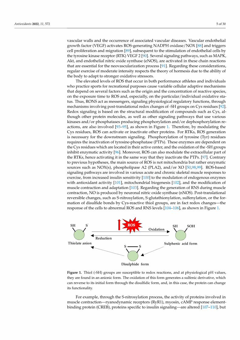
The elevated levels of ROS that occur in both performance athletes and individualswho practice sports for recreational purposes cause variable cellular adaptive mechanismsthat depend on several factors such as the origin and the concentration of reactive species,on the exposure time to ROS and, especially, on the particular/individual oxidative sta-tus. Thus, RONS act as messengers, signaling physiological regulatory functions, throughmechanisms involving post-translational redox changes of -SH groups on Cys residues [92].Redox signaling is based on the structural modification of compounds such as Cys, al-though other protein molecules, as well as other signaling pathways that use variouskinases and/or phosphatases producing phosphorylation and/or dephosphorylation re-actions, are also involved [93–95], as shown in Figure 1. Therefore, by modulating theCys residues, ROS can activate or inactivate other proteins. For RTKs, ROS generationis necessary for the downstream signaling. Phosphorylation of tyrosine (Tyr) residuesrequires the inactivation of tyrosine-phosphatase (PTPs). These enzymes are dependent onthe Cys residues which are located in their active center, and the oxidation of the -SH groupsinhibit enzymatic activity [96]. Moreover, ROS can also modulate the extracellular part ofthe RTKs, hence activating it in the same way that they inactivate the PTPs. [97]. Contraryto previous hypotheses, the main source of ROS is not mitochondria but rather enzymaticsources such as NOX(s), phospholipase A2 (PLA2), and/or XO [50,98,99]. ROS-basedsignaling pathways are involved in various acute and chronic skeletal muscle responses toexercise, from increased insulin sensitivity [100] to the modulation of endogenous enzymeswith antioxidant activity [101], mitochondrial biogenesis [102], and the modification ofmuscle contraction and adaptation [103]. Regarding the generation of RNS during musclecontraction, NO is produced by neuronal nitric oxide synthase (nNOS). Post-translationalreversible changes, such as S-nitrosylation, S-glutathionylation, sulfenylation, or the for-mation of disulfide bonds by Cys-reactive thiol groups, are in fact redox changes—theresponse of the cells to abnormal ROS and RNS levels [104–106], as shown in Figure 1.
Antioxidants 2022, 11, x FOR PEER REVIEW 5 of 31
factor (I-kB) [83,84] and translocation into the nucleus [76]. Moreover, in order to improve the vascularization of striated muscles via NF-kB and hypoxia-inducible factor 1 (HIF-1)—which is associated with peroxisome proliferator-activated receptor gamma coactiva-tor 1-alpha (PGC-1α) [85–87] and ROS and is especially, RNS-dependent through nitric oxide (NO)—angiogenesis is favored. High concentrations of H2O2, together with O∗ , are considered risk factors due to their pro-inflammatory effect, resulting in a thickening of vas-cular walls and the occurrence of associated vascular diseases. Vascular endothelial growth factor (VEGF) activates ROS-generating NADPH oxidase/NOX [88] and triggers cell prolifer-ation and migration [89], subsequent to the stimulation of endothelial cells by the tyrosine kinase receptor (RTK) VEGF 2 [90]. Several signaling pathways, such as MAPK, Akt, and en-dothelial nitric oxide synthase (eNOS), are activated in these chain reactions that are essential for the neovascularization process [91]. Regarding these considerations, regular exercise of moderate intensity respects the theory of hormesis due to the ability of the body to adapt to stronger oxidative stressors.
The elevated levels of ROS that occur in both performance athletes and individuals who practice sports for recreational purposes cause variable cellular adaptive mechanisms that depend on several factors such as the origin and the concentration of reactive species, on the exposure time to ROS and, especially, on the particular/individual oxidative status. Thus, RONS act as messengers, signaling physiological regulatory functions, through mechanisms involving post-translational redox changes of -SH groups on Cys residues [92]. Redox signaling is based on the structural modification of compounds such as Cys, although other protein molecules, as well as other signaling pathways that use various kinases and/or phosphatases producing phosphorylation and/or dephosphorylation reac-tions, are also involved [93–95], as shown in Figure 1. Therefore, by modulating the Cys residues, ROS can activate or inactivate other proteins. For RTKs, ROS generation is necessary for the downstream signaling. Phosphorylation of tyrosine (Tyr) residues requires the inacti-vation of tyrosine-phosphatase (PTPs). These enzymes are dependent on the Cys residues which are located in their active center, and the oxidation of the -SH groups inhibit enzymatic activity [96]. Moreover, ROS can also modulate the extracellular part of the RTKs, hence acti-vating it in the same way that they inactivate the PTPs. [97]. Contrary to previous hypotheses, the main source of ROS is not mitochondria but rather enzymatic sources such as NOX(s), phospholipase A2 (PLA2), and/or XO [50,98,99]. ROS-based signaling pathways are involved in various acute and chronic skeletal muscle responses to exercise, from increased insulin sen-sitivity [100] to the modulation of endogenous enzymes with antioxidant activity [101], mito-chondrial biogenesis [102], and the modification of muscle contraction and adaptation [103]. Regarding the generation of RNS during muscle contraction, NO is produced by neuronal nitric oxide synthase (nNOS). Post-translational reversible changes, such as S-nitrosylation, S-glutathionylation, sulfenylation, or the formation of disulfide bonds by Cys-reactive thiol groups, are in fact redox changes—the response of the cells to abnormal ROS and RNS levels [104–106], as shown in Figure 1.
Figure 1. Thiol (-SH) groups are susceptible to redox reactions, and at physiological pH values, they are found in an anionic form. The oxidation of this form generates a sulfenic derivative, which can reverse to its initial form through the disulfidic form, and, in this case, the protein can change its functionality.
Figure 1. Thiol (-SH) groups are susceptible to redox reactions, and at physiological pH values,they are found in an anionic form. The oxidation of this form generates a sulfenic derivative, whichcan reverse to its initial form through the disulfidic form, and, in this case, the protein can changeits functionality.
For example, through the S-nitrosylation process, the activity of proteins involved inmuscle contraction—ryanodynamic receptors (RyR1), myosin, cAMP response element-binding protein (CREB), proteins specific to insulin signaling—are altered [107–110], but
Antioxidants 2022, 11, 572 6 of 30
at the same time, these reversible redox changes in the -SH groups of Cys also have aprotective role for proteins as they make them less susceptible to oxidative processes [104].
For optimal muscle contraction, a certain level of reactive species is required, becausedepending on its level, the contraction of skeletal muscle can be improved by increasingthe sensitivity of muscle fibers to the Ca2+ ion, whereas post-effort, when the ROS levelis high in the muscle, the contraction force and sensitivity to Ca2+ is decreased [111]. Forthis reason, the administration of dietary supplements with antioxidant compounds maynot be beneficial as it might decrease the effort capacity [111–113]. In a study performedby Reid et al. the hypothesis of a “redox break” was introduced, stating that fatigue anddecreased muscle contraction is due to the generation of high levels of ROS, and it is actuallya negative feedback mechanism through which the damage caused by ROS to the skeletalmuscle is minimized [114,115]. In addition to altering the proteins involved in regulatingthe Ca2+ concentration or the sensitivity of the muscle fiber to this ion, they propose thehypothesis that ROS influences Na+/K+ pump activity and thus reduces muscle contractionforce in the case of endurance-based physical effort [116]. Physical effort causes depletion ofK+ ion sand a massive increase in the intracellular concentration of Na+ ions, which affectsthe membrane excitability, and therefore the force of muscle contractions is diminished.This idea is supported by the experimental data showing that the administration of anantioxidant, N-acetylcysteine (NAC), reduces muscle fatigue that occurs during exercise byregulating the intracellular concentration of K+ ions [117].
There are various communication pathways that use redox signaling and the mostimportant ones that also have an impact on physical effort are NF-kB, MAPK, and PGC-1α,and their activity is influenced by the concentration of H2O2 [118]. Another important as-pect to mention is that these communication pathways are interdependent and intersected,through a process known as “crosstalk” [119]. Studies also present data confirming theinfluence of PGC-1α on mitochondrial function and dynamics (fusion, fission) on musclefiber differentiation and on the expression of antioxidant enzymes, which are present inoxygen-consuming tissues (brain, heart, skeletal muscle) [120,121].
The mitochondrion cellular organelle is of vital importance. It exhibits bioenergetic andfunctional roles in maintaining cell viability, throughout the many biochemical processesthat are under its control (the Krebs cycle, β-oxidase, oxidative phosphorylation, OXPHOS).Concomitantly, the mitochondrion is a dynamic organelle as it can switch its morphologydepending on intra- and extracellular conditions in order to preserve cellular homeostasis.Thus, the presence of a stressor, for example, food deprivation, activates the mitochondrialfusion process, controlled by optical atrophy 1 (Opa1) and mitofusin 1/2 (Mfn1/2), togrow and enhance the OXPHOS rhythm. It is believed that mitochondrial fusion increasesactivity and energy production, whereas fission precedes mitophagy.
On the opposite side, in the case of nutrient excess, fragmentation of mitochondriatakes place, also known as fission [122,123], controlled by a pair of proteins, dynamin-related protein 1 (Drp-1) and mitochondrial fission 1 protein (Fis1) [98,124,125].
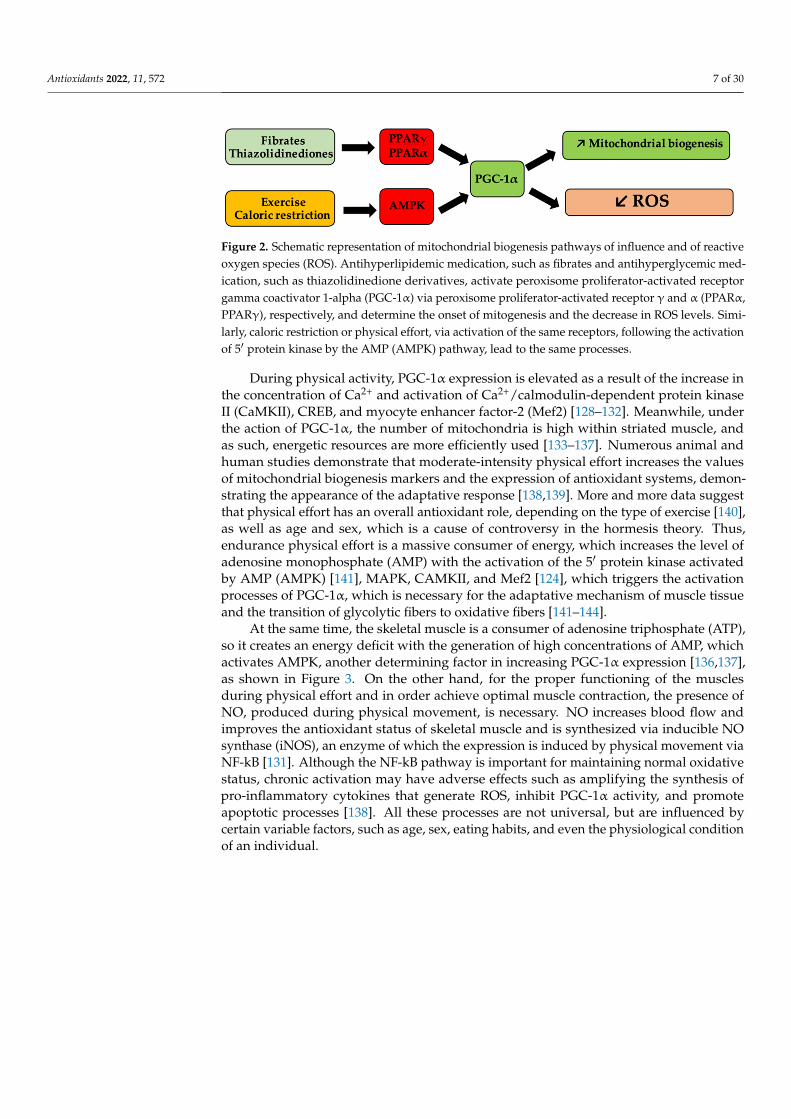
Mitochondrial homeostasis and biogenesis can be pharmacologically modulated byagonists (thiazolidinediones, fibrates) of peroxisome proliferator-activated receptor γ, andα (PPARγ, PPARα), respectively [126,127], as shown in Figure 2.
Antioxidants 2022, 11, 572 7 of 30Antioxidants 2022, 11, x FOR PEER REVIEW 7 of 31
Figure 2. Schematic representation of mitochondrial biogenesis pathways of influence and of reac-tive oxygen species (ROS). Antihyperlipidemic medication, such as fibrates and antihyperglycemic medication, such as thiazolidinedione derivatives, activate peroxisome proliferator-activated recep-tor gamma coactivator 1-alpha (PGC-1α) via peroxisome proliferator-activated receptor γ and α (PPARα, PPARγ), respectively, and determine the onset of mitogenesis and the decrease in ROS levels. Similarly, caloric restriction or physical effort, via activation of the same receptors, following the activation of 5′ protein kinase by the AMP (AMPK) pathway, lead to the same processes.
During physical activity, PGC-1α expression is elevated as a result of the increase in the concentration of Ca2+ and activation of Ca2+/calmodulin-dependent protein kinase II (CaMKII), CREB, and myocyte enhancer factor-2 (Mef2) [128–132]. Meanwhile, under the action of PGC-1α, the number of mitochondria is high within striated muscle, and as such, energetic resources are more efficiently used [133–137]. Numerous animal and human studies demonstrate that moderate-intensity physical effort increases the values of mito-chondrial biogenesis markers and the expression of antioxidant systems, demonstrating the appearance of the adaptative response [138,139]. More and more data suggest that physical effort has an overall antioxidant role, depending on the type of exercise [140], as well as age and sex, which is a cause of controversy in the hormesis theory. Thus, endur-ance physical effort is a massive consumer of energy, which increases the level of adeno-sine monophosphate (AMP) with the activation of the 5′ protein kinase activated by AMP (AMPK) [141], MAPK, CAMKII, and Mef2 [124], which triggers the activation processes of PGC-1α, which is necessary for the adaptative mechanism of muscle tissue and the transition of glycolytic fibers to oxidative fibers [141–144].
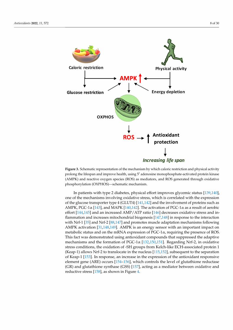
At the same time, the skeletal muscle is a consumer of adenosine triphosphate (ATP), so it creates an energy deficit with the generation of high concentrations of AMP, which activates AMPK, another determining factor in increasing PGC-1α expression [136,137], as shown in Figure 3. On the other hand, for the proper functioning of the muscles during physical effort and in order achieve optimal muscle contraction, the presence of NO, produced during phys-ical movement, is necessary. NO increases blood flow and improves the antioxidant status of skeletal muscle and is synthesized via inducible NO synthase (iNOS), an enzyme of which the expression is induced by physical movement via NF-kB [131]. Although the NF-kB pathway is important for maintaining normal oxidative status, chronic activation may have adverse effects such as amplifying the synthesis of pro-inflammatory cytokines that generate ROS, in-hibit PGC-1α activity, and promote apoptotic processes [138]. All these processes are not uni-versal, but are influenced by certain variable factors, such as age, sex, eating habits, and even the physiological condition of an individual.
Figure 2. Schematic representation of mitochondrial biogenesis pathways of influence and of reactiveoxygen species (ROS). Antihyperlipidemic medication, such as fibrates and antihyperglycemic med-ication, such as thiazolidinedione derivatives, activate peroxisome proliferator-activated receptorgamma coactivator 1-alpha (PGC-1α) via peroxisome proliferator-activated receptor γ and α (PPARα,PPARγ), respectively, and determine the onset of mitogenesis and the decrease in ROS levels. Simi-larly, caloric restriction or physical effort, via activation of the same receptors, following the activationof 5′ protein kinase by the AMP (AMPK) pathway, lead to the same processes.
During physical activity, PGC-1α expression is elevated as a result of the increase inthe concentration of Ca2+ and activation of Ca2+/calmodulin-dependent protein kinaseII (CaMKII), CREB, and myocyte enhancer factor-2 (Mef2) [128–132]. Meanwhile, underthe action of PGC-1α, the number of mitochondria is high within striated muscle, andas such, energetic resources are more efficiently used [133–137]. Numerous animal andhuman studies demonstrate that moderate-intensity physical effort increases the valuesof mitochondrial biogenesis markers and the expression of antioxidant systems, demon-strating the appearance of the adaptative response [138,139]. More and more data suggestthat physical effort has an overall antioxidant role, depending on the type of exercise [140],as well as age and sex, which is a cause of controversy in the hormesis theory. Thus,endurance physical effort is a massive consumer of energy, which increases the level ofadenosine monophosphate (AMP) with the activation of the 5′ protein kinase activatedby AMP (AMPK) [141], MAPK, CAMKII, and Mef2 [124], which triggers the activationprocesses of PGC-1α, which is necessary for the adaptative mechanism of muscle tissueand the transition of glycolytic fibers to oxidative fibers [141–144].
At the same time, the skeletal muscle is a consumer of adenosine triphosphate (ATP),so it creates an energy deficit with the generation of high concentrations of AMP, whichactivates AMPK, another determining factor in increasing PGC-1α expression [136,137],as shown in Figure 3. On the other hand, for the proper functioning of the musclesduring physical effort and in order achieve optimal muscle contraction, the presence ofNO, produced during physical movement, is necessary. NO increases blood flow andimproves the antioxidant status of skeletal muscle and is synthesized via inducible NOsynthase (iNOS), an enzyme of which the expression is induced by physical movement viaNF-kB [131]. Although the NF-kB pathway is important for maintaining normal oxidativestatus, chronic activation may have adverse effects such as amplifying the synthesis ofpro-inflammatory cytokines that generate ROS, inhibit PGC-1α activity, and promoteapoptotic processes [138]. All these processes are not universal, but are influenced bycertain variable factors, such as age, sex, eating habits, and even the physiological conditionof an individual.
Antioxidants 2022, 11, 572 8 of 30Antioxidants 2022, 11, x FOR PEER REVIEW 8 of 31
Figure 3. Schematic representation of the mechanism by which caloric restriction and physical ac-tivity prolong the lifespan and improve health, using 5′ adenosine monophosphate-activated pro-tein kinase (AMPK) and reactive oxygen species (ROS) as mediators, and ROS generated through oxidative phosphorylation (OXPHOS)—schematic mechanism.
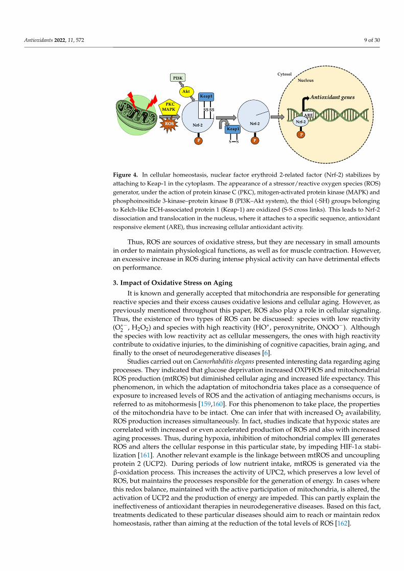
In patients with type 2 diabetes, physical effort improves glycemic status [139,140], one of the mechanisms involving oxidative stress, which is correlated with the expression of the glucose transporter type 4 (GLUT4) [141,142] and the involvement of proteins such as AMPK, PGC-1α [143], and MAPK [140,142]. The activation of PGC-1α as a result of aerobic effort [144,145] and an increased AMP/ATP ratio [146] decreases oxidative stress and inflammation and increases mitochondrial biogenesis [147,148] in response to the in-teraction with Nrf-1 [35] and Nrf-2 [88,147] and promotes muscle adaptation mechanisms following AMPK activation [31,148,149]. AMPK is an energy sensor with an important impact on metabolic status and on the mRNA expression of PGC-1α, requiring the pres-ence of ROS. This fact was demonstrated using antioxidant compounds that suppressed the adaptive mechanisms and the formation of PGC-1α [132,150,151]. Regarding Nrf-2, in oxidative stress conditions, the oxidation of -SH groups from Kelch-like ECH-associated protein 1 (Keap-1) allows Nrf-2 to translocate in the nucleus [115,152], subsequent to the separation of Keap-1 [153]. In response, an increase in the expression of the antioxidant responsive element gene (ARE) occurs [154–156], which controls the level of glutathione reductase (GR) and glutathione synthase (GSS) [157], acting as a mediator between oxida-tive and reductive stress [158], as shown in Figure 4.
Figure 3. Schematic representation of the mechanism by which caloric restriction and physical activityprolong the lifespan and improve health, using 5′ adenosine monophosphate-activated protein kinase(AMPK) and reactive oxygen species (ROS) as mediators, and ROS generated through oxidativephosphorylation (OXPHOS)—schematic mechanism.
In patients with type 2 diabetes, physical effort improves glycemic status [139,140],one of the mechanisms involving oxidative stress, which is correlated with the expressionof the glucose transporter type 4 (GLUT4) [141,142] and the involvement of proteins such asAMPK, PGC-1α [143], and MAPK [140,142]. The activation of PGC-1α as a result of aerobiceffort [144,145] and an increased AMP/ATP ratio [146] decreases oxidative stress and in-flammation and increases mitochondrial biogenesis [147,148] in response to the interactionwith Nrf-1 [35] and Nrf-2 [88,147] and promotes muscle adaptation mechanisms followingAMPK activation [31,148,149]. AMPK is an energy sensor with an important impact onmetabolic status and on the mRNA expression of PGC-1α, requiring the presence of ROS.This fact was demonstrated using antioxidant compounds that suppressed the adaptivemechanisms and the formation of PGC-1α [132,150,151]. Regarding Nrf-2, in oxidativestress conditions, the oxidation of -SH groups from Kelch-like ECH-associated protein 1(Keap-1) allows Nrf-2 to translocate in the nucleus [115,152], subsequent to the separationof Keap-1 [153]. In response, an increase in the expression of the antioxidant responsiveelement gene (ARE) occurs [154–156], which controls the level of glutathione reductase(GR) and glutathione synthase (GSS) [157], acting as a mediator between oxidative andreductive stress [158], as shown in Figure 4.
Antioxidants 2022, 11, 572 9 of 30Antioxidants 2022, 11, x FOR PEER REVIEW 9 of 31
Figure 4. In cellular homeostasis, nuclear factor erythroid 2-related factor (Nrf-2) stabilizes by at-taching to Keap-1 in the cytoplasm. The appearance of a stressor/ reactive oxygen species (ROS) generator, under the action of protein kinase C (PKC), mitogen-activated protein kinase (MAPK) and phosphoinositide 3-kinase–protein kinase B (PI3K–Akt system), the thiol (-SH) groups belong-ing to Kelch-like ECH-associated protein 1 (Keap-1) are oxidized (S-S cross links). This leads to Nrf-2 dissociation and translocation in the nucleus, where it attaches to a specific sequence, antioxidant responsive element (ARE), thus increasing cellular antioxidant activity.
Thus, ROS are sources of oxidative stress, but they are necessary in small amounts in order to maintain physiological functions, as well as for muscle contraction. However, an ex-cessive increase in ROS during intense physical activity can have detrimental effects on per-formance.
3. Impact of Oxidative Stress on Aging It is known and generally accepted that mitochondria are responsible for generating
reactive species and their excess causes oxidative lesions and cellular aging. However, as previously mentioned throughout this paper, ROS also play a role in cellular signaling. Thus, the existence of two types of ROS can be discussed: species with low reactivity (O∗ , H2O2) and species with high reactivity (HO∗, peroxynitrite, ONOO ). Although the species with low reactivity act as cellular messengers, the ones with high reactivity contribute to oxidative injuries, to the diminishing of cognitive capacities, brain aging, and finally to the onset of neurodegenerative diseases [6].
Studies carried out on Caenorhabditis elegans presented interesting data regarding ag-ing processes. They indicated that glucose deprivation increased OXPHOS and mitochon-drial ROS production (mtROS) but diminished cellular aging and increased life expec-tancy. This phenomenon, in which the adaptation of mitochondria takes place as a conse-quence of exposure to increased levels of ROS and the activation of antiaging mechanisms occurs, is referred to as mitohormesis [159,160]. For this phenomenon to take place, the properties of the mitochondria have to be intact. One can infer that with increased O2 availability, ROS production increases simultaneously. In fact, studies indicate that hy-poxic states are correlated with increased or even accelerated production of ROS and also with increased aging processes. Thus, during hypoxia, inhibition of mitochondrial com-plex III generates ROS and alters the cellular response in this particular state, by impeding HIF-1α stabilization [161]. Another relevant example is the linkage between mtROS and uncoupling protein 2 (UCP2). During periods of low nutrient intake, mtROS is generated via the β-oxidation process. This increases the activity of UPC2, which preserves a low level of ROS, but maintains the processes responsible for the generation of energy. In cases where this redox balance, maintained with the active participation of mitochondria, is al-tered, the activation of UCP2 and the production of energy are impeded. This can partly explain the ineffectiveness of antioxidant therapies in neurodegenerative diseases. Based on this fact, treatments dedicated to these particular diseases should aim to reach or main-tain redox homeostasis, rather than aiming at the reduction of the total levels of ROS [162].
Figure 4. In cellular homeostasis, nuclear factor erythroid 2-related factor (Nrf-2) stabilizes byattaching to Keap-1 in the cytoplasm. The appearance of a stressor/reactive oxygen species (ROS)generator, under the action of protein kinase C (PKC), mitogen-activated protein kinase (MAPK) andphosphoinositide 3-kinase–protein kinase B (PI3K–Akt system), the thiol (-SH) groups belongingto Kelch-like ECH-associated protein 1 (Keap-1) are oxidized (S-S cross links). This leads to Nrf-2dissociation and translocation in the nucleus, where it attaches to a specific sequence, antioxidantresponsive element (ARE), thus increasing cellular antioxidant activity.
Thus, ROS are sources of oxidative stress, but they are necessary in small amountsin order to maintain physiological functions, as well as for muscle contraction. However,an excessive increase in ROS during intense physical activity can have detrimental effectson performance.
3. Impact of Oxidative Stress on Aging
It is known and generally accepted that mitochondria are responsible for generatingreactive species and their excess causes oxidative lesions and cellular aging. However, aspreviously mentioned throughout this paper, ROS also play a role in cellular signaling.Thus, the existence of two types of ROS can be discussed: species with low reactivity(O∗−2 , H2O2) and species with high reactivity (HO∗, peroxynitrite, ONOO−). Althoughthe species with low reactivity act as cellular messengers, the ones with high reactivitycontribute to oxidative injuries, to the diminishing of cognitive capacities, brain aging, andfinally to the onset of neurodegenerative diseases [6].
Studies carried out on Caenorhabditis elegans presented interesting data regarding agingprocesses. They indicated that glucose deprivation increased OXPHOS and mitochondrialROS production (mtROS) but diminished cellular aging and increased life expectancy. Thisphenomenon, in which the adaptation of mitochondria takes place as a consequence ofexposure to increased levels of ROS and the activation of antiaging mechanisms occurs, isreferred to as mitohormesis [159,160]. For this phenomenon to take place, the propertiesof the mitochondria have to be intact. One can infer that with increased O2 availability,ROS production increases simultaneously. In fact, studies indicate that hypoxic states arecorrelated with increased or even accelerated production of ROS and also with increasedaging processes. Thus, during hypoxia, inhibition of mitochondrial complex III generatesROS and alters the cellular response in this particular state, by impeding HIF-1α stabi-lization [161]. Another relevant example is the linkage between mtROS and uncouplingprotein 2 (UCP2). During periods of low nutrient intake, mtROS is generated via theβ-oxidation process. This increases the activity of UPC2, which preserves a low level ofROS, but maintains the processes responsible for the generation of energy. In cases wherethis redox balance, maintained with the active participation of mitochondria, is altered, theactivation of UCP2 and the production of energy are impeded. This can partly explain theineffectiveness of antioxidant therapies in neurodegenerative diseases. Based on this fact,treatments dedicated to these particular diseases should aim to reach or maintain redoxhomeostasis, rather than aiming at the reduction of the total levels of ROS [162].
Antioxidants 2022, 11, 572 10 of 30
In the opposite situation, the altered and non-functional mitochondria are eliminatedvia mitophagy and the proteasome system [163]. On this note, studies state that an increasein the activity and effectiveness of mitophagy and the proteasome system can increase lifeexpectancy and slow down the aging rate, at least in some model organisms (rodents, flies,and worms) [164–166]. However, the notion that the mere presence of functional mito-chondria would prevent aging or the manifestation of already-present neurodegenerativediseases remains unclear.
Diseases with neurodegenerative components are characterized by affected functionsas well as progressive destruction of neuronal cells. The modification of redox homeostasisis a specific attribute of neurodegenerative conditions and despite the fact that in someexperimental models of Alzheimer’s, Parkinson’s, or other neurological conditions antioxi-dant agents demonstrated beneficial effects, in clinical studies, this neuroprotective effectcould not be demonstrated [167,168].
From a morphological perspective, the destruction of neuronal mass is linked toprotein misfolding and the accumulation of misfolded protein aggregates. An interestingfact is that some neuronal populations are resistant, whereas some areas are sensitive tooxidative stress, a phenomenon termed selective vulnerability. Finally, the associationbetween protein aggregates and oxidative stress triggers a chain reaction, which leads toneuronal death [169].
In the case of the dysfunction of the proteasome system, which is responsible for thedisposal of the altered proteins, and which is therefore indispensable for maintaining cellu-lar homeostasis and neuronal survival, the protein aggregates can trigger the generation ofROS. This phenomenon is often encountered in diseases with an oxidative component, espe-cially the neurodegenerative ones. It is known that the brain does not possess a remarkableantioxidant capacity, which makes it susceptible to oxidative damage, particularly duringaging, when the antioxidant systems are less active and favor the accumulation of reactivespecies and the progression of diseases such as Alzheimer’s and Parkinson’s [170–172].
Alzheimer’s disease is a widespread neurodegenerative condition, involving theprogressive loss of memory, cognitive capacities, and personality disorders as the mostimportant symptoms. Morphological particularities in Alzheimer’s are represented byamyloid plaques, dystrophic neurites, cerebral amyloid angiopathy, reactive astrogliosis, aswell as microglial activation [173]. The absence of the protein Nrf-2 from the nucleus ofthe hippocampal neurons is implicated in this disease, which can be confirmed throughbiochemical analysis of the patient’s brain. Concomitantly, it is likely that in these patients,the response of Nrf-2 to ROS and the mitochondrial dynamics are affected, which furtherleads to the progression of molecular mass loss [174]. The accumulation of β-amyloid andTau proteins is age-dependent—it increases as one person gets older, and this accumulationdisrupts Ca2+ ion homeostasis and the mitochondrial membrane potential is lost. Further-more, the internal membrane develops pores and ATP synthesis decreases, but the ROSlevel increases as a result of electron drain.
Regarding Parkinson’s disease, which is the second most widespread neurodegen-erative condition after Alzheimer’s disease [175], it mostly constitutes motor symptoms(tremor at rest, bradykinesia, muscular rigidity) and non-motor symptoms (constipation,depression, personality disorder). The symptomatology is the result of the degenerationof dopaminergic neurons in the substantia nigra [176,177]. In this case, the presence ofROS leads to Nrf-2 activation, which further disrupts α-synuclein (α-Syn) metabolism,which would thus prevent the loss of neuronal dopaminergic mass. However, in patientsdiagnosed with Parkinson’s disease, a deficit of the Nrf-2 protein is observed, and thisdeficit causes neuroinflammation and favors the progression of the disease [174]. In bothneurodegenerative diseases, age is one of the most important determining factors of theonset and/or the progression of the disease.
It is generally accepted that oxidative stress is involved in various diseases, suchas neurodegenerative diseases (Alzheimer’s, Parkinson’s), but it is possible that ROSare not actually the trigger for the disease but rather the cause of the exacerbation of
Antioxidants 2022, 11, 572 11 of 30
symptoms within these neurological disorders [178,179]. Therefore, various studies havebeen performed to investigate the influence of oxidative stress and physical effort on centralnervous system conditions. The data presented in the literature demonstrate an inverselyproportional relationship between the incidence of neurodegenerative diseases and physicalactivity, and a slowdown of their progression in the case of physical activity [135,180–183].
Moreover, as neuronal metabolism increases, there is a consecutive increase in oxygenconsumption and ROS production, triggering a positive feedback effect on antioxidantmechanisms and neurotrophin synthesis, which improves cognitive functions and offersprotection against neuronal degradation [184]. Brain-derived neurotrophic factor (BDNF)is required for neurogenesis and neuronal plasticity [159,185], and its release is dependenton oxidative status [88] and caloric restriction (CR) [186,187]. Evidence to support thesehypotheses was obtained through a study on Alzheimer’s disease [188], in which a decreasein the accumulation of β-amyloid [189] was observed, possibly due to the proteolysisand autophagy of denatured proteins [190], and the overexpression of neuroglobin as aprotective factor of the brain from the neurotoxic action of β-amyloid [188]. The datapresented in the literature suggest that the moderate level of reactive species formed duringexercise favors the expression of 8-oxoG DNA glycosylase [190], an enzyme involved inthe repair of nucleic acid chains due to the accumulation of 8-OHdG [52,184]. Preventingthe accumulation of degraded DNA presents beneficial effects on senescence and otherassociated degenerative diseases [191]. A study conducted by Radák et al. confirmed thebeneficial effect of moderate physical exertion on neurodegenerative diseases, mediated byproteasome activity and the degradation of altered proteins [192].
ROS are also involved in the formation and storage of memories via improvingsynaptic plasticity [192], suggesting the modulation of cognitive capacity, which entailsthe activation of hormetic processes at the neuronal level. The generation of free radicalsassociated with the influx of Ca2+ ions activate transcription factors CREB and NF-kB,causing a release of cytoprotective proteins [193]. Thus, it can be concluded that exposureof the brain to moderate levels of reactive species improves endurance and slows down theaging process of neuronal tissue [194]. Altogether, these data show a neurohormetic effectthat can suppress the progression of neurodegenerative diseases [195].
This is a paradox—exercise-induced oxidative stress, by activating and improvingadaptive mechanisms, is responsible for improving overall physiological function, reducingthe prevalence of various diseases, increasing the quality of life, and, consequently, inducingthe adaptability of the body to oxidative stress [196]. Thus, increasing the body’s capacityto trap and neutralize ROS can provide better protection and resistance during trainingand can also intervene in reducing the aging process. A study performed by Bouzid et al.supports the hypothesis that the aging process alters antioxidant activity regardless of thephysical activity level, thus affecting both sedentary and active people. However, regularphysical activity improves antioxidant capacity in the young population to a greater extentthan in the elderly, suggesting that the beneficial effects of regular physical activity couldbe affected by the aging process. In addition, the study data support the idea that regularphysical activity reduces the aging process [197].
Overall, physical activity must be performed throughout the lifetime in order to main-tain the benefits of adaptive mechanisms following exercise-induced oxidative stress [197].These data are supported by other studies concluding that with age, muscle mass decreases,a process known as sarcopenia. There are several factors involved in the development ofsarcopenia, such as neuro-endocrine factors, nutritional factors, decreased physical activ-ity, and especially oxidative stress caused by mitochondrial dysfunction [198–200]. Themitochondrial theory of sarcopenia claims that muscle weakness occurs due to decreasedoxidative capacity of mitochondria and thus a reduced amount of generated ATP. This isdue to the decreased ability to replace altered mitochondrial proteins involved in the ETCwith age [201,202]. In addition, in senescent muscles, NF-kB is constitutively activated,which leads to increased production of pro-inflammatory cytokines and ROS. At the sametime, the forkhead family of transcription factors (FoxO) is activated through ROS, which
Antioxidants 2022, 11, 572 12 of 30
decreases the expression of PGC-1α and favors the degradation of the neuromuscularjunction and mitochondrial trophicity [203].
Unlike the past decades, in which ROS were considered the main cause of the agingprocess, it has been confirmed that small amounts of free radicals prevent senescenceand improve the lifespan and the quality of life. Another associated theory is relatedto CR, demonstrated by the ketogenic diet [54,194,204,205], as it improves antioxidantactivity and reduces the amount of ROS to a level that slows down senescence. The exactmechanism through which CR reduces oxidative stress is not completely known andavailable data are ambiguous. A potential mechanism could involve increased degradationof the modified molecular structures with a consecutive increase in the synthesis of newmolecules to replace them, as well as a simultaneous decrease in the rate of production offree radicals by mitochondria [123]. With age, carbonylated proteins tend to accumulateas their degradation mechanisms decrease, but in physically active people due to ROSproduced during exertion, the expression of the proteasome complex [184,206], responsiblefor protein degradation, is potentiated [207].
Most frequently, the aging process is associated with the loss of tissue, the underlyingtheory being that of oxidative stress coupled with lesions caused by ROS. Thus, sustainedphysical activity is related to a better neutralization of ROS, which leads to protectionagainst oxidative injuries and the prevention of age-related conditions.
4. A Link between Oxidative Stress and Immune Response
In a general sense, a compound with antioxidant properties is considered to be able toneutralize the RONS species either directly, indirectly, or by inhibiting their subsequentgeneration [208]. As a result, antioxidants can be classified according to their mechanismof action [209,210] into:
• enzymes (SOD, CAT, GPx), proteins (ceruloplasmin, ferritin), and minerals (zinc, cop-per, selenium) considered to be agents that prevent the formation of new free radicals;
• agents considered to be scavengers act by inhibiting the initiation and propagation ofperoxidation reactions (vitamin C, vitamin E, β-carotenes, glutathione, flavonoids);
• enzymes involved in the reconsolidation of affected cell membranes (lipases, proteases,transferases, DNA repair enzymes).
Considering that the theory of hormesis is generally accepted and that RONS arenecessary for cellular homeostasis, muscle regeneration, slowing down the process of aging,protein degradation, and cellular signaling, it is still not possible to determine if a reductivestatus of the body is beneficial or not, as it generates reductive stress [211] that causes cellu-lar metabolism disorders and cytotoxicity [212]. Nonetheless, the literature emphasizes theinvolvement of mitochondrial oxidative metabolism in stem cell differentiation, such thatcomplete differentiation is dependent on the presence of ROS [213–218]. This informationdoes not exclude the fact that ROS or increased levels of ROS are not harmful, but thereare circumstances and types of cells (adipocytes, muscles) of which the differentiation andrenewal depend on the organic presence of these species [219,220]. In the meantime, ROSinfluence the successive phases that occur in the cellular cycle, and the administration ofthe compounds that donate -SH groups (NAC) apparently cause cycle arrest in the G1phase, whereas in the absence of these compounds, the S phase is prolonged and the G1phase is shortened [221,222]. Studies demonstrate that antioxidants have a wide range ofbeneficial effects, with most of these studies conducted in conditions of induced oxidativestress and with only a few studies questioning what happens under normal physiologicalconditions [223,224].
Therefore, in the absence of an oxidative status, the self-renewal capacity of cells andautophagy processes are disrupted, with the accumulation of chromosomal defects andabnormalities and the inability to repair DNA and induce apoptosis [225,226]. Highlyactive antioxidant systems induce reductive stress, influencing [130] cell growth and mi-tochondrial functions and are incriminated in the progression of pathological conditionssuch as Alzheimer’s disease [227] because of an impaired protein folding process [12],
Antioxidants 2022, 11, 572 13 of 30
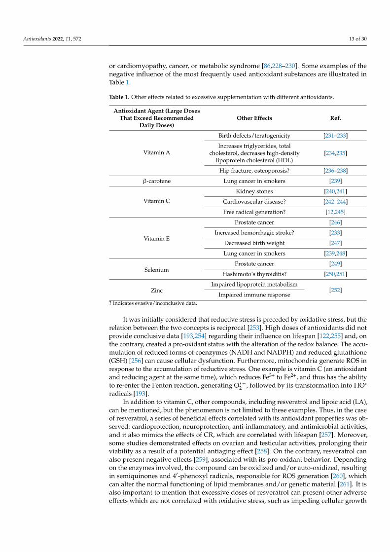
or cardiomyopathy, cancer, or metabolic syndrome [86,228–230]. Some examples of thenegative influence of the most frequently used antioxidant substances are illustrated inTable 1.
Table 1. Other effects related to excessive supplementation with different antioxidants.
Antioxidant Agent (Large DosesThat Exceed Recommended
Daily Doses)Other Effects Ref.
Vitamin A
Birth defects/teratogenicity [231–233]
Increases triglycerides, totalcholesterol, decreases high-density
lipoprotein cholesterol (HDL)[234,235]
Hip fracture, osteoporosis? [236–238]
β-carotene Lung cancer in smokers [239]
Vitamin C
Kidney stones [240,241]
Cardiovascular disease? [242–244]
Free radical generation? [12,245]
Vitamin E
Prostate cancer [246]
Increased hemorrhagic stroke? [233]
Decreased birth weight [247]
Lung cancer in smokers [239,248]
SeleniumProstate cancer [249]
Hashimoto’s thyroiditis? [250,251]
ZincImpaired lipoprotein metabolism
[252]Impaired immune response
? indicates evasive/inconclusive data.
It was initially considered that reductive stress is preceded by oxidative stress, but therelation between the two concepts is reciprocal [253]. High doses of antioxidants did notprovide conclusive data [193,254] regarding their influence on lifespan [122,255] and, onthe contrary, created a pro-oxidant status with the alteration of the redox balance. The accu-mulation of reduced forms of coenzymes (NADH and NADPH) and reduced glutathione(GSH) [256] can cause cellular dysfunction. Furthermore, mitochondria generate ROS inresponse to the accumulation of reductive stress. One example is vitamin C (an antioxidantand reducing agent at the same time), which reduces Fe3+ to Fe2+, and thus has the abilityto re-enter the Fenton reaction, generating O∗−2 , followed by its transformation into HO*radicals [193].
In addition to vitamin C, other compounds, including resveratrol and lipoic acid (LA),can be mentioned, but the phenomenon is not limited to these examples. Thus, in the caseof resveratrol, a series of beneficial effects correlated with its antioxidant properties was ob-served: cardioprotection, neuroprotection, anti-inflammatory, and antimicrobial activities,and it also mimics the effects of CR, which are correlated with lifespan [257]. Moreover,some studies demonstrated effects on ovarian and testicular activities, prolonging theirviability as a result of a potential antiaging effect [258]. On the contrary, resveratrol canalso present negative effects [259], associated with its pro-oxidant behavior. Dependingon the enzymes involved, the compound can be oxidized and/or auto-oxidized, resultingin semiquinones and 4′-phenoxyl radicals, responsible for ROS generation [260], whichcan alter the normal functioning of lipid membranes and/or genetic material [261]. It isalso important to mention that excessive doses of resveratrol can present other adverseeffects which are not correlated with oxidative stress, such as impeding cellular growth
Antioxidants 2022, 11, 572 14 of 30
and the activation of apoptosis in normal cells, atherogenesis, and phytoestrogen-likebehavior [262–264].
LA, also known as α-lipoic acid and/or thioctic acid, displays biochemical roles andantioxidant properties. LA is reduced to dihydrolipoic acid (DHLA) by GR and TRX in thepresence of NADPH. Both molecules, LA and DHLA, exhibit their antioxidant properties inhydrophilic as well as in lipophilic media because of their amphipathic character [265]. Theracemic mixture of LA is used in therapy, especially in diabetic neuropathy. Its antioxidantproperties are only evident in the interaction with other antioxidant systems. For example,the capacity of reducing GSSG to GSH is based on the initial reduction of LA to DHLA,which further reduces cystine to Cys, and the latter is used for GSH synthesis [266]. On amitochondrial level, along with other antioxidants (GSH, vitamin C, and vitamin E), LAincreases the activity of the enzymes involved in the Krebs cycle [267]. Furthermore, in astudy conducted by Marangon et al., supplementation with LA resulted in diminishingurinary levels of F2-isoprostan and oxLDL [268]. A potential antiaging effect based on theprotection of cardiac cells in aged rodents was also suggested [269].
However, its pro-oxidant properties, demonstrated in in vitro and in vivo studies,cannot be excluded. In vitro, DHLA forms chelates with both ferric and ferrous ions, whichcan extract the iron from ferritin and reduce it to Fe2+, at the same time increasing thepossibility of oxidative processes via the Fenton reaction. In another study conducted byÇatakay et al., it was observed that the administration of LA in aged rats resulted in higherlevels of carbonylated proteins compared to the control group [270].
The idea that substances/compounds with antioxidant activity are harmless is im-printed in popular belief and this conception is also supported by the media. However,there are questions as to how antioxidant compounds intervene in slowing down the agingprocess and the appearance of various diseases. The ability of antioxidants to preventthe accumulation of oxidizing molecules, thus helping to prevent aging, cancer, and eveninfections, has been also questioned.
The adaptation capacity of the immune system is essential for antimicrobial defense,as well as for the immunological memory. In this sense, the presence of mitochondria ismandatory, as it facilitates the aforementioned antimicrobial defense through ROS genera-tion. At the same time, mitochondria are necessary for T-cell activation, which generatesROS and activates the nuclear factor of activated T cells (NFAT), which further enhancesIL-2 synthesis [271,272]. This statement is supported by research studies that demonstratedthat T-cells, for which antioxidant agents were made available, showed reduced levels ofIL-2 and a reduction in the degree of proliferation was observed, sustaining an immunosup-pressive effect resulting from the exposure to antioxidant agents. The same effects couldalso be noted in the case of T-cells that lacked mitochondrial complex III, supporting theimportance of ROS in the immune response [273].
Macrophages and the neutrophils constrain the ability to phagocytize foreign com-pounds/microorganisms and subsequently to activate the immune system [274]. Whenmacrophages recognize the antigen, the assembly of the protein complex of cytochromeI from the ETC is prevented, and mitochondrial respiration on complexes I and II is in-creased under the influence of phagosomal NOX and ROS-dependent tyrosine kinaseFgr. This process leads in increase in the ROS concentration [275]. The presence of ROSleads to the activation of MAPK, extracellular signal-regulated kinases (ERK), c-Jun N-terminal kinases (JNK) and NF-kB, which are key factors in the secretion of chemokines andpro-inflammatory cytokines (TNF-α, IL-1β, IL-6, IFN-γ) [276,277], as shown in Figure 5.Likewise, in this process of T-cell activation, NOX is also involved, which is activated byreactive species generated at the mitochondrial level, in order to maintain the proper ROSlevels for this process [278]. Therefore, the presence of ROS is necessary for the destructionof the bacteria, which is contrast to the situation in chronic granulomatosis disease, inwhich ROS cannot be generated due to a genetic defect of NOX [279,280].
Antioxidants 2022, 11, 572 15 of 30
Antioxidants 2022, 11, x FOR PEER REVIEW 15 of 31
inflammatory cytokines (TNF-α, IL-1β, IL-6, IFN-γ) [276,277], as shown in Figure 5. Like-wise, in this process of T-cell activation, NOX is also involved, which is activated by reac-tive species generated at the mitochondrial level, in order to maintain the proper ROS levels for this process [278]. Therefore, the presence of ROS is necessary for the destruction of the bacteria, which is contrast to the situation in chronic granulomatosis disease, in which ROS cannot be generated due to a genetic defect of NOX [279,280].
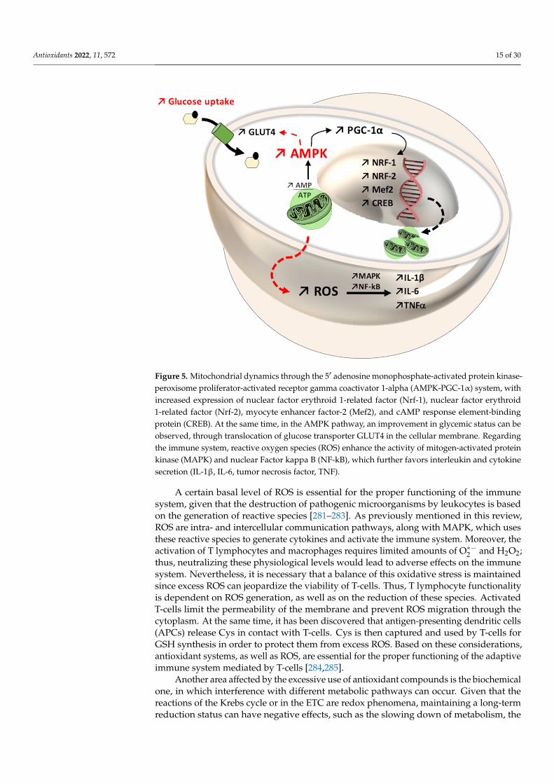
Figure 5. Mitochondrial dynamics through the 5′ adenosine monophosphate-activated protein ki-nase-peroxisome proliferator-activated receptor gamma coactivator 1-alpha (AMPK-PGC-1α) sys-tem, with increased expression of nuclear factor erythroid 1-related factor (Nrf-1), nuclear factor erythroid 1-related factor (Nrf-2), myocyte enhancer factor-2 (Mef2), and cAMP response element-binding protein (CREB). At the same time, in the AMPK pathway, an improvement in glycemic status can be observed, through translocation of glucose transporter GLUT4 in the cellular mem-brane. Regarding the immune system, reactive oxygen species (ROS) enhance the activity of mito-gen-activated protein kinase (MAPK) and nuclear Factor kappa B (NF-kB), which further favors interleukin and cytokine secretion (IL-1β, IL-6, tumor necrosis factor, TNF).
A certain basal level of ROS is essential for the proper functioning of the immune system, given that the destruction of pathogenic microorganisms by leukocytes is based on the generation of reactive species [281–283]. As previously mentioned in this review, ROS are intra- and intercellular communication pathways, along with MAPK, which uses these reactive species to generate cytokines and activate the immune system. Moreover, the activation of T lymphocytes and macrophages requires limited amounts of O∗ and H2O2; thus, neutralizing these physiological levels would lead to adverse effects on the immune system. Nevertheless, it is necessary that a balance of this oxidative stress is maintained since excess ROS can jeopardize the viability of T-cells. Thus, T lymphocyte functionality is dependent on ROS generation, as well as on the reduction of these species. Activated T-cells limit the permeability of the membrane and prevent ROS migration through the cytoplasm. At the same time, it has been discovered that antigen-presenting dendritic cells (APCs) release Cys in contact with T-cells. Cys is then captured and used by T-cells for GSH synthesis in order to protect them from excess ROS. Based on these
Figure 5. Mitochondrial dynamics through the 5′ adenosine monophosphate-activated protein kinase-peroxisome proliferator-activated receptor gamma coactivator 1-alpha (AMPK-PGC-1α) system, withincreased expression of nuclear factor erythroid 1-related factor (Nrf-1), nuclear factor erythroid1-related factor (Nrf-2), myocyte enhancer factor-2 (Mef2), and cAMP response element-bindingprotein (CREB). At the same time, in the AMPK pathway, an improvement in glycemic status can beobserved, through translocation of glucose transporter GLUT4 in the cellular membrane. Regardingthe immune system, reactive oxygen species (ROS) enhance the activity of mitogen-activated proteinkinase (MAPK) and nuclear Factor kappa B (NF-kB), which further favors interleukin and cytokinesecretion (IL-1β, IL-6, tumor necrosis factor, TNF).
A certain basal level of ROS is essential for the proper functioning of the immunesystem, given that the destruction of pathogenic microorganisms by leukocytes is basedon the generation of reactive species [281–283]. As previously mentioned in this review,ROS are intra- and intercellular communication pathways, along with MAPK, which usesthese reactive species to generate cytokines and activate the immune system. Moreover, theactivation of T lymphocytes and macrophages requires limited amounts of O∗−2 and H2O2;thus, neutralizing these physiological levels would lead to adverse effects on the immunesystem. Nevertheless, it is necessary that a balance of this oxidative stress is maintainedsince excess ROS can jeopardize the viability of T-cells. Thus, T lymphocyte functionalityis dependent on ROS generation, as well as on the reduction of these species. ActivatedT-cells limit the permeability of the membrane and prevent ROS migration through thecytoplasm. At the same time, it has been discovered that antigen-presenting dendritic cells(APCs) release Cys in contact with T-cells. Cys is then captured and used by T-cells forGSH synthesis in order to protect them from excess ROS. Based on these considerations,antioxidant systems, as well as ROS, are essential for the proper functioning of the adaptiveimmune system mediated by T-cells [284,285].
Another area affected by the excessive use of antioxidant compounds is the biochemicalone, in which interference with different metabolic pathways can occur. Given that thereactions of the Krebs cycle or in the ETC are redox phenomena, maintaining a long-termreduction status can have negative effects, such as the slowing down of metabolism, the
Antioxidants 2022, 11, 572 16 of 30
accumulation of intermediates, and decreased ATP synthesis, with all these reactions beinginterconnected [286,287]. Another mechanism to control free radicals generated by ROS isbased on heat shock proteins (HSPs) [288]. These proteins have properties to correct themisfolding of polypeptides and to promote the degradation of altered peptides. Underphysiological conditions, the expression of HSPs is induced by various factors, includingROS, but the irresponsible use of antioxidant compounds can have negative effects on theactivity and synthesis of HSPs [289–291].
The administration of antioxidants in viral infections has provided only evasive data,because in some studies, the benefits are not highlighted, and the antioxidants can favorthe progression of the infection [292]. In the antioxidant therapeutic approach in cancercells, the level of ROS must be considered because it can favor the situation in which, in theabsence of a proper level of ROS, the cancer therapy can promote carcinogenesis via NF-kB,HIFs, and MAPK activation [27]. Paradoxically, cancer cells induce their own antioxidantsystems with the purpose of reducing excess ROS, which would be detrimental throughcytotoxic effects. In this context, there are two therapeutic approaches, one of which is basedon antioxidant administration, which could reduce ROS levels required for the proliferationof cancer cells, making them susceptible to cellular apoptosis. However, this approach hasraised numerous questions on this topic, and the results have been evasive [293].
The second approach refers to inducing oxidative stress to reach levels that cannotbe neutralized by cancer cells. The therapy can be performed directly with therapeuticagents that promote ROS generation (radio- and chemotherapy) or indirectly, with agentsthat inhibit the antioxidant systems [294]. In this manner, the existence of the tumor cellsdepends on ROS levels; moderate levels activate NF-kB and promote survival mechanisms,whereas excessive levels degrade the cellular organelles, specifically the mitochondria, andalter the DNA and ARN molecules, favoring the initiation of apoptotic reactions [295].
For this reason, it is important to acknowledge the oxidative status of each individual,as antioxidants should not be administered to individuals without a proper immuneresponse (see Table 2). However, a decrease in the activity of the immune system requiresthe antioxidant therapy approach.
Table 2. Positive and negative effects related to the generation of oxidative stress on organs, systems,diseases, and physiological conditions.
Positive Effects Ref. Negative Effects Ref.
MuscleImproves antioxidant defense
[296–299] Contractile dysfunctionMuscle atrophy and weakness [300,301]Adaptive response (muscle fiber
type adaptation)Increases the number of mitochondria
BrainSynaptic plasticity [302–305]
Neuropsychiatric disorderNeuron apoptosis
NeurotoxicityNeurodegenerative diseases
[306–310]
Hippocampus-independentmemory formation
Immune system
Increases immune system activity↗NF-KB↗IL-1β↗TNF-α
[311]
Rheumatoid arthritisRespiratory diseases
PeriodontitisChronic kidney disease
Gastro-intestinal autoimmune diseaseMultiple sclerosis
Fibromyalgia
[312–315]
Vascular system
VasodilationRegulates blood pressure
AngiogenesisWound healing
Granulation tissue formation
[140,316,317]Hypertension
AtherosclerosisIschemia reperfusion
[318–320]
CancerCycle arrestApoptosis
Inhibits metastasis[321–326]
CarcinogenesisMetastasis
Therapy resistance[77,327,328]
AgingMuscle remodelingNeuronal activity
Preservation[12,159,197,212,329]
SarcopeniaMuscular dystrophy
DNA damageTelomere shortening
Neurodegenerative diseases
[202,330–335]
Antioxidants 2022, 11, 572 17 of 30
5. Conclusions
The data presented in the literature suggest that ROS and RNS negatively influencethe human body, but that this effect depends to a great extent on the individual’s oxidativestatus, as ROS are also involved in growth, differentiation, proliferation, and the processesof apoptosis and autophagy, which are highly important for maintaining cellular homeosta-sis. Medium-intensity aerobic exercise is responsible for generating ROS at levels that arebeneficial and have the ability to induce counterregulatory and adaptation mechanisms,such as PGC-1α. In striated muscles, adaptation mechanisms are induced by exposureto ROS and RNS produced during resistance training and are mainly mediated throughNF-kB and PGC-1α. With age, the incidence of neurodegenerative diseases increases, buta well-balanced CR seems to diminish this trend. Animal studies confirm the hormesistheory as CR is a form of stress itself, inducing adaptation mechanisms in the brain throughthe endocrine system. Thus, the expression of neurotrophic factors is dependent on CR andneurogenesis is favored.
In fact, the present article discusses an evolution paradox, the transition from anaerobicto aerobic metabolism and how, during evolution, the body manages to use ROS generatedfrom aerobic metabolism, which is harmful, in purposeful ways to ensure adaptability,homeostasis, and cell signaling. Another interesting fact to emphasize is the duality ofthese reactive species, which are harmful yet indispensable for living.
In addition, given the delicate regulation mechanisms that dictate the generation andelimination rates, the duality of ROS (beneficial and negative effects) must be consideredin therapy. In case of antitumor therapies, there are some limitations, i.e., toxic and/orimmunosuppressive effects of agents generating ROS. Therefore, the dosage of anti/pro-oxidant agents has to target the maintenance of redox signaling capacity between theunaffected cells.
The biological dose–response curve in oxidative stress is unpredictable as reactivespecies are clearly responsible for cellular degradation, whereas antioxidant therapiescan alleviate senescence by maintaining redox balance. Nevertheless, excessive dosesof the latter can modify the redox balance of the cell, leading to a negative outcome. Inorder to avoid redox lesions, a balanced diet and moderate-intensity physical exercises arerecommended to be performed regularly, especially for non-trained individuals.
The implication of ROS in many different processes, such as cell proliferation anddifferentiation, signal transduction, apoptosis, and autophagy, is not surprising, consideringthat these species have been present since the appearance of aerobic organisms, and havebecome indispensable for the abovementioned processes. It is supposed that cellularapoptosis does not occur directly due to oxidative stress, but due to ROS species that activatethe physiological processes of apoptosis. It has also been observed that the reduction ofROS levels below a certain threshold would impede cell proliferation and differentiationand the immune response. Based on this, we can infer the essential role of the presence ofROS in the homeostasis of the body.
For this reason, it is important to practice responsible behavior when using antioxidantcompounds, particularly for individuals who are known to have an affected antioxidant status.
Author Contributions: Writing—original draft preparation, G.J.; writing—review and editing, G.J.,B.E.O., C.E.V., A.T.-V., C.-M.R., A.P.M. and M.-G.B.; visualization, G.J., B.E.O., C.E.V., A.T.-V. andC.-M.R. All authors have read and agreed to the published version of the manuscript.
Funding: This work was supported by George Emil Palade University of Medicine, Pharmacy,Science, and Technology of Târgu Mures, Research Grant number 510/19/17.01.2022.
Acknowledgments: The authors would like to thank Adrian Năznean for the English languagerevision of the manuscript.
Conflicts of Interest: The authors declare no financial or other conflict of interest.
Antioxidants 2022, 11, 572 18 of 30
References1. Zhao, R.-Z.; Jiang, S.; Zhang, L.; Yu, Z.-B. Mitochondrial electron transport chain, ROS generation and uncoupling (Review). Int. J.
Mol. Med. 2019, 44, 3–15. [CrossRef]2. Nolfi-Donegan, D.; Braganza, A.; Shiva, S. Mitochondrial electron transport chain: Oxidative phosphorylation, oxidant production,
and methods of measurement. Redox Biol. 2020, 37, 101674. [CrossRef] [PubMed]3. Murphy, M.P. How mitochondria produce reactive oxygen species. Biochem. J. 2009, 417, 1–13. [CrossRef] [PubMed]4. Andreyev, A.Y.; Kushnareva, Y.E.; Starkov, A.A. Mitochondrial metabolism of reactive oxygen species. Biochemistry 2005, 70,
200–214. [CrossRef] [PubMed]5. Martinez-Reyes, I.; Diebold, L.P.; Kong, H.; Schieber, M.; Huang, H.; Hensley, C.T.; Mehta, M.; Wang, T.; Santos, J.H.; Woychik,
R.; et al. TCA Cycle and Mitochondrial Membrane Potential Are Necessary for Diverse Biological Functions. Mol. Cell 2016, 61,199–209. [CrossRef] [PubMed]
6. Kong, H.; Chandel, N.S. Regulation of redox balance in cancer and T cells. J. Biol. Chem. 2018, 293, 7499–7507. [CrossRef]7. Yazdanpanah, B.; Wiegmann, K.; Tchikov, V.; Krut, O.; Pongratz, C.; Schramm, M.; Kleinridders, A.; Wunderlich, T.; Kashkar, H.;
Utermöhlen, O.; et al. Riboflavin kinase couples TNF receptor 1 to NADPH oxidase. Nature 2009, 460, 1159–1163. [CrossRef][PubMed]
8. Rhee, S.G.; Woo, H.A.; Kil, I.S.; Bae, S.H. Peroxiredoxin Functions as a Peroxidase and a Regulator and Sensor of Local Peroxides.J. Biol. Chem. 2012, 287, 4403–4410. [CrossRef] [PubMed]
9. Taguchi, K.; Motohashi, H.; Yamamoto, M. Molecular mechanisms of the Keap1-Nrf2 pathway in stress response and cancerevolution. Genes Cells 2011, 16, 123–140. [CrossRef] [PubMed]
10. Mitsuishi, Y.; Taguchi, K.; Kawatani, Y.; Shibata, T.; Nukiwa, T.; Aburatani, H.; Yamamoto, M.; Motohashi, H. Nrf2 RedirectsGlucose and Glutamine into Anabolic Pathways in Metabolic Reprogramming. Cancer Cell 2012, 22, 66–79. [CrossRef] [PubMed]
11. Egea, J.; Fabregat, I.; Frapart, Y.M.; Ghezzi, P.; Görlach, A.; Kietzmann, T.; Kubaichuk, K.; Knaus, U.G.; Lopez, M.G.; Olaso-Gonzalez, G.; et al. European contribution to the study of ROS: A summary of the findings and prospects for the future from theCOST action BM1203 (EU-ROS). Redox Biol. 2017, 13, 94–162. [CrossRef] [PubMed]
12. Poljsak, B.; Milisav, I. The Neglected Significance of “Antioxidative Stress”. Oxidative Med. Cell. Longev. 2012, 2012, 1–12.[CrossRef] [PubMed]
13. He, L.; He, T.; Farrar, S.; Ji, L.; Liu, T.; Ma, X. Antioxidants Maintain Cellular Redox Homeostasis by Elimination of ReactiveOxygen Species. Cell. Physiol. Biochem. 2017, 44, 532–553. [CrossRef] [PubMed]
14. Milisav, I.; Poljsak, B.; Šuput, D. Adaptive Response, Evidence of Cross-Resistance and Its Potential Clinical Use. Int. J. Mol. Sci.2012, 13, 10771–10806. [CrossRef]
15. Galluzzi, L.; Vitale, I.; Abrams, J.M.; Alnemri, E.S.; Baehrecke, E.H.; Blagosklonny, M.V.; Dawson, T.M.; Dawson, V.L.; El-Deiry,W.S.; Fulda, S.; et al. Molecular definitions of cell death subroutines: Recommendations of the Nomenclature Committee on CellDeath. Cell Death Differ. 2012, 19, 107–120. [CrossRef] [PubMed]
16. Milisav, I.; Poljšak, B.; Ribaric, S. Reduced risk of apoptosis: Mechanisms of stress responses. Apoptosis 2016, 22, 265–283.[CrossRef] [PubMed]
17. Al-Mehdi, A.-B.; Pastukh, V.M.; Swiger, B.M.; Reed, D.J.; Patel, M.R.; Bardwell, G.C.; Pastukh, V.V.; Alexeyev, M.F.; Gillespie, M.N.Perinuclear Mitochondrial Clustering Creates an Oxidant-Rich Nuclear Domain Required for Hypoxia-Induced Transcription.Sci. Signal. 2012, 5, ra47. [CrossRef] [PubMed]
18. Bielski, B.H.; Arudi, R.L.; Sutherland, M.W. A study of the reactivity of HO2/O2-with unsaturated fatty acids. J. Biol. Chem. 1983,258, 4759–4761. [CrossRef]
19. Stankovic, M.; Radovanovic, D. Oxidative stress and physical activity. Sport Logia 2012, 8, 1–11.20. Wu, Q.; Ni, X. ROS-Mediated DNA Methylation Pattern Alterations in Carcinogenesis. Curr. Drug Targets 2015, 16, 13–19.
[CrossRef]21. Hussein, S.A.; Omayma, A.R.A.; Elsenosy, Y.A. Antioxidants on oxidative stress and mitochondrial dysfunction in brain of
experimentally induced hepatic failure in rats. Benha Vet. Med. J. 2012, 23, 1–12.22. Pellegrino, M.A.; Desaphy, J.-F.; Brocca, L.; Pierno, S.; Camerino, D.C.; Bottinelli, R. Redox homeostasis, oxidative stress and
disuse muscle atrophy. J. Physiol. 2011, 589, 2147–2160. [CrossRef] [PubMed]23. Constantini, D. Understanding diversity in oxidative status and oxidative stress: The opportunities and challenges ahead. J. Exp.
Biol. 2019, 222, 1–9.24. Schenk, B.; Fulda, S. Reactive oxygen species regulate Smac mimetic/TNFα-induced necroptotic signaling and cell death.
Oncogene 2015, 34, 5796–5806. [CrossRef]25. Kluckova, K.; Sticha, M.; Cerný, J.; Mracek, T.; Dong, L.; Drahota, Z.; Gottlieb, E.; Neuzil, J.; Rohlena, J. Ubiquinone-binding
site mutagenesis reveals the role of mitochondrial complex II in cell death initiation. Cell Death Dis. 2015, 6, e1749. [CrossRef][PubMed]
26. Hara-Chikuma, M.; Watanabe, S.; Satooka, H. Involvement of aquaporin-3 in epidermal growth factor receptor signaling viahydrogen peroxide transport in cancer cells. Biochem. Biophys. Res. Commun. 2016, 471, 603–609. [CrossRef]
27. Schieber, M.; Chandel, N.S. ROS Function in Redox Signaling and Oxidative Stress. Curr. Biol. 2014, 24, R453–R462. [CrossRef]28. Koyama, K. Exercise-induced oxidative stress: A tool for “hormesis” and “adaptive response”. The Journal of Physical Fitness
and Sports. Medicine 2014, 3, 115–120.
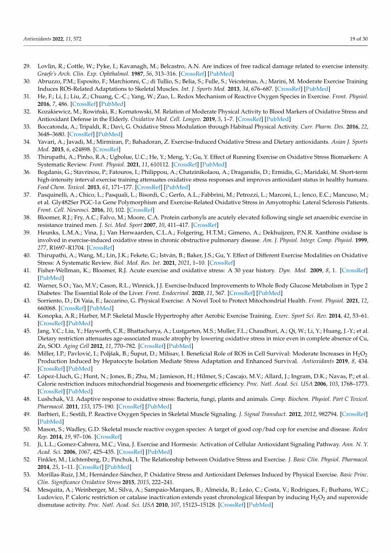
Antioxidants 2022, 11, 572 19 of 30
29. Lovlin, R.; Cottle, W.; Pyke, I.; Kavanagh, M.; Belcastro, A.N. Are indices of free radical damage related to exercise intensity.Graefe’s Arch. Clin. Exp. Ophthalmol. 1987, 56, 313–316. [CrossRef] [PubMed]
30. Abruzzo, P.M.; Esposito, F.; Marchionni, C.; di Tullio, S.; Belia, S.; Fulle, S.; Veicsteinas, A.; Marini, M. Moderate Exercise TrainingInduces ROS-Related Adaptations to Skeletal Muscles. Int. J. Sports Med. 2013, 34, 676–687. [CrossRef] [PubMed]
31. He, F.; Li, J.; Liu, Z.; Chuang, C.-C.; Yang, W.; Zuo, L. Redox Mechanism of Reactive Oxygen Species in Exercise. Front. Physiol.2016, 7, 486. [CrossRef] [PubMed]
32. Kozakiewicz, M.; Rowinski, R.; Kornatowski, M. Relation of Moderate Physical Activity to Blood Markers of Oxidative Stress andAntioxidant Defense in the Elderly. Oxidative Med. Cell. Longev. 2019, 5, 1–7. [CrossRef] [PubMed]
33. Boccatonda, A.; Tripaldi, R.; Davì, G. Oxidative Stress Modulation through Habitual Physical Activity. Curr. Pharm. Des. 2016, 22,3648–3680. [CrossRef] [PubMed]
34. Yavari, A.; Javadi, M.; Mirmiran, P.; Bahadoran, Z. Exercise-Induced Oxidative Stress and Dietary antioxidants. Asian J. SportsMed. 2015, 6, e24898. [CrossRef]
35. Thirupathi, A.; Pinho, R.A.; Ugbolue, U.C.; He, Y.; Meng, Y.; Gu, Y. Effect of Running Exercise on Oxidative Stress Biomarkers: ASystematic Review. Front. Physiol. 2021, 11, 610112. [CrossRef] [PubMed]
36. Bogdanis, G.; Stavrinou, P.; Fatouros, I.; Philippou, A.; Chatzinikolaou, A.; Draganidis, D.; Ermidis, G.; Maridaki, M. Short-termhigh-intensity interval exercise training attenuates oxidative stress responses and improves antioxidant status in healthy humans.Food Chem. Toxicol. 2013, 61, 171–177. [CrossRef] [PubMed]
37. Pasquinelli, A.; Chico, L.; Pasquali, L.; Bisordi, C.; Gerfo, A.L.; Fabbrini, M.; Petrozzi, L.; Marconi, L.; Ienco, E.C.; Mancuso, M.;et al. Gly482Ser PGC-1α Gene Polymorphism and Exercise-Related Oxidative Stress in Amyotrophic Lateral Sclerosis Patients.Front. Cell. Neurosci. 2016, 10, 102. [CrossRef]
38. Bloomer, R.J.; Fry, A.C.; Falvo, M.; Moore, C.A. Protein carbonyls are acutely elevated following single set anaerobic exercise inresistance trained men. J. Sci. Med. Sport 2007, 10, 411–417. [CrossRef]
39. Heunks, L.M.A.; Vina, J.; Van Herwaarden, C.L.A.; Folgering, H.T.M.; Gimeno, A.; Dekhuijzen, P.N.R. Xanthine oxidase isinvolved in exercise-induced oxidative stress in chronic obstructive pulmonary disease. Am. J. Physiol. Integr. Comp. Physiol. 1999,277, R1697–R1704. [CrossRef]
40. Thirupathi, A.; Wang, M.; Lin, J.K.; Fekete, G.; István, B.; Baker, J.S.; Gu, Y. Effect of Different Exercise Modalities on OxidativeStress: A Systematic Review. Biol. Med. Res. Int. 2021, 2021, 1–10. [CrossRef]
41. Fisher-Wellman, K.; Bloomer, R.J. Acute exercise and oxidative stress: A 30 year history. Dyn. Med. 2009, 8, 1. [CrossRef][PubMed]
42. Warner, S.O.; Yao, M.V.; Cason, R.L.; Winnick, J.J. Exercise-Induced Improvements to Whole Body Glucose Metabolism in Type 2Diabetes: The Essential Role of the Liver. Front. Endocrinol. 2020, 11, 567. [CrossRef] [PubMed]
43. Sorriento, D.; Di Vaia, E.; Iaccarino, G. Physical Exercise: A Novel Tool to Protect Mitochondrial Health. Front. Physiol. 2021, 12,660068. [CrossRef] [PubMed]
44. Konopka, A.R.; Harber, M.P. Skeletal Muscle Hypertrophy after Aerobic Exercise Training. Exerc. Sport Sci. Rev. 2014, 42, 53–61.[CrossRef] [PubMed]
45. Jang, Y.C.; Liu, Y.; Hayworth, C.R.; Bhattacharya, A.; Lustgarten, M.S.; Muller, F.L.; Chaudhuri, A.; Qi, W.; Li, Y.; Huang, J.-Y.; et al.Dietary restriction attenuates age-associated muscle atrophy by lowering oxidative stress in mice even in complete absence of Cu,Zn, SOD. Aging Cell 2012, 11, 770–782. [CrossRef] [PubMed]
46. Miller, I.P.; Pavlovic, I.; Poljšak, B.; Šuput, D.; Milisav, I. Beneficial Role of ROS in Cell Survival: Moderate Increases in H2O2Production Induced by Hepatocyte Isolation Mediate Stress Adaptation and Enhanced Survival. Antioxidants 2019, 8, 434.[CrossRef] [PubMed]
47. López-Lluch, G.; Hunt, N.; Jones, B.; Zhu, M.; Jamieson, H.; Hilmer, S.; Cascajo, M.V.; Allard, J.; Ingram, D.K.; Navas, P.; et al.Calorie restriction induces mitochondrial biogenesis and bioenergetic efficiency. Proc. Natl. Acad. Sci. USA 2006, 103, 1768–1773.[CrossRef] [PubMed]
48. Lushchak, V.I. Adaptive response to oxidative stress: Bacteria, fungi, plants and animals. Comp. Biochem. Physiol. Part C Toxicol.Pharmacol. 2011, 153, 175–190. [CrossRef] [PubMed]
49. Barbieri, E.; Sestili, P. Reactive Oxygen Species in Skeletal Muscle Signaling. J. Signal Transduct. 2012, 2012, 982794. [CrossRef][PubMed]
50. Mason, S.; Wadley, G.D. Skeletal muscle reactive oxygen species: A target of good cop/bad cop for exercise and disease. RedoxRep. 2014, 19, 97–106. [CrossRef]
51. Ji, L.L.; Gomez-Cabrera, M.C.; Vina, J. Exercise and Hormesis: Activation of Cellular Antioxidant Signaling Pathway. Ann. N. Y.Acad. Sci. 2006, 1067, 425–435. [CrossRef] [PubMed]
52. Finkler, M.; Lichtenberg, D.; Pinchuk, I. The Relationship between Oxidative Stress and Exercise. J. Basic Clin. Physiol. Pharmacol.2014, 25, 1–11. [CrossRef] [PubMed]
53. Morillas-Ruiz, J.M.; Hernández-Sánchez, P. Oxidative Stress and Antioxidant Defenses Induced by Physical Exercise. Basic Princ.Clin. Significance Oxidative Stress 2015, 2015, 222–241.
54. Mesquita, A.; Weinberger, M.; Silva, A.; Sampaio-Marques, B.; Almeida, B.; Leão, C.; Costa, V.; Rodrigues, F.; Burhans, W.C.;Ludovico, P. Caloric restriction or catalase inactivation extends yeast chronological lifespan by inducing H2O2 and superoxidedismutase activity. Proc. Natl. Acad. Sci. USA 2010, 107, 15123–15128. [CrossRef] [PubMed]
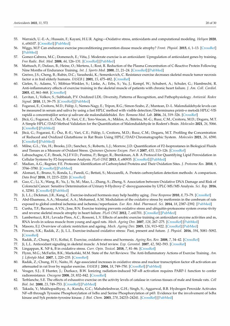
Antioxidants 2022, 11, 572 20 of 30
55. Warraich, U.-E.-A.; Hussain, F.; Kayani, H.U.R. Aging—Oxidative stress, antioxidants and computational modeling. Heliyon 2020,6, e04107. [CrossRef] [PubMed]
56. Wiggs, M.P. Can endurance exercise preconditioning prevention disuse muscle atrophy? Front. Physiol. 2015, 6, 1–13. [CrossRef][PubMed]
57. Gomez-Cabrera, M.C.; Domenech, E.; Viña, J. Moderate exercise is an antioxidant: Upregulation of antioxidant genes by training.Free Radic. Biol. Med. 2008, 44, 126–131. [CrossRef] [PubMed]
58. Mattusch, F.; Dufaux, B.; Heine, O.; Mertens, I.; Rost, R. Reduction of the Plasma Concentration of C-Reactive Protein FollowingNine Months of Endurance Training. Int. J. Sports Med. 2000, 21, 21–24. [CrossRef] [PubMed]
59. Greiwe, J.S.; Cheng, B.; Rubin, D.C.; Yarasheski, K.; Semenkovich, C. Resistance exercise decreases skeletal muscle tumor necrosisfactor α in frail elderly humans. FASEB J. 2001, 15, 475–482. [CrossRef]
60. Gielen, S.; Adams, V.; Möbius-Winkler, S.; Linke, A.; Erbs, S.; Yu, J.; Kempf, W.; Schubert, A.; Schuler, G.; Hambrecht, R.Anti-inflammatory effects of exercise training in the skeletal muscle of patients with chronic heart failure. J. Am. Coll. Cardiol.2003, 42, 861–868. [CrossRef]
61. Levitan, I.; Volkov, S.; Subbaiah, P.V. Oxidized LDL: Diversity, Patterns of Recognition, and Pathophysiology. Antioxid. RedoxSignal. 2010, 13, 39–75. [CrossRef] [PubMed]
62. Fogarasi, E.; Croitoru, M.D.; Fülöp, I.; Nemes-Nagy, E.; Tripon, R.G.; Simon-Szabo, Z.; Muntean, D.-L. Malondialdehyde levels canbe measured in serum and saliva by using a fast HPLC method with visible detection/Determinarea printr-o metodă HPLC-VISrapidă a concentratiilor serice si salivare ale malondialdehidei. Rev. Romana Med. Lab. 2016, 24, 319–326. [CrossRef]
63. Jîtcă, G.; Fogarasi, E.; Osz, B.-E.; Vari, C.E.; Tero-Vescan, A.; Miklos, A.; Bătrînu, M.-G.; Rusz, C.M.; Croitoru, M.D.; Dogaru, M.T.A Simple HPLC/DAD Method Validation for the Quantification of Malondialdehyde in Rodent’s Brain. Molecules 2021, 26, 5066.[CrossRef] [PubMed]
64. Jîtcă, G.; Fogarasi, E.; Osz, B.-E.; Vari, C.E.; Fülöp, I.; Croitoru, M.D.; Rusz, C.M.; Dogaru, M.T. Profiling the Concentrationof Reduced and Oxidized Glutathione in Rat Brain Using HPLC/DAD Chromatographic System. Molecules 2021, 26, 6590.[CrossRef] [PubMed]
65. Milne, G.L.; Yin, H.; Brooks, J.D.; Sanchez, S.; Roberts, L.J.; Morrow, J.D. Quantification of F2-Isoprostanes in Biological Fluidsand Tissues as a Measure of Oxidant Stress. Quinones Quinone Enzym. Part A 2007, 433, 113–126. [CrossRef]
66. Labuschagne, C.F.; Broek, N.J.F.V.D.; Postma, P.; Berger, R.; Brenkman, A.B. A Protocol for Quantifying Lipid Peroxidation inCellular Systems by F2-Isoprostane Analysis. PLoS ONE 2013, 8, e80935. [CrossRef] [PubMed]
67. Madian, A.G.; Regnier, F.E. Proteomic Identification of Carbonylated Proteins and Their Oxidation Sites. J. Proteome Res. 2010, 9,3766–3780. [CrossRef] [PubMed]
68. Alomari, E.; Bruno, S.; Ronda, L.; Paredi, G.; Bettati, S.; Mozzarelli, A. Protein carbonylation detection methods: A comparison.Data Brief 2018, 19, 2215–2220. [CrossRef]
69. Guo, C.; Li, X.; Wang, R.; Yu, J.; Ye, M.; Mao, L.; Zhang, S.; Zheng, S. Association between Oxidative DNA Damage and Risk ofColorectal Cancer: Sensitive Determination of Urinary 8-Hydroxy-2′-deoxyguanosine by UPLC-MS/MS Analysis. Sci. Rep. 2016,6, 32581. [CrossRef] [PubMed]
70. Ji, L.L.; Dickman, J.R.; Kang, C. Exercise-induced hormesis may help healthy aging. Dose Response 2010, 8, 73–79. [CrossRef]71. Abd-Elsameea, A.A.; Moustaf, A.A.; Mohamed, A.M. Modulation of the oxidative stress by metformin in the cerebrum of rats
exposed to global cerebral ischemia and ischemia/reperfusion. Eur. Rev. Med. Pharmacol. Sci. 2014, 18, 2387–2392. [PubMed]72. Cunha, T.F.; Bacurau, A.V.N.; Jose, B.N. Exercise training prevents oxidative stress and ubiquitin-proteasome system overac-tivity
and reverse skeletal muscle atrophy in heart failure. PLoS ONE 2012, 7, e41701. [CrossRef] [PubMed]73. Lambertucci, R.H.; Levada-Pires, A.C.; Rossoni, L.V. Effects of aerobic exercise training on antioxidant enzyme activities and m,
RNA levels in soleus muscle from young and aged rats. Mech. Ageing Dev. 2007, 128, 267–275. [CrossRef] [PubMed]74. Masoro, E.J. Overview of caloric restriction and ageing. Mech. Ageing Dev. 2005, 126, 913–922. [CrossRef] [PubMed]75. Powers, S.K.; Radák, Z.; Ji, L.L. Exercise-induced oxidative stress: Past, present and future. J. Physiol. 2016, 594, 5081–5092.
[CrossRef]76. Radák, Z.; Chung, H.Y.; Koltai, E. Exercise, oxidative stress and hormesis. Ageing Res, Rev. 2008, 7, 34–42. [CrossRef]77. Ji, L.L. Antioxidant signaling in skeletal muscle: A brief review. Exp. Gerontol. 2007, 42, 582–593. [CrossRef]78. Lingappan, K. NF-k, B in oxidative stress. Curr. Opin. Toxicol. 2018, 7, 81–86. [CrossRef]79. Flynn, M.G.; McFarlin, B.K.; Markofski, M.M. State of the Art Reviews: The Anti-Inflammatory Actions of Exercise Training. Am.
J. Lifestyle Med. 2007, 1, 220–235. [CrossRef]80. Radák, Z.; Chung, H.Y.; Naito, H. Age-associated increases in oxidative stress and nuclear transcription factor κB activation are
attenuated in rat liver by regular exercise. FASEB J. 2004, 18, 749–750. [CrossRef] [PubMed]81. Veuger, S.J.; E Hunter, J.; Durkacz, B.W. Ionizing radiation-induced NF-κB activation requires PARP-1 function to confer
radioresistance. Oncogene 2008, 28, 832–842. [CrossRef]82. Terblanche, S.E. The effects of exhaustive exercise on the activity levels of catalase in various tissues of male and female rats. Cell
Biol. Int. 2000, 23, 749–753. [CrossRef] [PubMed]83. Takada, Y.; Mukhopadhyay, A.; Kundu, G.C.; Mahabeleshwar, G.H.; Singh, S.; Aggarwal, B.B. Hydrogen Peroxide Activates
NF-κB through Tyrosine Phosphorylation of IκBα and Serine Phosphorylation of p65: Evidence for the involvement of IκBαkinase and Syk protein-tyrosine kinase. J. Biol. Chem. 2003, 278, 24233–24241. [CrossRef] [PubMed]
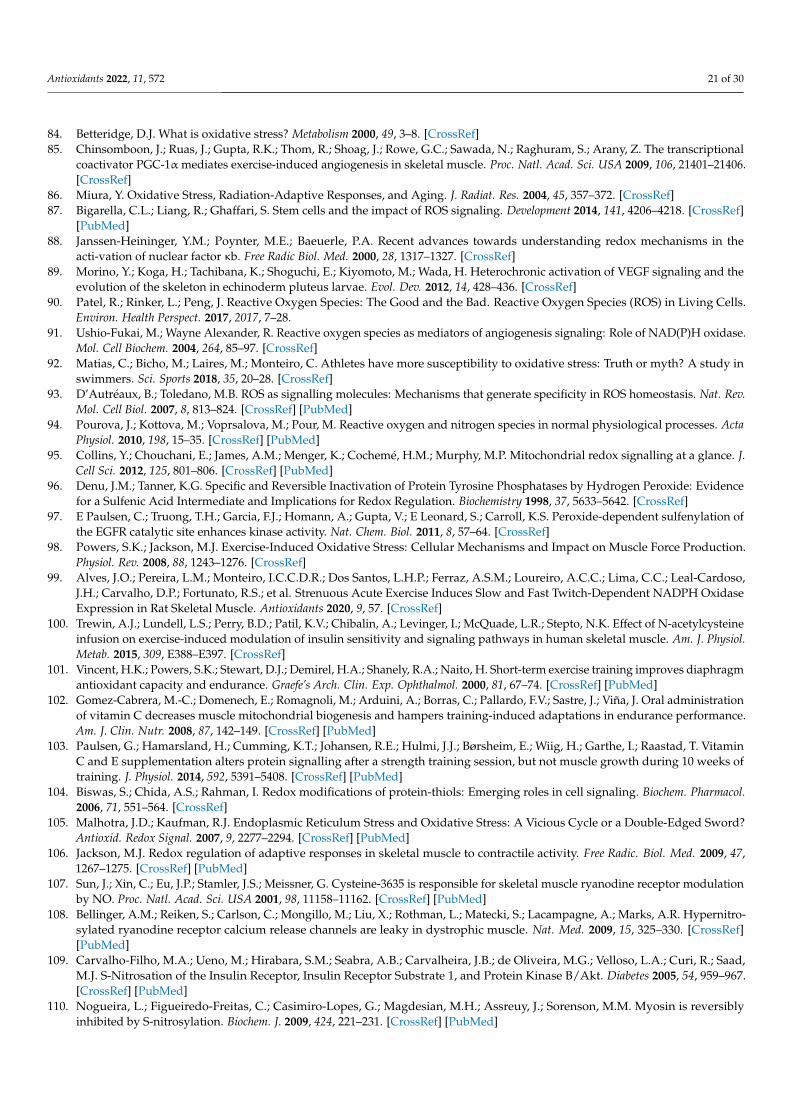
Antioxidants 2022, 11, 572 21 of 30
84. Betteridge, D.J. What is oxidative stress? Metabolism 2000, 49, 3–8. [CrossRef]85. Chinsomboon, J.; Ruas, J.; Gupta, R.K.; Thom, R.; Shoag, J.; Rowe, G.C.; Sawada, N.; Raghuram, S.; Arany, Z. The transcriptional
coactivator PGC-1α mediates exercise-induced angiogenesis in skeletal muscle. Proc. Natl. Acad. Sci. USA 2009, 106, 21401–21406.[CrossRef]
86. Miura, Y. Oxidative Stress, Radiation-Adaptive Responses, and Aging. J. Radiat. Res. 2004, 45, 357–372. [CrossRef]87. Bigarella, C.L.; Liang, R.; Ghaffari, S. Stem cells and the impact of ROS signaling. Development 2014, 141, 4206–4218. [CrossRef]
[PubMed]88. Janssen-Heininger, Y.M.; Poynter, M.E.; Baeuerle, P.A. Recent advances towards understanding redox mechanisms in the
acti-vation of nuclear factor κb. Free Radic Biol. Med. 2000, 28, 1317–1327. [CrossRef]89. Morino, Y.; Koga, H.; Tachibana, K.; Shoguchi, E.; Kiyomoto, M.; Wada, H. Heterochronic activation of VEGF signaling and the
evolution of the skeleton in echinoderm pluteus larvae. Evol. Dev. 2012, 14, 428–436. [CrossRef]90. Patel, R.; Rinker, L.; Peng, J. Reactive Oxygen Species: The Good and the Bad. Reactive Oxygen Species (ROS) in Living Cells.
Environ. Health Perspect. 2017, 2017, 7–28.91. Ushio-Fukai, M.; Wayne Alexander, R. Reactive oxygen species as mediators of angiogenesis signaling: Role of NAD(P)H oxidase.
Mol. Cell Biochem. 2004, 264, 85–97. [CrossRef]92. Matias, C.; Bicho, M.; Laires, M.; Monteiro, C. Athletes have more susceptibility to oxidative stress: Truth or myth? A study in
swimmers. Sci. Sports 2018, 35, 20–28. [CrossRef]93. D’Autréaux, B.; Toledano, M.B. ROS as signalling molecules: Mechanisms that generate specificity in ROS homeostasis. Nat. Rev.
Mol. Cell Biol. 2007, 8, 813–824. [CrossRef] [PubMed]94. Pourova, J.; Kottova, M.; Voprsalova, M.; Pour, M. Reactive oxygen and nitrogen species in normal physiological processes. Acta
Physiol. 2010, 198, 15–35. [CrossRef] [PubMed]95. Collins, Y.; Chouchani, E.; James, A.M.; Menger, K.; Cochemé, H.M.; Murphy, M.P. Mitochondrial redox signalling at a glance. J.
Cell Sci. 2012, 125, 801–806. [CrossRef] [PubMed]96. Denu, J.M.; Tanner, K.G. Specific and Reversible Inactivation of Protein Tyrosine Phosphatases by Hydrogen Peroxide: Evidence
for a Sulfenic Acid Intermediate and Implications for Redox Regulation. Biochemistry 1998, 37, 5633–5642. [CrossRef]97. E Paulsen, C.; Truong, T.H.; Garcia, F.J.; Homann, A.; Gupta, V.; E Leonard, S.; Carroll, K.S. Peroxide-dependent sulfenylation of
the EGFR catalytic site enhances kinase activity. Nat. Chem. Biol. 2011, 8, 57–64. [CrossRef]98. Powers, S.K.; Jackson, M.J. Exercise-Induced Oxidative Stress: Cellular Mechanisms and Impact on Muscle Force Production.
Physiol. Rev. 2008, 88, 1243–1276. [CrossRef]99. Alves, J.O.; Pereira, L.M.; Monteiro, I.C.C.D.R.; Dos Santos, L.H.P.; Ferraz, A.S.M.; Loureiro, A.C.C.; Lima, C.C.; Leal-Cardoso,
J.H.; Carvalho, D.P.; Fortunato, R.S.; et al. Strenuous Acute Exercise Induces Slow and Fast Twitch-Dependent NADPH OxidaseExpression in Rat Skeletal Muscle. Antioxidants 2020, 9, 57. [CrossRef]
100. Trewin, A.J.; Lundell, L.S.; Perry, B.D.; Patil, K.V.; Chibalin, A.; Levinger, I.; McQuade, L.R.; Stepto, N.K. Effect of N-acetylcysteineinfusion on exercise-induced modulation of insulin sensitivity and signaling pathways in human skeletal muscle. Am. J. Physiol.Metab. 2015, 309, E388–E397. [CrossRef]
101. Vincent, H.K.; Powers, S.K.; Stewart, D.J.; Demirel, H.A.; Shanely, R.A.; Naito, H. Short-term exercise training improves diaphragmantioxidant capacity and endurance. Graefe’s Arch. Clin. Exp. Ophthalmol. 2000, 81, 67–74. [CrossRef] [PubMed]
102. Gomez-Cabrera, M.-C.; Domenech, E.; Romagnoli, M.; Arduini, A.; Borras, C.; Pallardo, F.V.; Sastre, J.; Viña, J. Oral administrationof vitamin C decreases muscle mitochondrial biogenesis and hampers training-induced adaptations in endurance performance.Am. J. Clin. Nutr. 2008, 87, 142–149. [CrossRef] [PubMed]
103. Paulsen, G.; Hamarsland, H.; Cumming, K.T.; Johansen, R.E.; Hulmi, J.J.; Børsheim, E.; Wiig, H.; Garthe, I.; Raastad, T. VitaminC and E supplementation alters protein signalling after a strength training session, but not muscle growth during 10 weeks oftraining. J. Physiol. 2014, 592, 5391–5408. [CrossRef] [PubMed]
104. Biswas, S.; Chida, A.S.; Rahman, I. Redox modifications of protein-thiols: Emerging roles in cell signaling. Biochem. Pharmacol.2006, 71, 551–564. [CrossRef]
105. Malhotra, J.D.; Kaufman, R.J. Endoplasmic Reticulum Stress and Oxidative Stress: A Vicious Cycle or a Double-Edged Sword?Antioxid. Redox Signal. 2007, 9, 2277–2294. [CrossRef] [PubMed]
106. Jackson, M.J. Redox regulation of adaptive responses in skeletal muscle to contractile activity. Free Radic. Biol. Med. 2009, 47,1267–1275. [CrossRef] [PubMed]
107. Sun, J.; Xin, C.; Eu, J.P.; Stamler, J.S.; Meissner, G. Cysteine-3635 is responsible for skeletal muscle ryanodine receptor modulationby NO. Proc. Natl. Acad. Sci. USA 2001, 98, 11158–11162. [CrossRef] [PubMed]
108. Bellinger, A.M.; Reiken, S.; Carlson, C.; Mongillo, M.; Liu, X.; Rothman, L.; Matecki, S.; Lacampagne, A.; Marks, A.R. Hypernitro-sylated ryanodine receptor calcium release channels are leaky in dystrophic muscle. Nat. Med. 2009, 15, 325–330. [CrossRef][PubMed]
109. Carvalho-Filho, M.A.; Ueno, M.; Hirabara, S.M.; Seabra, A.B.; Carvalheira, J.B.; de Oliveira, M.G.; Velloso, L.A.; Curi, R.; Saad,M.J. S-Nitrosation of the Insulin Receptor, Insulin Receptor Substrate 1, and Protein Kinase B/Akt. Diabetes 2005, 54, 959–967.[CrossRef] [PubMed]
110. Nogueira, L.; Figueiredo-Freitas, C.; Casimiro-Lopes, G.; Magdesian, M.H.; Assreuy, J.; Sorenson, M.M. Myosin is reversiblyinhibited by S-nitrosylation. Biochem. J. 2009, 424, 221–231. [CrossRef] [PubMed]
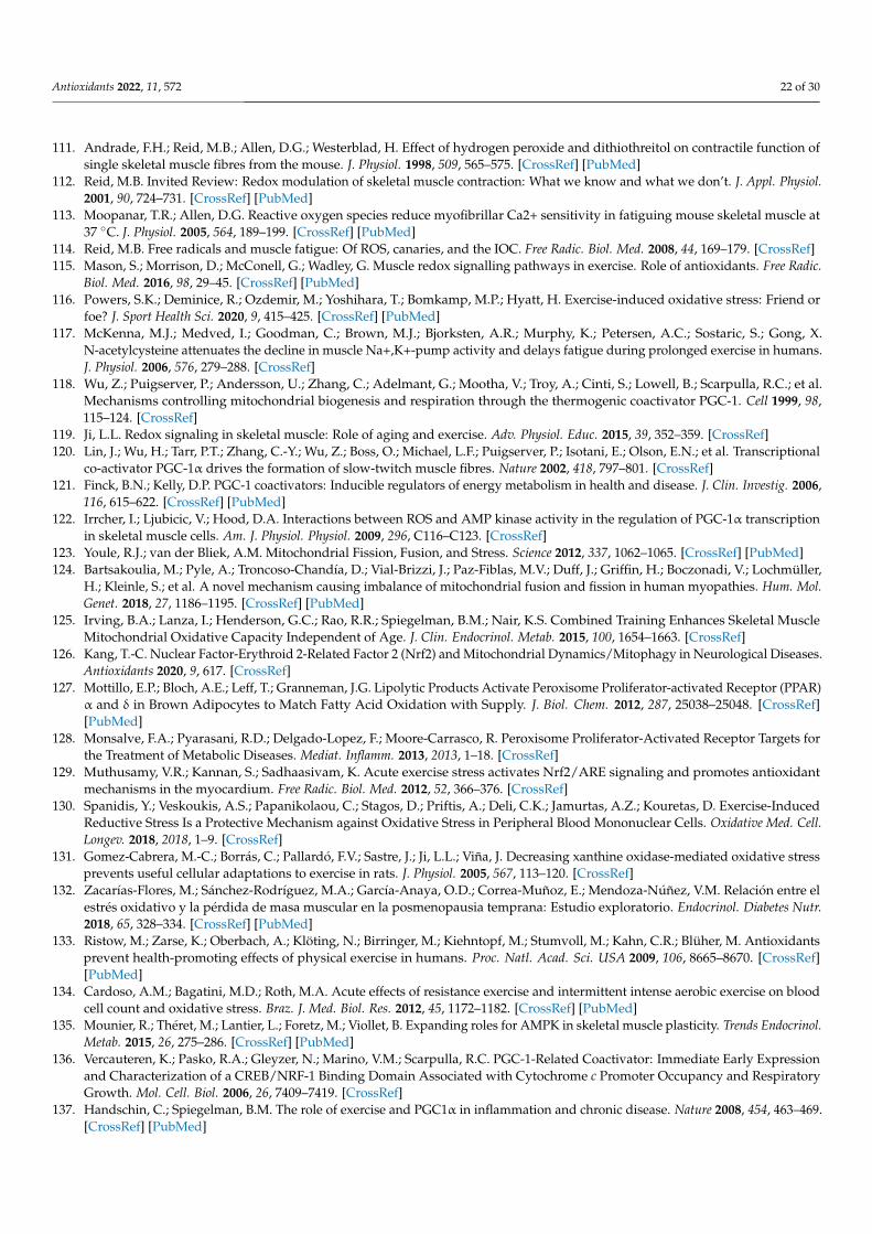
Antioxidants 2022, 11, 572 22 of 30
111. Andrade, F.H.; Reid, M.B.; Allen, D.G.; Westerblad, H. Effect of hydrogen peroxide and dithiothreitol on contractile function ofsingle skeletal muscle fibres from the mouse. J. Physiol. 1998, 509, 565–575. [CrossRef] [PubMed]
112. Reid, M.B. Invited Review: Redox modulation of skeletal muscle contraction: What we know and what we don’t. J. Appl. Physiol.2001, 90, 724–731. [CrossRef] [PubMed]
113. Moopanar, T.R.; Allen, D.G. Reactive oxygen species reduce myofibrillar Ca2+ sensitivity in fatiguing mouse skeletal muscle at37 ◦C. J. Physiol. 2005, 564, 189–199. [CrossRef] [PubMed]
114. Reid, M.B. Free radicals and muscle fatigue: Of ROS, canaries, and the IOC. Free Radic. Biol. Med. 2008, 44, 169–179. [CrossRef]115. Mason, S.; Morrison, D.; McConell, G.; Wadley, G. Muscle redox signalling pathways in exercise. Role of antioxidants. Free Radic.
Biol. Med. 2016, 98, 29–45. [CrossRef] [PubMed]116. Powers, S.K.; Deminice, R.; Ozdemir, M.; Yoshihara, T.; Bomkamp, M.P.; Hyatt, H. Exercise-induced oxidative stress: Friend or
foe? J. Sport Health Sci. 2020, 9, 415–425. [CrossRef] [PubMed]117. McKenna, M.J.; Medved, I.; Goodman, C.; Brown, M.J.; Bjorksten, A.R.; Murphy, K.; Petersen, A.C.; Sostaric, S.; Gong, X.
N-acetylcysteine attenuates the decline in muscle Na+,K+-pump activity and delays fatigue during prolonged exercise in humans.J. Physiol. 2006, 576, 279–288. [CrossRef]
118. Wu, Z.; Puigserver, P.; Andersson, U.; Zhang, C.; Adelmant, G.; Mootha, V.; Troy, A.; Cinti, S.; Lowell, B.; Scarpulla, R.C.; et al.Mechanisms controlling mitochondrial biogenesis and respiration through the thermogenic coactivator PGC-1. Cell 1999, 98,115–124. [CrossRef]
119. Ji, L.L. Redox signaling in skeletal muscle: Role of aging and exercise. Adv. Physiol. Educ. 2015, 39, 352–359. [CrossRef]120. Lin, J.; Wu, H.; Tarr, P.T.; Zhang, C.-Y.; Wu, Z.; Boss, O.; Michael, L.F.; Puigserver, P.; Isotani, E.; Olson, E.N.; et al. Transcriptional
co-activator PGC-1α drives the formation of slow-twitch muscle fibres. Nature 2002, 418, 797–801. [CrossRef]121. Finck, B.N.; Kelly, D.P. PGC-1 coactivators: Inducible regulators of energy metabolism in health and disease. J. Clin. Investig. 2006,
116, 615–622. [CrossRef] [PubMed]122. Irrcher, I.; Ljubicic, V.; Hood, D.A. Interactions between ROS and AMP kinase activity in the regulation of PGC-1α transcription
in skeletal muscle cells. Am. J. Physiol. Physiol. 2009, 296, C116–C123. [CrossRef]123. Youle, R.J.; van der Bliek, A.M. Mitochondrial Fission, Fusion, and Stress. Science 2012, 337, 1062–1065. [CrossRef] [PubMed]124. Bartsakoulia, M.; Pyle, A.; Troncoso-Chandía, D.; Vial-Brizzi, J.; Paz-Fiblas, M.V.; Duff, J.; Griffin, H.; Boczonadi, V.; Lochmüller,
H.; Kleinle, S.; et al. A novel mechanism causing imbalance of mitochondrial fusion and fission in human myopathies. Hum. Mol.Genet. 2018, 27, 1186–1195. [CrossRef] [PubMed]
125. Irving, B.A.; Lanza, I.; Henderson, G.C.; Rao, R.R.; Spiegelman, B.M.; Nair, K.S. Combined Training Enhances Skeletal MuscleMitochondrial Oxidative Capacity Independent of Age. J. Clin. Endocrinol. Metab. 2015, 100, 1654–1663. [CrossRef]
126. Kang, T.-C. Nuclear Factor-Erythroid 2-Related Factor 2 (Nrf2) and Mitochondrial Dynamics/Mitophagy in Neurological Diseases.Antioxidants 2020, 9, 617. [CrossRef]
127. Mottillo, E.P.; Bloch, A.E.; Leff, T.; Granneman, J.G. Lipolytic Products Activate Peroxisome Proliferator-activated Receptor (PPAR)α and δ in Brown Adipocytes to Match Fatty Acid Oxidation with Supply. J. Biol. Chem. 2012, 287, 25038–25048. [CrossRef][PubMed]
128. Monsalve, F.A.; Pyarasani, R.D.; Delgado-Lopez, F.; Moore-Carrasco, R. Peroxisome Proliferator-Activated Receptor Targets forthe Treatment of Metabolic Diseases. Mediat. Inflamm. 2013, 2013, 1–18. [CrossRef]
129. Muthusamy, V.R.; Kannan, S.; Sadhaasivam, K. Acute exercise stress activates Nrf2/ARE signaling and promotes antioxidantmechanisms in the myocardium. Free Radic. Biol. Med. 2012, 52, 366–376. [CrossRef]
130. Spanidis, Y.; Veskoukis, A.S.; Papanikolaou, C.; Stagos, D.; Priftis, A.; Deli, C.K.; Jamurtas, A.Z.; Kouretas, D. Exercise-InducedReductive Stress Is a Protective Mechanism against Oxidative Stress in Peripheral Blood Mononuclear Cells. Oxidative Med. Cell.Longev. 2018, 2018, 1–9. [CrossRef]
131. Gomez-Cabrera, M.-C.; Borrás, C.; Pallardó, F.V.; Sastre, J.; Ji, L.L.; Viña, J. Decreasing xanthine oxidase-mediated oxidative stressprevents useful cellular adaptations to exercise in rats. J. Physiol. 2005, 567, 113–120. [CrossRef]
132. Zacarías-Flores, M.; Sánchez-Rodríguez, M.A.; García-Anaya, O.D.; Correa-Muñoz, E.; Mendoza-Núñez, V.M. Relación entre elestrés oxidativo y la pérdida de masa muscular en la posmenopausia temprana: Estudio exploratorio. Endocrinol. Diabetes Nutr.2018, 65, 328–334. [CrossRef] [PubMed]
133. Ristow, M.; Zarse, K.; Oberbach, A.; Klöting, N.; Birringer, M.; Kiehntopf, M.; Stumvoll, M.; Kahn, C.R.; Blüher, M. Antioxidantsprevent health-promoting effects of physical exercise in humans. Proc. Natl. Acad. Sci. USA 2009, 106, 8665–8670. [CrossRef][PubMed]
134. Cardoso, A.M.; Bagatini, M.D.; Roth, M.A. Acute effects of resistance exercise and intermittent intense aerobic exercise on bloodcell count and oxidative stress. Braz. J. Med. Biol. Res. 2012, 45, 1172–1182. [CrossRef] [PubMed]
135. Mounier, R.; Théret, M.; Lantier, L.; Foretz, M.; Viollet, B. Expanding roles for AMPK in skeletal muscle plasticity. Trends Endocrinol.Metab. 2015, 26, 275–286. [CrossRef] [PubMed]
136. Vercauteren, K.; Pasko, R.A.; Gleyzer, N.; Marino, V.M.; Scarpulla, R.C. PGC-1-Related Coactivator: Immediate Early Expressionand Characterization of a CREB/NRF-1 Binding Domain Associated with Cytochrome c Promoter Occupancy and RespiratoryGrowth. Mol. Cell. Biol. 2006, 26, 7409–7419. [CrossRef]
137. Handschin, C.; Spiegelman, B.M. The role of exercise and PGC1α in inflammation and chronic disease. Nature 2008, 454, 463–469.[CrossRef] [PubMed]
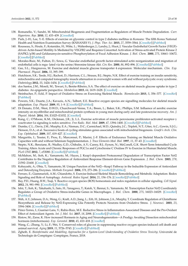
Antioxidants 2022, 11, 572 23 of 30
138. Romanello, V.; Sandri, M. Mitochondrial Biogenesis and Fragmentation as Regulators of Muscle Protein Degradation. Curr.Hypertens. Rep. 2010, 12, 433–439. [CrossRef]
139. Park, J.-H.; Lee, Y.-E. Effects of exercise on glycemic control in type 2 diabetes mellitus in Koreans: The fifth Korea NationalHealth and Nutrition Examination Survey (KNHANES V). J. Phys. Ther. Sci. 2015, 27, 3559–3564. [CrossRef] [PubMed]
140. Rousseau, S.; Houle, F.; Kotanides, H.; Witte, L.; Waltenberger, J.; Landry, J.; Huot, J. Vascular Endothelial Growth Factor (VEGF)-driven Actin-based Motility Is Mediated by VEGFR2 and Requires Concerted Activation of Stress-activated Protein Kinase 2(SAPK2/p38) and Geldanamycin-sensitive Phosphorylation of Focal Adhesion Kinase. J. Biol. Chem. 2000, 275, 10661–10672.[CrossRef] [PubMed]
141. Morales-Ruiz, M.; Fulton, D.; Sowa, G. Vascular endothelial growth factor-stimulated actin reorganization and migration ofendothelial cells is regu- lated via the serine/threonine kinase Akt. Circ Res. 2000, 86, 892–896. [CrossRef] [PubMed]
142. Gee, S.L.; Hargreaves, M. Exercise and skeletal muscle glucose transporter 4 expression: Molecular mechanism. Clin. Exp.Pharmacol. Physiol. 2006, 33, 395–399.
143. Hutchison, S.K.; Teede, H.J.; Rachon, D.; Harrison, C.L.; Strauss, B.J.; Stepto, N.K. Effect of exercise training on insulin sensitivity,mitochondria and computed tomography muscle attenuation in overweight women with and without polycystic ovary syndrome.Diabetology 2012, 55, 1424–1434. [CrossRef] [PubMed]
144. dos Santos, J.M.; Moreli, M.; Tewari, S.; Benite-Ribeiro, S.A. The effect of exercise on skeletal muscle glucose uptake in type 2diabetes: An epigenetic perspective. Metabolism 2015, 64, 1619–1628. [CrossRef]
145. Steinbacher, P.; Eckl, P. Impact of Oxidative Stress on Exercising Skeletal Muscle. Biomolecules 2015, 5, 356–377. [CrossRef][PubMed]
146. Powers, S.K.; Duarte, J.A.; Kavazis, A.N.; Talbert, E.E. Reactive oxygen species are signalling molecules for skeletal muscleadaptation. Exp. Physiol. 2009, 95, 1–9. [CrossRef] [PubMed]
147. Di Donato, D.M.; West, D.W.D.; Churchward-Venne, T.A.; Breen, L.; Baker, S.K.; Phillips, S.M. Influence of aerobic exerciseintensity on myofibrillar and mitochondrial protein synthesis in young men during early and late postexercise recovery. Am. J.Physiol. Metab. 2014, 306, E1025–E1032. [CrossRef]
148. Kang, C.; O’Moore, K.M.; Dickman, J.R.; Ji, L.L. Exercise activation of muscle peroxisome proliferator-activated receptor-γcoactivator-1α signaling is redox sensitive. Free Radic. Biol. Med. 2009, 47, 1394–1400. [CrossRef] [PubMed]
149. Dumke, C.L.; Davis, J.M.; Murphy, E.A.; Nieman, D.C.; Carmichael, M.D.; Quindry, J.C.; Triplett, N.T.; Utter, A.C.; Gowin, S.J.G.;Henson, D.A.; et al. Successive bouts of cycling stimulates genes associated with mitochondrial biogenesis. Graefe’s Arch. Clin.Exp. Ophthalmol. 2009, 107, 419–427. [CrossRef]
150. Margaritis, I.; Tessier, F.; Prou, E.; Marconnet, P.; Marini, J.-F. Effects of Endurance Training on Skeletal Muscle OxidativeCapacities with and without Selenium Supplementation. J. Trace Elements Med. Biol. 1997, 11, 37–43. [CrossRef]
151. Stepto, N.K.; Benziane, B.; Wadley, G.D.; Chibalin, A.V.; Canny, B.J.; Eynon, N.; McConell, G.K. Short-Term Intensified CycleTraining Alters Acute and Chronic Responses of PGC1α and Cytochrome C Oxidase IV to Exercise in Human Skeletal Muscle.PLoS ONE 2012, 7, e53080. [CrossRef] [PubMed]
152. McMahon, M.; Itoh, K.; Yamamoto, M.; Hayes, J. Keap1-dependent Proteasomal Degradation of Transcription Factor Nrf2Contributes to the Negative Regulation of Antioxidant Response Element-driven Gene Expression. J. Biol. Chem. 2003, 278,21592–21600. [CrossRef]
153. Kobayashi, A.; Ohta, T.; Yamamoto, M. Unique Function of the Nrf2—Keap1 Pathway in the Inducible Expression of Antioxidantand Detoxifying Enzymes. Methods Enzymol. 2004, 378, 273–286. [CrossRef] [PubMed]
154. Ferraro, E.; Giammarioli, A.M.; Chiandotto, S. Exercise-Induced Skeletal Muscle Remodeling and Metabolic Adaptation: RedoxSignaling and Role of Autophagy. Antioxid. Redox Signal. 2014, 21, 154–176. [CrossRef] [PubMed]
155. Ray, P.D.; Huang, B.W.; Tsuji, Y. Reactive oxygen species (ROS) homeostasis and redox regulation in cellular signaling. Cell Signal.2012, 24, 981–990. [CrossRef] [PubMed]
156. Ishii, T.; Itoh, K.; Takahashi, S.; Sato, H.; Yanagawa, T.; Katoh, Y.; Bannai, S.; Yamamoto, M. Transcription Factor Nrf2 CoordinatelyRegulates a Group of Oxidative Stress-inducible Genes in Macrophages. J. Biol. Chem. 2000, 275, 16023–16029. [CrossRef][PubMed]
157. Shih, A.Y.; Johnson, D.A.; Wong, G.; Kraft, A.D.; Jiang, L.; Erb, H.; Johnson, J.A.; Murphy, T. Coordinate Regulation of GlutathioneBiosynthesis and Release by Nrf2-Expressing Glia Potently Protects Neurons from Oxidative Stress. J. Neurosci. 2003, 23,3394–3406. [CrossRef] [PubMed]
158. Pérez-Torres, I.; Guarner-Lans, V.; Rubio-Ruiz, M.E. Reductive Stress in Inflammation-Associated Diseases and the Pro-OxidantEffect of Antioxidant Agents. Int. J. Mol. Sci. 2017, 18, 2098. [CrossRef] [PubMed]
159. Ristow, M.; Zarse, K. How increased Hormesis in Aging and Neurodegeneration—A Prodigy Awaiting Dissection mitochondrialhormesis (mitohormesis). Exp. Gerontol. 2010, 45, 410–418. [CrossRef]
160. Wang, C.; Zhang, Y.; Li, F.; Wei, Y. Conserved roles of glucose in suppressing reactive oxygen species-induced cell death andanimal survival. Aging 2019, 11, 5726–5743. [CrossRef] [PubMed]
161. Zgheib, E. Bioinformatic and Modelling Approaches for a System-Level Understanding of Oxidative Stress Toxicity; Université deTechnologie de Compiègne: Compiègne, France, 2018.
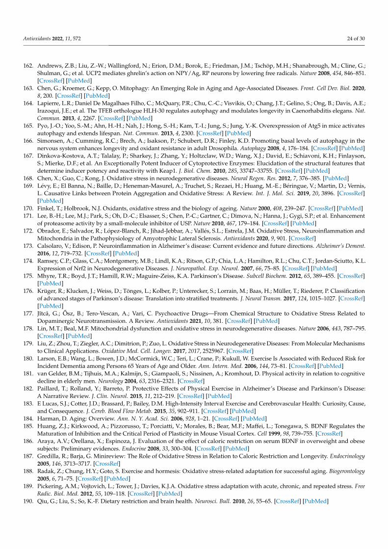
Antioxidants 2022, 11, 572 24 of 30
162. Andrews, Z.B.; Liu, Z.-W.; Wallingford, N.; Erion, D.M.; Borok, E.; Friedman, J.M.; Tschöp, M.H.; Shanabrough, M.; Cline, G.;Shulman, G.; et al. UCP2 mediates ghrelin’s action on NPY/Ag, RP neurons by lowering free radicals. Nature 2008, 454, 846–851.[CrossRef] [PubMed]
163. Chen, G.; Kroemer, G.; Kepp, O. Mitophagy: An Emerging Role in Aging and Age-Associated Diseases. Front. Cell Dev. Biol. 2020,8, 200. [CrossRef] [PubMed]
164. Lapierre, L.R.; Daniel De Magalhaes Filho, C.; McQuary, P.R.; Chu, C.-C.; Visvikis, O.; Chang, J.T.; Gelino, S.; Ong, B.; Davis, A.E.;Irazoqui, J.E.; et al. The TFEB orthologue HLH-30 regulates autophagy and modulates longevity in Caenorhabditis elegans. Nat.Commun. 2013, 4, 2267. [CrossRef] [PubMed]
165. Pyo, J.-O.; Yoo, S.-M.; Ahn, H.-H.; Nah, J.; Hong, S.-H.; Kam, T.-I.; Jung, S.; Jung, Y.-K. Overexpression of Atg5 in mice activatesautophagy and extends lifespan. Nat. Commun. 2013, 4, 2300. [CrossRef] [PubMed]
166. Simonsen, A.; Cumming, R.C.; Brech, A.; Isakson, P.; Schubert, D.R.; Finley, K.D. Promoting basal levels of autophagy in thenervous system enhances longevity and oxidant resistance in adult Drosophila. Autophagy 2008, 4, 176–184. [CrossRef] [PubMed]
167. Dinkova-Kostova, A.T.; Talalay, P.; Sharkey, J.; Zhang, Y.; Holtzclaw, W.D.; Wang, X.J.; David, E.; Schiavoni, K.H.; Finlayson,S.; Mierke, D.F.; et al. An Exceptionally Potent Inducer of Cytoprotective Enzymes: Elucidation of the structural features thatdetermine inducer potency and reactivity with Keap1. J. Biol. Chem. 2010, 285, 33747–33755. [CrossRef] [PubMed]
168. Chen, X.; Guo, C.; Kong, J. Oxidative stress in neurodegenerative diseases. Neural Regen. Res. 2012, 7, 376–385. [PubMed]169. Lévy, E.; El Banna, N.; Baïlle, D.; Heneman-Masurel, A.; Truchet, S.; Rezaei, H.; Huang, M.-E.; Béringue, V.; Martin, D.; Vernis,
L. Causative Links between Protein Aggregation and Oxidative Stress: A Review. Int. J. Mol. Sci. 2019, 20, 3896. [CrossRef][PubMed]
170. Finkel, T.; Holbrook, N.J. Oxidants, oxidative stress and the biology of ageing. Nature 2000, 408, 239–247. [CrossRef] [PubMed]171. Lee, B.-H.; Lee, M.J.; Park, S.; Oh, D.-C.; Elsasser, S.; Chen, P.-C.; Gartner, C.; Dimova, N.; Hanna, J.; Gygi, S.P.; et al. Enhancement
of proteasome activity by a small-molecule inhibitor of USP. Nature 2010, 467, 179–184. [CrossRef] [PubMed]172. Obrador, E.; Salvador, R.; López-Blanch, R.; Jihad-Jebbar, A.; Vallés, S.L.; Estrela, J.M. Oxidative Stress, Neuroinflammation and
Mitochondria in the Pathophysiology of Amyotrophic Lateral Sclerosis. Antioxidants 2020, 9, 901. [CrossRef]173. Calsolaro, V.; Edison, P. Neuroinflammation in Alzheimer’s disease: Current evidence and future directions. Alzheimer’s Dement.
2016, 12, 719–732. [CrossRef] [PubMed]174. Ramsey, C.P.; Glass, C.A.; Montgomery, M.B.; Lindl, K.A.; Ritson, G.P.; Chia, L.A.; Hamilton, R.L.; Chu, C.T.; Jordan-Sciutto, K.L.
Expression of Nrf2 in Neurodegenerative Diseases. J. Neuropathol. Exp. Neurol. 2007, 66, 75–85. [CrossRef] [PubMed]175. Mhyre, T.R.; Boyd, J.T.; Hamill, R.W.; Maguire-Zeiss, K.A. Parkinson’s Disease. Subcell Biochem. 2012, 65, 389–455. [CrossRef]
[PubMed]176. Krüger, R.; Klucken, J.; Weiss, D.; Tönges, L.; Kolber, P.; Unterecker, S.; Lorrain, M.; Baas, H.; Müller, T.; Riederer, P. Classification
of advanced stages of Parkinson’s disease: Translation into stratified treatments. J. Neural Transm. 2017, 124, 1015–1027. [CrossRef][PubMed]
177. Jîtcă, G.; Osz, B.; Tero-Vescan, A.; Vari, C. Psychoactive Drugs—From Chemical Structure to Oxidative Stress Related toDopaminergic Neurotransmission. A Review. Antioxidants 2021, 10, 381. [CrossRef] [PubMed]
178. Lin, M.T.; Beal, M.F. Mitochondrial dysfunction and oxidative stress in neurodegenerative diseases. Nature 2006, 443, 787–795.[CrossRef] [PubMed]
179. Liu, Z.; Zhou, T.; Ziegler, A.C.; Dimitrion, P.; Zuo, L. Oxidative Stress in Neurodegenerative Diseases: From Molecular Mechanismsto Clinical Applications. Oxidative Med. Cell. Longev. 2017, 2017, 2525967. [CrossRef]
180. Larson, E.B.; Wang, L.; Bowen, J.D.; McCormick, W.C.; Teri, L.; Crane, P.; Kukull, W. Exercise Is Associated with Reduced Risk forIncident Dementia among Persons 65 Years of Age and Older. Ann. Intern. Med. 2006, 144, 73–81. [CrossRef] [PubMed]
181. van Gelder, B.M.; Tijhuis, M.A.; Kalmijn, S.; Giampaoli, S.; Nissinen, A.; Kromhout, D. Physical activity in relation to cognitivedecline in elderly men. Neurology 2004, 63, 2316–2321. [CrossRef]
182. Paillard, T.; Rolland, Y.; Barreto, P. Protective Effects of Physical Exercise in Alzheimer’s Disease and Parkinson’s Disease:A Narrative Review. J. Clin. Neurol. 2015, 11, 212–219. [CrossRef] [PubMed]
183. E Lucas, S.J.; Cotter, J.D.; Brassard, P.; Bailey, D.M. High-Intensity Interval Exercise and Cerebrovascular Health: Curiosity, Cause,and Consequence. J. Cereb. Blood Flow Metab. 2015, 35, 902–911. [CrossRef] [PubMed]
184. Harman, D. Aging: Overview. Ann. N. Y. Acad. Sci. 2006, 928, 1–21. [CrossRef] [PubMed]185. Huang, Z.J.; Kirkwood, A.; Pizzorusso, T.; Porciatti, V.; Morales, B.; Bear, M.F.; Maffei, L.; Tonegawa, S. BDNF Regulates the
Maturation of Inhibition and the Critical Period of Plasticity in Mouse Visual Cortex. Cell 1999, 98, 739–755. [CrossRef]186. Araya, A.V.; Orellana, X.; Espinoza, J. Evaluation of the effect of caloric restriction on serum BDNF in overweight and obese
subjects: Preliminary evidences. Endocrine 2008, 33, 300–304. [CrossRef] [PubMed]187. Gredilla, R.; Barja, G. Minireview: The Role of Oxidative Stress in Relation to Caloric Restriction and Longevity. Endocrinology
2005, 146, 3713–3717. [CrossRef]188. Radak, Z.; Chung, H.Y.; Goto, S. Exercise and hormesis: Oxidative stress-related adaptation for successful aging. Biogerontology
2005, 6, 71–75. [CrossRef] [PubMed]189. Pickering, A.M.; Vojtovich, L.; Tower, J.; Davies, K.J.A. Oxidative stress adaptation with acute, chronic, and repeated stress. Free
Radic. Biol. Med. 2012, 55, 109–118. [CrossRef] [PubMed]190. Qiu, G.; Liu, S.; So, K.-F. Dietary restriction and brain health. Neurosci. Bull. 2010, 26, 55–65. [CrossRef] [PubMed]
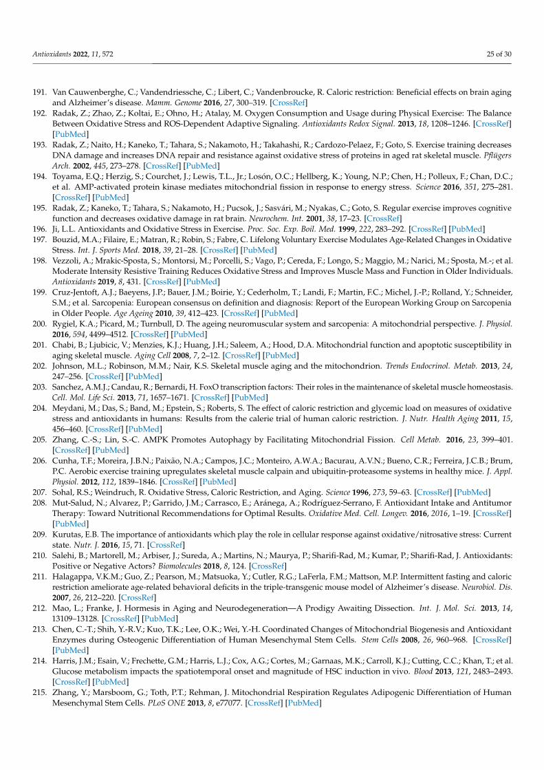
Antioxidants 2022, 11, 572 25 of 30
191. Van Cauwenberghe, C.; Vandendriessche, C.; Libert, C.; Vandenbroucke, R. Caloric restriction: Beneficial effects on brain agingand Alzheimer’s disease. Mamm. Genome 2016, 27, 300–319. [CrossRef]
192. Radak, Z.; Zhao, Z.; Koltai, E.; Ohno, H.; Atalay, M. Oxygen Consumption and Usage during Physical Exercise: The BalanceBetween Oxidative Stress and ROS-Dependent Adaptive Signaling. Antioxidants Redox Signal. 2013, 18, 1208–1246. [CrossRef][PubMed]
193. Radak, Z.; Naito, H.; Kaneko, T.; Tahara, S.; Nakamoto, H.; Takahashi, R.; Cardozo-Pelaez, F.; Goto, S. Exercise training decreasesDNA damage and increases DNA repair and resistance against oxidative stress of proteins in aged rat skeletal muscle. PflügersArch. 2002, 445, 273–278. [CrossRef] [PubMed]
194. Toyama, E.Q.; Herzig, S.; Courchet, J.; Lewis, T.L., Jr.; Losón, O.C.; Hellberg, K.; Young, N.P.; Chen, H.; Polleux, F.; Chan, D.C.;et al. AMP-activated protein kinase mediates mitochondrial fission in response to energy stress. Science 2016, 351, 275–281.[CrossRef] [PubMed]
195. Radak, Z.; Kaneko, T.; Tahara, S.; Nakamoto, H.; Pucsok, J.; Sasvári, M.; Nyakas, C.; Goto, S. Regular exercise improves cognitivefunction and decreases oxidative damage in rat brain. Neurochem. Int. 2001, 38, 17–23. [CrossRef]
196. Ji, L.L. Antioxidants and Oxidative Stress in Exercise. Proc. Soc. Exp. Boil. Med. 1999, 222, 283–292. [CrossRef] [PubMed]197. Bouzid, M.A.; Filaire, E.; Matran, R.; Robin, S.; Fabre, C. Lifelong Voluntary Exercise Modulates Age-Related Changes in Oxidative
Stress. Int. J. Sports Med. 2018, 39, 21–28. [CrossRef] [PubMed]198. Vezzoli, A.; Mrakic-Sposta, S.; Montorsi, M.; Porcelli, S.; Vago, P.; Cereda, F.; Longo, S.; Maggio, M.; Narici, M.; Sposta, M.-; et al.
Moderate Intensity Resistive Training Reduces Oxidative Stress and Improves Muscle Mass and Function in Older Individuals.Antioxidants 2019, 8, 431. [CrossRef] [PubMed]
199. Cruz-Jentoft, A.J.; Baeyens, J.P.; Bauer, J.M.; Boirie, Y.; Cederholm, T.; Landi, F.; Martin, F.C.; Michel, J.-P.; Rolland, Y.; Schneider,S.M.; et al. Sarcopenia: European consensus on definition and diagnosis: Report of the European Working Group on Sarcopeniain Older People. Age Ageing 2010, 39, 412–423. [CrossRef] [PubMed]
200. Rygiel, K.A.; Picard, M.; Turnbull, D. The ageing neuromuscular system and sarcopenia: A mitochondrial perspective. J. Physiol.2016, 594, 4499–4512. [CrossRef] [PubMed]
201. Chabi, B.; Ljubicic, V.; Menzies, K.J.; Huang, J.H.; Saleem, A.; Hood, D.A. Mitochondrial function and apoptotic susceptibility inaging skeletal muscle. Aging Cell 2008, 7, 2–12. [CrossRef] [PubMed]
202. Johnson, M.L.; Robinson, M.M.; Nair, K.S. Skeletal muscle aging and the mitochondrion. Trends Endocrinol. Metab. 2013, 24,247–256. [CrossRef] [PubMed]
203. Sanchez, A.M.J.; Candau, R.; Bernardi, H. FoxO transcription factors: Their roles in the maintenance of skeletal muscle homeostasis.Cell. Mol. Life Sci. 2013, 71, 1657–1671. [CrossRef] [PubMed]
204. Meydani, M.; Das, S.; Band, M.; Epstein, S.; Roberts, S. The effect of caloric restriction and glycemic load on measures of oxidativestress and antioxidants in humans: Results from the calerie trial of human caloric restriction. J. Nutr. Health Aging 2011, 15,456–460. [CrossRef] [PubMed]
205. Zhang, C.-S.; Lin, S.-C. AMPK Promotes Autophagy by Facilitating Mitochondrial Fission. Cell Metab. 2016, 23, 399–401.[CrossRef] [PubMed]
206. Cunha, T.F.; Moreira, J.B.N.; Paixão, N.A.; Campos, J.C.; Monteiro, A.W.A.; Bacurau, A.V.N.; Bueno, C.R.; Ferreira, J.C.B.; Brum,P.C. Aerobic exercise training upregulates skeletal muscle calpain and ubiquitin-proteasome systems in healthy mice. J. Appl.Physiol. 2012, 112, 1839–1846. [CrossRef] [PubMed]
207. Sohal, R.S.; Weindruch, R. Oxidative Stress, Caloric Restriction, and Aging. Science 1996, 273, 59–63. [CrossRef] [PubMed]208. Mut-Salud, N.; Alvarez, P.; Garrido, J.M.; Carrasco, E.; Aránega, A.; Rodríguez-Serrano, F. Antioxidant Intake and Antitumor
Therapy: Toward Nutritional Recommendations for Optimal Results. Oxidative Med. Cell. Longev. 2016, 2016, 1–19. [CrossRef][PubMed]
209. Kurutas, E.B. The importance of antioxidants which play the role in cellular response against oxidative/nitrosative stress: Currentstate. Nutr. J. 2016, 15, 71. [CrossRef]
210. Salehi, B.; Martorell, M.; Arbiser, J.; Sureda, A.; Martins, N.; Maurya, P.; Sharifi-Rad, M.; Kumar, P.; Sharifi-Rad, J. Antioxidants:Positive or Negative Actors? Biomolecules 2018, 8, 124. [CrossRef]
211. Halagappa, V.K.M.; Guo, Z.; Pearson, M.; Matsuoka, Y.; Cutler, R.G.; LaFerla, F.M.; Mattson, M.P. Intermittent fasting and caloricrestriction ameliorate age-related behavioral deficits in the triple-transgenic mouse model of Alzheimer’s disease. Neurobiol. Dis.2007, 26, 212–220. [CrossRef]
212. Mao, L.; Franke, J. Hormesis in Aging and Neurodegeneration—A Prodigy Awaiting Dissection. Int. J. Mol. Sci. 2013, 14,13109–13128. [CrossRef] [PubMed]
213. Chen, C.-T.; Shih, Y.-R.V.; Kuo, T.K.; Lee, O.K.; Wei, Y.-H. Coordinated Changes of Mitochondrial Biogenesis and AntioxidantEnzymes during Osteogenic Differentiation of Human Mesenchymal Stem Cells. Stem Cells 2008, 26, 960–968. [CrossRef][PubMed]
214. Harris, J.M.; Esain, V.; Frechette, G.M.; Harris, L.J.; Cox, A.G.; Cortes, M.; Garnaas, M.K.; Carroll, K.J.; Cutting, C.C.; Khan, T.; et al.Glucose metabolism impacts the spatiotemporal onset and magnitude of HSC induction in vivo. Blood 2013, 121, 2483–2493.[CrossRef] [PubMed]
215. Zhang, Y.; Marsboom, G.; Toth, P.T.; Rehman, J. Mitochondrial Respiration Regulates Adipogenic Differentiation of HumanMesenchymal Stem Cells. PLoS ONE 2013, 8, e77077. [CrossRef] [PubMed]
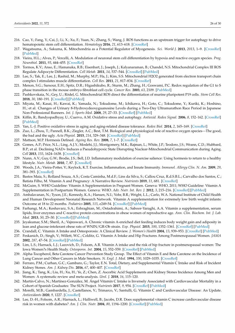
Antioxidants 2022, 11, 572 26 of 30
216. Cao, Y.; Fang, Y.; Cai, J.; Li, X.; Xu, F.; Yuan, N.; Zhang, S.; Wang, J. ROS functions as an upstream trigger for autophagy to drivehematopoietic stem cell differentiation. Hematology 2016, 21, 613–618. [CrossRef]
217. Wagatsuma, A.; Sakuma, K. Mitochondria as a Potential Regulator of Myogenesis. Sci. World J. 2013, 2013, 1–9. [CrossRef][PubMed]
218. Vieira, H.L.; Alves, P.; Vercelli, A. Modulation of neuronal stem cell differentiation by hypoxia and reactive oxygen species. Prog.Neurobiol. 2011, 93, 444–455. [CrossRef]
219. Tormos, K.V.; Anso, E.; Hamanaka, R.B.; Eisenbart, J.; Joseph, J.; Kalyanaraman, B.; Chandel, N.S. Mitochondrial Complex III ROSRegulate Adipocyte Differentiation. Cell Metab. 2011, 14, 537–544. [CrossRef] [PubMed]
220. Lee, S.; Tak, E.; Lee, J.; Rashid, M.; Murphy, M.P.; Ha, J.; Kim, S.S. Mitochondrial H2O2 generated from electron transport chaincomplex I stimulates muscle differentiation. Cell Res. 2011, 21, 817–834. [CrossRef]
221. Menon, S.G.; Sarsour, E.H.; Spitz, D.R.; Higashikubo, R.; Sturm, M.; Zhang, H.; Goswami, P.C. Redox regulation of the G1 to Sphase transition in the mouse embryo fibroblast cell cycle. Cancer Res. 2003, 63, 2109. [PubMed]
222. Pashkovskaia, N.; Gey, U.; Rödel, G. Mitochondrial ROS direct the differentiation of murine pluripotent P19 cells. Stem Cell Res.2018, 30, 180–191. [CrossRef] [PubMed]
223. Miyata, M.; Kasai, H.; Kawai, K.; Yamada, N.; Tokudome, M.; Ichikawa, H.; Goto, C.; Tokudome, Y.; Kuriki, K.; Hoshino,H.; et al. Changes of Urinary 8-Hydroxydeoxyguanosine Levels during a Two-Day Ultramarathon Race Period in JapaneseNon-Professional Runners. Int. J. Sports Med. 2008, 29, 27–33. [CrossRef] [PubMed]
224. Kiffin, R.; Bandyopadhyay, U.; Cuervo, A.M. Oxidative stress and autophagy. Antioxid. Redox Signal. 2006, 8, 152–162. [CrossRef][PubMed]
225. Yan, L.-J. Positive oxidative stress in aging and aging-related disease tolerance. Redox Biol. 2014, 2, 165–169. [CrossRef]226. Zuo, L.; Zhou, T.; Pannell, B.K.; Ziegler, A.C.; Best, T.M. Biological and physiological role of reactive oxygen species—The good,
the bad and the ugly. Acta Physiol. 2015, 214, 329–348. [CrossRef] [PubMed]227. Mattson, M.P. Hormesis Defined. Ageing Res. Rev. 2008, 7, 1–7. [CrossRef]228. Gomes, A.P.; Price, N.L.; Ling, A.J.Y.; Moslehi, J.J.; Montgomery, M.K.; Rajman, L.; White, J.P.; Teodoro, J.S.; Wrann, C.D.; Hubbard,
B.P.; et al. Declining NAD+ Induces a Pseudohypoxic State Disrupting Nuclear-Mitochondrial Communication during Aging.Cell 2013, 155, 1624–1638. [CrossRef]
229. Nunn, A.V.; Guy, G.W.; Brodie, J.S.; Bell, J.D. Inflammatory modulation of exercise salience: Using hormesis to return to a healthylifestyle. Nutr. Metab. 2010, 7, 87. [CrossRef]
230. Woods, J.A.; Vieira-Potter, V.; Keylock, K.T. Exercise, Inflammation, and Innate Immunity. Immunol. Allergy Clin. N. Am. 2009, 29,381–393. [CrossRef]
231. Bastos Maia, S.; Rolland Souza, A.S.; Costa Caminha, M.d.F.; Lins da Silva, S.; Callou Cruz, R.d.S.B.L.; Carvalho dos Santos, C.;Batista Filho, M. Vitamin A and Pregnancy: A Narrative Review. Nutrients 2019, 11, 681. [CrossRef]
232. McGuire, S. WHO Guideline: Vitamin A Supplementation in Pregnant Women. Geneva: WHO, 2011; WHO Guideline: Vitamin ASupplementation in Postpartum Women. Geneva: WHO. Adv. Nutr. Int. Rev. J. 2012, 3, 215–216. [CrossRef] [PubMed]
233. Ambalavanan, N.; Tyson, J.E.; Kennedy, K.A.; Hansen, N.I.; Vohr, B.R.; Wright, L.L.; Carlo, W.A. National Institute of Child Healthand Human Development Neonatal Research Network. Vitamin A supplementation for extremely low birth weight infants:Outcome at 18 to 22 months. Pediatrics 2005, 115, e249-54. [CrossRef] [PubMed]
234. Farhangi, M.A.; Keshavarz, S.A.; Eshraghian, M.; Ostadrahimi, A.; Saboor-Yaraghi, A.A. Vitamin a supplementation, serumlipids, liver enzymes and C-reactive protein concentrations in obese women of reproductive age. Ann. Clin. Biochem. Int. J. Lab.Med. 2013, 50, 25–30. [CrossRef] [PubMed]
235. Jeyakumar, S.M.; Sheril, A.; Vajreswari, A. Chronic vitamin A-enriched diet feeding induces body weight gain and adiposity inlean and glucose-intolerant obese rats of WNIN/GR-Ob strain. Exp. Physiol. 2015, 100, 1352–1361. [CrossRef] [PubMed]
236. Crandall, C. Vitamin A Intake and Osteoporosis: A Clinical Review. J. Women’s Health 2004, 13, 939–953. [CrossRef] [PubMed]237. Feskanich, D.; Singh, V.; Willett, W.C.; Colditz, G. Vitamin A Intake and Hip Fractures Among Postmenopausal Women. JAMA
2002, 287, 47–54. [CrossRef] [PubMed]238. Lim, L.S.; Harnack, L.J.; Lazovich, D.; Folsom, A.R. Vitamin A intake and the risk of hip fracture in postmenopausal women: The
Iowa Women?s Health Study. Osteoporos. Int. 2004, 15, 552–559. [CrossRef] [PubMed]239. Alpha-Tocopherol, Beta Carotene Cancer Prevention Study Group. The Effect of Vitamin E and Beta Carotene on the Incidence of
Lung Cancer and Other Cancers in Male Smokers. N. Engl. J. Med. 1994, 330, 1029–1035. [CrossRef]240. Ferraro, P.M.; Curhan, G.C.; Gambaro, G.; Taylor, E.N. Total, Dietary, and Supplemental Vitamin C Intake and Risk of Incident
Kidney Stones. Am. J. Kidney Dis. 2016, 67, 400–407. [CrossRef]241. Jiang, K.; Tang, K.; Liu, H.; Xu, H.; Ye, Z.; Chen, Z. Ascorbic Acid Supplements and Kidney Stones Incidence Among Men and
Women: A systematic review and meta-analysis. Urol. J. 2018, 16, 115–120.242. Martín-Calvo, N.; Martínez-González, M. Ángel Vitamin C Intake is Inversely Associated with Cardiovascular Mortality in a
Cohort of Spanish Graduates: The SUN Project. Nutrients 2017, 9, 954. [CrossRef] [PubMed]243. Morelli, M.B.; Gambardella, J.; Castellanos, V.; Trimarco, V.; Santulli, G. Vitamin C and Cardiovascular Disease: An Update.
Antioxidants 2020, 9, 1227. [CrossRef]244. Lee, D.-H.; Folsom, A.R.; Harnack, L.; Halliwell, B.; Jacobs, D.R. Does supplemental vitamin C increase cardiovascular disease
risk in women with diabetes? Am. J. Clin. Nutr. 2004, 80, 1194–1200. [CrossRef] [PubMed]
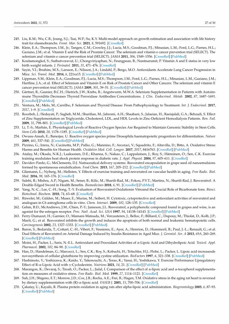
Antioxidants 2022, 11, 572 27 of 30
245. Liu, K.M.; Wu, C.B.; Joung, S.J.; Tsai, W.P.; Su, K.Y. Multi-model approach on growth estimation and association with life historytrait for elasmobranchs. Front. Mar. Sci. 2021, 8, 591692. [CrossRef]
246. Klein, E.A.; Thompson, I.M., Jr.; Tangen, C.M.; Crowley, J.J.; Lucia, M.S.; Goodman, P.J.; Minasian, L.M.; Ford, L.G.; Parnes, H.L.;Gaziano, J.M.; et al. Vitamin E and the Risk of Prostate Cancer: The selenium and vitamin e cancer prevention trial (SELECT). Theselenium and vitamin e cancer prevention trial (SELECT). JAMA 2011, 306, 1549–1556. [CrossRef] [PubMed]
247. Kositamongkol, S.; Suthutvoravut, U.; Chongviriyaphan, N.; Feungpean, B.; Nuntnarumit, P. Vitamin A and E status in very lowbirth weight infants. J. Perinatol. 2011, 31, 471–476. [CrossRef]
248. Sayin, V.I.; Ibrahim, M.X.; Larsson, E.; Nilsson, J.A.; Lindahl, P.; Bergo, M.O. Antioxidants Accelerate Lung Cancer Progression inMice. Sci. Transl. Med. 2014, 6, 221ra15. [CrossRef] [PubMed]
249. Lippman, S.M.; Klein, E.A.; Goodman, P.J.; Lucia, M.S.; Thompson, I.M.; Ford, L.G.; Parnes, H.L.; Minasian, L.M.; Gaziano, J.M.;Hartline, J.A.; et al. Effect of Selenium and Vitamin E on Risk of Prostate Cancer and Other Cancers: The selenium and vitamin Ecancer prevention trial (SELECT). JAMA 2009, 301, 39–51. [CrossRef] [PubMed]
250. Gärtner, R.; Gasnier, B.C.H.; Dietrich, J.W.; Krebs, B.; Angstwurm, M.W.A. Selenium Supplementation in Patients with Autoim-mune Thyroiditis Decreases Thyroid Peroxidase Antibodies Concentrations. J. Clin. Endocrinol. Metab. 2002, 87, 1687–1691.[CrossRef] [PubMed]
251. Ventura, M.; Melo, M.; Carrilho, F. Selenium and Thyroid Disease: From Pathophysiology to Treatment. Int. J. Endocrinol. 2017,2017, 1–9. [CrossRef]
252. Roozbeh, J.; Hedayati, P.; Sagheb, M.M.; Sharifian, M.; Jahromi, A.H.; Shaabani, S.; Jalaeian, H.; Raeisjalali, G.A.; Behzadi, S. Effectof Zinc Supplementation on Triglyceride, Cholesterol, LDL, and HDL Levels in Zinc-Deficient Hemodialysis Patients. Ren. Fail.2009, 31, 798–801. [CrossRef] [PubMed]
253. Li, T.-S.; Marbán, E. Physiological Levels of Reactive Oxygen Species Are Required to Maintain Genomic Stability in Stem Cells.Stem Cells 2010, 28, 1178–1185. [CrossRef] [PubMed]
254. Owusu-Ansah, E.; Banerjee, U. Reactive oxygen species prime Drosophila haematopoietic progenitors for differentiation. Nature2009, 461, 537–541. [CrossRef] [PubMed]
255. Pizzino, G.; Irrera, N.; Cucinotta, M.P.; Pallio, G.; Mannino, F.; Arcoraci, V.; Squadrito, F.; Altavilla, D.; Bitto, A. Oxidative Stress:Harms and Benefits for Human Health. Oxidative Med. Cell. Longev. 2017, 2017, 8416763. [CrossRef] [PubMed]
256. Atalay, M.; Oksala, N.K.J.; Laaksonen, D.E.; Khanna, S.; Nakao, C.; Lappalainen, J.; Roy, S.; Hänninen, O.; Sen, C.K. Exercisetraining modulates heat shock protein response in diabetic rats. J. Appl. Physiol. 2004, 97, 605–611. [CrossRef]
257. Davidov-Pardo, G.; McClements, D.J. Nutraceutical delivery systems: Resveratrol encapsulation in grape seed oil nanoemulsionsformed by spontaneous emulsification. Food Chem. 2015, 167, 205–212. [CrossRef] [PubMed]
258. Gliemann, L.; Nyberg, M.; Hellsten, Y. Effects of exercise training and resveratrol on vascular health in aging. Free Radic. Biol.Med. 2016, 98, 165–176. [CrossRef]
259. Salehi, B.; Mishra, A.P.; Nigam, M.; Sener, B.; Kilic, M.; Sharifi-Rad, M.; Fokou, P.V.T.; Martins, N.; Sharifi-Rad, J. Resveratrol: ADouble-Edged Sword in Health Benefits. Biomedicines 2018, 6, 91. [CrossRef] [PubMed]
260. Yang, N.-C.; Lee, C.-H.; Song, T.-Y. Evaluation of Resveratrol Oxidationin Vitroand the Crucial Role of Bicarbonate Ions. Biosci.Biotechnol. Biochem. 2010, 74, 63–68. [CrossRef]
261. Rüweler, M.; Gülden, M.; Maser, E.; Murias, M.; Seibert, H. Cytotoxic, cytoprotective and antioxidant activities of resveratrol andanalogues in C6 astroglioma cells in vitro. Chem. Interact. 2009, 182, 128–135. [CrossRef]
262. Gehm, B.D.; McAndrews, J.M.; Chien, P.-Y.; Jameson, J.L. Resveratrol, a polyphenolic compound found in grapes and wine, is anagonist for the estrogen receptor. Proc. Natl. Acad. Sci. USA 1997, 94, 14138–14143. [CrossRef] [PubMed]
263. Ferry-Dumazet, H.; Garnier, O.; Mamani-Matsuda, M.; Vercauteren, J.; Belloc, F.; Billiard, C.; Dupouy, M.; Thiolat, D.; Kolb, J.P.;Marit, G.; et al. Resveratrol inhibits the growth and induces the apoptosis of both normal and leukemic hematopoietic cells.Carcinogenesis 2002, 23, 1327–1333. [CrossRef] [PubMed]
264. Baron, S.; Bedarida, T.; Cottart, C.-H.; Vibert, F.; Vessieres, E.; Ayer, A.; Henrion, D.; Hommeril, B.; Paul, J.-L.; Renault, G.; et al.Dual Effects of Resveratrol on Arterial Damage Induced by Insulin Resistance in Aged Mice. J. Gerontol. Ser. A 2013, 69A, 260–269.[CrossRef] [PubMed]
265. Moini, H.; Packer, L.; Saris, N.-E.L. Antioxidant and Prooxidant Activities of α-Lipoic Acid and Dihydrolipoic Acid. Toxicol. Appl.Pharmacol. 2002, 182, 84–90. [CrossRef]
266. Han, D.; Handelman, G.; Marcocci, L.; Sen, C.K.; Roy, S.; Kobuchi, H.; Tritschler, H.J.; Flohé, L.; Packer, L. Lipoic acid increasesdenovosynthesis of cellular glutathione by improving cystine utilization. BioFactors 1997, 6, 321–338. [CrossRef] [PubMed]
267. Hashimoto, Y.; Yoshizawa, K.; Kaido, Y.; Takenouchi, A.; Terao, K.; Yasui, H.; Yoshikawa, Y. Exercise Performance UpregulatoryEffect of R-α-Lipoic Acid with γ-Cyclodextrin. Nutriens 2021, 14, 21. [CrossRef] [PubMed]
268. Marangon, K.; Devaraj, S.; Tirosh, O.; Packer, L.; Jialal, I. Comparison of the effect of α-lipoic acid and α-tocopherol supplementa-tion on measures of oxidative stress. Free Radic. Biol. Med. 1999, 27, 1114–1121. [CrossRef]
269. Suh, J.H.; Shigeno, E.T.; Morrow, J.D.; Cox, J.B.; Rocha, A.E.; Frei, B.; Hagen, T.M. Oxidative stress in the aging rat heart is reversedby dietary supplementation with (R)-α-lipoic acid. FASEB J. 2001, 15, 700–706. [CrossRef]
270. Çakatay, U.; Kayaliı, R. Plasma protein oxidation in aging rats after alpha-lipoic acid administration. Biogerontology 2005, 6, 87–93.[CrossRef] [PubMed]
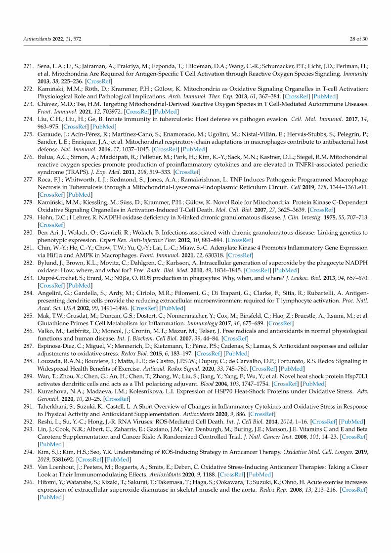
Antioxidants 2022, 11, 572 28 of 30
271. Sena, L.A.; Li, S.; Jairaman, A.; Prakriya, M.; Ezponda, T.; Hildeman, D.A.; Wang, C.-R.; Schumacker, P.T.; Licht, J.D.; Perlman, H.;et al. Mitochondria Are Required for Antigen-Specific T Cell Activation through Reactive Oxygen Species Signaling. Immunity2013, 38, 225–236. [CrossRef]
272. Kaminski, M.M.; Röth, D.; Krammer, P.H.; Gülow, K. Mitochondria as Oxidative Signaling Organelles in T-cell Activation:Physiological Role and Pathological Implications. Arch. Immunol. Ther. Exp. 2013, 61, 367–384. [CrossRef] [PubMed]
273. Chávez, M.D.; Tse, H.M. Targeting Mitochondrial-Derived Reactive Oxygen Species in T Cell-Mediated Autoimmune Diseases.Front. Immunol. 2021, 12, 703972. [CrossRef] [PubMed]
274. Liu, C.H.; Liu, H.; Ge, B. Innate immunity in tuberculosis: Host defense vs pathogen evasion. Cell. Mol. Immunol. 2017, 14,963–975. [CrossRef] [PubMed]
275. Garaude, J.; Acín-Pérez, R.; Martínez-Cano, S.; Enamorado, M.; Ugolini, M.; Nistal-Villán, E.; Hervás-Stubbs, S.; Pelegrín, P.;Sander, L.E.; Enríquez, J.A.; et al. Mitochondrial respiratory-chain adaptations in macrophages contribute to antibacterial hostdefense. Nat. Immunol. 2016, 17, 1037–1045. [CrossRef] [PubMed]
276. Bulua, A.C.; Simon, A.; Maddipati, R.; Pelletier, M.; Park, H.; Kim, K.-Y.; Sack, M.N.; Kastner, D.L.; Siegel, R.M. Mitochondrialreactive oxygen species promote production of proinflammatory cytokines and are elevated in TNFR1-associated periodicsyndrome (TRAPS). J. Exp. Med. 2011, 208, 519–533. [CrossRef]
277. Roca, F.J.; Whitworth, L.J.; Redmond, S.; Jones, A.A.; Ramakrishnan, L. TNF Induces Pathogenic Programmed MacrophageNecrosis in Tuberculosis through a Mitochondrial-Lysosomal-Endoplasmic Reticulum Circuit. Cell 2019, 178, 1344–1361.e11.[CrossRef] [PubMed]
278. Kaminski, M.M.; Kiessling, M.; Süss, D.; Krammer, P.H.; Gülow, K. Novel Role for Mitochondria: Protein Kinase C-DependentOxidative Signaling Organelles in Activation-Induced T-Cell Death. Mol. Cell. Biol. 2007, 27, 3625–3639. [CrossRef]
279. Hohn, D.C.; I Lehrer, R. NADPH oxidase deficiency in X-linked chronic granulomatous disease. J. Clin. Investig. 1975, 55, 707–713.[CrossRef]
280. Ben-Ari, J.; Wolach, O.; Gavrieli, R.; Wolach, B. Infections associated with chronic granulomatous disease: Linking genetics tophenotypic expression. Expert Rev. Anti-Infective Ther. 2012, 10, 881–894. [CrossRef]
281. Chin, W.-Y.; He, C.-Y.; Chow, T.W.; Yu, Q.-Y.; Lai, L.-C.; Miaw, S.-C. Adenylate Kinase 4 Promotes Inflammatory Gene Expressionvia Hif1α and AMPK in Macrophages. Front. Immunol. 2021, 12, 630318. [CrossRef]
282. Bylund, J.; Brown, K.L.; Movitz, C.; Dahlgren, C.; Karlsson, A. Intracellular generation of superoxide by the phagocyte NADPHoxidase: How, where, and what for? Free. Radic. Biol. Med. 2010, 49, 1834–1845. [CrossRef] [PubMed]
283. Dupré-Crochet, S.; Erard, M.; Nüβe, O. ROS production in phagocytes: Why, when, and where? J. Leukoc. Biol. 2013, 94, 657–670.[CrossRef] [PubMed]
284. Angelini, G.; Gardella, S.; Ardy, M.; Ciriolo, M.R.; Filomeni, G.; Di Trapani, G.; Clarke, F.; Sitia, R.; Rubartelli, A. Antigen-presenting dendritic cells provide the reducing extracellular microenvironment required for T lymphocyte activation. Proc. Natl.Acad. Sci. USA 2002, 99, 1491–1496. [CrossRef] [PubMed]
285. Mak, T.W.; Grusdat, M.; Duncan, G.S.; Dostert, C.; Nonnenmacher, Y.; Cox, M.; Binsfeld, C.; Hao, Z.; Bruestle, A.; Itsumi, M.; et al.Glutathione Primes T Cell Metabolism for Inflammation. Immunology 2017, 46, 675–689. [CrossRef]
286. Valko, M.; Leibfritz, D.; Moncol, J.; Cronin, M.T.; Mazur, M.; Telser, J. Free radicals and antioxidants in normal physiologicalfunctions and human disease. Int. J. Biochem. Cell Biol. 2007, 39, 44–84. [CrossRef]
287. Espinosa-Diez, C.; Miguel, V.; Mennerich, D.; Kietzmann, T.; Pérez, P.S.; Cadenas, S.; Lamas, S. Antioxidant responses and cellularadjustments to oxidative stress. Redox Biol. 2015, 6, 183–197. [CrossRef] [PubMed]
288. Louzada, R.A.N.; Bouviere, J.; Matta, L.P.; de Castro, J.P.S.W.; Dupuy, C.; de Carvalho, D.P.; Fortunato, R.S. Redox Signaling inWidespread Health Benefits of Exercise. Antioxid. Redox Signal. 2020, 33, 745–760. [CrossRef] [PubMed]
289. Wan, T.; Zhou, X.; Chen, G.; An, H.; Chen, T.; Zhang, W.; Liu, S.; Jiang, Y.; Yang, F.; Wu, Y.; et al. Novel heat shock protein Hsp70L1activates dendritic cells and acts as a Th1 polarizing adjuvant. Blood 2004, 103, 1747–1754. [CrossRef] [PubMed]
290. Kurashova, N.A.; Madaeva, I.M.; Kolesnikova, L.I. Expression of HSP70 Heat-Shock Proteins under Oxidative Stress. Adv.Gerontol. 2020, 10, 20–25. [CrossRef]
291. Taherkhani, S.; Suzuki, K.; Castell, L. A Short Overview of Changes in Inflammatory Cytokines and Oxidative Stress in Responseto Physical Activity and Antioxidant Supplementation. Antioxidants 2020, 9, 886. [CrossRef]
292. Reshi, L.; Su, Y.-C.; Hong, J.-R. RNA Viruses: ROS-Mediated Cell Death. Int. J. Cell Biol. 2014, 2014, 1–16. [CrossRef] [PubMed]293. Lin, J.; Cook, N.R.; Albert, C.; Zaharris, E.; Gaziano, J.M.; Van Denburgh, M.; Buring, J.E.; Manson, J.E. Vitamins C and E and Beta
Carotene Supplementation and Cancer Risk: A Randomized Controlled Trial. J. Natl. Cancer Inst. 2008, 101, 14–23. [CrossRef][PubMed]
294. Kim, S.J.; Kim, H.S.; Seo, Y.R. Understanding of ROS-Inducing Strategy in Anticancer Therapy. Oxidative Med. Cell. Longev. 2019,2019, 5381692. [CrossRef] [PubMed]
295. Van Loenhout, J.; Peeters, M.; Bogaerts, A.; Smits, E.; Deben, C. Oxidative Stress-Inducing Anticancer Therapies: Taking a CloserLook at Their Immunomodulating Effects. Antioxidants 2020, 9, 1188. [CrossRef] [PubMed]
296. Hitomi, Y.; Watanabe, S.; Kizaki, T.; Sakurai, T.; Takemasa, T.; Haga, S.; Ookawara, T.; Suzuki, K.; Ohno, H. Acute exercise increasesexpression of extracellular superoxide dismutase in skeletal muscle and the aorta. Redox Rep. 2008, 13, 213–216. [CrossRef][PubMed]
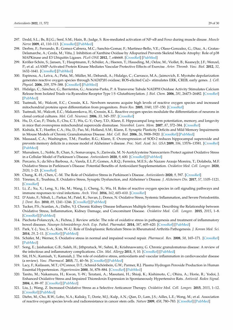
Antioxidants 2022, 11, 572 29 of 30
297. Dodd, S.L.; Bs, B.J.G.; Senf, S.M.; Hain, B.; Judge, S. Ros-mediated activation of NF-κB and Foxo during muscle disuse. MuscleNerve 2009, 41, 110–113. [CrossRef] [PubMed]
298. Derbre, F.; Ferrando, B.; Gomez-Cabrera, M.C.; Sanchis-Gomar, F.; Martinez-Bello, V.E.; Olaso-Gonzalez, G.; Diaz, A.; Gratas-Delamarche, A.; Cerda, M.; Viña, J. Inhibition of Xanthine Oxidase by Allopurinol Prevents Skeletal Muscle Atrophy: Role of p38MAPKinase and E3 Ubiquitin Ligases. PLoS ONE 2012, 7, e46668. [CrossRef] [PubMed]
299. Kröller-Schön, S.; Jansen, T.; Hauptmann, F.; Schüler, A.; Heeren, T.; Hausding, M.; Oelze, M.; Viollet, B.; KeaneyJr, J.F.; Wenzel,P.; et al. α1AMP-Activated Protein Kinase Mediates Vascular Protective Effects of Exercise. Arter. Thromb. Vasc. Biol. 2012, 32,1632–1641. [CrossRef] [PubMed]
300. Espinosa, A.; Leiva, A.; Peña, M.; Müller, M.; Debandi, A.; Hidalgo, C.; Carrasco, M.A.; Jaimovich, E. Myotube depolarizationgenerates reactive oxygen species through NAD(P)H oxidase; ROS-elicited Ca2+ stimulates ERK, CREB, early genes. J. Cell.Physiol. 2006, 209, 379–388. [CrossRef] [PubMed]
301. Hidalgo, C.; Sánchez, G.; Barrientos, G.; Aracena-Parks, P. A Transverse Tubule NADPH Oxidase Activity Stimulates CalciumRelease from Isolated Triads via Ryanodine Receptor Type 1 S -Glutathionylation. J. Biol. Chem. 2006, 281, 26473–26482. [CrossRef][PubMed]
302. Tsatmali, M.; Walcott, E.C.; Crossin, K.L. Newborn neurons acquire high levels of reactive oxygen species and increasedmitochondrial proteins upon differentiation from progenitors. Brain Res. 2005, 1040, 137–150. [CrossRef]
303. Tsatmali, M.; Walcott, E.C.; Makarenkova, H.; Crossin, K.L. Reactive oxygen species modulate the differentiation of neurons inclonal cortical cultures. Mol. Cell. Neurosci. 2006, 33, 345–357. [CrossRef]
304. Hu, D.; Cao, P.; Thiels, E.; Chu, C.T.; Wu, G.-Y.; Oury, T.D.; Klann, E. Hippocampal long-term potentiation, memory, and longevityin mice that overexpress mitochondrial superoxide dismutase. Neurobiol. Learn. Mem. 2007, 87, 372–384. [CrossRef]
305. Kishida, K.T.; Hoeffer, C.A.; Hu, D.; Pao, M.; Holland, S.M.; Klann, E. Synaptic Plasticity Deficits and Mild Memory Impairmentsin Mouse Models of Chronic Granulomatous Disease. Mol. Cell. Biol. 2006, 26, 5908–5920. [CrossRef] [PubMed]
306. Massaad, C.A.; Washington, T.M.; Pautler, R.G.; Klann, E. Overexpression of SOD-2 reduces hippocampal superoxide andprevents memory deficits in a mouse model of Alzheimer’s disease. Proc. Natl. Acad. Sci. USA 2009, 106, 13576–13581. [CrossRef][PubMed]
307. Mursaleen, L.; Noble, B.; Chan, S.; Somavarapu, S.; Zariwala, M. N-Acetylcysteine Nanocarriers Protect against Oxidative Stressin a Cellular Model of Parkinson’s Disease. Antioxidants 2020, 9, 600. [CrossRef] [PubMed]
308. Percario, S.; da Silva Barbosa, A.; Varela, E.L.P.; Gomes, A.R.Q.; Ferreira, M.E.S.; de Nazare Araujo Moreira, T.; Dolabela, M.F.Oxidative Stress in Parkinson’s Disease: Potential Benefits of Antioxidant Supplementation. Oxidative Med. Cell. Longev. 2020,2020, 1–23. [CrossRef]
309. Chang, K.-H.; Chen, C.-M. The Role of Oxidative Stress in Parkinson’s Disease. Antioxidants 2020, 9, 597. [CrossRef]310. Tönnies, E.; Trushina, E. Oxidative Stress, Synaptic Dysfunction, and Alzheimer’s Disease. J. Alzheimers Dis. 2017, 57, 1105–1121.
[CrossRef]311. Li, Z.; Xu, X.; Leng, X.; He, M.; Wang, J.; Cheng, S.; Wu, H. Roles of reactive oxygen species in cell signaling pathways and
immune responses to viral infections. Arch. Virol. 2016, 162, 603–610. [CrossRef]312. D’Aiuto, F.; Nibali, L.; Parkar, M.; Patel, K.; Suvan, J.; Donos, N. Oxidative Stress, Systemic Inflammation, and Severe Periodontitis.
J. Dent. Res. 2010, 89, 1241–1246. [CrossRef] [PubMed]313. Tucker, P.S.; Scanlan, A.; Dalbo, V.J. Chronic Kidney Disease Influences Multiple Systems: Describing the Relationship between
Oxidative Stress, Inflammation, Kidney Damage, and Concomitant Disease. Oxidative Med. Cell. Longev. 2015, 2015, 1–8.[CrossRef] [PubMed]
314. Piechota-Polanczyk, A.; Fichna, J. Review article: The role of oxidative stress in pathogenesis and treatment of inflammatorybowel diseases. Naunyn-Schmiedebergs Arch. Exp. Pathol. Pharmakol. 2014, 387, 605–620. [CrossRef] [PubMed]
315. Park, Y.-J.; Yoo, S.-A.; Kim, W.-U. Role of Endoplasmic Reticulum Stress in Rheumatoid Arthritis Pathogenesis. J. Korean Med. Sci.2014, 29, 2–11. [CrossRef] [PubMed]
316. Schäfer, M.; Werner, S. Oxidative stress in normal and impaired wound repair. Pharmacol. Res. 2008, 58, 165–171. [CrossRef][PubMed]
317. Song, E.; Jaishankar, G.B.; Saleh, H.; Jithpratuck, W.; Sahni, R.; Krishnaswamy, G. Chronic granulomatous disease: A review ofthe infectious and inflammatory complications. Clin. Mol. Allergy 2011, 9, 10. [CrossRef] [PubMed]
318. Siti, H.N.; Kamisah, Y.; Kamsiah, J. The role of oxidative stress, antioxidants and vascular inflammation in cardiovascular disease(a review). Vasc. Pharmacol. 2015, 71, 40–56. [CrossRef] [PubMed]
319. Lacy, F.; Kailasam, M.T.; O’Connor, D.T.; Schmid-Schönbein, G.W.; Parmer, R.J. Plasma Hydrogen Peroxide Production in HumanEssential Hypertension. Hypertension 2000, 36, 878–884. [CrossRef] [PubMed]
320. Tanito, M.; Nakamura, H.; Kwon, Y.-W.; Teratani, A.; Masutani, H.; Shioji, K.; Kishimoto, C.; Ohira, A.; Horie, R.; Yodoi, J.Enhanced Oxidative Stress and Impaired Thioredoxin Expression in Spontaneously Hypertensive Rats. Antioxid. Redox Signal.2004, 6, 89–97. [CrossRef] [PubMed]
321. Liu, J.; Wang, Z. Increased Oxidative Stress as a Selective Anticancer Therapy. Oxidative Med. Cell. Longev. 2015, 2015, 1–12.[CrossRef] [PubMed]
322. Diehn, M.; Cho, R.W.; Lobo, N.A.; Kalisky, T.; Dorie, M.J.; Kulp, A.N.; Qian, D.; Lam, J.S.; Ailles, L.E.; Wong, M.; et al. Associationof reactive oxygen species levels and radioresistance in cancer stem cells. Nature 2009, 458, 780–783. [CrossRef] [PubMed]
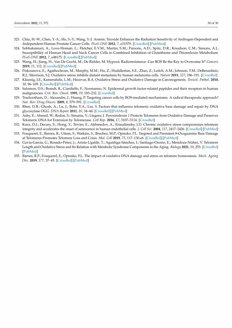
Antioxidants 2022, 11, 572 30 of 30
323. Chiu, H.-W.; Chen, Y.-A.; Ho, S.-Y.; Wang, Y.-J. Arsenic Trioxide Enhances the Radiation Sensitivity of Androgen-Dependent and-Independent Human Prostate Cancer Cells. PLoS ONE 2012, 7, e31579. [CrossRef] [PubMed]
324. Sobhakumari, A.; Love-Homan, L.; Fletcher, E.V.M.; Martin, S.M.; Parsons, A.D.; Spitz, D.R.; Knudson, C.M.; Simons, A.L.Susceptibility of Human Head and Neck Cancer Cells to Combined Inhibition of Glutathione and Thioredoxin Metabolism.PLoS ONE 2012, 7, e48175. [CrossRef] [PubMed]
325. Wang, H.; Jiang, H.; Van De Gucht, M.; De Ridder, M. Hypoxic Radioresistance: Can ROS Be the Key to Overcome It? Cancers2019, 11, 112. [CrossRef] [PubMed]
326. Piskounova, E.; Agathocleous, M.; Murphy, M.M.; Hu, Z.; Huddlestun, S.E.; Zhao, Z.; Leitch, A.M.; Johnson, T.M.; DeBerardinis,R.J.; Morrison, S.J. Oxidative stress inhibits distant metastasis by human melanoma cells. Nature 2015, 527, 186–191. [CrossRef]
327. Klaunig, J.E.; Kamendulis, L.M.; Hocevar, B.A. Oxidative Stress and Oxidative Damage in Carcinogenesis. Toxicol. Pathol. 2010,38, 96–109. [CrossRef] [PubMed]
328. Salomon, D.S.; Brandt, R.; Ciardiello, F.; Normanno, N. Epidermal growth factor-related peptides and their receptors in humanmalignancies. Crit. Rev. Oncol. 1995, 19, 183–232. [CrossRef]
329. Trachootham, D.; Alexandre, J.; Huang, P. Targeting cancer cells by ROS-mediated mechanisms: A radical therapeutic approach?Nat. Rev. Drug Discov. 2009, 8, 579–591. [CrossRef]
330. Rhee, D.B.; Ghosh, A.; Lu, J.; Bohr, V.A.; Liu, Y. Factors that influence telomeric oxidative base damage and repair by DNAglycosylase OGG. DNA Repair 2011, 10, 34–44. [CrossRef] [PubMed]
331. Aeby, E.; Ahmed, W.; Redon, S.; Simanis, V.; Lingner, J. Peroxiredoxin 1 Protects Telomeres from Oxidative Damage and PreservesTelomeric DNA for Extension by Telomerase. Cell Rep. 2016, 17, 3107–3114. [CrossRef]
332. Kurz, D.J.; Decary, S.; Hong, Y.; Trivier, E.; Akhmedov, A.; Erusalimsky, J.D. Chronic oxidative stress compromises telomereintegrity and accelerates the onset of senescence in human endothelial cells. J. Cell Sci. 2004, 117, 2417–2426. [CrossRef] [PubMed]
333. Fouquerel, E.; Barnes, R.; Uttam, S.; Watkins, S.; Bruchez, M.P.; Opresko, P.L. Targeted and Persistent 8-Oxoguanine Base Damageat Telomeres Promotes Telomere Loss and Crisis. Mol. Cell 2019, 75, 117–130.e6. [CrossRef] [PubMed]
334. Gavia-García, G.; Rosado-Pérez, J.; Arista-Ugalde, T.; Aguiñiga-Sánchez, I.; Santiago-Osorio, E.; Mendoza-Núñez, V. TelomereLength and Oxidative Stress and Its Relation with Metabolic Syndrome Components in the Aging. Biology 2021, 10, 253. [CrossRef][PubMed]
335. Barnes, R.P.; Fouquerel, E.; Opresko, P.L. The impact of oxidative DNA damage and stress on telomere homeostasis. Mech. AgeingDev. 2019, 177, 37–45. [CrossRef] [PubMed]
Related Documents