1 Mesenchymal stem cells in rheumatoid synovium: Enumeration and functional assessment in relation to synovial inflammation level Jones E, PhD 1 , Churchman SM, PhD 1 , English A, FIBMS 1 , Buch MH, PhD, MRCP 1 , Horner EA, PhD 1 , Burgoyne CH, PhD 1 , Reece R, FRCP 1 , Kinsey S, MD, FRCR, FRCPath 2 , Emery P, MD, FRCP 1 , McGonagle D, PhD, FRCI 1,3 and Ponchel F, PhD 1 . 1 - Academic Unit of Musculoskeletal Disease, Leeds Institute of Molecular Medicine, and 2 – Department of Paediatric Haematology, University of Leeds, Leeds, UK, 3 - National University of Ireland Galway, Republic of Ireland. Corresponding Author: Dr Elena Jones Email: [email protected] Word count: 2953 ARD Online First, published on April 2, 2009 as 10.1136/ard.2008.106435 Copyright Article author (or their employer) 2009. Produced by BMJ Publishing Group Ltd (& EULAR) under licence. group.bmj.com on May 17, 2016 - Published by http://ard.bmj.com/ Downloaded from group.bmj.com on May 17, 2016 - Published by http://ard.bmj.com/ Downloaded from group.bmj.com on May 17, 2016 - Published by http://ard.bmj.com/ Downloaded from

Welcome message from author
This document is posted to help you gain knowledge. Please leave a comment to let me know what you think about it! Share it to your friends and learn new things together.
Transcript

1
Mesenchymal stem cells in rheumatoid synovium: Enumeration and functional assessment in relation to synovial inflammation level Jones E, PhD1, Churchman SM, PhD1, English A, FIBMS1, Buch MH, PhD, MRCP1, Horner EA, PhD1, Burgoyne CH, PhD1, Reece R, FRCP1, Kinsey S, MD, FRCR, FRCPath2, Emery P, MD, FRCP1, McGonagle D, PhD, FRCI 1,3 and Ponchel F, PhD1. 1 - Academic Unit of Musculoskeletal Disease, Leeds Institute of Molecular Medicine, and 2 – Department of Paediatric Haematology, University of Leeds, Leeds, UK, 3 - National University of Ireland Galway, Republic of Ireland. Corresponding Author: Dr Elena Jones Email: [email protected] Word count: 2953
ARD Online First, published on April 2, 2009 as 10.1136/ard.2008.106435
Copyright Article author (or their employer) 2009. Produced by BMJ Publishing Group Ltd (& EULAR) under licence.
group.bmj.com on May 17, 2016 - Published by http://ard.bmj.com/Downloaded from group.bmj.com on May 17, 2016 - Published by http://ard.bmj.com/Downloaded from group.bmj.com on May 17, 2016 - Published by http://ard.bmj.com/Downloaded from

2
Abstract Objective. Achieving joint regeneration in rheumatoid arthritis (RA) represents a future challenge. Autologous synovial mesenchymal stem cells (MSCs) could be therapeutically exploited. However, the inflammatory milieu in the RA synovium could adversely affect endogenous MSC function. To test this hypothesis, the frequency and multipotency of RA synovial MSCs was evaluated in relation to existing synovial inflammation. Methods. Synovial inflammation was measured using arthroscopic visual analogue score (VAS) and further validated using immunohistochemistry and flow cytometry. Highly-proliferative clonogenic in vivo MSCs were enumerated following fluorescence-activated cell sorting and expansion for 20 population doublings. MSC multipotency was quantified following standard in vitro culture-expansion and trilineage differentiation assays. Real-time PCR, flow cytometry and ELISA were used to evaluate pro- and anti-chondrogenic molecules in standard polyclonal synovial MSCs. Results. Arthroscopic visual score of inflammation (VAS) significantly correlated with synovial macrophage infiltration. In RA, synovial MSC chondrogenesis was inhibited in direct relation to VAS (r=-0.777, p<0.05) and reduced compared to control OA-MSCs (p<0.05). In vivo MSCs resided in the synovial fibroblastic/stromal fraction (CD45-CD31-) and were reduced in frequency in relation to VAS (r=-0.695, p<0.05). In RA-MSCs, CD44 levels correlated negatively with inflammation and positively with chondrogenesis (r=-0.830 and r=0.865, respectively). Cytokine production and Sox9 expression was similar between RA- and OA-MSCs. Conclusions. Our findings demonstrate a negative relationship between synovial MSC chondrogenic and clonogenic capacities and the magnitude of synovitis in RA. Effective suppression of joint inflammation is therefore necessary for the development of autologous MSC therapies aimed at cartilage regeneration in RA. Key words: mesenchymal stem cells, synovium, inflammation, chondrogenesis
group.bmj.com on May 17, 2016 - Published by http://ard.bmj.com/Downloaded from

3
INTRODUCTION Current therapies have greatly reduced the degree of joint destruction in rheumatoid arthritis (RA); however achieving cartilage regeneration in RA remains a challenge. Regenerative medicine strategies, including autologous chondrocyte implantation, have already shown efficiency in the treatment of solitary chondral injuries. [1-2] Distinct chondral lesions in osteoarthritis (OA) are the next therapeutic target. [3-8] Developing similar strategies in RA is however much more challenging. RA afflicts multiple joints, is associated with widespread tissue inflammation and is characterised by a global denudation of articular cartilage. Classical tissue engineering approaches are therefore impractical necessitating the development of alternative strategies. Together with articular chondrocytes, multipotential mesenchymal stromal cells, commonly termed mesenchymal stem cells (MSCs), are recognised sources of regenerative cells for cartilage repair. [6, 9] Originally discovered in the bone marrow (BM), MSCs have now been found in several joint tissues, including the synovial membrane [7, 10], synovial fluid (SF) [11, 12], tendon [13], periosteum [14] and joint fat. [15, 16] Distinct niche-specific “identities” of BM- and joint MSCs have been recognised. [7, 14, 17, 18] This has challenged the original concept proposing systemic mobilisation of BM-MSCs, their circulation and homing to the joint in response to injury. [19] Alternatively, recent studies have highlighted an important role of local synovium-derived MSCs in joint repair responses. [12, 18] Direct recruitment of synovial cells into chondral defects [20] and their homing to injured sites [12] has been demonstrated in vivo. These findings provide a rationale for the study of autologous synovial MSCs in RA and suggest that redirecting in vivo MSC trafficking from synovium towards denuded cartilage could be explored as a potential ‘in situ’ cartilage regeneration strategy in RA. The synovium has a common embryological origin with articular cartilage [21], and synovial MSCs show good intrinsic chondrogenic activity in health. [7] However, the chondrogenic commitment of synovial MSCs is not stable in a non-chondrogenic environment in vivo. [22] With respect to RA, it is also critical to consider the effects of the disease-associated inflammatory milieu, since inflammatory cytokines have previously been shown to suppress differentiation of chondrocytes and BM-MSCs. [23-25] This study therefore tested the hypothesis that the pro-inflammatory environment associated with RA interferes with the normal function of synovial MSCs. We compared the capabilities of MSCs from RA synovium with control non-inflamed OA synovium and with normal BM MSCs. The extent of MSC abnormalities in RA was examined in relation to the magnitude of synovial inflammation, which was assessed by arthroscopy, immunohistochemistry and flow cytometry. METHODS
Detailed methods of synovial MSC expansion, differentiation, qPCR, ELISA and statistical analysis are presented in Supplementary Methods. Supplementary Tables 1, 2 and 3 detail patient sample distribution, primary antibodies and PCR primers, respectively. Patient cohorts and arthroscopy Ethical approval was obtained from Leeds Teaching Hospitals NHS Trust Ethics Committee. Synovial tissue biopsies from the suprapatella pouch (median weight 82 mg) were obtained from 45 RA and 5 OA patients using 3.5mm grasping biopsy forceps under direct vision using a Hopkins 2.7mm 300 arthroscope. Visual Analogue Score (VAS) was used as a measure of macroscopic joint inflammation. Scores (scale between 0 and 100) were established upon joint inspection based on visual impression of vasculature (vessels
group.bmj.com on May 17, 2016 - Published by http://ard.bmj.com/Downloaded from
4
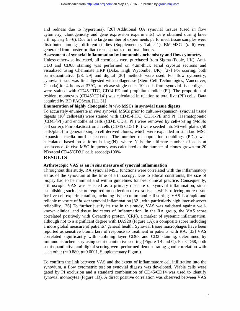
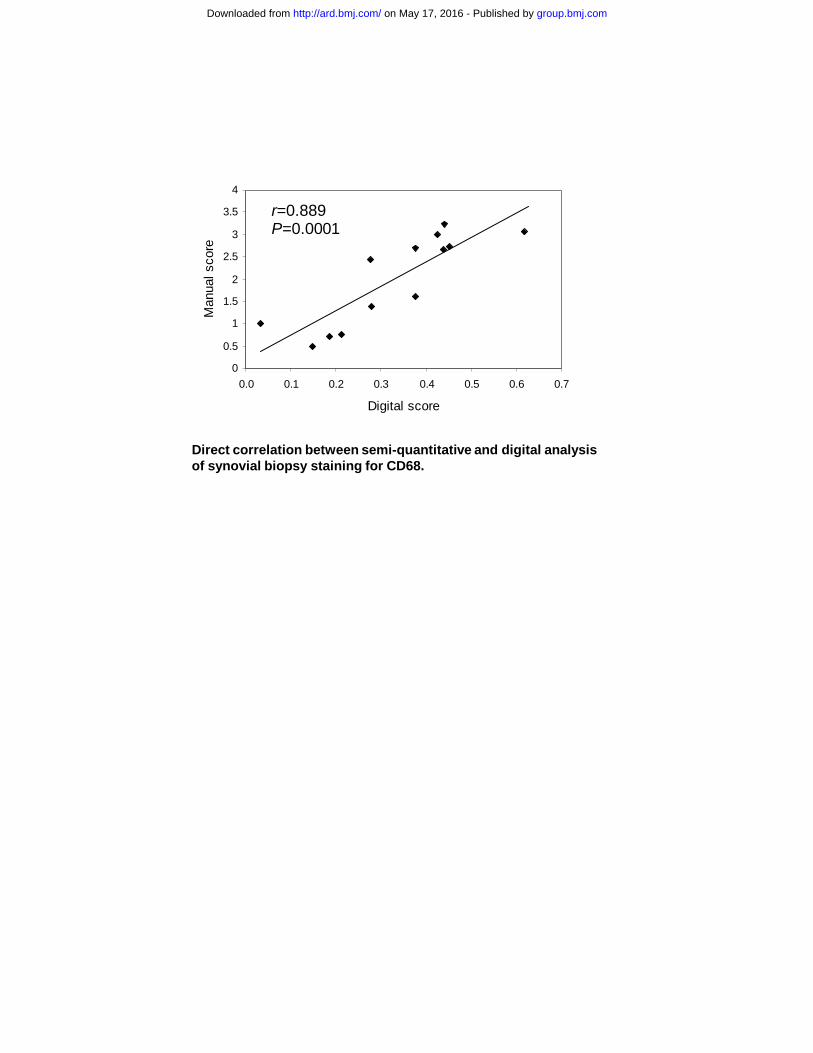
and redness due to hyperemia). [26] Additional OA synovial tissues (used in flow cytometry, clonogenicity and gene expression experiments) were obtained during knee arthroplasty (n=6). Due to the large number of experiments performed, tissue samples were distributed amongst different studies (Supplementary Table 1). BM-MSCs (n=6) were generated from posterior iliac crest aspirates of normal donors. Assessment of synovial inflammation by immunohistochemistry and flow cytometry Unless otherwise indicated, all chemicals were purchased from Sigma (Poole, UK). Anti-CD3 and CD68 staining was performed on 4μm-thick serial cryostat sections and visualized using Chemmate HRP (Dako, High Wycombe, UK). [27] For scoring, both semi-quantitative [28, 29] and digital [30] methods were used. For flow cytometry, synovial tissue was first digested with collagenase (Stem Cell Technologies, Vancouver, Canada) for 4 hours at 37°C, to release single cells. 106 cells from synovial tissue digests were stained with CD45-FITC, CD14-PE and propidium iodide (PI). The proportion of resident monocytes (CD45+CD14+) was calculated in relation to total live (PI-) cells. Data acquired by BD FACScan. [11, 31] Enumeration of highly clonogenic in vivo MSCs in synovial tissue digests To accurately enumerate in vivo synovial MSCs prior to culture-expansion, synovial tissue digests (106 cells/test) were stained with CD45-FITC, CD31-PE and PI. Haematopoietic (CD45+PI-) and endothelial cells (CD45-CD31+PI-) were removed by cell-sorting (MoFlo cell sorter). Fibroblastic/stromal cells (CD45-CD31-PI-) were seeded into 96 well plates (50 cells/plate) to generate single-cell derived clones, which were expanded in standard MSC expansion media until senescence. The number of population doublings (PDs) was calculated based on a formula log2(N), where N is the ultimate number of cells at senescence. In vivo MSC frequency was calculated as the number of clones grown for 20 PDs/total CD45-CD31- cells seeded)x100%. RESULTS Arthroscopic VAS as an in situ measure of synovial inflammation Throughout this study, RA synovial MSC functions were correlated with the inflammatory status of the synovium at the time of arthroscopy. Due to ethical constraints, the size of biopsy had to be minimal and within guidelines for best clinical practice. Consequently, arthroscopic VAS was selected as a primary measure of synovial inflammation, since establishing such a score required no collection of extra tissue, whilst offering more tissue for live cell experimentation, including tissue culture and cell sorting. VAS is a rapid and reliable measure of in situ synovial inflammation [32], with particularly high inter-observer reliability. [26] To further justify its use in this study, VAS was validated against well-known clinical and tissue indicators of inflammation. In the RA group, the VAS score correlated positively with C-reactive protein (CRP), a marker of systemic inflammation, although not to a significant degree with DAS28 (Figure 1A); a composite score including a more global measure of patients’ general health. Synovial tissue macrophages have been reported as sensitive biomarkers of response to treatment in patients with RA. [33] VAS correlated significantly with sublining layer CD68 and CD3 staining, determined by immunohistochemistry using semi-quantitative scoring (Figure 1B and C). For CD68, both semi-quantitative and digital scoring were performed demonstrating good correlation with each other (r=0.889, p=0.0001, Supplementary Figure). To confirm the link between VAS and the extent of inflammatory cell infiltration into the synovium, a flow cytometric test on synovial digests was developed. Viable cells were gated by PI exclusion and a standard combination of CD45/CD14 was used to identify synovial monocytes (Figure 1D). A direct positive correlation was observed between VAS
group.bmj.com on May 17, 2016 - Published by http://ard.bmj.com/Downloaded from

5
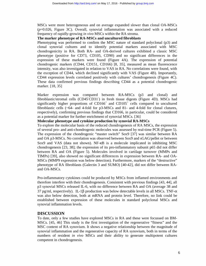
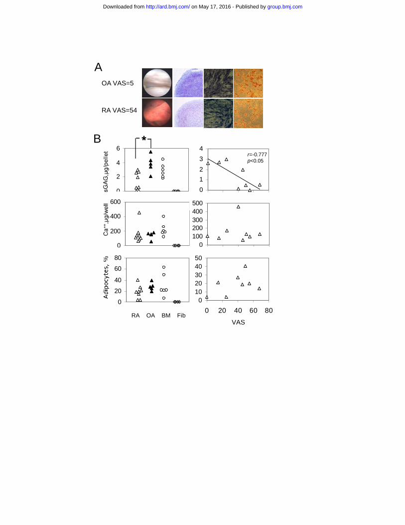
and the percentage of infiltrating monocytes (Figure 1D), consistent with the immunohistochemistry data. The proportion of monocytes in a sample of normal human synovium (generous gift from Prof Cosimo De Bari) was 10%. In summary, these data demonstrated that VAS correlated well with several objective measurements of inflammatory cell infiltration into the synovium and hence was a reliable measure of synovial inflammation. Characterisation of standard polyclonal synovial MSC cultures in RA Following synovial tissue digest, one million cells from 8 RA and 5 OA patients were used to initiate synovial MSC cultures (Table 1). Data from representative RA and OA patients are shown on Figure 2A. Compared to skin fibroblasts, all MSC cultures possessed mesenchymal tri-potentiality (Figure 2B). However, the chondrogenic potential of passage 3 (p3) RA-MSC cultures (measured by sulfated glycosaminoglycan (sGAG)/pellet) was ~3-fold lower compared to OA (p=0.027). Moreover, a direct inverse relationship was observed between chondrogenesis of RA cultures and level of synovial inflammation assessed by VAS (Figure 2B, top panel) or by the proportion of infiltrating monocytes (n=6, r=-0.807, p<0.05). No significant differences between RA and OA were found in adipo- or osteogenic assays (Figure 2B, middle and bottom panels). Therefore, synovial inflammation specifically affected synovial MSC chondrogenesis in RA.
Table 1. Synovial MSC differentiation experiments: Patient characteristics
RA (n=8) OA (n=5) Age 62 (31-73)* 62 (42-76) Sex 7 Female/1 Male 4 Female/1 Male VAS 43 (0-68) 5 (0-9)
Current medication Infliximab/MTX, n=4 Infliximab/MTX/NSAIDs, n=1
Etanercept, n=3
NSAIDs, n=2 No treatment, n=3
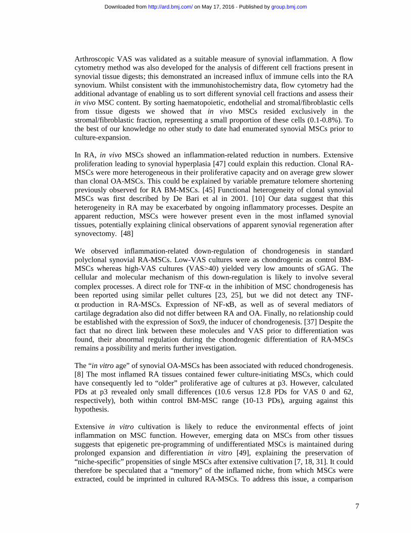
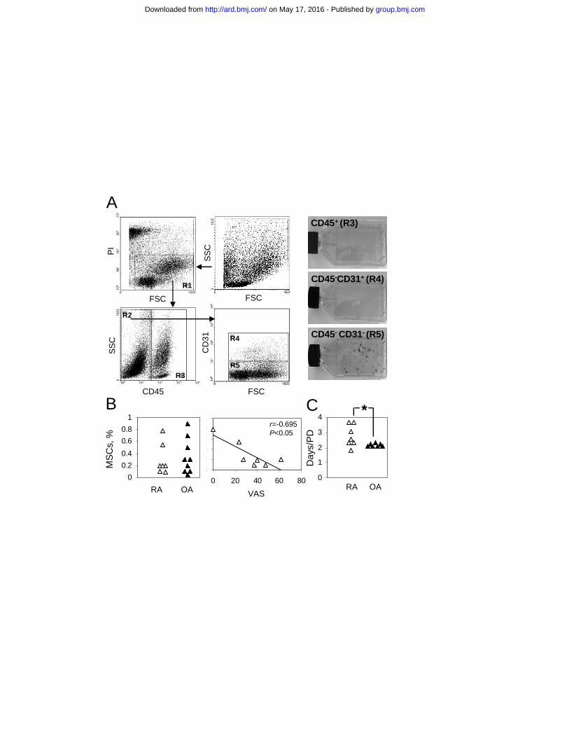
* - all values are shown as median (range), MTX – methotrexate, NSAIDs – non-steroid anti-inflammatory drugs. Enumeration of in vivo MSCs in RA synovium Although MSC definitions vary, most groups agree that in vivo MSCs are highly proliferative, clonogenic cells that reside within tissue stroma and proliferate for at least 20 PDs. [31, 34] To enumerate in vivo MSCs directly in RA and OA synovium, tissue digests were subjected to FACS sorting for haematopoietic (CD45+), endothelial (CD45-CD31+) and fibroblastic/stromal (CD45-CD31-) fractions. Colony-forming cells were exclusively present in the fibroblastic/stromal fraction (Figure 3A). Therefore in vivo MSC frequency was measured within this fraction, by seeding the sorted CD45-CD31- cells at a low density and expanding single colonies for at least 20 PDs. Altogether, 54 clonal cultures were generated – 33 from OA and 21 from RA synovium (n=9 and 7 patients, respectively). All nine randomly selected clonal cultures possessed MSC tripotentiality (data not shown). The proportion of in vivo MSCs was 1.5-fold higher in OA compared to RA, but the differences failed to reach statistical significance (Figure 3B). A significant negative correlation was however observed between the in vivo MSC content of RA stroma and VAS (Figure 3B, right panel). The frequency of less proliferative progenitors (capable of a maximum of 13 and 17 PDs prior to senescence) was similarly found to decline with VAS, albeit less significantly (r=-0.501 and r =-0.680, respectively). Furthermore, clonal RA-
group.bmj.com on May 17, 2016 - Published by http://ard.bmj.com/Downloaded from
6
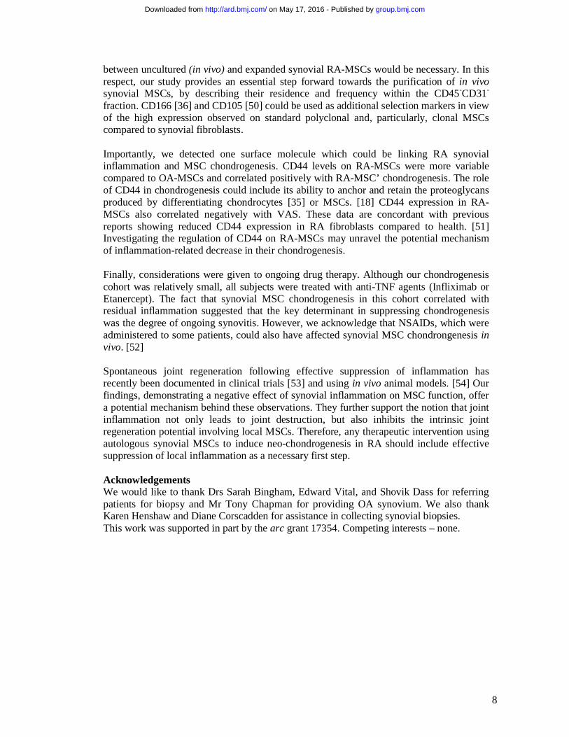
MSCs were more heterogeneous and on average expanded slower than clonal OA-MSCs (p=0.026, Figure 3C). Overall, synovial inflammation was associated with a reduced frequency of rapidly-growing in vivo MSCs within the RA stroma. The marker phenotype of RA-MSCs and uncultured fibroblasts Phenotyping was performed to confirm the MSC nature of standard polyclonal (p3) and clonal synovial cultures and to identify potential markers associated with MSC chondrogenicity in RA. Both RA- and OA-derived cultures exhibited a classic MSC phenotype (positive for CD73, CD105, CD90) and no significant differences in the expression of these markers were found (Figure 4A). The expression of potential chondrogenic markers (CD44, CD151, CD166) [8, 35], measured as mean fluorescence intensity, was also investigated in relation to VAS in RA. No correlations were found, with the exception of CD44, which declined significantly with VAS (Figure 4B). Importantly, CD44 expression levels correlated positively with cultures’ chondrogenesis (Figure 4C). These data confirmed previous findings describing CD44 as a potential chondrogenic marker. [18, 35] Marker expression was compared between RA-MSCs (p3 and clonal) and fibroblastic/stromal cells (CD45-CD31-) in fresh tissue digests (Figure 4D). MSCs had significantly higher proportions of CD166+ and CD105+ cells compared to uncultured fibroblastic cells (~64- and 4-fold for p3-MSCs and 81- and 4-fold for clonal clutures, respectively), confirming previous findings that CD166, in particular, could be considered as a potential marker for further enrichment of synovial MSCs. [36] Molecular phenotype and cytokine production by synovial RA-MSCs To explore the molecular basis of the reduced chondrogenesis of RA MSCs, the expression of several pro- and anti-chondrogenic molecules was assessed by real-time PCR (Figure 5). The expression of the chondrogenic “master switch” Sox9 [37] was similar between RA and OA p3-MSCs. No correlation was observed between Sox9 and sGAG/pellet or between Sox9 and VAS (data not shown). NF-κB is a molecule implicated in inhibiting MSC chondrogenesis [23, 38]; the expression of its pro-inflammatory subunit p65 did not differ between RA and OA (Figure 5). Molecules involved in cartilage turnover (MMPs and TIMPs) [39], also showed no significant differences in expression between RA- and OA-MSCs (MMP9 expression was below detection). Furthermore, markers of the “destructive” phenotype of RA fibroblasts (Galectin 3 and SUMO) [40-42], did not differ between RA- and OA-MSCs. Pro-inflammatory cytokines could be produced by MSCs from inflamed environments and therefore interfere with their chondrogenesis. Consistent with previous findings [43, 44], all p3 synovial MSCs released IL-6, with no difference between RA and OA (average 38 and 37 pg/ml, respectively). IL-1β production was below detectable levels in all MSCs. TNF-α was also below detection, both at mRNA and protein level. Therefore, no link could be established between expression of these molecules in standard polyclonal MSCs and synovial inflammation levels. DISCUSSION To date, only a few studies have explored MSCs in RA and these were focussed on BM- MSCs. [45, 46] This study is the first investigation of the regenerative “fitness” and the MSC content of RA synovium. It shows a negative relationship between the magnitude of synovial inflammation and the regenerative capacity of RA synovium, both in terms of the numbers of resident in vivo MSCs and their ability to generate multipotent cultures competent in chondrogenesis.
group.bmj.com on May 17, 2016 - Published by http://ard.bmj.com/Downloaded from
7
Arthroscopic VAS was validated as a suitable measure of synovial inflammation. A flow cytometry method was also developed for the analysis of different cell fractions present in synovial tissue digests; this demonstrated an increased influx of immune cells into the RA synovium. Whilst consistent with the immunohistochemistry data, flow cytometry had the additional advantage of enabling us to sort different synovial cell fractions and assess their in vivo MSC content. By sorting haematopoietic, endothelial and stromal/fibroblastic cells from tissue digests we showed that in vivo MSCs resided exclusively in the stromal/fibroblastic fraction, representing a small proportion of these cells (0.1-0.8%). To the best of our knowledge no other study to date had enumerated synovial MSCs prior to culture-expansion. In RA, in vivo MSCs showed an inflammation-related reduction in numbers. Extensive proliferation leading to synovial hyperplasia [47] could explain this reduction. Clonal RA-MSCs were more heterogeneous in their proliferative capacity and on average grew slower than clonal OA-MSCs. This could be explained by variable premature telomere shortening previously observed for RA BM-MSCs. [45] Functional heterogeneity of clonal synovial MSCs was first described by De Bari et al in 2001. [10] Our data suggest that this heterogeneity in RA may be exacerbated by ongoing inflammatory processes. Despite an apparent reduction, MSCs were however present even in the most inflamed synovial tissues, potentially explaining clinical observations of apparent synovial regeneration after synovectomy. [48] We observed inflammation-related down-regulation of chondrogenesis in standard polyclonal synovial RA-MSCs. Low-VAS cultures were as chondrogenic as control BM-MSCs whereas high-VAS cultures (VAS>40) yielded very low amounts of sGAG. The cellular and molecular mechanism of this down-regulation is likely to involve several complex processes. A direct role for TNF-α in the inhibition of MSC chondrogenesis has been reported using similar pellet cultures [23, 25], but we did not detect any TNF-α production in RA-MSCs. Expression of NF-κB, as well as of several mediators of cartilage degradation also did not differ between RA and OA. Finally, no relationship could be established with the expression of Sox9, the inducer of chondrogenesis. [37] Despite the fact that no direct link between these molecules and VAS prior to differentiation was found, their abnormal regulation during the chondrogenic differentiation of RA-MSCs remains a possibility and merits further investigation. The “in vitro age” of synovial OA-MSCs has been associated with reduced chondrogenesis. [8] The most inflamed RA tissues contained fewer culture-initiating MSCs, which could have consequently led to “older” proliferative age of cultures at p3. However, calculated PDs at p3 revealed only small differences (10.6 versus 12.8 PDs for VAS 0 and 62, respectively), both within control BM-MSC range (10-13 PDs), arguing against this hypothesis. Extensive in vitro cultivation is likely to reduce the environmental effects of joint inflammation on MSC function. However, emerging data on MSCs from other tissues suggests that epigenetic pre-programming of undifferentiated MSCs is maintained during prolonged expansion and differentiation in vitro [49], explaining the preservation of “niche-specific” propensities of single MSCs after extensive cultivation [7, 18, 31]. It could therefore be speculated that a “memory” of the inflamed niche, from which MSCs were extracted, could be imprinted in cultured RA-MSCs. To address this issue, a comparison
group.bmj.com on May 17, 2016 - Published by http://ard.bmj.com/Downloaded from
8
between uncultured (in vivo) and expanded synovial RA-MSCs would be necessary. In this respect, our study provides an essential step forward towards the purification of in vivo synovial MSCs, by describing their residence and frequency within the CD45-CD31- fraction. CD166 [36] and CD105 [50] could be used as additional selection markers in view of the high expression observed on standard polyclonal and, particularly, clonal MSCs compared to synovial fibroblasts. Importantly, we detected one surface molecule which could be linking RA synovial inflammation and MSC chondrogenesis. CD44 levels on RA-MSCs were more variable compared to OA-MSCs and correlated positively with RA-MSC’ chondrogenesis. The role of CD44 in chondrogenesis could include its ability to anchor and retain the proteoglycans produced by differentiating chondrocytes [35] or MSCs. [18] CD44 expression in RA-MSCs also correlated negatively with VAS. These data are concordant with previous reports showing reduced CD44 expression in RA fibroblasts compared to health. [51] Investigating the regulation of CD44 on RA-MSCs may unravel the potential mechanism of inflammation-related decrease in their chondrogenesis. Finally, considerations were given to ongoing drug therapy. Although our chondrogenesis cohort was relatively small, all subjects were treated with anti-TNF agents (Infliximab or Etanercept). The fact that synovial MSC chondrogenesis in this cohort correlated with residual inflammation suggested that the key determinant in suppressing chondrogenesis was the degree of ongoing synovitis. However, we acknowledge that NSAIDs, which were administered to some patients, could also have affected synovial MSC chondrongenesis in vivo. [52] Spontaneous joint regeneration following effective suppression of inflammation has recently been documented in clinical trials [53] and using in vivo animal models. [54] Our findings, demonstrating a negative effect of synovial inflammation on MSC function, offer a potential mechanism behind these observations. They further support the notion that joint inflammation not only leads to joint destruction, but also inhibits the intrinsic joint regeneration potential involving local MSCs. Therefore, any therapeutic intervention using autologous synovial MSCs to induce neo-chondrogenesis in RA should include effective suppression of local inflammation as a necessary first step. Acknowledgements We would like to thank Drs Sarah Bingham, Edward Vital, and Shovik Dass for referring patients for biopsy and Mr Tony Chapman for providing OA synovium. We also thank Karen Henshaw and Diane Corscadden for assistance in collecting synovial biopsies. This work was supported in part by the arc grant 17354. Competing interests – none.
group.bmj.com on May 17, 2016 - Published by http://ard.bmj.com/Downloaded from

9
FIGURE LEGENDS Figure 1. Arthroscopic synovitis visual analogue score (VAS) correlates with known measures of systemic and local inflammation in RA. A - Correlation between VAS and CRP (mg/l) (left panel) and lack of significant correlation between VAS and DAS28 (right panel). B - Correlations between VAS and semi-quantitative scoring of CD68 (left panel) and CD3 (right panel) staining assessed by immunohistochemistry. C – CD68 and CD3 staining of tissue biopsies of a representative RA patient with low VAS (left) and high VAS (right). Original magnification x100. D - Flow cytometry-based assessment of infiltrating monocytes in synovial tissue digests. Left panel - representative plot illustrating monocyte enumeration strategy, right panel - positive correlation between VAS and a percentage of monocytes. A cohort of 37 RA patients participated in this study and specimens were re-distributed between different aspects of this work depending on the size of biopsy. Figure 2. Inflammation reduces the chondrogenic potential of synovium-derived mesenchymal stem cell (MSC) cultures. A – Representative OA and RA patients: macroscopic appearance of synovium (far left panel), chondrogenesis (Toluidine Blue staining, middle left panel), osteogenesis (alkaline phosphatase staining, middle right panel) and adipogenesis (Oil Red staining, far right panel). Original magnification x100. B – Quantitative differentiation assays showing tri-potentiality of standard polyclonal (passage 3, p3) synovial MSCs from OA and RA patients and positive (BM) and negative (Fib) controls of differentiation. Right panels – Relationships between synovial MSCs chondro-, osteo- and adipogenesis and arthroscopic visual analogue score (VAS) of synovitis. Only chondrogenesis shows a direct inverse relationship with VAS. Open triangles=RA, dark triangles =OA. * - p<0.05. Figure 3. Inflammation affects the frequency of highly-proliferative in vivo MSCs in RA synovium. A – An assessment of the in vivo MSC frequency of synovial tissue digests. Left panel – sorting strategy: PI-positive dead cells/debris were gated out (R1). Hematopoetic cells (R1+R3), endothelial cells (R1+R2+R4) and fibroblastic stromal cells (R1+R2+R5) were sorted separately. Right panel: colony-forming cell assay (1000 cells/flask) confirmed the presence of in vivo MSCs in the fibroblastic stromal fraction. B – Frequencies of in vivo MSCs in this fraction in RA and OA (left panel) or in RA in relation to VAS (right panel). C – Average growth rates (measured as days/PD) of clonal MSC cultures from RA and OA patients. * - p<0.05. Figure 4. The marker phenotype of synovial MSC cultures and synovial stromal fibroblasts in RA. A – Expression of putative MSC markers on standard polyclonal (p3) synovial MSCs from RA (empty bars, n=9) and OA (dark bars, n=5) patients. B – Linear relationship between CD44 mean fluorescence intensity (MFI) and VAS in polyclonal RA-MSCs. C – Linear relationship between chondrogenesis of polyclonal RA-MSCs and CD44 MFI. D - Expression of putative MSC markers on RA synovial fibroblastic/stromal (CD45-
CD31-) cells (light grey bars, n=5), polyclonal RA-MSCs (empty bars, n=9) and clonal RA- MSCs (dark grey bars, n=4). ** indicate markers differentially expressed between both types of MSCs and synovial stromal fibroblasts, p<0.01. Figure 5. Expression of pro- and anti-chondrogenic molecular markers as well as markers of “aggressive” RA fibroblasts in RA and OA MSCs measured by real-time PCR. Real-time PCR was performed on polyclonal p3 cultures; cells were lysed for RNA extraction upon reaching confluence. RA – empty triangles, OA – filled triangles.
group.bmj.com on May 17, 2016 - Published by http://ard.bmj.com/Downloaded from

10
Supplementary Figure. Direct correlation between semi-quantitative and digital analysis of synovial biopsy staining for CD68. Semi-quantitative score (between 0 and 4) is assigned as described in [29]. Quantitative analysis is adapted from [30]. Quantification and analysis was performed using NIS-Elements BR software (Nikon Instruments Inc.). Five fields of view (x200) were photographed randomly, lining layer was removed digitally and areas of positive (DAB) stained cells were converted into binary data, before the total highlighted area was measured. This was repeated for the areas of positive cells plus negative cells. The score is shown as a ratio: area of positive (DAB) stain/(area of positive (DAB) stain + area of negative cells), roughly equating to the ratio of positive cells in relation to total cells. REFERENCES 1. Brittberg M, Lindahl A, Nilsson A, Mainard D. Treatment of deep cartilage defects in the knee with autologous chondrocyte transplantation. New Eng J Med 1994;331:889-95. 2. Saris DBF, Vanlauwe J, Victor J, Haspl M, Bohnsack M, Fortems Y, et al. Characterized chondrocyte implantation results in better structural repair when treating symptomatic cartilage defects of the knee in a randomized controlled trial versus microfracture. Am J Sports Med 2008;36:235-46. 3. Nesic D, Whiteside R, Brittberg M, Wendt D, Martin I, Mainil-Varlet P. Cartilage tissue engineering for degenerative joint disease. Adv Drug Delivery Rev 2006;58:300-22. 4. Murphy JM, Fink DJ, Hunziker EB, Barry FP. Stem cell therapy in a caprine model of osteoarthritis. Arthritis Rheum 2003;10:3464-74. 5. Wakitani S, Goto T, Pineda SJ, Young RG, Mansour JM, Caplan AI. Mesenchymal cell-based repair of large, full-thickness defects of articular-cartilage. J Bone Joint Surg-Am 1994;76A:579-92. 6. Kafienah W, Mistry S, Dickinson SC, Sims TJ, Learmonth I, Hollander AP. Three-dimensional cartilage tissue engineering using adult stem cells from osteoarthritis patients. Arthritis Rheum 2007;56:177-87. 7. Sakaguchi Y, Sekiya I, Yagishita K, Muneta T. Comparison of human stem cells derived from various mesenchymal tissues - Superiority of synovium as a cell source. Arthritis Rheum 2005;52:2521-29. 8. Nagase T, Muneta T, Ju YJ, Hara K, Morito T, Koga H, et al. Analysis of the chondrogenic potential of human synovial stem cells according to harvest site and culture parameters in knees with medial compartment osteoarthritis. Arthritis Rheum 2008;58:1389-98. 9. Tuan R. Stemming cartilage degeneration: Adult mesenchymal stem cells as a cell source for articular cartilage tissue engineering. Arthritis Rheum 2006;54:3075-78. 10. De Bari C, Dell'Accio F, Tylzanowski P, Luyten FP. Multipotent mesenchymal stem cells from adult human synovial membrane. Arthritis Rheum 2001;44:1928-42. 11. Jones EA, English A, Henshaw K, Kinsey SE, Markham AF, Emery P, et al. Enumeration and phenotypic characterization of synovial fluid multipotential mesenchymal progenitor cells in inflammatory and degenerative arthritis. Arthritis Rheum 2004;50:817-27. 12. Morito T, Muneta T, Hara K, Ju YJ, Mochizuki T, Makino H, et al. Synovial fluid-derived mesenchymal stem cells increase after intra-articular ligament injury in humans. Rheumatology 2008;47:1137-43. 13. Bi Y, Ehirchiou D, Kilts TM, Inkson CA, Embree MC, Sonoyama W, et al. Identification of tendon stem/progenitor cells and the role of the extracellular matrix in their niche. Nat Med 2007;13:1219-27.
group.bmj.com on May 17, 2016 - Published by http://ard.bmj.com/Downloaded from
11
14. De Bari C, Dell'Accio F, Vanlauwe J, Eyckmans J, Khan YM, Archer CW, et al. Mesenchymal multipotency of adult human periosteal cells demonstrated by single-cell lineage analysis. Arthritis Rheum 2006;54:1209-21. 15. Wickham MQ, Erickson GR, Gimble JM, Vail TP, Guilak F. Multipotent stromal cells derived from the infrapatellar fat pad of the knee. Clin Orthop Rel Res 2003;412:196-212. 16. English A, Jones EA, Corscadden D, Henshaw K, Chapman T, Emery P, et al. A comparative assessment of cartilage and joint fat pad as a potential source of cells for autologous therapy development in knee osteoarthritis. Rheumatology 2007;46:1676-83. 17. Djouad F, Bony C, Häupl T, Uzé G, Lahlou N, Louis-Plence P, et al. Transcriptional profiles discriminate bone marrow-derived and synovium-derived mesenchymal stem cells. Arthritis Res Ther 2005;7:1304-15. 18. Jones E, Crawford A, English A, Henshaw K, Mundy J, Corscadden D, et al. Synovial fluid mesenchymal stem cells in health and early osteoarthritis: Detection and functional evaluation at the single-cell level. Arthritis Rheum 2008;58:1731-40. 19. Li XL, Makarov SS. An essential role of NF-kappa B in the "tumor-like" phenotype of arthritic synoviocytes. PNAS 2006;103:17432-37. 20. Hunziker EB, Rosenberg LC. Repair of partial-thickness defects in articular cartilage: Cell recruitment from the synovial membrane. J Bone Joint Surg-Am 1996;78A:721-33. 21. Francis-West PH, Parish J, Lee K, Archer CW. BMP/GDF-signalling interactions during synovial joint development. Cell Tissue Res1999;296:111-19. 22. De Bari C, Dell'Accio F, Luyten FP. Failure of in vitro-differentiated mesenchymal stem cells from the synovial membrane to form ectopic stable cartilage in vivo. Arthritis Rheum 2004;50:142-50. 23. Murakami S, Lefebvre V, de Crombrugghe B. Potent inhibition of the master chondrogenic factor Sox9 gene by interleukin-1 and tumor necrosis factor-alpha. J Biol Chem 2000;275:3687-92. 24. Gilbert L, He XF, Farmer P, Rubin J, Drissi H, van Wijnen AJ, et al. Expression of the osteoblast differentiation factor RUNX2 (Cbfa1/AML3/Pebp2 alpha A) is inhibited by tumor necrosis factor-alpha. J Biol Chem 2002;277:2695-2701. 25. Lories RJU, Derese I, De Bari C, Luyten FP. Evidence for uncoupling of inflammation and joint remodeling in a mouse model of spondylarthritis. Arthritis Rheum 2007;56:489-97. 26. Reece RJ, Canete JD, Parsons WJ, Emery P, Veale DJ. Distinct vascular patterns of early synovitis in psoriatic, reactive, and rheumatoid arthritis. Arthritis Rheum 1999;42:1481-84. 27. Buch MH, Reece RJ, Quinn MA, English A, Cunnane G, Henshaw K, et al. The value of synovial cytokine expression in predicting the clinical response to TNF antagonist therapy (infliximab). Rheumatology 2008;47:1469-75. 28. Verburg R, Flierman R, Sont J, Ponchel F, van Dreunen L, Levahrt N, et al. The outcome of intensive immunosuppression and autologous stemcell transplantation in patients with severe rheumatoid arthritis is associated with the composition of synovial T cell infiltration. Ann Rheum Dis 2005;64:1397-1405. 29. Burgoyne C, Field S, Brown AK, Hensor E, English A, Bingham S, et al. Abnormal T-cell differentiation persists in rheumatoid arthritis patients in clinical remission and predicts relapse. Ann Rheum Dis 2008;67:750-7. 30. Cunnane G, Bjork L, Ulfgren AK, Lindblad S, FitzGerald O, Bresnihan B, et al. Quantitative analysis of synovial membrane inflammation: a comparison between automated and conventional microscopic measurements. Ann Rheum Dis 1999;58:493-99.
group.bmj.com on May 17, 2016 - Published by http://ard.bmj.com/Downloaded from

12
31. De Bari C, Dell'Accio F, Karystinou A, Guillot P, Fisk N, Jones E, et al. A biomarker-based mathematical model to predict bone-forming potency of human synovial and periosteal mesenchymal stem cells. Arthritis Rheum 2008;58:240-50. 32. Lindblad S, Hedfors E. Intraarticular variation in synovitis - local macroscopic and microscopic signs of inflammatory activity are significantly correlated. Arthritis Rheum 1985;28:977-86. 33. Haringman JJ, Gerlag DM, Zwinderman AH, Smeets TJM, Kraan MC, Baeten D, et al. Synovial tissue macrophages: a sensitive biomarker for response to treatment in patients with rheumatoid arthritis. Ann Rheum Dis 2005;64:834-38. 34. Bianco P, Robey PG, Simmons PJ. Mesenchymal stem cells: Revisiting history, concepts, and assays. Cell Stem Cell 2008;2:313-19. 35. Grogan SP, Barbero A, Diaz-Romero J, Cleton-Jansen AM, Soeder S, Whiteside R, et al. Identification of markers to characterize and sort human articular chondrocytes with enhanced in vitro chondrogenic capacity. Arthritis Rheum 2007;56:586-95. 36. Fickert S, Fiedler J, Brenner RE. Identification, quantification and isolation of mesenchymal progenitor cells from osteoarthritic synovium by fluorescence automated cell sorting. Osteoarthritis Cart 2003;11:790-800. 37. Sekiya I, Vuoristo JT, Larson BL, Prockop DJ. In vitro cartilage formation by human adult stem cells from bone marrow stroma defines the sequence of cellular and molecular events during chondrogenesis. PNAS 2002;99:4397-4402. 38. Sitcheran R, Cogswell PC, Baldwin AS. NF-kappa B mediates inhibition of mesenchymal cell differentiation through a posttranscriptional gene silencing mechanism. Genes Dev 2003;17:2368-73. 39. Tetlow LC, Woolley DE. Comparative immunolocalization studies of collagenase 1 and collagenase 3 production in the rheumatoid lesion, and by human chondrocytes and synoviocytes in vitro. Br J Rheum 1998;37:64-70. 40. Ohshima S, Kuchen S, Seemayer CA, Kyburz D, Hirt A, Klinzing S, et al. Galectin 3 and its binding protein in rheumatoid arthritis. Arthritis Rheum 2003;48:2788-95. 41. Baier A, Meineckel I, Gay S, Pap T. Apoptosis in rheumatoid arthritis. Curr Opinion Rheum 2003;15:274-79. 42. Aidinis V, Plows D, Haralambous S, Armaka M, Papadopoulos P, Kanaki MZ, et al. Functional analysis of an arthritogenic synovial fibroblast. Arthritis Res Ther 2003;5:R140-R157. 43. Parsonage G, Falciani F, Burman A, Filer A, Ross E, Bofill M, et al. Global gene expression profiles in fibroblasts from synovial, skin and lymphoid tissue reveals distinct cytokine and chemokine expression patterns. Thrombosis Haemostasis 2003;90:688-97. 44. Djouad F, Fritz V, Apparailly F, Louis-Plence P, Bony C, Sany J, et al. Reversal of the immunosuppressive properties of mesenchymal stem cells by tumor necrosis factor alpha in collagen-induced arthritis. Arthritis Rheum 2005;52:1595-1603. 45. Kastrinaki MC, Spanoudakis M, Sidiropoulos P, Kritikos H, Eliopoulos GD, Boumpas DT, et al. Bone marrow mesenchymal stem cell properties in patients with rheumatoid arthritis. Annals Rheum Dis 2006;65:A37-A38. 46. Dudics V, Kunstar A, Kovacs J, Lakatos T, Géher P, Gömör B, et al. Chondrogenic potential of mesenchymal stem cells from patients with rheumatoid arthritis and osteoarthritis: measurements in a microculture system. Cells Tissues Organs 2008 [Epub ahead of print]. 47. Firestein GS. Invasive fibroblast-like synoviocytes in rheumatoid arthritis - Passive responders or transformed aggressors? Arthritis Rheum1996;39:1781-90. 48. Ostergaard M, Ejbjerg B, Stoltenberg M, Gideon P, Volck B, Skov K, et al. Quantitative magnetic resonance imaging as marker of synovial membrane regeneration and
group.bmj.com on May 17, 2016 - Published by http://ard.bmj.com/Downloaded from

13
recurrence of synovitis after arthroscopic knee joint synovectomy: a one year follow up study. Ann Rheum Dis 2001;60:233-36. 49. Boquest AC, Noer A, Collas P. Epigenetic programming of mesenchymal stem cells from human adipose tissue. Stem Cell Reviews 2006;2:319-29. 50. Jo CH, Ahn HJ, Kim HJ, Seong SC, Lee MC. Surface characterization and chondrogenic differentiation of mesenchymal stromal cells derived from synovium. Cytotherapy 2007;9:316-27. 51. Henderson KJ, Edwards JCW, Worrall JG. Expression of CD44 in normal and rheumatoid synovium and cultured synovial fibroblasts. Annals Rheum Dis 1994;53:729-34. 52. Hart DA, Kydd AS, Frank CB, Hildebrand KA. Tissue repair in rheumatoid arthritis: challenges and opportunities in the face of a systemic inflammatory disease. Best Pract Res Clin Rheumat 2004;18:187-202. 53. Rau R, Herborn G, Wassenberg S. Healing of erosive changes in rheumatoid arthritis. Clin Exp Rheumat 2004;22:S44-S49. 54. Shealy DJ, Wooley PH, Emmell E, Volk A, Rosenberg A, Treacy G, et al. Anti-TNF-alpha antibody allows healing of joint damage in polyarthritic transgenic mice. Arthritis Res 2002;4:5-9. The Corresponding Author has the right to grant on behalf of all authors and does grant on behalf of all authors, an exclusive licence (or non-exclusive for government employees) on a worldwide basis to the BMJ Publishing Group Ltd and its Licensees to permit this article (if accepted) to be published in Annals of the Rheumatic Diseases and any other BMJPGL products to exploit all subsidiary rights, as set out in our licence (http://ard.bmj.com/ifora/licence.pdf).
group.bmj.com on May 17, 2016 - Published by http://ard.bmj.com/Downloaded from
SUPPLEMENTARY METHODS Trilineage differentiation of standard expanded synovial MSC cultures
For primary culture, 106 cells from synovial tissue digests were plated in 25cm2
flasks in standard media, containing DMEM, antibiotics and 10% FCS, optimised
for MSC growth (Mesencult, Stem Cell Technologies). Positive control BM-MSCs
were established from BM mononuclear cells (107/25cm2 flask) following
separation with Lymphoprep (Axis-Shield, Dundee, UK). Negative control skin
fibroblasts were purchased from ATCC (Rockville, MD, USA). Flow cytometry,
colony-forming unit-fibroblast (CFU-F), differentiation assays, real-time PCR and
ELISA were performed on passage 3 (p3) cultures. Passaging was performed by
standard trysinization [11, 16] of nearly-confluent cultures (1:2 splits). In vitro
“age” of control BM MSCs at p3 was 10-13 population doublings (PDs) based on
the number of seeded CFU-Fs.
Osteogenic and adipogenic differentiation was induced by placing cells in standard
osteo- and adipoinductive conditions as previously described [11, 16]. The extent of
osteogenesis was measured as the amount of calcium produced at the end of the
culture (day 21), using a commercial calcium kit (DLC, Charlottetown, Canada).
Adipogenesis was quantified as a percentage of fat-laden cells (Oil Red-positive) in
relation to all nucleated cells (following counterstaining with Harris’s Haematoxylin).
Chondrogenic assay was performed as previously described, with 2.5x10⁵ cells used
to initiate each pellet, in triplicate [11, 16]. Chondroinductive media contained high-
glucose DMEM (Invitrogen), 100 μg/ml sodium pyruvate, 40 μg/ml proline, 50
μg/ml ascorbic acid-2-phosphate, 1 mg/ml BSA, 1x insulin-transferrin-selenium plus,
100 nM dexamethasone (all from Sigma) and 10 ng/ml transforming growth factor β3
group.bmj.com on May 17, 2016 - Published by http://ard.bmj.com/Downloaded from

(R&D Systems, Abingdon, UK). Pellets were harvested at the end of the three-week
culture period and digested with 100 μl papain solution. The amount of sulphated
glycosaminoglycans (sGAG) (in µg/pellet) was measured by Alcian blue binding
assay (IDS, Boldon, UK) [11, 16]. Assay detection range was 12.5-400 µg of
sGAG/ml.
Flow cytometry and real-time PCR on standard expanded synovial MSC
cultures
Flow cytometry was performed using standard methods as described previously (11,
31). RNA was extracted [32] and cDNA synthesized using 400U Superscript II
reverse transcriptase (Invitrogen). Real-time PCR was performed using an ABI
Prism 7900 sequence detection system (Applied Biosystems, Warrington, UK) in
the presence of SYBR-green or Taqman. Expression was normalized using a house-
keeping gene GAPDH.
ELISA
Cytokine production (Tumour necrosis factor-α (TNFα), interleukin (IL)-6 and IL-
1β) in culture supernatants was measured by ELISA using HS Quantikine kits
according to manufacturer’s instructions (R&D Systems). Assay sensitivity was
0.039, 0.057 and 0.106 pg/ml for IL-6, IL-1β and TNFα, respectively. MSCs (5x10⁴
cells) were seeded in 12-well plates in 2 ml of MSC expansion media and
supernatants collected for measurements 48 hours later.
Statistical analysis
Spearman’s rank correlation coefficient was used to correlate two variables. Mann-
Whitney U test for 2 independent samples was used to compare groups. p<0.05 and
r>0.6 were considered significant.
group.bmj.com on May 17, 2016 - Published by http://ard.bmj.com/Downloaded from
0
0.5
1
1.5
2
2.5
3
3.5
4
0.0 0.1 0.2 0.3 0.4 0.5 0.6 0.7
Digital score
Man
ual s
core
r=0.889P=0.0001
Direct correlation between semi-quantitative and digital analysis of synovial biopsy staining for CD68.
group.bmj.com on May 17, 2016 - Published by http://ard.bmj.com/Downloaded from

Supplementary Table 1: RA and OA patient sample distribution into different
parts of the study
Type of study Diagnosis n*
Differentiation assays on
polyclonal MSC cultures
RA
OA
8
5
CRP/DAS28 analysis** and
immunochemistry
RA 37
Flow cytometry on synovial tissue
digests
RA
20
Flow cytometry on expanded
synovial MSCs (polyclonal and
clonal)
RA
OA
9
5
Cell sorting for the in vivo MSC
enumeration
RA
OA
7
9
Gene expression and ELISA RA
OA
6
4
* - tissues from the same patient were often distributed amongst several studies
* * - data obtained from clinical notes
group.bmj.com on May 17, 2016 - Published by http://ard.bmj.com/Downloaded from
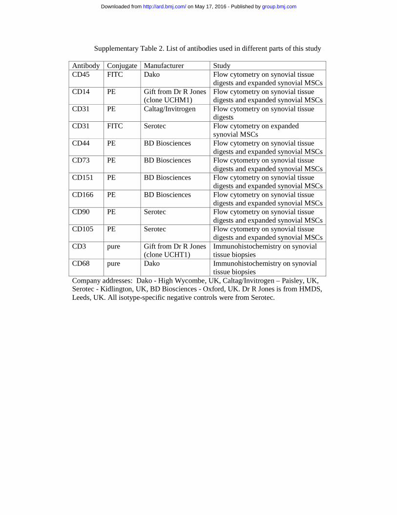
Supplementary Table 2. List of antibodies used in different parts of this study Antibody Conjugate Manufacturer Study CD45 FITC Dako Flow cytometry on synovial tissue
digests and expanded synovial MSCs CD14 PE Gift from Dr R Jones
(clone UCHM1) Flow cytometry on synovial tissue digests and expanded synovial MSCs
CD31 PE Caltag/Invitrogen Flow cytometry on synovial tissue digests
CD31 FITC Serotec Flow cytometry on expanded synovial MSCs
CD44 PE BD Biosciences Flow cytometry on synovial tissue digests and expanded synovial MSCs
CD73 PE BD Biosciences Flow cytometry on synovial tissue digests and expanded synovial MSCs
CD151 PE BD Biosciences Flow cytometry on synovial tissue digests and expanded synovial MSCs
CD166 PE BD Biosciences Flow cytometry on synovial tissue digests and expanded synovial MSCs
CD90 PE Serotec Flow cytometry on synovial tissue digests and expanded synovial MSCs
CD105 PE Serotec Flow cytometry on synovial tissue digests and expanded synovial MSCs
CD3 pure Gift from Dr R Jones (clone UCHT1)
Immunohistochemistry on synovial tissue biopsies
CD68 pure Dako Immunohistochemistry on synovial tissue biopsies
Company addresses: Dako - High Wycombe, UK, Caltag/Invitrogen – Paisley, UK, Serotec - Kidlington, UK, BD Biosciences - Oxford, UK. Dr R Jones is from HMDS, Leeds, UK. All isotype-specific negative controls were from Serotec.
group.bmj.com on May 17, 2016 - Published by http://ard.bmj.com/Downloaded from
Supplementary Table 3. Primers used for real time PCR.
Primer Forward primer (5’- 3’) Reverse primer (5’- 3’)
GAPDH aacagcgacacccactcctc cataccaggaaatgagcttgacaa
Sox9 gacacctctggggtcccttc tcctcaaggtcgagtgagctgt
NF-κB p65 atccacagtttccagaacctgg ttgttggtctggatgcgct
IκBa tgaaggctaccaactacaatggc agcacccaaggacaccaaaa
MMP1 agtccggtttttcaaagggaa gccaaaggagctgtagatgtcc
MMP2 actcctgagatctgcaaacagga ccgccaaatgaaccggt
MMP3 ggaaaacccaccttacatacagga ttcccagactttcagagctttctc
MMP9 gaggcgctcatgtaccctatgt agggcgaggaccatagaggt
TIMP1 ttctgcaattccgacctcgt ttggtcatcttgatctcataacgc
TIMP2 ctggaaacgacatttatggcaa tctcaggccctttgaacatctt
Galectin3 tgaagcccaatgcaaacaga tgttgttctcattgaagcgtgg
Osteopontin catcacctgtgccataccagtta ggccacagcatctgggtattt
SUMO1 gtgtcaaagacagggtgttcca cttcttcctccattcccagttgt
Taqman assays were used for TNFα normalised to GAPDH (Hs00174128 and Hs99999905, respectively, Applied Biosystems). All other primers (0.5 pmol/μl) were designed using Primer Express 2. Efficiencies calculated using the log-linear phase of dilution and were found to be approximately equal. All reactions were performed in triplicate and a melting curve analysis was used to ensure specificity of each PCR product.
group.bmj.com on May 17, 2016 - Published by http://ard.bmj.com/Downloaded from

A
DA
S28
CD
68
CD
3
CR
P
Mon
ocyt
es
(Q2)
, %
CD
14
CD45
0
5 0
10 0
15 0
0 2 0 4 0 6 0 8 0
3
5
7
9
0 2 0 4 0 60 8 0
50
100
150
0
0
0.5
1
1.5
2
2.5
0 20 40 60 80
0
1
2
3
4
5
0 20 40 60 80
0
5
10
1 5
20
2 5
30
3 5
0 20 40 6 0 80
3
5
7
9
0 20 40 60 80 0 20 40 60 80
B
01234
0
1
2
0 20 40 60 80 0 20 40 60 80VAS
VAS0 20 40 60 80
0
15
25
35D VAS
VAS VAS
Q3
Q1 Q2
Q4
Cr=0.835p<0.001
r=0.779 p<0.0001
r=0.817p<0.0001
CD
68C
D3
Neg
ativ
e
VAS 0 VAS 46
r=0.825p<0.001
group.bmj.com on May 17, 2016 - Published by http://ard.bmj.com/Downloaded from
OA VAS=5
RA VAS=54
A
VAS
0
2
4
6
0
200
400
600
0
20
40
60
80
0
1
2
3
4
0 20 40 60 80
0100200300400500
0 20 40 60 80
01020304050
0 20 40 60 80
Adipocytes, %
Br=-0.777p<0.05
*
RA OA BM Fib
group.bmj.com on May 17, 2016 - Published by http://ard.bmj.com/Downloaded from
FSC
CD45 FSC
CD
31S
SCPI
FSC
A
R1
R2
R3
R4
R5
SS
C
CD45+ (R3)
CD45-CD31+ (R4)
CD45- CD31- (R5)
0
1
2
3
4
RA OA
*
00.20.40.60.8
1
0 20 40 60 80
r=-0.695P<0.05
Day
s/P
D
C
VAS
0
0.2
0.4
0.6
0.8
1
RA OA
MS
Cs,
%
B
group.bmj.com on May 17, 2016 - Published by http://ard.bmj.com/Downloaded from

0
2
A
0 20 40 60 80 100
CD31
CD45
CD14
CD166
CD90
CD44
CD105
CD151
CD73
50
70
90
110
130
0 20 40 60 80 100
Positive cells, %B
r=-0.830P<0.01
CD
44, M
FI
C
0
1
2
3
50 70 90 110 130
VAS
r=0.865P<0.05
CD44, MFID
0 20 40 60 80 100
CD31
CD45
CD14
CD166
CD90
CD44
CD105
CD151
CD73
**
**
Positive cells, %
group.bmj.com on May 17, 2016 - Published by http://ard.bmj.com/Downloaded from

*
0.0000001
0.000001
0.00001
0.0001
0.001
0.01
0.1
1
TIMP1 TIMP2 MMP2 NFκB Sox9 Galectin IκB MMP1 SUMOp65 3
Exp
ress
ion
norm
aliz
edto
GA
PD
H
group.bmj.com on May 17, 2016 - Published by http://ard.bmj.com/Downloaded from

levelassessment in relation to synovial inflammationsynovium: Enumeration and functional Mesenchymal stem cells in rheumatoid
R Reece, S Kinsey, P Emery, D McGonagle and F PonchelE Jones, S M Churchman, A English, M H Buch, E A Horner, C H Burgoyne,
published online April 2, 2009Ann Rheum Dis
http://ard.bmj.com/content/early/2009/04/02/ard.2008.106435Updated information and services can be found at:
These include:
MaterialSupplementary
http://ard.bmj.com/content/suppl/2010/03/11/ard.2008.106435.DC1.htmlSupplementary material can be found at:
serviceEmail alerting
box at the top right corner of the online article. Receive free email alerts when new articles cite this article. Sign up in the
Errata
http://ard.bmj.com/content/70/8/1519.full.pdfor:
next pageAn erratum has been published regarding this article. Please see
CollectionsTopic Articles on similar topics can be found in the following collections
(3206)Rheumatoid arthritis (4181)Connective tissue disease
(922)Osteoarthritis (4874)Musculoskeletal syndromes
(4568)Degenerative joint disease (1227)Inflammation
(5057)Immunology (including allergy)
Notes
http://group.bmj.com/group/rights-licensing/permissionsTo request permissions go to:
http://journals.bmj.com/cgi/reprintformTo order reprints go to:
http://group.bmj.com/subscribe/To subscribe to BMJ go to:
group.bmj.com on May 17, 2016 - Published by http://ard.bmj.com/Downloaded from

Ann Rheum Dis August 2011 Vol 70 No 8 1519
1. Keystone E, Emery P, Peterfy CG, et al. Rituximab inhibits structural joint damage in
patients with rheumatoid arthritis with an inadequate response to tumour necrosis
factor inhibitor therapies. Ann Rheum Dis 2009;68:216–21.
2. Doward LC, McKenna SP, Whalley D, et al. The development of the L-QoL: a
quality-of-life instrument specifi c to systemic lupus erythematosus. Ann Rheum Dis
2009;68:196–200.
3. Potter C, Hyrich KL, Tracey A, et al. Association of rheumatoid factor and anti-
cyclic citrullinated peptide positivity, but not carriage of shared epitope or PTPN22
susceptibility variants, with anti-TNF response in RA. Ann Rheum Dis 2009;68:69–74.
4. Smolen JS, Han C, van der Heijde DM, et al.; Active-Controlled Study of Patients
Receiving Infl iximab for the Treatment of Rheumatoid Arthritis of Early Onset (ASPIRE)
Study Group. Radiographic changes in rheumatoid arthritis patients attaining
different disease activity states with methotrexate monotherapy and infl iximab plus
methotrexate: the impacts of remission and tumour necrosis factor blockade.
Ann Rheum Dis 2009;68:823–7.
5. Buch MH, Boyle DL, Rosengren S, et al. Mode of action of abatacept in rheumatoid
arthritis patients having failed tumour necrosis factor blockade: a histological, gene
expression and dynamic magnetic resonance imaging pilot study. Ann Rheum Dis
2009;68:1220–7.
6. Emery P, Van Vollenhoven R, Ostergaard M, et al. Guidelines for initiation of anti-
tumour necrosis factor therapy in rheumatoid arthritis: similarities and differences
across Europe. Ann Rheum Dis 2009;68:456–9.
7. Bejarano V, Conaghan PG, Proudman SM, et al. Long-term effi cacy and toxicity of
ciclosporin A in combination with methotrexate in poor prognosis rheumatoid arthritis.
Ann Rheum Dis 2009;68:761–3.
8. Rudwaleit M, Landewé R, van der Heijde D, et al. The development of
Assessment of SpondyloArthritis international Society classifi cation criteria for axial
spondyloarthritis (part I): classifi cation of paper patients by expert opinion including
uncertainty appraisal. Ann Rheum Dis 2009;68:770–6.
9. Bennett AN, Marzo-Ortega H, Emery P, et al.; Leeds Spondyloarthropathy Group.
Diagnosing axial spondyloarthropathy. The new Assessment in SpondyloArthritis
international Society criteria: MRI entering centre stage. Ann Rheum Dis 2009;68:765–7.
10. Marzo-Ortega H, McGonagle D, O’Connor P, et al. Baseline and 1-year magnetic
resonance imaging of the sacroiliac joint and lumbar spine in very early infl ammatory
back pain. Relationship between symptoms, HLA-B27 and disease extent and
persistence. Ann Rheum Dis 2009;68:1721–7.
11. Gilworth G, Emery P, Gossec L, et al. Adaptation and cross-cultural validation
of the rheumatoid arthritis work instability scale (RA-WIS). Ann Rheum Dis
2009;68:1686–90.
12. Gilworth G, Emery P, Gossec L, et al. Adaptation and cross-cultural validation of the
RA-WIS (Work Instability Scale). Ann Rheum Dis 2009;68:1686–90.
13. Jarrett SJ, Sivera F, Cawkwell LS, et al. MRI and clinical fi ndings in patients with
ankylosing spondylitis eligible for anti-tumour necrosis factor therapy after a short
course of etoricoxib. Ann Rheum Dis 2009;68:1466–9.
14. Haugeberg G, Conaghan PG, Quinn M, et al. Bone loss in patients with active early
rheumatoid arthritis: infl iximab and methotrexate compared with methotrexate
treatment alone. Explorative analysis from a 12-month randomised, double-blind,
placebo-controlled study. Ann Rheum Dis 2009;68:1898–901.
15. Genovese MC, Breedveld FC, Emery P, et al. Safety of biological therapies
following rituximab treatment in rheumatoid arthritis patients. Ann Rheum Dis
2009;68:1894–7.
16. Kekow J, Moots RJ, Emery P, et al. Patient-reported outcomes improve with
etanercept plus methotrexate in active early rheumatoid arthritis and the improvement
is strongly associated with remission: the COMET trial. Ann Rheum Dis 2010;69:222–5.
17. Furst DE, Keystone EC, Fleischmann R, et al. Updated consensus statement on
biological agents for the treatment of rheumatic diseases, 2009. Ann Rheum Dis
2010;69(Suppl 1):i2–29.
18. Freeston JE, Wakefi eld RJ, Conaghan PG, et al. A diagnostic algorithm for
persistence of very early infl ammatory arthritis: the utility of power Doppler ultrasound
when added to conventional assessment tools. Ann Rheum Dis 2010;69:417–9.
19. Jones E, Churchman SM, English A, et al. Mesenchymal stem cells in rheumatoid
synovium: enumeration and functional assessment in relation to synovial infl ammation
level. Ann Rheum Dis 2010;69:450–7.
20. Alten RE, Zerbini C, Jeka S, et al. Effi cacy and safety of pamapimod in patients with
active rheumatoid arthritis receiving stable methotrexate therapy. Ann Rheum Dis
2010;69:364–7.
CorrectionsThe department of one of the authors who co-authored all of the below papers has found that the affi liations were not correct. The correct affi liations for Professor P Emery, for all of the below articles, are: 1Section of Musculoskeletal Disease, Leeds Institute of Molecular Medicine, University of Leeds; 2NIHR Leeds Musculoskeletal Biomedical Research Unit, Leeds Teaching Hospitals Trust, Leeds, UK.
21. Machold KP, Landewé R, Smolen JS, et al. The Stop Arthritis Very Early (SAVE) trial,
an international multicentre, randomised, double-blind, placebo-controlled trial on
glucocorticoids in very early arthritis. Ann Rheum Dis 2010;69:495–502.
22. Schoels M, Knevel R, Aletaha D, et al. Evidence for treating rheumatoid arthritis to
target: results of a systematic literature search. Ann Rheum Dis 2010;69:638–43.
23. Smolen JS, Aletaha D, Bijlsma JW, et al.; T2T Expert Committee. Treating
rheumatoid arthritis to target: recommendations of an international task force.
Ann Rheum Dis 2010;69:631–7.
24. Burr ML, Naseem H, Hinks A, et al.; BIRAC Consortium; YEAR Consortium. PADI4
genotype is not associated with rheumatoid arthritis in a large UK Caucasian
population. Ann Rheum Dis 2010;69:666–70.
25. Emery P, Durez P, Dougados M, et al. Impact of T-cell costimulation modulation
in patients with undifferentiated infl ammatory arthritis or very early rheumatoid
arthritis: a clinical and imaging study of abatacept (the ADJUST trial). Ann Rheum Dis
2010;69:510–16.
26. Bennett AN, Rehman A, Hensor EM, et al. The fatty Romanus lesion: a non-
infl ammatory spinal MRI lesion specifi c for axial spondyloarthropathy. Ann Rheum Dis
2010;69:891–4.
27. Nam JL, Winthrop KL, van Vollenhoven RF, et al. Current evidence for the
management of rheumatoid arthritis with biological disease-modifying antirheumatic
drugs: a systematic literature review informing the EULAR recommendations for the
management of RA. Ann Rheum Dis 2010;69:976–86.
28. Smolen JS, Landewé R, Breedveld FC, et al. EULAR recommendations for the
management of rheumatoid arthritis with synthetic and biological disease-modifying
antirheumatic drugs. Ann Rheum Dis 2010;69:964–75.
29. Tan RJ, Gibbons LJ, Potter C, et al.; BRAGGSS. Investigation of rheumatoid arthritis
susceptibility genes identifi es association of AFF3 and CD226 variants with response
to anti-tumour necrosis factor treatment. Ann Rheum Dis 2010;69:1029–35.
30. Robinson JI, Barrett JH, Taylor JC, et al.; YEAR Consortium; BRAGGSS. Dissection of
the FCGR3A association with RA: increased association in men and with autoantibody
positive disease. Ann Rheum Dis 2010;69:1054–7.
31. Cohen SB, Keystone E, Genovese MC, et al. Continued inhibition of structural
damage over 2 years in patients with rheumatoid arthritis treated with rituximab in
combination with methotrexate. Ann Rheum Dis 2010;69:1158–61.
32. Haugeberg G, Bennett AN, McGonagle D, et al. Bone loss in very early infl ammatory
back pain in undifferentiated spondyloarthropathy: a 1-year observational study.
Ann Rheum Dis 2010;69:1364–6.
33. Schoels M, Aletaha D, Smolen JS, et al. Follow-up standards and treatment targets
in rheumatoid arthritis: results of a questionnaire at the EULAR 2008. Ann Rheum Dis
2010;69:575–8.
34. Eyre S, Flynn E, Martin P, et al. No evidence for association of the KLF12 gene with
rheumatoid arthritis in a large UK cohort. Ann Rheum Dis 2010;69:1407–8.
35. Eyre S, Hinks A, Flynn E, et al. Confi rmation of association of the REL locus
with rheumatoid arthritis susceptibility in the UK population. Ann Rheum Dis
2010;69:1572–3.
36. Orozco G, Eyre S, Hinks A, et al.; Wellcome Trust Case Control consortium YEAR
Consortium. Association of CD40 with rheumatoid arthritis confi rmed in a large UK
case-control study. Ann Rheum Dis 2010;69:813–16.
37. Emery P, Durez P, Dougados M, et al. Impact of T-cell costimulation modulation
in patients with undifferentiated infl ammatory arthritis or very early rheumatoid
arthritis: a clinical and imaging study of abatacept (the ADJUST trial). Ann Rheum Dis
2010;69:510–16.
38. Saleem B, Keen H, Goeb V, et al. Patients with RA in remission on TNF blockers: when
and in whom can TNF blocker therapy be stopped? Ann Rheum Dis 2010;69:1636–42.
39. Barkham N, Coates LC, Keen H, et al. Double-blind placebo-controlled trial of
etanercept in the prevention of work disability in ankylosing spondylitis. Ann Rheum Dis
2010;69:1926–8.
40. Emery P, Deodhar A, Rigby WF, et al. Effi cacy and safety of different doses and
retreatment of rituximab: a randomised, placebo-controlled trial in patients who
are biological naive with active rheumatoid arthritis and an inadequate response to
methotrexate (Study Evaluating Rituximab’s Effi cacy in MTX iNadequate rEsponders
(SERENE)). Ann Rheum Dis 2010;69:1629–35.
41. Dixon WG, Hyrich KL, Watson KD, et al.; BSRBR Control Centre Consortium; British
Society for Rheumatology Biologics Register. Infl uence of anti-TNF therapy on mortality
in patients with rheumatoid arthritis-associated interstitial lung disease: results from the
British Society for Rheumatology Biologics Register. Ann Rheum Dis 2010;69:1086–91.
Ann Rheum Dis 2011;70:1519. doi:10.1136/annrheumdis-2011-70-08
29_annrheumdis143438.indd 151929_annrheumdis143438.indd 1519 6/24/2011 7:19:54 PM6/24/2011 7:19:54 PM
Related Documents