Manejo de La IC en Urgencias
Oct 27, 2015
Welcome message from author
This document is posted to help you gain knowledge. Please leave a comment to let me know what you think about it! Share it to your friends and learn new things together.
Transcript
EditorialThe Race to Tissue Oxygenation:Special Teams GoGoGo
Ragavendra R. Baliga, MD, MBA James B. Young, MD
Consulting Editors
More than 50,000 patients die every year fromacute decompensated heart failure. It is the mostcommon reason for hospital admissions of pa-tients 65 years of age and older, and half thesepatients who are older than 70 years of age arereadmitted within 90 days. One million patientsare hospitalized every year with this conditionand 20% of them are rehospitalized for this condi-tion within 30 days of the initial admission.1 Oneyear from index hospitalization, mortality is about30%, whereas the mortality rate at 1 year for am-bulatory New York Heart Association (NYHA) classIII heart failure is substantially less than 10%. Theevent rate for patients hospitalized for heart failureresembles the postmyocardial infarction curve—with a very high event rate in the first 60 days, fol-lowed by a relatively flat curve.2
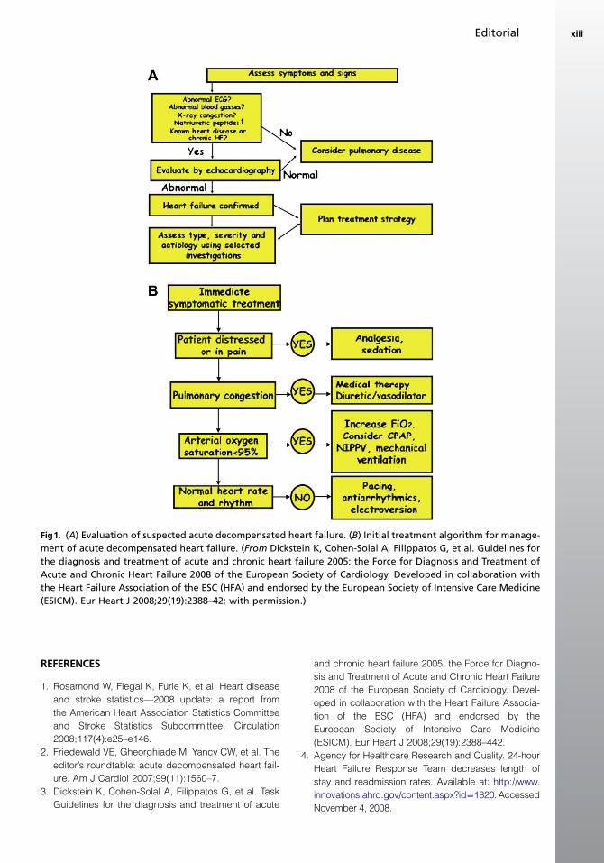
Acute decompensated heart failure may presentas pulmonary edema, features of decreased cardiacoutput, or hypoperfusion. In these patients, the needto restore tissue oxygenation is urgent. The ap-proach to restoration of tissue oxygenation includesrestoring euvolemia by relieving congestion, ad-dressing etiologic and precipitating factors (Box 1),and strategies to prevent recurrence of heart fail-ure.3 Typically, in the absence ofguidelinesand pau-city of evidence-based therapy recommendationsfor management of acute decompensated heart fail-ure, the care provided has been disparate.
With a view toward reducing the length of stayand preventing rehospitalization, hospitals are in-creasingly forming 24-hour on-call special teams,known as Heart Failure Response Teams, to man-age these patients. A typical in-patient HeartFailure Response Team comprises one of the hos-pitalists on-call and two heart failure nurses duringbusiness hours. This special team is typically acti-vated within 30 minutes after the patient is admit-ted to the emergency department (ED) and works,initially, with the ED physicians to stabilize thepatient; this strategy is based on an alogrithmpreviously prepared by a team of cardiologists,ED physicians, and hospitalists (Fig. 1). The nursesensure that all patients receive a dischargesummary explaining in detail dietary restrictions,including sodium restriction, medications and dos-ages, and instructions on whom to contact in caseof weight gain or other symptoms; both patientsand physicians must sign the forms. The nursesalso ensure that a follow-up appointment withthe primary care physician is scheduled for 72hours postdischarge. In the case of a schedulingconflict within this 72-hour window, the patient isscheduled for follow up at a hospital-based clinic.In one hospital, the length of stay decreased byone half of a day and the readmission rate (read-mitted for any reason) fell from 22% in 2006 to18.7% in 2007. Heart failure readmissions falling
Heart Failure Clin 5 (2009) xi–xivdoi:10.1016/j.hfc.2008.10.0011551-7136/08/$ – see front matter ª 2008 Elsevier Inc. All rights reserved. he
artf
ailu
re.th
ecli
nics
.com
Management of Heart Failure in the Emergent Situation
under diagnosis-related group 127 fell from 10.5%in 2006 to 6.4% in 2007.4
The Institute of Medicine, in a bid to reducemedical errors and improve quality, has beenchampioning special response teams to managecritical patients.5 The establishment of a typicalHeart Failure Response Team involves developingreliable processes, systems, or interventions todetermine left ventricular function; to provide pa-tients with smoking cessation counseling; to
review utilization of angiotensin-converting en-zyme inhibitor or angiotensin receptor blocker(or to ensure accurate and timely documentationof omission rationale); to screen for risk of deepvein thrombosis and apply prophylaxis; to providedietary counseling; to promote immunizationagainst pneumococcus or influenza; and to applystrategies to prevent hospitalization.6 All of thesepatients were discharged to a dedicated heartfailure clinic to help patients self-manage euvole-mia. After continuous process improvement, thisconsortium of hospitals was able to standardizedelivery of all these process measures and toreduce readmission rates from 25% in 2004 to10% in 2007.
One of the main goals of the Heart Failure Re-sponse Team is to rapidly restore tissue oxygena-tion by achieving euvolemia when there isvascular congestion or improve hemodynamicswhen there is hypoperfusion. In this issue, JamesF. Neunschwander II and W. Frank Peacock haveassembled a team of experts who have very suc-cinctly discussed these challenges in managingacute decompensated heart failure. Increasingly,it is becoming clear that the management of acutedecompensated heart failure requires offense, de-fense, and special teams to restore tissue oxygen-ation including achieving euvolemia and to reducelength of stay and re-hospitalizations. Emergencydepartment physicians are akin to the offensiveline; hospitalists are the defensive line; cardiolo-gists, heart failure nurses, and heart failure diseasemanagement programs are the coaches; and theHeart Failure Response Teams are the specialteams. Successful management of these patientsrequires skillful deployment of all teams, and therole of each team has to be tailored to each pa-tient’s needs. In our opinion, Heart Failure Re-sponse Teams add value in the management ofacute decompensated heart failure to promoterapid utilization of diuretics, restore tissue perfu-sion, and reduce length of stay, re-admissions,and early mortality. GoGoGo to these specialteams.
Ragavendra R. Baliga, MD, MBAThe Ohio State University
Columbus, OH, USA
James B. Young, MDDivision of Medicine and Lerner College of
MedicineCleveland Clinic
Cleveland, OH, USA
E-mail addresses:[email protected] (R.R. Baliga)
[email protected] (J.B. Young)
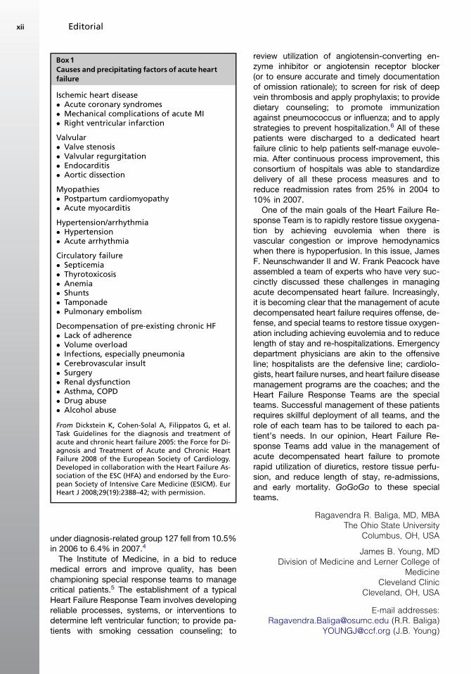
Box1Causes and precipitating factors of acute heartfailure
Ischemic heart disease� Acute coronary syndromes� Mechanical complications of acute MI� Right ventricular infarction
Valvular� Valve stenosis� Valvular regurgitation� Endocarditis� Aortic dissection
Myopathies� Postpartum cardiomyopathy� Acute myocarditis
Hypertension/arrhythmia� Hypertension� Acute arrhythmia
Circulatory failure� Septicemia� Thyrotoxicosis� Anemia� Shunts� Tamponade� Pulmonary embolism
Decompensation of pre-existing chronic HF� Lack of adherence� Volume overload� Infections, especially pneumonia� Cerebrovascular insult� Surgery� Renal dysfunction� Asthma, COPD� Drug abuse� Alcohol abuse
From Dickstein K, Cohen-Solal A, Filippatos G, et al.Task Guidelines for the diagnosis and treatment ofacute and chronic heart failure 2005: the Force for Di-agnosis and Treatment of Acute and Chronic HeartFailure 2008 of the European Society of Cardiology.Developed in collaboration with the Heart Failure As-sociation of the ESC (HFA) and endorsed by the Euro-pean Society of Intensive Care Medicine (ESICM). EurHeart J 2008;29(19):2388–42; with permission.
Editorialxii
REFERENCES
1. Rosamond W, Flegal K, Furie K, et al. Heart disease
and stroke statistics—2008 update: a report from
the American Heart Association Statistics Committee
and Stroke Statistics Subcommittee. Circulation
2008;117(4):e25–e146.
2. Friedewald VE, Gheorghiade M, Yancy CW, et al. The
editor’s roundtable: acute decompensated heart fail-
ure. Am J Cardiol 2007;99(11):1560–7.
3. Dickstein K, Cohen-Solal A, Filippatos G, et al. Task
Guidelines for the diagnosis and treatment of acute
and chronic heart failure 2005: the Force for Diagno-
sis and Treatment of Acute and Chronic Heart Failure
2008 of the European Society of Cardiology. Devel-
oped in collaboration with the Heart Failure Associa-
tion of the ESC (HFA) and endorsed by the
European Society of Intensive Care Medicine
(ESICM). Eur Heart J 2008;29(19):2388–442.
4. Agency for Healthcare Research and Quality. 24-hour
Heart Failure Response Team decreases length of
stay and readmission rates. Available at: http://www.
innovations.ahrq.gov/content.aspx?id51820. Accessed
November 4, 2008.
Fig1. (A) Evaluation of suspected acute decompensated heart failure. (B) Initial treatment algorithm for manage-
ment of acute decompensated heart failure. (From Dickstein K, Cohen-Solal A, Filippatos G, et al. Guidelines for
the diagnosis and treatment of acute and chronic heart failure 2005: the Force for Diagnosis and Treatment of
Acute and Chronic Heart Failure 2008 of the European Society of Cardiology. Developed in collaboration with
the Heart Failure Association of the ESC (HFA) and endorsed by the European Society of Intensive Care Medicine
(ESICM). Eur Heart J 2008;29(19):2388–42; with permission.)
Editorial xiii

5. Institute for Healthcare Improvement. Deliver
reliable, evidence-based care for congestive
heart failure. Available at http://www.ihi.org/IHI/
Programs/Campaign/CHF.htm. Accessed Novem-
ber 4, 2008.
6. Institute for Healthcare Improvement. Mentor
hospital registry: congestive heart failure. Available
at: http://www.ihi.org/IHI/Programs/Campaign/
mentor_registry_chf.htm. Accessed November 4,
2008.
xiv
Preface
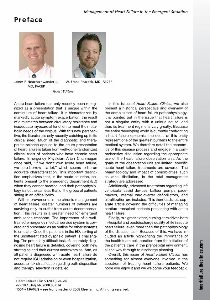
James F. Neuenschwander II,
MD, FACEP
W. Frank Peacock, MD, FACEP
Guest Editors
Acute heart failure has only recently been recog-nized as a presentation that is unique within thecontinuum of heart failure. It is characterized bymarkedly acute symptom exacerbation, the resultof a mismatch between circulatory resistance andinadequate myocardial function to meet the meta-bolic needs of the corpus. With this new perspec-tive, the literature is only recently catching up to itsclinical need. Much of the diagnostic and thera-peutic science applied to the acute presentationof heart failure is taken from well-done randomizedclinical trials of patients who have chronic heartfailure. Emergency Physician Arjun Chanmuganonce said, ‘‘If we don’t own acute heart failure,we sure borrow it a lot,’’ which seems to be anaccurate characterization. This important distinc-tion emphasizes that, in the acute situation, pa-tients present to the emergency department (ED)when they cannot breathe, and their pathophysio-logy is not the same as that of the group of patientssitting in an office lobby.
With improvements in the chronic managementof heart failure, greater numbers of patients aresurviving only to suffer from acute decompensa-tion. This results in a greater need for emergentambulance transport. The importance of a well-trained emergency medical service system is cov-ered and presented as an outline for other systemsto emulate. Once the patient is in the ED, sorting ofthe undifferentiated dyspneic patient is challeng-ing. The potentially difficult task of accurately diag-nosing heart failure is detailed, covering both newstrategies and their current controversies. And, asall patients diagnosed with acute heart failure donot require ICU admission or even hospitalization,accurate risk stratification guiding both dispositionand therapy selection is detailed.
In this issue of Heart Failure Clinics, we alsopresent a historical perspective and overview ofthe complexities of heart failure pathophysiology.It is pointed out in the issue that heart failure isnot a singular entity with a unique cause, andthus its treatment regimens vary greatly. Becausethe entire developing world is currently confrontinga heart failure epidemic, the costs of this entityrepresent one of the greatest burdens to the entiremedical system. We therefore detail the econom-ics of this disease process and engage in a com-prehensive discussion regarding the appropriateuse of the heart failure observation unit. As thegoals of the observation unit are limited, specificacute heart failure treatments are covered. Thepharmacology and impact of comorbidities, suchas atrial fibrillation, in the total managementstrategy are addressed.
Additionally, advanced treatments regarding leftventricular assist devices, balloon pumps, pace-makers, internal cardioverter defibrillators, andultrafiltration are included. This then leads to a sep-arate article covering the difficulties of managingcardiac transplant patients presenting with acuteheart failure.
Finally, to a great extent, nursing care drives bothin-hospital and postdischarge quality of life in acuteheart failure, even more than the pathophysiologyof the disease itself. Because of this, we have in-cluded an article highlighting the importance ofthe health team collaboration from the initiation ofthe patient’s care in the prehospital environment,all the way through to discharge planning.
Overall, this issue of Heart Failure Clinics hassomething for almost everyone involved in thespectrum of treating heart failure patients. Wehope you enjoy it and we welcome your feedback.
Heart Failure Clin 5 (2009) xv–xvidoi:10.1016/j.hfc.2008.08.0141551-7136/08/$ – see front matter ª 2008 Elsevier Inc. All rights reserved. he
artf
ailu
re.th
ecli
nics
.com
Management of Heart Failure in the Emergent Situation

I want to dedicate this book to my beautiful wifeColleen and our three wonderful children Elias,Arel, and Gabriel (J.F.N.).
And thanks to my family, without whom I wouldget nowhere (W.F.P.).
James F. Neuenschwander II, MD, FACEPThe Ohio State University Medical Center
Emergency Department376 West 10th Avenue
Columbus, Ohio 43210-1252, USA
W. Frank Peacock, MD, FACEPThe Cleveland Clinic
Department of Emergency Medicine9500 Euclid Avenue, Desk E-19
Cleveland, OH 44195, USA
E-mail addresses:[email protected]
(J.F. Neuenschwander)[email protected] (W.F. Peacock)
Prefacexvi

Heart Failure and theEmergency Department:Epidemiology,Characteristics,and OutcomesGary B. Green, MD, MPH, MBA*
TERMINOLOGYAND DEFINITIONS
A common language always greatly facilitatescommunication, whereas ambiguous or ill-definedterms are often a significant barrier to meaningfulidea exchange. This observation is especiallyapparent at the forefront of any rapidly evolvingfield of inquiry and is certainly the case for heartfailure (HF). Accordingly, considerable effort hasbeen made in recent years among HF researchers,professional societies, and policy makers toestablish consensus concerning the most appro-priate diagnostic terms and their definitions.Although progress has certainly been made, asthe science of HF has progressed it has also be-come increasingly clear that this is not a monolithicdisease with a single common pathway but rathera diverse and complex spectrum of pathologieshistorically bound by a limited number of sharedclinical characteristics. The nomenclature usedcontinues to rapidly expand and evolve as clinicalsyndromes are increasingly differentiated basedon measurable physiologic parameters andoutcomes rather than the more subjective charac-teristics relied on in the past.
HF itself has been most recently redefined bythe American College of Cardiology/AmericanHeart Association (ACC/AHA) task force on prac-tice guidelines as ‘‘a complex clinical syndromethat can result from any structural or functional
cardiac disorder that impairs the ability of theventricle to fill with or eject blood.’’ Becausevolume overload is not uniformly present in allpatients or at all presentations, use of the olderterm ‘‘congestive heart failure’’ is to be discour-aged.1 Various authors have used overlappingdiagnostic terms to stratify HF presentations byonset or temporal pattern, sometimes contribut-ing more confusion than clarity. For example,The European Society of Cardiology defines theterm acute heart failure as ‘‘the rapid onset ofsymptoms and signs secondary to abnormal car-diac function,’’ reserving the more specific termacute decompensated heart failure for ‘‘thosepatients with known HF who experience acuteor subacute worsening of their HF state.’’2
Many United States authors have used thesame terms somewhat differently, describingacute heart failure as ‘‘new onset of decompen-sated HF or decompensation of chronic, estab-lished HF with symptoms sufficient to warranthospitalization.’’3 To resolve this diagnostic am-biguity, a 2005 international working grouprecommended adoption of the inclusive termacute heart failure syndromes, defined as ‘‘grad-ual or rapid deterioration in HF signs and symp-toms resulting in a need for urgent therapy.’’4
Most recently, the 2007 American College ofEmergency Physicians Clinical Policies Commit-tee has also endorsed the term ‘‘acute heart
New York University Langone Medical Center, New York, NY, USA* Department of Emergency Medicine, New York University Langone Medical Center, Bellevue HospitalAdministration Building A345, New York, NY 10016.E-mail address: [email protected]
KEYWORDS� Acute heart failure syndromes � Emergency department� Epidemiology
Heart Failure Clin 5 (2009) 1–7doi:10.1016/j.hfc.2008.08.0011551-7136/08/$ – see front matter ª 2008 Elsevier Inc. All rights reserved. he
artf
ailu
re.th
ecli
nics
.com
failure syndromes’’ as defined by the interna-tional working group, and this term is usedhere.5
Although the clinical syndrome of HF is mostoften caused by myocardial disease, it may alsobe due to pericardial, endocardial, or great vesselpathology. Further, although most HF patients dohave some degree of left ventricular impairment,the causes and characteristics of ventricular func-tional abnormality are diverse. It is important thatthe term HF be differentiated from the morespecific physiologic descriptor, left ventriculardysfunction, and also not be confused with cardio-myopathy, defined by the AHA as any ‘‘disease ofthe myocardium associated with mechanical and/or electrical dysfunction.’’1,6
In the past, it had been believed that HF wasuniformly related to a decreased left ventricularejection fraction (LVEF). Through further studyand more routine use of echocardiography, it hasnow become apparent that a large proportion ofpatients who have HF actually have a normal ornear-normal LVEF. Until recently, these patientswere given the diagnosis of diastolic dysfunction,described as ‘‘prolonged, slowed, or incompleteability [of the myocardium to] generate force,shorten and return to an unstressed length.’’7 Rec-ognition that diastole is an active and complexphysiologic process rather than simply the passiveabsence of contraction was and remains a criticalconcept in the understanding of HF. However,more rigorous study has demonstrated that dia-stolic functional abnormalities also frequently oc-cur among patients who have reduced LVEF.Diastolic and systolic dysfunctions are thus notmutually exclusive and therefore diastolic dys-function should no longer be used as a differentiat-ing term. Accordingly, it is now recommended thatall patients who have HF undergo echocardiogra-phy soon after diagnosis and be classified ashaving either left ventricular systolic dysfunction(LVSD) or preserved systolic function (PSF). Thismore physiologically accurate nomenclature willlikely have increasingly significant clinical implica-tions, including implications for emergencydepartment (ED) treatment, as ongoing trialsfocused on each of these groups are completed.8
Predictably, some diagnostic controversy doesremain, particularly concerning the mostappropriate threshold EF below which a HF patientshould be classified as having LVSD. Althoughsome investigations have used a cutoff of EF lessthan 50%, the largest United States HF dataregistries, OPTIMIZE-HF and Acute Decompen-sated Heart Failure National Registry (ADHERE),currently define LVSD based on an EF less than40%.9–11
EPIDEMIOLOGYAND IMPACT
HF has emerged as a significant public healthproblem whose impact on quality of life, the healthcare system, and the economy is already stagger-ing and continues to grow each year as the popu-lation ages. The current prevalence of HF withinthe United States is 5.3 million, or 2.5% of the en-tire adult population, and it is estimated that660,000 new cases will be diagnosed this year.12
Among those older than 65, nearly 1 in 10 arenewly diagnosed annually, and in this age groupHF is the leading cause of hospitalization.13,14 In1979, HF accounted for just 400,000 United Stateshospital discharges. This number has increased to1.1 million hospitalizations a year for a primarydiagnosis of HF and a total of 3.6 million annualhospitalizations with HF as either a primary or sec-ondary cause, corresponding to an annual directcost of $23 billion.1,12 Beyond this, there aregreater than 3.4 million ambulatory care visits an-nually for HF, including approximately 1.1 millionED visits.12,14 The total cost (direct and indirect)for HF in the United States continues to steadilyincrease and is currently estimated to be $35billion to $60 billion.1,12
Beyond hospital days and dollars, the humancost of HF is devastating and frequently underap-preciated. The natural course of the disease ischaracterized by inevitable deterioration, with pro-gressive decline in functional capacity exacer-bated by frequent episodes of acute, sometimeslife-threatening, decompensation requiring re-peated ED visits and prolonged hospitalizations.Although somewhat variable based on cause andcomorbidity, once diagnosed with HF the progno-sis remains grim, with overall mortality rates similarto and sometimes surpassing those of many otherdisease states routinely labeled as terminal, suchas HIV/AIDS and many types of cancer.12 In theFramingham Heart Study, 80% of men and 70%of women diagnosed with HF died within 8 years,whereas the 1-year mortality approached 20%.15
Nationally, HF is recorded as the primary causein 2.2% of all deaths, and one in eight death certif-icates (284,365 deaths in 2004) list heart failure aseither a primary or contributory cause.12
Within this context of high overall morbidity andmortality among patients who have chronic HF, anED visit caused by an acute heart failure syndrome(AHFS) indicates a period of greatly increasedshort-term mortality risk. Fully 80% of patientswho have AHFS require admission and variousstudies have reported in-hospital mortality ratesof 2% to 20%.16,17 Among those requiring inten-sive care unit admission from the ED, in-hospitalmortality is greater than 10%,17 whereas an ED
Green2

presentation of acute pulmonary edema signalsa particularly poor prognosis. Twelve percent ofthese patients do not leave the hospital alive andthe 1-year mortality among this subgroup isgreater than 40%.18 Of those surviving to hospitaldischarge after any AHFS presentation, 11% diewithin 30 days, 44% require rehospitalizationwithin 6 months, and 33% do not survive 1 yearafter discharge.16
Although the overall burden of disease on soci-ety because of HF clearly continues to increaseover time, a review of outcomes investigationssuggest recent treatment advances may be havinga positive effect. Despite a significant increase indisease prevalence, total United States HF-relateddeaths in 2004 (284,365) were nearly identical tothose of 1994 (284,087).12 Further, although a pre-vious study of nearly 4 million Medicare patientsdid not show improvement in 30-day mortalitybetween 1992 and 1999,19 overall survival didimprove over a 2-decade period among a commu-nity-based longitudinal cohort.20 The mostencouraging data to date concern in-hospital out-comes and are from the ADHERE, which analyzedtrends from January, 2002 to December, 2004among 159,168 HF patients admitted from 285hospitals. During the 3-year study period the mul-tivariate risk-adjusted mortality rate declined from4.5% to 3.2%, the need for mechanical ventilationwas reduced from 5.3% to 3.4%, and hospitallength of stay was reduced from a mean of 6.3 to5.5 days. Although causation for theseimprovements cannot be demonstrated, inotropeuse decreased during the study period and signif-icant advances were also made in compliance withrecently adopted HF quality metrics, including rou-tine assessment of left ventricular function andbeta-blocker use.21
DEMOGRAPHICS
Although HF can occur at any age because ofseveral structural and functional cardiac abnor-malities, statistically speaking it is primarily a dis-ease of the aged. The estimated prevalence ofHF among those aged 20 to 39 years old is lessthan 0.3%, approaches 2% among those 40 to59, reaches 6% from 60 to 79, and exceeds 12%in those more than 80 years old.12 The mean agereported for ED patients who had AHFS in theADHERE database was 72.4 years,17 similar tothe 74.3 years mean age reported among ED HFvisits in the National Hospital Ambulatory CareSurvey (NHAMCS).14
Women and men have an equal lifetime risk fordeveloping HF (one in five), and the proportion ofED visits and hospitalizations for AHFS among
women roughly mirrors the gender distribution ofthe population at a similar age.12,22 Gender differ-ences do exist, however, in HF pathophysiologyand clinical characteristics. Women who have HFare generally older at disease onset comparedwith men and they have lower rates of coronaryartery disease and renal insufficiency. In contrast,women who have HF are more likely to havehypertension and, consistent with this risk profile,a higher proportion of women who have HF havePSFcomparedwithmen.Womenalsohaveahighermean LVEF.22 Studies evaluating gender dispar-ities in outcomes have reported conflictingresults.22,23 Several clinical trials of patients whohave chronic HF have suggested that women whohave HF have a lower mortality risk.23–25 Womenwere uniformly underrepresented in these studies,however, and selection bias may have influencedthe results. Gender analysis of the ADHERE data-base, adjusted for other predictive variables, foundsimilar in-hospital mortality and equivalent rates ofdialysis and mechanical ventilation among womenand men.22,26 Further, a prospective investigationof dyspneic ED patients identified as having AHFSby a B-type natriuretic peptide (BNP) measurementgreater than 500 found that women actually hada significantly higher 24-month mortality comparedwith men.26
Nearly 1 million African Americans have HF,corresponding to a 50% higher incidence of dis-ease compared with the general population.12
Overall, black patients develop HF symptoms atan earlier age and their disease progresses morerapidly compared with whites, yet they are gener-ally diagnosed at a more advanced stage of dis-ease.27–29 There have been many investigationsreporting on comparative mortality rates for blackswho have HF but they have provided conflictingresults and adequate comparative data is notavailable concerning HF among Hispanic, Asian,or other ethnic groups. There remains no clearconsensus concerning the influence of race/ethnicity on overall HF outcomes.1,30
Two recent studies have focused on the complexrole of race in the ED evaluation and treatment ofAHFS. Hugli and colleagues14 analyzed data fromall ED visits for AHFS in the NHAMCS databasefrom 1992 to 2001. They found a 53% higher EDvisit rate for AHFS among blacks compared withwhites but a 13% lower hospitalization rate despitea similar proportion of black patients receiving anurgent triage classification. After adjusting for otherpredictive variables, whites were still 1.7 timesmore likely to be admitted compared with blacks.Further, only 68% of blacks who had AHFS re-ceived a chest radiograph in the ED comparedwith 80% of whites.14 Although various
Heart Failure Characteristics and Outcomes 3
explanations for this apparent bias can be postu-lated, the data suggest a disturbing disparity inthe ED physicians’ perception of AHFS disease se-verity between black and white patients that is notsupported by existing physiologic or clinical data.Further insight into this issue is provided by a recentsubgroup analysis of data from a trial of serum BNPmeasurement in the evaluation of patients who hadHF, the Rapid Emergency Department Heart Fail-ure Outpatient Trial (REDHOT). BNP measurementis a robust predictor of HF severity and it is equallypredictive of outcome among black and white pa-tients. In the REDHOT study, BNP levels of blackpatients were not significantly different from thoseof white patients. However, whites were signifi-cantly more likely to be rated by ED physicians ashaving more severe HF by New York Heart Associ-ation classification (class III or IV), whereas blackswere more likely to be perceived as having milderheart failure (class I or II). Further, although as ex-pected admitted white patients had higher BNPlevels than those discharged, black patients whowere discharged home actually had higher BNPlevels compared with admitted blacks.30 This ‘‘per-ceptual bias,’’ in which disease severity is system-atically underestimated among blacks comparedwith whites, has also been reported in the evalua-tion of ED patients presenting with chest pain.31
COMORBIDITIES
Coronary artery disease (CAD) is the single great-est contributor to HF morbidity and mortality at allstages of disease. Among those who have LVSD,CAD is identified as the primary etiologic triggerin approximately two thirds of patients. Althoughother pathology, such as hypertension, valvedisease, and atrial fibrillation, is more often citedas the underlying cause in those who have PSF,30% of these patients also have a preceding diag-nosis of myocardial infarction (MI) or angina and atautopsy a majority of patients who have HF haveCAD.1,9–11,17,32 In HF registries and other studiesenrolling patients hospitalized for an AHFS,a past history of MI is reported in 31% to 48%and a previous diagnosis of CAD is documentedin 57% to 65%.10,11 Beyond this role in HF initia-tion, CAD also accelerates disease progressionsubsequent to HF onset through new infarction,acute or chronic ischemic dysfunction (ie, myocar-dial stunning or hibernation), and continued endo-thelial activation of adverse neurohormonalresponse cascades. Not surprisingly, the effectof CAD on outcome among patients who haveHF is devastating. Longitudinal studies have con-sistently demonstrated that the presence of CAD
increases overall mortality by 50% or more. CADis also a powerful independent predictor of mortal-ity among patients presenting to the ED withAHFS. In one European ED study, after adjustingfor all other known prognostic risk factors, thepresence of a CAD history increased the risk fordeath during the study period by 224%.33
Like CAD, hypertension (HTN) is both a causa-tive agent and comorbidity in HF. In cohort stud-ies, the presence of either systolic or diastolicHTN significantly increases the risk for a subse-quent HF diagnosis, whereas long-term treatmentof HTN dramatically reduces the likelihood of HFdevelopment.34–36 The major pathophysiologiclink between HTN and HF is HTN-induced leftventricular hypertrophy (LVH), which is itself anindependent risk factor for MI and HF.37 Overall,the contribution of HTN to the burden of HF dis-ease is dramatic, with untreated or inadequatelytreated HTN estimated to account for approxi-mately 40% of cases in men and approximately60% in women.1 Consistent with this, ED-basedinvestigations have found that 53% to 73% of allpatients presenting with AHFS have a history ofHTN,16,21 with this risk being somewhat morepredominant among those who have PSF thanamong those who have LVSD (76% versus 66%,P < .0001).11
Although eliciting a previous history of HTN mayprovide insight into the likely cause of HF in a givenpatient, presenting blood pressure in the ED isa more meaningful guide to risk stratification andtreatment in those presenting with AHFS. Lowsystolic blood pressure in the ED is a strong nega-tive prognostic indicator. The high morbidity andmortality in this group is a result of a heightenedstate of neurohormonal activation and the conse-quent cascade of increased fluid retention,hyponatremia, renal insufficiency, and treatmentresistance.38 Conversely, patients who haveAHFS and an elevated blood pressure on ED pre-sentation are generally characterized by shorterduration of symptoms before arrival and aremore likely to be female and older, consistentwith a greater likelihood of having PSF ratherthan LVSD. The implication is that fluid maldistri-bution rather than fluid overload may be the pre-dominant problem in this group and thereforea treatment focus on vasodilators rather than di-uretics may be more effective. Such a directed ap-proach to ED treatment based on presenting bloodpressure and other physiologic parameters hasbeen suggested but not yet operationally definedand has not been prospectively studied.8
Cardiovascular and renal disease often occurconcomitantly and each disease state has a nega-tive effect on the other. Renal impairment is a risk
Green4

factor for all-cause mortality in a wide variety ofcardiovascular diseases and, conversely, cardio-vascular disease is the most common cause ofdeath among patients who have chronic kidneydisease (CKD) and end-stage renal disease(ESRD), accounting for 50% of their mortality.39,40
A similar relationship is seen between renaldisease and HF. Approximately 50% of all patientswho have HF have an impaired glomerular filtrationrate, and concomitant HF is reported in up to 40%of those who have ESRD.40 In the ADHERE regis-try of ED patients who had AHFS, a history of CKDwas recorded in 30%, 21% had a creatininegreater than 2.0 mg/dL in the ED, and 5% were un-dergoing dialysis.17 Impaired renal function is alsostrongly associated with poorer outcome amongpatients who have HF, even after adjusting for allknown covariates. This negative effect on progno-sis is independent of LVEF and is of equivalentmagnitude among patients who have LVSD andthose who have PSF.39
The coexistence of HF and renal impairment isusually the result of one or more underlying vascularinsults, such as HTN, diabetes, or atherosclerosis.A mutually reinforcing effect on disease progres-sion is observable across the entire spectrum ofdisease severity but is most apparent and mostharmful in those approaching end-stage diseasestates as manifested by the cardiorenal syndrome.Although a consensus definition does not yet exist,the syndrome has been described as a state ofadvanced cardiorenal dysregulation occurring inpatients who have HF and concomitant renal dis-ease characterized by worsening renal functionand diuretic resistance during the treatment ofAHFS.41 Although elucidation of the complex phys-iology of the cardiorenal syndrome has become anincreasingly active area of investigation, the impli-cations of syndrome recognition on ED treatmentdecisions remains unclear at this time.40,41
Other conditions have also been found to havea significant impact on development, progression,or prognosis of HF. A history of atrial fibrillationwas present in 31% of patients who had HFenrolled in the ADHERE registry and longitudinalstudies report an increasing prevalence of atrialfibrillation with increasing severity of LVSD.42 Ane-mia is reported in up to 45% of patients who haveHF and occurs as frequently among those whohave reduced and preserved LVSF. Anemia andatrial fibrillation are also each associated withsignificantly increased mortality among patientswho have HF.42,43 Various other investigationshave reported a worse prognosis among patientswho have HF with diabetes, liver disease, chronicobstructive pulmonary disease, cerebrovasculardisease, cancer, and dementia.14,16,17
Obesity is highly associated with increased riskfor a wide variety of cardiovascular events, includ-ing the onset of HF. Surprisingly, among patientsalready diagnosed with HF, a higher body massindex (BMI) actually seems to have a protectiveeffect. Among nearly 110,000 patients who hadAHFS included in the ADHERE registry, in-hospitalmortality was inversely related to BMI across allBMI quartiles. This effect persists after adjustmentfor all other known prognostic factors and occursamong those who have both LVSD and PSF. Thephysiologic basis of this observation, labeled theobesity paradox, remains unclear at this time butis the subject of much speculation among HFinvestigators.44
SUMMARYAND CLINICAL IMPLICATIONS
Whether measured in deaths or dollars, it is nowwidely recognized that the impact of heart failureon society is enormous. Through governmentfunding and market forces the research commu-nity has responded, resulting in an ongoing periodof rapid advancement across a wide range offields from myocardial cell biology and neurohor-monal physiology to HF epidemiology and behav-ioral interactions. The pace of progress is perhapsmost apparent in the barrage of new and revisedterminology appearing in the HF literature as re-searchers and professional societies struggle toquickly translate emerging knowledge into clinicalpractice. Although sometimes confusing, the com-plexity of nomenclature directly reflects a growingappreciation that the symptom complex previ-ously labeled ‘‘heart failure’’ does not in factrepresent a single disease state but is actuallya spectrum of complex multisystem pathologies.Accordingly, clinicians must adopt a more sophis-ticated and more effective approach to evaluationand treatment that is increasingly based on objec-tive measurement of outcome-linked physiologicparameters rather than the subjectively describedsymptom constellations relied on previously.
Because HF prevalence continues to increasewith the aging population and its natural courseis marked by frequent and often life-threateningacute decompensation, ED visits for AHFS willlikely continue to increase in the future, furtherchallenging our resources and skills. Each ED pre-sentation of AHFS also represents an opportunityto intervene at a critical stage of illness and the po-tential to make a dramatic positive impact on dura-tion and quality of life. Emergency physicians musttherefore remain on the cutting edge of this rapidlyevolving field as real-time stratification of patientswho have AHFS into physiologic- and risk-basedsubgroups becomes a routine part of ED
Heart Failure Characteristics and Outcomes 5
evaluation and increasingly directs the applicationof emerging therapeutic approaches.
REFERENCES
1. Hunt SA, American College of Cardiology, American
Heart Association Task Force on Practice Guide-
lines. ACC/AHA 2005 guideline update for the diag-
nosis and management of chronic heart failure in the
adult: a report of the American College of Cardiol-
ogy/American Heart Association Task Force on Prac-
tice Guidelines. J Am Coll Cardiol 2005;46:e1–82.
2. Nieminen MS. The task force on acute heart failure of
the European Society of Cardiology. Executive sum-
mary of the guidelines on the diagnosis and treat-
ment of acute heart failure. Eur Heart J 2005;26:
384–416.
3. Fonarow GC, Adams KF Jr, Abraham WT, et al. Risk
stratification for in-hospital mortality in acutely
decompensated heart failure: classification and
regression tree analysis. JAMA 2005;293:572–80.
4. Gheorghiade M, Zannad F, Sopko G, et al. Interna-
tional working group on acute heart failure syn-
dromes. Acute heart failure syndromes: current
state and framework for future research. Circulation
2005;112:3958–68.
5. Silvers SM, ACEP. Clinical Policies Subcommittee on
Acute Heart Failure Syndromes. Clinical policy: crit-
ical issues in the evaluation and management of
adult patients presenting to the emergency depart-
ment with acute heart failure syndromes. Ann Emerg
Med 2007;49:627–69.
6. Maron BJ, Towbin JA, Thiene G, et al. Contemporary
definitions and classification of the cardiomyopa-
thies: an AHA scientific statement from the Council
on Clinical Cardiology, Heart Failure and Transplan-
tation Committee; Quality of Care and Outcomes
Research and Functional Genomics and Transla-
tional Biology Interdisciplinary Working Groups;
and Council on Epidemiology and Prevention. Circu-
lation 2006;113:1807–16.
7. Zile MR, Brutsaert DL. New concepts in diastolic
dysfunction and diastolic heart failure: Part I: diag-
nosis, prognosis and measurements of diastolic
function. Circulation 2002;105:1387–93.
8. Collins S, Storrow AB, Kirk JD, et al. Beyond pulmo-
nary edema: diagnostic, risk stratification, and treat-
ment challenges of acute heart failure management
in the emergency department. Ann Emerg Med
2008;51:45–57.
9. Redfield MM, Jocobson SJ, Burnett JC Jr, et al.
Burden of systolic and diastolic ventricular dysfunc-
tion in the community: appreciating the scope of the
heart failure epidemic. JAMA 2003;289:194–202.
10. Yancy CW, Lopatin M, Stevenson LW, et al. Clinical
presentation, management and in-hospital outcomes
of patients admitted with acute decompensated heart
failure with preserved systolic function; a report from
the ADHERE database. J Am Coll Cardiol 2006;47:
76–84.
11. Fonarow GC, Stough WG, Abraham WT, et al. Char-
acteristics, treatments and outcomes of patients with
preserved systolic function hospitalized for heart
failure: report from the OPTIMIZE-HF registry. J Am
Coll Cardiol 2007;50:768–77.
12. Rosamond W, Flegal K, Furie K, et al. Heart disease
and stroke statistics 2008 update: a report from the
American Heart Association Statistics Committee
and Stroke Statistics Committee. Circulation 2008;
117:e25–146.
13. O’Connell JB. The economic burden of heart failure.
Clin Cardiol 2000;23(Suppl. 3):6–10, III.
14. Hugli O, Braun JE, Kim S, et al. US emergency de-
partment visits for decompensated HF, 1992–2001.
Am J Cardiol 2005;96:1537–42.
15. Lloyd-Jones DM, Larson MG, Leip EP, et al. Fra-
mingham Heart Study. Lifetime risk for developing
congestive heart failure: The Framingham Heart
Study. Circulation 2002;106:3068–72.
16. Fonarow GC. Epidemiology and risk stratification in
acute heart failure. Am Heart J 2008;155:200–7.
17. Adams KF, Fonarow GC, Emerman CL, et al.
Characteristics and outcomes of patients hospital-
ized for heart failure in the US: rationale, design
and preliminary observations from the first 100,000
cases in the Acute Decompensated Heart Failure
National Registry [ADHERE]. Am Heart J 2005;149:
209–16.
18. Roguiin A, Behar D, Ben Ami H, et al. Long-term
prognosis of acute pulmonary edema—an ominous
outcome. Eur J Heart Fail 2000;2:137–44.
19. Kaiborod M, Lichtman JH, Heidenreich PA, et al.
National trends in outcomes among elderly patients
with heart failure. Am J Med 2006;119:e1–7.
20. Forger VL, Weston SA, Redfield MM, et al. Trends in
HF incidence and survival in a community-based
population. JAMA 2004;292:344–50.
21. Fonarow GC, Heywood T, Heidenreich PA, et al.
Temporal trends in clinical characteristics, treat-
ments, and outcomes for heart failure hospitaliza-
tions, 2002 to 2004: findings from the Acute
Decompensated Heart Failure National Registry
(ADHERE). Am Hear J 2007;153:1021–8.
22. Diercks DB, Fonarow GC, Kirk D, et al. Risk stratifi-
cation in women enrolled in the acute decompen-
sated heart failure national registry emergency
module (ADHERE – EM). Acad Emerg Med 2008;
15:151–8.
23. Galvao M, Kalman J, DeMarco T, et al. Gender differ-
ences in in-hospital management and outcomes in
patients with decompensated heart failure: analysis
from the Acute Decompensated Heart Failure
National Registry (ADHERE). J Card Fail 2006;12:
100–7.
Green6
24. Gustafsson F, Torp-Pederson C, Burchardt H, et al.
Female sex is associated with a better long-term sur-
vival in patients hospitalized with congestive heart
failure. Eur Heart J 2004;25:129–35.
25. Ghali JK,Krause-SteinfaufHJ,AdamsKF, etal.Gender
differences in advanced heart failure: Insights from the
BESTstudy. J Am Coll Cardiol 2003;42:2128–34.
26. Christ M, Laule-Kilian K, Hochholzer W, et al. Gender
specific risk stratification with BNP levels in patients
with acute dyspnea. J AM Coll Cardiol 2006;48:
1808–12.
27. Alexander M, Grumbach K, Remy L, et al. Conges-
tive heart failure hospitalizations and survival in Cal-
ifornia: patterns according to race/ethnicity. Am
Heart J 1999;137:919–27.
28. Yancy CW. Heart failure in African Americans: a car-
diovascular enigma. J Card Fail 2000;6:183–6.
29. Alexander M, Grumbach K, Selby J, et al. Hospital-
ization for congestive heart failure. Explaining racial
differences. JAMA 1995;274:1037–42.
30. Daniels LB, Bhalla V, Clopton P, et al. B-type natriuretic
peptide levels and ethnic disparities in perceived
severity of heart failure: results from the Rapid Emer-
gency Department Heart Failure Outpatient Trial (RED-
HOT) multicenter study of BNP levels and emergency
department decision making in patients presenting
with shortness of breath. J Card Fail 2006;12:281–5.
31. Keyle PM, Pezzin LE, Green GB. Disparities in the
emergency department evaluation of chest pain pa-
tients. Acad Emerg Med 2007;14(2):149–56.
32. Gheorghiade M, Bonow RO. Chronic heart failure in
the United States: a manifestation of coronary artery
disease. Circulation 1998;97:282–9.
33. Purek L, Laule-Kilian K, Christ A, et al. Coronary ar-
tery disease and outcome in acute congestive heart
failure. Heart 2006;92:598–602.
34. Levy D, Larson MG, Vasan RS, et al. The progres-
sion from hypertension to congestive heart failure.
JAMA 1996;275:1557–62.
35. Wilhelmsin L, Rosengren A, Eriksson H, et al. Heart
failure in the general population of men: morbidity,
risk factors and prognosis. J Intern Med 2001;249:
253–61.
36. Kostis JB, Davis BR, Cutler J, et al. Prevention of
heart failure by antihypertensive treatment in older
persons with isolated systolic hypertension. JAMA
1997;278:212–6.
37. Vakili BA, Okin PM, Devereux RB. Prognostic
implications of left ventricular hypertrophy. Am Heart
J 2001;141:334–41.
38. Gheorghiade M, Abramson WT, Albert NM, et al.
Systolic blood pressure at admission, clinical
characteristics and outcome in patients hospitalized
with acute heart failure. JAMA 2006;296:2217–26.
39. Hillege HL, Nitsch D, Pfeffer MA, et al. Renal func-
tion as a predictor of outcome in a broad spectrum
of patients with heart failure. Circulation 2006;113:
671–8.
40. Obialo CI. Cardiorenal consideration as a risk factor
for heart failure. Am J Cardiol 2007;99(Suppl):
21D–4D.
41. Liang KV, Williams AW, Greene EL, et al. Acute
decompensated heart failure and the cardiorenal
syndrome. Crit Care Med 2008;36(Suppl):
S75–88.
42. Parkish R, Maisel WH, Toca FM, et al. Atrial fibrilla-
tion in heart failure: high mortality risk even if ventric-
ular function is preserved. Am Heart J 2005;150:
701–6.
43. Berry C, Norrie J, Hogg K, et al. The prevalence, na-
ture and importance of hematologic abnormalities in
heart failure. Am Heart J 2006;151:1313–21.
44. Fonarow GC, Srikanthan P, Cosantzo MR, et al. An
obesity paradox in acute heart failure: analysis of
body mass index and inhospital mortality for
108,927 patients in the Acute Decompensated Heart
Failure National Registry. Am Heart J 2007;153:
74–81.
Heart Failure Characteristics and Outcomes 7
Pathophysiology ofAcute DecompensatedHeart FailureRichard L. Summers, MDa,*, Ezra Amsterdam, MDb
A disease condition synonymous with heart failurewas recognized as early as the ancient Egyptianand Byzantine Empires.1 Early descriptions of thecondition focused on the outward signs of edema,and the resulting diagnosis was often referred toas the dropsy from the Greek word hydrops mean-ing water.2 An understanding of the etiology andpathophysiologic mechanisms involved wereunknown at that time, however.
It was not until the early 17th century afterWilliam Harvey’s work in defining the functioningof the circulation, that the heart was implicated inthis disease process. In light of these emergingconcepts, another English physician, William With-ering, first documented a successful treatment forthis condition, in which the circulation was tar-geted with the use of foxglove (digitalis purpura).3
By the 20th century, fewer people were dying ofinfectious diseases, and heart failure becamea more commonplace source of morbidity andmortality. Most early descriptions were verycardiocentric, and the dropsy diagnosis wasreplaced with the more descriptive term conges-tive heart failure, emphasizing what was consid-ered at that time a central role of the heart in thepathophysiology.
Since that time, the definition of heart failure hasevolved as knowledge and understanding of thepathophysiology of the condition have changed.By the latter half of the 20th century, Arthur C.Guyton provided a description of the quantitativephysiologic relationships between cardiac output,extracellular fluid volume, and blood pressure
control, with a central role for the kidneys inlong-term regulation.4–6 This work was centeredon the premise that the primary goal of the circula-tion was to provide fluids and nutrients to thetissues of the body. A description of heart failurethat emerged from these concepts viewed thisdisease process as a more generalized failure ofthe circulation from a system’s perspective.4,6
Continuing basic science research and clinicalinvestigations have resulted in the cardiorenalmodel of heart failure, in which inappropriatelyelevated neurohormonal (NH) activity is pivotal inboth the etiology and progression of the disor-der.7,8 Also evident from this conceptual constructis an understanding that the pathophysiology ofacute decompensated heart failure (ADHF) issomewhat different from the chronic form of thedisease. The physiologic systems involved over-lap, however, and a modern-day approach tomanaging ADHF requires an understanding ofthis interplay of disease states.9
DEFINITION OFACUTE DECOMPENSATED HEARTFAILURE AND PATHOPHYSIOLOGIC OVERVIEW
From an analysis of the various clinical circum-stances that potentially can lead to a condition ofheart failure, it is obvious that this pathology isreally a spectrum of disease states. Therefore,a more generalized pathophysiologic definition ofheart failure should be based upon a broad-basedconsideration of the function of the circulation asa whole. Using the Guytonian framework of
a University of Mississippi Medical Center, Jackson, MS, USAb University of California School of Medicine (Davis) and Medical Center, Sacramento, CA, USA* Corresponding author. Department of Emergency Medicine, University of Mississippi Medical Center,2500 North State Street, Jackson, MS 39216.E-mail address: [email protected] (R.L. Summers).
KEYWORDS� Acute heart failure � Pathophysiology� Emergency medicine
Heart Failure Clin 5 (2009) 9–17doi:10.1016/j.hfc.2008.08.0051551-7136/08/$ – see front matter ª 2008 Elsevier Inc. All rights reserved. he
artf
ailu
re.th
ecli
nics
.com
circulatory control, a comprehensive modern-daydefinition of acute heart failure might be consid-ered as follows:
‘‘Acute decompensated heart failure isa hemodynamic state in which the systemiccirculation is unable to meet the immediateneeds of the body tissues secondary toa destabilization of the complex physiologicinteractions between the heart, peripheralvasculature, and their supporting neurohor-monal systems.’’10–12
Acute heart failure is characterized by a rapiddownward trending of cardiac output values paral-leled by an ongoing retention of fluid in an attemptto compensate and maintain blood flow to thetissues. As fluid is retained and pulmonary edemaand hypoxia ensue, there is a continuing down-ward spiral in cardiac and circulatory functioningthat is driven by positive feedback. This hemody-namic profile differs from chronic heart failure, inwhich steady-state circulatory conditions aremaintained by an interplay of compensatory phys-iologic mechanisms that result in a stable(although often tenuous) hemodynamic status. Inthis state, the cardiac output and arterial pressureare normalized at the cost of accumulation ofexcess body fluid volume. In practice, the differen-tiation between the two states (acute versuschronic) is not demarcated clearly. In actuality,acute heart failure usually occurs in the contextof an ongoing chronic heart failure condition.Time constants of the action of the physiologicmechanisms controlling cardiac output accountfor the apparent clinical differences. Themoment-to-moment hemodynamic state is con-trolled by short-term physiologic control mecha-nisms that determine the flows, resistances, andpressures of the circulation.13 These factors arethe evident determinants of the acute condition.Not so obvious are the NH and renal mechanismsthat control the long-term circulatory state anddetermine the background conditions in whichthe short-term controllers function.14
HEMODYNAMICS OFACUTE DECOMPENSATEDHEART FAILURE
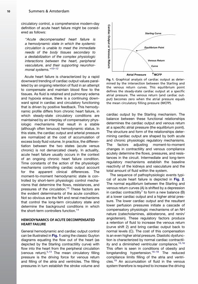
General hemodynamic and cardiac output controlcan be illustrated in Fig. 1 using the classic Guytondiagrams equating the flow out of the heart (asdepicted by the Starling contractility curve) withflow into the heart from the peripheral circulation(venous return).5,13 The mean circulatory fillingpressure is the driving force for venous returnand filling of the atria and ventricles. The fillingpressures in turn establish the stroke volume and
cardiac output by the Starling mechanism. Thebalance between these functional relationshipsdetermines the cardiac output and venous returnat a specific atrial pressure (the equilibrium point).The structure and form of the relationships deter-mining cardiac output are shaped by both acuteand chronic physiologic regulatory mechanisms.The factors adjusting moment-to-momentchanges in contractility and venous complianceacutely determine the flows, pressures, and resis-tances in the circuit. Intermediate and long-termregulatory mechanisms establish the baselinereactivity of the kidneys and vasculature and thetotal amount of fluid within the system.
The sequence of pathophysiologic events typi-cal of acute heart failure is depicted in Fig. 2.The normal equilibrium between the Starling andvenous return curves (A) is shifted by a depressionin cardiac contractility1 to form a new balance (B)at a lower cardiac output and a higher atrial pres-sure. The lower cardiac output and the resultantlower perfusion pressures initiate a cascade ofcompensatory physiologic mechanisms of an NHnature (catecholamines, aldosterone, and renin/angiotensin). These regulatory factors producea retention of fluid to increase the venous return(curve shift 2) and bring cardiac output back tonormal levels (C). The cost of this compensationis an even higher atrial pressure. Diastolic dysfunc-tion is characterized by normal cardiac contractil-ity and a diminished ventricular compliance.15,16
This often is seen in conditions of obesity andlongstanding hypertension.16–18 The reducedcompliance limits filling of the atria and ventri-cles.19 An accumulation of fluid in the venoussystem therefore is required to increase the driving
Equilibrium Point
Starling Curve
Venous Return
Curve
Atrial Pressure
Ca
rd
ia
c O
utp
ut o
r V
en
ou
s R
etu
rn
MCFP
Fig. 1. Graphical analysis of cardiac output as deter-mined by the intersection between the Starling andthe venous return curves. This equilibrium pointdefines the steady-state cardiac output at a specificatrial pressure. The venous return (and cardiac out-put) becomes zero when the atrial pressure equalsthe mean circulatory filling pressure (MCFP).
Summers & Amsterdam10

pressure for venous return and cardiac filling(Fig. 3). When this fluid accumulation occurs inthe peripheral circulation, there is potential forthe development of edema in the extremities.Likewise, left ventricular diastolic dysfunction canproduce pulmonary congestion.
Atrial pressures and pressures within the venousvasculature are the major determinants of pres-sure at the level of the pulmonary capillary.20
This pressure is the driving force for the forma-tion of pulmonary edema during acute
decompensation. By the Starling equation:
Jv 5 Kf ([Pc–Pi ] – s [ pc – pi ])where:
Jv is the net fluid movement betweencompartments.
Kf is the filtration coefficient.Pc is the pulmonary capillary hydrostatic
pressure.Pc is the interstitial hydrostatic pressure.pc is the pulmonary capillary osmotic pressure.pi is the interstitial osmotic pressure.s is the reflection coefficient.If the atrial or venous pressures become high
enough to cause pulmonary edema and hypoxia,there is further suppression of cardiac contractilityand the Starling curve, with an ensuing viciouscycle of progressive decompensation.13 A com-prehension of the integration of both acute andchronic dynamics is important for understandingmodern strategies for managing these patients.
THE ROLE OF NEUROHORMONALAND CYTOKINE ACTIVATION IN ACUTEDECOMPENSATED HEART FAILURE
As noted in the previous section, the homeostaticregulation of mammalian salt and water metabo-lism, circulatory function, and blood pressuredepends on the integration of multiple physiologicmechanisms. NH controls play an essential role inthese processes through the activity of the sympa-thetic nervous system (SNS), renin angiotensinaldosterone system (RAAS), arginine vasopressin(AV), and natriuretic peptides (NP). Endothelium-derived vasoactive factors and other mediatorsalso contribute to this physiologic organization.Several of these systems augment cardiac con-tractility, blood volume, sodium retention, andblood pressure, and others provide a counterbal-ance by promoting opposite cardiocirculatoryeffects. Under normal physiologic conditions,these mechanisms act in concert to modulate car-diac, renal, and vascular function for maintainingappropriate blood volume, perfusion pressure,cardiac output, and its distribution. When animpairment of myocardial function results inreduced blood supply to end organs, however,NH activity is augmented as a compensatoryresponse to support circulatory function by main-taining cardiac output and perfusion pressure.Whereas this activation may be helpful for limitedperiods, the deleterious effects of excessive andprolonged NH activation now are considered tobe central to the pathophysiology of heart failure.9
Conceptual models of heart failure have evolvedover the last 50 years to explain the derangedphysiology of this syndrome. According to the
Atrial Pressure
Card
iac O
utp
ut
Normal Systolic
Fluid Accumulation
Fig. 3. Diastolic dysfunction is characterized by normalcardiac contractility and diminished ventricularcompliance. An accumulation of fluid in the venoussystem is required to increase the driving pressurefor venous return and cardiac filling.
Atrial Pressure
A
B
C
1
2
Card
iac O
utp
ut
Fig. 2. As ventricular contractility is depressed (1) car-diac output is reduced, and atrial pressures areelevated as the equilibrium moves from point A topoint B. The reduced cardiac output results in less per-fusion of the kidneys and retention of fluid. This fluidretention shifts the venous return curve (2) to restorethe cardiac output to a normal state. The cost of theshift is a higher atrial pressure at the equilibriumpoint C. If the higher atrial pressure produces pulmo-nary edema, hypoxia, and further reductions incardiac function, a vicious cycle can occur.
Acute Decompensated Heart Failure 11
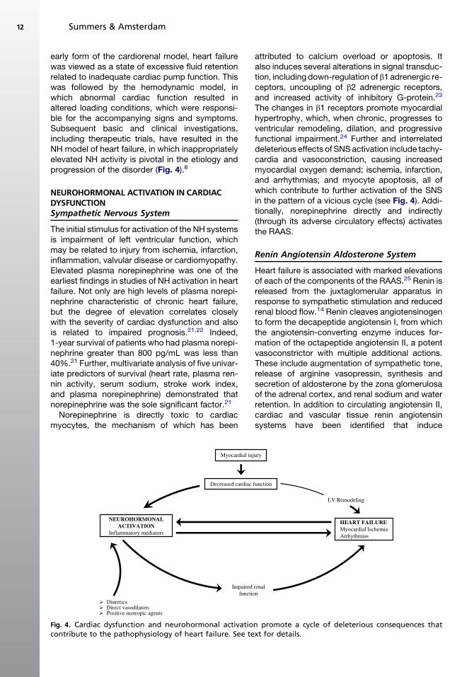
early form of the cardiorenal model, heart failurewas viewed as a state of excessive fluid retentionrelated to inadequate cardiac pump function. Thiswas followed by the hemodynamic model, inwhich abnormal cardiac function resulted inaltered loading conditions, which were responsi-ble for the accompanying signs and symptoms.Subsequent basic and clinical investigations,including therapeutic trials, have resulted in theNH model of heart failure, in which inappropriatelyelevated NH activity is pivotal in the etiology andprogression of the disorder (Fig. 4).8
NEUROHORMONAL ACTIVATION IN CARDIACDYSFUNCTIONSympathetic Nervous System
The initial stimulus for activation of the NH systemsis impairment of left ventricular function, whichmay be related to injury from ischemia, infarction,inflammation, valvular disease or cardiomyopathy.Elevated plasma norepinephrine was one of theearliest findings in studies of NH activation in heartfailure. Not only are high levels of plasma norepi-nephrine characteristic of chronic heart failure,but the degree of elevation correlates closelywith the severity of cardiac dysfunction and alsois related to impaired prognosis.21,22 Indeed,1-year survival of patients who had plasma norepi-nephrine greater than 800 pg/mL was less than40%.21 Further, multivariate analysis of five univar-iate predictors of survival (heart rate, plasma ren-nin activity, serum sodium, stroke work index,and plasma norepinephrine) demonstrated thatnorepinephrine was the sole significant factor.21
Norepinephrine is directly toxic to cardiacmyocytes, the mechanism of which has been
attributed to calcium overload or apoptosis. Italso induces several alterations in signal transduc-tion, including down-regulation of b1 adrenergic re-ceptors, uncoupling of b2 adrenergic receptors,and increased activity of inhibitory G-protein.23
The changes in b1 receptors promote myocardialhypertrophy, which, when chronic, progresses toventricular remodeling, dilation, and progressivefunctional impairment.24 Further and interrelateddeleterious effects of SNS activation include tachy-cardia and vasoconstriction, causing increasedmyocardial oxygen demand; ischemia, infarction,and arrhythmias; and myocyte apoptosis, all ofwhich contribute to further activation of the SNSin the pattern of a vicious cycle (see Fig. 4). Addi-tionally, norepinephrine directly and indirectly(through its adverse circulatory effects) activatesthe RAAS.
Renin Angiotensin Aldosterone System
Heart failure is associated with marked elevationsof each of the components of the RAAS.25 Renin isreleased from the juxtaglomerular apparatus inresponse to sympathetic stimulation and reducedrenal blood flow.14 Renin cleaves angiotensinogento form the decapeptide angiotensin I, from whichthe angiotensin-converting enzyme induces for-mation of the octapeptide angiotensin II, a potentvasoconstrictor with multiple additional actions.These include augmentation of sympathetic tone,release of arginine vasopressin, synthesis andsecretion of aldosterone by the zona glomerulosaof the adrenal cortex, and renal sodium and waterretention. In addition to circulating angiotensin II,cardiac and vascular tissue renin angiotensinsystems have been identified that induce
Myocardial injury
NEUROHORMONALACTIVATION
Inflammatory mediators
Decreased cardiac function
Impaired renalfunction
DiureticsDirect vasodilatorsPositive inotropic agents
LV Remodeling
HEART FAILUREMyocardial IschemiaArrhythmias
Fig. 4. Cardiac dysfunction and neurohormonal activation promote a cycle of deleterious consequences thatcontribute to the pathophysiology of heart failure. See text for details.
Summers & Amsterdam12

remodeling of these organs.26 Angiotensin II hasdirect myocardial toxic effects that include hyper-trophy and apoptosis, and its plasma levels corre-late with the severity of cardiac dysfunction andprognosis.27,28 Aldosterone is synthesized innumerous tissues, including the heart and bloodvessels, and the adrenal cortex, suggesting a para-crine effect of the hormone. It has potent renalsodium retaining actions and promotes cardiacand vascular fibrosis, endothelial dysfunction,myocardial infarction, cardiac hypertrophy, andmortality.29,30
Arginine Vasopressin
Vasopressin, a potent vasoconstrictor and impor-tant regulator of plasma osmolality and free waterclearance, is secreted by the pituitary gland and iselevated in patients who have heart failure. Itsrelease is influenced by osmotic and nonosmoticstimuli and is promoted by angiotensin II and acti-vation of osmotic receptors. Vasopressin isinhibited by baroreceptor stimulation and natri-uretic peptides. It induces vasoconstriction by ac-tivating vasopressin 1 receptors, and promotesrenal water reabsorption and secretion of reninby stimulating vasopressin 2 receptors. Vasopres-sin levels more than twice those in control subjectshave been reported in patients who have heart fail-ure.28 In a study of the relative contributions ofspecific NH systems to the augmented systemicvascular resistance in patients who had heart fail-ure, it was found that the SNS contribution washighest; the RAAS was also important, and vaso-pressin’s effect was least, suggesting that thishormone likely contributes significantly to vaso-constriction in heart failure only when its levelsare elevated markedly.31
Natriuretic Peptides
The major peptides of this family of compoundsare atrial natriuretic peptide (ANP) and brain(B-type) natriuretic peptide (BNP). They are acti-vated by atrial and ventricular volume and pres-sure receptors and are elevated in heart failure.These peptides promote natriuresis, reduce SNSand RAAS activity, inhibit vasopressin and endo-thelin, decrease systemic vascular resistance,and induce venodilation. The effects of BNP areparticularly prominent in this regard. The natri-uretic peptides, however, probably play little phys-iologic role in the control of the circulation or in theamelioration of ADHF.32 The clinical importance ofthese peptides is with regard to their use as a diag-nostic tool and therapeutic potentials when usedin pharmacologic doses.32 BNP initially was iden-tified in porcine brain but later found to be
produced in much greater concentrations inhuman ventricular myocardium. It is initially syn-thesized as a 134 amino acid peptide (preproBNP)from which is cleaved proBNP, which is cleavedfurther to form active BNP and the inactive N-ter-minal (NT)-proBNP. The significance of this degra-dation is the diagnostic implications of thedifferential half-lives of these compounds: BNP,22 minutes; NT-proBNP, 120 minutes. Levels ofthe natriuretic peptides increase in proportion tothe severity of the underlying cardiac disease. Fur-ther, in one study, multivariate analysis of patientswho had heart failure revealed that a high level ofBNP was the only independent NH factor predic-tive of mortality.33 BNP has assumed important di-agnostic, therapeutic, and prognostic roles formanaging patients who have heart failure.34,35
Inflammatory Mediators
Recent studies have implicated these agents in thepathophysiology of heart failure. The following isa brief presentation of several of the major inflam-matory mediators that have been demonstrated toinfluence the course of patients who have heartfailure.
EndothelinThe endothelins, produced by endothelial cells,are potent vasoconstrictors and are involved inmaintaining vascular tone and blood pressure.The major agent of this group is ET-1. This peptidecauses several important cardiovascular effects,including vasoconstriction, renal sodium retention,and production of tumor necrosis factor (TNF)-a. Italso has growth factor properties. ET-1 is elevatedin heart failure in which its major source is thepulmonary vasculature. High levels of ET-1 areassociated with increased mortality in thissetting.36
Tumor necrosis factorTNF-a plays an important role in the systemicinflammatory response. This mediator producescardiac structural and functional alterations,including fibrosis, remodeling, and apoptosis.37 Itis elevated in heart failure, in which its levels corre-late with the severity of cardiac impairment andmortality.38
InterleukinsInterleukins (IL), which are produced by variouscells, are prominent mediators in the pathogenesisof heart failure. Both IL-1 and IL-6 are increased inheart failure. IL-1 depresses myocardial contractil-ity, inhibits myocyte responsiveness to b-adrenergicstimulation, and promotes myocyte apoptosis.39
Acute Decompensated Heart Failure 13

Elevated levels of IL-6 are associated with unfavor-able clinical findings in patients who have heartfailure.40
ROLE OF NEUROHORMONAL ACTIVITYAND INFLAMMATION IN ACUTEDECOMPENSATED HEART FAILURE
NH mechanisms generally are considered long-term controllers of the circulation, and their roleis well known in patients who have chronic heartfailure. Most of the facts in the preceding sectionwere derived from studies of patients who hadchronic heart failure. There is less informationregarding the contribution of these maladaptivemechanisms in ADHF; however, it appears thatthey are also prominent in this setting. AlthoughADHF typically occurs as an exacerbation ofcompensated chronic cardiac failure, the clinicalpresentation may be de novo cardiac decompen-sation. Precipitating factors of ADHF are numer-ous and include noncompliance with diet andmedication; uncontrolled hypertension; cardiacischemia and arrhythmias; pulmonary embolism;infection; and other systemic illnesses.
In patients who have ADHF, there is evidence ofincreased NH activation,41–43 as reflected byelevated levels of norepinephrine, renin activity,aldosterone, ET-1 and other cytokines. These me-diators are associated with vasoconstriction, fluidretention, and cardiac arrhythmias.42 In addition,increased levels of cytokines have been docu-mented in patients who have ADHF and correlatewith prognosis.44–46 These recent findings supportthe role of NH activation and augmented cytokineactivity in the pathogenesis of ADHF and haveimportant implications for diagnosis, prognosis,and treatment.
Cardiorenal Syndrome
As is clear from the foregoing, activation of theSNS, RAAS, vasopressin, and inflammatorymarkers in patients with heart failure has a pro-found and adverse effect on cardiac and renalfunction. Whether worsening renal function specif-ically contributes to the progression of heart failureor is a marker of advanced cardiac and kidneyimpairment is unclear.47 The combination of thisdual organ malfunction, however, has beentermed the cardiorenal syndrome.7 It is associatedwith diuretic resistance and is common in ADHF.The pathophysiology of this syndrome appearsto be related to a complex interplay of NH andhemodynamic mechanisms. It has importanttherapeutic and prognostic implications, becauseconventional therapy is limited. Additionally,clinical outcomes are poor.
Clinical Pathophysiology of AcuteHeart Failure
In a typical clinical scenario, a patient who haschronic heart failure will preserve a stable condi-tion unless there is some perturbation (such asan increased fluid intake, worsening cardiac func-tion, or noncompliance with medications) thatrequires the systemic physiology to activate anadjustment.48 A crescendoing activation of thelong-term cardiac output control mechanismsseeking to maintain stability in response to a per-turbation ultimately can overwhelm the short-term controllers and result in the development ofADHF. When a patient arrives in the emergencydepartment in the acute throes of a decompen-sated heart failure event, the critical efforts in theresuscitative process are directed toward a rapidrelief of the pulmonary edema and an improve-ment in central oxygenation for vital organ sup-port. This goal requires that the physician createan immediate change in the hemodynamic statein a way that promptly reduces the atrial pressuresand alleviates the pulmonary congestion. Froma pathophysiologic perspective, the time-urgentnature of the condition obviates a role for thelong-term controllers of the circulation in the pri-mary phase of ADHF management. A treatmentscheme targeting the plumbing of the intravascularpressures and flows should be the first consider-ation in the emergent stabilization of the ADHFpatient.48 This objective often can be achievedby measures that reduce afterload or preload con-ditions in an attempt to reduce the atrial pressureand forces driving fluid extravasations at the levelof the capillary. Although it is common to observea rapid resolution of the patient’s congestion andhypoxia with these treatments, the physiologicadjustments in hemodynamics initiated by mostconventional acute therapies do not provide forlong-term circulatory stability.48,49 The derange-ments in the neurohormonal axis and other chroniccontrol mechanisms that led to the decompen-sated state are usually still present after the pri-mary resuscitation and drive a continued fluidretention by the kidneys even in the face of the ini-tial acute management. Therefore, it is importantto immediately begin to address the physiologicmechanisms responsible for the chronic regulationof the circulation in a secondary phase of resusci-tation of these patients.
Therapeutic implications of neurohormonalactivation in heart failureBased on an understanding of the NH model ofheart failure and the pharmacologic actions of cur-rent therapeutic modalities, the limitations and
Summers & Amsterdam14
potentially deleterious role of these approachescan be understood. Thus, although diuretics, vaso-dilators, and positive inotropic agents may affordsymptomatic relief, they tend to exacerbateunderlying detrimental NH overactivity on themyocardium, vasculature, kidney, and fluid andelectrolyte balance. Diuretics stimulate further acti-vation of the RAAS, SNS, vasopressin, and endo-thelin, as do direct vasodilators (see Fig. 4).50 Theunfavorable myocardial effects of positive inotropicagents are similar to those of the endogenous cat-echolamines described previously. These consid-erations have stimulated concern for judiciousand physiologically rational application of thesetherapeutic approaches based on underlying path-ophysiology to mitigate their undesirable effects.51
Special Pathophysiologic Conditionsof Heart Failure
The diverse physiologic factors affecting an over-lapping and time-varying control of cardiac outputand general circulatory functioning can result in anincongruence between a patient’s ventricularfunction, body fluid volume status, and state ofheart failure. There are several common clinicalscenarios in which such paradoxes can arise.
Pathophysiology of fluid volume overloadheart failureEven when there is normal cardiac function, it isstill possible to overwhelm the circulation with fluidto a point where the heart is ineffective as a pump.When this happens, the circulation becomes con-gested, and edema can develop. If the volumeoverload is prolonged, the heart function can beginto deteriorate under the strain, and a vicious cyclebegins.52 Chronic renal failure is the most commonclinical condition in which patients present withfluid volume overload. In these cases, a manipula-tion of the hemodynamics may be all that is possi-ble until the excess fluid is removed by dialysis orsome other method. If the renal failure is in the endstages, the NH systems have little or no role in thepathophysiology of the heart failure.
Pathophysiology of acute heart failurein the dehydrated patientHeart failure is typically thought of as a condition offluid volume excess.53 Elderly bed-ridden patientswho have marginal ventricular function and anongoing treatment with diuretics, however, canpresent with a paradoxical condition, in whichthey have evidence of pulmonary congestion andyet are volume contracted.10Just as pulmonaryedema can lead to myocardial hypoxia and wors-ening contractility, a progressing dehydration canresult in a decrease in coronary perfusion and
precipitate an acute heart failure event. Althoughit is counterintuitive, it may be necessary to begina fluid bolus as a part of the initial resuscitation ofthese patients. This rehydration process, however,should be done judiciously and with a concurrentmanipulation of the hemodynamics to optimizeflow around the circuit.
Pathophysiology of diuretic-resistantheart failureMost forms of acute heart failure are very respon-sive to diuretic treatment in the primary phase ofresuscitation. As noted previously, however, con-tinued aggressive treatment with diuretics canstimulate an already activated NH responsefurther. Increasing levels of angiotensin and cate-cholamines can restrict renal blood flow severely,while an elevated serum aldosterone enhancesfluid reabsorption in the distal tubule. This patho-physiologic condition attenuates the effectivenessof subsequent doses of diuretics and results inlimited urine output.48 If the patient is not volumecontracted, therapies targeting the NH systemsmight be used to enhance the effectiveness ofthe diuretic.
REFERENCES
1. Saba MM, Ventura HO, Saleh M, et al. Ancient
Egyptian medicine and the concept of heart failure.
J Card Fail 2006;12:416–21.
2. Lutz JE. A XII century description of congestive
heart failure. Am J Cardiol 1988;61(6):494–5.
3. Breckenridge A. William Withering’s legacy—for the
good of the patient. Clin Med 2006;6(4):393–7.
4. Guyton AC, Coleman TG, Granger HJ. Circulation:
overall regulation. Annu Rev Physiol 1972;34:13–46.
5. Guyton AC. The systemic venous system in cardiac
failure. J Chronic Dis 1959;9:465–75.
6. Montani JP, Adair TH, Summers RL, et al. A simula-
tion support system for solving large physiological
models on microcomputers. Int J Biomed Comput
1989;24:41–54.
7. Bongartz LG, Cramer MJ, Doevendans PA, et al. The
severe cardiorenal syndrome: Guyton revisited. Eur
Heart J 2005;26:11–7.
8. Mann DL, Bristow MR. Mechanisms and models in
heart failure: the biomechanical model and beyond.
Circulation 2005;111(21):2837–49.
9. Braunwald E, Bristow MR. Congestive heart failure:
fifty years of progress. Circulation 2000;102(4):
IV14–23.
10. Summers RL. Rapid clinical assessment of hemody-
namic profiles and targeted treatment of patients
with acutely decompensated heart failure: wet and
cold profile. Clin Cardiol 2004;27(Suppl V):V10–1.
Acute Decompensated Heart Failure 15
11. Summers RL. Cardiovascular emergencies: chapter
32: future diagnostics: impedance cardiography in
the assessment and management of acute heart
failure. New York: McGraw-Hill Companies; 2006.
357–63.
12. Summers RL. Emerging diagnostics: impedance
cardiography in the assessment and management
of acute heart failure. Critical Pathways in Cardiology
2005;4:134–9.
13. Guyton AC, Jones CE, Coleman TG. Circulatory
physiology: cardiac output and its regulation. (2nd
edition). Philadelphia: W.B. Saunders; 1973.
14. Hall JE, Guyton AC, Mizelle HL. Role of the renin–
angiotensin system in control of sodium excretion
and arterial pressure. Acta Physiol Scand Suppl
1990;591:48–62.
15. Andrew P. Diastolic heart failure demystified. Chest
2003;124:744–53.
16. Summers RL, Kolb JC, Woodward LH, et al. Differen-
tiating systolic from diastolic heart failure using
impedance cardiography. Acad Emerg Med 1999;
6(7):693–9.
17. Carroll JF, Summers RL, Dzielak DJ, et al. Diastolic
compliance is reduced in obese rabbits. Hyperten-
sion 1999;33:811–5.
18. Summers RL, Kolb JC. Left ventricular hypertrophy
as a marker of diastolic dysfunction in patients with
acute congestive heart failure. Eur J Emerg Med
2002;9(4):351.
19. Summers RL, Montani JP. Computer model of car-
diac diastolic dynamics. Comput Cardiol 1992;19:
697–700.
20. Gaar KA Jr, Taylor AE, Owens LJ. Effect of capillary
pressure and plasma protein on development of
pulmonary edema. Am J Phys 1967;213(1):79–82.
21. Cohn JN, Levine TB, Olivari MT, et al. Plasma norepi-
nephrine as a guide to prognosis in patients with
chronic congestive heart failure. N Engl J Med
1984;311(13):819–23.
22. Francis GS, Benedict C, Johnstone DE, et al. Com-
parison of neuroendocrine activation in patients
with left ventricular dysfunction with and without con-
gestive heart failure. A substudy of the studies of left
ventricular dysfunction (SOLVD). Circulation 1990;
82(5):1724–9.
23. Mann DL, Kent RL, Parsons B, et al. Adrenergic
effects on the biology of the adult mammalian cardi-
ocyte. Circulation 1992;85(2):790–804.
24. Adams JW, Sakata Y, Davis MG, et al. Enhanced
Galpha q signaling: a common pathway mediates
cardiac hypertrophy and apoptotic heart failure.
Proc Natl Acad Sci U S A 1998;95(17):10140–5.
25. Anand IS, Ferrari R, Kalra GS, et al. Edema of
cardiac origin. Studies of body water and sodium,
renal function, hemodynamic indexes, and plasma
hormones in untreated congestive cardiac failure.
Circulation 1989;80(2):299–305.
26. Bader M, Jorg P, Ovidiu B, et al. Tissue renin–
angiotensin systems: new insights from experimen-
tal animal models in hypertension research. J Mol
Med 2001;9(2):76–102.
27. Tan LB, Jalil JE, Pick R, et al. Cardiac myocyte
necrosis induced by angiotensin II. Circ Res 1991;
69(5):1185–95.
28. Francis GS, Cohn JN, Johnson G. Plasma norepi-
nephrine, plasma renin activity, and congestive heart
failure. Relations to survival and the effects of therapy
in V-HeFT II. The V-HeFT VA Cooperative Studies
Group. Circulation 1993;87(Suppl. 6):V140–8.
29. Loskutoff D, Quigley J. PAI-1, fibrosis, and the elu-
sive provisional fibrin matrix. J Clin Invest 2000;
106(12):1441–3.
30. Swedberg K, Eneroth P, Kjekshus J, et al. Hormones
regulating cardiovascular function in patients with
severe congestive heart failure and their relation to
mortality. CONSENSUS Trial Study Group. Circula-
tion 1990;82(5):1730–6.
31. Creager M, Faxon D, Cutler S. Contribution of vaso-
pressin to vasoconstriction in patients with conges-
tive heart failure: comparison with the renin–
angiotensin system and the sympathetic nervous
system. J Am Coll Cardiol 1986;7(4):758–65.
32. Summers RL, Montani JP. Computer model of ANP-
hemodynamic interactions. Comput Cardiol 1991;
18:697–700.
33. Tsutamoto T, Wada A, Maeda K, et al. Attenuation of
compensation of endogenous cardiac natriuretic
peptide system in chronic heart failure: prognostic
role of plasma brain natriuretic peptide concentration
in patients with chronic symptomatic left ventricular
dysfunction. Circulation 1997;96(2):509–16.
34. Colucci WS, Elkayam U, Horton DP, et al. Intrave-
nous nesiritide, a natriuretic peptide, in the treatment
of decompensated congestive heart failure. N Engl
J Med 2000;343(4):246–53.
35. Maisel AS, McCord J, Nowak RM, et al. Bedside
B-type natriuretic peptide in the emergency diagnosis
of heart failure with reduced or preserved ejection frac-
tion: results from the breathing not properly multina-
tional study. J Am Coll Cardiol 2003;1(11):2010–7.
36. Wei CM, Lerman A, Rodeheffer RJ, et al. Endothelin
in human congestive heart failure. Circulation 1994;
89(4):1580–6.
37. Bradham WS, Bozkurt B, Gunasinghe H, et al. Tumor
necrosis factor-alpha and myocardial remodeling in
progression of heart failure: a current perspective.
Cardiovasc Res 2002;53(4):822–30.
38. Torre-Amione G, Kapadia S, Benedict C, et al.
Proinflammatory cytokine levels in patients with
depressed left ventricular ejection fraction: a report
from the studies of left ventricular dysfunction
(SOLVD). J Am Coll Cardiol 1996;27(5):1201–6.
39. Francis SE, Holden H, Holt CM, et al. Interleukin-1 in
myocardium and coronary arteries of patients with
Summers & Amsterdam16

dilated cardiomyopathy. J Mol Cell Cardiol 1998;
30(2):215–23.
40. Aukrust P, Ueland T, Lien E, et al. Cytokine network
in congestive heart failure secondary to ischemic
or idiopathic dilated cardiomyopathy. Am J Cardiol
1999;83(3):376–82.
41. Aronson D, Burger AJ. Neurohormonal prediction of
mortality following admission for decompensated
heart failure. Am J Cardiol 2003;91(2):245–8.
42. Aronson D, Burger AJ. Neurohumoral activation and
ventricular arrhythmias in patients with decompen-
sated congestive heart failure: role of endothelin.
Pacing Clin Electrophysiol 2003;26(3):703–10.
43. Milo O, Cotter G, Kaluski E, et al. Comparison of in-
flammatory and neurohormonal activation in cardio-
genic pulmonary edema secondary to ischemic
versus nonischemic causes. Am J Cardiol 2003;
92(2):222–6.
44. Chin. Interleukin-6, tissue factorandvonWillebrand fac-
tor inacute decompensatedheart failure: relationship to
treatment and prognosis. Blood 2003;14(6):515.
45. Mueller C, Laule-Kilian K, Christ A, et al. Inflamma-
tion and long-term mortality in acute congestive
heart failure. Am Heart J 2006;151(4):845–50.
46. Peschel T, Schonauer M, Thiele H, et al. Invasive as-
sessment of bacterial endotoxin and inflammatory
cytokines in patients with acute heart failure. Eur
J Heart Fail 2003;5(5):609–14.
47. Liang K, Williams A, Greene E. Acute decompen-
sated heart failure and the cardiorenal syndrome.
Crit Care Med 2008;36(1 Suppl):S75–88.
48. Peacock WF, Allegra J, Ander D, et al. Management
of acute decompensated heart failure in the emer-
gency department. Congest Heart Fail 2003;
9(Suppl 1):3–18.
49. Rame JE, Sheffield MA, Dries DL, et al. Outcomes
after emergency department discharge with a pri-
mary diagnosis of heart failure. Am Heart J 2001;
142:714–9.
50. Shah M, Ali V, Lamba S, et al. Pathophysiology and
clinical spectrum of acute congestive heart failure.
Rev Cardiovasc Med 2001;(Suppl):9–18.
51. Burger AJ. A review of the renal and neurohormonal
effects of B-type natriuretic peptide. Congest Heart
Fail 2005;11(1):30–8.
52. Cotter G, Felker GM, Adams KF, et al. The patho-
physiology of acute heart failure—is it all about fluid
accumulation? Am Heart J. 2008;155:9–18.
53. Cotter G, Metra M, Milo-Cotter O, et al. Fluid over-
load in acute heart failure—redistribution and other
mechanisms beyond fluid accumulation. Eur J Heart
Fail 2008;10(2):165–9.
Acute Decompensated Heart Failure 17

PrehospitalManagement ofCongestive Heart FailureAmal Mattu, MD, FACEP, FAAEM*, Benjamin Lawner, DO, EMT-P
The prehospital evaluation and treatment ofdecompensated congestive heart failure (CHF) isas varied as there are emergency medical services(EMS) providers. The nation’s EMSs encompassa diverse group of volunteers, career profes-sionals, paraprofessionals, and physicians. Thescope of practice for management of decompen-sated CHF changes varies across jurisdictionsand is subject to change according to a medicaldirector’s level of comfort and preference. A majorreason for the disparities between practices is thelimited availability of data from the prehospitalsetting. Most treatment protocols are based onresearch on in-hospital patients that has beenextrapolated to the prehospital setting. The fewprehospital studies that do exist are relativelysmall and usually retrospective in nature. This arti-cle highlights some of those prehospital studies onwhich many treatment protocols are based. Adetailed discussion of the pathophysiology ofdecompensated CHF and the mechanisms under-lying common treatments is found in other articlesin this issue.
PREHOSPITAL PERSONNEL AND TRAINING
Physicians who interact with prehospital person-nel should have an understanding of the differentlevels of prehospital care providers, their training,and their capabilities. Basic life support treatmentis generally provided at the emergency medicaltechnician (EMT)–basic level. The National High-way Traffic and Safety Administration (NHTSA)recommends a 110-hour curriculum, but this
standard is not uniformly followed.1 State EMSoffices often modify the national curriculum tosuit their particular needs. For the purposes ofthis article, basic life support (BLS) treatmentrefers to prehospital care that is noninvasive. Firstresponders, basic-level EMTs, and ambulanceattendants deliver BLS care.
The paramedic provider is the product of a preho-spital training system that is still in evolution. In aneffort to promote a more advanced prehospital levelof care, the Los Angeles County Fire Departmentbegan training individuals in the 1970s to go beyondsimple BLS by administering intravenous fluids andmedications in the out-of-hospital setting. Later, thecity of Miami instituted the first organized prehospi-tal advanced life support (ALS) system. Paramedicsand intermediate-level EMTs in this system func-tioned at the ALS level and performed intravenousmedication therapy, defibrillation, endotrachealintubation, and external cardiac pacing. This sys-tem was gradually adopted around the country,and the NHTSA has endorsed a national standardcurriculum for paramedic training that requires1000 to 1200 hours of classroom instruction.2
Despite advanced training and an expandedscope of practice, paramedics still encounter sig-nificant challenges in diagnosis and managementof decompensated CHF. The diagnosis of a heartfailure syndrome is complicated by the oftenaustere out-of-hospital environment. Patients inextremis are often unable to provide an adequatehistory, and paramedics must rely on clinicalpresentation to formulate an initial diagnosticimpression. The lack of adequate lighting and
University of Maryland School of Medicine, Baltimore, MD, USA* Corresponding author. Department of Emergency Medicine, University of Maryland School of Medicine, 110South Paca Street, 6th Floor, Suite 200, Baltimore, MD 21201.E-mail address: [email protected] (A. Mattu).
KEYWORDS� Congestive heart failure � Pulmonary edema� Prehospital � Noninvasive ventilation
Heart Failure Clin 5 (2009) 19–24doi:10.1016/j.hfc.2008.08.0041551-7136/08/$ – see front matter ª 2008 Elsevier Inc. All rights reserved. he
artf
ailu
re.th
ecli
nics
.com
even basic diagnostic testing (eg, thermometer,chest radiology, B-type natriuretic peptide test)further obfuscates the identification of an acuteheart failure syndrome from noncardiac causesof dyspnea. Because of these challenges, para-medics may adopt a ‘‘shotgun-type’’ approach tothe management of the patient in respiratory dis-tress with the use of multiple, rather than targeted,therapies. Unfortunately, there is a relatively highmisdiagnosis rate for prehospital patients whohave presumed decompensated CHF; whenpatients are treated in this shotgun type of manner,misdiagnosed patients suffer adverse conse-quences. Paramedic training is beginning toaddress some of these issues through improvededucation, and an increasing number of stateprotocols are more frequently requiring discussionwith or preauthorization by a physician for certainmedications that were once considered basic(eg, furosemide). Many ALS systems still functionunder the doctrine of off-line medical control,however, which permits paramedics and EMT-intermediates to treat patients according to preap-proved algorithms.
BASIC LIFE SUPPORT INTERVENTION
Unfortunately the first responder and EMT-basichave a limited arsenal of tools at their disposal.BLS treatment is generally noninvasive and there-fore focuses on rapid transport to a hospital. EMTsand first responders are educated in signs andsymptoms suggestive of heart failure and severerespiratory distress. Recognition of abnormal vitalsigns may prompt a BLS crew to expedite trans-port to an emergency department or request as-sistance from an ALS provider. In more ruralareas of the country, this practice of ALS interceptfunctions to deliver paramedic-level care topatients who are farther away from definitivetherapy.
Oxygen remains the mainstay of BLS-level ther-apy. First responders are trained to administerhigh-flow oxygen by way of non-rebreather maskto patients in severe respiratory distress. Shoulda patient’s respiratory status further deteriorate,the BLS provider is capable of assisting respira-tions by way of a bag-valve mask device.Adjuncts, such as nasopharyngeal and oropharyn-geal airways, are available to BLS crews whoencounter a patient in cardiorespiratory arrest.Cardiopulmonary resuscitation and automatedexternal defibrillation would also be within thebasic provider’s scope of practice should thepatient who has acute heart failure decompensatefurther.
ADVANCED LIFE SUPPORTINTERVENTIONçAIRWAY SUPPORT
Paramedics clearly have greater resources at theirdisposal when caring for the patient who hasdecompensated heart failure. Although theadministration of high-flow oxygen by way ofa non-rebreather mask is still routine, the use ofnoninvasive positive pressure ventilation (NIPPV)is gaining attention and popularity in prehospitalsystems. The two main modalities of deliveringNIPPV are by continuous positive pressure ventila-tion (CPAP) and bi-level positive pressure ventila-tion (BiPAP). Three recent meta-analyses ofstudies evaluating NIPPV have demonstratedthat the use of either CPAP or BiPAP for patientswho have decompensated CHF is associatedwith decreased need for intubation, reduced ICUand hospital length of stay, reduced costs, anddecreased mortality.3–5 The studies on whichthese meta-analyses are based primarily evalu-ated emergency department and in-hospitalpatients. Based on these ED and in-hospital stud-ies, many EMS systems enthusiastically adoptedNIPPV for prehospital use also. Most of thesesystems have chosen to use CPAP over BiPAPbecause of its ease of use, and recent studiesevaluating prehospital use of CPAP have demon-strated encouraging results.
In 2001 Kosowsky and colleagues6 publisheda case series of 19 patients demonstrating theusefulness of prehospital CPAP. Cincinnati FireDepartment paramedics were authorized to imple-ment CPAP ‘‘at their discretion’’ for patientsbelieved to be suffering from acute cardiogenicpulmonary edema. CPAP was administered byface mask and maintained at a fixed pressure of10 cm H2O. CPAP was discontinued if the patientcould not tolerate the face mask or experiencedfurther deterioration. Emergency department phy-sicians corroborated the field diagnosis of pulmo-nary edema in 13 of 19 patients receiving CPAP.Patients undergoing CPAP therapy were less likelyto require intubation on hospital arrival and experi-enced a reduced length of stay.
In 2006 Hubble and colleagues7 conducteda nonrandomized trial that included 120 patientsand examined endotracheal intubation as theprimary endpoint. Other surrogate markers oftreatment efficacy that were included were respi-ratory rate, subjective dyspnea score, hospitallength of stay, and mortality. Similar to Kosow-sky’s series, paramedics administered CPAP byway of face mask at a fixed pressure. The authorsfound that patients treated with CPAP experi-enced decreased need for intubation, hospitallength of stay, and mortality.
Mattu & Lawner20

A more recent study by Plaisance and col-leagues8 evaluated early prehospital use ofCPAP (within 15 minutes of ambulance arrival)versus delayed use of CPAP (30–45 minutes afterarrival). Medical therapies, including use of nitratesand diuretics, were also evaluated in various com-binations with early and delayed CPAP. Earlyadministration of CPAP with or without medica-tions demonstrated superior outcomes whencompared with medical therapy alone or delayedCPAP with medications. Early use of CPAP wasassociated with greater improvements in oxygen-ation, dyspnea, and tracheal intubation rates.Investigators also discovered a trend towarddecreased mortality. The study emphasized theimportance of early application of CPAP, becauseeven a 15-minute delay in application of CPAP wasassociated with worse outcomes. CPAP continuesto gain widespread support for the prehospital set-ting, and studies that seek to affirm its effective-ness are ongoing. Although CPAP protocols existin many ALS jurisdictions, NIPPV is still not consid-ered the standard of prehospital care.
Prehospital care providers should be aware ofnot only the indications of NIPPV but also the con-traindications and shortcomings. NIPPV therapyshould be avoided in patients who have vomitingor altered mental status. Some patients may alsobe unable to tolerate the face mask because ofclaustrophobia, and thick beards may make a tightseal impossible. Finally, if a patient is in extremisand is rapidly decompensating, definitive airwaycontrol with endotracheal intubation should notbe delayed with a trial of NIPPV as long as the pre-hospital care providers have expertise in endotra-cheal or nasotracheal intubation. A more detaileddiscussion of NIPPV is provided in other articlesin this issue.
ADVANCED LIFE SUPPORTINTERVENTIONçMEDICALTHERAPIES
For hypertensive patients who have acute heartfailure, EMS systems have traditionally embracedthe use of a triple cocktail of nitroglycerine, mor-phine, and furosemide. This treatment has recentlyfallen under increased scrutiny, however. A pau-city of data supports the routine administration ofopiates and diuretics to prehospital patients whohave decompensated CHF.9 On the contrary,studies have demonstrated that these medicationsmay actually be harmful, especially when theprehospital diagnosis is incorrect. Hoffman andcolleagues10 compared nitroglycerin (NTG), furo-semide, and morphine in 57 patients who had pre-sumed decompensated CHF. The best outcomesin dyspnea and hospital morbidity (respiratory
depression, dehydration, prolonged in-hospitalcourse) were associated with NTG. The use offurosemide was associated with significant elec-trolyte abnormalities in some patients, and morethan 25% of patients later required fluid repletionbecause of dehydration and hypotension. Ofnote, 23% of patients were misdiagnosed anddid not have pulmonary edema. In these patientswho were inappropriately treated, the use of furo-semide and morphine was associated withincreased dyspnea and morbidity. Even in thesemisdiagnosed patients, however, the use of NTGalone was not associated with any adverse effects.
Wuerz and colleagues11 conducted the largestprehospital study evaluating the use of medicationsin the management of 599 patients who had pre-sumed decompensated CHF. Once again, a signif-icant number of patients (18%) were misdiagnosedand inappropriately treated for CHF. Most of thesemisdiagnosed patients were eventually found tohave acute asthma, chronic obstructive pulmonarydisease (COPD), pneumonia, or bronchitis. In thesemisdiagnosed patients, if NTG was the sole preho-spital treatment, the patients had a 2.2% mortality.In contrast, if these patients were treated with mor-phine or furosemide (with or without NTG), the mor-tality was found to be 21.7%.
A recent prehospital study of presumed decom-pensated CHF was conducted by Jaronik andcolleagues.12 They evaluated 144 prehospitalpatients who were treated with furosemide. Theyfound that 42% of the patients were misdiagnosedand did not actually have CHF. In fact 17% of thepatients were diagnosed as having sepsis, dehy-dration, or pneumonia (without CHF) and in thesepatients the administration of furosemide was con-sidered ‘‘potentially harmful.’’ Nine study patientsdied, 7 of whom were in the group of misdiag-nosed patients who had received furosemide‘‘inappropriately.’’
Although the total number of patients in thesestudies is relatively small, several important con-clusions can be made nevertheless. First, preho-spital health care providers often misdiagnosepatients as having decompensated CHF, withmisdiagnosis rates as high as 42%. Second,administration of furosemide or morphine to thesemisdiagnosed patients is associated with adverseconsequences. This finding should be no surprise;patients who have acute asthma or COPD exacer-bations or pneumonia are often dehydratedbecause of poor oral intake and the insensible fluidlosses associated with tachypnea and fever.Diuretics exacerbate fluid and electrolyte imbal-ance; furthermore, morphine, especially in patientswho have COPD exacerbations, promotes respira-tory depression. Even in patients who are correctly
Prehospital Management of Congestive Heart Failure 21

diagnosed, more than half of patients who haveseverely decompensated heart failure are not ac-tually fluid overloaded, but rather have pulmonaryedema because of fluid maldistribution.13–15 Thefocus of therapy for these patients should be vas-cular redistribution rather than diuresis, and in fact,they may actually suffer adverse neurohumoraland renal effects from aggressive diuresis. Third,it seems that the use of NTG is without significantadverse consequences even when the prehospitaldiagnosis is incorrect. If the diagnosis of decom-pensated CHF is correct, then NTG is clearly ben-eficial by providing rapid preload reduction andneeded vascular redistribution.
In addition to the evidence demonstratingadverse consequences of diuretics and morphinewhen the diagnosis is incorrect, there is alsoa lack of evidence suggesting that these medica-tions are quickly beneficial when the diagnosis iscorrect. Patients who have decompensated CHFand especially cardiogenic pulmonary edemahave decreased renal blood flow because of mark-edly increased afterload.13 As a result, the diureticand preload-reducing effect of furosemide may bedelayed 90 to 120 minutes.16–18 Unless transporttimes are extremely long, furosemide is unlikelyto exert a beneficial effect before hospital arrivaland may actually be harmful. Obstacles to accu-rate diagnosis, coupled with the lack of immediatebenefit, urge prehospital medical directors toreconsider the priority of diuresis.
Morphine also has limited benefit even incorrectly diagnosed patients who have decom-pensated CHF. The traditional teaching that mor-phine produces rapid preload reduction is notsupported by studies that used invasive hemody-namic monitoring, and in fact those studiesindicate that morphine is associated with tran-sient increases in right heart filling pressure andreductions in cardiac index.19,20 An ED-based ret-rospective study by Sacchetti and colleagues21
suggested that the routine use of morphine inthe early management of patients who had car-diogenic pulmonary edema was associated witha fivefold increase in ICU use and intubationrates. Although morphine is also promoted be-cause of its anxiolytic effect, this can actually beaccomplished quickly and more safely with smalldosages of benzodiazepines when anxiolysis istruly necessary. In all but rare cases, however,rapid treatment of hypoxia results in resolutionof anxiety, and additional pharmacologic agentstargeted at anxiety are usually not needed.
The use of bronchodilators is also commonamong prehospital providers when caring forpatients who have undifferentiated dyspnea.Because patients who have decompensated
CHF may present with wheezing, bronchodilatorsare also commonly used in these patients. Theprehospital study by Wuerz and colleagues11 sug-gested that bronchodilators were safe in all typesof patients who had dyspnea. There was noincrease in mortality when patients were treatedwith bronchodilators, regardless of the final diag-nosis. A recent study by Singer and colleagues,22
however, has suggested otherwise. They evalu-ated more than 10,000 patients from the AcuteDecompensated Heart Failure National RegistryEmergency Module who were provided broncho-dilators during acute treatment, in the prehospitalsetting or in the ED. They found that the use ofbronchodilators in patients who did not havea history of COPD was associated with a slightlygreater need for ‘‘aggressive interventions,’’including mechanical ventilation. Whether thebronchodilator use was causing adverse out-comes or was simply a marker of sicker patientsis unclear from this nonrandomized, retrospectivestudy. Nevertheless, the study does raise con-cerns regarding the liberal use of bronchodilatorsin the management of prehospital patients whohave undifferentiated dyspnea, especially if thepresumptive diagnosis is decompensated CHF.Further studies will need to clarify this question.
The use of angiotensin-converting enzyme(ACE) inhibitors in patients who have decompen-sated CHF in the ED has gained support in recentyears.13,15,23–30 A series of small studies has dem-onstrated that early use of ACE inhibitors in thesublingual (captopril) or intravenous (captopril,enalapril) formulation are associated with rapidimprovements in preload, afterload, cardiac out-put, and dyspnea,23–30 and perhaps also a signifi-cant decrease in need for ICU use andintubation.21 Prehospital studies of ACE inhibitorsare lacking; however, some prehospital systemsare now using ACE inhibitors for hypertensive pa-tients when the diagnosis of decompensated CHFis strongly suspected.9 In the state of Maryland, forexample, the administration of 25 mg sublingualcaptopril is incorporated into the standing protocolfor patients who have decompensated CHF afterthe aggressive use of nitrates (see Box 1). Preho-spital providers should be cautious to only usethese medications in patients in whom the diagno-sis is nearly certain, and only in patients whoremain normotensive or hypertensive after nitratetherapy. ACE inhibitors must also be avoided inpatients who have had prior adverse reactions(eg, angioedema). Further research is needed be-fore the prehospital use of ACE inhibitors is widelyrecommended.
Patients who have concurrent hypotension anddecompensated CHF pose a unique challenge for
Mattu & Lawner22
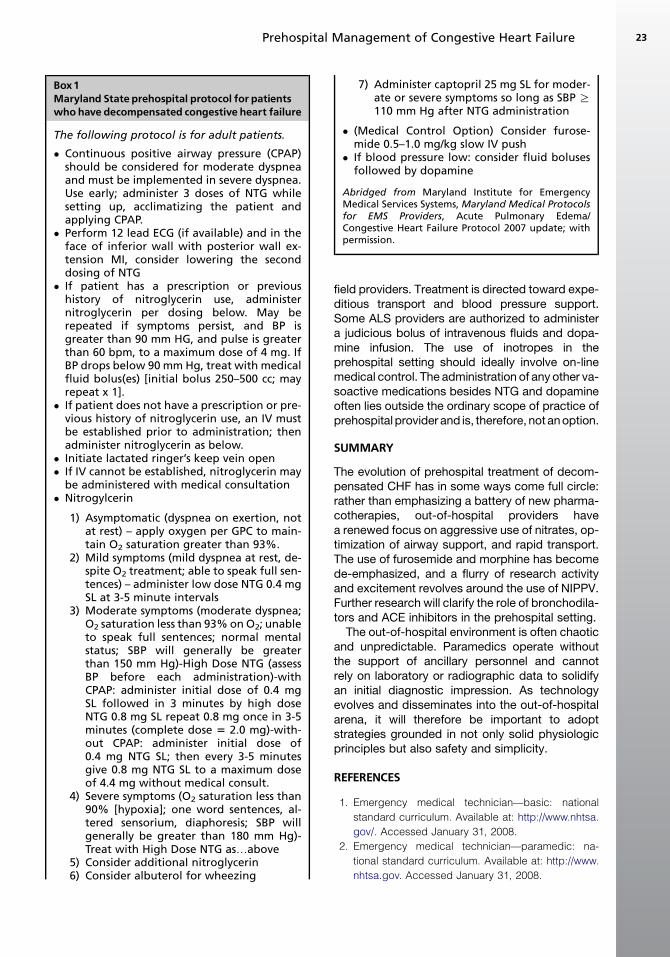
field providers. Treatment is directed toward expe-ditious transport and blood pressure support.Some ALS providers are authorized to administera judicious bolus of intravenous fluids and dopa-mine infusion. The use of inotropes in theprehospital setting should ideally involve on-linemedical control. The administration of any other va-soactive medications besides NTG and dopamineoften lies outside the ordinary scope of practice ofprehospital provider and is, therefore, not an option.
SUMMARY
The evolution of prehospital treatment of decom-pensated CHF has in some ways come full circle:rather than emphasizing a battery of new pharma-cotherapies, out-of-hospital providers havea renewed focus on aggressive use of nitrates, op-timization of airway support, and rapid transport.The use of furosemide and morphine has becomede-emphasized, and a flurry of research activityand excitement revolves around the use of NIPPV.Further research will clarify the role of bronchodila-tors and ACE inhibitors in the prehospital setting.
The out-of-hospital environment is often chaoticand unpredictable. Paramedics operate withoutthe support of ancillary personnel and cannotrely on laboratory or radiographic data to solidifyan initial diagnostic impression. As technologyevolves and disseminates into the out-of-hospitalarena, it will therefore be important to adoptstrategies grounded in not only solid physiologicprinciples but also safety and simplicity.
REFERENCES
1. Emergency medical technician—basic: national
standard curriculum. Available at: http://www.nhtsa.
gov/. Accessed January 31, 2008.
2. Emergency medical technician—paramedic: na-
tional standard curriculum. Available at: http://www.
nhtsa.gov. Accessed January 31, 2008.
Box1Maryland State prehospital protocol for patientswho have decompensated congestive heart failure
The following protocol is for adult patients.
� Continuous positive airway pressure (CPAP)should be considered for moderate dyspneaand must be implemented in severe dyspnea.Use early; administer 3 doses of NTG whilesetting up, acclimatizing the patient andapplying CPAP.� Perform 12 lead ECG (if available) and in the
face of inferior wall with posterior wall ex-tension MI, consider lowering the seconddosing of NTG� If patient has a prescription or previous
history of nitroglycerin use, administernitroglycerin per dosing below. May berepeated if symptoms persist, and BP isgreater than 90 mm HG, and pulse is greaterthan 60 bpm, to a maximum dose of 4 mg. IfBP drops below 90 mm Hg, treat with medicalfluid bolus(es) [initial bolus 250–500 cc; mayrepeat x 1].� If patient does not have a prescription or pre-
vious history of nitroglycerin use, an IV mustbe established prior to administration; thenadminister nitroglycerin as below.� Initiate lactated ringer’s keep vein open� If IV cannot be established, nitroglycerin may
be administered with medical consultation� Nitrogylcerin
1) Asymptomatic (dyspnea on exertion, notat rest) – apply oxygen per GPC to main-tain O2 saturation greater than 93%.
2) Mild symptoms (mild dyspnea at rest, de-spite O2 treatment; able to speak full sen-tences) – administer low dose NTG 0.4 mgSL at 3-5 minute intervals
3) Moderate symptoms (moderate dyspnea;O2 saturation less than 93% on O2; unableto speak full sentences; normal mentalstatus; SBP will generally be greaterthan 150 mm Hg)-High Dose NTG (assessBP before each administration)-withCPAP: administer initial dose of 0.4 mgSL followed in 3 minutes by high doseNTG 0.8 mg SL repeat 0.8 mg once in 3-5minutes (complete dose 5 2.0 mg)-with-out CPAP: administer initial dose of0.4 mg NTG SL; then every 3-5 minutesgive 0.8 mg NTG SL to a maximum doseof 4.4 mg without medical consult.
4) Severe symptoms (O2 saturation less than90% [hypoxia]; one word sentences, al-tered sensorium, diaphoresis; SBP willgenerally be greater than 180 mm Hg)-Treat with High Dose NTG as.above
5) Consider additional nitroglycerin6) Consider albuterol for wheezing
7) Administer captopril 25 mg SL for moder-ate or severe symptoms so long as SBP R110 mm Hg after NTG administration
� (Medical Control Option) Consider furose-mide 0.5–1.0 mg/kg slow IV push� If blood pressure low: consider fluid boluses
followed by dopamine
Abridged from Maryland Institute for EmergencyMedical Services Systems, Maryland Medical Protocolsfor EMS Providers, Acute Pulmonary Edema/Congestive Heart Failure Protocol 2007 update; withpermission.
Prehospital Management of Congestive Heart Failure 23
3. Masip J, Roque M, Sanchez B, et al. Noninvasive
ventilation in acute cardiogenic pulmonary edema:
a systematic review and meta-analysis. JAMA
2005;294:3124–30.
4. Peter JV, Moran JL, Phillips-Hughes J, et al. Effect of
non-invasive positive pressure ventilation (NIPPV) on
mortality in patients with acute cardiogenic pulmo-
nary oedema: a meta-analysis. Lancet 2006;367:
1155–63.
5. Collins SP, Mielniczuk LM, Whittingham HA, et al.
The use of noninvasive ventilation in emergency de-
partment patients with acute cardiogenic pulmonary
edema: a systematic review. Ann Emerg Med 2006;
48:260–9.
6. Kosowsky J, Stephanides S, Branson R, et al. Preho-
spital use of continuous positive airway pressure for
presumed pulmonary edema: a preliminary case
series. Prehosp Emerg Care 2001;5:190–6.
7. Hubble M, Richards M, Jarivs R, et al. Effectiveness
of prehospital continuous positive airway pressure in
the management of acute pulmonary edema. Preho-
spital Emergency Care 2006;10:430–9.
8. Plaisance P, Pirracchio R, Berton C, et al. A random-
ized study of out-of-hospital continuous positive
airway pressure for acute cardiogenic pulmonary
oedema: physiological and clinical effects. Eur Heart
J 2007;28:2895–901.
9. Mosesso V, Dunford J, Blackwell T, et al. Prehospital
therapy for acute congestive heart failure: state of
the art. Prehospital Emergency Care 2003;7:13–23.
10. Hoffman JR, Reynolds S. Comparison of nitroglyc-
erin, morphine and furosemide in treatment of
presumed pre-hospital pulmonary edema. Chest
1988;92:586–93.
11. Wuerz RC, Meador SA. Effects of prehospital medi-
cations on mortality and length of stay in congestive
heart failure. Ann Emerg Med 1992;21:669–74.
12. Jaronik J, Mikkelson P, Fales W, et al. Evaluation of
prehospital use of furosemide in patients with respi-
ratory distress. Prehospital Emergency Care 2006;
10(2):194–7.
13. Mattu A, Martinez JP, Kelly BS. Modern manage-
ment of cardiogenic pulmonary edema. Emerg
Med Clin North Am 2005;23:1105–25.
14. Cotter G, Felker GM, Adams KF, et al. The patho-
physiology of acute heart failure—Is it all about fluid
accumulation? Am Heart J 2008;155:9–18.
15. Collins S, Storrow AB, Kirk JD, et al. Beyond pulmo-
nary edema: diagnostic, risk stratification, and treat-
ment challenges of acute heart failure management
in the emergency department. Ann Emerg Med
2008;51:45–57.
16. Francis GS, Siegel RM, Goldsmith SR, et al. Acute
vasoconstrictor response to intravenous furosemide
in patients with chronic congestive heart failure. Ann
Intern Med 1985;103:1–6.
17. Ikram H, Chan W, Espiner EA, et al. Haemodynamic
and hormone responses to acute and chronic fruse-
mide therapy in congestive heart failure. Clin Sci
1980;59:443–9.
18. Nelson GI, Silke B, Ahuja RC, et al. Haemodynamic
advantages of isosorbide dinitrate over frusemide in
acute heart-failure following myocardial infarction.
Lancet 1983;1(8327):730–3.
19. Lappas DG, Geha D, Fischer JE, et al. Filling pres-
sures of the heart and pulmonary circulation of the
patient with coronary artery disease after large intra-
venous doses of morphine. Anesthesiology 1975;42:
153–9.
20. Timmis AD, Rothman MT, Henderson MA, et al. Hae-
modynamic effect of intravenous morphine in pa-
tients with acute myocardial infarction complicated
by severe left ventricular failure. Br Med J 1980;
280:980–2.
21. Sacchetti A, Ramoska E, Moakes ME, et al. Effect of
ED management on ICU use in acute pulmonary
edema. Am J Emerg Med 1999;17:571–4.
22. Singer AJ, Emergman C, Char DM, et al. Bronchodi-
lator therapy in acute decompensated heart failure
patients without a history of chronic obstructive
pulmonary disease. Ann Emerg Med 2008;51:
25–34.
23. Annane D, Bellissat E, Pussare E, et al. Placebo-
controlled, randomized, double-blind study of intra-
venous enalaprilat efficacy and safety in acute
cardiogenic pulmonary edema. Circulation 1996;
94:1316–24.
24. Barnett JC, Zink KM, Touchon RC. Sublingual capto-
pril in the treatment of acute heart failure. Curr Ther
Res 1991;49:274–81.
25. Brivet F, Delfraissy JF, Giudicelli JF, et al. Immediate
effects of captopril in acute left ventricular heart
failure secondary to myocardial infarction. Eur
J Clin Invest 1981;11:369–73.
26. Haude M, Steffen W, Erbel R, et al. Sublingual
administration of captopril versus nitroglycerin in
patients with severe congestive heart failure. Int
J Cardiol 1990;27:351–9.
27. Hamilton RJ, Carter WA, Gallagher EJ. Rapid
improvement of acute pulmonary edema with sublin-
gual captopril. Acad Emerg Med 1996;3:205–12.
28. Langes K, Siebels J, Kuck KH. Efficacy and safety of
intravenous captopril in congestive heart failure.
Curr Ther Res 1993;53:167–76.
29. Tohmo H, Karanko M, Korpilahti K. Haemodynamic
effects of enalaprilat and preload in acute severe
heart failure complicating myocardial infarction.
Eur Heart J 1994;15:523–7.
30. Varriale P, David W, Chryssos BE. Hemodynamic
response to intravenous enalaprilat in patients with se-
vere congestive heart failure and mitral regurgitation.
Clin Cardiol 1993;16:235–8.
Mattu & Lawner24

Diagnosis of HeartFailureAnnaMarie Chang, MDa,*, Alan S. Maisel, MD, FACCb,Judd E. Hollander, MDa
Heart failure is a major public health concern, with1.5 million new cases diagnosed and more than1 million hospital admissions yearly as a result ofheart failure.1–5 Most cases are admitted throughthe emergency department. Patients may com-plain of dyspnea, which is the subjective feelingof difficulty breathing, or an awareness of respira-tory distress. The differential diagnosis of dyspneais vast, however, and includes not only congestiveheart failure (CHF) but also chronic obstructivepulmonary disease (COPD), pneumonia, asthma,acute coronary syndrome, and neuromuscular dis-orders (Table 1). It is often difficult to distinguishheart failure from other causes of acute dyspnea.Failure to diagnose heart failure increases mortal-ity, prolongs hospital stay, and increases treat-ment costs.1–5 Clinicians rely on the history andphysical examination and laboratory and radio-graphic tests.
HISTORYAND PHYSICAL EXAMINATION
Several studies have examined the accuracy andreliability of history and physical examination find-ings for the diagnosis of CHF. Wang and col-leagues4,5 conducted a MEDLINE search ofarticles published between 1966 and 2005 andperformed a meta-analysis of 18 studies that eval-uated the usefulness of clinical history, physicalexamination, and basic tests and reported likeli-hood ratios for proper diagnosis of heart failure(Table 2). They found that a past medical historyof heart failure was the most useful historical pa-rameter. Risk factors for heart failure that alsomay be helpful include hypertension, diabetes,
valvular heart disease, old age, male sex, and obe-sity.6–9 The symptom with the highest sensitivityfor a diagnosis of heart failure was dyspnea on ex-ertion, but orthopnea and edema are also usefulsymptoms to assess.4,5 The most specific symp-toms were paroxysmal nocturnal dyspnea, orthop-nea, and edema,4,5 which increased the likelihoodof heart failure. In the meta-analysis by Wang et al,the overall clinical gestalt of the emergencyphysician was also associated with high sensitivityand specificity (LR 1, 4.4);4 however, others havefound that it was accurate approximately half thetime.10
On physical examination, the presence of a thirdheart sound had the highest likelihood ratio posi-tive (11) but was not useful as a negative predictor(LR�, 0.88).4 Jugular venous distension, rales, andlower extremity edema are other useful findingsthat should be ascertained.4,5 Jugular venous dis-tension correlates with an elevated right-sidedatrial pressure, which may occur secondary to el-evated left-sided filling pressure.11–14 Butmanand colleagues15 found that the presence of jugu-lar venous distension was specific and sensitivefor an elevated pulmonary capillary wedge pres-sure. A third heart sound is related to rapid fillingof a poorly compliant ventricle or increased fillingpressure.12 These physical examination findingsare indicative of unfavorable prognosis in patientswith heart failure. Drazner and colleagues11 foundthat even after adjusting for other signs of severeheart failure, elevated jugular venous pressureand a third heart sound were independently asso-ciated with an increased risk of hospitalization forheart failure, death or hospitalization for heart
a University of Pennsylvania, Philadelphia, PA, USAb San Diego VA Medical Center, San Diego, USA* Corresponding author. Department of Emergency Medicine, Ground Floor, Ravdin Building, University ofPennsylvania, Ground Ravdin, 3400 Spruce Street, Philadelphia, PA 19104-4283.E-mail address: [email protected] (A.M. Chang).
KEYWORDS� Heart failure � BNP � Nt-pro BNP� Diagnosis of heart failure
Heart Failure Clin 5 (2009) 25–35doi:10.1016/j.hfc.2008.08.0131551-7136/08/$ – see front matter ª 2008 Published by Elsevier Inc. he
artf
ailu
re.th
ecli
nics
.com
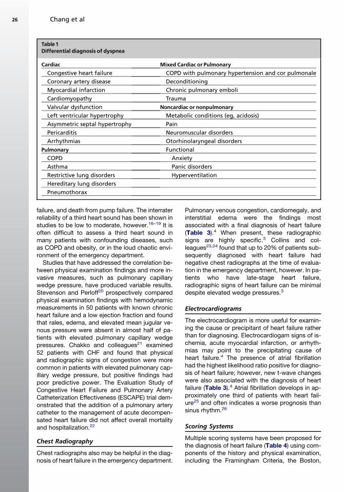
failure, and death from pump failure. The interraterreliability of a third heart sound has been shown instudies to be low to moderate, however.16–19 It isoften difficult to assess a third heart sound inmany patients with confounding diseases, suchas COPD and obesity, or in the loud chaotic envi-ronment of the emergency department.
Studies that have addressed the correlation be-tween physical examination findings and more in-vasive measures, such as pulmonary capillarywedge pressure, have produced variable results.Stevenson and Perloff20 prospectively comparedphysical examination findings with hemodynamicmeasurements in 50 patients with known chronicheart failure and a low ejection fraction and foundthat rales, edema, and elevated mean jugular ve-nous pressure were absent in almost half of pa-tients with elevated pulmonary capillary wedgepressures. Chakko and colleagues21 examined52 patients with CHF and found that physicaland radiographic signs of congestion were morecommon in patients with elevated pulmonary cap-illary wedge pressure, but positive findings hadpoor predictive power. The Evaluation Study ofCongestive Heart Failure and Pulmonary ArteryCatheterization Effectiveness (ESCAPE) trial dem-onstrated that the addition of a pulmonary arterycatheter to the management of acute decompen-sated heart failure did not affect overall mortalityand hospitalization.22
Chest Radiography
Chest radiographs also may be helpful in the diag-nosis of heart failure in the emergency department.
Pulmonary venous congestion, cardiomegaly, andinterstitial edema were the findings mostassociated with a final diagnosis of heart failure(Table 3).4 When present, these radiographicsigns are highly specific.3 Collins and col-leagues23,24 found that up to 20% of patients sub-sequently diagnosed with heart failure hadnegative chest radiographs at the time of evalua-tion in the emergency department, however. In pa-tients who have late-stage heart failure,radiographic signs of heart failure can be minimaldespite elevated wedge pressures.3
Electrocardiograms
The electrocardiogram is more useful for examin-ing the cause or precipitant of heart failure ratherthan for diagnosing. Electrocardiogam signs of is-chemia, acute myocardial infarction, or arrhyth-mias may point to the precipitating cause ofheart failure.4 The presence of atrial fibrillationhad the highest likelihood ratio positive for diagno-sis of heart failure; however, new t-wave changeswere also associated with the diagnosis of heartfailure (Table 3).4 Atrial fibrillation develops in ap-proximately one third of patients with heart fail-ure25 and often indicates a worse prognosis thansinus rhythm.26
Scoring Systems
Multiple scoring systems have been proposed forthe diagnosis of heart failure (Table 4) using com-ponents of the history and physical examination,including the Framingham Criteria, the Boston,
Table1Differential diagnosis of dyspnea
Cardiac Mixed Cardiac or PulmonaryCongestive heart failure COPD with pulmonary hypertension and cor pulmonale
Coronary artery disease Deconditioning
Myocardial infarction Chronic pulmonary emboli
Cardiomyopathy Trauma
Valvular dysfunction Noncardiac or nonpulmonaryLeft ventricular hypertrophy Metabolic conditions (eg, acidosis)
Asymmetric septal hypertrophy Pain
Pericarditis Neuromuscular disorders
Arrhythmias Otorhinolaryngeal disorders
Pulmonary Functional
COPD Anxiety
Asthma Panic disorders
Restrictive lung disorders Hyperventilation
Hereditary lung disorders
Pneumothorax
Chang et al26
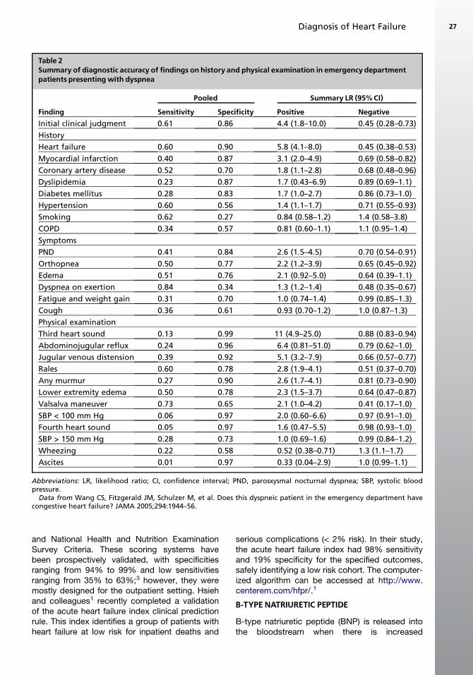
and National Health and Nutrition ExaminationSurvey Criteria. These scoring systems havebeen prospectively validated, with specificitiesranging from 94% to 99% and low sensitivitiesranging from 35% to 63%;3 however, they weremostly designed for the outpatient setting. Hsiehand colleagues1 recently completed a validationof the acute heart failure index clinical predictionrule. This index identifies a group of patients withheart failure at low risk for inpatient deaths and
serious complications (< 2% risk). In their study,the acute heart failure index had 98% sensitivityand 19% specificity for the specified outcomes,safely identifying a low risk cohort. The computer-ized algorithm can be accessed at http://www.centerem.com/hfpr/.1
B-TYPE NATRIURETIC PEPTIDE
B-type natriuretic peptide (BNP) is released intothe bloodstream when there is increased
Table 2Summary of diagnostic accuracy of findings on history and physical examination in emergency departmentpatients presentingwith dyspnea
Pooled Summary LR (95% CI)
Finding Sensitivity Specificity Positive NegativeInitial clinical judgment 0.61 0.86 4.4 (1.8–10.0) 0.45 (0.28–0.73)
History
Heart failure 0.60 0.90 5.8 (4.1–8.0) 0.45 (0.38–0.53)
Myocardial infarction 0.40 0.87 3.1 (2.0–4.9) 0.69 (0.58–0.82)
Coronary artery disease 0.52 0.70 1.8 (1.1–2.8) 0.68 (0.48–0.96)
Dyslipidemia 0.23 0.87 1.7 (0.43–6.9) 0.89 (0.69–1.1)
Diabetes mellitus 0.28 0.83 1.7 (1.0–2.7) 0.86 (0.73–1.0)
Hypertension 0.60 0.56 1.4 (1.1–1.7) 0.71 (0.55–0.93)
Smoking 0.62 0.27 0.84 (0.58–1.2) 1.4 (0.58–3.8)
COPD 0.34 0.57 0.81 (0.60–1.1) 1.1 (0.95–1.4)
Symptoms
PND 0.41 0.84 2.6 (1.5–4.5) 0.70 (0.54–0.91)
Orthopnea 0.50 0.77 2.2 (1.2–3.9) 0.65 (0.45–0.92)
Edema 0.51 0.76 2.1 (0.92–5.0) 0.64 (0.39–1.1)
Dyspnea on exertion 0.84 0.34 1.3 (1.2–1.4) 0.48 (0.35–0.67)
Fatigue and weight gain 0.31 0.70 1.0 (0.74–1.4) 0.99 (0.85–1.3)
Cough 0.36 0.61 0.93 (0.70–1.2) 1.0 (0.87–1.3)
Physical examination
Third heart sound 0.13 0.99 11 (4.9–25.0) 0.88 (0.83–0.94)
Abdominojugular reflux 0.24 0.96 6.4 (0.81–51.0) 0.79 (0.62–1.0)
Jugular venous distension 0.39 0.92 5.1 (3.2–7.9) 0.66 (0.57–0.77)
Rales 0.60 0.78 2.8 (1.9–4.1) 0.51 (0.37–0.70)
Any murmur 0.27 0.90 2.6 (1.7–4.1) 0.81 (0.73–0.90)
Lower extremity edema 0.50 0.78 2.3 (1.5–3.7) 0.64 (0.47–0.87)
Valsalva maneuver 0.73 0.65 2.1 (1.0–4.2) 0.41 (0.17–1.0)
SBP < 100 mm Hg 0.06 0.97 2.0 (0.60–6.6) 0.97 (0.91–1.0)
Fourth heart sound 0.05 0.97 1.6 (0.47–5.5) 0.98 (0.93–1.0)
SBP > 150 mm Hg 0.28 0.73 1.0 (0.69–1.6) 0.99 (0.84–1.2)
Wheezing 0.22 0.58 0.52 (0.38–0.71) 1.3 (1.1–1.7)
Ascites 0.01 0.97 0.33 (0.04–2.9) 1.0 (0.99–1.1)
Abbreviations: LR, likelihood ratio; CI, confidence interval; PND, parosxysmal nocturnal dyspnea; SBP, systolic bloodpressure.
Data from Wang CS, Fitzgerald JM, Schulzer M, et al. Does this dyspneic patient in the emergency department havecongestive heart failure? JAMA 2005;294:1944–56.
Diagnosis of Heart Failure 27
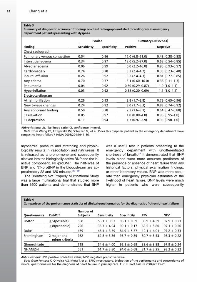
myocardial pressure and stretching and physio-logically results in vasodilation and natriuresis. Itis released as a prohormone and subsequentlycleaved into the biologically active BNP and the in-active component, NT-proBNP. The half-lives ofBNP and NT-proBNP in the bloodstream are ap-proximately 22 and 120 minutes.27–30
The Breathing Not Properly Multinational Studywas a large multinational trial that studied morethan 1500 patients and demonstrated that BNP
was a useful test in patients presenting to theemergency department with undifferentiatedshortness of breath.27 It demonstrated that BNPlevels alone were more accurate predictors ofthe presence or absence of heart failure than anyhistorical factors, physical examination findings,or other laboratory values. BNP was more accu-rate than emergency physician estimates of thelikelihood of heart failure. BNP levels were muchhigher in patients who were subsequently
Table 3Summary of diagnostic accuracy of findings on chest radiograph and electrocardiogram in emergencydepartment patients presentingwith dyspnea
Pooled Summary LR (95% CI)
Finding Sensitivity Specificity Positive NegativeChest radiograph
Pulmonary venous congestion 0.54 0.96 12.0 (6.8–21.0) 0.48 (0.28–0.83)
Interstitial edema 0.34 0.97 12.0 (5.2–27.0) 0.68 (0.54–0.85)
Alveolar edema 0.06 0.99 6.0 (2.2–16.0) 0.95 (0.93–0.97)
Cardiomegaly 0.74 0.78 3.3 (2.4–4.7) 0.33 (0.23–0.48)
Pleural effusion 0.26 0.92 3.2 (2.4–4.3) 0.81 (0.77–0.85)
Any edema 0.70 0.77 3.1 (0.60–16.0) 0.38 (0.11–1.3)
Pneumonia 0.04 0.92 0.50 (0.29–0.87) 1.0 (1.0–1.1)
Hyperinflation 0.03 0.92 0.38 (0.20–0.69) 1.1 (1.0–1.1)
Electrocardiogram
Atrial fibrillation 0.26 0.93 3.8 (1.7–8.8) 0.79 (0.65–0.96)
New t-wave changes 0.24 0.92 3.0 (1.7–5.3) 0.83 (0.74–0.92)
Any abnormal finding 0.50 0.78 2.2 (1.6–3.1) 0.64 (0.47–0.88)
ST elevation 0.05 0.97 1.8 (0.80–4.0) 0.96 (0.95–1.0)
ST depression 0.11 0.94 1.7 (0.97–2.9) 0.95 (0.90–1.0)
Abbreviations: LR, likelihood ratio; CI, confidence interval.Data from Wang CS, Fitzgerald JM, Schulzer M, et al. Does this dyspneic patient in the emergency department have
congestive heart failure? JAMA 2005;294:1944–56.
Table 4Comparison of the performance statistics of clinical questionnaires for the diagnosis of chronic heart failure
Questionnaire Cut-OffNumber ofSubjects Sensitivity Specificity PPV NPV
Boston R5(possible) 568 55.1 � 3.93 96.1 � 0.59 38.9 � 4.39 97.9 � 0.23
R8(probable) 296 35.3 � 4.04 99.1 � 0.17 63.5 � 5.80 97.1 � 0.26
Duke 469 46.1 � 3.59 84.9 � 5.57 12.1 � 4.01 97.2 � 0.33
Framingham 2 major andminor criteria
982 62.8 � 3.86 93.7 � 0.89 30.7 � 3.53 98.3 � 0.22
Gheorghiade 718 54.6 � 4.00 95.1 � 0.69 33.6 � 3.88 97.9 � 0.24
NHANES-I 551 61.7 � 3.80 94.0 � 0.68 31.7 � 3.25 98.2 � 0.22
Abbreviations: PPV, positive predictive value; NPV, negative predictive value.Data from Fonseca C, Oliveira AG, Mota T, et al. EPIC investigators. Evaluation of the performance and concordance of
clinical questionnaires for the diagnosis of heart failure in primary care. Eur J Heart Failure 2004;6:813–20.
Chang et al28

diagnosed with heart failure than in patients diag-nosed with noncardiac dyspnea (675 pg/mL ver-sus 110 pg/mL) (Fig. 1). A BNP value of morethan 500 pg/mL is highly suggestive of heart fail-ure. The negative predictive value of BNP at levelsless than 50 pg/mL was very high (96%).27 Pa-tients with systolic dysfunction also had higherlevels of BNP compared with patients with dia-stolic dysfunction.28
McCullough and colleagues29 did a subset anal-ysis from the Breathing Not Properly Study inwhich physicians were asked to assess the proba-bility of heart failure as the leading diagnosis forthe patients’ dyspnea. The addition of BNP levelsinto the clinical evaluation of HF raised the diag-nostic accuracy by 10% in patients for whom theemergency department physician had a high con-fidence in the diagnosis of heart failure. In the onethird of patients for whom the emergency depart-ment physician was uncertain of the diagnosis(20%–80% pretest probably of CHF diagnosis),the addition of BNP to clinical judgment correctlyclassified 74% of the patients and only misclassi-fied 7% of the patients as not having CHF whenthe final diagnosis was CHF (Fig. 2).
The Rapid Emergency Department Heart FailureOutpatient Trial (REDHOT) showed a disconnectbetween physician assessment of heart failure se-verity and BNP levels.31 In the first phase, 464 pa-tients visiting emergency departments withcomplaints of breathing difficulty had BNP mea-surements taken on arrival. Physicians wereblinded to BNP results; however, inclusion in thetrial required a BNP of more than 100 pg/mL. Pa-tients discharged from the emergency departmenthad higher BNP levels than patients admitted tothe hospital (976 pg/mL versus 766 pg/mL). Withrespect to the admitted patients, 11% had BNPlevels less than 200 pg/mL, which is indicative ofa non–heart failure diagnosis or less severe CHF.
Most of these patients were perceived to haveclass III or IV heart failure. Mortality rates for thesepatients were 0% at 30 days and only 2% at 90days, suggesting that patients with low levels ofBNP might have been safe for discharge. With re-spect to patients who were actually discharged,78% had BNP levels more than 400 pg/mL. At90 days, mortality was 9%. There was no mortalityof patients discharged with BNP levels less than400 pg/mL, which suggests that use of BNP inthe emergency department might also help deter-mine which well-appearing patients are at high riskfor a bad outcome over the short-term (90 days).The finding also suggests that when a clinicianthinks a patient is safe for discharge but the BNPis more than 400 pg/mL, the clinician may wishto reconsider the disposition decision. Almostone in ten patients with these characteristicswere dead by 90 days. They concluded that BNPlevels may be helpful to physicians in making tri-age decisions about whether to admit or dischargepatients.31
Elevations of BNP are also useful for assessingrisk stratification and prognosis in patients withheart failure. BNP levels are related to changes inlimitations of physical activities and functional sta-tus. Harrison and colleagues32 followed 325 pa-tients for 6 months after an index visit to theemergency department for dyspnea. Higher BNPlevels were associated with a progressively worseprognosis. In the ESCAPE trial, patients with BNPof more than 1500 pg/mL had greater mortalityand longer length of stay than patients with BNPof less than 500 pg/mL.33
Mueller and colleagues34 conducted the B-typeNatriuretic Peptide for Acute Shortness of BreathEvaluation (BASEL) study. Patients were randomlyassigned to receive a BNP measurement in theemergency department or not. The use of BNPlevels reduced the need for hospitalization and
0102030405060708090
100
No CHF Hx of CHF,no acute
exacerbation
CHF exacerbation
BN
P levels
Fig. 1. Results of the Breathing Not Prop-erly Study. Patients with a diagnosis ofacute congestive heart failure had mean(� SD) B-type natriuretic peptide levelsof 675 pg/mL, whereas patients withoutcongestive heart failure had B-type natri-uretic peptide levels of 110 pg/mL. The72 patients who had baseline ventriculardysfunction without an acute exacerba-tion had a mean B-type natriuretic pep-tide level of 346 pg/mL. (Data fromMaisel AS, Krishnaswamy P, Nowak RM,et al. Rapid measurement of B-type natri-uretic peptide in the emergency diagnosisof heart failure. N Engl J Med 2002;347:161–7.)
Diagnosis of Heart Failure 29
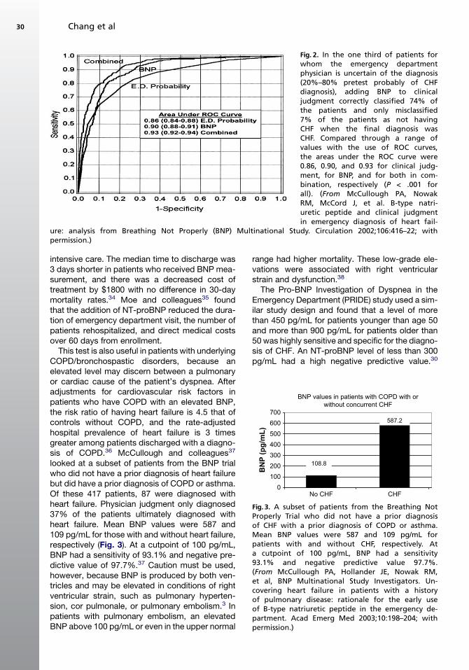
intensive care. The median time to discharge was3 days shorter in patients who received BNP mea-surement, and there was a decreased cost oftreatment by $1800 with no difference in 30-daymortality rates.34 Moe and colleagues35 foundthat the addition of NT-proBNP reduced the dura-tion of emergency department visit, the number ofpatients rehospitalized, and direct medical costsover 60 days from enrollment.
This test is also useful in patients with underlyingCOPD/bronchospastic disorders, because anelevated level may discern between a pulmonaryor cardiac cause of the patient’s dyspnea. Afteradjustments for cardiovascular risk factors inpatients who have COPD with an elevated BNP,the risk ratio of having heart failure is 4.5 that ofcontrols without COPD, and the rate-adjustedhospital prevalence of heart failure is 3 timesgreater among patients discharged with a diagno-sis of COPD.36 McCullough and colleagues37
looked at a subset of patients from the BNP trialwho did not have a prior diagnosis of heart failurebut did have a prior diagnosis of COPD or asthma.Of these 417 patients, 87 were diagnosed withheart failure. Physician judgment only diagnosed37% of the patients ultimately diagnosed withheart failure. Mean BNP values were 587 and109 pg/mL for those with and without heart failure,respectively (Fig. 3). At a cutpoint of 100 pg/mL,BNP had a sensitivity of 93.1% and negative pre-dictive value of 97.7%.37 Caution must be used,however, because BNP is produced by both ven-tricles and may be elevated in conditions of rightventricular strain, such as pulmonary hyperten-sion, cor pulmonale, or pulmonary embolism.3 Inpatients with pulmonary embolism, an elevatedBNP above 100 pg/mL or even in the upper normal
range had higher mortality. These low-grade ele-vations were associated with right ventricularstrain and dysfunction.38
The Pro-BNP Investigation of Dyspnea in theEmergency Department (PRIDE) study used a sim-ilar study design and found that a level of morethan 450 pg/mL for patients younger than age 50and more than 900 pg/mL for patients older than50 was highly sensitive and specific for the diagno-sis of CHF. An NT-proBNP level of less than 300pg/mL had a high negative predictive value.30
Fig. 2. In the one third of patients forwhom the emergency departmentphysician is uncertain of the diagnosis(20%–80% pretest probably of CHFdiagnosis), adding BNP to clinicaljudgment correctly classified 74% ofthe patients and only misclassified7% of the patients as not havingCHF when the final diagnosis wasCHF. Compared through a range ofvalues with the use of ROC curves,the areas under the ROC curve were0.86, 0.90, and 0.93 for clinical judg-ment, for BNP, and for both in com-bination, respectively (P < .001 forall). (From McCullough PA, NowakRM, McCord J, et al. B-type natri-uretic peptide and clinical judgmentin emergency diagnosis of heart fail-
ure: analysis from Breathing Not Properly (BNP) Multinational Study. Circulation 2002;106:416–22; withpermission.)
BNP values in patients with COPD with orwithout concurrent CHF
CHF0
100200300400500600700
No CHF
BN
P (p
g/m
L)
108.8
587.2
Fig. 3. A subset of patients from the Breathing NotProperly Trial who did not have a prior diagnosisof CHF with a prior diagnosis of COPD or asthma.Mean BNP values were 587 and 109 pg/mL forpatients with and without CHF, respectively. Ata cutpoint of 100 pg/mL, BNP had a sensitivity93.1% and negative predictive value 97.7%.(From McCullough PA, Hollander JE, Nowak RM,et al, BNP Multinational Study Investigators. Un-covering heart failure in patients with a historyof pulmonary disease: rationale for the early useof B-type natriuretic peptide in the emergency de-partment. Acad Emerg Med 2003;10:198–204; withpermission.)
Chang et al30

The International Collaborative of NT-proBNPStudy took the data from the PRIDE study andcombined it with multiple international sites andconfirmed that NT-proBNP was useful in the diag-nostic evaluation of acute heart failure.39
BNP and NT-proBNP must be used in conjunc-tion with physician judgment and other find-ings.29,30 Further studies and subset analysisshow that NP (natriuretic peptides; either BNP orNTproBNP) are affected by a multitude of factors.NP levels are not affected by the presence of dia-betes40,41 but are lower in overweight and obeseindividuals, and the test loses sensitivity in thispopulation.42–44 NPs have ethnic and age differ-ences;45,46 however, BNP is still effective in de-creasing time to discharge and total cost oftreatment in women and elderly patients.46–48
NPs are at least partially renally cleared, which im-pacts their clinical utility. For BNP, some re-searchers have suggested using a higher cutoff(200 pg/mL) value for patients with a glomerular fil-tration rate of less than 60 mL/min.27 Studies haveshown that a reduced baseline glomerular filtrationrate is associated with decreased survival in pa-tients with systolic heart failure.49 It is unclearwhether this is caused by poor kidney perfusionsecondary to low cardiac output or if it is part ofthe spectrum of cardiorenal syndrome.32 BNP isuseful in patients with end-stage renal diseaseon dialysis. It has been shown that levels candrop 20% to 40% after a dialysis session, andBNP could be used as an index of intravascularvolume.49–51
Limited data directly comparing BNP toNTproBNP are available. Heeschen and col-leagues52 completed a head-to-head comparisonof BNP to NT-proBNP for the diagnosis of heart
failure. In their study, age, sex, and renal functionhad no impact on the diagnostic utility of bothtests when compared by logistic regressionmodels. BNP and NT-proBNP were found to beequally sensitive and specific52,53
PHONOCARDIOGRAPHY
Auscultatory assessment of the S3 heart sound isdifficult in the emergency setting. Even when opti-mal, interobserver concordance is low.16–19 Pho-noelectrocardiographic devices can potentiallyimprove detection of an S3 or S4 heart soundcompared with auscultation. The Audicor systemis an acoustic cardiogram that replaces the stan-dard V3 and V4 leads with sensors and collectssound and electrical data. Sound data are col-lected at 10-second intervals and analyzed usinga signal-processing algorithm that has been previ-ously validated.54,55 Peacock and colleagues55
studied the device and found that in patients withindeterminate BNP levels (100–500 pg/mL), thepresence of an S3 increased the positive likelihoodratio to 4.3 for the diagnosis of CHF. Shapiro andcolleagues54 conducted a more invasive studythat compared the results of the BNP and comput-erized phonoelectrocardiographic system to car-diac catheterization and found that it was 88%accurate for diagnosis of left ventricular dysfunc-tion. These studies are small, however, and moredata are needed before the usefulness or clinicalimpact of the Audicor system can be determined.
IMPEDANCEMONITORS
Impedance cardiography is a noninvasive mea-surement of cardiac output, cardiac index, andthoracic fluid content.56,57 Electrical impedance
BNP and Troponin Groups
0
2
4
6
8
10
12
BNP <840, Tn -,n=20439
BNP >840, Tn -,n=19827
BNP <840 Tn +,n=734
BNP > 840 Tn +,n=1636
In h
ospi
tal m
orta
lity,
%
Fig. 4. In-hospital mortality rates (unadjusted). (From Fonarow GC, Peacock WF, Phillips CO, et al, ADHERE Scien-tific Advisory Committee and Investigators. Usefulness of B-type natriuretic peptide and cardiac troponin levels topredict in-hospital mortality from ADHERE. Am J Cardiol 2008;101:231–7; with permission.)
Diagnosis of Heart Failure 31
is defined as the resistance to flow of an electricalcurrent. Bone and tissue are poor conductors,whereas blood and fluids are good conductorswith lower impedance.5 The information madeavailable from these devices has been shown tohelp delineate a diagnosis in the emergency de-partment, identify patients at increased near-term risk of recurrent decompensation, andpredict rehospitalization.56
Impedance cardiography monitors can be usedat the bedside. Impedance is determined by ap-plying four pairs of electrodes to the chest andneck. Because electrical impedance changes pro-portionally to fluid flow, the machine may thencalculate several hemodynamic parameters, in-cluding stroke volume, systemic vascular resis-tance, cardiac output, and an index of thoracicfluid. The increase in thoracic fluid that occurs dur-ing a heart failure exacerbation decreases overallaverage impedance and can suggest a fluid over-loaded state.5,56,57
Implantable cardioverter/defibrillator devicesalso can monitor intrathoracic impedance.58–60
Yu and colleagues59 conducted a study using theOptivol intrathoracic impedance device (Med-tronic, Inc.) in NYHA class III and IV patients andconfirmed that there was an inverse correlationbetween intrathoracic impedance and intracardiacfilling pressure. The device recorded measure-ments starting at 34 days postimplantation and av-eraged a total of 64 measurements each daybetween 12PM and 5PM. Impedance measure-ments decreased (associated with increased tho-racic fluid) an average of 2 weeks beforesymptom onset and hospitalization, suggestingthat reporting of these data may make it feasiblefor physicians to intervene before symptom on-set.60 The default setting for the OptiVol systemhad a 76.9% sensitivity for predicting hospitaliza-tion.60 Vollman and colleagues61 found that thedevice had a sensitivity and positive predictivevalue of 60% to detect heart failure deteriorationbut had a 40% false-positive rate. One studyshowed that the combination of weight gain anda positive BNP had a sensitivity of 55%.61 This de-vice may not have more diagnostic accuracy butmight allow earlier detection. Larger randomizedcontrolled trials are needed to determine their clin-ical use.
CARDIAC TROPONIN I
Up to 40% of patients admitted for acute decom-pensated heart failure present with an elevatedtroponin level.62–70 Although troponin is useful inthe setting of acute coronary syndrome and canhelp identify acute coronary syndrome as the
precipitant of heart failure, it is also useful for riskstratification of patients who have heart failure inthe absence of ACS. An elevated cardiac troponinI level is a predictor of 30-day and 1-year mortality,even after adjusting for confounding factors. TheESCAPE and ADHERE trials showed that a multi-marker strategy (BNP and troponin) for the assess-ment of patients hospitalized with heart failureadds incremental prognostic information.33,67,68
In the ADHERE trial, troponin was increased in5.6% of 42,636 heart failure episodes. A combina-tion of a BNP above the median (> 840 pg/mL) withincreased troponin was associated with twice thein-hospital mortality (Fig. 4). After covariate adjust-ment, mortality was twice as high for patients withelevated BNP and troponin compared with pa-tients with solely elevated BNP.67
SUMMARY
The approach to the diagnosis of heart failure iscomplex, but the diagnostic armamentarium hasincreased significantly in the past decade. Diag-nostic markers such as BNP and NT proBNPhave proven value for the diagnosis of heart failureover and above the traditional tools that only in-cluded the history, physical examination, andchest radiography. Invasive and noninvasive im-pedance cardiography can be used to diagnoseor even predict development of heart failure, buttheir role in clinical practice still needs to be betterdefined.
REFERENCES
1. Hsieh M, Auble TE, Yealy DM. Validation of the acute
heart failure index. Ann Emerg Med 2008;51:37–44.
2. Mueller C, Laule-Kilian K, Frana B, et al. Use of
B-type natriuretic peptide in the management of
acute dyspnea in patients with pulmonary disease.
Am Heart J 2006;151:471–7.
3. Collins S, Storrow AB, Kirk JD, et al. Beyond pulmo-
nary edema: diagnostic, risk stratification, and treat-
ment challenges of acute heart failure management
in the emergency department. Ann Emerg Med
2008;51:45–57.
4. Wang CS, Fitzgerald JM, Schulzer M, et al. Does this
dyspneic patient in the emergency department have
congestive heart failure? J Am Med Assoc 2005;294:
1944–56.
5. Wong GC, Ayas NT. Clinical approaches to the diag-
nosis of acute heart failure. Curr Opin Cardiol 2007;
22:207–13.
6. Tsuyuki RT, McKelvie RS, Arnold JM, et al. Acute
precipitants of congestive heart failure exacerba-
tions. Arch Intern Med 2001;161:2337–42.
Chang et al32
7. Levy D, Larson MG, Vasan RS, et al. The progres-
sion from hypertension to congestive heart failure.
J Am Med Assoc 1996;275:1557–62.
8. Chen YT, Vaccarino V, Williams CS, et al. Risk factors
for heart failure in the elderly: a prospective commu-
nity-based study. Am J Med 1999;106:605–12.
9. Chae CU, Pfeffer MA, Glynn RJ, et al. Increased
pulse pressure and risk of heart failure in the elderly.
JAMA 1999;28:634–9.
10. Remes J, Miettinen H, Reunanen A, et al. Validity of
clinical diagnosis of heart failure in primary health
care. Eur Heart J;199(12):315–21.
11. Drazner MH, Rame E, Stevenson LW, et al. Prognos-
tic importance of elevated jugular venous pressure
and a third heart sound in patients with heart failure.
N Engl J Med 2001;345:574–81.
12. Drazner MH, Hamilton MA, Fonarow G, et al. Rela-
tionship between right and left-sided filling pres-
sures in 1000 patients with advanced heart failure.
J Heart Lung Transplant 1999;18:1126–32.
13. Collins SP, Lindsell CJ, Peacock WF, et al. The com-
bined utility of an S3 heart sound and B-type natri-
uretic peptide levels in emergency department
patients with dyspnea. J Card Fail 2006;12:286–92.
14. Collins SP, Lindsell CJ, Peacock WF, et al. The effect
of treatment on the presence of abnormal heart
sounds in emergency department patients with
heart failure. Am J Emerg Med 2006;24:25–32.
15. Butman SM, Ewy GA, Standen JR, et al. Bedside
cardiovascular examination in patients with severe
chronic heart failure: importance of rest or inducible
jugular venous distension. J Am Coll Cardiol 1993;
22:968–74.
16. Marcus GM, Vessey J, Jordan MV, et al. Relationship
between accurate auscultation of a clinically useful
third heart sound and level of experience. Arch In-
tern Med 2006;166:617–22.
17. Marcus GM, Michaels AD, De Marco TD, et al. Use-
fulness of the third heart sound in predicting an ele-
vated level of B-type natriuretic peptide. Am
J Cardiol 2004;93:1312–3.
18. Ishmail AA, Wing S, Ferguson J, et al. Interobserver
agreement by auscultation in the presence of a third
heart sound in patients with congestive heart failure.
Chest 1987;91:870–3.
19. Mangione S, Neiman LZ. Cardiac auscultatory skills
of internal medicine and family practice trainees:
a comparison of diagnostic proficiency. JAMA
1997;278:717–22.
20. Stevenson LW, Perloff JK. The limited reliability of
physical signs for estimating hemodynamics in
chronic heart failure. JAMA 1989;261:884–8.
21. Chakko S, Woska D, Martinez H, et al. Clinical,
radiographic, and hemodynamic correlations in
chronic congestive heart failure: conflicting results
may lead to inappropriate care. Am J Med 1991;
90:353–9.
22. Binanay C, Califf RM, Hasselblad V, et al. ESCAPE
Investigators and ESCAPE Study Coordinators.
Evaluation study of congestive heart failure and
pulmonary artery catheterization effectiveness: the
escape trial. J Am Med Assoc 2005;294:1625–33.
23. Collins SP, Lindsell CJ, Abraham WT, et al. The
emergency department chest radiograph is unreli-
able in establishing a diagnosis of heart failure.
J Card Fail 2004;10:S30.
24. Collins S, Lindsell CJ, Storrow AB, et al. Prevalence
of negative chest radiography in the emergency de-
partment patient with decompensated heart failure.
Ann Emerg Med 2006;47:13–8.
25. Middlekauff HR, Stevenson WG, Stevenson LW.
Prognostic significance of a trial fibrillation in
advanced heart failure: a study of 390 patients. Cir-
culation 1991;84:40–8.
26. Mathew J, Hunsberger S, Fleg J, et al. Incidence,
predictive factors, and prognostic significance of
supraventricular tachyarrhythmias in congestive
heart failure. Chest 2000;118:914–22.
27. Maisel AS, Krishnaswamy P, Nowak RM, et al.
Breathing Not Properly Multinational Study Investi-
gators. Rapid measurement of b-type natriuretic
peptide in the emergency diagnosis of heart failure.
N Engl J Med 2002;347:161–7.
28. Maisel AS, McCord J, Nowak RM, et al. Breathing
Not Properly Multinational Study Investigators. Bed-
side b-type natriuretic peptide in the emergency
diagnosis of heart failure with reduced or preserved
ejection fraction: results from the Breathing Not
Properly Multinational Study. J Am Coll Cardiol
2003;41:2010–7.
29. McCullough PA, Nowak RM, McCord J, et al. for the
BNP Multinational Study Investigators. B-type natri-
uretic peptide and clinical judgment in emergency di-
agnosis of heart failure. Circulation 2002;106:416–22.
30. Januzzi JL Jr, Camargo CA, Anwaruddin S, et al.
The N-terminal pro-BNP investigation of dyspnea in
the emergency department (PRIDE) study. Am
J Cardiol 2005;95:948–54.
31. Maisel AS, Hollander JE, Guss D, et al. Rapid Emer-
gency Department Heart Failure Outpatient Trial In-
vestigators. Primary results of the rapid emergency
department heart failure outpatient trial (redhot):
a multicenter study of b-type natriuretic peptide
levels, emergency department decision making,
and outcomes in patients presenting with shortness
of breath. J Am Coll Cardiol 2004;44:1328–33.
32. Harrison A, Morrison LK, Krishnaswamy P, et al.
B-type natriuretic peptide (BNP) predicts future car-
diac events in patients presenting to the emergency
department with dyspnea. Ann Emerg Med 2002;
39:131–8.
33. Shah MR, Hasselblad V, Tasissa G, et al. Rapid
assay brain natriuretic peptide and troponin I in
patients hospitalized with decompensated heart
Diagnosis of Heart Failure 33
failure. From the evaluation study of congestive
heart failure and pulmonary artery catheterization ef-
fectiveness trial. Am J Cardiol 2007;100:1427–33.
34. Mueller C, Scholer A, Laule-Kilian K, et al. Use of
B-type natriuretic peptide in the evaluation and man-
agement of acute dyspnea. N Engl J Med 2004;350:
647–54.
35. Moe GW, Howlett J, Januzzi JL, et al. Canadian Mul-
ticenter Improved Management of Patients with
Congestive Heart Failure (improve-chf) Study Inves-
tigators. N-terminal pro-b-type natriuretic peptide
testing improves the management of patients
with suspected acute heart failure: primary results
of the Canadian prospective randomized multi-
center improve-chf study. Circulation 2007;115:
3103–10.
36. LeJemtel TH, Padeletti M, Jelic S. Diagnostic and
therapeutic challenges in patients with coexistent
chronic obstructive pulmonary disease and chronic
heart failure. J Am Coll Cardiol 2007;49:171–80.
37. McCullough PA, Hollander JE, Nowak RM, et al. BNP
Multinational Study Investigators. Uncovering heart
failure in patients with a history of pulmonary dis-
ease: rationale for the early use of b-type natriuretic
peptide in the emergency department. Acad Emerg
Med 2003;10:198–204.
38. Wolde M, Tulevski II, Mulder JW, et al. Brain natri-
uretic peptide as a predictor of adverse outcome
in patients with pulmonary embolism. Circulation
2003;107:2082–4.
39. Januzzi JL, Van Kimmenade R, Lainchbury J, et al.
NT-proBNP testing for diagnosis and short-term
prognosis in acute destabilized heart failure: an
international pooled analysis of 1256 patients. The
international collaborative of NT-proBNP study. Eur
Heart J 2006;27:330–7.
40. Wu AH, Omland T, Duc P, et al. Breathing Not Prop-
erly Multinational Study Investigators. The effect of
diabetes on B-type natriuretic peptide concentra-
tions in patients with acute dyspnea: an analysis
from the Breathing Not Properly Multinational Study.
Diabetes Care 2004;27:2398–404.
41. O’Donoghue M, Kenney P, Oestreicher E, et al. Use-
fulness of aminoterminal pro-brain natriuretic pep-
tide testing for the diagnostic and prognostic
evaluation of dyspneic patients with diabetes melli-
tus seen in the emergency department (from the
PRIDE Study). Am J Cardiol 2007;100:1336–40.
42. McCord J, Mundy BJ, Hudson MP, et al, for the BNP
Multinational Study Investigators. The relationship
between obesity and b-type natriuretic peptide
levels. Arch Intern Med 2004;164:2247–52.
43. Mehra MR, Uber PA, Park MH, et al. Obesity and
suppressed B-type natriuretic peptide levels in heart
failure. J Am Coll Cardiol 2004;43:1590–5.
44. Krauser DG, Lloyd-Jones DM, Chae CU, et al. Effect
of body mass index on natriuretic peptide levels in
patients with acute congestive heart failure:
a proBNP investigation of dyspnea in the emer-
gency department (PRIDE) substudy. Am Heart J
2005;149(4):744–50.
45. Daniels LB, Bhalla V, Clopton P, et al. B-type natri-
uretic peptide (BNP) levels and ethnic disparities
in perceived severity of heart failure: results from
the rapid emergency department heart failure out-
patient trial (REDHOT) multicenter study of BNP
levels and emergency department decision making
in patients presenting with shortness of breath.
J Card Fail 2006;12:281–5.
46. Mueller C, Laule-Kilian K, Frana B, et al. The use of
B-type natriuretic peptide in the management of el-
derly patients with acute dyspnoea. J Intern Med
2005;258:77–85.
47. Weber M, Hamm C. Role of B-type natriuretic pep-
tide (BNP) and NT-proBNP in clinical routine. Heart
2006;92:843–9.
48. Mueller C, Laule-Kilian K, Scholer A, et al. Use of
B-type natriuretic peptide for the management of
women with dyspnea. Am J Cardiol 2004;94:1510–4.
49. Dries DL, Exner DV, Domanski MJ, et al. The prog-
nostic implications of renal insufficiency in asymp-
tomatic and symptomatic patients with left
ventricular systolic dysfunction. J Am Coll Cardiol
2000;35:681–9.
50. Heywood JT, Fonarow GC, Costanzo MR, et al. AD-
HERE Scientific Advisory Committee and Investiga-
tors. High prevalence of renal dysfunction and its
impact on outcome in 118,465 patients hospitalized
with acute decompensated heart failure: a report
from the adhere database. J Card Fail 2007;13:
422–30.
51. Akiba T, Tachibana K, Togashi K, et al. Plasma hu-
man brain natriuretic peptide in chronic renal failure.
Clin Nephrol 1995;44(Suppl 1):S61–4.
52. Heeschen C, Hamm CW, Mitrovic V, et al. Platelet
Receptor Inhibition in Ischemic Syndrome Manage-
ment (PRISM) Investigators. Diagnostic accuracy
of b type natriuretic peptide and amino terminal
proBNP in the emergency diagnosis of heart failure.
Heart 2005;91:606–12.
53. National Kidney Foundation. Clinical practice guide-
lines for chronic kidney disease: evaluation, classifi-
cation, and stratification. Am J Kidney Dis 2002;
2(Suppl 1):S46–75.
54. Shapiro M, Moyers B, Marcus GM, et al. Diagnostic
characteristics of combining phonocardiographic
third heart sound and systolic time intervals for the
prediction of left ventricular dysfunction. J Card
Fail 2007;13:18–24.
55. Peacock WF, Harrison A, Moffa D. Clinical and eco-
nomic benefits of using audicor s3 detection for di-
agnosis and treatment of acute decompensated
heart failure. Congest Heart Fail 2006;12(Suppl 1):
32–6.
Chang et al34

56. Packer M, Abraham WT, Mehra MR, et al. Prospec-
tive evaluation and identification of cardiac decom-
pensation by icg test (predict) study investigators
and coordinators utility of impedance cardiography
for the identification of short-term risk of clinical de-
compensation in stable patients with chronic heart
failure. J Am Coll Cardiol. 2006;47:2245–52.
57. Peacock WF, Summers RL, Vogel J, et al. Impact of
impedance cardiography on diagnosis and therapy
of emergent dyspnea: the ed-impact trial. Acad
Emerg Med 2006;13:365–71.
58. Yamokoski LM, Haas GJ, Gans B, et al. Optivol fluid
status monitoring with an implantable cardiac de-
vice: a heart failure management system. Expert
Rev Med Devices 2007;4:775–80.
59. Yu CM, Wang L, Chau E, et al. Intrathoracic imped-
ance monitoring in patients with heart failure: correla-
tion with fluid status and feasibility of early warning
preceding hospitalization. Circulation 2005;112:
841–8.
60. Wang L. Fundamentals of intrathoracic impedance
monitoring in heart failure. Am J Cardiol 2007;99:
3G–10G.
61. VollmannD,NageleH,Schauerte P, etal.European IN-
SYNC Sentry Observational Study Investigators. Clin-
ical utility of intrathoracic impedance monitoring to
alert patients with an implanted device of deteriorating
chronic heart failure. Eur Heart J 2007;28:1835–40.
62. Kuwabara Y, Sato Y, Miyamoto T, et al. Persistently
increased serum concentrations of cardiac troponin
in patients with acutely decompensated heart failure
are predictive of adverse outcomes. Circ J 2007;71:
1047–51.
63. Heeschen C, Hamm CW, Mitrovic V, et al. Platelet
Receptor Inhibition in Ischemic Syndrome
Management (PRISM) Investigators. N-terminal
pro-B-type natriuretic peptide levels for dynamic
risk stratification of patients with acute coronary syn-
dromes. Circulation 2004;110:3206–12.
64. You JJ, Austin PC, Alter DA, et al. Relation between
cardiac troponin I and mortality in acute decompen-
sated heart failure. Am Heart J 2007;153:462–70.
65. Sato Y, Kita T, Takatsu Y, et al. Biochemical markers
of myocyte injury in heart failure. Heart 2004;90:
1110–3.
66. Ishii J, Nomura M, Nakamura Y, et al. Risk stratifica-
tion using a combination of cardiac troponin T and
brain natriuretic peptide in patients hospitalized for
worsening chronic heart failure. Am J Cardiol 2002;
89:691–5.
67. Fonarow GC, Peacock WF, Phillips CO, et al.
ADHERE Scientific Advisory Committee and
Investigators. Usefulness of B-type natriuretic pep-
tide and cardiac troponin levels to predict in-hospital
mortality from ADHERE. Am J Cardiol 2008;101:
231–7.
68. Fonarow GC, Peacock WF, Phillips CO, et al.
ADHERE Scientific Advisory Committee and Investi-
gators. Admission B-type natriuretic peptide levels
and in-hospital mortality in acute decompensated
heart failure. J Am Coll Cardiol 2007;49:1943–50.
69. Horwich TB, Patel J, MacLellan WR, et al. Cardiac
troponin I is associated with impaired hemodynam-
ics, progressive left ventricular dysfunction, and
increased mortality rates in advanced heart failure.
Circulation 2003;108:833–8.
70. Demir M, Kanadasi M, Akpinar O, et al. Cardiac tro-
ponin T as a prognostic marker in patients with heart
failure: a 3-year outcome study. Angiology 2007;58:
603–9.
Diagnosis of Heart Failure 35

Emergency DepartmentStabilization of HeartFailurePreeti Jois-Bilowich, MDa,*, Deborah Diercks, MD, MScb
Patients who have heart failure (HF) make up a clin-ically diverse population. They are a heterogenousgroup that has multiple complicating comorbities,various etiologies of HF, and differing pathophysi-ologic triggers resulting in acute decompensa-tion.1,2 Increased understanding of the diversityof HF patients has led to new insights in the emer-gent management of these patients. Physiciansand researchers are re-evaluating the propertiesof intravenous diuretics, vasodilators, and ino-tropes commonly used to alleviate congestionand restore hemodynamic stability. In particular,the shift has been to re-examine how these thera-pies should be administered, which HF patientsshould receive them, and the consequences ofthese therapeutic decisions.
CLASSIFICATION OF HEART FAILURE
HF patient types have not been well described ortailored with specific treatment strategies in pro-spective randomized studies. The selection of ex-isting treatments tends to be empiric due to thepaucity of randomized clinical trial data. In addi-tion, HF trials have largely focused on enrollingsubjects based on prespecified ejection fractioncriteria.3 Some of the HF-specific treatments,when used without caution, may result in myocar-dial injury,4 impaired renal function,5 and in-creased mortality risk,6 further complicatingtherapeutic decisions.
The European Society of Cardiology guidelineswere the first to classify patients who have HFinto distinct clinical conditions.7 These guidelinesclassified patients into clinical conditions basedon symptoms and hemodynamic parameters.Despite the publication of these guidelines, thereare no inclusive, evidence-, or consensus-basedtreatment algorithms that address the individualtreatment needs of each type of HF patient, partic-ularly in the emergency department (ED) setting.Recommendations should focus on therapeuticmanagement, emphasizing the identification andmatching of HF patient types to specific treatmentstrategies. Management algorithms should sup-plement these recommendations.
Traditional methods of categorizing HF patienttypes use classification based on hemodynamiccharacteristics obtained through invasive monitor-ing at presentation and a clinical symptom profilethat suggests HF: peripheral edema, weight gain,fatigue, dyspnea due to pulmonary congestion,and history of HF.4,8–12 Although most EDs donot obtain hemodynamic parameters such as pul-monary capillary wedge pressure or cardiac out-put by way of invasive means, they do rely on aneasily obtainable parameter—blood pressure.
Patients can subsequently be classified intonormotensive, hypertensive, and hypotensive HF.Although the exact pathophysiology, clinical char-acteristics, and appropriate treatment options ofeach of these patient types has yet to be clarified,
a University of Florida, Gainesville, FL, USAb University of California–Davis Medical Center, Sacramento, CA, USA* Corresponding author. University of Florida, Department of Emergency Medicine, 1329 SW 16th Street, Room4270, Gainesville, FL 32610.E-mail address: [email protected] (P. Jois-Bilowich).
KEYWORDS� Heart failure � Dyspnea � Nitroglycerin � Diuretics� Ventilation � Vasodilators � Ultrafiltration � Interventions� Nesiritide � Morphine � Emergency department� Stabilization
Heart Failure Clin 5 (2009) 37–42doi:10.1016/j.hfc.2008.08.0061551-7136/08/$ – see front matter. Published by Elsevier Inc. he
artf
ailu
re.th
ecli
nics
.com
recommendations for the initial treatment, basedlargely on observational data and expert consen-sus, can be suggested. Signs, symptoms, andhemodynamic characteristics of the normotensiveand hypertensive groups are described in thefollowing paragraphs.
Normotensive Heart Failure
These patients may represent nearly half of the HFpopulation.2 Blood pressure is normal (systolicblood pressure range of 90–140 mm Hg), and thereis usually a history of progressive worsening ofchronic HF. In this group, symptoms and signs de-velop over days, and pulmonary and systemiccongestion (seen as jugular venous distensionand peripheral edema) are present. Ejection frac-tion is usually reduced. Management is often diffi-cult because many patients are refractory totherapy and continue to have signs of congestiondespite the initial improvement in symptoms. Insome patients, the clinical or radiographic signsof pulmonary congestion are not evident despiteelevated left ventricular filling pressures.2,4,7,13
These patients have acute decompensation asa result of their cardiac failure.14
Hypertensive Acute Heart Failure
Data from the Acute Decompensated Heart FailureNational Registry demonstrates that 50% of HFpatients have a systolic blood pressure greaterthan 140 mm Hg on presentation.2 These patientsare more likely to have diastolic dysfunction withpreserved left ventricular ejection fraction, aremore often women, and are older.1,4 Symptom on-set is generally acute, with severe dyspnea andsigns of end-organ hypoperfusion. Acute pulmo-nary edema is the hallmark of hypertensive HFand is usually auscultated on examination as ralesand identified on chest radiography as pulmonaryedema. The clinical target is systemic blood pres-sure control, with a focus on early, aggressive va-sodilation, more so than on diuresis. This holdsparticularly true when pulmonary congestion is re-lated to fluid maldistribution, rather than an in-crease in total fluid volume.4,8 These patientshave a syndrome that has been referred to as‘‘acute vascular failure,’’ and the initial treatmentin this group reflects that etiology.14
The novel concept of identifying and varyingtreatment based on systemic blood pressure ad-dresses the diversity of the presentation of HFthat is often seen in the ED. It is important tonote that this classification is not entirely inclusiveof all the challenges faced when evaluatingpatients who have HF but encompasses a largeproportion of the patients seen.12
MECHANISM OF SYMPTOMS IN HEART FAILUREAcute Decompensated Cardiac Heart Failure
In the euvolemic state, there is a well-definedbalance between the actions of the renin-angiotensin-aldosterone (RAA) system and thenatriuretic peptides that maintains fluid status.This perfect homeostasis, however, is lost in dis-ease states such as HF, in which the mechanismsof sodium and water retention far outweigh natri-uretic effects. In HF states, excess sodium andfluid retention occurs mainly within the extracellu-lar fluid volume space. This retention results in anequal increase of fluid volume in each of the inter-stitial and plasma spaces. Despite this rise in totalbody volume and, therefore, plasma volume (PV),the arterial filling pressure remains low, which inturn continues to stimulate retention of sodiumand water.15
Patients who have acute decompensated HFhave decreased cardiac reserve, and the acuteprocess occurs as progression of this state.Worsening cardiac contractility can be a result ofischemia, arrhythmias, inflammatory activation,or progressive deterioration in myocardialdysfunction due to the underlining mechanismcausing the HF process. Subjects who have poorcardiac contractility may also develop decompen-sation as a result of medication noncomplianceand may therefore not have further contractility im-pairment. The results of this event are worseningforward perfusion, increased left ventricular pres-sure, and alterations in the neurohormonal statesthat maintain fluid balance.14 Increased leftventricular filling pressures influence changes inneurohormonal activation, activation of geneexpression programs, and induction of myocte ap-optosis in HF patients. Through indirect activationof the RAA, adrenergic, and cytokine systems andby way of a direct effect on myocardial stretch,fluid accumulation fosters left ventricular remodel-ing. Increased intraventricular pressure can causecoronary hypoperfusion, leading to subendocar-dial ischemia and, thus, worsening cardiacfunction.16
One study looked at PV in acute HF patientscompared with normal subjects. The patientswho had acute HF had visible evidence of volumeoverload, such as peripheral edema, jugular ve-nous distention, and ascites; they also had PVmeasurements that were 34% higher comparedwith healthy subjects.17
Feigenbaum and colleagues18 looked at PV inHF patients undergoing treatment and founda 23% PV contraction in patients treated with di-uretics. They concluded that standard drug ther-apy may lead to a contracted PV in chronic HF
Jois-Bilowich & Diercks38

patients, highlighting an important segment of theHF population in whom PV is contracted eventhough the overall extracellular fluid volume is ele-vated. In these patients, therapy other than diure-sis alone may be more beneficial.
There are multiple mechanisms responsible forthe pathogenesis of HF; however, fluid accumula-tion is the most common reason for hospitalizationof patients who have HF. Dyspnea, rales, and pe-ripheral edema are the most common signs andsymptoms found in HF cases. In these patients,there is often an associated gradual increase inbody weight and peripheral edema; however,body weight is a fairly insensitive measure of fluidaccumulation. Current research focuses on meth-odologies aimed at earlier outpatient detection offluid accumulation.16
Acute Vascular Failure
Acute vascular failure is usually a result ofincreased vascular resistance in the setting ofreduced cardiac contractility and occurs in thesettings of abnormal and normal ejection fraction,although more commonly in the latter. The in-creased vascular mismatch can result in elevatedblood pressure that results in an increase inafterload and diastolic left ventricular failure. Inthis situation, the rapid change in vascular statusresults in dyspnea through fluid redistribution intothe pulmonary circulation due to increased pulmo-nary venous pressure. It is important to note thatdyspnea in these patients is due to overwhelmingthe absorptive capacity of the alveolar cells duringthis redistribution; it is not a result of an overall in-crease in extracellular fluid volume.12,14
EMERGENCY DEPARTMENTMANAGEMENTOF HEART FAILURE: INITIAL INTERVENTIONSOxygen Therapy
Most patients who present with HF require someform of oxygen supplementation. Nasal cannuladelivery for mild dyspnea and a nonrebreatherfacemask for moderate dyspnea are usually suffi-cient modalities of oxygen delivery. In patientswho have severe dyspnea, particularly those whohave acute pulmonary edema, ventilatory supportmay be required.
Ventilatory Support
Noninvasive ventilation—continuous positive air-way pressure (CPAP) or bilevel positive airwaypressure (BiPAP)—has been shown to be effectivein reducing the need for intubation, decreasingmortality, and reducing hospital length of stay.19
Its use should be considered in all patients who
have intact mental status and show early signs ofrespiratory embarrassment or fatigue. It is impera-tive to realize that noninvasive ventilation is not analternative to mechanical ventilation in patientswho have respiratory failure. Therefore, it shouldnot be used as a modality to ‘‘buy time’’ in antici-pation of endotracheal intubation.
Although both CPAP and BiPAP appear benefi-cial, there has been no demonstrated superiority ofone method. Both act to decrease preload andcardiac filling pressures; however, BiPAP pro-duces a more rapid decrease in blood pressure,whereas CPAP results in a greater initial reductionin mean pulmonary capillary wedge pressure.20,21
Prior studies have demonstrated more rapid reso-lution of dyspnea and superior improvement in PO2
and PCO2 values with BiPAP.22,23
When comparing clinical outcomes, the evi-dence is confusing and often contradictory. Oneprospective comparison trial suggested a higherrate of myocardial infarction with the use of BiPAP,but this has not been found in subsequent investi-gations.21–23 A recent meta-analysis found a signif-icant mortality reduction for patients treated withCPAP but not with BiPAP, with no overall differ-ence in effect on subsequent intubation rate.24 Incontrast, the 3CPO trial (a randomized controlledtrial of continuous positive airway pressure versusnoninvasive ventilation versus standard therapyfor acute cardiogenic pulmonary edema) demon-strated no significant outcome differences be-tween BiPAP and CPAP and claimed no mortalitybenefit to noninvasive ventilation in general.25
This conclusion was supported in another multi-center comparison study conducted in Francethat showed Boussignac CPAP and BiPAP to beeffective in improving respiratory distress even inhypercapnic patients, but with no differences inpatient outcomes.26
More than one third of acute HF patients whohave acute pulmonary edema require mechanicalventilation.27–29 Tachypnea, diaphoresis, fatigue,and confusion are the ominous signs of impendingrespiratory failure. In some patients, however,findings may be more subtle, calling on enhancedclinical suspicion from the treating physician. Ob-jective measures can be used to determine theneed for mechanical ventilation, including persis-tent hypoxia (SaO2 < 90) despite supplementaloxygen, hypercarbia (PaCO2 > 55 mm Hg), and ac-idosis (pH < 7.25).30
THERAPEUTIC INTERVENTIONS
The initial management of acute HF is focused onimproving symptoms and the hemodynamicprofile of the patient. Treatment end points vary
ED Stabilization of Heart Failure 39

among clinical trials and often include anassessment of dyspnea in combination with a he-modynamic parameter such as a change in pulmo-nary artery wedge pressure.31 Despite clinicaltrials and clinicians using these parameters astreatment end points, a large number of patientsare still symptomatic when discharged from thehospital.8 Currently, the treatment options in theED include diuretics and vasodilators.7,13 Althoughthese agents are commonly used, clinical trial datasupporting their use in the ED setting are lacking.Clinical trials largely done in the inpatient setting,however, have shown that these therapies im-prove hemodynamic function, decrease dyspnea,and diminish signs and symptoms of venous con-gestion.32–36 It is important to note that no phar-macologic agent has been shown to reducemortality when implemented in the ED for treat-ment of patients who have acute HF. Realizingthe limitation of extrapolating the results of clinicaltrials largely done in the inpatient setting afterhours of treatment, guidelines are available thatprovide some recommendations for pharmaco-logic treatment in the ED setting.7,13,37
Pharmacologic management is discussedelsewhere in this issue; however, a brief discus-sion of vaptans and ultrafiltration is presentedhere.
Ultrafiltration
Ultrafiltration allows removal of isotonic fluid and,in contrast to diuretic therapy, does not lead toneurohormonal activation.38 It has been associ-ated with improved weight loss and decreasedincidence of rehospitalization compared with di-uretic use.39 A recent trial of patients who haveacute HF with low ejection fraction and signs ofcongestion randomized to receive ultrafiltrationor intravenous diuretics showed no difference inrenal hemodynamics between the two groups. Inthis small trial by Rogers and colleagues40 in which19 patients were randomized, the change in glo-merular filtration rate was not different betweentwo groups. At 48 hours, however, there was nosignificant difference in fluid removal. The investi-gators concluded that ultrafiltration was as effec-tive as standard of care, without an adverseimpact on renal hemodynamics.
Vaptans
States of fluid imbalance or retention are often as-sociated with elevation in plasma levels of argininevasopressin. This neuropeptide is secreted by thehypothalamus and is instrumental in regulation ofserum osmolality and circulatory homeostasis.There are three receptors mediating the actions
of arginine vasopressin: V1a, V1b, and V2. V1a re-ceptors are primarily located in vascular smoothmuscle cells and respond to vasopressin by vaso-constricting.41 V1b regulates adrenocorticotropinhormone release. V2 receptor subtypes are foundin the renal collecting duct and regulate free waterexcretion.42
Owing to the roles of V1a and V2 receptors in thepathogenesis of HF, recent studies have shownpromise using vasopressin antagonism to treateuvolemic hyponatremia. Selective antagonistsinclude relcovaptan (V1a), tolvaptan (V2), and lixi-vaptan (V2). The nonselective agonist conivaptan(V1a/V2) is the first vaptan approved by the Foodand Drug Administration for the treatment of euvo-lemic hyponatremia.43,44 Large-scale clinical trialsare currently ongoing to evaluate the benefit ofvaptan use in patients who have HF.
DIAGNOSIS OFHEART FAILURE:THE IMPORTANCEOF GETTING IT RIGHT
A discussed throughout this article, the signs andsymptoms of HF are varied and therefore nonspe-cific, complicating accurate diagnosis. Severalstudies have shown the ED misdiagnosis rate ofHF to be 10% to 20%.45,46 ED misdiagnosisamounts to delay in treatment and gaps in appro-priate disposition of the patient. In addition, mis-diagnosed HF patients accrue roughly $2500more in-hospital charges than patients who arecorrectly diagnosed, which is particularly alarminggiven that the total yearly estimated cost due to HFhospitalization in the United States is $30 billion.45
For economic and patient outcome reasons, it isclearly important to make an accurate diagnosisof HF and to start early treatment in the ED.
REFERENCES
1. Cleland JG, Swedberg K, Follath F, et al. The Euro-
Heart Failure survey programme—a survey on the
quality of care among patients with heart failure in
Europe. Part 1: patient characteristics and diagno-
sis. Eur Heart J 2003;24:442–63.
2. Adams KF Jr, Fonarow GC, Emerman CL, et al.
Characteristics and outcomes of patients hospital-
ized for heart failure in the United States: rationale,
design, and preliminary observations from the first
100,000 cases in the Acute Decompensated Heart
Failure National Registry (ADHERE). Am Heart J
2005;149:209–16.
3. Gheorghiade M, Mebazaa A. The challenge of acute
heart failure syndromes. Am J Cardiol 2005;
96(Suppl 6A):86G–9G.
4. Gheorghiade M, De Luca L, Fonarow GC, et al. Path-
ophysiologic targets in the early phase of acute
Jois-Bilowich & Diercks40
heart failure syndromes. Am J Cardiol 2005;
96(Suppl 6A):11G–7G.
5. Sackner-Bernstein JD, Skopicki HA, Aaronson KD.
Risk of worsening renal function with nesiritide in pa-
tients with acutely decompensated heart failure. Cir-
culation 2005;111:1487–91.
6. Sackner-Bernstein JD, Kowalski M, Fox M, et al.
Short-term risk of death after treatment with
nesiritide for decompensated heart failure: a pooled
analysis of randomized controlled trials. JAMA 2005;
293:1900–5.
7. Nieminen MS, Bohm M, Cowie MR, et al. Executive
summary of the guidelines on the diagnosis and
treatment of acute heart failure: the Task Force on
Acute Heart Failure of the European Society of Car-
diology. Eur Heart J 2005;26:384–416.
8. Gheorghiade M, Zannad F, Sopko G, et al. Acute heart
failure syndromes: current state and framework for
future research. Circulation 2005;112:3958–68.
9. Cotter G, Moshkovitz Y, Milovanov O, et al. Acute
heart failure: a novel approach to its pathogenesis
and treatment. Eur J Heart Fail 2002;4:227–34.
10. Kirk JD, Costanza MR. Managing patients with acute
decompensated heart failure. Clinical Courier 2006;
23(56):1–14.
11. Gheorghiade M, Abraham W, Albert N, et al. for the
OPTIMIZE- HF [Organized Program to Initiate Life-
saving Treatment in Hospitalized Patients with Heart
Failure] Investigators and Coordinators. Systolic
blood pressure at admission, clinical characteris-
tics, and outcomes in patients hospitalized with
acute heart failure. JAMA 2006;296:2217–26.
12. Filippatos G, Zannad F. An introduction to acute
heart failure syndromes: definition and classification.
Heart Fail Rev 2007;12:87–90.
13. Adams KF, Lindenfeld J, Arnold JMO, et al. for the
Heart Failure Society of America. HFSA 2006 com-
prehensive heart failure practice guideline. J Card
Fail 2006;12:e1–122.
14. Cotter G, Felker GM, Adams K, et al. The patho-
physiology of acute heart failure—is it all about fluid
accumulation? Am Heart J 2008;155(1):9–18.
15. Kalra P, Anagnostopoulos C, Bolger A, et al. The
regulation and measurement of plasma volume in
heart failure. J Am Coll Cardiol 2002;39(12):1901–8.
16. Metra M, Dei Cas L, Bristow M. The pathophysiology
of heart failure: it is a lot about fluid accumulation.
Am Heart J 2008;155(1):1–5.
17. Anand I, Ferrari R, Kalra G, et al. Edema of cardiac
origin. Studies of body water and sodium, renal
function, hemodynamic indexes, and plasma
hormones in untreated congestive cardiac failure.
Circulation 1989;80:299–305.
18. Feigenbaum M, Welsch M, Mitchell M, et al. Con-
tracted plasma and blood volume in chronic heart
failure. J Am Coll Cardiol 2000;35:51–5.
19. Collins SP, Mielniczuk LM, Whittingham HA, et al.
The use of noninvasive ventilation in emergency
department patients with acute cardiogenic pulmo-
nary edema: a systematic review. Ann Emerg Med
2006;48(3):260–9, 9 e1–9 e4.
20. Philip-Joet FF, Paganelli FF, Dutau HL, et al.
Hemodynamic effects of bilevel nasal positive air-
way pressure ventilation in patients with heart failure.
Respiration 1999;66(2):136–43.
21. Levitt MA. A prospective, randomized trial of BiPAP
in severe acute congestive heart failure. J Emerg
Med 2001;21(4):363–9.
22. Mehta S, Jay GD, Woolard RH, et al. Randomized,
prospective trial of bilevel versus continuous posi-
tive airway pressure in acute pulmonary edema.
Crit Care Med 1997;25(4):620–8.
23. Nava S, Carbone G, DiBattista N, et al. Noninvasive
ventilation in cardiogenic pulmonary edema: a multi-
center randomized trial. Am J Respir Crit Care Med
2003;168(12):1432–7.
24. Masip J, Roque M, Sanchez B, et al. Noninvasive
ventilation in acute cardiogenic pulmonary edema:
systematic review and meta-analysis. JAMA 2005;
294(24):3124–30.
25. Cleland JG, Abdellah AT, Khaleva O, et al. Clinical
trials update from the European Society of Cardiol-
ogy Congress 2007: 3CPO, ALOFT, PROSPECT
and statins for heart failure. Eur J Heart Fail 2007;
9(10):1070–3.
26. Moritz F, Brousse B, Gellee B, et al. Continuous
positive airway pressure versus bilevel noninvasive
ventilation in acute cardiogenic pulmonary edema:
a randomized multicenter trial. Ann Emerg Med
2007;50(6):666–75.
27. Sacchetti A, Ramoska E, Moakes ME, et al. Effect of
ED management on ICU use in acute pulmonary
edema. Am J Emerg Med 1999;17(6):571–4.
28. Pang D, Keenan SP, Cook DJ, et al. The effect of
positive pressure airway support on mortality and
the need for intubation in cardiogenic pulmonary
edema: a systematic review. Chest 1998;114(4):
1185–92.
29. Yan AT, Bradley TD, Liu PP. The role of continuous
positive airway pressure in the treatment of conges-
tive heart failure. Chest 2001;120(5):1675–85.
30. Masip J, Paez J, Merino M, et al. Risk factors for in-
tubation as a guide for noninvasive ventilation in pa-
tients with severe acute cardiogenic pulmonary
edema. Intensive Care Med 2003;29(11):1921–8.
31. Teerlink JR. Dyspnea as an end point in clinical trials
of therapies for acute decompensated heart failure.
Am Heart J 2003;145(2):S26–33.
32. Bayram M, De Luca L, Massie MB, et al. Reassess-
ment of dobutamine, dopamine, and milrinone in the
management of acute heart failure syndromes. Am
J Cardiol 2005;96(Suppl):47G–58G.
ED Stabilization of Heart Failure 41
33. Brater DC. Diuretic therapy. N Engl J Med 1998;339:
387–95.
34. Stough WG, O’Connor CM, Gheorghiade M. Overview
of current noninodilator therapies for acute heart failure
syndromes. Am J Cardiol 2005;96(Suppl):41G–6G.
35. Moazemi K, Chana JS, Willard AM, et al. Intravenous
vasodilator therapy in congestive heart failure.
Drugs Aging 2003;20:485–508.
36. Elkayam U, Bitar F, Akhter MW, et al. Intravenous ni-
troglycerin in the treatment of decompensated heart
failure: potential benefits and limitations. J Cardio-
vasc Pharmacol Ther 2004;9:227–41.
37. Silvers SM, Howell JM, Kosowsky JM, et al. Clinical
policy: critical issues in the evaluation and manage-
ment of adult patients presenting to the emergency
department with acute heart failure syndromes.
Ann Emerg Med 2007;49(5):627–69.
38. Marenzi G,GaziS, Giraldi F, et al. Interrelation ofhumoral
factors,hemodynamics,and fluidandsaltmetabolism in
congestive heart failure: effects of extracorporeal ultrafil-
tration. Am J Med 1993;94:49–56.
39. Costanzo MR, Guglin ME, Saltzberg MT, et al. Ultra-
filtration versus intravenous diuretics for patients
hospitalized for acute decompensated heart failure.
J Am Coll Cardiol 2007;49:675–83.
40. Rogers H, Marshall J, Bock J, et al. A randomized,
controlled trial of the renal effects of ultrafiltration
as compared to furosemide in patients with acute
decompensated heart failure. J Card Fail 2008;
4(1):1–5.
41. Lee C, Watkins M, Patterson J, et al. Vasopressin:
a new target for the treatment of heart failure. Am
Heart J 2003;146:9–18.
42. Sanghi P, Uretsky B, Schwarz E. Vasopressin antag-
onism: a future treatment option in heart failure. Eur
Heart J 2005;26:538–43.
43. Lemmens-Gruber R, Kamyar M. Vasopressin
antagonists. Cell Mol Life Sci 2006;63(15):
1766–79.
44. Ali F, Raufi M, Washington B, et al. Conivaptan:
a dual receptor vasopressin v1a/v2 antagonist. Car-
diovasc Drug Rev 2007;25(3):261–79.
45. Dao Q, Krishnaswamy P, Kazanegra R, et al. Utility
of B-type natriuretic peptide in the diagnosis of con-
gestive heart failure in an urgent-care setting. J Am
Coll Cardiol 2001;37:379–85.
46. Collins S, Lindsell CJ, Storrow AB, et al. Prevalence
of negative chest radiography in the emergency de-
partment patient with decompensated heart failure.
Ann Emerg Med 2006;47:13–8.
Jois-Bilowich & Diercks42
PharmacologicStabilizationand Managementof Acute HeartFailure Syndromesin the EmergencyDepartmentJ. Douglas Kirk, MDa,*, John T. Parissis, MDb,Gerasimos Filippatos, MDb
Acute heart failure (AHF) can be defined as a grad-ual or rapid change in heart failure signs and symp-toms resulting in the need for urgent therapy.1
Although AHF is a common admission diagnosisassociated with high readmission and mortalityrates,2,3 its treatment remains largely empiric andevidence-based clinical practice guidelines thatspecifically address the management of thesepatients are lacking.4,5 The primary short-termtherapeutic targets are to stabilize hemodynamics,to alleviate congestion, and to improve symptoms;however, none of the agents used in AHF manage-ment have been shown to improve postdischargeclinical outcomes.6 This finding may be related tothe incomplete understanding of the pathophysiol-ogy of AHF, which has traditionally been consid-ered a disease of low cardiac output attributableto systolic dysfunction with subsequent fluidoverload.3 Consequently, diuretics and vasodila-tors became standard of care in the manage-ment of acute pulmonary edema, much asinotropes became standard of care in the treat-ment of hypoperfusion/cardiogenic shock. This
pathophysiologic model has been called intoquestion by emerging registry data,2,3 which sug-gest that AHF is not represented by a homoge-neous group of patients but rather multipletypes of patients who have heart failure with var-ious forms of acute decompensation. In addition,the limited AHF treatment success may be asso-ciated with the timing and modality of drug ad-ministration and patient and endpoint selection.1
Moreover, some of the drugs may cause myocar-dial injury,7 impair renal function,8 and increasemortality.9 Despite this lack of evidence-basedcare and associated poor prognosis, only re-cently have guidelines4,5 begun to address man-agement of these patients.
Systolic blood pressure (SBP) at presentation isincreasingly shown to be a major prognostic factorin AHF.10,11 Consequently, it has been recentlysuggested that patients who have AHF should beclassified on the basis of their SBP at presentationinto three groups: (1) hypertensive AHF, (2) normo-tensive AHF, and (3) hypotensive AHF.7,11–13 Thisnovel classification scheme may facilitate early
a University of California Davis, School of Medicine, Sacramento, CA, USAb Attikon University Hospital, Athens, Greece* Corresponding author. Department of Emergency Medicine, University of California Davis, School ofMedicine, Sacramento, CA 95817.E-mail address: [email protected] (J.D. Kirk).
KEYWORDS� Heart failure � Emergency department� Therapeutic options
Heart Failure Clin 5 (2009) 43–54doi:10.1016/j.hfc.2008.08.0031551-7136/08/$ – see front matter ª 2008 Elsevier Inc. All rights reserved. he
artf
ailu
re.th
ecli
nics
.com
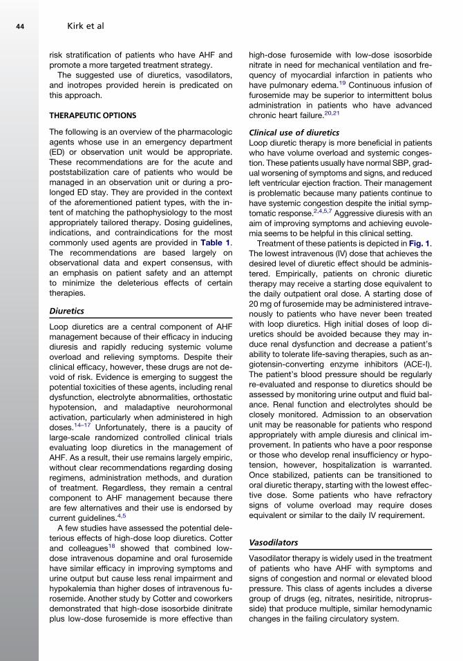
risk stratification of patients who have AHF andpromote a more targeted treatment strategy.
The suggested use of diuretics, vasodilators,and inotropes provided herein is predicated onthis approach.
THERAPEUTIC OPTIONS
The following is an overview of the pharmacologicagents whose use in an emergency department(ED) or observation unit would be appropriate.These recommendations are for the acute andpoststabilization care of patients who would bemanaged in an observation unit or during a pro-longed ED stay. They are provided in the contextof the aforementioned patient types, with the in-tent of matching the pathophysiology to the mostappropriately tailored therapy. Dosing guidelines,indications, and contraindications for the mostcommonly used agents are provided in Table 1.The recommendations are based largely onobservational data and expert consensus, withan emphasis on patient safety and an attemptto minimize the deleterious effects of certaintherapies.
Diuretics
Loop diuretics are a central component of AHFmanagement because of their efficacy in inducingdiuresis and rapidly reducing systemic volumeoverload and relieving symptoms. Despite theirclinical efficacy, however, these drugs are not de-void of risk. Evidence is emerging to suggest thepotential toxicities of these agents, including renaldysfunction, electrolyte abnormalities, orthostatichypotension, and maladaptive neurohormonalactivation, particularly when administered in highdoses.14–17 Unfortunately, there is a paucity oflarge-scale randomized controlled clinical trialsevaluating loop diuretics in the management ofAHF. As a result, their use remains largely empiric,without clear recommendations regarding dosingregimens, administration methods, and durationof treatment. Regardless, they remain a centralcomponent to AHF management because thereare few alternatives and their use is endorsed bycurrent guidelines.4,5
A few studies have assessed the potential dele-terious effects of high-dose loop diuretics. Cotterand colleagues18 showed that combined low-dose intravenous dopamine and oral furosemidehave similar efficacy in improving symptoms andurine output but cause less renal impairment andhypokalemia than higher doses of intravenous fu-rosemide. Another study by Cotter and coworkersdemonstrated that high-dose isosorbide dinitrateplus low-dose furosemide is more effective than
high-dose furosemide with low-dose isosorbidenitrate in need for mechanical ventilation and fre-quency of myocardial infarction in patients whohave pulmonary edema.19 Continuous infusion offurosemide may be superior to intermittent bolusadministration in patients who have advancedchronic heart failure.20,21
Clinical use of diureticsLoop diuretic therapy is more beneficial in patientswho have volume overload and systemic conges-tion. These patients usually have normal SBP, grad-ual worsening of symptoms and signs, and reducedleft ventricular ejection fraction. Their managementis problematic because many patients continue tohave systemic congestion despite the initial symp-tomatic response.2,4,5,7 Aggressive diuresis with anaim of improving symptoms and achieving euvole-mia seems to be helpful in this clinical setting.
Treatment of these patients is depicted in Fig. 1.The lowest intravenous (IV) dose that achieves thedesired level of diuretic effect should be adminis-tered. Empirically, patients on chronic diuretictherapy may receive a starting dose equivalent tothe daily outpatient oral dose. A starting dose of20 mg of furosemide may be administered intrave-nously to patients who have never been treatedwith loop diuretics. High initial doses of loop di-uretics should be avoided because they may in-duce renal dysfunction and decrease a patient’sability to tolerate life-saving therapies, such as an-giotensin-converting enzyme inhibitors (ACE-I).The patient’s blood pressure should be regularlyre-evaluated and response to diuretics should beassessed by monitoring urine output and fluid bal-ance. Renal function and electrolytes should beclosely monitored. Admission to an observationunit may be reasonable for patients who respondappropriately with ample diuresis and clinical im-provement. In patients who have a poor responseor those who develop renal insufficiency or hypo-tension, however, hospitalization is warranted.Once stabilized, patients can be transitioned tooral diuretic therapy, starting with the lowest effec-tive dose. Some patients who have refractorysigns of volume overload may require dosesequivalent or similar to the daily IV requirement.
Vasodilators
Vasodilator therapy is widely used in the treatmentof patients who have AHF with symptoms andsigns of congestion and normal or elevated bloodpressure. This class of agents includes a diversegroup of drugs (eg, nitrates, nesiritide, nitroprus-side) that produce multiple, similar hemodynamicchanges in the failing circulatory system.
Kirk et al44

NitroglycerinNitroglycerin is a potent venodilator and a mildarterial vasodilator at higher doses.22 It mimicsthe effects of nitric oxide by stimulating guanylatecyclase and leading to smooth muscle relaxationin the vascular wall. It reduces left ventricular fillingpressures, pulmonary congestion, wall stress, andmyocardial oxygen consumption without compro-mising cardiac output.23 At higher doses it may im-prove ischemia by inducing coronary arterydilation and increasing collateral blood flow. In ad-dition to the IV form, it is also available in sublin-gual and topical preparations. The latter arefrequently used in the ED but no clinical trial dataexist describing their usefulness. The most com-mon side effects are headache and hypotension.In addition, the potential benefit of nitroglycerinseems to be attenuated by a decreased vasodila-tory response in patients who have heart failure,which is associated with neurohumoral activationand the early development of nitratetolerance.23,24 Consequently, careful titrationand monitoring of nitroglycerin administration isimperative.
There is a paucity of controlled data concerningthe use of nitrates in AHF. The dose-dependentimprovement in hemodynamic function has beenestablished by several small-scale studies,23,25,26
whereas others have demonstrated the efficacyof high-dose IV nitroglycerin or isosorbide dinitratein the management of AHF, with a low incidence ofside effects.19,27,28
Recently a randomized placebo-controlledstudy showed a nonsignificant pulmonary capillarywedge pressure (PCWP) reduction with intrave-nous nitroglycerin.29 Other improvements in he-modynamics, dyspnea, and global assessmentwere evident with nitroglycerin, although mostdid not reach statistical significance versuscontrol.
NesiritideNesiritide is a recombinant form of human brainnatriuretic peptide with a balanced arterial, ve-nous, and coronary vasodilatory effect and a mod-est natriuretic effect.30 It has beneficial effects onhemodynamics (reduction in PCWP and increasein cardiac output) and symptoms.31,32 The Vasodi-lation in the Acute Management of Congestiveheart failure (VMAC) trial showed that comparedwith nitroglycerin, nesiritide produces a faster,greater, and more sustained reduction in PCWPwith no significant difference in dyspnea, globalclinical status, 30-day rehospitalization, and6-month mortality rate29 Hypotension is the mostcommon side effect. Recently, concerns haveemerged from two meta-analyses about the effect
of nesiritide on renal dysfunction and survival.8,9
The results from meta-analyses should be cau-tiously interpreted, however, because nesiritidestudies were not powered or designed to evaluatemortality or renal dysfunction.
NitroprussideSodium nitroprusside is an arterial vasodilator thatis mainly used in patients who have markedly in-creased afterload attributable to severe hyperten-sion.5 No randomized controlled mortality trialswith nitroprusside in AHF have been conducted.It reduces systemic vascular resistance, increasesstroke volume, and improves symptoms inAHF.33,34 Trials evaluating patients who have myo-cardial infarction suggest an increased risk fromnitroprusside use in patients who have associatedAHF.35 This increased risk is probably becausehigh doses of the agent cause reflex tachycardiaand may lead to ‘‘coronary steal,’’ which may ex-acerbate myocardial ischemia; therefore, it is gen-erally contraindicated in patients who have acutecoronary syndrome and AHF.5 Thiocyanate andcyanide levels increase in patients who have renaldysfunction and nitroprusside should be avoidedin this population.
Clinical use of vasodilatorsVasodilator therapy is recommended as first-linetherapy in patients who have AHF associatedwith an elevated SBP at presentation. These pa-tients represent more than 50% of patients whohave AHF in the ED.2 Most are elderly womenwho have relatively preserved ejection fractionand they frequently have a SBP greater than 160mm Hg.2,3,7 Symptoms develop abruptly and se-vere dyspnea is the cardinal manifestation. Redis-tribution of fluids from systemic to pulmonarycirculation leads to pulmonary rather than sys-temic congestion that is evident on examinationand chest radiography. Accordingly, the clinicaltarget is blood pressure control and response totherapy is relatively rapid in patients who aretreated with early, aggressive vasodilation, moreso than diuresis.1,5,7
Treatment of these patients is depicted in Fig. 2.Rapid sublingual administration of nitroglycerinspray or tablet either in the prehospital setting orin the ED dramatically improves the clinical statuswithin minutes.36 Intravenous vasodilator therapy(nitroglycerin or nesiritide) should then be started.The initial recommended dose of IV nitroglycerinis 10 to 20 mg/min, increased in increments of 5 to10 mg/min every 3 to 5 minutes as needed. Nesiri-tide is given IV as a 2 mg/kg bolus followed bya 0.01 mg/kg/min infusion. Slow titration of IV vaso-dilators and frequent blood pressure measurement
Pharmacologic Stabilization and Management 45
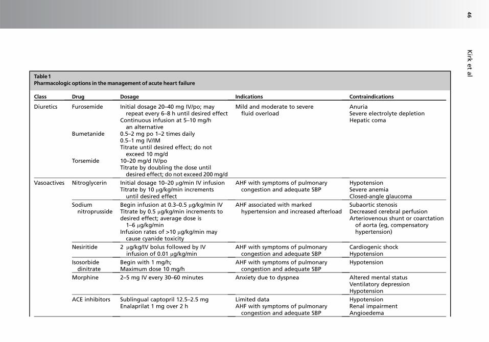
Table1Pharmacologic options in themanagement of acute heart failure
Class Drug Dosage Indications Contraindications
Diuretics Furosemide Initial dosage 20–40 mg IV/po; mayrepeat every 6–8 h until desired effect
Continuous infusion at 5–10 mg/han alternative
Mild and moderate to severefluid overload
AnuriaSevere electrolyte depletionHepatic coma
Bumetanide 0.5–2 mg po 1–2 times daily0.5–1 mg IV/IMTitrate until desired effect; do not
exceed 10 mg/dTorsemide 10–20 mg/d IV/po
Titrate by doubling the dose untildesired effect; do not exceed 200 mg/d
Vasoactives Nitroglycerin Initial dosage 10–20 mg/min IV infusionTitrate by 10 mg/kg/min increments
until desired effect
AHF with symptoms of pulmonarycongestion and adequate SBP
HypotensionSevere anemiaClosed-angle glaucoma
Sodiumnitroprusside
Begin infusion at 0.3–0.5 mg/kg/min IVTitrate by 0.5 mg/kg/min increments todesired effect; average dose is
1–6 mg/kg/minInfusion rates of >10 mg/kg/min may
cause cyanide toxicity
AHF associated with markedhypertension and increased afterload
Subaortic stenosisDecreased cerebral perfusionArteriovenous shunt or coarctation
of aorta (eg, compensatoryhypertension)
Nesiritide 2 mg/kg/IV bolus followed by IVinfusion of 0.01 mg/kg/min
AHF with symptoms of pulmonarycongestion and adequate SBP
Cardiogenic shockHypotension
Isosorbidedinitrate
Begin with 1 mg/h;Maximum dose 10 mg/h
AHF with symptoms of pulmonarycongestion and adequate SBP
Hypotension
Morphine 2–5 mg IV every 30–60 minutes Anxiety due to dyspnea Altered mental statusVentilatory depressionHypotension
ACE inhibitors Sublingual captopril 12.5–2.5 mgEnalaprilat 1 mg over 2 h
Limited dataAHF with symptoms of pulmonary
congestion and adequate SBP
HypotensionRenal impairmentAngioedema
Kirk
et
al
46

Inotropes Dobutamine 0.5 mg/kg/min IV infusionTitrate to maintain adequate systolic
BP and cardiac output
Low cardiac outputCardiogenic shockMilrinone preferred for patients
already taking b-blocker
Idiopathic hypertrophic subaorticstenosis
Atrial fibrillation or flutterDopamine 5 mg/kg/min IV infusion
Titrate to maintain adequate systolicBP and cardiac output
Do not exceed 20 mg/kg/min
PheochromocytomaVentricular fibrillation
Milrinone 50 mg/kg/min IV loading dose over10 minutes; then 0.25–1.0 mg/kg/mininfusion
Titrate to maintain adequate systolicBP and cardiac output
Obstructive hypertrophiccardiomyopathy
Levosimendan 0.05–0.2 mg/kg/min continuousIV infusion for 24 h withouta loading dose
Acutely decompensated chronicheart failure especially on b-blockerand systolic BP>100 mm Hg
Severe hypotensionHepatic impairmentLeft ventricular outflow obstruction
Abbreviation: AHF, acute heart failure; BP, blood pressure; IM, intramuscular; IV, intravenous; SBP, systolic blood pressure.Data from Kirk JD, Costanza MR. Managing patients with acute decompensated heart failure. Clin Courier 2006;23(56):1–14.
Ph
arm
aco
log
icSta
biliza
tion
an
dM
an
ag
em
en
t47
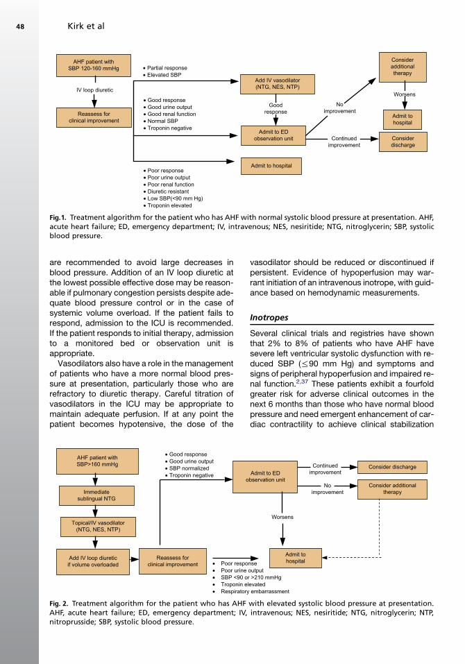
are recommended to avoid large decreases inblood pressure. Addition of an IV loop diuretic atthe lowest possible effective dose may be reason-able if pulmonary congestion persists despite ade-quate blood pressure control or in the case ofsystemic volume overload. If the patient fails torespond, admission to the ICU is recommended.If the patient responds to initial therapy, admissionto a monitored bed or observation unit isappropriate.
Vasodilators also have a role in the managementof patients who have a more normal blood pres-sure at presentation, particularly those who arerefractory to diuretic therapy. Careful titration ofvasodilators in the ICU may be appropriate tomaintain adequate perfusion. If at any point thepatient becomes hypotensive, the dose of the
vasodilator should be reduced or discontinued ifpersistent. Evidence of hypoperfusion may war-rant initiation of an intravenous inotrope, with guid-ance based on hemodynamic measurements.
Inotropes
Several clinical trials and registries have shownthat 2% to 8% of patients who have AHF havesevere left ventricular systolic dysfunction with re-duced SBP (%90 mm Hg) and symptoms andsigns of peripheral hypoperfusion and impaired re-nal function.2,37 These patients exhibit a fourfoldgreater risk for adverse clinical outcomes in thenext 6 months than those who have normal bloodpressure and need emergent enhancement of car-diac contractility to achieve clinical stabilization
Partial responseElevated SBP
Add IV vasodilator(NTG, NES, NTP)IV loop diuretic
Good responseGood urine outputGood renal functionNormal SBPTroponin negative
Continuedimprovement
AHF patient withSBP 120-160 mmHg
Consideradditionaltherapy
Admit to EDobservation unit
Reassess forclinical improvement
Admit to hospital
Worsens
Admit tohospital
Considerdischarge
Noimprovement
Goodresponse
Poor responsePoor urine outputPoor renal functionDiuretic resistantLow SBP(<90 mm Hg)Troponin elevated
Fig.1. Treatment algorithm for the patient who has AHF with normal systolic blood pressure at presentation. AHF,acute heart failure; ED, emergency department; IV, intravenous; NES, nesiritide; NTG, nitroglycerin; SBP, systolicblood pressure.
Poor responsePoor urine outputSBP <90 or >210 mmHgTroponin elevatedRespiratory embarrassment
Good responseGood urine outputSBP normalizedTroponin negative
Continuedimprovement
AHF patient withSBP>160 mmHg
Reassess forclinical improvement
Admit to EDobservation unit
Topical/IV vasodilator(NTG, NES, NTP)
Admit tohospital
Worsens
NoimprovementImmediate
sublingual NTG
Add IV loop diureticif volume overloaded
Consider additionaltherapy
Consider discharge
Fig. 2. Treatment algorithm for the patient who has AHF with elevated systolic blood pressure at presentation.AHF, acute heart failure; ED, emergency department; IV, intravenous; NES, nesiritide; NTG, nitroglycerin; NTP,nitroprusside; SBP, systolic blood pressure.
Kirk et al48
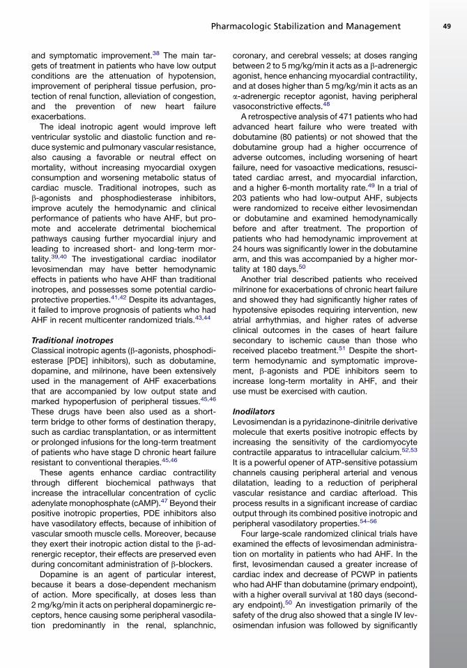
and symptomatic improvement.38 The main tar-gets of treatment in patients who have low outputconditions are the attenuation of hypotension,improvement of peripheral tissue perfusion, pro-tection of renal function, alleviation of congestion,and the prevention of new heart failureexacerbations.
The ideal inotropic agent would improve leftventricular systolic and diastolic function and re-duce systemic and pulmonary vascular resistance,also causing a favorable or neutral effect onmortality, without increasing myocardial oxygenconsumption and worsening metabolic status ofcardiac muscle. Traditional inotropes, such asb-agonists and phosphodiesterase inhibitors,improve acutely the hemodynamic and clinicalperformance of patients who have AHF, but pro-mote and accelerate detrimental biochemicalpathways causing further myocardial injury andleading to increased short- and long-term mor-tality.39,40 The investigational cardiac inodilatorlevosimendan may have better hemodynamiceffects in patients who have AHF than traditionalinotropes, and possesses some potential cardio-protective properties.41,42 Despite its advantages,it failed to improve prognosis of patients who hadAHF in recent multicenter randomized trials.43,44
Traditional inotropesClassical inotropic agents (b-agonists, phosphodi-esterase [PDE] inhibitors), such as dobutamine,dopamine, and milrinone, have been extensivelyused in the management of AHF exacerbationsthat are accompanied by low output state andmarked hypoperfusion of peripheral tissues.45,46
These drugs have been also used as a short-term bridge to other forms of destination therapy,such as cardiac transplantation, or as intermittentor prolonged infusions for the long-term treatmentof patients who have stage D chronic heart failureresistant to conventional therapies.45,46
These agents enhance cardiac contractilitythrough different biochemical pathways thatincrease the intracellular concentration of cyclicadenylate monophosphate (cAMP).47 Beyond theirpositive inotropic properties, PDE inhibitors alsohave vasodilatory effects, because of inhibition ofvascular smooth muscle cells. Moreover, becausethey exert their inotropic action distal to the b-ad-renergic receptor, their effects are preserved evenduring concomitant administration of b-blockers.
Dopamine is an agent of particular interest,because it bears a dose-dependent mechanismof action. More specifically, at doses less than2 mg/kg/min it acts on peripheral dopaminergic re-ceptors, hence causing some peripheral vasodila-tion predominantly in the renal, splanchnic,
coronary, and cerebral vessels; at doses rangingbetween 2 to 5 mg/kg/min it acts as a b-adrenergicagonist, hence enhancing myocardial contractility,and at doses higher than 5 mg/kg/min it acts as ana-adrenergic receptor agonist, having peripheralvasoconstrictive effects.48
A retrospective analysis of 471 patients who hadadvanced heart failure who were treated withdobutamine (80 patients) or not showed that thedobutamine group had a higher occurrence ofadverse outcomes, including worsening of heartfailure, need for vasoactive medications, resusci-tated cardiac arrest, and myocardial infarction,and a higher 6-month mortality rate.49 In a trial of203 patients who had low-output AHF, subjectswere randomized to receive either levosimendanor dobutamine and examined hemodynamicallybefore and after treatment. The proportion ofpatients who had hemodynamic improvement at24 hours was significantly lower in the dobutaminearm, and this was accompanied by a higher mor-tality at 180 days.50
Another trial described patients who receivedmilrinone for exacerbations of chronic heart failureand showed they had significantly higher rates ofhypotensive episodes requiring intervention, newatrial arrhythmias, and higher rates of adverseclinical outcomes in the cases of heart failuresecondary to ischemic cause than those whoreceived placebo treatment.51 Despite the short-term hemodynamic and symptomatic improve-ment, b-agonists and PDE inhibitors seem toincrease long-term mortality in AHF, and theiruse must be exercised with caution.
InodilatorsLevosimendan is a pyridazinone-dinitrile derivativemolecule that exerts positive inotropic effects byincreasing the sensitivity of the cardiomyocytecontractile apparatus to intracellular calcium.52,53
It is a powerful opener of ATP-sensitive potassiumchannels causing peripheral arterial and venousdilatation, leading to a reduction of peripheralvascular resistance and cardiac afterload. Thisprocess results in a significant increase of cardiacoutput through its combined positive inotropic andperipheral vasodilatory properties.54–56
Four large-scale randomized clinical trials haveexamined the effects of levosimendan administra-tion on mortality in patients who had AHF. In thefirst, levosimendan caused a greater increase ofcardiac index and decrease of PCWP in patientswho had AHF than dobutamine (primary endpoint),with a higher overall survival at 180 days (second-ary endpoint).50 An investigation primarily of thesafety of the drug also showed that a single IV lev-osimendan infusion was followed by significantly
Pharmacologic Stabilization and Management 49
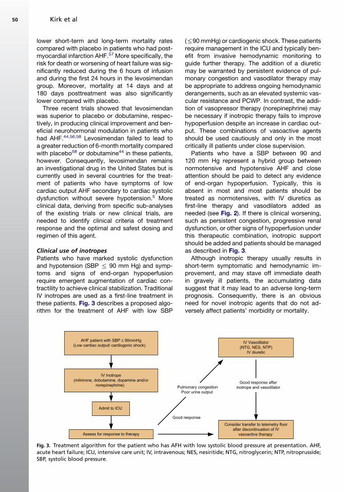
lower short-term and long-term mortality ratescompared with placebo in patients who had post-myocardial infarction AHF.57 More specifically, therisk for death or worsening of heart failure was sig-nificantly reduced during the 6 hours of infusionand during the first 24 hours in the levosimendangroup. Moreover, mortality at 14 days and at180 days posttreatment was also significantlylower compared with placebo.
Three recent trials showed that levosimendanwas superior to placebo or dobutamine, respec-tively, in producing clinical improvement and ben-eficial neurohormonal modulation in patients whohad AHF.44,56,58 Levosimendan failed to lead toa greater reduction of 6-month mortality comparedwith placebo58 or dobutamine44 in these patients,however. Consequently, levosimendan remainsan investigational drug in the United States but iscurrently used in several countries for the treat-ment of patients who have symptoms of lowcardiac output AHF secondary to cardiac systolicdysfunction without severe hypotension.5 Moreclinical data, deriving from specific sub-analysesof the existing trials or new clinical trials, areneeded to identify clinical criteria of treatmentresponse and the optimal and safest dosing andregimen of this agent.
Clinical use of inotropesPatients who have marked systolic dysfunctionand hypotension (SBP % 90 mm Hg) and symp-toms and signs of end-organ hypoperfusionrequire emergent augmentation of cardiac con-tractility to achieve clinical stabilization. TraditionalIV inotropes are used as a first-line treatment inthese patients. Fig. 3 describes a proposed algo-rithm for the treatment of AHF with low SBP
(%90 mmHg) or cardiogenic shock. These patientsrequire management in the ICU and typically ben-efit from invasive hemodynamic monitoring toguide further therapy. The addition of a diureticmay be warranted by persistent evidence of pul-monary congestion and vasodilator therapy maybe appropriate to address ongoing hemodynamicderangements, such as an elevated systemic vas-cular resistance and PCWP. In contrast, the addi-tion of vasopressor therapy (norepinephrine) maybe necessary if inotropic therapy fails to improvehypoperfusion despite an increase in cardiac out-put. These combinations of vasoactive agentsshould be used cautiously and only in the mostcritically ill patients under close supervision.
Patients who have a SBP between 90 and120 mm Hg represent a hybrid group betweennormotensive and hypotensive AHF and closeattention should be paid to detect any evidenceof end-organ hypoperfusion. Typically, this isabsent in most and most patients should betreated as normotensives, with IV diuretics asfirst-line therapy and vasodilators added asneeded (see Fig. 2). If there is clinical worsening,such as persistent congestion, progressive renaldysfunction, or other signs of hypoperfusion underthis therapeutic combination, inotropic supportshould be added and patients should be managedas described in Fig. 3.
Although inotropic therapy usually results inshort-term symptomatic and hemodynamic im-provement, and may stave off immediate deathin gravely ill patients, the accumulating datasuggest that it may lead to an adverse long-termprognosis. Consequently, there is an obviousneed for novel inotropic agents that do not ad-versely affect patients’ morbidity or mortality.
Good response
AHF patient with SBP ≤ 90mmHg(Low cardiac output/ cardiogenic shock)
Admit to ICU
Assess for response to therapy
IV Vasodilator(NTG, NES, NTP)
IV diuretic
Pulmonary congestionPoor urine output
Consider transfer to telemetry floorafter discontinuation of IV
vasoactive therapy
Good response afterinotrope and vasodilator
IV Inotrope(milnirone, dobutamine, dopamine and/or
norepinephrine)
Fig. 3. Treatment algorithm for the patient who has AFH with low systolic blood pressure at presentation. AHF,acute heart failure; ICU, intensive care unit; IV, intravenous; NES, nesiritide; NTG, nitroglycerin; NTP, nitroprusside;SBP, systolic blood pressure.
Kirk et al50

Other Vasoactive Agents
MorphineMorphine sulfate is a traditional therapy in AHF,despite the lack of much supporting data.Morphine reduces preload by producing mildvenodilatation, relieves breathlessness, and exertsa calming effect. A recently published retrospec-tive analysis of the Acute Decompensated HeartFailure National Registry showed that morphineis associated with a greater frequency of mechan-ical ventilation, prolonged hospitalization, moreICU admissions, and higher mortality.59 Additionalstudies are needed to clarify these safetyconcerns.
Angiotensin converting enzyme inhibitorsACE-I are considered standard of care in the man-agement of chronic heart failure. Despite the pleth-ora of data showing a substantial prognosticbenefit when ACE-I are used in the chronic setting,only limited data are available for the acutesetting.60,61 Although a few studies have demon-strated hemodynamic improvement in AHF withACE-I, their impact on clinical outcomes remainsobscure. Their use is therefore not routinely recom-mended in the acute stabilization of AHF patientsand additional research is needed.5 Pre-existingtherapy with ACE-I as a chronic heart failure med-ication should be continued during the AHF episodeat the highest tolerated dose. Dosages should bereduced or discontinued altogether if the patienthas symptomatic hypotension (SBP<100 mm Hg),any evidence of cardiogenic shock, elevated serumpotassium (>5 mmol/L) or severe renal dysfunction(serum creatinine>2.5 mg/dL). Patients not cur-rently being treated with ACE-I before admissionshould be started on this therapy during the hospi-talization if none of the aforementioned contraindi-cations exist.
Aldosterone antagonistPersistent activation of aldosterone plays an im-portant role in the pathophysiology of AHF; effec-tive antagonism provides a substantial reductionin mortality, a reduction in AHF hospitalizations,and an improvement in functional class.62 If serumpotassium is less than 5 mmol/L and serum creat-inine less than 2.5 mg/dL, an aldosterone antago-nist (spironolactone 25 mg), should be added withrepeat measurement of a chemistry panel andmodification of drug dosing at 4 to 6 days laterin-hospital or in outpatient heart failure clinic.
b-BlockersAlthough data from randomized clinical trials aremissing and the results from ongoing clinical trialsin this area will be available in a few years, data
from retrospective trials indicate that b-blockersshould be continued during the AHF episode atthe highest tolerated dose except for cases inwhich the patient has symptomatic hypotensionor bradycardia, is hemodynamically unstable, oris in cardiogenic shock.63
SUMMARY
Effective use of diuretics, vasodilators, and ino-tropes to stabilize AHF relies on matching themost appropriately tailored therapy to specificclinical profiles. Some of the drugs may be harm-ful, and therefore the emphasis should be onpatient safety and the attempt to minimize the del-eterious effects of these therapies. Diuretics seemto be most beneficial in patients who have sub-stantial volume overload, often seen in patientswho have normal SBP. Vasodilator therapy seemsto have its greatest usefulness in patients whohave acute pulmonary edema associated with anelevated SBP and is recommended as first-linetherapy in this group. Patients who have cardio-genic shock are at the highest risk and needemergent enhancement of cardiac contractility toachieve clinical stabilization. Inotropes are effec-tive in restoring hemodynamic stability in this lattergroup but may lead to increased short- and long-term mortality. To date, successful treatment hasbeen limited because no agent has been shownto reduce postdischarge mortality or readmissionrates, and patients frequently remain symptomaticafter treatment. Ongoing research is needed tofurther examine these agents and to develop noveltherapies to address the unmet needs of thepatient who has AHF.
ACKNOWLEDGMENT
The authors thank Dr. Lucas Pappas for hisassistance in the preparation of this manuscript.
REFERENCES
1. Gheorghiade M, Zannad F, Sopko G, et al. Interna-
tional working group on acute heart failure syn-
dromes. Acute heart failure syndromes: current
state and framework for future research. Circulation
2005;112:3958–68.
2. Adams KF Jr, Fonarow GC, Emerman CL, et al. AD-
HERE scientific advisory committee and investiga-
tors. Characteristics and outcomes of patients
hospitalized for heart failure in the United States:
rationale, design, and preliminary observations
from the first 100,000 cases in the Acute Decompen-
sated Heart Failure National Registry (ADHERE). Am
Heart J 2005;149:209–16.
Pharmacologic Stabilization and Management 51
3. Cleland JG, Swedberg K, Follath F, et al. Study
group on diagnosis of the working group on heart
failure of the European society of cardiology. The
Euroheart failure survey programme—a survey on
the quality of care among patients with heart failure
in Europe. Part 1: patient characteristics and diag-
nosis. Eur Heart J 2003;24:442–63.
4. Heart Failure Society Of America. Executive summary:
HFSA 2006 comprehensive heart failure practice
guideline. J Card Fail 2006;12:10–38.
5. Nieminen MS, Bohm M, Cowie MR, et al. ESC com-
mittee for practice guideline (CPG). Executive
summary of the guidelines on the diagnosis and
treatment of acute heart failure: the task force on
acute heart failure of the European society of cardi-
ology. Eur Heart J 2005;26:384–416.
6. De Luca L, Fonarow GC, Mebazaa A, et al. Early
pharmacological treatment of acute heart failure
syndromes: a systematic review of clinical trials.
Acute Card Care 2007;9:10–21.
7. Gheorghiade M, De Luca L, Fonarow GC, et al. Path-
ophysiologic targets in the early phase of acute
heart failure syndromes. Am J Cardiol 2005;96(6A):
11G–7G.
8. Sackner-Bernstein JD, Skopicki HA, Aaronson KD.
Risk of worsening renal function with nesiritide in
patients with acutely decompensated heart failure.
Circulation 2005;111:1487–91.
9. Sackner-Bernstein JD, Kowalski M, Fox M, et al. Short-
term risk of death after treatment with nesiritide for
decompensated heart failure: a pooled analysis of ran-
domized controlled trials. JAMA 2005;293:1900–5.
10. Zannad F, Mebazaa A, Juilliere Y, et al. Clinical pro-
file, contemporary management and one-year mor-
tality in patients with severe acute heart failure
syndromes: the EFICA study. Eur J Heart Fail
2006;8:697–705.
11. Gheorghiade M, Abraham WT, Albert NM, et al. Sys-
tolic blood pressure at admission, clinical character-
istics, and outcomes in patients hospitalized with
acute heart failure. JAMA 2006;296:2217–26.
12. Alla F, Zannad F, Filippatos G. Epidemiology of acute
heart failure syndromes. Heart Fail Rev 2007;12:91–5.
13. Chatti R, Fradj NB, Trabelsi W, et al. Algorithm for
therapeutic management of acute heart failure syn-
dromes. Heart Fail Rev 2007;12:113–7.
14. Weinfeld MS, Chertow GM, Stevenson LW. Aggra-
vated renal dysfunction during intensive therapy
for advanced chronic heart failure. Am Heart J
1999;138:285–90.
15. Cooper HA, Dries DL, Davis CE, et al. Diuretics and
risk of arrhythmic death in patients with left ventricu-
lar dysfunction. Circulation 1999;100:1311–5.
16. Jhund PS, McMurray J, Davie AP. The acute vascu-
lar effects of furosemide in heart failure. Br J Clin
Pharmacol 2000;50:9–13.
17. Gottlieb SS, Brater DC, Thomas I, et al. BG9719
(CVT-124), an A1 adenosine receptor antagonist,
protects against the decline in renal function ob-
served with diuretic therapy. Circulation 2002;105:
1348–53.
18. Cotter G, Weissgarten J, Metzkor E, et al. Increased
toxicity of high-dose furosemide versus low-dose
dopamine in the treatment of refractory congestive
heart failure. Clin Pharmacol Ther 1997;62:187–93.
19. Cotter G, Metzkor E, Kaluski E, et al. Randomised
trial of high-dose isosorbide dinitrate plus low-dose
furosemide versus high-dose furosemide plus low-
dose isosorbide dinitrate in severe pulmonary oe-
dema. Lancet 1998;351:389–93.
20. Lahav M, Regev A, Ra’anani P, et al. Intermittent ad-
ministration of furosemide vs continuous infusion
preceded by a loading dose for congestive heart
failure. Chest 1992;102:725–31.
21. Dormans TP, van Meyel JJ, Gerlag PG, et al. Diuretic
efficacy of high dose furosemide in severe heart
failure: bolus injection versus continuous infusion.
J Am Coll Cardiol 1996;28:376–82.
22. Cohn PF, Gorlin R. Physiologic and clinical actions of
nitroglycerin. Med Clin North Am 1974;58:407–15.
23. Elkayam U, Bitar F, Akhter MW, et al. Intravenous ni-
troglycerin in the treatment of decompensated heart
failure: potential benefits and limitations. J Cardio-
vasc Pharmacol Ther 2004;9:227–41.
24. Katz SD, Biasucci L, Sabba C, et al. Impaired
endothelium-mediated vasodilation in the peripheral
vasculature of patients with congestive heart failure.
J Am Coll Cardiol 1992;19:918–25.
25. Elkayam U, Roth A, Kumar A, et al. Hemodynamic
and volumetric effects of venodilation with nitroglyc-
erin in chronic mitral regurgitation. Am J Cardiol
1987;60:1106–11.
26. Loh E, Elkayam U, Cody R, et al. A randomized mul-
ticenter study comparing the efficacy and safety of
intravenous milrinone and intravenous nitroglycerin
in patients with advanced heart failure. J Card Fail
2001;7:114–21.
27. Nashed AH, Allegra JR. Intravenous nitroglycerin
boluses in treating patients with cardiogenic pulmo-
nary edema. Am J Emerg Med 1995;13:612–3.
28. Levy P, Compton S, Welch R, et al. Treatment of
severe decompensated heart failure with high-
dose intravenous nitroglycerin: a feasibility and out-
come analysis. Ann Emerg Med 2007;50:144–52.
29. Publication Committee for the VMAC Investigators
(Vasodilatation in the Management of Acute CHF)
Intravenous nesiritide vs nitroglycerin for treat-
ment of decompensated congestive heart failure:
a randomized controlled trial. JAMA 2002;287:
1531–40.
30. Levin ER, Gardner DG, Samson WK. Natriuretic
peptides. N Engl J Med 1998;339:321–8.
Kirk et al52
31. Mills RM, LeJemtel TH, Horton DP, et al. Sustained
hemodynamic effects of an infusion of nesiritide
(human B-type natriuretic peptide) in heart failure:
a randomized, double-blind, placebo-controlled
clinical trial. Natrecor study group. J Am Coll Cardiol
1999;34:155–62.
32. Colucci WS, Elkayam U, Horton DP, et al. Intrave-
nous nesiritide, a natriuretic peptide, in the treat-
ment of decompensated congestive heart failure.
Nesiritide study group. N Engl J Med 2000;343:
246–53.
33. Guiha NH, Cohn JN, Mikulic E, et al. Treatment of
refractory heart failure with infusion of nitroprusside.
N Engl J Med 1974;291:587–92.
34. Khot UN, Novaro GM, Popovic ZB, et al. Nitroprus-
side in critically ill patients with left ventricular dys-
function and aortic stenosis. N Engl J Med 2003;
348:1756–63.
35. Cohn JN, Franciosa JA, Francis GS, et al. Effect of
short-term infusion of sodium nitroprusside on mor-
tality rate in acute myocardial infarction complicated
by left ventricular failure: results of a Veterans Ad-
ministration cooperative study. N Engl J Med 1982;
306:1129–35.
36. Bussmann WD, Schupp D. Effect of sublingual
nitroglycerin in emergency treatment of severe pul-
monary edema. Am J Cardiol 1978;41:931–6.
37. Fonarow GC, Corday E. ADHERE scientific advisory
committee. Overview of acutely decompensated
congestive heart failure (ADHF): a report from the
ADHERE registry. Heart Fail Rev 2004;9:179–85.
38. Milo-Cotter O, Adams KF, O’Connor CM, et al. Acute
heart failure associated with high admission blood
pressure—a distinct vascular disorder? Eur J Heart
Fail 2007;9:178–83.
39. Thackray S, Easthaugh J, Freemantle N, et al. The
effectiveness and relative effectiveness of intrave-
nous inotropic drugs acting through the adrenergic
pathway in patients with heart failure-a meta-regres-
sion analysis. Eur J Heart Fail 2002;4:515–29.
40. Packer M. The search for the ideal positive inotropic
agent. N Engl J Med 1993;329:201–2.
41. Papp Z, Csapo K, Pollesello P, et al. Pharmacological
mechanisms contributing to the clinical efficacy of
levosimendan. Cardiovasc Drug Rev 2005;23:71–98.
42. Mebazaa A, Barraud D, Welschbillig S. Randomized
clinical trials with levosimendan. Am J Cardiol 2005;
96:74G–9G.
43. Cleland JG, Freemantle N, Coletta AP, et al. Clinical
trials update from the American Heart Association:
REPAIR-AMI, ASTAMI, JELIS, MEGA, REVIVE-II,
SURVIVE, and PROACTIVE. Eur J Heart Fail 2006;
8:105–10.
44. Mebazaa A, Nieminen MS, Packer M, et al. SURVIVE
investigators. Levosimendan vs dobutamine for pa-
tients with acute decompensated heart failure: the
SURVIVE randomized trial. JAMA 2007;297:1883–91.
45. Munger MA. Management of acute decompensated
heart failure: treatment, controversy, and future di-
rections. Pharmacotherapy 2006;26:131S–8S.
46. Zannad F, Adamopoulos C, Mebazaa A, et al. The
challenge of acute decompensated heart failure.
Heart Fail Rev 2006;11:135–9.
47. Felker GM, O’Connor CM. Inotropic therapy for heart
failure: an evidence-based approach. Am Heart J
2001;142:393–401.
48. Parissis J, Farmakis D, Nieminen M. Classical ino-
tropes and new cardiac enhancers. Heart Fail Rev
2007;12:149–56.
49. O’Connor CM, Gattis WA, Uretsky BF, et al. Continu-
ous intravenous dobutamine is associated with an
increased risk of death in patients with advanced
heart failure: insights from the Flolan International
Randomized Survival Trial (FIRST). Am Heart J
1999;138:78–86.
50. Follath F, Cleland JG, Just H, et al. Steering commit-
tee and investigators of the levosimendan infusion
versus dobutamine (LIDO) study. Efficacy and
safety of intravenous levosimendan compared with
dobutamine in severe low-output heart failure
(the LIDO study): a randomised double-blind trial.
Lancet 2002;360:196–202.
51. Cuffe MS, Califf RM, Adams KF Jr, et al. Outcomes
of a prospective trial of intravenous milrinone for ex-
acerbations of chronic heart failure (OPTIME-CHF)
investigators. Short-term intravenous milrinone for
acute exacerbation of chronic heart failure: a ran-
domized controlled trial. JAMA 2002;287:1541–7.
52. Figgit DP, Gilles PS, Goa KL. Levosimendan. Drugs
2001;61:613–27.
53. Perrone S, Kaplinsky EJ. Calcium sensitizer agents:
a new class of inotropic agents in the treatment of
decompensated heart failure. Int J Cardiol 2005;
103:248–55.
54. Kivikko M, Lehtonen L. Levosimendan: a new
inodilatory drug for the treatment of decompensated
heart failure. Curr Pharm Des 2005;11:435–55.
55. Parissis J, Filippatos G, Farmakis D, et al. Levosi-
mendan for the treatment of acute heart failure
syndromes. Expert Opin Pharmacother 2005;6(15):
2741–51.
56. DeLuca L, Colucci W, Nieminen M, et al. Evidence-
based use of levosimendan in different clinical
settings. Eur Heart J 2006;27:1908–20.
57. Moiseyev VS, P~oder P, Andrejevs N, et al. Study
investigators. Safety and efficacy of a novel calcium
sensitizer, levosimendan, in patients with left
ventricular failure due to an acute myocardial
infarction. A randomized, placebo-controlled,
double-blind study (RUSSLAN). Eur Heart J 2002;
23:1422–32.
58. Packer M. The randomized multicenter evaluation of
intravenous levosimendan efficacy-2 (REVIVE-2)
trial. Late-breaking clinical trials. American Heart
Pharmacologic Stabilization and Management 53

Association. Presented at the Annual Scientific
Session, Dallas (TX), 13–16 November 2005.
59. Peacock WF, Hollander JE, Diercks DB, et al. Mor-
phine and outcomes in acute decompensated heart
failure: an ADHERE analysis. Emerg Med J 2008;25:
205–9.
60. Swedberg K, Held P, Kjekshus J, et al. Effects of the
early administration of enalapril on mortality in patients
with acute myocardial infarction. Results of the coop-
erative new Scandinavian enalapril survival study II
(CONSENSUS II). N Engl J Med 1992;327:678–84.
61. Annane D, Bellissant E, Pussard E, et al. Placebo-
controlled, randomized, double-blind study of
intravenous enalaprilat efficacy and safety in acute
cardiogenic pulmonary edema. Circulation 1996;
94:1316–24.
62. Pitt B, Zannad F, Remme WJ, et al. For the random-
ized Aldactone evaluation study investigators. The
effect of spironolactone on morbidity and mortality
in patients with severe heart failure. N Engl J Med
1999;341:709–17.
63. Mebazaa A, Gheorghiade M, Pina IL, et al. Practical
recommendations for prehospital and early in-hospi-
tal management of patients presenting with acute
heart failure syndromes. Crit Care Med 2008;36:
S129–39.
Kirk et al54

Circulatory AssistDevices in HeartFailure PatientsBrian C. Hiestand, MD, MPH, FACEP*
Heart failure continues to increase in incidenceand prevalence. Despite optimum medical ther-apy, overall survival from time of diagnosis hasnot increased appreciably. Heart transplantationis an option for a select subpopulation of patientswho have heart failure; however, many patientshave comorbidities that preclude transplantationor are too ill to survive the waiting period engen-dered by the scarcity of available donor hearts.Ventricular assist devices can be used to providesupport to the failing heart, either as a bridge toplanned eventual transplantation or as destinationtherapy to palliate symptoms and prolong life. Inthe acute setting, intra-aortic balloon pumps(IABPs) can temporize acute cardiogenic shockand are often used in the postinfarction windowuntil stunned myocardium recovers, or as a transi-tional device to an implanted or percutaneousventricular assist device. IABP support has alsobeen described in the setting of beta-blocker andcalcium-channel antagonist overdose.1,2 In thissection, we discuss the use of mechanical supportfor cardiogenic shock, ranging from the emergentsetting to chronic indwelling device assistance.
INTRA-AORTIC BALLOON PUMPS
The IABP uses pressure-volume dynamics to opti-mize cardiac output and maximize coronary arteryperfusion in the setting of acute left ventricular sys-tolic dysfunction. Although frequently used in theperioperative setting during coronary artery bypassprocedures, the IABP can be used to provide circu-latory support for cardiogenic shock attributable tomyocardial ischemia and infarction. IABPs are
being increasingly used for postinfarction support,3
and aeromedical transfer of patients who have bal-loon pumps is common.4 The non-cardiologistphysician should not only be familiar with this po-tential modality for the patient who has acute car-diogenic shock, but may be called on to assistwith the management of a transfer patient whohas a balloon pump already in place.
The American College of Cardiology/AmericanHeart Association (ACC/AHA) guidelines for themanagement of ST segment elevation myocardialinfarction (STEMI) strongly recommend IABP usein the setting of STEMI complicated by acutecardiogenic shock refractory to pharmacologicmeasures, with persistent low output states, or inpatients refractory to medical management asa bridge to mechanical revascularization.5 The rec-ommendations are less robust in the setting of non–ST segment elevation myocardial infarction, butstate that IABP therapy may be considered for per-sistent hypotension in this population also.6 Balloonplacement is contraindicated in the setting of aorticregurgitation, because the pressure wave gener-ated by diastolic inflation simply bypasses the coro-naries and enters the left ventricle, worseningvalvular insufficiency. Balloon pump efficacy mayalso be compromised in the setting of irregular car-diac rhythms, because efficient inflation/deflationcycling depends on a fairly consistent interval be-tween systolic contractions. Modern sensing algo-rithms are less prone to this error, however.
The IABP is typically placed through the femoralartery and advanced so that the cephalad end ofthe balloon is just distal to the takeoff of the
The Ohio State University, Columbus, OH, USA* Department of Emergency Medicine, The Ohio State University, 149 Means Hall, 1654 Upham Drive,Columbus, OH 43210.E-mail address: [email protected]
KEYWORDS� Heart failure � Ventricular assist device� Aortic balloon pump
Heart Failure Clin 5 (2009) 55–62doi:10.1016/j.hfc.2008.08.0021551-7136/08/$ – see front matter ª 2008 Elsevier Inc. All rights reserved. he
artf
ailu
re.th
ecli
nics
.com

subclavian artery. With a more distal placementcoronary perfusion assistance is compromised;more proximal placement risks occlusion ordamage to the subclavian artery. During systole,the balloon deflates, decreasing afterload andventricular wall stress. During diastole, the balloonreinflates, increasing coronary perfusion pressureby way of retrograde pressure toward the aorticvalve and coronary ostia. The IABP inflation/defla-tion cycle can be triggered by cardiac
depolarization or pressure wave sensing, depend-ing on the specific manufacturer and device set-tings. Generally, balloon pump augmentation isstarted at a ratio of two systolic contractions toone augmentation cycle; this can be increased to1:1 if the patient’s condition warrants, and weanedto longer intervals as the patient improves. Fig. 1demonstrates arterial waveforms using a 2:1contraction/augmentation cycle and waveformsassociated with common timing errors.
Fig. 1. Arterial waveforms seen in correct (A) and incorrect inflation/deflation cycle timing (B–E). A 1:2 ratio ofassistance is shown. In (A), peak systolic pressure (1) is followed after systolic flow by balloon inflation (solidarrow), producing an augmented peak diastolic pressure (2). Balloon pressure drops, reaching a nadir (openarrow) that decreases the aortic end-diastolic pressure (3), decreasing afterload and subsequently the peaksystolic pressure (4). In (B), the balloon inflates before the end of systole (solid arrow), producing a spiked, narrowdiastolic augmentation (2) and resulting in a decrease in cardiac output. In (C), the inflation (solid arrow) beginslate, providing a suboptimal augmented peak diastolic pressure. In (D), early deflation results in a ‘‘scooped’’waveform, and afterload is not reduced, resulting in a peak systolic pressure (4) that remains elevated. Finally,(E) demonstrates late deflation with an elevated aortic end-diastolic pressure (3), resulting in compromisedcardiac output. (Illustrations from Arrow Cardiac Care, provided courtesy of Teleflex Medical, Reading, PA.)
Hiestand56

Complications due to IABP placement are notinfrequent, with a wide range of reported rates(1%–47%) depending on how complications aredefined.7 Modern complication rates are muchlower, with the incidence of most complicationsoccurring in less than 1% of cases.3 Ischemiccomplications, including impairment of distallimb, mesenteric, and splanchnic circulation, areknown to occur.7–10 Larger catheter size andincreased duration of IABP use increase the riskfor ischemia and arterial damage.7,8 Complica-tions caused by balloon thrombus emboli can bemitigated with anticoagulation, typically withheparin, but this also carries the risk for hemor-rhagic consequences and is potentially contraindi-cated in postoperative patients.
Air embolism can occur because of balloonrupture, with helium and carbon dioxide as themost frequently used gases. Carbon dioxide hasthe advantage of being partially soluble in blood,potentially mitigating the consequences of balloonrupture. The use of helium allows for a smaller-diameter catheter for the balloon pump becauseof the fluid dynamic properties of helium, thuspotentially decreasing the risk for complicationsdirectly associated with catheter placement.
Failure to improve hemodynamic parameterswith the IABP is likely to be caused by poor tim-ing of the inflation/deflation cycle, although mal-position of the balloon should be considered.Most balloon pumps have the ability to senseand initiate the inflation cycle, based either onQRS signal or pressure waveform. Manual ad-justment should be considered, however, if thepatient fails to improve or deteriorates with theinitiation of IABP support. Inflation should occurat the dicrotic notch on the pressure waveformdisplayed on the device monitor.11,12 Deflationshould be timed so that the nadir of the aorticpressure wave occurs just before the systoliccontraction, minimizing afterload. With optimaltiming, assisted peak diastolic pressure in theaorta should exceed systolic pressure, and as-sisted end-diastolic pressure in the aorta shouldbe 5 to 10 mmHg less than unassisted end-dia-stolic pressure.
If there is mistiming of the inflation/deflationcycle, several untoward hemodynamic effectscan occur. Early inflation induces prematureclosure of the aortic valve, decreasing strokevolume and cardiac output and increasing end-diastolic volume and left ventricular strain. Lateinflation produces suboptimal diastolic augmenta-tion and coronary support. Early deflation fails toprovide a reduction in afterload at the time ofsystole, and late deflation substantially increasesafterload, resulting in poor cardiac output.
IABP efficacy and outcomes in the acute settinghave been predominantly established from posthoc analysis of trials and registry data. Secondaryanalysis of data from GUSTO I, which randomizedpatients who had STEMI to various fibrinolyticregimens, evaluated the outcomes of patientswho had STEMI and shock, as categorized byIABP use.13 Out of more than 40,000 patients, 310met criteria for cardiogenic shock, with 62 receivingan IABP within the first day. Although immediatebleeding was increased and 30-day mortality wasunaffected, 1-year mortality was improved in thosepatients who received IAPB within the first day ofadmission (57% versus 67%, P 5 .04).
In data published in 2000 from the SHOCK(Should we emergently revascularize OccludedCoronaries for cardiogenic shocK) registry, consist-ing of 884 patients who had acute myocardial infarc-tion complicated by persistent hypotension andcardiogenic shock, IABP use (279 patientsreceived IABP alone, and 160 patients receivedIABP and fibrinolytic therapy) was associated witha substantial reduction in mortality, both as a maineffect and in conjunction with fibrinolytic therapy.14
This finding was confounded, however, by theuneven distribution of subsequent percutaneoustransluminal coronary angioplasty (PTCA) or coro-nary artery bypass grafting. Patients who had IABPswere more likely to undergo mechanicalrevascularization and to be transferred to tertiary re-ferral centers,15 which likely affected survival rates.
Data from the National Registry of Myocardial In-farction 2 provide the largest evidence base for theuse of IABPs in cardiogenic shock and myocardialinfarction. A comparison of 7268 patients who hadIABPs and 15,912who did not have IABPs16 demon-strated that mortality decreased with fibrinolytictherapy plus IABP versus fibrinolytic therapy alone(49% versus 67%). The use of IABPs in patients re-ceiving primary PTCA did not confer any survival ad-vantage, however, and was associated with anincreased risk for death (odds ratio 1.27, 95% confi-dence interval 1.07–1.50). The applicability of thesedata to current strategies in cardiac care may besomewhat limited, because 61% of patients re-ceived no reperfusion therapy in this cohort.
In summary, an IABP remains a valid short-termsolution in patients who have profound and refrac-tory cardiogenic shock. Physicians should beaware of this potential therapeutic option and thebasic operating and physiologic principles thedevice uses to provide maximum benefit.
VENTRICULAR ASSIST DEVICES
Ventricular assist devices (VADs) supplant the cir-culatory function of one or both sides of the heart.
Circulatory Assist Devices 57
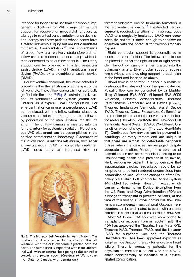
Intended for longer-term use than a balloon pump,general indications for VAD usage can includesupport for recovery of myocardial function, asa bridge to eventual transplantation, or as destina-tion therapy for those patients whose hearts havesuffered irreversible injury but are not candidatesfor cardiac transplantation.17 The biomechanicsof blood flow are relatively straightforward: aninflow cannula is connected to a pump, which isthen connected to an outflow cannula. Circulatorysupport can be provided with a left ventricularassist device (LVAD), a right ventricular assistdevice (RVAD), or a biventricular assist device(BiVAD).
For left ventricular support, the inflow catheter isplaced in either the left atrium or at the apex of theleft ventricle. The outflow cannula is then surgicallygrafted into the aorta.18 Fig. 2 illustrates the Nova-cor Left Ventricular Assist System (WorldHeart,Ontario) as a typical LVAD configuration. Foremergent, short-term use, a percutaneous LVADcan be placed, with the inflow catheter placed byvenous cannulation into the right atrium, followedby perforation of the atrial septum into the leftatrium. The outflow cannula is inserted into thefemoral artery for systemic circulation. Percutane-ous VAD placement can be accomplished in thecardiac catheterization laboratory. Placement ofthe inflow cannula into the left atrium, with eithera percutaneous LVAD or surgically implantedLVAD, does carry an increased risk for
thromboembolism due to thrombus formation inthe left ventricular cavity.18 If extended cardiacsupport is required, transition from a percutaneousLVAD to a surgically implanted LVAD can occurwhen the patient is stable enough to consider anoperation with the potential for cardiopulmonarybypass.
Right ventricular support is accomplished inmuch the same fashion. The inflow cannula canbe placed in either the right atrium or right ventri-cle. The outflow cannula is then grafted into thepulmonary artery. Biventricular support requirestwo devices, one providing support to each sideof the heart and inserted as above.
Blood flow is generated in either a pulsatile orcontinuous flow, depending on the specific device.Pulsatile flow can be generated by air bladderfilling Abiomed BVS 5000, Abiomed BVS 5000[Abiomed, Danvers, Massachusetts], ThoratecPercutaneous Ventricular Assist Device [PVAD],Thoratec Implantable Ventricular Assist Device[IVAD; Thoratec Corp., Pleasanton, California] orby a pusher plate that can be driven by either elec-tric motor (Thoratec HeartMate XVE, Novacor LeftVentricular Assist System [LVAS; WorldHeart, On-tario]) or pneumatic system (Thoratec HeartMateIP). Continuous flow devices can be powered bycentrifugal or rotary motors and are unique inthat there are almost no detectable peripheralpulses when the devices are engaged despiteadequate circulation. Although this absence ofpalpable pulse can be merely disconcerting to anunsuspecting health care provider in an awake,alert, responsive patient, it is conceivable thatinappropriate cardiac resuscitation could be at-tempted on a patient rendered unconscious fromnoncardiac causes. With the exception of the De-bakey VAD Child Left Ventricular Assist System(MicroMed Technology, Houston, Texas), whichcarries a Humanitarian Device Exemption fromthe US Food and Drug Administration (FDA) asa bridge to transplant in pediatric patients, at thetime of this writing all other continuous flow sys-tems are considered investigational. Outpatient en-counters can be anticipated to occur with patientsenrolled in clinical trials of these devices, however.
Most VADs are FDA approved as a bridge totransplant or recovery from an acute insult. TheFDA has approved the Thoratec HeartMate XVE,Thoratec IVAD, Thoratec PVAD, and the NovacorLVAS for outpatient use, and the ThoratecHeartMate XVE has been approved explicitly aslong-term destination therapy for end-stage heartfailure. There is increasing potential for thephysician to care for a patient who has a VAD,either coincidentally or because of a device-related complication.
Fig. 2. The Novacor Left Ventricular Assist System. Theintake conduit is attached to the apex of the leftventricle, with the outflow conduit grafted onto theaorta. The pump itself is implanted within the abdom-inal wall, with an external lead going to the controllerconsole and power packs. (Courtesy of WorldHeartInc., Ontario, Canada; with permission.)
Hiestand58
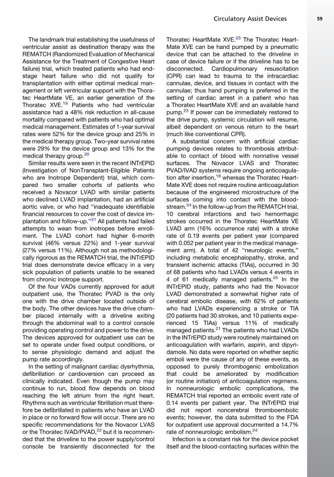
The landmark trial establishing the usefulness ofventricular assist as destination therapy was theREMATCH (Randomized Evaluation of MechanicalAssistance for the Treatment of Congestive Heartfailure) trial, which treated patients who had end-stage heart failure who did not qualify fortransplantation with either optimal medical man-agement or left ventricular support with the Thora-tec HeartMate VE, an earlier generation of theThoratec XVE.19 Patients who had ventricularassistance had a 48% risk reduction in all-causemortality compared with patients who had optimalmedical management. Estimates of 1-year survivalrates were 52% for the device group and 25% inthe medical therapy group. Two-year survival rateswere 29% for the device group and 13% for themedical therapy group.20
Similar results were seen in the recent INTrEPID(Investigation of NonTransplant-Eligible Patientswho are Inotrope Dependent) trial, which com-pared two smaller cohorts of patients whoreceived a Novacor LVAD with similar patientswho declined LVAD implantation, had an artificialaortic valve, or who had ‘‘inadequate identifiablefinancial resources to cover the cost of device im-plantation and follow-up.’’21 All patients had failedattempts to wean from inotropes before enroll-ment. The LVAD cohort had higher 6-monthsurvival (46% versus 22%) and 1-year survival(27% versus 11%). Although not as methodologi-cally rigorous as the REMATCH trial, the INTrEPIDtrial does demonstrate device efficacy in a verysick population of patients unable to be weanedfrom chronic inotrope support.
Of the four VADs currently approved for adultoutpatient use, the Thoratec PVAD is the onlyone with the drive chamber located outside ofthe body. The other devices have the drive cham-ber placed internally with a driveline exitingthrough the abdominal wall to a control consoleproviding operating control and power to the drive.The devices approved for outpatient use can beset to operate under fixed output conditions, orto sense physiologic demand and adjust thepump rate accordingly.
In the setting of malignant cardiac dysrhythmia,defibrillation or cardioversion can proceed asclinically indicated. Even though the pump maycontinue to run, blood flow depends on bloodreaching the left atrium from the right heart.Rhythms such as ventricular fibrillation must there-fore be defibrillated in patients who have an LVADin place or no forward flow will occur. There are nospecific recommendations for the Novacor LVASor the Thoratec IVAD/PVAD,22 but it is recommen-ded that the driveline to the power supply/controlconsole be transiently disconnected for the
Thoratec HeartMate XVE.23 The Thoratec Heart-Mate XVE can be hand pumped by a pneumaticdevice that can be attached to the driveline incase of device failure or if the driveline has to bedisconnected. Cardiopulmonary resuscitation(CPR) can lead to trauma to the intracardiaccannulae, device, and tissues in contact with thecannulae; thus hand pumping is preferred in thesetting of cardiac arrest in a patient who hasa Thoratec HeartMate XVE and an available handpump.23 If power can be immediately restored tothe drive pump, systemic circulation will resume,albeit dependent on venous return to the heart(much like conventional CPR).
A substantial concern with artificial cardiacpumping devices relates to thrombosis attribut-able to contact of blood with nonnative vesselsurfaces. The Novacor LVAS and ThoratecPVAD/IVAD systems require ongoing anticoagula-tion after insertion,18 whereas the Thoratec Heart-Mate XVE does not require routine anticoagulationbecause of the engineered microstructure of thesurfaces coming into contact with the blood-stream.24 In the follow-up from the REMATCH trial,10 cerebral infarctions and two hemorrhagicstrokes occurred in the Thoratec HeartMate VELVAD arm (16% occurrence rate) with a strokerate of 0.19 events per patient year (comparedwith 0.052 per patient year in the medical manage-ment arm). A total of 42 ‘‘neurologic events,’’including metabolic encephalopathy, stroke, andtransient ischemic attacks (TIAs), occurred in 30of 68 patients who had LVADs versus 4 events in4 of 61 medically managed patients.25 In theINTrEPID study, patients who had the NovacorLVAD demonstrated a somewhat higher rate ofcerebral embolic disease, with 62% of patientswho had LVADs experiencing a stroke or TIA(20 patients had 30 strokes, and 10 patients expe-rienced 15 TIAs) versus 11% of medicallymanaged patients.21 The patients who had LVADsin the INTrEPID study were routinely maintained onanticoagulation with warfarin, aspirin, and dipyri-damole. No data were reported on whether septicemboli were the cause of any of these events, asopposed to purely thrombogenic embolizationthat could be ameliorated by modification(or routine initiation) of anticoagulation regimens.In nonneurologic embolic complications, theREMATCH trial reported an embolic event rate of0.14 events per patient year. The INTrEPID trialdid not report noncerebral thromboembolicevents; however, the data submitted to the FDAfor outpatient use approval documented a 14.7%rate of nonneurologic embolism.24
Infection is a constant risk for the device pocketitself and the blood-contacting surfaces within the
Circulatory Assist Devices 59
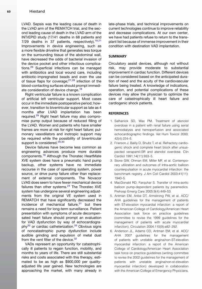
LVAD. Sepsis was the leading cause of death inthe LVAD arm of the REMATCH trial, and the sec-ond leading cause of death in the LVAD arm of theINTrEPID study (17/41 deaths in 68 patients and7/29 deaths in 37 patients, respectively).19,21
Improvements in device engineering, such asa more flexible driveline that generates less torqueon the surrounding tissue of the abdominal wall,have decreased the odds of bacterial invasion ofthe device pocket and other infectious complica-tions.26 Superficial infections can be managedwith antibiotics and local wound care, includingantibiotic-impregnated beads and even the useof tissue flaps for coverage;27,28 infection of theblood-contacting surfaces should prompt immedi-ate consideration of device change.18
Right ventricular failure is a known complicationof artificial left ventricular support. Most casesoccur in the immediate postoperative period; how-ever, transition to biventricular support as late as 4months after LVAD implantation has beenrequired.29 Right heart failure may also compro-mise pump output because of reduced filling ofthe LVAD. Women and patients who have smallerframes are more at risk for right heart failure; pul-monary vasodilators and inotropic support maybe required while the possibility of biventricularsupport is considered.29,30
Device failures have become less common asengineering advances produce more durablecomponents.18 Although the Thoratec HeartMateXVE system does have a pneumatic hand pumpbackup, other systems have no immediaterecourse in the case of operating system, powersource, or drive pump failure other than replace-ment of external components. The NovacorLVAS does seem to have fewer mechanical devicefailures than other systems.24 The Thoratec XVEsystem has undergone several engineering adjust-ments from the original VE system used inREMATCH that have significantly decreased theincidence of mechanical failure,31 but thereremains a need for long-term surveillance. Patientpresentation with symptoms of acute decompen-sated heart failure should prompt an evaluationfor VAD dysfunction by way of echocardiogra-phy32 or cardiac catheterization.33 Obvious signsof noncatastrophic pump dysfunction includeaudible grinding and expulsion of metal dustfrom the vent filter of the device.32
VADs represent an opportunity for catastrophi-cally ill patients to regain function, mobility, andmonths to years of life. There are still substantialrisks and costs associated with this therapy, esti-mated to be as high as $900,000 per quality-adjusted life year gained. New technologies areapproaching the market, with many already in
late-phase trials, and technical improvements oncurrent technologies continue to improve reliabilityand decrease complications. At our own center,we have had patients refuse to return to the trans-plant list because of immense improvement in theircondition with destination VAD implantation.
SUMMARY
Circulatory assist devices, although not withoutrisk, may provide moderate to substantialimprovement in cardiac function. Different devicescan be considered based on the anticipated dura-tion of need and the acuity of the cardiovascularfailure being treated. A knowledge of indications,operation, and potential complications of thesedevices may allow the physician to optimize thecare of catastrophically ill heart failure andcardiogenic shock patients.
REFERENCES
1. Salhanick SD, Wax PM. Treatment of atenolol
overdose in a patient with renal failure using serial
hemodialysis and hemoperfusion and associated
echocardiographic findings. Vet Hum Toxicol 2000;
42(4):224–5.
2. Frierson J, Bailly D, Shultz T, et al. Refractory cardio-
genic shock and complete heart block after unsus-
pected verapamil-SR and atenolol overdose. Clin
Cardiol 1991;14(11):933–5.
3. Stone GW, Ohman EM, Miller MF, et al. Contempo-
rary utilization and outcomes of intra-aortic balloon
counterpulsation in acute myocardial infarction: the
benchmark registry. J Am Coll Cardiol 2003;41(11):
1940–5.
4. MacDonald RD, Farquhar S. Transfer of intra-aortic
balloon pump-dependent patients by paramedics.
Prehosp Emerg Care 2005;9(4):449–53.
5. Antman EM, Anbe DT, Armstrong PW, et al. ACC/
AHA guidelines for the management of patients
with ST-elevation myocardial infarction: a report of
the American College of Cardiology/American Heart
Association task force on practice guidelines
(committee to revise the 1999 guidelines for the
management of patients with acute myocardial
infarction). Circulation 2004;110(9):e82–292.
6. Anderson JL, Adams CD, Antman EM, et al. ACC/
AHA 2007 guidelines for the management
of patients with unstable angina/non-ST-elevation
myocardial infarction: a report of the American
College of Cardiology/American Heart Association
task force on practice guidelines (writing committee
to revise the 2002 guidelines for the management of
patients with unstable angina/non-st-elevation
myocardial infarction) developed in collaboration
with the American College of Emergency Physicians,
Hiestand60
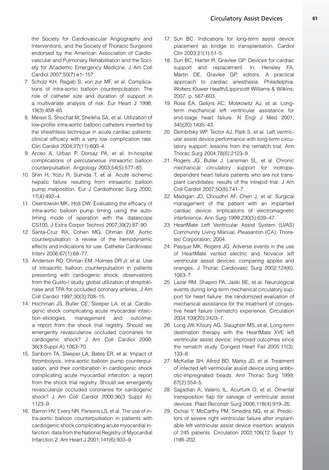
the Society for Cardiovascular Angiography and
Interventions, and the Society of Thoracic Surgeons
endorsed by the American Association of Cardio-
vascular and Pulmonary Rehabilitation and the Soci-
ety for Academic Emergency Medicine. J Am Coll
Cardiol 2007;50(7):e1–157.
7. Scholz KH, Ragab S, von zur MF, et al. Complica-
tions of intra-aortic balloon counterpulsation. The
role of catheter size and duration of support in
a multivariate analysis of risk. Eur Heart J 1998;
19(3):458–65.
8. Meisel S, Shochat M, Sheikha SA, et al. Utilization of
low-profile intra-aortic balloon catheters inserted by
the sheathless technique in acute cardiac patients:
clinical efficacy with a very low complication rate.
Clin Cardiol 2004;27(11):600–4.
9. Arceo A, Urban P, Dorsaz PA, et al. In-hospital
complications of percutaneous intraaortic balloon
counterpulsation. Angiology 2003;54(5):577–85.
10. Shin H, Yozu R, Sumida T, et al. Acute ischemic
hepatic failure resulting from intraaortic balloon
pump malposition. Eur J Cardiothorac Surg 2000;
17(4):492–4.
11. Osentowski MK, Holt DW. Evaluating the efficacy of
intra-aortic balloon pump timing using the auto-
timing mode of operation with the datascope
CS100. J Extra Corpor Technol 2007;39(2):87–90.
12. Santa-Cruz RA, Cohen MG, Ohman EM. Aortic
counterpulsation: a review of the hemodynamic
effects and indications for use. Catheter Cardiovasc
Interv 2006;67(1):68–77.
13. Anderson RD, Ohman EM, Holmes DR Jr, et al. Use
of intraaortic balloon counterpulsation in patients
presenting with cardiogenic shock: observations
from the Gusto-I study. global utilization of streptoki-
nase and TPA for occluded coronary arteries. J Am
Coll Cardiol 1997;30(3):708–15.
14. Hochman JS, Buller CE, Sleeper LA, et al. Cardio-
genic shock complicating acute myocardial infarc-
tion–etiologies, management and outcome:
a report from the shock trial registry. Should we
emergently revascularize occluded coronaries for
cardiogenic shock? J Am Coll Cardiol 2000;
36(3 Suppl A):1063–70.
15. Sanborn TA, Sleeper LA, Bates ER, et al. Impact of
thrombolysis, intra-aortic balloon pump counterpul-
sation, and their combination in cardiogenic shock
complicating acute myocardial infarction: a report
from the shock trial registry. Should we emergently
revascularize occluded coronaries for cardiogenic
shock? J Am Coll Cardiol 2000;36(3 Suppl A):
1123–9.
16. Barron HV, Every NR, Parsons LS, et al. The use of in-
tra-aortic balloon counterpulsation in patients with
cardiogenic shock complicating acute myocardial in-
farction: data from the National Registry of Myocardial
Infarction 2. Am Heart J 2001;141(6):933–9.
17. Sun BC. Indications for long-term assist device
placement as bridge to transplantation. Cardiol
Clin 2003;21(1):51–5.
18. Sun BC, Harter R, Gravlee GP. Devices for cardiac
support and replacement. In: Hensley FA,
Martin DE, Gravlee GP, editors. A practical
approach to cardiac anesthesia. Philadelphia:
Wolters Kluwer Health/Lippincott Williams & Wilkins;
2007. p. 587–603.
19. Rose EA, Gelijns AC, Moskowitz AJ, et al. Long-
term mechanical left ventricular assistance for
end-stage heart failure. N Engl J Med 2001;
345(20):1435–43.
20. Dembitsky WP, Tector AJ, Park S, et al. Left ventric-
ular assist device performance with long-term circu-
latory support: lessons from the rematch trial. Ann
Thorac Surg 2004;78(6):2123–9.
21. Rogers JG, Butler J, Lansman SL, et al. Chronic
mechanical circulatory support for inotrope-
dependent heart failure patients who are not trans-
plant candidates: results of the intrepid trial. J Am
Coll Cardiol 2007;50(8):741–7.
22. Madigan JD, Choudhri AF, Chen J, et al. Surgical
management of the patient with an implanted
cardiac device: implications of electromagnetic
interference. Ann Surg 1999;230(5):639–47.
23. HeartMate Left Ventricular Assist System (LVAS)
Community Living Manual. Pleasanton (CA): Thora-
tec Corporation; 2004.
24. Pasque MK, Rogers JG. Adverse events in the use
of HeartMate vented electric and Novacor left
ventricular assist devices: comparing apples and
oranges. J Thorac Cardiovasc Surg 2002;124(6):
1063–7.
25. Lazar RM, Shapiro PA, Jaski BE, et al. Neurological
events during long-term mechanical circulatory sup-
port for heart failure: the randomized evaluation of
mechanical assistance for the treatment of conges-
tive heart failure (rematch) experience. Circulation
2004;109(20):2423–7.
26. Long JW, Kfoury AG, Slaughter MS, et al. Long-term
destination therapy with the HeartMate XVE left
ventricular assist device: improved outcomes since
the rematch study. Congest Heart Fail 2005;11(3):
133–8.
27. McKellar SH, Allred BD, Marks JD, et al. Treatment
of infected left ventricular assist device using antibi-
otic-impregnated beads. Ann Thorac Surg 1999;
67(2):554–5.
28. Sajjadian A, Valerio IL, Acurturk O, et al. Omental
transposition flap for salvage of ventricular assist
devices. Plast Reconstr Surg 2006;118(4):919–26.
29. Ochiai Y, McCarthy PM, Smedira NG, et al. Predic-
tors of severe right ventricular failure after implant-
able left ventricular assist device insertion: analysis
of 245 patients. Circulation 2002;106(12 Suppl 1):
I198–202.
Circulatory Assist Devices 61

30. Dang NC, Topkara VK, Mercando M, et al. Right
heart failure after left ventricular assist device im-
plantation in patients with chronic congestive heart
failure. J Heart Lung Transplant 2006;25(1):1–6.
31. Pagani FD, Long JW, Dembitsky WP, et al. Improved
mechanical reliability of the HeartMate XVE left
ventricular assist system. Ann Thorac Surg 2006;
82(4):1413–8.
32. Myers TJ, Palanichamy N, La Francesca S, et al.
Management of multiple left ventricular assist device
failures in a patient. J Heart Lung Transplant 2007;
26(1):98–100.
33. Horton SC, Khodaverdian R, Powers A, et al. Left
ventricular assist device malfunction: a systematic
approach to diagnosis. J Am Coll Cardiol 2004;
43(9):1574–83.
Hiestand62

Cardiac Devicesin EmergencyDepartment HeartFailure PatientsJames F. Neuenschwander II, MD, FACEP*
Noncardiologists frequently provide care for pa-tients with cardiac devices in the event that a cardi-ologist is not available or the patient is too ill to waitfor a scheduled office appointment. As these de-vices are quite sophisticated, this article attemptsto provide a basic understanding of the indicationsfor these therapies, how to diagnose a potentialproblem, and whom to contact if a malfunctionhas occurred. The article also discusses the infor-mation obtainable from these devices and how itcan be incorporated into the clinical approach tohelp guide management.
IMPLANTABLE PACEMAKERS AND INTERNALCARDIAC DEFIBRILLATORSHistory and Background
With an aging United States population, the preva-lence of heart disease continues to increase. Pa-tients who previously would have died fromcomplications of their diseases now survive to pres-ent to the hospital in acute distress. Furthermore, anincreasing population of at-risk patients providesa greater number of patients who can derive benefitfrom devices to improve their cardiac function.
The first pacemaker was implanted in 1958 andthe first defibrillator was placed at the JohnsHopkins Hospital in 1980. When key studiesdemonstrated increased survival in patients withthe implantation of an internal cardioverter
defibrillator (ICD),1–3 the number of patients withsuch devices increased dramatically. More than100,000 such devices are implanted annually inthe United States alone, and the noncardiologistcan expect to encounter an increasing number ofpatients with these devices.4 Several studies haveshown the cost effectiveness of ICDs in patientsat high risk for ventricular tachycardia/ventricularfibrillation (VT/VF) when compared with antiar-rhythmic drug therapy.5 ICDs have been shown tobenefit patients with documented VT/VF (second-ary prevention) and patients at risk for VT/VF witha lower ejection fraction but no prior VT/VF (primaryprevention) when compared with antiarrhythmictherapy.6
An ICD consists of a pulse generator, a leadsystem, and electrodes capable of sensing andshocking. The size of the devices has decreasedand they can be implanted subcutaneously or inthe area of the pectoral muscle. Some of thesedevices are capable of defibrillation, cardiover-sion, and pacing both bradycardic and tachycar-dic rhythms. The monitoring of these devices hasalso become sophisticated enough that physio-logic data can be tracked in the patient’s homethrough telemetry and can provide reporting tomedical providers on a daily basis. Although anICD can only be programmed by highly trainedand qualified personnel, the interrogation of suchdevices is straight forward, requiring only the
The author has received funding support from Medtronic for Research Grant Speakers Bureau: Medtronic,St. Jude, Boston Scientific.The Ohio State University, Columbus, OH, USA* Department of Emergency Medicine, The Ohio State University, 376 West 10th Avenue, Columbus, OH43210-1252.E-mail address: [email protected]
KEYWORDS� Heart failure � Cardiac arrhythmias� Cardiac pacing � Ultrafiltration � Pacemaker� Internal cardioverter defibrillator
Heart Failure Clin 5 (2009) 63–73doi:10.1016/j.hfc.2008.08.0121551-7136/08/$ – see front matter ª 2008 Elsevier Inc. All rights reserved. he
artf
ailu
re.th
ecli
nics
.com
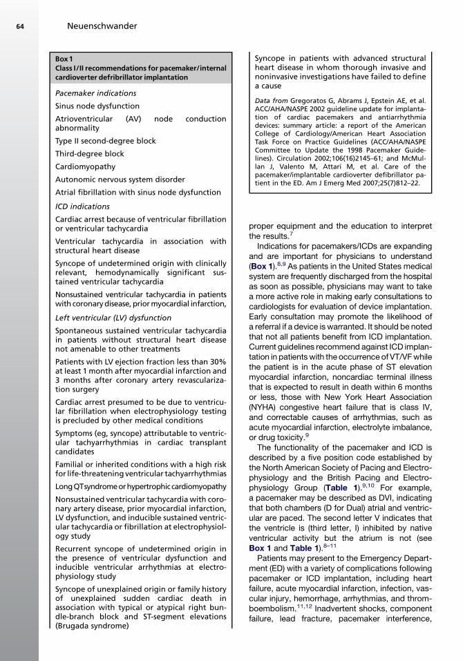
proper equipment and the education to interpretthe results.7
Indications for pacemakers/ICDs are expandingand are important for physicians to understand(Box 1).8,9 As patients in the United States medicalsystem are frequently discharged from the hospitalas soon as possible, physicians may want to takea more active role in making early consultations tocardiologists for evaluation of device implantation.Early consultation may promote the likelihood ofa referral if a device is warranted. It should be notedthat not all patients benefit from ICD implantation.Current guidelines recommend against ICD implan-tation in patients with the occurrence of VT/VF whilethe patient is in the acute phase of ST elevationmyocardial infarction, noncardiac terminal illnessthat is expected to result in death within 6 monthsor less, those with New York Heart Association(NYHA) congestive heart failure that is class IV,and correctable causes of arrhythmias, such asacute myocardial infarction, electrolyte imbalance,or drug toxicity.9
The functionality of the pacemaker and ICD isdescribed by a five position code established bythe North American Society of Pacing and Electro-physiology and the British Pacing and Electro-physiology Group (Table 1).9,10 For example,a pacemaker may be described as DVI, indicatingthat both chambers (D for Dual) atrial and ventric-ular are paced. The second letter V indicates thatthe ventricle is (third letter, I) inhibited by nativeventricular activity but the atrium is not (seeBox 1 and Table 1).8–11
Patients may present to the Emergency Depart-ment (ED) with a variety of complications followingpacemaker or ICD implantation, including heartfailure, acute myocardial infarction, infection, vas-cular injury, hemorrhage, arrhythmias, and throm-boembolism.11,12 Inadvertent shocks, componentfailure, lead fracture, pacemaker interference,
Box1Class I/II recommendations for pacemaker/internalcardioverter defribrillator implantation
Pacemaker indications
Sinus node dysfunction
Atrioventricular (AV) node conductionabnormality
Type II second-degree block
Third-degree block
Cardiomyopathy
Autonomic nervous system disorder
Atrial fibrillation with sinus node dysfunction
ICD indications
Cardiac arrest because of ventricular fibrillationor ventricular tachycardia
Ventricular tachycardia in association withstructural heart disease
Syncope of undetermined origin with clinicallyrelevant, hemodynamically significant sus-tained ventricular tachycardia
Nonsustained ventricular tachycardia in patientswith coronary disease, prior myocardial infarction,
Left ventricular (LV) dysfunction
Spontaneous sustained ventricular tachycardiain patients without structural heart diseasenot amenable to other treatments
Patients with LV ejection fraction less than 30%at least 1 month after myocardial infarction and3 months after coronary artery revasculariza-tion surgery
Cardiac arrest presumed to be due to ventricu-lar fibrillation when electrophysiology testingis precluded by other medical conditions
Symptoms (eg, syncope) attributable to ventric-ular tachyarrhythmias in cardiac transplantcandidates
Familial or inherited conditions with a high riskfor life-threatening ventricular tachyarrhythmias
LongQTsyndromeorhypertrophiccardiomyopathy
Nonsustained ventricular tachycardia with coro-nary artery disease, prior myocardial infarction,LV dysfunction, and inducible sustained ventric-ular tachycardia or fibrillation at electrophysiol-ogy study
Recurrent syncope of undetermined origin inthe presence of ventricular dysfunction andinducible ventricular arrhythmias at electro-physiology study
Syncope of unexplained origin or family historyof unexplained sudden cardiac death inassociation with typical or atypical right bun-dle-branch block and ST-segment elevations(Brugada syndrome)
Syncope in patients with advanced structuralheart disease in whom thorough invasive andnoninvasive investigations have failed to definea cause
Data from Gregoratos G, Abrams J, Epstein AE, et al.ACC/AHA/NASPE 2002 guideline update for implanta-tion of cardiac pacemakers and antiarrhythmiadevices: summary article: a report of the AmericanCollege of Cardiology/American Heart AssociationTask Force on Practice Guidelines (ACC/AHA/NASPECommittee to Update the 1998 Pacemaker Guide-lines). Circulation 2002;106(16)2145–61; and McMul-lan J, Valento M, Attari M, et al. Care of thepacemaker/implantable cardioverter defibrillator pa-tient in the ED. Am J Emerg Med 2007;25(7)812–22.
Neuenschwander64
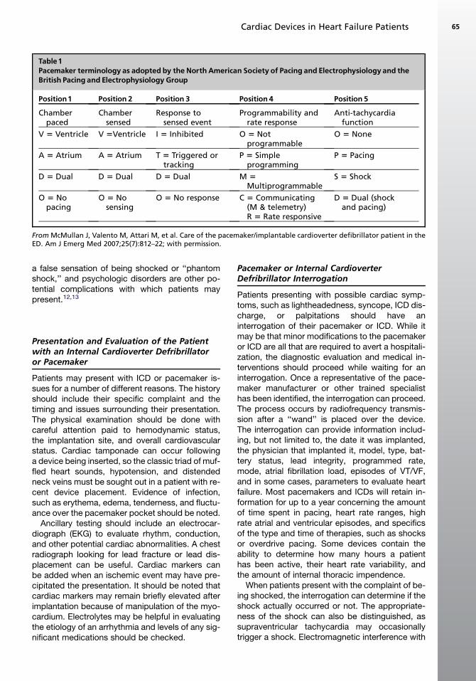
a false sensation of being shocked or ‘‘phantomshock,’’ and psychologic disorders are other po-tential complications with which patients maypresent.12,13
Presentation and Evaluation of the Patientwith an Internal Cardioverter Defribrillatoror Pacemaker
Patients may present with ICD or pacemaker is-sues for a number of different reasons. The historyshould include their specific complaint and thetiming and issues surrounding their presentation.The physical examination should be done withcareful attention paid to hemodynamic status,the implantation site, and overall cardiovascularstatus. Cardiac tamponade can occur followinga device being inserted, so the classic triad of muf-fled heart sounds, hypotension, and distendedneck veins must be sought out in a patient with re-cent device placement. Evidence of infection,such as erythema, edema, tenderness, and fluctu-ance over the pacemaker pocket should be noted.
Ancillary testing should include an electrocar-diograph (EKG) to evaluate rhythm, conduction,and other potential cardiac abnormalities. A chestradiograph looking for lead fracture or lead dis-placement can be useful. Cardiac markers canbe added when an ischemic event may have pre-cipitated the presentation. It should be noted thatcardiac markers may remain briefly elevated afterimplantation because of manipulation of the myo-cardium. Electrolytes may be helpful in evaluatingthe etiology of an arrhythmia and levels of any sig-nificant medications should be checked.
Pacemaker or Internal CardioverterDefribrillator Interrogation
Patients presenting with possible cardiac symp-toms, such as lightheadedness, syncope, ICD dis-charge, or palpitations should have aninterrogation of their pacemaker or ICD. While itmay be that minor modifications to the pacemakeror ICD are all that are required to avert a hospitali-zation, the diagnostic evaluation and medical in-terventions should proceed while waiting for aninterrogation. Once a representative of the pace-maker manufacturer or other trained specialisthas been identified, the interrogation can proceed.The process occurs by radiofrequency transmis-sion after a ‘‘wand’’ is placed over the device.The interrogation can provide information includ-ing, but not limited to, the date it was implanted,the physician that implanted it, model, type, bat-tery status, lead integrity, programmed rate,mode, atrial fibrillation load, episodes of VT/VF,and in some cases, parameters to evaluate heartfailure. Most pacemakers and ICDs will retain in-formation for up to a year concerning the amountof time spent in pacing, heart rate ranges, highrate atrial and ventricular episodes, and specificsof the type and time of therapies, such as shocksor overdrive pacing. Some devices contain theability to determine how many hours a patienthas been active, their heart rate variability, andthe amount of internal thoracic impendence.
When patients present with the complaint of be-ing shocked, the interrogation can determine if theshock actually occurred or not. The appropriate-ness of the shock can also be distinguished, assupraventricular tachycardia may occasionallytrigger a shock. Electromagnetic interference with
Table1Pacemaker terminology as adopted by the North American Society of Pacing and Electrophysiology and theBritish Pacing and Electrophysiology Group
Position1 Position 2 Position 3 Position 4 Position 5
Chamberpaced
Chambersensed
Response tosensed event
Programmability andrate response
Anti-tachycardiafunction
V 5 Ventricle V 5Ventricle I 5 Inhibited O 5 Notprogrammable
O 5 None
A 5 Atrium A 5 Atrium T 5 Triggered ortracking
P 5 Simpleprogramming
P 5 Pacing
D 5 Dual D 5 Dual D 5 Dual M 5Multiprogrammable
S 5 Shock
O 5 Nopacing
O 5 Nosensing
O 5 No response C 5 Communicating(M & telemetry)R 5 Rate responsive
D 5 Dual (shockand pacing)
From McMullan J, Valento M, Attari M, et al. Care of the pacemaker/implantable cardioverter defibrillator patient in theED. Am J Emerg Med 2007;25(7):812–22; with permission.
Cardiac Devices in Heart Failure Patients 65

the device can sometimes produce symptoms andwill be discussed in the subsection titled ‘‘Otherpacemaker issues.’’ Arrhythmias can produce diz-ziness or syncope and evaluating the pacemakercan help in the evaluation of the patient.
The patient with a pacemaker or ICD is sup-posed to carry a card with information includingmanufacturer, model number, therapeutic options,and lead system. Each device can only be interro-gated by the manufacturer’s programmer orreader. A representative of the device (pacemakeror ICD) company or other electrophysiology nurseor technician can be contacted to interrogate theequipment. The Appendix lists company namesand contact numbers for the majority of devicesused currently. If the patient does not havea card, a search of the medical records for the op-eration note will likely provide the make and modelof the implantable device. In addition, device man-ufacturers maintain registries with the patient’sidentification to assist with correctly matchinga patient to their device.
Using Device Data in HeartFailure Management
Some devices possess data that may be useful indiagnosing the presence or absence of heart fail-ure. Impedance cardiography (ICG) has beenshown to be a noninvasive measurement of car-diac output, cardiac index, and thoracic fluid con-tent.14,15 Electrical impedance is the resistance toflow of an electrical current. Blood and fluids aregood conductors and therefore have low imped-ance, while bone and tissue have higher imped-ance because of their poor conductivity.16 Thesedevices have been shown to provide informationthat is helpful in determining a diagnosis in theED, identifying patients at increased near-termrisk of recurrent decompensation, or predict hos-pitalization. ICG monitors can be used at the bed-side by applying four pairs of electrodes to theneck and thorax. As the flow of fluid changes,the electrical impedance changes proportionally.The machine can then calculate a number of he-modynamic parameters, including systemic vas-cular resistance, stroke volume, cardiac output,and an index of thoracic fluid. The increased fluidin the thorax that occurs in a heart failure exacer-bation decreases overall average impedance andcan suggest a fluid overloaded state.14–16
Some ICDs have the capability of monitoring in-trathoracic impedance.17–19 Selected MedtronicICDs have this capability in the form of Optivol,which has shown an inverse correlation betweenintrathoracic impedance and intracardiac fillingpressure in the NYHA class III and IV patients.18
In a study by Wang, the device recorded 64 mea-surements between noon and 5 PM and averagedthem for a single data point per day. Impedancemeasurement decreased with increased thoracicfluid an average of about 2 weeks before symptomonset and hospitalization, suggesting that physi-cians may be able to intervene before symptomonset. A default setting for Optivol had a 76.9%sensitivity for predicting hospitalization.19 Less en-couraging results were reported in another study,which demonstrated a sensitivity of 60% to detectheart failure decompensation but had a 40% false-positive rate.20 These devices show promise inpreliminary data, yet larger trials are needed inthe acute care setting.
Some manufacturers of pacemakers and ICDs,such as Boston Scientific, provide home monitor-ing of device functions, blood pressure, andweight measurements. This information has thepotential to be very helpful to a physician manag-ing the patient. The amount of time a patientspends in daily activity can be tracked and usedto help determine the likelihood of heart failure.Heart rate variability and atrial fibrillation burdencan also be incorporated into determining thepresence or absence of heart failure.21
Surgical Complications of Pacemakersand Internal Cardioverter Defribrillators
Surgical complications stemming from pacemakeror ICD implantation are numerous and can be en-countered in the ED. Arterial or venous bleedingbecause of dissection of fascial planes can causea hematoma at the implantation site. Up to 1% to2% of all large palpable hematomas will requiresurgical evacuation.22 Surgical intervention is rec-ommended and needle aspiration should beavoided because of its ineffectiveness and risk ofintroducing infection.23
Infections with ICDs can be classified as early(less than 60 days) after implantation or late. Upto 8% of patients will be affected by infection oftheir device.9 The early infections present as typi-cal skin infections with erythema, edema, and ten-derness at the wound site. Late infections havea more indolent presentation and pain can be theonly complaint. Staphylococcus aureus and epi-dermidis are cited as being responsible for about70% to 95% of all pacemaker and ICD infectionsand current recommendations state vancomycinshould be started in the ED.24,25 A complete bloodcount, blood cultures, and empiric antibioticsshould be started when an infection is suspected.Aspiration of the device pocket can be useful andis best left to a physician experienced with thisprocedure because of the potential of damaging
Neuenschwander66
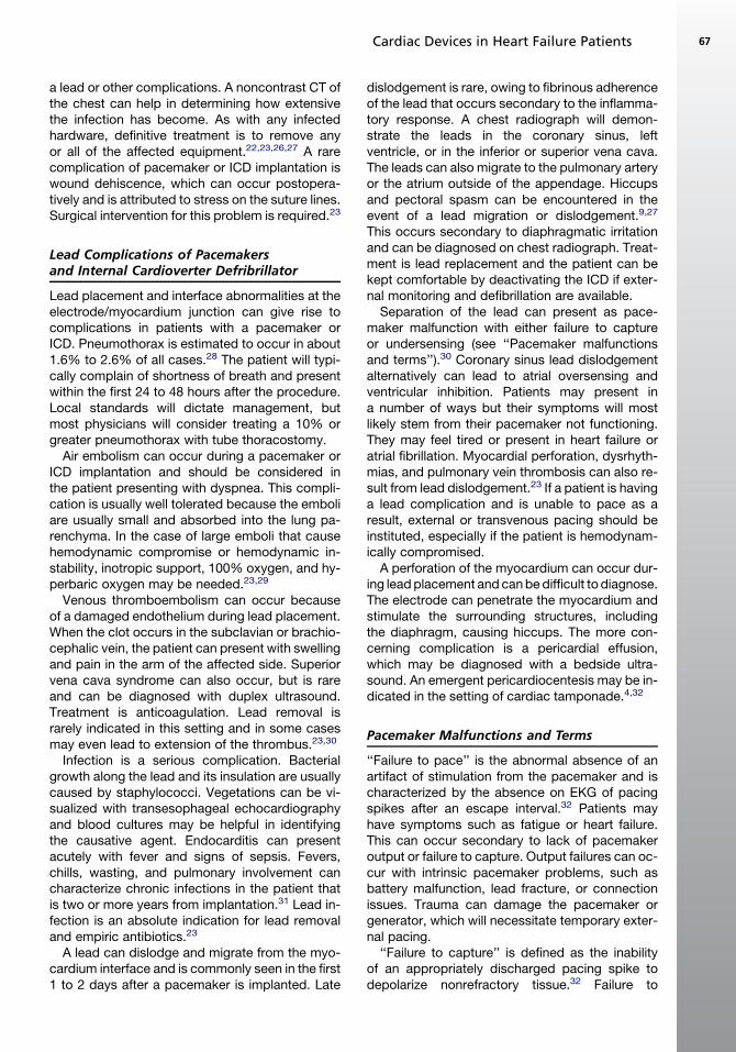
a lead or other complications. A noncontrast CT ofthe chest can help in determining how extensivethe infection has become. As with any infectedhardware, definitive treatment is to remove anyor all of the affected equipment.22,23,26,27 A rarecomplication of pacemaker or ICD implantation iswound dehiscence, which can occur postopera-tively and is attributed to stress on the suture lines.Surgical intervention for this problem is required.23
Lead Complications of Pacemakersand Internal Cardioverter Defribrillator
Lead placement and interface abnormalities at theelectrode/myocardium junction can give rise tocomplications in patients with a pacemaker orICD. Pneumothorax is estimated to occur in about1.6% to 2.6% of all cases.28 The patient will typi-cally complain of shortness of breath and presentwithin the first 24 to 48 hours after the procedure.Local standards will dictate management, butmost physicians will consider treating a 10% orgreater pneumothorax with tube thoracostomy.
Air embolism can occur during a pacemaker orICD implantation and should be considered inthe patient presenting with dyspnea. This compli-cation is usually well tolerated because the emboliare usually small and absorbed into the lung pa-renchyma. In the case of large emboli that causehemodynamic compromise or hemodynamic in-stability, inotropic support, 100% oxygen, and hy-perbaric oxygen may be needed.23,29
Venous thromboembolism can occur becauseof a damaged endothelium during lead placement.When the clot occurs in the subclavian or brachio-cephalic vein, the patient can present with swellingand pain in the arm of the affected side. Superiorvena cava syndrome can also occur, but is rareand can be diagnosed with duplex ultrasound.Treatment is anticoagulation. Lead removal israrely indicated in this setting and in some casesmay even lead to extension of the thrombus.23,30
Infection is a serious complication. Bacterialgrowth along the lead and its insulation are usuallycaused by staphylococci. Vegetations can be vi-sualized with transesophageal echocardiographyand blood cultures may be helpful in identifyingthe causative agent. Endocarditis can presentacutely with fever and signs of sepsis. Fevers,chills, wasting, and pulmonary involvement cancharacterize chronic infections in the patient thatis two or more years from implantation.31 Lead in-fection is an absolute indication for lead removaland empiric antibiotics.23
A lead can dislodge and migrate from the myo-cardium interface and is commonly seen in the first1 to 2 days after a pacemaker is implanted. Late
dislodgement is rare, owing to fibrinous adherenceof the lead that occurs secondary to the inflamma-tory response. A chest radiograph will demon-strate the leads in the coronary sinus, leftventricle, or in the inferior or superior vena cava.The leads can also migrate to the pulmonary arteryor the atrium outside of the appendage. Hiccupsand pectoral spasm can be encountered in theevent of a lead migration or dislodgement.9,27
This occurs secondary to diaphragmatic irritationand can be diagnosed on chest radiograph. Treat-ment is lead replacement and the patient can bekept comfortable by deactivating the ICD if exter-nal monitoring and defibrillation are available.
Separation of the lead can present as pace-maker malfunction with either failure to captureor undersensing (see ‘‘Pacemaker malfunctionsand terms’’).30 Coronary sinus lead dislodgementalternatively can lead to atrial oversensing andventricular inhibition. Patients may present ina number of ways but their symptoms will mostlikely stem from their pacemaker not functioning.They may feel tired or present in heart failure oratrial fibrillation. Myocardial perforation, dysrhyth-mias, and pulmonary vein thrombosis can also re-sult from lead dislodgement.23 If a patient is havinga lead complication and is unable to pace as aresult, external or transvenous pacing should beinstituted, especially if the patient is hemodynam-ically compromised.
A perforation of the myocardium can occur dur-ing lead placement and can be difficult to diagnose.The electrode can penetrate the myocardium andstimulate the surrounding structures, includingthe diaphragm, causing hiccups. The more con-cerning complication is a pericardial effusion,which may be diagnosed with a bedside ultra-sound. An emergent pericardiocentesis may be in-dicated in the setting of cardiac tamponade.4,32
Pacemaker Malfunctions and Terms
‘‘Failure to pace’’ is the abnormal absence of anartifact of stimulation from the pacemaker and ischaracterized by the absence on EKG of pacingspikes after an escape interval.32 Patients mayhave symptoms such as fatigue or heart failure.This can occur secondary to lack of pacemakeroutput or failure to capture. Output failures can oc-cur with intrinsic pacemaker problems, such asbattery malfunction, lead fracture, or connectionissues. Trauma can damage the pacemaker orgenerator, which will necessitate temporary exter-nal pacing.
‘‘Failure to capture’’ is defined as the inabilityof an appropriately discharged pacing spike todepolarize nonrefractory tissue.32 Failure to
Cardiac Devices in Heart Failure Patients 67

capture can be visualized on EKG as a pacerspike that does not produce an effective pacingstimulus. Multiple causes, including battery fail-ure, lead fracture or dislodgement, faulty connec-tions, and cardiac perforations can cause failureto capture. Electrolyte derangements can leadto this problem, as well as antidysrhythmic drugsat supratherapeutic levels and even flecainide attherapeutic levels.9
‘‘Oversensing’’ is the unintended detection ofelectrical complexes by the pacemaker and ischaracterized on EKG by inhibition of the pace-maker and by the absence of pacing spikes afteran exceeded escape interval.32 Oversensing canoccur when the pacemaker senses the ventricleis in its native rhythm by accidentally interpretinga skeletal muscle contraction as ventricular.Electromagnetic interference, lithotripsy, cellulartelephones, and lead fractures can lead to thiscomplication.
‘‘Undersensing’’ is defined as failure of thepacemaker to detect an electrical complex dem-onstrated on EKG by pacing spikes that are pre-ceded by intrinsic P waves or QRS complexes.This can occur with lead fracture, lead positioning,inappropriate program sensitivity, myocardialinfarction, myocardial fibrosis, electrolyte distur-bances, battery failure, antiarrhythmic medica-tions, and component failure.32
To manage these patients, physicians shouldfirst consider external pacing in symptomaticpacemaker-dependent patients while the sourceof the problem is sought. A chest radiograph canbe helpful in identifying a lead fracture or migra-tion. Correctable causes, such as hyperkalemiaor acidosis or alkalosis should be addressedquickly, as they can lead to failure to capture. Anti-dysrhythmic medication levels need evaluationand treatment in appropriate patients. An electro-physiologist should be consulted early in all theseprocesses for input and updates.
Pacemaker-Induced Tachycardias
Pacemaker-mediated tachycardia (PMT) can oc-cur when a re-entry dysrhythmia is triggered bya premature ventricular complex. When a retro-grade signal is transmitted through the AV node,the atria are depolarized and the atrial sensorthen stimulates the ventricle to contract, thus cre-ating a nonstop loop. The rate is limited by the pro-grammed limit so the tachycardia may not bepronounced, but in some instances ischemia candevelop. PMT can be treated by placing a magnetover the pacer that places it into an asynchronousmode. Another treatment option is adenosine,which effectively blocks retrograde AV
conduction.24 Most pacemakers on the market to-day have algorithms to prevent and terminatePMT.
Runaway pacemaker occurs because of gener-ator malfunction owing to external damage, bat-tery failure, or some other cause. A tachycardiaup to 400 beats per minute can occur, which cantrigger other arrhythmias such as VT/VF.24 Theemergency physician can place a magnet overthe pacer to slow the rate, but definitive treatment,such as reprogramming or generator removal, isoften necessary. In severe cases, severing theleads may be warranted.33 Most modern pace-makers are programmed not to discharge abovean upper limit, so encountering this situation isvery unlikely.
Other Pacemaker Issues
Pacemaker syndrome can occur when suboptimalpacing modes or programming lead to a loss of AVsynchrony. This situation is encountered most fre-quently in single chamber ventricular-paced sys-tems, but can occur in dual chamber systems aswell. Typically, the patient will present with symp-toms of decreased perfusion, such as fatigue,headache, confusion, dyspnea, and even syn-cope. Symptoms are because of the decrease inpreload, which is lost when the atrial contributionis negated and the pressure in the atria increases.Eventually cardiac output will fall and blood pres-sure can decrease, leading to alteration in barore-ceptors, which can lead to increased vagal tone.Acute heart failure can develop with this syndromeas well. The management of pacemaker syndromeis to restore AV synchrony through a mechanism,such as switching from a single chamber to dual-chamber pacing system.
Acute coronary syndrome is difficult to diagnosein pacemaker patients with ventricular pacing be-cause of their baseline left bundle branch patternon EKG. Sgarbossa and colleagues34 developedcriteria that include looking for ST elevation of 5mm or more that are discordant with (in the oppo-site direction from) the QRS complex, concordantST elevation of 1 mm or greater, and ST depres-sion of 1 mm or greater in leads V1, V2, or V3,which may be of some utility. In patients who arenot pacemaker-dependent, the rate can beslowed and the native rhythm may demonstratean injury current.
Interference with pacemakers is possible withsome forms of external electromagnetic interfer-ence (EMI), such as cell phones. Pacemaker set-tings, such as mode, sensitivity, polarity, andrefractory period, can be affected. Although per-manent damage is unlikely form most forms of
Neuenschwander68
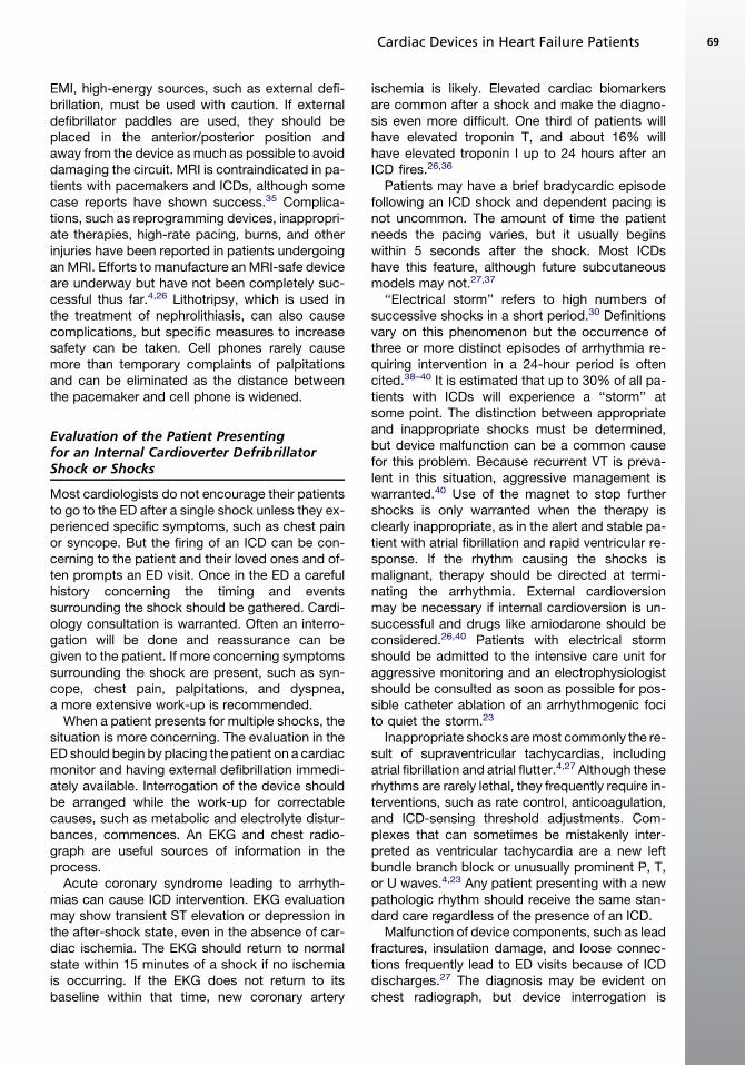
EMI, high-energy sources, such as external defi-brillation, must be used with caution. If externaldefibrillator paddles are used, they should beplaced in the anterior/posterior position andaway from the device as much as possible to avoiddamaging the circuit. MRI is contraindicated in pa-tients with pacemakers and ICDs, although somecase reports have shown success.35 Complica-tions, such as reprogramming devices, inappropri-ate therapies, high-rate pacing, burns, and otherinjuries have been reported in patients undergoingan MRI. Efforts to manufacture an MRI-safe deviceare underway but have not been completely suc-cessful thus far.4,26 Lithotripsy, which is used inthe treatment of nephrolithiasis, can also causecomplications, but specific measures to increasesafety can be taken. Cell phones rarely causemore than temporary complaints of palpitationsand can be eliminated as the distance betweenthe pacemaker and cell phone is widened.
Evaluation of the Patient Presentingfor an Internal Cardioverter DefribrillatorShock or Shocks
Most cardiologists do not encourage their patientsto go to the ED after a single shock unless they ex-perienced specific symptoms, such as chest painor syncope. But the firing of an ICD can be con-cerning to the patient and their loved ones and of-ten prompts an ED visit. Once in the ED a carefulhistory concerning the timing and eventssurrounding the shock should be gathered. Cardi-ology consultation is warranted. Often an interro-gation will be done and reassurance can begiven to the patient. If more concerning symptomssurrounding the shock are present, such as syn-cope, chest pain, palpitations, and dyspnea,a more extensive work-up is recommended.
When a patient presents for multiple shocks, thesituation is more concerning. The evaluation in theED should begin by placing the patient on a cardiacmonitor and having external defibrillation immedi-ately available. Interrogation of the device shouldbe arranged while the work-up for correctablecauses, such as metabolic and electrolyte distur-bances, commences. An EKG and chest radio-graph are useful sources of information in theprocess.
Acute coronary syndrome leading to arrhyth-mias can cause ICD intervention. EKG evaluationmay show transient ST elevation or depression inthe after-shock state, even in the absence of car-diac ischemia. The EKG should return to normalstate within 15 minutes of a shock if no ischemiais occurring. If the EKG does not return to itsbaseline within that time, new coronary artery
ischemia is likely. Elevated cardiac biomarkersare common after a shock and make the diagno-sis even more difficult. One third of patients willhave elevated troponin T, and about 16% willhave elevated troponin I up to 24 hours after anICD fires.26,36
Patients may have a brief bradycardic episodefollowing an ICD shock and dependent pacing isnot uncommon. The amount of time the patientneeds the pacing varies, but it usually beginswithin 5 seconds after the shock. Most ICDshave this feature, although future subcutaneousmodels may not.27,37
‘‘Electrical storm’’ refers to high numbers ofsuccessive shocks in a short period.30 Definitionsvary on this phenomenon but the occurrence ofthree or more distinct episodes of arrhythmia re-quiring intervention in a 24-hour period is oftencited.38–40 It is estimated that up to 30% of all pa-tients with ICDs will experience a ‘‘storm’’ atsome point. The distinction between appropriateand inappropriate shocks must be determined,but device malfunction can be a common causefor this problem. Because recurrent VT is preva-lent in this situation, aggressive management iswarranted.40 Use of the magnet to stop furthershocks is only warranted when the therapy isclearly inappropriate, as in the alert and stable pa-tient with atrial fibrillation and rapid ventricular re-sponse. If the rhythm causing the shocks ismalignant, therapy should be directed at termi-nating the arrhythmia. External cardioversionmay be necessary if internal cardioversion is un-successful and drugs like amiodarone should beconsidered.26,40 Patients with electrical stormshould be admitted to the intensive care unit foraggressive monitoring and an electrophysiologistshould be consulted as soon as possible for pos-sible catheter ablation of an arrhythmogenic focito quiet the storm.23
Inappropriate shocks are most commonly the re-sult of supraventricular tachycardias, includingatrial fibrillation and atrial flutter.4,27 Although theserhythms are rarely lethal, they frequently require in-terventions, such as rate control, anticoagulation,and ICD-sensing threshold adjustments. Com-plexes that can sometimes be mistakenly inter-preted as ventricular tachycardia are a new leftbundle branch block or unusually prominent P, T,or U waves.4,23 Any patient presenting with a newpathologic rhythm should receive the same stan-dard care regardless of the presence of an ICD.
Malfunction of device components, such as leadfractures, insulation damage, and loose connec-tions frequently lead to ED visits because of ICDdischarges.27 The diagnosis may be evident onchest radiograph, but device interrogation is
Cardiac Devices in Heart Failure Patients 69
needed for confirmation. Hospitalization is recom-mended for a device failure until the repair or re-placement is complete.
A multitude of electromagnetic interferencesources causing shocks have been named, in-cluding cell phones, television remote controls,large speakers, metal detectors, slot machines,electric razors, and antitheft detection devices.27
Although exposure to these sources may be iden-tified in some instances, work-up for other cor-rectable sources of pacemaker malfunction iswarranted.
The use of magnets to disable inappropriate ICDtherapies is necessary in some instances and canbe done without altering the back-up bradycardia-pacing function.26,27 Some ICDs will not restarttheir therapies after the magnet is removed; thus,all devices will need interrogation after a magnetis used. External pacing and defibrillation may beneeded. In some instances a patient’s body habi-tus may render a single magnet ineffective and theuse of two magnets in ‘‘stacked’’ fashion has beenreported.23
Cardiac arrest in the patient with an ICD raisescertain questions in health care providers butdoes not need to be treated much differentlythan usual. Advanced cardiac life-support proto-cols are recommended with the paddles beingplaced in anterior-posterior pattern to avoid dam-aging the device.4,26,32 An unpleasant tingle hasbeen described by resuscitation providers duringan ICD discharge; no injury to providers hasbeen reported and can be avoided by wearinggloves.4,41
An ICD may fail to rescue a patient in up to one-third of all cardiac deaths, even if the rhythm isappropriate for shock therapy.42 Failure of thesedevices to meet expectations has promptedresearchers, clinicians, government agencies,and industry to work to improve the effectivenessof these instruments. Many ICDs will only deliverfive successive shocks, to conserve battery lifeand to avoid the risk of damaging heart muscleand the heart’s conduction system.27,41 Addition-ally, lidocaine and amiodarone can increase thedefibrillation threshold, making ICD therapy inef-fective.26,27 If a patient is successfully resusci-tated, the ICD will need to be interrogated toinsure proper functioning before external monitor-ing and therapies are stopped.43
Ethical issues around the end of life can occurwith patients who have ICDs. The patient mayask a physician to discontinue the ICD therapybecause the shocks are painful and they have de-cided to reject further treatment. This is ethicallyequivalent to withdrawing specific therapies,such as hemodialysis, and is therefore
appropriate because of the firmly establishedright of informed patients to be free of unwantedmedical intervention.44 Certainly conversationswith the patient, the family, their primary carephysician, and other concerned parties can beentered with the hope of finding a solution thatbest fits each situation.
Central venous catheter placement can be af-fected if an ICD is present. Thrombosis along thelead system is common, with complete occlusionapproaching 20% at 2 years22 and subclavian ste-nosis is common too.27 Because of these factors,accessing the ipsilateral subclavian vein of a deviceis complicated. Additionally, contacting the leadsystem with a metal wire can cause an inappropri-ate shock, but can be avoided by temporarily de-activating the ICD or not using a metal guidewire.Pulmonary artery catheters offer unique challengesand are probably best inserted with the assistanceof fluoroscopy.4 Femoral vein catheters may be thesafest option, but the contralateral subclavian orinternal jugular can be accessed with caution.43
Electrocautery can be mistaken as an arrhyth-mia and lead to an ICD shock. Suggestions to dis-able the device before surgery have been made4,26
when using unipolar devices, although bipolardevices (ie, Bovie) are generally considered safe.Radiation therapy can be damaging to ICDs andshould be considered as a source of problemsfor patients undergoing that type of therapy forcancer. The device can be safely shielded toprevent malfunction.27
Occasionally patients will present to the ED witha beeping device. This can signal any number ofinternal dysfunctions, such as lead fracture, soft-ware failure, battery depletion or other issueswith the ICD.4,27 Interrogation is indicated whilethe patient is safely monitored with external defi-brillation and pacing immediately available.
New onset right heart failure can be encoun-tered when ICDs damage valve leaflets and pre-cipitate severe tricuspid valve regurgitation. Themechanism of damage can be from adhesion, per-foration, or primary impairment of valve leaflets.45
Echocardiography can confirm the diagnosis andlead replacement and valvular repair are urgentlyindicated.
Industry recalls and safety alerts can affect up to43% of all ICD patients.8,46,47 This has garnereda fair amount of anxiety and distrust among pa-tients and can make encounters somewhat diffi-cult. Little help, other than monitoring andsupportive care, can be offered by the physician.Information concerning what device or equipmentis under recall can be obtained from the manufac-turer (see Appendix) and distributed to the patientto discuss with their cardiologist.
Neuenschwander70
Succinylcholine can cause muscle fascicula-tions and can lead to inappropriate shocks associ-ated with oversensing.48 Nondepolarizing agentscan be used as an alternative, or the emergencyphysician can use a ‘‘defasciculating’’ dose beforeadministering succinylcholine.
ULTRAFILTRATION
Ultrafiltration has become a therapy option for pa-tients with an element of ‘‘diuretic resistance’’ thatare volume overloaded. Hydrostatic pressuresdraw off fluid across a semipermeable membrane,thus eliminating excess fluids. Small swings inelectrolyte balances have been experienced evenwhen large volumes are removed. The patientalso seems to experience fewer hemodynamic im-balances in comparison to hemodialysis, evenwhen 500 or more milliliters an hour have beentaken off.49,50 Using peripheral access points hasmade this modality possible in a multitude of clin-ical sites including the ED.51 More studies are tobe undertaken to assess the future role of ultrafil-tration in the management of heart failure.
SUMMARY
Patients with pacemakers and internal cardiovert-er defibrillators can experience a number of bene-fits and complications related to their devices.Physicians must be aware of the internal workingsand potential complications associated with thistechnology. The number of patients with these de-vices is likely to increase and so is their level of so-phistication, so consistent attention is necessaryto stay up to date with patient management. Alongwith the evolving pacemaker and ICD therapyavailable, ultrafiltration has the promise to improvepatient care.
APPENDIX: 24-HOUR CONTACT NUMBERSFOR INDUSTRY REPRESENTATIVESOF PACEMAKERS AND INTERNAL CARDIOVERTERDEFRIBRILLATORS
Biotronik: 800-284-6689Boston Scientific (formerly Guidant):
800-227-3422Medtronic: 800-633-8766St.Jude Medical: 800-722-3774
REFERENCES
1. Moss AJ, Hall WJ, Cannom DS, et al. Improved sur-
vival with an implanted defibrillator in patients with
coronary disease at high risk for ventricular arrhyth-
mia. Multicenter Automatic Defibrillator Implantation
Trial investigators. N Engl J Med 1996;335(26):
1933–40.
2. Moss AJ, Zareba W, Hall WJ, et al. Prophylactic im-
plantation of a defibrillator in patients with myocar-
dial infarction and reduced ejection fraction.
N Engl J Med 2002;346(12):877–83.
3. Bardy GH, Lee KL, Mark DB, et al. Amiodarone or an
implantable cardioverter-defibrillator for congestive
heart failure. N Engl J Med 2005;352(3):225–37.
4. Stevenson WG, Chaitman BR, Ellenbogen KA, et al.
Clinical assessment and management of patients with
implanted cardioverter-defibrillators presenting to non-
electrophysiologists. Circulation 2004;110(25):3866–9.
5. Lynd LD, O’Brien BJ. Cost-effectiveness of the
implantable cardioverter defibrillator: a review of cur-
rent evidence. J Cardiovasc Electrophysiol 2003;
14(Suppl 9):S99–103.
6. Bhatia A, Cooley R, Berger M, et al. The implantable
cardioverter defibrillator: technology, indications,
and impact on cardiovascular survival. Curr Probl
Cardiol 2004;29(6):303–56.
7. Neuenschwander J, Hiestand BC, Sondrup L, et al.
The benefit of interrogating defibrillators by emer-
gency department personnel [abstract]. Presented
at the American College of Emergency Physicians.
8. Gregoratos G, Abrams J, Epstein AE, et al. ACC/
AHA/NASPE 2002 guideline update for implantation
of cardiac pacemakers and antiarrhythmia devices:
summary article: a report of the American College
of Cardiology/American Heart Association Task
Force on Practice Guidelines (ACC/AHA/NASPE
Committee to Update the 1998 Pacemaker Guide-
lines). Circulation 2002;106(16):2145–61.
9. McMullan J, Valento M, Attari M, et al. Care of the
pacemaker/implantable cardioverter defibrillator pa-
tient in the ED. Am J Emerg Med 2007;25(7):812–22.
10. Bernstein AD, Daubert JC, Fletcher RD, et al. The
revised NASPE/BPEG generic code for antibrady-
cardia, adaptive-rate, and multisite pacing. North
American Society of Pacing and Electrophysiology/
British Pacing and Electrophysiology Group. Pacing
Clin Electrophysiol 2002;25(2):260–4.
11. Greene HL. Antiarrhythmic drugs versus implant-
able defibrillators: the need for a randomized con-
trolled study. Am Heart J 1994;127(4 Pt 2):1171–8.
12. Spratt KA, Blumberg EA, Wood CA, et al. Infections
of implantable cardioverter defibrillators: approach
to management. Clin Infect Dis 1993;17(4):679–85.
13. Herrmann C, von Zur MF, Schaumann A, et al. Stan-
dardized assessment of psychological well-being and
quality-of-life in patients with implanted defibrillators.
Pacing Clin Electrophysiol 1997;20(1 Pt 1):95–103.
14. Packer M, Abraham WT, Mehra MR, et al. Utility of
impedance cardiography for the identification of
short-term risk of clinical decompensation in stable
patients with chronic heart failure. J Am Coll Cardiol
2006;47(11):2245–52.
Cardiac Devices in Heart Failure Patients 71

15. Peacock WF, Summers RL, Vogel J, et al. Impact of
impedance cardiography on diagnosis and therapy
of emergent dyspnea: the ED-IMPACT trial. Acad
Emerg Med 2006;13(4):365–71.
16. Wong GC, Ayas NT. Clinical approaches to the diag-
nosis of acute heart failure. Curr Opin Cardiol 2007;
22(3):207–13.
17. Yamokoski LM, Haas GJ, Gans B, et al. Optivol fluid
status monitoring with an implantable cardiac de-
vice: a heart failure management system. Expert
Rev Med Devices 2007;4(6):775–80.
18. Yu CM, Wang L, Chau E,et al. Intrathoracic impedance
monitoring in patients with heart failure: correlation with
fluid status and feasibility of early warning preceding
hospitalization. Circulation 2005;112(6):841–8.
19. Wang L. Fundamentals of intrathoracic impedance
monitoring in heart failure. Am J Cardiol 2007;
99(10A):3G–10G.
20. Vollmann D, NageleH, SchauerteP, et al. Clinical utility
of intrathoracic impedancemonitoring to alert patients
with an implanted device of deteriorating chronic
heart failure. Eur Heart J 2007;28(15):1835–40.
21. Jensen SB, Galvin CA, Thompson B, et al.
Optimizing therapy for heart failure patients: cardiac
resynchronization and defibrillator therapy. J Cardio-
vasc Nurs 2007;22(2):118–24.
22. Pavia S, Wilkoff B. The management of surgical
complications of pacemaker and implantable cardi-
overter-defibrillators. Curr Opin Cardiol 2001;16(1):
66–71.
23. Trohman RG, Kim MH, Pinski SL. Cardiac pacing:
the state of the art. Lancet 2004;364(9446):1701–19.
24. Cardall TY, Brady WJ, Chan TC, et al. Permanent
cardiac pacemakers: issues relevant to the emer-
gency physician, part II. J Emerg Med 1999;17(4):
697–709.
25. Chambers ST. Diagnosis and management of staph-
ylococcal infections of pacemakers and cardiac de-
fibrillators. Intern Med J 2005;35(Suppl 2):S63–71.
26. Pinski SL. Emergencies related to implantable
cardioverter-defibrillators. Crit Care Med 2000;
28(Suppl 10):N174–80.
27. Glikson M, Friedman PA. The implantable cardi-
overter defibrillator. Lancet 2001;357(9262):
1107–17.
28. Burney K, Burchard F, Papouchado M, et al. Cardiac
pacing systems and implantable cardiac defibrilla-
tors (ICDs): a radiological perspective of equipment,
anatomy and complications. Clin Radiol 2004;59(8):
699–708.
29. Benson J, Adkinson C, Collier R. Hyperbaric oxygen
therapy of iatrogenic cerebral arterial gas embolism.
Undersea Hyperb Med 2003;30(2):117–26.
30. Byrd CL. Management of implant complications. In:
Ellenbogen KA, Kay GN, Wilkoff B, editors. Clinical
cardiac pacing. Philadelphia: W.B. Saunders Com-
pany; 1995. p. 491–522.
31. Klug D, Lacroix D, Savoye C, et al. Systemic infec-
tion related to endocarditis on pacemaker leads:
clinical presentation and management. Circulation
1997;95(8):2098–107.
32. Morales TY, Falcon Chevere JL. Implantable electri-
cal devices for dysrhythmia: what every emergency
physician must know. Bol Assoc Med P R 2004;
96(3):161–8.
33. Sarko JA, Tiffany BR. Cardiac pacemakers: evalua-
tion and management of malfunctions. Am J Emerg
Med 2000;18(4):435–40.
34. Sgarbossa EB, Pinski SL, Barbagelata A, et al. Elec-
trocardiographic diagnosis of evolving acute
myocardial infarction in the presence of left bun-
dle-branch block. GUSTO-1 (Global Utilization of
Streptokinase and Tissue Plasminogen Activator
for Occluded Coronary Arteries) Investigators.
N Engl J Med 1996;334(8):481–7.
35. Naehle CP, Sommer T, Meyer C, et al. Strategy for
safe performance of magnetic resonance imaging
on a patient with implantable cardioverter defibrilla-
tor. Pacing Clin Electrophysiol 2006;29(1):113–6.
36. Hurst TM, Hinrichs M, Breidenbach C, et al. Detec-
tion of myocardial injury during transvenous implan-
tation of automatic cardioverter-defibrillators. J Am
Coll Cardiol 1999;34(2):402–8.
37. Eysmann SB, Marchlinski FE, Buxton AE, et al. Elec-
trocardiographic changes after cardioversion of ven-
tricular arrhythmias. Circulation 1986;73(1):73–81.
38. Arya A, Haghjoo M, Dehghani MR, et al. Prevalence
and predictors of electrical storm in patients with im-
plantable cardioverter-defibrillator. Am J Cardiol
2006;97(3):389–92.
39. Credner SC, Klingenheben T, Mauss O, et al. Electri-
cal storm in patients with transvenous implantable
cardioverter-defibrillators: incidence, management
and prognostic implications. J Am Coll Cardiol
1998;32(7):1909–15.
40. Exner DV, Pinski SL, Wyse DG, et al. Electrical storm
presages nonsudden death: the antiarrhythmics ver-
sus implantable defibrillators (AVID) trial. Circulation
2001;103(16):2066–71.
41. Calle PA, Buylaert W. When an AED meets an ICD.
Automated external defibrillator. Implantable cardi-
overter defibrillator. Resuscitation 1998;38(3):
177–83.
42. Anderson KP. Sudden cardiac death unresponsive
to implantable defibrillator therapy: an urgent target
for clinicians, industry and government. J Interv
Card Electrophysiol 2005;14(2):71–8.
43. McPherson CA, Manthous C. Permanent pace-
makers and implantable defibrillators: consider-
ations for intensivists. Am J Respir Crit Care Med
2004;170(9):933–40.
44. Manganello TD. Disabling the pacemaker: the heart-
rending decision every competent patient has a right
to make. Health Care Law Mon 2000;3–15.
Neuenschwander72

45. Lin G, Nishimura RA, Connolly HM, et al. Severe
symptomatic tricuspid valve regurgitation due to
permanent pacemaker or implantable cardioverter-
defibrillator leads. J Am Coll Cardiol 2005;45(10):
1672–5.
46. Hauser RG, Kallinen L. Deaths associated with im-
plantable cardioverter defibrillator failure and deac-
tivation reported in the United States Food and Drug
Administration Manufacturer and User Facility De-
vice Experience Database. Heart Rhythm 2004;
1(4):399–405.
47. Maisel WH. Physician management of pacemaker
and implantable cardioverter defibrillator advisories.
Pacing Clin Electrophysiol 2004;27(4):437–42.
48. Stone KR, McPherson CA. Assessment and man-
agement of patients with pacemakers and implant-
able cardioverter defibrillators. Crit Care Med
2004;32(Suppl 4):S155–65.
49. Agostoni PG, Marenzi GC, Pepi M, et al. Isolated ul-
trafiltration in moderate congestive heart failure.
J Am Coll Cardiol 1993;21(2):424–31.
50. DiLeo M, Pacitti A, Bergerone S, et al. Ultrafiltration
in the treatment of refractory congestive heart fail-
ure. Clin Cardiol 1988;11(7):449–52.
51. Jaski BE, Ha J, Denys BG, et al. Peripherally
inserted veno-venous ultrafiltration for rapid treat-
ment of volume overloaded patients. J Card Fail
2003;9(3):227–31.
Cardiac Devices in Heart Failure Patients 73

Acute Heart Failure RiskStratification: CanWeDefine Low Risk?Sean P. Collins, MD, MSca,*, Alan B. Storrow, MDb
The emergency department (ED) evaluation andmanagement of patients who have potential acuteheart failure syndromes (AHFS) has remained a sig-nificant challenge for decades. Unlike advances inthe assessment and treatment of patients whohave acute coronary syndrome (Table 1), theemergency physician’s diagnostic tools for heartfailure have remained limited, and the complexityof the syndrome itself has led to risk-averse prac-tice styles with extremely high admission rates.
Moreover, the prevalence of AHFS continues toincrease as a result of an aging population, im-proved survival from acute myocardial infarction,and better management of chronic heart failure. Asa direct result, ED visits for AHFS are expected tocontinue to increase. Despite the development ofnew diagnostic and prognostic tools, patients whohave AHFS continue to have poorly defined treat-ment end points and a high rate of critical care ad-missions.1–3 Previous studies of risk stratificationhave identified markers of high risk in AHFS, butidentification of the ‘‘safe for ED discharge’’ patientat low risk remains elusive. Unfortunately, the lackof high-risk features does not necessarily equatewith low risk. Recently, new diagnostic markersand technology have become promising and evencommonplace to assist emergency physicians inrisk prediction for patients who have AHFS. Famil-iarity with these approaches is essential for im-proved care for patients who have heart failure andfor resourceuse.Thisarticle reviews the available lit-erature and describes patient features that need tobe accounted for in disposition decision-making.
IMPACT OFACUTE HEART FAILURE SYNDROMES
In 2005, more than 1 million hospital dischargeshad a primary diagnosis of acute heart failureand consumed 3% of the total national healthcare budget.4–6 The high incidence of adverseevents in patients who have AHFS has notchanged in decades: in-hospital mortality is 4%to 7%; 60-day mortality and recidivism rates areapproximately 10% and 25%, respectively.7–10
There is significant unpredictability about the nat-ural course of AHFS and uncertainty regardingacute clinical stability. Largely as a result of thisuncertainty, more than 80% of ED presentationsfor AHFS are admitted to the hospital. Patientswho have AHFS largely rely on EDs and emer-gency physicians for acute management, because80% of AHFS admissions originate in an ED. Pa-tients admitted and treated in an inpatient bedfor heart failure account for the majority of hospitalexpenditures.1,2,11 Based on American College ofCardiology/American Heart Association (ACC/AHA) or Agency for Healthcare Research andQuality guidelines, however, it has been suggestedthat up to 50% of admitted patients are at low riskand may be candidates for outpatient therapy,with a potential savings of $2.5 billion.3,12
Previous Guideline Recommendationsfor Acute Heart Failure Syndromes
Poor ED risk stratification, particularly overestima-tion of disease severity, is the fundamental cause
a University of Cincinnati School of Medicine, Cincinnati, OH, USAb Vanderbilt University School of Medicine, Nashville, TN, USA* Corresponding author. Department of Emergency Medicine, University of Cincinnati, Medical SciencesBuilding, Room 6109, 231 Albert Sabin Way, Cincinnati, OH 45267.E-mail address: [email protected] (S.P. Collins).
KEYWORDS� Acute heart failure � Risk-stratification� Emergency department
Heart Failure Clin 5 (2009) 75–83doi:10.1016/j.hfc.2008.08.0101551-7136/08/$ – see front matter ª 2008 Elsevier Inc. All rights reserved. he
artf
ailu
re.th
ecli
nics
.com

of the overuse of limited in-hospital resources forthis rapidly growing patient population.2,13
Improving the ability of the emergency physicianto decide on the most appropriate disposition ofpatients who have acute heart failure is criticalto maximize the allocation of in-hospitalresources.
A review shows that current guidelines for EDdisposition are based on little evidence or are pro-vided without any evidence whatsoever.14–17 The1995 ACC/AHA guidelines limit their dispositionrecommendations (class I and II) to hospital ad-mission for new-onset heart failure, chronic heartfailure with mild to moderate decompensation, orchronic heart failure complicated by acutelythreatening events or clinical situations. Criteriafor these conditions are not specifically defined,however. The 2005 ACC/AHA guidelinesaddressed the evaluation and management ofchronic heart failure.16 The Heart Failure Societyof America published detailed guidelines on heartfailure management but did not address ED dispo-sition.14 The European Society of Cardiology ad-dressed diagnosis and treatment of acute heartfailure but did not give recommendations on riskstratification or ED disposition.18 The AmericanCollege of Emergency Physicians recently pub-lished guidelines describing four topics for theemergency physician but, like their predecessorsdid not provide guidance for risk stratification ordisposition.19
Defining Heart Failure for the EmergencyDepartment: A New Paradigm
Heart failure can be simplistically defined as a clin-ical syndrome resulting from any structural or
functional cardiac disorder that impairs the abilityof the ventricle to fill with or eject blood.16 The car-dinal manifestations are dyspnea and fatigue(exercise intolerance), as well as fluid retention(pulmonary congestion and peripheral edema). Abetter ED or acute care term would be acute heartfailure syndrome (AHFS), defined as a gradual orrapid change in heart failure signs and symptoms,resulting in a need for urgent therapy.20 Thesesigns and symptoms are primarily due to pulmo-nary congestion from elevated LV filling pressuresand can occur in patients who have preserved orreduced ejection fraction. The term diastolic dys-function refers to an abnormality of LV filling or re-laxation; with the addition of effort intolerance anddyspnea, it is called ‘‘diastolic heart failure or’’‘‘acute heart failure with preserved ejection frac-tion.’’21–23 Acute heart failure syndrome admis-sions are about 50% female; approximately 75%will have known heart failure, and nearly 50% willhave preserved EF.7,8 For a substantial proportion,the causes in the western world are coronaryartery disease, hypertension, and dilatedcardiomyopathy.
HIGH-RISK FEATURES THAT CAN BE DETERMINEDAT EMERGENCY DEPARTMENT PRESENTATION
During the last 2 decades, many studies of AHFSrisk stratification have been conducted and haveidentified variables predicting early events in pa-tients who have AHFS (Table 2). Selker and col-leagues24 developed a model to predict acutehospital mortality from data available to the ED phy-sician within the first 10 minutes of presentation(patients’ age, systolic blood pressure and find-ings, and ECG abnormalities). The model was
Table1Characteristics of acutemyocardial infarction and acute heart failure syndromes resulting in hospitalizationin the United States
Characteristic AcuteMyocardial Infarction Acute Heart Failure Syndromes
Incidence 1 million/year 1 million/year
Mortality
Prehospital High ?
In-hospital 3%–4% 3%–4%
60–90 day 2% 10%
Targets Clearly defined (thrombosis) Uncertain
Interventions in clinical trials Beneficial Minimal/no benefit/harmful
ACC/AHA recommendations Level A None
From Gheorghiade M, Zannad F, Sopko G, et al. Acute heart failure syndromes: current state and framework for futureresearch. Circulation 2005;112(25):3959; with permission.
Collins & Storrow76

validated prospectively for mortality, but its validityfor morbidity and other acute sequelae is unknown.Additionally, the ability of the model to identifya low-risk patient who can be discharged homesafely has not been assessed. Thus, because themodel was developed to identify the high-risk pa-tient, and the absence of high-risk features doesnot define low risk, the usefulness of this model inidentifying the low-risk patient is unclear.
Chin and Goldman25,26 developed a risk modelusing a larger number of variables (vital signs, co-morbidities, ECG findings, and laboratory data).The model is successful in predicting morbidityas well as mortality, but it cannot delineate thelow-risk patient. Katz27 developed a model thatcould predict 81% of complications. This modelwas based on ED information but includeda 4-hour diuresis measure, making it less helpfulas a decision-making tool early in the emergencysetting. Delaying such decision-making can resultin a potentially life-threatening therapeutic delay.Additionally, the model missed 19% of cardiopul-monary complications, making it unsuitable forsafe implementation.
In one of the largest studies to date, classificationand regression tree methodology was used on 45variables in 65,275 patients who had heart failureto predict in-hospital mortality.28 Three variableswere used to differentiate high-risk patients fromlow-risk patients: blood urea nitrogen (BUN), sys-tolic blood pressure (SBP), and creatinine. Patientswho had a BUN level greater than 43 mg/dL, a SBPless than 115 mm Hg, and a creatinine level over2.75 mg/dL had a 22% in-hospital mortality rate.Further, the odds ratio for mortality between pa-tients identified as being at high or low risk was12.9 (95% confidence interval [CI], 10.4–15.9).Although this model is perhaps the most elegantavailable to date, it remains highly limited becauseonly 39 of more than 100 variables available to theED physician were considered, and the model wasdesigned only to predict inpatient mortality. Fur-ther, patients defined as being at low risk hadan inpatient mortality rate of 2.1%, a numberexceedingly high to be considered ‘‘low-risk’’ inthe ED.
Several recent retrospective analyses of clinicaltrials and registries reaffirm these findings.29,30 Hy-ponatremia (<135 mmol/L) on hospital admissionhas been associated with increased in-hospitaland postdischarge mortality and with increasedrates of readmission.30 Patients who had systolicblood pressure lower than 120 mm Hg on admis-sion had an almost threefold greater risk of in-hos-pital mortality than patients who had systolic bloodpressure higher than 140 mm Hg (7.2% vs. 2.5%,P < .001).29 Finally, renal dysfunction (elevated
BUN or creatinine levels) on hospital admissionalso has been associated with increased rates ofin-hospital and postdischarge mortality.31–33
In summary, several markers are associatedwith poor clinical outcomes: an elevated BUN orcreatinine level, a low SBP, hyponatremia, is-chemic ECG changes, and elevated cardiac bio-markers. Not clear, however, is whether theabsence of high-risk physiologic variables canindicate that a patient is at low risk of early events.
LOW-RISK FEATURES THAT CAN BE DETERMINEDAT EMERGENCY DEPARTMENT PRESENTATION
Conversely, little has been published to guide theemergency physician in identifying patients whomay be categorized as low-risk and possibly dis-charged after a brief ED stay. A retrospective anal-ysis of a statewide database was performed toidentify variables predictive of a low risk of inpatientdeath or serious complications.34 Recursive parti-tioning classified 17.2% of patients as low risk(0.3% mortality, 1.0% inpatient complications).The resultant model was somewhat cumbersomebut also identified serum sodium, SBP, and creati-nine as differentiators between patients at low andhigh risk. This model subsequently was validated inmore than 8300 admitted patients from a similar da-tabase. The authors found that 19% of patientscould be classified as low risk, defined as havinga 1% to 3% risk of serious complications or deathwithin 30 days of hospitalization.35 Diercks36 stud-ied a prospective convenience sample of patientswho had AHFS to identify a low-risk cohort of pa-tients who had AHFS suitable for observation unitmanagement. Patients who had a systolic bloodpressure over 160 mm Hg at ED presentation anda normal initial cardiac troponin I level were signifi-cantly more likely to be discharged from the obser-vation unit and not experience any 30-day adverseevents (death, readmission, myocardial infarction,or arrhythmias).
NATRIURETIC PEPTIDES AND RISKSTRATIFICATION
Several investigations have evaluated the prog-nostic ability of natriuretic peptides.37–43 A studyof 325 patients in the ED demonstrated the abilityof serum natriuretic peptide (BNP) to predict futurecardiac events.38 Patients presenting to the EDwith dyspnea had BNP levels drawn and were fol-lowed for 6 months for the combined end points ofdeath (both cardiac and noncardiac), hospital ad-mission with a cardiac diagnosis, and repeat EDvisits for heart failure. The area under the receiveroperating characteristic curve was 0.87 (95% CI,
Acute Heart Failure Risk Stratification 77
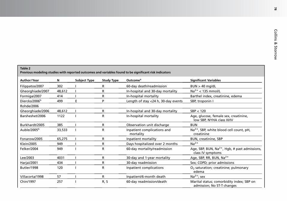
Table 2Previous modeling studies with reported outcomes and variables found to be significant risk indicators
Author/Year N Subject Type Study Type Outcomea Significant Variables
Filippatos/2007 302 I R 60-day death/readmission BUN > 40 mg/dL
Gheorghiade/2007 48,612 I R In-hospital and 30-day mortality Na21 < 135 mmol/L
Formiga/2007 414 I R In-hospital mortality Barthel index, creatinine, edema
Diercks/2006b 499 E P Length of stay <24 h, 30-day events SBP, troponin I
Rohde/2006
Gheorghiade/2006 48,612 I R In-hospital and 30-day mortality SBP < 120
Barsheshet/2006 1122 I R In-hospital mortality Age, glucose, female sex, creatinine,low SBP, NYHA class III/IV
Burkhardt/2005 385 I R Observation unit discharge BUN
Auble/2005b 33,533 I R Inpatient complications andmortality
Na21, SBP, white blood cell count, pH,creatinine
Fonarow/2005 65,275 I R Inpatient mortality BUN, creatinine, SBP
Klein/2005 949 I R Days hospitalized over 2 months Na21
Felker/2004 949 I R 60-day mortality/readmission Age, SBP, BUN, Na21, Hgb, # past admissions,class IV symptoms
Lee/2003 4031 I R 30-day and 1-year mortality Age, SBP, RR, BUN, Na21
Harjai/2001 434 I R 30-day readmission Sex; COPD; prior admissions
Butler/1998 120 I R Inpatient complications O2 saturation; creatinine; pulmonaryedema
Villacorta/1998 57 I R Inpatient/6-month death Na21; sex
Chin/1997 257 I R, S 60-day readmission/death Marital status; comorbidity index; SBP onadmission; No ST-T changes
Co
llins
&Sto
rrow
78
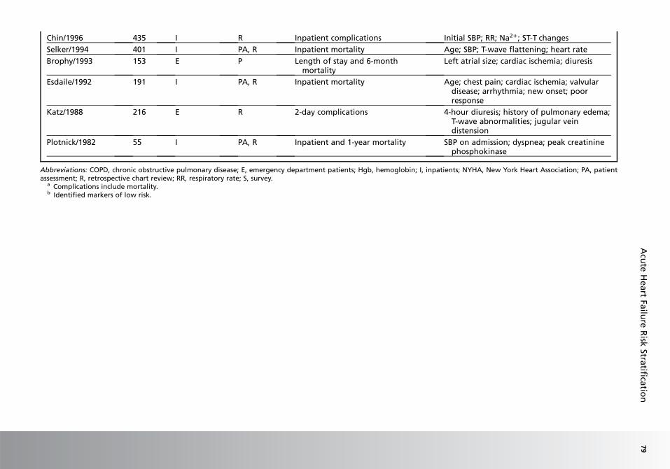
Chin/1996 435 I R Inpatient complications Initial SBP; RR; Na21; ST-T changes
Selker/1994 401 I PA, R Inpatient mortality Age; SBP; T-wave flattening; heart rate
Brophy/1993 153 E P Length of stay and 6-monthmortality
Left atrial size; cardiac ischemia; diuresis
Esdaile/1992 191 I PA, R Inpatient mortality Age; chest pain; cardiac ischemia; valvulardisease; arrhythmia; new onset; poorresponse
Katz/1988 216 E R 2-day complications 4-hour diuresis; history of pulmonary edema;T-wave abnormalities; jugular veindistension
Plotnick/1982 55 I PA, R Inpatient and 1-year mortality SBP on admission; dyspnea; peak creatininephosphokinase
Abbreviations: COPD, chronic obstructive pulmonary disease; E, emergency department patients; Hgb, hemoglobin; I, inpatients; NYHA, New York Heart Association; PA, patientassessment; R, retrospective chart review; RR, respiratory rate; S, survey.
a Complications include mortality.b Identified markers of low risk.
Acu
teH
eart
Failu
reR
iskStra
tificatio
n79
0.83–0.92) for BNP’s ability to predict a combinedend point. The cumulative probability of a heart-failure event within 6 months was 51% in the 67patients who had a BNP level higher than 480pg/mL, compared with 2.5% in the 205 patientswho had BNP values less than 230 pg/mL. BNPalso has been shown to predict adverse eventsand to determine a disposition strategy more ac-curately than a physician’s assessment based onlevel of severity.43 Finally, a retrospective analysisof 77,467 patients from the Acute DecompensatedHeart Failure National Registry found a nearlylinear relationship between BNP quartiles andin-hospital mortality.40 Similar findings havebeen reported for N-terminal proBNP (NT-proBNP).41,44–46 In a pooled analysis of 1256 pa-tients, NT-proBNP was a significant predictor ofsubsequent adverse events.41 An NT-proBNPconcentration greater than 5180 pg/mL wasstrongly predictive of death by 76 days (odds ratio,5.2; 95% CI, 2.2–8.1; P < .001).
PROMISING NEW TECHNOLOGIESAND TECHNIQUES
An S3 cardiac gallop is indicative of heart failure,and studies have demonstrated it has excellentspecificity but poor sensitivity.47 Although thepresence of an S3 gallop can be normal in adoles-cents and young adults, its detection in patientsolder than 40 years is considered abnormal.48–51
Further, it has been suggested that patients whohave a detectable S3 gallop have an increasedrisk of hospitalization and death compared withpatients without a detectable S3 gallop.52–54
Identification of an S3 gallop is difficult. In theaforementioned studies that suggest a low inci-dence of S3 detection in heart failure, the physi-cians may have been unable to detect a soundthat in fact was present. Technology has been de-veloped that may assist the clinician in detecting anS3 gallop at the bedside by measuring heart soundenergy using an electronic stethoscope or anothermeans of recording heart tones. Using a sophisti-cated software algorithm, information on the
presence of an S3 gallop, and potentially its inten-sity, is available. With continued development ofsuch technology, the ability to detect extra heartsounds should improve significantly. Early resultshave shown promising specificity, improved EDphysicians’ diagnostic confidence, and providedadditive, independent prognostic information.55–59
Finally, T-wave alternans has been investigatedas a potential risk-stratification tool in AHFS.T-wave alternans describes beat-to-beat fluctua-tions in T-wave morphology that have been asso-ciated anecdotally with the onset of ventricularfibrillation. Microvolt T-wave alternans recordingnow can be performed during submaximal exer-cise. A series of beats are recorded at a stableheart rate, and the T-wave amplitude is plottedwith respect to the QRS complex. These datathen undergo spectral analysis to determine ifthere are sufficient T-wave fluctuations to call thetest ‘‘positive.’’60 Some studies have suggestedan increased rate of long-term death and malig-nant arrhythmias in patients who have abnormalT-wave alternans.61,62 Yet to be determined isthe ability to perform this test in real time in theED and whether abnormal results carry an in-creased risk of near-term events.
SUMMARY
A change in the conservative decision paradigmfor patients who have heart failure will requirea novel approach; even with the development ofnew diagnostic and prognostic tools, poor EDrisk stratification and the high rate of critical careadmissions for patients who have heart failurehave not changed in decades.1–3 The traditionalhistory and physical examination have serious lim-itations. The current literature suggests that serumBUN, creatinine, sodium, cardiac biomarkers, andnatriuretic peptides are helpful for initial risk strat-ification (Table 3).
A decision tool based on a validated ED riskmodel could improve assessment and initial dis-position decisions. In other disease processes,such as acute coronary syndromes63–65 andcommunity-acquired pneumonia,66–68 such
Table 3Potential modifiable riskmarkers in acute heart failure
Source Marker
Past medical history Coronary artery disease
Physical examination Systolic blood pressure, respiratory rate, oxygen saturation
Laboratory findings BUN, creatinine, sodium, natriuretic peptide levels
Ancillary studies Ischemic ECG changes suggesting coronary artery disease
Collins & Storrow80
approaches have proven effective in safely de-creasing admissions for low-risk patients. The pro-cess of risk stratification in patients who haveheart failure lags decades behind the processesin place for these other conditions. A prospectivelyderived, multicenter, useful, ED risk-stratificationmodel for patients who have signs and symptomsof heart failure is needed and is the focus of an on-going National Heart, Lung, and Blood Institutegrant.69
REFERENCES
1. Polanczyk CA, Rohde LE, Philbin EA, et al. A new
casemix adjustment index for hospital mortality
among patients with congestive heart failure. Med
Care 1998;36(10):1489–99.
2. Smith WR, Poses RM, McClish DK, et al. Prognostic
judgments and triage decisions for patients with acute
congestive heart failure. Chest 2002;121(5):1610–7.
3. Graff L, Orledge J, Radford MJ, et al. Correlation of
the agency for health care policy and research con-
gestive heart failure admission guideline with mortal-
ity: peer review organization voluntary hospital
association initiative to decrease events (PROVIDE)
for congestive heart failure. Ann Emerg Med 1999;
34(4 Pt 1):429–37.
4. O’Connell JB, Bristow M. Economic impact of heart
failure in the United States: a time for a different ap-
proach. J Heart Lung Transplant 1994;13:S107–12.
5. Stevenson LW, Braunwald E. Recognition and man-
agement of patients with heart failure. In:
Goldman L, Braunwald E, editors. Primary cardiol-
ogy. Philadelphia: WB Saunders; 1998. p. 310–29.
6. American Heart Association. Heart disease and
stroke statistics–2006 update. Dallas (TX): American
Heart Association 2005.
7. Adams KF Jr, Fonarow GC, Emerman CL, et al.
Characteristics and outcomes of patients hospital-
ized for heart failure in the United States: rationale,
design, and preliminary observations from the first
100,000 cases in the Acute Decompensated Heart
Failure National Registry (ADHERE). Am Heart J
2005;149(2):209–16.
8. Cleland JG, Swedberg K, Follath F, et al. The Euro
heart failure survey programme—a survey on the
quality of care among patients with heart failure in
Europe. Part 1: patient characteristics and diagno-
sis. Eur Heart J 2003;24(5):442–63.
9. Cuffe MS, Califf RM, Adams KF Jr, et al. Short-term
intravenous milrinone for acute exacerbation of
chronic heart failure: a randomized controlled trial.
JAMA 2002;287(12):1541–7.
10. VMAC Investigators. Intravenous nesiritide vs nitro-
glycerin for treatment of decompensated congestive
heart failure: a randomized controlled trial. JAMA
2002;287(12):1531–40.
11. Institute NHLaB. Morbidity and mortality: 2002 chart
book on cardiovascular, lung, and blood diseases.
Bethesda (MD): National Institutes of Health; 2002.
12. Butler J, Hanumanthu S, Chomsky D, et al. Fre-
quency of low-risk hospital admissions for heart fail-
ure. Am J Cardiol 1998;81(1):41–4.
13. Poses RM, Smith WR, McClish DK, et al. Physicians’
survival predictions for patients with acute conges-
tive heart failure. Arch Intern Med 1997;157(9):
1001–7.
14. Heart Failure Society of America. HFSA 2006 com-
prehensive heart failure practice guideline. J Card
Fail 2006;12(1):e1–2.
15. Hsieh M, Auble TE, Yealy DM. Evidence-based
emergency medicine. Predicting the future: can
this patient with acute congestive heart failure be
safely discharged from the emergency department?
Ann Emerg Med 2002;39(2):181–9.
16. Hunt SA, Abraham WT, Chin MH, et al. ACC/AHA 2005
guideline update for the diagnosis and management
of chronic heart failure in the adult—summary article:
a report of the American College of Cardiology/Amer-
ican Heart Association task force on practice guide-
lines (writing committee to update the 2001
guidelines for the evaluation and management of
heart failure). J Am Coll Cardiol 2005;46(6):1116–43.
17. Konstam M, Dracup K, Baker D. Clinical practice
guidelines No 11: heart failure: evaluation and care
of patients with left-ventricular systolic dysfunction.
Agency for Health Care Policy and Research
1994;94(0612).
18. Nieminen MS, Bohm M, Cowie MR, et al. Executive
summary of the guidelines on the diagnosis and
treatment of acute heart failure: the task force on
acute heart failure of the European Society of Cardi-
ology. Eur Heart J 2005;26(4):384–416.
19. Silvers SM, Howell JM, Kosowsky JM, et al. Clinical
policy: critical issues in the evaluation and manage-
ment of adult patients presenting to the emergency
department with acute heart failure syndromes.
Ann Emerg Med 2007;49(5):627–69.
20. Gheorghiade M, Zannad F, Sopko G, et al. Acute
heart failure syndromes: current state and frame-
work for future research. Circulation 2005;112(25):
3958–68.
21. Aurigemma GP, Gaasch WH. Clinical practice. Dia-
stolic heart failure. N Engl J Med 2004;351(11):
1097–105.
22. Bhatia RS, Tu JV, Lee DS, et al. Outcome of heart
failure with preserved ejection fraction in a popula-
tion-based study. N Engl J Med 2006;355(3):260–9.
23. Owan TE, Hodge DO, Herges RM, et al. Trends in
prevalence and outcome of heart failure with pre-
served ejection fraction. N Engl J Med 2006;
355(3):251–9.
24. Selker HP, Griffith JL, D’Agostino RB. A time-insensi-
tive predictive instrument for acute hospital mortality
Acute Heart Failure Risk Stratification 81

due to congestive heart failure: development, test-
ing, and use for comparing hospitals: a multicenter
study. Med Care;32(10):1040–52.
25. Chin MH, Goldman L. Correlates of major complica-
tions or death in patients admitted to the hospital
with congestive heart failure. Arch Intern Med
1996;156(16):1814–20.
26. Chin MH, Goldman L. Correlates of early hospital re-
admission or death in patients with congestive heart
failure. Am J Cardiol 1997;79(12):1640–4.
27. Katz MH, Nicholson PW, Singer DE, et al. The triage
decision in pulmonary edema. J Gen Intern Med
1988;3(6):533–9.
28. Fonarow GC, Adams KF Jr, Abraham WT, et al. Risk
stratification for in-hospital mortality in acutely
decompensated heart failure: classification and
regression tree analysis. JAMA 2005;293(5):
572–80.
29. Gheorghiade M, Abraham WT, Albert NM, et al. Sys-
tolic blood pressure at admission, clinical character-
istics, and outcomes in patients hospitalized with
acute heart failure. JAMA 2006;296(18):2217–26.
30. Gheorghiade M, Rossi JS, Cotts W, et al. Character-
ization and prognostic value of persistent hyponatre-
mia in patients with severe heart failure in the
ESCAPE Trial. Arch Intern Med 2007;167(18):
1998–2005.
31. Damman K, Navis G, Voors AA, et al. Worsening re-
nal function and prognosis in heart failure: system-
atic review and meta-analysis. J Card Fail 2007;
13(8):599–608.
32. Krumholz HM, Chen YT, Vaccarino V, et al. Corre-
lates and impact on outcomes of worsening renal
function in patients > or 5 65 years of age with heart
failure. Am J Cardiol 2000;85(9):1110–3.
33. Smith GL, Vaccarino V, Kosiborod M, et al. Worsen-
ing renal function: what is a clinically meaningful
change in creatinine during hospitalization with
heart failure? J Card Fail 2003;9(1):13–25.
34. Auble TE, Hsieh M, Gardner W, et al. A prediction
rule to identify low-risk patients with heart failure.
Acad Emerg Med 2005;12(6):514–21.
35. Hsieh M, Auble TE, Yealy DM. Validation of the acute
heart failure index. Ann Emerg Med 2008;51(1):37–44.
36. Diercks DB, Peacock WF, Kirk JD, et al. ED patients
with heart failure: identification of an observational
unit-appropriate cohort. Am J Emerg Med 2006;
24(3):319–24.
37. Bayes-Genis A, Lopez L, Zapico E, et al. NT-ProBNP
reduction percentage duringadmission for acutely de-
compensated heart failure predicts long-term cardio-
vascular mortality. J Card Fail 2005;11(Suppl. 5):S3–8.
38. Harrison A, Morrison LK, Krishnaswamy P, et al. B-
type natriuretic peptide predicts future cardiac events
in patients presenting to the emergency department
with dyspnea. Ann Emerg Med 2002;39(2):131–8.
39. Cheng V, Kazanagra R, Garcia A, et al. A rapid bed-
side test for B-type peptide predicts treatment out-
comes in patients admitted for decompensated
heart failure: a pilot study. J Am Coll Cardiol 2001;
37(2):386–91.
40. Fonarow GC, Peacock WF, Phillips CO, et al. Admis-
sion B-type natriuretic peptide levels and in-hospital
mortality in acute decompensated heart failure.
J Am Coll Cardiol 2007;49(19):1943–50.
41. Januzzi JL, van Kimmenade R, Lainchbury J, et al.
NT-proBNP testing for diagnosis and short-term
prognosis in acute destabilized heart failure: an in-
ternational pooled analysis of 1256 patients: the In-
ternational Collaborative of NT-proBNP study. Eur
Heart J 2006;27(3):330–7.
42. Yu CM, Sanderson JE. Plasma brain natriuretic pep-
tide—an independent predictor of cardiovascular
mortality in acute heart failure. Eur J Heart Fail
1999;1(1):59–65.
43. Maisel A, Hollander JE, Guss D, et al. Primary results
of the rapid emergency department heart failure out-
patient trial (REDHOT). A multicenter study of B-type
natriuretic peptide levels, emergency department
decision making, and outcomes in patients present-
ing with shortness of breath. J Am Coll Cardiol 2004;
44(6):1328–33.
44. Kirk V, Bay M, Parner J, et al. N-terminal proBNP and
mortality in hospitalised patients with heart failure
and preserved vs. reduced systolic function: data
from the prospective Copenhagen Hospital Heart
Failure study (CHHF). Eur J Heart Fail 2004;6(3):
335–41.
45. Januzzi JL Jr, Sakhuja R, O’Donoghue M, et al. Utility
of amino-terminal pro-brain natriuretic peptide test-
ing for prediction of 1-year mortality in patients
with dyspnea treated in the emergency department.
Arch Intern Med 2006;166(3):315–20.
46. Chen AA, Wood MJ, Krauser DG, et al. NT-proBNP
levels, echocardiographic findings, and outcomes
in breathless patients: results from the ProBNP
Investigation of Dyspnoea in the Emergency Depart-
ment (PRIDE) echocardiographic substudy. Eur
Heart J 2006;27(7):839–45.
47. Wang CS, FitzGerald JM, Schulzer M, et al. Does
this dyspneic patient in the emergency department
have congestive heart failure? JAMA 2005;294(15):
1944–56.
48. Evans W. The use of phonocardiography in clinical
medicine. Lancet 1951;1:1083–5.
49. Reddy PS. The third heart sound. Int J Cardiol 1985;
7(3):213–21.
50. Reddy PS, Salerni R, Shaver JA. Normal and abnor-
mal heart sounds in cardiac diagnosis: part II. dia-
stolic sounds. Curr Probl Cardiol 1985;10(4):1–55.
51. Sloan A. Cardiac gallop rhythm. Medicine 1958;37:
197–215.
Collins & Storrow82

52. Drazner MH, Rame JE, Stevenson LW, et al. Prog-
nostic importance of elevated jugular venous pres-
sure and a third heart sound in patients with heart
failure. N Engl J Med 2001;345(8):574–81.
53. Glover DR, Littler WA. Factors influencing survival
and mode of death in severe chronic ischaemic car-
diac failure. Br Heart J 1987;57(2):125–32.
54. Rame JE, Dries DL, Drazner MH. The prognostic
value of the physical examination in patients with
chronic heart failure. Congest Heart Fail 2003;9(3):
170–5, 178.
55. Collins SP, Kontos M, Diercks D, et al. Heart failure
and audicor technology for rapid diagnosis and initial
treatment of ED patients with suspected heart failure
(HEARD-IT). Presented at the meeting of the Society
of Academic Emergency Medicine. Chicago, 2007.
56. Collins SP, Lindsell CJ, Peacock WF, et al. The com-
bined utility of an S3 heart sound and B-type natriuretic
peptide levels in emergency department patients with
dyspnea. J Card Fail 2006;12(4):286–92.
57. Collins SP, Lindsell CJ, Peacock WF, et al. Preva-
lence of S3 and S4 in ED patients with decompen-
sated heart failure. Presented at the meeting of the
American College of Emergency Physicians. San
Francisco, October 2004.
58. Collins SP, Lindsell CJ, Peacock WF, et al. Preva-
lence of electronically detected abnormal heart
sounds in acute decompensated heart failure before
and after treatment. Presented at the meeting of the
American College of Cardiology. Orlando (FL), 2005.
59. Storrow A, Sp C, Wf P, et al. Length of stay and char-
ges are increased in patients with digitally detected
third heart sounds. Paper presented at Society for
Academic Emergency Medicine. New York, 2005.
60. Myles RC, Jackson CE, Tsorlalis I, et al. Is microvolt
T-wave alternans the answer to risk stratification in
heart failure? Circulation 2007;116(25):2984–91.
61. Salerno-Uriarte JA, De Ferrari GM, Klersy C, et al.
Prognostic value of T-wave alternans in patients
with heart failure due to nonischemic cardiomyopa-
thy: results of the ALPHA Study. J Am Coll Cardiol
2007;50(19):1896–904.
62. Baravelli M, Salerno-Uriarte D, Guzzetti D, et al.
Predictive significance for sudden death of micro-
volt-level T wave alternans in New York Heart Asso-
ciation class II congestive heart failure patients:
a prospective study. Int J Cardiol 2005;105(1):53–7.
63. Tatum JL, Jesse RL, Kontos MC, et al. Comprehen-
sive strategy for the evaluation and triage of the
chest pain patient. Ann Emerg Med 1997;29(1):
116–25.
64. Gibler WB, Runyon JP, Levy RC, et al. A rapid diag-
nostic and treatment center for patients with chest
pain in the emergency department. Ann Emerg
Med 1995;25(1):1–8.
65. Storrow AB, Gibler WB. Chest pain centers: diagno-
sis of acute coronary syndromes. Ann Emerg Med
2000;35(5):449–61.
66. Yealy DM, Auble TE, Stone RA, et al. The emergency
department community-acquired pneumonia trial:
methodology of a quality improvement intervention.
Ann Emerg Med 2004;43(6):770–82.
67. Marrie TJ, Lau CY, Wheeler SL, et al. A controlled
trial of a critical pathway for treatment of community-
acquired pneumonia. CAPITAL study investigators.
Community-acquired pneumonia intervention
trial assessing levofloxacin. JAMA 2000;283(6):
749–55.
68. Fine MJ, Auble TE, Yealy DM, et al. A prediction rule
to identify low-risk patients with community-acquired
pneumonia. N Engl J Med 1997;336(4):243–50.
69. Storrow AB, Collins S, Disalvo T, et al. Improving heart
failure risk stratification in the ED: stratify
1r01hl088459-01. Vanderbilt University. NHLBI; 2007.
Acute Heart Failure Risk Stratification 83
Observation UnitManagement of AcuteDecompensatedHeart FailureJon W. Schrock, MD*, Charles L. Emerman, MD
Emergency department (ED) presentations foracute decompensated heart failure (ADHF) haveincreased at a dramatic rate. In the 1990s, ADHFpresentations accounted for between 1% and3% of all ED visits and increased at an averagerate of more than 18,000 visits annually.1,2 Provid-ing care for these large numbers of patients hassignificant societal implications because heart fail-ure is the leading diagnosis for Medicare patientsolder than 65 years and accounts for more billsmore than any other disease.3 Hospital chargesfor 1 week of inpatient treatment of ADHF averagedapproximately $10,000 in the 1990s, and is evenhigher today.4 Costs for the treatment of ADHFvary on a daily basis, with nearly 75% ofADHF-related costs occurring in the first 48 hours.5
The costs of ADHF are not just an ED problembut begin at its front door. Of patients who havea primary diagnosis of ADHF discharged fromthe hospital, nearly 80% arrive through the ED.6,7
On a national level, the direct and indirect costsfor treating ADHF were projected to reach morethan $34.8 billion dollars in 2008.8 The financial im-pact of ADHF is not stagnant; as the baby boomergeneration continues to age, the burden of heartfailure on society will continue to increase.9,10
To prepare for this growth, government and hos-pital administrators are looking for more effectiveand less costly methods to deliver care to thesepatients. ED observation units (OU) have beenfound to be one of the most cost-effective waysto manage patients who have mild to moderately
ill presentations of asthma, chest pain, and heartfailure.11–18 These units have been associatedwith a high degree of patient satisfaction andincreased diagnostic accuracy.19–23 When evi-dence-based protocols are used in observationcare for ADHF, return ED visits and readmissionsdecrease significantly.12
OU treatment of ADHF is cost-effective forseveral rather distinct reasons. OUs are able toprovide rapid and focused treatment for patientswho have ADHF through specific treatment proto-cols that follow current American Heart Associa-tion (AHA) guidelines and can be implementedimmediately. The several-hour delay in care oftenseen when patients are transferred from the EDto the hospital floor is greatly shortened becauseobservation unit beds are often more availabledue to greater turnover. The use of protocols canhelp reduce the number and severity of complica-tions seen with patients who have ADHF.24 Medi-cal therapies are started earlier in OUs than withtypical inpatient admission on a hospital floor.A significant secondary benefit is the decrease inED diversion and delays in care for future patients,because the ED beds are no longer occupied.25,26
The establishment of OUs for the extended careof patients in the ED has evolved over the past3 decades. Observation medicine initially beganas a method to contain costs for conditions withuncertain clinical courses or diagnoses. Themost common admissions to the initial OUs werefor alcohol intoxication or withdrawal and
MetroHealth Medical Center, Cleveland, OH, USA* Corresponding author. Department of Emergency Medicine, MetroHealth Medical Center, Case WesternReserve University School of Medicine, 2500 MetroHealth Drive, BG353, Cleveland, OH 44109-1998.E-mail address: [email protected] (J. W. Schrock).
KEYWORDS� Heart failure � Observation medicine
Heart Failure Clin 5 (2009) 85–100doi:10.1016/j.hfc.2008.08.0151551-7136/08/$ – see front matter. Published by Elsevier Inc. he
artf
ailu
re.th
ecli
nics
.com
overdoses.27,28 With little regulation and guidance,more than a decade passed before the idea ofobservational care caught on. Once the benefitsof OUs became more obvious, the model bloss-omed into a branch of emergency medicine withunique features and goals.
In 1987, more than 10 years after the inceptionof the OU, the American College of EmergencyPhysicians Practice Management Committee pub-lished a list of features it deemed important inmanaging observation patients. The committeestated that the goal was ‘‘to improve the deliveryand quality of medical care to all patients.’’29 Rec-ommendations included clearly designating is theindividuals responsible for observation patientsat all times, locating units within or adjacent tothe ED, and creating protocols for transferringpatients in and out of the units.
OU beds are designed for short-term care ofmild to moderately ill patients, typically 24 hoursand, except in extreme cases, less than 48hours.30 The physical placement of OUs can varydepending on the needs and restraints of the insti-tutions developing them. Most OUs are in dedi-cated areas within the ED using existing beds,31
allowing proximity for physicians and nurses andavoiding the financial expense of building andrunning a separate facility. Using beds within theED can contribute to overcrowding, which hasbecome an ever-increasing problem.
As EDs grow and invest in the infrastructure,many will create a separate adjacent clinical areafor an OU. The advantages of this include a stablelocation for patient care and a stable nursing poolcomposed of staff working solely in the OU. A sep-arate clinical area allows centralization of physicalresources required to run an OU and eases educa-tion of staff caring for patients in the unit. Physicianscaring for these patients are close if emergenciesarise, but patients are shielded from the volumeand commotion often seen in many EDs.
Some departments may place patients ina virtual observation unit with set protocols butno permanent physical space. This model allowsOU beds to be located literally on any floor in a hos-pital, assuming that the level of care for thesepatients remains constant. The disadvantages ofthis type of OU include difficulty maintainingappropriate training and certification of nursingstaff caring for patients in the unit, potentially largedistances between patients and physicians caringfor them, and lack of familiarity of other staff withOU protocols and practice.
The Joint Commission, formerly The JointCommission on the Accreditation of HealthcareOrganizations, requires that OU staffing approxi-mates nurse-to-patient ratios of a hospital floor
caring for similar patients. This directive oftenleads to a nursing-to-patient ratio of 1:4 or 1:5for the typical OU. Nursing staffing levels can beadjusted throughout the day as the number ofpatients requiring care in the OU changes, butshould not exceed predetermined nurse-to-patient ratios determined by that institution.
CENTERS FORMEDICARE & MEDICAIDSERVICES: CODING
Observation unit stays are billed differently fromED visits or hospitalized in-patient stays. TheCenters for Medicare and Medicaid Services(CMS) considers OU visits outpatient observationservices to be billed under revenue code 762.The observation period determines whetherfurther inpatient treatment is needed, thereforeallowing for its outpatient status. Observation ser-vices begin when the nurse notes the arrival of thepatient in observation status, because observationis not a physical location. Patients could beaccepted into observation status in the ED or anylocation within the hospital. Observation carecontinues until physicians order the patientsdischarged or admitted to the hospital.
Much like ED reimbursement from CMS, rates ofprofessional billing are related to the level of med-ical complexity of the case and the documentationsupporting that complexity. Observational billing isunique in that two separate sets of codes are used,depending on whether the patient is admitted anddischarged on the same calendar day. Codes99234 through 99236 are used for same-daydischarges and codes 99217 through 99220 areused for patients undergoing care for two ormore calendar days. CMS allows for professionaland technical billing for OU care.
CMS allows billable observation services for upto 48 hours for the technical fee but allows excep-tions if patients stay longer than 48 hours.However, these extensions cannot be preauthor-ized. For patients who stay longer than 48 hoursand are admitted to a hospital floor, the technicalportion of the OU bill often will be bundled intothe technical portion of the hospital bill, not allow-ing for separate collection by the hospital and OU.The professional fee does not change in thisscenario.
Until recently, CMS allowed a separate ambula-tory payment classification (APC) group paymentfor technical charges for only three diseases: heartfailure, asthma, and chest pain. This rule is chang-ing in 2008 with the adoption of a composite APC,which will provide payment for observationservices of all diagnoses. The codes include8002, which will cover observation care for
Schrock & Emerman86
patients admitted directly to an OU from a clinic oroutpatient setting, and 8003, which will reimburseobservation patients admitted after a high-level EDvisit signified by documentation supporting a level4 or 5 ED visit (current procedural terminology[CPT] codes 99284 and 99285). The requirementsto meet these codes include a minimum of 8 hoursof observational care, a qualifying ED visit, and theabsence of any surgical intervention requiringplacement in observational care. Patients whohave a stay of less than 8 hours can be paid underAPC 0604.32
As part of this plan, CMS will bundle technicalcharges from the ED visit with the observationalvisit. Although this procedure seems to be an effortto streamline the billing process and improveefficiency, how this change will affect overall reim-bursement for institutions is unclear.
STAGES OF OBSERVATION UNIT CARE
Patients presenting with ADHF must be correctlyassessed and stabilized in the ED before any dis-position decision is made. Although this statementis easy to make in clinical practice, the diagnosis ofADHF can be much more challenging. Using clini-cal judgment alone to diagnose ADHF resulted inan accuracy rate of 74%. The physicians caringfor these patients had a 27% rate of diagnosticuncertainty.33
Using markers such as B-type natriureticpeptide (BNP) and N-terminal pro-BNP increasesthe diagnostic accuracy of clinicians treatingpatients who have possible ADHF.34–37 Accuratediagnosis in the ED can be important, becausesubsequent providers may follow the same careplan until another diagnosis becomes obvious.Other conditions, such as pulmonary embolism,acute coronary syndrome, chronic obstructive pul-monary disease, and pulmonary hypertension,should be considered before patients are placedin the OU. A prominent problem with ADHF is itsrecidivism; the most common historical finding ina patient admitted for ADHF is a prior history ofheart failure.38
After diagnosis, stabilization can begin in theED. Several medications may be used for initialstabilization. Primary ED medications for ADHFinclude afterload reducers, such as nitroglycerin,nitroprusside, nesiritide, or hydralazine. Thesemedications can quickly reduce afterload,because approximately 50% of patients presentwith elevated systolic blood pressure.39,40
Diuretics are common first-line agents used inthe acute management of ADHF. Reports havesuggested they are used in approximately 70%to 90% of patients who have ADHF.40,41
Other medications used in the ED to treat ADHFinclude angiotensin-converting enzyme (ACE)inhibitors that block the renin-angiotensin-aldoste-rone system (RAAS), which is often elevated inpatients presenting in acute heart failure. ACEinhibitors can cause hypotension, and thereforeshould be used carefully or withheld in patientswho have borderline hypotension or are also beingtreated with vasoactive agents, such as nitroglyc-erin or nesiritide. For patients intolerant of ACEinhibitors because of cough or angioedema,angiotensin receptor blockers (ARBs) can beused. These medications block the RAAS throughinhibiting the angiotensin II receptor, and can alsocause hypotension.
Another nonpharmaceutical modalities for treat-ment of ADHF in the ED is noninvasive positivepressure ventilation (NPPV). This means of respira-tory assistance is useful in patients who are dys-pneic but alert and cooperative and do not havea large oxygen requirement. Forms of NPPVinclude continuous positive airway pressure andbilevel positive airway pressure. These noninva-sive means of ventilatory assistance are used asa bridge to avoid intubation until concurrentmedical therapy, such as preload reduction, after-load reduction, and diuretics, has time to work.Rotating tourniquets are not a usual means ofmodern care, although they were used previouslyfor initial stabilization.
CHOOSING PATIENTS FOR OBSERVATIONAL CARE
When treating patients who have ADHF in an EDthat has an OU, deciding which patients areappropriate for observational care is important.Experts have suggested that ED physicians oftenoverestimate the severity of illness in patientswho have ADHF and that this has led to excessexpense and unnecessary use of critical care hos-pital beds.42,43 Although this line of thought hassome truth, it is difficult to criticize the treatmentrendered by ADHF because no good risk stratifica-tion tools exist for this patient population.
The absence of a useful risk stratification tool iscertainly not cause by a lack of trying. Attempts byemergency physicians and cardiologists have hadlimited success. Multiple ED and inpatient studieshave attempted to risk-stratify patients who haveADHF.44–50 Unfortunately, most of these studiesuse mortality or complications, including ventricu-lar fibrillation, defibrillation, cardiopulmonaryresuscitation (CPR), and intubation, as end points.Other clinically relevant end points for an OU pop-ulation, such as dyspnea, return visits, length ofstay, have not been studied prospectively. Thesestudies have attempted to identify the high-risk
Acute Decompensated Heart Failure 87

population and assume that the patients not posi-tive for high-risk features are a low-risk population.
Only one study by Auble and colleagues51 hasevaluated multiple decision rules in a head-to-head fashion. In this study, the investigatorscompared four decision rules, including two byFonarrow and colleagues,47 using the AcuteDecompensated Heart Failure National Registry,a rule developed by Lee and colleagues48 fromthe Enhanced Feedback for Effective CardiologyTreatment trial, and the Brigham and Women’shospital rule.52 The rules varied from a rather sim-ple decision tree, to more complex point systems,to a rather complicated multivariate logisticregression model using blood urea nitrogen levels,vital signs, and age. Outcomes included death andlifesaving interventions, such as defibrillation,CPR, intubation, and coronary artery bypass graftsurgery.
Rates of inpatient death or complicationsamong the low-risk population groups rangedfrom 6.7% to 9.2%, and rates of 30-day mortalitywere between 4% and 6%. Most ED physicianswould find these rates unacceptably high foradmitting a population to an OU.
More recently, a study published by Hsieh andcolleagues53 attempted to validate the acute heartfailure index in a retrospective population of morethan 8000 patients. In this decision tree, patientswho had prior diagnosis of heart failure and noevidence of acute myocardial infarction or ische-mia were evaluated for low-risk features. Nineteenother variables, including medical history, labora-tory values, vital signs, EKG, and chest radiographresults, were used in a complex decision tree witha total of 14 possible low-risk end points.
The acute heart failure index classified 19.2% ofpatients as low-risk. Among these patients, deathat 30 days occurred in 2.9% (95% CI, 2.1%–3.7%), and 1.7% (95% CI, 1.1%–2.4%) experi-enced serious complications. These results seemto show a significant improvement over the priordecision rules. The complexity of this rule is prob-lematic and would realistically only allow its use ifincorporated into a computerized algorithm aspart of an electronic charting program or a programincorporated on a handheld computer. This algo-rithm, as part of a functional program using ‘‘yes’’or ‘‘no’’ answers to determine morbidity and mor-tality risk, can be found at http://www.pitt.edu/whfpr/. This decision rule still requires prospectivevalidation in multiple geographic regions before itcan be supported for widespread use.
Because few singularly useful low-risk charac-teristics exist, one must know which featuresmay represent a high risk for morbidity and mortal-ity for patients presenting with ADHF. Burkhardt
and colleagues45 retrospectively evaluated 385patients OU and found that only an elevated bloodurea nitrogen (BUN) of greater than 30 mg/dL wasassociated with OU treatment failure. Elevated se-rum creatinine levels and hyponatremia were as-sociated with nonsignificant trends toward OUtreatment failure.
Dierks and colleagues44 evaluated 499 patientswho had ADHF treated in an ED to determine whichcharacteristics were suitable for OU treatment.Patients were enrolled prospectively and followedup for 30 days. Outcomes included death, myocar-dial infarction, arrhythmia, and rehospitalization. Aninitial ED systolic blood pressure of greater than160 mm Hg and negative troponin I serum valuewere found to predict successful OU treatment.Other laboratory values associated with a poorprognosis in ADHF include anemia, elevated serumcreatinine, and hyponatremia.46,47,54,55
Diuretic resistance was also proven to be a pow-erful predictor of worse prognosis in patients whohave ADHF.56–58 Patients who have recentlynoticed decreased efficacy of their diuretics orare taking exceedingly high doses of diureticsmay be considered poor OU candidates.
Previously, patients who had a low ejectionfraction (EF) were often considered to have a worseprognosis59–61 However, more recent data evalu-ating patients who had newly diagnosed ADHFsuggests that the current rates of mortality at 30days and 1 year are not affected by the EF.62
Because no prospective studies have evaluatedEF as a risk factor for OU treatment failure, estab-lishing an absolute EF value below which patientswould not be considered for OU treatment wouldbe imprudent.
Another pragmatic feature to help determinea patient’s viability for OU treatment is averagelength of recent hospital admissions. If a patient’sprevious four admissions each resulted in a week-long hospital stay, that individual may not be thebest candidate for OU treatment. Patients whohave other active comorbid conditions that maycomplicate ADHF treatment, such as renal insuffi-ciency or active pulmonary disease, may morestrongly considered for inpatient admission. Thesedecisions should be made on an individual basis,and should include the patient’s primary carephysician or cardiologist.
TREATMENT IN THE OBSERVATION UNIT
Using processes of care, an effective, evidence-based treatment standard can be created forpatients who have ADHF admitted to an OU. Aswith most effective protocols, this process beginslong before any patient is admitted. ED physicians
Schrock & Emerman88

must meet with the hospital’s cardiology group orgroups to discuss which population of patientswho have heart failure would best benefit fromtreatment in an OU setting. Requests for uniqueexclusions can be discussed and enacted ifdeemed reasonable.
For example, when creating the ADHF protocol,cardiologists at Metro Health Medical Centerrequested that all patients who had newly diag-nosed ADHF be admitted directly to cardiology.This practice was based on the expectation thatpatient evaluation would require several days ofhospitalization. This process may not be requiredin all institutions, such as those in which an appro-priate rapid workup may still occur for patientswho have newly diagnosed heart failure.
OU treatment of ADHF consists largely of after-load reduction and diuretic therapy. Other thera-pies can be offered, such as nesiritide infusion orultrafiltration. More nursing-intensive therapies, in-cluding inotrope infusions or nitroglycerin drips,often require more care than an OU can provide,and patients taking these medications have a lowlikelihood of discharge home in 48 hours. Themainstay of treatment is diuretic therapy, whichis started in the ED and continued in the OU. After-load reduction and treatment of the hyperstimu-lated renin angiotensin system includes ACEinhibitors or ARBs, which could be given in theED or OU. Second-line agents include vasodilatortherapy, such as nesiritide or long-acting nitrates.For patients who have known diuretic resistance,ultrafiltration therapy is a reasonable treatment ifavailable.
Unfortunately, despite optimal medical treat-ment, some patients may decompensate, requir-ing additional higher-intensity care. Some signsof deterioration in patients who have ADHF thatmay be monitored in the OU setting include hypo-tension; worsening hypoxia; chest pain or symp-toms suggesting acute coronary syndrome;anuria or oliguria; and acute dysrhythmias, suchas ventricular tachycardia or atrial fibrillation withrapid ventricular response. Orders should includeparameters for unstable vital signs to signal physi-cian notification when patients experience clinicaldeterioration.
Most patients for whom OU treatment of ADHFfails can be admitted safely to a hospital floor forcontinued treatment. Patients who have symp-toms of acute coronary syndrome or dysrhythmiasmay require cardiac monitoring. Patients who havehypotension may require inotrope therapy anda higher level of nursing care. Systems should bein place to make these transitions as effortless aspossible well before patients are admitted to anOU.
Having a realistic idea of the total number ofpatients who have ADHF who will be admitted toan OU is ideal. This projection will provide theadministration with a reasonable estimate of thenumber of patients who will be placed in the OU.Recent studies have suggested that approxi-mately 30% of patients presenting to the ED withADHF would be suitable for OU care.44,45
Creating protocols to ease admission and cre-ate uniform treatment is ideal in an environmentin which physician care may be transferred twoor three times a day and dedicated unit coveragemay be limited to 8 hours per day. Fig. 1 providesan example of the heart failure admission protocolused at MetroHealth Medical Center. The orderswere created with assistance from cardiologistsand were designed to be filled out completelyand allow for no ambiguity. Patients should beweighed on arrival to allow comparison whenthey return in ADHF. Strict monitoring of oral con-sumption and urinary output is essential to deter-mine the efficacy of diuretic therapy. Parametersfor physician contact should be included, becausethe physician caring for these patients will oftennot be in the immediate vicinity. Low-salt anddiabetic diets should be available. Fluid restrictionmay be required if the patient is fluid overloadedand unable to comply with dietary demands.
Laboratory tests should include a completeblood cell count to check for anemia and a basicmetabolic panel to assess electrolyte status.BNP should be measured at admission and canbe followed serially to assess ventricular wallstress response to therapy. In patients treatedwith nesiritide, a recombinant form of BNP willbe measured along with native BNP, and thismeasurement should be performed 2 hours afterthe nesiritide infusion is discontinued.
Liver function tests (LFTs) should be measuredto determine if cholestatic hepatic dysfunction isoccurring as a result of poor perfusion, becausemore than 40% of patients in ADHF will presentwith abnormal LFTs.63 Thyroid function should beevaluated but may be withheld if performed inthe previous year and the patient is not currentlytaking thyroid replacement therapy.
Cardiac markers may be ordered at the physi-cian’s discretion for patients presenting withsymptoms of acute coronary syndrome andADHF. For patients who have no prior history ofcongestive heart failure, cardiac markers shouldbe ordered to rule out myocardial ischemia asa cause of the ADHF episode.
Use of cardiac markers in patients who haveknown congestive heart failure is more controver-sial. Although many institutions, including Metro-Health Medical Center, often admit patients who
Acute Decompensated Heart Failure 89
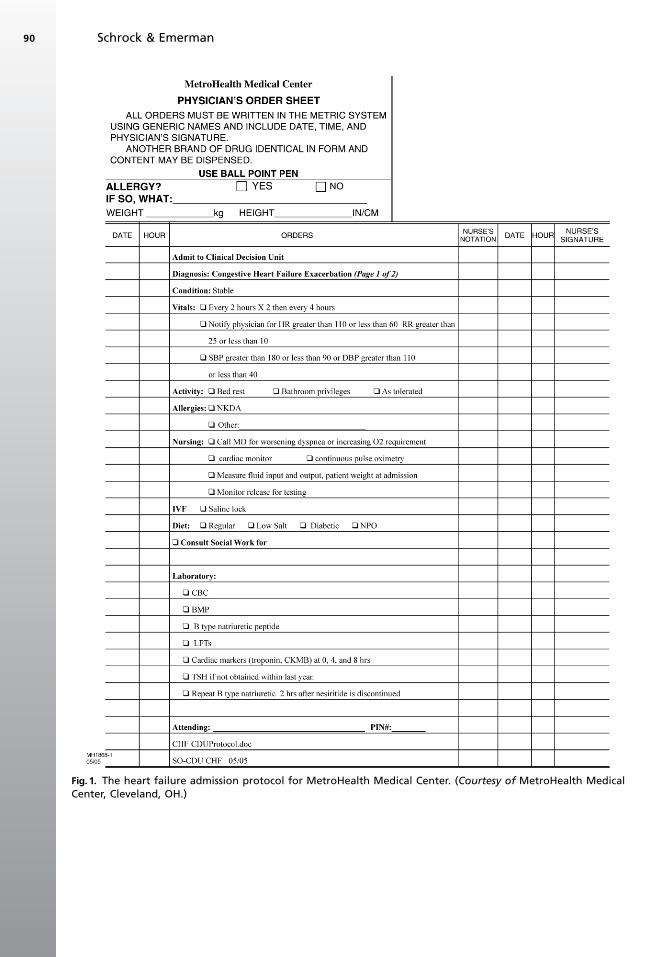
Fig. 1. The heart failure admission protocol for MetroHealth Medical Center. (Courtesy of MetroHealth MedicalCenter, Cleveland, OH.)
Schrock & Emerman90
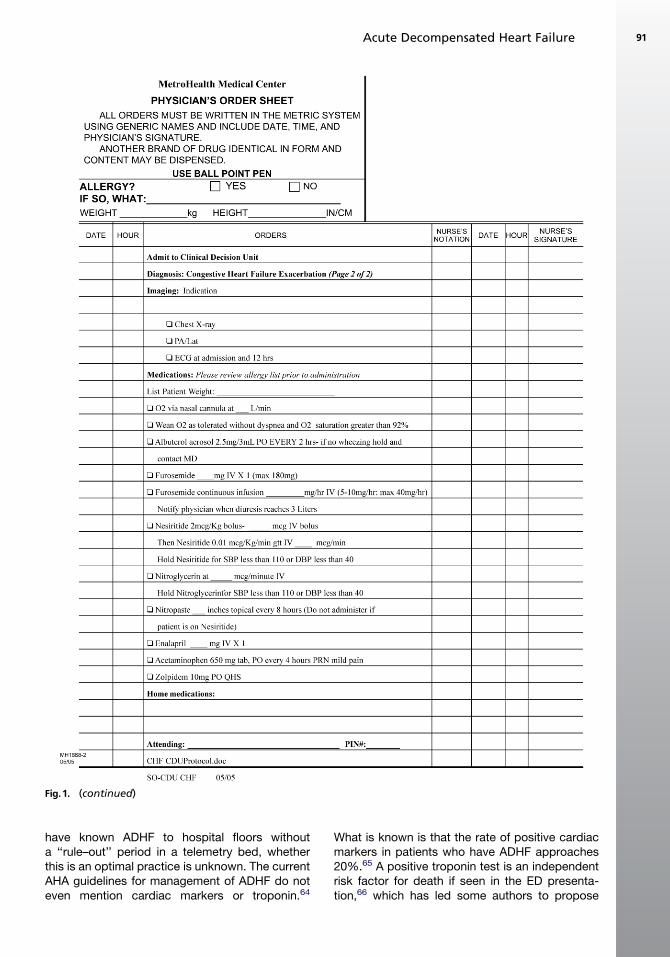
have known ADHF to hospital floors withouta ‘‘rule–out’’ period in a telemetry bed, whetherthis is an optimal practice is unknown. The currentAHA guidelines for management of ADHF do noteven mention cardiac markers or troponin.64
What is known is that the rate of positive cardiacmarkers in patients who have ADHF approaches20%.65 A positive troponin test is an independentrisk factor for death if seen in the ED presenta-tion,66 which has led some authors to propose
Fig.1. (continued)
Acute Decompensated Heart Failure 91
testing cardiac markers on all patients presentingwith ADHF.67 The rate of coronary artery diseasein patients who have known heart failure is morethan 50%. Baseline elevations in troponin I donot differ between patients who have idiopathicand ischemic cardiomyopathy.68,69
Echocardiography is recommended in the 2005AHA guidelines on the management of chronicheart failure.64 However, whether this study mustbe repeated in subsequent admissions is unclear.The authors’ practice is to obtain an echocardiog-raphy study if the patient has not been evaluated inthe prior year, or if new findings on physical exam-ination suggest structural cardiac changes thatmay influence outcome, such as mitral regurgita-tion or aortic stenosis not previously noted orworse than prior examinations.
Medical treatment of ADHF in the OU shouldcontinue the treatment plan begun in the ED. Forpatients placed on diuretics, two approaches areavailable. Many patients receive intravenousboluses of furosemide to encourage fluid loss. Ifthe initial dose is not completely effective, a sec-ond or third dose may be given. Several studieshave suggested that continuous intravenous infu-sions of furosemide result in greater diuresis andless ototoxicity, particularly if begun early in thehospitalization.70–73 Continuous infusion medica-tions present a risk for excessive diuresis, andtherefore stop-gap measures should appear inthe OU orders instructing the nursing staff todiscontinue the medication once a desired levelof diuresis is reached. Electrolytes should be mon-itored and replaced as needed when aggressivediuresis is being performed.
B-blockers have become a mainstay of heartfailure treatment because they have been shownto decrease morbidity and mortality.74–77 Patientswho have known heart failure should take a dailyb-blocker. Currently approved formulationsinclude metoprolol, bisoprolol, or carvedilol.B-blockers help decrease the heart’s exposureto chronic adrenergic stimulation and decreaseventricular remodeling.
Because most patients who have heart failureare currently undergoing this therapy, the questionarises of what to do if they are currently in ADHFand being admitted to an OU; should the b-blockerbe held? Prior dogma has instructed physicians tostop b-blockers in ADHF episodes. Althoughb-blockers can depress cardiac contraction, sud-denly stopping them may expose the heart toincreased endogenous catecholamines, whichcould produce tachydysrhythmias and myocardialischemia.
Experts have suggested that patients in mild tomoderate ADHF and who were not hypotensive
should be maintained on b-blockers.78–80 Evi-dence supports that patients who have acutemyocardial infarction experiencing ADHF benefitfrom b-blocker treatment.81 More recent studieshave determined an increase in morbidity andmortality and increased length of stay may beassociated with decreasing or stopping b-blockertherapy for patients in ADHF.82,83 The most recentpublished American College of Cardiology (ACC)guidelines suggest that patients who have beentaking b-blockers long-term decrease or stopthem only if evidence of hypoperfusion is pres-ent.64 Table 1 lists the starting dosages ofb-blockers approved for use in the treatment ofheart failure. Doses may be adjusted if bradycardiaor hypotension develops.
Like b-blockers, ACE inhibitors and ARBs haveshown direct long-term mortality benefits topatients who have heart failure.84–87 Patients diag-nosed with systolic heart failure and no contraindi-cation should be placed on an ACE inhibitor orARB. These medications should be started at lowdoses and gradually titrated upward. Patientswho have a history of renal insufficiency or hyper-kalemia should avoid these medications unlessclosely follow by their physician. For patients beingstarted on them, serum potassium levels shouldbe checked at 3 days and 1 week initially to checkfor hyperkalemia. If the patient seems unreliable orunable to return for blood draws, it may be moreprudent for the primary care physician to beginthis medication. Table 1 lists the starting dosesfor ACE inhibitors and ARBs.
Digitalis is a cardiac glycoside that has beenused in the treatment of ADHF for more than 2 cen-turies. Digitalis blocks myocardial sodium potas-sium adenosine triphosphatase (Na-K-ATPase).Enzymatic blockade in the kidney causes natriure-sis, which increases sodium excretion, anddecreases renin release. Although once a mainstayof heart failure treatment, digitalis has been rele-gated to a second-line therapy by some groupsand was changed from class I to class IIa classifi-cation in the most recent AHA update.64,88,89 Otherstudies have suggested that it may still be usefulbut only at a lower serum concentration thanused previously.90,91 Digitalis is still recommendedfor patients who have persistent heart failuresymptoms who have not responded to treatmentwith b-blockers, diuretics, or ACE inhibitors (orARBs). It is not indicated as a medication to stabi-lize an ADHF episode. Therefore, its usefulness inthe OU setting is limited, and it may be best newlyprescribed under the guidance of a consultingcardiologist.
Long-acting nitrates, such as isosorbide dini-trate, have been recommended for select patients
Schrock & Emerman92
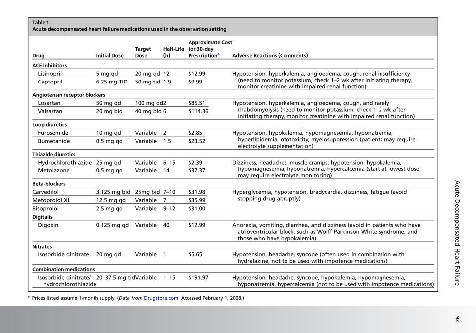
Table1Acute decompensated heart failuremedications used in the observation setting
Drug Initial DoseTargetDose
Half-Life(h)
Approximate Costfor 30-dayPrescriptiona Adverse Reactions (Comments)
ACE inhibitorsLisinopril 5 mg qd 20 mg qd 12 $12.99 Hypotension, hyperkalemia, angioedema, cough, renal insufficiency
(need to monitor potassium, check 1–2 wk after initiating therapy,monitor creatinine with impaired renal function)
Captopril 6.25 mg TID 50 mg tid 1.9 $9.99
Angiotensin receptor blockersLosartan 50 mg qd 100 mg qd2 $85.51 Hypotension, hyperkalemia, angioedema, cough, and rarely
rhabdomyolysis (need to monitor potassium, check 1–2 wk afterinitiating therapy, monitor creatinine with impaired renal function)
Valsartan 20 mg bid 40 mg bid 6 $114.36
Loop diureticsFurosemide 10 mg qd Variable 2 $2.85 Hypotension, hypokalemia, hypomagnesemia, hyponatremia,
hyperlipidemia, ototoxicity, myelosuppression (patients may requireelectrolyte supplementation)
Bumetanide 0.5 mg qd Variable 1.5 $23.52
Thiazide diureticsHydrochlorothiazide 25 mg qd Variable 6–15 $2.39 Dizziness, headaches, muscle cramps, hypotension, hypokalemia,
hypomagnesemia, hyponatremia, hypercalcemia (start at lowest dose,may require electrolyte monitoring)
Metolazone 0.5 mg qd Variable 14 $37.37
Beta-blockersCarvedilol 3.125 mg bid 25mg bid 7–10 $31.98 Hyperglycemia, hypotension, bradycardia, dizziness, fatigue (avoid
stopping drug abruptly)Metoprolol XL 12.5 mg qd Variable 7 $35.99
Bisoprolol 2.5 mg qd Variable 9–12 $31.00
DigitalisDigoxin 0.125 mg qd Variable 40 $12.99 Anorexia, vomiting, diarrhea, and dizziness (avoid in patients who have
atrioventricular block, such as Wolff-Parkinson-White syndrome, andthose who have hypokalemia)
NitratesIsosorbide dinitrate 20 mg qd Variable 1 $5.65 Hypotension, headache, syncope (often used in combination with
hydralazine, not to be used with impotence medications)
CombinationmedicationsIsosorbide dinitrate/
hydrochlorothiazide20–37.5 mg tidVariable 1–15 $191.97 Hypotension, headache, syncope, hypokalemia, hypomagnesemia,
hyponatremia, hypercalcemia (not to be used with impotence medications)
a Prices listed assume 1-month supply. (Data from Drugstore.com. Accessed February 1, 2008.)
Acu
teD
eco
mp
en
sate
dH
eart
Failu
re93

who have ADHF.64 These patients include thosewho are symptomatic on maximal therapy andthose who have exertional or nocturnal dyspnea.Long-acting nitrates have been shown to offer bet-ter survival than placebo but not ACE inhibitorsand therefore should not be given in their place.92
Side effects include headache and hypotension.Tolerance is common with extended use, andtherefore they are not prescribed around the clock.Because these are second-line agents for heartfailure, cardiology guidance may be useful inchoosing to which patients oral nitrates shouldbe prescribed.
Hydralazine is a direct vascular smooth musclerelaxant used often in combination with long-acting nitrates. It is not as effective as ACE inhibi-tors in reducing mortality in patients who haveheart failure but may be used in those intolerantof ACE inhibitors and ARBs.93 When used incombination with long-acting nitrates and ACEinhibitors or b-blockers, hydralazine was found tobe particularly effective at reducing mortality inAfrican Americans.94 The results of this researchled to the creation of BiDil, a combination medica-tion containing isosorbide dinitrate and hydral-azine. This medication was the first to receiveapproval from the U.S. Food and Drug Administra-tion (FDA) for treating disease in a specific race.Current guidelines do not suggest this combina-tion as a first-line agent in African Americanswho can tolerate ACE inhibitors and b-blockers.64
Nitroglycerin is a potent vasodilator with bothvenous and arteriole vasodilator properties. It iseffective at reducing preload and afterload rapidlyin patients presenting in ADHF. The half-life of in-travenous nitroglycerin is 1 to 4 minutes, requiringthat it be given in a continuous intravenous drip ortopical form. The intravenous form is often used forflash pulmonary edema or ADHF presentationswith elevated blood pressure. However, becausethis form requires frequent titration, it is not idealfor OU use. A sublingual tablet and spray formsare also available, with physiologic affects lastingfrom 1 to 6 hours after administration.95 The tabletand topical forms can be used in an OU setting butshould not supplant other more effective thera-pies, such as ACE inhibitors. Nitrates should beavoided in patients taking medications for erectiledysfunction, such as sildenafil, because the com-bination can cause severe hypotension.
Nesiritide is a recombinant form of BNP that isgiven intravenously and produces both natriuresisand vasodilation.96 Nesiritide has distinct advan-tages over other intravenous vasodilators, includ-ing nitroglycerin and nitroprusside, because itdoes not require intravenous titration or invasivehemodynamic monitoring, making it more
practical for OU use. Studies suggest that it pro-vides more rapid improvement of symptoms anddecreases mortality and hospital admissions com-pared with dobutamine, and facilitates more rapiddecrease in pulmonary capillary wedge pressurecompared with nitroglycerin.41,97
Nesiritide is given intravenously with an initialloading dose and then a maintenance infusion. Itcan cause hypotension, which may be potenti-ated by the use of other medications affectingpressure, and therefore ACE inhibitors are typi-cally withheld. In the OU setting, nesiritide wasshown to decrease the duration of hospitalizationby 4 days and showed nonsignificant trendsin decreased hospitalization after OU treatmentand 30-day hospital readmission.18
Although nesiritide was becoming widely usedfor the treatment of ADHF in ED and OU areas,this practice changed after two published meta-analyses suggested that it may worsen renal func-tion and increase mortality.98,99 These findings ledto a rapid decrease in the use of nesiritide forADHF.100 Multiple editorials have supported andrefuted the use of nesiritide in ADHF.101–103 Morerecent studies have suggested that nesiritidedoes not affect renal function or that it may bea dose-related phenomonon.104,105 A large-scaleprospective study is currently addressing thesafety and efficacy of nesiritide in ADHF. Informa-tion from this and other prospective studies will beneeded to determine the usefulness of nesiritideand which patients it would benefit most.Currently, nesiritide remains a second-line agentfor the treatment of ADHF.
Inotropes, including dobutamine, dopamine,milrinone, and amrinone, may be used to increasecardiac output and renal blood flow. They are oftenreserved for patients who have severelydepressed cardiac output not responding to after-load reduction and are administered to fewer than15% of patients who have ADHF.106 Inotropes areadministered as a constant infusion and are notused in OU care because they are often givenover several days.
Ultrafiltration recently became a treatmentoption for patients in ADHF. Ultrafiltration isa type of membrane filtration using hydrostaticpressure to force liquid and small solutes througha semipermeable membrane. One company hasreceived FDA approval for use of an ultrafiltrationdevice in ADHF. The Aquadex Flexflow requirestwo 18-gauge or larger peripheral intravenouscatheters or a double-lumen central venous cath-eter to draw off blood and return blood, minusthe filtered portion, to the patient. The patientmust be anticoagulated with heparin before useto prevent clotting of the filter. Patients who have
Schrock & Emerman94

contraindications to anticoagulation would not becandidates for this therapy. The device canremove up to 500 mL/h of ultrafiltrate, which con-sists of water and sodium. The rate of fluid removalis improved with the use of larger-gauge intrave-nous lines or catheters. Patients may be main-tained on ACE inhibitors, b-blockers, and evendiuretics, but intravenous vasodilators should beavoided.
The device seems to be effective in treatingpatients resistant to diuretics who are fluid over-loaded. One study showed an average fluidremoval of more than 8 L with no change insodium, potassium, creatinine, or BUN.107 The Ul-trafiltration versus Intravenous Diuretics forPatients Hospitalized for Acute DecompensatedCongestive Heart Failure trial compared ultrafiltra-tion with standard care for ADHF in hospitalizedpatients and showed a greater amount of weightand net fluid loss in the ultrafiltration group. Evenmore impressive was the 50% reduction in reho-spitalizations for heart failure and return ED orclinic visits within 90 days of discharge.108
Although this new technology seems promising,particularly for patients resistant to diuretics, itsusefulness in the OU setting is unclear. The needfor larger venous access and anticoagulationmake it less desirable for patients familiar withdiuretic treatment. Further studies are needed todetermine the usefulness of ultrafiltration in theOU population.
The long-term risk for sudden cardiac death inpatients who have severe systolic dysfunction issignificant, with one third to one half of patientswho have heart failure dying unexpectedly.109,110
Several trials have shown survival benefit forpatients who have ischemic heart failure and ejec-tion fractions less than 35%.111–114 Placement ofimplantable cardiac defibrillators (ICD) has nowbecome standard care, largely replacing antiar-rhythmic therapy, and the cost of placement iscurrently covered by Medicare for patients whomeet eligibility requirements. These criteria includeEF of 35% or less, New York Heart Associationclass II or III disease for greater than 9 months,and inducible or sustained ventricular tachycardia.
Patients who seem eligible for ICD placement orho have a history of ventricular tachycardia shouldbe referred to a cardiologist for electrophysiologicstudy. Patients who have a history of palpitations,syncope, or near-syncope may be referred for out-patient continuous cardiac monitoring, such asa Holter or event monitor, to determine if signifi-cant arrhythmias are occurring.
Some medications should likely be avoidedor at least not started while patients arebeing treated for ADHF, including nonsteroidal
anti-inflammatory agents, calcium channelblockers, and diabetic agents, such as metformin,thiazolidinedione, and cisapride.
Criteria for determining discharge are oftenbased on a patient’s clinical presentation aftera period of treatment. Several authors haveprovided more explicit discharge criteria forpatients in the OU treated for ADHF.115–117 Mostof these recommendations have not been pro-spectively studied but are pragmatic assessmentsof the patient’s clinical status. Evidence suggeststhat urine output less than 1 L after treatment pre-dicts treatment failure.118 Some findings that maysuggest that the patient has not improved includethe presence of a third heart sound, increasedBUN, hypotension, hypoxia, and dyspnea at restor with minimal exertion. Having patients ambulatethrough the department is often prudent to showthat they will be able to do so if discharged.Some suggested discharge criteria include:
Clinically improved dyspneaAbility to ambulate back to baselineUrine output greater than 1 LResting heart rate less than 100 beats/min
and systolic blood pressure greater than80 mm Hg
Pulse oximetry greater than 91% if notnormally on supplemental oxygen
No clinical or EKG evidence of cardiacischemia
No significant increase in serum BUN orcreatinine
No new or significant dysrhythmias
The most recent AHA/ACC guidelines for thetreatment of ADHF do not provide dischargecriteria but recommend that patients be euvolemicbefore discharge.64
It is extremely important to start recommendedheart failure medications and provide prescriptionsto patients discharged after OU therapy, becausestudies have shown that providing appropriatelong-term heart failure medications, such as b-blockers and ACE inhibitors, reduces mortality.119
Discharge education is an important aspect ofcare that is often overlooked but can have a largebeneficial effect if administered properly. Educationconcerning smoking and medication and dietarycompliance can reduce recidivism and decreasemedical expenses for these patients.120–123
The importance of smoking cessation educationand therapy deserves additional mention. Simplyby quitting smoking, patients can reduce theirmortality by 30% within 2 years. This mortalitybenefit is similar to that seen with the use ofb-blockers or ACE inhibitors in the treatment ofheart failure.124,125 Having patients receive the
Acute Decompensated Heart Failure 95
advice directly from the treating physician is veryinfluential and can increase rates of smokingcessation greater than 60%.126
If resources are available, a multidisciplinaryteam, including cardiologist dietitians, socialworkers, and heart failure educators such as nursepractitioners, would be ideal to provide patientinformation on the importance of medication anddietary compliance, exercise, smoking cessation,and following up with future tests and appoint-ments. Other areas for patient education includemonitoring dyspnea symptoms and weight andnotifying the physician for worsening conditions.The AHA Get with the Guidelines program offersInternet-based educational tools to educatepatients who have heart failure who are being dis-charged. Ensuring that patients are taking appro-priate medications, such as b-blockers and ACEinhibitors, is extremely important. Patients wholeave with prescriptions for cardiac medicationsare more likely to adhere to that medicationregimen.127
SUMMARY
ADHF is a common illness presenting to the EDthat is amenable to OU treatment. As the numberof baby boomers continues to grow and the inci-dence of heart failure increases, the financial impli-cations of ADHF treatment will become moreprominent. Obtaining institutional support anddeveloping a good working relationship with cardi-ology colleagues is vital to creating workableADHF protocols for whichever type of OU an insti-tution decides to use.
REFERENCES
1. Hugli O, Braun JE, Kim S, et al. United States emer-
gency department visits for acute decompensated
heart failure, 1992 to 2001. Am J Cardiol 2005;
96(11):1537–42.
2. Burt CW, Schappert SM. Ambulatory care visits to
physician offices, hospital outpatient departments,
and emergency departments: United States,
1999–2000. Vital Health Stat 2004;13(157):1–70.
3. Massie BM, Shah NB. Evolving trends in the epide-
miologic factors of heart failure: rationale for
preventive strategies and comprehensive disease
management. Am Heart J 1997;133(6):703–12.
4. O’Connell JB, Bristow MR. Economic impact of
heart failure in the United States: time for a different
approach. J Heart Lung Transplant 1994;13(4):
S107–12.
5. O’Connell JB. The economic burden of heart fail-
ure. Clin Cardiol 2000;23(Suppl.3):III6–10.
6. Graff L, Orledge J, Radford MJ, et al. Correlation of
the agency for health care policy and research
congestive heart failure admission guideline with
mortality: peer review organization voluntary hospi-
tal association initiative to decrease events
(provide) for congestive heart failure. Ann Emerg
Med 1999;34(4 Pt 1):429–37.
7. Polanczyk CA, Rohde LE, Philbin EA, et al. A new
casemix adjustment index for hospital mortality
among patients with congestive heart failure. Med
Care 1998;36(10):1489–99.
8. Rosamond W, Flegal K, Furie K, et al. Heart disease
and stroke statistics–2008 update: a report from the
American Heart Association Statistics Committee
and Stroke Statistics Subcommittee. Circulation
2008;117(4):e25–146.
9. Stewart S, MacIntyre K, Capewell S, et al. Heart
failure and the aging population: an increasing
burden in the 21st century? Heart 2003;89(1):
49–53.
10. Ansari M, Massie BM. Heart failure: how big is the
problem? Who are the patients? What does the
future hold? Am Heart J 2003;146(1):1–4.
11. Storrow AB, Collins SP, Lyons MS, et al. Emergency
department observation of heart failure: preliminary
analysis of safety and cost. Congest Heart Fail
2005;11(2):68–72.
12. Peacock WF, Remer EE, Aponte J, et al. Effective
observation unit treatment of decompensated heart
failure. Congest Heart Fail 2002;8(2):68–73.
13. Ross MA, Compton S, Medado P, et al. An emer-
gency department diagnostic protocol for patients
with transient ischemic attack: a randomized con-
trolled trial. Ann Emerg Med 2007;50(2):109–19.
14. Trommald M, Aaserud M, Bjorndal A. [Observational
units–same good service to lower costs?]. Tidsskr
NorLaegeforen2000;120(25):3029–34[inNorwegian].
15. Goodacre S, Nicholl J, Dixon S, et al. Randomised
controlled trial and economic evaluation of a chest
pain observation unit compared with routine care.
BMJ 2004;328(7434):1–6.
16. Roberts RR, Zalenski RJ, Mensah EK, et al. Costs
of an emergency department-based accelerated
diagnostic protocol vs hospitalization in patients
with chest pain: a randomized controlled trial.
JAMA 1997;278(20):1670–6.
17. Gaspoz JM, Lee TH, Weinstein MC, et al. Cost-
effectiveness of a new short-stay unit to ‘‘rule out’’
acute myocardial infarction in low risk patients.
J Am Coll Cardiol 1994;24(5):1249–59.
18. Peacock WFt, Holland R, Gyarmathy R, et al.
Observation unit treatment of heart failure with
nesiritide: results from the proaction trial. J Emerg
Med 2005;29(3):243–52.
19. McDermott MF, Murphy DG, Zalenski RJ, et al. A
comparison between emergency diagnostic and
treatment unit and inpatient care in the
Schrock & Emerman96
management of acute asthma. Arch Intern Med
1997;157(18):2055–62.
20. RydmanRJ,ZalenskiRJ,RobertsRR,etal.Patientsat-
isfaction with an emergency department chest pain
observation unit. Ann Emerg Med 1997;29(1):109–15.
21. Henneman PL, Marx JA, Cantrill SC, et al. The use
of an emergency department observation unit in
the management of abdominal trauma. Ann Emerg
Med 1989;18(6):647–50.
22. Shen WK, Decker WW, Smars PA, et al. Syncope
evaluation in the emergency department study
(seeds): a multidisciplinary approach to syncope
management. Circulation 2004;110(24):3636–45.
23. Graff L, Radford MJ, Werne C. Probability of
appendicitis before and after observation. Ann
Emerg Med 1991;20(5):503–7.
24. Aghababian RV. Acutely decompensated heart fail-
ure: opportunities to improve care and outcomes in
the emergency department. Rev Cardiovasc Med
2002;4(Suppl. 3):S3–9.
25. Kelen GD, Scheulen JJ, Hill PM. Effect of an emer-
gency department (ed) managed acute care unit
on ED overcrowding and emergency medical
services diversion. Acad Emerg Med 2001;8(11):
1095–100.
26. Ross MA, Wilson AG, McPherson M. The impact of
an ED observation unit bed on inpatient bed avail-
ability. Acad Emerg Med 2001;8(5):576.
27. Diamond NJ, Schofferman JA, Elliot JW. Evaluation
of an emergency department observation ward.
JACEP 1976;5(1):29–31.
28. Landers Fa WJ, McNbney WK. Observation ward
utilization. JACEP 1975;4:123–5.
29. Emergency department observation units. Ameri-
can college of emergency physicians. Ann Emerg
Med 1988;17(1):95–6.
30. Centers for Medicare & Medicaid Services. CMS
manual system. Department of Health & Human Ser-
vices. Pub 100–04 Medicare claims processing.
Available at: http://www.cms.hhs.gov/transmittals/
downloads/R1430CPTXT.pdf. Accessed September
15, 2008.
31. Yealy DM, De Hart DA, Ellis G, et al. A survey of ob-
servation units in the United States. Am J Emerg
Med 1989;7(6):576–80.
32. Department of Health and Human Services. Medi-
care and Medicaid Programs. Interim and Final
Rule. Federal Register. Available at: http://www.
cms.hhs.gov/HospitalOutpatientPPS/Downloads/
cms1392fc.pdf. Accessed December 13, 2007.
33. McCullough PA, Nowak RM, McCord J, et al.
B-type natriuretic peptide and clinical judgment in
emergency diagnosis of heart failure: analysis
from breathing not properly (BNP) multinational
study. Circulation 2002;106(4):416–22.
34. Clerico A, Fontana M, Zyw L, et al. Comparison of
the diagnostic accuracy of brain natriuretic peptide
(BNP) and the n-terminal part of the propeptide of
BNP immunoassays in chronic and acute heart fail-
ure: a systematic review. Clin Chem 2007;53(5):
813–22.
35. Dokainish H, Zoghbi WA, Lakkis NM, et al. Compar-
ative accuracy of B-type natriuretic peptide and tis-
sue Doppler echocardiography in the diagnosis of
congestive heart failure. Am J Cardiol 2004;93(9):
1130–5.
36. Latour-Perez J, Coves-Orts FJ, Abad-Terrado C,
et al. Accuracy of B-type natriuretic peptide levels
in the diagnosis of left ventricular dysfunction and
heart failure: a systematic review. Eur J Heart Fail.
2006;8(4):390–9.
37. Mueller T, Gegenhuber A, Poelz W, et al. Diagnostic
accuracy of B type natriuretic peptide and amino
terminal proBNP in the emergency diagnosis of
heart failure. Heart 2005;91(5):606–12.
38. Wang CS, FitzGerald JM, Schulzer M, et al. Does
this dyspneic patient in the emergency department
have congestive heart failure? JAMA 2005;294(15):
1944–56.
39. Benjamin EJ, Levy D, Vaziri SM, et al. Independent
risk factors for atrial fibrillation in a population-
based cohort. The Framingham Heart Study.
JAMA 1994;271(11):840–4.
40. Adams KF Jr, Fonarow GC, Emerman CL, et al.
Characteristics and outcomes of patients hospital-
ized for heart failure in the United States: rationale,
design, and preliminary observations from the first
100,000 cases in the acute decompensated heart
failure national registry (ADHERE). Am Heart J
2005;149(2):209–16.
41. VMAC Investigators. Intravenous nesiritide vs nitro-
glycerin for treatment of decompensated conges-
tive heart failure: a randomized controlled trial.
JAMA 2002;287(12):1531–40.
42. Smith WR, Poses RM, McClish DK, et al. Prognostic
judgments and triage decisions for patients with
acute congestive heart failure. Chest 2002;121(5):
1610–7.
43. Butler J, Hanumanthu S, Chomsky D, et al.
Frequency of low-risk hospital admissions for heart
failure. Am J Cardiol 1998;81(1):41–4.
44. Diercks DB, Peacock WF, Kirk JD, et al. ED patients
with heart failure: identification of an observational
unit-appropriate cohort. Am J Emerg Med 2006;
24(3):319–24.
45. Burkhardt J, Peacock WF, Emerman CL. Predictors
of emergency department observation unit out-
comes. Acad Emerg Med 2005;12(9):869–74.
46. Auble TE, Hsieh M, Gardner W, et al. A prediction
rule to identify low-risk patients with heart failure.
Acad Emerg Med 2005;12(6):514–21.
47. Fonarow GC, Adams KF Jr, Abraham WT, et al. Risk
stratification for in-hospital mortality in acutely
decompensated heart failure: classification and
Acute Decompensated Heart Failure 97
regression tree analysis. JAMA 2005;293(5):
572–80.
48. Lee DS, Austin PC, Rouleau JL, et al. Predicting
mortality among patients hospitalized for heart fail-
ure: derivation and validation of a clinical model.
JAMA 2003;290(19):2581–7.
49. Brophy JM, Deslauriers G, Rouleau JL. Long-term
prognosis of patients presenting to the emergency
room with decompensated congestive heart failure.
Can J Cardiol 1994;10(5):543–7.
50. Brophy JM, Deslauriers G, Boucher B, et al. The
hospital course and short term prognosis of
patients presenting to the emergency room with
decompensated congestive heart failure. Can
J Cardiol 1993;9(3):219–24.
51. Auble TE, Hsieh M, McCausland JB, et al. Compar-
ison of four clinical prediction rules for estimating
risk in heart failure. Ann Emerg Med 2007;50(2):
127–35.
52. Chin MH, Goldman L. Correlates of major compli-
cations or death in patients admitted to the hospital
with congestive heart failure. Arch Intern Med
1996;156(16):1814–20.
53. Hsieh M, Auble TE, Yealy DM. Validation of the
acute heart failure index. Ann Emerg Med 2008;
51(1):37–44.
54. Gheorghiade M, Rossi JS, Cotts W, et al. Charac-
terization and prognostic value of persistent hypo-
natremia in patients with severe heart failure in
the ESCAPE trial. Arch Intern Med 2007;167(18):
1998–2005.
55. Felker GM, Adams KF Jr, Gattis WA, et al. Anemia
as a risk factor and therapeutic target in heart fail-
ure. J Am Coll Cardiol 2004;44(5):959–66.
56. Butler J, Forman DE, Abraham WT, et al. Relationship
between heart failure treatment and development of
worsening renal function among hospitalized
patients. Am Heart J 2004;147(2):331–8.
57. Kramer BK, Schweda F, Riegger GA. Diuretic
treatment and diuretic resistance in heart failure.
Am J Med 1999;106(1):90–6.
58. Neuberg GW, Miller AB, O’Connor CM, et al.
Diuretic resistance predicts mortality in patients
with advanced heart failure. Am Heart J 2002;
144(1):31–8.
59. Redfield MM, Jacobsen SJ, Burnett JC Jr, et al.
Burden of systolic and diastolic ventricular dys-
function in the community: appreciating the scope
of the heart failure epidemic. JAMA 2003;289(2):
194–202.
60. Smith GL, Masoudi FA, Vaccarino V, et al. Out-
comes in heart failure patients with preserved ejec-
tion fraction: mortality, readmission, and functional
decline. J Am Coll Cardiol 2003;41(9):1510–8.
61. Vasan RS, Larson MG, Benjamin EJ, et al. Conges-
tive heart failure in subjects with normal versus
reduced left ventricular ejection fraction:
prevalence and mortality in a population-based co-
hort. J Am Coll Cardiol 1999;33(7):1948–55.
62. Bhatia RS, Tu JV, Lee DS, et al. Outcome of heart
failure with preserved ejection fraction in a popula-
tion-based study. N Engl J Med. 2006;355(3):
260–9.
63. George TL, Hiok CT, Leonard K. Type of liver dys-
function in heart failure and its relation to the sever-
ity of tricuspid regurgitation. Am J Cardiol 2002;
90(12):1405.
64. Hunt SA, Abraham WT, Chin MH, et al. ACC/AHA
2005 guideline update for the diagnosis and
management of chronic heart failure in the adult:
a report of the American college of cardiology/
American heart association task force on practice
guidelines (writing committee to update the 2001
guidelines for the evaluation and management of
heart failure): developed in collaboration with the
American college of chest physicians and the inter-
national society for heart and lung transplantation:
endorsed by the heart rhythm society. Circulation
2005;112(12):e154–235.
65. Demir M, Kanadasi M, Akpinar O, et al. Cardiac
troponin t as a prognostic marker in patients with
heart failure: a 3-year outcome study. Angiology
2007;58(5):603–9.
66. Parenti N, Bartolacci S, Carle F, et al. Cardiac
troponin I as prognostic marker in heart failure
patients discharged from emergency department.
Intern Emerg Med 2008;3:44–7.
67. Peacock WF, Emerman CE, Doleh M, et al. Retro-
spective review: the incidence of non-ST segment
elevation MI in emergency department patients
presenting with decompensated heart failure. Con-
gest Heart Fail 2003;9(6):303–8.
68. Missov E, Calzolari C, Pau B. Circulating cardiac
troponin I in severe congestive heart failure. Circu-
lation 1997;96(9):2953–8.
69. Glauser J, Erickson J, Bhatt D, et al. Elevated
serum cardiac markers predict coronary artery dis-
ease in patients with a history of heart failure who
present with chest pain: insights from the i*tracs
registry. Congest Heart Fail 2007;13(3):142–8.
70. Dormans TP, van Meyel JJ, Gerlag PG, et al.
Diuretic efficacy of high dose furosemide in severe
heart failure: bolus injection versus continuous infu-
sion. J Am Coll Cardiol 1996;28(2):376–82.
71. Pivac N, Rumboldt Z, Sardelic S, et al. Diuretic
effects of furosemide infusion versus bolus injec-
tion in congestive heart failure. Int J Clin Pharmacol
Res 1998;18(3):121–8.
72. Lahav M, Regev A, Ra’anani P, et al. Intermittent
administration of furosemide vs continuous infusion
preceded by a loading dose for congestive heart
failure. Chest 1992;102(3):725–31.
73. Salvador DR, Rey NR, Ramos GC, et al. Continuous
infusion versus bolus injection of loop diuretics in
Schrock & Emerman98
congestive heart failure. Cochrane Database Syst
Rev 2005;(3):CD003178.
74. Packer M, Coats AJS, Fowler MB, et al. Effect of
carvedilol on survival in severe chronic heart fail-
ure. N Engl J Med 2001;344(22):1651–8.
75. Effect of metoprolol CR/XL in chronic heart failure:
metoprolol CR/XL randomised intervention trial in
congestive heart failure (MERIT-HF). Lancet 1999;
353(9169):2001–7.
76. Hjalmarson A, Goldstein S, Fagerberg B, et al.
Effects of controlled-release metoprolol on total
mortality, hospitalizations, and well-being in pa-
tients with heart failure: the metoprolol CR/XL
randomized intervention trial in congestive heart
failure (MERIT-HF). JAMA 2000;283(10):1295–302.
77. The cardiac insufficiency bisoprolol study II (CIBIS-II):
a randomised trial. Lancet 1999;353(9146):9–13.
78. Kao W, Surjancev BP. Management of acute heart fail-
ure exacerbation. Crit Care Clin 2001;17(2):321–35.
79. Cleland JGF, Bristow MR, Erdmann E, et al. Beta-
blocking agents in heart failure: should they
be used and how? Eur Heart J 1996;17(11):
1629–39.
80. Gattis WA, O’Connor CM, Gallup DS, et al. Predis-
charge initiation of carvedilol in patients hospital-
ized for decompensated heart failure: results of
the initiation management predischarge: process
for assessment of carvedilol therapy in heart failure
(IMPACT-HF) trial. J Am Coll Cardiol 2004;43(9):
1534–41.
81. Chadda K, Goldstein S, Byington R, et al. Effect of
propranolol after acute myocardial infarction in
patients with congestive heart failure. Circulation
1986;73(3):503–10.
82. Metra M, Torp-Pedersen C, Cleland JG, et al.
Should beta-blocker therapy be reduced or with-
drawn after an episode of decompensated heart
failure? Results from COMET. Eur J Heart Fail
2007;9(9):901–9.
83. Shamimi-Noori S WD, Dasgupta A, Adams S, et al.
[abstract]. Beta blocker dose adjustment upon
hospitalization for acute decompensated heart fail-
ure. J Am Coll Cardiol 2007;40(11 Suppl A):
1028–66.
84. Garg R, Yusuf S. Overview of randomized trials of
angiotensin-converting enzyme inhibitors on mor-
tality and morbidity in patients with heart failure.
Collaborative Group on ACE Inhibitor trials. JAMA
1995;273(18):1450–6.
85. Nony P, Boissel JP, Girard P, et al. Relative efficacy
of angiotensin converting enzyme inhibitors on
mortality of patients with congestive heart failure:
implications of randomized trials and role of the
aetiology (ischaemic or non-ischaemic) of heart
failure. Eur Heart J 1992;13(8):1101–8.
86. Lee VC, Rhew DC, Dylan M, et al. Meta-analysis:
angiotensin-receptor blockers in chronic heart
failure and high-risk acute myocardial infarction.
Ann Intern Med 2004;141(9):693–704.
87. Yusuf S, Pfeffer MA, Swedberg K, et al. Effects of
candesartan in patients with chronic heart failure
and preserved left-ventricular ejection fraction:
the CHARM-preserved trial. Lancet 2003;
362(9386):777–81.
88. Morris SA, Hatcher HF, Reddy DK. Digoxin therapy
for heart failure: an update. Am Fam Physician
2006;74(4):613–8.
89. Ahmed A, Rich MW, Fleg JL, et al. Effects of
digoxin on morbidity and mortality in diastolic heart
failure: the ancillary digitalis investigation group
trial. Circulation 2006;114(5):397–403.
90. Rathore SS, Curtis JP, Wang Y, et al. Association of
serum digoxin concentration and outcomes in pa-
tients with heart failure. JAMA 2003;289(7):871–8.
91. Adams KF Jr, Patterson JH, Gattis WA, et al. Rela-
tionship of serum digoxin concentration to mortality
and morbidity in women in the digitalis investiga-
tion group trial: a retrospective analysis. J Am
Coll Cardiol 2005;46(3):497–504.
92. Loeb HS, Johnson G, Henrick A, et al. Effect of ena-
lapril, hydralazine plus isosorbide dinitrate, and
prazosin on hospitalization in patients with chronic
congestive heart failure. The V-HeFT VA coopera-
tive studies group. Circulation 1993;87(Suppl.6):
VI78–87.
93. Cohn JN, Johnson G, Ziesche S, et al. A compari-
son of enalapril with hydralazine-isosorbide dini-
trate in the treatment of chronic congestive heart
failure. N Engl J Med 1991;325(5):303–10.
94. Taylor AL, Ziesche S, Yancy C, et al. Combination
of isosorbide dinitrate and hydralazine in blacks
with heart failure. N Engl J Med 2004;351(20):
2049–57.
95. Patrick K, Noonan LZB. The bioavailability of oral
nitroglycerin. J Pharm Sci 1986;75(3):241–3.
96. Colucci WS, Elkayam U, Horton DP, et al. Intrave-
nous nesiritide, a natriuretic peptide, in the treat-
ment of decompensated congestive heart failure.
Nesiritide study group. N Engl J Med 2000;
343(4):246–53.
97. Silver MA, Horton DP, Ghali JK, et al. Effect of ne-
siritide versus dobutamine on short-term outcomes
in the treatment of patients with acutely decompen-
sated heart failure. J Am Coll Cardiol 2002;39(5):
798–803.
98. Sackner-Bernstein JD, Skopicki HA, Aaronson KD.
Risk of worsening renal function with nesiritide in
patients with acutely decompensated heart failure.
Circulation 2005;111(12):1487–91.
99. Sackner-Bernstein JD, Kowalski M, Fox M, et al.
Short-term risk of death after treatment with nesiri-
tide for decompensated heart failure: a pooled
analysis of randomized controlled trials. JAMA
2005;293(15):1900–5.
Acute Decompensated Heart Failure 99

100. Hauptman PJ, Schnitzler MA, Swindle J, et al. Use
of nesiritide before and after publications suggest-
ing drug-related risks in patients with acute de-
compensated heart failure. JAMA 2006;296(15):
1877–84.
101. Topol EJ. Nesiritide—not verified. N Engl J Med
2005;353(2):113–6.
102. Teerlink JR, Massie BM. Nesiritide and worsening
of renal function: the emperor’s new clothes? Circu-
lation 2005;111(12):1459–61.
103. Shlipak MG, Massie BM. The clinical challenge of
cardiorenal syndrome. Circulation 2004;110(12):
1514–7.
104. Witteles RM, Kao D, Christopherson D, et al.
Impact of nesiritide on renal function in patients
with acute decompensated heart failure and pre-
existing renal dysfunction: a randomized, double-
blind, placebo-controlled clinical trial. J Am Coll
Cardiol 2007;50(19):1835–40.
105. Riter HG, Redfield MM, Burnett JC, et al. Nonhypo-
tensive low-dose nesiritide has differential renal ef-
fects compared with standard-dose nesiritide in
patients with acute decompensated heart failure
and renal dysfunction. J Am Coll Cardiol 2006;
47(11):2334–5.
106. Fonarow GC, Heywood JT, Heidenreich PA, et al.
Temporal trends in clinical characteristics, treat-
ments, and outcomes for heart failure hospitaliza-
tions, 2002 to 2004: findings from Acute
Decompensated Heart Failure National Registry
(ADHERE). Am Heart J 2007;153(6):1021.
107. Costanzo MR, Saltzberg M, O’Sullivan J, et al. Early
ultrafiltration in patients with decompensated heart
failure and diuretic resistance. J Am Coll Cardiol
2005;46(11):2047–51.
108. Costanzo MR, Guglin ME, Saltzberg MT, et al.
Ultrafiltration versus intravenous diuretics for
patients hospitalized for acute decompensated
heart failure. J Am Coll Cardiol 2007;49(6):675–83.
109. Kannel WB, Plehn JF, Cupples LA. Cardiac failure
and sudden death in the Framingham study. Am
Heart J 1988;115(4):869–75.
110. Packer M. Lack of relation between ventricular ar-
rhythmias and sudden death in patients with chronic
heart failure. Circulation 1992;85(Suppl. I):I50–6.
111. Moss AJ, Zareba W, Hall WJ, et al. Prophylactic im-
plantation of a defibrillator in patients with myocar-
dial infarction and reduced ejection fraction. N Engl
J Med 2002;346(12):877–83.
112. Bardy GH, Lee KL, Mark DB, et al. Amiodarone or
an implantable cardioverter-defibrillator for con-
gestive heart failure. N Engl J Med 2005;352(3):
225–37.
113. AVID Investigators. The antiarrhythmics versus
implantable defibrillators I. A comparison of antiar-
rhythmic-drug therapy with implantable defibrilla-
tors in patients resuscitated from near-fatal
ventricular arrhythmias. N Engl J Med 1997;
337(22):1576–84.
114. Buxton AE, Lee KL, Fisher JD, et al. A randomized
study of the prevention of sudden death in patients
with coronary artery disease. N Engl J Med 1999;
341(25):1882–90.
115. Peacock WF. Using the emergency department
clinical decision unit for acute decompensated
heart failure. Cardiol Clin 2005;23(4):569–88, viii.
116. Deborah Diercks P, Frank W. Short stay manage-
ment of heart failure. Philadelphia: Lippincott
Williams & Wilkins; 2006.
117. Meldon S, Ma OJ, Woolard R, American college of
emergency medicine. Geriatric emergency medi-
cine. 1st edition. New York: McGraw-Hill, Health
Professions Division; 2004.
118. Peacock W, Frank AJH, Craig Mary T, et al. Predic-
tors of unsuccessful treatment for congestive heart
failure in the emergency department observation
unit. Acad Emerg Med 1997;4(5):493–4.
119. Johnson D, Jin Y, Quan H, et al. Beta-blockers and
angiotensin-converting enzyme inhibitors/receptor
blockers prescriptions after hospital discharge for
heart failure are associated with decreased mortal-
ity in Alberta, Canada. J Am Coll Cardiol 2003;
42(8):1438–45.
120. Koelling TM, Johnson ML, Cody RJ, et al. Dis-
charge education improves clinical outcomes in
patients with chronic heart failure. Circulation
2005;111(2):179–85.
121. Rich MW, Beckham V, Wittenberg C, et al. A multi-
disciplinary intervention to prevent the readmission
of elderly patients with congestive heart failure.
N Engl J Med 1995;333(18):1190–5.
122. Krumholz HM, Amatruda J, Smith GL, et al. Ran-
domized trial of an education and support interven-
tion to prevent readmission of patients with heart
failure. J Am Coll Cardiol 2002;39(1):83–9.
123. Phillips CO, Wright SM, Kern DE, et al. Comprehen-
sive discharge planning with postdischarge support
for older patients with congestive heart failure:
a meta-analysis. JAMA 2004;291(11):1358–67.
124. Lightwood J, Fleischmann KE, Glantz SA. Smoking
cessation in heart failure: it is never too late. J Am
Coll Cardiol 2001;37(6):1683–4.
125. Suskin N, Sheth T, Negassa A, et al. Relationship of
current and past smoking to mortality and morbid-
ity in patients with left ventricular dysfunction. J Am
Coll Cardiol 2001;37(6):1677–82.
126. Lancaster T, Stead L, Silagy C, et al. Effectiveness
of interventions to help people stop smoking: find-
ings from the cochrane library. BMJ 2000;
321(7257):355–8.
127. Simpson E, Beck C, Richard H, et al. Drug
prescriptions after acute myocardial infarction:
dosage, compliance, and persistence. Am Heart
J 2003;145(3):438–44.
Schrock & Emerman100
Observation UnitEconomicsSandra G. Sieck, RN, MBAa,*, Mark G. Moseley, MD, MHAb,c
With an aging population (projected 16.5% of thepopulation over the age of 65 in 2020), the UnitedStates health care delivery system is struggling tohandle an onslaught of chronic disease burden.1–3
Heart failure (HF) is one such chronic disease pro-cess whose incidence is increasing as patientswith chronic cardiac disease are living longer be-cause of improved technologic and therapeuticadvances. While longevity with chronic disease isdesirable to an individual patient, it increasinglyplaces a burden on providers, payers, and thehealth care infrastructure that must continue torender care for those with decompensated dis-ease states in economically uncertain times. Asa result, the delivery of care for acute exacerba-tions of chronic disease on an ongoing basis con-tinues to strain an already fragile and increasinglyfragmented delivery system ill equipped to handlethis burden.
Nowhere is this realized more painfully from aneconomic perspective, than for acute care facili-ties treating patients with acute heart failure(AHF). Such facilities face the tenuous balancingact between offering improved inpatient therapeu-tic modalities (mechanical and pharmacologic),while attempting to maintain economic viability un-der the microscope of use review and prospectivepayment systems.4 As payment from all payersources is increasingly linked to outcome metrics(pay-for-performance), defining and implementingoptimal care that is cost-effective and results in thebest clinical outcomes has become critical. Withthis said, improved therapy for AHF has come atthe price of increased costs for delivery of care
without the promise of increased reimbursementfor the same. Thus, there is a dichotomy for acutecare facilities to insure the provision of cost-efficientcare while concomitantly maintaining a high qualityof care.4 As a result, the health care delivery and re-imbursement system is at a crossroads, where thecare for patients with chronic disease states de-mands innovations that better match patient careneeds with economically feasible care models.
BURDEN OF DISEASE
In light of the above, it is not surprising that theclinical and financial burden to the United Stateshealth care system and acute care facilities isstaggering. Relevant examples of this burden in-clude the following:
In 2005, the American Heart Association esti-mated that there were almost 5 millionpeople with HF in the United States.5
In 2001, there were almost 1 million hospitali-zations in the United States with a firstlisted discharge diagnosis of HF (Fig. 1).
Nearly 20% of hospital admissions among per-sons older than 65 are because of HF, mak-ing it responsible for more elderly patienthospitalizations than any other disease.6
Readmission rates were as high as 20% at 30days.7
HF is the most commonly used Medicare di-agnosis-related group (DRG).3
HF accounts for 12 to 15 million physician of-fice visits annually.8
a Sieck HealthCare Consulting, Mobile, AL, USAb The Ohio State University, OH, USAc The Ohio State University Medical Center, Columbus, OH, USA* Corresponding author. Sieck HealthCare Consulting, 9431 Jeff Hamilton Road, Mobile, AL 36695, USA.E-mail address: [email protected] (S.G. Sieck).
KEYWORDS� Observation medicine � Economics� Heart failure � Emergency Department
Heart Failure Clin 5 (2009) 101–111doi:10.1016/j.hfc.2008.08.0081551-7136/08/$ – see front matter ª 2008 Published by Elsevier Inc. he
artf
ailu
re.th
ecli
nics
.com
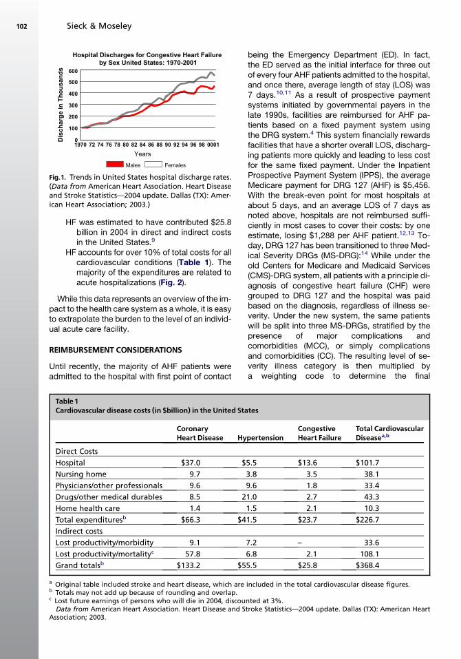
HF was estimated to have contributed $25.8billion in 2004 in direct and indirect costsin the United States.9
HF accounts for over 10% of total costs for allcardiovascular conditions (Table 1). Themajority of the expenditures are related toacute hospitalizations (Fig. 2).
While this data represents an overview of the im-pact to the health care system as a whole, it is easyto extrapolate the burden to the level of an individ-ual acute care facility.
REIMBURSEMENT CONSIDERATIONS
Until recently, the majority of AHF patients wereadmitted to the hospital with first point of contact
being the Emergency Department (ED). In fact,the ED served as the initial interface for three outof every four AHF patients admitted to the hospital,and once there, average length of stay (LOS) was7 days.10,11 As a result of prospective paymentsystems initiated by governmental payers in thelate 1990s, facilities are reimbursed for AHF pa-tients based on a fixed payment system usingthe DRG system.4 This system financially rewardsfacilities that have a shorter overall LOS, discharg-ing patients more quickly and leading to less costfor the same fixed payment. Under the InpatientProspective Payment System (IPPS), the averageMedicare payment for DRG 127 (AHF) is $5,456.With the break-even point for most hospitals atabout 5 days, and an average LOS of 7 days asnoted above, hospitals are not reimbursed suffi-ciently in most cases to cover their costs: by oneestimate, losing $1,288 per AHF patient.12,13 To-day, DRG 127 has been transitioned to three Med-ical Severity DRGs (MS-DRG):14 While under theold Centers for Medicare and Medicaid Services(CMS)-DRG system, all patients with a principle di-agnosis of congestive heart failure (CHF) weregrouped to DRG 127 and the hospital was paidbased on the diagnosis, regardless of illness se-verity. Under the new system, the same patientswill be split into three MS-DRGs, stratified by thepresence of major complications andcomorbidities (MCC), or simply complicationsand comorbidities (CC). The resulting level of se-verity illness category is then multiplied bya weighting code to determine the final
000198969490 9280 8884 8682787674721970
Years
Males Females
Dis
ch
arg
e in
T
ho
us
an
ds
600
300
500
400
200
100
0
Hospital Discharges for Congestive Heart Failure
by Sex United States: 1970-2001
Fig.1. Trends in United States hospital discharge rates.(Data from American Heart Association. Heart Diseaseand Stroke Statistics—2004 update. Dallas (TX): Amer-ican Heart Association; 2003.)
Table1Cardiovascular disease costs (in $billion) in the United States
CoronaryHeart Disease Hypertension
CongestiveHeart Failure
Total CardiovascularDiseasea,b
Direct Costs
Hospital $37.0 $5.5 $13.6 $101.7
Nursing home 9.7 3.8 3.5 38.1
Physicians/other professionals 9.6 9.6 1.8 33.4
Drugs/other medical durables 8.5 21.0 2.7 43.3
Home health care 1.4 1.5 2.1 10.3
Total expendituresb $66.3 $41.5 $23.7 $226.7
Indirect costs
Lost productivity/morbidity 9.1 7.2 – 33.6
Lost productivity/mortalityc 57.8 6.8 2.1 108.1
Grand totalsb $133.2 $55.5 $25.8 $368.4
a Original table included stroke and heart disease, which are included in the total cardiovascular disease figures.b Totals may not add up because of rounding and overlap.c Lost future earnings of persons who will die in 2004, discounted at 3%.
Data from American Heart Association. Heart Disease and Stroke Statistics—2004 update. Dallas (TX): American HeartAssociation; 2003.
Sieck & Moseley102

reimbursement. Note that the highest severity isreimbursed at more than twice the rate of thelowest.
MS-DRG 291, CHF with at least one MCCpresent Weight: 1.4850 ($7,923)
MS-DRG 292, CHF with at least one CC pres-ent Weight: 1.0216 ($5,450)
MS-DRG 293, CHF with no MCC or CC pres-ent Weight: 0.7317 ($3,903)
Hospitals today are struggling to balance clinicaloutcomes and financial sustainability. This hasforced hospital administrators to place greaterpressure on the financial side of the equation, at-tempting to cut to a profit in a system that histori-cally has rewarded facilities for shorter LOS. CMShas developed new target areas focused on theIPPS 1-day stays, including DRG 127 (heart failureand shock) and DRG 143 (chest pain) listed in thetop four. One-day stays excluding transfers andmedical 1-day stays and 1-day stays that haveprior observation (revenue code 760 or 762) ofgreater than 24 hours (as reported on the claim)will be excluded from the count of audited 1-daystays, as these admissions do not represent true‘‘1-day stays.’’ Additionally, analysis of paymenterrors for 1-day stays has identified that 1-daystays with prior observation of 24 hours or greaterhave a lower incidence of unnecessary admissionsthan other 1-day stays.15
Further compounding a hospital’s misery is thefact that Medicare is focusing on inappropriatepayments for unnecessary 1-day LOS, readmis-sions on the same day, and at 7 or 30 days tothe same or different facility.15 This leaves hospi-tals at jeopardy to potentially treat such patientsat their own expense, noting again that nearly20% of HF patients discharged after an acute hos-pitalization are readmitted within 30 days, and50% are readmitted within 6 months.7,16 An esti-mated 5.2 million Americans, and more than
550,000 new cases, are diagnosed annually.7,17
Each year heart failure is responsible for approxi-mately 1 million hospitalizations. The economicburden of heart failure is substantial. In 2007, theprojected direct and indirect costs of heart failureare $33.2 billion.17 Hospitalization accounts forthe largest portion of these costs (54%).18 Theseoverwhelming numbers have prompted hospitalsto re-evaluate the current treatment paradigm forAHF, with an emphasis on front-end diagnosisand management in the ambulatory setting.
THE OBSERVATION UNIT
Because of the reimbursement difficulties posedby the inpatient environment, there has beena considerable effort to push more and more ser-vices to the outpatient or ambulatory setting. Forinstance, chest pain centers began to emerge inthe late 1990s as an alternative to inpatient admis-sion for patients presenting to the ED with chestpain, as a more efficient way of ruling out acutecoronary syndrome in low-risk patient subsets.4
Not only was this method more efficient and costeffective when compared with a longer admissionfor patients of the same risk group, but quality ofcare was not compromised, allowing the samelevel of care in the ambulatory setting as offeredin a traditional inpatient environment.
As a result of the above, the CMS has targeteddisease processes, such as HF, chest pain, andasthma for ambulatory diagnosis and therapeuticintervention. Using the positive experience ofchest pain centers and the ED based observationunit (OU), observation has been defined as a ser-vice provided ‘‘on-hospital premises, includinguse of a bed and periodic monitoring by nursingor other staff, which are reasonable and necessaryto evaluate an outpatient’s condition or determinethe need for possible admission as an inpatient.’’19
As such, OUs serve two distinct patientpopulations:
Those with diagnostic uncertainty as to thenature of the disease process, who needan intensive period of diagnostic testingto either rule in or exclude the disease pro-cess. Example: chest pain for a rule out ofacute coronary syndromes.
Those with a confirmed disease process thatrequires intensive therapeutic interventionsto improve the disease process, sufficientto enable continued outpatient manage-ment. Example: HF or asthma.
Approximately 80% of patients placed under anobservation level of care can be discharged fromthe ED, thus preventing inpatient admission
Hospital,$13.6
NursingHome, $3.5
MD/professional, $1.8
Drugs, $2.7
HomeHealth, $2.1
IndirectCosts, $2.1
Hospital
Nursing Home
MD/professional
Drugs
Home Health
Indirect Costs
Fig. 2. Costs for heart failure in the United States (in$ billions). (Data from American Heart Association.Heart Disease and Stroke Statistics—2004 update. Dal-las (TX): American Heart Association; 2003.)
Observation Unit Economics 103
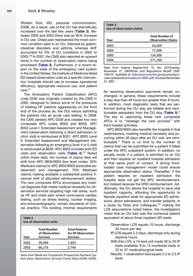
(Robbin Dick, MD, personal communication,2008). As a result, use of the OU has dramaticallyincreased over the last few years (Table 2). Be-tween 2002 and 2003 there was an 85% increasein OU use. Chest pain represented the most com-mon condition seen in an OU, followed by gastro-intestinal disorders and asthma, whereas AHFaccounted for 5% of OU conditions in 2002 to2003.20 In 2007, the CMS also reported an upwardtrend in the number of observation claims beingprocessed (Table 3). Furthermore, in a recent re-port on the state of the emergency care systemin the United States, the Institute of Medicine listedED-based observation units as a specific interven-tion hospitals should use to increase operationalefficiency, appropriate resource use, and patientcare.21
The Ambulatory Patient Classification (APC)code 0339 was originally created by the CMS in2002, designed to relieve some of the pressuresof treating HF patients aggressively on the frontend of the process (ie, the OU) versus admittingthe patients into an acute care setting. In 2008,the CMS deleted APC 0339 and created two newcomposite APC codes (8002 and 8003). APC8002 Level I: Extended Assessment and Manage-ment (observation following a direct admission orclinic visit) is reimbursed at $351. APC 8003 LevelII: Extended Assessment and Management (ob-servation following an emergency level 4 or 5 visit)is reimbursed at $639. APC 8003 includes both EDvisits and observation visits (Table 4).22 Notedwithin these data, the number of claims filed willshift from APC 0600/0604 (low level codes; 30%Medicare claims) to APC 8002/8003 (extended as-sessment and management; 70% Medicareclaims) making available a substantial positive fi-nancial shift of allocated reimbursement dollars.The new composite APCs encompass any medi-cal diagnosis that meets medical necessity for ob-servation services targeting high risk areas, suchas HF and chest pain where noninvasive cardiactesting, such as stress testing, nuclear imaging,and echocardiography, remain standards of clini-cal practice. The existing minimal requirements
for receiving observation payments remain un-changed. In general, these requirements includea stay less than 48 hours but greater than 8 hours.In addition, most diagnostic tests that are per-formed during the OU stay are billable and reim-bursable separately from the OU stay (Table 5).4
The key to optimizing these new compositeAPCs is to ‘‘redesign the care process’’ withfront-loaded activities.
APC 8002/8003 also benefits the hospital in thatreadmissions, meeting medical necessity and oc-curring within 30 days after an OU visit, are reim-bursable.4 There is no limit to the number ofclaims that can be submitted for a patient if billedunder the APC outpatient system. In addition, nopenalty exists if a patient is admitted to an OUand then requires an inpatient hospital admissionat that same point of contact. A strong front-loaded case management model could ensureappropriate observation status. Thereafter, if thepatient requires an inpatient admission thehospital does not get the APC reimbursement,but instead receives the DRG reimbursement. Ad-ditionally, the OU allows the hospital to save vitalinpatient capacity, affording the opportunity toliberate inpatient beds for appropriate ED admis-sions, direct admissions, and transfer patients. Ina study by Ross and colleagues,23 making theLOS assumptions noted below, the investigatorsnoted that an OU bed was the numerical patientequivalent of about three inpatient (IP) beds:
Observation LOS equals 15 hours, discharge24 hours per day
IP LOS equals 2.2 days, discharge only duringdaytime hours
With this LOS, a 14-bed unit made 30 to 50 IPbeds available: 8 to 13 monitored beds or22 to 37 medical/surgical beds
Results: 1 observation bed equals 2.2 to 3.5 IPbeds
Table 2Use of observation units
Year
Total Numberof ObservationServices
Total Patientsfor HFObservationServices
2002 30,094 1,603
2003 66,276 3,749
Data from Medicare Outpatient Prospective Payment Sys-tem Data: Observation Services Claims Data (G244, G263).
Table 3Use of observation claims
YearTotal Number ofObservation Claims
2003 56,000
2004 77,000
2005 124,300
2006 271,200
Data from Federal Register/Vol. 72, No. 227/Tuesday,November 27, 2007/Rules and Regulations 66,647 CMS-1392-FC. Available at: http://www.cms.hhs.gov/quarterlypro-viderupdates/downloads/cms1392fc.pdf.AccessedNovember27, 2007.
Sieck & Moseley104

The ED and inpatient physician providers alsobenefit from observation level of care. The OUallows for more efficient ED operations, andhas been demonstrated to decrease hours ofambulance diversion, decrease ED LOS, and im-prove patient satisfaction with their care (RobbinDick, MD, personal communication, 2008).24,25
Observation current procedural terminology co-des exist for additional reimbursement (thus in-crease in relative value unit compensation) forservices provided, and the increased ED effi-ciency allows for more rapid bed turnover, lead-ing to additional volume and reimbursement(Michael Granovsky, MD, personal communica-tion, 2008). In addition, longer periods of moni-toring for OU patients allows for a reasonablereduction in medical-legal risk through improveddiagnostic accuracy, allowing specialty consulta-tion (ie, cardiologist) for disease processes likeHF. Finally, inpatient physicians benefit by takingcare of fewer admissions directly from the EDthat do not meet inpatient criteria, saving timeand resources.
COST-EFFECTIVENESS OF THE OBSERVATION UNIT
The OU allows for the provision of intense therapyand observation services with close monitoringfor response to treatment. In the Acute Decompen-sated Heart Failure Registry (ADHERE) data
registry, the time to initiation of administration ofcertain intravenous medicines specifically directedat acute HF was 1 hour if the patient’s treatmentwas initiated in the ED, compared with 22 hours iftherapy was begun in an inpatient unit (Table 6).26
The OU has definitive protocols for both treatmentand timely adjustments in treatment plans basedon the clinical parameters being observed. Sucha methodology leads to more intense and timelyinitiation of therapy. This drastic variation in timingcan have remarkable differences in clinicaloutcomes, as well as a dramatic impact on financialimplications.
Treatment of AHF in an OU has resulted in re-duced 30-day readmissions and hospitalizations,and decreased LOS if a subsequent hospitalizationis required.27 The ADHERE data show that early ini-tiation of intravenous vasoactive therapy can re-duce the hospital LOS from an average of 7 daysto 4.5 days.28 Because LOS is the major contribu-tor to facility costs for AHF patients, and becausebetween 70% and 75% of patients admitted toan OU with HF can be dispositioned to home, thepotential impact on hospital bed use can be signif-icant.13,29 The OU experience with AHF at theCleveland Clinic showed the following positive out-comes:30 revisits were reduced by 44%; ED obser-vation discharges increased by 9%; HFrehospitalizations were reduced by 36%; and ob-servation rehospitalizations were reduced by 39%.
Table 4Observation FY 2005
Comparison of Medicare Reimbursements for AHF FY 2005
APC 0339 – CHF Inpatient DRG 127 – CHF Outpatient
Level V< 8 hours $239APC 0339> 8 hours $408
Avg. Reimbursement $4 617Mean Cost $5905Medicare Benchmark LOS 5.3 days
UB92 Billable ReimbursableEKG Yes YesLabs Yes YesECHO Yes YesCXR Yes YesIV Infusion Yes YesDrug therapy Yes Yes
UB92 Billable ReimbursableEKG Yes No Labs Yes NoECHO Yes NoCXR Yes NoIV infusion Yes NoDrug therapy Yes No ***No = bundled under the service provided
Total $1300/1400Fixed Cost (FC) ($570)FC varies per institutionMean Variable Cost (V C) ($252)
Fixed Cost (FC) ($1498)FC varies per institution usually 3 x outpatientMean Variable Cost (VC) ($1168)All diagnostics are bundled under corresponding DRG
$478 profit per case ($1288) loss per case
From Hospital Outpatient Prospective Payment System and CY 2008 payment rates, etc., 66,580–67,225 [07–5507].Available at: http://www.access.gpo.gov/su_docs/fedreg/a071127c.html. Accessed August 16, 2007.
Observation Unit Economics 105
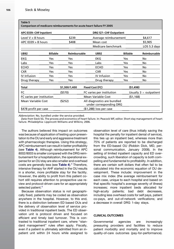
The authors believed this impact on outcomeswas because of application of testing upon presen-tation to the OU and early and aggressive treatmentwith pharmacologic therapies. Using this strategy,APC reimbursement can result in better profitability(see Table 4). Although reimbursement for APC8002/8003 is smaller compared with the DRG reim-bursement for a hospitalization, the operational ex-pense for an OU stay are also smaller and overheadcosts are generally less (see Table 4).4 Therefore,intensive therapy for AHF started in the OU resultsin a shorter, more profitable stay for the facility.However, the ability to profit from this patient co-hort still requires attention to prospective use re-view and protocol-driven care for an appropriatelyselected patient.4
Because observation status is not geographi-cally fixed, patients may be coded as observationanywhere in the hospital. However, to this end,there is a distinction between ED based OUs andthe delivery of observation level of service pro-vided in traditional inpatient beds. The ED obser-vation unit is protocol driven and focused onefficient and timely bed turnover. This is con-trasted to traditional inpatient care, where ‘‘stan-dard management’’ rules often apply. Fiscally,even if a patient is ultimately admitted from an in-patient unit within 24 hours while assigned to
observation level of care (thus initially saving thehospital the penalty for inpatient denial of service),this ties up an inpatient bed, whereas more than75% of patients are reported to be dischargedfrom the ED-based OU (Robbin Dick, MD, per-sonal communication, January 2008). In thesetting of limited inpatient capacity and ED over-crowding, such liberation of capacity is both com-pelling and fundamental to profitability. In addition,there are certain soft dollars that often fail to becalculated into the economic equation of OU de-velopment. These include: improvement in thecase mix index (the average reimbursement foreach case, unique to each hospital and based onthat specific hospital’s average severity of illness)increases; more inpatient beds allocated forhigh-acuity patients; bad debt decreases,including less overhead costs for self pays, patientco-pays, and out-of-network verifications; anda decrease in overall DRG 1-day stays.
CLINICAL OUTCOMES
Governmental agencies are increasinglypressuring providers and facilities to reducepatient morbidity and mortality and to improvequality-of-care outcomes (pay-for-performance).
Table 5Comparison of medicare reimbursements for acute heart failure FY 2005
APC 0339^CHF Inpatient DRG127^CHFOutpatient
Level V < 8 hours $239 Average reimbursement $4,617
APC 0339 > 8 hours $408 Mean cost $5,905
Medicare benchmark LOS 5.3 days
UB92 Billable Reimbursable UB92 Billable Reimbursable
EKG Yes Yes EKG Yes No
Labs Yes Yes Labs Yes No
ECHO Yes Yes ECHO Yes No
CXR Yes Yes CXR Yes No
IV Infusion Yes Yes IV Infusion Yes No
Drug therapy Yes Yes Drug therapy Yes No
Total $1,300/1,400 Fixed Cost (FC) ($1,498)
FC ($570) FC varies per institution Usually 3 � outpatient
FC varies per institution Mean Variable Cost ($1,168)
Mean Variable Cost ($252) All diagnostics are bundledunder corresponding DRG
$478 profit per case ($1,288) loss per case
Abbreviation: No, bundled under the service provided.Data from Sieck SG. The process and economics of heart failure. In: Peacock WF, editor. Short stay management of heart
failure. Philadelphia: Lippincott Williams and Wilkins; 2006.
Sieck & Moseley106
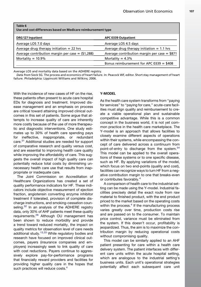
With the incidence of new cases of HF on the rise,these patients often present to acute care hospitalEDs for diagnosis and treatment. Improved dis-ease management and an emphasis on processare critical toward attaining improved clinical out-comes in this set of patients. Some argue that at-tempts to increase quality of care are inherentlymore costly because of the use of more therapeu-tic and diagnostic interventions. One study esti-mates up to 30% of health care spending paysfor ineffective, inappropriate, or redundantcare.31 Additional studies are needed for supportof comparative research and quality versus cost,and are essential to improving health care qualitywhile improving the affordability of care. This sug-gests the overall impact of high quality care canpotentially reduce total costs by diminishing un-necessary health care use that results from inap-propriate or inadequate care.
The Joint Commission on Accreditation ofHealthcare Organizations has created a set ofquality performance indicators for HF. These indi-cators include objective measurement of ejectionfraction, angiotensin converting enzyme inhibitortreatment if tolerated, provision of complete dis-charge instructions, and smoking-cessation coun-seling.32 In an analysis of the ADHERE registrydata, only 30% of AHF patients meet these qualityrequirements.26 Although OU management hasbeen shown to reduce morbidity and providea trend toward reduced mortality, the impact onquality metrics for observation level of care needsadditional study.13,33 While regulatory bodies andresearch have focused on improved clinical out-comes, payers (insurance companies and em-ployers) increasingly seek to link quality of carewith cost reductions. Payers continue to aggres-sively explore pay-for-performance programsthat financially reward providers and facilities forproviding higher quality care in the hopes thatsuch practices will reduce costs.4
Y-MODEL
As the health care system transforms from ‘‘payingfor services’’ to ‘‘paying for care,’’ acute-care facil-ities must align quality and reimbursement to cre-ate a viable operational plan and sustainablecompetitive advantage. While this is a commonconcept in the business world, it is not yet com-mon practice in the health care marketplace. TheY-model is an approach that allows facilities toclosely examine different aspects of operationswithin their systems, while encompassing the con-cept of care delivered across a continuum frompoint-of-entry to discharge from the system.34
This model can be applied to the overall opera-tions of these systems or to one specific disease,such as HF. By applying variations of the model,which focus on two end-points (quality and cost),facilities can recognize ways to turn HF from a neg-ative contribution margin to one that breaks-evenor contributes favorably.4
A comparison of health care to the industrial set-ting can be made using the Y-model. Industrial fa-cilities precisely detail the exact route from rawmaterial to finished product, with the end productpriced to the market based on the operating costswithin the process.4 If the manufacturing processvaries greatly over time, production costs riseand are passed on to the consumer. To maintainprice control, variance must be eliminated fromthe system. If this doesn’t occur, profitability isjeopardized. Thus, the aim is to maximize the con-tribution margin by reducing operational costswithout compromising quality.
This model can be similarly applied to an AHFpatient presenting for care within a health caredelivery system. The patient interfaces with differ-ent care units within the acute hospital setting,which are analogous to the industrial setting’sbusiness units. Each unit’s operational strategiespotentially affect each subsequent care unit
Table 6Use and cost differences based onMedicare reimbursement type
DRG127 Inpatient APC 0339 Outpatient
Average LOS 7.0 days Average LOS 4.5 days
Average drug therapy initiation 5 22 hrs Average drug therapy initiation 5 1.1 hrs
Average contribution margin per case 5 ($1,288) Average contribution margin per case 5 $871
Mortality 5 10.9% Mortality 5 4.3%
Bonus reimbursement for APC 0339 5 $408
Average LOS and mortality data based on the ADHERE registry.Data from Sieck SG. The process and economics of heart failure. In: Peacock WF, editor. Short stay management of heart
failure. Philadelphia: Lippincott Williams and Wilkins; 2006.
Observation Unit Economics 107
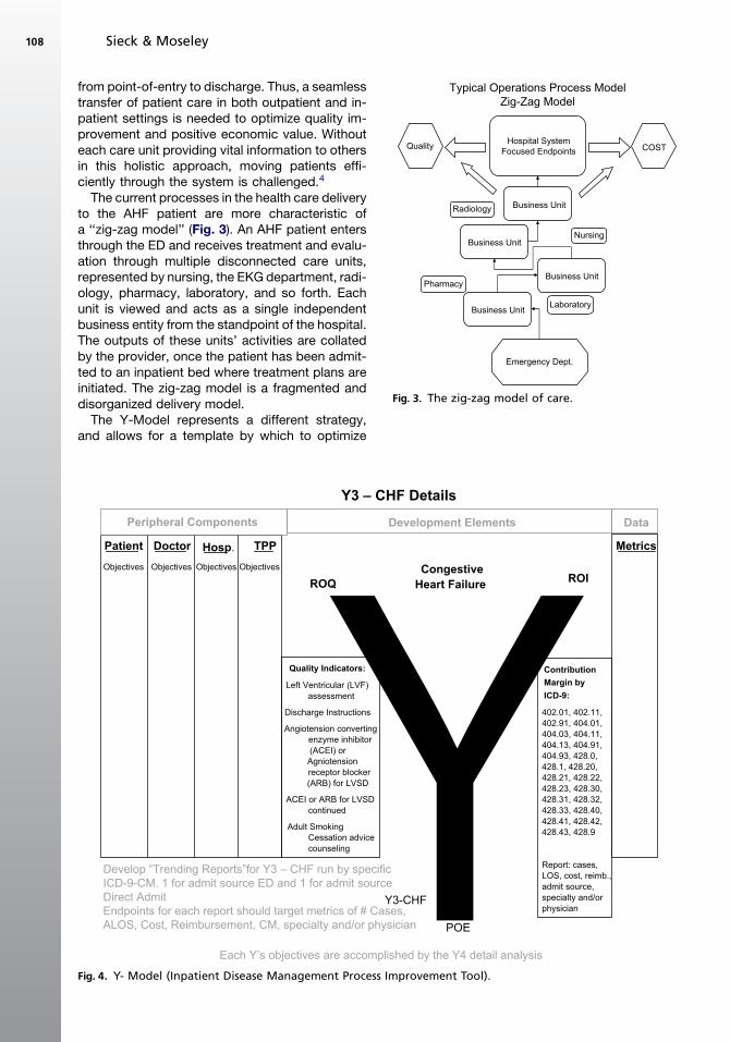
from point-of-entry to discharge. Thus, a seamlesstransfer of patient care in both outpatient and in-patient settings is needed to optimize quality im-provement and positive economic value. Withouteach care unit providing vital information to othersin this holistic approach, moving patients effi-ciently through the system is challenged.4
The current processes in the health care deliveryto the AHF patient are more characteristic ofa ‘‘zig-zag model’’ (Fig. 3). An AHF patient entersthrough the ED and receives treatment and evalu-ation through multiple disconnected care units,represented by nursing, the EKG department, radi-ology, pharmacy, laboratory, and so forth. Eachunit is viewed and acts as a single independentbusiness entity from the standpoint of the hospital.The outputs of these units’ activities are collatedby the provider, once the patient has been admit-ted to an inpatient bed where treatment plans areinitiated. The zig-zag model is a fragmented anddisorganized delivery model.
The Y-Model represents a different strategy,and allows for a template by which to optimize
Typical Operations Process ModelZig-Zag Model
Hospital SystemFocused Endpoints
Business Unit
Business Unit
Business Unit
Business Unit
Quality COST
Radiology
Emergency Dept.
Pharmacy
Nursing
Laboratory
Fig. 3. The zig-zag model of care.
Y3 – CHF Details
ROQROI
Patient Doctor Hosp. TPP Metrics
Peripheral Components
Congestive
Heart Failure
Quality Indicators:
Left Ventricular (LVF)assessment
Discharge Instructions
Angiotension convertingenzyme inhibitor(ACEI) orAgniotensionreceptor blocker(ARB) for LVSD
ACEI or ARB for LVSDcontinued
Adult SmokingCessation advicecounseling
Contribution
Margin by
ICD-9:
402.01, 402.11,402.91, 404.01,404.03, 404.11,404.13, 404.91,404.93, 428.0,428.1, 428.20,428.21, 428.22,428.23, 428.30,428.31, 428.32,428.33, 428.40,428.41, 428.42,428.43, 428.9
Report: cases,LOS, cost, reimb.,admit source,specialty and/orphysician
Objectives Objectives Objectives Objectives
Development Elements Data
Each Y’s objectives are accomplished by the Y4 detail analysis
Y3-CHF
POE
Develop “Trending Reports”for Y3 – CHF run by specificICD-9-CM. 1 for admit source ED and 1 for admit sourceDirect AdmitEndpoints for each report should target metrics of # Cases,ALOS, Cost, Reimbursement, CM, specialty and/or physician
Fig. 4. Y- Model (Inpatient Disease Management Process Improvement Tool).
Sieck & Moseley108

care costs by placing resources at the front end ofthe delivery process (at point-of-entry). The modelintegrates a financial strategy that meets qualitymetrics and produces positive net value bypoint-of-entry clinical case management. Begin-ning in the ED, this concept emphasizes an effi-cient, rapid assessment and action centered ona seamless integration of ancillary services, suchas the laboratory, diagnostic imaging, and skillednursing, while understanding the economic im-pacts on decisions made as the patient is directedthrough the system.4 Prospective case manage-ment at the initial point-of-care determines ‘‘whathappens next’’ for the ED patient: discharge, ob-servation level of service, or inpatient admission(and to what level of care).
The Y-Model can positively impact quality,costs, efficiency, and clinical outcomes by provid-ing drill downs on the exact volume by ICD-9 co-des instead of the inpatient DRG. This facilitatesa more accurate picture of the number of patientsprocessed in the ambulatory setting within thesystem. Patients who require inpatient admissionare admitted to an appropriate level of care, whilethose who can be effectively treated in the ambu-latory setting are treated and released. As inappro-priate admissions are weeded out of the system,the inpatient setting experiences a positive in-crease in the case-mix-index (because the aver-age inpatient is sicker and this results in greaterper case reimbursement) (Robbin Dick, MD, per-sonal communication, January 2008).
Patients with AHF can be appropriately riskstratified and identified for an ideal level of care us-ing the Y-model in a manner that positively im-pacts the contribution margins (Fig. 4). Thismodel emphasizes a multidisciplinary team ap-proach to align the ‘‘care units’’ that affect anAHF patient’s progress through the current sys-tem. The emphasis is on front-end case manage-ment to support the physician in establishing thepathway the patient will follow (Fig. 5). A patientis not arbitrarily admitted to an inpatient bed,treated, and then discharged. Rather, risk-stratified
Y-Model 2008Maximize Process Efficiency
Hospital SystemFocused Endpoints
Discharge
Quality Cost
Point of EntryEmergency Dept.
Process Drilldown
Font LoadedCase Mgmt
OBS
Discharge
IP
Fig. 5. Use of Y-Model to maximize cost-efficiency.
Y - Model: Y3 – CHF Details
ROQ ROICongestive
Heart Failure
Quality:
Trending Reports
ADHERE Registry
Financial:
Trending ReportsUB-04
Development Elements
Fron
t Loa
ded
Qua
lity
Focu
sed
Verti
cal C
are
Proc
esse
s Discharge
*POE: ED
Minimal Standards•ECHO•ECG•LAB (POCT)
Inpatient Admit
Diagnosis
ACC/AHAGuidelines Applied
in PracticeEarly Medical Therapy
Fig. 6. Quality and cost focus of theY-Model. *POE 5 point of entry.
Observation Unit Economics 109

patients are determined to need additional re-sources, and case management establishes themost ideal venue to undergo specific therapies.4
By integrating the clinical and financial pathways,facilities can realize the financial implications ofmaking decisions based on standard managementversus protocol driven care. The Y-model places anemphasis on process improvement while targetingthe end-points of quality and contribution margin(Fig. 6).
One medical center has reported its experiencein successfully implementing part of the model foran Acute Coronary Syndrome (ACS) ProcessImprovement Project.35 Before the initiative thehospital had a ‘‘zig-zag’’ model of care. Patientsentered through the ED and were admitted to theacute care bed, diagnostics were completed,and treatment was then initiated. With initiationof the Y-model, stratification was performed andappropriate therapy initiated in the ED with point-of-care testing in a patient-centric improvementeffort. The new design resulted in improvementsin turnaround time for therapy, reduced LOS, en-hanced patient placement in the most appropriatebed venue (eg, critical care unit, telemetry, or clin-ical decision unit), and improved patient satisfac-tion.35 Improvements demonstrated by the ACSredesign can be translated to the AHF setting.Similar to the ACS patient, not every HF patient re-quires inpatient admission. And similarly, not all HFpatients are candidates for the OU. Point-of-entrytriaging to the most appropriate care unit where anindividualized treatment plan is rendered allowsa facility to better merge quality care with positivefinancial outcomes.4,35
SUMMARY
The current process of regulatory oversight andpay-for-performance reimbursement is a realityin today’s health care delivery system. To maintainprofitability, facilities must be willing to implementnew strategies that marry operational redesign,quality care, and cost-effective treatment. No-where is this truer than with patients with AHF.Clinical care that is in the wrong setting, usingthe wrong treatment algorithms, is doomed forclinical and financial failure. As payers increasinglyfavor outpatient strategies for patient manage-ment, inpatient facilities must develop effectivestrategies to shift inpatient care into ambulatorysettings. The Y-model offers a solution that is fixedon process improvement techniques that levy pos-itive economic impact. The model is based onaligning quality and finance targets with attentionto clinical outcomes and is suggested as an
innovation of a redesigned care process for theAHF patient population.
REFERENCES
1. U.S. Census Bureau. Avaliable at: http://www.
census.gov/. Accessed November 20, 2007.
2. Peacock WF, Albert NM. Observation unit manage-
ment of heart failure. Emerg Med Clin North Am
2001;19:209–32.
3. Massie BM, Shah NB. Evolving trends in epidemio-
logic factors of heart failure: rationale for preventa-
tive strategies and comprehensive disease
management. Am Heart J 1997;133:703–12.
4. Sieck SG. The process and economics of heart fail-
ure. Short Stay Management of Heart Failure, vol. 5.
Philadelphia: Lippincott Williams & Wilkins; 2006.
5. American Heart Association. Heart disease and
stroke statistics—2005 update. Dallas (TX): Ameri-
can Heart Association; 2005.
6. Jessup M, Brozena S. Epidemiology and causes of
heart failure. N Engl J Med 2003;348(20):2007–18.
7. Aghababian RV. Acutely decompensated heart fail-
ure: opportunities to improve care and outcomes in
the emergency department. Rev Cardiovasc Med
2002;3(Suppl 4):S3–9.
8. O’Connell JB, Bristow M. Economic impact of heart
failure in the United States: time for a different ap-
proach. J Heart Lung Transplant 1993;13:S107–12.
9. Heart disease and stroke statistics: 2004 update.
Available at: http://www.americanheart.org. Ac-
cessed August 24, 2007.
10. ADHERE Registry data on file; January 2004.
11. Graff L, Orledge J, Radford MJ, et al. Correlation of
the Agency for Health care Policy and Research
congestive heart failure admission guideline with
mortality: peer review organization voluntary hospital
association initiative to decrease events (PROVIDE)
for congestive heart failure. Ann Emerg Med 1999;
34:429–37.
12. Emerman CL. Treatment of the acute decompensa-
tion of heart failure: efficacy and pharmacoeconom-
ics of early initiation of therapy in the emergency
department. Rev Cardiovasc Med 2003;4:S13–20.
13. Peacock WF. Heart failure management in the emer-
gency department observation unit. Prog Cardio-
vasc Dis 2004;46:465–85.
14. Centers for Medicare and Medicaid services.
Available at: http://www.cms.hhs.gov/MLNGenInfo.
Accessed September 22, 2007.
15. Short-term acute care program for evaluating
payment patterns electronic report (ST PEPPER)
user’s guide. October 2007. Available at: http://
www.hpmpresources.org/LinkClick.aspx?fileticket=
H5IICGIqbvM%3d&;tabid=1059&mid=1052. Ac-
cessed November 10, 2007.
Sieck & Moseley110
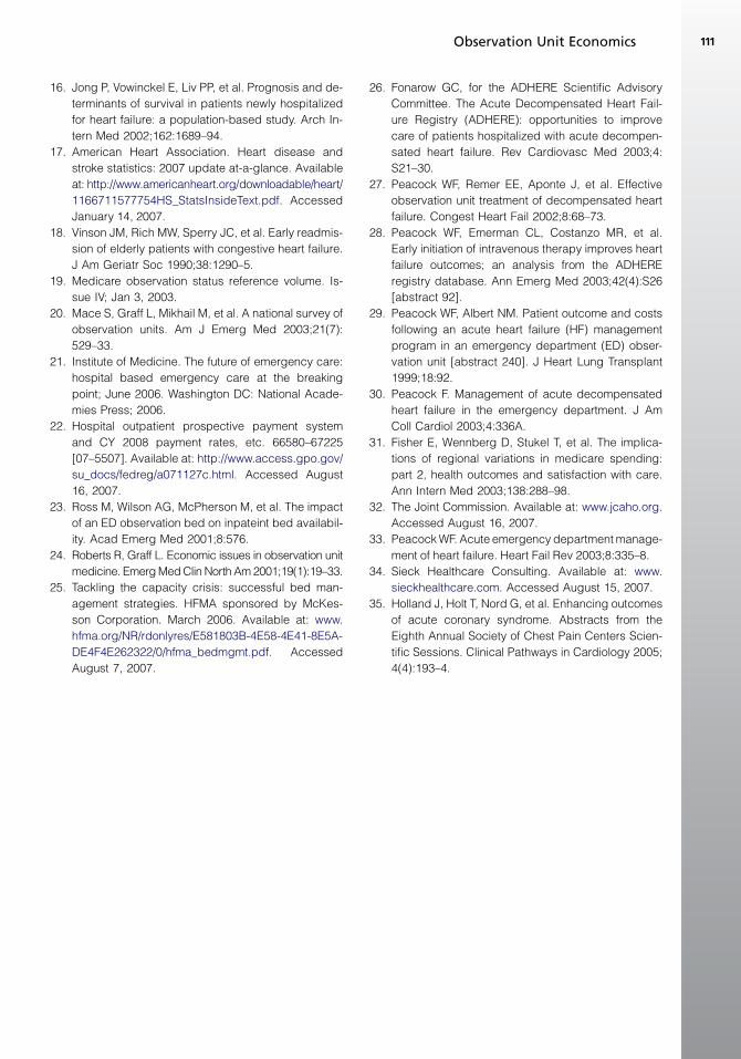
16. Jong P, Vowinckel E, Liv PP, et al. Prognosis and de-
terminants of survival in patients newly hospitalized
for heart failure: a population-based study. Arch In-
tern Med 2002;162:1689–94.
17. American Heart Association. Heart disease and
stroke statistics: 2007 update at-a-glance. Available
at: http://www.americanheart.org/downloadable/heart/
1166711577754HS_StatsInsideText.pdf. Accessed
January 14, 2007.
18. Vinson JM, Rich MW, Sperry JC, et al. Early readmis-
sion of elderly patients with congestive heart failure.
J Am Geriatr Soc 1990;38:1290–5.
19. Medicare observation status reference volume. Is-
sue IV; Jan 3, 2003.
20. Mace S, Graff L, Mikhail M, et al. A national survey of
observation units. Am J Emerg Med 2003;21(7):
529–33.
21. Institute of Medicine. The future of emergency care:
hospital based emergency care at the breaking
point; June 2006. Washington DC: National Acade-
mies Press; 2006.
22. Hospital outpatient prospective payment system
and CY 2008 payment rates, etc. 66580–67225
[07–5507]. Available at: http://www.access.gpo.gov/
su_docs/fedreg/a071127c.html. Accessed August
16, 2007.
23. Ross M, Wilson AG, McPherson M, et al. The impact
of an ED observation bed on inpateint bed availabil-
ity. Acad Emerg Med 2001;8:576.
24. Roberts R, Graff L. Economic issues in observation unit
medicine. Emerg Med Clin North Am 2001;19(1):19–33.
25. Tackling the capacity crisis: successful bed man-
agement strategies. HFMA sponsored by McKes-
son Corporation. March 2006. Available at: www.
hfma.org/NR/rdonlyres/E581803B-4E58-4E41-8E5A-
DE4F4E262322/0/hfma_bedmgmt.pdf. Accessed
August 7, 2007.
26. Fonarow GC, for the ADHERE Scientific Advisory
Committee. The Acute Decompensated Heart Fail-
ure Registry (ADHERE): opportunities to improve
care of patients hospitalized with acute decompen-
sated heart failure. Rev Cardiovasc Med 2003;4:
S21–30.
27. Peacock WF, Remer EE, Aponte J, et al. Effective
observation unit treatment of decompensated heart
failure. Congest Heart Fail 2002;8:68–73.
28. Peacock WF, Emerman CL, Costanzo MR, et al.
Early initiation of intravenous therapy improves heart
failure outcomes; an analysis from the ADHERE
registry database. Ann Emerg Med 2003;42(4):S26
[abstract 92].
29. Peacock WF, Albert NM. Patient outcome and costs
following an acute heart failure (HF) management
program in an emergency department (ED) obser-
vation unit [abstract 240]. J Heart Lung Transplant
1999;18:92.
30. Peacock F. Management of acute decompensated
heart failure in the emergency department. J Am
Coll Cardiol 2003;4:336A.
31. Fisher E, Wennberg D, Stukel T, et al. The implica-
tions of regional variations in medicare spending:
part 2, health outcomes and satisfaction with care.
Ann Intern Med 2003;138:288–98.
32. The Joint Commission. Available at: www.jcaho.org.
Accessed August 16, 2007.
33. Peacock WF. Acute emergency department manage-
ment of heart failure. Heart Fail Rev 2003;8:335–8.
34. Sieck Healthcare Consulting. Available at: www.
sieckhealthcare.com. Accessed August 15, 2007.
35. Holland J, Holt T, Nord G, et al. Enhancing outcomes
of acute coronary syndrome. Abstracts from the
Eighth Annual Society of Chest Pain Centers Scien-
tific Sessions. Clinical Pathways in Cardiology 2005;
4(4):193–4.
Observation Unit Economics 111

Special Cases in AcuteHeart FailureSyndromes: AtrialFibrillation and WideComplexTachycardiaPeter S. Pang, MD*, Mihai Gheorghiade, MD
Hospitalization for acute heart failure syndromes(AHFS) portends a significant in-hospital and post-discharge morbidity and mortality.1–5 Manage-ment of these patients presents significantchallenges, given the heterogeneity of the patientpopulation and the differing and complex etiolo-gies why patients present with acute decompen-sation.4 Approximately 50% of the AHFSpopulation has preserved systolic function; a sig-nificant number have underlying coronary arterydisease, are female, have diabetes, renal insuffi-ciency, and present with hypertension; with onlya very small percentage who present with cardio-genic shock.2,3,6
Approximately 30% of patients with AHFS pres-ent with atrial fibrillation (Tables 1 and 2).6–9 Thisarrhythmia may be the precipitating cause ofAHFS or may merely reflect the underlyingsubstrate of a patient with chronic HF who nowpresents with acute decompensation.10 In eithercase, management of the arrhythmia occurs withinthe overall context of HF management.
In contrast, although wide complex tachycardias,if defined broadly by premature ventricular con-tractions, nonsustained ventricular tachycardia,ventricular tachycardia (VT), polymorphic ventric-ular tachycardia, accelerated idioventricularrhythms, supraventricular tachycardia with aber-rancy, or ventricular fibrillation, are relatively
common in the chronic HF population atlarge,11–13 it has rarely been addressed in thesetting of AHFS outside of textbooks (seeTable 1).11
The pathophysiology of both AF and HF hasbeen extensively discussed elsewhere.5,14–16
This article focuses on both AF and WCT withspecific principles of emergency department (ED)management reviewed in the setting of AHFS.
AF is defined as a ‘‘supraventriculartachyarrhythmia characterized by uncoordinatedatrial activation with consequent deterioration ofmechanical function.’’14 How the ventricle re-sponds to this atrial activation depends on (1) prop-erties of the atrioventricular node, (2) presence ofaccessory pathways if any, (3) vagal and sympa-thetic tone, and (4) effects of medications.1 The au-thors will focus on the management of AF with rapidventricular response (RVR) in the setting of EDAHFS.
For patients with either AF or HF, developing theother condition predicts a worse prognosis, al-though in advanced HF some debate exists as towhether development of AF at this late juncture im-putes a worse prognosis.14,17–22 In general, rateversus rhythm control does not confer a mortalitybenefit.23–25 However, some controversy stillexists within the setting of HF, given the worseprognosis of those with HF who develop
Northwestern Memorial Hospital, Chicago, IL, USA* Corresponding author. Department of Emergency Medicine, Northwestern Memorial Hospital, 259 East ErieStreet, Suite 100, Chicago, IL 60611.E-mail address: [email protected] (P.S. Pang).
KEYWORDS� Acute heart failure syndromes � Atrial fibrillation� Wide complex tachycardia
Heart Failure Clin 5 (2009) 113–123doi:10.1016/j.hfc.2008.08.0091551-7136/08/$ – see front matter ª 2008 Elsevier Inc. All rights reserved. he
artf
ailu
re.th
ecli
nics
.com
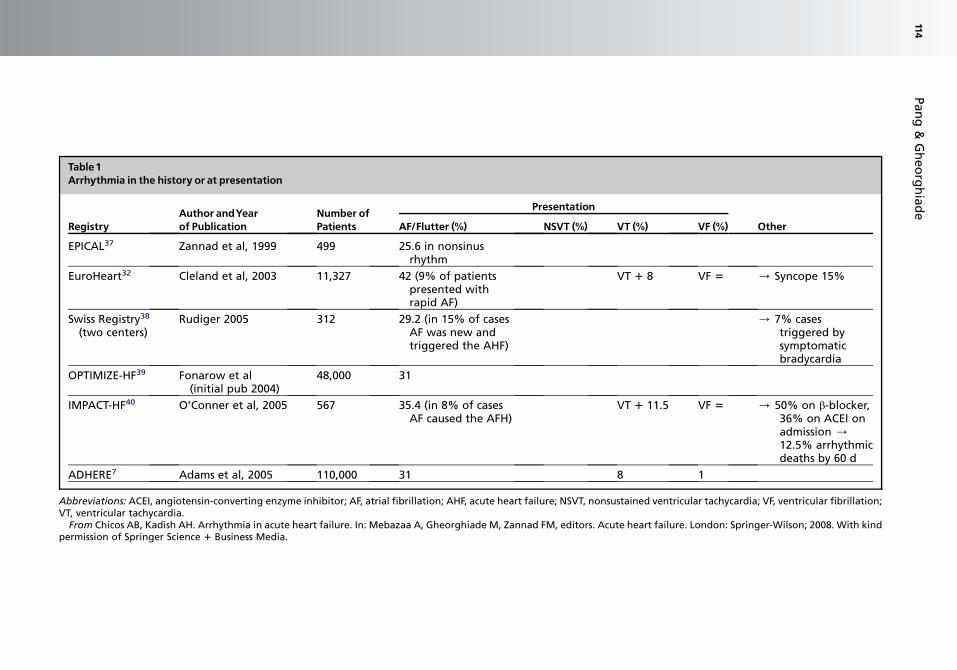
Table1Arrhythmia in the history or at presentation
RegistryAuthor andYearof Publication
Number ofPatients
Presentation
OtherAF/Flutter (%) NSVT (%) VT (%) VF (%)
EPICAL37 Zannad et al, 1999 499 25.6 in nonsinusrhythm
EuroHeart32 Cleland et al, 2003 11,327 42 (9% of patientspresented withrapid AF)
VT 1 8 VF 5 / Syncope 15%
Swiss Registry38
(two centers)Rudiger 2005 312 29.2 (in 15% of cases
AF was new andtriggered the AHF)
/ 7% casestriggered bysymptomaticbradycardia
OPTIMIZE-HF39 Fonarow et al(initial pub 2004)
48,000 31
IMPACT-HF40 O’Conner et al, 2005 567 35.4 (in 8% of casesAF caused the AFH)
VT 1 11.5 VF 5 / 50% on b-blocker,36% on ACEI onadmission /12.5% arrhythmicdeaths by 60 d
ADHERE7 Adams et al, 2005 110,000 31 8 1
Abbreviations: ACEI, angiotensin-converting enzyme inhibitor; AF, atrial fibrillation; AHF, acute heart failure; NSVT, nonsustained ventricular tachycardia; VF, ventricular fibrillation;VT, ventricular tachycardia.
From Chicos AB, Kadish AH. Arrhythmia in acute heart failure. In: Mebazaa A, Gheorghiade M, Zannad FM, editors. Acute heart failure. London: Springer-Wilson; 2008. With kindpermission of Springer Science 1 Business Media.
Pan
g&
Gh
eo
rgh
iad
e114

Table 2Epidemiology of arrhythmia in acute heart failure: data from selected acute heart failure trials
Trial InterventionNo.Patients
Arrhythmia in the History orat Presentation
NewArrhythmiasDuring Hospitalization
OtherAfib/Flutter (%)
NSVT(%)
Sust.VT (%)
VF(%)
Afib/Flutter (%)
NSVT(%)
Sust.VT (%) VF (%)
OPTIME-CHF41
Milrinoneversus placebo
949 32 10 3 M 0.5 2.1 1.2P 4.6
VMAC42 Nesiritideversus NTGversusplacebo
489 35 13 6 34% hadfrequent PVSs
LIDO43 Levosimendanversusdobutamine
203 13 7 1 L 4 L 1 L 1D 2 D 2 D 2
RITZ-244 Tezosen versusplacebo
285 24.6
RITZ-445 Tezosen versusplacebo
192 T 5.2 T 5.2 Patients excludedif heart rate>130/min
P 2.1 P 6.3
RITZ-546 Tezosen versusplacebo
84 21.4
PRECEDENT47 N1 versus N2versusdobutamine
255 27 26 7.5 N1 6N2 5D 13
IMPACT-HF48 Start carvedilol 363 22 VT 1 VF 5 9
Abbreviations: D, dobutamine; L, levosimendan; M, milrinone; N1, nesiritide, 0.015 mg/kg/min; N2, nesiritide, 0.03 mg/kg/min; NGT, nitroglycerin; NSVT, nonsustained ventriculartachycardia; P, placebo; Sust. VT, sustained ventricular tachycardia; T, tezosentan; VF, ventricular fibrillation.
From Chicos AB, Kadish AH. Arrhythmia in acute heart failure. In: Mebazaa A, Gheorghiade M, Zannad FM, editors. Acute heart failure. London: Springer-Wilson; 2008. With kindpermission of Springer Science 1 Business Media.
AF
an
dW
CT
inA
HFS
115
AF.18,26,27 Whether or not maintaining or restoringsinus rhythm confers a mortality benefit has notbeen prospectively investigated in a large random-ized controlled trial, however, although the AtrialFibrillation and Congestive Heart Failure trial iscurrently underway and was designed to answerthis question.26,28,29
Three general considerations determine man-agement: (1) rate control, (2) rhythm control, and(3) prevention of thromboembolism. Prioritizationof rate versus rhythm control strategy dependson the clinical presentation.14
DEMOGRAPHICS OFATRIAL FIBRILLATIONIN ACUTEHEART FAILURE SYNDROMES
Significant overlap exists in terms of risk factors fordeveloping either AF or HF.14,21,30 Not surprisingly,epidemiologic evidence of an association betweenthe two has been demonstrated.20,21,30 Datafrom the Framingham Heart Study suggest thatHF is the strongest independent risk factor forthe development of AF.20,30,31
HF registry data provide further insight into thecharacteristics of patients with AF and AHFS. Inthe EuroHeart Failure Survey-I, 42% of patientshad a history of AF with 9% of patients observedto have AF with RVR during their hospitalization.32
EuroHeart Failure Survey-I was not a survey of ad-missions, however, but of deaths and discharges.
Approximately 31% of patients in both the AcuteDecompensated Heart Failure National Registryand the Organized Program to Initiate Life-SavingTreatment in Hospitalized Patients with HeartFailure had a history of AF.6–9 From Acute Decom-pensated Heart Failure National Registry, 20% ofpatients had an initial EKG of AF, with 29% havingsome other abnormal rhythm.8
In a large nationwide Italian registry (N 5 2807)of AHFS patients admitted to a hospital with an in-tensive cardiac care unit, 44% of patients hadde novo HF with the remainder as acute-on-chronic HF.3 At time of admission, 798 patientshad AF with 21% of the total study population hav-ing a history of paroxysmal AF. Of the patients pre-senting with AF, 59.5% had a RVR greater than100 beats per minute.33
The Finnish Acute Heart Failure study was a na-tional prospective observational study of 620 pa-tients hospitalized for AHFS. Of these patients,27% had a history of chronic AF. A significantnumber (29.4%) had AF as the precipitating factorfor AHFS in 24.8% of those with chronic HF anda cause of de novo HF in 34.3%. One third of allpatients with de novo AF were determined tohave AF as the precipitating cause.34
In a retrospective cohort study of 216 patientswho presented to an emergency department withacute pulmonary edema (mean age, 75.3 years),24.3% had AF (14.6% as paroxysmal AF and9.7% as chronic).3 AF at the time of admissiondid not affect survival.35
THE EFFECT OFATRIAL FIBRILLATIONWITH RAPID VENTRICULAR RESPONSE
Four main adverse effects from AF with RVR wereoutlined in the 2005 American College of Cardiol-ogy and American Heart Association guidelinesfor chronic HF management:16 (1) loss of atrialkick to ventricular filling with subsequent decreasein cardiac output, (2) rapid HR might increase de-mand and decrease coronary perfusion, (3) RVRmay diminish both cardiac contraction and cardiacrelaxation, and (4) stasis of blood may predisposeto embolic phenomenon. These adverse effectsoutline potential targets and provide a rationalefor therapy.
ATRIAL FIBRILLATION ANDACUTE HEARTFAILURE SYNDROMES
Evidence for the management of AF and AHFS inthe ED is limited.10,18 Given the lack of prospectivedata in chronic HF and AF, it is not surprising thatevidence-based management in AHFS is lacking.Management is guided largely by principles asopposed to best evidence (Tables 3 and 4).
Management Principles
Rate control, consideration of rhythm control, andprevention of thromboembolism are the main con-siderations during acute management, with rate orrhythm control being dependent on the acuteclinical picture.11,14,18 Acutely, rate versus rhythmcontrol is often the primary decision, with keenattention to the risk and prevention of thromboem-bolism.14 Correction of any electrolyte abnormali-ties or other causes of AF (ie, thyrotoxicosis)should be performed.
In some cases, elucidating whether AF is thecause of AHFS versus a manifestation of sympa-thetic overdrive in patients who present withAHFS may be difficult to determine. In general, inpatients with chronic HF and chronic persistentAF, mitigation of their acute HF symptoms (ie,dyspnea) by therapies to relieve congestion maydecrease the RVR, assuming they are compliantwith their medications at baseline. In patientswith de novo HF and AF, it is much more likelythat AF is the cause of AHFS. Regardless, optimalarrhythmia management requires excellent HFmanagement.10
Pang & Gheorghiade116

Table 3Studies assessing cardioversion of acute atrial fibrillation in patients with chronic heart failure
StudyAuthor Type of Study N
% VentricularFunction
Durationof AF
Initial Heart Rate(Beats. MinL1)Mean � SD
Drug administered/Dose
Conversion toSinus Rhythm/Time to Conversion Adverse Event
DAAFtrialGroup,199749
Double-blind,randomized,placebocontrolleda
28Dig:15Placebo:13
Not given 21.7 �30.4 hb
122 �23 bmpc
Dig IV 0.88 �0.35 mg(mean � SD)
6/15—Dig group,5/13—placebogroup time toconversionnot given
None
Hou et al,199650
Single blind,randomized,placebocontrolleda
50Dig:24Am:26
Mean FS:Dig:27Am:26
Dig: 4 hAm: 14 h(median)
Dig: 163 � 26Am: 157 � 20
Dig: 0.013 mg/kg� 3 at 2 hintervals
Am: IV 300 mgin 1 h, 960 mgover next 23 h
Dig: 17/24 (71%)6.5 h (median)
Am: 24/26 (92%)2.5 h (median)
Dig:–noneAm:–2 patients
Clemo et al,199851
Retrospective 38 EF: 40 � 16 23/38patients(61%) hadan AF/SVTof <24 h
149 � 13 Am, IV: 242 �137 mg at 1 h(range, 60–1000)At 24 h 1137 �280 mg (range,99–2500)
11/38 (30%)in 24 h/timenot given
1 (non-CVSside effect)
Kumar,199652
Retrospective 8 <15 (EF) >30 min 159 � 9 Am. IV: 300 mgin 1 h
7/8 (87.5%)reversion27 � 13 min(mean)
None
Andrivetet al,199453
Prospective,randomizedd
16 Not given Not givene 140 forbothgroups(mean)
Oralgroup—2026� 79 mg in 24 h,IVgroup—1038 �62 mg in 24 h
7/16 (44%) usingeither oral or IVroute/ timenot given
None
Abbreviations: AF, atrial fibrillation; AFL, atrial flutter; Am, amiodarone; BMD, betamethyldigoxin; bmp, beats per minute; CHF, chronic heart failure; CVS, cardiovascular; D, dofe-tilide; DAAF, Digoxin in Acute Atrial Fibrillation Trial Group; Dig, digoxin; Dilt, diltiazem; EF, ejection fraction; FS, fraction shortening; I, ibutilide; IV, intravenous; NYHA, New YorkHeart Association Class; SD, standard deviation; SVT, supraventricular tachycardia.
a Subgroup of atrial fibrillation patients with coronary heart failure in a trial of 239 patients.b Mean � standard deviation for all patients.c Mean � standard deviation for both groups, no significant difference between groups.d Open label trial comparing oral with intravenous amiodarone, subgroup of chronic heart failure patients (16 of 72 patients in trial).e 56% of all patients in trial had atrial fibrillation duration of <48 h.From Khand AU, Rankin AC, Kaye GC, et al. Systematic review of the management of atrial fibrillation in patients with heart failure. Eur Heart J 2000;21:614; with permission.
AF
an
dW
CT
inA
HFS
117

Table 4Studies assessing ventricular rate control of acute atrial fibrillation in patients with chronic heart failure
Study author,Year, Reference Type of Study N
% VentricularFunction Duration of AF
Initial HeartRate (bmp)
DrugAdministered/Dose
Heart RateSlowingEffect of Drug Adverse Event
Hou et al,199550
Single-blind,randomized,digoxin-controlled
50Dig: 24Am: 26
Means FS:Dig: 27Am: 26
Median:Dig: 4 hAm: 14 h
Dig: 163 � 26Am: 157 � 20
Dig: 0.013 mg/kg � 3 at 2-hintervals
Am: IV 300 mgin 1 h, 960 mgover next 23 h
Dig: 150 bpmAm: 122 bmp
(at 1 h)
Dig: 0Am: 2
(1 worseningheart failureand 1 death)
Goldenberget al, 199454
Randomized,double-blind,placebo-controlled
37Dilt: 22Placebo:15
36 (EF) — >120 Dilt IL/ (firstdose 0.25 mg/kg over 2 min,if noteffective then0.35 mg/kg)
Dilt: 36/37response ratea
Placebo: 0/15Medianresponse time:5 min
3 (8%)symptomatichypotensionNo worseningof chronicheart failure
Clemo et al,199851
Retrospective 38 40 � 16 (EF) 23/38 patients(61%) had anAF for <24 h
149 � 13(mean � SD)
Am, IV: 1 h, 242� 137 mg(range,60–1000)At 24h 1137 � 280mg (range,99–2500)
15 min 134 � 141-h: 109 � 1824 h: 99 � 15(bmp, mean� SD)
1
Heywood et al,199855
Uncontrolledstudy
9 34 � 18 (meanEF � SD)
— 142 (mean) Dilt IV/ initialdose 0.25 mg/kg (0.30 mg/kg) given ifheart ratereduction<10%)
114 bmpMedianresponse time5 min
None
Abbreviations: AF, atrial fibrillation; Am, amiodarone; bmp, beats per minute; Dig, digoxin; Dilt, diltiazem; EF, ejection fraction; FS, fraction shortening; SD, standard deviation.a Response rate defined as 20% reduction in baseline heart rate or heart rate less than 100 achieved. Figures for diltiazem incorporate all placebo nonresponders who are given
diltiazem.From Khand AU, Rankin AC, Kaye GC, et al. Systematic review of the management of atrial fibrillation in patients with heart failure. Eur Heart J 2000;21:614; with permission.
Pan
g&
Gh
eo
rgh
iad
e118
GUIDELINE RECOMMENDATIONS
Various guideline recommendations help informthe acute management of AF and AHFS and arepresented as consensus recommendations giventhe paucity of data in AF and ED AHFS.5
European Society of Cardiology
Rate versus rhythm control depends on the clinicalpicture and duration of the AF. The EuropeanSociety of Cardiology guidelines on AHF recom-mend cardioversion if possible, after weighingthe risks and benefits.5 Pharmacologically,digoxin, b-blockers, and amiodarone are recom-mended along with anticoagulation.5 Both verapa-mil and diltiazem are to be avoided because of therisk of inducing heart block or worsening of HF,5
although b-blockers may have similar effects. Ifunderlying systolic function is known, rate controlwas further emphasized in those patients withdiastolic dysfunction.5 If AF is secondary to AHF,digoxin is recommended as a Class IIb, level ofevidence B recommendation.5
American College of Cardiologyand American Heart Association:Chronic Heart Failure Guidelines
Specific guidelines addressing acute HF manage-ment have not yet been published by the AmericanCollege of Cardiology and American Heart Associ-ation.16 In chronic HF, digoxin and b-blockers arerecommended, although caution is advised re-garding b-blockers in those with clinical decom-pensation.16 Both verapamil and diltiazem arenot recommended;16 however, it should be em-phasized that these are recommendations for thechronic setting.
American College of Cardiology, AmericanHeart Association, European Societyof Cardiology, European HeartRhythm Association, and HeartRhythm Society Guidelines
AF guidelines endorsed by the aforementioned so-cieties had the following Class I recommendationsin regards to AF in HF in those without an acces-sory pathway: (1) intravenous b-blockers or non-dihydropyridine calcium channel antagonists(verapamil, diltiazem) are recommended for ratecontrol acutely, but caution is needed in thosewith hypotension or HF (level of evidence: B); (2)intravenous digoxin or amiodarone are recom-mended for rate control in those patients with AFand HF (level of evidence: B); and (3) oral digoxinis recommended to control the heart rate at restin patients with AF and is indicated for patients
with HF and left ventricular dysfunction (level of ev-idence: C).14
Of note, avoiding intravenous nondihydropyri-dine calcium channel blockers (verapamil and dil-tiazem) in those patients with decompensatedHF and AF for risk of exacerbating hemodynamiccompromise is a class III recommendation (riskoutweighs benefit).14
Absent hemodynamic instability, direct-currentcardioversion was given a Class I, level of evi-dence C recommendation for those patients withRVR refractory to pharmacologic therapy with on-going myocardial ischemia, systemic hypotension,angina, or HF.14
Despite warnings against nondihydropyridinetherapy, a systematic review of AF in chronic heartfailure conducted in 2000 by Khand and col-leagues10 suggests the efficacy of diltiazem with-out safety concerns in the setting of AF and HF,although the data are limited. Acute AF was de-fined as AF of less than 48 hours in duration fromtime of onset.10 They systematically reviewedboth pharmacologic and electrical therapy on AFin HF (see Tables 3 and 4).
SCENARIOS OFATRIAL FIBRILLATIONANDACUTEHEART FAILURE SYNDROMESCardiogenic Shock
For the patient with hemodynamically unstable AFand AHFS, immediate cardioversion is recommen-ded, consistent with Advanced Cardiac Life Sup-port (ACLS) guidelines. Pharmacologic therapiesmay worsen hypotension and although risk ofthromboembolism merits careful consideration,treatment of the life-threat is paramount. Concom-itant use of pharmacologic therapies or immedi-ately after cardioversion may be required tomaintain sinus rhythm or rate control. In addition,thromboembolic prophylaxis is required, even if si-nus rhythm is restored. It should be rememberedthat end stage or advanced HF patients may pres-ent with alarming hemodynamic numbers, but mayin fact be maintaining adequate perfusion.
Rhythm Control Strategy
Rhythm control should be considered in the fol-lowing AF and AHFS patients with careful consid-eration of the risks of thromboembolism withpretreatment or concomitant thromboembolicprophylaxis that must be continued after restora-tion of sinus rhythm: cardiogenic shock, hemody-namic instability with failure to respond promptlyto pharmacologic therapy,14 and de novo AHFSsecondary to AF with definitive knowledge of AFduration less than 48 hours. Alternatively, a rate
AF and WCT in AHFS 119

control strategy might also be used with delayedcardioversion if indicated at a later date.
Rate Control Strategy
In general, rate control should be the predominantstrategy in management of AHFS and AF. Avail-able data to guide pharmacologic managementin the ED are limited.10,18 Based on the work byKhand and colleagues,10 intravenous diltiazem ismore effective than intravenous digoxin withoutany negative safety signals, although diltiazemhas strong cautions per established society rec-ommendations. Less data exist for intravenousb-blockers; however, after weighing the risk-bene-fit profile and still considered, use of short-actingagents (ie, esmolol) is recommended.
Atrial Fibrillation in Chronic Heart FailurePatients with Acute Decompensation
This category represents most patients who pres-ent with AHFS. Many have a history of underlyingAF; acute decompensation opens the door forAF with RVR as a result of sympathetic overdrive.In general, treatment of the HF by relief of conges-tion often results in rate control.
For patients with new-onset AF and chronic HF,restoration of sinus rhythm should be consideredassuming both thromboembolic risks and durationof AF less than 48 hours is clearly established.Rate control often is sufficient, however, and im-proved diastolic filling may improve hemodynam-ics and symptoms.
In addition, certain management strategies forHF may predispose to arrhythmias, especiallywith inotropic agents. Other AHFS therapies, mayadversely affect electrolytes (eg, IV loop diuretics),which may facilitate the development of arrhyth-mias. These disturbances require correction.
For those AHFS patients who present with AFand preserved systolic function, rate controlimproves diastolic filling time, decreases fillingpressure, and subsequently improves forwardflow. Agents, such as b-blockers and nondihydro-pyridine calcium channel blockers, might all beconsidered, although the use of verapamil or diltia-zem should be cautiously considered, givenguideline recommendations.
De Novo Heart Failure and Atrial Fibrillation
A rhythm control strategy might also be consid-ered. If another etiology of either the HF or AF ispresent, however, patients may relapse back intoAF (ie, severe hypokalemia; hypomagnesemia;thyrotoxicosis; toxicities, such as cocaine). As-suming hemodynamic stability and absent of
worsening HF, a rate control strategy might alsobe pursued.
Summary
Deciding between rate or rhythm control andthromboembolic risk management are among theinitial decisions in AF and AHFS management. Ingeneral, guideline recommended pharmacologicmanagement for AF and AHFS is limited to digoxinand amiodarone, with a specific warning againstuse of nondihyrdopyridine calcium channelblockers.14
WIDE COMPLEX TACHYCARDIA
WCT is defined as a rate greater than 100 beatsper minute with a QRS complex greater than orequal to 120 milliseconds.36 Despite diagnosticcriteria or algorithms, none are perfect, and diag-nostic uncertainty in regards to the WCT is thenorm. All WCT should be assumed to be of ventric-ular origin until proven otherwise. Immediate nextactions depend on hemodynamic stability andthe clinical picture.
Apart from ACLS recommendations for WCTand cardiac arrest or hemodynamic instability, ev-idence-based management for the treatment ofWCT and ED AHFS is limited. Epidemiologically,WCT, defined by nonsustained ventricular tac-hycardia, VT, ventricular fibrillation, prematureventricular contractions, and accelerated idioven-tricular rhythm in the chronic HF patient iscommon.12,13 Similar to AF, some general man-agement principles are presented.
Differentiation Between SupraventricularTachycardia and Wide Complex Tachycardia
Although this is a topic unto itself, it is worth re-viewing briefly given the potential for differencesin management. Concordance throughout thelimb leads, namely monophasic R or monophasicQS complexes with the same polarity, suggestsVT.36 AV dissociation, with no association be-tween P and QRS complexes or where the atrialrate is slower than the ventricular rate, suggestsVT.36 Dressler beats seen during the WCT alsosuggest VT.36 Other algorithms and diagnosticrules and aids are available.36 Essentially, treat-ment of VT with supraventricular tachycardia ther-apies has potentially catastrophic results, whereasthe converse is not true. In patients with HF, it ismore likely to be a genuine ventricular arrhythmiaas opposed to a supraventricular tachycardiawith aberrant features.
Pang & Gheorghiade120
General Management
WCT in AHFS presents significant challenges.Treatment of hypotension in AHFS might involvetherapies that are proarrhythmic, such as milri-none, dopamine, or dobutamine.11 Use of procai-namide or amiodarone to treat VT may worsenhypotension, an undesirable side effect in theAHFS patient.11
An important first question to answer is whetherthe WCT is the cause of AHFS or merely a markerof known underlying HF. In the chronic setting,asymptomatic nonsustained ventricular tachycar-dia has not been identified as a marker for in-creased risk of sudden death.13 In the emergencydepartment or other acute setting, in general, it issafer to assume the WCT is malignant before anotherwise benign assumption.
Similar to AF, electrolyte disturbances, such ashypokalemia and hypomagnesemia, should be ac-tively sought and treated with due consideration ofother precipitants (ie, ischemia) of the ventriculararrhythmia.
Treatment
Patients who are hemodynamically unstable requireimmediate cardioversion. In stable patients withventricular cause of their WCT, procainamide oramiodarone is recommended, with amiodaronepre-ferred because of lesser likelihood of subsequenthypotension. b-blockers and nondihydropyridinecalcium channel blockers in the setting of ventriculararrhythmias and AHFS should be avoided.
SUMMARY
There is a paucity of evidence to guide managementin patients who present with arrhythmias and EDAHFS, making definitive recommendations difficult.Hemodynamically unstable patients require immedi-ate cardioversion per ACLS guidelines. Managementof both AF and WCT occurs within the overall contextof AHFS management. In AF, a rate versus rhythmcontrol strategy with consideration of thromboem-bolic risks is the first decision branch point. Manage-ment of HF will reduce the sympathetic drive in somecases and thus lead to a decrease in the ventricularrate. In WCT and AHFS, clinicians should assumeventricular origin of the arrhythmia until proved other-wise.Further research isneeded toprovidebetterev-idence for the management of these commonarrhythmias in AHFS.
REFERENCES
1. Fonarow GC, Adams KF Jr, Abraham WT, et al. Risk
stratification for in-hospital mortality in acutely
decompensated heart failure: classification and re-
gression tree analysis. JAMA 2005;293:572.
2. Fonarow GC, Heywood JT, Heidenreich PA, et al. Tem-
poral trends in clinical characteristics, treatments,
and outcomes for heart failure hospitalizations, 2002
to 2004: findings from acute decompensated heart
failure national registry (ADHERE). Am Heart J 2007;
153:1021.
3. Fonarow GC, Stough WG, Abraham WT, et al. Char-
acteristics, treatments, and outcomes of patients
with preserved systolic function hospitalized for
heart failure: a report from the OPTIMIZE-HF Regis-
try. J Am Coll Cardiol 2007;50:768.
4. Gheorghiade M, Zannad F, Sopko G, et al. Acute
heart failure syndromes: current state and frame-
work for future research. Circulation 2005;112:3958.
5. Nieminen MS, Bohm M, Cowie MR, et al. Executive
summary of the guidelines on the diagnosis and
treatment of acute heart failure: the Task Force on
Acute Heart Failure of the European Society of
Cardiology. Eur Heart J 2005;26:384.
6. Gheorghiade M, Filippatos G, De Luca L, et al.
Congestion in acute heart failure syndromes: an es-
sential target of evaluation and treatment. Am J Med
2006;119:S3.
7. Adams KF Jr, Fonarow GC, Emerman CL, et al.
Characteristics and outcomes of patients hospital-
ized for heart failure in the United States: rationale,
design, and preliminary observations from the first
100,000 cases in the Acute Decompensated Heart
Failure National Registry (ADHERE). Am Heart J
2005;149:209.
8. ADHERE Scientific Advisory Committee. Acute
Decompensated Heart Failure National Registry
(ADHERE) Core Module Q1 2006 final cumulative
national benchmark report. In: Scios, Inc, 2006.
9. Fonarow GC. The Acute Decompensated Heart
Failure National Registry (ADHERE): opportunities
to improve care of patients hospitalized with acute
decompensated heart failure. Rev Cardiovasc Med
2003;4(Suppl 7):S21.
10. Khand AU, Rankin AC, Kaye GC, et al. Systematic
review of the management of atrial fibrillation in
patients with heart failure. Eur Heart J 2000;21:
614.
11. Chicos AB, Kadish AH. Arrhythmia in acute heart
failure. In: Mebazaa A, Gheorghiade M,
Zannad FM, editors. Acute heart failure. London:
Springer-Wilson; 2008.
12. Doval HC, Nul DR, Grancelli HO, et al. Nonsustained
ventricular tachycardia in severe heart failure: inde-
pendent marker of increased mortality due to
sudden death. GESICA-GEMA Investigators. Circu-
lation 1996;94:3198.
13. Teerlink JR, Jalaluddin M, Anderson S, et al. Ambu-
latory ventricular arrhythmias in patients with heart
failure do not specifically predict an increased risk
AF and WCT in AHFS 121

of sudden death. PROMISE (Prospective Random-
ized Milrinone Survival Evaluation) Investigators.
Circulation 2000;101:40.
14. Fuster V, Ryden LE, Cannom DS, et al. ACC/AHA/
ESC 2006 guidelines for the management of pa-
tients with atrial fibrillation–executive summary:
a report of the American College of Cardiology/
American Heart Association Task Force on Practice
Guidelines and the European Society of Cardiology
Committee for Practice Guidelines (writing commit-
tee to revise the 2001 guidelines for the manage-
ment of patients with atrial fibrillation). J Am Coll
Cardiol 2006;48:854.
15. Heart Failure Society of America. Executive sum-
mary: HFSA 2006 comprehensive heart failure prac-
tice guideline. J Card Fail 2006;12:10.
16. Hunt SA, Abraham WT, Chin MH, et al. ACC/AHA 2005
guideline update for the diagnosis and management
of chronic heart failure in the adult: a report of the
American College of Cardiology/American Heart As-
sociation Task Force on Practice Guidelines (writing
committee to update the 2001 guidelines for the eval-
uation and management of heart failure): developed
in collaboration with the American College of Chest
Physicians and the International Society for Heart
and Lung Transplantation: endorsed by the Heart
Rhythm Society. Circulation 2005;112:e154.
17. Crijns HJ, Tjeerdsma G, de Kam PJ, et al. Prognostic
value of the presence and development of atrial
fibrillation in patients with advanced chronic heart
failure. Eur Heart J 2000;21:1238.
18. Efremidis M, Pappas L, Sideris A, et al. Management
of atrial fibrillation in patients with heart failure.
J Card Fail 2008;14:232.
19. Folkeringa JR, Crijns HJ, Van Veldhuisen DJ. Prog-
nosis of atrial fibrillation in congestive heart failure.
Circulation 2004;109:e11.
20. Heist EK, Ruskin JN. Atrial fibrillation and congestive
heart failure: risk factors, mechanisms, and treat-
ment. Prog Cardiovasc Dis 2006;48:256.
21. Wang TJ, Larson MG, Levy D, et al. Temporal re-
lations of atrial fibrillation and congestive heart
failure and their joint influence on mortality: the
Framingham Heart Study. Circulation 2003;107:
2920.
22. Wasywich CA, Whalley GA, Gamble GD, et al. Does
rhythm matter? The prognostic importance of atrial fi-
brillation in heart failure. Heart Lung Circ 2006;15:353.
23. Cain ME. Atrial fibrillation: rhythm or rate control.
N Engl J Med 2002;347:1822.
24. Van Gelder IC, Hagens VE, Bosker HA, et al. A com-
parison of rate control and rhythm control in patients
with recurrent persistent atrial fibrillation. N Engl
J Med 2002;347:1834.
25. Wyse DG, Waldo AL, DiMarco JP, et al. A compari-
son of rate control and rhythm control in patients
with atrial fibrillation. N Engl J Med 2002;347:1825.
26. Al-Khatib SM, Shaw LK, Lee KL, et al. Is rhythm
control superior to rate control in patients with atrial
fibrillation and congestive heart failure? Am J Cardiol
2004;94:797.
27. Kareti KR, Chiong JR, Hsu SS, et al. Congestive
heart failure and atrial fibrillation: rhythm versus
rate control. J Card Fail 2005;11:164.
28. AF-CHF Investigators. Rationale and design of
a study assessing treatment strategies of atrial
fibrillation in patients with heart failure: the Atrial
Fibrillation and Congestive Heart Failure (AF-CHF)
trial. Am Heart J 2002;144:597.
29. Hagens VE, Crijns HJ, Van Veldhuisen DJ, et al. Rate
control versus rhythm control for patients with persis-
tent atrial fibrillation with mild to moderate heart failure:
results from the RAte Control Versus Electrical Cardio-
version (RACE) study. Am Heart J 2005;149:1106.
30. Van den Berg MP, Tuinenburg AE, Crijns HJ, et al.
Heart failure and atrial fibrillation: current concepts
and controversies. Heart 1997;77:309.
31. Benjamin EJ, Levy D, Vaziri SM, et al. Independent risk
factors foratrial fibrillation inapopulation-basedcohort.
The Framingham Heart Study. JAMA 1994;271:840.
32. Cleland JG, Swedberg K, Follath F, et al. The Euro-
Heart failure survey programme–a survey on the
quality of care among patients with heart failure in
Europe. Part 1: patient characteristics and diagno-
sis. Eur Heart J 2003;24:442.
33. Tavazzi L, Maggioni AP, Lucci D, et al. Nationwide
survey on acute heart failure in cardiology ward ser-
vices in Italy. Eur Heart J 2006;27:1207.
34. Siirila-Waris K, Lassus J, Melin J, et al. Characteris-
tics, outcomes, and predictors of 1-year mortality in
patients hospitalized for acute heart failure. Eur
Heart J 2006;27:3011.
35. Bentancur AG, Rieck J, Koldanov R, et al. Acute
pulmonary edema in the emergency department:
clinical and echocardiographic survey in an aged
population. Am J Med Sci 2002;323:238.
36. Podrid P, Ganz L. Approach to the diagnosis and
treatment of wide QRS complex tachycardia. Avail-
able at: www.UpToDate.com, Vol Online 15.3.
Accessed October 1, 2008.
37. Zannad F, Briancon S, Juilliere Y, et al. Incidence,
clinical and etiologic features, and outcomes of ad-
vanced chronic heart failure: the EPICAL Study. Epi-
demiologie de l’Insuffisance Cardiaque Avancee en
Lorraine. J Am Coll Cardiol 1999;33:734.
38. Rudiger A, Harjola VP, Muller A, et al. Acute heart
failure: clinical presentation, one-year mortality and
prognostic factors. Eur J Heart Fail 2005;7:662.
39. Fonarow GC, Abraham WT, Albert NM, et al. Orga-
nized program to initiate lifesaving treatment in hos-
pitalized patients with heart failure (OPTIMIZE-HF):
rationale and design. Am Heart J 2004;148:43.
40. O’Connor CM, Stough WG, Gallup DS, et al. Demo-
graphics, clinical characteristics, and outcomes of
Pang & Gheorghiade122

patients hospitalized for decompensated heart fail-
ure: observations from the IMPACT-HF registry.
J Card Fail 2005;11:200.
41. Cuffe MS, Califf RM, Adams KF Jr, et al. Short-term
intravenous milrinone for acute exacerbation of
chronic heart failure: a randomized controlled trial.
JAMA 2002;287:1541.
42. VMAC Investigators. Intravenous nesiritide vs nitro-
glycerin for treatment of decompensated congestive
heart failure: a randomized controlled trial. JAMA
2002;287:1531.
43. Follath F, Cleland JG, Just H, et al. Efficacy and
safety of intravenous levosimendan compared with
dobutamine in severe low-output heart failure
(the LIDO study): a randomised double-blind trial.
Lancet 2002;360:196.
44. Torre-Amione G, Young JB, Colucci WS, et al. Hemo-
dynamic and clinical effects of tezosentan, an intra-
venous dual endothelin receptor antagonist, in
patients hospitalized for acute decompensated
heart failure. J Am Coll Cardiol 2003;42:140.
45. O’Connor CM, Gattis WA, Adams KF Jr, et al. Tezo-
sentan in patients with acute heart failure and acute
coronary syndromes: results of the randomized
intravenous TeZosentan study (RITZ-4). J Am Coll
Cardiol 2003;41:1452.
46. Kaluski E, Kobrin I, Zimlichman R, et al. RITZ-5: ran-
domized intravenous TeZosentan (an endothelin-A/B
antagonist) for the treatment of pulmonary edema:
a prospective, multicenter, double-blind, placebo-
controlled study. J Am Coll Cardiol 2003;41:204.
47. Burger AJ, Horton DP, LeJemtel T, et al. Effect of nesiri-
tide (B-type natriuretic peptide) and dobutamine on
ventricular arrhythmias in the treatment of patients
with acutely decompensated congestive heart failure:
the PRECEDENTstudy. Am Heart J 2002;144:1102.
48. Gattis WA, O’Connor CM, Gallup DS, et al. Predis-
charge initiation of carvedilol in patients hospitalized
for decompensated heart failure: results of the initia-
tion management predischarge: process for
assessment of carvedilol therapy in heart failure
(IMPACT-HF) trial. J Am Coll Cardiol 2004;43:1534.
49. The Digitalis in Acute Atrial Fibrillation (DAAF) Trial
Group. Intravenous digoxin in acute atrial fibrilla-
tion. Results of a randomized, placebo-controlled
multicentre trial in 239 patients. Eur Heart J 1997;
18:649.
50. Hou ZY, Chang MS, Chen CY, et al. Acute treatment
of recent-onset atrial fibrillation and flutter with a tai-
lored dosing regimen of intravenous amiodarone:
a randomized, digoxin-controlled study. Eur Heart
J 1995;16:521.
51. Clemo HF, Wood MA, Gilligan DM, et al. Intravenous
amiodarone for acute heart rate control in the
critically ill patient with atrial tachyarrhythmias. Am
J Cardiol 1998;81:594.
52. Kumar A. Intravenous amiodarone for therapy of
atrial fibrillation and flutter in critically ill patients
with severely depressed left ventricular function.
South Med J 1996;89:779.
53. Andrivet P, Boubakri E, Dove PJ, et al. A clinical
study of amiodarone as a single oral dose in patients
with recent-onset atrial tachyarrhythmia. Eur Heart J
1994;15:1396.
54. Goldenberg IF, Lewis WR, Dias VC, et al. Intrave-
nous diltiazem for the treatment of patients with atrial
fibrillation or flutter and moderate to severe conges-
tive heart failure. Am J Cardiol 1994;74:884.
55. Heywood JT, Graham B, Marais GE, et al. Effects of
intravenous diltiazem on rapid atrial fibrillation ac-
companied by congestive heart failure. Am J Cardiol
1991;67:1150.
AF and WCT in AHFS 123
Nursing Considerationsfor the Managementof Heart Failurein the EmergencyDepartmentElsie M. Selby, MSN, ARNP, CCRN, CCNSa,Robin J.Trupp, PhD(c), MSN, ARNP, CCRNb,*
RAPID TRIAGE AND EARLY TREATMENT
The morbidity and mortality benefits resulting fromrapid triage and intervention for acute myocardialinfarction have been well documented. A similarapproach to acute decompensated heart failure(AHF) has demonstrated benefits as well. Analysisof the Acute Decompensated Heart Failure Re-gistry database revealed that patients receivingvasoactive therapy had a mean door-to-treatmenttime of 2 hours if treatment was initiated in theemergency department (ED) versus a dismal 23hours for treatment initiated in inpatient units.1,2
It is postulated that initiating vasoactive therapyin the ED can decrease acute care length of stayby as much as 3 days.3,4 More importantly topatients, early treatment means faster symptomimprovement. To accomplish this, ED nursesplay important roles in triaging and facilitating theprompt delivery of evidence-based therapies inthis population.
NURSING CONSIDERATIONSNurses as Detectives—Making the Connectionand Leading the Way
Heart failure (HF) represents a complex syndromethat is a challenge for both clinicians and patientsto manage. Patients face an extensive list of
medications for treating HF and its symptoms,such as angiotensin converting enzyme inhibitors,diuretics, beta-blockers, and electrolyte supple-ments. When the common comorbidities seen inthis population are factored in, such ashypertension, diabetes, or dyslipidemias,adherence to prescribed pharmacologic and non-pharmacologic therapies becomes even more diffi-cult. Patients typically receive verbal or writteninstructions on medications and dietary restrictions,as well as other important information on topicssuch as exercise, when to seek care, and smokingcessation. Because of the breadth of information re-quired to optimally manage their disease and symp-toms is great, it is critical that this data be reviewedwith patients at every opportunity, whether in con-junctionwith routinecare orwhile in theED or obser-vation unit (OU) during a HF exacerbation.5 Withinthe ED and OU, education must be tailored for thepatient and their family, taking into considerationthe severity and symptoms of the acute illness,any associated stress or anxiety, and the brevityof the interaction. For these reasons, the nursemust focus and concentrate on key concerns.
Unfortunately, there are no trials of ED- or OU-based education programs for patients with HF.However, some research has suggested thatthese patient-care locations may serve as a suit-able environment to initiate education on lifestyle
a Ephraim McDowell Regional Medical Center, Danville, KY, USAb Comprehensive CV Consulting LLC, Columbus, OH, USA* Corresponding author. 8640 Craigston Court, Dublin, OH 43017-9780.E-mail address: [email protected] (R.J. Trupp).
KEYWORDS� Heart failure � Emergency department nursing� Patient education
Heart Failure Clin 5 (2009) 125–128doi:10.1016/j.hfc.2008.08.0071551-7136/08/$ – see front matter ª 2008 Elsevier Inc. All rights reserved. he
artf
ailu
re.th
ecli
nics
.com
changes because patients may ‘‘make the con-nection’’ between decisions made about diet ormedications and the consequences of theirchoices.6,7 Given that medication and dietary non-adherence have been documented as significantcauses of decompensation and hospitalization,the role of education on adherence cannot beover-emphasized.8 On many occasions, AHF canbe directly linked to a choice made about the rec-ommended treatment plan. For example, a patientmay decide to skip a dose of diuretic because theyare going on an outing and do not want to makefrequent bathroom stops, may eat a food that ishigh in sodium, or may take a nonsteroidal anti-inflammatory drug (NSAID) for arthritic pain. All ofthese choices could ultimately lead to excessextracellular volume, worsening symptoms, andan ED visit.
ED and OU nurses should always maintaina high index of suspicion for dietary or medicationindiscretions in patients diagnosed with AHF. Inorder to make the connection, targeted questionsabout diet and medications must be asked. Forexample, rather than asking ‘‘Have you been eat-ing a low-sodium diet?’’, ‘‘What did you eat forlunch and dinner yesterday?’’ provides more de-tailed information on sodium consumption. ‘‘Tellme what medications you have taken today’’ ismore informative than ‘‘Have you taken yourmedications today?’’ Asking the family similarquestions can yield valuable information as well.Inquiring about prescribed and over-the-countermedications or herbal supplements may also re-veal hidden sources of sodium or contraindicatedtherapies.
It is estimated that only about 10% of patientsactually take their medications as prescribed.9 Bar-riers to adherence can be multifaceted and includesuch things as costs, forgetfulness, and real or per-ceived side effects.10 Additional medication issuesinclude the use of contraindicated medications,such as NSAIDs, calcium channel blockers, or spe-cific antibiotics. Adherence to a low sodium diet isperhaps the most significant nonpharmacologictreatment used in the management of HF symp-toms. Yet, despite its importance, compliance ismeager at best. Reported barriers include a lackof understanding of any diet restrictions, poor tasteof food, and lack of self-control as reasons for notadhering to a low-sodium diet.5 Irrespective ofthe short-stay interaction during an ED visit, theED nurse can impact a patient’s future diseasemanagement skills by embracing the opportunityto deliver education directed at specific issues iden-tified in the patient/family interview.
Learning from past experience is superior toreading information in a pamphlet or receiving
oral instructions. Therefore, linking behavioral de-cisions with the ensuing adverse consequencesassists the patient in making the connection thatideally will lead to better choices in the future. Ed-ucation can then be targeted around the identifiedarea of concern. Unfortunately, there is limited ev-idence on patient education and adherence issuesspecific to the acute-care setting. In the majority ofstudies, patient education is one of a numberof outpatient interventions targeting adherence,reduced health care expenditures, or both. There-fore, nurses must extrapolate and implementresults demonstrating efficacy in other healthcare settings. Fortunately, the current body of liter-ature suggests that patient education, usually inthe setting of multidisciplinary HF managementprograms, can enhance medication compliance,which in turn is associated with decreased hospitalutilization and improved quality-of-life. In the ED,the short-term interaction of the visit may precludethe nurse from addressing each of these issuesindividually, although the OU nurse would be ex-pected to have greater opportunity for interventionirrespective of the geographic location. Connec-ting the patient to resources, such as a socialworker, disease management clinic, telemedicineprogram, home health agency or alerting the inpa-tient nurse to these problems may be beneficial.
Self-Monitoring of Symptoms
Patients with heart failure often report difficulty inidentifying worsening signs and symptoms of HF,which may lead to a delay in seeking medical treat-ment.11,12 This delay results in worsening symp-toms that eventually send the patient to the ED.Patient-reported barriers to recognizing signsand symptoms of decompensation and to report-ing them include failure to recognize the symp-toms as being related to HF, lack of knowledgeabout worsening symptoms, and the desire notto ‘‘bother’’ their health care provider or to ‘‘worry’’family.13,14
Teaching patients to recognize early signs andsymptoms of worsening HF and to report themto their health care provider should prevent unnec-essary ED visits and hospitalizations. This earlycommunication alerts the health care provider ofa potential event so that treatment can be initiatedto prevent further decompensation and ultimatelyavoid an ED visit or hospitalization. Unfortunately,the common signs and symptoms of HF areneither specific nor sensitive enough for completeaccuracy, making a definitive diagnosis and treat-ment plan challenging for providers.15
While the ED is not the ideal location for in-depthinstruction on self-monitoring, a targeted review of
Selby & Trupp126

symptoms leading up to the current ED visit canpresent another teachable moment for the patientin the OU. Early signs and symptoms include de-creased appetite, increased fatigue, abdominalbloating, early satiety, and sleeping difficulties.12
Later signs and symptoms consist of increaseddyspnea, increased peripheral edema, increasedabdominal girth, orthopnea, and cough.12 TheOU nurse can assist the patient in identifying thosesigns and symptoms that have just been experi-enced. By helping the patient to acknowledgethe recent signs and symptoms of worsening HFand relating it to the current hospital visit, the OUnurse can assist the patient in making the connec-tion and potentially impact future decompensationevents.
Social Considerations
Support systems that include family, friends, andhealth care agencies play a vital role in the overallmanagement of HF patients.13 Stable supportsystems help patients with the complex adher-ence issues related to medications, diet, andself-monitoring.13 Although the ED stay does notallow for a thorough review of a patient’s availablesupport system, the OU can provide this opportu-nity, and the nurse may make an initial assess-ment and alert the social worker for furtherfollow-up. Furthermore, a social worker ora case manager in the OU may provide additionalsupport for these patients in collaboration withthe ED nurse.
Additional Barriers to Self-Care
Because of the complexity of the management ofHF, many patients face numerous barriers toself-care. Although some issues are likely toogreat for the ED nurse to undertake, there aresome simple and fixable barriers that could easilybe addressed in the OU. In addition to the issuesdiscussed previously, other considerations in-clude the availability of home scales for dailyweights and challenges associated with the com-plex medication regimen. Maintaining a supply ofscales and medication organizers in the OU, forpatients who do not have the resources to obtainthese things on their own, would be simple andbeneficial.
SUMMARY
Despite a lack of trials examining the impact ofeducational interventions in the ED and OU, thereis ample evidence in other health care settingssupporting its use in the management of patientswith HF. Given the large volume of these patients
that present to the hospital, implementation ofstrategies to increase the understanding andmanagement of HF, thereby reducing morbidityand improving quality-of-life, are warranted.Therefore, the challenge for the ED and OU nurseis to adapt these interventions to fit realisticallywithin their fast-paced environment. Future re-search is necessary to provide evidence support-ing the influence of education within the ED andOU setting.
REFERENCES
1. American Heart Association. Heart Disease and
Stroke Statistics—2005 Updated. Dallas (TX): Amer-
ican Heart Association 2005.
2. Fonarow GC, For the ADHERE Scientific Advisory
Committee. The Acute Decompensated Heart Fail-
ure National Registery (ADHERE): opportunities to
improve care of patients hospitalized with acute
decompensated heart failure. Rev Cardiovasc Med
2003;4(Suppl 7):S21–9.
3. Aghababian R. Acutely decompensated heart fail-
ure: opportunities to improve care and outcomes in
the Emergency Department. Rev Cardiovasc Med
2002;3(Suppl 4):S3–9.
4. Emerman CL, Peacock F, Fonarow GC. Effect of
emergency department initiation of vasoactive infu-
sion therapy on heart failure length of stay. Ann
Emerg Med 2002;40:S46.
5. Adams KF, Lindenfeld J, Arnold JMO, et al.
Executive Summary: HFSA 2006 comprehensive
heart failure practice guideline. J Card Fail 2006;
12:10–38.
6. Williams S, Brown A, Patton R, et al. The half-life of
the ‘‘teachable moment’’ for alcohol misusing
patients in the emergency department. Drug Alcohol
Depend 2005;77(2):205–8.
7. Dominique J, de Quervain DJF, Roozendaal B, et al.
Acute cortisol administration impairs retrieval of long
term declarative memory in humans. Nat Neurosci
2000;3:313–4.
8. Dunbar S, Clark P, Deaton C, et al. Family education
and support interventions in heart failure. Nurs Res
2005;54(3):158–66.
9. Levanthal MJE, Riegel B, Carlson B, et al Negotiat-
ing compliance in heart failure: remaining issues
and questions. Eur J Cardiovasc Nurs 2005;4:
298–307.
10. Evangelissta L, Doering LV, Dracup K, et al. Compli-
ance behaviors of elderly patients with advanced
heart failure. J Cardiovasc Nurs 2003;18(3):
197–206.
11. Carlson B, Reigel B, Moser DK. Self-care abilities of
patients with heart failure. Heart Lung 2001;30(5):
351–9.
Nursing Considerations 127

12. Frantz AK. Breaking down the barriers to heart
failure patient self-care. Home Healthc Nurse 2004;
22(2):109–15.
13. Reigel B, Carlson B. Facilitators and barriers to heart
failure self-care. Patient Educ Couns 2002;46:
287–95.
14. Jaarsma T, Huda HA, Dracup K. Self-care behavior
of patients with heart failure. Scand J Caring Sci
2000;14:112–9.
15. Dao Q, Krishnaswamy P, Kazanegra R, et al. Sensitiv-
ity, specificity and accuracy of signs and symptoms in
heart failure. J Am Coll Cardiol 2001;37(2):379–85.
Selby & Trupp128

Emergency DepartmentPresentation of HeartTransplant Recipientswith Acute Heart FailurePaul Chacko, MDa,*, Shibu Philip, MDb
Heart failure is a rapidly growing public healthproblem in the United States. It is estimated thatapproximately 5.2 million people (2.5% of theUnited States population) have a diagnosis ofheart failure. In addition, 550,000 new cases arediagnosed each year.1 Advances in medical man-agement (b-blockers, angiotensin-convertingenzyme inhibitors, and spironolactone) and thewidespread use of defibrillators have improvedthe quality of life and long-term outcome forpatients who have heart failure.2–5 Despite this,the outlook for patients who have symptomaticclass IV heart failure is dismal, with mortality ratesas high as 50% at 6 months.6 Heart transplanta-tion is one of the most effective treatments for pa-tients who have end-stage heart failure and whoremain symptomatic despite optimal medicalmanagement. Based on the recent report fromthe Registry of the International Society for Heartand Lung Transplantation (ISHLT), more than76,000 cardiac transplantations have been per-formed at 300 transplant centers since 1982.7 Be-cause heart transplant recipients requireimmunosuppressive treatment, they are more sus-ceptible to problems related to ‘‘under’’ immuno-suppression (allograft rejection and allograftvasculopathy) and ‘‘over’’ immunosuppression (in-fections and malignancies). Consequently, mor-bidity and mortality rates are high. Such
problems related to immunosuppressive treat-ment can lead to cardiac allograft dysfunction,which can present as acute heart failure in theemergency department (ED).
HEART TRANSPLANTATION: AN OVERVIEW
In 1967, Christiaan Barnard performed the firsthuman heart transplant using the heterotopic tech-nique, in which the donor heart was ‘‘piggy-backed’’ beside the native heart.8 Heterotopictransplantation is indicated in recipients whohave fixed, high-grade pulmonary hypertensionand when a smaller donor heart is transplantedinto a larger recipient. The orthotopic technique,which involves the implantation of the donor heartin place of the native heart, has been accepted asthe standard procedure in most centers. Depend-ing on the technique of anastomosis between thedonor and recipient heart, orthotopic heart trans-plantation can be classified further as biatrial (do-nor left and right atria anastomosed to that of therecipient), bicaval (anastomosis of superior and in-ferior vena cava instead of right atrium), or total (inaddition, anastomosis of pulmonary veins insteadof left atrium). The bicaval technique is performedat 75% of world heart transplant centers.9 Indica-tions and contraindications for heart transplantprocedure are listed in Box 1.10 Mortality rates
a The Ohio State University College of Medicine, Columbus, OH, USAb Jeanes Hospital, Philadelphia, PA, USA* Corresponding author. Division of Hospital Medicine, Department of Internal Medicine, The Ohio State Uni-versity College of Medicine, 4510 Carmblett Hall, 456 W. 10th Avenue, Columbus, OH 43210.E-mail address: [email protected] (P. Chacko).
KEYWORDS� Heart transplantation � Acute heart failure� Emergency department � Cardiac allograft dysfunction� Cardiac allograft vasculopathy � Arrythmia
Heart Failure Clin 5 (2009) 129–143doi:10.1016/j.hfc.2008.08.0111551-7136/08/$ – see front matter ª 2008 Elsevier Inc. All rights reserved. he
artf
ailu
re.th
ecli
nics
.com
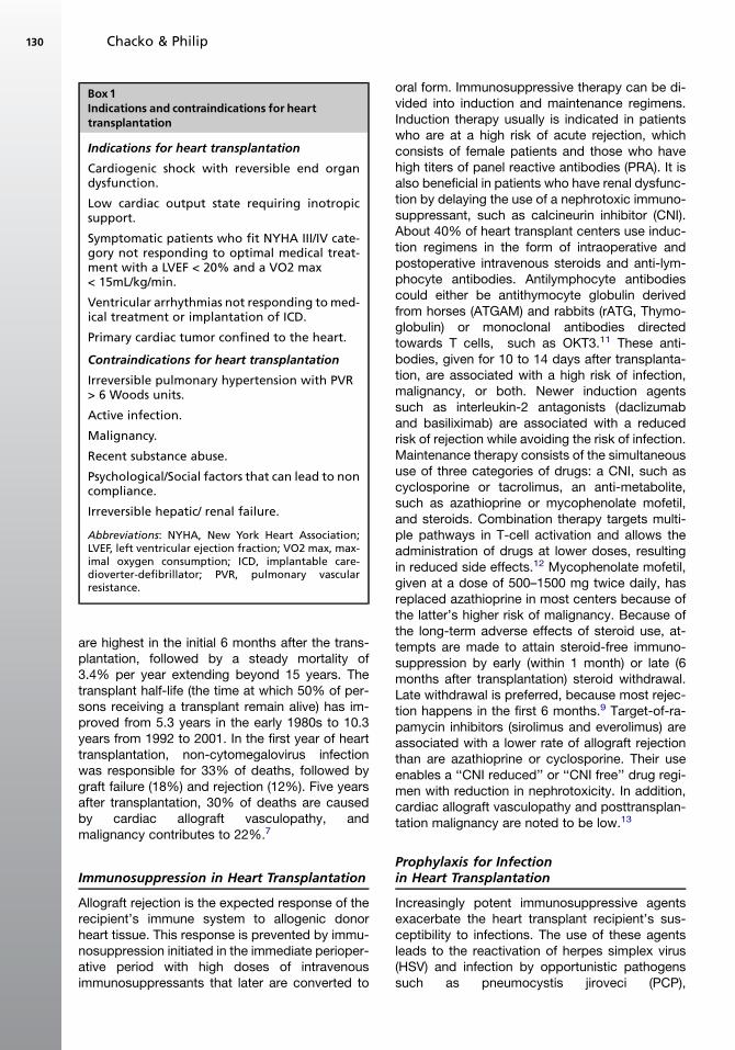
are highest in the initial 6 months after the trans-plantation, followed by a steady mortality of3.4% per year extending beyond 15 years. Thetransplant half-life (the time at which 50% of per-sons receiving a transplant remain alive) has im-proved from 5.3 years in the early 1980s to 10.3years from 1992 to 2001. In the first year of hearttransplantation, non-cytomegalovirus infectionwas responsible for 33% of deaths, followed bygraft failure (18%) and rejection (12%). Five yearsafter transplantation, 30% of deaths are causedby cardiac allograft vasculopathy, andmalignancy contributes to 22%.7
Immunosuppression in Heart Transplantation
Allograft rejection is the expected response of therecipient’s immune system to allogenic donorheart tissue. This response is prevented by immu-nosuppression initiated in the immediate perioper-ative period with high doses of intravenousimmunosuppressants that later are converted to
oral form. Immunosuppressive therapy can be di-vided into induction and maintenance regimens.Induction therapy usually is indicated in patientswho are at a high risk of acute rejection, whichconsists of female patients and those who havehigh titers of panel reactive antibodies (PRA). It isalso beneficial in patients who have renal dysfunc-tion by delaying the use of a nephrotoxic immuno-suppressant, such as calcineurin inhibitor (CNI).About 40% of heart transplant centers use induc-tion regimens in the form of intraoperative andpostoperative intravenous steroids and anti-lym-phocyte antibodies. Antilymphocyte antibodiescould either be antithymocyte globulin derivedfrom horses (ATGAM) and rabbits (rATG, Thymo-globulin) or monoclonal antibodies directedtowards T cells, such as OKT3.11 These anti-bodies, given for 10 to 14 days after transplanta-tion, are associated with a high risk of infection,malignancy, or both. Newer induction agentssuch as interleukin-2 antagonists (daclizumaband basiliximab) are associated with a reducedrisk of rejection while avoiding the risk of infection.Maintenance therapy consists of the simultaneoususe of three categories of drugs: a CNI, such ascyclosporine or tacrolimus, an anti-metabolite,such as azathioprine or mycophenolate mofetil,and steroids. Combination therapy targets multi-ple pathways in T-cell activation and allows theadministration of drugs at lower doses, resultingin reduced side effects.12 Mycophenolate mofetil,given at a dose of 500–1500 mg twice daily, hasreplaced azathioprine in most centers because ofthe latter’s higher risk of malignancy. Because ofthe long-term adverse effects of steroid use, at-tempts are made to attain steroid-free immuno-suppression by early (within 1 month) or late (6months after transplantation) steroid withdrawal.Late withdrawal is preferred, because most rejec-tion happens in the first 6 months.9 Target-of-ra-pamycin inhibitors (sirolimus and everolimus) areassociated with a lower rate of allograft rejectionthan are azathioprine or cyclosporine. Their useenables a ‘‘CNI reduced’’ or ‘‘CNI free’’ drug regi-men with reduction in nephrotoxicity. In addition,cardiac allograft vasculopathy and posttransplan-tation malignancy are noted to be low.13
Prophylaxis for Infectionin Heart Transplantation
Increasingly potent immunosuppressive agentsexacerbate the heart transplant recipient’s sus-ceptibility to infections. The use of these agentsleads to the reactivation of herpes simplex virus(HSV) and infection by opportunistic pathogenssuch as pneumocystis jiroveci (PCP),
Box1Indications and contraindications for hearttransplantation
Indications for heart transplantation
Cardiogenic shock with reversible end organdysfunction.
Low cardiac output state requiring inotropicsupport.
Symptomatic patients who fit NYHA III/IV cate-gory not responding to optimal medical treat-ment with a LVEF < 20% and a VO2 max< 15mL/kg/min.
Ventricular arrhythmias not responding to med-ical treatment or implantation of ICD.
Primary cardiac tumor confined to the heart.
Contraindications for heart transplantation
Irreversible pulmonary hypertension with PVR> 6 Woods units.
Active infection.
Malignancy.
Recent substance abuse.
Psychological/Social factors that can lead to noncompliance.
Irreversible hepatic/ renal failure.
Abbreviations: NYHA, New York Heart Association;LVEF, left ventricular ejection fraction; VO2 max, max-imal oxygen consumption; ICD, implantable care-dioverter-defibrillator; PVR, pulmonary vascularresistance.
Chacko & Philip130
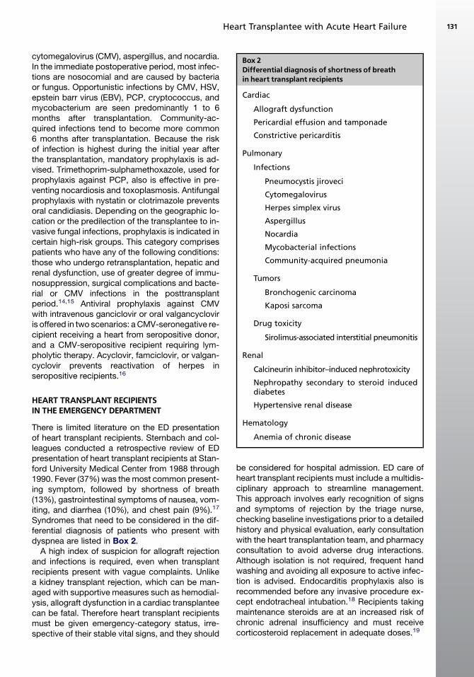
cytomegalovirus (CMV), aspergillus, and nocardia.In the immediate postoperative period, most infec-tions are nosocomial and are caused by bacteriaor fungus. Opportunistic infections by CMV, HSV,epstein barr virus (EBV), PCP, cryptococcus, andmycobacterium are seen predominantly 1 to 6months after transplantation. Community-ac-quired infections tend to become more common6 months after transplantation. Because the riskof infection is highest during the initial year afterthe transplantation, mandatory prophylaxis is ad-vised. Trimethoprim-sulphamethoxazole, used forprophylaxis against PCP, also is effective in pre-venting nocardiosis and toxoplasmosis. Antifungalprophylaxis with nystatin or clotrimazole preventsoral candidiasis. Depending on the geographic lo-cation or the predilection of the transplantee to in-vasive fungal infections, prophylaxis is indicated incertain high-risk groups. This category comprisespatients who have any of the following conditions:those who undergo retransplantation, hepatic andrenal dysfunction, use of greater degree of immu-nosuppression, surgical complications and bacte-rial or CMV infections in the posttransplantperiod.14,15 Antiviral prophylaxis against CMVwith intravenous ganciclovir or oral valgancycloviris offered in two scenarios: a CMV-seronegative re-cipient receiving a heart from seropositive donor,and a CMV-seropositive recipient requiring lym-pholytic therapy. Acyclovir, famciclovir, or valgan-cyclovir prevents reactivation of herpes inseropositive recipients.16
HEART TRANSPLANT RECIPIENTSIN THE EMERGENCY DEPARTMENT
There is limited literature on the ED presentationof heart transplant recipients. Sternbach and col-leagues conducted a retrospective review of EDpresentation of heart transplant recipients at Stan-ford University Medical Center from 1988 through1990. Fever (37%) was the most common present-ing symptom, followed by shortness of breath(13%), gastrointestinal symptoms of nausea, vom-iting, and diarrhea (10%), and chest pain (9%).17
Syndromes that need to be considered in the dif-ferential diagnosis of patients who present withdyspnea are listed in Box 2.
A high index of suspicion for allograft rejectionand infections is required, even when transplantrecipients present with vague complaints. Unlikea kidney transplant rejection, which can be man-aged with supportive measures such as hemodial-ysis, allograft dysfunction in a cardiac transplanteecan be fatal. Therefore heart transplant recipientsmust be given emergency-category status, irre-spective of their stable vital signs, and they should
be considered for hospital admission. ED care ofheart transplant recipients must include a multidis-ciplinary approach to streamline management.This approach involves early recognition of signsand symptoms of rejection by the triage nurse,checking baseline investigations prior to a detailedhistory and physical evaluation, early consultationwith the heart transplantation team, and pharmacyconsultation to avoid adverse drug interactions.Although isolation is not required, frequent handwashing and avoiding all exposure to active infec-tion is advised. Endocarditis prophylaxis also isrecommended before any invasive procedure ex-cept endotracheal intubation.18 Recipients takingmaintenance steroids are at an increased risk ofchronic adrenal insufficiency and must receivecorticosteroid replacement in adequate doses.19
Box 2Differential diagnosis of shortness of breathin heart transplant recipients
Cardiac
Allograft dysfunction
Pericardial effusion and tamponade
Constrictive pericarditis
Pulmonary
Infections
Pneumocystis jiroveci
Cytomegalovirus
Herpes simplex virus
Aspergillus
Nocardia
Mycobacterial infections
Community-acquired pneumonia
Tumors
Bronchogenic carcinoma
Kaposi sarcoma
Drug toxicity
Sirolimus-associated interstitial pneumonitis
Renal
Calcineurin inhibitor–induced nephrotoxicity
Nephropathy secondary to steroid induceddiabetes
Hypertensive renal disease
Hematology
Anemia of chronic disease
Heart Transplantee with Acute Heart Failure 131

CARDIAC ALLOGRAFT DYSFUNCTION
Dysfunction of the allograft may occur because ofleft ventricular (LV) and/or right ventricular (RV)dysfunction. RV dysfunction usually is seen imme-diately after heart transplantation and is related tohigh pulmonary artery pressure and resistance inthe recipient caused by long-standing heart fail-ure. The right ventricle, however, adapts to pulmo-nary pressure and resistance, leading tonormalization of RV function.20 LV dysfunction,which can be diastolic or systolic, is of major con-cern. The majority of the work that has been donehas focused on the systolic function of the trans-planted heart. Diastolic dysfunction is addressedin a later section of this article. Data on the evolu-tion of LV systolic function after heart transplanta-tion are scarce and conflicting. Some studiesshowed a decline in LVEF, whereas others havedemonstrated the EF to be maintained as long as4 years after transplantation.21,22 In one of the larg-est studies by Hershberger and colleagues, themean EF using radionuclide ventriculography de-creased from 63.8% to 55.6% in the first year aftertransplantation and remained stable thereafter.23
LV dysfunction can be divided further by the timefrom transplantation into early (observed intrao-peratively and in the immediate postoperative pe-riod) and late (after the first few postoperativeweeks to years after surgery). In the first year aftertransplantation, cardiac allograft rejection, infec-tions, and malignancies involving the myocardiumare responsible for late LV dysfunction, whereasallograft coronary artery disease primarily is
responsible for LV dysfunction that occurs lateron. Other causes of LV dysfunction include atrio-ventricular valve regurgitation, arrhythmias, recur-rence of the original disease such as in infiltrativedisorders (amyloidosis, sarcoidosis, and hemo-chromatosis), and myocarditis (giant cell or lym-phocytic).24 The causes of cardiac dysfunction inheart transplant recipients are shown in Fig. 1.
Cardiac Allograft Rejection
Rejection is an important cause of morbidity andmortality in the first year after transplantation,with the incidence declining thereafter over time.Based on the time after transplantation, rejectionis classified as hyperacute (within minutes to hoursafter transplantation), acute (weeks to months af-ter transplantation), or chronic (months to years af-ter transplantation). Acute rejection is dividedfurther into acute cellular rejection and acute hu-moral rejection. Acute cellular rejection, mostlyseen within the first 3 to 6 months, is a T-cell–me-diated response.25 Most of these events are diag-nosed by surveillance endomyocardial biopsy inasymptomatic recipients who have normal LVfunction. Occasionally, severe hemodynamiccompromise is noted in 5% of recipients whohave either LV or RV dysfunction.26 Diagnosisand treatment are based on the ISHLT grading ofendomyocardial biopsy, which was introduced in1990 and was revised in 2004.27 Hemodynamicallystable grade 1R rejection is not treated. Grade 2Rrejection is managed with a transient increase inthe steroid dose. Severe rejections (hemodynamic
Heart Failure
Cardiac dysfunction Non cardiac
Toxoplasmosis
Cardiac infectionsCMV
OthersRecurrent disease
AmylodosisSarcoidosis
Valve diseasePericardial
effusionConstrictive
pericarditis
CAVCoronary
EmbolismBiopsy mishap
Humoralrejection
Cellularrejection
Other Cardiacdysfunction
Transplantrejection
Ischemicinjury
SepsisAdrenal insufficiency
Arrhythmia
BradycardiaAFAFLSVTVT/VF
atherosclerosis
Fig.1. Differential diagnosis of heart failure in heart transplant recipients. AF, atrial fibrillation; AFL, atrial flutter;CAV, cardiac allograft vasculopathy; CMV, cytomegalovirus; SVT, supraventricular tachycardia; VF, ventricularfibrillation; VT, ventricular tachycardia. (Data from Semigram MJ, Stevenson LW. Case 37–2007: a 47 year oldman with left ventricular dysfunction after heart transplant. N Engl J Med 2007;357:2286–97.)
Chacko & Philip132
compromise or grade 3R) are treated with lympho-lytic therapy with antithymocyte globulin (Thymo-globulin or ATGAM) or OKT3.28 Acute humoral orantibody-mediated rejection, caused by anti-bodies against donor HLA or endothelial cellantigen, constitutes only 7% of all rejections. Anti-body-mediated rejection often occurs in the first4 months after transplantation and is associatedwith a high rate of hemodynamic compromise(29%–47%), graft loss (15%), and death (64%).Diagnosis is made by demonstrating immunoglob-ulins and complement in blood vessels by immu-nofluorescence or immunoperoxidase staining.The presence of endothelial cell swelling with in-travascular macrophage accumulation on hema-toxylin and eosin stain also is suggestive of thediagnosis. Assessment of circulatory antibodiesis restricted to patients who have hemodynamiccompromise. Treatment involves removal ofantibodies with plasmapheresis and suppressionof B-lymphocyte activity with high doses ofsteroids, lympholytic therapy, and intravenousimmunoglobulin.29
Allograft Coronary Artery Disease
LV dysfunction after heart transplantation canresult from coronary artery disease caused bycardiac allograft vasculopathy or coronary athero-sclerosis. Cardiac allograft vasculopathy orchronic rejection is a rapidly progressive athero-sclerosis reported in 7% of survivors at 1 year, in32.3% at 5 years, and in 45.7% of survivors at 8years after transplantation.7 It differs from athero-sclerosis by being concentric and diffuse, involv-ing the entire length of epicardial vessels. LVdysfunction is produced by myocardial infarction(which can be silent) or as a result of myocardial hi-bernation, often noticed because of the reductionof myocardial blood supply. Coronary angiogramsusually are needed to diagnose cardiac allograftvasculopathy, but the concentric involvement ofthe coronary vessels results in under diagnosis. In-travascular ultrasound is the most sensitive test,but widespread use is limited by its cost and inva-sive nature and the need for expertise. The mostsensitive noninvasive test is dobutamine stressechocardiography. Exercise nuclear imaging isspecific and can be used to confirm positivedobutamine echocardiograms. The roles of car-diac CT and MRI in this setting are emerging.30
Treatment with angioplasty, stenting, and coro-nary artery bypass has a limited role in diffuse dis-ease; retransplantation is the definitive option.Prevention is the best strategy and includes avoid-ing injury to the donor heart before transplantationand aggressive treatment of cardiac risk factors
such as diabetes, hypertension, and hyperlipid-emia. Prevention and treatment of CMV infectionand of allograft rejection also are important.31 Cor-onary atherosclerosis, seen in nontransplantedhearts, also can occur in transplanted hearts.Compared to cardiac allograft vasculopathy,corornary atherosclerosis is more localized, proxi-mal, and eccentric, so it is more amendable to cor-onary intervention. Other rare causes leading toischemic heart failure are myocardial infarctionfollowing endomyocardial biopsy, left anterior de-scending myocardial bridge, coronary vasospasm,and thrombosis.32–35
Arrhythmias
Arrhythmias can be a manifestation of cardiac dys-function as well as a precipitating factor inducingLV dysfunction. The common dysrhythmias en-countered in cardiac transplant patients are sum-marized in the following sections. Althoughtachyarrhythmias commonly are related to LV dys-function, failure to maintain adequate cardiac out-put in bradycardia is known to present ascongestive heart failure. Dysrhythmias encoun-tered in heart transplant patients are categorizedas early and late, depending on whether they oc-cur within or after 2 weeks of transplantation. Be-cause most early arrhythmias are observed in theimmediate postoperative period, this discussionfocuses on late-onset arrhythmias that oftenpose a challenge to ED physicians.
BradyarrhythmiasThe incidence of bradyarrhythmias varies from14% to 44% in United States and European studiesbut is as low as 11% in a study reported from Tai-wan.36 Most clinicians consider sinus bradycardiain the setting of heart transplantation to be a heartrate less than 80 beats/min, instead of the usualthreshold of 60 beats/min, given that the restingheart rate in transplant recipients often is veryhigh. Sinus node dysfunction is the most commontype encountered, while junctional bradycardiaand atrioventricular blocks (AVB) are responsiblefor the rest. Bradycardia that occurs in the early pe-riod often resolves on its own and is related mainlyto ischemia during surgery. Late-onset bradyar-rhythmias tend to be associated with AVB becauseof underlying cardiac allograft vasculopathy or re-jection.37 Long-term follow-up of a cohort of hearttransplant recipients who experienced symptom-atic bradycardia as a result of late-onset AVB re-vealed the absence of sinoatrial nodal artery.38
Hence late-onset bradyarrhythmias must be evalu-ated in detail with coronary angiography and bi-opsy to rule out possible rejection.
Heart Transplantee with Acute Heart Failure 133

Hemodynamically unstable situations warrantuse of isoproterenol while arrangements aremade for the implantation of a permanentpacemaker. It is important to bear in mind thatvagolytics (eg, atropine) have no effect on the atrio-ventricular node because of denervation. A trialwith methylxanthines can be attempted in stableconditions, but insertion of a permanent pace-maker often is needed in addition to addressingthe underlying ischemia caused by cardiac allo-graft vasculopathy or rejection. Current guidelinesrestrict the use of a permanent pacemaker to (1)symptomatic bradycardia not resolving with drugs,or (2) awake, asymptomatic third-degree AVB withassociated periods of asystole lasting 3 seconds orlonger or escape rhythms of less than 40 beats/min.39 The optimal mode of pacing is still debated,but the general consensus is to encourage atrio-ventricular synchronization and to provide rate-responsive pacing. Further discussion of modesettings is beyond the scope of this article.
Supraventricular TachyarrhythmiasStudies evaluating the incidence of supraventricu-lar tachyarrhythmia have been inclusive of bothearly and late-onset arrhythmias. In a recent eval-uation of 167 heart transplant recipients, Ahmariand colleagues noted the incidence of late-onsetatrial fibrillation and atrial flutter to be 9.5% and15%, respectively.40 Postoperative inflammationand altered hemodynamics may explain the higherincidence noted in initial studies, considering theirfrequent observation in the early period. Late-onset atrial fibrillation and atrial flutter, however,have a strong correlation with rejection and car-diac allograft vasculopathy.40,41 Macroentry cir-cuits as a mechanism for atrial flutter have beendemonstrated by Krishnan and colleagues.42
Because late-onset atrial fibrillation is related toan increased all-cause mortality, the identificationand treatment of the trigger factors is pivotal toprevent cardiac dysfunction.43 To date, no clearguidelines have been established for treating thesedysrhythmias. As in non–heart transplant patients,the choice between rate control and rhythmcontrol is an important consideration, becausethe persistence of atrial fibrillation can lead to LVdysfunction, even in the absence of cellular rejec-tion, humoral rejection, or cardiac allograft vascul-opathy.44 If there is evidence of hemodynamicinstability, cardioversion (via direct-current cardio-version [DCCV] or drugs) or overdrive pacing canbe considered. Electric cardioversion is preferredbecause of its efficacy and quick results. Chemicalcardioversion with amiodarone and ibutilide alsohas been effective, the latter being very beneficialwhen prior DCCV has been unsuccessful.45,46 For
chronic arrhythmias, class Ic drugs are used in theabsence of an ischemic myocardium. Flecainaide,a class Ia drug, is used where there is coexistingLV dysfunction or cardiac allograft vasculopathy.Rate control can be attained with b-blockers, cal-cium-channel blockers, or amiodarone. b-Blockers are considered first-choice therapybecause of the increased sensitivity of the myocar-dium to adrenergic chemicals after heart trans-plantation. Non-hydropyridines such as diltiazemneed to be used cautiously because their interac-tion with cyclosporine poses a difficulty in main-taining immunosuppression. Because of the riskof torsades, drugs that cause Q-T prolongationneed to be administered carefully whenever thereis concurrent use of tacrolimus. The benefits of an-ticoagulation should be weighed against the risk ofbleeding, because heart transplant recipients areat a high risk for embolic events. Aspirin is thechoice for lower-risk recipients, but all othersneed to be treated with warfarin. In patients whoexperience atrial flutter, electro-anatomic map-ping offers an advantage in identifying the re-entrycircuit, and radiofrequency ablation offers a goodresponse. In resistant scenarios, the option ofatrioventricular nodal ablation and the placementof a permanent pacemaker is always reserved.
The incidence of other supraventricular tachyar-rhythmias such as atrioventricular nodal re-entrytachycardia (AVNRT) and atrioventricular re-entrytachycardia have been described in heart trans-plant recipients in either case reports or case se-ries.47,48 Collins and colleagues noted a 1%incidence of AVNRT and a 2% incidence ofectopic atrial tachycardia among pediatric hearttransplant recipients.49 The higher incidence ofdual atrioventricular pathways in heart transplantrecipients puts them at elevated risk for AVNRT.The presence of excitable myocardial tissueacross suture lines in the atria can facilitate genesisof ectopic atrial tachycardia.50 Myocardial damageresulting from rejection or ischemia also can act asthe site of focal atrial tachycardia. All these types ofsupraventricular tachycardia need to be evaluatedpromptly with further invasive tests.
Treatment options are similar to those in patientswho have not had heart transplants. SynchronizedDCCV is the first choice in unstable patients; drugsare offered in stable patients. Adenosine is thedrug of first choice, but the dose needs to be halfthe usual dose or less because of the increasedadenosine sensitivity in heart transplant recipients.Vagal maneuvers and digoxin tend to be ineffec-tive because of the parasympathetic denervationfollowing heart transplantation. b-Blockers andcalcium-channel blockers are useful, but their abil-ity to exaggerate LV dysfunction needs to be
Chacko & Philip134
considered. If tachycardia persists despite initialmeasures, electrophysiologic evaluation can iden-tify pathways amenable to radiofrequency abla-tion, which often is curative.
Ventricular TachyarrhythmiasPremature ventricular complexes (PVC) in theform of isolated beats, ventricular couplets, andmultifocal PVC have been observed to decreasein frequency with time after heart transplantation.The same decrease is noted for accelerated idio-ventricular rhythm and nonsustained ventriculartachycardia.51 It has been suggested that epi-sodes occurring early after transplantation arecaused by increased catecholamine presence orby rejection. Later episodes, however, have beencorrelated with cardiac allograft vasculopathy.A recent report of frequent PVCs resulting in car-diomyopathy and its reversal with radiofrequencyablation has demonstrated the importance oftreating these events. As shown in these reports,focal radiofrequency ablation can be used to rec-tify the rhythm disturbance, but underlying rejec-tion and cardiac allograft vasculopathy must beruled out.
With the advent of better immunosuppression,ventricular tachycardia and ventricular fibrillationhave decreased from an initial incidence of25%.52 Treatment modalities are those used fornon–heart transplant patients and include directcurrent cardioversion and antiarrhythmics suchas amiodarone, procainamide, and, rarely, lido-caine. Sudden cardiac death as a result of fatal ar-rhythmias is believed to occur in 10% to 27% oftransplant survivors because of underlying rejec-tion or cardiac allograft vasculopathy.53,54 Data re-garding the use of an internal cardiac defibrillatorto prevent this outcome are limited by small sam-ple size and the availability of only retrospectiveanalyses. Montpetit and colleagues suggestedthat the threshold for internal cardiac defibrillatorinsertion be lowered to include patients whohave an EF less than 40%, considering the higherincidence of sudden cardiac death in that sub-group.55 Until prospective trials provide cleardata, clinicians must rely on existing recommen-dations for defibrillator implantation in heart trans-plant recipients.
In summary, all patients who have a history ofheart transplantation and who present to the EDwith cardiac dysfunction need to be evaluatedin detail with ECG and telemetry monitoring.Early consultation with an electrophysiologistand a transplant cardiologist is advised to iden-tify arrhythmias that can appear benign butreflect significant underlying cardiac pathologysuch as rejection or cardiac allograft
vasculopathy. In many cases, treatment of thesedysrhythmias has reversed the cardiomyopathyand thereby offered a better prognosis andquality of life.
Pericardial Diseases
A moderate to large pericardial effusion is seen in20% to 35% of recipients after heart transplantsurgery and usually resolves within 3 monthspostoperatively. Rarely it can lead to pericardialtamponade in the postoperative period, thus re-quiring drainage.56 Cardiac tamponade presentswith dyspnea, orthopnea, jugular venous engorge-ment, reduced heart sounds, and hypotension.Because of the increased risk of infections andmalignancies in heart transplant recipients, diag-nostic pericardiocentesis must be considered inlate pericardial effusion even in the absence oftamponade. Constrictive pericarditis usually oc-curs 3 months to 2 years after transplantationand may be related to pericardial effusion orhematoma, postpericardiotomy syndrome, media-stinitis, or intrathoracic infections.57 Immunosup-pression leads to an increased risk for infectiousconstrictive pericarditis such as purulent, tubercu-lous, or fungal pericarditis.58,59 Restrictive pericar-ditis physiology leads to a clinical presentationwith symptoms of fluid overload and decreasedcardiac output. Use of echocardiography as a di-agnostic modality can identify this pathology atthe bedside; however, CT and MRI of the heart,as well as invasive hemodynamic measurement,can be helpful in difficult scenarios. Transient con-striction as a sequel to postpericardiotomy syn-drome, infections, or malignancy may improvewith a variety of agents including nonsteroidalanti-inflammatory drugs, steroids, antibiotics, che-motherapy, angiotensin-converting enzyme inhibi-tors, and diuretics. Symptoms that persist after2 to 3 months of conservative treatment or thatoccur in patients who have hemodynamic com-promise may require pericardiectomy.60
Myocarditis and Infections
Immunosuppression leads to increased suscepti-bility to latent as well as new infections that canrarely involve the cardiac allograft. In addition tothe usual organisms (echovirus and coxsackievirus), CMV, EBV, toxoplasmosis, and Chagas dis-ease can lead to clinical and subclinical myocardi-tis in heart transplant recipients.61 Aspergillus andparvovirus B19 also have been reported to causemyocarditis.62,63 CMV infections can be acquiredfrom reactivation of endogenous viruses or via do-nated allograft and blood products from seropos-itive donors. In addition, CMV is implicated in LV
Heart Transplantee with Acute Heart Failure 135
dysfunction by direct myocardial infection besidesbeing associated with the pathogenesis of acuterejection and cardiac allograft vasculopathy.64,65
Sepsis also can cause LV dysfunction.66 Serologictests are of limited use in an immunocompromisedhost. Nucleic acid–based molecular assay (PCR)and antigen assay (pp65 in CMV) are of limitedvalue in the ED. Because symptoms are few andatypical, tissue histology is needed for the diagno-sis of infections. The reference standard test forthe diagnosis of myocarditis is endomyocardial bi-opsy. Effective treatments can be achieved withthe use of ganciclovir or valgancyclovir for CMV,trimethoprim-sulphamethoxazole for toxoplasmo-sis, and broad-spectrum antibiotics when sepsisis a consideration.67
Malignancy
The incidence of malignancy in transplant recipi-ents is noted to be about 5% to 6%. Because ofimmunosuppression, the risk of malignancy isabout 100 times greater in heart transplant recipi-ents than in the general population.68 Posttrans-plantation lymphoproliferative disorder, seen in2% to 5% of heart transplant recipients, is associ-ated with EBV exposure and the use of immuno-suppressants such as CNIs, azathioprine, andOKT3. Extranodal involvement is common; allo-graft involvement may be in the form of a cardiacmass or myocardial infiltration resulting in LV dys-function. Diagnosis is made on tissue biopsy fromthe endomyocardium, lymph node, or bone mar-row. Treatment modalities include reducing theextent of immunosuppression, surgical excision,chemotherapy, and rituximab.69
Valvular Disease
Tricuspid valve regurgitation (TR) is the most fre-quent valvular abnormality seen after orthotopicheart transplantation, with a prevalence of 67%to 85% as detected by echocardiogram. Risk fac-tors for the development of TR are size mismatchbetween the donor heart and pericardial cavity,biatrial technique, high-grade rejection, repeatedendomyocardial biopsy, pulmonary hypertension,and ischemic injury to the right ventricle.70 Mostcases of TR are mild and asymptomatic. Moderateto severe TR, seen in 25% to 34% of transplant re-cipients, presents with symptoms of RV volumeoverload. Surgical repair or replacement is re-quired in 4% of transplant recipients for refractoryright-sided heart failure.71 Left-sided valvular re-gurgitations are less common. Roig and col-leagues observed mitral regurgitation in 8 (6%) of141 heart transplant recipients and noted
associations with cardiac allograft vasculopathyand mortality.72
Diastolic Heart Failure
Diastolic dysfunction, measured by Doppler echo-cardiogram or pulmonary artery catheter, occursfrequently after transplant surgery but resolveswithin the first 4 to 8 weeks after transplanta-tion.73,74 It may persist or even reappear laterbecause of allograft rejection, constrictive or effu-sive-constrictive pericarditis, recurrence of infiltra-tive disorders, and increased incidence ofhypertension following the use of CNIs.75 Azizand colleagues found a restrictive filling pattern de-fined by mitral deceleration time in 41 of the 152recipients at 24 months after transplantation.76
Using a pulmonary artery catheter, Tallaj and col-leagues found diastolic dysfunction in 11% ofrecipients at 1 year after transplantation.77 Siostr-zonek and colleagues described a case in whichcyclosporine withdrawal led to relief of diastolicdysfunction.78 Lobato and colleagues reporteda case of postoperative pulmonary edema causedby diastolic dysfunction.79
EVALUATION OFALLOGRAFT DYSFUNCTION
LV dysfunction is suspected in heart transplantrecipients presenting with shortness of breath, or-thopnea, fatigue, palpitations, and signs of left- orright-sided heart failure on physical examination.Asymptomatic LV dysfunction can be found onroutine diagnostic tests such as echocardiograms,radionuclide ventriculography, or right heart cath-eterization. Natriuretic peptide, cardiac markers,ECG, and chest radiographs are helpful in identify-ing this pathology, but endomyocardial biopsy hasthe advantage of identifying the cause.
History and Physical Examination
Allograft rejection and cardiac allograft vasculop-athy, the two main causes of LV dysfunction, pres-ent with nonspecific symptoms. Most allograftrejection is diagnosed at surveillance endomyo-cardial biopsy. A few cases present with LVdysfunction, arrhytmias, or gastrointestinal symp-toms caused by hepatic congestion. Because ofthe denervation of the transplanted heart, the clas-sical symptoms of ischemia are absent. Eventhough 10% to 30% of heart transplant recipientshave partial reinnervation and may present withangina, cardiac allograft vasculopathy rarely pres-ents as chest pain. Most patients present late withsilent myocardial infarction, congestive heart fail-ure, arrhythmias, or sudden death.80 A detailedmedication history, including compliance, dosing,
Chacko & Philip136
and timing of immunosuppressive drugs, must betaken for proper adjustment of immunosuppres-sive drugs. Blood draws for drug-level monitoringare necessary also.
Natriuretic Peptides
Serum natriuretic peptide (BNP) and N-terminalproBNP (NT-proBNP) are established in the diag-nosis of heart failure in patients who present tothe ED with shortness of breath.81,82 Their role inheart transplant recipients is not clear, however.High levels of BNP are seen within the first monthafter heart transplantation, followed by a gradualdecline, but rarely do the levels return to normal.In one of the largest studies to date, Park andcolleagues observed that the BNP level was ele-vated three- to fourfold, with a median level of153 pg/mL, in stable heart transplant recipients.83
Similarly the mean NT-proBNP level was 4032 pg/mL within 6 months after heart transplantation,compared with 480 pg/mL beyond 6 months aftertransplantation. This elevation, observed despitea normal systolic function, could be attributed tohigh pulmonary capillary wedge pressure, hightranspulmonary gradient, diastolic dysfunction,RV enlargement or dilatation, or significant TR.84
BNP has been extensively studied as a noninva-sive marker of allograft performance to detect allo-graft rejection. Masters and colleagues showeda correlation between a BNP level higher than400 pg/mL and grade 2 or higher rejection in 10patients. Similarly, Herva and colleagues demon-strated a correlation between elevated natriureticpeptide levels with a rejection of grade 2 andhigher among transplantees in the initial 90 daysfollowing their heart transplantation. This correla-tion was supported by the observation of Ham-merer-Lercher and her colleagues as well as Wuand her team, who noticed the associationbetween elevated BNP levels and high-grade re-jection. (grade R 3). In a review of six other stud-ies, no significant increase in natriuretic peptidewas seen during acute allograft rejection.85 Thusthe association between BNP and rejection isseen only in severe rejection (grade R 3), espe-cially during the first 6 months after transplanta-tion. Therefore BNP cannot be used as a markerof rejection in stable heart transplant recipientsand is unlikely to replace or reduce the use of en-domyocardial biopsy in detecting rejection.
Cardiac Markers
Creatinine kinase (CK), its myocardial band iso-form (CK-MB), myoglobin, lactate dehydrogenase,and aspartate dehydrogenase have no role in thenoninvasive detection of rejection or cardiac
allograft vasculopathy because of lack of specific-ity. Cardiac troponin I and cardiac troponin T arecardiac myofibrillar proteins that can be detectedin blood with very small amounts of myocardial ne-crosis (<1 g). The role of troponin in cardiac rejec-tion has been investigated extensively, withconflicting results, and its use in evaluation of allo-graft rejection cannot be recommended.86 Ele-vated troponin levels, however, can identifya subset of recipients who are at increased riskfor the development of cardiac allograft vasculop-athy.87 The role of troponin in acute settings hasnot been well studied, but increasing levels, com-pared with baseline, may suggest myocardial in-jury resulting from rejection or coronary arterydisease.
Electrocardiogram
Denervation and the loss of autonomic nervoussystem control in transplanted hearts leads tohigher resting heart rates at around 100 beats/min, with tachycardia occurring in more than halfof recipients. A resting heart rate of less than 130beats/min usually has no significant adverse ef-fects. In recipients who have a biatrial transplant,two P waves may be seen because of the retainedrecipient’s right atrium.88 Right bundle branchblock, the most common ECG abnormality, isseen in approximately 50% of recipients andcarries a benign prognosis.89 New-onset bradyar-rhythmias (sinus bradycardia, advanced atrioven-tricular block) or tachyarrhythmias (atrial flutterand fibrillation) warrant evaluation for rejection orunderlying cardiac allograft vasculopathy.88
Radiology
Chest radiographs may show cardiomegaly andpulmonary edema suggestive of LV dysfunction.Conventional CT and MRI have limited roles inthe diagnosis of allograft rejection and cardiac al-lograft vasculopathy. MRI coronary angiogramscan be used to visualize coronary arteries and theirbranches and can be used as a screening tool toreduce the number of coronary angiograms. CTcoronary artery calcium scoring has a high nega-tive predictive value (up to 97%) and may be usefulin ruling out cardiac allograft vasculopathy. Themain role of imaging in heart transplant recipientsadmitted with shortness of breath is to rule out in-fections and malignancies involving the lung andheart.90
Drug-Level Monitoring
Nonadherence to immunosuppressive regimens isa risk factor for rejection and cardiac allograft vas-culopathy.91 Optimal immunosuppression is
Heart Transplantee with Acute Heart Failure 137
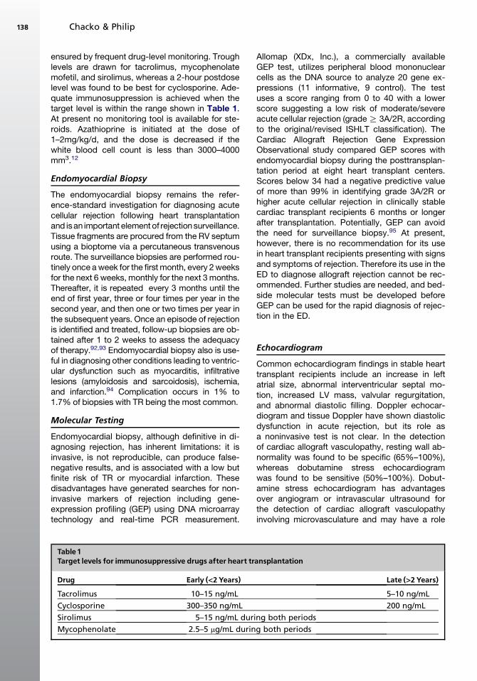
ensured by frequent drug-level monitoring. Troughlevels are drawn for tacrolimus, mycophenolatemofetil, and sirolimus, whereas a 2-hour postdoselevel was found to be best for cyclosporine. Ade-quate immunosuppression is achieved when thetarget level is within the range shown in Table 1.At present no monitoring tool is available for ste-roids. Azathioprine is initiated at the dose of1–2mg/kg/d, and the dose is decreased if thewhite blood cell count is less than 3000–4000mm3.12
Endomyocardial Biopsy
The endomyocardial biopsy remains the refer-ence-standard investigation for diagnosing acutecellular rejection following heart transplantationand isan important element of rejection surveillance.Tissue fragments are procured from the RV septumusing a bioptome via a percutaneous transvenousroute. The surveillance biopsies are performed rou-tinely once a week for the first month, every 2 weeksfor the next 6 weeks, monthly for the next 3 months.Thereafter, it is repeated every 3 months until theend of first year, three or four times per year in thesecond year, and then one or two times per year inthe subsequent years. Once an episode of rejectionis identified and treated, follow-up biopsies are ob-tained after 1 to 2 weeks to assess the adequacyof therapy.92,93 Endomyocardial biopsy also is use-ful in diagnosing other conditions leading to ventric-ular dysfunction such as myocarditis, infiltrativelesions (amyloidosis and sarcoidosis), ischemia,and infarction.94 Complication occurs in 1% to1.7% of biopsies with TR being the most common.
Molecular Testing
Endomyocardial biopsy, although definitive in di-agnosing rejection, has inherent limitations: it isinvasive, is not reproducible, can produce false-negative results, and is associated with a low butfinite risk of TR or myocardial infarction. Thesedisadvantages have generated searches for non-invasive markers of rejection including gene-expression profiling (GEP) using DNA microarraytechnology and real-time PCR measurement.
Allomap (XDx, Inc.), a commercially availableGEP test, utilizes peripheral blood mononuclearcells as the DNA source to analyze 20 gene ex-pressions (11 informative, 9 control). The testuses a score ranging from 0 to 40 with a lowerscore suggesting a low risk of moderate/severeacute cellular rejection (grade R 3A/2R, accordingto the original/revised ISHLT classification). TheCardiac Allograft Rejection Gene ExpressionObservational study compared GEP scores withendomyocardial biopsy during the posttransplan-tation period at eight heart transplant centers.Scores below 34 had a negative predictive valueof more than 99% in identifying grade 3A/2R orhigher acute cellular rejection in clinically stablecardiac transplant recipients 6 months or longerafter transplantation. Potentially, GEP can avoidthe need for surveillance biopsy.95 At present,however, there is no recommendation for its usein heart transplant recipients presenting with signsand symptoms of rejection. Therefore its use in theED to diagnose allograft rejection cannot be rec-ommended. Further studies are needed, and bed-side molecular tests must be developed beforeGEP can be used for the rapid diagnosis of rejec-tion in the ED.
Echocardiogram
Common echocardiogram findings in stable hearttransplant recipients include an increase in leftatrial size, abnormal interventricular septal mo-tion, increased LV mass, valvular regurgitation,and abnormal diastolic filling. Doppler echocar-diogram and tissue Doppler have shown diastolicdysfunction in acute rejection, but its role asa noninvasive test is not clear. In the detectionof cardiac allograft vasculopathy, resting wall ab-normality was found to be specific (65%–100%),whereas dobutamine stress echocardiogramwas found to be sensitive (50%–100%). Dobut-amine stress echocardiogram has advantagesover angiogram or intravascular ultrasound forthe detection of cardiac allograft vasculopathyinvolving microvasculature and may have a role
Table1Target levels for immunosuppressive drugs after heart transplantation
Drug Early (<2 Years) Late (>2 Years)
Tacrolimus 10–15 ng/mL 5–10 ng/mL
Cyclosporine 300–350 ng/mL 200 ng/mL
Sirolimus 5–15 ng/mL during both periods
Mycophenolate 2.5–5 mg/mL during both periods
Chacko & Philip138
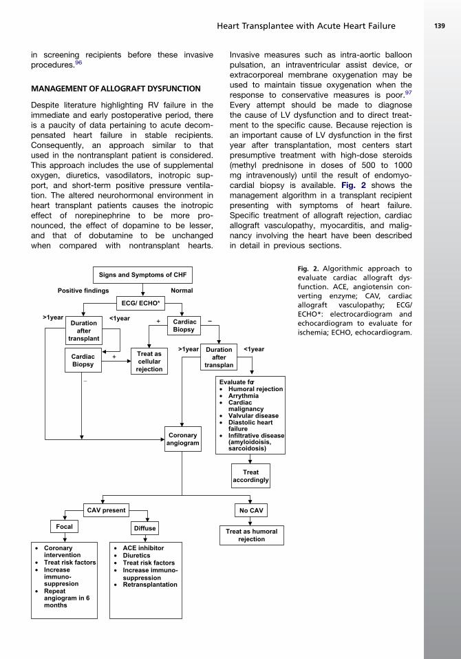
in screening recipients before these invasiveprocedures.96
MANAGEMENT OFALLOGRAFT DYSFUNCTION
Despite literature highlighting RV failure in theimmediate and early postoperative period, thereis a paucity of data pertaining to acute decom-pensated heart failure in stable recipients.Consequently, an approach similar to thatused in the nontransplant patient is considered.This approach includes the use of supplementaloxygen, diuretics, vasodilators, inotropic sup-port, and short-term positive pressure ventila-tion. The altered neurohormonal environment inheart transplant patients causes the inotropiceffect of norepinephrine to be more pro-nounced, the effect of dopamine to be lesser,and that of dobutamine to be unchangedwhen compared with nontransplant hearts.
Invasive measures such as intra-aortic balloonpulsation, an intraventricular assist device, orextracorporeal membrane oxygenation may beused to maintain tissue oxygenation when theresponse to conservative measures is poor.97
Every attempt should be made to diagnosethe cause of LV dysfunction and to direct treat-ment to the specific cause. Because rejection isan important cause of LV dysfunction in the firstyear after transplantation, most centers startpresumptive treatment with high-dose steroids(methyl prednisone in doses of 500 to 1000mg intravenously) until the result of endomyo-cardial biopsy is available. Fig. 2 shows themanagement algorithm in a transplant recipientpresenting with symptoms of heart failure.Specific treatment of allograft rejection, cardiacallograft vasculopathy, myocarditis, and malig-nancy involving the heart have been describedin detail in previous sections.
ECG/ ECHO*
Signs and Symptoms of CHF
Cardiac
Biopsy
Treat as
cellular
rejection
Duration
after
transplan
Evaluate for
• Humoral rejection
• Arrythmia
• Cardiac
malignancy
• Valvular disease
• Diastolic heart
failure
• Infiltrative disease
(amyloidoisis,
sarcoidosis)
Treat
accordingly
Coronary
angiogram
Duration
after
transplant
Cardiac
Biopsy
NormalPositive findings
>1year <1year–+
<1year>1year
+
_
No CAV
Treat as humoral
rejection
CAV present
DiffuseFocal
• Coronary
intervention
• Treat risk factors
• Increase
immuno-
suppresion
• Repeat
angiogram in 6
months
• ACE inhibitor
• Diuretics
• Treat risk factors
• Increase immuno-
suppression
• Retransplantation
Fig. 2. Algorithmic approach toevaluate cardiac allograft dys-function. ACE, angiotensin con-verting enzyme; CAV, cardiacallograft vasculopathy; ECG/ECHO*: electrocardiogram andechocardiogram to evaluate forischemia; ECHO, echocardiogram.
Heart Transplantee with Acute Heart Failure 139
SUMMARY
With an increasing number of heart transplanta-tions being performed around the world and theimprovement in the survival rates, more transplantrecipients may present to the ED with comorbid-ities unique to the transplanted heart and relatedimmunosuppression, including heart failure. Thisarticle is aimed at enabling the ED physician iden-tify and better manage this unique group of pa-tients for whom time is life. In this endeavor,physicians must strive to uphold Alexander Pope’swords:
‘‘A wise physician, skill’d our wounds to heal,Is more than armies to the public weal.’’
REFERENCES
1. Rosamond W, Flegal K, Friday G, et al. Heart disease
and stroke statistics—2007 update: a report from the
American Heart Association Statistics Committee
and Stroke Statistics Subcommittee. [erratum ap-
pears in Circulation. 2007;115(5):e172]. Circulation
2007;115(5):e69–171.
2. Shibata MC, Flather MD, Wang D. Systematic review
of the impact of beta blockers on mortality and hos-
pital admissions in heart failure [see comment]. Eur
J Heart Fail 2001;3(3):351–7.
3. Garg R, Yusuf S. Overview of randomized trials of
angiotensin-converting enzyme inhibitors on mortal-
ity and morbidity in patients with heart failure. Col-
laborative group on ACE inhibitor trials [erratum
appears in JAMA 1995 Aug 9;274(6):462]. JAMA
1995;273(18):1450–6.
4. Pitt B, Zannad F, Remme WJ, et al. The effect of spi-
ronolactone on morbidity and mortality in patients
with severe heart failure. Randomized Aldactone
evaluation study investigators [see comment]. N
Engl J Med 1999;341(10):709–17.
5. Rivero-Ayerza M, Theuns DAMJ, Garcia-Garcia HM,
et al. Effects of cardiac resynchronization therapy on
overall mortality and mode of death: a meta-analysis
of randomized controlled trials [see comment]. Eur
Heart J 2006;27(22):2682–8.
6. Aaronson KD, Mancini DM. Mortality remains high
for outpatient transplant candidates with prolonged
(>6 months) waiting list time. J Am Coll Cardiol
1999;33(5):1189–95.
7. Taylor DO, Edwards LB, Boucek MM, et al. Registry
of the International Society for Heart and Lung Trans-
plantation: twenty-fourth official adult heart trans-
plant report—2007. J Heart Lung Transplant 2007;
26(8):769–81.
8. Barnard CN. The operation. A human cardiac trans-
plant: an interim report of a successful operation
performed at Groote Schuur hospital, Cape Town.
S Afr Med J 1967;41:1271–4.
9. Al-khaldi A, Robbins RC. New directions in cardiac
transplantation. Annu Rev Med 2006;57:455–71.
10. Zakliczynski M, et al. New guidelines in the selection
of patients with heart insufficiency for heart trans-
plantation—discussion of standards of the Interna-
tional Society of Heart and Lung Transplantation
(ISHLT) in 2006. Kardiol Pol 2006;64(12):1462–4.
11. Woodley SL, Renlund DG, O’Connell JB, et al. Im-
munosuppression following cardiac transplantation.
Cardiol Clin 1990;8(1):83–96.
12. Lindenfeld J, Miller GG, Shakar SF, et al. Drug ther-
apy in the heart transplant recipient: part II: immuno-
suppressive drugs. Circulation 2004;110(25):
3858–65.
13. Gustafsson F, Ross HJ. Proliferation signal inhibitors
in cardiac transplantation. Curr Opin Cardiol 2007;
22(2):111–6.
14. Playford EG, Webster AC, Sorell TC, et al. Antifungal
agents for preventing fungal infections in solid organ
transplant recipients. Cochrane Database Syst Rev
2004;(3):CD004291.
15. Villacian JS, Paya CV. Prevention of infections in
solid organ transplant recipients [see comment].
Transpl Infect Dis 1999;1(1):50–64.
16. Lindenfeld J, Page RL 2nd, Zolty R, et al. Drug ther-
apy in the heart transplant recipient: part III: com-
mon medical problems. Circulation 2005;111(1):
113–7.
17. Sternbach GL, Varon J, Hunt SA. Emergency de-
partment presentation and care of heart and heart/
lung transplant recipients. Ann Emerg Med 1992;
21(9):1140–4.
18. Zavotsky KE, Sapienza J, Wood D. Nursing implica-
tions for ED care of patients who have received
heart transplants (CE). J Emerg Nurs 2001;27(1):
33–9.
19. Coursin DB, Wood KE. Corticosteroid supplementa-
tion for adrenal insufficiency. JAMA 2002;287(2):
236–40.
20. Bacal F, Pires PV, Moreira LF, et al. Normalization of
right ventricular performance and remodeling evalu-
ated by magnetic resonance imaging at late follow-
up of heart transplantation: relationship between
function, exercise capacity and pulmonary vascular
resistance. J Heart Lung Transplant 2005;24(12):
2031–6.
21. Hartmann A, Maul FD, Huth A, et al. Serial evaluation
of left ventricular function by radionuclide ventricu-
lography at rest and during exercise after orthotopic
heart transplantation. Eur J Nucl Med 1993;20(2):
146–50.
22. Tischler MD, Lee RT, Plappert T, et al. Serial as-
sessment of left ventricular function and mass after
orthotopic heart transplantation: a 4-year longitudi-
nal study. [erratum appears in J Am Coll Cardiol
Chacko & Philip140
1994 Jan;23(1):281]. J Am Coll Cardiol 1992;19(1):
60–6.
23. Hershberger RE, Ni H, Toy W, et al. Distribution and
declines in cardiac allograft radionuclide left ventric-
ular ejection fractions in relation to late mortality.
J Heart Lung Transplant 2001;20(4):417–24.
24. Jarcho JA, Mark EJ. Case 17-1998. A 53-year-old
man with left ventricular dysfunction four years
after a heart transplantation. N Engl J Med 1998;
338(22):1608–16.
25. Lindenfeld J, Miller GG, Shakar SF, et al. Drug ther-
apy in the heart transplant recipient: part I: cardiac
rejection and immunosuppressive drugs. Circulation
2004;110(24):3734–40.
26. Mills RM, Naftel DC, Kirklin JK, et al. Heart transplant
rejection with hemodynamic compromise:
a multiinstitutional study of the role of endomyocar-
dial cellular infiltrate. Cardiac Transplant Research
Database. J Heart Lung Transplant 1997;16(8):
813–21.
27. Stewart S, Winters GL, Fishbein MC, et al. Revision
of the 1990 working formulation for the standardiza-
tion of nomenclature in the diagnosis of heart rejec-
tion [see comment]. J Heart Lung Transplant 2005;
24(11):1710–20.
28. Delgado JF, Sanchez V, de la Calzada CS. Acute re-
jection after heart transplantation. Expert Opin Phar-
macother 2006;7(9):1139–49.
29. Uber WE, Self SE, Van Bakel AB, et al. Acute anti-
body-mediated rejection following heart transplanta-
tion. Am J Transplant 2007;7(9):2064–74.
30. Kass M, Allan R, Haddad H. Diagnosis of graft cor-
onary artery disease. Curr Opin Cardiol 2007;22(2):
139–45.
31. Kass M, Haddad H. Cardiac allograft vasculopathy:
pathology, prevention and treatment. Curr Opin Car-
diol 2006;21(2):132–7.
32. Drobinski G, et al. Myocardial infarction after endo-
myocardial biopsy in a heart transplant patient.
J Interv Cardiol 2002;15(5):403–5.
33. Pittaluga J, Dorent R, Ghossoub J-J, et al. Left ante-
rior descending coronary artery bridge. A cause of
early death after cardiac transplantation. Chest
1997;111(2):511–3.
34. Lipski A, Sahar G, De Bruyne Y, et al. Acute myocar-
dial infarction immediately after heart transplanta-
tion. J Heart Lung Transplant 1993;12(6 Pt 1):
1065–6.
35. Semigran MJ, Stevenson LW, Passeri JJ, et al. Case
records of the Massachusetts General Hospital.
Case 37-2007. A 47-year-old man with left ventricu-
lar dysfunction after heart transplantation. N Engl
J Med 2007;357(22):2286–97.
36. Lai CL, Chen WJ, Wang SS, et al. Bradyarrhythmias
and cardiac pacing after orthotopic heart transplan-
tation in a Chinese population. Transplant Proc
2002;34(8):3232–5.
37. Cui G, Kobashigawa J, Margarian A, et al. Cause of
atrioventricular block in patients after heart trans-
plantation. Transplantation 2003;76(1):137–42.
38. Weinfeld MS, Kartashov A, Piana R, et al. Bradycar-
dia: a late complication following cardiac transplan-
tation. Am J Cardiol 1996;78(8):969–71.
39. Gregoratos G, Abrams J, Epstein AE, et al. ACC/
AHA/NASPE 2002 guideline update for implantation
of cardiac pacemakers and antiarrhythmia devices:
summary article: a report of the American College of
Cardiology/American Heart Association Task Force
on Practice Guidelines (ACC/AHA/NASPE Commit-
tee to Update the 1998 Pacemaker Guidelines). Cir-
culation 2002;106(16):2145–61.
40. Ahmari SA, Bunch TJ, Chandra A, et al. Prevalence,
pathophysiology, and clinical significance of post-
heart transplant atrial fibrillation and atrial flutter.
J Heart Lung Transplant 2006;25(1):53–60.
41. Cui G, Tung T, Kobashigawa J, et al. Increased inci-
dence of atrial flutter associated with the rejection of
heart transplantation. Am J Cardiol 2001;88(3):280–4.
42. Krishnan SC, Falsone JM, Sanders WE, et al. Cath-
eter ablation of atrial flutter in a heart transplant re-
cipient. Pacing Clin Electrophysiol 2002;25(8):
1262–5.
43. Pavri BB, O’Nunain SS, Newell JB, et al. Prevalence
and prognostic significance of atrial arrhythmias
after orthotopic cardiac transplantation. J Am Coll
Cardiol 1995;25(7):1673–80.
44. Woo GW, Schofield RS, Klodell CT, et al. Atrial fibril-
lation as a cause of left ventricular dysfunction after
cardiac transplantation. J Heart Lung Transplant
2006;25(1):131–3.
45. Oral H, Souza JJ, Michaud GF, et al. Facilitating
transthoracic cardioversion of atrial fibrillation with
ibutilide pretreatment. N Engl J Med 1999;340(24):
1849–54.
46. Franco V, Tallaj JA, Rayburn BK, et al. 259: ibutilide
is safe and efficacious in heart transplant recipients.
J Heart Lung Transplant 2007;26(2 Suppl 1):S153.
47. Rothman SA, Hsia HH, Bove AA, et al. Radiofre-
quency ablation of Wolff-Parkinson-White syndrome
in a donor heart after orthotopic heart transplanta-
tion. J Heart Lung Transplant 1994;13(5):905–9.
48. Padder FA, Wilbur SL, Kantharia BK, et al. Radiofre-
quency catheter ablation of atrioventricular nodal re-
entrant tachycardia after orthotopic heart
transplantation. J Interv Card Electrophysiol 1999;
3(3):283–5.
49. Collins KK, Thiagarajan RR, Chin C, et al. Atrial
tachyarrhythmias and permanent pacing after pedi-
atric heart transplantation. J Heart Lung Transplant
2003;22(10):1126–33.
50. Lai W, Kao A, Silka MJ, et al. Recipient to donor con-
duction of atrial tachycardia following orthotopic
heart transplantation. Pacing Clin Electrophysiol
1998;21(6):1331–5.
Heart Transplantee with Acute Heart Failure 141
51. Scott CD, Dark JH, McComb JM. Arrhythmias after
cardiac transplantation. Am J Cardiol 1992;70(11):
1061–3.
52. Tagusari O, Kormos RL, Kawai A, et al. Native heart
complications after heterotopic heart transplanta-
tion: insight into the potential risk of left ventricular
assist device. J Heart Lung Transplant 1999;
18(11):1111–9.
53. Patel VS, Lim M, Massin EK, et al. Sudden cardiac
death in cardiac transplant recipients. Circulation
1996;94(9 Suppl):II273–7.
54. Alexander RT, Steenbergen C. Cause of death and
sudden cardiac death after heart transplantation.
An autopsy study. Am J Clin Pathol 2003;119(5):
740–8.
55. Montpetit M, Singh M, Muller E, et al. 340: sudden
cardiac death in heart transplant patients: is there
a role for defibrillators? J Heart Lung Transplant
2007;26(2 Suppl 1):S182.
56. Al-Dadah AS, Guthrie TJ, Pasque MK, et al. Clinical
course and predictors of pericardial effusion follow-
ing cardiac transplantation. Transplant Proc 2007;
39(5):1589–92.
57. Copeland JG, Riley JE, Fuller J. Pericardiectomy for
effusive constrictive pericarditis after heart trans-
plantation. J Heart Transplant 1986;5(2):171–2.
58. Canver CC, Patel AK, Kosolcharoen P, et al. Fungal
purulent constrictive pericarditis in a heart transplant
patient. Ann Thorac Surg 1998;65(6):1792–4.
59. Puius YA, Scully B. Treatment of Candida albicans
pericarditis in a heart transplant patient. Transpl In-
fect Dis 2007;9(3):229–32.
60. Mehta A, Mehta M, Jain AC. Constrictive pericardi-
tis. Clin Cardiol 1999;22(5):334–44.
61. Brady WJ, Ferguson JD, Ullman EA, et al. Myocardi-
tis: emergency department recognition and man-
agement. Emerg Med Clin North Am 2004;22(4):
865–85.
62. Rueter F, Hirsch HH, Kunz F, et al. Late Aspergillus
fumigatus endomyocarditis with brain abscess as
a lethal complication after heart transplantation.
J Heart Lung Transplant 2002;21(11):1242–5.
63. Heegaard ED, Eiskjaer H, Baandrup U, et al. Parvo-
virus B19 infection associated with myocarditis fol-
lowing adult cardiac transplantation. Scand J
Infect Dis 1998;30(6):607–10.
64. Gonwa TA, Capehart JE, Pilcher JW, et al. Cytomeg-
alovirus myocarditis as a cause of cardiac dysfunc-
tion in a heart transplant recipient. Transplantation
1989;47(1):197–9.
65. Partanen J, Nieminen MS, Krogerus L, et al. Cyto-
megalovirus myocarditis in transplanted heart veri-
fied by endomyocardial biopsy. Clin Cardiol 1991;
14(10):847–9.
66. Kumar A, Haery C, Parrillo JE. Myocardial dysfunction
in septic shock. Crit Care Clin 2000;16(2):251–87.
67. Fishman JA. Infection in solid-organ transplant re-
cipients. N Engl J Med 2007;357(25):2601–14.
68. Ippoliti G, Rinaldi M, Pellegrini C, et al. Incidence of
cancer after immunosuppressive treatment for heart
transplantation. Crit Rev Oncol Hematol 2005;56(1):
101–13.
69. Everly MJ, Bloom RD, Tsai DE, et al. Posttransplant
lymphoproliferative disorder. Ann Pharmacother
2007;41(11):1850–8.
70. Aziz TM, Burgess MI, Rahman AN, et al. Risk factors
for tricuspid valve regurgitation after orthotopic
heart transplantation. Ann Thorac Surg 1999;68(4):
1247–51.
71. Badiwala MV, Rao V. Tricuspid valve replacement af-
ter cardiac transplantation. Curr Opin Cardiol 2007;
22(2):123–7.
72. Roig E, Jacobo A, Sitges M, et al. Clinical implica-
tions of late mitral valve regurgitation appearance
in the follow-up of heart transplantation. Transplant
Proc 2007;39(7):2379–81.
73. StGoar FG, Gibbons R, Schnittger I, et al. Left ven-
tricular diastolic function. Doppler echocardio-
graphic changes soon after cardiac transplantation.
Circulation 1990;82(3):872–8.
74. Young JB, Leon CA, Short HD 3rd, et al. Evolution of
hemodynamics after orthotopic heart and heart–
lung transplantation: early restrictive patterns per-
sisting in occult fashion. J Heart Transplant 1987;
6(1):34–43.
75. Mena C, Wencker D, Krumholz HM, et al. Detection of
heart transplant rejection in adults by echocardio-
graphic diastolic indices: a systematic review of the liter-
ature. J Am Soc Echocardiogr 2006;19(10):1295–300.
76. Aziz TM, Burgess MI, Haselton PS, et al. Transform-
ing growth factor beta and diastolic left ventricular
dysfunction after heart transplantation: echocardio-
graphic and histologic evidence. J Heart Lung
Transplant 2003;22(6):663–73.
77. Tallaj JA, Kirklin JK, Brown RN, et al. Post-heart
transplant diastolic dysfunction is a risk factor
for mortality. J Am Coll Cardiol 2007;50(11):
1064–9.
78. Siostrzonek P, Teufelsbauer H, Kreiner G, et al. Relief
of diastolic cardiac dysfunction after cyclosporine
withdrawal in a cardiac transplant recipient. Eur
Heart J 1993;14(6):859–61.
79. Lobato EB, Tang YS, Cole PJ. Postoperative pulmo-
nary edema secondary to diastolic dysfunction in
a patient with a previous heart transplant. J Clin
Anesth 1998;10(4):331–7.
80. Aranda JM Jr, Hill J. Cardiac transplant vasculop-
athy. Chest 2000;118(6):1792–800.
81. Maisel AS, McCord J, Nowak RM, et al. Bedside
B-type natriuretic peptide in the emergency diagnosis
of heart failure with reduced or preserved ejection
fraction. Results from the Breathing Not Properly
Chacko & Philip142
multinational study [see comment]. J Am Coll Cardiol
2003;41(11):2010–7.
82. Januzzi JL Jr, Camargo CA, Anwaruddin S, et al.
The N-terminal Pro-BNP investigation of dyspnea in
the emergency department (PRIDE) study. Am
J Cardiol 2005;95(8):948–54.
83. Park MH, Scott RL, Uber PA, et al. Usefulness of
B-type natriuretic peptide levels in predicting hemo-
dynamic perturbations after heart transplantation
despite preserved left ventricular systolic function.
Am J Cardiol 2002;90(12):1326–9.
84. Park MH, Uber PA, Scott RL, et al. B-type natriuretic
peptide in heart transplantation: an important
marker of allograft performance. Heart Fail Rev
2003;8(4):359–63.
85. Shaw SM, Williams SG. Is brain natriuretic peptide
clinically useful after cardiac transplantation?
J Heart Lung Transplant 2006;25(12):1396–401.
86. Dengler TJ, Gleissner CA, Klingenberg R, et al. Bio-
markers after heart transplantation: nongenomic.
Heart Fail Clin 2007;3(1):69–81.
87. Faulk WP, Labarrere CA, Torry RJ, et al. Serum car-
diac troponin-T concentrations predict development
of coronary artery disease in heart transplant pa-
tients. Transplantation 1998;66(10):1335–9.
88. Stecker EC, Strelich KR, Chugh SS, et al. Arrhyth-
mias after orthotopic heart transplantation. J Card
Fail 2005;11(6):464–72.
89. Marcus GM, Hoang KL, Hunt SA, et al. Prevalence,
patterns of development, and prognosis of right
bundle branch block in heart transplant recipients.
Am J Cardiol 2006;98(9):1288–90.
90. Chughtai A, Cronin P, Kelly AM. Heart transplanta-
tion imaging in the adult. Semin Roentgenol 2006;
41(1):16–25.
91. De Geest S, Dobbels F, Fluri C, et al. Adherence to
the therapeutic regimen in heart, lung, and heart–
lung transplant recipients. J Cardiovasc Nurs
2005;20(5 Suppl):S88–98.
92. Stehlik J, Starling RC, Movsesian MA, et al. Utility of
long-term surveillance endomyocardial biopsy:
a multi-institutional analysis. J Heart Lung Transplant
2006;25(12):1402–9.
93. Miniati DN, Robbins RC. Heart transplantation:
a thirty-year perspective. Annu Rev Med 2002;53:
189–205.
94. Cunningham KS, Veinot JP, Butany J. An approach to
endomyocardial biopsy interpretation. J Clin Pathol
2006;59(2):121–9.
95. Starling RC, Starling RC, Movsesian MA, et al. Molec-
ular testing in the management of cardiac transplant
recipients: initial clinical experience. [Erratum ap-
pears in J Heart Lung Transplant. 2007;26(2):204].
J Heart Lung Transplant 2006;25(12):1389–95.
96. Thorn EM, de Filippi CR. Echocardiography in the
cardiac transplant recipient. Heart Fail Clin 2007;
3(1):51–67.
97. Onwuanyi A, Taylor M. Acute decompensated heart
failure: pathophysiology and treatment [see com-
ment]. Am J Cardiol 2007;99(6B):25D–30D.
Heart Transplantee with Acute Heart Failure 143
Related Documents