Citation: Drabent, P.; Fraitag, S. Malignant Superficial Mesenchymal Tumors in Children. Cancers 2022, 14, 2160. https://doi.org/10.3390/ cancers14092160 Academic Editor: Adam C. Berger Received: 19 March 2022 Accepted: 22 April 2022 Published: 26 April 2022 Publisher’s Note: MDPI stays neutral with regard to jurisdictional claims in published maps and institutional affil- iations. Copyright: © 2022 by the authors. Licensee MDPI, Basel, Switzerland. This article is an open access article distributed under the terms and conditions of the Creative Commons Attribution (CC BY) license (https:// creativecommons.org/licenses/by/ 4.0/). cancers Review Malignant Superficial Mesenchymal Tumors in Children Philippe Drabent 1,2 and Sylvie Fraitag 1,2, * 1 Department of Pathology, Necker-Enfants Malades Hospital, APHP, 75015 Paris, France; [email protected] 2 Faculté de Médecine, Université de Paris, 75005 Paris, France * Correspondence: [email protected] Simple Summary: Malignant tumors of the skin and subcutaneous tissue are rare in children. Most of these cancers are mesenchymal tumors and among these tumors, most have an intermediate malignant potential or are considered low grade sarcomas. In addition, some sarcomas of deep soft tissues may also involve the skin by contiguity. This review aims to sort out the diversity of these malignant mesenchymal tumors in children, with a particular focus on clinical features that may be useful for clinicians (especially age at presentation) and on the newest entities and genetic data. Abstract: Malignant superficial mesenchymal tumors are a very diverse group of neoplasms with few clinical and radiological discriminatory factors. Hence, some of these cancers are rarely suspected based on clinical and radiological grounds, others may be easily misdiagnosed, and the histological analysis of a biopsy or resection is central in the diagnostic process. In children, the age at presentation is a major element of the differential diagnosis. Some tumors have a very distinct epidemiology, while others may be seen at any age. More recently, the advances in molecular biology have greatly improved the diagnosis of mesenchymal tumors and new entities are still being described. In the present review, we provide an overview of the diversity of malignant superficial mesenchymal tumors in children, including new and/or rare entities. We discuss the important diagnostic features, be they clinical, histological, or molecular. Special attention was given to the genetic features of these tumors, particularly when they were helpful for the diagnosis or treatment. Keywords: children; skin; sarcoma; mesenchymal tumors; diagnosis; histology; genetics 1. Introduction This review focuses on primitive malignant mesenchymal tumors of the skin in chil- dren. Neuroectodermal tumors are included here as well, since they share some char- acteristics with soft tissue tumors of neural differentiation. Superficial leiomyosarcoma and epithelioid hemangioendothelioma are far too exceptional in children to have been included hereafter. Metastases are not discussed, or only briefly for differential diagnosis. Pseudotumors and benign tumors do not fall into the topic of this review. There are few studies that have focused on the epidemiology of malignant mesenchy- mal tumors of the skin specifically in children. It is important to bear in mind that, in children and adolescents, soft tissue tumors of intermediate malignant potential are by far more frequent than high grade sarcomas. In one American study, the most prevalent cancer was rhabdomyosarcoma (RMS), however, it was never primitively cutaneous and rather arose from the subcutis or deep soft tissues and involved the skin secondarily [1]. Although primitively cutaneous RMS is exceedingly rare, the fact that deeper seated RMS may involve the skin prompted us to discuss this cancer quite extensively. As expected, the second most prevalent cancer in the same study was dermatofibrosarcoma protuberans. However, this was followed by synovial sarcoma, primitive neuroectodermal tumor, and malignant peripheral nerve sheath tumor (MPNST), all tumors that, in our experience, are very rare in the skin and arise more deeply. This underlines how there may be wide Cancers 2022, 14, 2160. https://doi.org/10.3390/cancers14092160 https://www.mdpi.com/journal/cancers

Welcome message from author
This document is posted to help you gain knowledge. Please leave a comment to let me know what you think about it! Share it to your friends and learn new things together.
Transcript
Citation: Drabent, P.; Fraitag, S.
Malignant Superficial Mesenchymal
Tumors in Children. Cancers 2022, 14,
2160. https://doi.org/10.3390/
cancers14092160
Academic Editor: Adam C. Berger
Received: 19 March 2022
Accepted: 22 April 2022
Published: 26 April 2022
Publisher’s Note: MDPI stays neutral
with regard to jurisdictional claims in
published maps and institutional affil-
iations.
Copyright: © 2022 by the authors.
Licensee MDPI, Basel, Switzerland.
This article is an open access article
distributed under the terms and
conditions of the Creative Commons
Attribution (CC BY) license (https://
creativecommons.org/licenses/by/
4.0/).
cancers
Review
Malignant Superficial Mesenchymal Tumors in ChildrenPhilippe Drabent 1,2 and Sylvie Fraitag 1,2,*
1 Department of Pathology, Necker-Enfants Malades Hospital, APHP, 75015 Paris, France;[email protected]
2 Faculté de Médecine, Université de Paris, 75005 Paris, France* Correspondence: [email protected]
Simple Summary: Malignant tumors of the skin and subcutaneous tissue are rare in children. Mostof these cancers are mesenchymal tumors and among these tumors, most have an intermediatemalignant potential or are considered low grade sarcomas. In addition, some sarcomas of deep softtissues may also involve the skin by contiguity. This review aims to sort out the diversity of thesemalignant mesenchymal tumors in children, with a particular focus on clinical features that may beuseful for clinicians (especially age at presentation) and on the newest entities and genetic data.
Abstract: Malignant superficial mesenchymal tumors are a very diverse group of neoplasms with fewclinical and radiological discriminatory factors. Hence, some of these cancers are rarely suspectedbased on clinical and radiological grounds, others may be easily misdiagnosed, and the histologicalanalysis of a biopsy or resection is central in the diagnostic process. In children, the age at presentationis a major element of the differential diagnosis. Some tumors have a very distinct epidemiology,while others may be seen at any age. More recently, the advances in molecular biology have greatlyimproved the diagnosis of mesenchymal tumors and new entities are still being described. In thepresent review, we provide an overview of the diversity of malignant superficial mesenchymaltumors in children, including new and/or rare entities. We discuss the important diagnostic features,be they clinical, histological, or molecular. Special attention was given to the genetic features of thesetumors, particularly when they were helpful for the diagnosis or treatment.
Keywords: children; skin; sarcoma; mesenchymal tumors; diagnosis; histology; genetics
1. Introduction
This review focuses on primitive malignant mesenchymal tumors of the skin in chil-dren. Neuroectodermal tumors are included here as well, since they share some char-acteristics with soft tissue tumors of neural differentiation. Superficial leiomyosarcomaand epithelioid hemangioendothelioma are far too exceptional in children to have beenincluded hereafter. Metastases are not discussed, or only briefly for differential diagnosis.Pseudotumors and benign tumors do not fall into the topic of this review.
There are few studies that have focused on the epidemiology of malignant mesenchy-mal tumors of the skin specifically in children. It is important to bear in mind that, inchildren and adolescents, soft tissue tumors of intermediate malignant potential are byfar more frequent than high grade sarcomas. In one American study, the most prevalentcancer was rhabdomyosarcoma (RMS), however, it was never primitively cutaneous andrather arose from the subcutis or deep soft tissues and involved the skin secondarily [1].Although primitively cutaneous RMS is exceedingly rare, the fact that deeper seated RMSmay involve the skin prompted us to discuss this cancer quite extensively. As expected, thesecond most prevalent cancer in the same study was dermatofibrosarcoma protuberans.However, this was followed by synovial sarcoma, primitive neuroectodermal tumor, andmalignant peripheral nerve sheath tumor (MPNST), all tumors that, in our experience,are very rare in the skin and arise more deeply. This underlines how there may be wide
Cancers 2022, 14, 2160. https://doi.org/10.3390/cancers14092160 https://www.mdpi.com/journal/cancers
Cancers 2022, 14, 2160 2 of 36
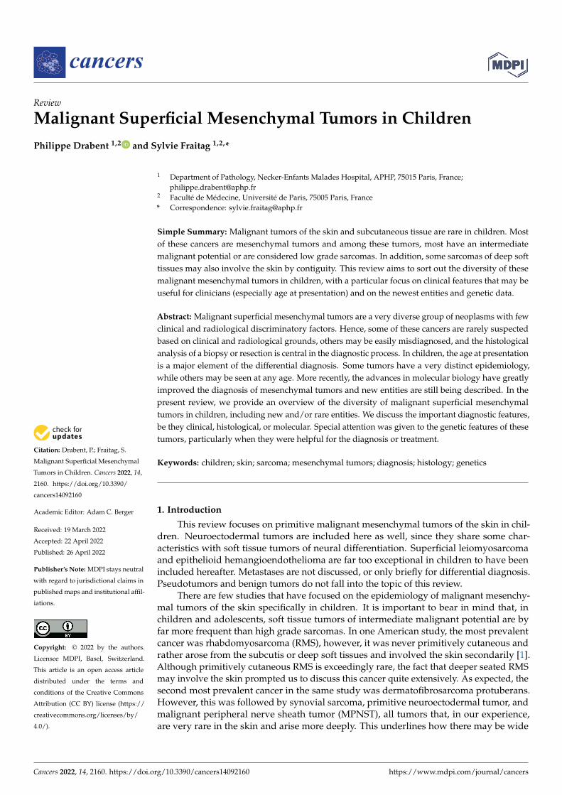
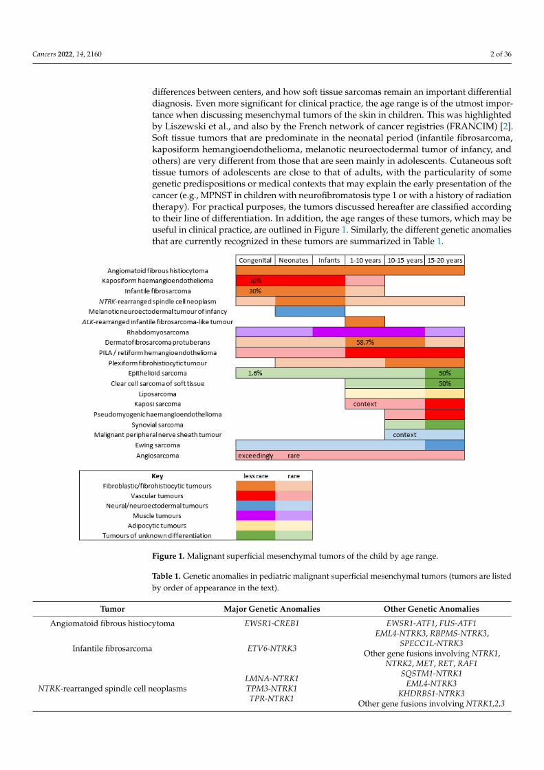
differences between centers, and how soft tissue sarcomas remain an important differentialdiagnosis. Even more significant for clinical practice, the age range is of the utmost impor-tance when discussing mesenchymal tumors of the skin in children. This was highlightedby Liszewski et al., and also by the French network of cancer registries (FRANCIM) [2].Soft tissue tumors that are predominate in the neonatal period (infantile fibrosarcoma,kaposiform hemangioendothelioma, melanotic neuroectodermal tumor of infancy, andothers) are very different from those that are seen mainly in adolescents. Cutaneous softtissue tumors of adolescents are close to that of adults, with the particularity of somegenetic predispositions or medical contexts that may explain the early presentation of thecancer (e.g., MPNST in children with neurofibromatosis type 1 or with a history of radiationtherapy). For practical purposes, the tumors discussed hereafter are classified accordingto their line of differentiation. In addition, the age ranges of these tumors, which may beuseful in clinical practice, are outlined in Figure 1. Similarly, the different genetic anomaliesthat are currently recognized in these tumors are summarized in Table 1.
Cancers 2022, 14, x FOR PEER REVIEW 2 of 39
wide differences between centers, and how soft tissue sarcomas remain an important dif-
ferential diagnosis. Even more significant for clinical practice, the age range is of the ut-
most importance when discussing mesenchymal tumors of the skin in children. This was
highlighted by Liszewski et al., and also by the French network of cancer registries
(FRANCIM) [2]. Soft tissue tumors that are predominate in the neonatal period (infantile
fibrosarcoma, kaposiform hemangioendothelioma, melanotic neuroectodermal tumor of
infancy, and others) are very different from those that are seen mainly in adolescents. Cu-
taneous soft tissue tumors of adolescents are close to that of adults, with the particularity
of some genetic predispositions or medical contexts that may explain the early presenta-
tion of the cancer (e.g., MPNST in children with neurofibromatosis type 1 or with a history
of radiation therapy). For practical purposes, the tumors discussed hereafter are classified
according to their line of differentiation. In addition, the age ranges of these tumors, which
may be useful in clinical practice, are outlined in Figure 1. Similarly, the different genetic
anomalies that are currently recognized in these tumors are summarized in Table 1.
Figure 1. Malignant superficial mesenchymal tumors of the child by age range.
Figure 1. Malignant superficial mesenchymal tumors of the child by age range.
Table 1. Genetic anomalies in pediatric malignant superficial mesenchymal tumors (tumors are listedby order of appearance in the text).
Tumor Major Genetic Anomalies Other Genetic Anomalies
Angiomatoid fibrous histiocytoma EWSR1-CREB1 EWSR1-ATF1, FUS-ATF1
Infantile fibrosarcoma ETV6-NTRK3
EML4-NTRK3, RBPMS-NTRK3,SPECC1L-NTRK3
Other gene fusions involving NTRK1,NTRK2, MET, RET, RAF1
NTRK-rearranged spindle cell neoplasmsLMNA-NTRK1TPM3-NTRK1TPR-NTRK1
SQSTM1-NTRK1EML4-NTRK3
KHDRBS1-NTRK3Other gene fusions involving NTRK1,2,3
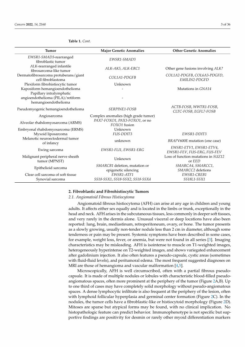
Cancers 2022, 14, 2160 3 of 36
Table 1. Cont.
Tumor Major Genetic Anomalies Other Genetic Anomalies
EWSR1-SMAD3-rearrangedfibroblastic tumor EWSR1-SMAD3
ALK-rearranged infantilefibrosarcoma-like tumor ALK-AK5, ALK-ERC1 Other gene fusions involving ALK?
Dermatofibrosarcoma protuberans/giantcell fibroblastoma COL1A1-PDGFB COL1A2-PDGFB, COL6A3-PDGFD,
EMILIN2-PDGFDPlexiform fibrohistiocytic tumor Unknown
Kaposiform hemangioendothelioma - Mutations in GNA14Papillary intralymphatic
angioendothelioma (PILA)/retiformhemangioendothelioma
-
Pseudomyogenic hemangioendothelioma SERPINE1-FOSB ACTB-FOSB, WWTR1-FOSB,CLTC-FOSB, EGFL7-FOSB
Angiosarcoma Complex anomalies (high grade tumor)
Alveolar rhabdomyosarcoma (ARMS) PAX7-FOXO1, PAX3-FOXO1, or noFOXO1 fusion
Embryonal rhabdomyosarcoma (ERMS) UnknownMyxoid liposarcoma FUS-DDIT3 EWSR1-DDIT3
Melanotic neuroectodermal tumorof infancy unknown BRAFV600E mutation (one case)
Ewing sarcoma EWSR1-FLI1, EWSR1-ERG EWSR1-ETV1, EWSR1-ETV4,EWSR1-FEV, FUS-ERG, FUS-FEV
Malignant peripheral nerve sheathtumor (MPNST) Unknown Loss of function mutations in SUZ12
or EED
Epithelioid sarcoma SMARCB1 deletion, mutation orepigenetic silencing
SMARCA4, SMARCC1,SMARCC2 deletions
Clear cell sarcoma of soft tissue EWSR1-ATF1 EWSR1-CREB1Synovial sarcoma SS18-SSX1, SS18-SSX2, SS18-SSX4 SS18L1-SSX1
2. Fibroblastic and Fibrohistiocytic Tumors2.1. Angiomatoid Fibrous Histiocytoma
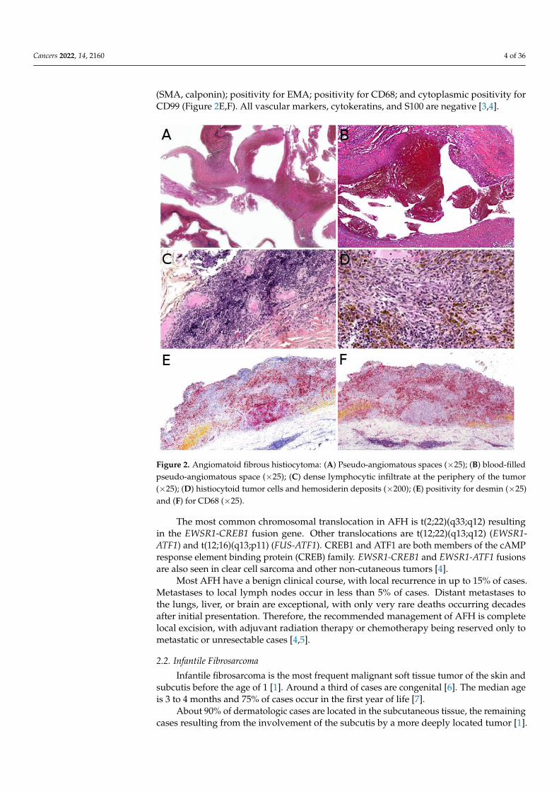
Angiomatoid fibrous histiocytoma (AFH) can arise at any age in children and youngadults. It affects either sex equally and is located in the limbs or trunk, exceptionally in thehead and neck. AFH arises in the subcutaneous tissues, less commonly in deeper soft tissues,and very rarely in the dermis alone. Unusual visceral or deep locations have also beenreported: lung, brain, mediastinum, retroperitoneum, ovary, or bone. The tumor presentsas a slowly growing, usually non-tender nodule less than 2 cm in diameter, although sometenderness or pain may be present. Systemic symptoms have been described in some cases,for example, weight loss, fever, or anemia, but were not found in all series [3]. Imagingcharacteristics may be misleading. AFH is isointense to muscle on T1-weighted images,heterogeneously hyperintense on T2-weighted images, and shows variegated enhancementafter gadolinium injection. It also often features a pseudo-capsule, cystic areas (sometimeswith fluid-fluid levels), and peritumoral edema. The most frequent suggested diagnoses onMRI are those of hemangioma and vascular malformation [4,5].
Microscopically, AFH is well circumscribed, often with a partial fibrous pseudo-capsule. It is made of multiple nodules or lobules with characteristic blood-filled pseudo-angiomatous spaces, often more prominent at the periphery of the tumor (Figure 2A,B). Upto one third of cases may have completely solid morphology without pseudo-angiomatousspaces. A dense lymphocytic infiltrate is also frequent at the periphery of the lesion, oftenwith lymphoid follicular hyperplasia and germinal center formation (Figure 2C). In thenodules, the tumor cells have a fibroblastic-like or histiocytoid morphology (Figure 2D).Mitoses are sparse but atypical forms may be found, with no clinical implication. Nohistopathologic feature can predict behavior. Immunophenotype is not specific but sup-portive findings are positivity for desmin or rarely other myoid differentiation markers
Cancers 2022, 14, 2160 4 of 36
(SMA, calponin); positivity for EMA; positivity for CD68; and cytoplasmic positivity forCD99 (Figure 2E,F). All vascular markers, cytokeratins, and S100 are negative [3,4].
Cancers 2022, 14, x FOR PEER REVIEW 5 of 39
Figure 2. Angiomatoid fibrous histiocytoma: (A) Pseudo-angiomatous spaces (×25); (B) blood-filled
pseudo-angiomatous space (×25); (C) dense lymphocytic infiltrate at the periphery of the tumor
(×25); (D) histiocytoid tumor cells and hemosiderin deposits (×200); (E) positivity for desmin (×25)
and (F) for CD68 (×25).
2.2. Infantile Fibrosarcoma
Infantile fibrosarcoma is the most frequent malignant soft tissue tumor of the skin
and subcutis before the age of 1 [1]. Around a third of cases are congenital [6]. The median
age is 3 to 4 months and 75% of cases occur in the first year of life [7].
About 90% of dermatologic cases are located in the subcutaneous tissue, the remain-
ing cases resulting from the involvement of the subcutis by a more deeply located tumor
[1]. There is a slight predilection for the lower extremities but all locations are seen [1,6].
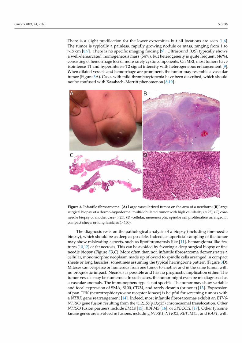
The tumor is typically a painless, rapidly growing nodule or mass, ranging from 1 to >15
cm [8,9]. There is no specific imaging finding [9]. Ultrasound (US) typically shows a well-
demarcated, homogeneous mass (54%), but heterogeneity is quite frequent (46%), consist-
ing of hemorrhage foci or more rarely cystic components. On MRI, most tumors have iso-
intense T1 and hyperintense T2 signal intensity with heterogeneous enhancement [9].
When dilated vessels and hemorrhage are prominent, the tumor may resemble a vascular
tumor (Figure 3A). Cases with mild thrombocytopenia have been described, which should
not be confused with Kasabach–Merritt phenomenon [8,10].
The diagnosis rests on the pathological analysis of a biopsy (including fine-needle
biopsy), which should be as deep as possible. Indeed, a superficial sampling of the tumor
may show misleading aspects, such as lipofibromatosis-like [11], hemangioma-like fea-
tures [10,12] or fat necrosis. This can be avoided by favoring a deep surgical biopsy or fine
needle biopsy (Figure 3B,C). More often than not, infantile fibrosarcoma demonstrates a
cellular, monomorphic neoplasm made up of ovoid to spindle cells arranged in compact
Figure 2. Angiomatoid fibrous histiocytoma: (A) Pseudo-angiomatous spaces (×25); (B) blood-filledpseudo-angiomatous space (×25); (C) dense lymphocytic infiltrate at the periphery of the tumor(×25); (D) histiocytoid tumor cells and hemosiderin deposits (×200); (E) positivity for desmin (×25)and (F) for CD68 (×25).
The most common chromosomal translocation in AFH is t(2;22)(q33;q12) resultingin the EWSR1-CREB1 fusion gene. Other translocations are t(12;22)(q13;q12) (EWSR1-ATF1) and t(12;16)(q13;p11) (FUS-ATF1). CREB1 and ATF1 are both members of the cAMPresponse element binding protein (CREB) family. EWSR1-CREB1 and EWSR1-ATF1 fusionsare also seen in clear cell sarcoma and other non-cutaneous tumors [4].
Most AFH have a benign clinical course, with local recurrence in up to 15% of cases.Metastases to local lymph nodes occur in less than 5% of cases. Distant metastases tothe lungs, liver, or brain are exceptional, with only very rare deaths occurring decadesafter initial presentation. Therefore, the recommended management of AFH is completelocal excision, with adjuvant radiation therapy or chemotherapy being reserved only tometastatic or unresectable cases [4,5].
2.2. Infantile Fibrosarcoma
Infantile fibrosarcoma is the most frequent malignant soft tissue tumor of the skin andsubcutis before the age of 1 [1]. Around a third of cases are congenital [6]. The median ageis 3 to 4 months and 75% of cases occur in the first year of life [7].
About 90% of dermatologic cases are located in the subcutaneous tissue, the remainingcases resulting from the involvement of the subcutis by a more deeply located tumor [1].
Cancers 2022, 14, 2160 5 of 36
There is a slight predilection for the lower extremities but all locations are seen [1,6].The tumor is typically a painless, rapidly growing nodule or mass, ranging from 1 to>15 cm [8,9]. There is no specific imaging finding [9]. Ultrasound (US) typically showsa well-demarcated, homogeneous mass (54%), but heterogeneity is quite frequent (46%),consisting of hemorrhage foci or more rarely cystic components. On MRI, most tumors haveisointense T1 and hyperintense T2 signal intensity with heterogeneous enhancement [9].When dilated vessels and hemorrhage are prominent, the tumor may resemble a vasculartumor (Figure 3A). Cases with mild thrombocytopenia have been described, which shouldnot be confused with Kasabach–Merritt phenomenon [8,10].
Cancers 2022, 14, x FOR PEER REVIEW 7 of 39
Figure 3. Infantile fibrosarcoma: (A) Large vascularized tumor on the arm of a newborn; (B) large
surgical biopsy of a dermo-hypodermal multi-lobulated tumor with high cellularity (×25); (C) core-
needle biopsy of another case (×25); (D) cellular, monomorphic spindle cell proliferation arranged
in compact sheets or long fascicles (×100).
2.3. NTRK-Rearranged Spindle Cell Neoplasms
This group of tumors is emerging as an entity based on molecular grounds. More
than 55% of pediatric cases occur in the first year of life [16,20]. Some cases are congenital
[33]. However, all ages can be affected [21,33,34].
Most NTRK-rearranged spindle cell neoplasms occur in the superficial soft tissues of
the extremities, other locations being the trunk and scalp. They present as a subcutaneous
nodule or mass ranging from 1 to 6 cm in greatest dimension [35]. Imaging characteristics
of these tumors have never been studied; however, it is likely that they overlap with in-
fantile fibrosarcoma, lipofibromatosis, and malignant peripheral nerve sheath tumor
(MPNST), and thus, are not specific.
Indeed, NTRK-rearranged spindle cell neoplasms include a wide variety of morphol-
ogies with tumors described as infantile fibrosarcoma-like, inflammatory myofibroblastic
tumor-like (IMT-like), lipofibromatosis-like neural tumor (LPF-NT), and MPNST-like
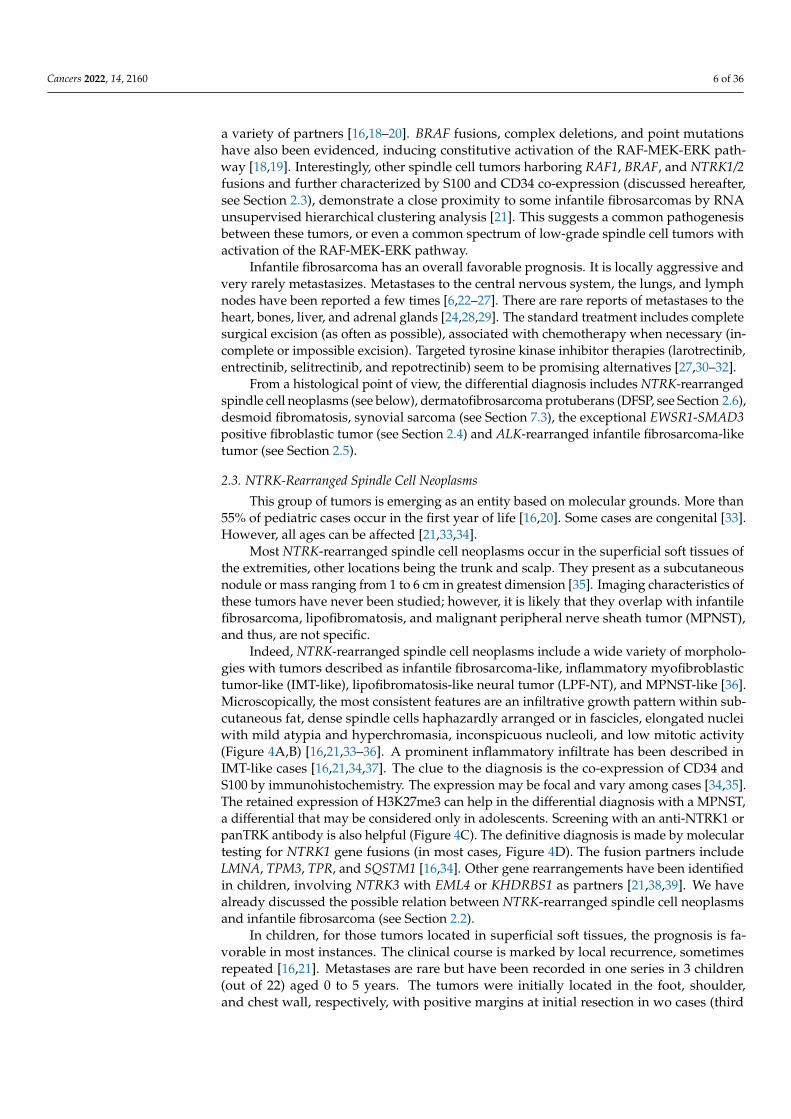
[36]. Microscopically, the most consistent features are an infiltrative growth pattern within
subcutaneous fat, dense spindle cells haphazardly arranged or in fascicles, elongated nu-
clei with mild atypia and hyperchromasia, inconspicuous nucleoli, and low mitotic activ-
ity (Figure 4A,B) [16,21,33–36]. A prominent inflammatory infiltrate has been described in
IMT-like cases [16,21,34,37]. The clue to the diagnosis is the co-expression of CD34 and
S100 by immunohistochemistry. The expression may be focal and vary among cases
[34,35]. The retained expression of H3K27me3 can help in the differential diagnosis with
a MPNST, a differential that may be considered only in adolescents. Screening with an
anti-NTRK1 or panTRK antibody is also helpful (Figure 4C). The definitive diagnosis is
made by molecular testing for NTRK1 gene fusions (in most cases, Figure 4D). The fusion
partners include LMNA, TPM3, TPR, and SQSTM1 [16,34]. Other gene rearrangements
have been identified in children, involving NTRK3 with EML4 or KHDRBS1 as partners
[21,38,39]. We have already discussed the possible relation between NTRK-rearranged
spindle cell neoplasms and infantile fibrosarcoma (see Section 2.2).
Figure 3. Infantile fibrosarcoma: (A) Large vascularized tumor on the arm of a newborn; (B) largesurgical biopsy of a dermo-hypodermal multi-lobulated tumor with high cellularity (×25); (C) core-needle biopsy of another case (×25); (D) cellular, monomorphic spindle cell proliferation arranged incompact sheets or long fascicles (×100).
The diagnosis rests on the pathological analysis of a biopsy (including fine-needlebiopsy), which should be as deep as possible. Indeed, a superficial sampling of the tumormay show misleading aspects, such as lipofibromatosis-like [11], hemangioma-like fea-tures [10,12] or fat necrosis. This can be avoided by favoring a deep surgical biopsy or fineneedle biopsy (Figure 3B,C). More often than not, infantile fibrosarcoma demonstrates acellular, monomorphic neoplasm made up of ovoid to spindle cells arranged in compactsheets or long fascicles, sometimes assuming the typical herringbone pattern (Figure 3D).Mitoses can be sparse or numerous from one tumor to another and in the same tumor, withno prognostic impact. Necrosis is possible and has no prognostic implication either. Thetumor vessels may be numerous. In such cases, the tumor might even be misdiagnosed asa vascular anomaly. The immunophenotype is not specific. The tumor may show variableand focal expression of SMA, S100, CD34, and rarely desmin (or none) [13]. Expressionof pan-TRK (neurotrophic tyrosine receptor kinase) is helpful for screening tumors witha NTRK gene rearrangement [14]. Indeed, most infantile fibrosarcomas exhibit an ETV6-NTRK3 gene fusion resulting from the t(12;15)(p13;q25) chromosomal translocation. OtherNTRK3 fusion partners include EML4 [15], RBPMS [16], or SPECC1L [17]. Other tyrosinekinase genes are involved in fusions, including NTRK1, NTRK2, RET, MET, and RAF1, with
Cancers 2022, 14, 2160 6 of 36
a variety of partners [16,18–20]. BRAF fusions, complex deletions, and point mutationshave also been evidenced, inducing constitutive activation of the RAF-MEK-ERK path-way [18,19]. Interestingly, other spindle cell tumors harboring RAF1, BRAF, and NTRK1/2fusions and further characterized by S100 and CD34 co-expression (discussed hereafter,see Section 2.3), demonstrate a close proximity to some infantile fibrosarcomas by RNAunsupervised hierarchical clustering analysis [21]. This suggests a common pathogenesisbetween these tumors, or even a common spectrum of low-grade spindle cell tumors withactivation of the RAF-MEK-ERK pathway.
Infantile fibrosarcoma has an overall favorable prognosis. It is locally aggressive andvery rarely metastasizes. Metastases to the central nervous system, the lungs, and lymphnodes have been reported a few times [6,22–27]. There are rare reports of metastases to theheart, bones, liver, and adrenal glands [24,28,29]. The standard treatment includes completesurgical excision (as often as possible), associated with chemotherapy when necessary (in-complete or impossible excision). Targeted tyrosine kinase inhibitor therapies (larotrectinib,entrectinib, selitrectinib, and repotrectinib) seem to be promising alternatives [27,30–32].
From a histological point of view, the differential diagnosis includes NTRK-rearrangedspindle cell neoplasms (see below), dermatofibrosarcoma protuberans (DFSP, see Section 2.6),desmoid fibromatosis, synovial sarcoma (see Section 7.3), the exceptional EWSR1-SMAD3positive fibroblastic tumor (see Section 2.4) and ALK-rearranged infantile fibrosarcoma-liketumor (see Section 2.5).
2.3. NTRK-Rearranged Spindle Cell Neoplasms
This group of tumors is emerging as an entity based on molecular grounds. More than55% of pediatric cases occur in the first year of life [16,20]. Some cases are congenital [33].However, all ages can be affected [21,33,34].
Most NTRK-rearranged spindle cell neoplasms occur in the superficial soft tissues ofthe extremities, other locations being the trunk and scalp. They present as a subcutaneousnodule or mass ranging from 1 to 6 cm in greatest dimension [35]. Imaging characteristics ofthese tumors have never been studied; however, it is likely that they overlap with infantilefibrosarcoma, lipofibromatosis, and malignant peripheral nerve sheath tumor (MPNST),and thus, are not specific.
Indeed, NTRK-rearranged spindle cell neoplasms include a wide variety of morpholo-gies with tumors described as infantile fibrosarcoma-like, inflammatory myofibroblastictumor-like (IMT-like), lipofibromatosis-like neural tumor (LPF-NT), and MPNST-like [36].Microscopically, the most consistent features are an infiltrative growth pattern within sub-cutaneous fat, dense spindle cells haphazardly arranged or in fascicles, elongated nucleiwith mild atypia and hyperchromasia, inconspicuous nucleoli, and low mitotic activity(Figure 4A,B) [16,21,33–36]. A prominent inflammatory infiltrate has been described inIMT-like cases [16,21,34,37]. The clue to the diagnosis is the co-expression of CD34 andS100 by immunohistochemistry. The expression may be focal and vary among cases [34,35].The retained expression of H3K27me3 can help in the differential diagnosis with a MPNST,a differential that may be considered only in adolescents. Screening with an anti-NTRK1 orpanTRK antibody is also helpful (Figure 4C). The definitive diagnosis is made by moleculartesting for NTRK1 gene fusions (in most cases, Figure 4D). The fusion partners includeLMNA, TPM3, TPR, and SQSTM1 [16,34]. Other gene rearrangements have been identifiedin children, involving NTRK3 with EML4 or KHDRBS1 as partners [21,38,39]. We havealready discussed the possible relation between NTRK-rearranged spindle cell neoplasmsand infantile fibrosarcoma (see Section 2.2).
In children, for those tumors located in superficial soft tissues, the prognosis is fa-vorable in most instances. The clinical course is marked by local recurrence, sometimesrepeated [16,21]. Metastases are rare but have been recorded in one series in 3 children(out of 22) aged 0 to 5 years. The tumors were initially located in the foot, shoulder,and chest wall, respectively, with positive margins at initial resection in wo cases (third
Cancers 2022, 14, 2160 7 of 36
case unknown). Metastases were to the lung in all cases. Two children were alive afterchemotherapy and one died of disease, in spite of the chemotherapy [16].
Cancers 2022, 14, x FOR PEER REVIEW 8 of 39
Figure 4. NTRK-rearranged spindle cell neoplasm: (A) Infiltrative growth pattern within the subcu-
taneous fat, with spindle cells haphazardly arranged or in fascicles (×100); (B) mild atypia and hy-
perchromasia (×100); (C) diffuse positivity for pan-TRK (×25); (D) fusion signals involving NTRK on
FISH.
In children, for those tumors located in superficial soft tissues, the prognosis is favor-
able in most instances. The clinical course is marked by local recurrence, sometimes re-
peated [16,21]. Metastases are rare but have been recorded in one series in 3 children (out
of 22) aged 0 to 5 years. The tumors were initially located in the foot, shoulder, and chest
wall, respectively, with positive margins at initial resection in wo cases (third case un-
known). Metastases were to the lung in all cases. Two children were alive after chemo-
therapy and one died of disease, in spite of the chemotherapy [16].
The treatment is, first and foremost, surgical. When excision cannot be complete,
chemotherapy may be beneficial [16]. Similar to infantile fibrosarcoma, anti-TRK tyrosine
kinase inhibitors (larotrectinib, entrectinib, and more recently selitrectinib and repotrec-
tinib) will probably become a standard in the treatment of these tumors.
2.4. EWSR1-SMAD3-Rearranged Fibroblastic Tumor
This tumor has been described mainly in young adults, with only two pediatric cases
to date. One was a congenital nodule of the foot in a boy [40], the second was a nodule of
the hand in a 5-year-old girl, initially diagnosed as “unusual lipofibromatosis” [41]. More
cases are needed to determine the epidemiology, clinical features, microscopy, and behav-
ior of this tumor in children.
The tumor seems to have a predilection for acral areas and can develop in the dermis
or subcutis. It has an infiltrative growth pattern. In the light of the adult cases, it seems
that the tumor typically has a biphasic appearance with a higher cellular density at the
periphery and central hyalinization. The spindle cells are arranged in fascicles and contain
elongated nuclei with no atypia. Calcifications may be seen in the hyalinized areas. The
immunophenotype is characteristic, with constant expression of ERG and negativity for
fibroblastic, muscular, and melanocytic markers (CD34, SMA, desmin, caldesmon, S100,
and SOX10). The detection of the EWSR1-SMAD3 (Ewing sarcoma breakpoint region 1–
SMA- and MAD-related protein 3) fusion gene is necessary to ensure diagnostic accuracy.
Figure 4. NTRK-rearranged spindle cell neoplasm: (A) Infiltrative growth pattern within the sub-cutaneous fat, with spindle cells haphazardly arranged or in fascicles (×100); (B) mild atypia andhyperchromasia (×100); (C) diffuse positivity for pan-TRK (×25); (D) fusion signals involving NTRKon FISH.
The treatment is, first and foremost, surgical. When excision cannot be complete,chemotherapy may be beneficial [16]. Similar to infantile fibrosarcoma, anti-TRK tyrosinekinase inhibitors (larotrectinib, entrectinib, and more recently selitrectinib and repotrectinib)will probably become a standard in the treatment of these tumors.
2.4. EWSR1-SMAD3-Rearranged Fibroblastic Tumor
This tumor has been described mainly in young adults, with only two pediatric casesto date. One was a congenital nodule of the foot in a boy [40], the second was a nodule ofthe hand in a 5-year-old girl, initially diagnosed as “unusual lipofibromatosis” [41]. Morecases are needed to determine the epidemiology, clinical features, microscopy, and behaviorof this tumor in children.
The tumor seems to have a predilection for acral areas and can develop in the dermisor subcutis. It has an infiltrative growth pattern. In the light of the adult cases, it seemsthat the tumor typically has a biphasic appearance with a higher cellular density at theperiphery and central hyalinization. The spindle cells are arranged in fascicles and containelongated nuclei with no atypia. Calcifications may be seen in the hyalinized areas. Theimmunophenotype is characteristic, with constant expression of ERG and negativity forfibroblastic, muscular, and melanocytic markers (CD34, SMA, desmin, caldesmon, S100,and SOX10). The detection of the EWSR1-SMAD3 (Ewing sarcoma breakpoint region1–SMA- and MAD-related protein 3) fusion gene is necessary to ensure diagnostic accuracy.
2.5. ALK-Rearranged Infantile Fibrosarcoma-like Tumor
Recently, a new ALK-rearranged spindle cell tumor has been described in a cohort offour children aged 2 to 10 years. Of these four cases, two cases arose in the superficial soft
Cancers 2022, 14, 2160 8 of 36
tissues of the scalp and the hand, respectively. The tumors were both clinically diagnosedas vascular tumors or malformations of about 4 to 6 cm in diameter, however, the histologicappearance was that of a low-grade spindle-cell sarcoma, reminiscent of infantile fibrosar-coma, with some intermixed round cells in one case. The immunoprofile showed negativityfor CD34, S100, and SMA, but diffuse positivity for ALK (anaplastic lymphoma kinase).A fusion transcript involving ALK was found, with AK5 and ERC1 as fusion partners.None of the two tumors recurred and there was no metastasis. However, of the two othercases in this study, which arose in the kidney, one recurred and metastasized to the liverand lung [42]. This child is currently in remission after nephrectomy and chemotherapybased on ifosfamide, doxorubicin, and ceritinib (ALK inhibitor). Therefore, it is possiblethat this new mesenchymal tumor with ALK rearrangement is of intermediate malignantpotential. More cases are needed to better understand the prognosis and evolution ofthis tumor, especially to determine whether cases arising in superficial soft tissues alwaysevolve in a benign manner or might metastasize in the same way that has been describedin kidney tumors.
2.6. Dermatofibrosarcoma Protuberans/Giant Cell Fibroblastoma
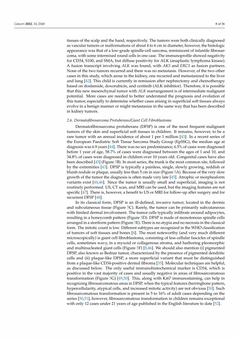
Dermatofibrosarcoma protuberans (DFSP) is one of the most frequent malignanttumors of the skin and superficial soft tissues in children. It remains, however, to be arare tumor with an annual incidence of about 1 per 1 million [43]. In a recent series ofthe European Paediatric Soft Tissue Sarcoma Study Group (EpSSG), the median age atdiagnosis was 6.9 years [44]. There was no sex predominance; 6.5% of cases were diagnosedbefore 1 year of age, 58.7% of cases were diagnosed between the ages of 1 and 10, and34.8% of cases were diagnosed in children over 10 years old. Congenital cases have alsobeen described [45] (Figure 5B). In most series, the trunk is the most common site, followedby the extremities [43]. DFSP is typically a painless, single, slowly growing, reddish orbluish nodule or plaque, usually less than 5 cm in size (Figure 5A). Because of the very slowgrowth of the tumor the diagnosis is often made very late [45]. Atrophic or morpheaformvariants exist [44,46]. Since the tumor is usually small and superficial, imaging is notroutinely performed. US, CT scan, and MRI can be used, but the imaging features are notspecific [47]. There is, however, a benefit to US or MRI for follow-up after surgery and forrecurrent DFSP [48].
In its classical form, DFSP is an ill-defined, invasive tumor, located in the dermisand subcutaneous tissue (Figure 5C). Rarely, the tumor can be primarily subcutaneouswith limited dermal involvement. The tumor cells typically infiltrate around adipocytes,resulting in a honeycomb pattern (Figure 5D). DFSP is made of monotonous spindle cellsarranged in a storiform pattern (Figure 5E). There is no atypia and no necrosis in the classicalform. The mitotic count is low. Different subtypes are recognized in the WHO classificationof tumors of soft tissues and bones [8]. The most noteworthy (and very much differentmicroscopically) is giant cell fibroblastoma, consisting of less cellular fascicles of spindlecells, sometimes wavy, in a myxoid or collagenous stroma, and harboring pleomorphicand multinucleated giant cells (Figure 5F) [8,46]. We should also mention (i) pigmentedDFSP, also known as Bednar tumor, characterized by the presence of pigmented dendriticcells and (ii) plaque-like DFSP, a more superficial variant that must be distinguishedfrom a plaque-like CD34-positive dermal fibroma [35]. Molecular techniques are helpful,as discussed below. The only useful immunohistochemical marker is CD34, which ispositive in the vast majority of cases and usually negative in areas of fibrosarcomatoustransformation (Figure 5G) [49,50]. This, along with Ki67 immunostaining, can help inrecognizing fibrosarcomatous areas in DFSP, when the typical features (herringbone pattern,hypercellularity, atypical cells, and increased mitotic activity) are not obvious [50]. Suchfibrosarcomatous transformation is present in 5 to 16% of adult cases depending on theseries [50,51], however, fibrosarcomatous transformation in children remains exceptionalwith only 12 cases under 21 years of age published in the English literature to date [52].
Cancers 2022, 14, 2160 9 of 36
Cancers 2022, 14, x FOR PEER REVIEW 10 of 39
cellularity, atypical cells, and increased mitotic activity) are not obvious [50]. Such fibro-
sarcomatous transformation is present in 5 to 16% of adult cases depending on the series
[50,51], however, fibrosarcomatous transformation in children remains exceptional with
only 12 cases under 21 years of age published in the English literature to date [52].
Figure 5. Dermatofibrosarcoma protuberans: (A) Plaque-like lesion on the flank of a teenager; (B)
irregular plaque-like lesion on the buttock of neonate (congenital lesion); (C) dense cellular prolif-
eration infiltrating into the subcutaneous fat (×25); (D) infiltration of the subcutaneous adipose tis-
sue (×100); (E) spindle cells in a typical storiform pattern (×200); (F) giant cell fibroblastoma: less
cellular tumor with giant cells and a loose stroma (×100); (G) diffuse strong CD34 positivity (×100);
(H) COL1A1-PDGFB fusion on FISH.
Figure 5. Dermatofibrosarcoma protuberans: (A) Plaque-like lesion on the flank of a teenager;(B) irregular plaque-like lesion on the buttock of neonate (congenital lesion); (C) dense cellularproliferation infiltrating into the subcutaneous fat (×25); (D) infiltration of the subcutaneous adiposetissue (×100); (E) spindle cells in a typical storiform pattern (×200); (F) giant cell fibroblastoma: lesscellular tumor with giant cells and a loose stroma (×100); (G) diffuse strong CD34 positivity (×100);(H) COL1A1-PDGFB fusion on FISH.
Cancers 2022, 14, 2160 10 of 36
The hallmark of DFSP in children is the presence of a balanced or unbalanced translo-cation involving the long arms of chromosomes 17 and 22. The most frequent is thebalanced t(17;22) (q21.3;q13.1) translocation, resulting in a COL1A1-PDGFB fusion gene(Figure 5H). The unbalanced der(22)t(17;22) is also found in a subset of patients, withpossible co-existing balanced and unbalanced translocations in different subclones of thesame tumor. Supernumerary ring chromosomes harboring the same t(17;22) translocationseem to be present in adults but not in children [53,54]. This kind of translocations ispresent in about 90% of cases. In the remaining cases, COL1A2-PDGFB, COL6A3-PDGFD,and EMILIN2-PDGFD fusion transcripts have been identified [55]. A case of DFSP-liketumor with COL1A1 copy number gain has also been described. In this case, there was nott(17;22) translocation, however, the morphology and immunophenotype were similar tothat of DFSP, raising the possibility of a new molecular variant of DFSP [56].
DFSP treatments must achieve the goal of clear surgical margins, which is the only wayto avoid recurrences. For this reason, wide local excision is recommended, ideally by Mohsmicrographic surgery (MMS). The term “slow-Mohs micrographic surgery” (sMMS) hasbeen used for the FFPE technique (by contrast with immediate frozen section examination),which is a little slower but provides a better morphology and allows the pathologist touse CD34 immunostaining if necessary. For these reasons, the sMMS technique has beenpreferred by many teams, with the same results as MMS [57–60]. The surgical marginsshould be 10 to 11 mm with sMMS and 20 to 40 mm with conventional surgical techniques,or wider in cases of fibrosarcomatous transformation [61–63]. After complete resection,the recurrence rate is very low, ensuring a good prognosis of DFSP in children. When acomplete resection is impossible, treatment options are limited. DFSP does not respond toconventional chemotherapy and responds moderately to radiotherapy. Targeted therapies(imatinib, sunitinib, sorafenib, pazopanib, everolimus, among others) might be interestingoptions and the reader is referred to relevant publications on this topic [55,64–70].
2.7. Plexiform Fibrohistiocytic Tumor
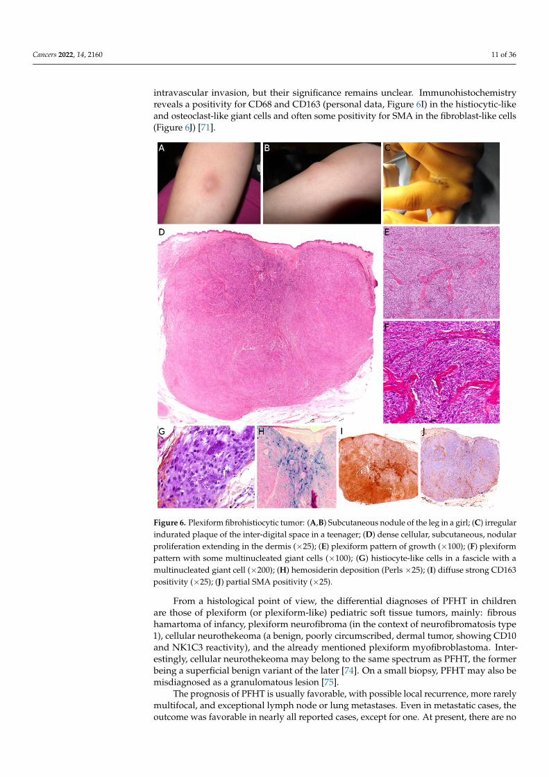
Plexiform fibrohistiocytic tumor (PFHT) is a rare neoplasm of intermediate malignantpotential with a predilection for children and young adults. The mean age is around 15but the age range is very wide (from the first to the eighth decade). Originally, a femalepredominance was described, but this was not obvious in the literature [71]. The mostfrequent locations are the upper limbs (50%), followed by the lower limbs, and more rarelythe trunk and head and neck region. PFHT presents as a slowly growing, painless nodule,usually below 3 cm in greater diameter (Figure 6A,B). Some cases may present as a flatindurated plaque (Figure 6C). Imaging of PFHT is best achieved using MRI and showseither a round mass or a plaque-like lesion, often with infiltrative growth. In about 75%of cases, the tumor is in contact with a bone or tendon. The lesion is typically iso- orhyperintense on T1-weighted imaging, and always hyperintense on T2-weighted imagingwith fat suppression. There is enhancement after gadolinium injection [72].
Histologically, PFHT is located in the subcutis and may extend in the dermis (Figure 6D).In most cases, the tumor is poorly circumscribed. Some cases of purely dermal lesions havebeen described and tend to be better delineated. It is composed of two components: onehistiocytic-like component and one fibroblast-like component. These components can bepresent in variable proportions, thus, defining, classically, three subtypes: fibrohistiocytic,fibroblastic, and mixed [71]. However, in a fibroblastic predominant PFHT, a fibrohis-tiocytic component should be carefully sought. Indeed, it has been recently stated thatpurely fibroblastic PFHT might not exist and could actually be reclassified as plexiformmyofibroblastoma, a recently described benign neoplasm [35,73]. The characteristic fea-tures of PFHT are a plexiform architecture made of nodules of histiocytic cells with blandcytology, frequently admixed with osteoclast-like giant cells, and intersecting bundles offibroblast-like spindle cells at the periphery of the nodules (Figure 6E–G). There is little tono pleomorphism and the mitotic activity is low. Iron deposition is common (Figure 6H).Some atypical cases have been reported, with pleomorphism, atypical mitoses, or even
Cancers 2022, 14, 2160 11 of 36
intravascular invasion, but their significance remains unclear. Immunohistochemistryreveals a positivity for CD68 and CD163 (personal data, Figure 6I) in the histiocytic-likeand osteoclast-like giant cells and often some positivity for SMA in the fibroblast-like cells(Figure 6J) [71].
Cancers 2022, 14, x FOR PEER REVIEW 12 of 39
Some atypical cases have been reported, with pleomorphism, atypical mitoses, or even
intravascular invasion, but their significance remains unclear. Immunohistochemistry re-
veals a positivity for CD68 and CD163 (personal data, Figure 6I) in the histiocytic-like and
osteoclast-like giant cells and often some positivity for SMA in the fibroblast-like cells
(Figure 6J) [71].
Figure 6. Plexiform fibrohistiocytic tumor: (A,B) Subcutaneous nodule of the leg in a girl; (C) irreg-
ular indurated plaque of the inter-digital space in a teenager; (D) dense cellular, subcutaneous, nod-
ular proliferation extending in the dermis (×25); (E) plexiform pattern of growth (×100); (F) plexiform
pattern with some multinucleated giant cells (×100); (G) histiocyte-like cells in a fascicle with a mul-
tinucleated giant cell (×200); (H) hemosiderin deposition (Perls ×25); (I) diffuse strong CD163 posi-
tivity (×25); (J) partial SMA positivity (×25).
From a histological point of view, the differential diagnoses of PFHT in children are
those of plexiform (or plexiform-like) pediatric soft tissue tumors, mainly: fibrous
hamartoma of infancy, plexiform neurofibroma (in the context of neurofibromatosis type
1), cellular neurothekeoma (a benign, poorly circumscribed, dermal tumor, showing CD10
and NK1C3 reactivity), and the already mentioned plexiform myofibroblastoma. Interest-
ingly, cellular neurothekeoma may belong to the same spectrum as PFHT, the former be-
ing a superficial benign variant of the later [74]. On a small biopsy, PFHT may also be
misdiagnosed as a granulomatous lesion [75].
The prognosis of PFHT is usually favorable, with possible local recurrence, more
rarely multifocal, and exceptional lymph node or lung metastases. Even in metastatic
Figure 6. Plexiform fibrohistiocytic tumor: (A,B) Subcutaneous nodule of the leg in a girl; (C) irregularindurated plaque of the inter-digital space in a teenager; (D) dense cellular, subcutaneous, nodularproliferation extending in the dermis (×25); (E) plexiform pattern of growth (×100); (F) plexiformpattern with some multinucleated giant cells (×100); (G) histiocyte-like cells in a fascicle with amultinucleated giant cell (×200); (H) hemosiderin deposition (Perls ×25); (I) diffuse strong CD163positivity (×25); (J) partial SMA positivity (×25).
From a histological point of view, the differential diagnoses of PFHT in childrenare those of plexiform (or plexiform-like) pediatric soft tissue tumors, mainly: fibroushamartoma of infancy, plexiform neurofibroma (in the context of neurofibromatosis type1), cellular neurothekeoma (a benign, poorly circumscribed, dermal tumor, showing CD10and NK1C3 reactivity), and the already mentioned plexiform myofibroblastoma. Inter-estingly, cellular neurothekeoma may belong to the same spectrum as PFHT, the formerbeing a superficial benign variant of the later [74]. On a small biopsy, PFHT may also bemisdiagnosed as a granulomatous lesion [75].
The prognosis of PFHT is usually favorable, with possible local recurrence, more rarelymultifocal, and exceptional lymph node or lung metastases. Even in metastatic cases, theoutcome was favorable in nearly all reported cases, except for one. At present, there are no
Cancers 2022, 14, 2160 12 of 36
known parameters, either histological, cytogenetic, or molecular, to predict the evolutionof the tumor. No genetic anomaly has been described so far [75]. Therefore, the treatmentof choice is the complete excision of the tumor.
3. Vascular Tumors3.1. Kaposiform Hemangioendothelioma
The incidence of kaposiform hemangioendothelioma (KHE) is probably under evalu-ated. It has been suggested that small asymptomatic or atypical KHE may be misdiagnosedas variants of other vascular tumors [76]. Although the exact incidence and prevalenceof this vascular tumor are not known, it is clear that there is a peak within the first yearof life. Around 50% of superficial cases are congenital [76]. It is admitted now that KHEand tufted angioma belong to the same spectrum and that they share the same biologyand probably the same pathogenesis [77,78]. Actually, some specialists argue that KHEmight only be a florid presentation of tufted angioma, and that the adverse clinical courseof this tumor is purely dependent on the occurrence of Kasabach–Merritt phenomenon(KMP) or vital organ compression. For this reason, KHE may well be benign, the morbidityand mortality being linked to secondary complications. We decided to include KHE in thisreview anyway, but this decision is debatable and it will not be discussed extensively.
KHE usually involves the deep dermis and subcutis, but deeper lesions are possible,with no cutaneous signs in about 12% of cases [76,78]. There is a slightly higher prevalencein the limbs, but all sites are possible. Most of the time, the tumor is unique, and can adoptmany clinical aspects: erythematous sometimes purple; papules, nodules or plaques oran indurated mass; with varying degrees of infiltration [78]. It is a big, rapidly growingtumor, 3 to 27 cm in greater dimension [79]. One of the most severe complications of KHE,also seen in tufted angioma, is the KMP, a life-threatening consumptive coagulopathy withsevere thrombocytopenia. It occurs in 42 to 71% of cases [80–82]. There is a higher frequencyof KMP in congenital, large KHE, especially above 8 cm [83]. In cases of KMP, the KHElesions appear purpuric, congestive, and painful [84]. Spontaneous hemorrhage is rare butany invasive procedure (biopsy, excision), trauma, or ulceration may lead to significantbleeding. Other complications of KHE include musculoskeletal disorders due to tumorinfiltration, lymphedema, and compression of vital structures (e.g., airway obstruction in aKHE of the neck) [78].
US reveals an ill-defined, heterogeneous lesion, most often hyperechoic [79]. On colorDoppler US, the tumor is hypervascular. MRI reveals a heterogeneous, hyperintense ap-pearance on T2-weighted images, with heterogeneous, generally intense enhancement [79].Overall, the imaging findings lack specificity and are characterized by a wide range of as-pects regarding the degree of infiltration, signal intensities, and enhancement patterns [79].
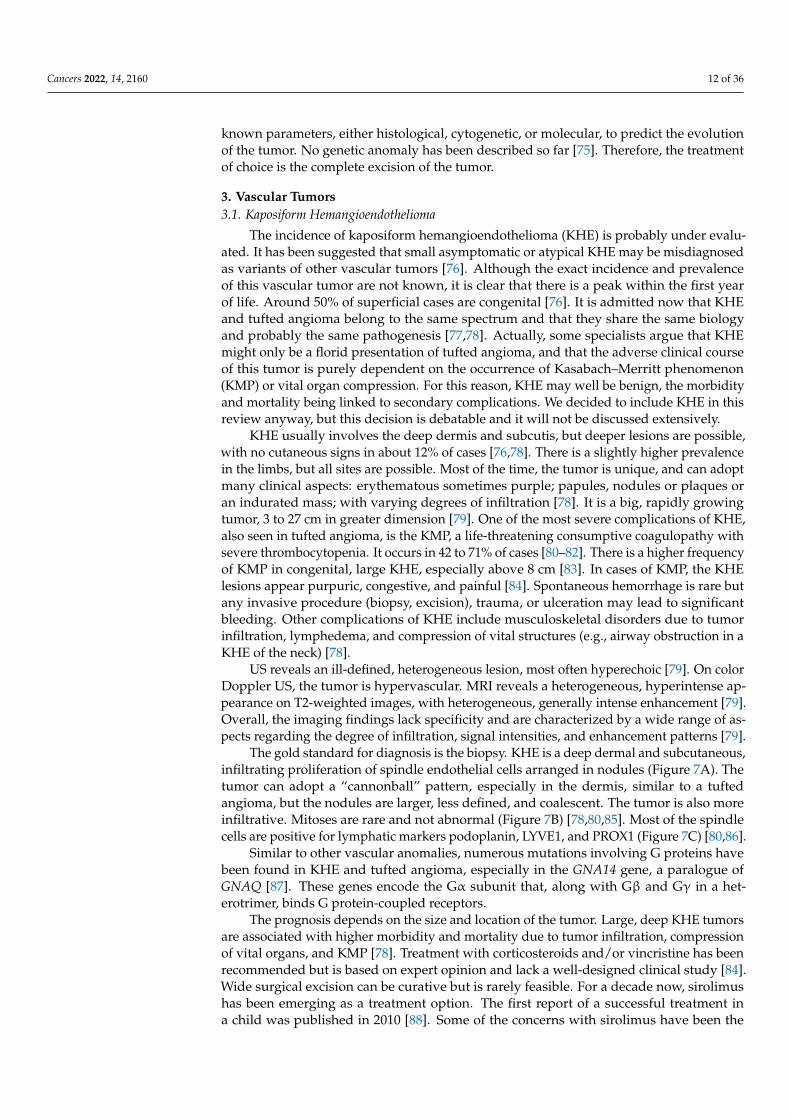
The gold standard for diagnosis is the biopsy. KHE is a deep dermal and subcutaneous,infiltrating proliferation of spindle endothelial cells arranged in nodules (Figure 7A). Thetumor can adopt a “cannonball” pattern, especially in the dermis, similar to a tuftedangioma, but the nodules are larger, less defined, and coalescent. The tumor is also moreinfiltrative. Mitoses are rare and not abnormal (Figure 7B) [78,80,85]. Most of the spindlecells are positive for lymphatic markers podoplanin, LYVE1, and PROX1 (Figure 7C) [80,86].
Similar to other vascular anomalies, numerous mutations involving G proteins havebeen found in KHE and tufted angioma, especially in the GNA14 gene, a paralogue ofGNAQ [87]. These genes encode the Gα subunit that, along with Gβ and Gγ in a het-erotrimer, binds G protein-coupled receptors.
The prognosis depends on the size and location of the tumor. Large, deep KHE tumorsare associated with higher morbidity and mortality due to tumor infiltration, compressionof vital organs, and KMP [78]. Treatment with corticosteroids and/or vincristine has beenrecommended but is based on expert opinion and lack a well-designed clinical study [84].Wide surgical excision can be curative but is rarely feasible. For a decade now, sirolimushas been emerging as a treatment option. The first report of a successful treatment ina child was published in 2010 [88]. Some of the concerns with sirolimus have been the
Cancers 2022, 14, 2160 13 of 36
occurrence of adverse events and the issue of the right dose in the pediatric population [89].Recently, recommendations for the initial dose in children were proposed. Due to sirolimusbeing metabolized by cytochrome P450 3A (CYP3A), the initial dose depends both onthe weight and the CYP3A5 genotype [90]. A recent prospective study of sirolimus forcomplicated vascular anomalies underlined the importance of closely monitoring possibleadverse events, while confirming the effectiveness of the treatment [91]. In this study, themost frequent adverse events (>20% of cases) were mucositis, upper respiratory infections,and nausea/vomiting.
Cancers 2022, 14, x FOR PEER REVIEW 14 of 39
Figure 7. Kaposiform hemangioendothelioma: (A) Deep dermal and subcutaneous, infiltrating pro-
liferation of spindle cells arranged in nodules (×25); (B) spindle endothelial cells with no mitoses
(×200); (C) positivity for the lymphatic marker podoplanin in the spindle cells (×200).
Similar to other vascular anomalies, numerous mutations involving G proteins have
been found in KHE and tufted angioma, especially in the GNA14 gene, a paralogue of
GNAQ [87]. These genes encode the Gα subunit that, along with Gβ and Gγ in a hetero-
trimer, binds G protein-coupled receptors.
The prognosis depends on the size and location of the tumor. Large, deep KHE tu-
mors are associated with higher morbidity and mortality due to tumor infiltration, com-
pression of vital organs, and KMP [78]. Treatment with corticosteroids and/or vincristine
has been recommended but is based on expert opinion and lack a well-designed clinical
study [84]. Wide surgical excision can be curative but is rarely feasible. For a decade now,
sirolimus has been emerging as a treatment option. The first report of a successful treat-
ment in a child was published in 2010 [88]. Some of the concerns with sirolimus have been
the occurrence of adverse events and the issue of the right dose in the pediatric population
[89]. Recently, recommendations for the initial dose in children were proposed. Due to
sirolimus being metabolized by cytochrome P450 3A (CYP3A), the initial dose depends
both on the weight and the CYP3A5 genotype [90]. A recent prospective study of sirolimus
for complicated vascular anomalies underlined the importance of closely monitoring pos-
sible adverse events, while confirming the effectiveness of the treatment [91]. In this study,
the most frequent adverse events (>20% of cases) were mucositis, upper respiratory infec-
tions, and nausea/vomiting.
3.2. Papillary Intralymphatic Angioendothelioma (PILA)/Retiform Hemangioendothelioma
Papillary intralymphatic angioendothelioma (PILA, also known as Dabska tumor)
and retiform hemangioendothelioma belong to the same spectrum. These two rare tumors
are lymphatic in origin. Cases with overlapping features of PILA and retiform hemangio-
endothelioma have been described, which is the reason why some authors have suggested
to include both tumors under the name hobnail hemangioendothelioma [92]. However, pure
forms of retiform hemangioendothelioma are very rare in children; PILA or mixed tumors
are much more frequent. PILA usually involves the dermis and subcutaneous tissues of
the head and neck in children and adolescents, whereas retiform hemangioendothelioma
Figure 7. Kaposiform hemangioendothelioma: (A) Deep dermal and subcutaneous, infiltratingproliferation of spindle cells arranged in nodules (×25); (B) spindle endothelial cells with no mitoses(×200); (C) positivity for the lymphatic marker podoplanin in the spindle cells (×200).
3.2. Papillary Intralymphatic Angioendothelioma (PILA)/Retiform Hemangioendothelioma
Papillary intralymphatic angioendothelioma (PILA, also known as Dabska tumor) andretiform hemangioendothelioma belong to the same spectrum. These two rare tumors arelymphatic in origin. Cases with overlapping features of PILA and retiform hemangioen-dothelioma have been described, which is the reason why some authors have suggestedto include both tumors under the name hobnail hemangioendothelioma [92]. However, pureforms of retiform hemangioendothelioma are very rare in children; PILA or mixed tumorsare much more frequent. PILA usually involves the dermis and subcutaneous tissues ofthe head and neck in children and adolescents, whereas retiform hemangioendotheliomamore often involves the upper and lower limbs of young adults and adolescents, with somereported cases in the head or trunk [93]. Similar to Masson’s tumor, PILA has been reportedto arise in a pre-existing vascular malformation, but also on chronic lymphedema and inintramuscular hemangiomas. There are a few reports of a retiform hemangioendotheliomaarising on a pre-existing lymphatic malformation [94].
PILA and retiform hemangioendothelioma both present as a slowly growing, firm,solitary nodule or mass, more rarely plaque; all nonspecific findings, which explainsthe diagnosis not being raised by dermatologists or pediatricians. To the best of ourknowledge, there has been no study of the imaging characteristics of these tumors in theEnglish literature.
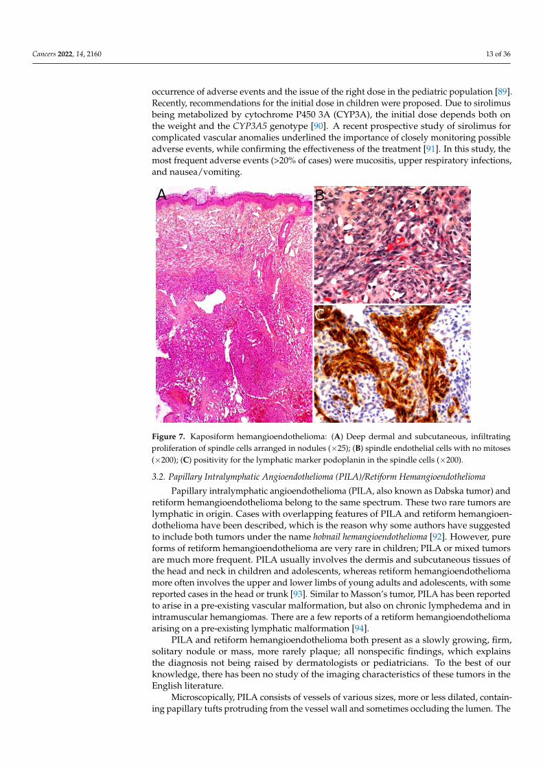
Microscopically, PILA consists of vessels of various sizes, more or less dilated, contain-ing papillary tufts protruding from the vessel wall and sometimes occluding the lumen. The
Cancers 2022, 14, 2160 14 of 36
papillae are centered by a hyalinized core and lined by hobnail endothelial cells with largehyperchromatic nuclei located towards the luminal pole of the cell (Figure 8A). Mitoses aresparse. Retiform hemangioendothelioma involves the whole dermis and often infiltratesthe subcutaneous tissue. It is made of elongated, anastomosing vessels reminiscent of thearchitecture of the rete testis. In both PILA and retiform hemangioendothelioma, thereis often a prominent lymphocytic infiltrate. The immunophenotype is similar in bothtumors, with positivity for CD31 and CD34 in the endothelial cells. Prox1 and podoplanin(D2-40) are positive in most cases of PILA (Figure 8B) [95–98]. Of note, rare cases ofcomposite hemangioendothelioma have been described in adolescents, consisting of areasof retiform hemangioendothelioma and solid areas with a vaguely neuroendocrine mor-phology. These cases showed no recurrence nor metastasis in children, however, both ofthese have been reported in adults, especially bone and lung metastases [96]. PILA hasintermediate malignant potential with possible local recurrences and exceptional lymphnode metastases. Lung metastases have been described in at least one case [99]. Retiformhemangioendothelioma shares a similar clinical course.
Cancers 2022, 14, x FOR PEER REVIEW 15 of 39
more often involves the upper and lower limbs of young adults and adolescents, with
some reported cases in the head or trunk [93]. Similar to Masson’s tumor, PILA has been
reported to arise in a pre-existing vascular malformation, but also on chronic lymphedema
and in intramuscular hemangiomas. There are a few reports of a retiform hemangioendo-
thelioma arising on a pre-existing lymphatic malformation [94].
PILA and retiform hemangioendothelioma both present as a slowly growing, firm,
solitary nodule or mass, more rarely plaque; all nonspecific findings, which explains the
diagnosis not being raised by dermatologists or pediatricians. To the best of our
knowledge, there has been no study of the imaging characteristics of these tumors in the
English literature.
Microscopically, PILA consists of vessels of various sizes, more or less dilated, con-
taining papillary tufts protruding from the vessel wall and sometimes occluding the lu-
men. The papillae are centered by a hyalinized core and lined by hobnail endothelial cells
with large hyperchromatic nuclei located towards the luminal pole of the cell (Figure 8A).
Mitoses are sparse. Retiform hemangioendothelioma involves the whole dermis and often
infiltrates the subcutaneous tissue. It is made of elongated, anastomosing vessels reminis-
cent of the architecture of the rete testis. In both PILA and retiform hemangioendotheli-
oma, there is often a prominent lymphocytic infiltrate. The immunophenotype is similar
in both tumors, with positivity for CD31 and CD34 in the endothelial cells. Prox1 and
podoplanin (D2-40) are positive in most cases of PILA (Figure 8B) [95–98]. Of note, rare
cases of composite hemangioendothelioma have been described in adolescents, consisting
of areas of retiform hemangioendothelioma and solid areas with a vaguely neuroendo-
crine morphology. These cases showed no recurrence nor metastasis in children, however,
both of these have been reported in adults, especially bone and lung metastases [96]. PILA
has intermediate malignant potential with possible local recurrences and exceptional
lymph node metastases. Lung metastases have been described in at least one case [99].
Retiform hemangioendothelioma shares a similar clinical course.
Figure 8. Papillary intralymphatic angioendothelioma: (A) Vascular proliferation with papillary
tufts protruding from the vessel wall, lined by hobnail endothelial cells with large hyperchromatic
nuclei (×200); (B) partial positivity for the lymphatic marker podoplanin (×100).
The treatment of choice of PILA and retiform hemangioendothelioma is complete
surgical resection, in order to avoid recurrences. However, clear surgical margins may be
hard to achieve in some cases of retiform hemangioendothelioma, due to its infiltrative
growth pattern. In such cases, MMS may be useful, as reported in a 11-year-old girl with
a tumor of the finger [100]. In unresectable cases, association of radiation therapy and
chemotherapy with cisplatin might be an option, as reported in an adult [101].
Figure 8. Papillary intralymphatic angioendothelioma: (A) Vascular proliferation with papillary tuftsprotruding from the vessel wall, lined by hobnail endothelial cells with large hyperchromatic nuclei(×200); (B) partial positivity for the lymphatic marker podoplanin (×100).
The treatment of choice of PILA and retiform hemangioendothelioma is completesurgical resection, in order to avoid recurrences. However, clear surgical margins may behard to achieve in some cases of retiform hemangioendothelioma, due to its infiltrativegrowth pattern. In such cases, MMS may be useful, as reported in a 11-year-old girl witha tumor of the finger [100]. In unresectable cases, association of radiation therapy andchemotherapy with cisplatin might be an option, as reported in an adult [101].
3.3. Pseudomyogenic Haemangioendothelioma
Pseudomyogenic hemangioendothelioma (PMHE) is a rare vascular tumor of inter-mediate malignant potential. It affects adolescents and young adults, with a mean age atdiagnosis close to 30 years and a strong male predominance (sex ratio about 4:1) [102,103].The most frequently involved sites are the extremities, especially the lower limbs, followedby the trunk and upper limbs, but the tumor has been described in other locations. PMHEarises mainly in the soft tissues but may occur in the dermis, muscle, or bone [102]. Acharacteristic feature of this tumor is the frequent involvement of different tissue planes,resulting in a mass or deep-seated nodule, with pain in about half of cases. Multicentricpresentation is also common [102,104,105]. On MRI, PMHE is hypointense on T1-weightedimages and hyperintense on T2-weighted and stir-weighted images. It has been suggestedthat positron emission tomography scan (PET-scan) might be helpful to reveal possibleoccult deep lesions [8].
Grossly, PMHE is usually made of multifocal, ill-defined, 1 to 3 cm large, white-or brown-colored nodules [105]. Microscopically, PMHE has an infiltrative pattern of
Cancers 2022, 14, 2160 15 of 36
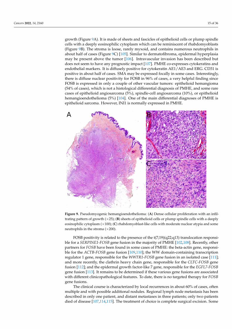
growth (Figure 9A). It is made of sheets and fascicles of epithelioid cells or plump spindlecells with a deeply eosinophilic cytoplasm which can be reminiscent of rhabdomyoblasts(Figure 9B). The stroma is loose, rarely myxoid, and contains numerous neutrophils inabout half of cases (Figure 9C) [105]. Similar to dermatofibroma, epidermal hyperplasiamay be present above the tumor [106]. Intravascular invasion has been described butdoes not seem to have any prognostic impact [107]. PMHE co-expresses cytokeratins andendothelial markers. It is diffusely positive for cytokeratin AE1/AE3 and ERG. CD31 ispositive in about half of cases. SMA may be expressed focally in some cases. Interestingly,there is diffuse nuclear positivity for FOSB in 96% of cases, a very helpful finding sinceFOSB is expressed in only a couple of other vascular tumors: epithelioid hemangioma(54% of cases), which is not a histological differential diagnosis of PMHE, and some rarecases of epithelioid angiosarcoma (5%), spindle-cell angiosarcoma (10%), or epithelioidhemangioendothelioma (5%) [104]. One of the main differential diagnoses of PMHE isepithelioid sarcoma. However, INI1 is normally expressed in PMHE.
Cancers 2022, 14, x FOR PEER REVIEW 17 of 39
Figure 9. Pseudomyogenic hemangioendothelioma: (A) Dense cellular proliferation with an infil-
trating pattern of growth (×25); (B) sheets of epithelioid cells or plump spindle cells with a deeply
eosinophilic cytoplasm (×100); (C) rhabdomyoblast-like cells with moderate nuclear atypia and
some neutrophils in the stroma (×200).
3.4. Angiosarcoma
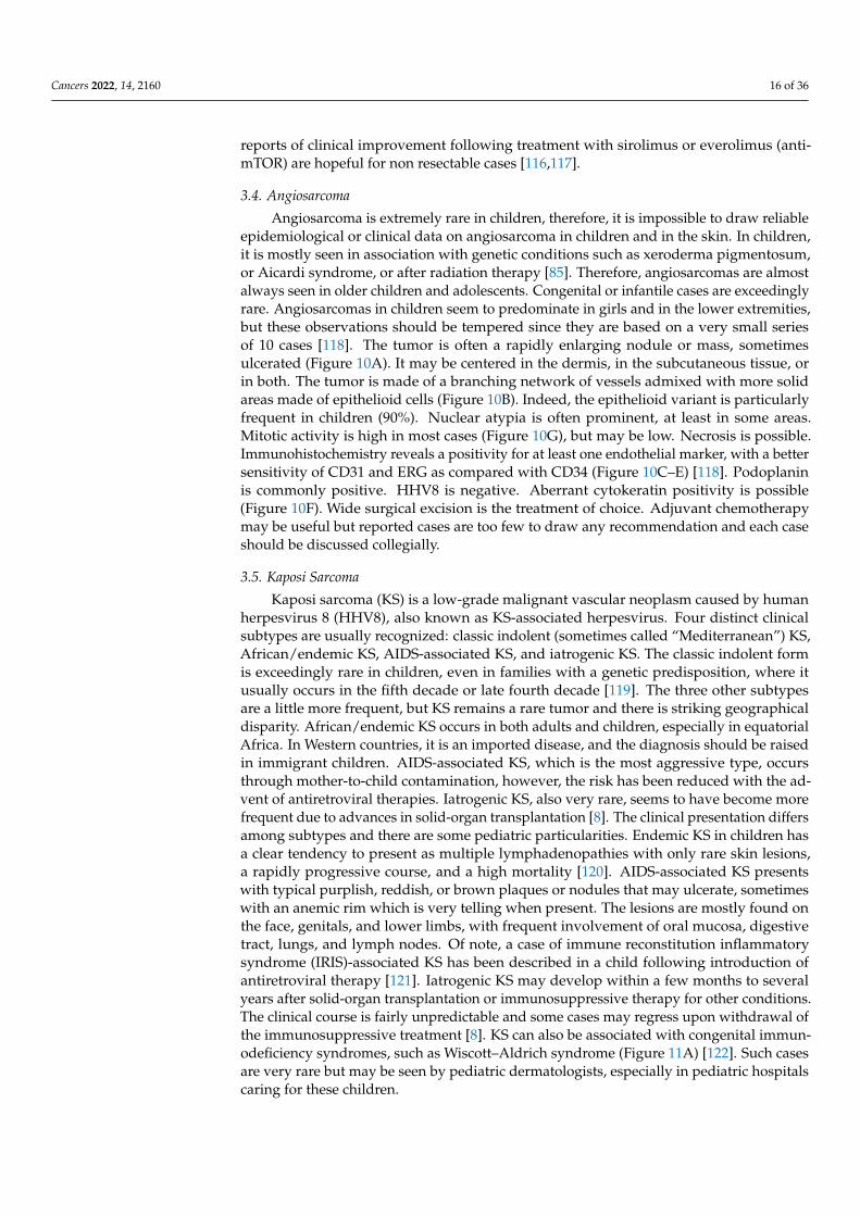
Angiosarcoma is extremely rare in children, therefore, it is impossible to draw relia-
ble epidemiological or clinical data on angiosarcoma in children and in the skin. In chil-
dren, it is mostly seen in association with genetic conditions such as xeroderma pigmen-
tosum, or Aicardi syndrome, or after radiation therapy [85]. Therefore, angiosarcomas are
almost always seen in older children and adolescents. Congenital or infantile cases are
exceedingly rare. Angiosarcomas in children seem to predominate in girls and in the
lower extremities, but these observations should be tempered since they are based on a
very small series of 10 cases [118]. The tumor is often a rapidly enlarging nodule or mass,
sometimes ulcerated (Figure 10A). It may be centered in the dermis, in the subcutaneous
tissue, or in both. The tumor is made of a branching network of vessels admixed with
more solid areas made of epithelioid cells (Figure 10B). Indeed, the epithelioid variant is
particularly frequent in children (90%). Nuclear atypia is often prominent, at least in some
areas. Mitotic activity is high in most cases (Figure 10G), but may be low. Necrosis is pos-
sible. Immunohistochemistry reveals a positivity for at least one endothelial marker, with
a better sensitivity of CD31 and ERG as compared with CD34 (Figure 10C–E) [118].
Podoplanin is commonly positive. HHV8 is negative. Aberrant cytokeratin positivity is
possible (Figure 10F). Wide surgical excision is the treatment of choice. Adjuvant chemo-
therapy may be useful but reported cases are too few to draw any recommendation and
each case should be discussed collegially.
Figure 9. Pseudomyogenic hemangioendothelioma: (A) Dense cellular proliferation with an infil-trating pattern of growth (×25); (B) sheets of epithelioid cells or plump spindle cells with a deeplyeosinophilic cytoplasm (×100); (C) rhabdomyoblast-like cells with moderate nuclear atypia and someneutrophils in the stroma (×200).
FOSB positivity is related to the presence of the t(7;19)(q22;q13) translocation responsi-ble for a SERPINE1-FOSB gene fusion in the majority of PMHE [102,108]. Recently, otherpartners for FOSB have been found in some cases of PMHE: the beta-actin gene, responsi-ble for the ACTB-FOSB gene fusion [109,110]; the WW domain-containing transcriptionregulator 1 gene, responsible for the WWTR1-FOSB gene fusion in an isolated case [111];and more recently, the clathrin heavy chain gene, responsible for the CLTC-FOSB genefusion [112]; and the epidermal growth factor-like 7 gene, responsible for the EGFL7-FOSBgene fusion [113]. It remains to be determined if these various gene fusions are associatedwith different clinicopathological features. To date, there is no targeted therapy for FOSBgene fusions.
The clinical course is characterized by local recurrences in about 60% of cases, oftenmultiple and with possible additional nodules. Regional lymph node metastasis has beendescribed in only one patient, and distant metastases in three patients; only two patientsdied of disease [107,114,115]. The treatment of choice is complete surgical excision. Some
Cancers 2022, 14, 2160 16 of 36
reports of clinical improvement following treatment with sirolimus or everolimus (anti-mTOR) are hopeful for non resectable cases [116,117].
3.4. Angiosarcoma
Angiosarcoma is extremely rare in children, therefore, it is impossible to draw reliableepidemiological or clinical data on angiosarcoma in children and in the skin. In children,it is mostly seen in association with genetic conditions such as xeroderma pigmentosum,or Aicardi syndrome, or after radiation therapy [85]. Therefore, angiosarcomas are almostalways seen in older children and adolescents. Congenital or infantile cases are exceedinglyrare. Angiosarcomas in children seem to predominate in girls and in the lower extremities,but these observations should be tempered since they are based on a very small seriesof 10 cases [118]. The tumor is often a rapidly enlarging nodule or mass, sometimesulcerated (Figure 10A). It may be centered in the dermis, in the subcutaneous tissue, orin both. The tumor is made of a branching network of vessels admixed with more solidareas made of epithelioid cells (Figure 10B). Indeed, the epithelioid variant is particularlyfrequent in children (90%). Nuclear atypia is often prominent, at least in some areas.Mitotic activity is high in most cases (Figure 10G), but may be low. Necrosis is possible.Immunohistochemistry reveals a positivity for at least one endothelial marker, with a bettersensitivity of CD31 and ERG as compared with CD34 (Figure 10C–E) [118]. Podoplaninis commonly positive. HHV8 is negative. Aberrant cytokeratin positivity is possible(Figure 10F). Wide surgical excision is the treatment of choice. Adjuvant chemotherapymay be useful but reported cases are too few to draw any recommendation and each caseshould be discussed collegially.
3.5. Kaposi Sarcoma
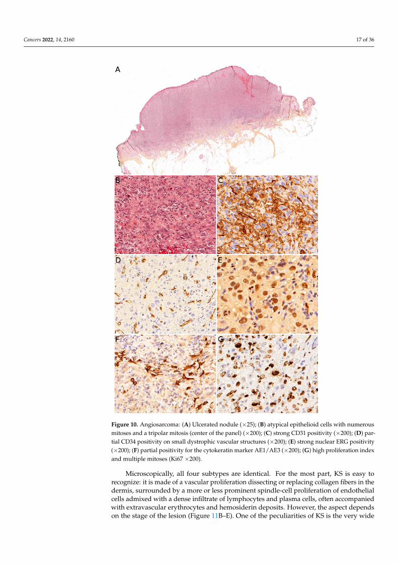
Kaposi sarcoma (KS) is a low-grade malignant vascular neoplasm caused by humanherpesvirus 8 (HHV8), also known as KS-associated herpesvirus. Four distinct clinicalsubtypes are usually recognized: classic indolent (sometimes called “Mediterranean”) KS,African/endemic KS, AIDS-associated KS, and iatrogenic KS. The classic indolent formis exceedingly rare in children, even in families with a genetic predisposition, where itusually occurs in the fifth decade or late fourth decade [119]. The three other subtypesare a little more frequent, but KS remains a rare tumor and there is striking geographicaldisparity. African/endemic KS occurs in both adults and children, especially in equatorialAfrica. In Western countries, it is an imported disease, and the diagnosis should be raisedin immigrant children. AIDS-associated KS, which is the most aggressive type, occursthrough mother-to-child contamination, however, the risk has been reduced with the ad-vent of antiretroviral therapies. Iatrogenic KS, also very rare, seems to have become morefrequent due to advances in solid-organ transplantation [8]. The clinical presentation differsamong subtypes and there are some pediatric particularities. Endemic KS in children hasa clear tendency to present as multiple lymphadenopathies with only rare skin lesions,a rapidly progressive course, and a high mortality [120]. AIDS-associated KS presentswith typical purplish, reddish, or brown plaques or nodules that may ulcerate, sometimeswith an anemic rim which is very telling when present. The lesions are mostly found onthe face, genitals, and lower limbs, with frequent involvement of oral mucosa, digestivetract, lungs, and lymph nodes. Of note, a case of immune reconstitution inflammatorysyndrome (IRIS)-associated KS has been described in a child following introduction ofantiretroviral therapy [121]. Iatrogenic KS may develop within a few months to severalyears after solid-organ transplantation or immunosuppressive therapy for other conditions.The clinical course is fairly unpredictable and some cases may regress upon withdrawal ofthe immunosuppressive treatment [8]. KS can also be associated with congenital immun-odeficiency syndromes, such as Wiscott–Aldrich syndrome (Figure 11A) [122]. Such casesare very rare but may be seen by pediatric dermatologists, especially in pediatric hospitalscaring for these children.
Cancers 2022, 14, 2160 17 of 36Cancers 2022, 14, x FOR PEER REVIEW 18 of 39
Figure 10. Angiosarcoma: (A) Ulcerated nodule (×25); (B) atypical epithelioid cells with numerous
mitoses and a tripolar mitosis (center of the panel) (×200); (C) strong CD31 positivity (×200); (D)
partial CD34 positivity on small dystrophic vascular structures (×200); (E) strong nuclear ERG pos-
itivity (×200); (F) partial positivity for the cytokeratin marker AE1/AE3 (×200); (G) high proliferation
index and multiple mitoses (Ki67 ×200).
3.5. Kaposi Sarcoma
Kaposi sarcoma (KS) is a low-grade malignant vascular neoplasm caused by human
herpesvirus 8 (HHV8), also known as KS-associated herpesvirus. Four distinct clinical
subtypes are usually recognized: classic indolent (sometimes called “Mediterranean”) KS,
African/endemic KS, AIDS-associated KS, and iatrogenic KS. The classic indolent form is
exceedingly rare in children, even in families with a genetic predisposition, where it usu-
ally occurs in the fifth decade or late fourth decade [119]. The three other subtypes are a
little more frequent, but KS remains a rare tumor and there is striking geographical dis-
parity. African/endemic KS occurs in both adults and children, especially in equatorial
Figure 10. Angiosarcoma: (A) Ulcerated nodule (×25); (B) atypical epithelioid cells with numerousmitoses and a tripolar mitosis (center of the panel) (×200); (C) strong CD31 positivity (×200); (D) par-tial CD34 positivity on small dystrophic vascular structures (×200); (E) strong nuclear ERG positivity(×200); (F) partial positivity for the cytokeratin marker AE1/AE3 (×200); (G) high proliferation indexand multiple mitoses (Ki67 ×200).
Microscopically, all four subtypes are identical. For the most part, KS is easy torecognize: it is made of a vascular proliferation dissecting or replacing collagen fibers in thedermis, surrounded by a more or less prominent spindle-cell proliferation of endothelialcells admixed with a dense infiltrate of lymphocytes and plasma cells, often accompaniedwith extravascular erythrocytes and hemosiderin deposits. However, the aspect dependson the stage of the lesion (Figure 11B–E). One of the peculiarities of KS is the very wide
Cancers 2022, 14, 2160 18 of 36
range of morphologies that the tumor can adopt [123]. The most specific immunostainingis nuclear HHV8. In rare negative cases, the diagnosis may be confirmed by PCR.
The treatment is adapted to the clinical subtype but also depends on available drugsand facilities. It includes surgery, radiation therapy, chemotherapy, and of course, antiretro-viral drugs in AIDS-associated KS. Treatment recommendations in children are effectivelysummarized in a paper by Molyneux et al. [124].
Cancers 2022, 14, x FOR PEER REVIEW 20 of 39
Figure 11. Kaposi sarcoma: (A) Multiple vascular nodules on the face of a child with Wiscott–Al-
drich syndrome; (B) rare spindle cells infiltrating between the collagen bundles in a clinically mac-
ular lesion (×100); (C) irregular vascular spaces and lymphocytic infiltrate in a clinically papular
lesion (×100); (D) nodular spindle-cell proliferation in a clinically nodular lesion (×25); (E) spindle-
cell proliferation admixed with lymphocytes in a nodular stage lesion (×200).
The treatment is adapted to the clinical subtype but also depends on available drugs
and facilities. It includes surgery, radiation therapy, chemotherapy, and of course, an-
tiretroviral drugs in AIDS-associated KS. Treatment recommendations in children are ef-
fectively summarized in a paper by Molyneux et al. [124].
4. Muscular Tumors: Rhabdomyosarcomas
Primary cutaneous rhabdomyosarcoma (RMS) is a rare tumor, with about 55 cases
reported in the literature [125,126]. It occurs mainly in the head and neck, and there seem
to be no sex predominance [125]. The age range is wide with rare congenital/neonatal
cases, but also cases in older children and adolescents. Importantly, the two main histo-
logical subtypes of RMS, embryonal RMS (ERMS) and alveolar RMS (ARMS), have dis-
tinct clinical and histological characteristics. Therefore, they are discussed separately here-
after. Contrary to non-cutaneous RMS, where ERMS account for nearly 75% of all RMS,
primarily cutaneous RMS are more often of the alveolar subtype [125], even in congeni-
tal/neonatal cases. Indeed, in the skin and superficial soft tissues, after a short review of
the literature, we found that congenital/neonatal ARMS were twice as often represented
Figure 11. Kaposi sarcoma: (A) Multiple vascular nodules on the face of a child with Wiscott–Aldrichsyndrome; (B) rare spindle cells infiltrating between the collagen bundles in a clinically macularlesion (×100); (C) irregular vascular spaces and lymphocytic infiltrate in a clinically papular lesion(×100); (D) nodular spindle-cell proliferation in a clinically nodular lesion (×25); (E) spindle-cellproliferation admixed with lymphocytes in a nodular stage lesion (×200).
4. Muscular Tumors: Rhabdomyosarcomas
Primary cutaneous rhabdomyosarcoma (RMS) is a rare tumor, with about 55 casesreported in the literature [125,126]. It occurs mainly in the head and neck, and there seemto be no sex predominance [125]. The age range is wide with rare congenital/neonatalcases, but also cases in older children and adolescents. Importantly, the two main histologi-cal subtypes of RMS, embryonal RMS (ERMS) and alveolar RMS (ARMS), have distinctclinical and histological characteristics. Therefore, they are discussed separately hereafter.Contrary to non-cutaneous RMS, where ERMS account for nearly 75% of all RMS, primarilycutaneous RMS are more often of the alveolar subtype [125], even in congenital/neonatal
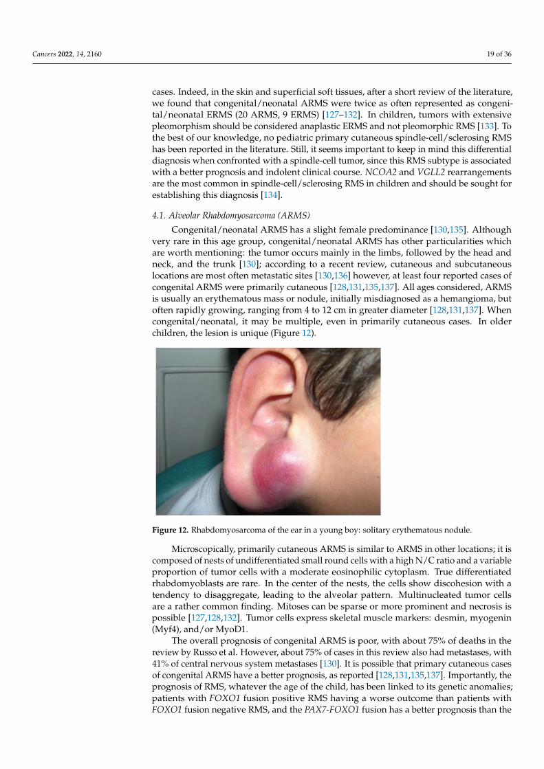
Cancers 2022, 14, 2160 19 of 36
cases. Indeed, in the skin and superficial soft tissues, after a short review of the literature,we found that congenital/neonatal ARMS were twice as often represented as congeni-tal/neonatal ERMS (20 ARMS, 9 ERMS) [127–132]. In children, tumors with extensivepleomorphism should be considered anaplastic ERMS and not pleomorphic RMS [133]. Tothe best of our knowledge, no pediatric primary cutaneous spindle-cell/sclerosing RMShas been reported in the literature. Still, it seems important to keep in mind this differentialdiagnosis when confronted with a spindle-cell tumor, since this RMS subtype is associatedwith a better prognosis and indolent clinical course. NCOA2 and VGLL2 rearrangementsare the most common in spindle-cell/sclerosing RMS in children and should be sought forestablishing this diagnosis [134].
4.1. Alveolar Rhabdomyosarcoma (ARMS)
Congenital/neonatal ARMS has a slight female predominance [130,135]. Althoughvery rare in this age group, congenital/neonatal ARMS has other particularities whichare worth mentioning: the tumor occurs mainly in the limbs, followed by the head andneck, and the trunk [130]; according to a recent review, cutaneous and subcutaneouslocations are most often metastatic sites [130,136] however, at least four reported cases ofcongenital ARMS were primarily cutaneous [128,131,135,137]. All ages considered, ARMSis usually an erythematous mass or nodule, initially misdiagnosed as a hemangioma, butoften rapidly growing, ranging from 4 to 12 cm in greater diameter [128,131,137]. Whencongenital/neonatal, it may be multiple, even in primarily cutaneous cases. In olderchildren, the lesion is unique (Figure 12).
Cancers 2022, 14, x FOR PEER REVIEW 21 of 39
as congenital/neonatal ERMS (20 ARMS, 9 ERMS) [127–132]. In children, tumors with ex-
tensive pleomorphism should be considered anaplastic ERMS and not pleomorphic RMS
[133]. To the best of our knowledge, no pediatric primary cutaneous spindle-cell/scle-
rosing RMS has been reported in the literature. Still, it seems important to keep in mind
this differential diagnosis when confronted with a spindle-cell tumor, since this RMS sub-
type is associated with a better prognosis and indolent clinical course. NCOA2 and VGLL2
rearrangements are the most common in spindle-cell/sclerosing RMS in children and
should be sought for establishing this diagnosis [134].
4.1. Alveolar Rhabdomyosarcoma (ARMS)
Congenital/neonatal ARMS has a slight female predominance [130,135]. Although
very rare in this age group, congenital/neonatal ARMS has other particularities which are
worth mentioning: the tumor occurs mainly in the limbs, followed by the head and neck,
and the trunk [130]; according to a recent review, cutaneous and subcutaneous locations
are most often metastatic sites [130,136] however, at least four reported cases of congenital
ARMS were primarily cutaneous [128,131,135,137]. All ages considered, ARMS is usually
an erythematous mass or nodule, initially misdiagnosed as a hemangioma, but often rap-
idly growing, ranging from 4 to 12 cm in greater diameter [128,131,137]. When congeni-
tal/neonatal, it may be multiple, even in primarily cutaneous cases. In older children, the
lesion is unique (Figure 12).
Figure 12. Rhabdomyosarcoma of the ear in a young boy: solitary erythematous nodule.
Microscopically, primarily cutaneous ARMS is similar to ARMS in other locations; it
is composed of nests of undifferentiated small round cells with a high N/C ratio and a
variable proportion of tumor cells with a moderate eosinophilic cytoplasm. True differen-
tiated rhabdomyoblasts are rare. In the center of the nests, the cells show discohesion with
a tendency to disaggregate, leading to the alveolar pattern. Multinucleated tumor cells are
a rather common finding. Mitoses can be sparse or more prominent and necrosis is possi-
ble [127,128,132]. Tumor cells express skeletal muscle markers: desmin, myogenin (Myf4),
and/or MyoD1.
The overall prognosis of congenital ARMS is poor, with about 75% of deaths in the
review by Russo et al. However, about 75% of cases in this review also had metastases,
with 41% of central nervous system metastases [130]. It is possible that primary cutaneous
cases of congenital ARMS have a better prognosis, as reported [128,131,135,137]. Im-
portantly, the prognosis of RMS, whatever the age of the child, has been linked to its ge-
netic anomalies; patients with FOXO1 fusion positive RMS having a worse outcome than
patients with FOXO1 fusion negative RMS, and the PAX7-FOXO1 fusion has a better
prognosis than the PAX3-FOXO1 fusion [138]. Since 70% of ARMS show either one of the
Figure 12. Rhabdomyosarcoma of the ear in a young boy: solitary erythematous nodule.
Microscopically, primarily cutaneous ARMS is similar to ARMS in other locations; it iscomposed of nests of undifferentiated small round cells with a high N/C ratio and a variableproportion of tumor cells with a moderate eosinophilic cytoplasm. True differentiatedrhabdomyoblasts are rare. In the center of the nests, the cells show discohesion with atendency to disaggregate, leading to the alveolar pattern. Multinucleated tumor cellsare a rather common finding. Mitoses can be sparse or more prominent and necrosis ispossible [127,128,132]. Tumor cells express skeletal muscle markers: desmin, myogenin(Myf4), and/or MyoD1.
The overall prognosis of congenital ARMS is poor, with about 75% of deaths in thereview by Russo et al. However, about 75% of cases in this review also had metastases, with41% of central nervous system metastases [130]. It is possible that primary cutaneous casesof congenital ARMS have a better prognosis, as reported [128,131,135,137]. Importantly, theprognosis of RMS, whatever the age of the child, has been linked to its genetic anomalies;patients with FOXO1 fusion positive RMS having a worse outcome than patients withFOXO1 fusion negative RMS, and the PAX7-FOXO1 fusion has a better prognosis than the
Cancers 2022, 14, 2160 20 of 36
PAX3-FOXO1 fusion [138]. Since 70% of ARMS show either one of the FOXO1 fusions, itmakes sense that ARMS should have a poorer prognosis than ERMS. However, in congeni-tal/neonatal ARMS cases, only three FOXO1 fusions have been reported, to our knowledge,whereas the majority of cases seem to be negative for FOXO1 fusions [127,129,130,135].This suggests a possible different pathogenesis for congenital/neonatal ARMS. For in-stance, some cases have been linked to Beckwith–Wiedemann syndrome and the tumorsseem to arise from silencing of the CDKN1C gene through epigenetic mechanisms in thissyndrome [129,135].
4.2. Embryonal Rhabdomyosarcoma (ERMS)
Congenital/neonatal ERMS mainly occurs in the head and neck, followed by the trunk.The male/female ratio is about two [130]. Only nine cases of congenital/neonatal ERMS ofthe skin or superficial soft tissues have been reported, to the best of our knowledge, withfragmentary data, which makes it difficult to draw meaningful conclusions [127,139–141].The locations include the cheek, scalp, neck, hand, foot, and lower back. All ages considered,the tumor is firm, ranges from 3 to 15 cm in greater dimension, may be ulcerated if large,with possible associated lymphadenopathy [127,139,141].
Microscopically, ERMS shows alternating loose and dense cellularity in a variablymyxoid stroma. The tumor cells range from stellate to small round undifferentiated tospindle. Unlike ARMS, myogenin expression is more variable.
The prognosis is deemed to be better than that of ARMS, however, in congeni-tal/neonatal cases at least three out of nine ERMS cases were fatal after only a few months:the tumor recurred in one case in spite of surgery, chemotherapy (specific RMS protocol),and radiation therapy; the tumor recurred in one other case after surgery; and one childdid not receive any treatment [127,139,141]. The outcome was not known or favorablein other cases. Interestingly, one case exhibited a t(2;8)(q35;q13) translocation. When itwas performed, cytogenetic techniques never evidenced any FOXO1 fusion. One case wasassociated with Apert syndrome (craniosynostosis, midface hypoplasia, and syndactylyof the hands and feet), a syndrome driven by mutations in FGFR2. In a recent study ofgermline predisposition variants in pediatric RMS, the authors found some cancer predis-position gene variants in ERMS occurring before 1 year of age, but not in ARMS in this agegroup. The exact age and location of the tumors are not discussed in the paper, but the mostfrequently involved genes have been NF1 (neurofibromatosis type 1), TP53 (Li–Fraumenisyndrome), HRAS (Costello syndrome), CBL (Noonan syndrome), and BRCA2 [142]. It isworth mentioning that some cases of congenital/neonatal ERMS may arise on congenitalmelanocytic nevi [141].
4.3. Rhabdomyosarcoma Risk Group Stratification and Treatment
The outcome and treatment of RMS are dependent on risk groups. Of note, the perineallocation seems to have prognostic significance as well, with a poorer prognosis in thosecases [126].
The stratification of patients into risk groups at diagnosis is essential. The definitionof risk groups is different in Europe and in North America, with different treatmentstrategies. In Europe, four risk groups (low, standard, high, and very high) have beendefined by the European Paediatric Soft Tissue Sarcoma Group (EpSSG), based on theIntergroup Rhabdomyosarcoma Study Group (IRS) post-surgical stage, the age, the tumorsize, the histopathological subtype, the site of the primary tumor, and the nodal stage.According to the EpSSG, surgical resection follows induction chemotherapy when anyresidual mass can be completely excised (R0/R1) without causing a significant organ orfunctional impairment. For some standard risk cases, radiation therapy may be avoidedif the resection is complete (R0). For more details on the management of RMS and thedifferences between North American and European approaches, the reader is referred tothe review by Yechieli et al. [143].
Cancers 2022, 14, 2160 21 of 36
5. Adipocytic Tumors: Liposarcomas
Liposarcomas of the dermis and subcutaneous tissue (also named superficial liposar-comas) are exceedingly rare in children. They occur almost exclusively in adolescents, andit remains unclear if boys or girls are more affected [144,145]. Nonetheless, in two seriesof superficial liposarcomas, cases were described in patients as young as 5 and 6 yearsold [146,147]. In a series of pediatric cases, there was a strong predilection for the lowerlimbs (~60%), followed by the head and neck, and upper limbs [144]. The tumors werequite large, with about 50% of cases >5 cm. Lymph node involvement was present in 6% ofcases and metastases in only one case (3%). Given the rarity of both superficial liposarcomaand pediatric liposarcoma, they are discussed in detail.
Three types of superficial liposarcomas are recognized in children, by order of fre-quency: myxoid liposarcoma, pleomorphic liposarcoma, and atypical lipomatous tumor (aterm preferred to “well-differentiated/dedifferentiated liposarcoma” in superficial loca-tions, given the indolent course and favorable prognosis of this tumor).
The diagnosis of myxoid liposarcoma is confirmed by the presence of the t(12;16)(q13;p11)translocation, resulting in a FUS-DDIT3 gene fusion. EWSR1-DDIT3 fusions have alsobeen reported.
It is unclear if pleomorphic liposarcoma per se really exists in children. Indeed, a“myxoid pleomorphic liposarcoma” subtype, with a predilection for children and poorprognosis, has recently been added to the WHO classification of tumors of soft tissues [8].Thus, previously reported cases of pleomorphic liposarcomas in children might have beencases of myxoid pleomorphic liposarcomas. However, to the best of our knowledge, thisnew subtype has never been described in the superficial soft tissues.
The prognosis depends on the histologic subtype. Myxoid liposarcoma has an ex-cellent prognosis, myxoid pleomorphic liposarcoma has a poor prognosis, and atypicallipomatous tumor has an indolent behavior. Wide local excision is the treatment of choice.Of note, liposarcomas in children may be associated with predisposition syndromes suchas Aicardi syndrome, Li–Fraumeni syndrome, Muir–Torre syndrome, PTEN hamartomatumor syndromes, probably most lipomatoses, and syndromes belonging to the PIK3CA-related overgrowth spectrum [145].
6. Neuroectodermal and Neural Tumors6.1. Melanotic Neuroectodermal Tumor of Infancy
Melanotic neuroectodermal tumor of infancy (MNTI) is a rare tumor occurring mostlyin infants under 1 year of age. Various names have been used for this tumor, includ-ing melanotic progonoma, retinal anlage tumor, pigmented ameloblastoma, melanoticadamantinoma, melanotic epithelial odontoma, pigmented congenital epulis, pigmentedteratoma. The designation MNTI is retained in the WHO classification of tumors of softtissues and bone [8]. Although its pathogenesis is not understood, it is established thatMNTI derives from neural crest cells. Recently, DNA methylation profiling demonstrated acommon signature between MNTI, some medulloblastomas, and possibly pineal anlagetumor [148]. MNTI is rare in the skin and superficial soft tissues, with less than 10 casesreported in the English literature [149–151]. However, our experience is that this tumormay be more frequent than reported.
Most cutaneous cases are located on the limbs and present as a non-tender, firmswelling. The tumor can grow rapidly but is not adherent to the overlying skin and mostoften mobile. The skin can be erythematous or show prominent telangiectasia. The tumoris often quite large, between 2 and 6 cm in greater dimension [149–152]. The typical bluish-black color of the tumor is not always visible on dermatological examination, but it is astriking finding on sampling of the tumor, which is better performed by incisional biopsyor direct excision surgery when possible [149,150]. The 24-hour urinary vanillylmandelicacid levels can be elevated in some cases [151]. Imaging findings are not specific, unlessinvolvement of an underlying bone is seen, which may trigger suspicion of MNTI, but alsoof Ewing sarcoma or another bone tumor extending to the superficial soft tissues [151].
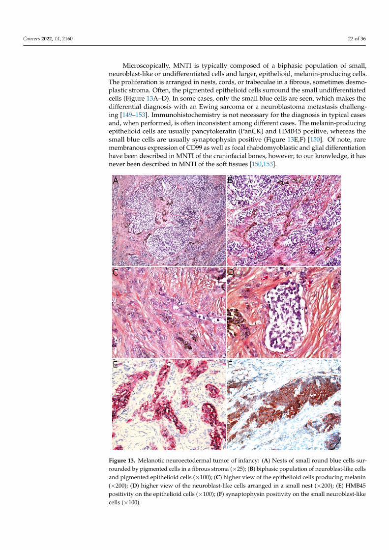
Cancers 2022, 14, 2160 22 of 36
Microscopically, MNTI is typically composed of a biphasic population of small,neuroblast-like or undifferentiated cells and larger, epithelioid, melanin-producing cells.The proliferation is arranged in nests, cords, or trabeculae in a fibrous, sometimes desmo-plastic stroma. Often, the pigmented epithelioid cells surround the small undifferentiatedcells (Figure 13A–D). In some cases, only the small blue cells are seen, which makes thedifferential diagnosis with an Ewing sarcoma or a neuroblastoma metastasis challeng-ing [149–153]. Immunohistochemistry is not necessary for the diagnosis in typical casesand, when performed, is often inconsistent among different cases. The melanin-producingepithelioid cells are usually pancytokeratin (PanCK) and HMB45 positive, whereas thesmall blue cells are usually synaptophysin positive (Figure 13E,F) [150]. Of note, raremembranous expression of CD99 as well as focal rhabdomyoblastic and glial differentiationhave been described in MNTI of the craniofacial bones, however, to our knowledge, it hasnever been described in MNTI of the soft tissues [150,153].
Cancers 2022, 14, x FOR PEER REVIEW 24 of 39
also of Ewing sarcoma or another bone tumor extending to the superficial soft tissues
[151].
Microscopically, MNTI is typically composed of a biphasic population of small, neu-
roblast-like or undifferentiated cells and larger, epithelioid, melanin-producing cells. The
proliferation is arranged in nests, cords, or trabeculae in a fibrous, sometimes desmo-
plastic stroma. Often, the pigmented epithelioid cells surround the small undifferentiated
cells (Figure 13A–D). In some cases, only the small blue cells are seen, which makes the
differential diagnosis with an Ewing sarcoma or a neuroblastoma metastasis challenging
[149–153]. Immunohistochemistry is not necessary for the diagnosis in typical cases and,
when performed, is often inconsistent among different cases. The melanin-producing ep-
ithelioid cells are usually pancytokeratin (PanCK) and HMB45 positive, whereas the small
blue cells are usually synaptophysin positive (Figure 13E–F) [150]. Of note, rare membra-
nous expression of CD99 as well as focal rhabdomyoblastic and glial differentiation have
been described in MNTI of the craniofacial bones, however, to our knowledge, it has never
been described in MNTI of the soft tissues [150,153].
As of today, a BRAFV600E mutation has been described in one MNTI, and a case of
CDKN2A germline mutation too [154,155].
Figure 13. Melanotic neuroectodermal tumor of infancy: (A) Nests of small round blue cells sur-
rounded by pigmented cells in a fibrous stroma (×25); (B) biphasic population of neuroblast-like Figure 13. Melanotic neuroectodermal tumor of infancy: (A) Nests of small round blue cells sur-rounded by pigmented cells in a fibrous stroma (×25); (B) biphasic population of neuroblast-like cellsand pigmented epithelioid cells (×100); (C) higher view of the epithelioid cells producing melanin(×200); (D) higher view of the neuroblast-like cells arranged in a small nest (×200); (E) HMB45positivity on the epithelioid cells (×100); (F) synaptophysin positivity on the small neuroblast-likecells (×100).
Cancers 2022, 14, 2160 23 of 36
As of today, a BRAFV600E mutation has been described in one MNTI, and a case ofCDKN2A germline mutation too [154,155].
Superficial soft tissue cases of MNTI have an overall good prognosis with five patientsalive without recurrence or metastasis (with a 3 to 40 months follow-up) out of eight cases.Follow-up was not available for two cases. Only one patient had metastases and died ofdisease [149–151]. Interestingly, this was the only case treated only with chemotherapy andradiation therapy. In all other cases, surgical excision of the tumor was performed (afterinduction neoadjuvant chemotherapy in one case) and was sufficient. It has been suggestedthat the “debulking effect” of surgery might be a trigger for body antitumor defenses andsubsequent tumor regression [156].
6.2. Ewing Sarcoma Family of Tumors
Ewing sarcomas and associated tumors belonging to the Ewing sarcoma family areexceedingly rare in the superficial soft tissues and the skin. Less than 150 cases of primarysuperficial Ewing sarcoma have been reported in the literature. Ewing sarcoma usuallyarises in bone, with about 15% of cases arising in deep soft tissues and even less in superfi-cial soft tissues and skin. Superficial Ewing sarcoma (sES) shows a female predominanceand the median age ranges from 17 to 21.5 years depending on the series [157]. There hasbeen reported cases in younger children, or even congenital cases [158,159]. Consideringboth children and adults, the most common primary sites are the trunk and lower limbs,followed by the upper limbs and head and neck. However, as far as children are concerned,the distribution pattern is less clear. The tumor’s median size is about 3.5 cm in greaterdiameter. Fever may be a symptom in 8.5% of cases. Metastases at diagnosis are rare (2 to3.5% of cases), the lung being the most frequent site [157]. Microscopically, sES are identicalto their deep counterparts: they are made of sheets of small round blue undifferentiatedcells with fine chromatin and inconspicuous nucleoli, sometimes with PAS-positive cyto-plasmic granulations, and with a very sensitive diffuse membranous CD99 staining pattern.Importantly, CD99 is not specific and may be positive in many other tumors: synovial sar-coma, acute lymphoblastic leukemia, melanoma, solitary fibrous tumor, BCOR-rearrangedsarcoma, angiomatoid fibrous histiocytoma, neurothekeoma, and calcifying aponeuroticfibroma. This list is not exhaustive. As a consequence, the diagnosis needs to be validatedmolecularly. The most frequent translocation is t(11;22)(q24;q12) resulting in the EWSR1-FLI1 gene fusion [160]. The second most common translocation is t(21;22)(q22;q12) resultingin the EWSR1-ERG gene fusion [161]. Importantly, ERG immunostaining is positive inthese cases [162]. Other rare fusions involve ETV1, ETV4, and FEV, but also FUS in placeof EWSR1, resulting in FUS-ERG or FUS-FEV gene fusions [163]. Given the rarity of sES,there is currently no specific recommendation for treatment. However, sES has a moreindolent course than its bone counterpart [164]. Metastatic disease is associated with aworse prognosis. In addition, metastatic relapse after surgery for localized disease has beenreported. Therefore, chemotherapy has an important place in the treatment of sES, andit has been stated by some authors that the outcome might be better with pre-operativechemotherapy, since nearly 50% of primary surgeries were intralesional or marginal in theirseries [157].
6.3. Malignant Peripheral Nerve Sheath Tumor (Associated with Neurofibromatosis Type 1)
A few cases of malignant peripheral nerve sheath tumors (MPNST) have been reportedin adolescents with neurofibromatosis type 1 (NF1). However, a diagnosis of MPNSTin a child without NF1 should be considered with the utmost circumspection, unlessprevious irradiation exists. NF1 is an autosomal dominant genetic disease with an estimatedincidence between 1/2500 and 1/3000 individuals. The NF1 gene, located at 17q11.2,encodes the protein neurofibromin, whose tumor suppressor function consists in thedownregulation of the RAS-RAF-MEK pathway [165]. Clinical criteria for the diagnosisof NF1 have been issued by the NIH and include multiple café-au-lait macules (CALM),skin-fold freckling, Lisch nodules, optic pathway gliomas, distinctive bone lesions such
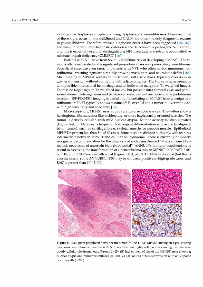
Cancers 2022, 14, 2160 24 of 36
as long-bone dysplasia and sphenoid wing dysplasia, and neurofibromas. However, mostof these signs occur in late childhood and CALM are often the only diagnostic featurein young children. Therefore, revised diagnostic criteria have been suggested [166,167].The most important new diagnostic criterion is the detection of a pathogenic NF1 variant,and this is especially useful in distinguishing NF1 from Legius syndrome or constitutivemismatch repair deficiency (CMMRD) [167].
Patients with NF1 have from 8% to 12% lifetime risk of developing a MPNST. The tu-mor is often deep seated and a significant proportion arises on a pre-existing neurofibroma.Superficial cases are even rarer. In patients with NF1, who often harbor numerous neu-rofibromas, warning signs are a rapidly growing mass, pain, and neurologic deficit [168].MRI imaging of MPNST reveals an ill-defined, soft tissue mass, typically over 4 cm ingreater dimension, without contiguity with adjacent nerves. The tumor is heterogeneouswith possible intralesional hemorrhage and an infiltrative margin on T1-weighted images.There is no target sign on T2-weighted images, but possible intra-tumoral cysts and perile-sional edema. Heterogeneous and perilesional enhancement are present after gadoliniuminjection. 18F-FDG-PET imaging is useful in differentiating an MPNST from a benign neu-rofibroma; MPNST typically shows maximal SUV over 3.5 and a tumor-to-liver ratio >2.6,with high sensitivity and specificity [169].
Microscopically, MPNST may adopt very diverse appearances. They often show aherringbone, fibrosarcoma-like architecture, or more haphazardly oriented fascicles. Thetumor is densely cellular, with mild nuclear atypia. Mitotic activity is often elevated(Figure 14A,B). Necrosis is frequent. A divergent differentiation is possible (malignanttriton tumor), such as cartilage, bone, skeletal muscle, or smooth muscle. EpithelioidMPNST represent less than 5% of all cases. Some cases are difficult to classify, with featuresintermediate between MPNST and cellular neurofibroma. There is currently no widelyrecognized recommendation for the diagnosis of such cases, termed “atypical neurofibro-matosis neoplasms of uncertain biologic potential” (ANNUBP). Immunohistochemistry isuseful in assessing the transformation of a neurofibroma into an MPNST. In MPNST, S100,SOX10, and H3K27me3 are often lost (Figure 14C); p16/CDKN2A is also lost (but this isalso the case in some ANNUBP); TP53 may be diffusely positive in high-grade cases; andKi67 is greater than 10% [170].
Cancers 2022, 14, x FOR PEER REVIEW 26 of 39
the downregulation of the RAS-RAF-MEK pathway [165]. Clinical criteria for the diagno-
sis of NF1 have been issued by the NIH and include multiple café-au-lait macules
(CALM), skin-fold freckling, Lisch nodules, optic pathway gliomas, distinctive bone le-
sions such as long-bone dysplasia and sphenoid wing dysplasia, and neurofibromas.
However, most of these signs occur in late childhood and CALM are often the only diag-
nostic feature in young children. Therefore, revised diagnostic criteria have been sug-
gested [166,167]. The most important new diagnostic criterion is the detection of a patho-
genic NF1 variant, and this is especially useful in distinguishing NF1 from Legius syn-
drome or constitutive mismatch repair deficiency (CMMRD) [167].
Patients with NF1 have from 8% to 12% lifetime risk of developing a MPNST. The
tumor is often deep seated and a significant proportion arises on a pre-existing neurofi-
broma. Superficial cases are even rarer. In patients with NF1, who often harbor numerous
neurofibromas, warning signs are a rapidly growing mass, pain, and neurologic deficit
[168]. MRI imaging of MPNST reveals an ill-defined, soft tissue mass, typically over 4 cm
in greater dimension, without contiguity with adjacent nerves. The tumor is heterogene-
ous with possible intralesional hemorrhage and an infiltrative margin on T1-weighted im-
ages. There is no target sign on T2-weighted images, but possible intra-tumoral cysts and
perilesional edema. Heterogeneous and perilesional enhancement are present after gado-
linium injection. 18F-FDG-PET imaging is useful in differentiating an MPNST from a be-
nign neurofibroma; MPNST typically shows maximal SUV over 3.5 and a tumor-to-liver
ratio >2.6, with high sensitivity and specificity [169].
Microscopically, MPNST may adopt very diverse appearances. They often show a
herringbone, fibrosarcoma-like architecture, or more haphazardly oriented fascicles. The
tumor is densely cellular, with mild nuclear atypia. Mitotic activity is often elevated (Fig-
ure 14A–B). Necrosis is frequent. A divergent differentiation is possible (malignant triton
tumor), such as cartilage, bone, skeletal muscle, or smooth muscle. Epithelioid MPNST
represent less than 5% of all cases. Some cases are difficult to classify, with features inter-
mediate between MPNST and cellular neurofibroma. There is currently no widely recog-
nized recommendation for the diagnosis of such cases, termed “atypical neurofibromato-
sis neoplasms of uncertain biologic potential” (ANNUBP). Immunohistochemistry is use-
ful in assessing the transformation of a neurofibroma into an MPNST. In MPNST, S100,
SOX10, and H3K27me3 are often lost (Figure 14C); p16/CDKN2A is also lost (but this is
also the case in some ANNUBP); TP53 may be diffusely positive in high-grade cases; and
Ki67 is greater than 10% [170].
Figure 14. Malignant peripheral nerve sheath tumor (MPNST): (A) MPNST arising on a pre-existing
plexiform neurofibroma in a child with NF1, note the two highly cellular areas among the otherwise
poorly cellular plexiform neurofibroma (×25); (B) higher view of one of the MPNST areas showing
Figure 14. Malignant peripheral nerve sheath tumor (MPNST): (A) MPNST arising on a pre-existingplexiform neurofibroma in a child with NF1, note the two highly cellular areas among the otherwisepoorly cellular plexiform neurofibroma (×25); (B) higher view of one of the MPNST areas showingnuclear atypia and numerous mitoses (×100); (C) partial loss of S100 expression with only sparsepositive cells (×200).
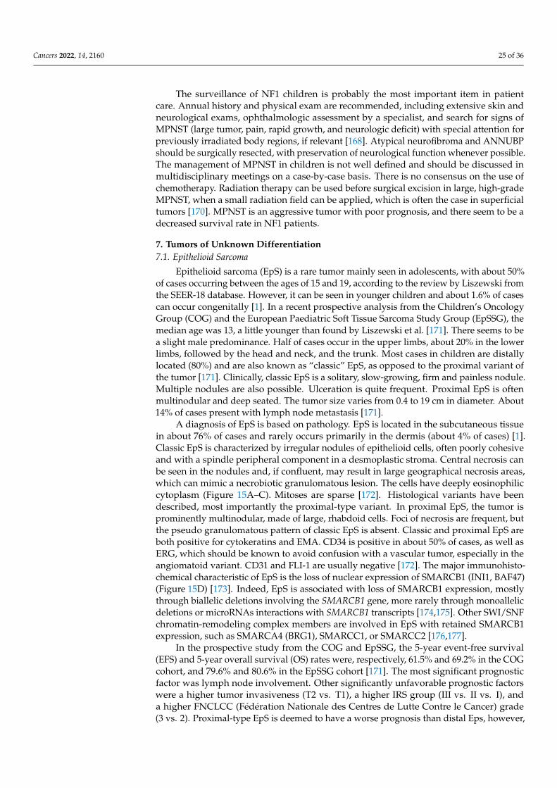
Cancers 2022, 14, 2160 25 of 36
The surveillance of NF1 children is probably the most important item in patientcare. Annual history and physical exam are recommended, including extensive skin andneurological exams, ophthalmologic assessment by a specialist, and search for signs ofMPNST (large tumor, pain, rapid growth, and neurologic deficit) with special attention forpreviously irradiated body regions, if relevant [168]. Atypical neurofibroma and ANNUBPshould be surgically resected, with preservation of neurological function whenever possible.The management of MPNST in children is not well defined and should be discussed inmultidisciplinary meetings on a case-by-case basis. There is no consensus on the use ofchemotherapy. Radiation therapy can be used before surgical excision in large, high-gradeMPNST, when a small radiation field can be applied, which is often the case in superficialtumors [170]. MPNST is an aggressive tumor with poor prognosis, and there seem to be adecreased survival rate in NF1 patients.
7. Tumors of Unknown Differentiation7.1. Epithelioid Sarcoma
Epithelioid sarcoma (EpS) is a rare tumor mainly seen in adolescents, with about 50%of cases occurring between the ages of 15 and 19, according to the review by Liszewski fromthe SEER-18 database. However, it can be seen in younger children and about 1.6% of casescan occur congenitally [1]. In a recent prospective analysis from the Children’s OncologyGroup (COG) and the European Paediatric Soft Tissue Sarcoma Study Group (EpSSG), themedian age was 13, a little younger than found by Liszewski et al. [171]. There seems to bea slight male predominance. Half of cases occur in the upper limbs, about 20% in the lowerlimbs, followed by the head and neck, and the trunk. Most cases in children are distallylocated (80%) and are also known as “classic” EpS, as opposed to the proximal variant ofthe tumor [171]. Clinically, classic EpS is a solitary, slow-growing, firm and painless nodule.Multiple nodules are also possible. Ulceration is quite frequent. Proximal EpS is oftenmultinodular and deep seated. The tumor size varies from 0.4 to 19 cm in diameter. About14% of cases present with lymph node metastasis [171].
A diagnosis of EpS is based on pathology. EpS is located in the subcutaneous tissuein about 76% of cases and rarely occurs primarily in the dermis (about 4% of cases) [1].Classic EpS is characterized by irregular nodules of epithelioid cells, often poorly cohesiveand with a spindle peripheral component in a desmoplastic stroma. Central necrosis canbe seen in the nodules and, if confluent, may result in large geographical necrosis areas,which can mimic a necrobiotic granulomatous lesion. The cells have deeply eosinophiliccytoplasm (Figure 15A–C). Mitoses are sparse [172]. Histological variants have beendescribed, most importantly the proximal-type variant. In proximal EpS, the tumor isprominently multinodular, made of large, rhabdoid cells. Foci of necrosis are frequent, butthe pseudo granulomatous pattern of classic EpS is absent. Classic and proximal EpS areboth positive for cytokeratins and EMA. CD34 is positive in about 50% of cases, as well asERG, which should be known to avoid confusion with a vascular tumor, especially in theangiomatoid variant. CD31 and FLI-1 are usually negative [172]. The major immunohisto-chemical characteristic of EpS is the loss of nuclear expression of SMARCB1 (INI1, BAF47)(Figure 15D) [173]. Indeed, EpS is associated with loss of SMARCB1 expression, mostlythrough biallelic deletions involving the SMARCB1 gene, more rarely through monoallelicdeletions or microRNAs interactions with SMARCB1 transcripts [174,175]. Other SWI/SNFchromatin-remodeling complex members are involved in EpS with retained SMARCB1expression, such as SMARCA4 (BRG1), SMARCC1, or SMARCC2 [176,177].
In the prospective study from the COG and EpSSG, the 5-year event-free survival(EFS) and 5-year overall survival (OS) rates were, respectively, 61.5% and 69.2% in the COGcohort, and 79.6% and 80.6% in the EpSSG cohort [171]. The most significant prognosticfactor was lymph node involvement. Other significantly unfavorable prognostic factorswere a higher tumor invasiveness (T2 vs. T1), a higher IRS group (III vs. II vs. I), anda higher FNCLCC (Fédération Nationale des Centres de Lutte Contre le Cancer) grade(3 vs. 2). Proximal-type EpS is deemed to have a worse prognosis than distal Eps, however,
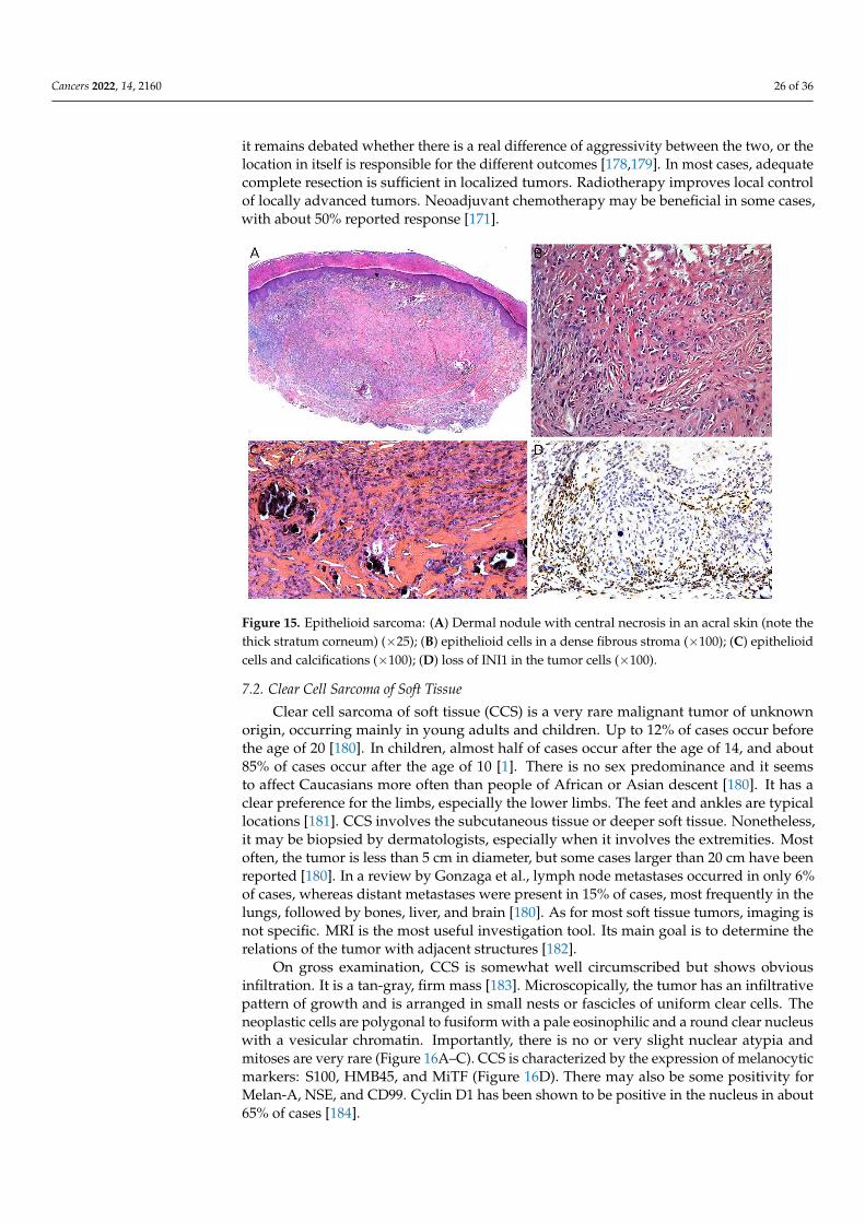
Cancers 2022, 14, 2160 26 of 36
it remains debated whether there is a real difference of aggressivity between the two, or thelocation in itself is responsible for the different outcomes [178,179]. In most cases, adequatecomplete resection is sufficient in localized tumors. Radiotherapy improves local controlof locally advanced tumors. Neoadjuvant chemotherapy may be beneficial in some cases,with about 50% reported response [171].
Cancers 2022, 14, x FOR PEER REVIEW 28 of 39
Figure 15. Epithelioid sarcoma: (A) Dermal nodule with central necrosis in an acral skin (note the
thick stratum corneum) (×25); (B) epithelioid cells in a dense fibrous stroma (×100); (C) epithelioid
cells and calcifications (×100); (D) loss of INI1 in the tumor cells (×100).
In the prospective study from the COG and EpSSG, the 5-year event-free survival
(EFS) and 5-year overall survival (OS) rates were, respectively, 61.5% and 69.2% in the
COG cohort, and 79.6% and 80.6% in the EpSSG cohort [171]. The most significant prog-
nostic factor was lymph node involvement. Other significantly unfavorable prognostic
factors were a higher tumor invasiveness (T2 vs. T1), a higher IRS group (III vs. II vs. I),
and a higher FNCLCC (Fédération Nationale des Centres de Lutte Contre le Cancer) grade
(3 vs. 2). Proximal-type EpS is deemed to have a worse prognosis than distal Eps, however,
it remains debated whether there is a real difference of aggressivity between the two, or
the location in itself is responsible for the different outcomes [178,179]. In most cases, ad-
equate complete resection is sufficient in localized tumors. Radiotherapy improves local
control of locally advanced tumors. Neoadjuvant chemotherapy may be beneficial in some
cases, with about 50% reported response [171].
7.2. Clear Cell Sarcoma of Soft Tissue
Clear cell sarcoma of soft tissue (CCS) is a very rare malignant tumor of unknown
origin, occurring mainly in young adults and children. Up to 12% of cases occur before
the age of 20 [180]. In children, almost half of cases occur after the age of 14, and about
85% of cases occur after the age of 10 [1]. There is no sex predominance and it seems to
affect Caucasians more often than people of African or Asian descent [180]. It has a clear
preference for the limbs, especially the lower limbs. The feet and ankles are typical loca-
tions [181]. CCS involves the subcutaneous tissue or deeper soft tissue. Nonetheless, it
may be biopsied by dermatologists, especially when it involves the extremities. Most of-
ten, the tumor is less than 5 cm in diameter, but some cases larger than 20 cm have been
reported [180]. In a review by Gonzaga et al., lymph node metastases occurred in only 6%
of cases, whereas distant metastases were present in 15% of cases, most frequently in the
lungs, followed by bones, liver, and brain [180]. As for most soft tissue tumors, imaging
is not specific. MRI is the most useful investigation tool. Its main goal is to determine the
relations of the tumor with adjacent structures [182].
On gross examination, CCS is somewhat well circumscribed but shows obvious in-
filtration. It is a tan-gray, firm mass [183]. Microscopically, the tumor has an infiltrative
Figure 15. Epithelioid sarcoma: (A) Dermal nodule with central necrosis in an acral skin (note thethick stratum corneum) (×25); (B) epithelioid cells in a dense fibrous stroma (×100); (C) epithelioidcells and calcifications (×100); (D) loss of INI1 in the tumor cells (×100).
7.2. Clear Cell Sarcoma of Soft Tissue
Clear cell sarcoma of soft tissue (CCS) is a very rare malignant tumor of unknownorigin, occurring mainly in young adults and children. Up to 12% of cases occur beforethe age of 20 [180]. In children, almost half of cases occur after the age of 14, and about85% of cases occur after the age of 10 [1]. There is no sex predominance and it seemsto affect Caucasians more often than people of African or Asian descent [180]. It has aclear preference for the limbs, especially the lower limbs. The feet and ankles are typicallocations [181]. CCS involves the subcutaneous tissue or deeper soft tissue. Nonetheless,it may be biopsied by dermatologists, especially when it involves the extremities. Mostoften, the tumor is less than 5 cm in diameter, but some cases larger than 20 cm have beenreported [180]. In a review by Gonzaga et al., lymph node metastases occurred in only 6%of cases, whereas distant metastases were present in 15% of cases, most frequently in thelungs, followed by bones, liver, and brain [180]. As for most soft tissue tumors, imaging isnot specific. MRI is the most useful investigation tool. Its main goal is to determine therelations of the tumor with adjacent structures [182].
On gross examination, CCS is somewhat well circumscribed but shows obviousinfiltration. It is a tan-gray, firm mass [183]. Microscopically, the tumor has an infiltrativepattern of growth and is arranged in small nests or fascicles of uniform clear cells. Theneoplastic cells are polygonal to fusiform with a pale eosinophilic and a round clear nucleuswith a vesicular chromatin. Importantly, there is no or very slight nuclear atypia andmitoses are very rare (Figure 16A–C). CCS is characterized by the expression of melanocyticmarkers: S100, HMB45, and MiTF (Figure 16D). There may also be some positivity forMelan-A, NSE, and CD99. Cyclin D1 has been shown to be positive in the nucleus in about65% of cases [184].
Cancers 2022, 14, 2160 27 of 36
Cancers 2022, 14, x FOR PEER REVIEW 29 of 39
pattern of growth and is arranged in small nests or fascicles of uniform clear cells. The
neoplastic cells are polygonal to fusiform with a pale eosinophilic and a round clear nu-
cleus with a vesicular chromatin. Importantly, there is no or very slight nuclear atypia and
mitoses are very rare (Figure 16A–C). CCS is characterized by the expression of melano-
cytic markers: S100, HMB45, and MiTF (Figure 16D). There may also be some positivity
for Melan-A, NSE, and CD99. Cyclin D1 has been shown to be positive in the nucleus in
about 65% of cases [184].
Similar to what is known in melanoma, there are alterations of the p16INK4a/p14ARF
pathway in CCS, which explains the decreased expression of p16 in some CCS cases, and
increased expression of cyclin D1 [184]. Importantly, CCS is characterized by the
t(12;22)(q13;q12) chromosomal translocation resulting in the EWSR1-ATF1 gene fusion, in
up to 90% of cases. In a small subset of cases, the t(2;22)(q34;q12) translocation resulting
in the EWSR1-CREB1 fusion gene is found. Recently, a retrospective whole-exome se-
quencing study of 21 CCS revealed interesting data. Deletions of ATM and CHEK2 in the
p53 pathway were particularly interesting. The use of poly-ADP-ribose polymerase
(PARP) inhibitors (such as olaparib) or ATR inhibitors might be useful in the treatment of
CCS, as reported by the Chinese team which performed in vitro treatment of CCS cell lines
with such drugs [185]. However, the primary treatment of CCS is complete surgical exci-
sion with wide margins (1 cm or more for most authors). When the recommended margins
cannot be ensured, adjuvant treatment is necessary, i.e., radiation therapy. Although its
efficiency is debated, chemotherapy is recommended in metastatic cases, with only partial
responses using anthracycline-based protocols [182].
Figure 16. Clear cell sarcoma of soft tissue: (A) Densel cellular dermal and subcutaneous prolifera-
tion (×25); (B) uniform clear cells in fascicles (×100); (C) clear cells with no or slight nuclear atypia
(×100); (D) diffuse HMB45 positivity (×100).
7.3. Synovial Sarcoma
Figure 16. Clear cell sarcoma of soft tissue: (A) Densel cellular dermal and subcutaneous proliferation(×25); (B) uniform clear cells in fascicles (×100); (C) clear cells with no or slight nuclear atypia (×100);(D) diffuse HMB45 positivity (×100).
Similar to what is known in melanoma, there are alterations of the p16INK4a/p14ARF
pathway in CCS, which explains the decreased expression of p16 in some CCS cases,and increased expression of cyclin D1 [184]. Importantly, CCS is characterized by thet(12;22)(q13;q12) chromosomal translocation resulting in the EWSR1-ATF1 gene fusion, inup to 90% of cases. In a small subset of cases, the t(2;22)(q34;q12) translocation resulting inthe EWSR1-CREB1 fusion gene is found. Recently, a retrospective whole-exome sequencingstudy of 21 CCS revealed interesting data. Deletions of ATM and CHEK2 in the p53pathway were particularly interesting. The use of poly-ADP-ribose polymerase (PARP)inhibitors (such as olaparib) or ATR inhibitors might be useful in the treatment of CCS, asreported by the Chinese team which performed in vitro treatment of CCS cell lines withsuch drugs [185]. However, the primary treatment of CCS is complete surgical excisionwith wide margins (1 cm or more for most authors). When the recommended marginscannot be ensured, adjuvant treatment is necessary, i.e., radiation therapy. Although itsefficiency is debated, chemotherapy is recommended in metastatic cases, with only partialresponses using anthracycline-based protocols [182].
7.3. Synovial Sarcoma
Synovial sarcoma is a malignant mesenchymal neoplasm of unknown origin, thus, thehistorical name “synovial sarcoma” is misleading. This tumor actually shows a variable ep-ithelial differentiation and, most importantly, is characterized by a specific SS18-SSX fusiongene. It is mostly found in adolescents and young adults, with no gender predilection. Itinvolves the deep soft tissues of the extremities, often in close proximity with articulations.Therefore, it falls at the limits of this review’s topic and it will not be discussed extensively.Suffice it to say that it is often a slow-growing mass that may be mistaken as benign, withno specific imaging features, although often showing areas of varied intensities on MRI,and microscopic examination may reveal either a monophasic or biphasic appearance.
Cancers 2022, 14, 2160 28 of 36
Monophasic synovial sarcoma is made of dense cellular sheets and fascicles of uniformspindle cells associated with a typical, but often lacking, staghorn vascular pattern. Biphasicsynovial sarcoma is similar to the monophasic variant with the addition of an epithelialcomponent, usually focal, arranged in nests or glandular structures. On immunohisto-chemistry, synovial sarcoma is positive for EMA and cytokeratins, but more importantlyfor TLE1, a useful marker, although not specific and also expressed in solitary fibroustumors and malignant peripheral nerve sheath tumors. Most cases of synovial sarcomasalso express CD99 (membranous staining) and Bcl-2. INI1 is partially or completely lostin some cases, and this should be known to avoid a misdiagnosis of rhabdoid tumor. Inany case, the final diagnosis of synovial sarcoma is now made by the presence of one ofthe following gene fusions: SS18-SSX1, SS18-SSX2, SS18-SSX4, or SS18L1-SSX1. Synovialsarcoma has a propensity for late local recurrence and late metastasis, most often in thelungs and pleura [186]. The treatment of choice for localized disease is wide surgicalexcision. If not immediately possible, neoadjuvant chemotherapy for tumor reduction isrecommended before surgery, most commonly with doxorubicin and/or ifosfamide. Theuse of local adjuvant radiation therapy is still debated [187].
8. Conclusions
Malignant superficial mesenchymal tumors are rare in children; however, they remainto be the most frequent group of tumors as compared with the even rarer hematologic,melanocytic, or epithelial malignancies. Recently, new entities have been described basedon molecular anomalies, such as the NTRK-rearranged spindle cell neoplasms, the EWSR1-SMAD3-rearranged fibroblastic tumor, or the ALK-rearranged infantile fibrosarcoma-liketumor. At the same time, the complexity of genetically classifying these tumors has in-creased as a result of the discovery of new fusion transcripts in already well-defined entities,for example, COL1A2-PDGFB, COL6A3-PDGFD, or EMILIN2-PDGFD fusion-positive DFSP,the various NTRK fusions in infantile fibrosarcoma, or the new FOSB fusions in pseu-domyogenic hemangioendothelioma. These advances may lead to redefinition of sometumor groups, as exemplified by the proximity between infantile fibrosarcoma and NTRK-rearranged spindle cell neoplasms, which may belong to a common spectrum.
While a minority of tumors remain strictly histo-clinical entities with no known geneticanomalies to date, such as PFHT, the vast majority of the tumors discussed in the presentreview are defined, partially in most cases, on molecular grounds. Therefore, there is astrong need for histo-molecular cooperation. This is true for diagnosis, but also moreand more relevant in therapeutic approaches, as proven by the development of NTRKinhibitors in infantile fibrosarcoma, mTOR-inhibitors (sirolimus, everolimus) in KHE or inpseudomyogenic hemangioendothelioma, or even PARP inhibitors in clear cell sarcoma ofsoft tissues. Nevertheless, surgery remains the most valuable treatment in a lot of cases, thebest example of this being, arguably, Mohs micrographic surgery in the treatment of DFSP.
Lastly, the specificities of children and skin tumors should not be forgotten: particularclinical presentations (e.g., “blueberry muffin rash” or congenital tumors); the possibilityof a cancer predisposition syndrome (NF1, Li–Fraumeni, among others) or congenitalimmunodeficiency syndrome (e.g., Wiscott-Aldrich syndrome); and the particular behaviorof some superficial tumors (e.g., the more indolent course of superficial RMS as comparedwith deeper RMS) should be kept in mind by all specialists dealing with skin tumorsin children.
Author Contributions: Conceptualization, S.F.; investigation, P.D. and S.F.; writing—original draftpreparation, P.D.; writing—review and editing, P.D. and S.F.; supervision, S.F. All authors have readand agreed to the published version of the manuscript.
Funding: This research received no external funding.
Cancers 2022, 14, 2160 29 of 36
Informed Consent Statement: Informed consent has been obtained from the patients to use theclinical pictures.
Conflicts of Interest: The authors declare no conflict of interest.
References1. Liszewski, W.; Maguiness, S.; Greengard, E.; Boull, C. The Incidence of Pediatric Malignant Soft Tissue Tumors of the Skin and
Subcutaneous Tissue. Pediatr. Dermatol. 2018, 35, e427–e429. [CrossRef] [PubMed]2. Amadeo, B.; Penel, N.; Coindre, J.-M.; Ray-Coquard, I.; Ligier, K.; Delafosse, P.; Bouvier, A.-M.; Plouvier, S.; Gallet, J.;
Lacourt, A.; et al. Incidence and Time Trends of Sarcoma (2000–2013): Results from the French Network of Cancer Registries(FRANCIM). BMC Cancer 2020, 20, 190. [CrossRef]
3. Bohelay, G.; Kluger, N.; Battistella, M.; Biaggi-Frassati, A.; Plantier, F.; Harraudeau, A.; Avril, M.-F.; Pedeutour, F.; Fraitag, S.Angiomatoid fibrous histiocytoma in children: 6 cases. Ann. Dermatol. Venereol. 2015, 142, 541–548. [CrossRef] [PubMed]
4. Thway, K.; Fisher, C. Angiomatoid Fibrous Histiocytoma: The Current Status of Pathology and Genetics. Arch. Pathol. Lab. Med.2015, 139, 674–682. [CrossRef] [PubMed]
5. Saito, K.; Kobayashi, E.; Yoshida, A.; Araki, Y.; Kubota, D.; Tanzawa, Y.; Kawai, A.; Yanagawa, T.; Takagishi, K.; Chuman, H.Angiomatoid Fibrous Histiocytoma: A Series of Seven Cases Including Genetically Confirmed Aggressive Cases and a LiteratureReview. BMC Musculoskelet. Disord. 2017, 18, 31. [CrossRef]
6. Chung, E.B.; Enzinger, F.M. Infantile Fibrosarcoma. Cancer 1976, 38, 729–739. [CrossRef]7. Orbach, D.; Rey, A.; Cecchetto, G.; Oberlin, O.; Casanova, M.; Thebaud, E.; Scopinaro, M.; Bisogno, G.; Carli, M.; Ferrari, A.
Infantile Fibrosarcoma: Management Based on the European Experience. J. Clin. Oncol. Off. J. Am. Soc. Clin. Oncol. 2010, 28,318–323. [CrossRef]
8. WHO Classification of Tumours Editorial Board. Soft Tissue and Bone Tumours; World Health Organization: Geneva, Switzerland,2020; ISBN 978-92-832-4502-5.
9. Ainsworth, K.E.; Chavhan, G.B.; Gupta, A.A.; Hopyan, S.; Taylor, G. Congenital Infantile Fibrosarcoma: Review of ImagingFeatures. Pediatr. Radiol. 2014, 44, 1124–1129. [CrossRef]
10. Yan, A.C.; Chamlin, S.L.; Liang, M.G.; Hoffman, B.; Attiyeh, E.F.; Chang, B.; Honig, P.J. Congenital Infantile Fibrosarcoma: AMasquerader of Ulcerated Hemangioma. Pediatr. Dermatol. 2006, 23, 330–334. [CrossRef]
11. Swiadkiewicz, R.; Galmiche, L.; Belhous, K.; Boccara, O.; Fraitag, S.; Pedeutour, F.; Dadone, B.; Buis, J.; Picard, A.; Orbach, D.; et al.Congenital Infantile Fibrosarcoma Associated with a Lipofibromatosis-Like Component: One Train May Be Hiding Another. Am.J. Dermatopathol. 2017, 39, 463–467. [CrossRef]
12. Enos, T.; Hosler, G.A.; Uddin, N.; Mir, A. Congenital Infantile Fibrosarcoma Mimicking a Cutaneous Vascular Lesion: A CaseReport and Review of the Literature. J. Cutan. Pathol. 2017, 44, 193–200. [CrossRef] [PubMed]
13. Coffin, C.M.; Alaggio, R. Fibroblastic and Myofibroblastic Tumors in Children and Adolescents. Pediatr. Dev. Pathol. 2012, 15,127–180. [CrossRef] [PubMed]
14. Hung, Y.P.; Fletcher, C.D.M.; Hornick, J.L. Evaluation of Pan-TRK Immunohistochemistry in Infantile Fibrosarcoma,Lipofibromatosis-like Neural Tumour and Histological Mimics. Histopathology 2018, 73, 634–644. [CrossRef] [PubMed]
15. Church, A.J.; Calicchio, M.L.; Nardi, V.; Skalova, A.; Pinto, A.; Dillon, D.A.; Gomez-Fernandez, C.R.; Manoj, N.; Haimes, J.D.;Stahl, J.A.; et al. Recurrent EML4-NTRK3 Fusions in Infantile Fibrosarcoma and Congenital Mesoblastic Nephroma Suggest aRevised Testing Strategy. Mod. Pathol. 2018, 31, 463–473. [CrossRef]
16. Davis, J.L.; Lockwood, C.M.; Stohr, B.; Boecking, C.; Al-Ibraheemi, A.; DuBois, S.G.; Vargas, S.O.; Black, J.O.; Cox, M.C.;Luquette, M.; et al. Expanding the Spectrum of Pediatric NTRK-Rearranged Mesenchymal Tumors. Am. J. Surg. Pathol. 2019, 43,435–445. [CrossRef]
17. Zhao, X.; Kotch, C.; Fox, E.; Surrey, L.F.; Wertheim, G.B.; Baloch, Z.W.; Lin, F.; Pillai, V.; Luo, M.; Kreiger, P.A.; et al. NTRKFusions Identified in Pediatric Tumors: The Frequency, Fusion Partners, and Clinical Outcome. JCO Precis. Oncol. 2021, 1, 204–214.[CrossRef]
18. Kao, Y.-C.; Fletcher, C.D.M.; Alaggio, R.; Wexler, L.; Zhang, L.; Sung, Y.-S.; Orhan, D.; Chang, W.-C.; Swanson, D.;Dickson, B.C.; et al. Recurrent BRAF Gene Fusions in a Subset of Pediatric Spindle Cell Sarcomas: Expanding the GeneticSpectrum of Tumors with Overlapping Features with Infantile Fibrosarcoma. Am. J. Surg. Pathol. 2018, 42, 28–38. [CrossRef][PubMed]
19. Wegert, J.; Vokuhl, C.; Collord, G.; Velasco-Herrera, M.D.C.; Farndon, S.J.; Guzzo, C.; Jorgensen, M.; Anderson, J.; Slater, O.;Duncan, C.; et al. Recurrent Intragenic Rearrangements of EGFR and BRAF in Soft Tissue Tumors of Infants. Nat. Commun. 2018,9, 2378. [CrossRef]
20. Davis, J.L.; Vargas, S.O.; Rudzinski, E.R.; Marti, J.M.L.; Janeway, K.; Forrest, S.; Winsnes, K.; Pinto, N.; Yang, S.E.;VanSandt, M.; et al. Recurrent RET Gene Fusions in Paediatric Spindle Mesenchymal Neoplasms. Histopathology 2020,76, 1032–1041. [CrossRef]
21. Suurmeijer, A.J.H.; Dickson, B.C.; Swanson, D.; Zhang, L.; Sung, Y.-S.; Cotzia, P.; Fletcher, C.D.M.; Antonescu, C.R. A NovelGroup of Spindle Cell Tumors Defined by S100 and CD34 Co-Expression Shows Recurrent Fusions Involving RAF1, BRAF, andNTRK1/2 Genes. Genes Chromosomes Cancer 2018, 57, 611–621. [CrossRef]
Cancers 2022, 14, 2160 30 of 36
22. Shankwiler, R.A.; Athey, P.A.; Lamki, N. Aggressive Infantile Fibromatosis. Pulmonary Metastases Documented by Plain Filmand Computed Tomography. Clin. Imaging 1989, 13, 127–129. [CrossRef]
23. Ramphal, R.; Manson, D.; Viero, S.; Zielenska, M.; Gerstle, T.; Pappo, A. Retroperitoneal Infantile Fibrosarcoma: Clinical,Molecular, and Therapeutic Aspects of an Unusual Tumor. Pediatr. Hematol. Oncol. 2003, 20, 635–642. [CrossRef] [PubMed]
24. Coulibaly, B.; Barel, E.; Soulier, M.; Bouvier, C.; Chaumoitre, K.; D’Ercole, C.; Liprandi, A. Prenatal Diagnosis of InfantileFibrosarcoma with Diffuse Metastases. Prenat. Diagn. 2008, 28, 773–775. [CrossRef]
25. Parida, L.; Fernandez-Pineda, I.; Uffman, J.K.; Davidoff, A.M.; Krasin, M.J.; Pappo, A.; Rao, B.N. Clinical Management of InfantileFibrosarcoma: A Retrospective Single-Institution Review. Pediatr. Surg. Int. 2013, 29, 703–708. [CrossRef] [PubMed]
26. Lo, C.-H.; Cheng, S.-N.; Lin, K.-T.; Jen, Y.-M. Successful Treatment of Infantile Fibrosarcoma Spinal Metastasis by Chemotherapyand Stereotactic Hypofractionated Radiotherapy. J. Korean Neurosurg. Soc. 2013, 54, 528–531. [CrossRef] [PubMed]
27. Orbach, D.; Sparber-Sauer, M.; Laetsch, T.W.; Minard-Colin, V.; Bielack, S.S.; Casanova, M.; Corradini, N.; Koscielniak, E.;Scheer, M.; Hettmer, S.; et al. Spotlight on the Treatment of Infantile Fibrosarcoma in the Era of Neurotrophic TropomyosinReceptor Kinase Inhibitors: International Consensus and Remaining Controversies. Eur. J. Cancer 2020, 137, 183–192. [CrossRef][PubMed]
28. Shah, N.N.; Price, M.R.; Loeb, D.M. Cardiac Metastasis and Hypertrophic Osteoarthropathy in Recurrent Infantile Fibrosarcoma.Pediatr. Blood Cancer 2012, 59, 179–181. [CrossRef]
29. Hiradfar, A.; Pourlak, T.; Badebarin, D. Primary Pulmonary Fibrosarcoma With Bone Metastasis: A Successful Treatment withPost-Operation Adjuvant Chemotherapy. Iran. J. Cancer Prev. 2015, 8, e2328. [CrossRef]
30. Laetsch, T.W.; DuBois, S.G.; Mascarenhas, L.; Turpin, B.; Federman, N.; Albert, C.M.; Nagasubramanian, R.; Davis, J.L.;Rudzinski, E.; Feraco, A.M.; et al. Larotrectinib for Paediatric Solid Tumours Harbouring NTRK Gene Fusions: Phase 1 Resultsfrom a Multicentre, Open-Label, Phase 1/2 Study. Lancet Oncol. 2018, 19, 705–714. [CrossRef]
31. Shulman, D.S.; DuBois, S.G. The Evolving Diagnostic and Treatment Landscape of NTRK-Fusion-Driven Pediatric Cancers.Pediatr. Drugs 2020, 22, 189–197. [CrossRef]
32. Caldwell, K.J.; De La Cuesta, E.; Morin, C.; Pappo, A.; Helmig, S. A Newborn with a Large NTRK Fusion Positive InfantileFibrosarcoma Successfully Treated with Larotrectinib. Pediatr. Blood Cancer 2020, 67, e28330. [CrossRef] [PubMed]
33. Kao, Y.; Suurmeijer, A.J.H.; Argani, P.; Dickson, B.C.; Zhang, L.; Sung, Y.; Agaram, N.P.; Fletcher, C.D.M.; Antonescu, C.R. SoftTissue Tumors Characterized by a Wide Spectrum of Kinase Fusions Share a Lipofibromatosis-like Neural Tumor Pattern. GenesChromosomes Cancer 2020, 59, 575–583. [CrossRef] [PubMed]
34. Agaram, N.P.; Zhang, L.; Sung, Y.-S.; Chen, C.-L.; Chung, C.T.; Antonescu, C.R.; Fletcher, C.D. Recurrent NTRK1 Gene FusionsDefine a Novel Subset of Locally Aggressive Lipofibromatosis-like Neural Tumors. Am. J. Surg. Pathol. 2016, 40, 1407–1416.[CrossRef] [PubMed]
35. Drabent, P.; Fraitag, S. Update on Superficial Spindle Cell Mesenchymal Tumors in Children. Dermatopathology 2021, 8, 285–300.[CrossRef] [PubMed]
36. Siozopoulou, V.; Smits, E.; De Winne, K.; Marcq, E.; Pauwels, P. NTRK Fusions in Sarcomas: Diagnostic Challenges and ClinicalAspects. Diagnostics 2021, 11, 478. [CrossRef]
37. Michal, M.; Hájková, V.; Skálová, A.; Michal, M. STRN-NTRK3-Rearranged Mesenchymal Tumor of the Uterus: Expanding theMorphologic Spectrum of Tumors with NTRK Fusions. Am. J. Surg. Pathol. 2019, 43, 1152–1154. [CrossRef]
38. Davis, J.L.; Lockwood, C.M.; Albert, C.M.; Tsuchiya, K.; Hawkins, D.S.; Rudzinski, E.R. Infantile NTRK-Associated MesenchymalTumors. Pediatr. Dev. Pathol. Off. J. Soc. Pediatr. Pathol. Paediatr. Pathol. Soc. 2018, 21, 68–78. [CrossRef]
39. Tallegas, M.; Fraitag, S.; Binet, A.; Orbach, D.; Jourdain, A.; Reynaud, S.; Pierron, G.; Machet, M.-C.; Maruani, A. Novel KHDRBS1-NTRK3 Rearrangement in a Congenital Pediatric CD34-Positive Skin Tumor: A Case Report. Virchows Arch. Int. J. Pathol. 2019,474, 111–115. [CrossRef]
40. Kao, Y.-C.; Flucke, U.; Eijkelenboom, A.; Zhang, L.; Sung, Y.-S.; Suurmeijer, A.J.H.; Antonescu, C.R. Novel EWSR1-SMAD3 GeneFusions in a Group of Acral Fibroblastic Spindle Cell Neoplasms. Am. J. Surg. Pathol. 2018, 42, 522–528. [CrossRef]
41. Michal, M.; Berry, R.S.; Rubin, B.P.; Kilpatrick, S.E.; Agaimy, A.; Kazakov, D.V.; Steiner, P.; Ptakova, N.; Martinek, P.; Hadravsky,L.; et al. EWSR1-SMAD3—Rearranged Fibroblastic Tumor: An Emerging Entity in an Increasingly More Complex Group ofFibroblastic/Myofibroblastic Neoplasms. Am. J. Surg. Pathol. 2018, 42, 1325–1333. [CrossRef]
42. Tan, S.Y.; Al-Ibraheemi, A.; Ahrens, W.A.; Oesterheld, J.E.; Fanburg-Smith, J.C.; Liu, Y.J.; Spunt, S.L.; Rudzinski, E.R.; Coffin, C.;Davis, J.L. ALK Rearrangements in Infantile Fibrosarcoma-like Spindle Cell Tumours of Soft Tissue and Kidney. Histopathology2022, 80, 698–707. [CrossRef] [PubMed]
43. Rubio, G.A.; Alvarado, A.; Gerth, D.J.; Tashiro, J.; Thaller, S.R. Incidence and Outcomes of Dermatofibrosarcoma Protuberans inthe US Pediatric Population. J. Craniofac. Surg. 2017, 28, 182–184. [CrossRef] [PubMed]
44. Brennan, B.; Zanetti, I.; De Salvo, G.L.; Orbach, D.; Gallego, S.; Francotte, N.; Schifflers, S.; Van Noesel, M.; Kelsey, A.;Casanova, M.; et al. Dermatofibrosarcoma Protuberans in Children and Adolescents: The European Paediatric Soft TissueSarcoma Study Group Prospective Trial (EpSSG NRSTS 2005). Pediatr. Blood Cancer 2020, 67, e28351. [CrossRef] [PubMed]
45. Maire, G.; Fraitag, S.; Galmiche, L.; Keslair, F.; Ebran, N.; Terrier-Lacombe, M.-J.; de Prost, Y.; Pedeutour, F. A Clinical, Histologic,and Molecular Study of 9 Cases of Congenital Dermatofibrosarcoma Protuberans. Arch. Dermatol. 2007, 143, 203–210. [CrossRef]
Cancers 2022, 14, 2160 31 of 36
46. Terrier-Lacombe, M.-J.; Guillou, L.; Maire, G.; Terrier, P.; Vince, D.R.; Somerhausen, N.D.S.A.; Collin, F.; Pedeutour, F.;Coindre, J.-M. Dermatofibrosarcoma Protuberans, Giant Cell Fibroblastoma, and Hybrid Lesions in Children: ClinicopathologicComparative Analysis of 28 Cases with Molecular Data—A Study from the French Federation of Cancer Centers Sarcoma Group.Am. J. Surg. Pathol. 2003, 27, 27–39. [CrossRef]
47. Mujtaba, B.; Wang, F.; Taher, A.; Aslam, R.; Madewell, J.E.; Spear, R.; Nassar, S. Dermatofibrosarcoma Protuberans: Pathologicaland Imaging Review. Curr. Probl. Diagn. Radiol. 2021, 50, 236–240. [CrossRef]
48. Zou, M.-H.; Huang, Q.; Yang, T.; Jiang, Y.; Zhang, L.-J.; Xie, Y.; Zheng, R.-Q. Role of Ultrasound in the Diagnosis of Primary andRecurrent Dermatofibrosarcoma Protuberans. BMC Cancer 2021, 21, 909. [CrossRef]
49. Llombart, B.; Monteagudo, C.; Sanmartín, O.; López-Guerrero, J.A.; Serra-Guillén, C.; Poveda, A.; Jorda, E.; Fernandez-Serra, A.;Pellín, A.; Guillén, C.; et al. Dermatofibrosarcoma Protuberans: A Clinicopathological, Immunohistochemical, Genetic (COL1A1-PDGFB), and Therapeutic Study of Low-Grade versus High-Grade (Fibrosarcomatous) Tumors. J. Am. Acad. Dermatol. 2011, 65,564–575. [CrossRef]
50. Li, Y.; Liang, J.; Xu, X.; Jiang, X.; Wang, C.; Chen, S.; Xiang, B.; Ji, Y. Clinicopathological Features of Fibrosarcomatous Dermatofi-brosarcoma Protuberans and the Construction of a Back-Propagation Neural Network Recognition Model. Orphanet J. Rare Dis.2021, 16, 48. [CrossRef]
51. Bowne, W.B.; Antonescu, C.R.; Leung, D.H.Y.; Katz, S.C.; Hawkins, W.G.; Woodruff, J.M.; Brennan, M.F.; Lewis, J.J. Dermatofi-brosarcoma Protuberans: A Clinicopathologic Analysis of Patients Treated and Followed at a Single Institution. Cancer 2000, 88,2711–2720. [CrossRef]
52. Chicaud, M.; Frassati-Biaggi, A.; Kaltenbach, S.; Karanian, M.; Orbach, D.; Fraitag, S. Dermatofibrosarcoma Protuberans,Fibrosarcomatous Variant: A Rare Tumor in Children. Pediatr. Dermatol. 2021, 38, 217–222. [CrossRef] [PubMed]
53. Sirvent, N.; Maire, G.; Pedeutour, F. Genetics of Dermatofibrosarcoma Protuberans Family of Tumors: From Ring Chromosomesto Tyrosine Kinase Inhibitor Treatment. Genes Chromosomes Cancer 2003, 37, 1–19. [CrossRef] [PubMed]
54. Köster, J.; Arbajian, E.; Viklund, B.; Isaksson, A.; Hofvander, J.; Haglund, F.; Bauer, H.; Magnusson, L.; Mandahl, N.; Mertens, F.Genomic and Transcriptomic Features of Dermatofibrosarcoma Protuberans: Unusual Chromosomal Origin of the COL1A1-PDGFB Fusion Gene and Synergistic Effects of Amplified Regions in Tumor Development. Cancer Genet. 2020, 241, 34–41.[CrossRef] [PubMed]
55. Iwasaki, T.; Yamamoto, H.; Oda, Y. Current Update on the Molecular Biology of Cutaneous Sarcoma: DermatofibrosarcomaProtuberans. Curr. Treat. Options Oncol. 2019, 20, 29. [CrossRef]
56. Saab, J.; Rosenthal, I.M.; Wang, L.; Busam, K.J.; Nehal, K.S.; Dickson, M.A.; Hameed, M.R.; Hollmann, T.J. DermatofibrosarcomaProtuberans-Like Tumor With COL1A1 Copy Number Gain in the Absence of t(17;22). Am. J. Dermatopathol. 2017, 39, 304–309.[CrossRef]
57. Chaput, B.; Filleron, T.; Le Guellec, S.; Meresse, T.; Courtade-Saïdi, M.; Grolleau, J.-L.; Chevreau, C.; Garrido, I.; Gangloff, D.Dermatofibrosarcoma Protuberans: Margins Reduction Using Slow-Mohs Micrographic Surgery. Experience with 35 Patients.Ann. Chir. Plast. Esthét. 2014, 59, 219–225. [CrossRef]
58. Verbruggen, C.; Ricard, A.S.; Cogrel, O.; Bondaz, M.; Carrier, S. Marges d’exérèse des dermatofibrosarcomes cervico-faciaux partechnique de Slow-Mohs: Étude clinique rétrospective sur 20 cas. Ann. Chir. Plast. Esthét. 2018, 63, 47–53. [CrossRef]
59. Asilian, A.; Honarjou, N.; Faghihi, G.; Saber, M.; Mozafarpoor, S.; Hafezi, H. An Experience of Slow-Mohs Micrographic Surgeryfor the Treatment of Dermatofibrosarcoma Protuberans: A Long-term Cohort Study. J. Cosmet. Dermatol. 2020, 19, 2701–2705.[CrossRef]
60. Sleiwah, A.; Psomadakis, C.; Craythorne, E.; Stefanato, C.M.; Rickaby, W.; Robson, A.; Mellerio, J.E.; Greig, A. Dermatofi-brosarcoma Protuberans (DFSP) in Children: A Combined Multidisciplinary Approach. Pediatr. Dermatol. 2021, 38, 233–236.[CrossRef]
61. Foroozan, M.; Sei, J.-F.; Amini, M.; Beauchet, A.; Saiag, P. Efficacy of Mohs Micrographic Surgery for the Treatment of Dermatofi-brosarcoma Protuberans: Systematic Review. Arch. Dermatol. 2012, 148, 1055–1063. [CrossRef]
62. Malan, M.; Xuejingzi, W.; Quan, S.J. The Efficacy of Mohs Micrographic Surgery over the Traditional Wide Local Excision Surgeryin the Cure of Dermatofibrosarcoma Protuberans. Pan Afr. Med. J. 2019, 33, 297. [CrossRef] [PubMed]
63. Chappell, A.G.; Doe, S.C.; Worley, B.; Yoo, S.S.; Gerami, P.; Alam, M.; Buck, D.W.; Kim, J.Y.S.; Wayne, J.D. MultidisciplinarySurgical Treatment Approach for Dermatofibrosarcoma Protuberans: An Update. Arch. Dermatol. Res. 2021, 313, 367–372.[CrossRef] [PubMed]
64. Kérob, D.; Porcher, R.; Vérola, O.; Dalle, S.; Maubec, E.; Aubin, F.; D’Incan, M.; Bodokh, I.; Boulinguez, S.; Madelaine-Chambrin, I.; et al.Imatinib Mesylate as a Preoperative Therapy in Dermatofibrosarcoma: Results of a Multicenter Phase II Study on 25 Patients.Clin. Cancer Res. 2010, 16, 3288–3295. [CrossRef] [PubMed]
65. Stacchiotti, S.; Pedeutour, F.; Negri, T.; Conca, E.; Marrari, A.; Palassini, E.; Collini, P.; Keslair, F.; Morosi, C.; Gronchi, A.; et al.Dermatofibrosarcoma Protuberans-Derived Fibrosarcoma: Clinical History, Biological Profile and Sensitivity to Imatinib. Int. J.Cancer 2011, 129, 1761–1772. [CrossRef]
66. Kamar, F.G.; Kairouz, V.F.; Sabri, A.N. Dermatofibrosarcoma Protuberans (DFSP) Successfully Treated with Sorafenib: CaseReport. Clin. Sarcoma Res. 2013, 3, 5. [CrossRef]
Cancers 2022, 14, 2160 32 of 36
67. Ugurel, S.; Mentzel, T.; Utikal, J.; Helmbold, P.; Mohr, P.; Pföhler, C.; Schiller, M.; Hauschild, A.; Hein, R.; Kämpgen, E.; et al.Neoadjuvant Imatinib in Advanced Primary or Locally Recurrent Dermatofibrosarcoma Protuberans: A Multicenter Phase IIDeCOG Trial with Long-Term Follow-Up. Clin. Cancer Res. 2014, 20, 499–510. [CrossRef]
68. Fu, Y.; Kang, H.; Zhao, H.; Hu, J.; Zhang, H.; Li, X.; Du, N.; Huang, Y. Sunitinib for Patients with Locally Advanced or DistantlyMetastatic Dermatofibrosarcoma Protuberans but Resistant to Imatinib. Int. J. Clin. Exp. Med. 2015, 8, 8288–8294.
69. Miyagawa, T.; Kadono, T.; Kimura, T.; Saigusa, R.; Yoshizaki, A.; Miyagaki, T.; Yamada, D.; Masui, Y.; Fujita, H.; Sato, S.Pazopanib Induced a Partial Response in a Patient with Metastatic Fibrosarcomatous Dermatofibrosarcoma Protuberans withoutGenetic Translocations Resistant to Mesna, Doxorubicin, Ifosfamide and Dacarbazine Chemotherapy and Gemcitabine-DocetaxelChemotherapy. J. Dermatol. 2017, 44, e21–e22. [CrossRef]
70. Xiao, W.; Que, Y.; Peng, R.; Ding, Y.; Zhao, J.; Wen, X.; Weng, D.; Zhang, X.; Guan, Y.; Zhang, X. A Favorable Outcome of AdvancedDermatofibrosarcoma Protuberans under Treatment with Sunitinib after Imatinib Failure. OncoTargets Ther. 2018, 11, 2439–2443.[CrossRef]
71. Luzar, B.; Calonje, E. Cutaneous Fibrohistiocytic Tumours—An Update. Histopathology 2010, 56, 148–165. [CrossRef]72. Ghuman, M.; Hwang, S.; Antonescu, C.R.; Panicek, D.M. Plexiform Fibrohistiocytic Tumor: Imaging Features and Clinical
Findings. Skelet. Radiol. 2019, 48, 437–443. [CrossRef] [PubMed]73. Papke, D.J.; Al-Ibraheemi, A.; Fletcher, C.D.M. Plexiform Myofibroblastoma: Clinicopathologic Analysis of 36 Cases of a
Distinctive Benign Tumor of Soft Tissue Affecting Mainly Children and Young Adults. Am. J. Surg. Pathol. 2020, 44, 1469–1478.[CrossRef] [PubMed]
74. Leclerc-Mercier, S.; Brousse, N.; Fraitag, S. Is Plexiform Fibro-Histiocytic Tumor a Deep Form of Cellular Neurothekeoma? J.Cutan. Pathol. 2009, 36, 1123–1125. [CrossRef] [PubMed]
75. Leclerc-Mercier, S.; Pedeutour, F.; Fabas, T.; Glorion, C.; Brousse, N.; Fraitag, S. Plexiform Fibrohistiocytic Tumor with Molecularand Cytogenetic Analysis: Plexiform Fibrohistiocytic Tumor. Pediatr. Dermatol. 2011, 28, 26–29. [CrossRef]
76. Ji, Y.; Yang, K.; Peng, S.; Chen, S.; Xiang, B.; Xu, Z.; Li, Y.; Wang, Q.; Wang, C.; Xia, C.; et al. Kaposiform Haemangioendothelioma:Clinical Features, Complications and Risk Factors for Kasabach-Merritt Phenomenon. Br. J. Dermatol. 2018, 179, 457–463.[CrossRef]
77. Ten Broek, R.W.; Koelsche, C.; Eijkelenboom, A.; Mentzel, T.; Creytens, D.; Vokuhl, C.; van Gorp, J.M.; Versleijen-Jonkers, Y.M.;van der Vleuten, C.J.; Kemmeren, P.; et al. Kaposiform Hemangioendothelioma and Tufted Angioma—(Epi)Genetic AnalysisIncluding Genome-Wide Methylation Profiling. Ann. Diagn. Pathol. 2020, 44, 151434. [CrossRef]
78. Ji, Y.; Chen, S.; Yang, K.; Xia, C.; Li, L. Kaposiform Hemangioendothelioma: Current Knowledge and Future Perspectives.Orphanet J. Rare Dis. 2020, 15, 39. [CrossRef]
79. Ryu, Y.J.; Choi, Y.H.; Cheon, J.-E.; Kim, W.S.; Kim, I.-O.; Park, J.E.; Kim, Y.J. Imaging Findings of Kaposiform Hemangioendothe-lioma in Children. Eur. J. Radiol. 2017, 86, 198–205. [CrossRef]
80. Lyons, L.L.; North, P.E.; Mac-Moune Lai, F.; Stoler, M.H.; Folpe, A.L.; Weiss, S.W. Kaposiform Hemangioendothelioma: A Studyof 33 Cases Emphasizing Its Pathologic, Immunophenotypic, and Biologic Uniqueness from Juvenile Hemangioma. Am. J. Surg.Pathol. 2004, 28, 559–568. [CrossRef]
81. Rodriguez, V.; Lee, A.; Witman, P.M.; Anderson, P.A. Kasabach-Merritt Phenomenon: Case Series and Retrospective Review ofthe Mayo Clinic Experience. J. Pediatr. Hematol. Oncol. 2009, 31, 522–526. [CrossRef]
82. Croteau, S.E.; Liang, M.G.; Kozakewich, H.P.; Alomari, A.I.; Fishman, S.J.; Mulliken, J.B.; Trenor, C.C. Kaposiform Heman-gioendothelioma: Atypical Features and Risks of Kasabach-Merritt Phenomenon in 107 Referrals. J. Pediatr. 2013, 162, 142–147.[CrossRef] [PubMed]
83. Gruman, A.; Liang, M.G.; Mulliken, J.B.; Fishman, S.J.; Burrows, P.E.; Kozakewich, H.P.W.; Blei, F.; Frieden, I.J. KaposiformHemangioendothelioma without Kasabach-Merritt Phenomenon. J. Am. Acad. Dermatol. 2005, 52, 616–622. [CrossRef] [PubMed]
84. Drolet, B.A.; Trenor, C.C.; Brandão, L.R.; Chiu, Y.E.; Chun, R.H.; Dasgupta, R.; Garzon, M.C.; Hammill, A.M.; Johnson, C.M.;Tlougan, B.; et al. Consensus-Derived Practice Standards Plan for Complicated Kaposiform Hemangioendothelioma. J. Pediatr.2013, 163, 285–291. [CrossRef]
85. Colmenero, I.; Hoeger, P.H. Vascular Tumours in Infants. Part II: Vascular Tumours of Intermediate Dignity and MalignantTumours. Br. J. Dermatol. 2014, 171, 474–484. [CrossRef] [PubMed]
86. Le Huu, A.R.; Jokinen, C.H.; Rubin, B.P.; Ruben, B.P.; Mihm, M.C.; Weiss, S.W.; North, P.E.; Dadras, S.S. Expression of Prox1,Lymphatic Endothelial Nuclear Transcription Factor, in Kaposiform Hemangioendothelioma and Tufted Angioma. Am. J. Surg.Pathol. 2010, 34, 1563–1573. [CrossRef]
87. Lim, Y.H.; Bacchiocchi, A.; Qiu, J.; Straub, R.; Bruckner, A.; Bercovitch, L.; Narayan, D.; Yale Center for Mendelian Genomics;McNiff, J.; Ko, C.; et al. GNA14 Somatic Mutation Causes Congenital and Sporadic Vascular Tumors by MAPK Activation. Am. J.Hum. Genet. 2016, 99, 443–450. [CrossRef] [PubMed]
88. Blatt, J.; Stavas, J.; Moats-Staats, B.; Woosley, J.; Morrell, D.S. Treatment of Childhood Kaposiform Hemangioendothelioma withSirolimus: Hemangioendothelioma and Sirolimus. Pediatr. Blood Cancer 2010, 55, 1396–1398. [CrossRef] [PubMed]
89. Rössler, J.; Baselga, E.; Davila, V.; Celis, V.; Diociaiuti, A.; El Hachem, M.; Mestre, S.; Haeberli, D.; Prokop, A.; Hanke, C.; et al.Severe Adverse Events during Sirolimus “Off-label” Therapy for Vascular Anomalies. Pediatr. Blood Cancer 2021, 68, e28936.[CrossRef]

Cancers 2022, 14, 2160 33 of 36
90. Chen, X.; Wang, D.-D.; Xu, H.; Li, Z.-P. Initial Dose Recommendation for Sirolimus in Paediatric Kaposiform Haemangioen-dothelioma Patients Based on Population Pharmacokinetics and Pharmacogenomics. J. Int. Med. Res. 2020, 48, 030006052094762.[CrossRef]
91. Ji, Y.; Chen, S.; Yang, K.; Zhou, J.; Zhang, X.; Jiang, X.; Xu, X.; Lu, G.; Qiu, L.; Kong, F.; et al. A Prospective Multicenter Study ofSirolimus for Complicated Vascular Anomalies. J. Vasc. Surg. 2021, 74, 1673–1681.e3. [CrossRef]
92. Folpe, A.L.; Veikkola, T.; Valtola, R.; Weiss, S.W. Vascular Endothelial Growth Factor Receptor-3 (VEGFR-3): A Marker of VascularTumors with Presumed Lymphatic Differentiation, Including Kaposi’s Sarcoma, Kaposiform and Dabska-Type Hemangioen-dotheliomas, and a Subset of Angiosarcomas. Mod. Pathol. 2000, 13, 180–185. [CrossRef] [PubMed]
93. Requena, L.; Kutzner, H. Hemangioendothelioma. Semin. Diagn. Pathol. 2013, 30, 29–44. [CrossRef] [PubMed]94. Albertini, A.-F.; Brousse, N.; Bodemer, C.; Calonje, E.; Fraitag, S. Retiform Hemangioendothelioma Developed on the Site of an
Earlier Cystic Lymphangioma in a Six-Year-Old Girl. Am. J. Dermatopathol. 2011, 33, e84–e87. [CrossRef] [PubMed]95. Miettinen, M.; Wang, Z.-F. Prox1 Transcription Factor as a Marker for Vascular Tumors—Evaluation of 314 Vascular Endothelial
and 1086 Nonvascular Tumors. Am. J. Surg. Pathol. 2012, 36, 351–359. [CrossRef] [PubMed]96. Perry, K.D.; Al-lbraheemi, A.; Rubin, B.P.; Jen, J.; Ren, H.; Jang, J.S.; Nair, A.; Davila, J.; Pambuccian, S.; Horvai, A.; et al. Composite
Hemangioendothelioma with Neuroendocrine Marker Expression: An Aggressive Variant. Mod. Pathol. 2017, 30, 1589–1602.[CrossRef] [PubMed]
97. Fukunaga, M. Expression of D2-40 in Lymphatic Endothelium of Normal Tissues and in Vascular Tumours. Histopathology 2005,46, 396–402. [CrossRef] [PubMed]
98. Parsons, A.; Sheehan, D.J.; Sangueza, O.P. Retiform Hemangioendotheliomas Usually Do Not Express D2-40 and VEGFR-3. Am. J.Dermatopathol. 2008, 30, 31–33. [CrossRef]
99. Schwartz, R.A.; Dabski, C.; Dabska, M. The Dabska Tumor: A Thirty-Year Retrospect. Dermatology 2000, 201, 1–5. [CrossRef]100. Keiler, S.A.; Honda, K.; Bordeaux, J.S. Retiform Hemangioendothelioma Treated with Mohs Micrographic Surgery. J. Am. Acad.
Dermatol. 2011, 65, 233–235. [CrossRef]101. Hirsh, A.Z.; Yan, W.; Wei, L.; Wernicke, A.G.; Parashar, B. Unresectable Retiform Hemangioendothelioma Treated with External
Beam Radiation Therapy and Chemotherapy: A Case Report and Review of the Literature. Sarcoma 2010, 2010, 756246. [CrossRef]102. Walther, C.; Tayebwa, J.; Lilljebjörn, H.; Magnusson, L.; Nilsson, J.; von Steyern, F.V.; Øra, I.; Domanski, H.A.; Fioretos, T.;
Nord, K.H.; et al. A Novel SERPINE1-FOSB Fusion Gene Results in Transcriptional up-Regulation of FOSB in PseudomyogenicHaemangioendothelioma: SERPINE1-FOSB Fusion in Pseudomyogenic Haemangioendothelioma. J. Pathol. 2014, 232, 534–540.[CrossRef] [PubMed]
103. Raftopoulos, E.; Royer, M.; Warren, M.; Zhao, J.; Rush, W. Pseudomyogenic Hemangioendothelioma: Case Report and Review ofthe Literature. Am. J. Dermatopathol. 2018, 40, 597–601. [CrossRef] [PubMed]
104. Hung, Y.P.; Fletcher, C.D.M.; Hornick, J.L. FOSB Is a Useful Diagnostic Marker for Pseudomyogenic Hemangioendothelioma. Am.J. Surg. Pathol. 2017, 41, 596–606. [CrossRef]
105. Caballero, G.A.; Roitman, P.D. Pseudomyogenic Hemangioendothelioma (Epithelioid Sarcoma-Like Hemangioendothelioma).Arch. Pathol. Lab. Med. 2020, 144, 529–533. [CrossRef] [PubMed]
106. Requena, L.; Santonja, C.; Martinez-Amo, J.L.; Saus, C.; Kutzner, H. Cutaneous Epithelioid Sarcomalike (Pseudomyogenic)Hemangioendothelioma: A Little-Known Low-Grade Cutaneous Vascular Neoplasm. JAMA Dermatol. 2013, 149, 459–465.[CrossRef]
107. Hornick, J.L.; Fletcher, C.D.M. Pseudomyogenic Hemangioendothelioma: A Distinctive, Often Multicentric Tumor with IndolentBehavior. Am. J. Surg. Pathol. 2011, 35, 190–201. [CrossRef]
108. Trombetta, D.; Magnusson, L.; von Steyern, F.V.; Hornick, J.L.; Fletcher, C.D.M.; Mertens, F. Translocation t(7;19)(Q22;Q13)—ARecurrent Chromosome Aberration in Pseudomyogenic Hemangioendothelioma? Cancer Genet. 2011, 204, 211–215. [CrossRef]
109. Agaram, N.P.; Zhang, L.; Cotzia, P.; Antonescu, C.R. Expanding the Spectrum of Genetic Alterations in PseudomyogenicHemangioendothelioma with Recurrent Novel ACTB-FOSB Gene Fusions. Am. J. Surg. Pathol. 2018, 42, 1653–1661. [CrossRef]
110. Zhu, G.; Benayed, R.; Ho, C.; Mullaney, K.; Sukhadia, P.; Rios, K.; Berry, R.; Rubin, B.P.; Nafa, K.; Wang, L.; et al. Diagnosis ofKnown Sarcoma Fusions and Novel Fusion Partners by Targeted RNA Sequencing with Identification of a Recurrent ACTB-FOSBFusion in Pseudomyogenic Hemangioendothelioma. Mod. Pathol. 2019, 32, 609–620. [CrossRef]
111. Panagopoulos, I.; Lobmaier, I.; Gorunova, L.; Heim, S. Fusion of the Genes WWTR1 and FOSB in Pseudomyogenic Hemangioen-dothelioma. Cancer Genom. Proteom. 2019, 16, 293–298. [CrossRef]
112. Bridge, J.A.; Sumegi, J.; Royce, T.; Baker, M.; Linos, K. A Novel CLTC-FOSB Gene Fusion in Pseudomyogenic Hemangioendothe-lioma of Bone. Genes Chromosomes Cancer 2021, 60, 38–42. [CrossRef]
113. Hakar, M.H.; White, K.; Hansford, B.G.; Swensen, J.; Davis, J.L. Novel EGFL7-FOSB Fusion in Pseudomyogenic Haemangioen-dothelioma with Widely Metastatic Disease. Histopathology 2021, 79, 888–891. [CrossRef] [PubMed]
114. Mirra, J.M.; Kessler, S.; Bhuta, S.; Eckardt, J. The Fibroma-like Variant of Epithelioid Sarcoma. A Fibrohistiocytic/Myoid CellLesion Often Confused with Benign and Malignant Spindle Cell Tumors. Cancer 1992, 69, 1382–1395. [CrossRef]
115. Billings, S.D.; Folpe, A.L.; Weiss, S.W. Epithelioid Sarcoma-like Hemangioendothelioma. Am. J. Surg. Pathol. 2003, 27, 48–57.[CrossRef] [PubMed]
116. Ozeki, M.; Nozawa, A.; Kanda, K.; Hori, T.; Nagano, A.; Shimada, A.; Miyazaki, T.; Fukao, T. Everolimus for Treatment ofPseudomyogenic Hemangioendothelioma. J. Pediatr. Hematol. Oncol. 2017, 39, e328–e331. [CrossRef] [PubMed]
Cancers 2022, 14, 2160 34 of 36
117. Gabor, K.M.; Sapi, Z.; Tiszlavicz, L.G.; Fige, A.; Bereczki, C.; Bartyik, K. Sirolimus Therapy in the Treatment of PseudomyogenicHemangioendothelioma. Pediatr. Blood Cancer 2018, 65, e26781. [CrossRef]
118. Deyrup, A.T.; Miettinen, M.; Khoury, J.D.; Parham, D.M.; Shehata, B.M. Pediatric Cutaneous Angiosarcomas: A ClinicopathologicStudy of 10 Cases. Am. J. Surg. Pathol. 2011, 35, 6. [CrossRef]
119. Rinne, S.J.; Sipilä, L.J.; Sulo, P.; Jouanguy, E.; Béziat, V.; Abel, L.; Casanova, J.-L.; Parvaneh, N.; Balighi, K.; Guttman-Yassky, E.; et al.Candidate Predisposition Variants in Kaposi Sarcoma as Detected by Whole-Genome Sequencing. Open Forum Infect. Dis. 2019,6, ofz337. [CrossRef]
120. Kamiyango, W.; Villiera, J.; Silverstein, A.; Peckham-Gregory, E.; Campbell, L.R.; El-Mallawany, N.K. Navigating the Heteroge-neous Landscape of Pediatric Kaposi Sarcoma. Cancer Metastasis Rev. 2019, 38, 749–758. [CrossRef]
121. Silva, L.; Azurara, L.; Monteiro, A.F.; Miroux-Catarino, A.; Amaro, C.; Viana, I. Pediatric Kaposi’s Sarcoma Associated withImmune Reconstitution Inflammatory Syndrome. Pediatr. Dermatol. 2020, 37, 239–240. [CrossRef]
122. Picard, C.; Mellouli, F.; Duprez, R.; Chédeville, G.; Neven, B.; Fraitag, S.; Delaunay, J.; Le Deist, F.; Fischer, A.; Blanche, S.; et al.Kaposi’s Sarcoma in a Child with Wiskott-Aldrich Syndrome. Eur. J. Pediatr. 2006, 165, 453. [CrossRef] [PubMed]
123. Bunn, B.K.; Carvalho, M.d.V.; Louw, M.; Vargas, P.A.; van Heerden, W.F. Microscopic Diversity in Oral Kaposi Sarcoma. OralSurg. Oral Med. Oral Pathol. Oral Radiol. 2013, 115, 241–248. [CrossRef]
124. Molyneux, E.; Davidson, A.; Orem, J.; Hesseling, P.; Balagadde-Kambugu, J.; Githanga, J.; Israels, T. The Management of Childrenwith Kaposi Sarcoma in Resource Limited Settings. Pediatr. Blood Cancer 2013, 60, 538–542. [CrossRef]
125. Kim, Y.S.; Lee, J.H.; Lee, J.Y.; Park, Y.M. Primary Cutaneous Rhabdomyosarcoma: Case Report and Review of Published Work. J.Dermatol. 2015, 42, 1014–1015. [CrossRef] [PubMed]
126. Gökdemir, G.; Ekmen, S.; Gungor, S.; Singer, R. Perianal Rhabdomyosarcoma: Report of a Case in an Infant and Review of theLiterature: A Polypoid Anal Mass as Embryonal Rhabdomyosarcoma. Pediatr. Dermatol. 2013, 30, 97–99. [CrossRef]
127. Rekhi, B.; Qureshi, S.S.; Narula, G.; Gujral, S.; Kurkure, P. Rapidly Progressive Congenital Rhabdomyosarcoma Presenting withMultiple Cutaneous Lesions: An Uncommon Diagnosis and a Therapeutic Challenge. Pathol.-Res. Pract. 2014, 210, 328–333.[CrossRef]
128. Gavrilova, M.; Cabezas Macian, M.; Martin-Gorgojo, A.; Beteta, L.G.; Posadas, V.; Jorda-Cuevas, E. A Rapidly Enlarging Mass onthe Right Leg. Pediatr. Dermatol. 2015, 32, 143–144. [CrossRef]
129. Piersigilli, F.; Auriti, C.; Mondì, V.; Francalanci, P.; Salvatori, G.; Danhaive, O. Decreased CDKN1C Expression in CongenitalAlveolar Rhabdomyosarcoma Associated with Beckwith-Wiedemann Syndrome. Indian J. Pediatr. 2016, 83, 1476–1478. [CrossRef]
130. Russo, I.; Di Paolo, V.; Gurnari, C.; Mastronuzzi, A.; Del Bufalo, F.; Di Paolo, P.L.; Di Giannatale, A.; Boldrini, R.; Milano, G.M.Congenital Rhabdomyosarcoma: A Different Clinical Presentation in Two Cases. BMC Pediatr. 2018, 18, 166. [CrossRef] [PubMed]
131. Gong, Y.; Chao, J.; Bauer, B.; Sun, X.; Chou, P.M. Primary Cutaneous Alveolar Rhabdomyosarcoma of the Perineum. Arch. Pathol.Lab. Med. 2002, 126, 982–984. [CrossRef]
132. Grundy, R.; Anderson, J.; Gaze, M.; Gerrard, M.; Glaser, A.; Gordon, A.; Malone, M.; Pritchard-Jones, K.; Michalski, A. CongenitalAlveolar Rhabdomyosarcomam: Clinical and Molecular Distinction from Alveolar Rhabdomyosarcoma in Older Children. Cancer2001, 91, 606–612. [CrossRef]
133. Qualman, S.; Lynch, J.; Bridge, J.; Parham, D.; Teot, L.; Meyer, W.; Pappo, A. Prevalence and Clinical Impact of Anaplasia inChildhood Rhabdomyosarcoma: A Report from the Soft Tissue Sarcoma Committee of the Children’s Oncology Group. Cancer2008, 113, 3242–3247. [CrossRef]
134. Montoya-Cerrillo, D.M.; Diaz-Perez, J.A.; Velez-Torres, J.M.; Montgomery, E.A.; Rosenberg, A.E. Novel Fusion Genes in SpindleCell Rhabdomyosarcoma: The Spectrum Broadens. Genes Chromosomes Cancer 2021, 60, 687–694. [CrossRef] [PubMed]
135. Kuroiwa, M.; Sakamoto, J.; Shimada, A.; Suzuki, N.; Hirato, J.; Park, M.-J.; Sotomatsu, M.; Hayashi, Y. Manifestation of AlveolarRhabdomyosarcoma as Primary Cutaneous Lesions in a Neonate with Beckwith-Wiedemann Syndrome. J. Pediatr. Surg. 2009, 44,e31–e35. [CrossRef] [PubMed]
136. Fraitag, S.; Boccara, O. What to Look Out for in a Newborn with Multiple Papulonodular Skin Lesions at Birth. Dermatopathology2021, 8, 390–417. [CrossRef]
137. Brecher, A.R.; Reyes-Mugica, M.; Kamino, H.; Chang, M.W. Congenital Primary Cutaneous Rhabdomyosarcoma in a Neonate.Pediatr. Dermatol. 2003, 20, 335–338. [CrossRef] [PubMed]
138. Skapek, S.X.; Ferrari, A.; Gupta, A.A.; Lupo, P.J.; Butler, E.; Shipley, J.; Barr, F.G.; Hawkins, D.S. Rhabdomyosarcoma. Nat. Rev.Dis. Primer 2019, 5, 1. [CrossRef]
139. Marburger, T.B.; Gardner, J.M.; Prieto, V.G.; Billings, S.D. Primary Cutaneous Rhabdomyosarcoma: A Clinicopathologic Reviewof 11 Cases: Cutaneous Rhabdomyosarcoma. J. Cutan. Pathol. 2012, 39, 987–995. [CrossRef]
140. Esmaeili, H.; Azimpouran, M. Congenital Embryonal Rhabdomyosarcoma; Multiple Lesions. Int. J. Surg. Case Rep. 2017, 31,47–50. [CrossRef]
141. Pitjadi, T.M.; Wadee, R.; Grayson, W. Rhabdomyosarcoma Arising in a Giant Congenital Melanocytic Naevus: Case Report andLiterature Review. Dermatopathology 2019, 6, 91–98. [CrossRef]
142. Li, H.; Sisoudiya, S.D.; Martin-Giacalone, B.A.; Khayat, M.M.; Dugan-Perez, S.; Marquez-Do, D.A.; Scheurer, M.E.; Muzny, D.;Boerwinkle, E.; Gibbs, R.A.; et al. Germline Cancer Predisposition Variants in Pediatric Rhabdomyosarcoma: A Report from theChildren’s Oncology Group. J. Natl. Cancer Inst. 2021, 113, 875–883. [CrossRef]
Cancers 2022, 14, 2160 35 of 36
143. Yechieli, R.L.; Mandeville, H.C.; Hiniker, S.M.; Bernier-Chastagner, V.; McGovern, S.; Scarzello, G.; Wolden, S.; Cameron, A.;Breneman, J.; Fajardo, R.D.; et al. Rhabdomyosarcoma. Pediatr. Blood Cancer 2021, 68, e28254. [CrossRef] [PubMed]
144. Huh, W.W.; Yuen, C.; Munsell, M.; Hayes-Jordan, A.; Lazar, A.J.; Patel, S.; Wang, W.-L.; Barahmani, N.; Okcu, M.F.; Hicks, J.; et al.Liposarcoma in Children and Young Adults: A Multi-Institutional Experience. Pediatr. Blood Cancer 2011, 57, 1142–1146. [CrossRef][PubMed]
145. Putra, J.; Al-Ibraheemi, A. Adipocytic Tumors in Children: A Contemporary Review. Semin. Diagn. Pathol. 2019, 36, 95–104.[CrossRef] [PubMed]
146. Mariño-Enriquez, A.; Nascimento, A.F.; Ligon, A.H.; Liang, C.; Fletcher, C.D.M. Atypical Spindle Cell Lipomatous Tumor:Clinicopathologic Characterization of 232 Cases Demonstrating a Morphologic Spectrum. Am. J. Surg. Pathol. 2017, 41, 234–244.[CrossRef]
147. Gardner, J.M.; Dandekar, M.; Thomas, D.; Goldblum, J.R.; Weiss, S.W.; Billings, S.D.; Lucas, D.R.; McHugh, J.B.; Patel, R.M.Cutaneous and Subcutaneous Pleomorphic Liposarcoma: A Clinicopathologic Study of 29 Cases with Evaluation of MDM2 GeneAmplification in 26. Am. J. Surg. Pathol. 2012, 36, 1047–1051. [CrossRef]
148. Lopez-Nunez, O.; Alaggio, R.; John, I.; Ciolfi, A.; Pedace, L.; Mastronuzzi, A.; Gianno, F.; Giangaspero, F.; Rossi, S.;Donofrio, V.; et al. Melanotic Neuroectodermal Tumor of Infancy (MNTI) and Pineal Anlage Tumor (PAT) Harbor A Medulloblas-toma Signature by DNA Methylation Profiling. Cancers 2021, 13, 706. [CrossRef]
149. Creytens, D.; Ferdinande, L.; Lecoutere, E.; Van Dorpe, J. Melanotic Neuroectodermal Tumour of Infancy Presenting as anUndifferentiated Round Cell Tumour in the Soft Tissue of the Forearm. Pathology 2017, 49, 87–90. [CrossRef]
150. Ma, Y.; Zheng, J.; Yang, S.; Zhu, H.; Dong, K.; Xiao, X.; Chen, L. Melanotic Neuroectodermal Tumor of Infancy in the Soft Tissue ofthe Forearm: Report of a Case. Int. J. Clin. Exp. Pathol. 2015, 8, 13584–13589.
151. Rekhi, B.; Suryavanshi, P.; Desai, S.; Gulia, A.; Desai, S.; Juvekar, S.L.; Puri, A.; Jambhekar, N.A. Melanotic NeuroectodermalTumor of Infancy in Thigh of an Infant—A Rare Case Report with Diagnostic Implications. Skelet. Radiol. 2011, 40, 1079–1084.[CrossRef]
152. Al-Marzooq, Y.M.; Al-Bagshi, M.H.; Chopra, R.; Hashish, H. Melanotic Neuroectodermal Tumor of Infancy in the Soft Tissuesof the Arm: Fine Needle Aspiration Biopsy and Histologic Correlation-a Case Report. Diagn. Cytopathol. 2003, 29, 352–355.[CrossRef] [PubMed]
153. Moreau, A.; Galmiche, L.; Minard-Colin, V.; Rachwalski, M.; Belhous, K.; Orbach, D.; Joly, A.; Picard, A.; Kadlub, N. MelanoticNeuroectodermal Tumor of Infancy (MNTI) of the Head and Neck: A French Multicenter Study. J. Cranio-Maxillofac. Surg. 2018,46, 201–206. [CrossRef]
154. Gomes, C.C.; Diniz, M.G.; de Menezes, G.H.F.; Castro, W.H.; Gomez, R.S. BRAFV600E Mutation in Melanotic NeuroectodermalTumor of Infancy: Toward Personalized Medicine? Pediatrics 2015, 136, e267–e269. [CrossRef] [PubMed]
155. Barnes, D.J.; Hookway, E.; Athanasou, N.; Kashima, T.; Oppermann, U.; Hughes, S.; Swan, D.; Lueerssen, D.; Anson, J.; Hassan,A.B. A Germline Mutation of CDKN2A and a Novel RPLP1-C19MC Fusion Detected in a Rare Melanotic Neuroectodermal Tumorof Infancy: A Case Report. BMC Cancer 2016, 16, 629. [CrossRef] [PubMed]
156. Judd, P.L.; Harrop, K.; Becker, J. Melanotic Neuroectodermal Tumor of Infancy. Oral Surg. Oral Med. Oral Pathol. 1990, 69, 723–726.[CrossRef]
157. Di Giannatale, A.; Frezza, A.M.; Le Deley, M.-C.; Marec-Bérard, P.; Benson, C.; Blay, J.-Y.; Bui, B.; Judson, I.; Oberlin, O.;Whelan, J.; et al. Primary Cutaneous and Subcutaneous Ewing Sarcoma: Primary Cutaneous/Subcutaneous Ewing Sarcoma.Pediatr. Blood Cancer 2015, 62, 1555–1561. [CrossRef]
158. Lv, M.; Shen, L.; Li, J.; Xie, Y.; Wei, L. Congenital Cutaneous Peripheral Primitive Neuroectodermal Tumor/Ewing Sarcoma: ARare Case in a Male Neonate. Pediatr. Blood Cancer 2019, 66, e27958. [CrossRef]
159. Dekeuleneer, V.; El Nemnom, P.; Vervier, J.; Brichard, B.; Libert, J.; Van Eeckhout, P.; Marot, L.; Tennstedt, D.; Levy, G.;De Ville De Goyet, M.; et al. Primary Cutaneous Ewing Sarcoma in a Young Girl. Eur. J. Dermatol. 2019. epub ahead of print.[CrossRef]
160. Sankar, S.; Lessnick, S.L. Promiscuous Partnerships in Ewing’s Sarcoma. Cancer Genet. 2011, 204, 351–365. [CrossRef]161. Chen, S.; Deniz, K.; Sung, Y.-S.; Zhang, L.; Dry, S.; Antonescu, C.R. Ewing Sarcoma with ERG Gene Rearrangements: A
Molecular Study Focusing on the Prevalence of FUS-ERG and Common Pitfalls in Detecting EWSR1-ERG Fusions by FISH. GenesChromosomes Cancer 2016, 55, 340–349. [CrossRef]
162. Wang, W.-L.; Patel, N.R.; Caragea, M.; Hogendoorn, P.C.W.; López-Terrada, D.; Hornick, J.L.; Lazar, A.J. Expression of ERG, an EtsFamily Transcription Factor, Identifies ERG-Rearranged Ewing Sarcoma. Mod. Pathol. 2012, 25, 1378–1383. [CrossRef] [PubMed]
163. Kilpatrick, S.E.; Reith, J.D.; Rubin, B. Ewing Sarcoma and the History of Similar and Possibly Related Small Round Cell Tumors:From Whence Have We Come and Where Are We Going? Adv. Anat. Pathol. 2018, 25, 314–326. [CrossRef] [PubMed]
164. Murphy, L.D.; Orman, G.M.; Bridge, J.A.; Bajaj, G.; Gardner, J.M.; Douglass, D.P. Primary Superficial Ewing Sarcoma: A UniqueEntity? A Case Report Including Novel Findings of ELF3 and TNFRSF14 Copy Number Loss. J. Cutan. Pathol. 2020, 47, 970–975.[CrossRef] [PubMed]
165. Pacot, L.; Vidaud, D.; Sabbagh, A.; Laurendeau, I.; Briand-Suleau, A.; Coustier, A.; Maillard, T.; Barbance, C.; Morice-Picard, F.;Sigaudy, S.; et al. Severe Phenotype in Patients with Large Deletions of NF1. Cancers 2021, 13, 2963. [CrossRef] [PubMed]
Cancers 2022, 14, 2160 36 of 36
166. Legius, E.; Messiaen, L.; Wolkenstein, P.; Pancza, P.; Avery, R.A.; Berman, Y.; Blakeley, J.; Babovic-Vuksanovic, D.; Cunha, K.S.;Ferner, R.; et al. Revised Diagnostic Criteria for Neurofibromatosis Type 1 and Legius Syndrome: An International ConsensusRecommendation. Genet. Med. Off. J. Am. Coll. Med. Genet. 2021, 23, 1506–1513. [CrossRef]
167. Kehrer-Sawatzki, H.; Cooper, D.N. Challenges in the Diagnosis of Neurofibromatosis Type 1 (NF1) in Young Children Facilitatedby Means of Revised Diagnostic Criteria Including Genetic Testing for Pathogenic NF1 Gene Variants. Hum. Genet. 2022, 141,177–191. [CrossRef]
168. Evans, D.G.R.; Salvador, H.; Chang, V.Y.; Erez, A.; Voss, S.D.; Schneider, K.W.; Scott, H.S.; Plon, S.E.; Tabori, U. Cancer and CentralNervous System Tumor Surveillance in Pediatric Neurofibromatosis 1. Clin. Cancer Res. 2017, 23, e46–e53. [CrossRef]
169. Ahlawat, S.; Blakeley, J.O.; Langmead, S.; Belzberg, A.J.; Fayad, L.M. Current Status and Recommendations for Imaging inNeurofibromatosis Type 1, Neurofibromatosis Type 2, and Schwannomatosis. Skelet. Radiol. 2020, 49, 199–219. [CrossRef]
170. Reilly, K.M.; Kim, A.; Blakely, J.; Ferner, R.E.; Gutmann, D.H.; Legius, E.; Miettinen, M.M.; Randall, R.L.; Ratner, N.;Jumbé, N.L.; et al. Neurofibromatosis Type 1-Associated MPNST State of the Science: Outlining a Research Agenda for theFuture. J. Natl. Cancer Inst. 2017, 109, djx124. [CrossRef]
171. Spunt, S.L.; Francotte, N.; De Salvo, G.L.; Chi, Y.-Y.; Zanetti, I.; Hayes-Jordan, A.; Kao, S.C.; Orbach, D.; Brennan, B.;Weiss, A.R.; et al. Clinical Features and Outcomes of Young Patients with Epithelioid Sarcoma: An Analysis from the Chil-dren’s Oncology Group and the European Paediatric Soft Tissue Sarcoma Study Group Prospective Clinical Trials. Eur. J. Cancer2019, 112, 98–106. [CrossRef]
172. Armah, H.B.; Parwani, A.V. Epithelioid Sarcoma. Arch. Pathol. Lab. Med. 2009, 133, 814–819. [CrossRef] [PubMed]173. Hornick, J.L.; Dal Cin, P.; Fletcher, C.D.M. Loss of INI1 Expression Is Characteristic of Both Conventional and Proximal-Type
Epithelioid Sarcoma. Am. J. Surg. Pathol. 2009, 33, 542–550. [CrossRef] [PubMed]174. Papp, G.; Krausz, T.; Stricker, T.P.; Szendroi, M.; Sápi, Z. SMARCB1 Expression in Epithelioid Sarcoma Is Regulated by MiR-206,
MiR-381, and MiR-671-5p on Both MRNA and Protein Levels. Genes Chromosomes Cancer 2014, 53, 168–176. [CrossRef] [PubMed]175. Sápi, Z.; Papp, G.; Szendroi, M.; Pápai, Z.; Plótár, V.; Krausz, T.; Fletcher, C.D.M. Epigenetic Regulation of SMARCB1 By MiR-206,
-381 and -671-5p Is Evident in a Variety of SMARCB1 Immunonegative Soft Tissue Sarcomas, While MiR-765 Appears Specific forEpithelioid Sarcoma. A MiRNA Study of 223 Soft Tissue Sarcomas. Genes Chromosomes Cancer 2016, 55, 786–802. [CrossRef]
176. Jamshidi, F.; Bashashati, A.; Shumansky, K.; Dickson, B.; Gokgoz, N.; Wunder, J.S.; Andrulis, I.L.; Lazar, A.J.; Shah, S.P.;Huntsman, D.G.; et al. The Genomic Landscape of Epithelioid Sarcoma Cell Lines and Tumours. J. Pathol. 2016, 238, 63–73.[CrossRef]
177. Kohashi, K.; Yamamoto, H.; Yamada, Y.; Kinoshita, I.; Taguchi, T.; Iwamoto, Y.; Oda, Y. SWI/SNF Chromatin-RemodelingComplex Status in SMARCB1/INI1-Preserved Epithelioid Sarcoma. Am. J. Surg. Pathol. 2018, 42, 312–318. [CrossRef]
178. Fisher, C. Epithelioid Sarcoma of Enzinger. Adv. Anat. Pathol. 2006, 13, 114–121. [CrossRef]179. Thway, K.; Jones, R.L.; Noujaim, J.; Fisher, C. Epithelioid Sarcoma: Diagnostic Features and Genetics. Adv. Anat. Pathol. 2016, 23,
41–49. [CrossRef] [PubMed]180. Gonzaga, M.I.; Grant, L.; Curtin, C.; Gootee, J.; Silberstein, P.; Voth, E. The Epidemiology and Survivorship of Clear Cell Sarcoma:
A National Cancer Database (NCDB) Review. J. Cancer Res. Clin. Oncol. 2018, 144, 1711–1716. [CrossRef]181. Fletcher, C.D.M.; McKEE, P.H. Sarcomas-a Clinicopathological Guide with Special Reference to Cutaneous Manifestation IV.
Extraskeletal Osteosarcoma, Extraskeletal Chondrosarcoma, Alveolar Soft Part Sarcoma, Clear Cell Sarcoma and Discussion. Clin.Exp. Dermatol. 1985, 10, 523–539. [CrossRef]
182. Ibrahim, R.M.; Jensen, S.S.; Juel, J. Clear Cell Sarcoma-A Review. J. Orthop. 2018, 15, 963–966. [CrossRef] [PubMed]183. Dim, D.C.; Cooley, L.D.; Miranda, R.N. Clear Cell Sarcoma of Tendons and Aponeuroses: A Review. Arch. Pathol. Lab. Med. 2007,
131, 152–156. [CrossRef] [PubMed]184. Takahira, T.; Oda, Y.; Tamiya, S.; Yamamoto, H.; Kawaguchi, K.; Kobayashi, C.; Iwamoto, Y.; Tsuneyoshi, M. Alterations of the
P16INK4a/P14ARF Pathway in Clear Cell Sarcoma. Cancer Sci. 2004, 95, 651–655. [CrossRef] [PubMed]185. Li, J.; Chen, C.; Liu, W.; Liu, S.; Hu, W.; Gao, X.; Liu, G.; Li, D.; Ding, Y.; Wen, X.; et al. Whole-exome Sequencing in Clear Cell
Sarcoma of Soft Tissue Uncovers Novel Prognostic Categorization and Drug Targets. Clin. Transl. Med. 2021, 11, e640. [CrossRef]186. Thway, K.; Fisher, C. Synovial Sarcoma: Defining Features and Diagnostic Evolution. Ann. Diagn. Pathol. 2014, 18, 369–380.
[CrossRef]187. Kerouanton, A.; Jimenez, I.; Cellier, C.; Laurence, V.; Helfre, S.; Pannier, S.; Mary, P.; Freneaux, P.; Orbach, D. Synovial Sarcoma in
Children and Adolescents. J. Pediatr. Hematol. Oncol. 2014, 36, 257–262. [CrossRef]
Related Documents