1 ice | science Plant proteins show a great advantage over animal-based proteins for biomaterial applications attributable to their ease of availability and suitable biodegradability. In this study, composite foams based on zein plant protein and 45S5 bioactive glass (BG) particles were produced through salt leaching for potential applications in bone tissue engineering (BTE) applications. Different characterization techniques were used including scanning electron microscopy for morphological and microstructural analysis, ATR-FTIR spectroscopy and X-ray diffraction for composition characterization and mechanical tests. Zein–BG composite scaffolds demonstrate enhanced bioactivity with a compressive modulus and strength around 5·7 MPa and 1·9 MPa, respectively. Zein–BG composite scaffolds constitute a new bioactive material with potential applications in BTE. 1. Introduction Proteins, as one of the major components of the extracellular matrix, are attractive biological molecules for biomedical applications. 1 Beyond proteins derived from the animal kingdom (e.g. collagen), vegetable-derived proteins attract increasing research interest. 2,3 Compared to animal proteins, plant proteins offer the advantage of being more abundant, biodegradable and biocompatible, and do not induce any immunogenic or allergic responses. 3 One plant protein of current interest is zein, a natural polymer that can be found in the seeds of maize. 4 Zein is a round protein with a diameter of 1–2 μm located in the endosperm that can appear in four different fractions (i.e. α, β, γ and δ). 5 It is not water-soluble but dissolves in aqueous alcohol solutions. 6 Commercial zein contains proteins with different molecular sizes, 6 which usually only include the hydrophobic α fraction of the protein. Due to the biocompatability of zein, this protein is a good choice for biotechnology applications and has been used for drug delivery 9 and antimicrobial food packaging. 10 Because of its high biological properties and ease of availability, zein has a great potential for bone tissue engineering (BTE) applications. However there are a remarkable limited number of studies concerning the application of zein in BTE in comparison to other biopolymers. A zein scaffold fabricated by the salt leaching technique was introduced by Gong et al., 11 which had high porosity (between 75·3% and 79%), good mechanical properties similar to cancellous bone (elastic modulus of 28·2 ± 6·7–86 ± 19·9 MPa), along with suitable degradation behavior and biocompatibility. The mechanical properties of a similar scaffold were later improved by Wang et al., 12 who used a club-shaped mannitol as porogen and fatty acids as plastisizer to fabricate a scaffold without affecting the cytotoxicity. In vivo studies involving zein scaffolds and rabbit mesenchymal stem cells (MSCs) showed successful healing of critical size bone defect and vascularization. 13 To improve the biological properties and biomineralization, a zein–hydroxyapatite (i.e. HA) composite scaffold was made. 14 However, coating zein with HA decreased Fabrication and characterization of zein– bioactive glass scaffolds Naseri et al. ICE Publishing: All rights reserved Keywords: bioactive glass/bioactivity/composite materials/scaffolds/zein 1 2 3 4 5 6 *Corresponding author e-mail address: [email protected] 1 Shiva Naseri MSc Hons Institute of Biomaterials, University of Erlangen-Nuremberg, Erlangen, Germany 2 Jasmin Hum Dipl.-Ing. Institute of Biomaterials, University of Erlangen-Nuremberg, Erlangen, Germany 3 William C. Lepry BSc Department of Mining and Materials Engineering, McGill University, Montreal, Quebec, Canada 4 Amir K. Miri PhD Department of Mining and Materials Engineering, McGill University, Montreal, Quebec, Canada 5 Showan N. Nazhat PhD Department of Mining and Materials Engineering, McGill University, Montreal, Quebec, Canada 6 Aldo R. Boccaccini Dr.-Ing. habil.* Institute of Biomaterials, University of Erlangen-Nuremberg, Erlangen, Germany AQ1 AQ2 AQ3 Fabrication and characterization of zein–bioactive glass scaffolds Bioinspired, Biomimetic and Nanobiomaterials Volume 3 Issue BBN4 Pages 1–6 http://dx.doi.org/10.1680/bbn.14.00025 Themed Issue Research Paper Received 23/06/2014 Accepted 08/09/2014 Published online 11/09/2014

Welcome message from author
This document is posted to help you gain knowledge. Please leave a comment to let me know what you think about it! Share it to your friends and learn new things together.
Transcript
1
ice | science
Plant proteins show a great advantage over animal-based proteins for biomaterial applications attributable to their
ease of availability and suitable biodegradability. In this study, composite foams based on zein plant protein and 45S5
bioactive glass (BG) particles were produced through salt leaching for potential applications in bone tissue engineering
(BTE) applications. Different characterization techniques were used including scanning electron microscopy for
morphological and microstructural analysis, ATR-FTIR spectroscopy and X-ray diffraction for composition characterization
and mechanical tests. Zein–BG composite scaffolds demonstrate enhanced bioactivity with a compressive modulus and
strength around 5·7 MPa and 1·9 MPa, respectively. Zein–BG composite scaffolds constitute a new bioactive material
with potential applications in BTE.
1. IntroductionProteins, as one of the major components of the extracellular matrix, are attractive biological molecules for biomedical applications.1 Beyond proteins derived from the animal kingdom (e.g. collagen), vegetable-derived proteins attract increasing research interest.2,3 Compared to animal proteins, plant proteins offer the advantage of being more abundant, biodegradable and biocompatible, and do not induce any immunogenic or allergic responses.3 One plant protein of current interest is zein, a natural polymer that can be found in the seeds of maize.4 Zein is a round protein with a diameter of 1–2 μm located in the endosperm that can appear in four different fractions (i.e. α, β, γ and δ).5 It is not water-soluble but dissolves in aqueous alcohol solutions.6 Commercial zein contains proteins with different molecular sizes,6 which usually only include the hydrophobic α fraction of the protein.
Due to the biocompatability of zein, this protein is a good choice for biotechnology applications and has been used for drug delivery9
and antimicrobial food packaging.10 Because of its high biological properties and ease of availability, zein has a great potential for bone tissue engineering (BTE) applications. However there are a remarkable limited number of studies concerning the application of zein in BTE in comparison to other biopolymers. A zein scaffold fabricated by the salt leaching technique was introduced by Gong et al.,11 which had high porosity (between 75·3% and 79%), good mechanical properties similar to cancellous bone (elastic modulus of 28·2 ± 6·7–86 ± 19·9 MPa), along with suitable degradation behavior and biocompatibility. The mechanical properties of a similar scaffold were later improved by Wang et al.,12 who used a club-shaped mannitol as porogen and fatty acids as plastisizer to fabricate a scaffold without affecting the cytotoxicity. In vivo studies involving zein scaffolds and rabbit mesenchymal stem cells (MSCs) showed successful healing of critical size bone defect and vascularization.13 To improve the biological properties and biomineralization, a zein–hydroxyapatite (i.e. HA) composite scaffold was made.14 However, coating zein with HA decreased
Fabrication and characterization of zein–bioactive glass scaffoldsNaseri et al.
ICE Publishing: All rights reserved
Keywords: bioactive glass/bioactivity/composite materials/scaffolds/zein
1 2 3 4 5 6
*Corresponding author e-mail address: [email protected]
1 Shiva Naseri MSc HonsInstitute of Biomaterials, University of Erlangen-Nuremberg, Erlangen, Germany
2 Jasmin Hum Dipl.-Ing.Institute of Biomaterials, University of Erlangen-Nuremberg, Erlangen, Germany
3 William C. Lepry BScDepartment of Mining and Materials Engineering, McGill University, Montreal, Quebec, Canada
4 Amir K. Miri PhDDepartment of Mining and Materials Engineering, McGill University, Montreal, Quebec, Canada
5 Showan N. Nazhat PhDDepartment of Mining and Materials Engineering, McGill University, Montreal, Quebec, Canada
6 Aldo R. Boccaccini Dr.-Ing. habil.*Institute of Biomaterials, University of Erlangen-Nuremberg, Erlangen, Germany
AQ1
AQ2
AQ3
Fabrication and characterization of zein–bioactive glass scaffolds
Bioinspired, Biomimetic and NanobiomaterialsVolume 3 Issue BBN4
Pages 1–6 http://dx.doi.org/10.1680/bbn.14.00025Themed Issue Research PaperReceived 23/06/2014 Accepted 08/09/2014Published online 11/09/2014
2
Fabrication and characterization of zein–bioactive glass scaffoldsNaseri et al.
the compressive modulus dramatically, from 240·1 ± 96·8 to 34·4 ± 12·6 MPa. There is a need to further investigate the effects of combining zein with inorganic bioactive materials. In this context, zein–BG-based composites are reported for the first time in this study. Fabrication methods, mechanical properties and bioactivity were also investigated.
2. Materials and methods
2.1 Fabrication of zein scaffoldsFor the fabrication of a three-dimensional porous structure, zein powder (Sigma-Aldrich, Germany) and sodium chloride particles (72–175 µm) were mixed in 1:2 ratio (w/w) using a mortar and pestle to provide a homogenous distribution. In the case of zein–BG scaffolds, 60 wt% commercially available 45S5 BG powder (composition in wt%: 45% (SiO
2), 24·5% (CaO), 24·5% (Na
2O),
and 6% (P2O
5)) of particle size 2 µm was added to the mixture of
sodium chloride and zein. The components were then mixed using mortar and pestle. The mixed powders were compressed using compression mould device under 20 kN load in order to make pellets of nominal dimensions, 10 mm in diameter and 4 mm in height. The fabricated pellets were immersed in distilled water for 2 h at 80°C to leach out sodium chloride in order to create a porous structure.11
2.2 In vitro bioactivity studyTo measure bioactivity, all samples were immersed in 50 ml of simulated body fluid (SBF), as described by Kokubo and Takadama15, for up to 14 d while replacing the solution every 3 d.16,17 The solution provides the same ion concentration as blood plasma and it is conventionally used to detect the inorganic bioactive character of materials used in bone regeneration, which is given by the formation of HA on scaffold surfaces in contact with SBF over time.18 Although some criticism has been expressed on the use of SBF,19 it was considered in this study as a suitable test to compare the bioactive character of the scaffolds and the effect of BG. After different time points (1, 3, 7 and 14 d), the samples were removed and freeze dried to prevent structural deformation of the scaffolds.
2.3 Scanning electron microscopy (SEM)A high-resolution field emission, scanning electron microscope (JEOL JSM7400F, Japan) was used to characterize the morphology of scaffolds including examining pore size and HA formation. The samples were mounted using carbon cement, sputter coated with gold, and examined using an applied voltage of 15 kV.
2.4 Fourier transform infrared spectroscopy (FTIR)ATR-FTIR spectroscopy was performed using a Spectrum 400 (Perkin-Elmer, Waltham, MA, USA) equipped with a single-bounce ZnSe diamond-coated ATR crystal. The samples were analyzed with
4 cm−1 resolution in the wavenumber range 4000–650 cm−1 with 64 scans for each sample. All graphs were normalized according to the amide I peak at 1700–1650 cm−1 with an absorption intensity of 1·5.
2.5 X-ray diffraction (XRD)A Philips PW 1710 powder X-ray diffractometer was used to characterize the crystalline phases of scaffolds and determine if HA formation occurred after conditioning in SBF. Fine powder of each sample was scanned in the 2θ range of 10°–80° with 0·02 step size and 0·5-s dwell time.
2.6 Mechanical characterizationCircular-disk specimens (n = 3), with a nominal height of 4 mm and a diameter of 10 mm, were used for mechanical characterization. The dimensions of each scaffold were measured using a digital caliper (±0·01 mm). The samples were subjected to unconfined uniaxial compression at a rate of 0·05 N/s up to a 30 N load using a commercial tester (Biodynamic System 5170, Bose Corp., Eden Prairie, MN, USA), followed by a ramp loading at a rate of 0·5 N/s up to the fracture point. Load and displacement data were collected at a rate of 2 Hz using a 50 lb load cell and linear variable displacement transducer, respectively. The compressive modulus was measured as the tangential slope of the stress–strain curve at 20% compressive strain using a linear least squares regression (MATLAB, The Mathworks, Natick, MA, USA). The strength was measured as the maximum stress experienced by the sample, immediately before the fracture, and the corresponding deformation as the maximum strain. The toughness values, which indicate the capacity of scaffolds in absorbing mechanical energy, were also estimated from the area under the stress–strain curves.
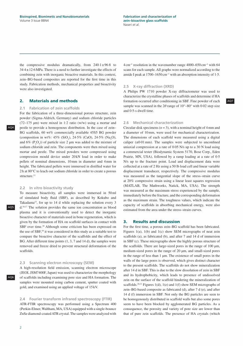
3. Results and discussionFor the first time, a porous zein–BG scaffold has been fabricated. Figures 1(a), 1(b) and 1(c) show SEM micrographs of neat zein scaffolds (a), as fabricated (b), and after 7 and 14 d of immersion in SBF (c). These micrographs show the highly porous structure of the scaffolds. There are large-sized pores in the range of 100 µm, medium-sized pores in the range of 20 µm, and small-sized pores in the range of less than 1 µm. The existence of small pores in the walls of the large pores is observed, which gives distinct character to the present scaffolds. The scaffolds do not show mineralization after 14 d in SBF. This is due to the slow dissolution of zein in SBF and its hydrophobicity, which leads to presence of undissolved zein on the surface of the scaffold hindering the mineralization of scaffolds.20,21 Figures 1(d), 1(e) and 1(f) show SEM micrographs of zein–BG-based composite as fabricated (d), after 7 d (e), and after 14 d (f) immersion in SBF. Not only the BG particles are seen to be homogenously distributed in scaffold walls but also some pores seem to have been blocked by agglomerated BG particles. As a consequence, the porosity and variety of pore size are lower than that of pure zein scaffolds. The presence of HA crystals (which
AQ4
AQ5
AQ6
AQ7
Bioinspired, Biomimetic and NanobiomaterialsVolume 3 Issue BBN4
3
Fabrication and characterization of zein–bioactive glass scaffoldsNaseri et al.
Figure 1. SEM micrographs of (a) pure zein, (b) zein after 7 d in SBF,
(c) zein after 14 d in SBF, (d) zein-60 wt% BG, (e) zein-60 wt% BG
after 7 d in SBF, and (f) zein-60 wt% BG after 14 d in SBF
(a.1) (a.2) (d.1) (d2)
(b.1) (b.2) (e.1) (e2)
(c.1) (c.2) (f.1) (f.2)
100 µm 15 µm
100 µm 15 µm
100 µm 15 µm
100 µm 15 µm
100 µm 1 µm
100 µm 1 µm
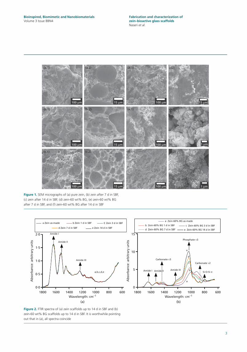
Figure 2. FTIR spectra of (a) zein scaffolds up to 14 d in SBF and (b)
zein-60 wt% BG scaffolds up to 14 d in SBF. It is worthwhile pointing
out that in (a), all spectra coincide
2·0 15
Carbonate ν3
Si-O-Si σ
Carbonate ν2
Amide IIIAmide I Amide II
Phosphate ν3
e
d
c
b
a
10
5
0
Amide I
(a) (b)
Amide II
Amide III
Ab
sorb
ance
: arb
itra
ry u
nit
s
Ab
sorb
ance
: arb
itra
ry u
nit
s
a,b,c,d,e
1·5
1·0
0·5
0·01800
Wavelength: cm−11600 1400 1200 1000 800 600 1800
Wavelength: cm−11600 1400 1200 1000 800 600
c Zein 3 d in SBFa Zein as-madea Zein-60% BG as-made
b Zein-60% BG 1 d in SBF c Zein-60% BG 3 d in SBF
e Zein-60% BG 14 d in SBFd Zein-60% BG 7 d in SBF
b Zein 1 d in SBF
d Zein 7 d in SBF e Zein 14 d in SBF
Bioinspired, Biomimetic and NanobiomaterialsVolume 3 Issue BBN4
4
Fabrication and characterization of zein–bioactive glass scaffoldsNaseri et al.
is confirmed by other techniques as discussed below) after 14 d of immersion in SBF is indicated due to the observation of a fluffy (granular) structure, which suggests that adding BG to zein enhances bioactivity.
FTIR spectra of the as-made and SBF-soaked scaffolds are shown in Figures 2(a) and 2(b) for zein and zein–BG scaffolds, respectively. Figure 2(a) shows three typical amide peaks for zein protein at 1750–1600, 1500–1400, and 1300–1200 cm−1.22 After submersion in SBF, there are no specific peaks that can be related to phosphate or carbonate bonds characteristic of HA formation. FTIR results can thus confirm that pure zein scaffolds do not show bioactivity and there is no HA formation on the scaffold surfaces upon conditioning in SBF for 14 d.
Figure 2(b) shows FTIR spectra of zein-60 wt% BG-based composite scaffolds. Typical amide peaks confirm the presence of zein. HA formation is confirmed by a carbonate ν3 peak at 1454 cm−1, and peaks at 1010 and 700 cm−1 are characteristic of PO43− and Si–O–Si bonding, respectively.23 However, due so some peak overlapping it is difficult to attribute these to specific
components. The phosphate peak not only increases with time but also shifts to higher wavenumber, which is explained by the formation of amorphous calcium phosphate followed by the slow conversion to crystalline HA.
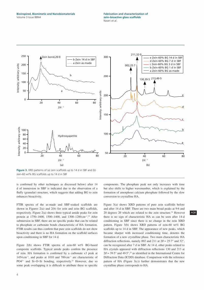
Figure 3(a) shows XRD patterns of pure zein scaffolds before and after 14 d in SBF. There are two main broad peaks at 9·6 and 20 degrees 2θ which are related to the zein structure.24 However there is no sign of characteristic HA as can be seen after 14-d conditioning in SBF since there is no change in the zein XRD pattern. Figure 3(b) shows XRD patterns of zein-60 wt% BG scaffolds up to 14 d in SBF. The appearance of new peaks, which became sharper with increased conditioning time, denotes the formation of a new crystalline phase. Two main characteristic HA diffraction reflections, namely 002 and 211 at 2θ = 25·7° and 32°, can be recognized after 7 d in SBF. At 14 d, other peaks related to HA crystals appeared with diffraction reflections 130 and 213 at 2θ = 39·5° and 49·5°,25 as identified in the International Centre for Diffraction Data (ICDD) database. Comparison with the reference pattern of HA (Figure 3(c)) further demonstrates that the new crystalline phase corresponds to HA.
AQ8
Figure 3. XRD patterns of (a) zein scaffolds up to 14 d in SBF and (b)
zein-60 wt% BG scaffolds up to 14 d in SBF
(a) (b)
b Zein 14 d in SBF
b
a
a Zein as-made
250 300
002,25·7
130,39·5
e
d
c
b
a
213,49·5
211,32·0e Zein-60% BG 14 d in SBFd Zein-60% BG 7 d in SBFc Zein-60% BG 3 d in SBFb Zein-60% BG 1 d in SBFa Zein-60% BG as-made
200
200
150
100
50
200
Zein bond,20·0
150
100
100
Hydroxyapetite
20
2θ : °
2θ : ° 2θ : °
40 60 80 20 40 60 80
80
60
40
20
0
Inte
nsi
ty: a
rbit
rary
un
its
Inte
nsi
ty: a
rbit
rary
un
its
Inte
nsi
ty: a
rbit
rary
un
its
50
020 40 60 80
Bioinspired, Biomimetic and NanobiomaterialsVolume 3 Issue BBN4
5
Fabrication and characterization of zein–bioactive glass scaffoldsNaseri et al.
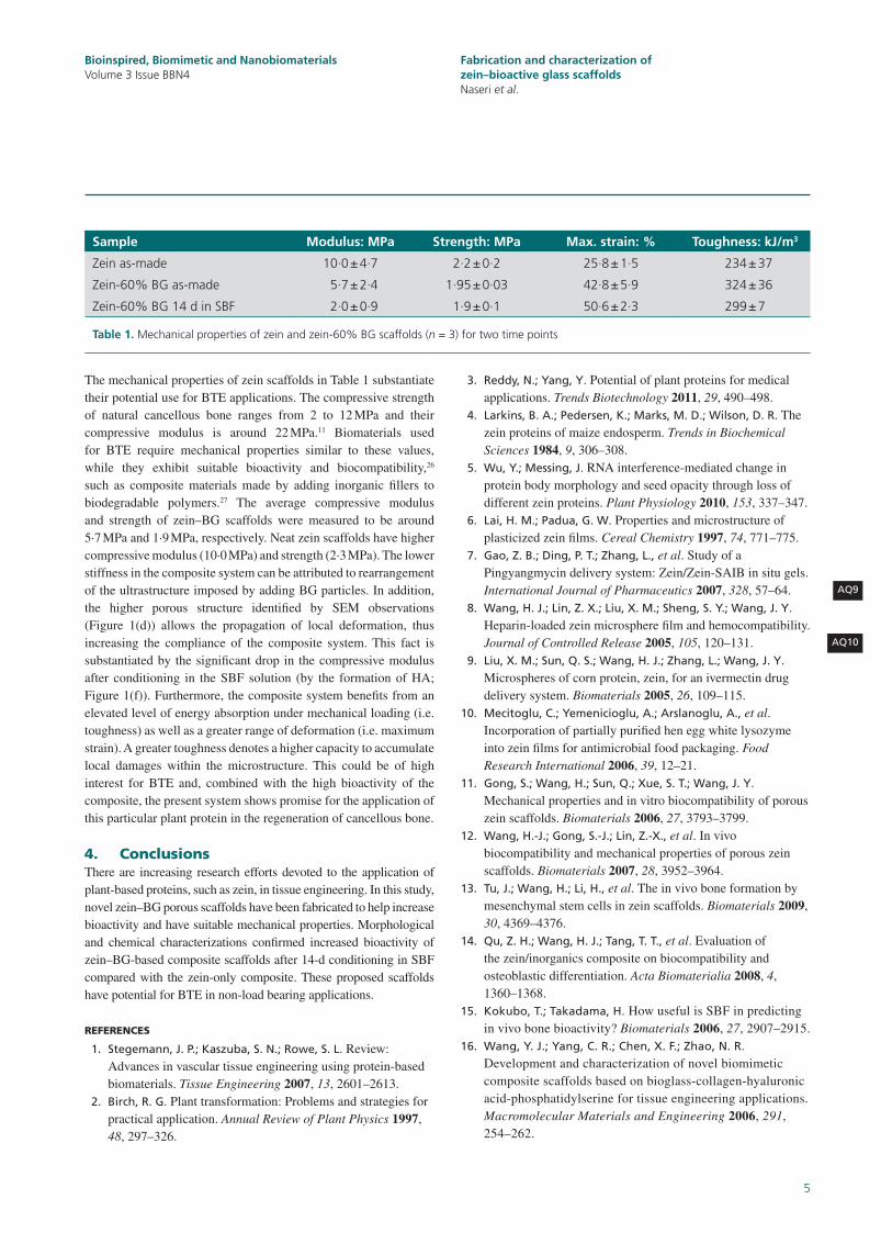
The mechanical properties of zein scaffolds in Table 1 substantiate their potential use for BTE applications. The compressive strength of natural cancellous bone ranges from 2 to 12 MPa and their compressive modulus is around 22 MPa.11 Biomaterials used for BTE require mechanical properties similar to these values, while they exhibit suitable bioactivity and biocompatibility,26 such as composite materials made by adding inorganic fillers to biodegradable polymers.27 The average compressive modulus and strength of zein–BG scaffolds were measured to be around 5·7 MPa and 1·9 MPa, respectively. Neat zein scaffolds have higher compressive modulus (10·0 MPa) and strength (2·3 MPa). The lower stiffness in the composite system can be attributed to rearrangement of the ultrastructure imposed by adding BG particles. In addition, the higher porous structure identified by SEM observations (Figure 1(d)) allows the propagation of local deformation, thus increasing the compliance of the composite system. This fact is substantiated by the significant drop in the compressive modulus after conditioning in the SBF solution (by the formation of HA; Figure 1(f)). Furthermore, the composite system benefits from an elevated level of energy absorption under mechanical loading (i.e. toughness) as well as a greater range of deformation (i.e. maximum strain). A greater toughness denotes a higher capacity to accumulate local damages within the microstructure. This could be of high interest for BTE and, combined with the high bioactivity of the composite, the present system shows promise for the application of this particular plant protein in the regeneration of cancellous bone.
4. ConclusionsThere are increasing research efforts devoted to the application of plant-based proteins, such as zein, in tissue engineering. In this study, novel zein–BG porous scaffolds have been fabricated to help increase bioactivity and have suitable mechanical properties. Morphological and chemical characterizations confirmed increased bioactivity of zein–BG-based composite scaffolds after 14-d conditioning in SBF compared with the zein-only composite. These proposed scaffolds have potential for BTE in non-load bearing applications.
REFERENCES
1. Stegemann, J. P.; Kaszuba, S. N.; Rowe, S. L. Review: Advances in vascular tissue engineering using protein-based biomaterials. Tissue Engineering 2007, 13, 2601–2613.
2. Birch, R. G. Plant transformation: Problems and strategies for practical application. Annual Review of Plant Physics 1997, 48, 297–326.
3. Reddy, N.; Yang, Y. Potential of plant proteins for medical applications. Trends Biotechnology 2011, 29, 490–498.
4. Larkins, B. A.; Pedersen, K.; Marks, M. D.; Wilson, D. R. The zein proteins of maize endosperm. Trends in Biochemical Sciences 1984, 9, 306–308.
5. Wu, Y.; Messing, J. RNA interference-mediated change in protein body morphology and seed opacity through loss of different zein proteins. Plant Physiology 2010, 153, 337–347.
6. Lai, H. M.; Padua, G. W. Properties and microstructure of plasticized zein films. Cereal Chemistry 1997, 74, 771–775.
7. Gao, Z. B.; Ding, P. T.; Zhang, L., et al. Study of a Pingyangmycin delivery system: Zein/Zein-SAIB in situ gels. International Journal of Pharmaceutics 2007, 328, 57–64.
8. Wang, H. J.; Lin, Z. X.; Liu, X. M.; Sheng, S. Y.; Wang, J. Y. Heparin-loaded zein microsphere film and hemocompatibility. Journal of Controlled Release 2005, 105, 120–131.
9. Liu, X. M.; Sun, Q. S.; Wang, H. J.; Zhang, L.; Wang, J. Y. Microspheres of corn protein, zein, for an ivermectin drug delivery system. Biomaterials 2005, 26, 109–115.
10. Mecitoglu, C.; Yemenicioglu, A.; Arslanoglu, A., et al. Incorporation of partially purified hen egg white lysozyme into zein films for antimicrobial food packaging. Food Research International 2006, 39, 12–21.
11. Gong, S.; Wang, H.; Sun, Q.; Xue, S. T.; Wang, J. Y. Mechanical properties and in vitro biocompatibility of porous zein scaffolds. Biomaterials 2006, 27, 3793–3799.
12. Wang, H.-J.; Gong, S.-J.; Lin, Z.-X., et al. In vivo biocompatibility and mechanical properties of porous zein scaffolds. Biomaterials 2007, 28, 3952–3964.
13. Tu, J.; Wang, H.; Li, H., et al. The in vivo bone formation by mesenchymal stem cells in zein scaffolds. Biomaterials 2009, 30, 4369–4376.
14. Qu, Z. H.; Wang, H. J.; Tang, T. T., et al. Evaluation of the zein/inorganics composite on biocompatibility and osteoblastic differentiation. Acta Biomaterialia 2008, 4, 1360–1368.
15. Kokubo, T.; Takadama, H. How useful is SBF in predicting in vivo bone bioactivity? Biomaterials 2006, 27, 2907–2915.
16. Wang, Y. J.; Yang, C. R.; Chen, X. F.; Zhao, N. R. Development and characterization of novel biomimetic composite scaffolds based on bioglass-collagen-hyaluronic acid-phosphatidylserine for tissue engineering applications. Macromolecular Materials and Engineering 2006, 291, 254–262.
AQ9
AQ10
Sample Modulus: MPa Strength: MPa Max. strain: % Toughness: kJ/m3
Zein as-made 10·0 ± 4·7 2·2 ± 0·2 25·8 ± 1·5 234 ± 37
Zein-60% BG as-made 5·7 ± 2·4 1·95 ± 0·03 42·8 ± 5·9 324 ± 36
Zein-60% BG 14 d in SBF 2·0 ± 0·9 1·9 ± 0·1 50·6 ± 2·3 299 ± 7
Table 1. Mechanical properties of zein and zein-60% BG scaffolds (n = 3) for two time points
Bioinspired, Biomimetic and NanobiomaterialsVolume 3 Issue BBN4
6
Fabrication and characterization of zein–bioactive glass scaffoldsNaseri et al.
17. Marelli, B.; Ghezzi, C. E.; Barralet, J. E.; Boccaccini, A.
R.; Nazhat, S. N. Three-Dimensional Mineralization of Dense Nanofibrillar Collagen-Bioglass Hybrid Scaffolds. Biomacromolecules 2010, 11, 1470–1479.
18. Ogino, M.; Ohuchi, F.; Hench, L. L. Compositional dependence of the formation of calcium phosphate films on bioglass. Journal of Biomedical Materials Research 1980, 14, 55–64.
19. Bohner, M.; Lemaitre, J. Can bioactivity be tested in vitro with SBF solution? Biomaterials 2009, 30, 2175–2179.
20. Bai, J.; Alleyne, V.; Hagenmaier, R. D.; Mattheis, J. P.;
Baldwin, E. A. Formulation of zein coatings for apples (Malus domestica Borkh). Postharvest Biology and Technology 2003, 28, 259–268.
21. Zhou, P. Y.; Xia, Y.; Wang, J., et al. Antibacterial properties and bioactivity of HACC- and HACC-Zein-modified mesoporous bioactive glass scaffolds. Journal of Materials Chemistry B 2013, 1, 685–692.
22. Forato, L. A.; Bicudo, T. C.; Colnago, L. A. Conformation of alpha zeins in solid state by Fourier transform IR. Biopolymers 2003, 72, 421–426.
23. Rehman, I.; Knowles, J. C.; Bonfield, W. Analysis of in vitro reaction layers formed on Bioglass (R) using thin-film X-ray diffraction and ATR-FTIR microspectroscopy. Journal of Biomedical Materials Research 1998, 41, 162–166.
24. Lai, H.-M.; Geil, P. H.; Padua, G. W. X-ray diffraction characterization of the structure of zein-oleic acid films. Journal of Applied Polymer Science 1999, 71, 1267–1281.
25. Li, Y.; Yao, C. Mineralization of hydroxyapatite crystallites on zein microspheres. Polymer Composites 2012, 33, 961–966.
26. Jiang, T.; Abdel-Fattah, W. I.; Laurencin, C. T. In vitro evaluation of chitosan/poly(lactic acid-glycolic acid) sintered microsphere scaffolds for bone tissue engineering. Biomaterials 2006, 27, 4894–4903.
27. Rezwan, K.; Chen, Q. Z.; Blaker, J. J.; Boccaccini, A. R. Biodegradable and bioactive porous polymer/inorganic composite scaffolds for bone tissue engineering. Biomaterials 2006, 27, 3413–3431.
WHAT DO YOU THINK?
To discuss this paper, please email up to 500 words to the managing editor at [email protected]
Your contribution will be forwarded to the author(s) for a reply and, if considered appropriate by the editor-in-chief, will be published as a discussion in a future issue of the journal.
ICE Science journals rely entirely on contributions sent in by professionals, academics and students coming from the field of materials science and engineering. Articles should be within 5000-7000 words long (short communications and opinion articles should be within 2000 words long), with adequate illustrations and references. To access our author guidelines and how to submit your paper, please refer to the journal website at www.icevirtuallibrary.com/bbn
Bioinspired, Biomimetic and NanobiomaterialsVolume 3 Issue BBN4
Related Documents


![Bioactive Scaffolds for Regeneration of Cartilage and … · role in reducing cartilage degeneration and decreasing chondrocyte apoptosis on the osteoarthritis therapy [21, 22]. Recent](https://static.cupdf.com/doc/110x72/5f0e7a4e7e708231d43f706e/bioactive-scaffolds-for-regeneration-of-cartilage-and-role-in-reducing-cartilage.jpg)


![Hypoxia-mimicking mesoporous bioactive glass scaffolds with … Biomaterials 2012.pdf · 2016. 2. 6. · biomaterials could induce a hypoxia function [27]. To our best knowledge,](https://static.cupdf.com/doc/110x72/5fe35ec466c7c06113333f28/hypoxia-mimicking-mesoporous-bioactive-glass-scaffolds-with-biomaterials-2012pdf.jpg)