18 Bioactive Scaffolds for the Controlled Formation of Complex Skeletal Tissues Sandra Hofmann 1 and Marcos Garcia-Fuentes 2 1 Institute for Biomechanics, ETH Zurich, Zurich, 2 NANOBIOFAR Group, Dep. Pharmacy and Pharmaceutical Technology, Campus Vida, University of Santiago de Compostela, Santiago de Compostela 1 Switzerland 2 Spain 1. Introduction Most skeletal tissues have the innate capacity to regenerate, at least to a certain extent, without therapeutic intervention. Bone for example has the capacity to repair fractures up to a critical size with the help of local or recruited cells of the patient’s body, and the defects heal by forming new bone that is indistinguishable from uninjured tissue (Deschaseaux et al., 2009; Place et al., 2009). However, traumatic fractures of the lower extremities are one group of orthopedic injuries that often require prolonged rehabilitation, or multiple procedures to achieve maximal functional recovery, and comprise the bulk of nonunion or delayed-union fractures (Deschaseaux et al., 2009). In contrast, articular cartilage, ligaments and tendons are known to have a limited capacity for self-repair and methods to augment their natural healing response are still highly investigated. Biomaterials used to restore the structure and function of lost tissues and organs have evolved greatly due to the large knowledge accumulated on material-protein and material- cell interactions. Current commercially available biomaterials are engineered to be non- toxic, non-immunogenic, and hemocompatible. In contrast to elements of the extracellular matrix of living systems, however, these scaffolds are very poor in information, which makes them suboptimal for many tissue engineering applications. These passive biomaterials are unlikely to guide cell migration and differentiation or controlled matrix deposition, a problem that becomes even more evident in complex tissues with more than one cell type. Furthermore, they also cannot induce tissue neoformation while preventing other undesirable tissue repair processes such as scarring, and they are unable –with the notable exception of some materials in bone regeneration- to promote functional tissue integration in the host, including vascular and/or nervous connectivity. Finally, these passive scaffolds largely lack the capacity to induce cell differentiation, which becomes a major limitation for their use together with current stem cell-based therapies (Huebsch & Mooney, 2009). A promising strategy to overcome these limitations is to design bioactive scaffolds capable of recruiting tissue-forming cells from the body in such a way that they promote cell adhesion and tissue formation within their matrix. Moreover, materials that can recruit endogenous cells of the patient into scaffolds, avoid the expense and difficulties

Welcome message from author
This document is posted to help you gain knowledge. Please leave a comment to let me know what you think about it! Share it to your friends and learn new things together.
Transcript
18
Bioactive Scaffolds for the Controlled Formation of Complex Skeletal Tissues
Sandra Hofmann1 and Marcos Garcia-Fuentes2 1Institute for Biomechanics, ETH Zurich, Zurich,
2NANOBIOFAR Group, Dep. Pharmacy and Pharmaceutical Technology, Campus Vida, University of Santiago de Compostela, Santiago de Compostela
1Switzerland 2Spain
1. Introduction
Most skeletal tissues have the innate capacity to regenerate, at least to a certain extent, without therapeutic intervention. Bone for example has the capacity to repair fractures up to a critical size with the help of local or recruited cells of the patient’s body, and the defects heal by forming new bone that is indistinguishable from uninjured tissue (Deschaseaux et al., 2009; Place et al., 2009). However, traumatic fractures of the lower extremities are one group of orthopedic injuries that often require prolonged rehabilitation, or multiple procedures to achieve maximal functional recovery, and comprise the bulk of nonunion or delayed-union fractures (Deschaseaux et al., 2009). In contrast, articular cartilage, ligaments and tendons are known to have a limited capacity for self-repair and methods to augment their natural healing response are still highly investigated. Biomaterials used to restore the structure and function of lost tissues and organs have evolved greatly due to the large knowledge accumulated on material-protein and material-cell interactions. Current commercially available biomaterials are engineered to be non-toxic, non-immunogenic, and hemocompatible. In contrast to elements of the extracellular matrix of living systems, however, these scaffolds are very poor in information, which makes them suboptimal for many tissue engineering applications. These passive biomaterials are unlikely to guide cell migration and differentiation or controlled matrix deposition, a problem that becomes even more evident in complex tissues with more than one cell type. Furthermore, they also cannot induce tissue neoformation while preventing other undesirable tissue repair processes such as scarring, and they are unable –with the notable exception of some materials in bone regeneration- to promote functional tissue integration in the host, including vascular and/or nervous connectivity. Finally, these passive scaffolds largely lack the capacity to induce cell differentiation, which becomes a major limitation for their use together with current stem cell-based therapies (Huebsch & Mooney, 2009). A promising strategy to overcome these limitations is to design bioactive scaffolds capable of recruiting tissue-forming cells from the body in such a way that they promote cell adhesion and tissue formation within their matrix. Moreover, materials that can recruit endogenous cells of the patient into scaffolds, avoid the expense and difficulties

Regenerative Medicine and Tissue Engineering - Cells and Biomaterials
394
associated with culture, storage and distribution of cells, not to mention other immunological considerations (Discher et al., 2009; Place, ES et al., 2009). Bioactivity is defined as having the capacity to interact with a living tissue or system. In terms of scaffold design, we define a bioactive scaffold as one in which biological functionality has been integrated to provide an information-rich support material for tissue engineering. Bioactive scaffolds are designed to control cell- and tissue responses, and to provide a more efficient integration with the host. In this manuscript we will cover the most important scaffolds presenting a biomimetic composition (Section 2), the main strategies to introduce biomimetic features in scaffolds (Section 3), their expected applications in skeletal tissue engineering and the main challenges remaining for the successful application of this concept (Section 4). We will highlight methods of how scaffold materials can be modified to be bioactive, how a scaffold can promote the body’s innate capabilities to heal and recruit cells to the site of injury. Bioactive scaffolds have already found their way into clinics and hold great promise to serve as a manageable alternative to tissue engineered implants that face a vast array of regulatory and logistical issues. In the end, products on the market need to show convincing clinical performance as well as cost-effectiveness. We will therefore not cover the traditional tissue engineering approach to seed and culture scaffolds ex vivo with cells before implantation. However, many of the methods presented here have been discovered from in vitro cell cultures and might as well be of high importance in these strategies.
2. Scaffolds with a biomimetic composition
Three dimensional biopolymer networks are major players in physiological wound healing. It is assumed that wound healing progresses through three stages: (i) blood clotting and inflammation; (ii) granulation tissue; (iii) tissue remodeling. Each of these stages can be associated to the prevalence of a particular biopolymer network, i.e. fibrin (stage i), unstructured collagen/glycosaminoglycan (GAG, for an acronyms list refer to Section 6) extracellular matrix (stage ii), structured collagen/GAG extracellular matrix (stage iii) (Braiman-Wiksman et al., 2007). The delicate choreography of deposition and remodeling of these biopolymer networks is coupled to the activation of relevant signaling cascades, directing immune cell invasion, inflammatory processes, progenitor cell attraction, differentiation, extracellular matrix (ECM) deposition, ECM remodeling and revascularization of the newly formed tissue graft (Gillitzer & Goebeler, 2001). These critical processes in wound healing underline the importance of fibrin, collagens and GAGs in tissue formation and fundament their recognition as bioactive polymers for tissue engineering. Besides, this biomimetic approach has also suggested the potential of reprocessed tissues for tissue/organ engineering. Reprocessed tissues are complex, structured scaffolds prepared by performing different extraction treatments (i.e. demineralization, decellularization) to biological tissues.
2.1 Fibrin Fibrin is a biopolymer derived from the crosslinking of fibrinogen, the major protein involved in blood coagulation. As a medical product, fibrin gel is a blood derivative, typically presented as a two syringe-system with a fibrinogen solution and a thrombin solution having a common port. Once casted, fibrinogen is cleaved by thrombin, resulting in
Bioactive Scaffolds for the Controlled Formation of Complex Skeletal Tissues
395
the formation of a gel in a few seconds. In Europe, fibrin glue has been available as tissue adhesive, sealant and hemostat from the 1980’s. Its approval by the FDA was delayed until 1998 due to security standards regarding blood derivative processing. Fibrin is now regularly used in surgery around the world for a broad number of indications (Spotnitz, 2010). Fostered by its success in surgery, fibrin has gained increased attention in tissue engineering strategies. Fibrin has the great advantage that it can be delivered by minimally-invasive methods to the tissue defect, a property that is only shared with a few materials capable of gelling in situ (Kretlow et al., 2007). Due to this controlled gelling capacity, fibrin glue can be casted in defined shapes and be applied to fill wound beds. For cell delivery, fibrin glue excels in many ways, as cells can be injected with fibrinogen solution and are then retained at the wound site as the fibrin gels. As fibrin has been designed by nature as the emergency “physiological scaffold” used in wound healing, fibrin gels are well structured to accommodate cells (T.A.E. Ahmed et al., 2008). As a cell delivery matrix, fibrin glue has been used with mesenchymal stem cells (MSCs) for bone (Dozza et al., 2011; Liao et al., 2011), cartilage (T.A. Ahmed et al., 2011; J.S. Park et al., 2011), ligament and tendon regeneration (Lim et al., 2004; Soon et al., 2007). It has also been used with chondrocytes in cartilage regeneration (Eyrich et al., 2007; S.J. Lee et al., 2011; Peretti et al., 2006; Singh et al., 2011), with osteoblasts for bone regeneration (S.J Kim et al., 2007), and fibroblasts in tendon regeneration (Chun et al., 2003). Fibrin glue can also be used as a drug delivery device for growth factors (GFs), either in combination with cell transplantation or not. Several studies have shown that fibrin glue can provide sustained release of GFs during a few days to one week (Spicer & Mikos, 2010). Longer GF retention times can be achieved by modifications in the GF (Schmoekel et al., 2004), by integrating high GF-affinity moieties in the fibrin chains (Merritt et al., 2010), or by covalent linking of the GFs (Drinnan et al., 2010; Schmoekel et al., 2005). Examples of GFs that have been integrated in fibrin gels in skeletal tissue engineering applications include BMP-2 (Kang et al., 2011; Schmoekel et al., 2004; Schmoekel et al., 2005), TGF-β1 and TGF-β3 (Drinnan et al., 2010; J.S. Park et al., 2011; W. Wang et al., 2010b). A similar concept to GF-loaded fibrin is platelet-rich fibrin, a new blood-derivative formed by platelet concentration. Platelet-rich fibrin also comprises fibrinogen for its polymerization in situ, although handling is different as compared to fibrin glue (Dohan et al., 2006). Due to this different processing, platelet-rich fibrin contains a concentrate of GFs that are present in platelets (i.e. PDGF, TGF, IGF, VEGF, EGF), transforming this product in a truly blood-clot biomimetic (Eppley et al., 2004). Platelet-rich fibrin, however, presents some disadvantages compared to fibrin glue encapsulating GFs. These are mainly related to potential variability in GF-content between batches and undefined GF doses. Besides those indicated for GF delivery, fibrin glue has some important shortcomings. Firstly, fibrin is known to shrink as gelation occurs, and overall, fibrin lacks adequate mechanical properties (Sierra, 1993). Those limitations are particularly critical to skeletal tissue engineering, as many tissues to be regenerated are load bearing. Moreover, fibrin gels are known to degrade in a few days in vivo, due to the activation of plasminogen by the cells, which leads to formation of the serine protease plasmin and to subsequent fibrinolysis (Ye et al., 2000). Again, this short biodegradation time is probably inadequate for most applications in skeletal tissue engineering, as regeneration of these tissues usually takes several weeks or months. To address these limitations, fibrin glue scaffolds can be optimized

Regenerative Medicine and Tissue Engineering - Cells and Biomaterials
396
by using a solid porous scaffold for reinforcement (Schagemann et al., 2010; Z.H. Wang et al., 2010a; W. Wang et al., 2010b), or by the use of enzymatic inhibitors (Fussenegger et al., 2003). Eyrich et al. reported some optimized conditions that lead to fibrin gels that were stable for up to three weeks in vitro (Eyrich et al., 2007). Despite being a material with a long history of application, fibrin has been studied recently in some advanced tissue engineering strategies for bone regeneration. Dozza et al. showed that MSCs seeded in fibrin scaffolds resulted in better bone formation around an uncemented hip prosthesis and better bone-prosthesis contact compared to the control (Dozza et al., 2011). Liao et al. showed that MSCs seeded in either platelet-rich fibrin or a Medpor® sheet filled with platelet-rich fibrin resulted in approximately 4-fold higher bone formation as compared to empty defects in a mandibular reconstruction model (Liao et al., 2011). Leong et al. confirmed the osteogenic differentiation of MSCs in fibrin glue filled polycaprolactone/tricalcium phosphate scaffolds (Leong et al., 2008). Fibrin-based devices have also been recently studied for cartilage and ligament/tendon regeneration. MSCs seeded in fibrin glue containing TGF-β3 showed the capacity to regenerate articular cartilage (J.S. Park et al., 2011). Eyrich et al. demonstrated the capacity to form cartilage with chondrocyte-seeded stable fibrin gels (Eyrich et al., 2007). Wegener et al. used polyester scaffolds filled with a fibrin gel (with or without chondrocytes) to repair cartilage defects in the femoral condyle in sheep (Wegener et al., 2010). This study confirmed cartilage formation and improved O’Driscoll scores in defects filled with these scaffolds. Lee et al. formed a cartilage layer by seeding chondrocytes in fibrin gels on top of auricular implants as a strategy to minimize morbidity associated with implant dislodgement (S.J. Lee et al., 2011). Wang et al. used polyester/fibrin gel constructs loaded with MSCs and TGF-β1 to promote restoration of full-thickness cartilage defects (W. Wang et al., 2010b). Also recently, Scotti et al. used fibrin gels to promote cell adhesion to devitalized spongiosa cylinders used for engineering osteochondral grafts (Scotti et al., 2010). In two studies by the laboratory of Lee, MSCs embedded in fibrin glue were able to promote better osseointegration of tendon grafts (Lim et al., 2004; Soon et al., 2007). A clinical study on mandibular degree II furcation defects has shown that patients treated with platelet-rich fibrin glue and an open flap debridement presented statistical improvements in all clinical parameters as compared to patients treated with just open flap debridement (Pradeep & Sharma, 2011). In another clinical study, MSCs in platelet-rich fibrin glue were implanted in patients with condyle defects for cartilage regeneration (Haleem et al., 2010). The study showed improvement of patient’s symptoms in all subjects over the follow-up period of 12 months, achieving nearly normal arthroscopic scores after the follow-up and partial to total cartilage surface regeneration.
2.2 Collagen Collagen is a family of proteins that are the main components of connective tissue, and the most abundant proteins in mammals (>90% of the ECM) (Van der Rest & Garrone, 1991). Collagen type-I is a major component of scar tissue, while this and other types of collagens are present in physiological tissue (and regenerated tissue if correctly remodeled) (Fratzl 2008). Specifically in skeletal tissues, collagen type-I is preeminent in bone, tendons and muscle. Collagens in cartilage change with specific tissue types: collagen type-II is preeminent in hyaline cartilage, collagens type-I and –II are present in fibrocartilage and collagen type-X is a marker of hyperthrophic cartilage. Collagen type-I is a long, stiff, triple-

Bioactive Scaffolds for the Controlled Formation of Complex Skeletal Tissues
397
stranded helical structure comprising two identical chains (i.e. one α1(I)-chain and one α2(I)-chain). This α2(I)-chain contains the sequence repeat (G-X-Y)n, X being frequently proline and Y hydroxyproline, that allow the formation of a triple helix (Ricard-Blum & Ruggiero, 2005). In physiological tissues, collagens show very defined structures. For instance, collagen type-I forms parallel, longitudinal fibers in tendons and ligaments and long bones. In lamellar bone, collagen type-I fibrils run in parallel to other fibrils in the same layer, but are aligned in the opposite direction in alternating layers (Fratzl 2008). In cartilage, collagen type-II is crosslinked to proteoglycans by the action of collagen type-IX that acts as a biological “spacer” (Fratzl 2008; Lodish, H et al., 2002). As materials for tissue scaffolds, collagens have been thoroughly investigated due to their biomimetic nature, their biocompatibility and abundance. Indeed, collagen type-I has shown to be degradable, and highly biocompatible, particularly after proteolytic removal of telopeptides (Glowacki & S. Mizuno, 2008). Currently, collagen is FDA approved for implantation. Collagen scaffolds can be prepared in two forms: as hydrogels or as fibers in a mesh like structure (also named as “collagen sponges” or “collagen foams”). Collagen fibers can be reinforced by crosslinking, a modification that can be performed with either physical or chemical methods (Glowacki & S. Mizuno, 2008). Some chemical crosslinkers such as glutaraldehyde have been related to cytotoxicity (Middelkoop et al., 1995), although others have shown that this limitation can be overcome by protocol optimization (Nimni et al., 1987). Today, other more biocompatible crosslinkers can be selected (Kubow et al., 2009; Mekhail et al., 2010). Collagen fibers manufactured by optimized protocols present an adequate porous microstructure and promote cell adhesion (Glowacki & S. Mizuno, 2008). In skeletal tissue engineering, collagen scaffolds have been used for seeding osteoblasts (Laflamme & Rouabhia, 2008), periosteal cells (Ryu et al., 2010), MSCs (Brady et al., 2010; Dozza et al., 2011; Jäger et al., 2008), chondrocytes (Oliveira et al., 2010; Zhou et al., 2011) and fibroblasts (George et al., 2008; Mekhail et al., 2010). Collagen type-I has shown the capacity to specifically upregulate osteoblastic function and bone formation (Lynch et al., 1995), and this property has been linked to the interaction of this collagen with integrin α2β1 (M. Mizuno et al., 2000). On the other hand, collagen type-II scaffolds have shown favorable interactions for cartilage tissue engineering and cartilage regeneration, particularly in combination with GAGs (Chang et al., 2007; Wu et al., 2010). These results highlight, once again, the importance of providing the cells with scaffold compositions that mimic their natural environment. To enhance their regenerative properties even further, collagen scaffolds can be loaded with GFs such as BMPs (Laflamme & Rouabhia, 2008), TGF (Pabbruwe et al., 2010) and FGF (Pang et al., 2010). Collagen-based sponge constructs be impregnated in BMP-2 are already available as a FDA-approved commercial product for spinal reconstruction (InfuseTM, Medtronic, Inc.). Nevertheless, collagen has rather limited controlled release capacity, with most occurring in the first days (Maeda et al., 1999). Release properties can be improved to a limited extent by crosslinking (Fujioka et al., 1998) or drastically, by embedding in the scaffold a suitable drug delivery device (Fujioka et al., 1998; J.E. Lee et al., 2004). Apart from its limited capacity to control drug release, the main limitations of collagen scaffolds are their poor mechanical properties. Indeed, collagen scaffolds show no rigidity and shrink due to the cohesive forces that appear as ECM is deposited. As with fibrin, this shrinking is observed already in vitro, and clearly show the intrinsic limitations of this material to reconstruct tissue constructs with defined shapes: ultimately, one of the main objectives of scaffold implantation. Besides, this lack of mechanical competence might

Regenerative Medicine and Tissue Engineering - Cells and Biomaterials
398
compromise the integrity of the scaffold upon implantation resulting in implant failure, non-union or even ectopic tissue formation. Again, it needs to be considered that for most skeletal tissue engineering applications, scaffolds will be exposed to considerable mechanical stress. The mechanical properties of collagen scaffolds can be improved by the integration of a reinforcing substructure (Zhou et al., 2011) or using composites of collagen with mechanically competent materials (Lee et al., 2004). Apart from this limitation, collagen scaffolds might lead to some minor immunological problems upon implantation if the telopeptides are not correctly removed from the materials by proteolysis (S. Mizuno & Glowacki, 1996). Due to their many intrinsic advantages, collagen scaffolds have been applied to the regeneration of all skeletal tissues. Zhang et al. studied chitosan/collagen scaffolds bioactivated with an adenovirus carrying a BMP-7 transgene and seeded with periodontal ligament cells in dental implant defects (Y. Zhang et al., 2007). Histomorphological and qRT-PCR analysis were consistent with more bone formation and higher expression of bone-matrix markers (alkaline phosphatase, osteopontin, bone sialoprotein). Xu et al. studied collagen type-I scaffolds integrating bone sialoprotein for their capacity to induce osteoblast differentiation, and found that this treatment resulted in early mineral deposition by proliferating repair-cells in calvarial defects. Pabburke et al. implanted collagen-based constructs seeded with MSCs in sheep meniscal disks, and confirmed by histomorphometric analysis that they improve integration compared to the scaffolds alone and the control (Pabbruwe et al., 2009; Pabbruwe et al., 2010). Zhou et al. showed that substructure-reinforced collagen matrixes seeded with chondrocytes supported neocartilage formation (Zhou et al., 2011). Chen et al. tested the potential of genipin-crosslinked collagen type-II scaffolds seeded with MSCs for cartilage repair in an osteochondral defect and observed that after 2 months, there were clear signs of cartilage formation in the defect (W.C. Chen et al., 2011). After 24 weeks, cartilage in the defect presented the same structure as native cartilage. A recent study has confirmed the efficacy of knitted silk-collagen sponge scaffolds as support for embryonic stem cell-derived MSCs in tendon regeneration (J.L. Chen et al., 2010). Being one of the first and most widely used materials studied for tissue engineering, collagen has already been investigated in several clinical studies (Rodkey et al., 1999; Stone et al., 1997) that have ultimately resulted in collagen regulatory approval. Intense clinical research continues, as new indications for collagen-based scaffold technologies are sought and long-term clinical outcomes are reevaluated. In a recent study, Crawford et al. studied the safety and healing potential of commercial cartilage matrix seeded with autologous chondrocytes (NeoCart) in the human knee (Crawford et al., 2009). The study revealed significantly reduced pain in the treatment group, with neocartilage formation that was well integrated. Welsch et al. compared cartilage repair in the femoral condyle after autologous chondrocyte transplantation in a hyaluronan-based scaffold or a collagen-based scaffold (Welsch et al., 2010). Results based on morphological scores and T2-mapping showed that most parameters were similar for both scaffolds, however, collagen showed significantly better constitution of the surface and higher T2-relaxation times.
2.3 Glycosaminoglycans Glycosaminoglycans (GAGs) are linear polysaccharides consisting of repeating hexuronic acid bonded to a hexosamine (Iozzo, 2000). GAGs are usually sulfated and linked to a protein domain (a structure that is named proteoglycans). Hyaluronan is exceptional in this regard as it is the only kind of GAG that is neither modified by sulfonation nor attached

Bioactive Scaffolds for the Controlled Formation of Complex Skeletal Tissues
399
covalently to proteins. Nevertheless, hyaluronan does attach by non-covalent interactions to other molecules of the ECM and to cells. Other important GAGs are dermatan sulfate, chondroitin sulfate, heparin, heparan sulfate and keratan sulfate. GAGs have important physiological functions as “space fillers”, in shock absorption, in preventing coagulation, and in GF binding. Studies have also shown the importance of GAGs in embryogenesis and tissue regeneration (Fedarko et al., 1992; Toole, 2001). However, the complete functionality of GAGs is still a topic requiring further investigation. Unmodified GAGs tend to form polymer solutions or gels that dissolve slowly due to the high molecular weight of the components. This dissolution of the matrices together with their poor mechanical properties are unsuitable for tissue engineering. Therefore, GAGs are most often not used by themselves to form scaffolds, but rather as composites (Garcia-Fuentes et al., 2008; Min et al., 2010; Moss et al., 2010; S.J. Park et al., 2009; Schagemann et al., 2010). This role is even more prominent for heparin and heparan sulfate, which are typically added in very small proportions as GF-complexing agents in drug delivery formulations (Biondi et al., 2008; N.X. Wang & von Recum, 2011). Alternatively, GAGs can be chemically modified as a more insoluble compound (e.g. Hyaff®, Fidia Farmaceutici s.p.a) or as crosslinked networks (Crescenzi et al., 2003; Zheng Shu et al., 2004). In skeletal tissue engineering studies, GAG-containing scaffolds have been seeded with osteoblast-like cells (Wagner et al., 2007), chondrocytes (Nuernberger et al., 2011; Schagemann et al., 2010), MSCs (J.L Chen et al., 2010; Min et al., 2010; Schwartz et al., 2011), and fibroblasts (Irie et al., 2011; Sawaguchi et al., 2010). Hyaluronan interacts specifically with cells through CD44, RHAMM and ICAM-1 (Tammi et al., 2002; Toole, 2004). These interactions trigger signaling cascades that could result in the observed capacity of hyaluronan to instruct tissue neo-formation and regeneration. For illustration, hyaluronan has shown pro-proliferative properties (Zou et al., 2004), capacity to enhance unspecific ECM deposition (Garcia-Fuentes et al., 2009), to induce cartilage (Allemann et al., 2001; Williams et al., 2003; Yamane et al., 2005) and ligament (Cristino et al., 2005) formation, capacity to maintain embryonic stem cells in undifferentiated state (Gerecht et al., 2007), and capacity to modulate inflammatory and catabolic markers (Grigolo et al., 2005; Homandberg et al., 2004). Hyaluronan biological properties, though, seem to be very dependent on its molecular weight, and the presence of hyaluronan oligosaccharides have been connected to chondrocyte-driven chondrolysis (Knudson et al., 2000). GAG-based or GAG-modified scaffolds have also been applied with GFs for sustained delivery. Again, this mimics a physiological process, as sulfated GAGs are GF reservoirs in the ECM. For most GFs, GAG binding occurs through specific non-covalent interactions between a so-called heparin binding domain in the polypeptide and the sulfated GAG (Iozzo, 2000). Modification of drug delivery systems with sulfated GAGs for specific binding of GFs is a usual technological strategy, commonly practiced as a surface modification (i.e. “surface heparinization”) (Biondi et al., 2008; N.X. Wang & von Recum, 2011). An alternative but still rather unexplored strategy is the design of delivery systems based on crosslinked sulfated GAGs for GF-delivery applications. Drug delivery devices modified with sulfated GAGs have been applied to the delivery of TGF-β1 (Chou et al., 2006; Lee et al., 2004) and FGF-2 (Mi et al., 2006) among others. Heparin and other sulfated GAGs known to interact with GFs can enhance markedly the affinity of these molecules for the drug delivery system and change the release profile of the device, as reviewed before (N.X. Wang & von Recum, 2011). In general terms, when sulfated GAGs are integrated in the composition of a
Regenerative Medicine and Tissue Engineering - Cells and Biomaterials
400
hydrogel, they show high affinity for the GF and result in sustained release profiles (Benoit & Anseth, 2005b). In the work of Cai et al. (Cai et al., 2005), FGF-2 was released from several crosslinked GAG-based hydrogels in a sustained way for over a month. Moreover, it was found that hydrogels presenting immobilized heparin further enhanced the retention of FGF-2. Interestingly, the bioactivity of the released GF was confirmed both in the study of Benoit et al. (Benoit & Anseth, 2005b) and in that from Cai et al. (Cai et al., 2005). GAG-based materials, particularly hyaluronan, have been extensively investigated for skeletal tissue regeneration. Patterson et al. prepared hyaluronan hydrogels with different degradation rates by photo-crosslinking to control the release of BMP-2 in a rat calvarial defect model (Patterson et al., 2010). They showed that tissue regeneration could be achieved with any of the BMP-2 loaded hydrogels tested. However, the fastest and the slowest degrading hydrogels resulted in more organized bone structures. Kang et al. investigated the possibility of coating porous polyester scaffolds with a fibrin/hyaluronan matrix loaded with BMP-2, and the effect of such constructs in MSCs (Kang et al., 2011). The results showed that MSCs were differentiated more efficiently and resulted in better bone formation when BMP-2 was sustainably released from the fibrin/hyaluronan constructs as compared to BMP-2 in solution. Chen et al. showed that polyester scaffolds coated with hyaluronan/methylated collagen were able to enhance bone formation by MSCs as compared to non-coated polyester systems (M. Chen, et al., 2010). In the study published by Irie et al., chitosan/hyaluronan fibers seeded with fibroblasts were tested for rabbit medial collateral ligament reconstruction (Irie et al., 2011). The in vivo test indicated that ligaments treated with cell-seeded scaffolds showed enhanced collagen type-I deposition and mechanical strength as compared to non cell-seeded ones. Cartilage and osteochondral structures have been the main focus of attention for GAG-based scaffolds, as GAGs are particularly important components of cartilage ECM. Tan et al. showed the potential of new oxidized hyaluronan/N-succinyl chitosan systems that are able to gel in situ for the delivery of chondrocytes (Tan et al., 2009). Eggelet et al. tested a cell-free polyester/hyaluronan scaffold to cover microfractured full-thickness articular cartilage defects in sheep, and achieved significant improvements in histological structure, and in collagen type-II content as detected by immunohistochemistry (Erggelet et al., 2009). Im et al. designed a hyaluronan-atelocollagen/hydroxyapatite-tricalcium phosphate composite scaffold for testing in osteochondral defects in minipigs (Im et al., 2010). Clinical scoring, histological and mechanical results showed comparable results for this composite scaffold with or without seeded chondrocytes and for defects refilled with the removed osteochondral pieces. Moreover, all these groups showed significantly higher performance than the negative control (empty defects), but comparable in many aspects to native cartilage. In the studies of Moss et al. and Huang et al., GAG-based scaffolds were used for reconstitution of the nucleus pulposus of the intervertebral disc (Huang et al., 2011; Moss et al., 2010). The results of these studies sustained the possibility to maintain disc height and induce intervertebral disc regeneration upon implantation of these scaffolds. Interestingly, the study of Moss et al. compared hyaluronan scaffolds with a composite of hyaluronan and an elastin-based peptide (Moss et al., 2010). Although the composites presented better mechanical properties, this improvement did not translate in enhanced regeneration. Nehrer et al. and Welsch et al. have published clinical evaluations of hyaluronan scaffolds seeded with chonodrocytes for cartilage regeneration (Nehrer et al., 2009; Welsch et al., 2010). The study by Nehrer supported the interest of Hyalograft C for cartilage regeneration in healthy young patients with isolated cartilage defects, but not for salvage procedures

Bioactive Scaffolds for the Controlled Formation of Complex Skeletal Tissues
401
(Nehrer et al., 2009). Similarly positive results were obtained by Welsch et al. for condyle regeneration, although this study showed some slight advantages of collagen-based scaffolds compared to the hyaluronan-based ones.
2.4 Reprocessed tissue scaffolds From the point of view of biomimetism, reprocessed tissue scaffolds (e.g. demineralized bone matrix and decellularized tissues) represent probably the most advanced scaffolds available. These natural scaffolds recapitulate not only the complex composition, but also the structure of the native tissues (Danti et al., 2007; Traphagen & Yelick, 2009). This cannot be achieved with the scaffolds prepared from extracted ECM compounds such as those presented before, because the processing steps used to extract the biopolymers completely erases the morphology, histological topology, the precise interactions between different components, the molecular alignment, and other subtle cues from the materials. While both demineralized bone matrix (DBM) and decellularized tissue can be considered similar concepts (“reprocessed tissue scaffolds”), their processing methods and characteristics differ and are discussed separately herein. Demineralized bone matrix is prepared by acid extraction of allograft bone (Urist, 1965). It results in the elimination of the major part of the mineral phase and the immunogenic components of bone, but retains collagen that provides a structured osteoconductive scaffold and a soluble protein fraction comprising several GFs, BMPs among them (Dinopoulos & Giannoudis, 2006; Reddi, 1998). It is also usual to retain a very low fraction of mineral bone phase after the demineralization step (2% approx.). The exact composition of DBM -including the GF content- will depend heavily on the exact protocol followed for mineral extraction, and thus, DBM from different commercial sources might show considerable variability. Extensive studies have shown that DBM is osteoinductive and osteoconductive (Dahners & Jacobs, 1985; Martin Jr et al., 1999). Osteoinduction mechanism by DBM seems to be very dependent on the environment. DBM implanted in submuscular/subcutaneous tissue undergoes ossification through a process recapitulating several steps from the endochondral bone formation process. DBM implanted in calvarial defects direct MSC differentiation to osteoblasts, similarly to the process followed in intramembranous ossification (J. Wang & Glimcher, 1999a, 1999b; J. Wang et al., 2000). DBM by itself is presented in the form of a powder, but is usually mixed with other suitable materials (e.g. hyaluronan, poloxamer) to form a paste that can be easily casted at the bone defect site (Dinopoulos & Giannoudis, 2006; Juang Ming Yee et al., 2003). Mixing DBM with other materials, however, changes the product from a regulatory perspective. DBM can be supplemented with suitable cell types as a strategy to promote tissue regeneration. For instance, some articles report on seeding chondroblasts in DBM as a potential alternative in cartilage tissue engineering (Jin et al., 2006; Z.H. Wang et al., 2010a). A large number of studies have also investigated MSC-seeded DBM (e.g. see Gurevitch et al., 2003; Liu et al., 2010a). DBM can also be supplemented with additional GFs in an attempt to boost their regenerative capacity. DBM has been mainly loaded with BMPs (Lammens et al., 2009) –and particularly, with BMP-2- for bone tissue engineering (Lammens et al., 2009; H. Lin et al., 2008). DBM has also been loaded with other GFs such as TGF-β1 (Moxham et al., 2009). Overall, DBM presents very little capacity to bind additional GFs, and therefore is not an ideal choice as a sustained delivery system. Modification, for instance by heparin addition, can increase GF binding affinity. First DBMs were introduced

Regenerative Medicine and Tissue Engineering - Cells and Biomaterials
402
to the clinics during the 1990’s, and there are currently more than 20 commercial products available based on this technology. Therefore, there is currently large clinical evidence supporting their efficacy for several orthopedic conditions (Irinakis, 2011; Y.K. Kim et al., 2010; Topuz et al., 2009). Decellularized tissue scaffolds are prepared by cell lysis, induced by physical and/or chemical treatments, followed by cell component removal treatments. Mostly, chemical methods are used for this last step (Gilbert et al., 2006). Similarly to DBM, decellularized tissues present the biomimetic topology, tissue-conductive capacity and regeneration-inductive properties arising from the presence of physiological amounts of GFs in the ECM. However, these beneficial characteristics are heavily dependent on the processing protocol followed for decellularization (Gilbert et al., 2006; Ingram et al., 2007; Woods & Gratzer, 2005). Moreover, this protocol might also influence other critical parameters such as the decellularization efficacy and the complete removal of chemical agents from the natural scaffold. Although decellularized tissues and organs are being investigated in several tissue engineering applications, they have not been so popular for skeletal tissue engineering, as scientist have focused on these technologies for engineering high-complexity, localized organs such as the heart (Ott et al., 2008). That is except for DBM, which can be considered also a decellularized tissue scaffold. Some articles have supported the concept of decellularized cartilage as a chondroconductive matrix in cartilage tissue engineering (Elder et al., 2010; Gong et al., 2011; Hou et al., 2011; Secretan et al., 2010; Stabile et al., 2010). Indeed, in vitro tests have shown the possibility to form cartilage tissue in vitro by chondrocyte seeding on a decellularized tissue scaffold (Gong et al., 2011). This ability to support cartilage formation translates to advanced in vivo models such as a for larynx reconstruction in a rabbit model (Hou et al., 2011), or for meniscus reconstruction in sheep (Stabile et al., 2010). First proof-of-concept of the therapeutic interest of this concept in humans is available through the successful transplantation of an MSC-seeded decellularized airway in a 30-years old patient with bronchomalacia (Macchiarini et al., 2008). Decellularized tissues have also been applied to ligament and tendon tissue engineering in vitro, with successful results (Abousleiman et al., 2009; Deeken et al., 2011; Ingram et al., 2007; MacLean & Gratzer, 2011; Woods & Gratzer, 2005). Further animal experiments and clinical studies will be necessary to validate the potential of this strategy. Similarly, other studies have indicated the possibility to use decellularized tissue scaffolds also for skeletal muscle repair and adipose-tissue engineering (L. Flynn et al., 2007; L.E. Flynn, 2010; Gillies et al., 2010; Merritt et al., 2010).
3. Strategies for scaffold bioactivation
As compared to nature’s derived biopolymers, synthetic materials for tissue engineering offer important advantages such as more reproducible manufacture, tailored mechanical and biodegradation properties, and diverse processing options. With a few exceptions (e.g. titanium) synthetic materials do not interact favorably with the body, rendering them incapable of actively promoting tissue regeneration. Despite this limitation, synthetic biomaterials can be engineered to promote their capacity to cross-talk with relevant cell populations. Indeed, bioactive scaffolds can also be prepared from synthetic materials by physical adsorption or chemical immobilization of biomolecules or oligopeptides on the scaffold surface, or by physical entrapment of bioactive molecules alone or incorporated in a

Bioactive Scaffolds for the Controlled Formation of Complex Skeletal Tissues
403
drug delivery system into the scaffold. These strategies can also be applied to enhance the bioactivity of scaffolds made from ECM-native materials such as those covered in the previous section.
3.1 Scaffolds as drug delivery systems Engineered tissues need not only to fill a defect and to integrate into a host tissue, but they also need to meet the demands of a constantly changing - through growth or adaptation - tissue. It was hypothesized that those tissues capable of growing with time could be engineered by supplying growth stimulus signals to cells from the biomaterial used for cell transplantation (Alsberg et al., 2002). Smart drug delivery systems might not only be able to transmit one single signal to the cells, but multiple signals in concert in a timely controlled release pattern. This release may be controlled through properties of the drug delivery system itself: examples of those are biodegradation-controlled release devices or stimuli-sensitive systems (polymeric networks that react to changes in pH, temperature or to the presence of enzymes). In a further degree of sophistication, drug release can also be triggered through external physical stimuli such as ultrasounds or electrical fields (Deckers et al., 2008; Sirivisoot et al., 2011). Another method of remote drug delivery relies on magnetic nanoparticles and thermally sensitive vesicles (de Cogan et al., 2011; Mart et al., 2009a; Mart et al., 2009b). In these devices, heat originating from magnetic stimulation of nanoparticles induces swelling of the themosensitive polymer and the subsequent drug release. In tissue engineering, polymeric matrices used as scaffolds can double as drug delivery systems, a strategy that has been mainly used for soluble signalling molecules such as GFs (e.g. BMPs or TGF-β). GFs and cytokines have shown to be potent inducers of cell migration, proliferation, differentiation and new tissue formation. Many of them are also important morphogens in embryogenesis and natural wound healing through their action on stem cells. Therefore, there are large expectations set on GF therapeutic efficacy, particularly in combination with stem-cell-based therapies. Besides, many researchers believe that through the chemoattractant properties of these molecules, drug delivery devices will be able to mobilize the resident stem cell pool from the injured host, making reiterative any strategy based on stem cell supplementation. Cell recruitment and migration to the site of injury may be promoted through various signaling molecules. Many of these factors, e.g. TGF-βs, BMPs and IGF-1, are not only involved in cell attraction but also affect cell proliferation and differentiation (Lieberman et al., 2002; Reddi, 2001a; Reddi, 2001b; Sundelacruz & Kaplan, 2009). Growth factors and cytokines – although very potent – usually present short half-lives, especially in physiological media. Also, their potential is highly dependent on their spatial and temporal action pattern, and a change in local concentration may have a huge effect (Uebersax et al., 2009). It follows that systemic administration of GFs is inherently inefficient, since it results in fast GF elimination and indiscriminate biodistribution. Ultimately, the therapeutic concentration of the GF may never be reached at the site of injury. To overcome this, drug delivery strategies are designed to (i) provide a platform for the localized delivery of the GF at the site of implantation creating a favorable concentration gradient, (ii) to protect the bioactivity of the molecule and (iii) to provide a controlled release pattern of the drug over a desired time frame. To add a further layer complexity, in developmental pathways, different factors become active at different times, and GF release

Regenerative Medicine and Tissue Engineering - Cells and Biomaterials
404
profiles that recapitulate these dynamics are likely to provide more leverage over cell behavior than those that apply these signals indiscriminately (Place et al., 2009). This is a central dilemma in tissue regeneration: on one hand, biomimetic platforms integrating complex release behavior are required for an optimal therapeutic outcome, but on the other hand, engineering needs to be kept to a reasonable level to provide technologically-feasible medical solutions (see section 4.2). Two approaches have been mainly used for scaffold bioactivation: GFs can be encapsulated in a selected drug delivery system such as a microsphere or nanoparticle formulation, and these can be incorporated into the scaffolds. Alternatively, GFs might be incorporated directly into the scaffold itself (Holland & Mikos, 2006; Holland et al., 2007; Liu et al., 2010b; Luginbuehl et al., 2004; Shi et al., 2011; Uebersax et al., 2009). For example, IGF-1 has been incorporated into biodegradable poly(lactide-co-glycolide) microspheres and used as a treatment for 10-mm segmental tibial defects in sheep (Meinel et al., 2003). The application of 100 µg IGF-1 per defect resulted in the downregulation of inflammatory marker genes at the site of injury and the induction of new bone formation that bridged the defect within 8 weeks. Uebersax et al. explored the alternative approach, where IGF-1 was directly incorporated into porous 3D silk fibroin scaffolds (Uebersax et al., 2008). Silk scaffolds incorporating IGF-1 were able to preserve GF bioactivity, and prompted chondrogenic stimuli to seeded MSCs in vitro. By definition, implantation of GF-loaded scaffolds results in the localized delivery of the signaling molecule. Still, a certain fraction of the incorporated drug can reach the lymphatics or the circulation and distribute to non-target tissues. Therefore, even for these localized therapies, potential adverse effects of GF need to be carefully monitored. For example IGF-1 has been shown to be involved in diabetes or in the development of colorectal and breast cancers (Dunger et al., 2005; Putney & Burke, 1998; Sandhu et al., 2002). Controlled or sustained release patterns may also be achieved through non-covalent association with matrix components, for example, with GAGs (section 2.3) (L. Zhang et al., 2006). In vivo, GAGs have critical roles in the regulation of GF activity. This includes sequestering GFs from the tissue liquid phase, acting as GF reservoir, preventing their degradation and presenting them to cell-surface receptors (Place et al., 2009). Regiospecific sulfation patterns that enable specific interactions between GFs and GAGs have been introduced in other biomaterials that are not natively from the ECM. This modification resulted in specific GF-scaffold bindings. For example, this has been done in alginate hydrogels and in silk fibroin scaffolds that have been decorated with sulfonated moieties capable of influencing the release of GFs such as FGF-2 (Freeman et al., 2008; Wenk et al., 2010). Release upon cell demand is mainly based on protease-sensitive polymer modifications. For this strategy, peptide sequences that can be cleaved by proteases are introduced to hydrogel crosslinking groups, and the GF is loaded in this hydrogel. In the absence of proteases, the small mesh size of the hydrogels prevents the GF from being released. In their presence, proteolytic activity cleaves the crosslinks, increases the mesh size of the hydrogels resulting in the GF diffusion out of the polymeric mesh. This is again a biomimetic concept, since physiologically, the ECM not only provides structural and biochemical cues for cells in contact, but can also promote cell invasion and remodeling upon cell-triggered proteolysis, a process that ultimately results in tissue regeneration. Lutolf et al. used synthetic poly(ethylene glycol) (PEG) hydrogels containing crosslinking matrix metalloproteinase substrates that can undergo cell-mediated proteolytic degradation (Lutolf et al., 2003a). Upon degradation of the
Bioactive Scaffolds for the Controlled Formation of Complex Skeletal Tissues
405
matrix, entrapped BMP-2 was released and bone regeneration was achieved within 5 weeks in critical size cranial defects in rats (Lutolf et al., 2003b). This effect was highly reduced when BMP-2 was entrapped into matrix metalloproteinase-insensitive hydrogels. All drug release strategies mentioned above may also be combined resulting in even more complex strategies. For example Haberstroh et al. showed that human intervertebral disc-derived nucleus pulposus cell migration can be promoted through a serum fraction gradient in a concentration dependent manner and that both TGF-β3 and hyaluronan were able to promote cell differentiation and matrix generation (Haberstroh et al., 2009). They concluded that a bioactive scaffold containing serum and TGF-β3 or hyaluronan might be an excellent candidate for cell-free biological treatment strategies in preventive and curative approaches for degenerative disc disease. There are other complex drug delivery strategies for tissue engineering that have not been covered in this manuscript. For example cells can be used as drug delivery systems. Especially immunocytes that are mobile and can migrate across impermeable barriers can be exploited as trojan horses for drug delivery (Batrakova et al., 2011). Applied cells may also be genetically modified in order to support immunomodulating, homing or paracrine activities at the implant (Myers et al., 2010). Besides, scaffolds can also be used for gene delivery, together with gene nanocarriers or by themselves. This last concept is usually referred as “gene activated matrices” and has attracted some attention for their use for skeletal tissue regeneration (Geiger et al., 2005; Guo et al., 2006). Besides this alternative methodologies based on genetically engineered cells and gene therapy, it is important to bear in mind that not only physiological factors can influence cell differentiation and tissue regeneration. Synthetic small molecular drugs such as dexamethasone, β-glycerophosphate or ions such as strontium released from bioactive glasses have also been shown to influence cell proliferation and differentiation (Chung et al., 1992; Isaac et al., 2011). Indeed, the potential of small molecular weight drugs for tissue engineering has been dramatically demonstrated by their capacity to markedly enhance cell-reprogramming efficiency, and even substitute many of the transgenes used in these processes (Huangfu et al., 2008).
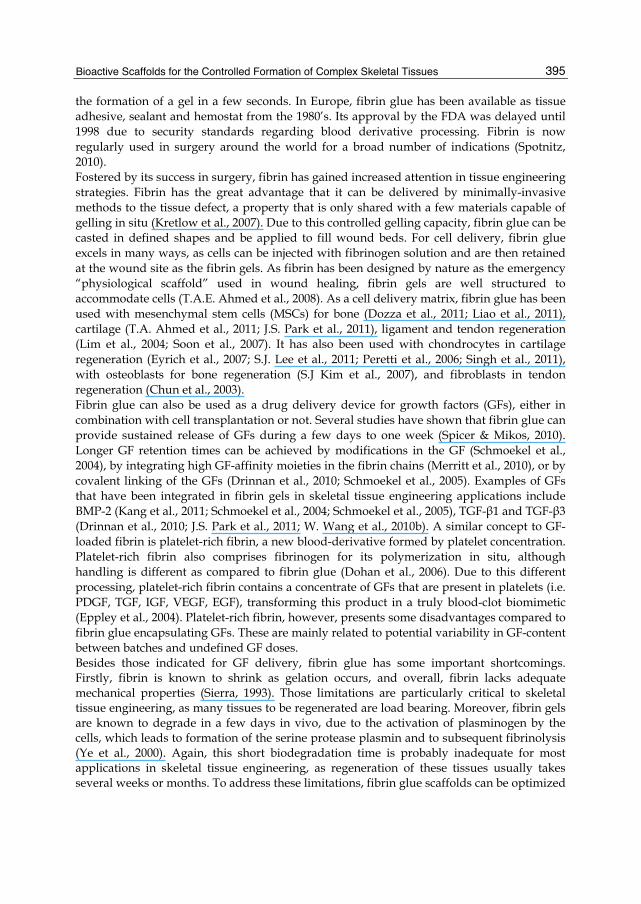
3.2 Scaffolds bound to biological functionalities In section 2, we highlighted the capacity of purified ECM compound scaffolds to provide sites for cell attachment and cell-substrate crosstalk (Fig.1). These scaffolds exert bioactivity because they bear complex information, coded in their physical and chemical structures. Unfortunately, scaffolds fully made out of natural ECM molecules present several limitations related to their difficult purification and processing and their suboptimal mechanical properties (Place et al., 2009). These limitations have broadened the range of scaffold materials investigated towards synthetic scaffolds that are functionalized with bioactive moieties. Most cell receptors do not require interacting with the whole ligand for their activation. Often, a relatively short peptide fragment of about 40 amino acids is sufficient to provide the desired signal to the cells. Indeed, it has been shown that small peptides can activate specific cellular pathways: binding their respective receptors, triggering signal transduction and leading to appropriate cells responses (Chung & T.G. Park, 2007; Place et al., 2009). The required peptide sequences can either be obtained from biological sources or can be chemically synthesized (Sreejalekshmi & Nair, 2011). As these small fragments are easier to produce synthetically and usually show quick refolding under physiological conditions,

Regenerative Medicine and Tissue Engineering - Cells and Biomaterials
406
they might represent a more cost-effective, easier to manipulate, alternative to GFs or other signaling proteins. Moreover, they have been shown to have a higher stability against conformational change, easy controllability of surface density and orientation, and more favorable ligand-receptor interaction (Chung & T.G. Park, 2007; Hersel et al., 2003; Lutolf et al., 2003b; Ruoslahti, 1996). Sreejalekshmi et al. has recently reviewed peptide-modified scaffolds, a manuscript that includes a decision-tree-type flow chart indicating probable cellular outcomes resulting from a given modification (Sreejalekshmi & Nair, 2011).
Fig. 1. Scaffolds as devices capable to activate the host stem cell pool. Growth factors activate the stem cell pool and induce their migration to the scaffold. Stem cells adhere to the scaffold by integrin binding and undergo GF/scaffold-directed differentiation
3.2.1 Fibronectin and small cell adhesion motifs Cell adhesion is a prerequisite to the success of scaffold-based tissue engineering strategies. In native tissues, fibronectin is one essential component of the ECM that mediates cell-matrix interaction. Cells can bind to fibronectin through transmembrane receptor proteins of the integrin family, which mechanically interlink the actin cytoskeleton to the ECM through an elaborate adhesion complex. The binding process between ECM and the dimeric integrin receptors on the cells surface is then followed by a cascade of signaling events leading to the up- or downregulation of the expression of several genes. It is important to note that the binding of integrins happens through dimerization of different alpha and beta subunits, and therefore, association with diverse ligands can lead to different effects in the cell. Some specific functions of biopolymers can be attributed to small functional domains, and these may be incorporated in synthetic analogs in place of the full protein (Dunehoo et al., 2006; Schense et al., 2000; Silva et al., 2004). Probably the best-known sequence binding to integrins is arginine-glycine-aspartic acid (RGD). It is found in many ECM proteins, including fibronectin, laminin, collagen type-IV, tenascin and thrombospondin (Benoit & Anseth, 2005a; Comisar et al., 2007; Underwood et al., 1995). However, the RGD motif and its derivatives are not the only integrin-binding sequence used for scaffold modification. For example, polyethylene terephthalate surfaces modified with the cell adhesion motif

Bioactive Scaffolds for the Controlled Formation of Complex Skeletal Tissues
407
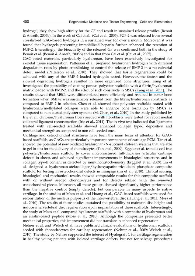
GRGDSPC showed improved differentiation of pre-osteoblastic cells as compared to non-grafted surfaces (Zouani et al., 2010). Alginate hydrogels modified with G4RGDY were able to support the formation of growth-plate-like structures in a co-culture of mouse osteoblasts and chondrocytes (Alsberg et al., 2002). In order to promote cell attachment, an adhesion motif can be simply mixed with the scaffold material or coated on the scaffold surface. Syndecan-binding peptides such as AG73 (RKRLQVQLSIRT) have shown enhanced integrin-mediated biological activities in scaffolds made out of collagen, laminin-11 and fibronectin (Yamada et al., 2011). Morphological analysis indicated that these effects were, at least partially, mediated by enhanced cellular attachment and cell spreading. Attia et al. have shown that polyurethane scaffolds containing an anionic dihydroxy oligomer coated with fibronectin promoted not just cell attachment, but also cell alignment parallel to the scaffold fibers (Attia et al., 2010). This featured scaffold topology was able to increase collagen production after seeding with annulus fibrosus cells. Genetically engineered biopolymers provide another method for the integration of bioactive sequences. Girotti et al. engineered an ECM-analogue of a recombinant multi(bio)functional elastin-like protein polymer with integrated REDV cell adhesion sequences for tissue engineering purposes (Girotti et al., 2004). Cells are known to be very sensitive to the mechanical properties of the substrate (Engler et al., 2006), but also to directional mechanical forces transduced through the ECM (Kurpinski et al., 2006). In skeletal tissues, where mechanical stresses are so important and even necessary for cell stimulation, the integration of adhesion motifs capable of transducing mechanical forces into the right cell signals is of critical importance (Fig. 2).
Fig. 2. Cell differentiation and extracellular matrix deposition can be affected by many different signals, e.g. mechanotransduction or cell signaling
3.2.2 Growth factors Scaffolds may not only be modified with bioactive sequences, whole GFs have been shown to retain their bioactivity upon conjugation to scaffold materials. Through covalent

Regenerative Medicine and Tissue Engineering - Cells and Biomaterials
408
immobilization on the scaffold surface in either random or specific orientation many disadvantages of controlled GF delivery can be circumvented. However, in order to transmit the desired signal to the cells, the moiety must be accessible. This requires the GF to be exposed on the scaffold surface and not incorporated in the polymer core. The application of polymeric spacers such as PEG can provide higher biological activity due to reduced interaction between the GF and the scaffold. For example, collagen matrices were covalently modified with horseradish peroxidase as a model protein with or without PEG as a linker. Introduction of PEG resulted in higher long-term stability of the enzyme (Chen et al., 2002). Recombinant TGF-β2 was either covalently bound through a PEG spacer or admixed to injectable bovine dermal fibrillar collagen (Bentz et al., 1998). PEG-TGF-β2 showed a significantly higher in vitro and in vivo bioactivity than admixed TGF-β2. The authors concluded that covalent binding of TGF-β2 by their method allowed to preserve the GF’s biological activity in vitro, and even potentiated their response in vivo. Karageorgiou et al. have shown that immobilization of BMP-2 on a silk fibroin surface was more efficient in terms of osteogenic differentiation of MSCs in vitro than delivery of the GFs in solution (Karageorgiou et al., 2004). In a follow-up study, they could show that with the same protocol, porous 3D scaffolds modified with BMP-2 implanted into rat critical size femoral defects improved bone formation similarly to the same scaffolds seeded with either undifferentiated MSCs or with MSCs predifferentiated for 4 weeks in vitro before implantation (Kirker-Head et al., 2007). Clearly, the protocol used for biofunctionalization is a critical step for these technologies. De Mel et al. have recently published a review on biofunctionalization of biomaterials for accelerated in situ endothelialization (de Mel et al., 2008). Not only complete GFs can be attached to scaffold surfaces, but also oligopeptides such as those derived from BMP-2 or BMP-7 that have been immobilized on alginate hydrogels and glass, respectively (Kirkwood et al., 2003; Saito et al., 2003; Suzuki et al., 2000). These in vitro studies, performed with a murine multipotent MSC culture or with primary rat calvarial osteoblastic cell populations showed that immobilized oligopeptides may have the capacity to induce osteoblastic differentiation and mineralization in a more predictable manner than the entire GF. This might be attributed to the lower complexity of the molecule and therefore increased stability. Implantation of a BMP-2 derived oligopeptide covalently coupled to alginate into the calf muscle of rats resulted in ectopic bone formation (Suzuki et al., 2000). Synthetic peptide mimics have also been applied for the promotion of angiogenic responses (X. Lin et al., 2006): F2A4-K-NS is a synthetic mimic of FGF-2 that has been shown to trigger signal transduction as monitored by the stimulation of ERK1/2 phosphorylation in human umbilical cord endothelial cells. In cell-based assays, it increased cell migration, cell proliferation and gelatinase secretion. When used in similar quantities, F2A4-K-NS achieved comparable response endpoints to those associated with FGF-2 stimulation. As mentioned before, wound healing and tissue repair might be stimulated by GF release, but optimal effects are only achieved when several chemical signals are provided in the necessary order and at precise times during the regeneration process. This is probably one of the main disadvantages of GF conjugation to scaffolds, as there is no easy way to provide a time-dependent switch to activate/deactivate the provided signaling.
3.2.3 Other binding moieties: cleavage sequences and oligosaccharide domains Incorporation of cleavage sequences is an interesting concept to provide on-demand GF delivery (see 3.1), and also to control cell invasion (Place et al., 2009). In this concept,
Bioactive Scaffolds for the Controlled Formation of Complex Skeletal Tissues
409
cleavage sequences engineered in multidomain peptides are integrated in the polymeric network of the scaffold. This function mimics the multilayered bioactivity of the ECM, where enzymatic remodeling can liberate “cryptic sites“ contained within the ECM proteins. A nice example was provided by Lutolf et al., who rendered a synthetic PEG hydrogel network as amenable to both proteolytic degradation and cell invasion through the integration of integrin binding sites and substrates for matrix metalloproteases (MMP) (Lutolf et al., 2003a). Primary human fibroblasts were demonstrated to proteolytically invade these networks, a process that depended on MMP substrate activity, adhesion ligand concentration and network crosslinking density. While most ligands that have been tested for tissue engineering so far were proteins, many scientists are turning to other biomolecules of high biological functionality: polysaccharides. Indeed, small oligosaccharide domains are involved in many cell-cell and cell-substrate interactions (Brown et al., 2008; Scaglione et al., 2010). Recent developments in understanding the role of natural polysaccharides and improved chemical synthesis of defined oligosaccharides will potentially result in the discovery several new targets of interest in skeletal tissue engineering.
4. Key challenges
4.1 Engineering large tissue constructs – mechanistic issues Since the first works showing the possibility to instruct cells to aggregate and form 3D-tissue constructs, tissue engineers have debated between forming tissues and organs de novo or just boosting the body’s intrinsic capacity for self-repair. Although in the long run the first of these concepts will probably result in technologies and therapeutic approaches of high medical interests, nowadays the second approach has gathered all the big success stories from a clinical perspective (Place et al., 2009). Devices that enhance tissue regeneration take advantage of environmental cues present at the wound site (i.e. progenitor cells, GFs and tissue ECM present at the wound borders) and accelerate the repair process by establishing a conductive matrix and by providing supplementary simulative factors. These factors can coordinate cell chemokinesis, cell differentiation and induction of tissue-specific ECM formation by committed cells. Many of these technologies in the form of bioactivated scaffolds and drug delivery devices are already a reality (InfuseTM, Medtronic, Inc.) and others will become part of the orthopaedist’s arsenal in the next few years. A second generation of these technologies could integrate stem cells as a potential opportunity to promote further regeneration. However, it is still unclear whether additional stem cell transplantation results in enhanced regeneration in biomimetic scaffolds (Im et al., 2010). Devices driven by this concept of enhanced regeneration will most likely not be applicable to wounds above a certain size. In those cases, it is easy to argue that regeneration will be limited by nutrient and oxygen diffusion, as well as by the diffusion/migration of pro-regenerative factors (i.e. GFs, cells) (Muschler et al., 2004). Tissue engineers study in vitro tissue formation as a potential way to generate tissues that could be later implanted in larger defects. While this concept presents several practical disadvantages compared to the most direct approach described above, it is possible that it could result in well-structured tissues that could ultimately be combined in the form of larger constructs. Still, this larger constructs will be deprived of oxygen once formed and implanted, making it critical to induce the quick vascularization of the implanted tissue. In this approach, enhanced tissue construct survival could be achieved by co-implantation of

Regenerative Medicine and Tissue Engineering - Cells and Biomaterials
410
drug delivery devices capable of releasing proangiogenic GFs (Richardson et al., 2001) or by transplanting cells genetically modified to enhance their survival under hypoxic conditions (R.P. Ahmed et al., 2010). Still, conventional in vitro tissue engineering strategies will be unsuitable for very large tissue construct or for regenerating complex tissues/organs. Some very recent work indicated that through the use of relevant progenitor cells and sophisticated bioreactors, complex and even vascularized tissues might be formed in vitro (Tsigkou et al., 2010). Additionally, other studies have shown that even small organs can be engineered in vitro by culturing cells in reprocessed tissue scaffolds (i.e. decellularized tissues) (Ott et al., 2008). While these advances are really impressive, we must bear in mind that the mechanisms used for tissue/organ neogenesis in vitro are far closer to those involved in tissue regeneration than in physiological organogenesis. In vitro tissue formation occurs by the penetration of committed cell populations into a scaffold, the formation of cell-cell and cell-scaffold interactions and the formation of new tissue. That would also be, simplified, the sequence of events leading to tissue remodeling after cell colonization of bloods clots during wound regeneration. On the other hand, organ morphogenesis is a highly orchestrated process occurring during embryogenesis where the tissues are formed by a sequence of very defined events in a growing construct. In morphogenesis, cells undergo sequential differentiation steps under a very defined spatial and temporal regulation. This regulation is usually defined by gradients of morphogens, but also by cell polarity occurring from integrin-mediated cell-cell interactions (Krasnow, 1997). Further studies will define if in vitro strategies based on tissue regeneration mechanisms such as those tested by Ott et al. and Tsigkou et al. can result in functional, clinically useful tissues and organs. In any case, knowing which tissue formation mechanism you are trying to recapitulate is important when designing a tissue-engineering strategy. For example, this is pertinent to bone engineering, where most strategies try to induce MSC differentiation to osteoblasts despite of the fact that this process is only physiological in intramembranous bone regeneration. For long bones, the physiological growth and regeneration process is through endochondral ossification. Endochondral ossification is a multistep process where cartilage is initially formed at the defect site, the cartilage becomes hyperthropic, and then it is remodeled into bone by osteoclasts and osteoblasts. Some steps of endochondral bone formation are recapitulated in DBM implanted subcutaneously (J. Wang & Glimcher, 1999a), and this process has also been studied as an advanced tissue engineering strategy in some recent works (Oliveira et al., 2010).
4.2 Engineering a complex environment Tissue regeneration and tissue neoformation are very complex phenomena. Indeed, the cascade of events after a lesion is formed comprises blood clot formation, inflammation, formation of scar tissue and tissue remodeling. Every one of these processes requires a finely orchestrated cascade of cellular and molecular events. As an illustration, Gerstenfeld et al. have investigated the levels of GFs, other pro-inflammatory cytokines and MMPs occurring during bone regeneration after a traumatic injury (Gerstenfeld et al., 2003), and their studies show the involvement of a myriad of different molecules, present only at specific stages in the process. This complexity is not as surprising: GFs are known to have synergistic effect for many applications as they trigger coordinated effects in their effector cells. Moreover, it is known that all GF effects are very context-specific and concentration-dependent, and therefore, changes in the target cells induced by one GF could drastically change the effect of

Bioactive Scaffolds for the Controlled Formation of Complex Skeletal Tissues
411
a second GF (Massagué, 2000). This results in the necessity to design a very complex environment if we want to recreate all this cues in biomimetic scaffolds. But is this complexity compatible with realistic engineering solutions for tissue regeneration? If something we have learnt from the decline and rise of the tissue engineering field it is the necessity to look for realistic objectives and to avoid overengineered technologies (Place et al., 2009). In short, technologies that are now a success are those that have kept concepts simple. Dealing with the problem of engineering acceptably simple technologies to manipulate a complex process requires a practical approach. First, it is necessary to study how sophisticated your system needs to be for each specific application. For many applications, just the addition of a good tissue-conductive material might suffice. For other applications, this material could be bioactivated with an inducer or seeded with relevant cell populations. Optimization of these strategies could probably rely on high-throughput analysis and systems biology to identify the main signals required for inducing tissue regeneration (Anderson et al., 2004; Anderson et al., 2005; Langer & Tirrell, 2004). For more complex challenges, engineers could turn to reprocessed tissue scaffolds that might be capable of producing some complex tissue structures. Addressing the challenges with technologies that integrate the level of complexity needed to meet the demands of the medical problem addresses complexity from a management perspective. From a technical perspective, many research groups are working in technical solutions that allow implementing complex signaling in the scaffolds with relatively simple chemistries. Examples highlighted in this chapter on the use of small synthetic peptides (Sreejalekshmi & Nair, 2011) or molecules (Ding et al., 2003) and on the formation of material composites including biomimetic compounds might be just some of these simple technologies.
4.3 Controlling stem cell fate and individual variability New tissue engineering strategies can benefit enormously from the concomitant use of stem cells. However, to fulfill the potential of these technologies, improved control over stem cell fate needs to be exerted, particularly in some applications. For instance, some scaffolds have been designed to maintain stem cells in an undifferentiated state for several passages, thus enhancing their capacity for renovation and cell population growth. Differentiation of stem cells can be achieved by their exposition to defined media. This approach is very efficient for some skeletal tissues (e.g. bone), but leads to suboptimal results for others: hyaline cartilage, skeletal muscles and tendon (Brent et al., 2005; Buckwalter & Brown, 2004). When designing tissue engineering strategies for these applications it is important to consider whether environmental cues from the host tissue might help to improve the differentiation process. Indeed, some experiments have shown tissue repair upon implantation of stem cells despite the questionable ability of these cells to differentiate to these tissues. In cases with modest stem cell contribution to tissue regeneration, bioactive scaffold materials can help to direct stem cell differentiation by introducing important environmental cues: nanotopology (Yim & Leong, 2005), mechanical properties of the supporting scaffold (Engler et al., 2006), or chemical moieties (Anderson et al., 2004). In extremely difficult situations, directed cell differentiation might be addressed by gene therapy approaches (Selvaraj et al., 2010). Controlling stem cell differentiation is critical not only to engineer tissues matching the structure, composition and properties characteristics of native ones, but also to prevent undesirable processes such as orthotopic tissue formation, scar formation and even, potential tumor induction (Amariglio et al., 2009).

Regenerative Medicine and Tissue Engineering - Cells and Biomaterials
412
Reliance on stem cell therapies raises also the topic of individual variability (Odorico et al., 2001). Stem cells collected from different donors are known to respond very differently to inductive stimuli, to present different capacities for GF secretion and ultimately, to have extremely different regenerative capacities. When designing tissue engineering devices based on stem cells, it would be critical to have quick tests ensuring a minimum of activity, similarly to the approach taken with conventional drugs. However, this concern does not only apply to tissue engineering products containing stem cells, but also to scaffolds and drug delivery devices intended for conduction and induction of a stem cell-mediated responses. A case in point would be the elder or diseased population, where resident stem cell pools might be partially depleted and their full pro-regenerative capacity compromised. The efficacy of tissue engineering devices will need to be tested in these populations, and specific recommendations be issued to take into account this potential variability. Tissue engineered products based on in vitro cultured stem cells face another important limitation related to their necessary culture time. In vitro, tissues need to be cultured for several weeks and in many cases, this time is not an option for a therapeutic intervention. Even in those cases where this time is available, the postponed surgery will result in an uncomfortable situation for the patient, in increased medical and social costs and in potential worsening of the medical condition. Medical technologies that can be applied immediately out-of-the-box are therefore preferred, and bioengineers should consider this important limitation and the competing therapeutic options when deciding for these technologies.
4.4 Technical challenges Although encapsulation and controlled release of macromolecules has now been studied for several decades, they are difficult technologies to get right due to the easy denaturation of the drug, to the difficulties to predict release in vivo, and even to potential patient-specific issues that could change the release kinetics of the system. This problem translates to GF-release devices, where potential denaturation of the drug combines with the added concern of their very high manufacturing costs. Growth factor denaturation can occur at three steps during their integration in a drug delivery device: (i) during encapsulation, (ii) upon storage and (iii) upon implantation in the release phase. In practical terms degradation during storage (ii) is infrequent and only applies to formulations in aqueous phases, or with materials that could cross-react with the GFs. For most solid formulations where the matrix is stable for long periods of time, this process should not be a concern. GF degradation during encapsulation (i) is usually a concern, and in most cases, can only be minimized and not completely avoided (Sah, 1999). To prevent GF degradation, exposure to high temperatures should be avoided, and high shearing forces and solvent/water interfaces minimized as possible. In any case, it is possible to enhance GF stability by adding other proteins and protecting groups that block the solvent/water interface. This works particularly well with GFs because of the low loading required that could lead to a high disproportion between the protecting protein/compound and the GF. Growth factor denaturation during the release phase (iii) is a concern for many polymeric systems whose degradation byproducts might react with the proteins. This is best illustrated by polyesters, probably the most used materials for sustained release of GFs. When implanted in the body, polyesters are known to hydrate and start their decomposition, which can occur at the surface but also at the inner matrix (Rezwan et al., 2006; Zhu et al., 2000). As the polyester

Bioactive Scaffolds for the Controlled Formation of Complex Skeletal Tissues
413
discompose in the core of the matrix forming acidic oligomers, these remain trapped in the polymeric network and induce an acidic microclimate that can degrade the GF. This general mechanism applies similarly to polymers where the degradation products can react with or just degrade the GFs. For GF delivery, it is probably best to directly select materials that do not result in reactive degradation products, or that undergo only surface-erosion (e.g. polyanhydrides) (Tabata et al., 1993). However, further protection of GFs in polyesters can be achieved by incorporating buffering agents (Zhu et al., 2000). Interestingly, some polyester composites have shown capacity to protect macromolecules both during encapsulation and release (Csaba et al., 2005), and these technologies have already been applied for GF-delivery (d'Angelo et al., 2010). In any case, designing devices capable of providing GF release for several months and in a bioactive form continues to be a challenging objective. Another strategy to achieve sustained exposure of cells to GFs or other signaling molecules is covalent tethering to the scaffold. Covalent tethering of proteins presents the difficulty of unselective binding of the protein to the polymer. Indeed, although some more selective reactions have been designed (i.e. cysteine based linking) (Backer et al., 2006), most protocols use reactions where protein can be bound through several aminoacids of the peptidic chain. This lack of specificity could result in a potential bioactivity loss if a critical aminoacid region is modified with the polymer. However, in general terms good bioactivity has been observed with immobilized GFs (Bentz et al., 1998). On the other hand, lack of specificity when linking biomolecules to synthetic materials raises another concern, linked to a difficult regulatory pathway with materials where no clear molecular structure can be defined.
5. Concluding remarks
We have gone a long way in the field of tissue engineering from the first discoveries pointing to the conductive properties of tissue scaffolds. The experience from reprocessed tissues (DBM, for example) and other ECM-containing products soon pointed to the importance of a biomimetic approach in scaffold design. In the last years, biopolymer-derived and bioactivated synthetic scaffolds have been thoroughly studied and GF delivery integrated in many devices. With a good share of hype and bumps on the way, we have now several tissue engineering products available on the market, most for therapeutic indications related to skeletal tissue engineering, and some having been transformed in great commercial successes (e.g. InfuseTM from Medtronic, Inc.). Tissue engineering potential, however, does not need to be limited to regeneration in relatively simple injury models. Recent studies showed the possibility to engineer body parts integrating several tissues (e.g. ostochondral grafts, vascularized bone) or even small organs. Whether these studies could translate into products of clinical interest is still uncertain, but worth exploring. Probably further optimization of the biomimetic scaffolds together with improved bioreactors will be necessary to realize these challenging goals. Another technology to be expected commercially in the next few years are biomaterials integrating stem cells. For those applications, scaffolds integrating functionalities capable of instructing stem cell proliferation, differentiation and tissue formation will be required, and this concept should be a top priority in tissue engineering, bioregenerative medicine and in the government’s scientific agendas.
Regenerative Medicine and Tissue Engineering - Cells and Biomaterials
414
6. Acronyms used
BMP: bone morphogenic protein DBM: demineralized bone matrix ECM: extracellular matrix EGF: epidermal growth factor FGF: fibroblast growth factor GF: growth factor IGF: insulin-like growth factor MMP: matrix metalloprotease MSC: mesenchymal stem cell PDGF: platelet-derived growth factor PEG: poly(ethylene glycol) qRT-PCR: quantitative reverse transcription polymerase chain reaction TGF: transforming growth factor VEGF: vascular endothelial growth factor
7. Acknoledgments
MGF acknowledges an Isidro Parga Pondal contract from Xunta de Galicia. This work was financed by Xunta de Galicia (PGIDIT07PXIB203067PR) and the Spanish Ministry of Science and Innovation (Programa de Investigación en Salud, PS09/01786).
8. References
Abousleiman, R.I., Reyes, Y., McFetridge, P., & Sikavitsas, V. (2009). Tendon Tissue Engineering Using Cell-Seeded Umbilical Veins Cultured in a Mechanical Stimulator. Tissue Engineering. Part A, Vol.15, No.4, (April 2009), pp. (787-795), ISSN 1937-3341
Ahmed, T.A.E., Dare, E.V., & Hincke, M. (2008). Fibrin: A Versatile Scaffold for Tissue Engineering Applications. Tissue Engineering Part B: Reviews, Vol.14, No.2, (2008), pp. (199-215), ISSN 1937-3368
Ahmed, T.A., Giulivi, A., Griffith, M., & Hincke, M. (2011). Fibrin Glues in Combination with Mesenchymal Stem Cells to Develop a Tissue-Engineered Cartilage Substitute. Tissue Engineering. Part A, Vol.17, No.3-4, (February 2011), pp. (323-335), ISSN 1937-335X
Ahmed, R.P., Haider, K.H., Shujia, J., Afzal, M.R., & Ashraf, M. (2010). Sonic Hedgehog Gene Delivery to the Rodent Heart Promotes Angiogenesis Via Inos/Netrin-1/Pkc Pathway. Plos One, Vol.5, No.1, (2010), p. (e8576), ISSN 1932-6203
Allemann, F., Mizuno, S., Eid, K., Yates, K.E., Zaleske, D., & Glowacki, J. (2001). Effects of Hyaluronan on Engineered Articular Cartilage Extracellular Matrix Gene Expression in 3-Dimensional Collagen Scaffolds. Journal of Biomedical Materials Research. Part A, Vol.55, No.1, (2001), pp. (13-19), ISSN 1552-4965
Alsberg, E., Anderson, K.W., Albeiruti, A., Rowley, J.A., & Mooney, D.J. (2002). Engineering Growing Tissues. Proceedings of the National Academy of Sciences of the United States of America, Vol.99, No.19, (2002), p. (12025), ISSN 1091-6490
Bioactive Scaffolds for the Controlled Formation of Complex Skeletal Tissues
415
Amariglio, N., Hirshberg, A., Scheithauer, B.W., Cohen, Y., Loewenthal, R., Trakhtenbrot, L., Paz, N., Koren-Michowitz, M., Waldman, D., Leider-Trejo, L., Toren, A., Constantini, S., & Rechavi, G. (2009). Donor-Derived Brain Tumor Following Neural Stem Cell Transplantation in An Ataxia Telangiectasia Patient. Plos Med, Vol.6, No.2, (July 2009), p. (e1000029)
Anderson, D.G., Levenberg, S., & Langer, R. (2004). Nanoliter-Scale Synthesis of Arrayed Biomaterials and Application to Human Embryonic Stem Cells. Nature Biotechnology, Vol.22, No.7, (2004), pp. (863-866), ISSN 1087-0156
Anderson, D.G., Putnam, D., Lavik, E.B., Mahmood, T.A., & Langer, R. (2005). Biomaterial Microarrays: Rapid, Microscale Screening of Polymer-Cell Interaction. Biomaterials, Vol.26, No.23, (2005), pp. (4892-4897), ISSN 0142-9612
Attia, M., Santerre, J.P., & Kandel, R.A. (2010). The Response of Annulus Fibrosus Cell to Fibronectin-Coated Nanofibrous Polyurethane-Anionic Dihydroxyoligomer Scaffolds. Biomaterials, Vol.32, No.2, (January 2011), pp. (450-602010), ISSN 0142-9612
Backer, M.V., Patel, V., Jehning, B.T., Claffey, K.P., & Backer, J.M. (2006). Surface Immobilization of Active Vascular Endothelial Growth Factor Via a Cysteine-Containing Tag. Biomaterials, Vol.27, No.31, (2006), pp. (5452-5458), ISSN 0142-9612
Batrakova, E.V., Gendelman, H.E., & Kabanov, A.V. (2011). Cell-Mediated Drug Delivery. Expert Opinion on Drug Delivery, Vol. 8, No.4, (April 2011), pp. (415-433), ISSN 1742-5247
Benoit, D.S.W. & Anseth, K.S. (2005a). The Effect on Osteoblast Function of Colocalized RGD and PHSRN Epitopes on PEG Surfaces. Biomaterials, Vol.26, No.25, (2005), pp. (5209-5220), ISSN 0142-9612
Benoit, D.S.W. & Anseth, K.S. (2005b). Heparin Functionalized PEG Gels That Modulate Protein Adsorption for hMSC Adhesion and Differentiation. Acta Biomaterialia, Vol.1, No.4, (2005), pp. (461-470), ISSN 1742-7061
Bentz, H., Schroeder, J.A., & Estridge, T.D. (1998). Improved Local Delivery of Tgf-β2 by Binding to Injectable Fibrillar Collagen Via Difunctional Polyethylene Glycol. Journal of Biomedical Materials Research, Vol.39, No.4, (1998), pp. (539-548), ISSN 1097-4636
Biondi, M., Ungaro, F., Quaglia, F., & Netti, P.A. (2008). Controlled Drug Delivery in Tissue Engineering. Advanced Drug Delivery Reviews, Vol.60, No.2, (2008), pp. (229-242), ISSN 0169-409X
Brady, M.A., Sivananthan, S., Mudera, V., Liu, Q., Wiltfang, J., & Warnke, P.H. (2010). The Primordium of a Biological Joint Replacement: Coupling of Two Stem Cell Pathways in Biphasic Ultrarapid Compressed Gel Niches. Journal of Cranio-Maxillo-Facial Surgery, Epub ahead of print, (August 2010), ISSN 1878-4119
Braiman-Wiksman, L., Solomonik, I., Spira, R., & Tennenbaum, T. (2007). Novel Insights Into Wound Healing Sequence of Events. Toxicologic Pathology, Vol.35, No.6, (2007), pp. (767-779), ISSN 0192-6233
Brent, A.E., Braun, T., & Tabin, C.J. (2005). Genetic Analysis of Interactions Between the Somitic Muscle, Cartilage and Tendon Cell Lineages During Mouse Development. Development, Vol.132, No.3, (2005), p. (515), ISSN 0950-1991
Brown, A.J., Alicknavitch, M., D'Souza, S.S., Daikoku, T., Kirn-Safran, C.B., Marchetti, D., Carson, D.D., & Farach-Carson, M.C. (2008). Heparanase Expression and Activity

Regenerative Medicine and Tissue Engineering - Cells and Biomaterials
416
Influences Chondrogenic and Osteogenic Processes During Endochondral Bone Formation. Bone, Vol.43, No.4, (2008), pp. (689-699), ISSN 8756-3282
Buckwalter, J.A. & Brown, T.D. (2004). Joint Injury, Repair, and Remodeling: Roles in Post-Traumatic Osteoarthritis. Clinical Orthopaedics and Related Research, Vol.423, (2004), p. (7), ISSN 0009-921X
Cai, S., Liu, Y., Zheng Shu, X., & Prestwich, G.D. (2005). Injectable Glycosaminoglycan Hydrogels for Controlled Release of Human Basic Fibroblast Growth Factor. Biomaterials, Vol.26, No.30, (2005), pp. (6054-6067), ISSN 0142-9612
Chang, C.F., Lee, M.W., Kuo, P.Y., Wang, Y.J., Tu, Y.H., & Hung, S.C. (2007). Three-Dimensional Collagen Fiber Remodeling by Mesenchymal Stem Cells Requires the Integrin--Matrix Interaction. Journal of Biomedical Materials Research. Part A, Vol.80, No.2, (2007), pp. (466-474), ISSN 1552-4965
Chen, M., Le, D.Q., Baatrup, A., Nygaard, J.V., Hein, S., Bjerre, L., Kassem, M., Zou, X., & Bünger, C. (2010). Self-Assembled Composite Matrix in a Hierarchical 3-D Scaffold for Bone Tissue Engineering. Acta Biomaterialia, Epub ahead of print, (December 2010), ISSN 1878-7568
Chen, J.S., Noah, E.M., Pallua, N., & Steffens, G.C.M. (2002). The Use of Bifunctional Polyethyleneglycol Derivatives for Coupling of Proteins to and Cross-Linking of Collagen Matrices. Journal of Materials Science. Materials in Medicine, Vol.13, No.11, (2002), pp. (1029-1035), ISSN 0957-4530
Chen, W.C., Yao, C.L., Wei, Y.H., & Chu, I.M. (2011). Evaluating Osteochondral Defect Repair Potential of Autologous Rabbit Bone Marrow Cells on Type II Collagen Scaffold. Cytotechnology, Vol.63, No.1, (January 2011), pp. (13-23), ISSN 0920-9069
Chen, J.L., Yin, Z., Shen, W.L., Chen, X., Heng, B.C., Zou, X.H., & Ouyang, H.W. (2010). Efficacy of hESC-MSCs in Knitted Silk-Collagen Scaffold for Tendon Tissue Engineering and Their Roles. Biomaterials, Vol.31, No.36, (December 2010), pp. (9438-9451), ISSN 1878-5905
Chou, C.H., Cheng, W.T., Lin, C.C., Chang, C.H., Tsai, C.C., & Lin, F.H. (2006). Tgf-β1 Immobilized Tri-Co-Polymer for Articular Cartilage Tissue Engineering. Journal of Biomedical Materials Research. Part B, Applied Biomaterials, Vol.77, No.2, (May 2006), pp. (338-348), ISSN 1552-4973
Chun, J., Tuan, T.L., Han, B., Vangsness, C.T., & Nimni, M.E. (2003). Cultures of Ligament Fibroblasts in Fibrin Matrix Gel. Connective Tissue Research, Vol.44, No.2, (2003), pp. (81-87), ISSN 0300-8207
Chung, C.H., Golub, E.E., Forbes, E., Tokuoka, T., & Shapiro, I.M. (1992). Mechanism of Action of Β-Glycerophosphate on Bone Cell Mineralization. Calcified Tissue International, Vol.51, No.4, (1992), pp. (305-311), ISSN 0171-967X
Chung, H.J. & Park, T.G. (2007). Surface Engineered and Drug Releasing Pre-Fabricated Scaffolds for Tissue Engineering. Advanced Drug Delivery Reviews, Vol.59, No.4-5, (2007), pp. (249-262), ISSN 0169-409X
de Cogan, F., Gough, J.E., & Webb, S.J. (2011). Adhesive Interactions Between Cells and Biotinylated Phospholipid Vesicles in Alginate: Towards New Responsive Biomaterials. Journal of Materials Science. Materials in Medicine, Epub ahead of print, (March 2011), pp. (1-7), ISSN 0957-4530
Comisar, W.A., Kazmers, N.H., Mooney, D.J., & Linderman, J.J. (2007). Engineering RGD Nanopatterned Hydrogels to Control Preosteoblast Behavior: A Combined

Bioactive Scaffolds for the Controlled Formation of Complex Skeletal Tissues
417
Computational and Experimental Approach. Biomaterials, Vol.28, No.30, (2007), pp. (4409-4417), ISSN 0142-9612
Crawford, D.C., Heveran, C.M., Cannon, W.D., Foo, L.F., & Potter, H.G. (2009). An Autologous Cartilage Tissue Implant Neocart for Treatment of Grade III Chondral Injury to the Distal Femur: Prospective Clinical Safety Trial at 2 Years. The American Journal of Sports Medicine, Vol.37, No.7, (July 2009), pp. (1334-1343), ISSN 1552-3365
Crescenzi, V., Francescangeli, A., Capitani, D., Mannina, L., Renier, D., & Bellini, D. (2003). Hyaluronan Networking Via Ugi's Condensation Using Lysine As Cross-Linker Diamine. Carbohydrate Polymers, Vol.53, No.3, (2003), pp. (311-316), ISSN 0144-8617
Cristino, S., Grassi, F., Toneguzzi, S., Piacentini, A., Grigolo, B., Santi, S., Riccio, M., Tognana, E., Facchini, A., & Lisignoli, G. (2005). Analysis of Mesenchymal Stem Cells Grown on a Three-Dimensional HYAFF 11®-Based Prototype Ligament Scaffold. Journal of Biomedical Materials Research. Part A, Vol.73, No.3, (2005), pp. (275-283), ISSN 1552-4965
Csaba, N., Caamaño, P., Sánchez, A., Domínguez, F., & Alonso, M.J. (2005). Plga:Poloxamer and Plga:Poloxamine Blend Nanoparticles: New Carriers for Gene Delivery. Biomacromolecules, Vol.6, No.1, (2005), pp. (271-278), ISSN 1525-7797
Dahners, L.E. & Jacobs, R.R.. (1985). Long Bone Defects Treated with Demineralized Bone. Southern Medical Journal, Vol.78, No.8, (August 1985), p. (933-934), ISSN 0038-4348
d'Angelo, I., Garcia-Fuentes, M., Parajo , Y., Welle, A., Vantus, T., Horva th, A., Boko nyi, G., Ke ri, G., & Alonso, M.J. (2010). Nanoparticles Based on Plga:Poloxamer Blends for the Delivery of Proangiogenic Growth Factors. Molecular Pharmaceutics, Vol.7(5), (August 2010), pp. (1724-1733), ISSN 1543-8392
Danti, S., Lisanti, M., Berrettini, S., Petrini, M., & Pietrabissa, A. (2007). Bone Tissue Engineering: Natural Origination or Synthetic Polymeric Scaffolds? Advanced Materials Research, Vol.15, (2007), pp. (65-70), ISSN 1662-8985
Deckers, R., Rome, C., & Moonen, C.T.W. (2008). The Role of Ultrasound and Magnetic Resonance in Local Drug Delivery. Journal of Magnetic Resonance Imaging, Vol.27, No.2, (2008), pp. (400-409), ISSN 1522-2586
Deeken, C.R., White, A.K., Bachman, S.L., Ramshaw, B.J., Cleveland, D.S., Loy, T.S., & Grant, S.A. (2011). Method of Preparing a Decellularized Porcine Tendon Using Tributyl Phosphate. Journal of Biomedical Materials Research. Part B, Applied Biomaterials, Vol.96, No.2, (February 2011), pp. (199-206), ISSN 1552-4981
Deschaseaux, F., Sensébé, L., & Heymann, D. (2009). Mechanisms of Bone Repair and Regeneration. Trends in Molecular Medicine, Vol.15, No.9, (2009), pp. (417-429), ISSN 1471-4914
Ding, S., Wu, T.Y.H., Brinker, A., Peters, E.C., Hur, W., Gray, N.S., & Schultz, P.G. (2003). Synthetic Small Molecules That Control Stem Cell Fate. Proceedings of the National Academy of Sciences of the United States of America, Vol.100, No.13, (2003), p. (7632), ISSN 1091-6490
Dinopoulos, H.T. & Giannoudis, P.V. (2006). Safety and Efficacy of Use of Demineralised Bone Matrix in Orthopaedic and Trauma Surgery. Expert Opinion in Drug Safety, Vol.5, No.6, (November 2006), pp. (847-866), ISSN 1744-764X
Discher, D.E., Mooney, D.J., & Zandstra, P.W. (2009). Growth Factors, Matrices, and Forces Combine and Control Stem Cells. Science, Vol.324, No.5935, (June 2009), pp. (1673-1677), ISSN 0036-8075

Regenerative Medicine and Tissue Engineering - Cells and Biomaterials
418
Dohan, D.M., Choukroun, J., Diss, A., Dohan, S.L., Dohan, A.J., Mouhyi, J., & Gogly, B. (2006). Platelet-Rich Fibrin (PRF): A Second-Generation Platelet Concentrate. Part I: Technological Concepts and Evolution. Oral Surgery, Oral Medicine, Oral Pathology, Oral Radiology, and Endodontology, Vol.101, No.3, (2006), pp. (e37-e37)
Dozza, B., Di Bella, C., Lucarelli, E., Giavaresi, G., Fini, M., Tazzari, P.L., Giannini, S., & Donati, D. (2011). Mesenchymal Stem Cells and Platelet Lysate in Fibrin or Collagen Scaffold Promote Non-Cemented Hip Prosthesis Integration. Journal of Orthopaedic Research, Epub ahead of print, (January 2011), ISSN 1554-527X
Drinnan, C.T., Zhang, G., Alexander, M.A., Pulido, A.S., & Suggs, L.J. (2010). Multimodal Release of Transforming Growth Factor-β1 and the BB Isoform of Platelet Derived Growth Factor From Pegylated Fibrin Gels. Journal of Controlled Release, Vol.147, No.2, (October 2010), pp. (180-186), ISSN 1873-4995
Dunehoo, A.L., Anderson, M., Majumdar, S., Kobayashi, N., Berkland, C., & Siahaan, T.J. (2006). Cell Adhesion Molecules for Targeted Drug Delivery. Journal of Pharmaceutical Sciences, Vol.95, No.9, (2006), pp. (1856-1872), ISSN 1520-6017
Dunger, D.B., Regan, A.C.L., & Acerini, C.L. (2005). Childhood and Adolescent Diabetes. Endocr Dev, Vol.9, (2005), p. (107)
Elder, B.D., Kim, D.H., & Athanasiou, K.A. (2010). Developing An Articular Cartilage Decellularization Process Toward Facet Joint Cartilage Replacement. Neurosurgery, Vol.66, No.4, (April 2010), pp. (722-7), ISSN 1524-4040
Engler, A.J., Sen, S., Sweeney, H.L., & Discher, D.E. (2006). Matrix Elasticity Directs Stem Cell Lineage Specification. Cell, Vol.126, No.4, (2006), pp. (677-689), ISSN 0092-8674
Eppley, B.L., Woodell, J.E., & Higgins, J. (2004). Platelet Quantification and Growth Factor Analysis From Platelet-Rich Plasma: Implications for Wound Healing. Plastic and Reconstructive Surgery, Vol.114, No.6, (2004), p. (1502), ISSN 0032-1052
Erggelet, C., Endres, M., Neumann, K., Morawietz, L., Ringe, J., Haberstroh, K., Sittinger, M., & Kaps, C. (2009). Formation of Cartilage Repair Tissue in Articular Cartilage Defects Pretreated with Microfracture and Covered with Cell-Free Polymer-Based Implants. Journal of Orthopaedic Research, Vol.27, No.10, (October 2009), pp. (1353-1360), ISSN 1554-527X
Eyrich, D., Brandl, F., Appel, B., Wiese, H., Maier, G., Wenzel, M., Staudenmaier, R., Goepferich, A., & Blunk, T. (2007). Long-Term Stable Fibrin Gels for Cartilage Engineering. Biomaterials, Vol.28, No.1, (January 2007), pp. (55-65), ISSN 0142-9612
Fedarko, N.S., Vetter, U.K., Weinstein, S., & Robey, P.G. (1992). Age-Related Changes in Hyaluronan, Proteoglycan, Collagen, and Osteonectin Synthesis by Human Bone Cells. Journal of Cellular Physiology, Vol.151, No.2, (1992), pp. (215-227), ISSN 1097-4652
Flynn, L.E., (2010). The Use of Decellularized Adipose Tissue to Provide An Inductive Microenvironment for the Adipogenic Differentiation of Human Adipose-Derived Stem Cells. Biomaterials, Vol.31, No.17, (June 2010), pp. (4715-4724), ISSN 1878-5905
Flynn, L., Prestwich, G.D., Semple, J.L., & Woodhouse, K.A. (2007). Adipose Tissue Engineering with Naturally Derived Scaffolds and Adipose-Derived Stem Cells. Biomaterials, Vol.28, No.26, (September 2007), pp. (3834-3842), ISSN 0142-9612
Fratzl, P. (Ed(s).). (2008). Collagen: structure and mechanics, Springer, ISBN 038773905X, New York

Bioactive Scaffolds for the Controlled Formation of Complex Skeletal Tissues
419
Freeman, I., Kedem, A., & Cohen, S. (2008). The Effect of Sulfation of Alginate Hydrogels on the Specific Binding and Controlled Release of Heparin-Binding Proteins. Biomaterials, Vol.29, No.22, (2008), pp. (3260-3268), ISSN 0142-9612
Fujioka, K., Maeda, M., Hojo, T., & Sano, A. (1998). Protein Release From Collagen Matrices. Advanced Drug Delivery Reviews, Vol.31, No.3, (1998), pp. (247-266), ISSN 0169-409X
Fussenegger, M., Meinhart, J., Höbling, W., Kullich, W., Funk, S., & Bernatzky, G. (2003). Stabilized Autologous Fibrin-Chondrocyte Constructs for Cartilage Repair in Vivo. Annals of Plastic Surgery, Vol.51, No.5, (2003), p. (493-498), ISSN 0148-7043
Garcia-Fuentes, M., Giger, E., Meinel, L., & Merkle, H.P. (2008). The Effect of Hyaluronic Acid on Silk Fibroin Conformation. Biomaterials, Vol.29, No.6, (February 2008), pp. (633-642), ISSN 0142-9612
Garcia-Fuentes, M., Meinel, A.J., Hilbe, M., Meinel, L., & Merkle, H.P. (2009). Silk Fibroin/Hyaluronan Scaffolds for Human Mesenchymal Stem Cell Culture in Tissue Engineering. Biomaterials, Vol.30, No.28, (October 2009), pp. (5068-5076), ISSN 1878-5905
Geiger, F., Bertram, H., Berger, I., Lorenz, H., Wall, O., Eckhardt, C., Simank, H.G., & Richter, W. (2005). Vascular Endothelial Growth Factor Gene-Activated Matrix (VEGF165-GAM) Enhances Osteogenesis and Angiogenesis in Large Segmental Bone Defects. Journal of Bone and Mineral Research, Vol.20, No.11, (2005), pp. (2028-2035), ISSN 1523-4681
George, J., Onodera, J., & Miyata, T. (2008). Biodegradable Honeycomb Collagen Scaffold for Dermal Tissue Engineering. Journal of Biomedical Materials Research. Part A, Vol.87, No.4, (December 2008), pp. (1103-1111), ISSN 1552-4965
Gerecht, S., Burdick, J.A., Ferreira, L.S., Townsend, S.A., Langer, R., & Vunjak-Novakovic, G. (2007). Hyaluronic Acid Hydrogel for Controlled Self-Renewal and Differentiation of Human Embryonic Stem Cells. Proceedings of the National Academy of Sciences, Vol.104, No.27, (2007), p. (11298–11303)
Gerstenfeld, L.C., Cullinane, D.M., Barnes, G.L., Graves, D.T., & Einhorn, T.A. (2003). Fracture Healing As a Post-Natal Developmental Process: Molecular, Spatial, and Temporal Aspects of Its Regulation. Journal of Cellular Biochemistry, Vol.88, No.5, (2003), pp. (873-884)
Gilbert, T.W., Sellaro, T.L., & Badylak, S.F. (2006). Decellularization of Tissues and Organs. Biomaterials, Vol.27, No.19, (July 2006), pp. (3675-3683), ISSN 0142-9612
Gillies, A.R., Smith, L.R., Lieber, R.L., & Varghese, S. (2010). Method for Decellularizing Skeletal Muscle Without Detergents or Proteolytic Enzymes. Tissue Eng Part C Methods(December 2010), ISSN 1937-3392
Gillitzer, R. & Goebeler, M. (2001). Chemokines in Cutaneous Wound Healing. Journal of Leukocyte Biology, Vol.69, No.4, (April 2001), p. (513-521), ISSN 0741-5400
Girotti, A., Reguera, J., Rodriguez-Cabello, J.C., Arias, F.J., Alonso, M., & Testera, A.M. (2004). Design and Bioproduction of a Recombinant Multi (Bio) Functional Elastin-Like Protein Polymer Containing Cell Adhesion Sequences for Tissue Engineering Purposes. Journal of Materials Science. Materials in Medicine, Vol.15, No.4, (2004), pp. (479-484), ISSN 0957-4530
Glowacki, J. & Mizuno, S. (2008). Collagen Scaffolds for Tissue Engineering. Biopolymers, Vol.89, No.5, (2008), pp. (338-344), ISSN 1097-0282

Regenerative Medicine and Tissue Engineering - Cells and Biomaterials
420
Gong, Y.Y., Xue, J.X., Zhang, W.J., Zhou, G.D., Liu, W., & Cao, Y. (2011). A Sandwich Model for Engineering Cartilage with Acellular Cartilage Sheets and Chondrocytes. Biomaterials, Vol.32, No.9, (March 2011), pp. (2265-2273), ISSN 1878-5905
Grigolo, B., De Franceschi, L., Roseti, L., Cattini, L., & Facchini, A. (2005). Down Regulation of Degenerative Cartilage Molecules in Chondrocytes Grown on a Hyaluronan-Based Scaffold. Biomaterials, Vol.26, No.28, (2005), pp. (5668-5676), ISSN 0142-9612
Guo, T., Zeng, X., Hong, H., Diao, H., Wangrui, R., Zhao, J., Zhang, J., & Li, J. (2006). Gene-Activated Matrices for Cartilage Defect Reparation. The International Journal of Artificial Organs, Vol.29, No.6, (June 2006), p. (612-621), ISSN 0391-3988
Gurevitch, O., Kurkalli, B.G., Prigozhina, T., Kasir, J., Gaft, A., & Slavin, S. (2003). Reconstruction of Cartilage, Bone, and Hematopoietic Microenvironment with Demineralized Bone Matrix and Bone Marrow Cells. Stem Cells (Dayton, Ohio), Vol.21, No.5, (2003), pp. (588-597), ISSN 1066-5099
Haberstroh, K., Enz, A., Zenclussen, M.L., Hegewald, A.A., Neumann, K., Abbushi, A., Thomé, C., Sittinger, M., Endres, M., & Kaps, C. (2009). Human Intervertebral Disc-Derived Cells Are Recruited by Human Serum and Form Nucleus Pulposus-Like Tissue Upon Stimulation with TFG-β3 or Hyaluronan in Vitro. Tissue & Cell, Vol.41, No.6, (2009), p. (414), ISSN 1532-3072
Haleem, A.M., Singergy, A.A., Sabry, D., Atta, H.M., Rashed, L.A., Chu, C.R., El Shewy, M.T., Azzam, A., & Abdel Aziz, M.T. (2010). The Clinical Use of Human Culture-Expanded Autologous Bone Marrow Mesenchymal Stem Cells Transplanted on Platelet-Rich Fibrin Glue in the Treatment of Articular Cartilage Defects: A Pilot Study and Preliminary Results. Cartilage, Vol.1, No.4, (October 2010), pp. (253-261), ISSN 1947-6043
Hersel, U., Dahmen, C., & Kessler, H. (2003). RGD Modified Polymers: Biomaterials for Stimulated Cell Adhesion and Beyond. Biomaterials, Vol.24, No.24, (2003), pp. (4385-4415), ISSN 0142-9612
Holland, T.A., Bodde, E.W.H., Cuijpers, V., Baggett, L.S., Tabata, Y., Mikos, A.G., & Jansen, J.A. (2007). Degradable Hydrogel Scaffolds for in Vivo Delivery of Single and Dual Growth Factors in Cartilage Repair. Osteoarthritis and Cartilage, Vol.15, No.2, (2007), pp. (187-197), ISSN 1063-4584
Holland, T.A. & Mikos, A.G. (2006). Biodegradable Polymeric Scaffolds. Improvements in Bone Tissue Engineering Through Controlled Drug Delivery. Advances in Biochemical Engineering/Biotechnology, Vol.102, (2006), p. (161), ISSN 0724-6145
Homandberg, G.A., Ummadi, V., & Kang, H. (2004). Hyaluronan Enhances Cartilage Repair Through Low Grade Tissue Remodeling Involving Cytokines and Matrix Metalloproteinases. Inflammation Research, Vol.53, No.10, (2004), pp. (534-543), ISSN 1023-3830
Hou, N., Cui, P., Luo, J., Ma, R., & Zhu, L. (2011). Tissue-Engineered Larynx Using Perfusion-Decellularized Technique and Mesenchymal Stem Cells in a Rabbit Model. Acta Oto-laryngologica, Epub ahead of print, (February 2011), ISSN 1651-2251
Huang, B., Zhuang, Y., Li, C.Q., Liu, L.T., & Zhou, Y. (2011). Regeneration of the Intervertebral Disc with Nucleus Pulposus Cell-Seeded Collagen-α/Hyaluronan/Chondroitin-6-Sulfate Tri-Copolymer Constructs in a Rabbit Disc Degeneration Model. Spine, Epub ahead of print, (February 2011), ISSN 1528-1159

Bioactive Scaffolds for the Controlled Formation of Complex Skeletal Tissues
421
Huangfu, D., Maehr, R., Guo, W., Eijkelenboom, A., Snitow, M., Chen, A.E., & Melton, D.A. (2008). Induction of Pluripotent Stem Cells by Defined Factors Is Greatly Improved by Small-Molecule Compounds. Nature Biotechnology, Vol.26, No.7, (2008), pp. (795-797), ISSN 1087-0156
Huebsch, N. & Mooney, D.J. (2009). Inspiration and Application in the Evolution of Biomaterials. Nature, Vol.462, No.7272, (November 2009), p. (426-432), ISSN 1476-4687
Im, G.I., Ahn, J.H., Kim, S.Y., Choi, B.S., & Lee, S.W. (2010). A Hyaluronate-Atelocollagen/Beta-Tricalcium Phosphate-Hydroxyapatite Biphasic Scaffold for the Repair of Osteochondral Defects: A Porcine Study. Tissue Engineering. Part A, Vol.16, No.4, (April 2010), pp. (1189-1200), ISSN 1937-335X
Ingram, J.H., Korossis, S., Howling, G., Fisher, J., & Ingham, E. (2007). The Use of Ultrasonication to Aid Recellularization of Acellular Natural Tissue Scaffolds for Use in Anterior Cruciate Ligament Reconstruction. Tissue Engineering, Vol.13, No.7, (July 2007), pp. (1561-1572), ISSN 1076-3279
Iozzo, R.V. (2000). Proteoglycans: Structure, Biology, and Molecular Interactions, Marcel Dekker, ISBN 0824703340, New York
Irie, T., Majima, T., Sawaguchi, N., Funakoshi, T., Nishimura, S.I., & Minami, A. (2011). Biomechanical and Histologic Evaluation of Tissue Engineered Ligaments Using Chitosan and Hyaluronan Hybrid Polymer Fibers: A Rabbit Medial Collateral Ligament Reconstruction Model. Journal of Biomedical Materials Research. Part A, Epub ahead of print, (March 2011), ISSN 1552-4965
Irinakis, T., (2011). Efficacy of Injectable Demineralized Bone Matrix As Graft Material During Sinus Elevation Surgery with Simultaneous Implant Placement in the Posterior Maxilla: Clinical Evaluation of 49 Sinuses. J Oral Maxillofac Surg, Vol.69, No.1, (January 2011), pp. (134-141), ISSN 1531-5053
Isaac, J., Nohra, J., Lao, J., Jallot, E., Nedelec, J.M., Berdal, A., & Sautier, J.M. (2011). Effects of Strontium-Doped Bioactive Glass on the Differentiation of Cultured Osteogenic Cells. European Cells & Materials, Vol.21, (February 2011), p. (130-143), ISSN 1473-2262
Jäger, M., Fischer, J., Dohrn, W., Li, X., Ayers, D.C., Czibere, A., Prall, W.C., Lensing-Höhn, S., & Krauspe, R. (2008). Dexamethasone Modulates BMP-2 Effects on Mesenchymal Stem Cells in Vitro. Journal of Orthopaedic Research, Vol.26, No.11, (November 2008), pp. (1440-1448), ISSN 1554-527X
Jin, X.B., Luo, Z.J., & Wang, J. (2006). Treatment of Rabbit Growth Plate Injuries with An Autologous Tissue-Engineered Composite. An Experimental Study. Cells Tissues Organs, Vol.183, No.2, (2006), pp. (62-67), ISSN 1422-6405
Juang Ming Yee, A., Bae, H.W., Friess, D., Robbin, M., Johnstone, B., & Yoo, J.U. (2003). Augmentation of Rabbit Posterolateral Spondylodesis Using a Novel Demineralized Bone Matrix-Hyaluronan Putty. Spine, Vol.28, No.21, (November 2003), p. (2435-2440), ISSN 0362-2436
Kang, S.W., Kim, J.S., Park, K.S., Cha, B.H., Shim, J.H., Kim, J.Y., Cho, D.W., Rhie, J.W., & Lee, S.H. (2011). Surface Modification with Fibrin/Hyaluronic Acid Hydrogel on Solid-Free Form-Based Scaffolds Followed by BMP-2 Loading to Enhance Bone Regeneration. Bone, Vol.48, No.2, (February 2011), pp. (298-306), ISSN 1873-2763
Regenerative Medicine and Tissue Engineering - Cells and Biomaterials
422
Karageorgiou, V., Meinel, L., Hofmann, S., Malhotra, A., Volloch, V., & Kaplan, D. (2004). Bone Morphogenetic Protein-2 Decorated Silk Fibroin Films Induce Osteogenic Differentiation of Human Bone Marrow Stromal Cells. Journal of Biomedical Materials Research. Part A, Vol.71, No.3, (2004), pp. (528-537), ISSN 1552-4965
Kim, S.J., Jang, J.D., & Lee, S.K. (2007). Treatment of Long Tubular Bone Defect of Rabbit Using Autologous Cultured Osteoblasts Mixed with Fibrin. Cytotechnology, Vol.54, No.2, (June 2007), pp. (115-120), ISSN 0920-9069
Kim, Y.K., Kim, S.G., Lim, S.C., Lee, H.J., & Yun, P.Y. (2010). A Clinical Study on Bone Formation Using a Demineralized Bone Matrix and Resorbable Membrane. Oral Surg Oral Med Oral Pathol Oral Radiol Endod, Vol.109, No.6, (June 2010), pp. (e6-11), ISSN 1528-395X
Kirker-Head, C., Karageorgiou, V., Hofmann, S., Fajardo, R., Betz, O., Merkle, H.P., Hilbe, M., Von Rechenberg, B., McCool, J., & Abrahamsen, L. (2007). BMP-Silk Composite Matrices Heal Critically Sized Femoral Defects. Bone, Vol.41, No.2, (2007), pp. (247-255), ISSN 8756-3282
Kirkwood, K., Rheude, B., Kim, Y.J., White, K., & Dee, K.C. (2003). In Vitro Mineralization Studies with Substrate-Immobilized Bone Morphogenetic Protein Peptides. Journal of Oral Implantology, Vol.29, No.2, (2003), pp. (57-65), ISSN 1548-1336
Knudson, W., Casey, B., Nishida, Y., Eger, W., Kuettner, K.E., & Knudson, C.B. (2000). Hyaluronan Oligosaccharides Perturb Cartilage Matrix Homeostasis and Induce Chondrocytic Chondrolysis. Arthritis & Rheumatism, Vol.43, No.5, (2000), pp. (1165-1174), ISSN 1529-0131
Krasnow, M.A., (1997). Genes That Control Organ Form: Lessons From Bone and Branching Morphogenesis. Cold Spring Harbor Symposia on Quantitative Biology, Vol.62, (1997), p. (235), ISSN 0091-7451
Kretlow, J.D., Klouda, L., & Mikos, A.G. (2007). Injectable Matrices and Scaffolds for Drug Delivery in Tissue Engineering. Advanced Drug Delivery Reviews, Vol.59, No.4-5, (2007), pp. (263 - 273), ISSN 0169-409X
Kubow, K.E., Klotzsch, E., Smith, M.L., Gourdon, D., Little, W.C., & Vogel, V. (2009). Crosslinking of Cell-Derived 3D Scaffolds Up-Regulates the Stretching and Unfolding of New Extracellular Matrix Assembled by Reseeded Cells. Integr. Biol., Vol.1, No.11-12, (2009), pp. (635-648)
Kurpinski, K., Chu, J., Hashi, C., & Li, S. (2006). Anisotropic Mechanosensing by Mesenchymal Stem Cells. Proceedings of the National Academy of Sciences of the United States of America, Vol.103, No.44, (2006), pp. (16095-16100), ISSN 0027-8424
Laflamme, C. & Rouabhia, M. (2008). Effect of BMP-2 and BMP-7 Homodimers and a Mixture of BMP-2/BMP-7 Homodimers on Osteoblast Adhesion and Growth Following Culture on a Collagen Scaffold. Biomedical Materials, Vol.3, No.1, (March 2008), p. (015008), ISSN 1748-605X
Lammens, J., Nijs, J., Schepers, E., Ectors, N., Lismont, D., & Verduyckt, B. (2009). The Effect of Bone Morphogenetic Protein-7 (OP-1) and Demineralized Bone Matrix (DBM) in the Rabbit Tibial Distraction Model. Acta Orthop Belg, Vol.75, No.1, (February 2009), pp. (103-109), ISSN 0001-6462
Langer, R. & Tirrell, D.A. (2004). Designing Materials for Biology and Medicine. Nature, Vol.428, No.6982, (2004), pp. (487-492), ISSN 0028-0836
Bioactive Scaffolds for the Controlled Formation of Complex Skeletal Tissues
423
Lee, S.J., Broda, C., Atala, A., & Yoo, J.J. (2011). Engineered Cartilage Covered Ear Implants for Auricular Cartilage Reconstruction. Biomacromolecules, Vol.12, No.2, (February 2011), pp. (306-313), ISSN 1526-4602
Lee, J.E., Kim, K.E., Kwon, I.C., Ahn, H.J., Lee, S.H., Cho, H., Kim, H.J., Seong, S.C., & Lee, M.C. (2004). Effects of the Controlled-Released Tgf-β1 From Chitosan Microspheres on Chondrocytes Cultured in a Collagen/Chitosan/Glycosaminoglycan Scaffold. Biomaterials, Vol.25, No.18, (August 2004), pp. (4163-4173), ISSN 0142-9612
Leong, D.T., Nah, W.K., Gupta, A., Hutmacher, D.W., & Woodruff, M.A. (2008). The Osteogenic Differentiation of Adipose Tissue-Derived Precursor Cells in a 3D Scaffold/Matrix Environment. Current Drug Discovery Technologies, Vol.5, No.4, (December 2008), pp. (319-327), ISSN 1570-1638
Liao, H.T., Chen, C.T., Chen, C.H., Chen, J.P., & Tsai, J.C. (2011). Combination of Guided Osteogenesis with Autologous Platelet-Rich Fibrin Glue and Mesenchymal Stem Cell for Mandibular Reconstruction. The Journal of Trauma, Vol.70, No.1, (January 2011), pp. (228-237), ISSN 1529-8809
Lieberman, J.R., Daluiski, A., & Einhorn, T.A. (2002). The Role of Growth Factors in the Repair of Bone: Biology and Clinical Applications. The Journal of Bone and Joint Surgery, Vol.84, No.6, (June 2002), p. (1032-1044)
Lim, J.K., Hui, J., Li, L., Thambyah, A., Goh, J., & Lee, E.H. (2004). Enhancement of Tendon Graft Osteointegration Using Mesenchymal Stem Cells in a Rabbit Model of Anterior Cruciate Ligament Reconstruction. Arthroscopy: The Journal of Arthroscopic & Related Surgery, Vol.20, No.9, (November 2004), pp. (899-910), ISSN 1526-3231
Lin, X., Takahashi, K., Campion, S.L., Liu, Y., Gustavsen, G.G., Pena, L.A., & Zamora, P.O. (2006). Synthetic Peptide F2A4-K-NS Mimics Fibroblast Growth Factor-2 in Vitro and Is Angiogenic in Vivo. International Journal of Molecular Medicine, Vol.17, No.5, (2006), p. (833), ISSN 1107-3756
Lin, H., Zhao, Y., Sun, W., Chen, B., Zhang, J., Zhao, W., Xiao, Z., & Dai, J. (2008). The Effect of Crosslinking Heparin to Demineralized Bone Matrix on Mechanical Strength and Specific Binding to Human Bone Morphogenetic Protein-2. Biomaterials, Vol.29, No.9, (March 2008), pp. (1189-1197), ISSN 0142-9612
Liu, G., Li, Y., Sun, J., Zhou, H., Zhang, W., Cui, L., & Cao, Y. (2010a). In Vitro and in Vivo Evaluation of Osteogenesis of Human Umbilical Cord Blood-Derived Mesenchymal Stem Cells on Partially Demineralized Bone Matrix. Tissue Engineering. Part A, Vol.16, No.3, (March 2010), pp. (971-982), ISSN 1937-335X
Liu, H., Zhang, L., Shi, P., Zou, Q., Zuo, Y., & Li, Y. (2010b). Hydroxyapatite/Polyurethane Scaffold Incorporated with Drug-Loaded Ethyl Cellulose Microspheres for Bone Regeneration. Journal of Biomedical Materials Research Part B: Applied Biomaterials, Vol.95, No.1, (2010), pp. (36-46), ISSN 1552-4981
Lodish, H, Berk, A, & Zipursky, SL (2002). Molecular Cell Biology (4th), W H Freeman & Co, New York
Luginbuehl, V., Meinel, L., Merkle, H.P., & Gander, B. (2004). Localized Delivery of Growth Factors for Bone Repair. European Journal of Pharmaceutics and Biopharmaceutics, Vol.58, No.2, (2004), pp. (197-208), ISSN 0939-6411
Lutolf, M.P., Lauer-Fields, J.L., Schmoekel, H.G., Metters, A.T., Weber, F.E., Fields, G.B., & Hubbell, J.A. (2003a). Synthetic Matrix Metalloproteinase-Sensitive Hydrogels for the Conduction of Tissue Regeneration: Engineering Cell-Invasion Characteristics.

Regenerative Medicine and Tissue Engineering - Cells and Biomaterials
424
Proceedings of the National Academy of Sciences of the United States of America, Vol.100, No.9, (2003), p. (5413), ISSN 1091-6490
Lutolf, M.P., Weber, F.E., Schmoekel, H.G., Schense, J.C., Kohler, T., Müller, R., & Hubbell, J.A. (2003b). Repair of Bone Defects Using Synthetic Mimetics of Collagenous Extracellular Matrices. Nature Biotechnology, Vol.21, No.5, (2003), pp. (513-518), ISSN 1087-0156
Lynch, M.P., Stein, J.L., Stein, G.S., & Lian, J.B. (1995). The Influence of Type I Collagen on the Development and Maintenance of the Osteoblast Phenotype in Primary and Passaged Rat Calvarial Osteoblasts: Modification of Expression of Genes Supporting Cell Growth, Adhesion, and Extracellular Matrix Mineralization. Experimental Cell Research, Vol.216, No.1, (1995), pp. (35-45), ISSN 0014-4827
Macchiarini, P., Jungebluth, P., Go, T., Asnaghi, M., Rees, L.E., Cogan, T.A., Dodson, A., Martorell, J., Bellini, S., & Parnigotto, P.P. (2008). Clinical Transplantation of a Tissue-Engineered Airway. The Lancet, Vol.372, No.9655, (2008), pp. (2023-2030), ISSN 0140-6736
MacLean, S.B. & Gratzer, P.F. (2011). Effect of Basic Fibroblast Growth Factor on the Cellular Repopulation of Decellularized Anterior Cruciate Ligament Allografts. J Tissue Eng Regen Med, Vol.5, No.3, (March 2011), pp. (201-209), ISSN 1932-7005
Maeda, M., Tani, S., Sano, A., & Fujioka, K. (1999). Microstructure and Release Characteristics of the Minipellet, a Collagen-Based Drug Delivery System for Controlled Release of Protein Drugs. Journal of Controlled Release, Vol.62, No.3, (1999), pp. (313-324), ISSN 0168-3659
Mart, R.J., Liem, K.P., & Webb, S.J. (2009a). Creating Functional Vesicle Assemblies From Vesicles and Nanoparticles. Pharmaceutical Research, Vol.26, No.7, (2009), pp. (1701-1710), ISSN 0724-8741
Mart, R.J., Liem, K.P., & Webb, S.J. (2009b). Magnetically-Controlled Release From Hydrogel-Supported Vesicle Assemblies. Chemical Communications, Vol.7, No.17, (2009), pp. (2287-2289)
Martin Jr, G.J., Boden, S.D., Titus, L., & Scarborough, N.L. (1999). New Formulations of Demineralized Bone Matrix As a More Effective Graft Alternative in Experimental Posterolateral Lumbar Spine Arthrodesis. Spine, Vol.24, No.7, (1999), p. (637), ISSN 0362-2436
Massagué, J., (2000). How Cells Read Tgf-Β Signals. Nature Reviews Molecular Cell Biology, Vol.1, No.3, (2000), pp. (169-178), ISSN 1471-0072
Meinel, L., Zoidis, E., Zapf, J., Hassa, P., Hottiger, M.O., Auer, J.A., Schneider, R., Gander, B., Luginbuehl, V., & Bettschart-Wolfisberger, R. (2003). Localized Insulin-Like Growth Factor I Delivery to Enhance New Bone Formation. Bone, Vol.33, No.4, (2003), pp. (660-672), ISSN 8756-3282
Mekhail, M., Wong, K.K., Padavan, D.T., Wu, Y., O'Gorman, D.B., & Wan, W. (2010). Genipin-Cross-Linked Electrospun Collagen Fibers. Journal of Biomaterials Science. Polymer Edition, Epub ahead of print, (November 2010), ISSN 1568-5624
de Mel, A., Jell, G., Stevens, M.M., & Seifalian, A.M. (2008). Biofunctionalization of Biomaterials for Accelerated in Situ Endothelialization: A Review. Biomacromolecules, Vol.9, No.11, (2008), pp. (2969-2979), ISSN 1525-7797
Merritt, E.K., Hammers, D.W., Tierney, M., Suggs, L.J., Walters, T.J., & Farrar, R.P. (2010). Functional Assessment of Skeletal Muscle Regeneration Utilizing Homologous

Bioactive Scaffolds for the Controlled Formation of Complex Skeletal Tissues
425
Extracellular Matrix As Scaffolding. Tissue Engineering. Part A, Vol.16, No.4, (April 2010), pp. (1395-1405), ISSN 1937-335X
Mi, F.L., Shyu, S.S., Peng, C.K., Wu, Y.B., Sung, H.W., Wang, P.S., & Huang, C.C. (2006). Fabrication of Chondroitin Sulfate-Chitosan Composite Artificial Extracellular Matrix for Stabilization of Fibroblast Growth Factor. Journal of Biomedical Materials Research. Part A, Vol.76, No.1, (January 2006), pp. (1-15), ISSN 1549-3296
Middelkoop, E., Vries, H.J.C., Ruuls, L., Everts, V., Wildevuur, C.H.R., & Westerhof, W. (1995). Adherence, Proliferation and Collagen Turnover by Human Fibroblasts Seeded Into Different Types of Collagen Sponges. Cell and Tissue Research, Vol.280, No.2, (1995), pp. (447-453), ISSN 0302-766X
Min, B.H., Park, S., Choi, B.H., & Park, S.R. (2010). Chondrogenesis of Rabbit Mesenchymal Stem Cells in Fibrin/Hyaluronan Composite Scaffold in Vitro. Tissue Engineering. Part A, Epub ahead of print, (December 2010), ISSN 1937-335X
Mizuno, M., Fujisawa, R., & Kuboki, Y. (2000). Type I Collagen-Induced Osteoblastic Differentiation of Bone-Marrow Cells Mediated by Collagen-α2β1 Integrin Interaction. Journal of Cellular Physiology, Vol.184, No.2, (2000), pp. (207-213), ISSN 1097-4652
Mizuno, S. & Glowacki, J. (1996). Three-Dimensional Composite of Demineralized Bone Powder and Collagen for in Vitro Analysis of Chondroinduction of Human Dermal Fibroblasts. Biomaterials, Vol.17, No.18, (1996), pp. (1819-1825), ISSN 0142-9612
Moss, I.L., Gordon, L., Woodhouse, K.A., Whyne, C.M., & Yee, A.J. (2010). A Novel Thiol-Modified-Hyaluronan and Elastin-Like Polypetide Composite Material for Tissue Engineering of the Nucleus Pulposus of the Intervertebral Disc. Spine, Epub ahead of print, (December 2010), ISSN 1528-1159
Moxham, J.P., Wong, K.K., & Kibblewhite, D.J. (2009). Transforming Growth Factor-β1 Shows An Incremental Osteoinductive Dose-Response Relationship. Laryngoscope, Vol.119, No.1, (January 2009), pp. (126-130), ISSN 1531-4995
Muschler, G.F., Nakamoto, C., & Griffith, L.G. (2004). Engineering Principles of Clinical Cell-Based Tissue Engineering. The Journal of Bone and Joint Surgery, Vol.86, No.7, (2004), p. (1541)
Myers, T.J., Granero-Molto, F., Longobardi, L., Li, T., Yan, Y., & Spagnoli, A. (2010). Mesenchymal Stem Cells at the Intersection of Cell and Gene Therapy. Expert Opinion on Biological Therapy, Vol.10, No.12, (2010), pp. (1663-1679), ISSN 1471-2598
Nehrer, S., Dorotka, R., Domayer, S., Stelzeneder, D., & Kotz, R. (2009). Treatment of Full-Thickness Chondral Defects with Hyalograft C in the Knee: A Prospective Clinical Case Series with 2 to 7 Years' Follow-Up. The American Journal of Sports Medicine, Vol.37 Suppl 1, (November 2009), pp. (81S-87S), ISSN 1552-3365
Nimni, M.E., Cheung, D., Strates, B., Kodama, M., & Sheikh, K. (1987). Chemically Modified Collagen: A Natural Biomaterial for Tissue Replacement. Journal of Biomedical Materials Research, Vol.21, No.6, (June 1987), pp. (741-771), ISSN 0021-9304
Nuernberger, S., Cyran, N., Albrecht, C., Redl, H., Vécsei, V., & Marlovits, S. (2011). The Influence of Scaffold Architecture on Chondrocyte Distribution and Behavior in Matrix-Associated Chondrocyte Transplantation Grafts. Biomaterials, Vol.32, No.4, (February 2011), pp. (1032-1040), ISSN 1878-5905

Regenerative Medicine and Tissue Engineering - Cells and Biomaterials
426
Odorico, J.S., Kaufman, D.S., & Thomson, J.A. (2001). Multilineage Differentiation From Human Embryonic Stem Cell Lines. Stem Cells, Vol.19, No.3, (2001), pp. (193-204), ISSN 1549-4918
Oliveira, S.M., Ringshia, R.A., Legeros, R.Z., Clark, E., Yost, M.J., Terracio, L., & Teixeira, C.C. (2010). An Improved Collagen Scaffold for Skeletal Regeneration. Journal of Biomedical Materials Research. Part A, Vol.94, No.2, (August 2010), pp. (371-379), ISSN 1552-4965
Ott, H.C., Matthiesen, T.S., Goh, S.-K., Black, L.D., Kren, S.M., Netoff, T.I., Taylor, D.A., Ott, H.C., Matthiesen, T.S., Goh, S.-K., Black, L.D., Kren, S.M., Netoff, T.I., & Taylor, D.A. (2008). Perfusion-Decellularized Matrix: Using Nature's Platform to Engineer a Bioartificial Heart. Nature Medicine, Vol.14, No.2, (January 2008), p. (213-221), ISSN 1078-8956
Pabbruwe, M.B., Esfandiari, E., Kafienah, W., Tarlton, J.F., & Hollander, A.P. (2009). Induction of Cartilage Integration by a Chondrocyte/Collagen-Scaffold Implant. Biomaterials, Vol.30, No.26, (September 2009), pp. (4277-4286), ISSN 1878-5905
Pabbruwe, M.B., Kafienah, W., Tarlton, J.F., Mistry, S., Fox, D.J., & Hollander, A.P. (2010). Repair of Meniscal Cartilage White Zone Tears Using a Stem Cell/Collagen-Scaffold Implant. Biomaterials, Vol.31, No.9, (March 2010), pp. (2583-2591), ISSN 1878-5905
Pang, Y., Wang, X., Ucuzian, A.A., Brey, E.M., Burgess, W.H., Jones, K.J., Alexander, T.D., & Greisler, H.P. (2010). Local Delivery of a Collagen-Binding FGF-1 Chimera to Smooth Muscle Cells in Collagen Scaffolds for Vascular Tissue Engineering. Biomaterials, Vol.31, No.5, (February 2010), pp. (878-885), ISSN 1878-5905
Park, S.H., Cui, J.H., Park, S.R., & Min, B.H. (2009). Potential of Fortified Fibrin/Hyaluronic Acid Composite Gel As a Cell Delivery Vehicle for Chondrocytes. Artificial Organs, Vol.33, No.6, (June 2009), pp. (439-447), ISSN 1525-1594
Park, J.S., Yang, H.N., Woo, D.G., Jeon, S.Y., & Park, K.H. (2011). Chondrogenesis of Human Mesenchymal Stem Cells in Fibrin Constructs Evaluated in Vitro and in Nude Mouse and Rabbit Defects Models. Biomaterials, Vol.32, No.6, (February 2011), pp. (1495-1507), ISSN 1878-5905
Patterson, J., Siew, R., Herring, S.W., Lin, A.S., Guldberg, R., & Stayton, P.S. (2010). Hyaluronic Acid Hydrogels with Controlled Degradation Properties for Oriented Bone Regeneration. Biomaterials, Vol.31, No.26, (September 2010), pp. (6772-6781), ISSN 1878-5905
Peretti, G.M., Xu, J.W., Bonassar, L.J., Kirchhoff, C.H., Yaremchuk, M.J., & Randolph, M.A. (2006). Review of Injectable Cartilage Engineering Using Fibrin Gel in Mice and Swine Models. Tissue Engineering, Vol.12, No.5, (May 2006), pp. (1151-1168), ISSN 1076-3279
Place, E.S., Evans, N.D., & Stevens, M.M. (2009). Complexity in Biomaterials for Tissue Engineering. Nature Materials, Vol.8, No.6, (2009), pp. (457-470), ISSN 1476-1122
Pradeep, A.R. & Sharma, A. (2011). Autologous Platelet Rich Fibrin in the Treatment of Mandibular Degree Ii Furcation Defects: A Randomized Clinical Trial. Journal of Periodontology, Epub ahead of print, (February 2011), ISSN 1943-3670
Putney, S.D. & Burke, P.A. (1998). Improving Protein Therapeutics with Sustained-Release Formulations. Nature Biotechnology, Vol.16, No.2, (1998), pp. (153-157), ISSN 1087-0156

Bioactive Scaffolds for the Controlled Formation of Complex Skeletal Tissues
427
Reddi, A.H., (1998). Role of Morphogenetic Proteins in Skeletal Tissue Engineering and Regeneration. Nature Biotechnology, Vol.16, No.3, (1998), pp. (247-252), ISSN 1087-0156
Reddi, A.H., (2001a). Bone Morphogenetic Proteins: From Basic Science to Clinical Applications. The Journal of Bone and Joint Surgery, Vol.83, No.S1, Part 1, (2001), p. (S1-S6)
Reddi, A.H., (2001b). Interplay Between Bone Morphogenetic Proteins and Cognate Binding Proteins in Bone and Cartilage Development: Noggin, Chordin and DAN. Arthritis Research, Vol.3, No.1, (2001), pp. (1-5), ISSN 1465-9905
Rezwan, K., Chen, Q.Z., Blaker, J.J., & Boccaccini, A.R. (2006). Biodegradable and Bioactive Porous Polymer/Inorganic Composite Scaffolds for Bone Tissue Engineering. Biomaterials, Vol.27, No.18, (2006), pp. (3413-3431), ISSN 0142-9612
Ricard-Blum, S. & Ruggiero, F. (2005). The Collagen Superfamily: From the Extracellular Matrix to the Cell Membrane. Pathologie Biologie, Vol.53, No.7, (2005), pp. (430-442), ISSN 0369-8114
Richardson, T.P., Peters, M.C., Ennett, A.B., & Mooney, D.J. (2001). Polymeric System for Dual Growth Factor Delivery. Nature Biotechnology, Vol.19, No.11, (2001), pp. (1029-1034), ISSN 1087-0156
Rodkey, W.G., Steadman, J.R., & Li, S.T. (1999). A Clinical Study of Collagen Meniscus Implants to Restore the Injured Meniscus. Clinical Orthopaedics and Related Research, Vol. 367, No.Suppl, (October 1999), pp. (S281-S292), ISSN 0009-921X
Ruoslahti, E., (1996). RGD and Other Recognition Sequences for Integrins. Annual Review of Cell and Developmental Biology, Vol.12, No.1, (1996), pp. (697-715), ISSN 1081-0706
Ryu, Y.M., Hah, Y.S., Park, B.W., Kim, D.R., Roh, G.S., Kim, J.R., Kim, U.K., Rho, G.J., Maeng, G.H., & Byun, J.H. (2010). Osteogenic Differentiation of Human Periosteal-Derived Cells in a Three-Dimensional Collagen Scaffold. Molecular Biology Reports, Epub ahead of print, (January 2010), ISSN 1573-4978
Sah, H., (1999). Protein Instability Toward Organic Solvent/Water Emulsification: Implications for Protein Microencapsulation Into Microspheres. PDA Journal of Pharmaceutical Science and Technology, Vol.53, No.1, (1999), p. (3), ISSN 1079-7440
Saito, A., Suzuki, Y., Ogata, S., Ohtsuki, C., & Tanihara, M. (2003). Activation of Osteo-Progenitor Cells by a Novel Synthetic Peptide Derived From the Bone Morphogenetic Protein-2 Knuckle Epitope. Biochimica Et Biophysica Acta (BBA)-Proteins & Proteomics, Vol.1651, No.1-2, (2003), pp. (60-67), ISSN 1570-9639
Sandhu, M.S., Dunger, D.B., & Giovannucci, E.L. (2002). Insulin, Insulin-Like Growth Factor-I (IGF-I), IGF Binding Proteins, Their Biologic Interactions, and Colorectal Cancer. Journal of the National Cancer Institute, Vol.94, No.13, (2002), p. (972), ISSN 0027-8874
Sawaguchi, N., Majima, T., Funakoshi, T., Shimode, K., Harada, K., Minami, A., & Nishimura, S. (2010). Effect of Cyclic Three-Dimensional Strain on Cell Proliferation and Collagen Synthesis of Fibroblast-Seeded Chitosan-Hyaluronan Hybrid Polymer Fiber. Journal of Orthopaedic Science, Vol.15, No.4, (July 2010), pp. (569-577), ISSN 1436-2023
Scaglione, S., Lazzarini, E., Ilengo, C., & Quarto, R. (2010). A Composite Material Model for Improved Bone Formation. Journal of Tissue Engineering and Regenerative Medicine, Vol.4, No.7, (2010), pp. (505-513), ISSN 1932-7005

Regenerative Medicine and Tissue Engineering - Cells and Biomaterials
428
Schagemann, J.C., Kurz, H., Casper, M.E., Stone, J.S., Dadsetan, M., Yu-Long, S., Mrosek, E.H., Fitzsimmons, J.S., O'Driscoll, S.W., & Reinholz, G.G. (2010). The Effect of Scaffold Composition on the Early Structural Characteristics of Chondrocytes and Expression of Adhesion Molecules. Biomaterials, Vol.31, No.10, (April 2010), pp. (2798-2805), ISSN 1878-5905
Schense, J.C., Bloch, J., Aebischer, P., & Hubbell, J.A. (2000). Enzymatic Incorporation of Bioactive Peptides Into Fibrin Matrices Enhances Neurite Extension. Nature Biotechnology, Vol.18, No.4, (2000), pp. (415-419), ISSN 1087-0156
Schmoekel, H., Schense, J.C., Weber, F.E., Grätz, K.W., Gnägi, D., Müller, R., & Hubbell, J.A. (2004). Bone Healing in the Rat and Dog with Nonglycosylated BMP-2 Demonstrating Low Solubility in Fibrin Matrices. Journal of Orthopaedic Research, Vol.22, No.2, (2004), pp. (376-381), ISSN 1554-527X
Schmoekel, H.G., Weber, F.E., Schense, J.C., Grätz, K.W., Schawalder, P., & Hubbell, J.A. (2005). Bone Repair with a Form of BMP-2 Engineered for Incorporation Into Fibrin Cell Ingrowth Matrices. Biotechnology and Bioengineering, Vol.89, No.3, (2005), pp. (253-262), ISSN 1097-0290
Schwartz, Z., Griffon, D.J., Fredericks, L.P., Lee, H.B., & Weng, H.Y. (2011). Hyaluronic Acid and Chondrogenesis of Murine Bone Marrow Mesenchymal Stem Cells in Chitosan Sponges. American Journal of Veterinary Research, Vol.72, No.1, (January 2011), pp. (42-50), ISSN 0002-9645
Scotti, C., Wirz, D., Wolf, F., Schaefer, D.J., Bürgin, V., Daniels, A.U., Valderrabano, V., Candrian, C., Jakob, M., Martin, I., & Barbero, A. (2010). Engineering Human Cell-Based, Functionally Integrated Osteochondral Grafts by Biological Bonding of Engineered Cartilage Tissues to Bony Scaffolds. Biomaterials, Vol.31, No.8, (March 2010), pp. (2252-2259), ISSN 1878-5905
Secretan, C., Bagnall, K.M., & Jomha, N.M. (2010). Effects of Introducing Cultured Human Chondrocytes Into a Human Articular Cartilage Explant Model. Cell Tissue Res, Vol.339, No.2, (February 2010), pp. (421-427), ISSN 1432-0878
Selvaraj, V., Plane, J.M., Williams, A.J., & Deng, W. (2010). Switching Cell Fate: The Remarkable Rise of Induced Pluripotent Stem Cells and Lineage Reprogramming Technologies. Trends in Biotechnology, Vol.28, No.4, (April 2010), pp. (214-223)
Shi, M., Kretlow, J.D., Spicer, P.P., Tabata, Y., Demian, N., Wong, M.E., Kasper, F.K., & Mikos, A.G. (2011). Antibiotic-Releasing Porous Polymethylmethacrylate/Gelatin/Antibiotic Constructs for Craniofacial Tissue Engineering. Journal of Controlled Release, Epub ahead of print, (2011), ISSN 0168-3659
Sierra, D.H., (1993). Fibrin Sealant Adhesive Systems: A Review of Their Chemistry, Material Properties and Clinical Applications. Journal of Biomaterials Applications, Vol.7, No.4, (April 1993), pp. (309-352), ISSN 0885-3282
Silva, G.A., Czeisler, C., Niece, K.L., Beniash, E., Harrington, D.A., Kessler, J.A., & Stupp, S.I. (2004). Selective Differentiation of Neural Progenitor Cells by High-Epitope Density Nanofibers. Science, Vol.303, No.5662, (2004), p. (1352), ISSN 0036-8075
Singh, K., Moyer, H., Williams, J.K., Schwartz, Z., & Boyan, B.D. (2011). Fibrin Glue: A Scaffold for Cellular-Based Therapy in a Critical-Sized Defect. Annals of Plastic Surgery, Vol.66, No.3, (March 2011), pp. (301-305), ISSN 1536-3708
Bioactive Scaffolds for the Controlled Formation of Complex Skeletal Tissues
429
Sirivisoot, S., Pareta, R., & Webster, T.J. (2011). Electrically Controlled Drug Release From Nanostructured Polypyrrole Coated on Titanium. Nanotechnology, Vol.22, (2011), p. (085101)
Soon, M.Y., Hassan, A., Hui, J.H., Goh, J.C., & Lee, E.H. (2007). An Analysis of Soft Tissue Allograft Anterior Cruciate Ligament Reconstruction in a Rabbit Model: A Short-Term Study of the Use of Mesenchymal Stem Cells to Enhance Tendon Osteointegration. The American Journal of Sports Medicine, Vol.35, No.6, (June 2007), pp. (962-971), ISSN 0363-5465
Spicer, P.P. & Mikos, A.G. (2010). Fibrin Glue As a Drug Delivery System. Journal of Controlled Release, Epub ahead of print (July 2010), ISSN 1873-4995
Spotnitz, W.D., (2010). Fibrin Sealant: Past, Present, and Future: A Brief Review. World Journal of Surgery, Vol.34, No.4, (April 2010), pp. (632-634), ISSN 1432-2323
Sreejalekshmi, K.G. & Nair, P.D. (2011). Biomimeticity in Tissue Engineering Scaffolds Through Synthetic Peptide Modifications-Altering Chemistry for Enhanced Biological Response. Journal of Biomedical Materials Research. Part A, Vol.96, No.2, (February 2011), pp. (477-491), ISSN 1552-4965
Stabile, K.J., Odom, D., Smith, T.L., Northam, C., Whitlock, P.W., Smith, B.P., Van Dyke, M.E., & Ferguson, C.M. (2010). An Acellular, Allograft-Derived Meniscus Scaffold in An Ovine Model. Arthroscopy: The Journal of Arthroscopic & Related Surgery, Vol.26, No.7, (July 2010), pp. (936-948), ISSN 1526-3231
Stone, K.R., Steadman, J.R., Rodkey, W.G., & Li, S.T. (1997). Regeneration of Meniscal Cartilage with Use of a Collagen Scaffold. Analysis of Preliminary Data. The Journal of Bone and Joint Surgery. American Volume, Vol.79, No.12, (December 1997), pp. (1770-1777), ISSN 0021-9355
Sundelacruz, S. & Kaplan, D.L. (2009). Stem Cell-And Scaffold-Based Tissue Engineering Approaches to Osteochondral Regenerative Medicine. Seminars in Cell & Developmental Biology, Vol.20, No.6, (August 2009), pp. (646-655), ISSN 1096-3634
Suzuki, Y., Tanihara, M., Suzuki, K., Saitou, A., Sufan, W., & Nishimura, Y. (2000). Alginate Hydrogel Linked with Synthetic Oligopeptide Derived From BMP-2 Allows Ectopic Osteoinduction in Vivo. Journal of Biomedical Materials Research, Vol.50, No.3, (2000), pp. (405-409), ISSN 1097-4636
Tabata, Y., Gutta, S., & Langer, R. (1993). Controlled Delivery Systems for Proteins Using Polyanhydride Microspheres. Pharmaceutical Research, Vol.10, No.4, (1993), pp. (487-496)
Tammi, M.I., Day, A.J., & Turley, E.A. (2002). Hyaluronan and Homeostasis: A Balancing Act. The Journal of Biological Chemistry, Vol.277, No.7, (February 2002), p. (4581-4584), ISSN 0021-9258
Tan, H., Chu, C.R., Payne, K.A., & Marra, K.G. (2009). Injectable in Situ Forming Biodegradable Chitosan-Hyaluronic Acid Based Hydrogels for Cartilage Tissue Engineering. Biomaterials, Vol.30, No.13, (May 2009), pp. (2499-2506), ISSN 1878-5905
Toole, B.P., (2001). Hyaluronan in Morphogenesis. Seminars in Cell & Developmental Biology, Vol.12, No.2, (April 2001), pp. (79-87), ISSN 1084-9521
Toole, B.P., (2004). Hyaluronan: From Extracellular Glue to Pericellular Cue. Nature Reviews Cancer, Vol.4, No.7, (2004), pp. (528-539), ISSN 1474-175X

Regenerative Medicine and Tissue Engineering - Cells and Biomaterials
430
Topuz, K., Colak, A., Kaya, S., Simsek, H., Kutlay, M., Demircan, M.N., & Velioglu, M. (2009). Two-Level Contiguous Cervical Disc Disease Treated with Peek Cages Packed with Demineralized Bone Matrix: Results of 3-Year Follow-Up. Eur Spine J, Vol.18, No.2, (February 2009), pp. (238-243), ISSN 1432-0932
Traphagen, S. & Yelick, P.C. (2009). Reclaiming a Natural Beauty: Whole-Organ Engineering with Natural Extracellular Materials. Regenerative Medicine, Vol.4, No.5, (2009), pp. (747-758)
Tsigkou, O., Pomerantseva, I., Spencer, J.A., Redondo, P.A., Hart, A.R., O’Doherty, E., Lin, Y., Friedrich, C.C., Daheron, L., Lin, C.P., & others (2010). Engineered Vascularized Bone Grafts. Proceedings of the National Academy of Sciences, Vol.107, No.8, (February 2010), p. (3311-3316)
Uebersax, L., Merkle, H.P., & Meinel, L. (2008). Insulin-Like Growth Factor I Releasing Silk Fibroin Scaffolds Induce Chondrogenic Differentiation of Human Mesenchymal Stem Cells. Journal of Controlled Release, Vol.127, No.1, (2008), pp. (12-21), ISSN 0168-3659
Uebersax, L., Merkle, H.P., & Meinel, L. (2009). Biopolymer-Based Growth Factor Delivery for Tissue Repair: From Natural Concepts to Engineered Systems. Tissue Engineering Part B: Reviews, Vol.15, No.3, (2009), pp. (263-289), ISSN 1937-3368
Underwood, P.A., Bennett, F.A., Kirkpatrick, A., Bean, P.A., & Moss, B.A. (1995). Evidence for the Location of a Binding Sequence for the Alpha 2 Beta 1 Integrin of Endothelial Cells, in the Beta 1 Subunit of Laminin. Biochemical Journal, Vol.309, No.Pt 3, (1995), p. (765)
Urist, M.R., (1965). Bone: Formation by Autoinduction. Science, Vol.150, No.3698, (1965), p. (893), ISSN 0036-8075
Van der Rest, M. & Garrone, R. (1991). Collagen Family of Proteins. The FASEB Journal, Vol.5, No.13, (1991), p. (2814-2823)
Wagner, M., Kiapur, N., Wiedmann-Al-Ahmad, M., Hübner, U., Al-Ahmad, A., Schön, R., Schmelzeisen, R., Mülhaupt, R., & Gellrich, N.C. (2007). Comparative in Vitro Study of the Cell Proliferation of Ovine and Human Osteoblast-Like Cells on Conventionally and Rapid Prototyping Produced Scaffolds Tailored for Application As Potential Bone Replacement Material. Journal of Biomedical Materials Research. Part A, Vol.83, No.4, (December 2007), pp. (1154-1164), ISSN 1549-3296
Wang, J. & Glimcher, M.J. (1999a). Characterization of Matrix-Induced Osteogenesis in Rat Calvarial Bone Defects: I. Differences in the Cellular Response to Demineralized Bone Matrix Implanted in Calvarial Defects and in Subcutaneous Sites. Calcified Tissue International, Vol.65, No.2, (1999), pp. (156-165), ISSN 0171-967X
Wang, J. & Glimcher, M.J. (1999b). Characterization of Matrix-Induced Osteogenesis in Rat Calvarial Bone Defects: II. Origins of Bone-Forming Cells. Calcified Tissue International, Vol.65, No.6, (1999), pp. (486-493), ISSN 0171-967X
Wang, Z.H., He, X.J., Yang, Z.Q., & Tu, J.B. (2010a). Cartilage Tissue Engineering with Demineralized Bone Matrix Gelatin and Fibrin Glue Hybrid Scaffold: An in Vitro Study. Artificial Organs, Vol.34, No.2, (February 2010), pp. (161-166), ISSN 1525-1594
Wang, W., Li, B., Yang, J., Xin, L., Li, Y., Yin, H., Qi, Y., Jiang, Y., Ouyang, H., & Gao, C. (2010b). The Restoration of Full-Thickness Cartilage Defects with BMSCs and TGF-β1 Loaded PLGA/Fibrin Gel Constructs. Biomaterials, Vol.31, No.34, (December 2010), pp. (8964-8973), ISSN 1878-5905

Bioactive Scaffolds for the Controlled Formation of Complex Skeletal Tissues
431
Wang, N.X. & von Recum, H.A. (2011). Affinity-Based Drug Delivery. Macromolecular Bioscience, Vol.11, No.3, (March 2011), pp. (321-332), ISSN 1616-5195
Wang, J., Yang, R., Gerstenfeld, L.C., & Glimcher, M.J. (2000). Characterization of Demineralized Bone Matrix-Induced Osteogenesis in Rat Calvarial Bone Defects: III. Gene and Protein Expression. Calcified Tissue International, Vol.67, No.4, (2000), pp. (314-320), ISSN 0171-967X
Wegener, B., Schrimpf, F.M., Bergschmidt, P., Pietschmann, M.F., Utzschneider, S., Milz, S., Jansson, V., & Müller, P.E. (2010). Cartilage Regeneration by Bone Marrow Cells-Seeded Scaffolds. Journal of Biomedical Materials Research. Part A, Vol.95, No.3, (December 2010), pp. (735-740), ISSN 1552-4965
Welsch, G.H., Mamisch, T.C., Zak, L., Blanke, M., Olk, A., Marlovits, S., & Trattnig, S. (2010). Evaluation of Cartilage Repair Tissue After Matrix-Associated Autologous Chondrocyte Transplantation Using a Hyaluronic-Based or a Collagen-Based Scaffold with Morphological MOCART Scoring and Biochemical T2 Mapping: Preliminary Results. The American Journal of Sports Medicine, Vol.38, No.5, (May 2010), pp. (934-942), ISSN 1552-3365
Wenk, E., Murphy, A.R., Kaplan, D.L., Meinel, L., Merkle, H.P., & Uebersax, L. (2010). The Use of Sulfonated Silk Fibroin Derivatives to Control Binding, Delivery and Potency of FGF-2 in Tissue Regeneration. Biomaterials, Vol.31, No.6, (2010), pp. (1403-1413), ISSN 0142-9612
Williams, C.G., Kim, T.K., Taboas, A., Malik, A., Manson, P., & Elisseeff, J. (2003). In Vitro Chondrogenesis of Bone Marrow-Derived Mesenchymal Stem Cells in a Photopolymerizing Hydrogel. Tissue Engineering, Vol.9, No.4, (2003), pp. (679-688), ISSN 1076-3279
Woods, T. & Gratzer, P.F. (2005). Effectiveness of Three Extraction Techniques in the Development of a Decellularized Bone-Anterior Cruciate Ligament-Bone Graft. Biomaterials, Vol.26, No.35, (December 2005), pp. (7339-7349), ISSN 0142-9612
Wu, C.H., Ko, C.S., Huang, J.W., Huang, H.J., & Chu, I.M. (2010). Effects of Exogenous Glycosaminoglycans on Human Chondrocytes Cultivated on Type II Collagen Scaffolds. Journal of Materials Science. Materials in Medicine, Vol.21, No.2, (2010), pp. (725-729), ISSN 0957-4530
Yamada, Y., Katagiri, F., Hozumi, K., Kikkawa, Y., & Nomizu, M. (2011). Cell Behavior on Protein Matrices Containing Laminin [Alpha] 1 Peptide AG73. Biomaterials, Article in press, (2011), ISSN 0142-9612
Yamane, S., Iwasaki, N., Majima, T., Funakoshi, T., Masuko, T., Harada, K., Minami, A., Monde, K., & Nishimura, S. (2005). Feasibility of Chitosan-Based Hyaluronic Acid Hybrid Biomaterial for a Novel Scaffold in Cartilage Tissue Engineering* 1. Biomaterials, Vol.26, No.6, (2005), pp. (611-619), ISSN 0142-9612
Ye, Q., Zünd, G., Jockenhoevel, S., Hoerstrup, S.P., Schoeberlein, A., Grunenfelder, J., & Turina, M. (2000). Tissue Engineering in Cardiovascular Surgery: New Approach to Develop Completely Human Autologous Tissue* 1. European Journal of Cardio-Thoracic Surgery, Vol.17, No.4, (2000), pp. (449-454), ISSN 1010-7940
Yim, E.K.F. & Leong, K.W. (2005). Significance of Synthetic Nanostructures in Dictating Cellular Response. Nanomedicine: Nanotechnology, Biology, and Medicine, Vol.1, No.1, (2005), pp. (10-21)

Regenerative Medicine and Tissue Engineering - Cells and Biomaterials
432
Zhang, L., Furst, E.M., & Kiick, K.L. (2006). Manipulation of Hydrogel Assembly and Growth Factor Delivery Via the Use of Peptide-Polysaccharide Interactions. Journal of Controlled Release : Official Journal of the Controlled Release Society, Vol.114, No.2, (2006), pp. (130-142), ISSN 0168-3659
Zhang, Y., Song, J., Shi, B., Wang, Y., Chen, X., Huang, C., Yang, X., Xu, D., Cheng, X., & Chen, X. (2007). Combination of Scaffold and Adenovirus Vectors Expressing Bone Morphogenetic Protein-7 for Alveolar Bone Regeneration at Dental Implant Defects. Biomaterials, Vol.28, No.31, (November 2007), pp. (4635-4642), ISSN 0142-9612
Zheng Shu, X., Liu, Y., Palumbo, F.S., Luo, Y., & Prestwich, G.D. (2004). In Situ Crosslinkable Hyaluronan Hydrogels for Tissue Engineering. Biomaterials, Vol.25, No.7-8, (2004), pp. (1339-1348), ISSN 0142-9612
Zhou, L., Pomerantseva, I., Bassett, E.K., Bowley, C.M., Zhao, X., Bichara, D.A., Kulig, K., Vacanti, J., Randolph, M., & Sundback, C. (2011). Engineering Ear Constructs with a Composite Scaffold to Maintain Dimensions. Tissue Engineering. Part A, Epub ahead of print, (February 2011), ISSN 1937-335X
Zhu, G., Mallery, S.R., & Schwendeman, S.P. (2000). Stabilization of Proteins Encapsulated in Injectable Poly (Lactide- Co-Glycolide). Nature Biotechnology, Vol.18, No.1, (January 2000), pp. (52-57), ISSN 1087-0156
Zou, X., Li, H., Chen, L., Baatrup, A., Bünger, C., & Lind, M. (2004). Stimulation of Porcine Bone Marrow Stromal Cells by Hyaluronan, Dexamethasone and rhBMP-2. Biomaterials, Vol.25, No.23, (2004), pp. (5375-5385), ISSN 0142-9612
Zouani, O.F., Chollet, C., Guillotin, B., & Durrieu, M.C. (2010). Differentiation of Pre-Osteoblast Cells on Poly (Ethylene Terephthalate) Grafted with RGD and/or BMPs Mimetic Peptides. Biomaterials, Vol.31, No.32, (November 2010), pp. (8245-8253), ISSN 0142-9612
Related Documents