Three-Dimensional Poly(ε-caprolactone) Bioactive Scaffolds with Controlled Structural and Surface Properties A. Gloria,* ,† F. Causa, ‡ T. Russo, † E. Battista, ‡ R. Della Moglie, ‡ S. Zeppetelli, † R. De Santis, † P. A. Netti, ‡,§ and L. Ambrosio †,§ † Institute of Composite and Biomedical Materials, National Research Council, P.le Tecchio 80, 80125, Naples, Italy ‡ Interdisciplinary Research Centre on Biomaterials (CRIB), University of Naples “Federico II”, and Center for Advanced Biomaterials for Healthcare (CABHC), Istituto Italiano di Tecnologia (IIT), P.le Tecchio 80, 80125, Naples, Italy * S Supporting Information ABSTRACT: The requirement of a multifunctional scaffold for tissue engineering capable to offer at the same time tunable structural properties and bioactive interface is still unpaired. Here we present three-dimensional (3D) biodegradable polymeric (PCL) scaffolds with controlled morphology, macro-, micro-, and nano-mechanical performances endowed with bioactive moieties (RGD peptides) at the surface. Such result was obtained by a combination of rapid prototyping (e.g., 3D fiber deposition) and surface treatment approach (aminolysis followed by peptide coupling). By properly designing process conditions, a control over the mechanical and biological performances of the structure was achieved with a capability to tune the value of compressive modulus (in the range of 60-90 MPa, depending on the specific lay-down pattern). The macromechanical behavior of the proposed scaffolds was not affected by surface treatment preserving bulk properties, while a reduction of hardness from 0.50-0.27 GPa to 0.1-0.03 GPa was obtained. The penetration depth of the chemical treatment was determined by nanoindentation measurements and confocal microscopy. The efficacy of both functionalization and the following bioactivation was monitored by analytically quantifying functional groups and/or peptides at the interface. NIH3T3 fibroblast adhesion studies evidenced that cell attachment was improved, suggesting a correct presentation of the peptide. Accordingly, the present work mainly focuses on the effect of the surface modification on the mechanical and functional performances of the scaffolds, also showing a morphological and analytical approach to study the functionalization/bioactivation treatment, the distribution of immobilized ligands, and the biological features. 1. INTRODUCTION The loss or failure of an organ or tissue represents a frequent and devastating problem in health care. Thus, the need for substitutes to replace or repair tissues or organs problems is overwhelming. For this reason, tissue engineering is becoming an important field of research. 1-3 Basically, three approaches have been studied singularly or in combination in order to achieve tissue regeneration: cell-based therapies, tissue-inducing factors, and biocompatible scaf- folds. 3-9 In the field of tissue engineering, the design of a scaffold able to guide the process of tissue regeneration is one of the most challenging goals. The ideal scaffold should promote and control specific events at the cellular and tissue levels, as a results of its unique chemical, biochemical, and biophysical cues. 7 Consequently, a scaffold has to satisfy several requirements: 3,10,11 it should be made of highly biocompatible material that does not have the potential to elicit an immunological or clinically detectable foreign body reaction. 10 Over the last two decades, the concept of cell guidance has also been progressively revised 7-13 since a new knowledge about the complex features of cell-material interaction has come to light. Thus, novel scaffold materials based on the cell guidance concept have been developed, benefiting from contemporary advances in the fields of molecular biology and materials science. 3,14 Several polymeric and composite materials have been used to make three-dimensional (3D) porous scaffolds, using both conventional methods and more advanced manufacturing processes. 10,11,15-24 Rapid prototyping is a common name for a group of techniques that can generate a physical model directly from computer-aided design (CAD) data and may act as a methodical interface between tissue and engineering. 25-27 Differently from conventional processing techniques, rapid prototyping offers the possibility to strictly control pore geometry, size, and interconnectivity, as well as the spatial distribution of pores within the structure. Among novel rapid prototyping techniques for scaffold fabrication, 3D fiber deposition 2,19-24 has recently emerged as a method to manufacture well-defined and custom-made scaffolds for tissue regeneration, with 100% interconnected pores. Received: May 25, 2012 Revised: October 2, 2012 Published: October 2, 2012 Article pubs.acs.org/Biomac © 2012 American Chemical Society 3510 dx.doi.org/10.1021/bm300818y | Biomacromolecules 2012, 13, 3510-3521

Welcome message from author
This document is posted to help you gain knowledge. Please leave a comment to let me know what you think about it! Share it to your friends and learn new things together.
Transcript
Three-Dimensional Poly(ε-caprolactone) Bioactive Scaffolds withControlled Structural and Surface PropertiesA. Gloria,*,† F. Causa,‡ T. Russo,† E. Battista,‡ R. Della Moglie,‡ S. Zeppetelli,† R. De Santis,†
P. A. Netti,‡,§ and L. Ambrosio†,§
†Institute of Composite and Biomedical Materials, National Research Council, P.le Tecchio 80, 80125, Naples, Italy‡Interdisciplinary Research Centre on Biomaterials (CRIB), University of Naples “Federico II”, and Center for Advanced Biomaterialsfor Healthcare (CABHC), Istituto Italiano di Tecnologia (IIT), P.le Tecchio 80, 80125, Naples, Italy
*S Supporting Information
ABSTRACT: The requirement of a multifunctional scaffoldfor tissue engineering capable to offer at the same time tunablestructural properties and bioactive interface is still unpaired.Here we present three-dimensional (3D) biodegradablepolymeric (PCL) scaffolds with controlled morphology,macro-, micro-, and nano-mechanical performances endowedwith bioactive moieties (RGD peptides) at the surface. Suchresult was obtained by a combination of rapid prototyping(e.g., 3D fiber deposition) and surface treatment approach(aminolysis followed by peptide coupling). By properlydesigning process conditions, a control over the mechanical and biological performances of the structure was achieved with acapability to tune the value of compressive modulus (in the range of 60−90 MPa, depending on the specific lay-down pattern).The macromechanical behavior of the proposed scaffolds was not affected by surface treatment preserving bulk properties, whilea reduction of hardness from 0.50−0.27 GPa to 0.1−0.03 GPa was obtained. The penetration depth of the chemical treatmentwas determined by nanoindentation measurements and confocal microscopy. The efficacy of both functionalization and thefollowing bioactivation was monitored by analytically quantifying functional groups and/or peptides at the interface. NIH3T3fibroblast adhesion studies evidenced that cell attachment was improved, suggesting a correct presentation of the peptide.Accordingly, the present work mainly focuses on the effect of the surface modification on the mechanical and functionalperformances of the scaffolds, also showing a morphological and analytical approach to study the functionalization/bioactivationtreatment, the distribution of immobilized ligands, and the biological features.
1. INTRODUCTION
The loss or failure of an organ or tissue represents a frequentand devastating problem in health care. Thus, the need forsubstitutes to replace or repair tissues or organs problems isoverwhelming. For this reason, tissue engineering is becomingan important field of research.1−3
Basically, three approaches have been studied singularly or incombination in order to achieve tissue regeneration: cell-basedtherapies, tissue-inducing factors, and biocompatible scaf-folds.3−9 In the field of tissue engineering, the design of ascaffold able to guide the process of tissue regeneration is oneof the most challenging goals. The ideal scaffold shouldpromote and control specific events at the cellular and tissuelevels, as a results of its unique chemical, biochemical, andbiophysical cues.7 Consequently, a scaffold has to satisfy severalrequirements:3,10,11 it should be made of highly biocompatiblematerial that does not have the potential to elicit animmunological or clinically detectable foreign body reaction.10
Over the last two decades, the concept of cell guidance hasalso been progressively revised7−13 since a new knowledgeabout the complex features of cell-material interaction has cometo light. Thus, novel scaffold materials based on the cell
guidance concept have been developed, benefiting fromcontemporary advances in the fields of molecular biology andmaterials science.3,14
Several polymeric and composite materials have been used tomake three-dimensional (3D) porous scaffolds, using bothconventional methods and more advanced manufacturingprocesses.10,11,15−24 Rapid prototyping is a common name fora group of techniques that can generate a physical modeldirectly from computer-aided design (CAD) data and may actas a methodical interface between tissue and engineering.25−27
Differently from conventional processing techniques, rapidprototyping offers the possibility to strictly control poregeometry, size, and interconnectivity, as well as the spatialdistribution of pores within the structure. Among novel rapidprototyping techniques for scaffold fabrication, 3D fiberdeposition2,19−24 has recently emerged as a method tomanufacture well-defined and custom-made scaffolds for tissueregeneration, with 100% interconnected pores.
Received: May 25, 2012Revised: October 2, 2012Published: October 2, 2012
Article
pubs.acs.org/Biomac
© 2012 American Chemical Society 3510 dx.doi.org/10.1021/bm300818y | Biomacromolecules 2012, 13, 3510−3521
Three-dimensional (3D) fiber deposition represents amodified technique of 3D plotting15,16 for the extrusion ofhighly viscous polymers, and is essentially a fused depositiontechnique in which a molten polymer is extruded and depositedfrom a servo-mechanically controlled syringe applying pressure.With regard to materials, many biodegradable synthetic
polymers such as polyglycolic acid (PGA), poly(lactic acid)(PLA), poly(lactide-co-glycotide) (PLGA) and poly-(caprolactone) (PCL) have been already used for makingscaffolds to support the regeneration of several tissue-engineered organs.3,7 However, the poor cytocompatibility ofthe synthetic polymers leads to the inefficiency of the scaffoldin obtaining a friendly interface with living cells. Consequently,in order to improve their cytocompatibility, modifications ofthe tissue engineering polymer-based materials are needed. Intissue engineering, cell adhesion to the scaffold surface is acritical factor since adhesion occurs before other biologicalevents such as cell spreading, migration and differentiation. Inparticular, cell adhesion is strongly related to the surfaceproperties of biomaterials, and it is influenced by substratumsurface properties, such as surface charge, wettability, rough-ness, and topography. Most conventional materials do not meetthe criteria for serving as tissue engineering scaffolds and, forthis reason, many surface modification techniques have beendeveloped to alter the surface properties of these materials.28−30
As the interaction between living cells and materials occursmainly on the interfacial layer, many surface modificationtechniques such as γ-ray irradiation, plasma treatment, end-grafting, ozone oxidization, or in situ polymerization have beenalready considered to modify the materials surface properties,the aim being to improve the cytocompatibility of thepolymeric materials without altering their bulk properties.28−30
However, PCL is a biodegradable aliphatic polyester that hasbeen suggested for a wide range of applications such as drugdelivery systems, tissue-engineered skin, scaffolds for support-ing fibroblast and osteoblast growth.30−34 A previous study alsohighlighted that a dynamic seeding of osteoblasts andendothelial cells onto a 3D fiber-deposited PCL scaffold mayrepresent a useful approach in order to achieve a functionalhybrid in which angiogenesis, furnished by neo-vascularorganization of endothelial cells, may further supportosteoblasts growth.24 PCL molecules are characterized by agreat number of ester groups (−COO−) that can behydrolyzed to carboxylic acid under alkaline conditions.Moreover, amino groups can be introduced onto the polyestersurface through a reaction with diamine, providing that oneamino group reacts with the −COO− group to form a covalentbond, −CONH−, while the other amino group is unreactedand free.29 However, the hydroxylterminated chains will also beyielded on the polyester surface during this process. Thedecreasing of surface hydrophobicity, neutralization of the acidoriginated from the scaffold degradation, and the possibility toprovide active sites through which other biomolecules such asgelatin, collagen, or arginine-glycine-aspartic acid (Arg-Gly-Aspor RGD) peptides can be immobilized, are the main advantagesin tissue engineering that can be derived from the introductionof the amino groups.29
To understand the mechanisms of cell-material interaction,the structure of biological tissue has to be taken intoconsideration. Biological tissues consist of cells immersed inthe extracellular matrix (ECM), which is a complex mixture ofproteins and glycosaminoglycans with both mechanical andsignaling functions. Integrin heterodimers bind to specific
amino acid sequences, such as the RGD recognition motif thatis largely present in many ECM proteins.35 Thus, smallsynthetic peptides (a few hundred daltons) that contain theRGD amino acid sequence can mediate cell attachment as wellas the large parental molecule (a hundred thousand daltons).30
Benefiting from this basis, great efforts have been made todevelop biomimetic approaches for immobilizing short peptides(i.e., RGD) onto synthetic or natural substrates, the aim beingto obtain multifunctional biomaterials able to promote andimprove cell attachment.36,37 In this context, interestingquantitative results have also been obtained about cellspreading and formation of focal contacts. In particular, aminimum RGD density of 1.0 × 10⁻¹⁵ mol/cm2, thatcorresponds to a spacing of 140 nm between peptide ligands,seems to be sufficient to promote cell spreading, while a densityof 1.0 × 10⁻¹⁴ mol/cm2 has been found to favor the formationof focal contacts.30,38
As PCL is a synthetic polymer, it does not possess molecularmotifs for cell recognition.30,39 To promote cell adhesion, itsbackbone has to be suitably modified by introducing functionalgroups for the following RGD conjugation.40,41
Most of modification methods may show many problemssuch as low level of functional groups, lack of control of peptideimmobilized on PCL surface, and the eventual presence ofuncontrolled degradation products. In addition, the bioactivegroups may not be covalently attached but only adsorbed ontothe surface, thus leading to the possibility of being removed orexchanged upon introduction into in vitro culture or in vivoimplantation.On the other hand, it has been demonstrated that chemical
methods can be successfully used for bioactivating polymersurfaces. Functional groups have been introduced on PCL viahydrolysis,39,42 aminolysis,39 plasma treatment,43,44 or copoly-merization.45
In the case of PCL, amine groups were introduced onto filmsurfaces through treatment with a diamine, before attachingpeptide sequences (i.e., RGD) using either glutaraldehyde,carbodiimide, or epoxy-amine chemistry.30,46 Biological studiesthen evidenced an enhancement in cell adhesion and spreadingon these modified surfaces,47 especially on PCL containinglaminin-derived peptides sequences IKVAV (Ile-Lys-Val-Ala-Va), YIGSR (Tyr-Ile-Gly-Ser-Arg), or integrin-derived peptidessequence RGD, covalently linked to the polymer surface.Santiago et al.47 used a two step-procedure to immobilize
peptides onto the polymer surface, involving a treatment with1,6-hexanediamine followed by the use of 1-ethyl-3-(dimethy-laminopropyl) carbodiimide; while Taniguchi et al.48 alsomodified PCL with poly(ethylene oxide) grafts before couplingwith RGD containing peptides, thus obtaining an improvementin cellular responses. Further recent works49,50 have highlightedthe possibility to bioactivate 3D PCL scaffolds with RGD afteraminolysis.Causa et al.30 also proposed a systematic study of peptide
ligand organization and spatial distribution on PCL surfaces,evaluating the effective peptide distribution able to activatespecific cell functions (i.e., adhesion or differentiation). Inparticular, they performed the grafting of the synthetic peptideGly-Arg-GlyAsp-Tyr (GRGDY), which contains the RGDsequence of several adhesion molecules, onto PCL sheetsusing a two-step procedure similar to those already reported inthe literature46,47 involving a polymer aminolysis to graftprimary amines on the film surface and a subsequentconjugation of the RGD motif. Unlike other works, each step
Biomacromolecules Article
dx.doi.org/10.1021/bm300818y | Biomacromolecules 2012, 13, 3510−35213511

was precisely controlled through functional groups determi-nation as well as chemical and physical parameter evaluation.Peptide surface density and distribution as well as penetrationdepth in the polymer substrate were deeply investigatedthrough morphological and topological measurements. It wasdemonstrated that the conjugation of amine-terminatedpeptides by means of reductive amination after tether insertionmay show a specific recognition of the solid signal to NIH3T3integrin cell receptors suggesting a correct presentation of thepeptide sequences.30
The possibility to extend this controlled two-step procedureto immobilize RGD motifs on 3D well-organized scaffoldscould be very interesting, taking into consideration its effect ontheir macromechanical behavior.Accordingly, the present work mainly analyzes the effect of
the surface treatment on the mechanical performance atdifferent scales, also showing a morphological and analyticalapproach to study the functionalization/bioactivation treat-ment, the distribution of immobilized ligands, and thebiological features of the 3D rapid prototyped scaffolds.
2. EXPERIMENTAL SECTIONMaterials and Methods. 3D Scaffolds Design and Preparation.
3D block-shaped scaffolds characterized by a length (l) of 7.0 mm, awidth (w) of 7.0 mm, and a height (h0) of 7.8 mm, were fabricatedthrough 3D fiber deposition technique, using a Bioplotter dispensingmachine (Envisiontec GmbH, Germany) equipped with a CAD/CAMsystem.Poly(ε-caprolactone) (PCL) pellets (Aldrich, Mw = 65000) were
initially placed in a stainless steel syringe and then heated at atemperature of 120 °C through a cartridge unit placed on the mobilearm of the XYZ plotter. As PCL reached the molten phase, a nitrogenpressure of 8−8.5 bar was applied to the syringe through a cap. 3Dmodels were loaded on the Bioplotter CAD/CAM system.3D scaffolds were obtained by alternatively extruding and depositing
the polymer fibers with different angle steps between two successivelayers, making two different patterns: 0°/90° and 0°/45°/90°/135°.The nozzle used to extrude PCL fibers was a stainless steel needlecharacterized by an inner diameter of 400 μm. Each scaffold wascharacterized not only by the fiber diameter (depending on the needlediameter and/or the deposition speed), but also by the fiber spacing(strand distance, i.e. center-to-center distance) and layer thickness,which influence the overall pore size. The values of strand distancewere set to 640 μm, while the layer thickness was chosen to be 320μm. A deposition speed of 50−55 mm/min was used.
Micro-Computed Tomography. Micro-computed tomography(Micro-CT) was performed through a SkyScan 1072 system(Aartselaar, Belgium) using a rotational step of 0.9° over an angle of180°, in order to analyze the internal structure of the 3D scaffoldsfabricated, pore shape, and size. Cross sections and 3D model of PCLscaffolds were reconstructed using Skyscan’s software package andImage J software, which allowed us to visualize and analyze the resultsfrom the Micro-CT system scan. The pore network was visualized andthe pore interconnectivity was studied.
Aminolysis of 3D PCL Scaffolds. A specific procedure, which wasalready shown in a previous work,30 was used to insert covalentlyamino groups onto the fiber surface of the 3D PCL scaffolds, using1,6-hexanediamine (DEA). Briefly, 3D fiber-deposited scaffolds wereimmersed at different times in 0.08 g/mL DEA/isopropanol (IPA)solution at 37 °C. The aminolysis reaction was carried out in a custom-made reactor thermostat in a water bath with adequate magneticstirring for a suitable time, in batch mixer processing conditions. Afterthe aminolysis treatment, solution was removed, and the scaffolds wererinsed with deionized water at room temperature for 24 h.Successively, they were dried in a vacuum desiccator and stored atroom temperature for further modifications.
Determination of Engrafted Amines. A ninhidryn-based procedure(Kaiser test, Aldrich) was employed to assess the amount of aminogroups on the PCL-NH2 scaffolds. The samples were dissolved in kitsolutions and heated at 100 °C for 15 min. Afterward, at roomtemperature methylenecloryde/ethanol solution was added to stabilizethe blue compound and to bring the polymer mixture into solution.The absorbance was measured at 570 nm using an UV−visspectrophotometer (Lambda 25, Perkin-Elmer). A calibration curvewas obtained by 1,6-hexaneamine in methylenecloryde/ethanolsolution. Each experiment was performed at least three times intriplicate.
Peptide Conjugation. Peptide sequences were covalently graftedonto the surface in a two-step way by using an epoxy cross-linker inmild aqueous condition. First, the aminolyzed 3D scaffolds weretreated with a 5% diethylene glycol diglycidyl ether (DGDGE) in asodium carbonate solution (50 mM, pH 8.5) at room temperaturegently shaking for 3 h. Subsequently, the tether solution was rinsedout, and the scaffolds were washed thoroughly with water. Theconjugation step was performed adding 0.2 mg/mL of GRGDY(Inbios, Italy) in sodium carbonate solution (50 mM, pH = 8.5), and ascrambled peptide sequence (GYDGR) was used as a negative controlfor biological assessment. In both cases, an ethanolamine (2 mM)aqueous solution was used to deactivate unreacted groups.
A scheme of the two-step procedure employed to graft GRGDYpeptides on PCL fibers of the 3D scaffolds is reported in Figure 1.
Determination of Conjugated Peptide. Micro-BCA assay (Sigma-Aldrich) was used to quantify the peptide density directly onto thebioactivated surfaces. The bicinchoninic acid assay is a biochemical
Figure 1. Scheme of the two-step procedure used to graft GRGDY peptides to PCL fibers of the 3D scaffolds. DEA and IPA indicate 1,6-hexanediamine and isopropanol, respectively. The chemical structure of DGDGE is also reported.
Biomacromolecules Article
dx.doi.org/10.1021/bm300818y | Biomacromolecules 2012, 13, 3510−35213512
assay for determining the total level of peptide immobilized on solidsupport in a solution using colorimetric techniques. This methodcombines the reduction of the Cu2+ to Cu1+ by peptide or protein inan alkaline medium with the highly sensitive and selective colorimetricdetection of the Cu1+ using a BCA containing reagent. The purplecolored reaction product of this assay is formed by the chelation51 oftwo molecules of BCA and one Cu1+. This water-soluble compoundexhibits a strong absorbance at 562 nm that is linearly proportional tothe peptide concentration. The amount of peptide bound wasdetermined with a standard curve for known quantity of the samepeptide.The number of peptide bonds and the presence of four amino acids
(cysteine, cystine, tryptophan, and tyrosine) have been reported to beresponsible for color formation in peptide samples when assayed withBCA.The kit solutions were prepared as described by the supplier in a
reduced volume (1 mL) and added to the sample. After heating themixture at 37 °C for 2 h, the absorbance was read at 562 nm andcompared with a calibration curve, obtained each time by using astandard solution of GRGDY peptides in the range of concentrationbetween 0.01 and 0.50 mM. Unmodified PCL 3D scaffolds were usedas a negative control. Each experiment was performed at least threetimes in triplicate.Nanoindentation Tests. Nanoindentation tests were carried out on
aminolyzed and not-aminolyzed PCL fibers, which were characterizedby a diameter (D) of 340−360 μm and obtained through a Bioplotterdispensing machine (Envisiontec GmbH, Germany). All the tests wereperformed on dry specimens in a specific load range (1−5 mN), usinga Nanotest Platform (Micromaterials, U.K.) with a diamond pyramid-shaped Berkovich-type indenter tip. Trapezoidal load functionscharacterized by a loading−unloading rate of 300 μN/s and a peak-load hold period of 20 s were imposed. Load-depth curves, values ofhardness, and reduced modulus were evaluated. Hardness and reducedmodulus were calculated using the methods introduced by Oliver andPharr (1992) (see Supporting Information).In particular, hardness (H) was evaluated considering the applied
peak load (Pmax) and the projected contact area (Ac) at the specifiedload, according to the equation
=HPAc
max
The projected contact area Ac is strongly related to the geometry ofthe indenter and it is calculated from the penetration depth.Tensile Tests. Tensile tests were performed on unmodified PCL and
PCL-NH2 fibers according to the ASTM D3822 standard. The testswere performed on fibers (340−360 μm in diameter - D) in thestandard atmosphere, which is 21 ± 1 °C and 65 ± 2% relativehumidity. The gauge length was 20 mm and a rate of 48 mm/min wasemployed taking into consideration the table of the rate of extension.All the tests were carried out using an INSTRON 5566 testingmachine.The engineering stress (σ) was calculated as follows:
σ = FA
where F is the force measured by the load cell and A (πD2/4)represents the fiber cross section.The engineering strain (ε) was evaluated as the ratio between the
fiber elongation Δl and the initial fiber length l0 (i.e., the initial gripsseparation):
ε = Δll0
Compression tests. Compression tests were performed on the 3Dfiber-deposited scaffolds in the form of block-shaped specimencharacterized by a length (l) of 7.0 mm, a width (w) of 7.0 mm anda height (h0) of 7.8 mm. All the tests were carried out at a rate of 1mm/min up to a strain value of 0.5 mm/mm, at 21 ± 1 °C and 65 ±2% relative humidity, using an INSTRON 5566 testing system.
The stress (σ) was evaluated as follows:
σ = FA 0
where F represents the force measured by the load cell divided and A0(l·w) is the total area of the apparent cross section of the scaffold.
The strain (ε) was defined as the ratio between the scaffold heightvariation Δh and its initial height h0:
ε = Δhh0
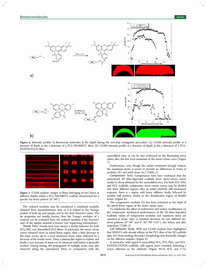
Spatial Distribution of Surface Treatment. Confocal laser scanningmicroscopy (CLSM) was used to investigate the penetration depth oftreatment by LSM 510 Zeiss confocal inverted microscope equippedwith a Zeiss 20X/3 NA objective and an argon laser. Each stage ofbioactivation (aminolysis and peptide covalent coupling) werefollowed conjugating the surface samples with two different dyes.First, the aminolysis treatment were showed by coupling PCL-NH2surfaces with 0.1 mg/mL of Rhodamine B isothiocyanate (RBITC,Sigma - R1755) in IPA overnight at 4 °C. Subsequently surfaces wererinsed thoroughly with IPA and then with water for 24 h to removeany non covalent bound dye molecule.
In order to mimic the peptide route, Fluoresceinamine (FLUO,Fluka -07980) was linked to the epoxy-functionalized surfaces by firstdissolving the dye in carbonate buffer at pH = 8.5 in the sameconditions used for peptide conjugation. Finally, samples were rinsedwith buffer followed by copious amounts of distilled water to removeany non-covalently linked molecules. The PCL fluorescent sampleswere then left to dry overnight in a vacuum desiccator before analysis.Unmodified PCL surfaces were equally processed as a control.Samples after RBITC or FLUO conjugation were respectivelyvisualized using the characteristic wavelengths of RBITC (λex = 543nm; λem = 572 nm) and FLUO (λex = 496 nm; λem= 518 nm).
To compare the results, CLSM settings, in particular, laser power,pinhole aperture, detector gain, and amplifier offset, were keptconstant for both kind of observations. The penetration depth oftreatment was visualized by z stack acquisitions through the fiber bystarting from the outer part. Intensity profiles of fluorescent dyes as afunction of penetration depth were obtained along a line drawn in theradius direction (here indicated as the z-direction).
Cell Adhesion Study. Mouse embryo fibroblasts NIH3T3 weremaintained at 37 °C and 5% CO2 in Dulbecco’s modified Eaglemedium (DMEM) supplemented with 10% fetal bovine serum (FBS,BioWhittaker, Walkersville, MD), 2 mM L-glutamine (Sigma, St. Louis,MO), 1000 U/l penicillin (Sigma, St. Louis, MO) and 100 mg/Lstreptomycin (Sigma, St. Louis, MO). In this study, 70−80% confluentcells were used. In particular, PCL, PCL-NH2, PCL-DGDGE-GYDGR,and PCL-DGDGE-GRGDY 3D scaffolds were sterilized with anti-biotics and preincubated in serum-free medium for 16−18 h asreported in a previous work.30 After the incubation, 5 × 10⁴ cells wereseeded on all the different kinds of 3D scaffolds and grown in DMEMw/o FBS to avoid unspecific cell adhesion depending on serum proteinadsorption.
Scanning electron microscopy (SEM) was performed by a Leica 420microscope in order to evaluate cell adhesion and shape. At differenttimes after cell seeding, the 3D fiber-deposited scaffolds were rinsedwith phosphate-buffered saline (PBS) and fixed with 2.5%glutaraldehyde (pH= 7.4) (Sigma-Aldrich, Italy) for 2h at roomtemperature. The cell-scaffold constructs were dehydrated in gradedethanol concentrations (from 50% to 100% v/v in ethanol), air-dried,gold sputtered and analyzed by SEM. Four different kinds of 3Dscaffolds were studied: PCL, PCL-NH2, PCL-DGDGE-GYDGR, andPCL- DGDGE-GRGDY.
Furthermore, the several cell-scaffold constructs were also analyzed(PCL, PCL-NH2, PCL-DGDGE-GYDGR, and PCL-DGDGE-GRGDY) through CLSM. They were fixed with 4% paraformaldehydefor 20 min at room temperature, after 24 h from seeding, rinsed twicewith PBS buffer, and incubated with 0.5% PBS-BSA (BSA = bovineserum albumin) to block unspecific binding. Actin microfilaments were
Biomacromolecules Article
dx.doi.org/10.1021/bm300818y | Biomacromolecules 2012, 13, 3510−35213513

stained with phalloidin-tetramethylrhodamine B isothiocyanate(Sigma-Aldrich). Phalloidin was diluted in 0.5% PBS-BSA andincubated for 30 min at room temperature. The images were acquiredby using a He−Ne excitation laser at the wavelength of 543 nm and a20× objective.Alamar Blue assay (AbD Serotec Ltd., UK) was also performed on
cell-scaffold constructs (PCL, PCL-NH2, PCL-DGDGE-GYDGR, andPCL-DGDGE-GRGDY) in order to evaluate cell viability. Thismethod is based on a redox reaction that occurs in the mitochondria ofthe cells; the colored product is transported out of the cell and can bemeasured spectrophotometrically. The optical density was measuredwith a spectrophotometer (Sunrise; Tecan, Man̈nedorf, Zurich,Switzerland) at wavelengths of 570 and 595 nm. The number ofviable cells correlates with the magnitude of dye reduction and isexpressed as a percentage of Alamar Blue reduction, according to themanufacturer’s protocol. Each experiment was performed at least threetimes in triplicate.Results. Micro-Computed Tomography. Micro-CT, which is an
attractive single and nondestructive method to study the characteristicsof scaffolds, has first allowed the morphological and architecturalfeatures of the 3D fiber-deposited PCL structures to be evaluated. Thisanalysis has confirmed that well-organized PCL scaffolds have beenobtained, showing precise pore size and shape, as well as a repeatablemicrostructure (Figure 2).
In particular, imaging analyses have evidenced a sufficientconsistency between real and theoretical values, showing a meanfiber diameter of 340−360 μm and a center-to-center fiber distance ofabout 640−660 μm between two fibers in a common layer, as expectedon the basis of the process/instrument parameters employed duringthe fabrication (i.e., needle inner diameter, deposition speed, fiberspacing...). Scaffold interconnectivity, which is normally defined as100% × volume of interconnected pores/volume sum of intercon-nected and closed pores,52 has been also evaluated and found to beequal to 100% .Determination of Engrafted Amines and Conjugated Peptide.
After aminolysis, an analytical determination of amino groupsengrafted onto the surface of PCL fibers constituting the well-organized scaffold was performed by a slight modification procedurebased on the Kaiser test.30,53,54
With regard to the evolution of aminolysis treatment performed onthe 3D scaffolds, a high amino-density of 161.3 ± 15.3 nmol/cm2 wasreached after 30 min of aminolysis, and then a decrease of surfaceamines was observed over time (Figure 3).The amount of peptides immobilized on PCL fibers of the scaffolds
was then evaluated using a one-pot colorimetric assay based on theBCA-Cu1+ purple color complex. This assay is widely employed toassess proteins both in solution and on adsorbing solid substrates in avery reproducible way and with high sensitivity (picomolar scale).51
The results obtained from micro-BCA assay have highlighted thatthe tyrosine present in our peptide provides a significantly differentamount of color formation at 37 °C, thus suggesting a partial reaction
of the BCA reagent with tyrosine. Moreover, the BCA reagent requiresat least a tripeptide in order to oxidize the peptide backbone. As in thecase of amino groups, the density of peptides covalently coupled to thePCL has been calculated by taking into account the effective scaffoldsurface area. A peptide density of 20.13 ± 4.68 nmol/cm2 has beenachieved (Figure 3).
Spatial Distribution of Surface Treatment. To analyze the spatialdistribution and penetration depth during each step of bioactivation,CLSM was employed. For this reason, two different dyes werecoupled: RBITC was used to label free amino groups after aminolysistreatment, whereas in a second step, FLUO was conjugated to theepoxy-activated PCL surface to mimic the peptide behavior studyingits distribution on the scaffold fiber surface.
Figure 4 clearly shows the intensity profile of the fiber cross-sectionalong the radius (here indicated as z-direction), evidencing apenetration depth of more than 140 μm for the aminolysis treatment.With regard to the FLUO bound surfaces, the total penetration depthof the treatment was about 80 μm, thus providing importantinformation on the potential peptide penetration depth.
The selected pore size and the fully interconnected pore network ofthe 3D rapid prototyped scaffold allowed us to optimize the masstransport properties (permeability and diffusion). The diffusionthrough the pores of the well organized structure is favored, thusobtaining a uniform distribution of the treatment within the innerstructure. Accordingly, the amino groups grafted onto the fiberscomposing all the layers of the structure were uniformly distributedwithin the scaffolds (Figure 5) and would be used for peptideconjugation after tether (DGDGE) insertion. On the other hand,regarding the penetration depth of the single fibers, the differences inthe central part of the fibers with respect to the crossing points of thefibers (fiber junctions) represent the only inhomogeneities.
Nanoindentation Tests. Nanoindentation measurements on PCLfibers have displayed differences in terms of load-depth curves and,hence, of hardness values. Both aminolyzed (PCL-NH2) andunmodified (PCL) fibers have evidenced hardness (H) values thatgenerally decrease as load increases from 1 to 5 mN. In particular,measurements on unmodified PCL fibers have evidenced hardnessvalues spanning from 0.50 to 0.27 GPa in the load range investigated.These values are greater than those obtained for PCL fibers that weremodified via aminolysis (0.1−0.03 GPa) (Figure 6a). This suggeststhat after aminolysis the fiber surface becomes softer. Consistentlywith hardness values, the reduced modulus (Er) of unmodified PCLfibers (4.2−1.2 GPa) is higher than that obtained from the PCL-NH2ones (1−0.3 GPa) (Figure 6b).
Figure 2. 3D reconstructions obtained from Micro-CT analysis onPCL fiber-deposited scaffolds with 0°/45°/90°/135° (a) and 0°/90°(b) lay-down patterns.
Figure 3. Amine and peptide density as a function of time ofaminolysis treatment. Data are graphically reported as mean value, andbars represent the standard deviation.
Biomacromolecules Article
dx.doi.org/10.1021/bm300818y | Biomacromolecules 2012, 13, 3510−35213514
The reduced modulus may be considered a combined modulusobtained from nanoindentation tests as it is related to the Young’smoduli of both tip and sample, and to the their Poisson’s ratios. Thetip properties are usually known, thus the Young’s modulus of amaterial can be evaluated from the reduced modulus if the Poisson’sratio of the sample material is known (see Supporting Information).Tensile Tests. Tensile tests have shown a ductile behavior for both
PCL-NH2 and unmodified PCL fibers. In particular, the stress−straincurves obtained show an initial linear region, then a little decrease inthe slope occurs up to a local maximum stress value, followed by adecrease of the tensile stress. Then, a plateau-like region is evident, andfinally a new increase of stress can be observed until failure is generallyreached. During testing, the propagation of multiple necks were alsoobserved along the aminolyzed fibers in comparison with the
unmodified ones, as can be also evidenced by the fluctuating stressvalues after the first local maximum of the stress−strain curve (Figure7).
Furthermore, even though the surface treatment strongly reducesthe maximum strain, it seems to provide no differences in terms ofmodulus (E) and yield stress (σy) (Table 1).
Compression Tests. Compression tests have evidenced that theaminolyzed 3D fiber-deposited scaffolds show stress−strain curvessimilar to those obtained for the unmodified ones. For both PCL-NH2
and PCL scaffolds, compressive stress−strain curves may be dividedinto three different regions: after an initial relatively stiff mechanicalresponse, there is a region with lower stiffness, finally followed byanother stiff portion, similar to the densification region of flexiblefoams (Figure 8).
The compressive modulus (E) has been evaluated as the slope ofthe initial linear region of the stress−strain curve.
To emphasize the effect of architecture and surface modification onthe compressive mechanical performances of the 3D fiber-depositedscaffolds, values of compressive modulus and maximum stress arereported as mean value ± standard deviation for two different lay-down patterns (0°/90° and 0°/45°/90°/135°), also before and afteraminolysis (Table 2).
Cell Adhesion Study. SEM and CLSM analyses have highlightedthat NIH3T3 cells already adhere on the PCL fiber of the 3D scaffoldsafter 24 h from seeding, showing a morphology that drastically changeson the different samples (Figure 9).
In particular, with regard to unmodified PCL, PCL-NH2, and PCL-DGDGE-GYDGR scaffolds, cells appear more rounded, indicating ascarce adhesion to the substrate (Figure 9A/E, B/F, and C/G,
Figure 4. Intensity profiles of fluorescent molecules in the depth during the two-step conjugation procedure: (a) CLSM intensity profile as afunction of depth in the z-direction of a PCL-NH-RBITC fiber; (b) CLSM intensity profile as a function of depth in the z-direction of a PCL-DGDGE-FLUO fiber.
Figure 5. CLSM analysis: images of fibers belonging to two layers atdifferent depths within a PCL-NH-RBITC scaffold characterized by aspecific lay-down pattern (0°/90°).
Biomacromolecules Article
dx.doi.org/10.1021/bm300818y | Biomacromolecules 2012, 13, 3510−35213515

respectively). Conversely, as for PCL bioactivated with RGD peptide,cells adhered and were well-spread on the fiber surface, evidencing agood interaction with the material (Figure 9 D/H).The effect of PCL functionalization/bioactivation in enhancing cell
adhesion was further confirmed by actin cytoskeleton staining. Thisqualitative analysis indicated that cells better adhered on RGDbioactivated scaffolds compared to cells seeded on unmodified PCL,PCL-NH2, and PCL-DGDGE-GYDGR surfaces, where there is noevidence of a complete cytoskeleton organization. Just as an example,Figure 10 reports a SEM micrograph of a bridging event after 48 hfrom seeding (PCL-DGDGE-GRGDY). SEM analyses on the different3D constructs allowed us to observe cells attached to single fibers or tomultiple fibers, stretching across void spaces within the scaffold(bridging morphology). Even though cells attached to single fibers ofthe scaffold were mainly observed in all the analyzed 3D constructs,
bridging morphology was also frequent within the fully interconnectedpore network.
Furthermore, Figure 11 shows SEM micrographs of cells attached tofibers belonging to the inner layers of the PCL-DGDGE-GRGDYscaffolds after 4 days from seeding.
Results from Alamar Blue assay suggest cell viability over time as thevalues reported in Table 3 increase with time.
Discussions. The four steps initially carried out in our work can besummarized as follows: (a) design and fabrication of “morphologically-controlled” scaffolds through 3D fiber-deposition technique; (b)optimization of a one-step surface treatment, especially through thequantification of amino-groups and the analysis of the penetrationdepth; (c) conjugation and quantification of RGD peptides; (d)mechanical characterization performed to assess the effect on thesurface and bulk PCL properties, as well as on the macromechanicalcompression performances of the structures.
3D rapid prototyped PCL scaffolds were fabricated through the 3Dfiber-deposition technique, then functionalized via aminolysis reactionand bioactived by grafting RGD peptides to the surface-modifiedfibers.
It has already been demonstrated that aminolysis may be consideredan easy-to-perform chemical technique to engraft amino groups alongpolyesters chains.30 By treating the 3D well-organized PCL scaffoldswith a 1,6-hexandiamine in an aprotic solvent at a 37 °C, a high densityof amino groups were rapidly obtained onto the PCL fiber surface ofthe structure. The reaction starts by a nucleophilic attack onto theester by an amino group at one end of diamine leading to theformation of an amide and leaving at the other end a free amino groupemerging the PCL fiber surface. The functionalization pathway of thePCL scaffolds, which means the functionalization of the fiber surface ofthe porous structure, is reported in Figure 1. In addition to the newbond formation along the polymer surface, from the rupture of estergroup were produced hydroxyl groups that could remain attached ontofiber surface or leached out during the washes.
As summarized in Figure 1, the scaffold surface bioactivation wasthen performed by covalently grafting the GRGDY peptide via ahomobifunctional cross-linker. DGDGE is a water-soluble epoxy cross-linker, that reacts quite well with amine in mild condition.
The amount of peptides grafted to PCL fibers of the scaffolds wasthen quantified by using the one-pot colorimetric micro-BCA assay.The number of peptide bonds and the presence of four amino acids(cysteine, cystine, tryptophan and tyrosine) have been reported to beresponsible for color formation in the peptide sample when assayedwith BCA. Studies with tri- and tetra-peptides suggest that the extentof color formation is due to the presence of several functionalgroups.51 The advantages in using BCA methods include acompatibility with ionic and non ionic detergents, a stable working
Figure 6. Results obtained from nanoindentation tests on PCL and PCL-NH2 fibers: hardness (a) and reduced modulus (b) as a function of theapplied load (1−5 mN). Data are graphically reported as mean value, and bars represent the standard deviation. The dashed lines are just a guide forthe eye.
Figure 7. Typical stress−strain curves obtained from tensile tests onPCL and PCL-NH2 fibers.
Table 1. Results from Tensile Tests Performed on PCL andPCL-NH2 Microfibers: Tensile Modulus (E), Yield Stress(σy), and Maximum Strain (εmax), Reported as Mean Value ±Standard Deviation
Fibers E (MPa) σy (MPa) εmax (mm/mm)
PCL 570.5 ± 50.1 25.0 ± 3.5 12.7 ± 1.1PCL-NH2 550.0 ± 48.6 24.2 ± 3.7 6.5 ± 0.5
Biomacromolecules Article
dx.doi.org/10.1021/bm300818y | Biomacromolecules 2012, 13, 3510−35213516

reagent and a tolerance to the presence of compounds that couldinterfere. As described by Tyllianakis et al.,55 this method can be usedto determine the total solid supports functionalized with cysteine andtyrosine, requires only one incubation step, and allows one to
determine the amount of the functional groups. The quantification ofthe different groups should be calculated from a standard curve of anappropriate substance.
In particular, the one-step colorimetric method has been consideredto quantify the surface concentration of GRGDY peptides covalently
Figure 8. Effect of lay-down pattern and surface modification via aminolysis on the mechanical properties of 3D rapid prototyped scaffolds: (a)Typical stress−strain curves for PCL scaffolds characterized by two different lay-down patterns (0°/90° and 0°/45°/90°/135°), before (PCL) andafter aminolysis (PCL-NH2); (b) Stress−strain curves reported up to a strain level of 0.04 mm/mm in order to better highlight the different initialstiffness of the 3D morphologically controlled structures.
Table 2. Effect of Lay-down Pattern and SurfaceModification via Aminolysis on the Mechanical Properties of3D Rapid Prototyped Scaffoldsa
compressive modulus E(MPa) maximum stress σ (MPa)
lay-down pattern PCL PCL-NH2 PCL PCL-NH2
0°/45°/90°/135°
63.0 ± 4.7 61.1 ± 5.1 12.3 ± 1.1 12.0 ± 1.3
0°/90° 89.1 ± 6.9 87.9 ± 8.1 13.5 ± 1.3 13.2 ± 1.5aCompressive modulus and maximum stress reported as mean value ±standard deviation, for PCL scaffolds characterized by two differentlay-down patterns (0°/90° and 0°/45°/90°/135°), before (PCL) andafter aminolysis (PCL-NH2).
Figure 9. Cell adhesion study after 24 h from seeding: SEM micrographs (A: PCL; B: PCL-NH2; C: PCL-DGDGE-GYDGR; D: PCL-DGDGE-GRGDY), bar 20 μm; CLSM images of phalloidin staining of microfilaments (E: PCL; F: PCL-NH2; G: PCL- DGDGE-GYDGR; H: PCL-DGDGE-GRGDY).
Figure 10. Cell adhesion study after 48 h from seeding: SEMmicrograph (PCL-DGDGE-GRGDY), bar 50 μm.
Biomacromolecules Article
dx.doi.org/10.1021/bm300818y | Biomacromolecules 2012, 13, 3510−35213517

bound on the fibers of the 3D PCL scaffolds. The quantification wascarried out by a standard curve using a known concentration of 1,6-hexanediamine. In our work, a peptide density of 20.13 ± 4.68 nmol/cm2 has been achieved, while a value of 2.81 ± 0.35 nmol/cm2 hasbeen assessed by Causa et al. (2010) in their study.30 As reported inthe literature, a minimum RGD density of 1.0 × 10⁻¹⁵ mol/cm2, thatcorresponds to a spacing of 140 nm between peptide ligands, seems tobe sufficient to promote cell spreading, while a density of 1.0 × 10⁻¹⁴
mol/cm2 has been found to favor the formation of focal contacts.30,38
The value of peptide density obtained in our work would seem wellbeyond the minimum value reported in the literature. Anyway, it isdifficult to numerically compare our results obtained for 3D rapidprototyped scaffolds with those reported in literature for polymericsheets.At each step of functionalization/bioactivation, the spatial
distribution and penetration depth was properly evaluated throughCLSM considering two different dyes (RBITC and FLUO).In the literature, many works have already shown how several
surface modifications and RGD immobilization may improve thewettability and/or the biological performances of PCL scaffolds. Forexample, Mattanavee et al.56 performed the immobilization of variousbiomolecules (i.e., collagen, chitosan, and GRGDS peptide) on thesurface of electrospun PCL fibrous scaffold, using N,N′-disuccinimi-dylcarbonate as the coupling agent. They demonstrated that theaminolyzed and biomolecule-immobilized PCL fibrous scaffoldsbecame more hydrophilic as evidenced by dynamic water contactangle measurements. Furthermore, cell culture experiments withdifferent cell lines have highlighted that among the bioactivatedelectrospun PCL scaffolds, the type I collagen- and GRGDS-immobilized scaffolds evidenced the greatest ability to promote cellattachment proliferation.56
Furthermore, Zhang et al.57 previously extended a simple method toimmobilize RGD peptide on 2D PCL films to the case of 3D well-organized scaffolds, investigating the bone marrow stromal cell(BMSC) behavior. Their modification strategy allowed one to obtaina successful Arg−Gly−Asp−Cys (RGDC) immobilization on 3D rapidprototyped PCL scaffolds via aminolysis and a heterobifunctionalcross-linker sulfosuccinimidyl 4-(N-maleimidomethyl)cyclohexane-1-carboxylate (sulfo-SMCC). In particular, Zhang et al.57 have widelystudied BMSC attachment, cellular distribution, signal transduction,and survival on their RGD-modified PCL scaffolds, demonstrating thatthe modification elicits specific cellular responses and improves cell−biomaterial interactions.
Wojtowicz et al. (2010)58 implanted GFOGER-coated well-organized PCL 3D scaffolds into critically sized femoral defects inrats. If compared to uncoated PCL scaffolds, and empty defects,GFOGER-coated scaffolds seemed to enhance osteoblastic differ-entiation and subsequent bone repair without the aid of implantedcells, genetic material or growth factors. Taubenberger et al. (2010)59
showed that the exposure of RGD-motifs in collagen may present amechanism to induce tissue remodeling and wound healing. Inparticular, partially denatured collagen I guided the binding of a varietyof integrins, thereby improving cell adhesion and subsequent cellularmigration and spreading.
However, it is worth noting that none of the above-mentionedstudies has assessed the effect of the surface modification on themechanical behavior of the 3D rapid prototyped scaffolds.Consequently, trying to fill this gap present in the literature,nanoindentation, tensile, and compression tests were carried outwith the aim to highlight the effect of aminolysis on the surface andbulk material properties, as well as on the macro-mechanicalperformances of the 3D fiber-deposited PCL scaffolds.
Advanced materials as well as biological tissues show hierarchicalstructures with particular features down to the nanometer ormicrometer scale. For this reason, a technique that can probemechanical properties at these scales has to be considered. In thiscontext, nanoindentation is emerging as a valuable mechanical testingtechnique for biomaterials. Hardness and microhardness testing (e.g.,Vickers and Knoop indentation)60,61 have been already considered toinvestigate the mechanical properties of hard tissues such as teeth andbones.62−65 However, nanoindentation enhances upon the spatial,force, and displacement resolutions of these traditional techniques,thus providing a powerful tool to study tissues and biomaterials withsubmicrometer resolution. Nanoindentation is also useful formeasuring mechanical properties of microstructural features withinbulk samples, characterizing the properties of individual constituentswithin composite or heterogeneous samples, or mapping mechanicalproperties across a sample surface. Because of its small probe size,nanoindentation can be used to measure local material properties insmall, thin, and heterogeneous samples. Nanoindentation, which is aninstrumented or depth-sensing indentation, involves, for example, theapplication of a controlled load to the surface inducing local surfacedeformation. Load and displacement are monitored during the loadingand unloading phases. Thus, properties such as hardness and reducedmodulus are calculated from the unloading curves using well-established equations. Considering its typical working force rangeand displacement range (1 μN−500 mN and 1 nm−20 μm,respectively),60,66 nanoindentation technique surely bridges the gapbetween atomic force microscopy (AFM) and macroscale mechanicaltesting.
Accordingly, we decided to carry out nanoindentation measure-ments on PCL fibers in order to highlight the differences in terms ofhardness and modulus values. Results from nanoindentation tests haveevidenced that after aminolysis the fiber surface becomes softer, sincethe hardness values decrease if compared to those obtained for theunmodified PCL (Figure 6a).
This softening effect could be ascribed to the reduction of theentanglement density due to the surface treatment. It is well-knownthat hardness and modulus of a polymer depend on its structure,
Figure 11. Cell adhesion study after 4 days from seeding: different SEM micrographs (PCL-DGDGE-GRGDY).
Table 3. Alamar Blue Assay Performed on Cell-ScaffoldConstructs (PCL, PCL-NH2, PCL-DGDGE-GYDGR, andPCL-DGDGE-GRGDY): Results (Mean Value ± StandardDeviation) Reported at 1 and 5 Days
Alamar Blue reduction (%)
sample 1 day 5 days
PCL 8.2 ± 1.3 18.3 ± 4.9PCL-NH2 9.7 ± 2.9 22.1 ± 5.4PCL-DGDGE-GRGDY 10.5 ± 1.8 27.9 ± 5.2PCL-DGDGE-GYDGR 10.3 ± 1.6 20.7 ± 3.5
Biomacromolecules Article
dx.doi.org/10.1021/bm300818y | Biomacromolecules 2012, 13, 3510−35213518

molecular weight, and number of segments between entanglements.67
In particular, values of reduced modulus and hardness are related topolymer chain flexibility (i.e., physical and chemical entanglements).The rigidity of amorphous region of the polymers depends on thedensity of entanglements among molecular chains, which represent thetopological restriction of molecular motion by other chains. Theindenter tip mainly interacts with molecular chains of the surface,which are relatively more flexible than those in the bulk because of thegreater mobility of surface chains. Within the deformation zone underthe tip, the superposition of simultaneous responses of severalmolecular chains determines the nanoindentation behavior. It is well-known that in the first stage, the aminolysis starts preferentially at theamorphous regions of the polymer causing the scission of the chainspresent on the surface, thus reducing the density of entanglements.Also taking into account the penetration depth of the treatment, as aconsequence of the scission of the chains undergoing functionalization,the flexibility and mobility of surface molecular chains increases, andthe surface of the aminolyzed fiber becomes softer.On the other hand, tensile tests performed on PCL fibers have
basically evidenced how the surface treatment strongly reduces themaximum strain without altering the values of modulus and yield stress(Figure 7 and Table 1).Although nanoindentation and tensile measurements on PCL fibers
have shown some differences, results from compression tests havesuggested that fixing a specific lay-down pattern the aminolyzed 3Dfiber-deposited scaffolds and the unmodified ones show similarmechanical properties. Furthermore, their stress−strain curves (Figure8) are similar to those of flexible foams. Anyway, in contrast to thetypical behavior of a flexible foam,68 the central zone is not a plateau,i.e., it does not have zero slope but just a lower one if compared withthe other two portions of the stress−strain curve. This behavior is alsoconsistent with that already reported for 3D fiber-deposited PCLscaffolds.24,69
As expected, the architecture, which means the specific lay-downpattern used (0°/90° or 0°/45°/90°/135°), also influences themechanical behavior of the 3D PCL scaffolds in compression.The lay-down pattern strongly affects the mechanical behavior of
the 3D fiber-deposited PCL scaffolds, especially in terms of initialstiffness. As reported in Table 2, before the surface modification viaaminolysis, PCL scaffolds characterized by a 0°/90° pattern exhibit acompressive modulus (89.1 ± 6.9 MPa), which is greater than thatobtained for a 0°/45°/90°/135° pattern (63.0 ± 4.7 MPa). At a strainvalue of 50%, a maximum stress of 13.5 ± 1.3 MPa and 12.3 ± 1.1MPa has been evaluated for 0°/90° and 0°/45°/90°/135° patterns,respectively.Furthermore, the surface treatment via aminolysis does not
negatively affect the macromechanical behavior of the 3D fiber-deposited scaffolds, as evaluated through compression tests. Forexample, after aminolysis, PCL scaffolds characterized by a 0°/90°pattern have shown values of compressive modulus (87.9 ± 8.1 MPa)that are similar to those achieved before the surface treatment (89.1 ±6.9 MPa), as well as a maximum stress of 13.2 ± 1.2 MPa compared to13.5 ± 1.3 MPa obtained for the corresponding not- aminolyzedstructures.Similar observations might be made for the 3D fiber-deposited PCL
scaffolds with a 0°/45°/90°/135° pattern, taking into account theresults numerically reported in terms of modulus and maximum stress,before and after the surface treatment via aminolysis (Table 2).Consequently, the surface treatment via aminolysis reduces the
hardness, but it does not negatively affect the compressive mechanicalbehavior of the rapid prototyped scaffolds. Unlike non-porousstructure, this effect may be ascribed to the specific load transfermechanism occurring in the 3D fiber-deposited scaffolds, where fibersact as load-bearing beams. The main contribution to the macro-mechanical performance is basically due to the crossing points of thefibers (fiber junctions) undergoing compression (i.e., a “column-likebehavior”) which seem to be less affected by the treatment.Although the above-mentioned steps are crucial, the knowledge of
cell-material interactions turns out to be a key element in designing 3Dadvanced multifunctional scaffolds with suitable morphology and
properties that are able to guide cell adhesion. For this reason, as afinal step of this research, in order to analyze the effect offunctionalization/bioactivation at the cell-material interface, theinteraction of 3D fiber-deposited scaffolds with fibroblast cells wasstudied through SEM and CLSM. Results from these analyses havequalitatively allowed to show the cytoskeleton organization and todemonstrate that cells better adhered on RGD bioactivated scaffolds.
3. CONCLUSIONSRGD motifs are being widely considered to design biomimeticsurfaces that could trigger a specific function in cell behavior atthe cell-material interface. Accordingly, cell adhesion should besuitably enhanced and tailored since it represents the basicfeature in the cell-material interaction. Previous works havealready evidenced that aminolysis represents an easy route tointroduce primary amines with high yield that can be easilyoptimized. Here, the controlled two-step procedure proposedby Causa et al.30 for 2D PCL films was extended to immobilizeRGD motifs on 3D rapid prototyped scaffolds, designing 3Dadvanced scaffolds. Unlike other works on 3D RGD-modifiedscaffolds, the present study basically focuses on the effect of thesurface modification on the mechanical properties at differentscales, however, showing an approach to analyze thefunctionalization/bioactivation, the distribution of immobilizedligands, and the biological features. The determination ofamines and peptides effectively engrafted on the fiber surface of3D scaffolds and the treatment penetration depth were suitablyassessed and discussed. Nanoindentation and tensile measure-ments carried out on PCL fibers allowed us to underline theeffect of the functionalization on the surface and bulkproperties. More importantly, the surface modification didnot negatively affect the macromechanical behavior of the 3Drapid prototyped scaffolds as evaluated through compressiontests. On the other hand, results from cell adhesion studyevidenced that the conjugation of peptides through aminolysisand tether insertion enhanced NIH3T3 cell adhesion andspreading.
■ ASSOCIATED CONTENT*S Supporting InformationNanoindentation: theory and methods, adaptation for inden-tation of polymers. This material is available free of charge viathe Internet at http://pubs.acs.org
■ AUTHOR INFORMATIONCorresponding Author*Mailing address: Institute of Composite and BiomedicalMaterials, National Research Council, P.le Tecchio 80, 80125,Naples, Italy. Telephone number: +39-081-2425925; faxnumber: +39-081-2425932; e-mail address: [email protected] Contributions§These authors contributed equally to this work.NotesThe authors declare no competing financial interest.
■ ACKNOWLEDGMENTSThe authors wish to thank Mr. Mario De Angioletti and Mr.Rodolfo Morra for performing nanoidentation measurementsand mechanical tests, respectively.
■ REFERENCES(1) Langer, R.; Vacanti, J. P. Science 1993, 260, 920−926.
Biomacromolecules Article
dx.doi.org/10.1021/bm300818y | Biomacromolecules 2012, 13, 3510−35213519

(2) Gloria, A.; Russo, T.; De Santis., R.; Ambrosio, L. J. Appl.Biomater. Biomech. 2009, 7, 141−152.(3) Gloria, A.; De Santis, R.; Ambrosio, L. J. Appl. Biomater. Biomech.2010, 8, 57−67.(4) Giordano, C.; Causa, F.; Candiani, G. J. Appl. Biomater. Biomech.2006, 4, 73−79.(5) Brochhausen, C.; Zehbe, R.; Gross, U.; Schubert, H.; Kirkpatrick,C. J. J. Appl. Biomater. Biomech. 2007, 5, 70−81.(6) Giordano, C.; Causa, F.; Di Silvio, L.; Ambrosio, L. J. Mater. Sci.:Mater. Med. 2007, 18, 653−660.(7) Causa, F.; Netti, P. A.; Ambrosio, L. Biomaterials 2007, 28,5093−5099.(8) Perale, G.; Bianco, F.; Giordano, C.; Matteoli, M.; Masi, M.;Cigada, A. J. Appl. Biomater. Biomech. 2008, 6, 1−18.(9) Pertici, G.; Maccagnan, S.; Mueller, M.; Mueller, M.; Rossi, F.;Daniele, F.; Tunesi, M.; Perale, G. J. Appl. Biomater. Biomech. 2008, 6,186−192.(10) Hutmacher, D. W. J. Biomater. Sci., Polym. Ed. 2001, 12, 107−124.(11) Hutmacher, D. W.; Schantz, T.; Zein, I.; Ng, K. W.; Teoh, S. H.;Tan, K. C. J. Biomed. Mater. Res. 2001, 55, 203−216.(12) Yu, X. J.; Dillon, G. P.; Bellamkonda, R. B. Tissue Eng. 1999, 5,291−304.(13) Hacker, M. C.; Mikos, A. G. Tissue Eng. 2006, 12, 2049−2057.(14) Nair, L. S.; Laurencin, C. T. Adv. Biochem. Eng./Biotechnol. 2006,102, 47−90.(15) Landers, R.; Hubner, U.; Schmelzeisen, R.; Mulhaupt, R.Biomaterials 2000, 23, 4437−4447.(16) Landers, R.; Pfister, A.; Hubner, U.; John, H.; Schmelzeisen, R.;Mulhaupt, R. J. Mater. Sci. 2002, 37, 3107−3116.(17) Yang, S.; Leong, K. F.; Du, Z.; Chua, C. K. Tissue Eng. 2002, 8,1−11.(18) Taboas, J. M.; Maddox, R. D.; Krebsbach, P. H.; Hollister, S. J.Biomaterials 2003, 24, 181−194.(19) Woodfield, T. B. F.; Malda, J.; de Wijn, J.; Peters, F.; Riesle, J.;van Blitterswijk, C. A. Biomaterials 2004, 25, 4149−4161.(20) Malda, J.; Woodfield, T. B.; van der Vloodt, F.; Wilsond, C.;Martens, D. E.; Tramper, J.; van Blitterswijk, C. A.; Riesle, J.Biomaterials 2004, 26, 63−72.(21) Moroni, L.; de Wijn, J. R.; van Blitterswijk, C. A. Biomaterials2006, 27, 974−985.(22) Moroni, L.; Schotel, R.; Sohier, J.; de Wijn, J. R.; vanBlitterswijk, C. A. Biomaterials 2006, 27, 5918−5926.(23) Moroni, L.; Schotel, R.; Hamann, D.; de Wijn, J. R.; vanBlitterswijk, C. A. Adv. Funct. Mater. 2008, 18, 53−60.(24) Kyriakidou, K.; Lucarini, G.; Zizzi, A.; Salvolini, E.; MattioliBelmonte, M.; Mollica, F.; Gloria, A; Ambrosio, L. J. Bioact. Compat.Polym. 2008, 23, 227−243.(25) Peltola, S. M.; Melchels, F. P. W.; Grijpma, D. K.; Kellomak̈i, M.Ann. Med. 2008, 40, 268−280.(26) Hutmacher, D. W. Biomaterials 2000, 21, 2529−2543.(27) Chua, C. K.; Leong, K. F.; Lim, C. S. In Rapid Prototyping:Principles and Applications; Chua, C. K., Leong, K. F., Lim, C. S., Eds;World Scientific Pub Co: Singapore, 2003; pp 25−33.(28) Ko, Y. G.; Kim, Y. H.; Park, K. D.; Lee, H. J.; Lee, W. K.; Park,H. D.; Kim, S. H.; Lee, G. S.; Ahn, D. J. Biomaterials 2001, 22, 2115−2123.(29) Zhu, Y.; Gao, C.; Liu, X.; Shen, J. Biomacromolecules 2002, 3,1312−1319.(30) Causa, F.; Battista, E.; Della Moglie, R.; Guarnieri, D.; Iannone,M.; Netti, P. A. Langmuir 2010, 26, 9875−9884.(31) Eldsater, C.; Erlandsson, B.; Renstad, R. A.; Albertsson, C.;Karlsson, S. Polymer 2000, 41, 1297−1304.(32) Choi, E. J.; Kim, C. H.; Park, J. K. Macromolecules 1999, 32,7402−7408.(33) Zhong, Z. K.; Sun, X. Z. S. Polymer 2001, 42, 6961−6969.(34) Ng, K. W.; Hutmacher, D. W.; Schantz, J. T.; Ng, C. S.; Too, H.P.; Lim, T. C.; Phan, T. T.; Teoh, S. H. Tissue Eng. 2001, 7, 441−455.(35) Garcia, A. J.; Reyes, C. D. J. Dent. Res. 2005, 84, 407−413.
(36) Hersel, U.; Dahmen, C.; Kessler, H. Biomaterials 2003, 24,4385−4415.(37) El-Amin, S. F.; Kofron, M. D.; Attawia, M. A.; Lu, H. H.; Tuan,R. S.; Laurencin, C. T. Clin. Orthop. Rel. Res. 2004, 427, 220−225.(38) Massia, S. P.; Hubbell, J. A. J. Cell Biol. 1991, 114, 1089−1100.(39) Croll, T. I.; O’Connor, A. J.; Stevens, G. W.; Cooper-White, J. J.Biomacromolecules 2004, 5, 463−473.(40) Healy, K. E.; Tsai, D.; Kim, J. Eur. Mater. Res. Soc. Symp. Proc.1992, 252, 109−114.(41) McConachie, A.; Newman, D.; Tucci, M.; Puckett, A.; Tsao, A.;Hughes, J.; Benghuzzi, H. Biomed. Sci. Instrum. 1999, 35, 45−50.(42) Cai, K.; Yao, K.; Cui, Y.; Yang, Z.; Li, X.; Xie, H.; Qing, T.; Gao,L. Biomaterials 2002, 23, 1603−1611.(43) Yang, J.; Bei, J.; Wang, S. Biomaterials 2002, 23, 2607−2614.(44) Chu, P. K.; Chen, J. Y.; Wang, L. P.; Huang, N. Mater. Sci. Eng.2002, R36, 143−206.(45) Zhu, Y.; Chian, K. S.; Chan-Park, M. B.; Mhaisalkara, P. S.;Ratner, B. D. Biomaterials 2006, 27, 68−78.(46) Gabriel, M.; Van Nieuw Amerongen, G. P.; Van Hinsbergh, V.W. M.; Van Nieuw Amerongen, A. V.; Zentner, A. J. Biomater. Sci.,Polym. Ed. 2006, 17, 567−577.(47) Santiago, L. Y.; Nowak, R. W.; Rubin, J. P.; Marra, K. G.Biomaterials 2006, 27, 2962−2969.(48) Taniguchi, I.; Kuhlman, W. A.; Mayes, A. M.; Griffith, L. G.Polym. Int. 2006, 55, 1385−1397.(49) Dalton, P. D.; Woodfield, T.; Hutmacher, D. W. Biomaterials2009, 30, 701−702.(50) Lam, C. X.; Savalani, M. M.; Hutmacher, D. W. Biomed. Mater.2008, 3, 1−15.(51) Wiechelman, K. J.; Braun, R. D.; Fitzpatrick, J. D. Anal. Biochem.1988, 175, 231−237.(52) Ho, S. T.; Hutmacher, D. W. Biomaterials 2006, 27, 1362−1376.(53) Kaiser, E.; Colescott, R. L.; Bossinger, C. D.; Cook, P. I. Anal.Biochem. 1970, 34, 595−598.(54) Sarin, V. K.; Kent, S. B.; Tam, J. P.; Merrifield, R. B. Anal.Biochem. 1981, 117, 147−157.(55) Tyllianakis, E. P.; Kakabakos, S. E.; Evangelatos, G. P.;Ithakissios, D. S. Anal. Biochem. 1994, 219, 335−340.(56) Mattanavee, W.; Puthong, S; Suwantong, O.; Hoven, V. P.;Supaphol, P.; Bunaprasert, T. ACS Appl. Mater. Interfaces 2009, 1,1076−1085.(57) Zhang, H.; Lin, C. Y.; Hollister, S. J. Biomaterials 2009, 30,4063−4069.(58) Wojtowicz, A. M.; Shekaran, A.; Oest, M. E.; Dupont, K. M.;Templeman, K. L.; Hutmacher, D. W.; Guldberg, R. E.; Garcìa, A. J.Biomaterials 2010, 31, 2574−2582.(59) Taubenberger, A. V.; Woodruff, M. A.; Bai, H.; Muller, D. J.;Hutmacher, D. W. Biomaterials 2010, 31, 2827−2835.(60) Ebenstein, D. M.; Pruitt, L. A. Nano Today 2006, 1, 26−33.(61) Tabor, D. The Hardness of Metals; Clarendon Press: London,UK, 1951; pp 151−160.(62) Kinney, J. H.; Marshall, S. J.; Marshall, G. W. Crit. Rev. Oral Biol.Med. 2003, 14, 13−29.(63) Waters, N. E. Symp. Soc. Exp. Biol. 1980, 34, 99−135.(64) Riches, P. E.; Everitt, N. M.; McNally, D. S. J. Biomech. 2000, 33,1551−1557.(65) Weaver, J. K. J. Bone Joint Surg. 1966, A48, 273−288.(66) Fischer-Cripps, A. C. Nanoindentation; Springer-Verlag: Berlin,Germany, 2002; pp 1−217.(67) Zhou, J.; Komvopoulos, K. J. Appl. Phys. 2006, 100, 114329−114329−8.(68) Gibson, L. J.; Ashby, M. F. Cellular Solids: Structure andProperties; Cambridge University Press: Cambridge, UK:, 1999; pp175−231.(69) Domingos, M.; Chiellini, F.; Gloria, A.; Ambrosio, L.; Bartolo, P.J.; Chiellini, E. Rapid Prototyping J. 2012, 18, 56−67.(70) Oliver, W. C.; Pharr, G. M. J. Mater. Res. 1992, 7, 1564−1583.(71) Oliver, W. C.; Pharr, G. M. J. Mater. Res. 2004, 19, 3−20.
Biomacromolecules Article
dx.doi.org/10.1021/bm300818y | Biomacromolecules 2012, 13, 3510−35213520

(72) Briscoe, B. J.; Fiori, L.; Pelillo, E. J. Phys. D: Appl. Phys. 1998, 31,2395−2405.(73) Hu, Y.; Shen, L.; Yang, H.; Wang, M.; Liu, T.; Liang, T.; Zhang,J. Polym. Test. 2006, 25, 492−497.(74) Klapperich, C.; Komvopoulos, K.; Pruitt, L. J. Tribol.: Trans.ASME 2001, 123, 624−631.
Biomacromolecules Article
dx.doi.org/10.1021/bm300818y | Biomacromolecules 2012, 13, 3510−35213521
Related Documents
![Bioactive Scaffolds for Regeneration of Cartilage and … · role in reducing cartilage degeneration and decreasing chondrocyte apoptosis on the osteoarthritis therapy [21, 22]. Recent](https://static.cupdf.com/doc/110x72/5f0e7a4e7e708231d43f706e/bioactive-scaffolds-for-regeneration-of-cartilage-and-role-in-reducing-cartilage.jpg)


![Hypoxia-mimicking mesoporous bioactive glass scaffolds with … Biomaterials 2012.pdf · 2016. 2. 6. · biomaterials could induce a hypoxia function [27]. To our best knowledge,](https://static.cupdf.com/doc/110x72/5fe35ec466c7c06113333f28/hypoxia-mimicking-mesoporous-bioactive-glass-scaffolds-with-biomaterials-2012pdf.jpg)


