Nanomaterials 2022, 12, 576. https://doi.org/10.3390/nano12030576 www.mdpi.com/journal/nanomaterials Review Exploring Various Techniques for the Chemical and Biological Synthesis of Polymeric Nanoparticles Thiruchelvi Pulingam, Parisa Foroozandeh, Jo-Ann Chuah and Kumar Sudesh * Ecobiomaterial Research Laboratory, School of Biological Sciences, Universiti Sains Malaysia, Gelugor 11800, Penang, Malaysia; [email protected] (T.P.); [email protected] (P.F.); [email protected] (J.-A.C.) * Correspondence: [email protected] Abstract: Nanoparticles (NPs) have remarkable properties for delivering therapeutic drugs to the body’s targeted cells. NPs have shown to be significantly more efficient as drug delivery carriers than micron-sized particles, which are quickly eliminated by the immune system. Biopolymer-based polymeric nanoparticles (PNPs) are colloidal systems composed of either natural or synthetic poly- mers and can be synthesized by the direct polymerization of monomers (e.g., emulsion polymeriza- tion, surfactant-free emulsion polymerization, mini-emulsion polymerization, micro-emulsion polymerization, and microbial polymerization) or by the dispersion of preformed polymers (e.g., nanoprecipitation, emulsification solvent evaporation, emulsification solvent diffusion, and salting- out). The desired characteristics of NPs and their target applications are determining factors in the choice of method used for their production. This review article aims to shed light on the different methods employed for the production of PNPs and to discuss the effect of experimental parameters on the physicochemical properties of PNPs. Thus, this review highlights specific properties of PNPs that can be tailored to be employed as drug carriers, especially in hospitals for point-of-care diag- nostics for targeted therapies. Keywords: polymeric nanoparticles; nanoprecipitation; emulsification solvent evaporation; emul- sification solvent diffusion; polyhydroxyalkanoates (PHA); natural nanoparticles 1. Introduction Nanoparticles (NPs) are defined as particles with all three dimensions confined within the range of 1 to 100 nm [1–4]. The growing attention towards NPs stems from the fact that their mechanical, chemical, optical, electrical, and magnetic properties differ from those of their bulk counterparts, and these properties can be altered by varying the size of NPs [5,6]. NPs are of great interest in a variety of sectors, including physics, agriculture, chemistry, engineering, electronics, biology, food technology, medicine, and bioengineer- ing, due to their small size and ability to tailor their properties for specific requirements [7–17]. NPs offer the perfect characteristics for delivering therapeutic medications to the body’s target sites [18]. In contrast to micron-sized particles that are rapidly eliminated by the immune system, NPs demonstrated much higher efficiency as drug delivery carri- ers [19–21]. Because of their larger surface area, NPs can effectively penetrate cells and traverse the blood–brain barrier and they are easily destroyed [22–24]. NPs can be pro- duced using a variety of natural and synthetic materials, which are biodegradable or non- biodegradable [25]. Examples of NPs include solid–lipid nanoparticles, silver nanoparti- cles, gold nanoparticles, magnetic nanoparticles, mesoporous silica nanoparticles, nano- crystals, carbon nanotubes, albumin nanoparticles, fullerene nanoparticles, and polymeric nanoparticles (PNPs). Citation: Pulingam, T.; Foroozandeh, P.; Chuah, J.-A.; Sudesh, K. Exploring Various Techniques for the Chemical and Biological Synthesis of Polymeric Nanoparticles. Nanomaterials 2022, 12, 576. https:// doi.org/10.3390/nano12030576 Academic Editor: Thierry Rabilloud Received: 30 December 2021 Accepted: 6 February 2022 Published: 8 February 2022 Publisher’s Note: MDPI stays neu- tral with regard to jurisdictional claims in published maps and institu- tional affiliations. Copyright: © 2022 by the authors. Li- censee MDPI, Basel, Switzerland. This article is an open access article distributed under the terms and con- ditions of the Creative Commons At- tribution (CC BY) license (https://cre- ativecommons.org/licenses/by/4.0/).

Welcome message from author
This document is posted to help you gain knowledge. Please leave a comment to let me know what you think about it! Share it to your friends and learn new things together.
Transcript
Nanomaterials 2022, 12, 576. https://doi.org/10.3390/nano12030576 www.mdpi.com/journal/nanomaterials
Review
Exploring Various Techniques for the Chemical and Biological
Synthesis of Polymeric Nanoparticles
Thiruchelvi Pulingam, Parisa Foroozandeh, Jo-Ann Chuah and Kumar Sudesh *
Ecobiomaterial Research Laboratory, School of Biological Sciences, Universiti Sains Malaysia, Gelugor 11800,
Penang, Malaysia; [email protected] (T.P.); [email protected] (P.F.); [email protected] (J.-A.C.)
* Correspondence: [email protected]
Abstract: Nanoparticles (NPs) have remarkable properties for delivering therapeutic drugs to the
body’s targeted cells. NPs have shown to be significantly more efficient as drug delivery carriers
than micron-sized particles, which are quickly eliminated by the immune system. Biopolymer-based
polymeric nanoparticles (PNPs) are colloidal systems composed of either natural or synthetic poly-
mers and can be synthesized by the direct polymerization of monomers (e.g., emulsion polymeriza-
tion, surfactant-free emulsion polymerization, mini-emulsion polymerization, micro-emulsion
polymerization, and microbial polymerization) or by the dispersion of preformed polymers (e.g.,
nanoprecipitation, emulsification solvent evaporation, emulsification solvent diffusion, and salting-
out). The desired characteristics of NPs and their target applications are determining factors in the
choice of method used for their production. This review article aims to shed light on the different
methods employed for the production of PNPs and to discuss the effect of experimental parameters
on the physicochemical properties of PNPs. Thus, this review highlights specific properties of PNPs
that can be tailored to be employed as drug carriers, especially in hospitals for point-of-care diag-
nostics for targeted therapies.
Keywords: polymeric nanoparticles; nanoprecipitation; emulsification solvent evaporation; emul-
sification solvent diffusion; polyhydroxyalkanoates (PHA); natural nanoparticles
1. Introduction
Nanoparticles (NPs) are defined as particles with all three dimensions confined
within the range of 1 to 100 nm [1–4]. The growing attention towards NPs stems from the
fact that their mechanical, chemical, optical, electrical, and magnetic properties differ from
those of their bulk counterparts, and these properties can be altered by varying the size of
NPs [5,6]. NPs are of great interest in a variety of sectors, including physics, agriculture,
chemistry, engineering, electronics, biology, food technology, medicine, and bioengineer-
ing, due to their small size and ability to tailor their properties for specific requirements
[7–17].
NPs offer the perfect characteristics for delivering therapeutic medications to the
body’s target sites [18]. In contrast to micron-sized particles that are rapidly eliminated
by the immune system, NPs demonstrated much higher efficiency as drug delivery carri-
ers [19–21]. Because of their larger surface area, NPs can effectively penetrate cells and
traverse the blood–brain barrier and they are easily destroyed [22–24]. NPs can be pro-
duced using a variety of natural and synthetic materials, which are biodegradable or non-
biodegradable [25]. Examples of NPs include solid–lipid nanoparticles, silver nanoparti-
cles, gold nanoparticles, magnetic nanoparticles, mesoporous silica nanoparticles, nano-
crystals, carbon nanotubes, albumin nanoparticles, fullerene nanoparticles, and polymeric
nanoparticles (PNPs).
Citation: Pulingam, T.; Foroozandeh,
P.; Chuah, J.-A.; Sudesh, K.
Exploring Various Techniques for the
Chemical and Biological Synthesis of
Polymeric Nanoparticles.
Nanomaterials 2022, 12, 576. https://
doi.org/10.3390/nano12030576
Academic Editor: Thierry Rabilloud
Received: 30 December 2021
Accepted: 6 February 2022
Published: 8 February 2022
Publisher’s Note: MDPI stays neu-
tral with regard to jurisdictional
claims in published maps and institu-
tional affiliations.
Copyright: © 2022 by the authors. Li-
censee MDPI, Basel, Switzerland.
This article is an open access article
distributed under the terms and con-
ditions of the Creative Commons At-
tribution (CC BY) license (https://cre-
ativecommons.org/licenses/by/4.0/).
Nanomaterials 2022, 12, 576 2 of 30
Many types of NPs have been investigated for clinical use but have not been accepted
widely due to their toxicity to some extent [26]. Biopolymers are employed in the manu-
facturing of NPs for biomedical applications to avoid cytotoxicity concerns [27,28]. Biopol-
ymers are well-known for being non-toxic, biodegradable, and biocompatible [29,30]. De-
pending on the intended uses, PNPs can be simply and cost-effectively generated on a
wide scale using a variety of technologies. PNPs have applications in different fields such
as electronics [31], photonics [32], environmental technology [33], medicine [34], bio-im-
aging [35], diagnostics [36], biotechnology [37], biomedical drug delivery [38–40], and en-
ergy harvesting [41].
Due to their subcellular size, biodegradability, biocompatibility with tissue and cells,
and controlled and sustained-release capabilities, PNPs are attractive candidates for the
delivery of vaccinations, antibiotics, and cancer treatments [42–46]. PNPs can enhance the
bioavailability, solubility, and retention time of drugs. Moreover, PNPs do not cause any
toxic, inflammatory, or immunogenic side effects [47,48]. Different polymers such as pol-
yhydroxyalkanoate (PHA) [49–52], polylactic acid (PLA) [53–55], poly(lactic-co-glycolic
acid) (PLGA) [56,57], polycaprolactone (PCL) [58–60], polyglycolide (PGA) [61], polyan-
hydride [62], polycyanoacrylate [63], poly glutamic acid [64], polymalic acid [65,66],
poly(N-vinyl pyrrolidone) [66,67], poly(methyl methacrylate) (PMMA) [68,69], poly(vinyl
alcohol) [70,71], poly(acrylic acid) [72,73], poly acrylamide [74,75], and poly(methacrylic
acid) [76,77] have been used for the synthesis of PNPs.
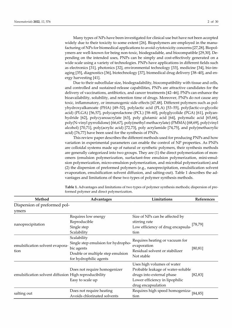
This review paper describes the different methods used for producing PNPs and how
variation in experimental parameters can enable the control of NP properties. As PNPs
are colloidal systems made up of natural or synthetic polymers, their synthesis methods
are generally categorized into two groups. They are (1) the direct polymerization of mon-
omers (emulsion polymerization, surfactant-free emulsion polymerization, mini-emul-
sion polymerization, micro-emulsion polymerization, and microbial polymerization) and
(2) the dispersion of preformed polymers (e.g., nanoprecipitation, emulsification solvent
evaporation, emulsification solvent diffusion, and salting-out). Table 1 describes the ad-
vantages and limitations of these two types of polymer synthesis methods.
Table 1. Advantages and limitations of two types of polymer synthesis methods; dispersion of pre-
formed polymer and direct polymerization.
Method Advantages Limitations References
Dispersion of preformed pol-
ymers
nanoprecipitation
Requires low energy
Reproducible
Single step
Scalability
Size of NPs can be affected by
stirring rate
Low efficiency of drug encapsula-
tion
[78,79]
emulsification solvent evapora-
tion
Scalability
Single step emulsion for hydropho-
bic agents
Double or multiple step emulsion
for hydrophilic agents
Requires heating or vacuum for
evaporation
Residual solvent or stabilizer
Not stable
[80,81]
emulsification solvent diffusion
Does not require homogenizer
High reproducibility
Easy to scale up
Uses high volumes of water
Probable leakage of water-soluble
drugs into external phase
Lower efficiency in lipophilic
drug encapsulation
[82,83]
salting out Does not require heating
Avoids chlorinated solvents
Requires high speed homogeniza-
tion [84,85]
Nanomaterials 2022, 12, 576 3 of 30
Suitable for DNA, RNA, and pro-
teins
Exclusive for the encapsulation of
lipophilic drugs
Time-consuming
Limited scalability
Direct polymerization
emulsion
Produce polymers with high molar
mass
Uses water as dispersion medium
Excellent heat dissipation
Requires removal of surfactant
Time consuming
High cost
[86,87]
surfactant-free emulsion
Does not require surfactant
Simple and green process
Uses water-soluble initiators
Requires the preparation of mon-
odisperse and uniformly distrib-
uted particle sizes
[88,89]
mini emulsion
Uses a low molecular mass co-stabi-
lizer
Small particle size
Low volume of surfactant
Uses a high-shear device
Surfactant is retained in the poly-
mer
[90,91]
micro-emulsion Uses water-soluble initiators
Thermodynamically stable
Formation of empty micelles
Destabilized microdroplets
Increased particle size
Requires a high ratio of surfactant
[92,93]
microbial
Non-toxic
Eco-friendly
Biocompatible
High production cost [94,95]
2. Methods for Producing PNPs
PNP preparation can be divided into two categories: monomer polymerization and
preformed polymer dispersion [96–98]. Emulsion polymerization, surfactant-free emul-
sion polymerization, mini-emulsion polymerization, and micro-emulsion polymerization
are all processes that can be used to polymerize monomers [99,100]. Likewise, nanopre-
cipitation, emulsification solvent evaporation, emulsification solvent diffusion, and salt-
ing-out can all be utilized to make PNPs from preformed polymers [101–103]. The type of
polymer, size requirement, and application region all influence the method of preparation
[104,105]. The technique of preparation is crucial to achieving the desired qualities. PNPs
made for biological applications, for example, should be free of additives and reactants
[106].
The type of polymer used determines the features of the produced NPs that are de-
signed for a certain purpose [107,108]. The drug delivery capabilities of PLGA and poly(3-
hydroxybutyrate) P(3HB) were studied by employing docetaxel (DTXL). Although the
toxicity profiles of P(3HB) and PLGA were similar, P(3HB) had a nearly two-fold higher
loading efficacy and poorer retention rates than PLGA [109]. Dissolution, solubility, cel-
lular uptake, release of drugs, bio-distribution, and circulatory half-life are all influenced
by the size of PNPs [110–112]. The challenge in the preparation of PNPs is the ability to
produce uniform particles to have consistent performance [113,114]. NPs with a broad
size distribution result in difficulty in establishing their applications [115].
2.1. Formation of NPs from Preformed Polymers
This section discusses the many ways to make PNPs from pre-formed polymers, in-
cluding nanoprecipitation, emulsification solvent evaporation, emulsification solvent dif-
fusion, and salting-out [101–103]. The initial stage in all of these approaches is to prepare
an emulsification system, which is the same for all of them. The second step is the for-
mation of PNP, which is different for each method. The name of the method is conferred
Nanomaterials 2022, 12, 576 4 of 30
by the principles of the second step, which can occur either by precipitation or by the
evaporation of the organic solvent [116,117].
2.1.1. Nanoprecipitation
Fessi et al. devised the nanoprecipitation approach, often known as the solvent dis-
placement, antisolvent precipitation, solvent shifting, and desolvation methods, for the
creation of PNPs in 1989 [118]. Nanoprecipitation is a simple, easy, fast, and reproducible
single-step method. This approach does not demand a lot of energy and can be scaled up
simply [119]. Nanoprecipitation is time-efficient, inexpensive, and does not need a pre-
cursor emulsion like other methods [120]. The size of the NPs generated by this approach
is changed by altering the parameters, and they are small with a limited size distribution
[121]. Nanoprecipitation is based on interfacial deposition, in which the transport of a sol-
vent into a non-solvent causes the polymer to dissolve, leading to nuclei growth, crystal
growth, and nanoprecipitation [122–124].
An organic phase is introduced to the aqueous phase during nanoprecipitation. The
polymer and water-miscible organic solvent, which must be miscible in the aqueous me-
dium, make up the organic phase, which has a diffusion effect [125–131]. To slow aggre-
gation, the polymer must be insoluble in the aqueous solution, which might contain a
stabilizer like a surfactant [132–135]. Dropwise addition of the organic phase to the aque-
ous phase with moderate agitation produces NPs [136,137]. Ultracentrifugation is used to
collect the NPs, which are subsequently rinsed with water to remove the surfactant. The
organic solvent evaporates, hardening the NPs, which are subsequently recovered by fil-
tering, spinning, or freeze-drying [138,139]. Organic solvents that evaporate easily such
as ethanol, acetone, hexane, or methylene chloride should be chosen as a polymer solvent.
Binary solvent blends such as combinations of acetone with either ethanol or methanol
can also be used. Likewise, a mixture of non-solvents can be used to form NPs in this
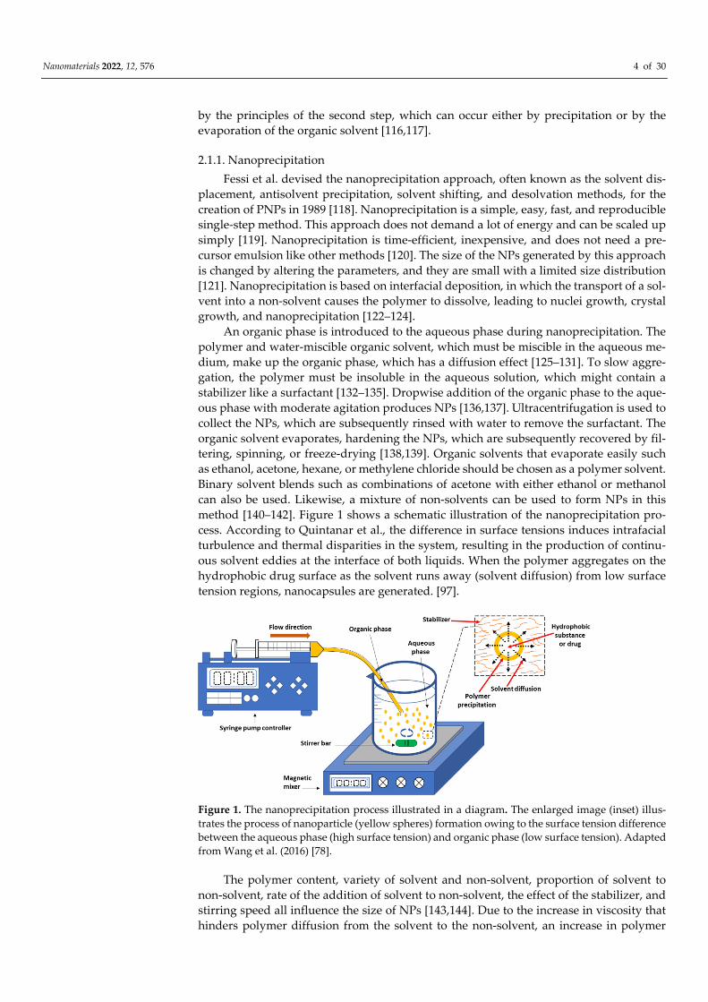
method [140–142]. Figure 1 shows a schematic illustration of the nanoprecipitation pro-
cess. According to Quintanar et al., the difference in surface tensions induces intrafacial
turbulence and thermal disparities in the system, resulting in the production of continu-
ous solvent eddies at the interface of both liquids. When the polymer aggregates on the
hydrophobic drug surface as the solvent runs away (solvent diffusion) from low surface
tension regions, nanocapsules are generated. [97].
Figure 1. The nanoprecipitation process illustrated in a diagram. The enlarged image (inset) illus-
trates the process of nanoparticle (yellow spheres) formation owing to the surface tension difference
between the aqueous phase (high surface tension) and organic phase (low surface tension). Adapted
from Wang et al. (2016) [78].
The polymer content, variety of solvent and non-solvent, proportion of solvent to
non-solvent, rate of the addition of solvent to non-solvent, the effect of the stabilizer, and
stirring speed all influence the size of NPs [143,144]. Due to the increase in viscosity that
hinders polymer diffusion from the solvent to the non-solvent, an increase in polymer
Nanomaterials 2022, 12, 576 5 of 30
concentration leads to the creation of bigger nanoparticles [144]. Smaller NPs in a narrow
size range are produced by solvents with high diffusion coefficients, such as acetone and
acetonitrile [145]. It has also been established that a decrease in the solvent-to-non-solvent-
volume ratio results in smaller NP sizes [146]. The nature of the stabilizer and its concen-
tration has been shown to influence the size of NPs [147,148].
A study found that increasing the amount of surfactant (e.g., Pluronic) reduced the
size of NPs by lowering interfacial tension [149]. In addition to that, employing a surfac-
tant in the nanoprecipitation method is not necessary, enabling the production of surfac-
tant-free particles [150]. Meanwhile, higher stirring rates have been found to produce
smaller NPs [151,152]. Zhang and colleagues demonstrated that raising the stirring speed
from 300 to 1200 rpm reduces particle diameter from 800 to 300 nm [153]. Specifically, low
external energy input is sufficient for the nanoprecipitation method, hence a moderate
stirring speed is required instead of a high stirring speed that raises the temperature [154–
156].
NP formation using the nanoprecipitation method occurs through three different
steps; particle nucleation, molecular growth and particle agglomeration [157]. Nucleation
takes place when the polymer concentration reaches the saturation level, i.e., when the
polymer solute in the solution is more than the amount that the solvent can dissolve [158].
The mean particle size increased significantly as the polymer concentration was increased
[159,160]. Molecular growth and particle agglomeration occur with a release of energy
[161,162].
Chorny and coworkers used the nanoprecipitation approach to make PLA NPs
loaded with tyrphostin. Particle size increases from 70 nm to 140 nm when the polymer
concentration is increased from 100 mg (5 mg/mL) to 300 mg (15 mg/mL) [163]. NPs of
poly(lactide)-poly(ethylene glycol)-poly(lactide) (PLA-PEG-PLA) were synthesized by
the nanoprecipitation method under different conditions. It was discovered that increas-
ing the agitation rate resulted in a reduction in particle size [164]. Meanwhile, in another
study comparing two methods for NP preparation, the nanoprecipitation method was
found to be more efficient for preparing PLGA NPs encapsulating cucurbitacin compared
to using the emulsion solvent evaporation method [165].
In the preparation of cellulose NPs loaded with mefenamic acid [166] and PLGA NPs
loaded with N-acetylcysteine (NAC), the solvent/nonsolvent ratio, the concentration of
polymer and the choice of solvent as well as nonsolvent were found to affect the size of
NPs. The nanoprecipitation method, in addition to efficiently entrapping hydrophobic
molecules, also has a great potential as an alternate entrapment method for hydrophilic
chemicals, according to the findings [167]. Chidambaram et al. proposed changes to the
traditional nanoprecipitation process in order to reduce NP size and create NPs with a
narrow size distribution. They used sonication to prepare both the organic and aqueous
phases, yielding Eudragit E100 NPs with a particles size of 114 nm and a uniformity of
0.259 [168].
Three distinct proteins (tetanus toxoid, lysozyme, and insulin) were entrapped in
poly(D, L-lactic acid) and poly(D, L-lactic-co-glycolic acid) NPs using modified nanopre-
cipitation and double emulsion (w1/o/w2) techniques in a separate investigation. The use
of miscible organic solvents like dimethylsulfoxide (DMSO) rather than conventional or-
ganic solvents like acetone or ethanol, as well as non-solvents like methanol or ethanol
rather than water, have all been added to the nanoprecipitation process. Nanoprecipita-
tion proved to be a suitable option to the extensively employed double emulsion ap-
proach. Nanoprecipitation was found to be the best approach for protein trapping in
small, densely loaded NPs [169]. Luo et al. applied a combination of electrospraying and
nanoprecipitation to produce multifunctional superhydrophobic polymethylsilsesquiox-
ane (PMSQ) NPs with sizes smaller than 100 nm [170].
Additionally, continuous flow microfluidics is a great solution for nanoprecipitation
operations, enhancing product controllability, homogeneity, and reproducibility. Nano-
precipitation through a hydrodynamic flow-focusing microchannel was used to
Nanomaterials 2022, 12, 576 6 of 30
synthesize PLGA-poly(ethylene glycol) nanoparticles (PLGA-PEG NPs). Variations in
flow rates, polymer concentration, and polymer composition can be used to obtain the
preferred size, drug loading, and polydispersity of the synthesized product [171]. Poly-
caprolactone (PCL) nanoparticles, which are biodegradable and have a tremendous po-
tential for controlled drug delivery, were synthesized through a similar nanoprecipitation
process [172]. Moreover, this technique may be used to assemble other polymers like chi-
tosan, heparin, and hyaluronic acid in microfluidic devices, especially to produce PNPs
for controlled release as well as drug delivery [173].
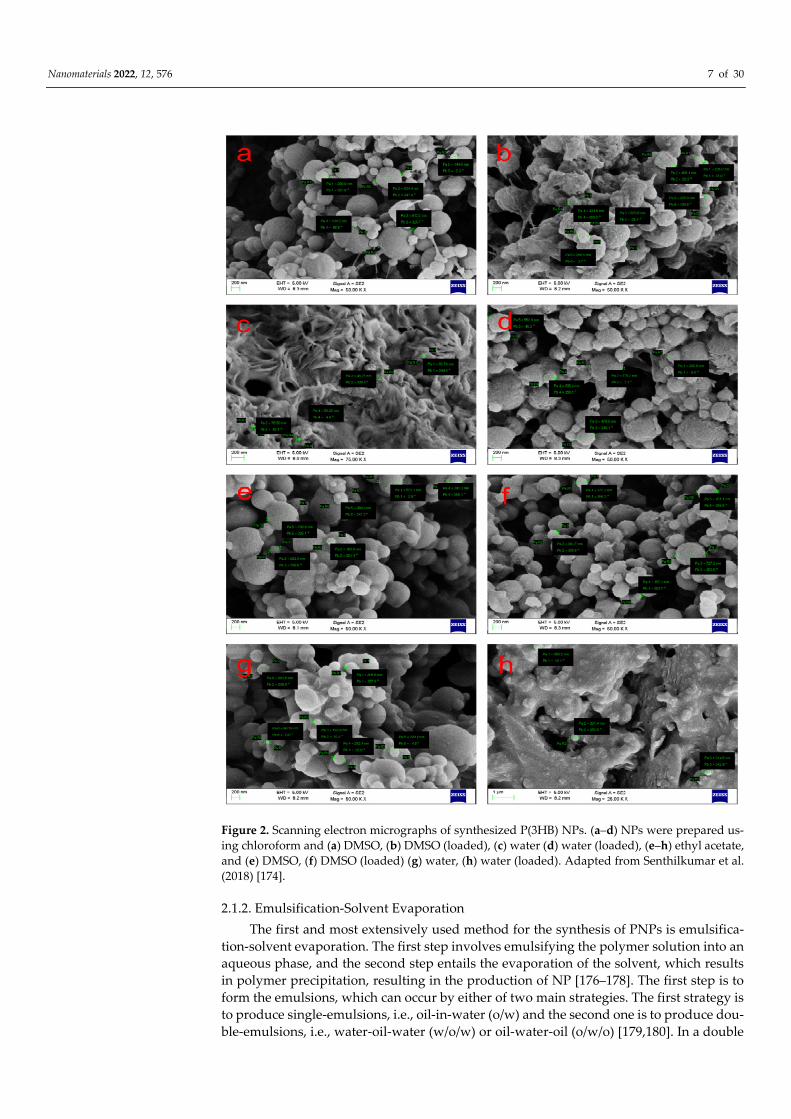
Meanwhile, P(3HB) NPs were prepared by nanoprecipitation with a variety of sol-
vent/non-solvent combinations such as ethyl acetate:DMSO, chloroform:water, chloro-
form:DMSO, and ethyl acetate:water. In the reported study, spherically shaped P(3HB)
NPs with sizes ranging from 40 to 100 nm were successfully formed while the size of
loaded PNPs were typically between 200 to 600 nm as shown in Figure 2 [174]. In another
attempt, P(3HB) NPs were prepared by nanoprecipitation with a low concentration of
Tween 80 as a surfactant. The size and size distribution of NPs decreased as the amount
of Tween 80 in water increased to 1% (v/v) [175].
In all the examples mentioned above, P(3HB) is initially biosynthesized by microor-
ganisms and stored in the microbial cell cytoplasm. The produced and accumulated nat-
ural polyester is then removed from the bacterial cells using suitable solvents like chloro-
form and purified by reprecipitation in a non-solvent like methanol. The purified P(3HB)
can be mixed in solvents and used in the nanoprecipitation process to make NPs.
Nanomaterials 2022, 12, 576 7 of 30
Figure 2. Scanning electron micrographs of synthesized P(3HB) NPs. (a–d) NPs were prepared us-
ing chloroform and (a) DMSO, (b) DMSO (loaded), (c) water (d) water (loaded), (e–h) ethyl acetate,
and (e) DMSO, (f) DMSO (loaded) (g) water, (h) water (loaded). Adapted from Senthilkumar et al.
(2018) [174].
2.1.2. Emulsification-Solvent Evaporation
The first and most extensively used method for the synthesis of PNPs is emulsifica-
tion-solvent evaporation. The first step involves emulsifying the polymer solution into an
aqueous phase, and the second step entails the evaporation of the solvent, which results
in polymer precipitation, resulting in the production of NP [176–178]. The first step is to
form the emulsions, which can occur by either of two main strategies. The first strategy is
to produce single-emulsions, i.e., oil-in-water (o/w) and the second one is to produce dou-
ble-emulsions, i.e., water-oil-water (w/o/w) or oil-water-oil (o/w/o) [179,180]. In a double
Nanomaterials 2022, 12, 576 8 of 30
emulsion, the primary emulsion (w1/o) is first prepared by dispersing the aqueous phase
in an immiscible organic solvent containing the polymer. Subsequently, the primary
emulsion is homogenized in an outer aqueous phase containing the emulsifier using a
high-shear homogenizer to form the organic phase and then emulsified in the aqueous
phase containing a surfactant [181–186].
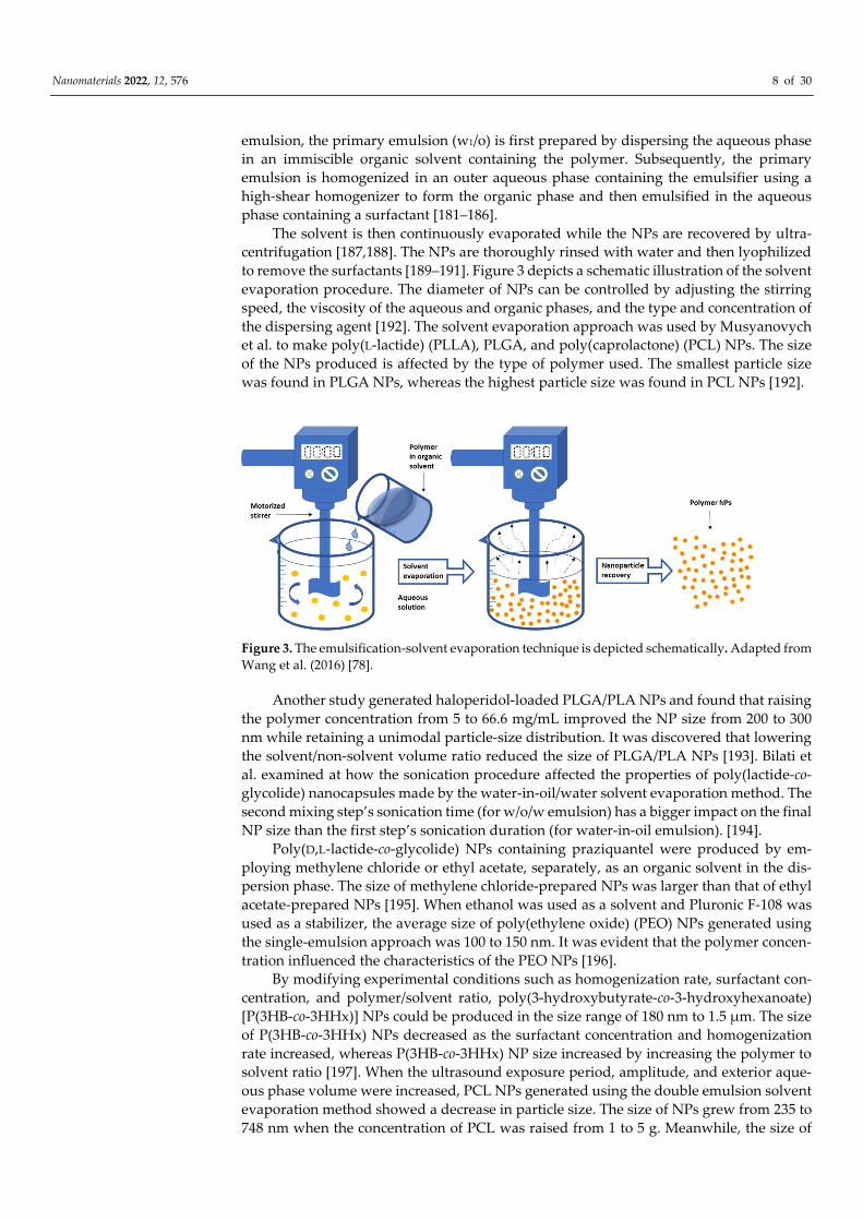
The solvent is then continuously evaporated while the NPs are recovered by ultra-
centrifugation [187,188]. The NPs are thoroughly rinsed with water and then lyophilized
to remove the surfactants [189–191]. Figure 3 depicts a schematic illustration of the solvent
evaporation procedure. The diameter of NPs can be controlled by adjusting the stirring
speed, the viscosity of the aqueous and organic phases, and the type and concentration of
the dispersing agent [192]. The solvent evaporation approach was used by Musyanovych
et al. to make poly(L-lactide) (PLLA), PLGA, and poly(caprolactone) (PCL) NPs. The size
of the NPs produced is affected by the type of polymer used. The smallest particle size
was found in PLGA NPs, whereas the highest particle size was found in PCL NPs [192].
Figure 3. The emulsification-solvent evaporation technique is depicted schematically. Adapted from
Wang et al. (2016) [78].
Another study generated haloperidol-loaded PLGA/PLA NPs and found that raising
the polymer concentration from 5 to 66.6 mg/mL improved the NP size from 200 to 300
nm while retaining a unimodal particle-size distribution. It was discovered that lowering
the solvent/non-solvent volume ratio reduced the size of PLGA/PLA NPs [193]. Bilati et
al. examined at how the sonication procedure affected the properties of poly(lactide-co-
glycolide) nanocapsules made by the water-in-oil/water solvent evaporation method. The
second mixing step’s sonication time (for w/o/w emulsion) has a bigger impact on the final
NP size than the first step’s sonication duration (for water-in-oil emulsion). [194].
Poly(D,L-lactide-co-glycolide) NPs containing praziquantel were produced by em-
ploying methylene chloride or ethyl acetate, separately, as an organic solvent in the dis-
persion phase. The size of methylene chloride-prepared NPs was larger than that of ethyl
acetate-prepared NPs [195]. When ethanol was used as a solvent and Pluronic F-108 was
used as a stabilizer, the average size of poly(ethylene oxide) (PEO) NPs generated using
the single-emulsion approach was 100 to 150 nm. It was evident that the polymer concen-
tration influenced the characteristics of the PEO NPs [196].
By modifying experimental conditions such as homogenization rate, surfactant con-
centration, and polymer/solvent ratio, poly(3-hydroxybutyrate-co-3-hydroxyhexanoate)
[P(3HB-co-3HHx)] NPs could be produced in the size range of 180 nm to 1.5 µm. The size
of P(3HB-co-3HHx) NPs decreased as the surfactant concentration and homogenization
rate increased, whereas P(3HB-co-3HHx) NP size increased by increasing the polymer to
solvent ratio [197]. When the ultrasound exposure period, amplitude, and exterior aque-
ous phase volume were increased, PCL NPs generated using the double emulsion solvent
evaporation method showed a decrease in particle size. The size of NPs grew from 235 to
748 nm when the concentration of PCL was raised from 1 to 5 g. Meanwhile, the size of
Nanomaterials 2022, 12, 576 9 of 30
PCL NPs decreased with increasing surfactant (e.g., PVA) concentration from 0.05 to 0.2%
[198].
Folate-targeted poly(3-hydroxybutyrate-co-3-hydroxyoctanoate) P(3HB-co-3HO)
NPs were prepared by the w1/o/w2 solvent evaporation method. These NPs were loaded
with doxorubicin (DOX), a chemotherapeutic drug in cancer treatment. An in vivo anti-
tumor study of the NPs revealed a great potential of these NPs to improve the sustained
release profile of doxorubicin [199]. This approach produced PEG end-capped P(3HB-co-
3HHx) with a particle size of roughly 200 nm, which showed promise as a nanocarrier for
sustained rapamycin delivery with increased cellular absorption and kinase inhibitory ef-
ficacy [200]. In addition to the P(3HB-co-3HO) NPs, poly(3-hydroxyvalerate-co-4-hydroxy-
butyrate) P(3HV-co-4HB) NPs could also be synthesized by the emulsification–solvent
evaporation method. It was found that the cisplatin-loaded NPs accumulated more effi-
ciently in the tumor cells and had a higher tumor regression effect than freely adminis-
tered cisplatin, indicating that this nanocarrier was suitable for drug delivery applications
[201]. Curcumin was loaded into the P(3HB-co-3HHx) NPs for use in breast cancer treat-
ment. Higher drug release and better decline in tumor cell activity were observed in cur-
cumin-loaded P(3HB-co-3HHx) NPs than curcumin alone, indicating that the P(3HB-co-
3HHx) NPs are a promising tool to enable the sustained and controlled release of some
drugs [202].
As a nanocarrier for ellipticine, poly(3-hydroxybutyrate-co-3-hydroxyvalerate)
P(3HB-co-3HV) NPs were produced by solvent evaporation (EPT). In an in vitro test, the
percentage of inhibition for EPT-PHBV NPs was around two times that of free EPT, show-
ing that P(3HB-co-3HV) NPs are a viable vehicle for the administration of hydrophobic
medicines for cancer treatment [203]. For cisplatin delivery, poly(4-hydroxybutyrate)-
mPEG (P(4HB)-mPEG nanocarriers were developed. The cisplatin-loaded P(4HB)-mPEG
NPs were shown to be more effective than free cisplatin, demonstrating that the P(4HB)-
mPEG) nanocarriers are effective in delivering cisplatin to cancer cells [204].
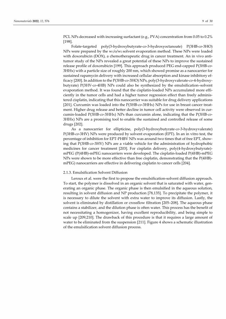
2.1.3. Emulsification Solvent Diffusion
Leroux et al. were the first to propose the emulsification-solvent diffusion approach.
To start, the polymer is dissolved in an organic solvent that is saturated with water, gen-
erating an organic phase. The organic phase is then emulsified in the aqueous solution,
resulting in solvent diffusion and NP production [78,135]. To precipitate the polymer, it
is necessary to dilute the solvent with extra water to improve its diffusion. Lastly, the
solvent is eliminated by distillation or crossflow filtration [205–208]. The aqueous phase
contains a stabilizer, and the dilution phase is often water. This process has the benefit of
not necessitating a homogenizer, having excellent reproducibility, and being simple to
scale up [209,210]. The drawback of this procedure is that it requires a large amount of
water to be eliminated from the suspension [211]. Figure 4 shows a schematic illustration
of the emulsification solvent diffusion process.
Nanomaterials 2022, 12, 576 10 of 30
Figure 4. Diagrammatic representation of the emulsification solvent diffusion method.
Quintanar et al. proposed a mechanism for NP formation in which each droplet
forms several NPs [209]. Perez et al. and Ma et al. then proceeded to modify the method
suggested by Quintanar et al. for the nanoencapsulation of hydrophilic active substances.
In their proposed method, the aqueous inner phase includes an active substance as well
as a stabilizing agent such as PVA or poly(vinylpyrrolidone) (PVP), while the external
phase comprises the polymer and organic solvent. The emulsion was initially diluted with
the solvent (ethanol), resulting in organic solvent migration. Then, water was added to
facilitate the collection of NPs [212,213]. Hassou and Moinard-Chécot et al. used a step-
by-step diffusion analysis using the stopped-flow methodology to represent different
states that occur in the emulsification solvent diffusion method during the dilution stage.
It was discovered that the solvent diffuses quickly from the droplets, taking less than 20
ms [214,215]. Pramual et al. formulated 5,10,15,20-Tetrakis(4-hydroxy-phenyl)-21H, 23H-
porphine pTHPP (hydrophobic photosensitizer) loaded P(3HB-co-3HV) NPs for photody-
namic therapy (PDT) by the emulsification-diffusion method. The size distribution of
P(3HB-co-3HV) NPs was narrow, ranging from 169.0 to 211.2 nm. The pTHPP-loaded
P(3HB-co-3HV) NPs exhibited high photocytotoxicity towards HT-29 human colon cancer
cells compared to pTHPP alone. These results indicated that the P(3HB-co-3HV) NPs are
potential vehicles for the delivery of hydrophobic photosensitizer drugs in photodynamic
therapy [216]. PHA NPs encapsulating TGX221 anti-cancer drugs were also developed.
TGX221 was slowly liberated from the PHA-based NP and proliferation in NP-TGX221-
treated cells was considerably slower than in cells receiving free TGX221 [217].
Using a modified emulsification solvent diffusion process, Chen et al. developed cur-
cumin-loaded PLGA (PLGA-Cur) NPs with a mean range of 190 nm. Anti-tumor activity
was successfully detected following the delivery of PLGA-Cur NPs into cells, and in com-
parison, with free curcumin, PLGA-Cur NPs demonstrated the increased inhibition of
HL60 and HepG2 cancer cells with lower IC50 values. Moreover, confocal microscopy
analysis showed that the curcumin-loaded PLGA NPs increased apoptosis in cancer cells
when compared with free curcumin [218]. PCL NPs were made using ethyl acetate as the
solvent and PVA as the stabilizing agent, respectively. The polymer concentration, solvent
volume, type and amount of the surfactant, as well as the concentration of oil in the or-
ganic phase, were all observed to affect the size of PCL NPs [219]. The solvent and stabi-
lizing agents utilized to make PLA NPs were ethyl acetate and Pluronic F68, respectively.
Particle size increased from 260 to 530 nm as PLA content increased [220]. In another in-
vestigation, as the amount of surfactant was raised, the size of PCL NPs shrank [221]. A
comparison made using different stabilizers, di-dodecyl dimethylammonium bromide
(DMAB) and PVA, for the production of PLGA NPs revealed that DMAB produced
smaller PLGA NPs [222]. The influence of homogenization and sonication on the size of
PLGA NPs was explored by Jain et al., who discovered that sonication resulted in smaller
Nanomaterials 2022, 12, 576 11 of 30
particles with an average size of 165 nm, whereas homogenization resulted in particles
with an average size of 225 nm [223].
2.1.4. Salting-Out Technique
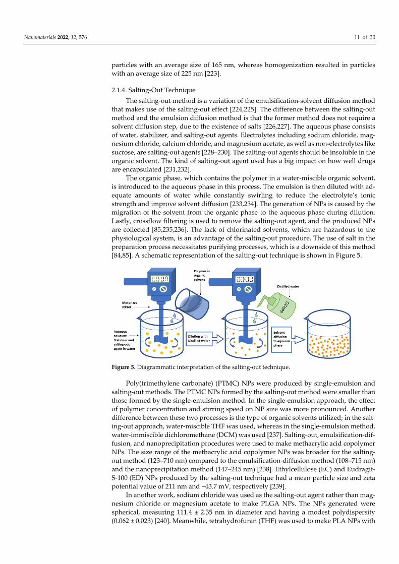
The salting-out method is a variation of the emulsification-solvent diffusion method
that makes use of the salting-out effect [224,225]. The difference between the salting-out
method and the emulsion diffusion method is that the former method does not require a
solvent diffusion step, due to the existence of salts [226,227]. The aqueous phase consists
of water, stabilizer, and salting-out agents. Electrolytes including sodium chloride, mag-
nesium chloride, calcium chloride, and magnesium acetate, as well as non-electrolytes like
sucrose, are salting-out agents [228–230]. The salting-out agents should be insoluble in the
organic solvent. The kind of salting-out agent used has a big impact on how well drugs
are encapsulated [231,232].
The organic phase, which contains the polymer in a water-miscible organic solvent,
is introduced to the aqueous phase in this process. The emulsion is then diluted with ad-
equate amounts of water while constantly swirling to reduce the electrolyte’s ionic
strength and improve solvent diffusion [233,234]. The generation of NPs is caused by the
migration of the solvent from the organic phase to the aqueous phase during dilution.
Lastly, crossflow filtering is used to remove the salting-out agent, and the produced NPs
are collected [85,235,236]. The lack of chlorinated solvents, which are hazardous to the
physiological system, is an advantage of the salting-out procedure. The use of salt in the
preparation process necessitates purifying processes, which is a downside of this method
[84,85]. A schematic representation of the salting-out technique is shown in Figure 5.
Figure 5. Diagrammatic interpretation of the salting-out technique.
Poly(trimethylene carbonate) (PTMC) NPs were produced by single-emulsion and
salting-out methods. The PTMC NPs formed by the salting-out method were smaller than
those formed by the single-emulsion method. In the single-emulsion approach, the effect
of polymer concentration and stirring speed on NP size was more pronounced. Another
difference between these two processes is the type of organic solvents utilized; in the salt-
ing-out approach, water-miscible THF was used, whereas in the single-emulsion method,
water-immiscible dichloromethane (DCM) was used [237]. Salting-out, emulsification-dif-
fusion, and nanoprecipitation procedures were used to make methacrylic acid copolymer
NPs. The size range of the methacrylic acid copolymer NPs was broader for the salting-
out method (123–710 nm) compared to the emulsification-diffusion method (108–715 nm)
and the nanoprecipitation method (147–245 nm) [238]. Ethylcellulose (EC) and Eudragit-
S-100 (ED) NPs produced by the salting-out technique had a mean particle size and zeta
potential value of 211 nm and −43.7 mV, respectively [239].
In another work, sodium chloride was used as the salting-out agent rather than mag-
nesium chloride or magnesium acetate to make PLGA NPs. The NPs generated were
spherical, measuring 111.4 ± 2.35 nm in diameter and having a modest polydispersity
(0.062 ± 0.023) [240]. Meanwhile, tetrahydrofuran (THF) was used to make PLA NPs with
Nanomaterials 2022, 12, 576 12 of 30
a diameter of less than 200 nm [241], while paracetamol-loaded Eudragit S100 NPs were
produced using ethanol as solvent, sodium carboxymethylcellulose as a stabilizer, and
zinc sulfate heptahydrate (ZnSO4·7H2O) as the salting-out agent [242]. Zweers et al. used
acetone and magnesium chloride hexahydrate (MgCl2·6H2O) as the organic solvent and
salting-out agent, respectively, to make PEO-PLGA NPs with a size of about 200 nm
[243,244]. Similarly, PLA end-capped with 1- pyrenebutanol (PLAP) NPs were synthe-
sized using MgCl2·6H2O as the salting-out agent [245].
2.2. Formation of Nanoparticles by Polymerization of Monomers
The methods that were explained in the previous sections are used to produce PNPs
from preformed polymers. PNPs can also be produced by the polymerization of mono-
mers. This section explores the methods employed for the polymerization of monomers
such as emulsion polymerization, surfactant-free emulsion polymerization, mini-emul-
sion polymerization and micro-emulsion polymerization.
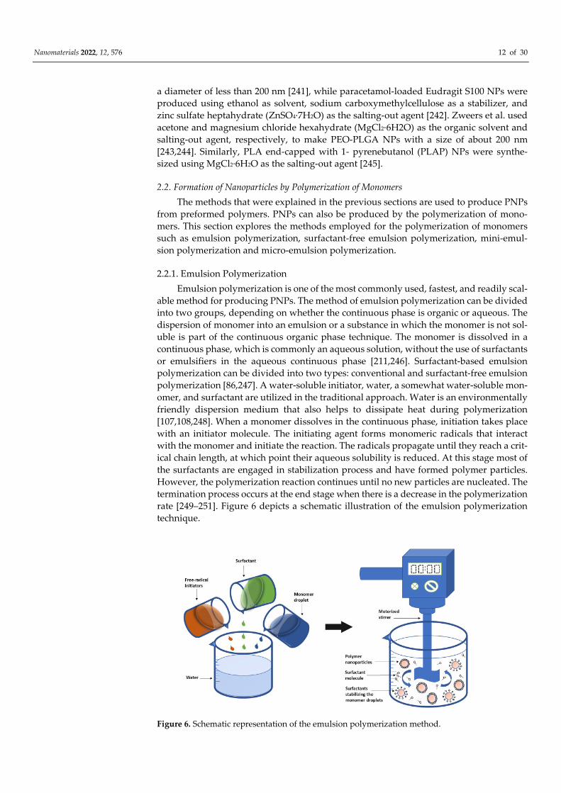
2.2.1. Emulsion Polymerization
Emulsion polymerization is one of the most commonly used, fastest, and readily scal-
able method for producing PNPs. The method of emulsion polymerization can be divided
into two groups, depending on whether the continuous phase is organic or aqueous. The
dispersion of monomer into an emulsion or a substance in which the monomer is not sol-
uble is part of the continuous organic phase technique. The monomer is dissolved in a
continuous phase, which is commonly an aqueous solution, without the use of surfactants
or emulsifiers in the aqueous continuous phase [211,246]. Surfactant-based emulsion
polymerization can be divided into two types: conventional and surfactant-free emulsion
polymerization [86,247]. A water-soluble initiator, water, a somewhat water-soluble mon-
omer, and surfactant are utilized in the traditional approach. Water is an environmentally
friendly dispersion medium that also helps to dissipate heat during polymerization
[107,108,248]. When a monomer dissolves in the continuous phase, initiation takes place
with an initiator molecule. The initiating agent forms monomeric radicals that interact
with the monomer and initiate the reaction. The radicals propagate until they reach a crit-
ical chain length, at which point their aqueous solubility is reduced. At this stage most of
the surfactants are engaged in stabilization process and have formed polymer particles.
However, the polymerization reaction continues until no new particles are nucleated. The
termination process occurs at the end stage when there is a decrease in the polymerization
rate [249–251]. Figure 6 depicts a schematic illustration of the emulsion polymerization
technique.
Figure 6. Schematic representation of the emulsion polymerization method.
Nanomaterials 2022, 12, 576 13 of 30
Other PNPs successfully produced by the same method include polystyrene-b-
poly[poly(ethylene glycol) methyl ether methacrylate] (PS-b-P(PEGMA300), and PS-b-
P(PEGMA1100)) PNPs [252]. Using SDS as a surfactant, Garay-Jimenez et al. synthesised
polyacrylate NPs by the emulsion polymerization of acrylate compounds in a mixture of
butyl acrylate and styrene. Anionic, cationic, zwitterionic, and noncharged (amphiphilic)
surfactants were used to create poly(butyl acrylate-styrene) emulsions. The emulsions’
cytotoxicity and microbiological activity were compared before and after purification. The
findings showed that attaching a polymerizable surface to the nanoparticle matrix has no
effect on the emulsion’s cytotoxic or antibacterial effects, irrespective of whether the emul-
sion is purified or not and that the perfect properties are associated with using non-ionic
surfactants rather than those with zwitterionic, cationic, or anionic [253].
To encapsulate magnetite particles and improve particle-size distribution, emulsion
polymerization was used to create magnetic polymer matrix composite nanoparticles
(MPCNPs) (PSD). Transmission electron microscopy (TEM) and vibrating sample magne-
tometry were used to characterize MPCNPs (VSM). The results showed that the emulsion
polymerization approach was successful in encapsulating magnetite particles [254]. Un-
der microwave radiation, styrene emulsion polymerization was carried out at 70 °C using
sodium dodecyl sulphate (SDS) as a surfactant and potassium persulfate (KPS) as an ini-
tiator [255]. Another study used microwave irradiation to accomplish the emulsion
polymerization of methyl methacrylate (MMA) and butyl acrylate (BuA) using potassium
persulfate (K2S2O8) as an initiator and Disponil A3065 as an emulsifier [256]. The size of
polystyrene NPs produced by the ultrasonic irradiation emulsion polymerization of sty-
rene using polymeric carboxymethyl cellulose and alkyl poly(etheroxy) acrylate (CMCA9)
as surfactant was 30 to 60 nm [257]. The size of PVK NPs is regulated by the concentration
of VCz. Polyvinylcarbazole (PVK) NPs were generated via emulsion polymerization of N-
vinylcarbazole (VCz) [258].
2.2.2. Surfactant-Free Emulsion Polymerization
Surfactants are utilized in the traditional emulsion polymerization procedure and
should be eliminated from the finished product. Surfactant removal is a time-consuming
operation that raises manufacturing costs [89,259]. An emulsion polymerization process
without surfactants, i.e., a surfactant-free emulsion polymerization method, was devised
to alleviate this limitation [260,261]. To generate PNPs, this method offers a straightfor-
ward, green approach that does not require the inclusion and subsequent removal of sta-
bilizing chemicals. A water-soluble initiator (KPS, potassium persulfate), monomers, and
water are the reagents utilized in this process. The stabilization of PNPs is achieved using
ionizable initiators or ionic co-monomers in this technique [262–265].
The surfactant-free emulsion polymerization procedure using microwave irradiation
produced PMMA NPs with a narrow size distribution. When the monomer concentration
was increased from 0 to 0.3 mol/L, the size of the NPs rose from 103 to 215 nm [266]. The
Cu2+/HSO3− redox initiation system was used to commence the surfactant-free emulsion
polymerization of MMA and PMMA NPs with a negative charge in the size range of 165
to 223 nm were produced [267]. PMMA NPs in the size range of 200 to 600 nm were suc-
cessfully developed using hydrophilic laponite clay to stabilize methyl methacrylate
emulsions dispersed in distilled water [268], while NPs with a dimension less than 100
nm and high solid content were accomplishment of this project using KPS and acetone as
initiator and co-solvent, respectively [269]. Using NaSS as a stabilizing agent and water as
the reaction medium, poly-acrylate NPs with fluorine and silicon in the shell with a mean
range of 172.5 nm were produced [270]. By the surfactant-free emulsion polymerization
of styrene utilizing ultrasonic irradiation in the presence of potassium persulfate (KPS) as
an anionic initiator and cetyl alcohol as a co-stabilizer, Faridi Majidi et al. produced poly-
styrene NPs in the size range of 200–250 nm [271]. Surfactant-free emulsion polymeriza-
tion produced poly(hydroxyethyl methacrylate) (PHEMA) NPs with a mean size of 150
nm, a polydispersity index of 1.171, and a surface area of 17,779 m2/g [272]. Lee et al. used
Nanomaterials 2022, 12, 576 14 of 30
Fe3+ catalyzed emulsion polymerization to produce poly(styrene/thiophene) NPs with
particle sizes ranging from 300 to 800 nm [273]. Polyimide NPs were synthesized in a con-
tinuous phase by heterophase polycondensation of various aromatic tetra-carboxylic ac-
ids and diamines in imidazolium-based ionic liquids (IL) [274]. Kim et al. synthesized
polypyrrole NPs utilizing benzene octanol and ethyl acetate as continuous phases. Chang-
ing the water and octanol volume ratios led to fewer particles with an average size of 60
nm [275].
Colloidal NPs with a PMMA or poly(butyl methacrylate) core and a cationic polymer
stabilizing shell were produced using reversible addition fragmentation chain transfer-
mediated surfactant-free emulsion polymerization and had hydrodynamic diameters
ranging from 32 to 96 nm. The wetting behaviour of such core-shell NPs, which can be
fine-tuned depending on the internal nanostructure (soft or rigid core) and external tem-
perature, allows for the creation of controllable functional hybrid colloidal arrays [276].
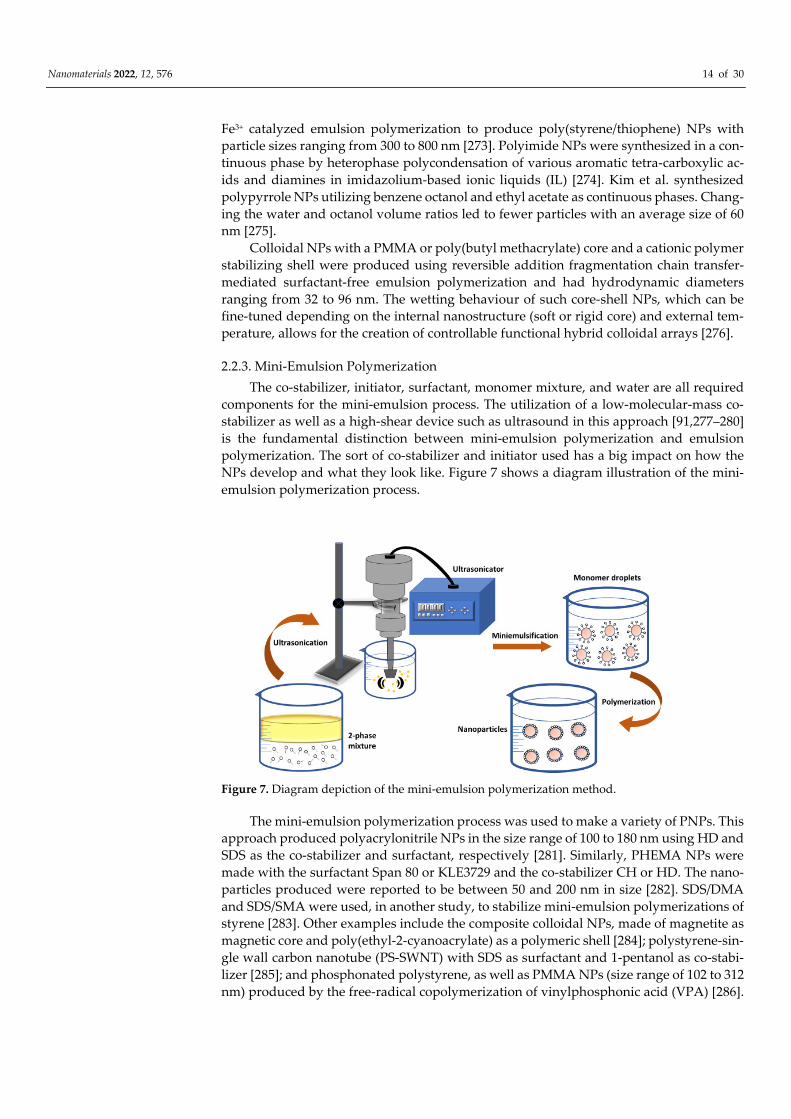
2.2.3. Mini-Emulsion Polymerization
The co-stabilizer, initiator, surfactant, monomer mixture, and water are all required
components for the mini-emulsion process. The utilization of a low-molecular-mass co-
stabilizer as well as a high-shear device such as ultrasound in this approach [91,277–280]
is the fundamental distinction between mini-emulsion polymerization and emulsion
polymerization. The sort of co-stabilizer and initiator used has a big impact on how the
NPs develop and what they look like. Figure 7 shows a diagram illustration of the mini-
emulsion polymerization process.
Figure 7. Diagram depiction of the mini-emulsion polymerization method.
The mini-emulsion polymerization process was used to make a variety of PNPs. This
approach produced polyacrylonitrile NPs in the size range of 100 to 180 nm using HD and
SDS as the co-stabilizer and surfactant, respectively [281]. Similarly, PHEMA NPs were
made with the surfactant Span 80 or KLE3729 and the co-stabilizer CH or HD. The nano-
particles produced were reported to be between 50 and 200 nm in size [282]. SDS/DMA
and SDS/SMA were used, in another study, to stabilize mini-emulsion polymerizations of
styrene [283]. Other examples include the composite colloidal NPs, made of magnetite as
magnetic core and poly(ethyl-2-cyanoacrylate) as a polymeric shell [284]; polystyrene-sin-
gle wall carbon nanotube (PS-SWNT) with SDS as surfactant and 1-pentanol as co-stabi-
lizer [285]; and phosphonated polystyrene, as well as PMMA NPs (size range of 102 to 312
nm) produced by the free-radical copolymerization of vinylphosphonic acid (VPA) [286].
Nanomaterials 2022, 12, 576 15 of 30
2.2.4. Micro-Emulsion Polymerization
A new method for manufacturing nanosized PNPs is micro-emulsion polymeriza-
tion. Despite the fact that emulsion polymerization and micro-emulsion polymerization
are similar processes that form polymers with high molar mass, their kinetics differ, re-
sulting in micro-emulsion polymerization having smaller particle sizes and fewer chains
per particle [287–290]. A water-soluble initiator is introduced to the aqueous phase, which
contains a lot of surfactants, in the microemulsion polymerization technique. Because in-
itiation cannot occur simultaneously in all microdroplets, polymer chains begin to form
only in some of them. Due to osmotic and elastic forces, microdroplets will collapse later,
resulting in larger particles and the development of empty micelles [291–294]. In a micro-
emulsion, polymerization kinetics, PNP properties, and the concentration and type of in-
itiator, surfactant and monomer are determining factors [293]. Some researchers have
been carried out to see how these parameters affect the characteristics of NPs.
Micro-emulsion polymerization was used to create poly(vinyl acetate) lattices with a
high total solid [93]. On the micro-emulsion polymerization of vinyl acetate stabilized
with Aerosol OT (AOT), the effects of temperature, concentration and type of initiator (V-
50 and KPS) were investigated. It was found that the reaction rates increased with the
concentration of V-50 and temperature. Furthermore, the differences in electrostatic at-
traction between KPS and V-50 free radicals, as well as charged micro-emulsion droplets,
resulted in quicker polymerization rates for KPS [295]. A cationic surfactant (e.g., CTAB)
and a non-ionic surfactant were used to make poly(dimethylsiloxane) (PDMS) NPs in the
range of sizes of 12–80 nm [296]. Furthermore, stabilizers of dodecyltrimethylammonium
bromide (DTAB) and didodecyldimethylammonium bromide (DDAB) were used to make
polyhexylmethacrylate NPs with a size range of 38 to 53 nm [297]. SABS-8 and SABS-10,
two polymerizable anionic surfactants, were employed successfully in microemulsion
polymerization of butyl methacrylate (BMA) at room temperature utilizing the redox ini-
tiator ammonium persulfate (APS)/ tetramethylethylenediamine (TMEDA) [298]. Some
other work used the cationic surfactant decyltrimethylammonium bromide DeTAB to
make polypyrrole NPs with a particle size of 2 nm [299]. The polymerization of butyl
acrylate with a sodium dodecyl sulfate/Aerosol OT surfactant combination and potassium
peroxodisulfate as an initiator yielded particles smaller than 40 nm [300].
3. Biologically Synthesized Biodegradable Polyhydroxyalkanoate-Based Nanoparti-
cles
A non-toxic, reliable, and eco-friendly experimental protocol for the synthesis of NPs
is highly in demand. Natural entities such as secondary metabolites, enzymes, polysac-
charides, biodegradable polymers, vitamins, and microorganisms can be utilized for the
synthesis of NPs [95,301]. One such promising approach is the biosynthesis of NPs using
bacteria. To date, a large variety of bacterial species have already been studied in the
hopes of developing alternate NP synthesis techniques. For the time being, scientists are
producing NPs using bacteria’s biomass or cell extracts [302]. In comparison to other bio-
logical entities, bacteria are thought to be a promising biofactory for the synthesis of NPs.
Bacterial biosynthesis of NPs is a fast-growing study area in the field of science and nan-
otechnology, with many species of bacteria being used to synthesize NPs all around the
world [303].
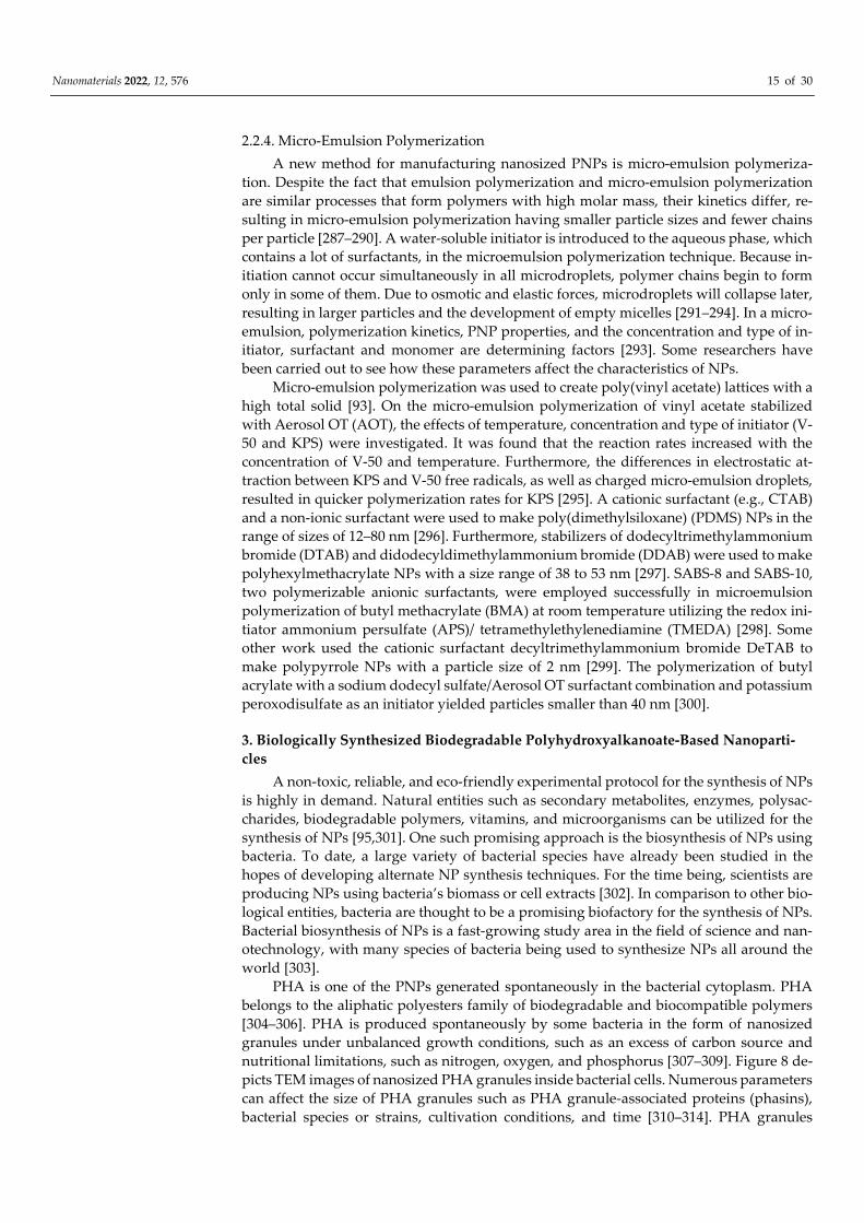
PHA is one of the PNPs generated spontaneously in the bacterial cytoplasm. PHA
belongs to the aliphatic polyesters family of biodegradable and biocompatible polymers
[304–306]. PHA is produced spontaneously by some bacteria in the form of nanosized
granules under unbalanced growth conditions, such as an excess of carbon source and
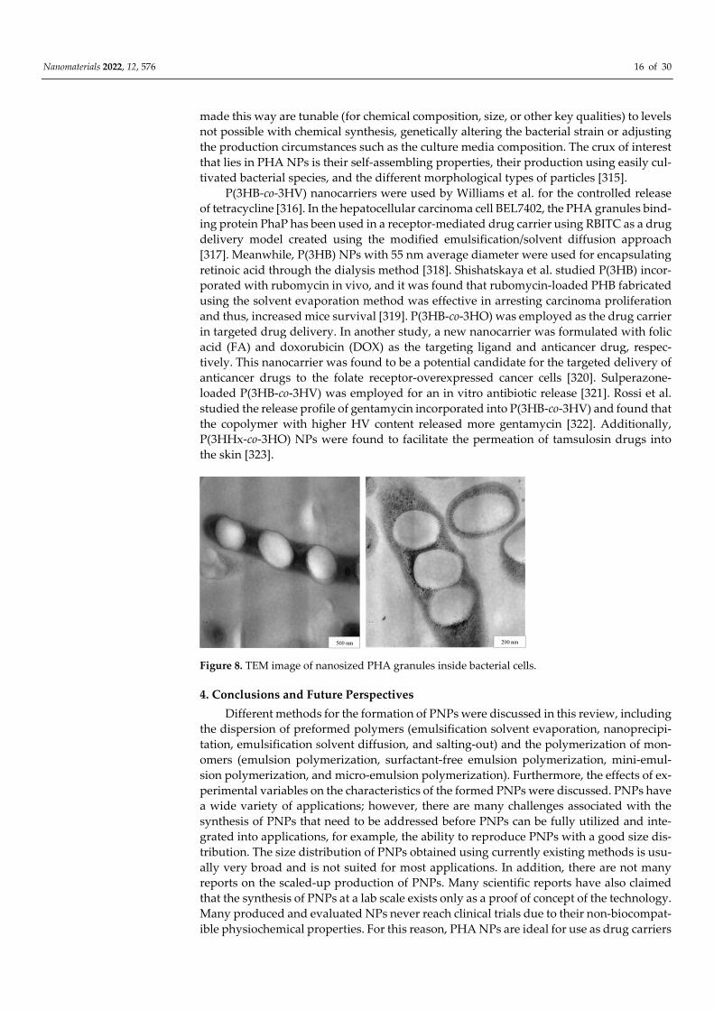
nutritional limitations, such as nitrogen, oxygen, and phosphorus [307–309]. Figure 8 de-
picts TEM images of nanosized PHA granules inside bacterial cells. Numerous parameters
can affect the size of PHA granules such as PHA granule-associated proteins (phasins),
bacterial species or strains, cultivation conditions, and time [310–314]. PHA granules
Nanomaterials 2022, 12, 576 16 of 30
made this way are tunable (for chemical composition, size, or other key qualities) to levels
not possible with chemical synthesis, genetically altering the bacterial strain or adjusting
the production circumstances such as the culture media composition. The crux of interest
that lies in PHA NPs is their self-assembling properties, their production using easily cul-
tivated bacterial species, and the different morphological types of particles [315].
P(3HB-co-3HV) nanocarriers were used by Williams et al. for the controlled release
of tetracycline [316]. In the hepatocellular carcinoma cell BEL7402, the PHA granules bind-
ing protein PhaP has been used in a receptor-mediated drug carrier using RBITC as a drug
delivery model created using the modified emulsification/solvent diffusion approach
[317]. Meanwhile, P(3HB) NPs with 55 nm average diameter were used for encapsulating
retinoic acid through the dialysis method [318]. Shishatskaya et al. studied P(3HB) incor-
porated with rubomycin in vivo, and it was found that rubomycin-loaded PHB fabricated
using the solvent evaporation method was effective in arresting carcinoma proliferation
and thus, increased mice survival [319]. P(3HB-co-3HO) was employed as the drug carrier
in targeted drug delivery. In another study, a new nanocarrier was formulated with folic
acid (FA) and doxorubicin (DOX) as the targeting ligand and anticancer drug, respec-
tively. This nanocarrier was found to be a potential candidate for the targeted delivery of
anticancer drugs to the folate receptor-overexpressed cancer cells [320]. Sulperazone-
loaded P(3HB-co-3HV) was employed for an in vitro antibiotic release [321]. Rossi et al.
studied the release profile of gentamycin incorporated into P(3HB-co-3HV) and found that
the copolymer with higher HV content released more gentamycin [322]. Additionally,
P(3HHx-co-3HO) NPs were found to facilitate the permeation of tamsulosin drugs into
the skin [323].
Figure 8. TEM image of nanosized PHA granules inside bacterial cells.
4. Conclusions and Future Perspectives
Different methods for the formation of PNPs were discussed in this review, including
the dispersion of preformed polymers (emulsification solvent evaporation, nanoprecipi-
tation, emulsification solvent diffusion, and salting-out) and the polymerization of mon-
omers (emulsion polymerization, surfactant-free emulsion polymerization, mini-emul-
sion polymerization, and micro-emulsion polymerization). Furthermore, the effects of ex-
perimental variables on the characteristics of the formed PNPs were discussed. PNPs have
a wide variety of applications; however, there are many challenges associated with the
synthesis of PNPs that need to be addressed before PNPs can be fully utilized and inte-
grated into applications, for example, the ability to reproduce PNPs with a good size dis-
tribution. The size distribution of PNPs obtained using currently existing methods is usu-
ally very broad and is not suited for most applications. In addition, there are not many
reports on the scaled-up production of PNPs. Many scientific reports have also claimed
that the synthesis of PNPs at a lab scale exists only as a proof of concept of the technology.
Many produced and evaluated NPs never reach clinical trials due to their non-biocompat-
ible physiochemical properties. For this reason, PHA NPs are ideal for use as drug carriers
Nanomaterials 2022, 12, 576 17 of 30
as they abide by the present regulatory requirements in terms of biodegradability, stabil-
ity, and non-toxicity. Furthermore, because the size distribution is extremely large, the
particle size loses certainty due to the wide range of size distribution. This situation poses
a great challenge in using PNPs for drug delivery applications. Furthermore, PHA NPs
can be produced using bacteria, which allows for green synthesis to produce nanocarriers
that can be used extensively in the field of nanomedicine. With regards to application,
PNPs can be used as drug carriers to target specific sites within cells or organs for more
advanced treatment due to their unique properties and size. This would greatly improve
the performance of targeted therapies. The PNPs can also be used for diagnostic purposes,
either in the lab or in hospitals (point-of-care diagnostics).
Author Contributions: T.P., P.F., J.-A.C., and K.S. wrote the manuscript. All authors have read and
agreed to the published version of the manuscript.
Funding: Authors would like to thank Ministry of Education Malaysia (203/PBIOLOGI/67811001),
titled “Soil Analysis and Value-Addition to Oil Palm Trunk (OPT) and Sap Through Biotechnology”
as well as Science and Technology Research Partnership for Sustainable Development (SATREPS)
(grant number JPMJSA1801) of the Japan Science and Technology Agency (JST)/ Japan International
Cooperation Agency (JICA) for their financial support.
Data Availability Statement: Data sharing is not applicable for this review.
Conflicts of Interest: The authors declare that they have no competing interests.
References
1. Bhatia, S. Nanoparticles Types, Classification, Characterization, Fabrication Methods and Drug Delivery Applications. In Nat-
ural Polymer Drug Delivery Systems; Springer: Switzerland, 2016; pp. 33–93, https://doi.org/10.1007/978-3-319-41129-3_2.
2. Albanese, A.; Tang, P.S.; Chan, W.C. The Effect of Nanoparticle Size, Shape, and Surface Chemistry on Biological Systems.
Annu. Rev. Biomed. Eng. 2012, 14, 1–16, https://doi.org/10.1146/annurev-bioeng-071811-150124.
3. Docter, D.; Strieth, S.; Westmeier, D.; Hayden, O.; Gao, M.; Knauer, S.K.; Stauber, R.H. No king without a crown—impact of the
nanomaterial-protein corona on nanobiomedicine. Nanomedicine 2015, 10, 503–519, https://doi.org/10.2217/nnm.14.184.
4. Joye, I.J.; McClements, D.J. Production of Nanoparticles by Anti-Solvent Precipitation for Use in Food Systems. Trends Food Sci.
Technol. 2013, 34, 109–123, https://doi.org/10.1016/j.tifs.2013.10.002.
5. Rahman, M.; Laurent, S.; Tawil, N.; Yahia, L.H.; Mahmoudi, M. Nanoparticle and Protein Corona. In Protein-nanoparticle inter-
actions; Springer: Switzerland, 2013; pp. 21–44, https://doi.org/10.1007/978-3-642-37555-2_2.
6. Mahmoudi, M.; Lynch, I.; Ejtehadi, M.R.; Monopoli, M.P.; Bombelli, F.B.; Laurent, S. Protein−Nanoparticle Interactions: Oppor-
tunities and Challenges. Chem. Rev. 2011, 111, 5610–5637, https://doi.org/10.1021/cr100440g.
7. Ezhilarasi, P.N.; Karthik, P.; Chhanwal, N.; Anandharamakrishnan, C. Nanoencapsulation Techniques for Food Bioactive Com-
ponents: A Review. Food Bioprocess Technol. 2013, 6, 628–647, doi:10.1007/s11947-012-0944-0.
8. Sripriyalakshmi, S.; Jose, P.; Ravindran, A.; Anjali, C.H. Recent Trends in Drug Delivery System Using Protein Nanoparticles.
Cell Biophys. 2014, 70, 17–26, https://doi.org/10.1007/s12013-014-9896-5.
9. Bonifácio, B.V.; Silva, P.B.; Ramos, M.A.; Negri, K.M.; Bauab, T.M.; Chorilli, M. Nanotechnology-based drug delivery systems
and herbal medicines: A review. Int. J. Nanomed. 2013, 9, 1–15, https://doi.org/10.2147/IJN.S52634.
10. Weiss, J.; Takhistov, P.; McClements, D.J. Functional Materials in Food Nanotechnology. J. Food Sci. 2006, 71, R107–R116,
https://doi.org/10.1111/j.1750-3841.2006.00195.x.
11. Kuhlbusch, T.A.; Asbach, C.; Fissan, H.; Göhler, D.; Stintz, M. Nanoparticle exposure at nanotechnology workplaces: A review.
Part. Fibre Toxicol. 2011, 8, 22–22, https://doi.org/10.1186/1743-8977-8-22.
12. Sanguansri, P.; Augustin, M.A. Nanoscale Materials Development–A Food Industry Perspective. Trends Food Sci. Tech-
nol. 2006, 17, 547–556, https://doi.org/10.1016/j.tifs.2006.04.010.
13. Rai, M.; Ingle, A. Role of nanotechnology in agriculture with special reference to management of insect pests. Appl. Microbiol.
Biotechnol. 2012, 94, 287–293, https://doi.org/10.1007/s00253-012-3969-4.
14. Justin, C.; Philip, S.A.; Samrot, A.V. Synthesis and characterization of superparamagnetic iron-oxide nanoparticles (SPIONs)
and utilization of SPIONs in X-ray imaging. Appl. Nanosci. 2017, 7, 463–475, https://doi.org/10.1007/s13204-017-0583-x.
15. Aggarwal, P.; Hall, J.B.; McLeland, C.B.; Dobrovolskaia, M.A.; McNeil, S.E. Nanoparticle interaction with plasma proteins as it
relates to particle biodistribution, biocompatibility and therapeutic efficacy. Adv. Drug Deliv. Rev. 2009, 61, 428–437,
https://doi.org/10.1016/j.addr.2009.03.009.
16. Kharazian, B.; Hadipour, N.; Ejtehadi, M. Understanding the nanoparticle–protein corona complexes using computational and
experimental methods. Int. J. Biochem. Cell Biol. 2016, 75, 162–174, https://doi.org/10.1016/j.biocel.2016.02.008.
17. Schöttler, S.; Landfester, K.; Mailänder, V. Controlling the Stealth Effect of Nanocarriers through Understanding the Protein
Corona. Angew. Chem. Int. Ed. 2016, 55, 8806–8815, https://doi.org/10.1002/anie.201602233.
Nanomaterials 2022, 12, 576 18 of 30
18. Mirshafiee, V.; Mahmoudi, M.; Lou, K.; Cheng, J.; Kraft, M.L. Protein corona significantly reduces active targeting yield. Chem.
Commun. 2013, 49, 2557–2559, https://doi.org/10.1039/c3cc37307j.
19. Lee, Y.K.; Choi, E.-J.; Webster, T.J.; Kim, S.-H.; Khang, D. Effect of the protein corona on nanoparticles for modulating cytotox-
icity and immunotoxicity. Int. J. Nanomed. 2014, 10, 97–113, doi:10.2147/ijn.s72998.
20. Rak, J. Microparticles in Cancer. Semin. Thromb. Hemost. 2010, 36, 888–906, https://doi.org/10.1055/s-0030-1267043.
21. Mause, S.F.; Weber, C. Microparticles. Circulation Research 2010, 107, 1047–1057,
https://doi.org/10.1161/CIRCRESAHA.110.226456.
22. Rizvi, S.A.; Saleh, A.M. Applications of nanoparticle systems in drug delivery technology. Saudi Pharm. J. 2017, 26, 64–70,
https://doi.org/10.1016/j.jsps.2017.10.012.
23. Chenthamara, D.; Subramaniam, S.; Ramakrishnan, S.G.; Krishnaswamy, S.; Essa, M.M.; Lin, F.H.; Qoronfleh, M.W. Therapeutic
efficacy of nanoparticles and routes of administration. Biomater. Res. 2019, 23, 20, doi:10.1186/s40824-019-0166-x.
24. Zhou, Y.; Peng, Z.; Seven, E.S.; Leblanc, R.M. Crossing the blood-brain barrier with nanoparticles. J. Control. Release 2017, 270,
290–303, https://doi.org/10.1016/j.jconrel.2017.12.015.
25. Khan, R.; Ahmad, E.; Zaman, M.; Qadeer, A.; Rabbani, G. Nanoparticles in relation to peptide and protein aggregation. Int. J.
Nanomed. 2014, 9, 899–912, https://doi.org/10.2147/ijn.s54171.
26. Magazù, S.; Migliardo, F.; Telling, M. Structural and dynamical properties of water in sugar mixtures. Food Chem. 2008, 106,
1460–1466, https://doi.org/10.1016/j.foodchem.2007.05.097.
27. Samrot, A.; Burman, U.; Philip, S.A.; N, S.; Chandrasekaran, K. Synthesis of curcumin loaded polymeric nanoparticles from crab
shell derived chitosan for drug delivery. Informatics Med. Unlocked 2018, 10, 159–182, https://doi.org/10.1016/j.imu.2017.12.010.
28. Panhwar, A.H.; Tuzen, M.; Hazer, B.; Kazi, T.G. Solid phase microextraction method using a novel polystyrene oleic acid imid-
azole polymer in micropipette tip of syringe system for speciation and determination of antimony in environmental and food
samples. Talanta 2018, 184, 115–121, https://doi.org/10.1016/j.talanta.2018.03.004.
29. Honarkar, H.; Barikani, M. Applications of Biopolymers I: Chitosan. Monatsh Chem 2009, 140, 1403,
https://doi.org/10.1007/s00706-009-0197-4.
30. Saleh, T.A.; Tuzen, M.; Sarı, A. Polyamide magnetic palygorskite for the simultaneous removal of Hg(II) and methyl mercury;
with factorial design analysis. J. Environ. Manag. 2018, 211, 323–333, https://doi.org/10.1016/j.jenvman.2018.01.050.
31. Nasir, A.; Kausar, A.; Younus, A. A Review on Preparation, Properties and Applications of Polymeric Nanoparticle-Based Ma-
terials. Polym. Technol. Eng. 2014, 54, 325–341, https://doi.org/10.1080/03602559.2014.958780.
32. Geckeler, K.E.; Nishide, H. Advanced Nanomaterials; Wiley Online Library: Weinheim, Germany, 2010; Volume 2,
https://doi.org/10.1002/9783527628940.
33. Derman, S.; Kizilbey, K.; Akdeste, Z.M. Polymeric Nanoparticles. Sigma Journal of Engineering and Natural Sciences 2013, 31, 107–
120.
34. Öztürk, K. Serbest Radikal Temizleyici Madde İçeren Nanopartiküler Taşıyıcı Sistemlerin Tasarımı Ve Değerlendirilmesi. Mas-
ter’s Thesis, Hacettepe University, Ankara, Turkey, 2010. Available online: http://nek.istanbul.edu.tr:4444/ekos/TEZ/47041.pdf
(accessed on 20 December 2021).
35. Wang, Y.-J.; Larsson, M.; Huang, W.-T.; Chiou, S.-H.; Nicholls, S.J.; Chao, J.-I.; Liu, D.-M. The use of polymer-based nanoparti-
cles and nanostructured materials in treatment and diagnosis of cardiovascular diseases: Recent advances and emerging de-
signs. Prog. Polym. Sci. 2016, 57, 153–178, https://doi.org/10.1016/j.progpolymsci.2016.01.002.
36. Chang, M.-W.; Edirisinghe, M.; Stride, E. Ultrasound mediated release from stimuli-responsive core–shell capsules. J. Mater.
Chem. B 2013, 1, 3962–3971, https://doi.org/10.1039/c3tb20465k.
37. Muller, R.H.; Keck, C.M. Challenges and Solutions for the Delivery of Biotech Drugs–A Review of Drug Nanocrystal Technol-
ogy and Lipid Nanoparticles. J. Biotechnol. 2004, 113, 151–170, https://doi.org/10.1016/j.jbiotec.2004.06.007.
38. Reis, C.; Neufeld, R.J.; Ribeiro, A.; Veiga, F. Nanoencapsulation I. Methods for preparation of drug-loaded polymeric nanopar-
ticles. Nanomedicine: Nanotechnology, Biol. Med. 2006, 2, 8–21, https://doi.org/10.1016/j.nano.2005.12.003.
39. Anderson, D.G.; Burdick, J.A.; Langer, R. Smart Biomaterials. Science 2004, 305, 1923–1924, doi: 10.1126/science.1099987.
40. Klodzinska, S.N.; Wan, F.; Jumaa, H.; Sternberg, C.; Rades, T.; Nielsen, H.M. Improved drug loading and antibacterial activity
of minocycline-loaded PLGA nanoparticles prepared by solid/oil/water ion pairing method. Int. J. Nanomed. 2012, 7, 221–234,
https://doi.org/10.2147/ijn.s27709.
41. Hornig, S.; Heinze, T.; Becer, C.R.; Schubert, U.S. Synthetic polymeric nanoparticles by nanoprecipitation. J. Mater. Chem. 2009,
19, 3838–3840, https://doi.org/10.1039/b906556n.
42. Nagarwal, R.C.; Kant, S.; Singh, P.N.; Maiti, P.; Pandit, J.K. Polymeric nanoparticulate system: A potential approach for ocular
drug delivery. J. Control. Release 2009, 136, 2–13, https://doi.org/10.1016/j.jconrel.2008.12.018.
43. Moreno-Vega, A.-I.; Gómez-Quintero, T.; Nuñez-Anita, R.E.; Acosta-Torres, L.-S.; Castaño, V. Polymeric and Ceramic Nano-
particles in Biomedical Applications. J. Nanotechnol. 2012, 2012, 1–10, https://doi.org/10.1155/2012/936041.
44. Yadav, H.K.; Almokdad, A.A.; Shaluf, S.I.; Debe, M.S. Polymer-Based Nanomaterials for Drug-Delivery Carriers. 2018, 531–556,
https://doi.org/10.1016/b978-0-12-814033-8.00017-5.
45. Sailaja, A.K. A Review on Biomedical Applications of Polymeric Nanoparticles. Drug Designing & Intellectual Properties Interna-
tional Journal 2018, 2, 216–220, https://doi.org/10.32474/DDIPIJ.2018.02.000140.
46. Parveen S.; Misra, R.; Sahoo, S.K. Nanoparticles: A Boon to Drug Delivery, Therapeutics, Diagnostics and Imaging. Nanomedi-
cine: Nanotechnology, Biol. Med. 2012, 8, 147–166, https://doi.org/10.1016/j.nano.2011.05.016.
Nanomaterials 2022, 12, 576 19 of 30
47. De Jong, W.H.; Borm, P.J.A. Drug delivery and Nanoparticles: Applications and Hazards. Int. J. Nanomedicine 2008, 3, 133-149,
doi: 10.2147/ijn.s596.
48. Patra, J.K.; Das, G.; Fraceto, L.F.; Campos, E.V.R.; del Pilar Rodriguez-Torres, M.; Acosta-Torres, L.S.; Diaz-Torres, L.A.; Grillo,
R.; Swamy, M.K.; Sharma, S.; et al. Nano based drug delivery systems: Recent developments and future prospects. J. Nanobi-
otechnol. 2018, 16, 71, doi:10.1186/s12951-018-0392-8.
49. Pignatello, R.; Impallomeni, G.; Cupri, S.; Puzzo, G.; Curcio, C.; Rizzo, M.G.; Guglielmino, S.; Ballistreri, A. Unsaturated
Poly(Hydroxyalkanoates) for the Production of Nanoparticles and the Effect of Cross-Linking on Nanoparticle Features. Mate-
rials 2019, 12, 868, https://doi.org/10.3390/ma12060868.
50. Umesh, M.; Priyanka, K.; Thazeem, B.; Preethi, K. Biogenic PHA nanoparticle synthesis and characterization from Bacillus sub-
tilis NCDC0671 using orange peel medium. Int. J. Polym. Mater. Polym. Biomater. 2017, 67, 996–1004,
https://doi.org/10.1080/00914037.2017.1417284.
51. Koosha, F.; Muller, R.; Washington, C. Production of Polyhydroxybutyrate (PHB) Nanoparticles for Drug Targeting. J. Pharm.
Pharmacol. 1987, 39, 136P.
52. Koosha, F.; Muller, R.; Davis, S.S.; Davies, M.C. The surface chemical structure of poly(β-hydroxybutyrate) microparticles pro-
duced by solvent evaporation process. J. Control. Release 1989, 9, 149–157, https://doi.org/10.1016/0168-3659(89)90005-9.
53. Seyler, I.; Appel, M.; Devissaguet, J.-P.; Legrand, P.; Barratt, G. Macrophage Activation by a Lipophilic Derivative of Mura-
myldipeptide within Nanocapsules: Investigation of the Mechanism of Drug Delivery. J. Nanoparticle Res. 1999, 1, 91–97,
https://doi.org/10.1023/a:1010016128378.
54. Legrand, P.; Lesieur, S.; Bochot, A.; Gref, R.; Raatjes, W.; Barratt, G.; Vauthier, C. Influence of polymer behaviour in organic
solution on the production of polylactide nanoparticles by nanoprecipitation. Int. J. Pharm. 2007, 344, 33–43,
https://doi.org/10.1016/j.ijpharm.2007.05.054.
55. Ueda, M.; Kreuter, J. Optimization of the preparation of loperamide-loaded poly (L-lactide) nanoparticles by high pressure
emulsification-solvent evaporation. J. Microencapsul. 1997, 14, 593–605, https://doi.org/10.3109/02652049709006812.
56. Nehilla, B.J.; Bergkvist, M.; Popat, K.C.; A Desai, T. Purified and surfactant-free coenzyme Q10-loaded biodegradable nanopar-
ticles. Int. J. Pharm. 2008, 348, 107–114, https://doi.org/10.1016/j.ijpharm.2007.07.001.
57. Yallapu, M.M.; Gupta, B.K.; Jaggi, M.; Chauhan, S.C. Fabrication of curcumin encapsulated PLGA nanoparticles for improved
therapeutic effects in metastatic cancer cells. J. Colloid Interface Sci. 2010, 351, 19–29, https://doi.org/10.1016/j.jcis.2010.05.022.
58. Prado, L.B.; Huber, S.C.; Barnabé, A.; Bassora, F.D.S.; Paixão, D.S.; Durán, N.; Annichino-Bizzacchi, J.M. Characterization of
PCL and Chitosan Nanoparticles as Carriers of Enoxaparin and Its Antithrombotic Effect in Animal Models of Venous Throm-
bosis. J. Nanotechnol. 2017, 2017, 1–7, https://doi.org/10.1155/2017/4925495.
59. Ajiboye, A.L.; Trivedi, V.; Mitchell, J. Preparation of polycaprolactone nanoparticles via supercritical carbon dioxide extraction
of emulsions. Drug Deliv. Transl. Res. 2017, 8, 1790–1796, https://doi.org/10.1007/s13346-017-0422-3.
60. Shokri, N.; Javar, H.A.; Fouladdel, S.; Khalaj, A.; Khoshayand, M.R.; Dinarvand, R.; Atyabi, F.; Nomani, A.; Azizi, E. Preparation
and Evaluation of Poly (Caprolactone Fumarate) Nanoparticles Containing Doxorubicin HCl. Daru: Journal of Faculty of Phar-
macy, Tehran University of Medical Sciences 2011, 19, 12–22.
61. Kateb, B.; Chiu, K.; Black, K.L.; Yamamoto, V.; Khalsa, B.; Ljubimova, J.Y.; Ding, H.; Patil, R.; Portilla-Arias, J.A.; Modo, M.; et
al. Nanoplatforms for constructing new approaches to cancer treatment, imaging, and drug delivery: What should be the pol-
icy?. NeuroImage 2011, 54, S106–S124, https://doi.org/10.1016/j.neuroimage.2010.01.105.
62. Mansour, H.M.; Sohn, M.; Al-Ghananeem, A.; DeLuca, P.P. Materials for Pharmaceutical Dosage Forms: Molecular Pharmaceu-
tics and Controlled Release Drug Delivery Aspects. Int. J. Mol. Sci. 2010, 11, 3298–3322, https://doi.org/10.3390/ijms11093298.
63. Sundar, S.; Kundu, J.; Kundu, S.C. Biopolymeric Nanoparticles. Science and Technology of Advanced Materials 2010, 11, 014104,
https://doi.org/10.1088/1468-6996/11/1/014104.
64. Ludwig, A. The use of mucoadhesive polymers in ocular drug delivery. Adv. Drug Deliv. Rev. 2005, 57, 1595–1639,
https://doi.org/10.1016/j.addr.2005.07.005.
65. Lanz-Landázuri, A.; Portilla-Arias, J.; De Ilarduya, A.M.; Álvarez, M.G.; Holler, E.; Ljubimova, J.; Muñoz-Guerra, S. Nanopar-
ticles of Esterified Polymalic Acid for Controlled Anticancer Drug Release. Macromol. Biosci. 2014, 14, 1325–1336,
https://doi.org/10.1002/mabi.201400124.
66. Loyer, P.; Cammas-Marion, S. Natural and synthetic poly(malic acid)-based derivates: A family of versatile biopolymers for the
design of drug nanocarriers. J. Drug Target. 2014, 22, 556–575, https://doi.org/10.3109/1061186x.2014.936871.
67. Gutul, T.; Rusu, E.; Condur, N.; Ursaki, V.; Goncearenco, E.; Vlazan, P. Preparation of poly(N-vinylpyrrolidone)-stabilized ZnO
colloid nanoparticles. Beilstein J. Nanotechnol. 2014, 5, 402–406, https://doi.org/10.3762/bjnano.5.47.
68. Sahu, A.; Solanki, P.; Mitra, S. Curcuminoid-loaded poly(methyl methacrylate) nanoparticles for cancer therapy. Int. J. Nanomed.
2018, ume 13, 101–105, https://doi.org/10.2147/ijn.s124021.
69. Mendes, A.N.; Hubber, I.; Siqueira, M.; Barbosa, G.M.; Moreira, D.D.L.; Holandino, C.; Pinto, J.C.; Nele, M. Preparation and
Cytotoxicity of Poly(Methyl Methacrylate) Nanoparticles for Drug Encapsulation. Macromol. Symp. 2012, 319, 34–40,
https://doi.org/10.1002/masy.201100248.
70. Andreasen, S..; Chong, S.-F.; Wohl, B.M.; Goldie, K.N.; Zelikin, A.N. Poly(vinyl alcohol) Physical Hydrogel Nanoparticles, Not
Polymer Solutions, Exert Inhibition of Nitric Oxide Synthesis in Cultured Macrophages. Biomacromolecules 2013, 14, 1687–1695,
https://doi.org/10.1021/bm400369u.

Nanomaterials 2022, 12, 576 20 of 30
71. Madlova, M.; Jones, S.; Zwerschke, I.; Ma, Y.; Hider, R.; Forbes, B. Poly(vinyl alcohol) nanoparticle stability in biological media
and uptake in respiratory epithelial cell layers in vitro. Eur. J. Pharm. Biopharm. 2009, 72, 438–443,
https://doi.org/10.1016/j.ejpb.2009.01.009.
72. Hu, Y.; Jiang, X.; Ding, Y.; Ge, H.; Yuan, Y.; Yang, C. Synthesis and characterization of chitosan–poly(acrylic acid) nanoparticles.
Biomaterials 2002, 23, 3193–3201, https://doi.org/10.1016/s0142-9612(02)00071-6.
73. Molnar, R.M.; Bodnar, M.; Hartmann, J.F.; Borbely, J. Preparation and characterization of poly(acrylic acid)-based nanoparticles.
Colloid Polym. Sci. 2009, 287, 739–744, https://doi.org/10.1007/s00396-009-2033-0.
74. Gualdesi, M.; Igarzabal, C.A.; Vara, J.; Ortiz, C. Synthesis and physicochemical properties of polyacrylamide nanoparticles as
photosensitizer carriers. Int. J. Pharm. 2016, 512, 213–218, https://doi.org/10.1016/j.ijpharm.2016.08.051.
75. Giuntini, F.; Dumoulin, F.; Daly, R.; Ahsen, V.; Scanlan, E.M.; Lavado, A.S.P.; Aylott, J.W.; Rosser, G.A.; Beeby, A.; Boyle, R.W.
Orthogonally bifunctionalised polyacrylamide nanoparticles: A support for the assembly of multifunctional nanodevices. Na-
noscale 2012, 4, 2034–2045, https://doi.org/10.1039/c2nr11947a.
76. López-Muñoz, R.; Treviño, M.E.; Morales, G.; Valdez-Garza, J.A.; de León, R.D.; Saade, H.; Enríquez-Medrano, F.J.; López, R.G.
Ultrafine Nanoparticles of Poly(Methyl Methacrylate-co-Methacrylic Acid) Loaded with Aspirin. J. Nanomater. 2019, 2019, 1–9,
https://doi.org/10.1155/2019/3059098.
77. Zhong, J.X.; Clegg, J.R.; Ander, E.W.; Peppas, N.A. Tunable poly(methacrylic acid-co-acrylamide) nanoparticles through inverse
emulsion polymerization. J. Biomed. Mater. Res. Part A 2018, 106, 1677–1686, https://doi.org/10.1002/jbm.a.36371.
78. Wang, Y.; Li, P.; Tran, T.T.-D.; Zhang, J.; Kong, L. Manufacturing Techniques and Surface Engineering of Polymer Based Nano-
particles for Targeted Drug Delivery to Cancer. Nanomaterials 2016, 6, 26, https://doi.org/10.3390/nano6020026.
79. Zielińska, A.; Carreiró, F.; Oliveira, A.; Neves, A.; Pires, B.; Venkatesh, D.; Durazzo, A.; Lucarini, M.; Eder, P.; Silva, A.; et al.
Polymeric Nanoparticles: Production, Characterization, Toxicology and Ecotoxicology. Molecules 2020, 25, 3731,
https://doi.org/10.3390/molecules25163731.
80. Crucho, C.I.C.; Barros, M.T. Polymeric nanoparticles: A study on the preparation variables and characterization methods. Mater.
Sci. Eng. C Mater. Biol. Appl. 2017, 80, 771–784, doi:10.1016/j.msec.2017.06.004.
81. Ibraheem, D.; Fessi, H.; Elaissari, A. DNA Encapsulation via Double Emulsion Like Process. J. Colloid Sci. Biotechnol. 2013, 2,
328–333, https://doi.org/10.1166/jcsb.2013.1060.
82. Hao, S.; Wang, B.; Wang, Y.; Zhu, L.; Wang, B.; Guo, T. Preparation of Eudragit L 100-55 enteric nanoparticles by a novel emul-
sion diffusion method. Colloids Surfaces B: Biointerfaces 2013, 108, 127–133, https://doi.org/10.1016/j.colsurfb.2013.02.036.
83. Saranya, S.; Radha, K.V. Review of Nanobiopolymers for Controlled Drug Delivery. Polym. Technol. Eng. 2014, 53, 1636–1646,
https://doi.org/10.1080/03602559.2014.915035.
84. Valente, I.M.; Moreira, M.M.; Neves, P.; Da Fé, T.; Gonçalves, L.M.; Almeida, P.J.; Rodrigues, J.A. An Insight on Salting-out
Assisted Liquid-Liquid Extraction for Phytoanalysis. Phytochem. Anal. 2017, 28, 297–304, https://doi.org/10.1002/pca.2676.
85. Hyde, A.M.; Zultanski, S.L.; Waldman, J.H.; Zhong, Y.-L.; Shevlin, M.; Peng, F. General Principles and Strategies for Salting-
Out Informed by the Hofmeister Series. Org. Process Res. Dev. 2017, 21, 1355–1370, https://doi.org/10.1021/acs.oprd.7b00197.
86. Distler, D.; Neto, W.S.; Machado, F, Emulsion Polymerization. Reference Module in Materials Science and Materials Engineering,
2017, https://doi.org/10.1016/B978-0-12-803581-8.03746-2.
87. Yamak, H.B., Emulsion Polymerization: Effects of Polymerization Variables on The Properties of Vinyl Acetate Based Emulsion
Polymers. In Polymer Science; Yilmaz, F, Ed.; IntechOpen, London, UK, 2013, pp. 35–72, http://dx.doi.org/10.5772/51498.
88. Zhou, J.; Yao, H.; Ma, J. Recent advances in RAFT-mediated surfactant-free emulsion polymerization. Polym. Chem. 2018, 9,
2532–2561, https://doi.org/10.1039/c8py00065d.
89. Bilgin, S.; Tomovska, R.; Asua, J.M. Surfactant-free high solids content polymer dispersions. Polymer 2017, 117, 64–75,
https://doi.org/10.1016/j.polymer.2017.04.014.
90. Faucheu, J.; Gauthier, C.; Chazeau, L.; Cavaillé, J.-Y.; Mellon, V.; Lami, E.B. Miniemulsion polymerization for synthesis of struc-
tured clay/polymer nanocomposites: Short review and recent advances. Polymer 2010, 51, 6–17, https://doi.org/10.1016/j.poly-
mer.2009.11.044.
91. Crespy, D.; Landfester, K. Miniemulsion polymerization as a versatile tool for the synthesis of functionalized polymers. Beilstein
J. Org. Chem. 2010, 6, 1132–1148, https://doi.org/10.3762/bjoc.6.130.
92. Iqbal, M.; Zafar, N.; Fessi, H.; Elaissari, A. Double emulsion solvent evaporation techniques used for drug encapsulation. Int. J.
Pharm. 2015, 496, 173–190, https://doi.org/10.1016/j.ijpharm.2015.10.057.
93. Sosa, N.; Peralta, R.; López, R.; Ramos, L.; Katime, I.; Cesteros, C.; Mendizábal, E.; Puig, J. A comparison of the characteristics
of poly(vinyl acetate) latex with high solid content made by emulsion and semi-continuous microemulsion polymerization.
Polymer 2001, 42, 6923–6928, https://doi.org/10.1016/s0032-3861(01)00157-4.
94. Rehm, B.H.A. Bacterial polymers: Biosynthesis, modifications and applications. Nat. Rev. Microbiol. 2010, 8, 578–592,
doi:10.1038/nrmicro2354.
95. Moradali, M.F.; Rehm, B.H.A. Bacterial biopolymers: From pathogenesis to advanced materials. Nat. Rev. Microbiol. 2020, 18,
195–210, doi:10.1038/s41579-019-0313-3.
96. Zhang, G.; Niu, A.; Peng, S.; Jiang, M.; Tu, Y.; Li, M.; Wu, C. Formation of Novel Polymeric Nanoparticles. Accounts Chem. Res.
2001, 34, 249–256, https://doi.org/10.1021/ar000011x.
Nanomaterials 2022, 12, 576 21 of 30
97. Quintanar-Guerrero, D.; Allémann, E.; Fessi, H.; Doelker, E. Preparation Techniques and Mechanisms of Formation of Biode-
gradable Nanoparticles from Preformed Polymers. Drug Dev. Ind. Pharm. 1998, 24, 1113–1128,
https://doi.org/10.3109/03639049809108571.
98. Couvreur, P.; Dubernet, C.; Puisieux, F. Controlled Drug Delivery with Nanoparticles: Current Possibilities and Future Trends.
European Journal of Pharmaceutics and Biopharmaceutics 1995, 41, 2–13.
99. Wang, Q.; Wu, P.; Ren, W.; Xin, K.; Yang, Y.; Xie, C.; Yang, C.; Liu, Q.; Yu, L.; Jiang, X.; et al. Comparative studies of salinomycin-
loaded nanoparticles prepared by nanoprecipitation and single emulsion method. Nanoscale Res. Lett. 2014, 9, 351–351,
https://doi.org/10.1186/1556-276x-9-351.
100. Kumar, A.; Sawant, K. Encapsulation of exemestane in polycaprolactone nanoparticles: Optimization, characterization, and
release kinetics. Cancer Nanotechnol. 2013, 4, 57–71, https://doi.org/10.1007/s12645-013-0037-4.
101. Moorthi, C.; Kathiresan, K. Fabrication of Dual Drug Loaded Polymeric Nanosuspension: Incorporating Analytical Hierarchy
Process and Data Envelopment Analysis in the Selection of A Suitable Method. Int J Pharm Pharm Sci. 2013, 5, 499–504.
102. Mora-Huertas, C.E.; Fessi, H.; Elaissari, A. Polymer-based nanocapsules for drug delivery. Int. J. Pharm. 2010, 385, 113–142,
https://doi.org/10.1016/j.ijpharm.2009.10.018.
103. Noriega-Peláez, E.K.; Mendoza-Muñoz, N.; Ganem-Quintanar, A.; Quintanar-Guerrero, D. Optimization of the emulsification
and solvent displacement method for the preparation of solid lipid nanoparticles. Drug Dev. Ind. Pharm. 2010, 37, 160–166,
https://doi.org/10.3109/03639045.2010.501800.
104. Müller, K.; Bugnicourt, E.; Latorre, M.; Jorda, M.; Echegoyen Sanz, Y.E.; Lagaron, J.M.; Miesbauer, O.; Bianchin, A.; Hankin, S.;
Bölz, U.; et al. Review on the Processing and Properties of Polymer Nanocomposites and Nanocoatings and Their Applications
in the Packaging, Automotive and Solar Energy Fields. Nanomaterials 2017, 7, 74, doi:10.3390/nano7040074.
105. de Oliveira, A.D.; Beatrice, C.A.G. Polymer Nanocomposites with Different Types of Nanofiller. In Nanocomposites-Recent Evo-
lutions; IntechOpen: London, UK, 2018, pp. 103–128, https://dx.doi.org/10.5772/intechopen.81329.
106. Parambath, A. Engineering of Biomaterials for Drug Delivery Systems: Beyond Polyethylene Glycol; Woodhead Publishing: Kidling-
ton, UK, 2018.
107. Sailaja, A.K.; Shreya, M. Preparation and Characterization of Naproxen Loaded Niosomes by Ether Injection Method. Nano
Biomed. Eng. 2018, 10, https://doi.org/10.5101/nbe.v10i2.p174-180.
108. Chuah, J.-A.; Kaplan, D.L.; Numata, K. Engineering Peptide-based Carriers for Drug and Gene Delivery. 2013, 667–689,
https://doi.org/10.1007/978-1-4471-4372-7_25.
109. Di Mascolo, D.; Basnett, P.; Palange, A.L.; Francardi, M.; Roy, I.; Decuzzi, P. Tuning core hydrophobicity of spherical polymeric
nanoconstructs for docetaxel delivery. Polym. Int. 2016, 65, 741–746, https://doi.org/10.1002/pi.5072.
110. Hoshyar, N.; Gray, S.; Han, H.; Bao, G. The effect of nanoparticle size on in vivo pharmacokinetics and cellular interaction.
Nanomedicine 2016, 11, 673–692, https://doi.org/10.2217/nnm.16.5.
111. Yu, X.; Trase, I.; Ren, M.; Duval, K.; Guo, X.; Chen, Z. Design of Nanoparticle-Based Carriers for Targeted Drug Delivery. Avail-
able online: https://www.hindawi.com/journals/jnm/2016/1087250/ (accessed on 14 February 2021).
112. Ageitos, J.M.; Chuah, J.-A.; Numata, K. Chapter Design Considerations for Properties of Nanocarriers on Disposition and Effi-
ciency of Drug and Gene Delivery. 2016, 1–22, https://doi.org/10.1039/9781782622536-00001.
113. Chidambaram, M.; Krishnasamy, K. Application of Plackett-Burman Factorial Design in The Development of Curcumin Loaded
Eudragit E 100 Nanoparticles. Nano Biomed. Eng. 2013, 5, https://doi.org/10.5101/nbe.v5i1.p28-33.
114. Hickey, J.; Santos, J.L.; Williford, J.-M.; Mao, H.-Q. Control of polymeric nanoparticle size to improve therapeutic delivery. J.
Control. Release 2015, 219, 536–547, https://doi.org/10.1016/j.jconrel.2015.10.006.
115. Khan, I.; Saeed, K.; Khan, I. Nanoparticles: Properties, Applications and Toxicities. Arabian J. Chem. 2019, 12, 908–931,
https://doi.org/10.1016/j.arabjc.2017.05.011.
116. Ding, S.; Serra, C.A.; Vandamme, T.F.; Yu, W.; Anton, N. Double Emulsions Prepared by Two–Step Emulsification: History,
State-Of-The-Art and Perspective. J. Controlled Release 2019, 295, 31–49, https://doi.org/10.1016/j.jconrel.2018.12.037.
117. Goodarzi, F.; Zendehboudi, S. A Comprehensive Review on Emulsions and Emulsion Stability in Chemical and Energy Indus-
tries. Can. J. Chem. Eng. 2018, 97, 281–309, https://doi.org/10.1002/cjce.23336.
118. Aubry, J.; Ganachaud, F.; Addad, J.-P.C.; Cabane, B. Nanoprecipitation of Polymethylmethacrylate by Solvent Shifting:Bound-
aries. Langmuir 2009, 25, 1970–1979, https://doi.org/10.1021/la803000e.
119. Mora-Huertas, C.E.; Garrigues, O.; Fessi, H.; Elaissari, A. Nanocapsules prepared via nanoprecipitation and emulsification–
diffusion methods: Comparative study. Eur. J. Pharm. Biopharm. 2012, 80, 235–239, https://doi.org/10.1016/j.ejpb.2011.09.013.
120. Lepeltier, E.; Bourgaux, C.; Couvreur, P. Nanoprecipitation and the “Ouzo effect”: Application to Drug Delivery Devices. Adv.
Drug Delivery Rev. 2014, 71, 86–97, https://doi.org/10.1016/j.addr.2013.12.009.
121. Amoyav, B.; Benny, O. Controlled and tunable polymer particles’ production using a single microfluidic device. Appl. Nanosci.
2018, 8, 905–914, https://doi.org/10.1007/s13204-018-0790-0.
122. Vauthier, C.; Dubernet, C.; Fattal, E.; Pinto-Alphandary, H.; Couvreur, P. Poly(alkylcyanoacrylates) as biodegradable materials
for biomedical applications. Adv. Drug Delivery Rev. 2003, 55, 519–548, https://doi.org/10.1016/s0169-409x(03)00041-3.
123. Mishra, B.; Patel, B.B.; Tiwari, S. Colloidal nanocarriers: A review on formulation technology, types and applications toward
targeted drug delivery. Nanomedicine: Nanotechnology, Biol. Med. 2010, 6, 9–24, https://doi.org/10.1016/j.nano.2009.04.008.
124. Moshfeghi, A.A.; Peyman, G.A. Micro-and Nanoparticulates. Adv. Drug Delivery Rev. 2005, 57, 2047–2052,
https://doi.org/10.1016/j.addr.2005.09.006.
Nanomaterials 2022, 12, 576 22 of 30
125. Álvarez-Román, R.; Cavazos-Rodríguez, M.; Chávez-Montes, A.; Castro-Ríos, R.; Waksman de Torres, N.; Salazar-Cavazos, M.;
Galindo Rodríguez, S. Formulación y caracterización de nanocápsulas con un antioxidante natural para su aplicación cutánea
(Formulation and Characterization of Nanocapsules as a Natural Antioxidant for Cutaneous Application). Química Hoy
(Chemistry Sciences) 2011, 1, 29–35. Available online: http://eprints.uanl.mx/13446/1/Art6.pdf (accessed on 15 November 2021).
126. Shi, W.; Zhang, Z.-J.; Yuan, Y.; Xing, E.-M.; Qin, Y.; Peng, Z.-J.; Zhang, Z.-P.; Yang, K.-Y. Optimization of parameters for prep-
aration of docetaxel-loaded PLGA nanoparticles by nanoprecipitation method. J. Huazhong Univ. Sci. Technol. 2013, 33, 754–758,
https://doi.org/10.1007/s11596-013-1192-x.
127. Teng, Z.; Luo, Y.; Wang, T.; Zhang, B.; Wang, Q. Development and Application of Nanoparticles Synthesized with Folic Acid
Conjugated Soy Protein. J. Agric. Food Chem. 2013, 61, 2556–2564, https://doi.org/10.1021/jf4001567.
128. Kim, S.; Kim, Y.S. Production of gliadin-poly(ethyl cyanoacrylate) nanoparticles for hydrophilic coating. J. Nanoparticle Res.
2014, 16, https://doi.org/10.1007/s11051-014-2277-6.
129. Lebouille, J.G.J.L.; Stepanyan, R.; Slot, J.J.M.; Stuart Cohen, M.A.; Tuinier, R. Nanoprecipitation of Polymers in a Bad Solvent.
Colloids and Surfaces A: Physicochemical and Engineering Aspects 2014, 460, 225–235,
https://doi.org/10.1016/j.colsurfa.2013.11.045.
130. Moorthi, C.; Kathiresan, K. Fabrication of highly stable sonication assisted curcumin nanocrystals by nanoprecipitation method.
Drug Inven. Today 2013, 5, 66–69, https://doi.org/10.1016/j.dit.2013.02.003.
131. He, Y.; Huang, Y.; Cheng, Y. Structure Evolution of Curcumin Nanoprecipitation from a Micromixer. Cryst. Growth Des. 2010,
10, 1021–1024, https://doi.org/10.1021/cg9009916.
132. Peltonen, L.; Aitta, J.; Hyvönen, S.; Karjalainen, M.; Hirvonen, J. Improved Entrapment Efficiency of Hydrophilic Drug Sub-
stance During Nanoprecipitation of Poly(I)Lactide Nanoparticles. AAPS PharmSciTech 2004, 5, 115,
https://doi.org/10.1208/pt050116.
133. He, W.; Lu, Y.; Qi, J.; Chen, L.; Hu, F.; Wu, W. Food proteins as novel nanosuspension stabilizers for poorly water-soluble drugs.
Int. J. Pharm. 2013, 441, 269–278, https://doi.org/10.1016/j.ijpharm.2012.11.033.
134. Yan, X.; Delgado, M.; Fu, A.; Alcouffe, P.; Gouin, S.G.; Fleury, E.; Katz, J.L.; Ganachaud, F.; Bernard, J. Simple but Precise Engi-
neering of Functional Nanocapsules through Nanoprecipitation. Angew. Chem. Int. Ed. 2014, 53, 6910–6913,
https://doi.org/10.1002/anie.201402825.
135. Sah, E.; Sah, H. Recent Trends in Preparation of Poly(lactide- co -glycolide) Nanoparticles by Mixing Polymeric Organic Solution
with Antisolvent. J. Nanomater. 2015, 2015, 1–22, https://doi.org/10.1155/2015/794601.
136. Barreras-Urbina, C.G.; Ramírez-Wong, B.; López-Ahumada, G.A.; Ibarra, S.E.B.; Martínez-Cruz, O.; Tapia-Hernández, J.A.;
Félix, F.R. Nano- and Micro-Particles by Nanoprecipitation: Possible Application in the Food and Agricultural Industries. Int.
J. Food Prop. 2016, 19, 1912–1923, https://doi.org/10.1080/10942912.2015.1089279.
137. Ansari, M.J.; Alshahrani, S. Nano-encapsulation and characterization of baricitinib using poly-lactic-glycolic acid co-polymer.
Saudi Pharm. J. 2019, 27, 491–501, https://doi.org/10.1016/j.jsps.2019.01.012.
138. Sharma, N.; Madan, P.; Lin, S. Effect of process and formulation variables on the preparation of parenteral paclitaxel-loaded
biodegradable polymeric nanoparticles: A co-surfactant study. Asian J. Pharm. Sci. 2016, 11, 404–416,
https://doi.org/10.1016/j.ajps.2015.09.004.
139. Calzoni, E.; Cesaretti, A.; Polchi, A.; Di Michele, A.; Tancini, B.; Emiliani, C. Biocompatible Polymer Nanoparticles for Drug
Delivery Applications in Cancer and Neurodegenerative Disorder Therapies. J. Funct. Biomater. 2019, 10, 4,
https://doi.org/10.3390/jfb10010004.
140. Chang, J.; Jallouli, Y.; Kroubi, M.; Yuan, X.-B.; Feng, W.; Kang, C.-S.; Pu, P.-Y.; Betbeder, D. Characterization of endocytosis of
transferrin-coated PLGA nanoparticles by the blood–brain barrier. Int. J. Pharm. 2009, 379, 285–292,
https://doi.org/10.1016/j.ijpharm.2009.04.035.
141. Nassar, T.; Rom, A.; Nyska, A.; Benita, S. Novel double coated nanocapsules for intestinal delivery and enhanced oral bioavail-
ability of tacrolimus, a P-gp substrate drug. J. Control. Release 2009, 133, 77–84, https://doi.org/10.1016/j.jconrel.2008.08.021.
142. de Assis, D.N.; Mosqueira, V.C.F.; Vilela, J.M.C.; Andrade, M.S.; Cardoso, V.N. Release Profiles and Morphological Character-
ization by Atomic Force Microscopy and Photon Correlation Spectroscopy of 99mTechnetium-Fluconazole Nanocapsules. Inter-
national Journal of Pharmaceutics 2008, 349, 152–160, https://doi.org/10.1016/j.ijpharm.2007.08.002.
143. Mugheirbi, N.A.; Paluch, K.J.; Tajber, L. Heat induced evaporative antisolvent nanoprecipitation (HIEAN) of itraconazole. Int.
J. Pharm. 2014, 471, 400–411, https://doi.org/10.1016/j.ijpharm.2014.05.045.
144. Salatin, S.; Barar, J.; Barzegar-Jalali, M.; Adibkia, K.; Kiafar, F.; Jelvehgari, M. Development of a nanoprecipitation method for
the entrapment of a very water soluble drug into Eudragit RL nanoparticles. Res. Pharm. Sci. 2017, 12, 1–14,
https://doi.org/10.4103/1735-5362.199041.
145. Huang, W.; Zhang, C. Tuning the Size of Poly(lactic-co-glycolic Acid) (PLGA) Nanoparticles Fabricated by Nanoprecipitation.
Biotechnol. J. 2017, 13, https://doi.org/10.1002/biot.201700203.
146. Madani, F.; Esnaashari, S.S.; Mujokoro, B.; Dorkoosh, F.; Khosravani, M.; Adabi, M. Investigation of Effective Parameters on
Size of Paclitaxel Loaded PLGA Nanoparticles. Adv. Pharm. Bull. 2018, 8, 77–84, https://doi.org/10.15171/apb.2018.010.
147. Pandey, S.K.; Patel, D.K.; Thakur, R.; Mishra, D.P.; Maiti, P.; Haldar, C. Anti-cancer evaluation of quercetin embedded PLA
nanoparticles synthesized by emulsified nanoprecipitation. Int. J. Biol. Macromol. 2015, 75, 521–529, https://doi.org/10.1016/j.ijbi-
omac.2015.02.011.
Nanomaterials 2022, 12, 576 23 of 30
148. Vuddanda, P.R.; Mishra, A.; Singh, S.K. Development of polymeric nanoparticles with highly entrapped herbal hydrophilic
drug using nanoprecipitation technique: An approach of quality by design. Pharm. Dev. Technol. 2014, 20, 579–587,
https://doi.org/10.3109/10837450.2014.908302.
149. Kara, A.; Ozturk, N.; Sarisozen, C.; Vural, I. Investigation of Formulation Parameters of PLGA Nanoparticles Prepared by Na-
noprecipitation Technique. In Proceedings of the 5th International Conference on Nanotechnology: Fundamentals and Appli-
cation, Prague, Czech Republic, 11-13 August 2014, paper no. 94.
150. Lucas, P.; Vaysse, M.; Aubry, J.; Mariot, D.; Sonnier, R.; Ganachaud, F. Finest nanocomposite films from carbon nanotube-loaded
poly(methyl methacrylate) nanoparticles obtained by the Ouzo effect. Soft Matter 2011, 7, 5528–5531,
https://doi.org/10.1039/c1sm05609c.
151. Yadav, K.S.; Sawant, K.K. Modified Nanoprecipitation Method for Preparation of Cytarabine-Loaded PLGA Nanoparticles.
AAPS PharmSciTech 2010, 11, 1456–1465, https://doi.org/10.1208/s12249-010-9519-4.
152. Khan, S.A.; Schneider, M. Improvement of Nanoprecipitation Technique for Preparation of Gelatin Nanoparticles and Potential
Macromolecular Drug Loading. Macromol. Biosci. 2013, 13, 455–463, https://doi.org/10.1002/mabi.201200382.
153. Zhang, J.-Y.; Shen, Z.-G.; Zhong, J.; Hu, T.-T.; Chen, J.-F.; Ma, Z.-Q.; Yun, J. Preparation of amorphous cefuroxime axetil nano-
particles by controlled nanoprecipitation method without surfactants. Int. J. Pharm. 2006, 323, 153–160,
https://doi.org/10.1016/j.ijpharm.2006.05.048.
154. Ansari, M. Factors Affecting Preparation and Properties of Nanoparticles by Nanoprecipitation Method. Indo Am. J. Pharm. Sci.
2017, 4, 4854–4858.
155. Zhuang, J.; Fang, R.H.; Zhang, L. Preparation of Particulate Polymeric Therapeutics for Medical Applications. Small Methods
2017, 1, 1700147, https://doi.org/10.1002/smtd.201700147.
156. Bilati, U.; Allémann, E.; Doelker, E. Development of a nanoprecipitation method intended for the entrapment of hydrophilic
drugs into nanoparticles. Eur. J. Pharm. Sci. 2005, 24, 67–75, https://doi.org/10.1016/j.ejps.2004.09.011.
157. Lince, F.; Marchisio, D.; Barresi, A. Strategies to control the particle size distribution of poly-ε-caprolactone nanoparticles for
pharmaceutical applications. J. Colloid Interface Sci. 2008, 322, 505–515, https://doi.org/10.1016/j.jcis.2008.03.033.
158. Liu, Y.; Lu, Y.C.; Luo, G.S. Modified nanoprecipitation method for polysulfone nanoparticles preparation. Soft Matter 2014, 10,
3414–3420, https://doi.org/10.1039/c3sm53003e.
159. de Oliveira, A.M.; Jäger, E.; Jäger, A.; Stepánek, P.; Giacomelli, F.C. Physicochemical aspects behind the size of biodegradable
polymeric nanoparticles: A step forward. Colloids Surfaces A: Physicochem. Eng. Asp. 2013, 436, 1092–1102,
https://doi.org/10.1016/j.colsurfa.2013.08.056.
160. Olenius, T.; Yli-Juuti, T.; Elm, J.; Kontkanen, J.; Riipinen, I. New Particle Formation and Growth: Creating a New Atmospheric
Phase Interface. In Physical Chemistry of Gas-Liquid Interface; Faust, J.A.; House, J.E., Eds.; House, J.E., Series Ed.; Elsevier: Am-
sterdam, Netherlands, 2018; Chapter 11, pp. 315–352, https://doi.org/10.1016/B978-0-12-813641-6.00011-X.
161. Cheng, J.C.; Vigil, R.; Fox, R. A competitive aggregation model for Flash NanoPrecipitation. J. Colloid Interface Sci. 2010, 351,
330–342, https://doi.org/10.1016/j.jcis.2010.07.066.
162. Rao, J.P.; Geckeler, K.E. Polymer nanoparticles: Preparation techniques and size-control parameters. Prog. Polym. Sci. 2011, 36,
887–913, doi:10.1016/j.progpolymsci.2011.01.001.
163. Chorny, M.; Fishbein, I.; Danenberg, H.D.; Golomb, G. Lipophilic drug loaded nanospheres prepared by nanoprecipitation:
Effect of formulation variables on size, drug recovery and release kinetics. J. Control. Release 2002, 83, 389–400,
https://doi.org/10.1016/s0168-3659(02)00211-0.
164. Asadi, H.; Rostamizadeh, K.; Salari, D.; Hamidi, M. Preparation of biodegradable nanoparticles of tri-block PLA–PEG–PLA
copolymer and determination of factors controlling the particle size using artificial neural network. J. Microencapsul. 2011, 28,
406–416, https://doi.org/10.3109/02652048.2011.576784.
165. Alshamsan, A. Nanoprecipitation is more efficient than emulsion solvent evaporation method to encapsulate cucurbitacin I in
PLGA nanoparticles. Saudi Pharm. J. 2013, 22, 219–222, https://doi.org/10.1016/j.jsps.2013.12.002.
166. Patel, K.C.; Pramanik, S. Formulation and Characterization of Mefenamic Acid Loaded Polymeric Nanoparticles. World J. Pharm.
Pharm. Sci. 2014, 3, 1391–1405.
167. Lancheros, R.; Guerrero, C.A.; Godoy-Silva, R.D. Improvement of N-Acetylcysteine Loaded in PLGA Nanoparticles by Nano-
precipitation Method. J. Nanotechnol. 2018, 2018, 1–11, https://doi.org/10.1155/2018/3620373.
168. Chidambaram, M.; Krishnasamy, K. Modifications to the Conventional Nanoprecipitation Technique: An Approach to Fabricate
Narrow Sized Polymeric Nanoparticles. Adv. Pharm. Bull. 2013, 4, 205–208, https://doi.org/10.5681/apb.2014.030.
169. Bilati, U.; Allémann, E.; Doelker, E. Nanoprecipitation versus emulsion-based techniques for the encapsulation of proteins into
biodegradable nanoparticles and process-related stability issues. AAPS PharmSciTech 2005, 6, E594–E604,
https://doi.org/10.1208/pt060474.
170. Luo, C.; Okubo, T.; Nangrejo, M.; Edirisinghe, M. Preparation of polymeric nanoparticles by novel electrospray nanoprecipita-
tion. Polym. Int. 2014, 64, 183–187, https://doi.org/10.1002/pi.4822.
171. Zhao, C.-X.; He, L.; Qiao, S.Z.; Middelberg, A.P. Nanoparticle synthesis in microreactors. Chem. Eng. Sci. 2011, 66, 1463–1479,
https://doi.org/10.1016/j.ces.2010.08.039.
172. Heshmatnezhad, F.; Nazar, A.R.S. On-chip controlled synthesis of polycaprolactone nanoparticles using continuous-flow mi-
crofluidic devices. J. Flow Chem. 2020, 10, 533–543, https://doi.org/10.1007/s41981-020-00092-8.
Nanomaterials 2022, 12, 576 24 of 30
173. Zhang, L.; Chen, Q.; Ma, Y.; Sun, J. Microfluidic Methods for Fabrication and Engineering of Nanoparticle Drug Delivery Sys-
tems. ACS Appl. Bio Mater. 2019, 3, 107–120, https://doi.org/10.1021/acsabm.9b00853.
174. Senthilkumar, P.; Dawn, S.S.; Saipriya, C.; Samrot, A.V. Synthesis of polyhydroxybutyrate nanoparticles using surfactant
(SPAN20) for hydrophobic drug delivery. Rasayan J. Chem. 2018, 11, 1686–1695, https://doi.org/10.31788/rjc.2018.1144053.
175. Shakeri, F.; Shakeri, S.; Hojjatoleslami, M. Preparation and Characterization of Carvacrol Loaded Polyhydroxybutyrate Nano-
particles by Nanoprecipitation and Dialysis Methods. J. Food Sci. 2014, 79, N697–N705, https://doi.org/10.1111/1750-3841.12406.
176. Anton, N.; Benoit, J.-P.; Saulnier, P. Design and production of nanoparticles formulated from nano-emulsion templates—A
review. J. Control. Release 2008, 128, 185–199, doi:10.1016/j.jconrel.2008.02.007.
177. Pal, S.L.; Jana, U.; Manna, P.K.; Mohanta, G.P.; Manavalan, R. Nanoparticle: An Overview of Preparation and Characterization.
Journal of Applied Pharmaceutical Science 2011, 1, 228–234.
178. Håkansson, A.; Rayner, M. General Principles of Nanoemulsion Formation by High-Energy Mechanical Methods. Nanoemul-
sions 2018, 103–139, https://doi.org/10.1016/b978-0-12-811838-2.00005-9.
179. Goyal, A.K.; Garg, T.; Bhandari, S.; Rath, G. Advancement in pulmonary drug delivery systems for treatment of tuberculosis.
2017, 669–695, https://doi.org/10.1016/b978-0-323-46143-6.00022-1.
180. Wang, J.; Shi, A.; Agyei, D.; Wang, Q. Formulation of water-in-oil-in-water (W/O/W) emulsions containing trans-resveratrol.
RSC Adv. 2017, 7, 35917–35927, https://doi.org/10.1039/c7ra05945k.
181. Stauffer, F.; Peter, B.; Alem, H.; Funfschilling, D.; Dumas, N.; Serra, C.; Roques-Carmes, T. Polyelectrolytes layer-by-layer sur-
face modification of PDMS microchips for the production of simple O/W and double W/O/W emulsions: From global to local-
ized treatment. Chem. Eng. Process. Process Intensif. 2019, 146, 107685, https://doi.org/10.1016/j.cep.2019.107685.
182. Khadem, B.; Sheibat-Othman, N. Modeling droplets swelling and escape in double emulsions using population balance equa-
tions. Chem. Eng. J. 2019, 382, 122824, https://doi.org/10.1016/j.cej.2019.122824.
183. Khadem, B.; Khellaf, M.; Sheibat-Othman, N. Investigating Swelling-Breakdown in Double Emulsions. Colloids and Surfaces A:
Physicochemical and Engineering Aspects 2020, 585, 124181, https://doi.org/10.1016/j.colsurfa.2019.124181.
184. Mendoza-Muñoz, N.; Alcalá-Alcalá, S.; Quintanar-Guerrero, D. Preparation of Polymer Nanoparticles by the Emulsification-
Solvent Evaporation Method: From Vanderhoff’s Pioneer Approach to Recent Adaptations. 2016, 87–121,
https://doi.org/10.1007/978-3-319-41421-8_4.
185. Wang, Y.; Li, P.; Peng, Z.; She, F.H.; Kong, L.X. Microencapsulation of Nanoparticles with Enhanced Drug Loading for pH-
Sensitive Oral Drug Delivery for the Treatment of Colon Cancer. Journal of Applied Polymer Science 2013, 129, 714–720,
https://doi.org/10.1002/app.38582.
186. Rosca, I.D.; Watari, F.; Uo, M. Microparticle formation and its mechanism in single and double emulsion solvent evaporation.
J. Control. Release 2004, 99, 271–280, https://doi.org/10.1016/j.jconrel.2004.07.007.
187. Soppimath, K.S.; Aminabhavi, T.M.; Kulkarni, A.R.; Rudzinski, W.E. Biodegradable polymeric nanoparticles as drug delivery
devices. J. Control. Release 2001, 70, 1–20, doi:10.1016/s0168-3659(00)00339-4.
188. Pisani, E.; Fattal, E.; Paris, J.; Ringard, C.; Rosilio, V.; Tsapis, N. Surfactant dependent morphology of polymeric capsules of
perfluorooctyl bromide: Influence of polymer adsorption at the dichloromethane–water interface. J. Colloid Interface Sci. 2008,
326, 66–71, https://doi.org/10.1016/j.jcis.2008.07.013.
189. Jeong, Y.-I.; Cho, C.-S.; Kim, S.-H.; Ko, K.-S.; Kim, S.-I.; Shim, Y.-H.; Nah, J.-W. Preparation of poly(DL-lactide-co-glycolide)
nanoparticles without surfactant. J. Appl. Polym. Sci. 2001, 80, 2228–2236, https://doi.org/10.1002/app.1326.
190. Staff, R.H.; Landfester, K.; Crespy, D. Recent Advances in the Emulsion Solvent Evaporation Technique for the Preparation of
Nanoparticles and Nanocapsules. 2013, 329–344, https://doi.org/10.1007/12_2013_233.
191. Deshmukh, R.; Wagh, P.; Naik, J. Solvent evaporation and spray drying technique for micro- and nanospheres/particles prepa-
ration: A review. Dry. Technol. 2016, 34, 1758–1772, https://doi.org/10.1080/07373937.2016.1232271.
192. Musyanovych, A.; Schmitz-Wienke, J.; Mailänder, V.; Walther, P.; Landfester, K. Preparation of Biodegradable Polymer Nano-
particles by Miniemulsion Technique and Their Cell Interactions. Macromol. Biosci. 2008, 8, 127–139,
https://doi.org/10.1002/mabi.200700241.
193. Budhian, A.; Siegel, S.J.; Winey, K.I. Haloperidol-loaded PLGA nanoparticles: Systematic study of particle size and drug con-
tent. Int. J. Pharm. 2007, 336, 367–375, https://doi.org/10.1016/j.ijpharm.2006.11.061.
194. Bilati, U.; Allémann, E.; Doelker, E. Sonication Parameters for the Preparation of Biodegradable Nanocapsules of Controlled
Size by the Double Emulsion Method. Pharm. Dev. Technol. 2003, 8, 1–9, https://doi.org/10.1081/PDT-120017517.
195. Mainardes, R.M.; Evangelista, R.C. Praziquantel-loaded PLGA nanoparticles: Preparation and characterization. J. Microencapsul.
2005, 22, 13–24, https://doi.org/10.1080/02652040400026285.
196. Potineni, A.; Lynn, D.M.; Langer, R.; Amiji, M.M. Poly (Ethylene Oxide)-Modified Poly (β-amino ester) Nanoparticles as A pH-
Sensitive Biodegradable System for Paclitaxel Delivery. J. Controlled Release 2003, 86, 223–234, https://doi.org/10.1016/S0168-
3659(02)00374-7.
197. Kılıçay, E.; Demirbilek, M.; Türk, M.; Güven, E.; Hazer, B.; Denkbas, E.B. Preparation and characterization of poly(3-hydroxy-
butyrate-co-3-hydroxyhexanoate) (PHBHHX) based nanoparticles for targeted cancer therapy. Eur. J. Pharm. Sci. 2011, 44, 310–
320, https://doi.org/10.1016/j.ejps.2011.08.013.
198. Iqbal, M.; Valour, J.-P.; Fessi, H.; Elaissari, A. Preparation of biodegradable PCL particles via double emulsion evaporation
method using ultrasound technique. Colloid Polym. Sci. 2014, 293, 861–873, https://doi.org/10.1007/s00396-014-3464-9.
Nanomaterials 2022, 12, 576 25 of 30
199. Zhang, C.; Zhang, Z.; Zhao, L. Folate-decorated Poly(3-Hydroxybutyrate-co-3-Hydroxyoctanoate) Nanoparticles for Targeting
Delivery: Optimization and In Vivo Antitumor Activity. Drug Delivery 2016, 23, 1830–1837,
https://doi.org/10.3109/10717544.2015.1122675.
200. Lu, X.-Y.; Li, M.; Zhu, X.-L.; Fan, F.; Wang, L.-L.; Ma, J.-G. Microbial synthesized biodegradable PHBHHxPEG hybrid copolymer
as an efficient intracellular delivery nanocarrier for kinase inhibitor. BMC Biotechnol. 2014, 14, 4–4, https://doi.org/10.1186/1472-
6750-14-4.
201. Shah, M.; Ullah, N.; Choi, M.H.; Kim, M.O.; Yoon, S.C. Amorphous Amphiphilic P(3HV-co-4HB)-b-mPEG Block Copolymer
Synthesized from Bacterial Copolyester Via Melt Transesterification: Nanoparticle Preparation, Cisplatin-Loading for Cancer
Therapy and In Vitro Evaluation. European Journal of Pharmaceutics and Biopharmaceutics 2012, 80, 518–527,
https://doi.org/10.1016/j.ejpb.2011.11.014.
202. Kilicay, E.; Karahaliloglu, Z.; Hazer, B.; Tekin, I..; Denkbas, E.B. Concanavaline A conjugated bacterial polyester-based PHBHHx
nanoparticles loaded with curcumin for breast cancer therapy. J. Microencapsul. 2016, 33, 274–285,
https://doi.org/10.3109/02652048.2016.1169325.
203. Masood, F.; Chen, P.; Yasin, T.; Fatima, N.; Hasan, F.; Hameed, A. Encapsulation of Ellipticine in poly-(3-hydroxybutyrate-co-
3-hydroxyvalerate) based nanoparticles and its in vitro application. Mater. Sci. Eng. C 2013, 33, 1054–1060,
https://doi.org/10.1016/j.msec.2012.11.025.
204. Shah, M.; Ullah, N.; Choi, M.H.; Yoon, S.C. Nanoscale Poly(4-Hydroxybutyrate)-mPEG Carriers for Anticancer Drugs Delivery.
Journal of Nanoscience and Nanotechnology 2014, 14, 8416–8421, https://doi.org/10.1166/jnn.2014.9924.
205. Ben David-Naim, M.; Grad, E.; Aizik, G.; Nordling-David, M.M.; Moshel, O.; Granot, Z.; Golomb, G. Polymeric nanoparticles
of siRNA prepared by a double-emulsion solvent-diffusion technique: Physicochemical properties, toxicity, biodistribution and
efficacy in a mammary carcinoma mice model. Biomaterials 2017, 145, 154–167, https://doi.org/10.1016/j.biomaterials.2017.08.036.
206. Haque, S.; Boyd, B.J.; McIntosh, M.P.; Pouton, C.W.; Kaminskas, L.M.; Whittaker, M. Suggested Procedures for The Reproduc-
ible Synthesis of Poly(D,L-Lactide-co-Glycolide) Nanoparticles using The Emulsification Solvent Diffusion Platform. Current
Nanoscience 2018, 14, 448–453, https://doi.org/10.2174/1573413714666180313130235.
207. Baena-Aristizábal, C.M.; Fessi, H.; Elaissari, A.; Mora-Huertas, C.E. Biodegradable microparticles preparation by double emul-
sification—Solvent extraction method: A Systematic study. Colloids Surfaces A: Physicochem. Eng. Asp. 2016, 492, 213–229,
https://doi.org/10.1016/j.colsurfa.2015.11.067.
208. Miyazaki, Y.; Sugihara, H.; Nishiura, A.; Kadota, K.; Tozuka, Y.; Takeuchi, H. Appropriate Selection of An Aggregation Inhibitor
of Fine Particles Used for Inhalation Prepared by Emulsion Solvent Diffusion. Drug Development and Industrial Pharmacy 2017,
43, 30–41, https://doi.org/10.1080/03639045.2016.1201099.
209. Quintanar-Guerrero, D.; Allémann, E.; Doelker, E.; Fessi, H. A mechanistic study of the formation of polymer nanoparticles by
the emulsification-diffusion technique. Colloid Polym. Sci. 1997, 275, 640–647, https://doi.org/10.1007/s003960050130.
210. Mohanraj, V.; Chen, Y. Nanoparticles-A Review. Trop. J. Pharm. Res. 2006, 5, 561–573, https://doi.org/10.4314/tjpr.v5i1.14634.
211. Nagavarma, B.; Yadav, H.K.; Ayaz, A.; Vasudha, L.; Shivakumar, H. Different Techniques for Preparation of Polymeric Nano-
particles-A Review. Asian J. Pharm. Clin. Res. 2012, 5, 16–23.
212. Perez, C.; Sanchez, A.; Putnam, D.; Ting, D.; Langer, R.; Alonso, M. Poly(lactic acid)-poly(ethylene glycol) nanoparticles as new
carriers for the delivery of plasmid DNA. J. Control. Release 2001, 75, 211–224, https://doi.org/10.1016/s0168-3659(01)00397-2.
213. Ma, J.; Feng, P.; Ye, C.; Wang, Y.; Fan, Y. An improved interfacial coacervation technique to fabricate biodegradable nanocap-
sules of an aqueous peptide solution from polylactide and its block copolymers with poly(ethylene glycol). Colloid Polym. Sci.
2001, 279, 387–392, https://doi.org/10.1007/s003960000467.
214. Hassou, M. Modélisation et Simulation de la Formation des Nanocapsules Polymériques Par la Méthode D’émulsion-Diffusion.
Ph.D. Thesis, Université Claude Bernard-Lyon I, France, 2007 Available online: https://tel.archives-ouvertes.fr/tel-
00264755v2/document (accessed on 25 August 2021).
215. Moinard-Chécot, D.; Chevalier, Y.; Briancon, S.; Beney, L.; Fessi, H. Mechanism of nanocapsules formation by the emulsion–
diffusion process. J. Colloid Interface Sci. 2008, 317, 458–468, https://doi.org/10.1016/j.jcis.2007.09.081.
216. Pramual, S.; Assavanig, A.; Bergkvist, M.; Batt, C.A.; Sunintaboon, P.; Lirdprapamongkol, K.; Svasti, J.; Niamsiri, N. Develop-
ment and characterization of bio-derived polyhydroxyalkanoate nanoparticles as a delivery system for hydrophobic photody-
namic therapy agents. J. Mater. Sci. Mater. Med. 2015, 27, 1–11, https://doi.org/10.1007/s10856-015-5655-4.
217. Lu, X.-Y.; Ciraolo, E.; Stefenia, R.; Chen, G.-Q.; Zhang, Y.; Hirsch, E. Sustained release of PI3K inhibitor from PHA nanoparticles
and in vitro growth inhibition of cancer cell lines. Appl. Microbiol. Biotechnol. 2011, 89, 1423–1433, https://doi.org/10.1007/s00253-
011-3101-1.
218. Chen, C.; Yang, W.; Wang, D.-T.; Chen, C.-L.; Zhuang, Q.-Y.; Kong, X.-D. A modified spontaneous emulsification solvent dif-
fusion method for the preparation of curcumin-loaded PLGA nanoparticles with enhanced in vitro anti-tumor activity. Front.
Mater. Sci. 2014, 8, 332–342, https://doi.org/10.1007/s11706-014-0268-2.
219. Guinebretière, S.; Briancon, S.; Lieto, J.; Mayer, C.; Fessi, H. Study of the emulsion-diffusion of solvent: Preparation and charac-
terization of nanocapsules. Drug Dev. Res. 2002, 57, 18–33, https://doi.org/10.1002/ddr.10054.
220. Trimaille, T.; Pichot, C.; Fessi, H.; Delair, T. Poly(d,l-lactic acid) nanoparticle preparation and colloidal characterization. Colloid
Polym. Sci. 2003, 281, 1184–1190, https://doi.org/10.1007/s00396-003-0894-1.
Nanomaterials 2022, 12, 576 26 of 30
221. Surassmo, S.; Min, S.-G.; Bejrapha, P.; Choi, M.-J. Effects of surfactants on the physical properties of capsicum oleoresin-loaded
nanocapsules formulated through the emulsion–diffusion method. Food Res. Int. 2010, 43, 8–17, https://doi.org/10.1016/j.food-
res.2009.07.008.
222. Sahana, D.; Mittal, G.; Bhardwaj, V.; Kumar, M. PLGA Nanoparticles for Oral Delivery of Hydrophobic Drugs: Influence of
Organic Solvent on Nanoparticle Formation and Release Behavior In Vitro and In Vivo Using Estradiol as a Model Drug. J.
Pharm. Sci. 2008, 97, 1530–1542, https://doi.org/10.1002/jps.21158.
223. Jain, A.K.; Swarnakar, N.K.; Godugu, C.; Singh, R.; Jain, S. The effect of the oral administration of polymeric nanoparticles on
the efficacy and toxicity of tamoxifen. Biomaterials 2011, 32, 503–515, https://doi.org/10.1016/j.biomaterials.2010.09.037.
224. Fonseca, A.C.; Ferreira, P.; Cordeiro, R.A.; Mendonça, P.V.; Góis, J.R.; Gil, M.H.; Coelho, J.F.J. Drug Delivery Systems for Pre-
dictive Medicine: Polymers as Tools for Advanced Applications. 2013, 399–455, https://doi.org/10.1007/978-94-007-5971-8_16.
225. Zhang, J.; Xiong, X. Salting-Out Assisted Liquid–Liquid Extraction (SALLE) in LC-MS Bioanalysis. In Sample Preparation in LC-
MS Bioanalysis; Li, W.; Jian, W.; Fu, Y., Eds.; Wiley: New York, NY, USA, 2019, Chapter 5, pp. 68-75,
https://doi.org/10.1002/9781119274315.ch5.
226. De, A.; Bose, R.; Kumar, A.; Mozumdar, S. Targeted Delivery of Pesticides Using Biodegradable Polymeric Nanoparticles; Springer:
Switzerland, 2014.
227. Li, T.; Ma, L.; Sun, D.; Liu, L.; Qayum, A.; Jiang, Z.; Hou, J. Purification of lactoperoxidase from bovine milk by integrating the
technique of salting-out extraction with cation exchange chromatographic separation. J. Food Meas. Charact. 2019, 13, 1400–1410,
https://doi.org/10.1007/s11694-019-00056-0.
228. Eley, J.G.; Pujari, V.D.; McLane, J. Poly (Lactide-co-Glycolide) Nanoparticles Containing Coumarin-6 for Suppository Delivery:
In Vitro Release Profile and In Vivo Tissue Distribution. Drug Deliv. 2004, 11, 255–261,
https://doi.org/10.1080/10717540490467384.
229. Grumezescu, A.M. Drug Targeting and Stimuli Sensitive Drug Delivery Systems; William Andrew: London, UK, 2018.
230. Niknafs, M.; Kaviani, R.; Gharekhani, A.; Jouyban, A.; Shayanfar, A. Salting-out liquid–liquid microextraction to the determi-
nation of mycophenolic acid in plasma samples. Chem. Pap. 2019, 74, 1663–1668, https://doi.org/10.1007/s11696-019-01018-y.
231. Shaik, M.; Shivanna, D.K.; Kamate, M.; AB, V.; Tp, K.-V. Single Lysis-Salting Out Method of Genomic DNA Extraction From
Dried Blood Spots. J. Clin. Lab. Anal. 2016, 30, 1009–1012, https://doi.org/10.1002/jcla.21972.
232. Fujii, T.; Kawasaki, S.-I. Salting-out effects on vanillin extraction by supercritical carbon dioxide from aqueous vanillin solution
containing salts. J. Supercrit. Fluids 2019, 152, 104550, https://doi.org/10.1016/j.supflu.2019.104550.
233. Fan, Y.F.; Wang, Y.N.; Ma, J.B. Preparation of insulin nanoparticles and their encapsulation with biodegradable polyelectrolytes
via the layer-by-layer adsorption. Int. J. Pharm. 2006, 324, 158–167, https://doi.org/10.1016/j.ijpharm.2006.05.062.
234. Jaeghere, F.; Allémann, E.; Feijen, J.; Kissel, T.; Doelker, E.; Gurny, R. Cellular Uptake of PEO Surface-Modified Nanoparticles:
Evaluation of Nanoparticles Made of PLA: PEO Diblock and Triblock Copolymers. J. Drug Target. 2000, 8, 143–153,
https://doi.org/10.3109/10611860008996860.
235. Lim, K.; Hamid, Z.A. Polymer Nanoparticle Carriers in Drug Delivery Systems: Research Trend. In Applications of Nanocomposite
Materials in Drug Delivery; Elsevier: Amsterdam, Netherlands, 2018; Chapter 10, pp. 217-237, https://doi.org/10.1016/B978-0-12-
813741-3.00010-8.
236. Dubey, S.; Mody, N.; Sharma, R.; Agrawal, U.; Vyas, S.P. Nanobiomaterials: Novel Nanoplatforms for Protein and Peptide
Delivery. In Nanobiomaterials in Drug Delivery; Elsevier: Amsterdam, Netherlands, 2016; Chapter 4, pp. 111-146,
https://doi.org/10.1016/B978-0-323-42866-8.00004-6.
237. Zhang, Z.; Grijpma, D.W.; Feijen, J. Poly(trimethylene carbonate) and monomethoxy poly(ethylene glycol)-block-poly(tri-
methylene carbonate) nanoparticles for the controlled release of dexamethasone. J. Control. Release 2006, 111, 263–270,
https://doi.org/10.1016/j.jconrel.2005.12.001.
238. Galindo-Rodriguez, S.; Allémann, E.; Fessi, H.; Doelker, E. Physicochemical Parameters Associated with Nanoparticle For-
mation in the Salting-Out, Emulsification-Diffusion, and Nanoprecipitation Methods. Pharm. Res. 2004, 21, 1428–1439,
https://doi.org/10.1023/b:pham.0000036917.75634.be.
239. Krishnasailaja, A.; Sarithareddy, A.; Abbaraju, K.; Aenugu, S. Preparation and Characterisation of Sulfasalazine Loaded Poly-
meric Nanoparticles by Salting Out Technique. J. Bionanoscience 2017, 11, 17–23, https://doi.org/10.1166/jbns.2017.1410.
240. Song, X.; Zhao, Y.; Wu, W.; Bi, Y.; Cai, Z.; Chen, Q.; Li, Y.; Hou, S. PLGA nanoparticles simultaneously loaded with vincristine
sulfate and verapamil hydrochloride: Systematic study of particle size and drug entrapment efficiency. Int. J. Pharm. 2008, 350,
320–329, https://doi.org/10.1016/j.ijpharm.2007.08.034.
241. Konan, Y.N.; Gurny, R.; Allémann, E. Preparation and characterization of sterile and freeze-dried sub-200 nm nanoparticles.
Int. J. Pharm. 2001, 233, 239–252, https://doi.org/10.1016/s0378-5173(01)00944-9.
242. Gazi, A.S.; Sailaja, A.K. Preparation and Characterization of Paracetamol Loaded Eudragit S100 Nanoparticles by Salting Out
Technique. J. Dev. Drugs 2018, 7, 1–4, https://doi.org/10.4172/2329-6631.1000183.
243. Zweers, M.L.; Engbers, G.H.; Grijpma, D.W.; Feijen, J. Release of anti-restenosis drugs from poly(ethylene oxide)-poly(dl-lactic-
co-glycolic acid) nanoparticles. J. Control. Release 2006, 114, 317–324, https://doi.org/10.1016/j.jconrel.2006.05.021.
244. Zweers, M.L.; Engbers, G.H.; Grijpma, D.W.; Feijen, J. In vitro degradation of nanoparticles prepared from polymers based on
dl-lactide, glycolide and poly(ethylene oxide). J. Control. Release 2004, 100, 347–356, https://doi.org/10.1016/j.jconrel.2004.09.008.
Nanomaterials 2022, 12, 576 27 of 30
245. Nguyen, C.A.; Allémann, E.; Schwach, G.; Doelker, E.; Gurny, R. Synthesis of A Novel Fluorescent Poly (D, L-Lactide) End-
Capped with 1-Pyrenebutanol Used for The Preparation of Nanoparticles. European Journal of Pharmaceutical Sciences 2003, 20,
217–222, https://doi.org/10.1016/S0928-0987(03)00196-9.
246. Chern, C. Emulsion polymerization mechanisms and kinetics. Prog. Polym. Sci. 2006, 31, 443–486, doi:10.1016/j.prog-
polymsci.2006.02.001.
247. Verma, G.; Rajagopalan, M.D.; Valluru, R.; Sridhar, K.A. Nanoparticles: A Novel Approach to Target Tumors. In Nano-and
Microscale Drug Delivery Systems; Elsevier: Amsterdam, Netherlands, 2017; Chapter 7, pp. 113–129.
248. Yang, Y.; Fang, Z.; Chen, X.; Zhang, W.; Xie, Y.; Chen, Y.; Liu, Z.; Yuan, W. An Overview of Pickering Emulsions: Solid-Particle
Materials, Classification, Morphology, and Applications. Front. Pharmacol. 2017, 8, 287, https://doi.org/10.3389/fphar.2017.00287.
249. Guerra, F.E.B. Emulsion Polymerization of Superhydrophobic Monomers. Ph.D. Thesis, University of Basque Country,
Donostia-San Sebastian, Spain, 2017. Available online: https://addi.ehu.es/bitstream/handle/10810/24229/TESIS_BOS-
CAN_GUERRA_FREDDY%20ENRIQUE.pdf?sequence=1 (accessed on 20 November 2021).
250. Khan, M.U.; Reddy, K.R.; Snguanwongchai, T.; Haque, E.; Gomes, V.G. Polymer brush synthesis on surface modified carbon
nanotubes via in situ emulsion polymerization. Colloid Polym. Sci. 2016, 294, 1599–1610, https://doi.org/10.1007/s00396-016-3922-
7.
251. Gharieh, A.; Khoee, S.; Mahdavian, A.R. Emulsion and miniemulsion techniques in preparation of polymer nanoparticles with
versatile characteristics. Adv. Colloid Interface Sci. 2019, 269, 152–186, https://doi.org/10.1016/j.cis.2019.04.010.
252. Muñoz-Bonilla, A.; van Herk, A.M.; Heuts, J.P. Preparation of Hairy Particles and Antifouling Films using Brush-Type Am-
phiphilic Block Copolymer Surfactants in Emulsion Polymerization. Macromolecules 2010, 43, 2721–2731,
https://doi.org/10.1021/ma9027257.
253. Garay-Jimenez, J.C.; Gergeres, D.; Young, A.; Lim, D.V.; Turos, E. Physical properties and biological activity of poly(butyl acry-
late–styrene) nanoparticle emulsions prepared with conventional and polymerizable surfactants. Nanomedicine: Nanotechnology,
Biol. Med. 2009, 5, 443–451, https://doi.org/10.1016/j.nano.2009.01.015.
254. Lu, S.; Qu, R.; Forcada, J. Preparation of magnetic polymeric composite nanoparticles by seeded emulsion polymerization. Ma-
ter. Lett. 2009, 63, 770–772, https://doi.org/10.1016/j.matlet.2008.12.045.
255. Gao, J.; Wu, C. Modified Structural Model for Predicting Particle Size in the Microemulsion and Emulsion Polymerization of
Styrene under Microwave Irradiation. Langmuir 2004, 21, 782–785, https://doi.org/10.1021/la048972y.
256. Costa, C.; Santos, A.; Fortuny, M.; Araújo, P.; Sayer, C. Kinetic advantages of using microwaves in the emulsion polymerization
of MMA. Mater. Sci. Eng. C 2009, 29, 415–419, https://doi.org/10.1016/j.msec.2008.08.013.
257. Zhang, J.; Cao, Y.; He, Y. Ultrasonically irradiated emulsion polymerization of styrene in the presence of a polymeric surfactant.
J. Appl. Polym. Sci. 2004, 94, 763–768, https://doi.org/10.1002/app.20948.
258. Yoon, S.-J.; Chun, H.; Lee, M.-S.; Kim, N. Preparation of poly(N-vinylcarbazole) (PVK) nanoparticles by emulsion polymeriza-
tion and PVK hollow particles. Synth. Met. 2009, 159, 518–522, https://doi.org/10.1016/j.synthmet.2008.11.011.
259. Bourgeat-Lami, E.; França, A.J.P.G.; Chaparro, T.C.; Silva, R.D.; Dugas, P.-Y.; Alves, G.M.; Santos, A.M. Synthesis of Polymer/Sil-
ica Hybrid Latexes by Surfactant-Free RAFT-Mediated Emulsion Polymerization. Macromolecules 2016, 49, 4431–4440,
doi:10.1021/acs.macromol.6b00737.
260. Errezma, M.; Ben Mabrouk, A.; Magnin, A.; Dufresne, A.; Boufi, S. Surfactant-free emulsion Pickering polymerization stabilized
by aldehyde-functionalized cellulose nanocrystals. Carbohydr. Polym. 2018, 202, 621–630, https://doi.org/10.1016/j.car-
bpol.2018.09.018.
261. Farias-Cepeda, L.; Herrera-Ordonez, J.; Estevez, M.; Luna-Barcenas, G.; Rosales-Marines, L. New Insights on surfactant-free
styrene emulsion polymerization in The presence of sodium styrene sulfonate. Colloid Polym. Sci. 2016, 294, 1571–1576,
https://doi.org/10.1007/s00396-016-3917-4.
262. Sahiner, N.; Sengel, S.B. Surfactant Free Synthesis and Characterization of Poly(Vinyl Carbazole) Microgel and its Chemical
Modifications. Colloids and Surfaces A: Physicochemical and Engineering Aspects 2017, 514, 243–250,
https://doi.org/10.1016/j.colsurfa.2016.11.068.
263. Heo, H.J.; Park, I.J.; Lee, S.G.; Ha, J.-W.; Lee, S.-B.; Sohn, E.-H. Surfactant-free preparation of poly(vinylidene fluoride) nanopar-
ticle dispersions and their use as surface coating agents. Green Chem. 2017, 20, 502–505, https://doi.org/10.1039/c7gc02719b.
264. Kassim, S.; Zahari, S.B.; Tahrin, R.A.A.; Harun, N.A. Co-polymerization of methyl methacrylate and styrene via surfactant-free
emulsion polymerization, as a potential material for photonic crystal application. 2017, 1885, 20018,
https://doi.org/10.1063/1.5002212.
265. Chen, Z.; Zhao, Y.; Zhao, Y.; Thomas, H.; Zhu, X.; Moller, M. Inclusion of Phase-Change Materials in Submicron Silica Capsules
Using a Surfactant-Free Emulsion Approach. Langmuir 2018, 34, 10397–10406, https://doi.org/10.1021/acs.langmuir.8b02435.
266. An, Z.; Tang, W.; Hawker, C.J.; Stucky, G.D. One-Step Microwave Preparation of Well-Defined and Functionalized Polymeric
Nanoparticles. J. Am. Chem. Soc. 2006, 128, 15054–15055, https://doi.org/10.1021/ja065250f.
267. Chiu, T.; Don, T. Synthesis and characterization of poly(methyl methacrylate) nanoparticles by emulsifier-free emulsion
polymerization with a redox-initiated system. J. Appl. Polym. Sci. 2008, 109, 3622–3630, https://doi.org/10.1002/app.28386.
268. Fang, F.F.; Kim, J.H.; Choi, H.J.; Kim, C.A. Synthesis and electrorheological response of nano-sized laponite stabilized poly(me-
thyl methacrylate) spheres. Colloid Polym. Sci. 2009, 287, 745–749, https://doi.org/10.1007/s00396-009-2030-3.
Nanomaterials 2022, 12, 576 28 of 30
269. Camli, S.T.; Buyukserin, F.; Balci, O.; Budak, G.G. Size controlled synthesis of sub-100nm monodisperse poly(methylmethacry-
late) nanoparticles using surfactant-free emulsion polymerization. J. Colloid Interface Sci. 2010, 344, 528–532,
https://doi.org/10.1016/j.jcis.2010.01.041.
270. Cui, X.; Zhong, S.; Wang, H. Emulsifier-free core–shell polyacrylate latex nanoparticles containing fluorine and silicon in shell.
Polymer 2007, 48, 7241–7248, https://doi.org/10.1016/j.polymer.2007.10.019.
271. Faridi-Majidi, R.; Sharifi-Sanjani, N. Emulsifier-free miniemulsion polymerization of styrene and the investigation of encapsu-
lation of nanoparticles with polystyrene via this procedure using an anionic initiator. J. Appl. Polym. Sci. 2007, 105, 1244–1250,
https://doi.org/10.1002/app.26114.
272. Öztürk, N.; Bereli, N.; Akgöl, S.; Denizli, A. High capacity binding of antibodies by poly(hydroxyethyl methacrylate) nanopar-
ticles. Colloids Surfaces B: Biointerfaces 2008, 67, 14–19, https://doi.org/10.1016/j.colsurfb.2008.07.005.
273. Lee, J.M.; Lee, S.J.; Jung, Y.J.; Kim, J.H. Fabrication of nano-structured polythiophene nanoparticles in aqueous dispersion. Curr.
Appl. Phys. 2008, 8, 659–663, https://doi.org/10.1016/j.cap.2007.04.049.
274. Frank, H.; Ziener, U.; Landfester, K. Formation of Polyimide Nanoparticles in Heterophase with an Ionic Liquid as Continuous
Phase. Macromolecules 2009, 42, 7846–7853, https://doi.org/10.1021/ma901392h.
275. Kim, S.W.; Cho, H.G.; Park, C.R. Fabrication of Unagglomerated Polypyrrole Nanospheres with Controlled Sizes From a Sur-
factant-Free Emulsion System. Langmuir 2009, 25, 9030–9036, https://doi.org/10.1021/la9007872.
276. Engström, J.; Brett, C.J.; Körstgens, V.; Müller-Buschbaum, P.; Ohm, W.; Malmström, E.; Roth, S.V. Core–Shell Nanoparticle
Interface and Wetting Properties. Advanced Functional Materials 2020, 30, 1907720, https://doi.org/10.1002/adfm.201907720.
277. Kedzior, S.A.; Marway, H.S.; Cranston, E.D. Tailoring Cellulose Nanocrystal and Surfactant Behavior in Miniemulsion Polymer-
ization. Macromolecules 2017, 50, 2645–2655, https://doi.org/10.1021/acs.macromol.7b00516.
278. Nauman, N.; Zaquen, N.; Junkers, T.; Boyer, C.; Zetterlund, P.B. Particle Size Control in Miniemulsion Polymerization via Mem-
brane Emulsification. Macromolecules 2019, 52, 4492–4499, https://doi.org/10.1021/acs.macromol.9b00447.
279. Li, W.S.J.; Negrell, C.; Ladmiral, V.; Lai-Kee-Him, J.; Bron, P.; Lacroix-Desmazes, P.; Joly-Duhamel, C.; Caillol, S. Cardanol-
based polymer latex by radical aqueous miniemulsion polymerization. Polym. Chem. 2018, 9, 2468–2477,
https://doi.org/10.1039/c8py00167g.
280. Bao, Z.; Smith, K.W. Miniemulsion Polymerization to Prepare Drag Reducers. US20110184121A1, 24 April 2018.
281. Landfester, K.; Antonietti, M. The polymerization of acrylonitrile in miniemulsions: “Crumpled latex particles” or polymer
nanocrystals. Macromol. Rapid Commun. 2000, 21, 820–824, https://doi.org/10.1002/1521-3927(20000801)21:12<820::aid-
marc820>3.0.co;2-9.
282. Landfester, K.; Willert, M.; Antonietti, M. Preparation of Polymer Particles in Nonaqueous Direct and Inverse Miniemulsions.
Macromolecules 2000, 33, 2370–2376, https://doi.org/10.1021/ma991782n.
283. Chern, C.-S.; Sheu, J.-C. Effects of carboxylic monomers on the styrene miniemulsion polymerizations stabilized by SDS/alkyl
methacrylates. Polymer 2001, 42, 2349–2357, https://doi.org/10.1016/s0032-3861(00)00608-x.
284. Arias, J.L.; Gallardo, V.; Gómez-Lopera, S.A.; Plaza, R.; Delgado, A. Synthesis and characterization of poly(ethyl-2-cyanoacry-
late) nanoparticles with a magnetic core. J. Control. Release 2001, 77, 309–321, https://doi.org/10.1016/s0168-3659(01)00519-3.
285. Ham, H.T.; Choi, Y.S.; Chee, M.G.; Chung, I.J. Singlewall carbon nanotubes covered with polystyrene nanoparticles byin-situ
miniemulsion polymerization. J. Polym. Sci. Part A: Polym. Chem. 2005, 44, 573–584, https://doi.org/10.1002/pola.21185.
286. Ziegler, A.; Landfester, K.; Musyanovych, A. Synthesis of phosphonate-functionalized polystyrene and poly(methyl methacry-
late) particles and their kinetic behavior in miniemulsion polymerization. Colloid Polym. Sci. 2009, 287, 1261–1271,
https://doi.org/10.1007/s00396-009-2087-z.
287. van Herk, A.; Forcada, J.; Pastorin, G. Synthetic Strategies for Synthesis of Polymer Nanoparticles. In Controlled Release Systems;
Jenny Stanford Publishing: Boca Raton, FL, USA, 2016; Chapter 5, pp. 61-78, https://doi.org/10.1201/b19860.
288. A Ponzio, R.; Marcato, Y.L.; Gomez, M.L.; Waiman, C.V.; A Chesta, C.; E Palacios, R. Crosslinked polymer nanoparticles con-
taining single conjugated polymer chains. Methods Appl. Fluoresc. 2017, 5, 024001, https://doi.org/10.1088/2050-6120/aa6405.
289. Ghayempour, S.; Montazer, M. A modified microemulsion method for fabrication of hydrogel Tragacanth nanofibers. Int. J.
Biol. Macromol. 2018, 115, 317–323, https://doi.org/10.1016/j.ijbiomac.2018.04.037.
290. Cankaya, N. Recent Research in Polymerization, IntechOpen: London, UK, 2018, https://doi.org/10.5772/intechopen.68265.
291. Puig, J.E.; Rabelero, M. Semicontinuous Microemulsion Polymerization. Curr. Opin. Colloid Interface Sci. 2016, 25, 83–88,
https://doi.org/10.1016/j.cocis.2016.07.003.
292. Candau, F.; Anquetil, J.-Y. New Developments in Polymerization in Bicontinuous Microemulsions. 2018, 193–213,
https://doi.org/10.1201/9780203747339-8.
293. Sarov, Y.; Capek, I. Kinetic events of (micro)emulsion polymerization of styrene. Polym. Bull. 2019, 77, 4851–4865,
https://doi.org/10.1007/s00289-019-02976-9.
294. Guerrero-Ramírez, L.G.; Nuño-Donlucas, S.M.; Cesteros, L.C.; Katime. I. Novel Functionalized Nanohydrogels, Synthesis and
Some Applications. Journal of Physics: Conference Series, IOP Publishing: 2008, 127, 012010, https://doi.org/10.1088/1742-
6596/127/1/012010.
295. Sosa, N.; Zaragoza, E.A.; Lopez, R.G.; Peralta, R.D.; Katime, I.; Becerra, F.; Mendizabal, E.; Puig, J.E. Unusual Free Radical
Polymerization of Vinyl Acetate in Anionic Microemulsion Media. Langmuir 2000, 16, 3612–3619,
https://doi.org/10.1021/la991065m.
Nanomaterials 2022, 12, 576 29 of 30
296. Barrère, M.; da Silva, S.C.; Balic, R.; Ganachaud, F. Synthesis of Monodisperse Poly(dimethylsiloxane) Micro- and Macroemul-
sions. Langmuir 2001, 18, 941–944, https://doi.org/10.1021/la0155956.
297. Hermanson, K.D.; Kaler, E.W. Kinetics and Mechanism of the Multiple Addition Microemulsion Polymerization of Hexyl Meth-
acrylate. Macromolecules 2003, 36, 1836–1842, https://doi.org/10.1021/ma0216223.
298. Xu, X.-J.; Chen, F. Semi-Continuous Emulsion Copolymerization of Butyl Methacrylate with Polymerizable Anionic Surfactants.
Polymer 2004, 45, 4801–4810, https://doi.org/10.1016/j.polymer.2004.04.060.
299. Jang, J.; Oh, J.H.; Stucky, G.D. Fabrication of Ultrafine Conducting Polymer and Graphite Nanoparticles. Angew. Chem. Int. Ed.
2002, 41, 4016–4019, https://doi.org/10.1002/1521-3773(20021104)41:21<4016::aid-anie4016>3.0.co;2-g.
300. Ramírez, A.G.; López, R.G.; Tauer, K. Studies on Semibatch Microemulsion Polymerization of Butyl Acrylate: Influence of the
Potassium Peroxodisulfate Concentration. Macromolecules 2004, 37, 2738–2747, https://doi.org/10.1021/ma030218g.
301. Song, R.; Murphy, M.; Li, C.; Ting, K.; Soo, C.; Zheng, Z. Current development of biodegradable polymeric materials for bio-
medical applications. Drug Des. Dev. Ther. 2018, ume 12, 3117–3145, https://doi.org/10.2147/dddt.s165440.
302. Nguyen, P.Q.; Courchesne, N.M.D.; Duraj-Thatte, A.; Praveschotinunt, P.; Joshi, N.S. Engineered Living Materials: Prospects
and Challenges for Using Biological Systems to Direct the Assembly of Smart Materials. Advanced Materials 2018, 30, 1704847.
https://doi.org/10.1002/adma.201704847.
303. Stanley, S. Biological nanoparticles and their influence on organisms. Curr. Opin. Biotechnol. 2014, 28, 69–74,
https://doi.org/10.1016/j.copbio.2013.11.014.
304. Pertici, G. Introduction to Bioresorbable Polymers for Biomedical Applications. In Bioresorbable polymers for Biomedical Applica-
tions; Elsevier: Amsterdam, Netherlands, 2017; Chapter 1, pp. 3-29, https://doi.org/10.1016/B978-0-08-100262-9.00001-X.
305. Hiroe, A.; Chek, M.F.; Hakoshima, T.; Sudesh, K.; Taguchi, S. Synthesis of Polyesters III: Acyltransferase as Catalyst. In Enzy-
matic Polymerization towards Green Polymer Chemistry; Springer: Singapore, 2019; pp. 199-231, https://doi.org/10.1007/978-981-13-
3813-7_7.
306. Zain, N.-A.A.; Ng, L.-M.; Foong, C.P.; Tai, Y.T.; Nanthini, J.; Sudesh, K. Complete Genome Sequence of a Novel Polyhydroxy-
alkanoate (PHA) Producer, Jeongeupia sp. USM3 (JCM 19920) and Characterization of Its PHA Synthases. Curr. Microbiol. 2020,
77, 500–508, https://doi.org/10.1007/s00284-019-01852-z.
307. Kourmentza, C.; Plácido, J.; Venetsaneas, N.; Burniol-Figols, A.; Varrone, C.; Gavala, H.N.; Reis, M.A.M. Recent Advances and
Challenges towards Sustainable Polyhydroxyalkanoate (PHA) Production. Bioengineering 2017, 4, 55, https://doi.org/10.3390/bi-
oengineering4020055.
308. Mukheem, A.; Hossain, M.; Shahabuddin, S.; Muthoosamy, K.; Manickam, S.; Sudesh, K.; Saidur, R.; Sridewi, N.; Campus, N.M.
Bioplastic Polyhydroxyalkanoate (PHA): Recent Advances in Modification and Medical Applications. arXiv 2018,
arXiv:10.20944/preprints201808.0271.v1. Available online: https://europepmc.org/article/ppr/ppr48118 (accessed on 30 August
2021).
309. Chek, M.F.; Hiroe, A.; Hakoshima, T.; Sudesh, K.; Taguchi, S. PHA synthase (PhaC): Interpreting the functions of bioplastic-
producing enzyme from a structural perspective. Appl. Microbiol. Biotechnol. 2018, 103, 1131–1141,
https://doi.org/10.1007/s00253-018-9538-8.
310. Maestro, B.; Sanz, J.M. Polyhydroxyalkanoate-Associated Phasins as Phylogenetically Heterogeneous, Multipurpose Proteins.
Microbial Biotechnology 2017, 10, 1323–1337, https://doi.org/10.1111/1751-7915.12718.
311. Hokamura, A.; Fujino, K.; Isoda, Y.; Arizono, K.; Shiratsuchi, H.; Matsusaki, H. Characterization and identification of the pro-
teins bound to two types of polyhydroxyalkanoate granules in Pseudomonas sp. 61-Biosci. Biotechnol. Biochem. 2015, 79, 1369–
1377, https://doi.org/10.1080/09168451.2015.1023250.
312. Dinjaski, N.; Prieto, M.A. Smart polyhydroxyalkanoate nanobeads by protein based functionalization. Nanomedicine: Nanotech-
nology, Biol. Med. 2015, 11, 885–899, https://doi.org/10.1016/j.nano.2015.01.018.
313. Rodríguez-Carmona, E.; Villaverde, A. Nanostructured bacterial materials for innovative medicines. Trends Microbiol. 2010, 18,
423–430, https://doi.org/10.1016/j.tim.2010.06.007.
314. Koller, M.J.M. Biodegradable and Biocompatible Polyhydroxy-alkanoates (PHA): Auspicious Microbial Macromolecules for
Pharmaceutical and Therapeutic Applications. Molecules 2018, 23, 362, https://doi.org/10.3390/molecules23020362.
315. Prakash, P.; Lee, W.-H.; Loo, C.-Y.; Wong, H.S.J.; Parumasivam, T. Advances in Polyhydroxyalkanoate Nanocarriers for Effec-
tive Drug Delivery: An Overview and Challenges. Nanomaterials 2022, 12, 175, https://doi.org/10.3390/nano12010175.
316. Williams, S.F.; Martin, D.P. Applications of Polyhydroxyalkanoates (PHA) in Medicine and Pharmacy. 2002,
https://doi.org/10.1002/3527600035.bpol4004.
317. Yao, Y.-C.; Zhan, X.-Y.; Zhang, J.; Zou, X.-H.; Wang, Z.-H.; Xiong, Y.-C.; Chen, J.; Chen, G.-Q. A specific drug targeting system
based on polyhydroxyalkanoate granule binding protein PhaP fused with targeted cell ligands. Biomaterials 2008, 29, 4823–4830,
https://doi.org/10.1016/j.biomaterials.2008.09.008.
318. Errico, C.; Bartoli, C.; Chiellini, F.; Chiellini, E. Poly(hydroxyalkanoates)-Based Polymeric Nanoparticles for Drug Delivery. J.
Biomed. Biotechnol. 2009, 2009, 1–10, https://doi.org/10.1155/2009/571702.
319. Shishatskaya, E.I.; Goreva, A.V.; Voinova, O.N.; Inzhevatkin, E.V.; Khlebopros, R.G.; Volova, T.G. Evaluation of antitumor ac-
tivity of rubomycin deposited in absorbable polymeric microparticles. Bull. Exp. Biol. Med. 2008, 145, 358–361,
https://doi.org/10.1007/s10517-008-0091-9.
320. Zhang, C.; Zhao, L.; Dong, Y.; Zhang, X.; Lin, J.; Chen, Z. Folate-mediated poly(3-hydroxybutyrate-co-3-hydroxyoctanoate)
nanoparticles for targeting drug delivery. Eur. J. Pharm. Biopharm. 2010, 76, 10–16, https://doi.org/10.1016/j.ejpb.2010.05.005.
Nanomaterials 2022, 12, 576 30 of 30
321. Gursel, I.; Yagmurlu, F.; Korkusuz, F.; Hasirci, V. In vitro antibiotic release from poly(3-hydroxybutyrate-co-3-hydroxyvalerate)
rods. J. Microencapsul. 2002, 19, 153–164, https://doi.org/10.1080/02652040110065413.
322. Rossi, S.; Azghani, A.O.; Omri, A. Antimicrobial efficacy of a new antibiotic-loaded poly(hydroxybutyric-co-hydroxyvaleric
acid) controlled release system. J. Antimicrob. Chemother. 2004, 54, 1013–1018, https://doi.org/10.1093/jac/dkh477.
323. Wang, Z.; Itoh, Y.; Hosaka, Y.; Kobayashi, I.; Nakano, Y.; Maeda, I.; Umeda, F.; Yamakawa, J.; Nishimine, M.; Suenobu, T.
Mechanism of Enhancement Effect of Dendrimer on Transdermal Drug Permeation Through Polyhydroxyalkanoate Matrix.
Journal of Bioscience and Bioengineering 2003, 96, 537–540, https://doi.org/10.1016/S1389-1723(04)70146-2.
Related Documents