Citation: Liotti, F.; Marotta, M.; Melillo, R.M.; Prevete, N. The Impact of Resolution of Inflammation on Tumor Microenvironment: Exploring New Ways to Control Cancer Progression. Cancers 2022, 14, 3333. https://doi.org/10.3390/ cancers14143333 Academic Editor: Marcel Deckert Received: 10 June 2022 Accepted: 7 July 2022 Published: 8 July 2022 Publisher’s Note: MDPI stays neutral with regard to jurisdictional claims in published maps and institutional affil- iations. Copyright: © 2022 by the authors. Licensee MDPI, Basel, Switzerland. This article is an open access article distributed under the terms and conditions of the Creative Commons Attribution (CC BY) license (https:// creativecommons.org/licenses/by/ 4.0/). cancers Review The Impact of Resolution of Inflammation on Tumor Microenvironment: Exploring New Ways to Control Cancer Progression Federica Liotti 1,2 , Maria Marotta 1 , Rosa Marina Melillo 1,2, * and Nella Prevete 2,3, * 1 Department of Molecular Medicine and Medical Biotechnology, University of Naples Federico II, 80131 Naples, Italy; [email protected] (F.L.); [email protected] (M.M.) 2 Institute of Experimental Endocrinology and Oncology (IEOS), CNR, 80131 Naples, Italy 3 Department of Translational Medical Sciences, University of Naples Federico II, 80131 Naples, Italy * Correspondence: [email protected] (R.M.M.); [email protected] (N.P.); Tel.: +39-081-7463604 (R.M.M. & N.P.); Fax: +39-081-2296674 (R.M.M. & N.P.) Simple Summary: The evolution of cancer is strongly influenced by the context in which tumor cells develop and grow, known as the tumor microenvironment (TME). The TME is constituted of a set of cells with different natures, which can produce various factors or interact with cancer cells, thus favoring or inhibiting cancer growth. Specific factors with the ability to shape the TME, in order to create an unfavorable context for tumor cells, are the Specialized Pro-resolving Mediators (SPMs). SPMs are small lipid molecules derived from ω-3 and ω-6 fatty acids, exerting the physiologic role of dampening the inflammatory responses and helping tissues to regain their homeostasis after insults. Here, we present the knowledge relative to the action of SPMs on each component of the TME and its effects on tumor growth and progression. These summarized findings highlight novel potential strategies to manage cancer progression. Abstract: Non-resolving inflammation is an enabling feature of cancer. A novel super-family of lipid mediators termed Specialized Pro-resolving Mediators (SPMs) have a role as bioactive molecules mediating the resolution of inflammation in cancer biology. SPMs are derived from ω-3 and ω-6 polyunsaturated fatty acids through the activity of lipoxygenases. SPMs have been described to directly modulate cancer progression by interfering with the epithelial to mesenchymal transition and invasion of cancer cells. SPMs have also been demonstrated to act on several components of the tumor microenvironment (TME). Consistently with their natural immunomodulatory and anti-inflammatory properties, SPMs are able to reprogram macrophages to favor phagocytosis of cell debris, which are an important source of pro-inflammatory and pro-angiogenic signals; sustain a direct cytotoxic immune response against cancer cells; stimulate neutrophils anti-tumor activities; and inhibit the development of regulatory T and B cells, thus indirectly leading to enhanced anti-tumor immunity. Furthermore, the resolution pathways exert crucial anti-angiogenic functions in lung, liver, and gastrointestinal cancers, and inhibit cancer-associated fibroblast differentiation and functions in hepatocellular carcinoma and pancreatic cancer. The present review will be focused on the potential protective effects of resolution pathways against cancer, exerted by modulating different components of the TME. Keywords: resolution of inflammation; specialized pro-resolving mediators; tumor microenvironment 1. Resolution of Inflammation Chronic inflammation, together with genome instability, are now considered enabling features of cancer, fostering the other established hallmark functions (i.e., sustained prolif- eration and resistance to cell death, increased angiogenesis, and invasion capability, etc.) [1]. Cancers 2022, 14, 3333. https://doi.org/10.3390/cancers14143333 https://www.mdpi.com/journal/cancers

Welcome message from author
This document is posted to help you gain knowledge. Please leave a comment to let me know what you think about it! Share it to your friends and learn new things together.
Transcript
Citation: Liotti, F.; Marotta, M.;
Melillo, R.M.; Prevete, N. The Impact
of Resolution of Inflammation on
Tumor Microenvironment: Exploring
New Ways to Control Cancer
Progression. Cancers 2022, 14, 3333.
https://doi.org/10.3390/
cancers14143333
Academic Editor: Marcel Deckert
Received: 10 June 2022
Accepted: 7 July 2022
Published: 8 July 2022
Publisher’s Note: MDPI stays neutral
with regard to jurisdictional claims in
published maps and institutional affil-
iations.
Copyright: © 2022 by the authors.
Licensee MDPI, Basel, Switzerland.
This article is an open access article
distributed under the terms and
conditions of the Creative Commons
Attribution (CC BY) license (https://
creativecommons.org/licenses/by/
4.0/).
cancers
Review
The Impact of Resolution of Inflammation on TumorMicroenvironment: Exploring New Ways to ControlCancer ProgressionFederica Liotti 1,2, Maria Marotta 1, Rosa Marina Melillo 1,2,* and Nella Prevete 2,3,*
1 Department of Molecular Medicine and Medical Biotechnology, University of Naples Federico II,80131 Naples, Italy; [email protected] (F.L.); [email protected] (M.M.)
2 Institute of Experimental Endocrinology and Oncology (IEOS), CNR, 80131 Naples, Italy3 Department of Translational Medical Sciences, University of Naples Federico II, 80131 Naples, Italy* Correspondence: [email protected] (R.M.M.); [email protected] (N.P.);
Tel.: +39-081-7463604 (R.M.M. & N.P.); Fax: +39-081-2296674 (R.M.M. & N.P.)
Simple Summary: The evolution of cancer is strongly influenced by the context in which tumor cellsdevelop and grow, known as the tumor microenvironment (TME). The TME is constituted of a setof cells with different natures, which can produce various factors or interact with cancer cells, thusfavoring or inhibiting cancer growth. Specific factors with the ability to shape the TME, in order tocreate an unfavorable context for tumor cells, are the Specialized Pro-resolving Mediators (SPMs).SPMs are small lipid molecules derived fromω-3 andω-6 fatty acids, exerting the physiologic role ofdampening the inflammatory responses and helping tissues to regain their homeostasis after insults.Here, we present the knowledge relative to the action of SPMs on each component of the TME andits effects on tumor growth and progression. These summarized findings highlight novel potentialstrategies to manage cancer progression.
Abstract: Non-resolving inflammation is an enabling feature of cancer. A novel super-family of lipidmediators termed Specialized Pro-resolving Mediators (SPMs) have a role as bioactive moleculesmediating the resolution of inflammation in cancer biology. SPMs are derived from ω-3 and ω-6polyunsaturated fatty acids through the activity of lipoxygenases. SPMs have been described todirectly modulate cancer progression by interfering with the epithelial to mesenchymal transitionand invasion of cancer cells. SPMs have also been demonstrated to act on several componentsof the tumor microenvironment (TME). Consistently with their natural immunomodulatory andanti-inflammatory properties, SPMs are able to reprogram macrophages to favor phagocytosis of celldebris, which are an important source of pro-inflammatory and pro-angiogenic signals; sustain adirect cytotoxic immune response against cancer cells; stimulate neutrophils anti-tumor activities; andinhibit the development of regulatory T and B cells, thus indirectly leading to enhanced anti-tumorimmunity. Furthermore, the resolution pathways exert crucial anti-angiogenic functions in lung, liver,and gastrointestinal cancers, and inhibit cancer-associated fibroblast differentiation and functions inhepatocellular carcinoma and pancreatic cancer. The present review will be focused on the potentialprotective effects of resolution pathways against cancer, exerted by modulating different componentsof the TME.
Keywords: resolution of inflammation; specialized pro-resolving mediators; tumor microenvironment
1. Resolution of Inflammation
Chronic inflammation, together with genome instability, are now considered enablingfeatures of cancer, fostering the other established hallmark functions (i.e., sustained prolif-eration and resistance to cell death, increased angiogenesis, and invasion capability, etc.) [1].
Cancers 2022, 14, 3333. https://doi.org/10.3390/cancers14143333 https://www.mdpi.com/journal/cancers
Cancers 2022, 14, 3333 2 of 17
Consistently, several studies have demonstrated that inflammation intervenes in tumorinitiation, growth, and progression in different cancer models [2].
In recent years, various mechanisms underlying the onset of a chronic inflammatoryprocess have been defined. In particular, it is now clear that inflammation intensity andduration are the result of the balance of two active processes: the first is the activation ofinflammatory response with production of pro-inflammatory mediators (e.g., eicosanoidsand cytokines) and recruitment of innate immune cells; the second is its resolution pro-cess mediated by different actively produced mediators, whose goal is to dampen theinflammatory response and allow the return to tissue homeostasis [3].
While inflammation is characterized by the production of inflammatory mediatorsfacilitating leukocyte recruitment and activation, the resolution phase stands out for pro-inflammatory mediator degradation and/or reduced production. When the concentrationsof pro-inflammatory factors reach a plateau, neutrophils and the other leukocytes infiltrat-ing the inflamed site initiate a switch in the mediators produced, from pro-inflammatoryto pro-resolutive, in order to counteract further recruitment of inflammatory neutrophilsand to favor non-inflammatory monocytes. An example of this fine adjustment is thechange in the lipidic mediators present in the exudates: in the first phase of inflammation,prostaglandins and leukotrienes are abundant, while during the resolution process differentkinds of lipidic autacoids can be detected (e.g., lipoxins and resolvins). Pro-resolving medi-ators not only limit the inflammatory response, but also contribute to tissue homeostasis,causing the clearance of apoptotic neutrophils (aka, efferocytosis), sustaining several stepsof tissue repair, and limiting fibrosis and scar formation [4].
Different kinds of mediators can actively sustain the resolution response [4]. Lipidicmediators intervene in inflammation resolution: these autacoids are known as Special-ized Pro-resolving Mediators [lipoxins (LX), resolvins (Rv), protectins (PD), and maresins(MaR)] [5]. Furthermore, annexin A1 (AnxA1), together with other proteins (e.g., adreno-corticotropic hormone, chemerin peptides, and galectin-1) can sustain the resolution pro-gram [4,6]. Interestingly, gaseous mediators (nitric oxide, hydrogen sulfide, and carbonmonoxide), adenosine, and neuromodulators such as acetylcholine could also exert anti-inflammatory and pro-resolving functions [7,8].
An insufficient resolution response can lead to chronic inflammation, exactly as in thepersistent activation of a pro-inflammatory program [9]. However, while the study of theresolution of inflammation in acute inflammatory response has been widely dissected [10],the investigation of its role in chronic inflammatory processes is more recent and thereforeless detailed [11].
2. Specialized Pro-Resolving Mediators and Cancer
Specialized Pro-resolving Mediators (SPMs) are bioactive lipid molecules producedfromω-3 (docosahexaenoic (DHA) and eicosapentaenoic (EPA) acids) andω-6 (arachidonicacid (AA)) polyunsaturated fatty acids (PUFA) thanks to the activity of specific enzymes(lipoxygenases (ALOX5, ALOX15)). Lipoxins (LXA4, LXB4) are SPMs derived from theω-6 AA; E-series Resolvins (RvEs) from the ω-3 EPA; D-series Resolvins (RvDs), Protectins(PD), and Maresins (MaR) from theω-3 DHA [12]. These molecules exert their action byinteracting with G protein coupled receptors (e.g., GPR18, GPR32, GPR37, ChemR23, FPR2,BLT1) [13,14].
FPR2, aka ALX/FPR2, contributes to the resolution of inflammation by bindingLXA4 [15–17], RvD1, and RvD3 [18,19]. RvD1 and RvD3 have also been described asbinding GPR32 [16,17], together with RvD5 [14]. ChemR23 was identified as the highaffinity receptor for RvE1 [20]. RvE1, in addition to ChemR23, has also been describedas binding with low affinity the BLT1 receptor [21]. ChemR23 also recognizes RvE2 [22].Merlin and coll. recently published that they were not able to observe ChemR23 activationupon RvE1 stimulation of HEK293 cells [23], thus suggesting that more in depth studies areneeded to define the pairs of receptor–ligands or to describe context-dependent effects ofthese receptor(s). GPR18, an already known receptor for cannabinoids [24], also recognizes
Cancers 2022, 14, 3333 3 of 17
RvD2 [25]. GPR37 is a candidate receptor for NPD1/PD1 [26], together with BLT1 [23].More recently, other studies have shown that MaR1 is able to bind and activate the LGR6receptor [27] and that N-3 docosapentaenoic acid-derived resolvin D5 (RvD5n-3 DPA) canactivate resolution responses through the engagement of GPR101 receptor [28].
Due to their crucial role in the regulation of inflammatory responses, SPM deficiencyhas been linked to several pathologic conditions in which an inflammatory microenviron-ment is established and is the cause of disease, including cancer [29].
The strongest evidence of the crucial role of SPMs in the modulation of cancer initiationand progression comes from the studies of Serhan et al., who demonstrated that theprotective role of aspirin against some cancer types could be ascribed to its ability tomediate COX2 acetylation. This COX2 modification is responsible for the production of theSPM epimers (aka, aspirin-triggered (AT) lipoxins/epi-lipoxins and aspirin-triggered (AT)resolvins/epi-resolvins) [12]. These effects are typical of aspirin and not common to othernonsteroidal anti-inflammatory drugs (NSAIDs); consistently, aspirin triggers beneficialeffects on tumorigenesis that have not been observed with NSAIDs [30,31].
Although the study of SPM functions in cancer is new and the data produced are stilllimited, some evidence in humans supports a tumor suppressor function of SPMs [29,32–34].
The cancer types in which a protective role of SPMs has been described are oral, gastric,colon, pancreatic, liver, lung cancers, melanoma, papilloma, and non-solid tumors such asleukemia [35].
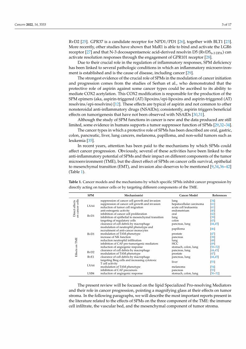
In recent years, attention has been paid to the mechanisms by which SPMs couldaffect cancer progression. Obviously, several of these activities have been linked to theanti-inflammatory potential of SPMs and their impact on different components of the tumormicroenvironment (TME), but the direct effect of SPMs on cancer cells survival, epithelialto mesenchymal transition (EMT), and invasion also deserves to be mentioned [9,34,36–42](Table 1).
Table 1. Cancer models and the mechanisms by which specific SPMs inhibit cancer progression bydirectly acting on tumor cells or by targeting different components of the TME.
SPM Mechanism(s) Cancer Model References
Dir
ecte
ffec
tson
canc
erce
lls
LXA4
suppression of cancer cell growth and invasion lung [34]suppression of cancer cell growth and invasion hepatocellular carcinoma [41]reduction of tumor cell migration acute cell leukaemia [41]anti-estrogenic activity endometrium [40]
RvD1inhibition of cancer cell proliferation oral [42]inhibition of epithelial to mesenchymal transition lung [39]
Effe
cts
onTM
E
RvD1
targeting of regulatory cells colon [43]clearance of cell debris by macrophage pancreas, lung [44,45]modulation of neutrophil phenotype andrecruitment of anti-cancer monocytes papilloma [46]
modulation of TAM phenotype prostate [47]increase of NK function pancreas [38]reduction neutrophil infiltration lung [48]inhibition of CAF pro-tumorigenic mediators HCC [49]reduction of angiogenic response stomach, colon, lung [50–52]
RvD2clearance of cell debris by macrophage pancreas, lung [44,45]modulation of TAM phenotype prostate [47]
RvE1 clearance of cell debris by macrophage pancreas, lung [44,45]
LXA4
targeting Breg cells and increasing cytotoxicT cell activity liver [53]
modulation of TAM phenotype melanoma [54]inhibition of CAF precursors pancreas [55]
LXB4 reduction of angiogenic response stomach, colon, lung [50–52]
The present review will be focused on the lipid Specialized Pro-resolving Mediatorsand their role in cancer progression, pointing a magnifying glass at their effects on tumorstroma. In the following paragraphs, we will describe the most important reports present inthe literature related to the effects of SPMs on the three component of the TME: the immunecell infiltrate, the vascular bed, and the mesenchymal component of tumor stroma.
Cancers 2022, 14, 3333 4 of 17
3. Effects of SPMs on the Immune Cell Compartment InfiltratingTumor Microenvironment
Tumor-infiltrating immune cells play a key role in the evolution of cancer. Typically,immune cells should recognize and eliminate cancer cells [56]. Despite this, cancer cellscontinue to grow and expand, due to several escape mechanisms [57]. Furthermore,several immune cell classes can orchestrate cancer growth, by producing mediators directlysustaining cancer progression [58].
The most abundant immune cell population infiltrating tumors are the macrophages,dendritic cells (DCs), myeloid-derived suppressor cells (MDSCs), T cells, mast cells, andnatural killer (NK) cells. Some of them have been clearly associated to anti-tumor immuneresponses: lymphoid cells, including natural killer (NK) cells, CD8+ T lymphocytes, CD4+
helper T (Th) cells, pro-inflammatory macrophages (M1), and DCs. Moreover, MDSCs andregulatory T (Treg) cells have been shown to exert a pro-tumorigenic action [59,60].
In the context of anti-tumor immune response, a primary regulatory role has to beascribed to immune checkpoint (IC) molecules (e.g., Programmed Cell Death-1 (PD-1), Pro-grammed death ligand 1 (PD-L1), Cytotoxic T-Lymphocyte Antigen 4 (CTLA-4), etc.). Dueto its ability to dampen anti-tumor immune response, immune checkpoint blockade (ICB) istoday considered, among the various types of immunotherapy, the pivotal approach [61]. Itis now clear that a continuous influence of IC on TME, and vice versa, exists. Thus, it wouldalso be simplistic not to consider, in the context of immune response to cancer, the possibilitythat resolution of inflammation could affect immune-checkpoint expression and functions.
In this scenario, an RNA-seq analysis of patients with head and neck squamous cellcarcinoma showed that a higher score for RvD metabolism-associated markers directlycorrelates with a better clinical outcome. A high RvD score also associates to a signaturesuggestive of enhanced anti-tumor immunity, as demonstrated by its association to genesinvolved in the cytotoxic activity of immune cells (e.g., granzymes and perforin). Finally,a high RvD score is presumably linked to increased responsivity to immune-checkpointinhibitors (ICI), as demonstrated by its significant correlation with the expression of fourimmune-checkpoint inhibitor targets (CD274, CTLA4, LAG3, and PDCD1) [62].
Consistently, preliminary data from Gartung et al. reported that, in a murine modelof head and neck cancer, resolvins and immune checkpoint inhibitors (e.g., anti-PD-1) actsynergistically, by suppressing tumor growth [63]. Furthermore, in a model of colorectalcarcinoma (CRC), administration of AT-SPM regulated macrophage trafficking by stimulat-ing the clearance of apoptotic cells and reduced the expression of the immunosuppressivereceptor PD-1 in both macrophages and CD8+ T cells [64]. Similarly, SPM LXA4 wasreported to reduce the expression of PD-L1 in a Kaposi’s sarcoma-associated herpesvirus(KSHV)-induced model [65].
3.1. SPM Effects on Innate Immune Cell Compartment
Due to their natural role in inhibiting the inflammatory response, most of the studiespresent in the literature, including those related to cancer, have been focused on the effectsof SPMs in modulating the functions of innate immune cells (Table 1).
The most abundant innate immune cells infiltrating tumors are neutrophils andmacrophages, which sometimes act against cancer cells through cytotoxic and phago-cytic activities, while in other contexts they sustain tumor growth through the productionof mediators hijacked by cancer cells to favor their spread [66].
Here, we will summarize the knowledge regarding SPM effects on neutrophils andmacrophages. Furthermore, a paragraph will be dedicated to DCs, the innate immune cellpopulation essential for the induction of an adaptive immune response.
3.1.1. Neutrophils
While several research programs have highlighted the role of macrophages in cancerprogression [67], less information is available concerning the function of neutrophils in thecontext of the tumor microenvironment [46]. To date, most reports have sustained the pro-
Cancers 2022, 14, 3333 5 of 17
tumorigenic role of neutrophils, and more generally of polymorphonuclear (PMN) cells [46,48],while others have demonstrated that PMN are able to inhibit cancer progression [68–71]. Theliterature suggests that the different functions of neutrophils in supporting or inhibiting cancerdepends on the tumor stage and the cancer tissue [71].
Nevertheless, neutrophils are targets of resolution molecules, as demonstrated by theevidence that BLT1 receptor expressed on neutrophil surface is crucial in the regulationof their mitochondrial functions. In more detail, the RvE1-BLT1 signal mediates the ac-tivation of apoptotic responses through the induction of reactive oxygen species (ROS)and subsequent activation of caspases, and through the inhibition of ERK and Akt anti-apoptotic signals [72]. Furthermore, GPR18 knockout in mice displayed reduced neutrophilinfiltration at the infection site and reduced tissue recovery following injuries [25].
The few studies regarding the effects of pro-resolving mediators on cancer-associatedneutrophils were directed to the definition of the effects of SPMs on neutrophil phenotypeand functions and on the impact of these on cancer progression.
Mattoscio and coll. demonstrated that RvD1 reduces the growth and proliferation oftumors in vivo using a human papilloma virus (HPV) tumorigenesis model. This effect ismediated by the ability of RvD1 to change the phenotype of neutrophils in a “pro-resolvinganti-cancer phenotype”, characterized by increased anti-tumor properties. In addition,RvD1 stimulated PMN to secrete increased levels of chemoattractants for monocytes ableto inhibit tumor growth in vivo [46].
Vannitamby et al. focused their studies on the effects of AT-RvD1 on neutrophilsin a mouse model of lung cancer. AT-RvD1 is produced by aspirin-acetylated COX2and 5-lipoxygenase (ALOX5) enzymes [48]. The authors found that increased neutrophilinfiltration in the TME of lung adenocarcinoma patients correlated with a low level of ex-pression of ALOX5 enzyme. Based upon this observation, they investigated the possibilitythat AT-RvD1 could exert a therapeutic activity in a mice model of lung adenocarcinoma,demonstrating that AT-RvD1 treatment significantly reduced lung adenocarcinoma growthin vivo by reducing the infiltration of neutrophil, which is responsible for lymphocyteactivity suppression [48].
3.1.2. Macrophages
Macrophages represent probably the most important target cell of SPMs. In thephysiology of inflammation resolution, SPMs sustain the recruitment of the macrophagesresponsible for the non-inflammatory efferocytosis of apoptotic neutrophils [73,74].
The classic view of macrophage biology classifies them as M1, with pro-inflammatory,microbicidal, and anti-tumor functions; or as M2, displaying anti-inflammatory, immuno-suppressive, and pro-tumorigenic properties [75]. However, this polarization model showssome limitations: it reflects well the characteristics of macrophages induced to polarizein vitro by specific cytokines and mediators, but it does not describe well the characteristicsof macrophages present in tissues [76]. In the cancer context, tumor associated macrophages(TAM) display phenotypes that are a continuum between the two phenotypes, being con-tinuously conditioned by the TME [76]. For example, the most abundant phenotype ofmacrophages in TME is M2, but these cells can exert a more efficient phagocytosis of cancercells than M1 cells, thus potentially favoring an anti-tumor response [29].
A characterization of the lipid mediators produced by the two different classes ofmacrophages demonstrated that M1 produce pro-inflammatory lipid mediators, such asprostaglandins and leukotrienes, while M2 macrophages produce SPMs. This suggeststhat the different classes of lipid autacoids could be useful in re-shaping the macrophagephenotype to activate a protective anti-tumor response [29]. A critical role of resolutionmolecules in macrophage function regulation was also revealed in studies on SPM receptorsknock-out: (i) macrophages deleted for GPR32 become unresponsive to RvD1 and lostthe ability to polarize toward a pro-resolutive phenotype [19]; (ii) GPR18 knock-out inmacrophages reduced their ability to phagocyte debris and dead cells [25]; (iii) ChemR23blocking antibody reduced the phagocytic activity of RvE1-stimulated macrophages in
Cancers 2022, 14, 3333 6 of 17
acute and chronic inflammatory models [77,78]; and (iv) mice knocked-out for GPR37displayed a reduced macrophage phagocytic activity in a model of inflammatory pain. [26].
Physiologically, SPMs increase macrophage survival [79], sustain the phagocytic activ-ity of macrophages against microbes and apoptotic neutrophils [16,80], reduce the secretionof pro-inflammatory cytokines via inhibition of NF-kB [29,80], and, in the meantime, in-crease the production of anti-inflammatory cytokines [22,81].
Although it would be expected that the SPM-sustained changes of macrophage phe-notype towards a M2 type would favor neoplastic progression, the available literaturepoints to a protective anti-tumor effect of SPM-conditioned TAM [29,54]. The analysis ofthe effects of SPMs on TAM supports this evidence and demonstrates that the scenario ismore complex than expected. Indeed, SPMs sustain changes in TAM that place them inbetween the M1 and M2 phenotypes [82–84].
Several research groups have investigated the role of SPMs in conditioning macrophagephenotype in the cancer context [29]. ATL-1, a synthetic analogue of LXA4, alters TAMprofile by decreasing M2 surface markers, triggering ROS production, and increasing thecytotoxic and decreasing the anti-apoptotic properties of TAM against melanoma cancercells. Consistently, ATL-1 inhibited cancer progression in a murine model by affectingmacrophage phenotype [29]. Similarly, RvD1 and RvD2 inhibited cancer cell proliferationby affecting macrophage polarization in a prostate cancer model [47].
A very interesting research line has focused on the effects of SPMs on macrophageefferocytosis of chemotherapies-induced tumor cell debris. Debris of tumor cells havebeen demonstrated to be produced following several types of cancer therapies, includingradiation and chemotherapy, and to sustain tumor engraftment, growth, and metastasisby activating a pro-inflammatory response [85,86]. Furthermore, tumor cell debris af-fect M1 TAM, inducing an immunosuppressive response, and thus limiting anti-cancerimmunity [87,88].
The involvement of lipidic autacoids in the regulation of inflammatory responseassociated to cancer cell debris is now clear, since it has been recognized that they stimulatetumor growth by sustaining the production of the pro-inflammatory autacoid prostaglandinE2 (PGE2) in the TME [89]. On the other hand, in several tumor types, SPMs RvD1, RvD2,and RvE1 block the ability of tumor cell debris to sustain cancer progression via theactivation of macrophage phagocytosis and clearance of cell debris [44]. SPMs sustainedpolarization of macrophages in these models is also characterized by a reduced productionof pro-inflammatory cytokines [44]. Furthermore, the anti-tumor effect of aspirin was linkedto its ability to induce the production of SPMs, enhance the macrophage phagocytosis oftumor cell debris, and reduce macrophage production of inflammatory mediators [45].
3.1.3. Dendritic Cells
This cell type represents a crucial link between innate immunity and the activationof the adaptive response, due to its ability to professionally present antigens to lympho-cytes [90]. DCs express receptors to SPMs, thus representing a target of these molecules [91].To date, evidence has been produced demonstrating that SPMs could affect DC maturationand functions, with the aim of reducing inflammation and allowing tissue restitution.SPMs reduce the DC migration and production of pro-inflammatory cytokines [92,93].Furthermore, RvD1 reduces the levels of Major Histocompatibility Complex II (MHC II)and costimulatory molecules on DCs [94]. As an example, RvE1 treatment blocks DCs in animmature state [95] and prevents their migration to draining lymph-nodes, thus resulting ina reduced T cell response [96,97]. All these evidence has been produced in disease modelsin which SPMs work in order to facilitate tissue homeostasis (e.g., infection, autoimmunediseases, etc.), but currently no data have been published on SPM effects on DCs in a cancercontext, where the separation between tissue homeostasis and tumor growth is less clear.
Cancers 2022, 14, 3333 7 of 17
3.2. Effects of SPMs on the Adaptive Immune Cell Compartment of the TME
Very few studies are available on the effects of SPMs on the adaptive immune branchof the immune system, and even less in the specific context of cancer, although the potentialimmunomodulatory effects of SPMs on adaptive immune response could be of greatinterest [98].
3.2.1. Effector T and B lymphocytes
Since the activation of T or B lymphocytes is the goal of the adaptive immune response,an active area of study focuses on SPM activity with T and B cells. The effector functions ofthe different T and B cell subsets are strictly linked to the characteristics of the producedcytokines [99].
The most plastic subset of human cells is probably represented by the CD4+ T lym-phocytes: the effector functions against different classes of pathogens are assigned to Thelper (Th)1, Th2, and Th17 subsets; follicular helper T (Thf) cells assist B lymphocytes intheir maturation, and Treg cells, instead, control self-tolerance [100]. Chiurchiù and coll.investigated the effects of SPMs on T CD4+ lymphocytes, demonstrating that RvD1, RvD2,and MaR1 are able to modulate T cell differentiation programs suppressing Th1 and Th17 Tcell subtypes, as well as their ability to produce pro-inflammatory cytokines and to increase,in the meantime, the production of Treg cells [101]. Similarly, the inhibition of cytokineproduction mediated by SPMs on CD4+ Th1 and Th17 T cells has also been describedfor PD1, RvD3, LXA4, and LXB4 [102,103]. This inhibitory effect of SPMs on cytokineproduction has a great impact on pro-inflammatory T cell subsets (i.e., Th1 and Th17) but aless important effect on the Th2 phenotype. An exception was reported for RvE1, whichhas been described to dampen the Th2 T cell response in a model of asthma [104,105].
Both in vitro and in vivo data support the hypothesis that SPMs are instead able toaugment Treg generation, consistently with their ability to produce immunomodulatoryfactors and to exert pro-resolving functions [101,106,107]. Since more data are availableon the effects of SPMs on regulatory subsets of lymphocytes, a specific paragraph will bededicated to these.
Unfortunately, to date, no direct and structured data have been published on theeffects of SPMs on the cytotoxic response of CD8+ T lymphocytes, although they representthe key to directly killing cancer cells. However, in support of a role of SPMs in sustain-ing an anti-tumor cytotoxic response, it has been observed that RvD1 and its precursor(DHA) affect natural killer (NK) cells, the innate immune counterpart of CD8+ T cells, bypreventing their death and sustaining their tumoricidal activity [38]. Consistently, it hasbeen demonstrated that LXA4 is able to improve the anti-tumor activities of CD8+ T cellsindirectly, by reducing regulatory subsets of T and B cells [53]. Furthermore, it has beendescribed that GPR18 is expressed in CD8+ T lymphocytes and regulates their developmentand homing in epithelia [108], suggesting the crucial importance of resolution responses intumor immune response and immunotherapy [108]. Interestingly, it has been demonstratedthat BLT1 regulates anti-tumor immune responses by favoring CD8+ T cell migration inmelanoma [109] and the cervical cancer murine microenvironment [110].
Some authors have also addressed the role of inflammation resolution on B cell com-partment. SPMs have been described as potent modulators of B cell functions [111]: RvD1increases IgM and IgG and decreases IL-6 production from B cells, as well as favoring thedifferentiation of B cells in antibody secreting cells [112]. Several other reports have pointedto a role of SPMs in sustaining antibody production from B cells [113,114]. LXA4 has beendescribed to be able to accelerate B cell migration to the spleen, increasing the response toantigens [115].
These data perfectly fit into the scenario of SPMs as negative modulators of inflammatoryresponse when acting on the adaptive immune compartment, although no structured dataare available in cancer context nor a clear definition of potential pro- or anti-tumor effects.
Cancers 2022, 14, 3333 8 of 17
3.2.2. Regulatory Lymphocytes
The immune response against cancer cells is the result of the balance between immuneeffector cells such as CD8+ T cytotoxic cells or NK cells and immunosuppressive cells suchas Treg or MDSC [116–118].
The resolution of inflammation affects Treg regulation and function(s). It has beendemonstrated that SPMs (RvD1, RvD2, and Mar1) exert a regulatory role on the balancebetween pathogenic Th1 and tolerogenic Treg cells [101,119–121] during autoimmuneresponses. Furthermore, the reduced expression of ALOX15 enzyme in Treg is associatedwith a lower expression of Foxp3, the master regulator of Treg lineage, and alterationof several cellular metabolic pathways [122]. Consistently, GPR32 plays a crucial rolein regulating the adaptive immune responses, as demonstrated by the evidence that itsexpression is crucial for the generation and function of Treg cells [101].
This evidence suggests that the resolution of inflammation could also impact on therole and function of Treg cells in cancer, although several questions remain open. The firstpoint is that the Treg role in cancer initiation/progression is complex: the most accreditedtheory is that Treg cells exert a pro-tumor effect by inhibiting anti-cancer immune response;however, in some cancer contexts, an anti-tumor effect of Treg was described and associatedwith their ability to prevent tumor inflammation [123]. Indeed, it has been demonstratedthat Treg depletion favors cancer development in a model of inflammation induced coloncancer [43]. Zhang and coll. postulated that Treg could promote early tumor growth, whileinhibiting tumor progression later on [123]. Interestingly, they demonstrated that inhibitionof Treg in large tumors favors growth, by inducing the generation of MDSCs in the TME,and that this phenomenon is associated with increased levels of LXA4 [123].
A specific subpopulation of B cells, namely B regulatory (Breg) cells, have been recentlydescribed as able to modulate the immune response in the context of TME [124]. Breg cellsare able to produce large amounts of immunosuppressive cytokines (IL-10 and TGF-β)and to express immunoregulatory molecules on their membrane (e.g., PD-L1). Breg cellshave been demonstrated to suppress T and NK cells responses in several models of tumorgrowth [125]. Furthermore, Breg cells facilitate the conversion of CD4+ T lymphocyte inTME in CD4+CD25+FoxP3+ Treg cells [125].
Wang and coll. demonstrated that LXA4 is able to suppress the generation of Bregcells in tumor-bearing mice, thus dampening tumor growth. Consistently, the depletion ofBreg cells in mice abolishes the anti-tumor activity of LXA4 [53]. Interestingly, LXA4 notonly affects Breg cell induction, but is also able to reduce the number of Treg cells, both inthe draining lymph nodes and in the TME, and simultaneously to enhance the cytotoxicactivities of T lymphocytes [53].
These data suggest that other studies are necessary to clarify the role of SPMs in themodulation of regulatory lymphocytes in the TME, in order to define the effects of SPMson these immune populations and their impact on cancer progression.
4. Effects of SPMs on Cancer-Associated Fibroblasts
Cancer-associated fibroblasts (CAFs) are the most abundant cells in the TME, wherethey represent a key source of extracellular matrix components, thus contributing to theformation of a desmoplastic stroma and playing crucial roles during malignant cancerprogression and metastasis [126].
SPMs promote the regeneration of damaged tissue by finely regulating the fibroticresponse, both in the physiology of wound healing and in several pathologic scenariosin which an excessive fibrotic response is established [127]. This is the case of idiopathicpulmonary fibrosis (IPF), in which LXA4 has been reported to inhibit the pro-fibroticpulmonary response through different mechanisms [128].
The effects of SPMs on CAFs have been studied and described in a few model systems.RvD1 has been demonstrated to inhibit the production of pro-tumorigenic mediatorsfrom CAFs in a model of hepatocellular carcinoma (HCC), thus suppressing EMT and thestemness of cancer cells [49]. Furthermore, LXA4 attenuates pancreatic tumor growth by
Cancers 2022, 14, 3333 9 of 17
inhibiting the function of human pancreatic stellate cells (hPSCs), which are the precursorsof pancreatic CAFs; in more detail, LXA4 attenuates hPSC-induced desmoplasia, revertshPSC activation into CAFs, and modulates several other hPSC tumor-promoting effects [55].
Since one of the problems of the fibrotic process associated with tumors, in addition tothe growth-promoting effect, is the formation of a barrier that is difficult to penetrate fordrugs, these papers suggest a further potential use of SPMs (i.e., RvD1 and LXA4) as usefulagents, in conjunction with conventional therapy, to improve their efficacy.
5. Effects of SPMs on the Tumor Vascular Bed
Other than in immune cells, SPM receptors have also been described in endothelialcells (ECs) and vascular smooth muscle cells (VSMCs), and to be modulators of theirphenotype and function(s) [13]. In particular, it has been described that ECs expressALX/FPR2, GPR32, GPR18, BLT1, and ChemR23 [16,129–132]. Among the other types, theanti-angiogenic properties of SPMs have also been described in several contexts [9], forexample, mediated by the SPM (LXA4) ability to inhibit EC proliferation and migration, tointerfere with vascular endothelial growth factor (VEGF) signaling, and to dampen VEGFreceptor expression [133,134]. LXA4 has also been reported to modulate EC functions byincreasing NO production [135]. Furthermore, most SPMs affect leukocyte–EC interactionsby reducing the expression of adhesion molecules and the production of inflammatorycytokines [13]. Finally, the genetic ablation of GPR32 in ECs demonstrated that the receptoris crucial for endothelial cell integrity and barrier function [136].
However, to date, only a few studies have investigated the role of pro-resolvingpathways in the modulation of cancer angiogenesis [9].
In a model of Kaposi Sarcoma (KS), LXA4 inhibited the secretion of angiogenic fac-tors [137]. Similarly, LXA4 decreased the production of the angiogenic mediators fromHCC cells [138]. Interestingly, SPMs (RvD4 or RvD5), in combination with anti-angiogenictherapy (i.e., the thrombospondin (TSP)-1 peptide 3 TSR or anti-VEGF via DC101), inducedsynergistic anti-tumor activity in xenograft models of ovarian cancer [138].
Related to cancers of the gastrointestinal (GI) tract, the authors recently described anovel function of SPMs in gastric cancer (GC), demonstrating that two SPMs (i.e., RvD1and LXB4) suppress angiogenesis, thus inhibiting tumor growth [50]. A key modulatorof SPM production was the Formyl Peptide Receptor 1 (FPR1), which functions in theGI tract as a tumor suppressor [139]. FPR1 is a member of an innate immune receptorfamily of formyl peptide receptors (FPR1, 2, and 3), which are pattern recognition receptors(PRRs) belonging to the G-protein coupled receptor family and able to recognize conservedmicrobe- or damage-associated molecular structures (PAMPs/DAMPs) and initiate bothinflammation and its resolution, depending on the environmental context and on thespecific ligand [140,141]. We demonstrated that GC cells silenced for FPR1, but not forFPR2 or FPR3, displayed an in vivo growth advantage, due to their increased ability toform new vessels. In culture, these cells displayed an increase in the production of pro-inflammatory and pro-angiogenic mediators and a drop in the levels of various componentsof the pro-resolving pathways (ALOX5/15, SPMs (RvD1 and LXB4), and SPM receptors(BLT1, ChemR23 and GPR32)) [50,139]. The same effects were observed when FPR1 waspharmacologically antagonized by cyclosporin H. Consistently, FPR1 activation or increasedexpression mediates the opposite effects [50]. FPR1-mediated anti-angiogenic potentialdepends on pro-resolving pathways, as witnessed by the finding that blockade of ALOXsor of SPM receptors (i.e., GPR32) blunted this FPR1 activity in GC cells. Moreover, theincreased angiogenic potential of FPR1-depleted GC cells was reverted by exogenousadministration of SPMs (RvD1 or LXB4) [50]. The blockade of the pro-resolving receptorGPR32, or of ALOX15, enhanced the angiogenesis and tumorigenic activity of GC cells,mimicking FPR1-depletion. Thus, GC cells are endowed with an intrinsic angiogenicpotential, negatively controlled by SPMs. These, in turn, are positively controlled by FPR1.Consistently, a diet enriched in the precursors of SPMs,ω-3 orω-6 PUFA, [142] inhibitedthe growth of FPR1-silenced GC cells in mice, by specifically impairing angiogenesis [50].
Cancers 2022, 14, 3333 10 of 17
Several reports have also pointed to a crucial protective role of pro-resolving pathwaysin colorectal cancer (CRC) [143–145]. Unpublished results from the authors’ group alsoindicate a crucial anti-angiogenic function of SPMs in CRC: the activation of FPR1 mediatedby the probiotic Lactobacillus Rhamnosus (LGG) or specific bacterial products in CRC cellscaused an increase of pro-resolving mediators (RvD1 and LXB4) and the consequent inhibi-tion of angiogenesis [51]. LGG is a commensal bacterium used as a probiotic and describedas able to dampen the chronic inflammation associated with CRC development [146]. LGGsustains colonic wound healing in mice [147–149] and activates pro-apoptotic and anti-metastatic responses [150,151] through the interaction with FPR1 [147]. Our data highlightnew pro-resolving and anti-angiogenic functions of this bacterium mediated by FPR1 [51].
By asking whether similar mechanisms could also intervene in cancers derivedfrom other epithelia, we demonstrated that pro-resolving pathways could also exert anti-angiogenic activity in a lung cancer model; however, in this context, the innate immunereceptor that controls SPM production is the Toll-like receptor 7 (TLR7) [52]. In this modelsystem, TLR7 activation, similarly to FPR1 in the GI tract, sustains the increased expressionof the enzymes responsible for SPMs biosynthesis, the increased secretion of SPMs fromcancer cells, the augmented expression of SPM receptors, and finally the reduction of theangiogenic potential of lung cancer cells [52].
6. Conclusions
Inflammation has long been considered a hallmark of cancer, both for its ability tointervene in cancer initiation and to sustain cancer progression. Recently, the discovery ofthe resolution of inflammation opened the way for new therapeutic possibilities that, ratherthan block the inflammatory response, could instead support the induction of resolution ofthe inflammation.
Among others, Specialized Pro-resolving Mediators (SPMs) are “bioactive lipids” al-ready described as key players in regulating inflammatory response and tissue homeostasisin several physiologic and pathologic conditions, by acting on several cell types in thecontext of inflammatory reactions. The present review focused on the possibility that SPMscould shape the TME, to affect cancer cell growth and progression.
Strong evidence has been provided supporting the possibility that SPMs act on the innateimmune cell compartment infiltrating tumors from an anti-cancer perspective (Figure 1).
Cancers 2022, 14, x FOR PEER REVIEW 11 of 18
Strong evidence has been provided supporting the possibility that SPMs act on the innate immune cell compartment infiltrating tumors from an anti-cancer perspective (Fig-ure 1).
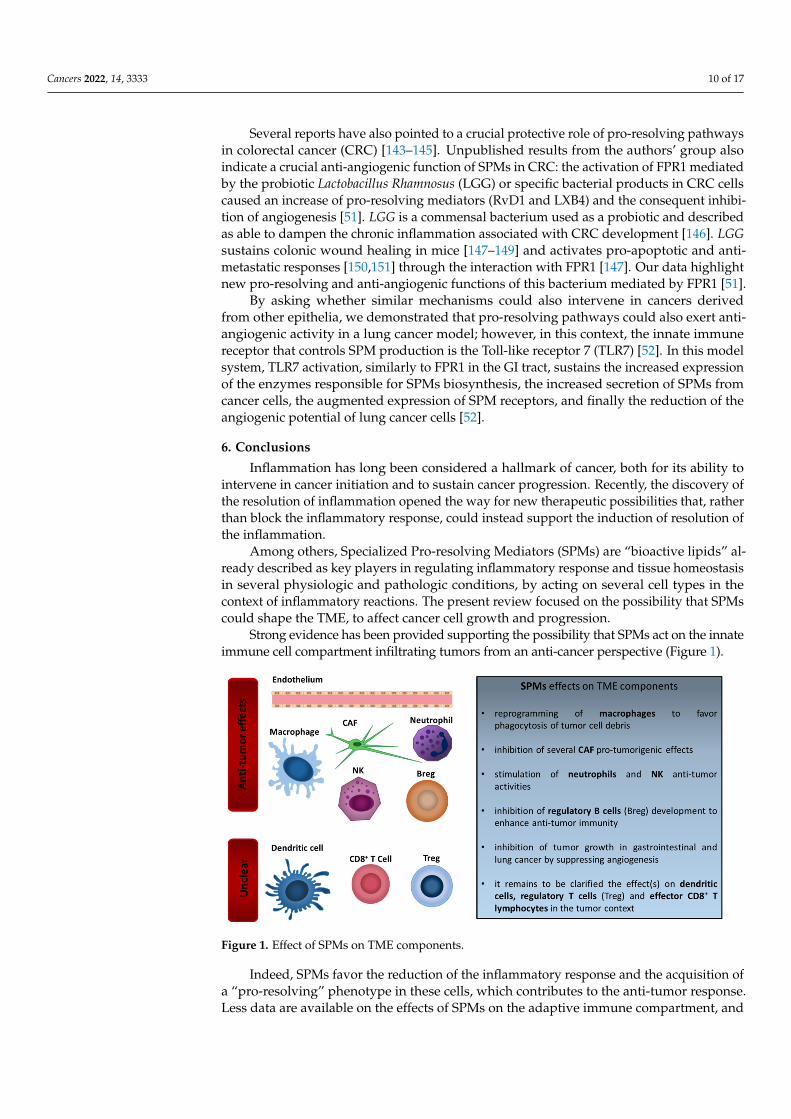
Figure 1. Effect of SPMs on TME components.
Indeed, SPMs favor the reduction of the inflammatory response and the acquisition of a “pro-resolving” phenotype in these cells, which contributes to the anti-tumor re-sponse. Less data are available on the effects of SPMs on the adaptive immune compart-ment, and no clear definition of the function of SPMs in the context of an adaptive anti-cancer response has been defined (Figure 1).
Interestingly, SPMs have been described to act on various other cells constituting tu-mor stroma (Figure 1). Consistently with their natural anti-fibrotic and anti-angiogenic properties, SPMs have been described as able to counteract the CAF ability to sustain can-cer progression and to dampen the angiogenic response vital for cancer dissemination.
Although further studies are needed to define the role of these bioactive lipids in cancer, they appear to be a potential and promising approach to fight cancer progression.
Author Contributions: F.L. and M.M.; writing-original draft preparation, N.P. and R.M.M.; writing-review and editing. All authors have read and agreed to the published version of the manuscript.
Funding: PRIN 2017 no. 2017XJ38A4; Istituto Superiore di Oncologia grant (MIUR PON01_02782/12); POR Campania FESR 2014-2020 “SATIN” grant; POR Campania FESR 2014-2020 “RARE.PLAT.NET” grant.
Conflicts of Interest: The authors declare no conflict of interest.
References 1. Hanahan, D.; Weinberg, R.A. Hallmarks of cancer: The next generation. Cell 2011, 144, 646–674.
https://doi.org/10.1016/j.cell.2011.02.013. 2. Fishbein, A.; Hammock, B.D.; Serhan, C.N.; Panigrahy, D. Carcinogenesis: Failure of resolution of inflammation? Pharmacol.
Ther. 2021, 218, 107670. https://doi.org/10.1016/j.pharmthera.2020.107670. 3. Fredman, G. DELineating resolution of inflammation. Nat. Immunol. 2019, 20, 2–3. https://doi.org/10.1038/s41590-018-0278–9. 4. Sugimoto, M.A.; Sousa, L.P.; Pinho, V.; Perretti, M.; Teixeira, M.M. Resolution of Inflammation: What Controls Its Onset? Front.
Immunol. 2016, 7, 160. https://doi.org/10.3389/fimmu.2016.00160. 5. Serhan, C.N.; Savill, J. Resolution of inflammation: The beginning programs the end. Nat. Immunol. 2005, 6, 1191–1197.
https://doi.org/10.1038/ni1276. 6. Perretti, M.; D'Acquisto, F. Annexin A1 and glucocorticoids as effectors of the resolution of inflammation. Nat. Rev. Immunol.
2009, 9, 62–70. https://doi.org/10.1038/nri2470. 7. Hasko, G.; Cronstein, B. Regulation of inflammation by adenosine. Front. Immunol. 2013, 4, 85.
https://doi.org/10.3389/fimmu.2013.00085.
Figure 1. Effect of SPMs on TME components.
Indeed, SPMs favor the reduction of the inflammatory response and the acquisition ofa “pro-resolving” phenotype in these cells, which contributes to the anti-tumor response.Less data are available on the effects of SPMs on the adaptive immune compartment, and
Cancers 2022, 14, 3333 11 of 17
no clear definition of the function of SPMs in the context of an adaptive anti-cancer responsehas been defined (Figure 1).
Interestingly, SPMs have been described to act on various other cells constitutingtumor stroma (Figure 1). Consistently with their natural anti-fibrotic and anti-angiogenicproperties, SPMs have been described as able to counteract the CAF ability to sustain cancerprogression and to dampen the angiogenic response vital for cancer dissemination.
Although further studies are needed to define the role of these bioactive lipids incancer, they appear to be a potential and promising approach to fight cancer progression.
Author Contributions: F.L. and M.M.; writing-original draft preparation, N.P. and R.M.M.; writing-review and editing. All authors have read and agreed to the published version of the manuscript.
Funding: PRIN 2017 no. 2017XJ38A4; Istituto Superiore di Oncologia grant (MIUR PON01_02782/12);POR Campania FESR 2014–2020 “SATIN” grant; POR Campania FESR 2014-2020 “RARE.PLAT.NET” grant.
Conflicts of Interest: The authors declare no conflict of interest.
References1. Hanahan, D.; Weinberg, R.A. Hallmarks of cancer: The next generation. Cell 2011, 144, 646–674. [CrossRef] [PubMed]2. Fishbein, A.; Hammock, B.D.; Serhan, C.N.; Panigrahy, D. Carcinogenesis: Failure of resolution of inflammation? Pharmacol. Ther.
2021, 218, 107670. [CrossRef] [PubMed]3. Fredman, G. DELineating resolution of inflammation. Nat. Immunol. 2019, 20, 2–3. [CrossRef] [PubMed]4. Sugimoto, M.A.; Sousa, L.P.; Pinho, V.; Perretti, M.; Teixeira, M.M. Resolution of Inflammation: What Controls Its Onset? Front.
Immunol. 2016, 7, 160. [CrossRef] [PubMed]5. Serhan, C.N.; Savill, J. Resolution of inflammation: The beginning programs the end. Nat. Immunol. 2005, 6, 1191–1197. [CrossRef]6. Perretti, M.; D’Acquisto, F. Annexin A1 and glucocorticoids as effectors of the resolution of inflammation. Nat. Rev. Immunol.
2009, 9, 62–70. [CrossRef] [PubMed]7. Hasko, G.; Cronstein, B. Regulation of inflammation by adenosine. Front. Immunol. 2013, 4, 85. [CrossRef] [PubMed]8. Mirakaj, V.; Dalli, J.; Granja, T.; Rosenberger, P.; Serhan, C.N. Vagus nerve controls resolution and pro-resolving mediators of
inflammation. J. Exp. Med. 2014, 211, 1037–1048. [CrossRef]9. Prevete, N.; Liotti, F.; Amoresano, A.; Pucci, P.; de Paulis, A.; Melillo, R.M. New perspectives in cancer: Modulation of lipid
metabolism and inflammation resolution. Pharmacol. Res. 2018, 128, 80–87. [CrossRef]10. Serhan, C.N.; Gupta, S.K.; Perretti, M.; Godson, C.; Brennan, E.; Li, Y.; Soehnlein, O.; Shimizu, T.; Werz, O.; Chiurchiu, V.; et al.
The Atlas of Inflammation Resolution (AIR). Mol. Asp. Med. 2020, 74, 100894. [CrossRef]11. Schett, G.; Neurath, M.F. Resolution of chronic inflammatory disease: Universal and tissue–specific concepts. Nat. Commun. 2018,
9, 3261. [CrossRef] [PubMed]12. Serhan, C.N.; Chiang, N.; Dalli, J.; Levy, B.D. Lipid mediators in the resolution of inflammation. Cold Spring Harb. Perspect. Biol.
2014, 7, a016311. [CrossRef] [PubMed]13. Diaz Del Campo, L.S.; Rodrigues-Diez, R.; Salaices, M.; Briones, A.M.; Garcia-Redondo, A.B. Specialized Pro-Resolving Lipid
Mediators: New Therapeutic Approaches for Vascular Remodeling. Int. J. Mol. Sci. 2022, 23, 3592. [CrossRef] [PubMed]14. Luo, X.; Gu, Y.; Tao, X.; Serhan, C.N.; Ji, R.R. Resolvin D5 Inhibits Neuropathic and Inflammatory Pain in Male but Not Female
Mice: Distinct Actions of D-Series Resolvins in Chemotherapy-Induced Peripheral Neuropathy. Front. Pharmacol. 2019, 10, 745.[CrossRef]
15. Gronert, K.; Gewirtz, A.; Madara, J.L.; Serhan, C.N. Identification of a human enterocyte lipoxin A4 receptor that is regulated byinterleukin (IL)-13 and interferon gamma and inhibits tumor necrosis factor alpha-induced IL-8 release. J. Exp. Med. 1998, 187,1285–1294. [CrossRef]
16. Krishnamoorthy, S.; Recchiuti, A.; Chiang, N.; Yacoubian, S.; Lee, C.H.; Yang, R.; Petasis, N.A.; Serhan, C.N. Resolvin D1 bindshuman phagocytes with evidence for proresolving receptors. Proc. Natl. Acad. Sci. USA 2010, 107, 1660–1665. [CrossRef]
17. Dalli, J.; Consalvo, A.P.; Ray, V.; Di Filippo, C.; D’Amico, M.; Mehta, N.; Perretti, M. Proresolving and tissue-protective actions ofannexin A1-based cleavage-resistant peptides are mediated by formyl peptide receptor 2/lipoxin A4 receptor. J. Immunol. 2013,190, 6478–6487. [CrossRef]
18. Krishnamoorthy, S.; Recchiuti, A.; Chiang, N.; Fredman, G.; Serhan, C.N. Resolvin D1 receptor stereoselectivity and regulation ofinflammation and proresolving microRNAs. Am. J. Pathol. 2012, 180, 2018–2027. [CrossRef]
19. Arnardottir, H.H.; Dalli, J.; Norling, L.V.; Colas, R.A.; Perretti, M.; Serhan, C.N. Resolvin D3 Is Dysregulated in Arthritis andReduces Arthritic Inflammation. J. Immunol. 2016, 197, 2362–2368. [CrossRef]
20. Back, M.; Powell, W.S.; Dahlen, S.E.; Drazen, J.M.; Evans, J.F.; Serhan, C.N.; Shimizu, T.; Yokomizo, T.; Rovati, G.E. Update onleukotriene, lipoxin and oxoeicosanoid receptors: IUPHAR Review 7. Br. J. Pharmacol. 2014, 171, 3551–3574. [CrossRef]
21. Arita, M.; Ohira, T.; Sun, Y.P.; Elangovan, S.; Chiang, N.; Serhan, C.N. Resolvin E1 selectively interacts with leukotriene B4receptor BLT1 and ChemR23 to regulate inflammation. J. Immunol. 2007, 178, 3912–3917. [CrossRef] [PubMed]

Cancers 2022, 14, 3333 12 of 17
22. Oh, S.F.; Dona, M.; Fredman, G.; Krishnamoorthy, S.; Irimia, D.; Serhan, C.N. Resolvin E2 formation and impact in inflammationresolution. J. Immunol. 2012, 188, 4527–4534. [CrossRef] [PubMed]
23. Merlin, J.; Park, J.; Vandekolk, T.H.; Fabb, S.A.; Allinne, J.; Summers, R.J.; Langmead, C.J.; Riddy, D.M. Multipathway In VitroPharmacological Characterization of Specialized Proresolving G Protein-Coupled Receptors. Mol. Pharmacol. 2022, 101, 246–256.[CrossRef]
24. Finlay, D.B.; Joseph, W.R.; Grimsey, N.L.; Glass, M. GPR18 undergoes a high degree of constitutive trafficking but is unresponsiveto N-Arachidonoyl Glycine. PeerJ 2016, 4, e1835. [CrossRef] [PubMed]
25. Chiang, N.; Dalli, J.; Colas, R.A.; Serhan, C.N. Identification of resolvin D2 receptor mediating resolution of infections and organprotection. J. Exp. Med. 2015, 212, 1203–1217. [CrossRef] [PubMed]
26. Bang, S.; Xie, Y.K.; Zhang, Z.J.; Wang, Z.; Xu, Z.Z.; Ji, R.R. GPR37 regulates macrophage phagocytosis and resolution ofinflammatory pain. J. Clin. Investig. 2018, 128, 3568–3582. [CrossRef]
27. Chiang, N.; Libreros, S.; Norris, P.C.; de la Rosa, X.; Serhan, C.N. Maresin 1 activates LGR6 receptor promoting phagocyteimmunoresolvent functions. J. Clin. Investig. 2019, 129, 5294–5311. [CrossRef]
28. Flak, M.B.; Koenis, D.S.; Sobrino, A.; Smith, J.; Pistorius, K.; Palmas, F.; Dalli, J. GPR101 mediates the pro-resolving actions ofRvD5n-3 DPA in arthritis and infections. J. Clin. Investig. 2020, 130, 359–373. [CrossRef]
29. Lavy, M.; Gauttier, V.; Poirier, N.; Barille-Nion, S.; Blanquart, C. Specialized Pro-Resolving Mediators Mitigate Cancer-RelatedInflammation: Role of Tumor-Associated Macrophages and Therapeutic Opportunities. Front. Immunol. 2021, 12, 702785.[CrossRef]
30. Sleire, L.; Forde, H.E.; Netland, I.A.; Leiss, L.; Skeie, B.S.; Enger, P.O. Drug repurposing in cancer. Pharmacol. Res. 2017, 124, 74–91.[CrossRef]
31. Ramon, S.; Woeller, C.F.; Phipps, R.P. The influence of Cox-2 and bioactive lipids on hematological cancers. Curr. Angiogenesis2013, 2, 135–142. [CrossRef] [PubMed]
32. Liu, H.; Zeng, J.; Huang, W.; Xu, Q.; Ye, D.; Sun, R.; Zhang, D. Colorectal Cancer Is Associated with a Deficiency of Lipoxin A4, anEndogenous Anti-inflammatory Mediator. J. Cancer 2019, 10, 4719–4730. [CrossRef] [PubMed]
33. Thun, M.J.; Jacobs, E.J.; Patrono, C. The role of aspirin in cancer prevention. Nat. Rev. Clin. Oncol. 2012, 9, 259–267. [CrossRef][PubMed]
34. Claria, J.; Lee, M.H.; Serhan, C.N. Aspirin-triggered lipoxins (15-epi-LX) are generated by the human lung adenocarcinoma cellline (A549)-neutrophil interactions and are potent inhibitors of cell proliferation. Mol. Med. 1996, 2, 583–596. [CrossRef]
35. Sulciner, M.L.; Gartung, A.; Gilligan, M.M.; Serhan, C.N.; Panigrahy, D. Targeting lipid mediators in cancer biology. CancerMetastasis Rev. 2018, 37, 557–572. [CrossRef] [PubMed]
36. Tsai, W.H.; Shih, C.H.; Wu, H.Y.; Chien, H.Y.; Chiang, Y.C.; Lai, S.L.; Hsu, S.C.; Kou, Y.R.; Hsu, H.C. Role of lipoxin A4 in thecell-to-cell interaction between all-trans retinoic acid-treated acute promyelocytic leukemic cells and alveolar macrophages. J. Cell.Physiol. 2012, 227, 1123–1129. [CrossRef] [PubMed]
37. Chen, Y.; Hao, H.; He, S.; Cai, L.; Li, Y.; Hu, S.; Ye, D.; Hoidal, J.; Wu, P.; Chen, X. Lipoxin A4 and its analogue suppress the tumorgrowth of transplanted H22 in mice: The role of antiangiogenesis. Mol. Cancer Ther. 2010, 9, 2164–2174. [CrossRef]
38. Halder, R.C.; Almasi, A.; Sagong, B.; Leung, J.; Jewett, A.; Fiala, M. Curcuminoids and omega-3 fatty acids with anti-oxidantspotentiate cytotoxicity of natural killer cells against pancreatic ductal adenocarcinoma cells and inhibit interferon gammaproduction. Front. Physiol. 2015, 6, 129. [CrossRef]
39. Lee, H.J.; Park, M.K.; Lee, E.J.; Lee, C.H. Resolvin D1 inhibits TGF-beta1-induced epithelial mesenchymal transition of A549lung cancer cells via lipoxin A4 receptor/formyl peptide receptor 2 and GPR32. Int. J. Biochem. Cell Biol. 2013, 45, 2801–2807.[CrossRef]
40. Canny, G.O.; Lessey, B.A. The role of lipoxin A4 in endometrial biology and endometriosis. Mucosal Immunol. 2013, 6, 439–450.[CrossRef]
41. Zhou, X.Y.; Li, Y.S.; Wu, P.; Wang, H.M.; Cai, Z.Y.; Xu, F.Y.; Ye, D.Y. Lipoxin A(4) inhibited hepatocyte growth factor-inducedinvasion of human hepatoma cells. Hepatol. Res. Off. J. Jpn. Soc. Hepatol. 2009, 39, 921–930. [CrossRef]
42. Ye, Y.; Scheff, N.N.; Bernabe, D.; Salvo, E.; Ono, K.; Liu, C.; Veeramachaneni, R.; Viet, C.T.; Viet, D.T.; Dolan, J.C.; et al. Anti-cancerand analgesic effects of resolvin D2 in oral squamous cell carcinoma. Neuropharmacology 2018, 139, 182–193. [CrossRef] [PubMed]
43. Erdman, S.E.; Sohn, J.J.; Rao, V.P.; Nambiar, P.R.; Ge, Z.; Fox, J.G.; Schauer, D.B. CD4+CD25+ regulatory lymphocytes induceregression of intestinal tumors in ApcMin/+ mice. Cancer Res. 2005, 65, 3998–4004. [CrossRef] [PubMed]
44. Sulciner, M.L.; Serhan, C.N.; Gilligan, M.M.; Mudge, D.K.; Chang, J.; Gartung, A.; Lehner, K.A.; Bielenberg, D.R.; Schmidt, B.;Dalli, J.; et al. Resolvins suppress tumor growth and enhance cancer therapy. J. Exp. Med. 2018, 215, 115–140. [CrossRef]
45. Gilligan, M.M.; Gartung, A.; Sulciner, M.L.; Norris, P.C.; Sukhatme, V.P.; Bielenberg, D.R.; Huang, S.; Kieran, M.W.; Serhan, C.N.;Panigrahy, D. Aspirin-triggered proresolving mediators stimulate resolution in cancer. Proc. Natl. Acad. Sci. USA 2019, 116, 6292–6297.[CrossRef]
46. Mattoscio, D.; Isopi, E.; Lamolinara, A.; Patruno, S.; Medda, A.; De Cecco, F.; Chiocca, S.; Iezzi, M.; Romano, M.; Recchiuti, A.Resolvin D1 reduces cancer growth stimulating a protective neutrophil-dependent recruitment of anti-tumor monocytes. J. Exp.Clin. Cancer Res. CR 2021, 40, 129. [CrossRef]
47. Shan, K.; Feng, N.; Cui, J.; Wang, S.; Qu, H.; Fu, G.; Li, J.; Chen, H.; Wang, X.; Wang, R.; et al. Resolvin D1 and D2 inhibit tumourgrowth and inflammation via modulating macrophage polarization. J. Cell. Mol. Med. 2020, 24, 8045–8056. [CrossRef]
Cancers 2022, 14, 3333 13 of 17
48. Vannitamby, A.; Saad, M.I.; Aloe, C.; Wang, H.; Kumar, B.; Vlahos, R.; Selemidis, S.; Irving, L.; Steinfort, D.; Jenkins, B.J.; et al.Aspirin-Triggered Resolvin D1 Reduces Proliferation and the Neutrophil to Lymphocyte Ratio in a Mutant KRAS-Driven LungAdenocarcinoma Model. Cancers 2021, 13, 3224. [CrossRef]
49. Sun, L.; Wang, Y.; Wang, L.; Yao, B.; Chen, T.; Li, Q.; Liu, Z.; Liu, R.; Niu, Y.; Song, T.; et al. Resolvin D1 prevents epithelial-mesenchymal transition and reduces the stemness features of hepatocellular carcinoma by inhibiting paracrine of cancer-associatedfibroblast-derived COMP. J. Exp. Clin. Cancer Res. CR 2019, 38, 170. [CrossRef] [PubMed]
50. Prevete, N.; Liotti, F.; Illiano, A.; Amoresano, A.; Pucci, P.; de Paulis, A.; Melillo, R.M. Formyl peptide receptor 1 suppressesgastric cancer angiogenesis and growth by exploiting inflammation resolution pathways. Oncoimmunology 2017, 6, e1293213.[CrossRef]
51. Liotti, F.; Marotta, M.; Sorriento, D.; Pagliuca, C.; Caturano, C.; Mantova, G.; Scaglione, E.; Salvatore, P.; Melillo, R.M.; Prevete,N. The probiotic Lactobacillus rhamnosus LGG restrains the angiogenic potential of colorectal carcinoma cells by activating apro-resolving program via Formyl Peptide Receptor 1. Mol. Oncol. 2022; in press.
52. Liotti, F.; Marotta, M.; Sorriento, D.; Pone, E.; Morra, F.; Melillo, R.M.; Prevete, N. Toll-Like Receptor 7 Mediates InflammationResolution and Inhibition of Angiogenesis in Non-Small Cell Lung Cancer. Cancers 2021, 13, 740. [CrossRef] [PubMed]
53. Wang, Z.; Cheng, Q.; Tang, K.; Sun, Y.; Zhang, K.; Zhang, Y.; Luo, S.; Zhang, H.; Ye, D.; Huang, B. Lipid mediator lipoxin A4inhibits tumor growth by targeting IL-10-producing regulatory B (Breg) cells. Cancer Lett. 2015, 364, 118–124. [CrossRef]
54. Simoes, R.L.; De-Brito, N.M.; Cunha-Costa, H.; Morandi, V.; Fierro, I.M.; Roitt, I.M.; Barja-Fidalgo, C. Lipoxin A4 selectively programsthe profile of M2 tumor-associated macrophages which favour control of tumor progression. Int. J. Cancer 2017, 140, 346–357.[CrossRef]
55. Schnittert, J.; Heinrich, M.A.; Kuninty, P.R.; Storm, G.; Prakash, J. Reprogramming tumor stroma using an endogenous lipidlipoxin A4 to treat pancreatic cancer. Cancer Lett. 2018, 420, 247–258. [CrossRef] [PubMed]
56. Labani-Motlagh, A.; Ashja-Mahdavi, M.; Loskog, A. The Tumor Microenvironment: A Milieu Hindering and ObstructingAntitumor Immune Responses. Front. Immunol. 2020, 11, 940. [CrossRef]
57. Galli, F.; Aguilera, J.V.; Palermo, B.; Markovic, S.N.; Nistico, P.; Signore, A. Relevance of immune cell and tumor microenvironmentimaging in the new era of immunotherapy. J. Exp. Clin. Cancer Res. CR 2020, 39, 89. [CrossRef]
58. Wang, J.; Li, D.; Cang, H.; Guo, B. Crosstalk between cancer and immune cells: Role of tumor-associated macrophages in thetumor microenvironment. Cancer Med. 2019, 8, 4709–4721. [CrossRef]
59. Pages, F.; Galon, J.; Dieu-Nosjean, M.C.; Tartour, E.; Sautes-Fridman, C.; Fridman, W.H. Immune infiltration in human tu-mors: A prognostic factor that should not be ignored. Oncogene 2010, 29, 1093–1102. [CrossRef]
60. Bremnes, R.M.; Al-Shibli, K.; Donnem, T.; Sirera, R.; Al-Saad, S.; Andersen, S.; Stenvold, H.; Camps, C.; Busund, L.T. The role oftumor-infiltrating immune cells and chronic inflammation at the tumor site on cancer development, progression, and prognosis:Emphasis on non-small cell lung cancer. J. Thorac. Oncol. Off. Publ. Int. Assoc. Study Lung Cancer 2011, 6, 824–833. [CrossRef]
61. Petitprez, F.; Meylan, M.; de Reynies, A.; Sautes-Fridman, C.; Fridman, W.H. The Tumor Microenvironment in the Response toImmune Checkpoint Blockade Therapies. Front. Immunol. 2020, 11, 784. [CrossRef] [PubMed]
62. Mattoscio, D.; Ferri, G.; Miccolo, C.; Chiocca, S.; Romano, M.; Recchiuti, A. Gene Expression of the D-Series Resolvin PathwayPredicts Activation of Anti-Tumor Immunity and Clinical Outcomes in Head and Neck Cancer. Int. J. Mol. Sci. 2022, 23, 6473.[CrossRef] [PubMed]
63. Gartung, A.; Gilligan, M.M.; Bielenberg, D.R.; Fishbein, A.; Wells, S.; Huang, S.; Kieran, M.W.; Serhan, C.N.; Panigrahy, D. Synergybetween Resolvins and Immune Checkpoint Blockade in a Novel Transplantable FANCC(−/−) Murine Head and Neck TumorModel. Faseb J. 2019, 33, 496.10. [CrossRef]
64. De Matteis, R.; Flak, M.B.; Gonzalez-Nunez, M.; Austin-Williams, S.; Palmas, F.; Colas, R.A.; Dalli, J. Aspirin activates resolutionpathways to reprogram T cell and macrophage responses in colitis-associated colorectal cancer. Sci. Adv. 2022, 8, eabl5420.[CrossRef]
65. Asha, K.; Balfe, N.; Sharma-Walia, N. Concurrent Control of the Kaposi’s Sarcoma-Associated Herpesvirus Life Cycle throughChromatin Modulation and Host Hedgehog Signaling: A New Prospect for the Therapeutic Potential of Lipoxin A4. J. Virol. 2020,94, e02177-19. [CrossRef]
66. Lecoultre, M.; Dutoit, V.; Walker, P.R. Phagocytic function of tumor-associated macrophages as a key determinant of tumorprogression control: A review. J. Immunother. Cancer 2020, 8, e001408. [CrossRef]
67. Mantovani, A.; Marchesi, F.; Malesci, A.; Laghi, L.; Allavena, P. Tumour-associated macrophages as treatment targets in oncology.Nat. Rev. Clin. Oncol. 2017, 14, 399–416. [CrossRef]
68. Coffelt, S.B.; Wellenstein, M.D.; de Visser, K.E. Neutrophils in cancer: Neutral no more. Nat. Rev. Cancer 2016, 16, 431–446.[CrossRef]
69. Powell, D.R.; Huttenlocher, A. Neutrophils in the Tumor Microenvironment. Trends Immunol. 2016, 37, 41–52. [CrossRef]70. Gentles, A.J.; Newman, A.M.; Liu, C.L.; Bratman, S.V.; Feng, W.; Kim, D.; Nair, V.S.; Xu, Y.; Khuong, A.; Hoang, C.D.; et al. The
prognostic landscape of genes and infiltrating immune cells across human cancers. Nat. Med. 2015, 21, 938–945. [CrossRef]71. Shaul, M.E.; Fridlender, Z.G. Tumour-associated neutrophils in patients with cancer. Nat. Rev. Clin. Oncol. 2019, 16, 601–620.
[CrossRef] [PubMed]72. El Kebir, D.; Gjorstrup, P.; Filep, J.G. Resolvin E1 promotes phagocytosis-induced neutrophil apoptosis and accelerates resolution
of pulmonary inflammation. Proc. Natl. Acad. Sci. USA 2012, 109, 14983–14988. [CrossRef] [PubMed]
Cancers 2022, 14, 3333 14 of 17
73. Serhan, C.N. Pro-resolving lipid mediators are leads for resolution physiology. Nature 2014, 510, 92–101. [CrossRef] [PubMed]74. Doran, A.C.; Yurdagul, A., Jr.; Tabas, I. Efferocytosis in health and disease. Nat. Rev. Immunol. 2020, 20, 254–267. [CrossRef]75. Mills, C.D.; Kincaid, K.; Alt, J.M.; Heilman, M.J.; Hill, A.M. M-1/M-2 macrophages and the Th1/Th2 paradigm. J. Immunol. 2000,
164, 6166–6173. [CrossRef]76. Sica, A.; Mantovani, A. Macrophage plasticity and polarization: In vivo veritas. J. Clin. Investig. 2012, 122, 787–795. [CrossRef]77. Ohira, T.; Arita, M.; Omori, K.; Recchiuti, A.; Van Dyke, T.E.; Serhan, C.N. Resolvin E1 receptor activation signals phosphorylation
and phagocytosis. J. Biol. Chem. 2010, 285, 3451–3461. [CrossRef]78. Trilleaud, C.; Gauttier, V.; Biteau, K.; Girault, I.; Belarif, L.; Mary, C.; Pengam, S.; Teppaz, G.; Thepenier, V.; Danger, R.; et al.
Agonist anti-ChemR23 mAb reduces tissue neutrophil accumulation and triggers chronic inflammation resolution. Sci. Adv. 2021,7, eabd1453. [CrossRef]
79. Prieto, P.; Cuenca, J.; Traves, P.G.; Fernandez-Velasco, M.; Martin-Sanz, P.; Bosca, L. Lipoxin A4 impairment of apoptotic signalingin macrophages: Implication of the PI3K/Akt and the ERK/Nrf-2 defense pathways. Cell Death Differ. 2010, 17, 1179–1188.[CrossRef]
80. Prescott, D.; McKay, D.M. Aspirin-triggered lipoxin enhances macrophage phagocytosis of bacteria while inhibiting inflammatorycytokine production. Am. J. Physiol. Gastrointest. Liver Physiol. 2011, 301, G487–G497. [CrossRef]
81. Croasdell, A.; Thatcher, T.H.; Kottmann, R.M.; Colas, R.A.; Dalli, J.; Serhan, C.N.; Sime, P.J.; Phipps, R.P. Resolvins attenuateinflammation and promote resolution in cigarette smoke-exposed human macrophages. Am. J. Physiol. Lung Cell. Mol. Physiol.2015, 309, L888–L901. [CrossRef] [PubMed]
82. Dalli, J.; Zhu, M.; Vlasenko, N.A.; Deng, B.; Haeggstrom, J.Z.; Petasis, N.A.; Serhan, C.N. The novel 13S,14S-epoxy-maresin isconverted by human macrophages to maresin 1 (MaR1), inhibits leukotriene A4 hydrolase (LTA4H), and shifts macrophagephenotype. FASEB J. Off. Publ. Fed. Am. Soc. Exp. Biol. 2013, 27, 2573–2583. [CrossRef]
83. Schmid, M.; Gemperle, C.; Rimann, N.; Hersberger, M. Resolvin D1 Polarizes Primary Human Macrophages toward a Proresolu-tion Phenotype through GPR32. J. Immunol. 2016, 196, 3429–3437. [CrossRef]
84. Herova, M.; Schmid, M.; Gemperle, C.; Hersberger, M. ChemR23, the receptor for chemerin and resolvin E1, is expressed andfunctional on M1 but not on M2 macrophages. J. Immunol. 2015, 194, 2330–2337. [CrossRef] [PubMed]
85. Correa, M.; Machado, J., Jr.; Carneiro, C.R.; Pesquero, J.B.; Bader, M.; Travassos, L.R.; Chammas, R.; Jasiulionis, M.G. Transientinflammatory response induced by apoptotic cells is an important mediator of melanoma cell engraftment and growth. Int. J.Cancer 2005, 114, 356–363. [CrossRef]
86. Chaurio, R.A.; Munoz, L.E.; Maueroder, C.; Janko, C.; Harrer, T.; Furnrohr, B.G.; Niederweis, M.; Bilyy, R.; Schett, G.; Herrmann,M.; et al. The progression of cell death affects the rejection of allogeneic tumors in immune-competent mice-implications forcancer therapy. Front. Immunol. 2014, 5, 560. [CrossRef]
87. Kim, S.H.; Saeidi, S.; Zhong, X.; Gwak, S.Y.; Muna, I.A.; Park, S.A.; Kim, S.H.; Na, H.K.; Joe, Y.; Chung, H.T.; et al. Breastcancer cell debris diminishes therapeutic efficacy through heme oxygenase-1-mediated inactivation of M1-like tumor-associatedmacrophages. Neoplasia 2020, 22, 606–616. [CrossRef]
88. Weigert, A.; Mora, J.; Sekar, D.; Syed, S.; Brune, B. Killing Is Not Enough: How Apoptosis Hijacks Tumor-Associated Macrophagesto Promote Cancer Progression. Adv. Exp. Med. Biol. 2016, 930, 205–239. [CrossRef]
89. Karpisheh, V.; Nikkhoo, A.; Hojjat-Farsangi, M.; Namdar, A.; Azizi, G.; Ghalamfarsa, G.; Sabz, G.; Yousefi, M.; Yousefi, B.;Jadidi-Niaragh, F. Prostaglandin E2 as a potent therapeutic target for treatment of colon cancer. Prostaglandins Other Lipid Mediat.2019, 144, 106338. [CrossRef]
90. Hilligan, K.L.; Ronchese, F. Antigen presentation by dendritic cells and their instruction of CD4+ T helper cell responses. Cell.Mol. Immunol. 2020, 17, 587–599. [CrossRef]
91. Chen, K.; Le, Y.; Liu, Y.; Gong, W.; Ying, G.; Huang, J.; Yoshimura, T.; Tessarollo, L.; Wang, J.M. A critical role for the gprotein-coupled receptor mFPR2 in airway inflammation and immune responses. J. Immunol. 2010, 184, 3331–3335. [CrossRef]
92. Kojima, F.; Kapoor, M.; Kawai, S.; Crofford, L.J. New insights into eicosanoid biosynthetic pathways: Implications for arthritis.Expert Rev. Clin. Immunol. 2006, 2, 277–291. [CrossRef] [PubMed]
93. Aliberti, J.; Hieny, S.; Reis e Sousa, C.; Serhan, C.N.; Sher, A. Lipoxin-mediated inhibition of IL-12 production by DCs: A mechanismfor regulation of microbial immunity. Nat. Immunol. 2002, 3, 76–82. [CrossRef] [PubMed]
94. Hua, J.; Jin, Y.; Chen, Y.; Inomata, T.; Lee, H.; Chauhan, S.K.; Petasis, N.A.; Serhan, C.N.; Dana, R. The resolvin D1 analoguecontrols maturation of dendritic cells and suppresses alloimmunity in corneal transplantation. Investig. Ophthalmol. Vis. Sci. 2014,55, 5944–5951. [CrossRef] [PubMed]
95. Arita, M.; Yoshida, M.; Hong, S.; Tjonahen, E.; Glickman, J.N.; Petasis, N.A.; Blumberg, R.S.; Serhan, C.N. Resolvin E1, anendogenous lipid mediator derived from omega-3 eicosapentaenoic acid, protects against 2,4,6-trinitrobenzene sulfonic acid-induced colitis. Proc. Natl. Acad. Sci. USA 2005, 102, 7671–7676. [CrossRef]
96. Sawada, Y.; Honda, T.; Hanakawa, S.; Nakamizo, S.; Murata, T.; Ueharaguchi-Tanada, Y.; Ono, S.; Amano, W.; Nakajima, S.;Egawa, G.; et al. Resolvin E1 inhibits dendritic cell migration in the skin and attenuates contact hypersensitivity responses. J. Exp.Med. 2015, 212, 1921–1930. [CrossRef]
97. Vassiliou, E.K.; Kesler, O.M.; Tadros, J.H.; Ganea, D. Bone marrow-derived dendritic cells generated in the presence of resolvin E1induce apoptosis of activated CD4+ T cells. J. Immunol. 2008, 181, 4534–4544. [CrossRef]
Cancers 2022, 14, 3333 15 of 17
98. Duffney, P.F.; Falsetta, M.L.; Rackow, A.R.; Thatcher, T.H.; Phipps, R.P.; Sime, P.J. Key roles for lipid mediators in the adaptiveimmune response. J. Clin. Investig. 2018, 128, 2724–2731. [CrossRef]
99. Golubovskaya, V.; Wu, L. Different Subsets of T Cells, Memory, Effector Functions, and CAR-T Immunotherapy. Cancers 2016, 8, 36.[CrossRef]
100. Geginat, J.; Paroni, M.; Maglie, S.; Alfen, J.S.; Kastirr, I.; Gruarin, P.; De Simone, M.; Pagani, M.; Abrignani, S. Plasticity of humanCD4 T cell subsets. Front. Immunol. 2014, 5, 630. [CrossRef]
101. Chiurchiu, V.; Leuti, A.; Dalli, J.; Jacobsson, A.; Battistini, L.; Maccarrone, M.; Serhan, C.N. Proresolving lipid mediators resolvinD1, resolvin D2, and maresin 1 are critical in modulating T cell responses. Sci. Transl. Med. 2016, 8, 353ra111. [CrossRef] [PubMed]
102. Ariel, A.; Li, P.L.; Wang, W.; Tang, W.X.; Fredman, G.; Hong, S.; Gotlinger, K.H.; Serhan, C.N. The docosatriene protectin D1 isproduced by TH2 skewing and promotes human T cell apoptosis via lipid raft clustering. J. Biol. Chem. 2005, 280, 43079–43086.[CrossRef] [PubMed]
103. Ariel, A.; Chiang, N.; Arita, M.; Petasis, N.A.; Serhan, C.N. Aspirin-triggered lipoxin A4 and B4 analogs block extracellularsignal-regulated kinase-dependent TNF-alpha secretion from human T cells. J. Immunol. 2003, 170, 6266–6272. [CrossRef][PubMed]
104. Aoki, H.; Hisada, T.; Ishizuka, T.; Utsugi, M.; Ono, A.; Koga, Y.; Sunaga, N.; Nakakura, T.; Okajima, F.; Dobashi, K.; et al. Protectiveeffect of resolvin E1 on the development of asthmatic airway inflammation. Biochem. Biophys. Res. Commun. 2010, 400, 128–133.[CrossRef] [PubMed]
105. Aoki, H.; Hisada, T.; Ishizuka, T.; Utsugi, M.; Kawata, T.; Shimizu, Y.; Okajima, F.; Dobashi, K.; Mori, M. Resolvin E1 dampensairway inflammation and hyperresponsiveness in a murine model of asthma. Biochem. Biophys. Res. Commun. 2008, 367, 509–515.[CrossRef]
106. Raphael, I.; Nalawade, S.; Eagar, T.N.; Forsthuber, T.G. T cell subsets and their signature cytokines in autoimmune andinflammatory diseases. Cytokine 2015, 74, 5–17. [CrossRef]
107. Poisson, L.M.; Suhail, H.; Singh, J.; Datta, I.; Denic, A.; Labuzek, K.; Hoda, M.N.; Shankar, A.; Kumar, A.; Cerghet, M.; et al.Untargeted Plasma Metabolomics Identifies Endogenous Metabolite with Drug-like Properties in Chronic Animal Model ofMultiple Sclerosis. J. Biol. Chem. 2015, 290, 30697–30712. [CrossRef]
108. Wang, X.; Sumida, H.; Cyster, J.G. GPR18 is required for a normal CD8alphaalpha intestinal intraepithelial lymphocyte compart-ment. J. Exp. Med. 2014, 211, 2351–2359. [CrossRef]
109. Chheda, Z.S.; Sharma, R.K.; Jala, V.R.; Luster, A.D.; Haribabu, B. Chemoattractant Receptors BLT1 and CXCR3 Regulate AntitumorImmunity by Facilitating CD8+ T Cell Migration into Tumors. J. Immunol. 2016, 197, 2016–2026. [CrossRef]
110. Sharma, R.K.; Chheda, Z.; Jala, V.R.; Haribabu, B. Expression of leukotriene B(4) receptor-1 on CD8(+) T cells is required for theirmigration into tumors to elicit effective antitumor immunity. J. Immunol. 2013, 191, 3462–3470. [CrossRef]
111. Ramon, S.; Baker, S.F.; Sahler, J.M.; Kim, N.; Feldsott, E.A.; Serhan, C.N.; Martinez-Sobrido, L.; Topham, D.J.; Phipps, R.P. Thespecialized proresolving mediator 17-HDHA enhances the antibody-mediated immune response against influenza virus: A newclass of adjuvant? J. Immunol. 2014, 193, 6031–6040. [CrossRef] [PubMed]
112. Ramon, S.; Gao, F.; Serhan, C.N.; Phipps, R.P. Specialized proresolving mediators enhance human B cell differentiation toantibody-secreting cells. J. Immunol. 2012, 189, 1036–1042. [CrossRef] [PubMed]
113. Kosaraju, R.; Guesdon, W.; Crouch, M.J.; Teague, H.L.; Sullivan, E.M.; Karlsson, E.A.; Schultz-Cherry, S.; Gowdy, K.; Bridges, L.C.;Reese, L.R.; et al. B Cell Activity Is Impaired in Human and Mouse Obesity and Is Responsive to an Essential Fatty Acid uponMurine Influenza Infection. J. Immunol. 2017, 198, 4738–4752. [CrossRef] [PubMed]
114. Teague, H.; Fhaner, C.J.; Harris, M.; Duriancik, D.M.; Reid, G.E.; Shaikh, S.R. n-3 PUFAs enhance the frequency of murine B-cellsubsets and restore the impairment of antibody production to a T-independent antigen in obesity. J. Lipid Res. 2013, 54, 3130–3138.[CrossRef] [PubMed]
115. Cheng, Q.; Wang, Z.; Ma, R.; Chen, Y.; Yan, Y.; Miao, S.; Jiao, J.; Cheng, X.; Kong, L.; Ye, D. Lipoxin A4 protects againstlipopolysaccharide-induced sepsis by promoting innate response activator B cells generation. Int. Immunopharmacol. 2016,39, 229–235. [CrossRef]
116. Lesokhin, A.M.; Merghoub, T.; Wolchok, J.D. Myeloid-derived suppressor sells and the efficacy of CD8(+) T-cell immunotherapy.Oncoimmunology 2013, 2, e22764. [CrossRef]
117. Gabrilovich, D.I.; Nagaraj, S. Myeloid-derived suppressor cells as regulators of the immune system. Nat. Rev. Immunol. 2009, 9,162–174. [CrossRef]
118. Orentas, R.J.; Kohler, M.E.; Johnson, B.D. Suppression of anti-cancer immunity by regulatory T cells: Back to the future. Semin.Cancer Biol. 2006, 16, 137–149. [CrossRef]
119. Luo, B.; Han, F.; Xu, K.; Wang, J.; Liu, Z.; Shen, Z.; Li, J.; Liu, Y.; Jiang, M.; Zhang, Z.Y.; et al. Resolvin D1 ProgramsInflammation Resolution by Increasing TGF-beta Expression Induced by Dying Cell Clearance in Experimental AutoimmuneNeuritis. J. Neurosci. Off. J. Soc. Neurosci. 2016, 36, 9590–9603. [CrossRef]
120. Cheng, T.; Ding, S.; Liu, S.; Li, X.; Tang, X.; Sun, L. Resolvin D1 Improves the Treg/Th17 Imbalance in Systemic LupusErythematosus Through miR-30e-5p. Front. Immunol. 2021, 12, 668760. [CrossRef]
121. Krishnamoorthy, N.; Burkett, P.R.; Dalli, J.; Abdulnour, R.E.; Colas, R.; Ramon, S.; Phipps, R.P.; Petasis, N.A.; Kuchroo, V.K.;Serhan, C.N.; et al. Cutting edge: Maresin-1 engages regulatory T cells to limit type 2 innate lymphoid cell activation and promoteresolution of lung inflammation. J. Immunol. 2015, 194, 863–867. [CrossRef] [PubMed]
Cancers 2022, 14, 3333 16 of 17
122. Marques, R.M.; Gonzalez-Nunez, M.; Walker, M.E.; Gomez, E.A.; Colas, R.A.; Montero-Melendez, T.; Perretti, M.; Dalli, J. Lossof 15-lipoxygenase disrupts Treg differentiation altering their pro-resolving functions. Cell Death Differ. 2021, 28, 3140–3160.[CrossRef] [PubMed]
123. Zhang, B.; Jia, H.; Liu, J.; Yang, Z.; Jiang, T.; Tang, K.; Li, D.; Huang, C.; Ma, J.; Shen, G.X.; et al. Depletion of regulatory T cellsfacilitates growth of established tumors: A mechanism involving the regulation of myeloid-derived suppressor cells by lipoxinA4. J. Immunol. 2010, 185, 7199–7206. [CrossRef] [PubMed]
124. Catalan, D.; Mansilla, M.A.; Ferrier, A.; Soto, L.; Oleinika, K.; Aguillon, J.C.; Aravena, O. Immunosuppressive Mechanisms ofRegulatory B Cells. Front. Immunol. 2021, 12, 611795. [CrossRef]
125. Schwartz, M.; Zhang, Y.; Rosenblatt, J.D. B cell regulation of the anti-tumor response and role in carcinogenesis. J. Immunother.Cancer 2016, 4, 40. [CrossRef]
126. Xing, F.; Saidou, J.; Watabe, K. Cancer associated fibroblasts (CAFs) in tumor microenvironment. Front. Biosci. 2010, 15, 166–179.[CrossRef]
127. Recchiuti, A.; Serhan, C.N. Pro-Resolving Lipid Mediators (SPMs) and Their Actions in Regulating miRNA in Novel ResolutionCircuits in Inflammation. Front. Immunol. 2012, 3, 298. [CrossRef]
128. Roach, K.M.; Feghali-Bostwick, C.A.; Amrani, Y.; Bradding, P. Lipoxin A4 Attenuates Constitutive and TGF-beta1-DependentProfibrotic Activity in Human Lung Myofibroblasts. J. Immunol. 2015, 195, 2852–2860. [CrossRef]
129. Chiang, N.; Arita, M.; Serhan, C.N. Anti-inflammatory circuitry: Lipoxin, aspirin-triggered lipoxins and their receptor ALX.Prostaglandins Leukot. Essent. Fat. Acids 2005, 73, 163–177. [CrossRef]
130. Offertaler, L.; Mo, F.M.; Batkai, S.; Liu, J.; Begg, M.; Razdan, R.K.; Martin, B.R.; Bukoski, R.D.; Kunos, G. Selective ligands andcellular effectors of a G protein-coupled endothelial cannabinoid receptor. Mol. Pharmacol. 2003, 63, 699–705. [CrossRef]
131. Qiu, H.; Johansson, A.S.; Sjostrom, M.; Wan, M.; Schroder, O.; Palmblad, J.; Haeggstrom, J.Z. Differential induction of BLT receptorexpression on human endothelial cells by lipopolysaccharide, cytokines, and leukotriene B4. Proc. Natl. Acad. Sci. USA 2006, 103,6913–6918. [CrossRef] [PubMed]
132. Kaur, J.; Adya, R.; Tan, B.K.; Chen, J.; Randeva, H.S. Identification of chemerin receptor (ChemR23) in human endothelialcells: Chemerin-induced endothelial angiogenesis. Biochem. Biophys. Res. Commun. 2010, 391, 1762–1768. [CrossRef]
133. Cezar-de-Mello, P.F.; Nascimento-Silva, V.; Villela, C.G.; Fierro, I.M. Aspirin-triggered Lipoxin A4 inhibition of VEGF-inducedendothelial cell migration involves actin polymerization and focal adhesion assembly. Oncogene 2006, 25, 122–129. [CrossRef]
134. Fierro, I.M.; Kutok, J.L.; Serhan, C.N. Novel lipid mediator regulators of endothelial cell proliferation and migration: Aspirin-triggered-15R-lipoxin A(4) and lipoxin A(4). J. Pharmacol. Exp. Ther. 2002, 300, 385–392. [CrossRef] [PubMed]
135. Paul-Clark, M.J.; Van Cao, T.; Moradi-Bidhendi, N.; Cooper, D.; Gilroy, D.W. 15-epi-lipoxin A4-mediated induction of nitric oxideexplains how aspirin inhibits acute inflammation. J. Exp. Med. 2004, 200, 69–78. [CrossRef] [PubMed]
136. Chattopadhyay, R.; Raghavan, S.; Rao, G.N. Resolvin D1 via prevention of ROS-mediated SHP2 inactivation protects endothelialadherens junction integrity and barrier function. Redox Biol. 2017, 12, 438–455. [CrossRef] [PubMed]
137. Marginean, A.; Sharma-Walia, N. Lipoxins exert antiangiogenic and anti-inflammatory effects on Kaposi’s sarcoma cells. Transl.Res. J. Lab. Clin. Med. 2015, 166, 111–133. [CrossRef]
138. Jin, Y.; Arita, M.; Zhang, Q.; Saban, D.R.; Chauhan, S.K.; Chiang, N.; Serhan, C.N.; Dana, R. Anti-angiogenesis effect of the novelanti-inflammatory and pro-resolving lipid mediators. Investig. Ophthalmol. Vis. Sci. 2009, 50, 4743–4752. [CrossRef]
139. Prevete, N.; Liotti, F.; Visciano, C.; Marone, G.; Melillo, R.M.; de Paulis, A. The formyl peptide receptor 1 exerts a tumor suppressorfunction in human gastric cancer by inhibiting angiogenesis. Oncogene 2015, 34, 3826–3838. [CrossRef]
140. Takeuchi, O.; Akira, S. Pattern recognition receptors and inflammation. Cell 2010, 140, 805–820. [CrossRef]141. Prevete, N.; Liotti, F.; Marone, G.; Melillo, R.M.; de Paulis, A. Formyl peptide receptors at the interface of inflammation,
angiogenesis and tumor growth. Pharmacol. Res. 2015, 102, 184–191. [CrossRef] [PubMed]142. Gobbetti, T.; Dalli, J.; Colas, R.A.; Federici Canova, D.; Aursnes, M.; Bonnet, D.; Alric, L.; Vergnolle, N.; Deraison, C.; Hansen,
T.V.; et al. Protectin D1n-3 DPA and resolvin D5n-3 DPA are effectors of intestinal protection. Proc. Natl. Acad. Sci. USA 2017, 114,3963–3968. [CrossRef] [PubMed]
143. Ungaro, F.; D’Alessio, S.; Danese, S. The Role of Pro-Resolving Lipid Mediators in Colorectal Cancer-Associated Inflammation: Im-plications for Therapeutic Strategies. Cancers 2020, 12, 2060. [CrossRef] [PubMed]
144. Gravina, A.G.; Prevete, N.; Tuccillo, C.; De Musis, C.; Romano, L.; Federico, A.; de Paulis, A.; D’Argenio, G.; Romano, M. PeptideHp(2-20) accelerates healing of TNBS-induced colitis in the rat. United Eur. Gastroenterol. J. 2018, 6, 1428–1436. [CrossRef][PubMed]
145. Prevete, N.; de Paulis, A.; Sgambato, D.; Melillo, R.M.; D’Argenio, G.; Romano, L.; Zagari, R.M.; Romano, M. Role of FormylPeptide Receptors in Gastrointestinal Healing. Curr. Pharm. Des. 2018, 24, 1966–1971. [CrossRef]
146. Banna, G.L.; Torino, F.; Marletta, F.; Santagati, M.; Salemi, R.; Cannarozzo, E.; Falzone, L.; Ferrau, F.; Libra, M. Lactobacillusrhamnosus GG: An Overview to Explore the Rationale of Its Use in Cancer. Front. Pharmacol. 2017, 8, 603. [CrossRef]
147. Alam, A.; Leoni, G.; Wentworth, C.C.; Kwal, J.M.; Wu, H.; Ardita, C.S.; Swanson, P.A.; Lambeth, J.D.; Jones, R.M.; Nusrat, A.; et al.Redox signaling regulates commensal-mediated mucosal homeostasis and restitution and requires formyl peptide receptor 1.Mucosal Immunol. 2014, 7, 645–655. [CrossRef]
148. Alam, A.; Leoni, G.; Quiros, M.; Wu, H.; Desai, C.; Nishio, H.; Jones, R.M.; Nusrat, A.; Neish, A.S. The microenvironment ofinjured murine gut elicits a local pro-restitutive microbiota. Nat. Microbiol. 2016, 1, 15021. [CrossRef]

Cancers 2022, 14, 3333 17 of 17
149. Gao, J.; Li, Y.; Wan, Y.; Hu, T.; Liu, L.; Yang, S.; Gong, Z.; Zeng, Q.; Wei, Y.; Yang, W.; et al. A Novel Postbiotic From Lactobacillusrhamnosus GG with a Beneficial Effect on Intestinal Barrier Function. Front. Microbiol. 2019, 10, 477. [CrossRef]
150. Orlando, A.; Linsalata, M.; Russo, F. Antiproliferative effects on colon adenocarcinoma cells induced by co-administration ofvitamin K1 and Lactobacillus rhamnosus GG. Int. J. Oncol. 2016, 48, 2629–2638. [CrossRef]
151. Escamilla, J.; Lane, M.A.; Maitin, V. Cell-free supernatants from probiotic Lactobacillus casei and Lactobacillus rhamnosus GGdecrease colon cancer cell invasion in vitro. Nutr. Cancer 2012, 64, 871–878. [CrossRef] [PubMed]
Related Documents