“Dermal dendritic cells” comprise two distinct populations: CD1+ dendritic cells and CD209+ macrophages Maria Teresa Ochoa 1 , Anya Loncaric 1 , Stephan R. Krutzik 1 , Todd C. Becker 1 , and Robert L. Modlin 1,2,3 1 Division of Dermatology, David Geffen School of Medicine at UCLA, Los Angeles, CA, 90095, USA 2 Department of Microbiology and Immunology, David Geffen School of Medicine at UCLA, Los Angeles, CA, 90095, USA 3 Molecular Biology Institute, David Geffen School of Medicine at UCLA, Los Angeles, CA, 90095, USA Abstract A key cell type of the resident skin immune system is the dendritic cell, which in normal skin is located in two distinct microanatomical compartments: Langerhans cells (LC) mainly in the epidermis and dermal dendritic cells (DDC) in the dermis. Here, the lineage of dermal dendritic cells was investigated using monoclonal antibodies and immunohistology. We provide evidence that “dermal dendritic cells” comprise at least two major phenotypic populations of dendritic appearing cells: immature DC expressing CD1, CD11c and CD208, and macrophages expressing CD209, CD206, CD163 and CD68. These data suggest that dermal dendritic-appearing macrophages comprise a novel part of the innate immune response in the resident skin immune system. Introduction The resident skin immune system functions to defend the host against external threats including microbial pathogens and allergens, as well as physical assaults including trauma and ultraviolet light. The innate immune response is rapid, involving cells expressing germ-line encoded pattern recognition receptors, and has a role in directly dealing with the threat by triggering microbicidal mechanisms, inflammation and/or apoptosis. Cells of the innate immune response include keratinocytes, endothelial cells, Langerhans cells and dermal dendritic cells (DDC). In contrast, the adaptive immune response is slower in onset, involves receptors clonally selected after gene rearrangement, results in immunologic memory and is mediated by T and B lymphocytes. A major skin resident innate immune cell subset is the “dermal dendritic cell”. Initially, DDC were identified using a polyclonal antibody against clotting factor XIIIa and thought to be a homogeneous population (Cerio et al., 1988; Cerio et al., 1989). As such, they have been isolated from skin using various markers and used in functional assays (Duraiswamy et al., 1994; Meunier et al., 1993; Nestle et al., 1993a; Tse and Cooper, 1990). Since DDC are classified as dendritic cells (DC), they are presumed to be professional antigen presenting cells (APCs) that have been recognized as crucial regulators of cutaneous adaptive immune response in humans and mice (Dupasquier et al., 2004). However, the homogeneity and origin of these cells has also been questioned (Adany et al., 1985; Adany et al., 1988; Muszbek et al., 1985). Corresponding author: Dr. Robert L. Modlin, UCLA Division of Dermatology, 52-121 CHS, 10833 Le Conte Avenue, Los Angeles, CA 90095, Tel.: (310) 825 5420, Fax: (310) 267 2121, email: E-mail: [email protected]. Conflict of Interest: The authors state no conflict of interest NIH Public Access Author Manuscript J Invest Dermatol. Author manuscript; available in PMC 2009 May 14. Published in final edited form as: J Invest Dermatol. 2008 September ; 128(9): 2225–2231. doi:10.1038/jid.2008.56. NIH-PA Author Manuscript NIH-PA Author Manuscript NIH-PA Author Manuscript

Welcome message from author
This document is posted to help you gain knowledge. Please leave a comment to let me know what you think about it! Share it to your friends and learn new things together.
Transcript
“Dermal dendritic cells” comprise two distinct populations: CD1+dendritic cells and CD209+ macrophages
Maria Teresa Ochoa1, Anya Loncaric1, Stephan R. Krutzik1, Todd C. Becker1, and Robert L.Modlin1,2,3
1 Division of Dermatology, David Geffen School of Medicine at UCLA, Los Angeles, CA, 90095, USA
2 Department of Microbiology and Immunology, David Geffen School of Medicine at UCLA, Los Angeles,CA, 90095, USA
3 Molecular Biology Institute, David Geffen School of Medicine at UCLA, Los Angeles, CA, 90095, USA
AbstractA key cell type of the resident skin immune system is the dendritic cell, which in normal skin islocated in two distinct microanatomical compartments: Langerhans cells (LC) mainly in theepidermis and dermal dendritic cells (DDC) in the dermis. Here, the lineage of dermal dendritic cellswas investigated using monoclonal antibodies and immunohistology. We provide evidence that“dermal dendritic cells” comprise at least two major phenotypic populations of dendritic appearingcells: immature DC expressing CD1, CD11c and CD208, and macrophages expressing CD209,CD206, CD163 and CD68. These data suggest that dermal dendritic-appearing macrophagescomprise a novel part of the innate immune response in the resident skin immune system.
IntroductionThe resident skin immune system functions to defend the host against external threats includingmicrobial pathogens and allergens, as well as physical assaults including trauma and ultravioletlight. The innate immune response is rapid, involving cells expressing germ-line encodedpattern recognition receptors, and has a role in directly dealing with the threat by triggeringmicrobicidal mechanisms, inflammation and/or apoptosis. Cells of the innate immune responseinclude keratinocytes, endothelial cells, Langerhans cells and dermal dendritic cells (DDC).In contrast, the adaptive immune response is slower in onset, involves receptors clonallyselected after gene rearrangement, results in immunologic memory and is mediated by T andB lymphocytes.
A major skin resident innate immune cell subset is the “dermal dendritic cell”. Initially, DDCwere identified using a polyclonal antibody against clotting factor XIIIa and thought to be ahomogeneous population (Cerio et al., 1988; Cerio et al., 1989). As such, they have beenisolated from skin using various markers and used in functional assays (Duraiswamy et al.,1994; Meunier et al., 1993; Nestle et al., 1993a; Tse and Cooper, 1990). Since DDC areclassified as dendritic cells (DC), they are presumed to be professional antigen presenting cells(APCs) that have been recognized as crucial regulators of cutaneous adaptive immune responsein humans and mice (Dupasquier et al., 2004). However, the homogeneity and origin of thesecells has also been questioned (Adany et al., 1985; Adany et al., 1988; Muszbek et al., 1985).
Corresponding author: Dr. Robert L. Modlin, UCLA Division of Dermatology, 52-121 CHS, 10833 Le Conte Avenue, Los Angeles, CA90095, Tel.: (310) 825 5420, Fax: (310) 267 2121, email: E-mail: [email protected] of Interest: The authors state no conflict of interest
NIH Public AccessAuthor ManuscriptJ Invest Dermatol. Author manuscript; available in PMC 2009 May 14.
Published in final edited form as:J Invest Dermatol. 2008 September ; 128(9): 2225–2231. doi:10.1038/jid.2008.56.
NIH
-PA Author Manuscript
NIH
-PA Author Manuscript
NIH
-PA Author Manuscript
One of the problems in determining whether DDC comprise a single homogeneous populationor represent two or more subsets has been the lack of specific markers. We recentlydemonstrated that activation of Toll-like receptors causes the rapid differentiation of distinctprecursors of human peripheral monocytes into CD1b+ and CD209+ (DC-SIGN) cells (Krutziket al., 2005). CD1b+ cells had an immature dendritic cell phenotype, release pro-inflammatorycytokines and function as efficient antigen presenting cells. The CD209+ (DC-SIGN) cells hada macrophage-like phenotype, were phagocytic, and utilized CD209to facilitate the uptake ofbacteria. These data provide evidence that the dual host defense roles of the innate immunesystem are mediated by the induction of two distinct phenotypic and functional populations,CD1b+ dendritic cells and CD209+ (DC-SIGN) macrophages. The surprising finding was thatCD209+ (DC-SIGN), which was previously thought to be a marker for dendritic cells (Engeringet al., 2002; Geijtenbeek et al., 2000), was found on a population of macrophages in tonsil andinflamed skin. Given this observation, we sought to investigate the phenotype of various “DC”populations in the skin with monoclonal antibodies that discriminate against dendritic cellsand macrophages, by using immunoperoxidase and confocal laser microscopy.
RESULTSIdentification of cells bearing dendritic cell markers in normal human skin
We investigated the in situ expression of different DC markers (Supplemental Table 1) innormal human skin tissue to determine their morphology, phenotype and distribution (Figure1). Cells with a dendritic morphology were found in the epidermis and dermis. CD1a+ andCD207+ (Langerin) cells, presumably Langerhans cells, were located in the epidermis. OtherDC markers including CD1b, CD1c and CD11c were found on dendritic-appearing cells mainlyin the superior dermis. We also used markers for mature DC such as CD83 and CD208 (DC-LAMP), however very few DC expressing these markers could be identified in normal skin.
Identification of cells expressing monocyte/macrophage markers in normal human skinThe highest level of expression (i.e. greatest frequency of cells) using characteristic monocyte/macrophage markers such as CD14, CD68 and CD163 (Supplemental Table 1) was detectedin the dermis. Similar to the DC, cells bearing monocyte/macrophage markers were alsocharacterized by a dendritic morphology and were located at the level of the capillaries and inthe higher part of the reticular dermis. The distribution of CD209+ cells were similar to thedistribution of cells expressing CD14, CD68, CD163 and CD206 (mannose receptor) (Figure2).
Dendritic cells and macrophages in the dermis have similar morphologyAlthough DC can be distinguished from monocytes/macrophages by their dendriticmorphology, we found that cells expressing characteristic macrophage markers can alsodisplay a dendritic morphology (Figure 3 and 4a). The morphology of the macrophages in thepapillary and reticular dermis ranged from elongated and spindled to compact and stellate-shaped. (Figure 4b)
Macrophage and dendritic cell markers are expressed on distinct subsets of cells in normalhuman skin
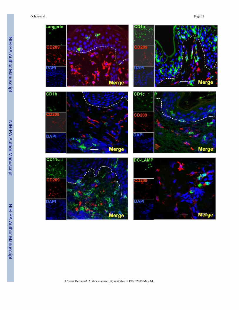
To determine the phenotype of CD209+ (DC-SIGN) cells in normal human skin tissue, frozentissue sections were labelled with antibodies to CD209 (DC-SIGN) and paired with thedifferent DC and macrophage markers. CD209+ (DC-SIGN) cells did not express CD207+
(Langerin), CD1a, CD1b, CD1c, CD11c, CD83 and CD208 (DC-LAMP). These data indicatethat CD209+ (DC-SIGN) expression is not restricted to DCs in the skin (Fig. 5a). In addition
Ochoa et al. Page 2
J Invest Dermatol. Author manuscript; available in PMC 2009 May 14.
NIH
-PA Author Manuscript
NIH
-PA Author Manuscript
NIH
-PA Author Manuscript
CD1b+ cells did not express macrophage markers (CD163, CD68 and CD14) suggesting thatCD1 expression is restricted to DCs in the skin (Fig. 5b).
We reasoned that CD209+ (DC-SIGN) and other monocyte/macrophage markers mightcolocalize on cells in the dermis of normal human skin. Therefore the expression of CD209+
(DC-SIGN) and CD68, CD14, CD163 and CD206+ (macrophage mannose receptor or MMR)was examined in normal human skin tissue using immunofluorescence. A major population ofdendritic appearing cells was located at the level of the capillaries and in the higher part of thereticular dermis and co-expressed CD209+ (DC-SIGN), CD206+ (macrophage mannosereceptor or MMR) and the monocyte/macrophage markers CD14, CD163, CD68, but not DCmarkers (CD1a, CD1b, CD1c, CD11c, DC-LAMP, Langerin). (Fig 6).
Therefore, we conclude that CD209+ (DC-SIGN) macrophages, expressing CD68, CD14, andCD163, markers typically found on monocytes/macrophages, comprise a major subset ofdendritic-appearing dermal cells.
Isolated dermal cells from normal human skin contain CD209+CD11clo cellsTo substantiate the immunohistologic identification of distinct populations of macrophagesand DC, dermal cells were isolated from normal human skin and analyzed them directly byflow cytometry. Distinct populations of CD209+CD11clo and CD209-CD11chi+ cells wereidentified (Figure 7). These data were obtained by separating epidermis from dermis, thendispersing cells in the dermis using a scalpel, which resulted in a low yield of mononuclearcells. Treatment of the dermis with collagenase overnight as published (Zaba et al., 2007)resulted in loss of CD209 expression as did treatment of peripheral blood derived CD209+cells (data not shown). The loss of CD209 expression may be due to dedifferentiation ofmacrophages during the collagenase treatment or the proteolytic digestion of the CD209epitope. Therefore, additional technologic considerations will be required to obtain sufficientnumbers of CD209+ cells from normal skin for functional study
DiscussionThe resident skin immune system stands guard to deal with attacks from the outsideenvironment including microbes, allergens and ultraviolet light. The epidermis serves as theinitial barrier, however the resident immune cells in the dermis must serve to deal with hostthreats that either escape detection by the epidermis or enter via the cutaneous vasculature. Thedermal immune system is known to involve circulating T cells that survey the skin for foreignantigens, but also a population of “dermal dendritic cells”. Here we provide evidence that“dermal dendritic cells” comprise at least two major phenotypic populations of dendriticappearing cells, immature DC that express CD1, CD11c and CD208, and macrophages thatexpress CD209, CD206, CD163 and CD68. We conclude that dermal dendritic-appearingmacrophages are a novel component of the skin immune system.
DDCs were initially identified and characterized using a polyclonal antibody that recognizesclotting factor XIIIa. This antibody was used to identify spindle or stellate-shaped cells inhuman connective tissue (Fear et al., 1984). Later on, Reid et al. called them fibroblasts (Reidet al., 1986). But it was Headington who in1986 named them “Dermal Dendrocytes” –oruncommitted stem cells – using a polyclonal antibody for FXIIIa (Cerio et al., 1989;Headington, 1986). Unfortunately, Factor XIIIa is not a specific marker, as it can also beexpressed on several cell types, including endothelial cells (Suvarna and Cotton, 1993).However, in 1987, using macrophage markers such as RFD7, RFDR2 (HLA-DR), KiM7(CD68) (Adany and Muszbek, 1989; Adany and Muszbek, 1987) suggested that these cellswere tissue macrophages. Functionally, DDCs demonstrate features of both macrophages anddendritic cells. A key macrophage function of DDCs is their ability to efficiently phagocytose
Ochoa et al. Page 3
J Invest Dermatol. Author manuscript; available in PMC 2009 May 14.
NIH
-PA Author Manuscript
NIH
-PA Author Manuscript
NIH
-PA Author Manuscript
hemosiderin, melanin and bacteria (Altman et al., 1992; Filgueira et al., 1996; Nestle et al.,1998). A key dendritic cell function of DDCs is their ability to take up and process antigens,migrate to lymphatic organs and activate naive T cells (Nestle et al., 1993b; Nestle andNickoloff, 1995). Our data indicate that in fact resident DDCs in normal skin are comprisedof at least two phenotypically distinct populations of CD209+ macrophages and CD1+ DCs.
The CD209+ dermal dendritic-appearing macrophage population is also present in inflamedskin, as we reported in leprosy lesions (Krutzik et al., 2005). The phenotype of these dermaldendritic-appearing macrophages includes several markers of phagocytic function, CD209(DC-SIGN), CD206 (macrophage mannose receptor) and CD163 (Figure 5). Both CD209 andCD206 are pattern recognition receptors; CD209 (DC-SIGN) is a C-type lectin that bindsvarious glycoproteins and glycolipids (Ebner et al., 2004; Geijtenbeek et al., 2000). CD209has been shown to facilitate the uptake of bacteria including Mycobacterium tuberculosis andviruses including HIV (Pohlmann et al., 2001). CD206 (macrophage mannose receptor) isknown to facilitate uptake of microbial molecules containing a repeating mannose motif(Sallusto et al., 1995; Wollenberg et al., 2002). CD163 is the haptoglobin receptor, involvedin the transport of iron into cells (Kristiansen et al., 2001). CD206 (mannose receptor) andCD11c (myeloid marker) can be expressed in the two populations –dendritic cells andmacrophages- as we previously reported (Krutzik et al., 2005). Together, the co-expression ofthese molecules indicates that dermal dendritic-appearing macrophages are resident in normalskin, and are likely to recognize microbial pathogens via multiple pattern recognition receptorsand phagocytize them. The present study points the way to devise strategies to isolate thesecells from normal skin and study their function in vitro.
The function of dermal CD1+ DC, in terms of antigen presentation activity has beendemonstrated in earlier studies (Meunier et al., 1993; Nestle et al., 1993a; Nestle et al.,1993b; Turville et al., 2002). It has also been suggested that human Langerhans cells arereplenished by precursors in the dermis and that CD14+ dermal antigen presenting cells arethe main precursor population (Larregina et al., 2001). Further examination of leprosy skinlesions using confocal laser microscopy revealed that the distribution and frequency of theseresident skin populations may correlate with disease status (Krutzik et al., 2005). Within self-limited tuberculoid leprosy lesions, CD1a+ cells were frequent in the epidermis, and CD209(DC-SIGN) and CD1b are expressed on distinct, non-overlapping dermal cell populations. Incontrast, within the progressive lepromatous leprosy lesions, the frequency of CD1a+
Langerhans cells and CD1b+ dermal dendritic cells was fewer. The CD209+ cells in tuberculoidleprosy lesions were found to express the monocyte/macrophage markers CD14, CD16, CD64and CD68 but did not express the dendritic cells markers CD1a, CD1b, CD63, CD83, or DC-LAMP. These data indicate that the majority of CD209+ cells express cell surface markerstypically found on monocytes/macrophages, and reflect the phenotype found here in normalskin. Future studies should be directed towards identifying the phenotype of CD209+ cells inother disease states.
The studies by Zaba et al (Zaba et al., 2007) support the presence of distinct populations ofmacrophages and dendritic cells in normal human dermis, but differ in identifying the specificphenotypic of the dermal macrophages. Our study clearly identifies a CD209+CD163+macrophage population in normal skin that do not express characteristic DC markers CD1a,CD1b, CD1c and DC-LAMP, in addition to CD11c. The restricted expression of CD209 is ofparticular interest because it mediates the phagocytosis of a range of microbial pathogens. Zabaet al. demonstrated CD209 expression on both macrophage and dendritic cell populations, bututilized fluorescence microscopy for their analysis. Our studies used confocal microscopy,which has the capability to analyze thin optical sections that helps differentiate epitopecolocalization from cell overlap.
Ochoa et al. Page 4
J Invest Dermatol. Author manuscript; available in PMC 2009 May 14.
NIH
-PA Author Manuscript
NIH
-PA Author Manuscript
NIH
-PA Author Manuscript
The mechanism by which these dermal DC and macrophages differentiate from monocyteprecursors is fundamental to understanding the resident skin immune system. We previouslydemonstrated that human peripheral monocytes activated with a Toll-like receptor 2/1 ligandwere differentiated into two distinct subsets: CD209+ CD16+ macrophages and CD1b+
CD209− dendritic cells. CD209+ cells were expanded by TLR-mediated upregulation ofinterleukin (IL)-15, had a macrophage-like phenotype, were phagocytic, and used CD209+ tofacilitate the uptake of bacteria. In contrast, CD1b+ DC were expanded by TLR-mediatedupregulation of granulocyte-macrophage colony-stimulating factor (GM-CSF), promoted Tcell activation and secreted pro-inflammatory cytokines. The mechanisms by which thesedermal DC and dendritic appearing macrophages accumulate in normal skin and diseaselesions, as well as their functional role in disease pathogenesis should provide new therapeutictargets for skin disease.
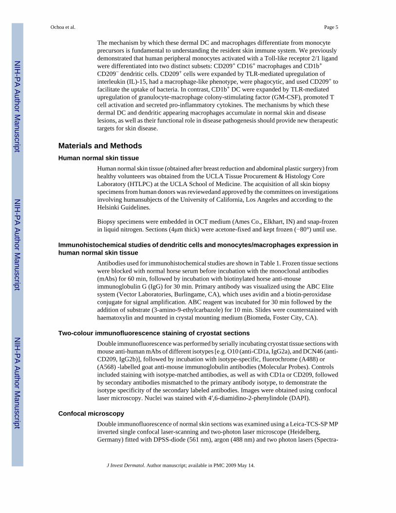
Materials and MethodsHuman normal skin tissue
Human normal skin tissue (obtained after breast reduction and abdominal plastic surgery) fromhealthy volunteers was obtained from the UCLA Tissue Procurement & Histology CoreLaboratory (HTLPC) at the UCLA School of Medicine. The acquisition of all skin biopsyspecimens from human donors was reviewedand approved by the committees on investigationsinvolving humansubjects of the University of California, Los Angeles and according to theHelsinki Guidelines.
Biopsy specimens were embedded in OCT medium (Ames Co., Elkhart, IN) and snap-frozenin liquid nitrogen. Sections (4μm thick) were acetone-fixed and kept frozen (−80°) until use.
Immunohistochemical studies of dendritic cells and monocytes/macrophages expression inhuman normal skin tissue
Antibodies used for immunohistochemical studies are shown in Table 1. Frozen tissue sectionswere blocked with normal horse serum before incubation with the monoclonal antibodies(mAbs) for 60 min, followed by incubation with biotinylated horse anti-mouseimmunoglobulin G (IgG) for 30 min. Primary antibody was visualized using the ABC Elitesystem (Vector Laboratories, Burlingame, CA), which uses avidin and a biotin-peroxidaseconjugate for signal amplification. ABC reagent was incubated for 30 min followed by theaddition of substrate (3-amino-9-ethylcarbazole) for 10 min. Slides were counterstained withhaematoxylin and mounted in crystal mounting medium (Biomeda, Foster City, CA).
Two-colour immunofluorescence staining of cryostat sectionsDouble immunofluorescence was performed by serially incubating cryostat tissue sections withmouse anti-human mAbs of different isotypes [e.g. O10 (anti-CD1a, IgG2a), and DCN46 (anti-CD209, IgG2b)], followed by incubation with isotype-specific, fluorochrome (A488) or(A568) -labelled goat anti-mouse immunoglobulin antibodies (Molecular Probes). Controlsincluded staining with isotype-matched antibodies, as well as with CD1a or CD209, followedby secondary antibodies mismatched to the primary antibody isotype, to demonstrate theisotype specificity of the secondary labeled antibodies. Images were obtained using confocallaser microscopy. Nuclei was stained with 4′,6-diamidino-2-phenylindole (DAPI).
Confocal microscopyDouble immunofluorescence of normal skin sections was examined using a Leica-TCS-SP MPinverted single confocal laser-scanning and two-photon laser microscope (Heidelberg,Germany) fitted with DPSS-diode (561 nm), argon (488 nm) and two photon lasers (Spectra-
Ochoa et al. Page 5
J Invest Dermatol. Author manuscript; available in PMC 2009 May 14.
NIH
-PA Author Manuscript
NIH
-PA Author Manuscript
NIH
-PA Author Manuscript
Physics Millenia X 532 diode pump laser and Tsunami picosecond Ti-sapphire laser), at theCarol Moss Spivak Cell Imaging Facility in the UCLA Brain Research Institute. Sections wereilluminated with 488 and 561 nm for red and green labels and Ti-sapphire laser set at 770 nmfor DAPI excitation. Images of specimens with Alexa 488, or Alexa 568, (Molecular Probes)or DAPI were recorded sequentially through the spectral emission filters set from 500–550 nmfor Alexa 488, from 580–700 nm for Alexa 568 and from 410–490 nm for DAPI. Pairs of singleimages were superimposed for co-localization analysis. All micrographs are compiled serialZ-stack images 1 μm apart.
Isolation of Dermal cells—Human normal skin was placed in cold RPMI and minced usingtwo scalpels until single cells were obtained. Non-specific staining was blocked by incubatingwith non-specific mouse IgG for 10 minutes in FACS buffer (PBS 0.1% sodium azide and 2%FBS). Cells were then incubated with fluorescently-labeled primary antibodies (CD209-FITCand CD11c-PE) (BD Biosciences). Cells were washed two times before fixing in 1%paraformaldehyde. Cells were then acquired using a FACSCalibur (BD Biosciences) in theUCLA Flow Cytometry Core Facility. Data analysis was done using FlowJo software(Treestar).
Supplementary MaterialRefer to Web version on PubMed Central for supplementary material.
AcknowledgementsWe thank Dr. Matthew Schibler and the Carol Moss Spivak Cell Imaging Facility in the UCLA Brain Research Institutefor the use of the confocal laser microscope, Ping Fu, Christopher Creencia, and Delia Adefuin at the UCLA TissueProcurement & Histology Core Laboratory for providing normal skin and preparation of tissue sections, Dr. JennyKim, Jenny Phan and Dr. Peter Sieling for helpful technical assistance. We acknowledge the financial support receivedfrom the National Institutes of Health (AI 22553, AI 056489, AI 47868) to R. L. M
Reference ListAdany R, Belkin A, Vasilevskaya T, Muszbek L. Identification of blood coagulation factor XIII in human
peritoneal macrophages. Eur J Cell Biol 1985;38:171–173. [PubMed: 3896808]Adany R, Glukhova MA, Kabakov AY, Muszbek L. Characterisation of connective tissue cells containing
factor XIII subunit a. J Clin Pathol 1988;41:49–56. [PubMed: 2893813]Adany R, Muszbek L. Immunohistochemical detection of factor XIII subunit a in histiocytes of human
uterus. Histochemistry 1989;91:169–174. [PubMed: 2737926]Adany R, Muszbek L. Cells containing factor XIII subunit a in benign and soft tissue tumours.
Histopathology 1987;11:1341–1343. [PubMed: 3326817]Altman DA, Fivenson DP, Lee MW. Minocycline hyperpigmentation: model for in situ phagocytic
activity of factor XIIIa positive dermal dendrocytes. J Cutan Pathol 1992;19:340–345. [PubMed:1358929]
Behar SM, Porcelli SA, Beckman EM, Brenner MB. A pathway of costimulation that prevents anergy inCD28- T cells: B7-independent costimulation of CD1-restricted T cells. J Exp Med 1995;182:2007–2018. [PubMed: 7500046]
Cerio R, Griffiths CE, Cooper KD, Nickoloff BJ, Headington JT. Characterization of factor XIIIa positivedermal dendritic cells in normal and inflamed skin. Br J Dermatol 1989;121:421–431. [PubMed:2576222]
Cerio R, Spaull J, Jones EW. Identification of factor XIIIa in cutaneous tissue. Histopathology1988;13:362–364. [PubMed: 2903846]
Dupasquier M, Stoitzner P, van OA, Romani N, Leenen PJ. Macrophages and dendritic cells constitutea major subpopulation of cells in the mouse dermis. J Invest Dermatol 2004;123:876–879. [PubMed:15482474]
Ochoa et al. Page 6
J Invest Dermatol. Author manuscript; available in PMC 2009 May 14.
NIH
-PA Author Manuscript
NIH
-PA Author Manuscript
NIH
-PA Author Manuscript
Duraiswamy N, Tse Y, Hammerberg C, Kang S, Cooper KD. Distinction of class II MHC+ Langerhanscell-like interstitial dendritic antigen-presenting cells in murine dermis from dermal macrophages. JInvest Dermatol 1994;103:678–683. [PubMed: 7963656]
Ebner S, Ehammer Z, Holzmann S, Schwingshackl P, Forstner M, Stoitzner P, Huemer GM, Fritsch P,Romani N. Expression of C-type lectin receptors by subsets of dendritic cells in human skin. IntImmunol 2004;16:877–887. [PubMed: 15113774]
Engering A, Geijtenbeek TB, Van Vliet SJ, Wijers M, van Liempt E, Demaurex N, Lanzavecchia A,Fransen J, Figdor CG, Piguet V, Van Kooyk Y. The dendritic cell-specific adhesion receptor DC-SIGN internalizes antigen for presentation to T cells. J Immunol 2002;168:2118–2126. [PubMed:11859097]
Fear JD, Jackson P, Gray C, Miloszewski KJ, Losowsky MS. Localisation of factor XIII in human tissuesusing an immunoperoxidase technique. J Clin Pathol 1984;37:560–563. [PubMed: 6373832]
Filgueira L, Nestle FO, Rittig M, Joller HI, Groscurth P. Human dendritic cells phagocytose and processBorrelia burgdorferi. J Immunol 1996;157:2998–3005. [PubMed: 8816408]
Geijtenbeek TB, Torensma R, Van Vliet SJ, van Duijnhoven GC, Adema GJ, Van Kooyk Y, Figdor CG.Identification of DC-SIGN, a novel dendritic cell-specific ICAM-3 receptor that supports primaryimmune responses. Cell 2000;100:575–585. [PubMed: 10721994]
Headington JT. The dermal dendrocyte. Adv Dermatol 1986;1:159–171. [PubMed: 3079241]Kristiansen M, Graversen JH, Jacobsen C, Sonne O, Hoffman HJ, Law SK, Moestrup SK. Identification
of the haemoglobin scavenger receptor. Nature 2001;409:198–201. [PubMed: 11196644]Krutzik SR, Tan B, Li H, Ochoa MT, Liu PT, Sharfstein SE, Graeber TG, Sieling PA, Liu YJ, Rea TH,
Bloom BR, Modlin RL. TLR activation triggers the rapid differentiation of monocytes intomacrophages and dendritic cells. Nat Med 2005;11:653–660. [PubMed: 15880118]
Larregina AT, Morelli AE, Spencer LA, Logar AJ, Watkins SC, Thomson AW, Falo LD Jr. Dermal-resident CD14+ cells differentiate into Langerhans cells. Nat Immunol 2001;2:1151–1158. [PubMed:11702065]
Melian A, Geng YJ, Sukhova GK, Libby P, Porcelli SA. CD1 expression in human atherosclerosis. Apotential mechanism for T cell activation by foam cells. Am J Pathol 1999;155:775–786. [PubMed:10487835]
Meunier L, Gonzalez-Ramos A, Cooper KD. Heterogeneous populations of class II MHC+ cells in humandermal cell suspensions. Identification of a small subset responsible for potent dermal antigen-presenting cell activity with features analogous to Langerhans cells. J Immunol 1993;151:4067–4080.[PubMed: 8409386]
Muszbek L, Adany R, Szegedi G, Polgar J, Kavai M. Factor XIII of blood coagulation in humanmonocytes. Thromb Res 1985;37:401–410. [PubMed: 2859670]
Nestle FO, Filgueira L, Nickoloff BJ, Burg G. Human dermal dendritic cells process and present solubleprotein antigens. J Invest Dermatol 1998;110:762–766. [PubMed: 9579542]
Nestle FO, Nickoloff BJ. A fresh morphological and functional look at dermal dendritic cells. J CutanPathol 1995;22:385–393. [PubMed: 8594070]
Nestle FO, Zheng XG, Thompson CB, Turka LA, Nickoloff BJ. Characterization of dermal dendriticcells obtained from normal human skin reveals phenotypic and functionally distinctive subsets. JImmunol 1993b;151:6535–6545. [PubMed: 7504023]
Nestle FO, Zheng XG, Thompson CB, Turka LA, Nickoloff BJ. Characterization of dermal dendriticcells obtained from normal human skin reveals phenotypic and functionally distinctive subsets. JImmunol 1993a;151:6535–6545. [PubMed: 7504023]
Pohlmann S, Baribaud F, Lee B, Leslie GJ, Sanchez MD, Hiebenthal-Millow K, Munch J, Kirchhoff F,Doms RW. DC-SIGN interactions with human immunodeficiency virus type 1 and 2 and simianimmunodeficiency virus. J Virol 2001;75:4664–4672. [PubMed: 11312337]
Reid MB, Gray C, Fear JD, Bird CC. Immunohistological demonstration of factors XIIIa and XIIIs inreactive and neoplastic fibroblastic and fibro-histiocytic lesions. Histopathology 1986;10:1171–1178. [PubMed: 3542785]
Sallusto F, Cella M, Danieli C, Lanzavecchia A. Dendritic cells use macropinocytosis and the mannosereceptor to concentrate macromolecules in the major histocompatibility complex class II
Ochoa et al. Page 7
J Invest Dermatol. Author manuscript; available in PMC 2009 May 14.
NIH
-PA Author Manuscript
NIH
-PA Author Manuscript
NIH
-PA Author Manuscript

compartment: downregulation by cytokines and bacterial products. J Exp Med 1995;182:389–400.[PubMed: 7629501]
Suvarna SK, Cotton DW. Dermal dendrocytes and other factor XIIIa-positive cells. J Pathol1993;171:251–252. [PubMed: 7908965]
Tse Y, Cooper KD. Cutaneous dermal Ia+ cells are capable of initiating delayed type hypersensitivityresponses. J Invest Dermatol 1990;94:267–272. [PubMed: 2407786]
Turville SG, Cameron PU, Handley A, Lin G, Pohlmann S, Doms RW, Cunningham AL. Diversity ofreceptors binding HIV on dendritic cell subsets. Nat Immunol 2002;3:975–983. [PubMed: 12352970]
Wollenberg A, Mommaas M, Oppel T, Schottdorf EM, Gunther S, Moderer M. Expression and functionof the mannose receptor CD206 on epidermal dendritic cells in inflammatory skin diseases. J InvestDermatol 2002;118:327–334. [PubMed: 11841552]
Zaba LC, Fuentes-Duculan J, Steinman RM, Krueger JG, Lowes MA. Normal human dermis containsdistinct populations of CD11c+BDCA-1+ dendritic cells and CD163+FXIIIA+ macrophages. J ClinInvest 2007;117:2517–2525. [PubMed: 17786242]
Ochoa et al. Page 8
J Invest Dermatol. Author manuscript; available in PMC 2009 May 14.
NIH
-PA Author Manuscript
NIH
-PA Author Manuscript
NIH
-PA Author Manuscript
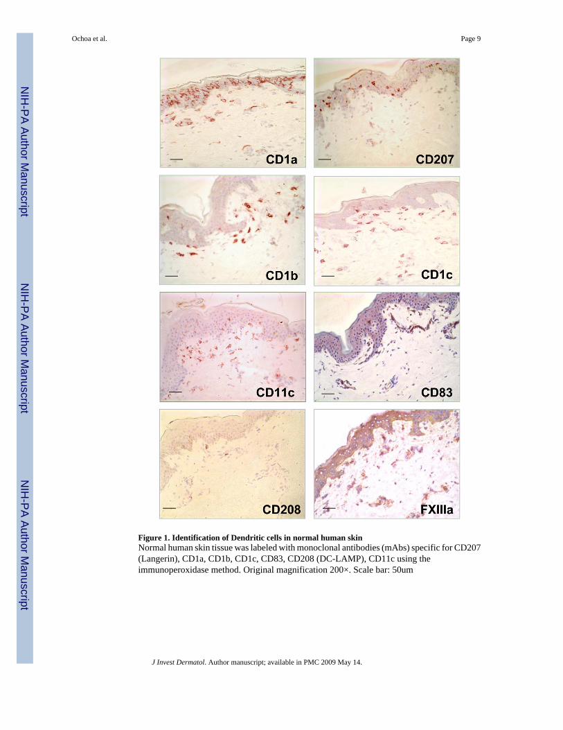
Figure 1. Identification of Dendritic cells in normal human skinNormal human skin tissue was labeled with monoclonal antibodies (mAbs) specific for CD207(Langerin), CD1a, CD1b, CD1c, CD83, CD208 (DC-LAMP), CD11c using theimmunoperoxidase method. Original magnification 200×. Scale bar: 50um
Ochoa et al. Page 9
J Invest Dermatol. Author manuscript; available in PMC 2009 May 14.
NIH
-PA Author Manuscript
NIH
-PA Author Manuscript
NIH
-PA Author Manuscript
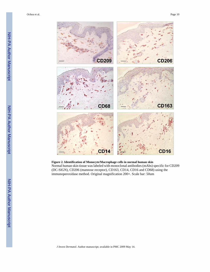
Figure 2. Identification of Monocyte/Macrophage cells in normal human skinNormal human skin tissue was labeled with monoclonal antibodies (mAbs) specific for CD209(DC-SIGN), CD206 (mannose receptor), CD163, CD14, CD16 and CD68) using theimmunoperoxidase method. Original magnification 200×. Scale bar: 50um
Ochoa et al. Page 10
J Invest Dermatol. Author manuscript; available in PMC 2009 May 14.
NIH
-PA Author Manuscript
NIH
-PA Author Manuscript
NIH
-PA Author Manuscript
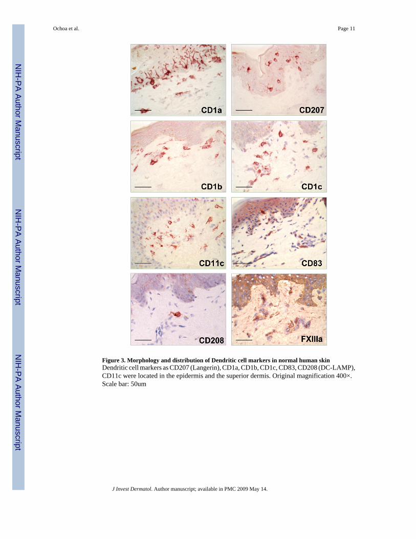
Figure 3. Morphology and distribution of Dendritic cell markers in normal human skinDendritic cell markers as CD207 (Langerin), CD1a, CD1b, CD1c, CD83, CD208 (DC-LAMP),CD11c were located in the epidermis and the superior dermis. Original magnification 400×.Scale bar: 50um
Ochoa et al. Page 11
J Invest Dermatol. Author manuscript; available in PMC 2009 May 14.
NIH
-PA Author Manuscript
NIH
-PA Author Manuscript
NIH
-PA Author Manuscript
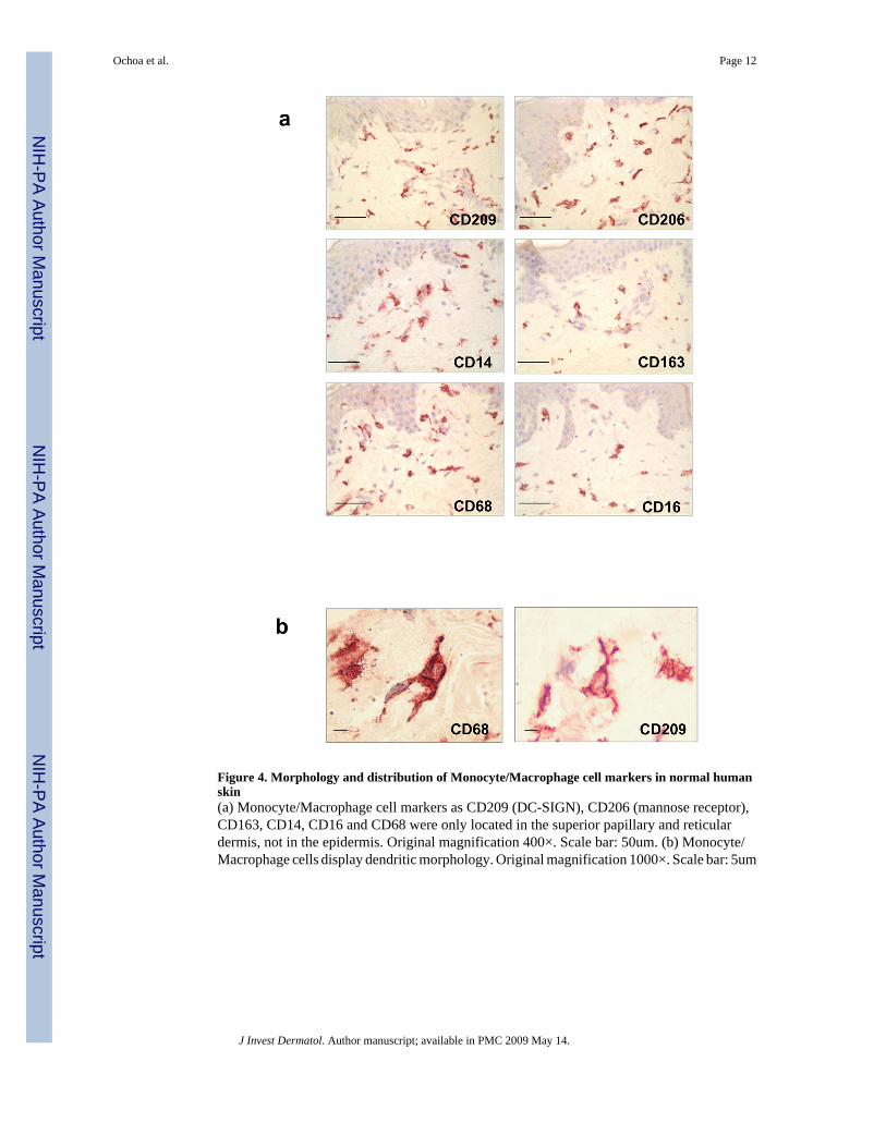
Figure 4. Morphology and distribution of Monocyte/Macrophage cell markers in normal humanskin(a) Monocyte/Macrophage cell markers as CD209 (DC-SIGN), CD206 (mannose receptor),CD163, CD14, CD16 and CD68 were only located in the superior papillary and reticulardermis, not in the epidermis. Original magnification 400×. Scale bar: 50um. (b) Monocyte/Macrophage cells display dendritic morphology. Original magnification 1000×. Scale bar: 5um
Ochoa et al. Page 12
J Invest Dermatol. Author manuscript; available in PMC 2009 May 14.
NIH
-PA Author Manuscript
NIH
-PA Author Manuscript
NIH
-PA Author Manuscript
Ochoa et al. Page 13
J Invest Dermatol. Author manuscript; available in PMC 2009 May 14.
NIH
-PA Author Manuscript
NIH
-PA Author Manuscript
NIH
-PA Author Manuscript
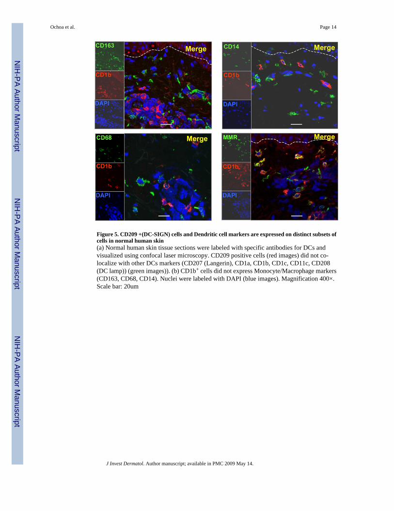
Figure 5. CD209 +(DC-SIGN) cells and Dendritic cell markers are expressed on distinct subsets ofcells in normal human skin(a) Normal human skin tissue sections were labeled with specific antibodies for DCs andvisualized using confocal laser microscopy. CD209 positive cells (red images) did not co-localize with other DCs markers (CD207 (Langerin), CD1a, CD1b, CD1c, CD11c, CD208(DC lamp)) (green images)). (b) CD1b+ cells did not express Monocyte/Macrophage markers(CD163, CD68, CD14). Nuclei were labeled with DAPI (blue images). Magnification 400×.Scale bar: 20um
Ochoa et al. Page 14
J Invest Dermatol. Author manuscript; available in PMC 2009 May 14.
NIH
-PA Author Manuscript
NIH
-PA Author Manuscript
NIH
-PA Author Manuscript
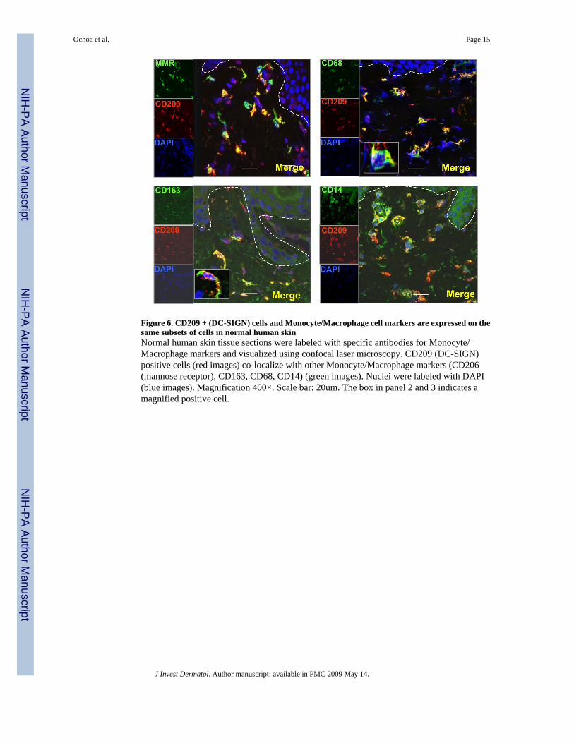
Figure 6. CD209 + (DC-SIGN) cells and Monocyte/Macrophage cell markers are expressed on thesame subsets of cells in normal human skinNormal human skin tissue sections were labeled with specific antibodies for Monocyte/Macrophage markers and visualized using confocal laser microscopy. CD209 (DC-SIGN)positive cells (red images) co-localize with other Monocyte/Macrophage markers (CD206(mannose receptor), CD163, CD68, CD14) (green images). Nuclei were labeled with DAPI(blue images). Magnification 400×. Scale bar: 20um. The box in panel 2 and 3 indicates amagnified positive cell.
Ochoa et al. Page 15
J Invest Dermatol. Author manuscript; available in PMC 2009 May 14.
NIH
-PA Author Manuscript
NIH
-PA Author Manuscript
NIH
-PA Author Manuscript
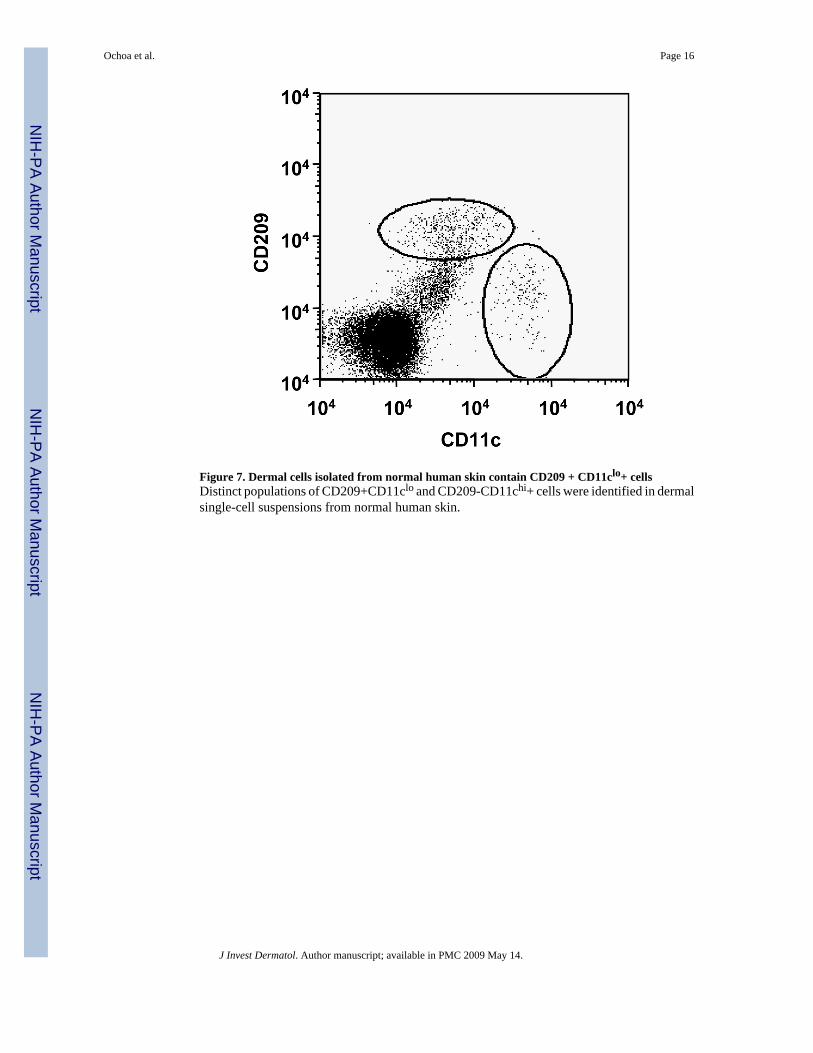
Figure 7. Dermal cells isolated from normal human skin contain CD209 + CD11clo+ cellsDistinct populations of CD209+CD11clo and CD209-CD11chi+ cells were identified in dermalsingle-cell suspensions from normal human skin.
Ochoa et al. Page 16
J Invest Dermatol. Author manuscript; available in PMC 2009 May 14.
NIH
-PA Author Manuscript
NIH
-PA Author Manuscript
NIH
-PA Author Manuscript
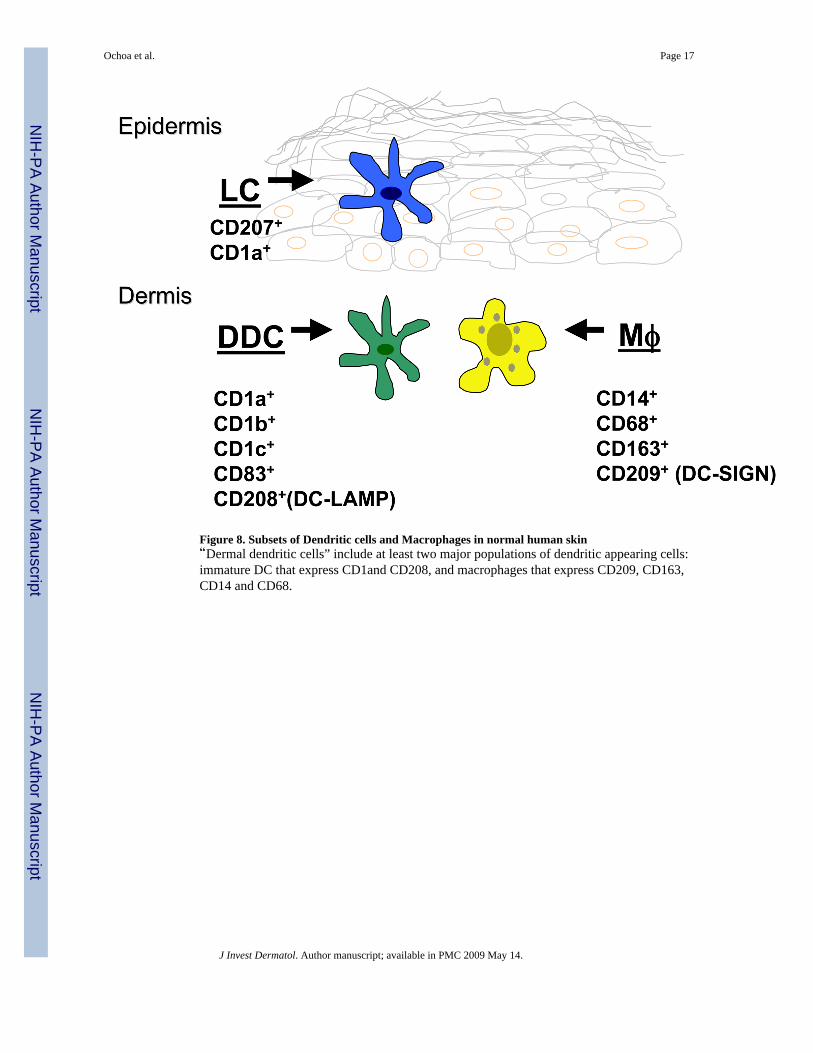
Figure 8. Subsets of Dendritic cells and Macrophages in normal human skin“Dermal dendritic cells” include at least two major populations of dendritic appearing cells:immature DC that express CD1and CD208, and macrophages that express CD209, CD163,CD14 and CD68.
Ochoa et al. Page 17
J Invest Dermatol. Author manuscript; available in PMC 2009 May 14.
NIH
-PA Author Manuscript
NIH
-PA Author Manuscript
NIH
-PA Author Manuscript
Related Documents