Aryl Hydrocarbon Receptor-Dependent Retention of Nuclear HuR Suppresses Cigarette Smoke-Induced Cyclooxygenase-2 Expression Independent of DNA- Binding Michela Zago 1 , Jared A. Sheridan 1 , Parameswaran Nair 4 , Angela Rico de Souza 2 , Imed-Eddine Gallouzi 3 , Simon Rousseau 1,2 , Sergio Di Marco 3 , Qutayba Hamid 1,2 , David H. Eidelman 1,2 , Carolyn J. Baglole 1,2 * 1 Department of Medicine, McGill University, Montreal, Quebec, Canada, 2 Research Institute of the McGill University Health Centre, Montreal, Quebec, Canada, 3 Department of Biochemistry and the Goodman Cancer Centre, McGill University, Montreal, Quebec, Canada, 4 Department of Medicine, McMaster University, Hamilton, Ontario, Canada Abstract The aryl hydrocarbon receptor (AhR), a ligand-activated transcription factor that responds to man-made environmental toxicants, has emerged as an endogenous regulator of cyclooxygenase-2 (Cox-2) by a mechanism that is poorly understood. In this study, we first used AhR-deficient (AhR 2/2 ) primary pulmonary cells, together with pharmacological tools to inhibit new RNA synthesis, to show that the AhR is a prominent factor in the destabilization of Cox-2 mRNA. The destabilization of Cox-2 mRNA and subsequent suppression of cigarette smoke-induced COX-2 protein expression by the AhR was independent of its ability to bind the dioxin response element (DRE), thereby differentiating the DRE-driven toxicological AhR pathway from its anti-inflammatory abilities. We further describe that the AhR destabilizes Cox-2 mRNA by sequestering HuR within the nucleus. The role of HuR in AhR stabilization of Cox-2 mRNA was confirmed by knockdown of HuR, which resulted in rapid Cox-2 mRNA degradation. Finally, in the lungs of AhR 2/2 mice exposed to cigarette smoke, there was little Cox-2 mRNA despite robust COX-2 protein expression, a finding that correlates with almost exclusive cytoplasmic HuR within the lungs of AhR 2/2 mice. Therefore, we propose that the AhR plays an important role in suppressing the expression of inflammatory proteins, a function that extends beyond the ability of the AhR to respond to man-made toxicants. These findings open the possibility that a DRE-independent AhR pathway may be exploited therapeutically as an anti- inflammatory target. Citation: Zago M, Sheridan JA, Nair P, Rico de Souza A, Gallouzi I-E, et al. (2013) Aryl Hydrocarbon Receptor-Dependent Retention of Nuclear HuR Suppresses Cigarette Smoke-Induced Cyclooxygenase-2 Expression Independent of DNA-Binding. PLoS ONE 8(9): e74953. doi:10.1371/journal.pone.0074953 Editor: Salik Hussain, National Institute of Health (NIH), United States of America Received May 30, 2013; Accepted August 7, 2013; Published September 27, 2013 Copyright: ß 2013 Zago et al. This is an open-access article distributed under the terms of the Creative Commons Attribution License, which permits unrestricted use, distribution, and reproduction in any medium, provided the original author and source are credited. Funding: This work was supported by the American Thoracic Society Research Grant; the Department of Medicine of McGill University; the Research Institute of the McGill University Health Centre; the Canada Foundation for Innovation-Leaders Opportunities Fund and the Natural Sciences and Engineering Research Council of Canada. CJB was supported by a salary award from the Fonds de recherche du Quebec-Sante (FRQ-S). MZ is the recipient of a Meakins-Christie Post- Doctoral Fellowship Award. Dr Nair is supported by a Canada Research Chair in Airway Inflammometry. The funders had no role in study design, data collection and analysis, decision to publish, or preparation of the manuscript. Competing Interests: Both I, (Carolyn J. Baglole) and Dr. Imed Gallouzi are Editorial Board Members. This does not alter the authors’ adherence to all the PLOS ONE policies on sharing data and materials. * E-mail: [email protected] Introduction Cigarette smoke is the leading cause of preventable death worldwide and is the primary risk factor for the top three mortalities: cardiovascular disease (CVD), cancer and respiratory disease, which includes chronic obstructive pulmonary disease (COPD). COPD affects some 200 million people worldwide [1] and is estimated to become the third leading cause of death within the next decade [2]. COPD is characterized by progressive airflow limitation that is not fully reversible and is associated with chronic inflammation. Cigarette smoke incites and perpetuates this inflammatory response by inducing pro-inflammatory mediator production (lipids, chemokines and cytokines). We recently identified that the aryl hydrocarbon receptor (AhR), a receptor/ transcription factor that is highly expressed in the human lung [3], is a novel and potent suppressor of cigarette smoke-induced inflammation [4,5]. The AhR is a member of the basic helix-loop- helix Per-Arnt-Sim (bHLH-PAS) transcription factor family that is well-known to respond to man-made xenobiotics such as 2,3,7,8- tetrachlorodibenzo-p-dioxin (TCDD; dioxin) and related planar aromatic hydrocarbons [6]. In the absence of ligand, the AhR is found in the cytoplasm complexed with chaperone proteins, including a dimer of heat shock protein 90 (HSP90) and the immunophilin hepatitis B virus X-associated protein 2 (XAP2) [7,8,9]. After ligand binding, the AhR translocates to the nucleus, dissociates from these chaperones and forms a heterodimer with the AhR nuclear transporter (ARNT). This AhR:ARNT complex then binds to a dioxin responsive element (DRE) and initiates the transcription of genes that comprise the AhR gene battery, including cytochrome P450 (CYP) enzymes. Numerous early-response genes encoding inflammatory medi- ators such as cyclooxygenase-2 (Cox-2) also contain a DRE in the PLOS ONE | www.plosone.org 1 September 2013 | Volume 8 | Issue 9 | e74953

Welcome message from author
This document is posted to help you gain knowledge. Please leave a comment to let me know what you think about it! Share it to your friends and learn new things together.
Transcript
Aryl Hydrocarbon Receptor-Dependent Retention ofNuclear HuR Suppresses Cigarette Smoke-InducedCyclooxygenase-2 Expression Independent of DNA-BindingMichela Zago1, Jared A. Sheridan1, Parameswaran Nair4, Angela Rico de Souza2, Imed-Eddine Gallouzi3,
Simon Rousseau1,2, Sergio Di Marco3, Qutayba Hamid1,2, David H. Eidelman1,2, Carolyn J. Baglole1,2*
1Department of Medicine, McGill University, Montreal, Quebec, Canada, 2 Research Institute of the McGill University Health Centre, Montreal, Quebec, Canada,
3Department of Biochemistry and the Goodman Cancer Centre, McGill University, Montreal, Quebec, Canada, 4Department of Medicine, McMaster University, Hamilton,
Ontario, Canada
Abstract
The aryl hydrocarbon receptor (AhR), a ligand-activated transcription factor that responds to man-made environmentaltoxicants, has emerged as an endogenous regulator of cyclooxygenase-2 (Cox-2) by a mechanism that is poorly understood.In this study, we first used AhR-deficient (AhR2/2) primary pulmonary cells, together with pharmacological tools to inhibitnew RNA synthesis, to show that the AhR is a prominent factor in the destabilization of Cox-2 mRNA. The destabilization ofCox-2 mRNA and subsequent suppression of cigarette smoke-induced COX-2 protein expression by the AhR wasindependent of its ability to bind the dioxin response element (DRE), thereby differentiating the DRE-driven toxicologicalAhR pathway from its anti-inflammatory abilities. We further describe that the AhR destabilizes Cox-2 mRNA by sequesteringHuR within the nucleus. The role of HuR in AhR stabilization of Cox-2 mRNA was confirmed by knockdown of HuR, whichresulted in rapid Cox-2 mRNA degradation. Finally, in the lungs of AhR2/2 mice exposed to cigarette smoke, there was littleCox-2 mRNA despite robust COX-2 protein expression, a finding that correlates with almost exclusive cytoplasmic HuRwithin the lungs of AhR2/2 mice. Therefore, we propose that the AhR plays an important role in suppressing the expressionof inflammatory proteins, a function that extends beyond the ability of the AhR to respond to man-made toxicants. Thesefindings open the possibility that a DRE-independent AhR pathway may be exploited therapeutically as an anti-inflammatory target.
Citation: Zago M, Sheridan JA, Nair P, Rico de Souza A, Gallouzi I-E, et al. (2013) Aryl Hydrocarbon Receptor-Dependent Retention of Nuclear HuR SuppressesCigarette Smoke-Induced Cyclooxygenase-2 Expression Independent of DNA-Binding. PLoS ONE 8(9): e74953. doi:10.1371/journal.pone.0074953
Editor: Salik Hussain, National Institute of Health (NIH), United States of America
Received May 30, 2013; Accepted August 7, 2013; Published September 27, 2013
Copyright: � 2013 Zago et al. This is an open-access article distributed under the terms of the Creative Commons Attribution License, which permitsunrestricted use, distribution, and reproduction in any medium, provided the original author and source are credited.
Funding: This work was supported by the American Thoracic Society Research Grant; the Department of Medicine of McGill University; the Research Institute ofthe McGill University Health Centre; the Canada Foundation for Innovation-Leaders Opportunities Fund and the Natural Sciences and Engineering ResearchCouncil of Canada. CJB was supported by a salary award from the Fonds de recherche du Quebec-Sante (FRQ-S). MZ is the recipient of a Meakins-Christie Post-Doctoral Fellowship Award. Dr Nair is supported by a Canada Research Chair in Airway Inflammometry. The funders had no role in study design, data collectionand analysis, decision to publish, or preparation of the manuscript.
Competing Interests: Both I, (Carolyn J. Baglole) and Dr. Imed Gallouzi are Editorial Board Members. This does not alter the authors’ adherence to all the PLOSONE policies on sharing data and materials.
* E-mail: [email protected]
Introduction
Cigarette smoke is the leading cause of preventable death
worldwide and is the primary risk factor for the top three
mortalities: cardiovascular disease (CVD), cancer and respiratory
disease, which includes chronic obstructive pulmonary disease
(COPD). COPD affects some 200 million people worldwide [1]
and is estimated to become the third leading cause of death within
the next decade [2]. COPD is characterized by progressive airflow
limitation that is not fully reversible and is associated with chronic
inflammation. Cigarette smoke incites and perpetuates this
inflammatory response by inducing pro-inflammatory mediator
production (lipids, chemokines and cytokines). We recently
identified that the aryl hydrocarbon receptor (AhR), a receptor/
transcription factor that is highly expressed in the human lung [3],
is a novel and potent suppressor of cigarette smoke-induced
inflammation [4,5]. The AhR is a member of the basic helix-loop-
helix Per-Arnt-Sim (bHLH-PAS) transcription factor family that is
well-known to respond to man-made xenobiotics such as 2,3,7,8-
tetrachlorodibenzo-p-dioxin (TCDD; dioxin) and related planar
aromatic hydrocarbons [6]. In the absence of ligand, the AhR is
found in the cytoplasm complexed with chaperone proteins,
including a dimer of heat shock protein 90 (HSP90) and the
immunophilin hepatitis B virus X-associated protein 2 (XAP2)
[7,8,9]. After ligand binding, the AhR translocates to the nucleus,
dissociates from these chaperones and forms a heterodimer with
the AhR nuclear transporter (ARNT). This AhR:ARNT complex
then binds to a dioxin responsive element (DRE) and initiates the
transcription of genes that comprise the AhR gene battery,
including cytochrome P450 (CYP) enzymes.
Numerous early-response genes encoding inflammatory medi-
ators such as cyclooxygenase-2 (Cox-2) also contain a DRE in the
PLOS ONE | www.plosone.org 1 September 2013 | Volume 8 | Issue 9 | e74953
promoter region and can be increased due to AhR activation by
dioxin [10,11,12]. COX-2 is an inducible enzyme that catalyzes
the transformation of arachidonic acid into thromboxanes and
prostaglandins (PG) such as PGE2. COX-2 is robustly increased by
cigarette smoke exposure [13] and is elevated in patients with
inflammation-associated diseases including COPD [14,15]. Al-
though cigarette smoke contains components capable of activating
the AhR, including benzo[a]pyrene (B[a]P) [16], our published
data demonstrate that expression of the AhR suppresses COX-2
protein expression and PG production due to cigarette smoke
exposure [4]. Interestingly, AhR expression was associated with a
rapid, significant but transient increase in Cox-2 mRNA upon
smoke exposure. Despite this increase in Cox-2 mRNA, there is
little COX-2 protein expression [4], suggesting that the AhR
suppress COX-2 protein by post-transcriptional regulatory mech-
anisms.
Post-transcriptional regulation of protein expression is an
adaptive mechanism that is crucial in regulating the timing and
the amount of inflammatory proteins. Although the Cox-2 gene is
transcriptionally-controlled, the level of COX-2 protein is
determined in large part by changes in the half-life of the mRNA.
Thus, there is often a poor correlation between Cox-2 mRNA and
protein levels because Cox-2 mRNA is rapidly degraded. The
instability of Cox-2 mRNA is due to the presence of adenylate- and
uridylate- rich element (ARE) in the 39-untranslated region (UTR)
[17], which can be bound by proteins that can alter Cox-2 mRNA
stability and translation [18]. RNA-binding proteins that interact
with the Cox-2 ARE include the CELF/Bruno-like family member
CUGBP2 [19] and the embryonic lethal abnormal vision (ELAV)-
like protein Human antigen R (HuR) [20]. HuR is a ubiquitous
RNA-binding protein that is abundantly localized to the nucleus,
where it is first interacts with Cox-2 mRNA. HuR subsequently
shuttles between the nucleus and cytoplasm upon stimulation. It is
believed that cytoplasmic localization is important for the mRNA-
stabilizing effects of HuR [21,22,23]. Whether the AhR regulates
Cox-2 mRNA stability by controlling HuR expression or localiza-
tion is not known.
Herein, we used lung cells devoid of AhR expression, together
with our established in vitro and in vivo models of cigarette smoke
exposure [4,5,24] and show that the AhR-dependent retention of
nuclear HuR is responsible for the destabilization of Cox-2 mRNA
by a mechanism that was independent of AhR:DNA binding
activity. Therefore, despite its dubious distinction as a transcrip-
tional regulator of toxicological outcomes, we propose that the
AhR plays an important role in the suppression of inflammation
that extends beyond its ability to respond to man-made toxicants.
Materials and Methods
ChemicalsAll chemicals were purchased from Sigma (St. Louis, MO)
unless otherwise indicated. Actinomycin D (ActD) was purchased
from Biomol (Plymouth Meeting, PA). Recombinant mouse IL-1bwas purchased from R&D Systems (Minneapolis, MN). CH-
223191 (1-Methyl-N-[2-methyl-4-[2-(2-methylphenyl) diazenyl]
phenyl-1H-pyrazole-5-carboxamide) was from Tocris Bioscience
(Minneapolis, MN).
Cell CultureMouse lung fibroblasts. Primary lung fibroblasts were
generated from AhR+/+, AhR heterozygous (AhR+/2) and AhR2/2
C57BL/6 mice (Jackson Laboratory, Bar Harbor, ME) [25] and
cultured under standard conditions [4,24]. Lung fibroblasts were
also generated from a novel lineage of mice harboring a mutant
AhR that is incapable of binding to DNA (referred to hereafter as
AhRDBD/DBD) [26], a kind gift of Dr. Chris Bradfield (University of
Wisconsin); lung fibroblasts from littermate heterozygotes
(AhRDBD/B6) are used as corresponding controls. Unless otherwise
indicated, all pulmonary fibroblasts were plated at a density of
10,000 cells/cm2 and most experiments were conducted using
fibroblasts generated from at two different AhR2/2 mice. Lung
fibroblasts from wild-type or heterozygous mice do not exhibit any
difference in the ability to be activated by AhR ligands and are
used interchangeably as AhR-expressing cells [4,24].
Human lung fibroblasts. Primary lung fibroblasts were
cultured and characterized as previously described [25] from lung
tissue derived from individuals undergoing lung resection surgery
for suspected lung cancer at McMaster University. Only tissue
from disease-free regions was used for the derivation of fibroblasts
and all subjects were reported never-smokers. This study was
approved by the Research Ethics Board of St Joseph’s Healthcare
Hamilton and all patients gave written informed consent. All
fibroblast strains were used at the earliest possible passage.
Hepa.2DLuc.3A4 (Hepa.2Dluc). Mouse hepatoma cells
stably transfected with the luciferase reporter plasmid p2DLuc,
which contains two copies of the DRED consensus sequence
[27,28] and is thus a direct measure of classic AhR activation.
Derivation of Hepa.2Dluc cells were previously described [27] and
were a kind gift of Dr. Tom Gasiewicz (University of Rochester).
Hepa.2Dluc cells were cultured in minimum essential media
(MEM) supplemented with 2 mM glutamine (Invitrogen, Carls-
bad, CA), 10% fetal bovine serum (FBS) (Hyclone Labs, Logan,
UT) and antibiotics/antimycotics (penicillin G, streptomycin and
amphotericin; Invitrogen, Carlsbad, CA).
Lung epithelial cells. MLE-12 cells, a distal bronchiolar and
alveolar epithelial cell line (ATCC, Manassas, VA) [29], were
cultured in HITES medium (50:50 DMEM: Ham’s F12) supple-
mented with 2% FBS, 2 mM L-Glutamine, 10 mM HEPES,
1:100 Insulin-Transferrin-Selenium supplement (Invitrogen) and
antibiotics/antimycotics.
In Vivo Cigarette Smoke ExposureAge- and gender-matched AhR2/2 or AhR+/2 littermate
controls were exposed to cigarette smoke as previously described
[5]. Briefly, research cigarettes (University of Kentucky, Lexing-
ton, KY) were smoked according to the Federal Trade Commis-
sion protocol (1 puff/minute/cigarette of 2 seconds duration and
35-ml volume). Control and AhR2/2 mice were exposed to
cigarette smoke for 5 days a week for 2 and 4 weeks (sub-chronic
exposures). Daily exposures were for one hour, twice daily at four-
hour intervals. Control mice were exposed to filtered air. As we
have previously published that there is no difference between wild-
type (AhR+/+) C57BL/6 and AhR+/2 mice [5], AhR+/2 mice are
used for the in vivo studies. All animal procedures were approved
by the McGill University Animal Care Committee (Protocol
Number: 5933) and were carried out in accordance with the
Canadian Council on Animal Care. Following exposure, mice
were anesthetized with Avertin (2,2,2-tribromoethanol, 250 mg/
kg i.p.; Sigma-Aldrich) and euthanized by exsanguination. The
lungs were immediately excised, the left lung inflated with OCT
and snap-frozen in liquid nitrogen. A portion of the right lung was
immediately placed in RNAlaterH (Qiagen, Toronto ON) or
frozen in liquid nitrogen for further protein analysis.
Preparation of Cigarette Smoke Extract (CSE)Research grade cigarettes (2R3F) with a filter were obtained
from the Kentucky Tobacco Research Council (Lexington, KT)
and CSE generated as previously described [4,30,31,32]. Briefly,
AhR Regulates Cox-2 via Nuclear Retention of HuR
PLOS ONE | www.plosone.org 2 September 2013 | Volume 8 | Issue 9 | e74953
CSE was prepared by bubbling smoke from two cigarettes into
20 ml of serum-free MEM, the pH adjusted to 7.4, sterile- filtered
with a 0.45-mm filter (25-mm Acrodisc; Pall Corp., Ann Arbor,
MI) and was used within 30 minutes of preparation. An optical
density of 0.65 (320 nm) was considered to represent 100% CSE
[4,24] which was diluted to the appropriate concentration in
serum-free MEM.
Western BlotFibroblasts were grown to sub-confluence and cultured in
serum-free MEM for 24 hours before being treated with CSE for
the indicated times. Total cellular protein was prepared using 1%
IGEPAL lysis buffer [31]; nuclear and cytoplasmic fractions were
prepared using a nuclear extract kit (Active Motif, Carlsbad, CA).
Five to ten mg of cellular proteins were fractionated on SDS-PAGE
gels, electroblotted onto PVDF membranes and antibodies against
AhR (1:5000; Enzo Life Sciences, NY, USA), COX-2 (1:1000,
Cayman Chemical, Michigan, USA), HuR (1:2000), CUGBP2,
CYP1A1, CYP1B1 (1:500, Santa Cruz, Santa Cruz, CA) and actin
(1:20,000; Millipore, MA, USA) were used to assess changes in
protein levels by enhanced chemiluminescence (ECL). Protein
bands were visualized using a gel documentation system (Alpha
Innotech, San Leandro, CA).
Analysis of Gene ExpressionTotal RNA was harvested and quantification was conducted on
a Nanodrop 1000 spectrophotometer (Thermo Fisher Scientific,
Wilmington, DE). Reverse transcription of total RNA was carried
out using iScript IITM Reverse Transcription Supermix (Bio-Rad
Laboratories, Mississauga, ON). Quantitative PCR was then
performed by addition of 1 ml cDNA and 0.5 mM primers with
SsoFastTM EvaGreenH (Bio-Rad). The primer sequences were:
Cox-2- TGCCTGGTCTGATGATGTATGCCA (f) and AG-
TAGTCGCACACTCTGTTGTGCT (r); Cyp1a1- CCTTAC-
CAAGTGCTAGGATACAGTCATAGA (f) and CAGTAAA-
GAAGAGAGACCAAGAGCTGAT (r); Cyp1b1-
AAAATGTAAAGACCAGAAGTC CTCCTACC (f) and
AGAAAGCCTCATCCAGGGCTATAAA (r) and b-actin- CTA-CAATGAGCTGCGTGTG (f) and
TGGGGTGTTGAAGGTCTC (r). PCR amplification was per-
formed using a CFX96 Real-Time PCR Detection System (Bio-
Rad). Melt curve analysis was performed to ensure that
nonspecific products were absent. The fluorescence detection
threshold was set above the non-template control background
within the linear phases of PCR amplifications and the cycle
threshold (Ct) of each reaction was detected. Gene expression was
analyzed using the DDCt method and results are presented as fold-
change normalized to housekeeping gene (b-actin).
Determination of Cox-2 mRNA StabilityAhR2/2 and AhR-expressing lung fibroblasts were cultured in 6-
well culture plates until near confluence and switched to serum-
free media for 24 hours. Then the fibroblasts were exposed to 1%
CSE for 3 hours followed by treatment with ActD (1 mg/ml), an
inhibitor of RNA synthesis [33], for 30 minutes or for 1 or 3 hours.
Total RNA was harvested and qPCR performed as described
above to determine the remaining levels of mRNA. In separate
experiments, AhRDBD/DBD and AhRDBD/B6 cells were also exposed
to ActD with and without CSE. To determine if inhibition of AhR
activity altered Cox-2 mRNA stability, AhRDBD/DBD and AhRDBD/B6
cells were treated with CH-223191 together with ActD and 1%
CSE and Cox-2 mRNA levels assessed. The concentration of ActD
used in these experiments did not affect cell viability (data not
shown). To verify inhibition of Cox-2 mRNA synthesis, in separate
experiments, AhR+/2 fibroblasts were pretreated with 1 mg/ml of
ActD followed by treatment with IL-1b (10 ng/ml) for 6 hours.
Reporter Gene AssayHepa.2Dluc cells were seeded in six-well plates (46105 cells/
well) and allowed to grow overnight. Cells were then pretreated
with vehicle (DMSO), 1 or 10 mM CH-223191 for 1 hour
following by 6 hour treatment with 1 mM B[a]P. After treatments,
cell lysates were collected and luciferase activity measured using
the Luciferase Assay System (Promega, Madison, WI) and read on
the Infinite M1000 microplate reader (Tecan, Mannedorf,
Switzerland).
ImmunofluorescencePrimary Cell Culture. Fibroblasts were seeded on 8-well
glass chamber slides at a density of 16104 cells/well and allowed
to adhere for 24 h. Following serum starvation for 24 h, the cells
were treated with media only, 1% CSE or B[a]P for 1, 4 or 24
hours to assess HuR and CUGBP2 localization or COX-2
expression. Following treatments, the cells were washed once with
PBS/Tween, permeabilized/fixed using 3% H2O2/methanol for
10 min, and blocked with Universal Blocking Solution (Dako, ON,
CA) for 30 minutes at room temperature. The antibodies against
HuR, CUGBP2 (Santa Cruz) and COX-2 (Cayman) were diluted
1:200 in PBS/bovine serum albumin (BSA) and incubated
overnight at 4uC. Levels of non-specific staining were assessed
by incubating cells under identical conditions using the isotype-
matched non-immune antibody (Santa Cruz) at the same
concentration or by omission of the primary antibody. In all
cases, the level of non-specific staining was negligible (data not
shown). Alexa Fluor-488 anti-mouse or anti-rabbit IgG antibody
was used for secondary binding (1:1000) and incubated for 1 hour
at room temperature. Slides were then mounted in ProLongHGold Anti Fade (Invitrogen), viewed on an Olympus BX51
fluorescent microscope (Olympus, Ontario, Canada) and photo-
graphed using a Retiga 2000R Camera with ImagePro Plus
software. Fluorescent images of nuclei are visualized by Hoechst
staining (1:2000, Molecular Probes). All photographs were taken at
the same time with identical image settings. For quantification,
positive and negative cells were counted in each picture taken and
recorded per randomly-selected field (minimum of five separate
fields per experiment). Cells were considered positive based on
fluorescence intensity within the cytoplasm. Positive cells were
compared to the total counted cells for each individual experiment
and expressed as a percentage of the total cells present.
Mouse lung tissue. OCT-embedded lung from air- or CS-
exposed mice were sectioned, fixed in 70% ethanol for 3 minutes
and permeabilized in 0.5% PBS/Tween20 for 10 at room
temperature (RT). Then, the sections were blocked with the
Universal Blocking Solution (Dako, ON, CA) for 30 minutes. For
detection of HuR and COX-2/vimentin, the lung sections were
incubated with goat anti-mouse HuR (1:300) or COX-2/vimentin
antibodies (1:200/1:100) (Cayman) for 1 hour at RT. After rinsing
with PBS/Tween, the sections were then incubated with Alexa
555-conjugated rabbit anti-mouse IgG (vimentin and HuR) and
Alexa-488-conjugated donkey anti-rabbit IgG (COX-2) (Molecu-
lar Probes Inc., ON, CA), diluted at 1:1000 in Dako antibody
diluent for 1 hour. The sections were then cover-slipped with
ProLongH Gold Anti Fade mounting medium (Invitrogen).
Fluorescent images for COX-2/vimentin were detected via a
fluorescence microscope (Olympus BX51TF) whereas HuR
localization was assessed by a Laser Scanning Microscope-LSM
78 (RI-MUHC Imaging Facility, McGill University, Montreal,
Canada).
AhR Regulates Cox-2 via Nuclear Retention of HuR
PLOS ONE | www.plosone.org 3 September 2013 | Volume 8 | Issue 9 | e74953
Enzyme ImmunoassayEquivalent numbers of AhRDBD/DBD and AhRDBD/B6 fibroblasts
were allowed to reach confluence and serum-starved for 24 hours
prior to stimulation with CSE for varying time-points. Controls
included incubation with serum-free MEM. The resulting amount
of PGE2 in the cell culture supernatant was determined via specific
enzyme immunoassay (EIA) as described previously [4,13].
siRNA Knockdown StudiesAhR+/+ and AhR2/2 lung fibroblasts were grown to approxi-
mately 60–80% confluence, after which the cells were transiently
transfected with 60 nM siRNA against HuR or with control
siRNA. Two different siRNA targeting HuR were utilized for
these experiments (siRNA-1: Santa Cruz, CA; siRNA-2: Dhar-
macon, ON). Separate controls for each HuR siRNA were also
used. Transfections were performed according to the manufac-
turer’s instructions for 24–48 hours. During the transfection
process, cells were pre-treated with 1% CSE for 3 hours followed
by 1 mg/ml ActD for 30 minutes or for 1 or 3 hours. Then, total
RNA was isolated as described above and qPCR performed for
Cox-2. Knockdown of HuR was confirmed by western blot
analysis.
Statistical AnalysisStatistical analysis was performed using JMPH8 (SAS Institute,
Cary, NC). An analysis of variance (ANOVA) with Tukey-Kramer
post-hoc test was used to assess differences between treatment
groups of more than two. Results are expressed as the mean 6
SEM. In all cases, a p value,0.05 is considered statistically
significant.
Results
Antagonism of the AhR by CH-223191 Promotes CSE-induced COX-2 Protein Expression in Primary LungFibroblastsOur published data show that CSE robustly increases COX-2
protein expression in AhR-deficient cells with no induction of Cox-
2 mRNA [4], supporting a post-transcriptional regulatory role for
the AhR in regulating COX-2 expression. Now, we first sought to
determine if AhR activation by CSE was necessary to suppresses
COX-2 expression. Utilizing cytochrome P450 (CYP) induction as
a well-defined marker of AhR activation [34], we show there was a
significant increase in Cyp1a1 mRNA in AhR+/+ cells by CSE,
similar to that induced by the classic AhR ligand B[a]P (Figure 1A).
Consistent with previous reports [35,36], there was no CYP1A1
protein in lung fibroblasts (Figure 1B). Cyp1b1 expression, the
predominant CYP isoform expressed by fibroblasts [24,35,36],
was increased by CSE and B[a]P in AhR+/+ cells (Figure 1C).
CYP1B1 protein increased only in B[a]P-exposed control cells but
not those exposed to CSE (Figure 1D). Note the increase in COX-
2 protein expression is only in the AhR2/2 fibroblasts exposed to
CSE, consistent with our published data [4].
To further evaluate if the ability of the AhR to suppress COX-2
protein induction by CSE requires AhR activation, we used the
pharmacological AhR antagonist CH-223191, which binds to the
AhR and prevents ligand-induced AhR translocation to the
nucleus and subsequent DRE-mediated transcription. Using
Hepa.2DLuc cells, a mouse hepatoma cell line stably transfected
with the AhR reporter plasmid p2Dluc [27,28], we determined
that there was no significant change in DRE-mediated transcrip-
tion (Figure 2A), indicating that CH-223191 does not exhibit any
agonist activity [37]. Furthermore, CH-223191 dose-dependently
antagonized B[a]P-induced AhR activation (Figure 2A). Exposure
of AhR+/2 cells to B[a]P increased CYP1B1 protein expression,
which was significantly reduced by CH-223191 (Figure 2B). As
CH-223191 may exhibit ligand-selective antagonism of the AhR
[38], we confirmed that the significant increase in Cyp1A1 mRNA
in response to CSE was also significantly attenuated by CH-
223191 (Figure 2C).
We then evaluated if inhibition of AhR activity by CH-223191
would affect COX-2 protein expression. Following exposure of
AhR-expressing lung fibroblasts to 1% CSE for 24 hours, there
was no increase in the expression of COX-2 protein (Figure 3A,
3B and 3C). When AhR activity was inhibited with CH-223191,
concurrent with exposure to 1% CSE, there was a significant (4.9-
fold) increase in COX-2 protein levels (Figure 3B and 3C). We also
utilized primary lung fibroblasts derived from healthy, non-
smoking adults. When human lung fibroblasts were exposed to 1%
CSE, together with CH-223191, there was a marked and
significant increase in COX-2 (Figure 3D and 3E). Densitometric
analysis of COX-2 western blots also revealed a significant
increase in COX-2 protein expression when AhR activity is
inhibited with CH-223191 and the cells are exposed to 1% CSE
(Figure 3F). Thus, inhibition of AhR activity potentiates CSE-
induced COX-2 protein expression.
The Ability of the AhR to Attenuate CSE Induction ofCOX-2 Protein Expression and PG Production does notRequire a Functional DNA Binding DomainThe ability of the AhR to suppress inflammation may be DRE-
independent [39]. To determine whether the suppression of
cigarette smoke-induced COX-2 protein by the AhR requires
DRE binding, we utilized primary lung fibroblasts derived from
mice which express an AhR that can bind ligand and translocate
to the nucleus, but is incapable of binding the DRE due to a
mutation in the AhR DNA-binding domain [26]. We first
confirmed that these AhR mutant cells (referred to AhRDBD/DBD)
poorly increase CYP1B1 expression in response to classic AhR
ligands (Figure 4A) before evaluating COX-2 expression with
CSE. In AhRDBD/B6 cells, there was a significant increase in Cox-
2 mRNA when cells were exposed to CSE for 3 hours (Figure 4B,
black bars). However, CSE failed to significantly increase Cox-
2 mRNA in AhRDBD/DBD fibroblasts (Figure 4B, open bars), similar
to our published data in AhR2/2cells [4]. However, the level of
COX-2 protein expression in CSE-exposed AhRDBD/DBD fibro-
blasts was similar to AhRDBD/B6 cells (Figure 4C). We then
determined that downstream PGE2 production was not signifi-
cantly different between AhRDBD/DBD or AhRDBD/B6 pulmonary
fibroblasts (Figure 4D). Collectively, these data support that the
ability of the AhR to suppress CSE-induced COX-2 protein
expression requires AhR activity but is independent of its DNA-
binding abilities.
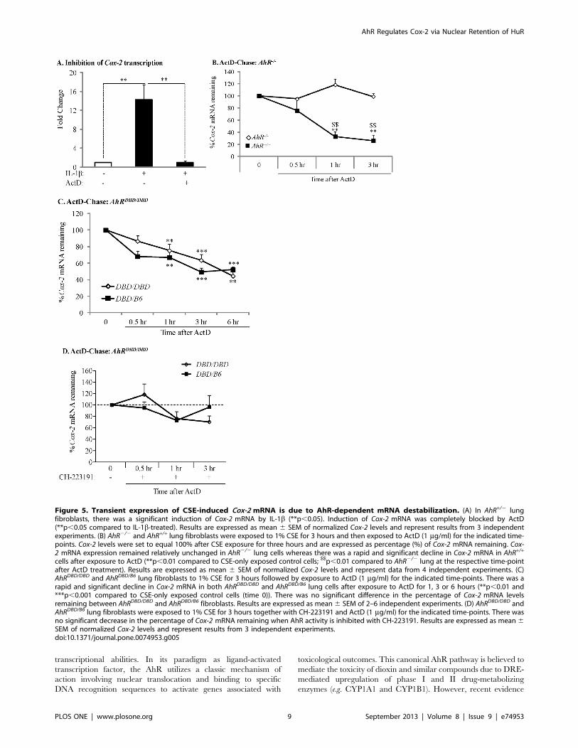
Transient Expression of CSE-induced Cox-2 mRNA is dueto AhR-dependent mRNA DestabilizationAfter demonstrating that ActD, at the concentrations used in
this study (1 mg/ml), prevents IL-1b-induced Cox-2 mRNA
expression (Figure 5A), we performed ActD-chase experiments
and quantified the decay in Cox-2 mRNA by qRT-PCR analysis.
There was a significant decline in Cox-2 mRNA levels only when
AhR+/+ lung fibroblasts were exposed to ActD for 1 or 3 hours
(Figure 5B, black squares). In contrast, levels of Cox-2 remained
constant and were unaltered in AhR2/2 lung fibroblasts exposed to
ActD (Figure 5B, open diamonds), suggesting that the AhR
destabilizes Cox-2 mRNA expression. We also utilized AhRDBD/
AhR Regulates Cox-2 via Nuclear Retention of HuR
PLOS ONE | www.plosone.org 4 September 2013 | Volume 8 | Issue 9 | e74953
DBD fibroblasts to evaluate Cox-2 mRNA stability. There was a
rapid and significant decline in steady-state Cox-2 mRNA levels
following ActD exposure for 1, 3 or 6 hours in AhRDBD/B6 cells,
which express a functional AhR and are thus used as a control to
compare with AhRDBD/DBD cells (Figure 5C, closed squares). A
parallel and significant decline in Cox-2 mRNA was also observed
in cells derived from AhRDBD/DBD mice (Figure 5C, open diamonds).
There was no significant difference in the percentage of remaining
Cox-2 mRNA between AhRDBD/DBD and control lung fibroblasts.
Inhibition of AhR activity with CH-223191 prevented the
significant decline in Cox-2 mRNA in both control and AhRDBD/
DBD fibroblasts (Figure 5D), supporting that AhR activity is
necessary for destabilization of the Cox-2 transcript. Collectively,
these data demonstrate that Cox-2 mRNA stability is governed by
a mechanism independent of the DNA-binding abilities of the
AhR.
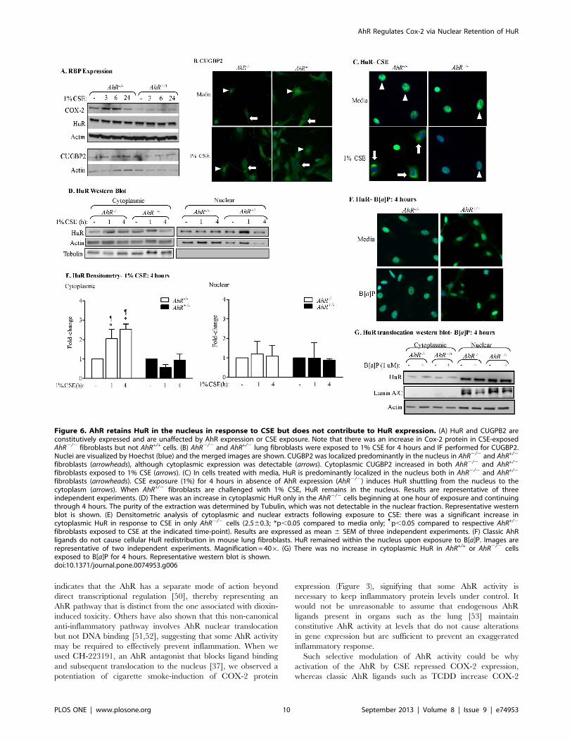
The AhR Controls the Nuclear-cytoplasmic Shuttling ofHuR in Response to CSEThe instability of Cox-2 mRNA is due to the presence of an
ARE in the 39-untranslated region which facilitates recruitment of
RNA-binding proteins [40], including HuR and CUGBP2
[20,41]. As there is no information on whether RNA-binding
protein expression is altered by cigarette smoke exposure or the
AhR, we first evaluated the expression of HuR and CUGBP2 by
western blot analysis. In both AhR2/2 and AhR+/+ lung fibroblasts,
there was similar expression of HuR and CUGBP2 (Figure 6A).
There was also no detectible difference in the expression of these
RNA-binding proteins upon exposure to 1% CSE in AhR2/2 and
AhR+/+ lung cells. Note that there is an increase in COX-2 protein
levels only in AhR2/2 cells exposed to CSE (Figure 6A; compared
with Figure 1D and [4]).
CUGBP2 and HuR are localized mainly in the nucleus,
shuttling to the cytoplasm when appropriately activated [20]. The
cytoplasmic localization of HuR and CUGBP2 correlate with their
ability to stabilize target mRNAs, including Cox-2 [21,22,23].
CUGBP2 was predominantely nuclear and translocated to the
cytoplasm upon exposure to 1% CSE, with little detectible
difference between AhR2/2 and AhR+/2 cells (Figure 6B). HuR
was also almost entirely restricted to the nucleus (Figure 6C-
arrowheads). In AhR+/2 cells exposed to 1% CSE, there was no
change in the subcellular distribution of HuR (Figure 6C-
arrowheads- and 6D). In contrast, there was a significant translo-
cation of HuR to the cytoplasm in AhR2/2 cells in response to 1%
CSE for either 1 or 4 hours (Figure 6C- arrows and 6D and 6E).
The relative level of nuclear HuR was not significantly altered by
CSE exposure (Figure 6E). AhR activation by the classic ligand
B[a]P did not alter HuR localization (Figure 6F and 6G).
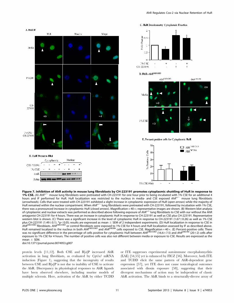
To then evaluate if AhR activity is necessary to retain HuR in
the nucleus, we evaluated HuR localization in AhR-expressing
cells which had been pretreated with CH-223191 and then treated
with CSE for 4 hours. Exposure of AhR-expressing lung
fibroblasts to 1% CSE failed to promote cytoplasmic redistribution
of HuR, and HuR remained predominantly nuclear (Figure 7A-
arrowheads). Inhibition of the AhR by CH-223191 alone resulted in
a small but significant increase in cytoplasmic HuR (Figure 7A-
arrows; Figure 7B and 7C). However, exposure of lung fibroblasts
to CH-223191 in conjunction with CSE caused nuclear-cytoplas-
mic shuttling of HuR (Figure 7A- arrows; Figure 7B). This increase
in cytoplasmic HuR localization was significant compared to CSE
alone (Figure 7C). In both AhRDBD/DBD and AhRDBD/B6 cells
exposed only to media, HuR was mostly nuclear (Figure 7D,
arrowheads). In response to 1% CSE, HuR remained predominantly
nuclear (Figure 7D, arrowheads) although there was detectable
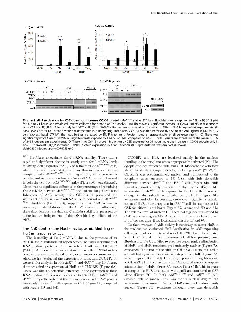
Figure 1. AhR activation by CSE does not increase COX-2 protein. AhR2/2 and AhR+/+ lung fibroblasts were exposed to CSE or B[a]P (1 mM)for 3, 6 or 24 hours and whole cell lysates collected for protein or RNA analysis. (A) There was a significant increase in Cyp1a1 mRNA in response toboth CSE and B[a]P for 6 hours only in AhR+/+ cells (***p,0.0001). Results are expressed as the mean 6 SEM of 3–6 independent experiments. (B)Basal levels of CYP1A1 protein were not detectable in primary lung fibroblasts. CYP1A1 was not increased by CSE or the AhR ligand TCDD. MLE-12cells express basal CYP1A1 that was further increased by B[a]P treatment. Western blot is representative of three experiments. (C) There wassignificantly more Cyp1b1 mRNA in lung fibroblasts exposed to 1% CSE or B[a]P compared to AhR2/2 cells. Results are expressed as the mean 6 SEMof 3–8 independent experiments. (D) There is no CYP1B1 protein induction by CSE exposure for 24 hours; note the increase in COX-2 protein only inAhR2/2 fibroblasts. B[a]P increased CYP1B1 protein expression in AhR+/+ fibroblasts. Representative western blot is shown.doi:10.1371/journal.pone.0074953.g001
AhR Regulates Cox-2 via Nuclear Retention of HuR
PLOS ONE | www.plosone.org 5 September 2013 | Volume 8 | Issue 9 | e74953
cytoplasmic HuR that was similar in intensity between AhRDBD/
DBD and AhRDBD/B6 cells, (Figure 7D, arrows). The percentage of
cells positive for cytoplasmic HuR was not different between
AhRDBD/DBD and AhRDBD/B6 cells (Figure 7E). Collectively, these
data show that the AhR controls the nuclear-cytoplasmic shuttling
of HuR in response to cigarette smoke by a mechanism that is
independent of DNA binding.
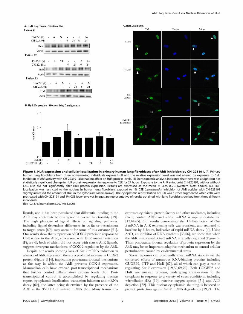
HuR expression has been shown in human fetal lung fibroblasts
[42] but the expression of HuR in adult lung fibroblasts is not
known. Our results now show that HuR is expressed in adult
human lung fibroblasts (Figure 8A). Densitometric analysis
revealed that there was a slight, but not statistically-significant
increase in the expression of HuR upon exposure of human lung
fibroblasts to CSE (Figure 8B). Exposure to the AhR antagonist
CH-223191, with or without CSE, had no significant effect on
HuR expression (Figure 8A and 8B). However, when human lung
fibroblasts were exposed to a combination of CSE and CH-
223191, there was an increase in cytoplasmic HuR (Figure 8C).
Collectively these data strongly support the notion that the AhR is
a critical regulator of HuR translocation in pulmonary cells, and
whose activity is important in retaining HuR in nucleus.
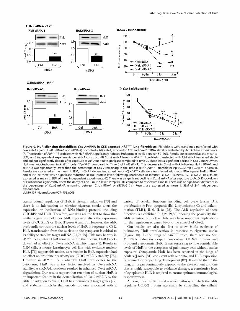
siRNA Knockdown of HuR in AhR-deficient Primary LungFibroblasts Destabilizes Cox-2 mRNATo confirm the implication of HuR in regulating Cox-2 stability
in AhR2/2 lung fibroblasts, RNA interference was used to
decrease HuR protein expression by at least 60% utilizing two
different siRNA targeting HuR (Figure 9A and 9C). In AhR2/2
cells, there was no significant change in the steady-state levels of
Cox-2 mRNA following exposure to ActD for as long as 3 hours
(Figure 9B). However, in AhR2/2 lung fibroblasts in which HuR
expression was reduced, there was a significant decline in Cox-
2 mRNA expression (siRNA-1 and siRNA-2; Figure 9B). In AhR+/
+ cells, HuR knock-down did not affect the decline in Cox-
2 mRNA (Figure 9D), demonstrating that HuR is the principle
factor that stabilizes Cox-2 mRNA in AhR-deficient pulmonary
cells.
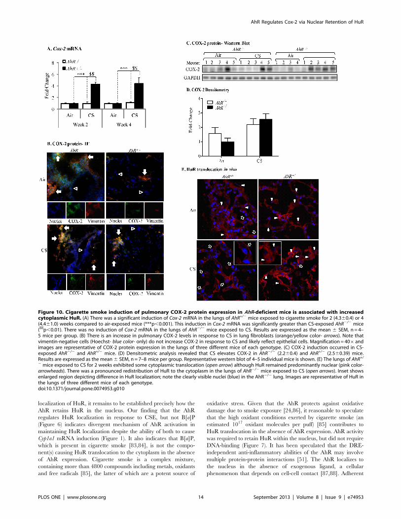
Cigarette Smoke Exposure Increases Pulmonary COX-2Protein Expression and Nuclear-Cytoplasmic Shuttling ofHuR in AhR2/2 MiceTo confirm our in vitro findings on the AhR regulation of COX-
2 protein levels via nuclear retention of HuR, we utilized our
preclinical murine model and exposed AhR2/2 and AhR+/2 mice
to cigarette smoke. There was a significant induction in Cox-
2 mRNA only in the lungs of AhR+/2 mice following cigarette
smoke exposure for 2 or 4 weeks compared to animals exposed
only to room air (Figure 10A). There was no increase in Cox-
2 mRNA in the lungs of AhR2/2 mice (Figure 10A) at either time-
point. Because there was no difference in the induction of Cox-
2 mRNA between 2- and 4-week exposures, only the 2-week
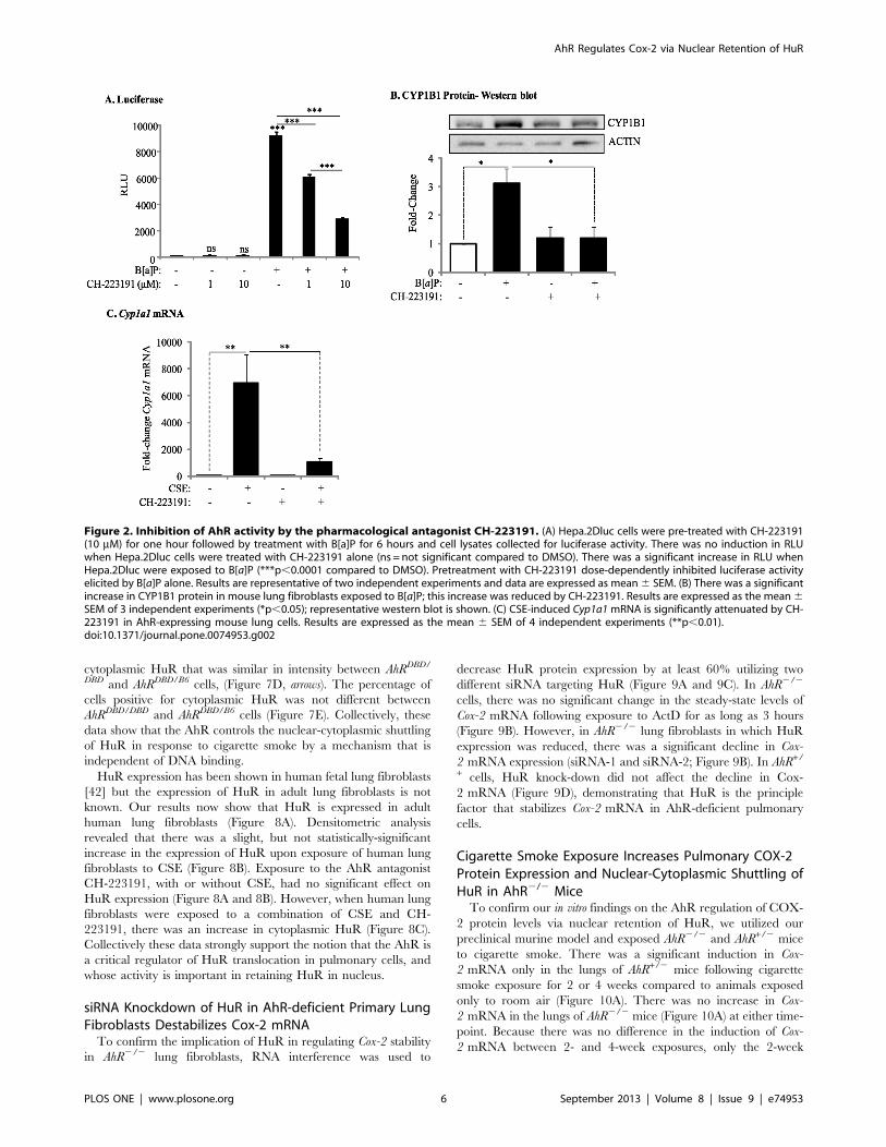
Figure 2. Inhibition of AhR activity by the pharmacological antagonist CH-223191. (A) Hepa.2Dluc cells were pre-treated with CH-223191(10 mM) for one hour followed by treatment with B[a]P for 6 hours and cell lysates collected for luciferase activity. There was no induction in RLUwhen Hepa.2Dluc cells were treated with CH-223191 alone (ns = not significant compared to DMSO). There was a significant increase in RLU whenHepa.2Dluc were exposed to B[a]P (***p,0.0001 compared to DMSO). Pretreatment with CH-223191 dose-dependently inhibited luciferase activityelicited by B[a]P alone. Results are representative of two independent experiments and data are expressed as mean6 SEM. (B) There was a significantincrease in CYP1B1 protein in mouse lung fibroblasts exposed to B[a]P; this increase was reduced by CH-223191. Results are expressed as the mean6SEM of 3 independent experiments (*p,0.05); representative western blot is shown. (C) CSE-induced Cyp1a1mRNA is significantly attenuated by CH-223191 in AhR-expressing mouse lung cells. Results are expressed as the mean 6 SEM of 4 independent experiments (**p,0.01).doi:10.1371/journal.pone.0074953.g002
AhR Regulates Cox-2 via Nuclear Retention of HuR
PLOS ONE | www.plosone.org 6 September 2013 | Volume 8 | Issue 9 | e74953
exposure regime was analyzed for COX-2 protein expression and
HuR localization.
Despite the lack of Cox-2 mRNA induction in AhR2/2 mice
exposed to cigarette smoke, there was an increase in pulmonary
COX-2 protein expression (Figure 10B) and a trend towards
higher basal COX-2 protein in the lungs of AhR2/2 mice
(Figure 10C and 10D). There is also COX-2 protein expression in
the lungs of AhR+/2 mice exposed to cigarette smoke (Figure 10C
and 10D). Double-immunofluorescence utilizing antibodies for
both COX-2 and vimentin (a fibroblast marker) indicated that
COX-2 protein was readily evident in lung fibroblasts (Figure 10B,
arrows- orange/yellow color), with stronger fluorescence occurring
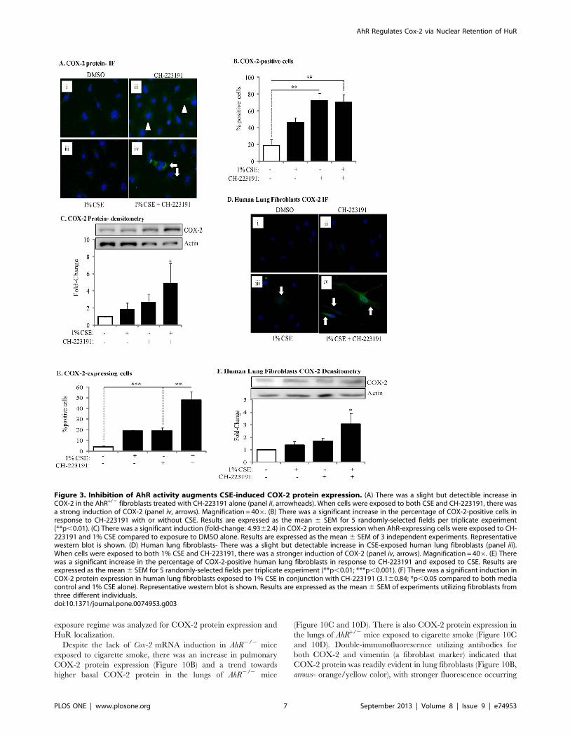
Figure 3. Inhibition of AhR activity augments CSE-induced COX-2 protein expression. (A) There was a slight but detectible increase inCOX-2 in the AhR+/2 fibroblasts treated with CH-223191 alone (panel ii, arrowheads). When cells were exposed to both CSE and CH-223191, there wasa strong induction of COX-2 (panel iv, arrows). Magnification = 406. (B) There was a significant increase in the percentage of COX-2-positive cells inresponse to CH-223191 with or without CSE. Results are expressed as the mean 6 SEM for 5 randomly-selected fields per triplicate experiment(**p,0.01). (C) There was a significant induction (fold-change: 4.9362.4) in COX-2 protein expression when AhR-expressing cells were exposed to CH-223191 and 1% CSE compared to exposure to DMSO alone. Results are expressed as the mean 6 SEM of 3 independent experiments. Representativewestern blot is shown. (D) Human lung fibroblasts- There was a slight but detectable increase in CSE-exposed human lung fibroblasts (panel iii).When cells were exposed to both 1% CSE and CH-223191, there was a stronger induction of COX-2 (panel iv, arrows). Magnification = 406. (E) Therewas a significant increase in the percentage of COX-2-positive human lung fibroblasts in response to CH-223191 and exposed to CSE. Results areexpressed as the mean6 SEM for 5 randomly-selected fields per triplicate experiment (**p,0.01; ***p,0.001). (F) There was a significant induction inCOX-2 protein expression in human lung fibroblasts exposed to 1% CSE in conjunction with CH-223191 (3.160.84; *p,0.05 compared to both mediacontrol and 1% CSE alone). Representative western blot is shown. Results are expressed as the mean 6 SEM of experiments utilizing fibroblasts fromthree different individuals.doi:10.1371/journal.pone.0074953.g003
AhR Regulates Cox-2 via Nuclear Retention of HuR
PLOS ONE | www.plosone.org 7 September 2013 | Volume 8 | Issue 9 | e74953
in the lungs of AhR2/2 mice. There were also abundant vimentin-
negative cells without COX-2 expression (blue color-nuclei only)
and likely represent epithelial cells (Figure 10B, open arrowheads). In
the lungs of air-exposed mice, HuR was abundantly expressed and
was localized predominantly in the nucleus (Figure 10E, arrowheads)
although cytoplasmic localization was evident. In the lungs of
AhR+/2 mice exposed to cigarette smoke for 2 weeks, there was
some translocation of HuR to the cytoplasm (Figure 10E, open
arrow). There was also considerable nuclear HuR remaining
(arrowheads). In stark contrast, redistribution of HuR to the
cytoplasm was unmistakable in the lungs of AhR2/2 mice exposed
to cigarette smoke (Figure 10E, open arrows; inset), indicating in vivo
that the AhR controls the cellular localization of HuR in response
to respiratory toxicants. When considered together, our in vivo and
in vitro data show for the first time that the AhR is a potent
suppressor of cigarette smoke-induced pulmonary COX-2 protein
due to post-transcriptional regulatory mechanisms that prevent the
cytoplasmic translocation of the RNA-binding protein HuR.
Discussion
The AhR was discovered nearly four decades ago as the
receptor responsible for the induction of aryl hydrocarbon
hydroxylase (CYP1A1) activity in response to the potent anthro-
pogenic ligand dioxin [43]. Although it is generally accepted that
the majority of deleterious effects of dioxin arise from dioxin
binding to the AhR and subsequent alterations in gene expression
patterns [44,45], one of the eminent unresolved questions is why
organisms would possess a receptor for dioxin at all [46]. The fact
though that the AhR is ubiquitously expressed in mammals, being
present in all major cell types in humans [47], and is highly-
conserved throughout evolution suggests a prominent role for this
receptor in mammalian physiology [48]. Early pioneering studies
using AhR-null mice not only revealed that the AhR is responsible
for dioxin toxicity, but have also implicated the AhR in cell
proliferation, differentiation, migration, development, tissue ho-
meostasis and vasculogenesis [48,49]. We have published that
low/absent AhR levels increase inflammation and structural cell
apoptosis [4,24], findings which argue for a prominent role of the
AhR in normal physiology. In our current study, we sought to
identify the mechanism by which the AhR prevents inflammatory
protein expression and report that AhR-dependent retention of
nuclear HuR suppresses COX-2 expression by a post-transcrip-
tional mechanism.
One of the most significant findings from this study is that the
AhR suppresses COX-2 protein expression in the absence of a
functional DNA-binding domain. This suggests that the AhR
suppresses inflammation by a mechanism that is independent of its
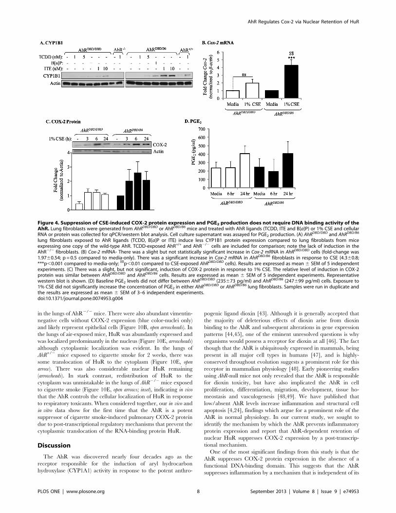
Figure 4. Suppression of CSE-induced COX-2 protein expression and PGE2 production does not require DNA binding activity of theAhR. Lung fibroblasts were generated from AhRDBD/DBD or AhRDBD/B6 mice and treated with AhR ligands (TCDD, ITE and B[a]P) or 1% CSE and cellularRNA or protein was collected for qPCR/western blot analysis. Cell culture supernatant was assayed for PGE2 production. (A) AhR
DBD/DBD and AhRDBD/B6
lung fibroblasts exposed to AhR ligands (TCDD, B[a]P or ITE) induce less CYP1B1 protein expression compared to lung fibroblasts from miceexpressing one copy of the wild-type AhR. TCDD-exposed AhR+/+ and AhR2/2 cells are included for comparison; note the lack of induction in theAhR2/2 fibroblasts. (B) Cox-2 mRNA- There was a slight but not statistically significant increase in Cox-2 mRNA in AhRDBD/DBD cells (fold-change was1.9760.54; p = 0.5 compared to media-only). There was a significant increase in Cox-2 mRNA in AhRDBD/B6 fibroblasts in response to CSE (4.360.8;***p,0.001 compared to media-only; $$p,0.01 compared to CSE-exposed AhRDBD/DBD cells). Results are expressed as mean 6 SEM of 5 independentexperiments. (C) There was a slight, but not significant, induction of COX-2 protein in response to 1% CSE. The relative level of induction in COX-2protein was similar between AhRDBD/DBD and AhRDBD/B6 cells. Results are expressed as mean 6 SEM of 5 independent experiments. Representativewestern blot is shown. (D) Baseline PGE2 levels did not differ between AhRDBD/DBD (235673 pg/ml) and AhRDBD/B6 (247699 pg/ml) cells. Exposure to1% CSE did not significantly increase the concentration of PGE2 in either AhRDBD/DBD or AhRDBD/B6 lung fibroblasts. Samples were run in duplicate andthe results are expressed as mean 6 SEM of 3–6 independent experiments.doi:10.1371/journal.pone.0074953.g004
AhR Regulates Cox-2 via Nuclear Retention of HuR
PLOS ONE | www.plosone.org 8 September 2013 | Volume 8 | Issue 9 | e74953
transcriptional abilities. In its paradigm as ligand-activated
transcription factor, the AhR utilizes a classic mechanism of
action involving nuclear translocation and binding to specific
DNA recognition sequences to activate genes associated with
toxicological outcomes. This canonical AhR pathway is believed to
mediate the toxicity of dioxin and similar compounds due to DRE-
mediated upregulation of phase I and II drug-metabolizing
enzymes (e.g. CYP1A1 and CYP1B1). However, recent evidence
Figure 5. Transient expression of CSE-induced Cox-2 mRNA is due to AhR-dependent mRNA destabilization. (A) In AhR+/2 lungfibroblasts, there was a significant induction of Cox-2 mRNA by IL-1b (**p,0.05). Induction of Cox-2 mRNA was completely blocked by ActD(**p,0.05 compared to IL-1b-treated). Results are expressed as mean 6 SEM of normalized Cox-2 levels and represent results from 3 independentexperiments. (B) AhR2/2 and AhR+/+ lung fibroblasts were exposed to 1% CSE for 3 hours and then exposed to ActD (1 mg/ml) for the indicated time-points. Cox-2 levels were set to equal 100% after CSE exposure for three hours and are expressed as percentage (%) of Cox-2 mRNA remaining. Cox-2 mRNA expression remained relatively unchanged in AhR2/2 lung cells whereas there was a rapid and significant decline in Cox-2 mRNA in AhR+/+
cells after exposure to ActD (**p,0.01 compared to CSE-only exposed control cells; $$p,0.01 compared to AhR2/2 lung at the respective time-pointafter ActD treatment). Results are expressed as mean 6 SEM of normalized Cox-2 levels and represent data from 4 independent experiments. (C)AhRDBD/DBD and AhRDBD/B6 lung fibroblasts to 1% CSE for 3 hours followed by exposure to ActD (1 mg/ml) for the indicated time-points. There was arapid and significant decline in Cox-2 mRNA in both AhRDBD/DBD and AhRDBD/B6 lung cells after exposure to ActD for 1, 3 or 6 hours (**p,0.01 and***p,0.001 compared to CSE-only exposed control cells (time 0)). There was no significant difference in the percentage of Cox-2 mRNA levelsremaining between AhRDBD/DBD and AhRDBD/B6 fibroblasts. Results are expressed as mean6 SEM of 2–6 independent experiments. (D) AhRDBD/DBD andAhRDBD/B6 lung fibroblasts were exposed to 1% CSE for 3 hours together with CH-223191 and ActD (1 mg/ml) for the indicated time-points. There wasno significant decrease in the percentage of Cox-2 mRNA remaining when AhR activity is inhibited with CH-223191. Results are expressed as mean6SEM of normalized Cox-2 levels and represent results from 3 independent experiments.doi:10.1371/journal.pone.0074953.g005
AhR Regulates Cox-2 via Nuclear Retention of HuR
PLOS ONE | www.plosone.org 9 September 2013 | Volume 8 | Issue 9 | e74953
indicates that the AhR has a separate mode of action beyond
direct transcriptional regulation [50], thereby representing an
AhR pathway that is distinct from the one associated with dioxin-
induced toxicity. Others have also shown that this non-canonical
anti-inflammatory pathway involves AhR nuclear translocation
but not DNA binding [51,52], suggesting that some AhR activity
may be required to effectively prevent inflammation. When we
used CH-223191, an AhR antagonist that blocks ligand binding
and subsequent translocation to the nucleus [37], we observed a
potentiation of cigarette smoke-induction of COX-2 protein
expression (Figure 3), signifying that some AhR activity is
necessary to keep inflammatory protein levels under control. It
would not be unreasonable to assume that endogenous AhR
ligands present in organs such as the lung [53] maintain
constitutive AhR activity at levels that do not cause alterations
in gene expression but are sufficient to prevent an exaggerated
inflammatory response.
Such selective modulation of AhR activity could be why
activation of the AhR by CSE repressed COX-2 expression,
whereas classic AhR ligands such as TCDD increase COX-2
Figure 6. AhR retains HuR in the nucleus in response to CSE but does not contribute to HuR expression. (A) HuR and CUGPB2 areconstitutively expressed and are unaffected by AhR expression or CSE exposure. Note that there was an increase in Cox-2 protein in CSE-exposedAhR2/2 fibroblasts but not AhR+/+ cells. (B) AhR2/2 and AhR+/2 lung fibroblasts were exposed to 1% CSE for 4 hours and IF performed for CUGBP2.Nuclei are visualized by Hoechst (blue) and the merged images are shown. CUGBP2 was localized predominantly in the nucleus in AhR2/2 and AhR+/2
fibroblasts (arrowheads), although cytoplasmic expression was detectable (arrows). Cytoplasmic CUGBP2 increased in both AhR2/2 and AhR+/2
fibroblasts exposed to 1% CSE (arrows). (C) In cells treated with media, HuR is predominantly localized in the nucleus both in AhR2/2 and AhR+/2
fibroblasts (arrowheads). CSE exposure (1%) for 4 hours in absence of AhR expression (AhR2/2) induces HuR shuttling from the nucleus to thecytoplasm (arrows). When AhR+/2 fibroblasts are challenged with 1% CSE, HuR remains in the nucleus. Results are representative of threeindependent experiments. (D) There was an increase in cytoplasmic HuR only in the AhR2/2 cells beginning at one hour of exposure and continuingthrough 4 hours. The purity of the extraction was determined by Tubulin, which was not detectable in the nuclear fraction. Representative westernblot is shown. (E) Densitometric analysis of cytoplasmic and nuclear extracts following exposure to CSE: there was a significant increase incytoplasmic HuR in response to CSE in only AhR2/2 cells (2.560.3; *p,0.05 compared to media only; "p,0.05 compared to respective AhR+/2
fibroblasts exposed to CSE at the indicated time-point). Results are expressed as mean 6 SEM of three independent experiments. (F) Classic AhRligands do not cause cellular HuR redistribution in mouse lung fibroblasts. HuR remained within the nucleus upon exposure to B[a]P. Images arerepresentative of two independent experiments. Magnification = 406. (G) There was no increase in cytoplasmic HuR in AhR+/+ or AhR2/2 cellsexposed to B[a]P for 4 hours. Representative western blot is shown.doi:10.1371/journal.pone.0074953.g006
AhR Regulates Cox-2 via Nuclear Retention of HuR
PLOS ONE | www.plosone.org 10 September 2013 | Volume 8 | Issue 9 | e74953
protein levels [11,12]. Both CSE and B[a]P increased AhR
activation in lung fibroblasts, as evaluated by Cyp1a1 mRNA
induction (Figure 1), suggesting that the incongruity of results
between CSE and B[a]P is not due to inability of CSE to activate
the AhR. Discrepancy in physiological responses to AhR ligands
have been observed elsewhere, including murine models of
multiple sclerosis. Here, activation of the AhR by either TCDD
or ITE suppresses experimental autoimmune encephalomyelitis
(EAE) [54,55] yet is enhanced by FICZ [56]. Moreover, both ITE
and TCDD elicit the same pattern of AhR-dependent gene
expression [57], yet ITE does not cause toxicological outcomes
associated with dioxin exposure [58], suggesting that their
divergent mechanisms of action may be independent of classic
AhR activation. The AhR binds to a structurally-diverse array of
Figure 7. Inhibition of AhR activity in mouse lung fibroblasts by CH-223191 promotes cytoplasmic shuttling of HuR in response to1% CSE. (A) AhR+/2 mouse lung fibroblasts were pretreated with CH-223191 for one hour prior to being incubated with 1% CSE for an additional 4hours and IF performed for HuR. HuR localization was restricted to the nucleus in media- and CSE exposed AhR+/2 mouse lung fibroblasts(arrowheads). Cells that were treated with CH-223191 exhibited a slight increase in cytoplasmic expression of HuR (open arrows) while the majority ofHuR remained within the nuclear compartment. When AhR+/2 lung fibroblasts were pretreated with CH-223191, followed by incubation with 1% CSE,there was a pronounced increase in cytoplasmic HuR (closed arrows). Magnification = 406; representative images are shown. (B) Western blot analysisof cytoplasmic and nuclear extracts was performed as described above following exposure of AhR+/+ lung fibroblasts to CSE with our without the AhRantagonist CH-223191 for 4 hours. There was an increase in cytoplasmic HuR in response to CH-223191 as well as CSE plus CH-223191. Representativewestern blot is shown. (C) There was a significant increase in the level of cytoplasmic HuR in response to CH-223191 (1.6760.26) as well as 1% CSEplus CH-223191 (1.4960.1). *p,0.05; results are expressed as mean 6 SEM of 2 independent experiments. (D) HuR localization in response to CSE inAhRDBD/DBD fibroblasts. AhRDBD/DBD or control fibroblasts were exposed to 1% CSE for 4 hours and HuR localization assessed by IF as described above.HuR remained localized to the nucleus in both AhRDBD/DBD and AhRDBDB6 cells exposed to CSE. Magnification = 406. (E) Percent-positive cells: Therewas no significant difference in the percentage of cells positive for cytoplasmic HuR between AhRDBD/DBD (16.667.5) and AhRDBD/B6 (2062) cells afterexposure to 1% CSE for 4 hours. The number of positive cells was also not different between media or exposure to CSE. Results are expressed as themean 6 SEM.doi:10.1371/journal.pone.0074953.g007
AhR Regulates Cox-2 via Nuclear Retention of HuR
PLOS ONE | www.plosone.org 11 September 2013 | Volume 8 | Issue 9 | e74953
ligands, and it has been postulated that differential binding to the
AhR may contribute to divergence in overall functionality [59].
The high plasticity of ligand effects on signaling pathways,
including ligand-dependent differences in co-factor recruitment
to target genes [60], may account for some of this variance [61].
Our results show that suppression of COX-2 protein in response to
CSE is due to the AhR, concurrent with HuR nuclear retention
(Figure 6), both of which did not occur with classic AhR ligands,
suggests divergent mechanisms of COX-2 regulation by the AhR.
Despite our results showing lack of Cox-2 mRNA induction in
absence of AhR expression, there is a profound increase in COX-2
protein (Figure 1) [4], implicating post-transcriptional mechanisms
as the way in which the AhR prevents COX-2 expression.
Mammalian cells have evolved post-transcriptional mechanisms
that further control inflammatory protein levels [40]. Post-
transcriptional control is accomplished by regulating nuclear
export, cytoplasmic localization, translation initiation and mRNA
decay [62], the latter being determined by the presence of the
ARE in the 39-UTR of mature mRNA [63]. Many transiently-
expresses cytokines, growth factors and other mediators, including
Cox-2, contain AREs and whose mRNA is rapidly destabilized
[17,64,65]. Our results demonstrate that CSE-induction of Cox-
2 mRNA in AhR-expressing cells was transient, and returned to
baseline by 6 hours, indicative of rapid mRNA decay [4]. Using
ActD, an inhibitor of RNA synthesis [33,66], we show that when
the AhR is expressed, Cox-2 mRNA is rapidly degraded (Figure 5).
Thus, post-transcriptional regulation of protein expression by the
AhR may be an important adaptive mechanism to control cellular
perturbations caused by environmental stress.
Stress responses can profoundly affect mRNA stability via the
concerted efforts of numerous RNA-binding proteins including
CUGBP2, TTP and HuR [67], all of which can play a role in
regulating Cox-2 expression [19,68,69,70]. Both CUGBP2 and
HuR are nuclear proteins, undergoing translocation to the
cytoplasm in response to a variety of stress conditions, including
c-irradiation (IR) [19], reactive oxygen species [71] and ATP
depletion [72]. This nuclear-cytoplasmic shuttling is believed to
provide protection against Cox-2 mRNA degradation [19,21]. The
Figure 8. HuR expression and cellular localization in primary human lung fibroblasts after AhR inhibition by CH-223191. (A) Primaryhuman lung fibroblasts from three non-smoking individuals express HuR and the relative expression level was not altered by exposure to CSE.Inhibition of AhR activity with CH-223191 also had no affect on HuR protein levels. (B) Densitometric analysis indicated that there was a slight but notstatistically significant change in HuR protein expression in response to CSE for 24 hours. Exposure to the AhR antagonist CH-223191, with or withoutCSE, also did not significantly alter HuR protein expression. Results are expressed as the mean 6 SEM, n = 3 (western blots above). (C). HuRlocalization was restricted to the nucleus in human lung fibroblasts exposed to 1% CSE (arrowheads). Inhibition of AhR activity with CH-223191slightly increased the amount of HuR in the cytoplasm (open arrows). The cytoplasmic redistribution of HuR was further augmented when cells werepretreated with CH-223191 and 1% CSE (open arrows). Images are representative of results obtained with lung fibroblasts derived from three differentindividuals.doi:10.1371/journal.pone.0074953.g008
AhR Regulates Cox-2 via Nuclear Retention of HuR
PLOS ONE | www.plosone.org 12 September 2013 | Volume 8 | Issue 9 | e74953
transcriptional regulation of HuR is virtually unknown [73] and
there is no information on whether cigarette smoke alters the
expression or localization of RNA-binding proteins, including
CUGBP2 and HuR. Therefore, our data are the first to show that
neither cigarette smoke nor AhR expression alters the expression
levels of CUGBP2 or HuR (Figures 6 and 8). However, the AhR
profoundly controls the nuclear levels of HuR in response to CSE.
HuR translocation from the nucleus to the cytoplasm is critical to
its ability to stabilize target mRNA [21,74,75]. This may be why in
AhR+/+ cells, where HuR remains within the nucleus, HuR knock-
down had no effect on Cox-2 mRNA stability (Figure 9). Results in
C5N cells, a mouse keratinocyte cell line with exclusive nuclear
HuR [76] support this notion, as reduction in HuR expression had
no effect on ornithine decarboxylase (ODC) mRNA stability [76].
However in AhR2/2 cells whereby HuR translocates to the
cytoplasm, HuR was a key factor involved in Cox-2 mRNA
stability, as siRNA-knockdown resulted in enhanced Cox-2 mRNA
degradation. Our results support that retention of nuclear HuR is
an important feature in the destabilization of Cox-2 mRNA by the
AhR. In addition to Cox-2, HuR has thousands of target genes [77]
and stabilizes mRNAs that encode proteins associated with a
variety of cellular functions including cell cycle (cyclin D1),
proliferation (c-Fos), apoptosis (Bcl-2, cytochrome C) and inflam-
mation (TLR4, IL-6, IL-8) [78]. The AhR regulation of these
functions is established [4,5,24,79,80] opening the possibility that
AhR retention of nuclear HuR may have important implications
for the regulation of genes beyond the control of Cox-2.
Our results are also the first to show in vivo evidence of
pulmonary HuR translocation in response to cigarette smoke
(Figure 10). In the lungs of AhR2/2 mice, there was no Cox-
2 mRNA induction despite concordant COX-2 protein and
profound cytoplasmic HuR. It was surprising to note considerable
levels of HuR in the cytoplasm of pulmonary cells without smoke
exposure. Cytoplasmic HuR has been reported in the lungs of
adult A/J mice [81], consistent with our data, and HuR expression
is required for proper lung development [82]. It may be that in the
lung, an organ continuously exposed to the environment and one
that is highly susceptible to oxidative damage, a constitutive level
of cytoplasmic HuR is required to ensure optimum immunological
responsiveness.
Although our results reveal a novel pathway in which the AhR
regulates COX-2 protein expression by controlling the cellular
Figure 9. HuR silencing destabilizes Cox-2 mRNA in CSE-exposed AhR2/2 lung fibroblasts. Fibroblasts were transiently transfected withtwo siRNA against HuR (siRNA-1 and siRNA-2) or control (Ctrl) siRNA, exposed to CSE and Cox-2 mRNA stability evaluated by ActD chase experiments.(A) Transfection of AhR2/2 fibroblasts with HuR siRNA significantly reduced HuR protein levels between 50–70%. Results are expressed as the mean6SEM, n = 3 independent experiments per siRNA construct. (B) Cox-2 mRNA levels in AhR2/2 fibroblasts transfected with Ctrl siRNA remained stableand did not significantly decline after exposure to ActD (ns = not significant compared to time 0). There was a significant decline in Cox-2 mRNA whenHuR was knocked-down in AhR2/2 cells (**p,0.01 compared to Time 0 of HuR siRNA). This decrease in Cox-2 mRNA following HuR siRNA-1 andsiRNA-2 was significantly lower than the percentage of Cox-2 remaining in the Time 0 siRNA AhR2/2 fibroblasts (*p,0.05; **p,0.01; ***p,0.001).Results are expressed as the mean 6 SEM, n = 2–5 independent experiments. (C) AhR+/+ cells were transfected with two siRNA against HuR (siRNA-1and siRNA-2); there was a significant reduction in HuR protein levels following knockdown (0.3860.09- siRNA-1; 0.3960.012- siRNA-2). Results areexpressed as mean6 SEM of three independent experiments. (D) There was a significant decline in Cox-2 mRNA after exposure to ActD. Knock-downof HuR did not significantly affect the decay of Cox-2 mRNA levels (***p,0.001 compared to respective Time 0). There was no significant difference inthe percentage of Cox-2 mRNA remaining between Ctrl, siRNA-1 or siRNA-2 (ns). Results are expressed as mean 6 SEM of 2–4 independentexperiments.doi:10.1371/journal.pone.0074953.g009
AhR Regulates Cox-2 via Nuclear Retention of HuR
PLOS ONE | www.plosone.org 13 September 2013 | Volume 8 | Issue 9 | e74953
localization of HuR, it remains to be established precisely how the
AhR retains HuR in the nucleus. Our finding that the AhR
regulates HuR localization in response to CSE, but not B[a]P
(Figure 6) indicates divergent mechanism of AhR activation in
maintaining HuR localization despite the ability of both to cause
Cyp1a1 mRNA induction (Figure 1). It also indicates that B[a]P,
which is present in cigarette smoke [83,84], is not the compo-
nent(s) causing HuR translocation to the cytoplasm in the absence
of AhR expression. Cigarette smoke is a complex mixture,
containing more than 4800 compounds including metals, oxidants
and free radicals [85], the latter of which are a potent source of
oxidative stress. Given that the AhR protects against oxidative
damage due to smoke exposure [24,86], it reasonable to speculate
that the high oxidant conditions exerted by cigarette smoke (an
estimated 1017 oxidant molecules per puff) [85] contributes to
HuR translocation in the absence of AhR expression. AhR activity
was required to retain HuR within the nucleus, but did not require
DNA-binding (Figure 7). It has been speculated that the DRE-
independent anti-inflammatory abilities of the AhR may involve
multiple protein-protein interactions [51]. The AhR localizes to
the nucleus in the absence of exogenous ligand, a cellular
phenomenon that depends on cell-cell contact [87,88]. Adherent
Figure 10. Cigarette smoke induction of pulmonary COX-2 protein expression in AhR-deficient mice is associated with increasedcytoplasmic HuR. (A) There was a significant induction of Cox-2 mRNA in the lungs of AhR+/2 mice exposed to cigarette smoke for 2 (4.360.4) or 4(4.461.0) weeks compared to air-exposed mice (***p,0.001). This induction in Cox-2 mRNA was significantly greater than CS-exposed AhR2/2 mice($$p,0.01). There was no induction of Cox-2 mRNA in the lungs of AhR2/2 mice exposed to CS. Results are expressed as the mean 6 SEM, n= 4–5 mice per group. (B) There is an increase in pulmonary COX-2 levels in response to CS in lung fibroblasts (orange/yellow color- arrows). Note thatvimentin-negative cells (Hoechst- blue color- only) do not increase COX-2 in response to CS and likely reflect epithelial cells. Magnification= 406andimages are representative of COX-2 protein expression in the lungs of three different mice of each genotype. (C) COX-2 induction occurred in CS-exposed AhR2/2 and AhR+/2 mice. (D) Densitometric analysis revealed that CS elevates COX-2 in AhR2/2 (2.260.4) and AhR+/2 (2.560.39) mice.Results are expressed as the mean6 SEM, n= 7–8 mice per group. Representative western blot of 4–5 individual mice is shown. (E) The lungs of AhR+/2 mice exposed to CS for 2 weeks exhibited some cytoplasmic translocation (open arrow) although HuR remained predominantly nuclear (pink color-arrowheads). There was a pronounced redistribution of HuR to the cytoplasm in the lungs of AhR2/2 mice exposed to CS (open arrows). Inset showsenlarged region depicting difference in HuR localization; note the clearly visible nuclei (blue) in the AhR2/2 lung. Images are representative of HuR inthe lungs of three different mice of each genotype.doi:10.1371/journal.pone.0074953.g010
AhR Regulates Cox-2 via Nuclear Retention of HuR
PLOS ONE | www.plosone.org 14 September 2013 | Volume 8 | Issue 9 | e74953
cells grown to sub-confluence, methodologically similar to the
experiments conducted here, exhibit both cytoplasmic and nuclear
AhR [87], making interaction with AhR and HuR within the
nucleus a plausible assumption. Thus, while there is no known
physical association between AhR and HuR, it is interesting to
speculate that the AhR may interact with HuR to prevent its
nuclear export, a notion we are actively pursuing.
It is believed that the AhR plays an important role in physiology
independent of its ability to respond to dioxin. Our previous work
highlights the AhR as a key anti-inflammatory protein by an
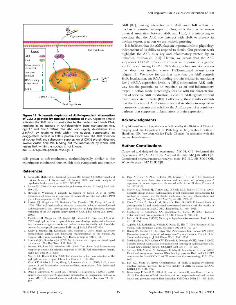
unknown mechanism [4,5]. Herein, we report that the AhR
suppresses COX-2 protein expression in response to cigarette
smoke by enhancing Cox-2 mRNA decay, a fundamental process
that does not involve classic DRE-mediated transcription
(Figure 11). We show for the first time that the AhR controls
HuR localization, an RNA-binding protein critical in stabilizing
Cox-2 mRNA expression levels. A DRE-independent AhR path-
way has the potential to be exploited as an anti-inflammatory
target, a notion made increasingly feasible with the characteriza-
tion of selective AhR modulators, a class of AhR ligands without
dioxin-associated toxicity [89]. Collectively, these results establish
that the function of AhR extends beyond its ability to respond to
man-made toxicants and solidifies the AhR as part of a regulatory
pathway that suppresses inflammatory protein expression.
Acknowledgments
Acquisition of human lung tissue was facilitated by the Division of Thoracic
Surgery and the Department of Pathology of St Joseph’s Healthcare
Hamilton, ON. We acknowledge Fazila Chouiali for assistance with the
immunohistochemistry.
Author Contributions
Conceived and designed the experiments: MZ SR CJB. Performed the
experiments: MZ JAS ARS CJB. Analyzed the data: MZ JAS ARS CJB.
Contributed reagents/materials/analysis tools: PN IEG SR SDM QH.
Wrote the paper: MZ DHE CJB.
References
1. Lopez AD, Mathers CD, Ezzati M, Jamison DT, Murray CJ (2006) Global and
regional burden of disease and risk factors, 2001: systematic analysis of
population health data. Lancet 367: 1747–1757.
2. Barnes PJ (2000) Chronic obstructive pulmonary disease. N Engl J Med 343:
269–280.
3. Hayashi S, Watanabe J, Nakachi K, Eguchi H, Gotoh O, et al. (1994)
Interindividual difference in expression of human Ah receptor and related P450
genes. Carcinogenesis 15: 801–806.
4. Baglole CJ, Maggirwar SB, Gasiewicz TA, Thatcher TH, Phipps RP, et al.
(2008) The aryl hydrocarbon receptor attenuates tobacco smoke-induced
cyclooxygenase-2 and prostaglandin production in lung fibroblasts through
regulation of the NF-kappaB family member RelB. J Biol Chem 283: 28944–
28957.
5. Thatcher TH, Maggirwar SB, Baglole CJ, Lakatos HF, Gasiewicz TA, et al.
(2007) Aryl hydrocarbon receptor-deficient mice develop heightened inflamma-
tory responses to cigarette smoke and endotoxin associated with rapid loss of the
nuclear factor-kappaB component RelB. Am J Pathol 170: 855–864.
6. Racky J, Schmitz HJ, Kauffmann HM, Schrenk D (2004) Single nucleotide
polymorphism analysis and functional characterization of the human Ah
receptor (AhR) gene promoter. Arch Biochem Biophys 421: 91–98.
7. Rowlands JC, Gustafsson JA (1997) Aryl hydrocarbon receptor-mediated signal
transduction. Crit Rev Toxicol 27: 109–134.
8. Furness SG, Lees MJ, Whitelaw ML (2007) The dioxin (aryl hydrocarbon)
receptor as a model for adaptive responses of bHLH/PAS transcription factors.
FEBS Lett 581: 3616–3625.
9. Nguyen LP, Bradfield CA (2008) The search for endogenous activators of the
aryl hydrocarbon receptor. Chem Res Toxicol 21: 102–116.
10. Vogel CF, Sciullo E, Li W, Wong P, Lazennec G, et al. (2007) RelB, a new
partner of aryl hydrocarbon receptor-mediated transcription. Mol Endocrinol
21: 2941–2955.
11. Dong B, Nishimura N, Vogel CF, Tohyama C, Matsumura F (2010) TCDD-
induced cyclooxygenase-2 expression is mediated by the nongenomic pathway in
mouse MMDD1 macula densa cells and kidneys. Biochem Pharmacol 79: 487–
497.
12. Puga A, Hoffer A, Zhou S, Bohm JM, Leikauf GD, et al. (1997) Sustained
increase in intracellular free calcium and activation of cyclooxygenase-2
expression in mouse hepatoma cells treated with dioxin. Biochem Pharmacol
54: 1287–1296.
13. Martey CA, Pollock SJ, Turner CK, O’Reilly KM, Baglole CJ, et al. (2004)
Cigarette smoke induces cyclooxygenase-2 and microsomal prostaglandin E2
synthase in human lung fibroblasts: implications for lung inflammation and
cancer. Am J Physiol Lung Cell Mol Physiol 287: L981–991.
14. Chen Y, Chen P, Hanaoka M, Droma Y, Kubo K (2008) Enhanced levels of
prostaglandin E2 and matrix metalloproteinase-2 correlate with the severity of
airflow limitation in stable COPD. Respirology 13: 1014–1021.
15. Montuschi P, Kharitonov SA, Ciabattoni G, Barnes PJ (2003) Exhaled
leukotrienes and prostaglandins in COPD. Thorax 58: 585–588.
16. Lofroth G, Rannug A (1988) Ah receptor ligands in tobacco smoke. Toxicol Lett
42: 131–136.
17. Appleby SB, Ristimaki A, Neilson K, Narko K, Hla T (1994) Structure of the
human cyclo-oxygenase-2 gene. Biochem J 302 (Pt 3): 723–727.
18. Dixon DA, Kaplan CD, McIntyre TM, Zimmerman GA, Prescott SM (2000)
Post-transcriptional control of cyclooxygenase-2 gene expression. The role of the
39-untranslated region. J Biol Chem 275: 11750–11757.
19. Mukhopadhyay D, Houchen CW, Kennedy S, Dieckgraefe BK, Anant S (2003)
Coupled mRNA stabilization and translational silencing of cyclooxygenase-2 by
a novel RNA binding protein, CUGBP2. Mol Cell 11: 113–126.
20. Sureban SM, Murmu N, Rodriguez P, May R, Maheshwari R, et al. (2007)
Functional antagonism between RNA binding proteins HuR and CUGBP2
determines the fate of COX-2 mRNA translation. Gastroenterology 132: 1055–
1065.
21. Fan XC, Steitz JA (1998) Overexpression of HuR, a nuclear-cytoplasmic
shuttling protein, increases the in vivo stability of ARE-containing mRNAs.
EMBO J 17: 3448–3460.
22. Beauchamp P, Nassif C, Hillock S, van der Giessen K, von Roretz C, et al.
(2010) The cleavage of HuR interferes with its transportin-2-mediated nuclear
import and promotes muscle fiber formation. Cell Death Differ 17: 1588–1599.
Figure 11. Schematic depiction of AhR-dependent attenuationof COX-2 protein by nuclear retention of HuR. Cigarette smokeactivates the AhR, which translocates to the nucleus and binds DNA,resulting in an increase in AhR-dependent gene transcription (e.g.Cyp1A1 and Cox-2 mRNA). The AhR also rapidly destabilizes Cox-2 mRNA by retaining HuR within the nucleus, suppressing anexaggerated increase in COX-2 protein expression. The AhR retentionof nuclear HuR and subsequent suppression of COX-2 protein does notinvolve classic AhR:DNA binding but the mechanism by which AhRretains HuR within the nucleus is not known.doi:10.1371/journal.pone.0074953.g011
AhR Regulates Cox-2 via Nuclear Retention of HuR
PLOS ONE | www.plosone.org 15 September 2013 | Volume 8 | Issue 9 | e74953
23. van der Giessen K, Gallouzi IE (2007) Involvement of transportin 2-mediated
HuR import in muscle cell differentiation. Mol Biol Cell 18: 2619–2629.
24. Rico de Souza A, Zago M, Pollock SJ, Sime PJ, Phipps RP, et al. (2011) GeneticAblation of the Aryl Hydrocarbon Receptor Causes Cigarette Smoke-induced
Mitochondrial Dysfunction and Apoptosis. J Biol Chem 286: 43214–43228.
25. Baglole CJ, Reddy SY, Pollock SJ, Feldon SE, Sime PJ, et al. (2005) Isolationand phenotypic characterization of lung fibroblasts. Methods Mol Med 117:
115–127.
26. Bunger MK, Glover E, Moran SM, Walisser JA, Lahvis GP, et al. (2008)
Abnormal liver development and resistance to 2,3,7,8-tetrachlorodibenzo-p-dioxin toxicity in mice carrying a mutation in the DNA-binding domain of the
aryl hydrocarbon receptor. Toxicol Sci 106: 83–92.
27. Henry EC, Kende AS, Rucci G, Totleben MJ, Willey JJ, et al. (1999) Flavoneantagonists bind competitively with 2,3,7, 8-tetrachlorodibenzo-p-dioxin
(TCDD) to the aryl hydrocarbon receptor but inhibit nuclear uptake and
transformation. Mol Pharmacol 55: 716–725.
28. Gasiewicz TA, Kende AS, Rucci G, Whitney B, Willey JJ (1996) Analysis ofstructural requirements for Ah receptor antagonist activity: ellipticines, flavones,
and related compounds. Biochem Pharmacol 52: 1787–1803.
29. Wikenheiser KA, Vorbroker DK, Rice WR, Clark JC, Bachurski CJ, et al.(1993) Production of immortalized distal respiratory epithelial cell lines from
surfactant protein C/simian virus 40 large tumor antigen transgenic mice. ProcNatl Acad Sci U S A 90: 11029–11033.
30. Baglole CJ, Bushinsky SM, Garcia TM, Kode A, Rahman I, et al. (2006)Differential induction of apoptosis by cigarette smoke extract in primary human
lung fibroblast strains: implications for emphysema. Am J Physiol Lung Cell MolPhysiol 291: L19–29.
31. Baglole CJ, Sime PJ, Phipps RP (2008) Cigarette smoke-induced expression of
heme oxygenase-1 in human lung fibroblasts is regulated by intracellularglutathione. Am J Physiol Lung Cell Mol Physiol 295: L624–636.
32. Carp H, Janoff A (1978) Possible mechanisms of emphysema in smokers. In vitro
suppression of serum elastase-inhibitory capacity by fresh cigarette smoke and its
prevention by antioxidants. Am Rev Respir Dis 118: 617–621.
33. Hyman RW, Davidson N (1970) Kinetics of the in vitro inhibition oftranscription by actinomycin. J Mol Biol 50: 421–438.
34. Denison MS, Nagy SR (2003) Activation of the aryl hydrocarbon receptor by
structurally diverse exogenous and endogenous chemicals. Annu Rev PharmacolToxicol 43: 309–334.
35. Christou M, Savas U, Schroeder S, Shen X, Thompson T, et al. (1995)
Cytochromes CYP1A1 and CYP1B1 in the rat mammary gland: cell-specific
expression and regulation by polycyclic aromatic hydrocarbons and hormones.Mol Cell Endocrinol 115: 41–50.
36. Eltom SE, Larsen MC, Jefcoate CR (1998) Expression of CYP1B1 but not
CYP1A1 by primary cultured human mammary stromal fibroblasts constitu-tively and in response to dioxin exposure: role of the Ah receptor.
Carcinogenesis 19: 1437–1444.
37. Kim SH, Henry EC, Kim DK, Kim YH, Shin KJ, et al. (2006) Novel compound
2-methyl-2H-pyrazole-3-carboxylic acid (2-methyl-4-o-tolylazo-phenyl)-amide(CH-223191) prevents 2,3,7,8-TCDD-induced toxicity by antagonizing the aryl
hydrocarbon receptor. Mol Pharmacol 69: 1871–1878.
38. Zhao B, Degroot DE, Hayashi A, He G, Denison MS (2010) CH223191 is aligand-selective antagonist of the Ah (Dioxin) receptor. Toxicol Sci 117: 393–
403.
39. Murray IA, Flaveny CA, Chiaro CR, Sharma AK, Tanos RS, et al. (2011)
Suppression of cytokine-mediated complement factor gene expression throughselective activation of the Ah receptor with 39,49-dimethoxy-alpha-naphtho-
flavone. Mol Pharmacol 79: 508–519.
40. Anderson P, Phillips K, Stoecklin G, Kedersha N (2004) Post-transcriptionalregulation of proinflammatory proteins. J Leukoc Biol 76: 42–47.
41. Cok SJ, Acton SJ, Morrison AR (2003) The proximal region of the 39-
untranslated region of cyclooxygenase-2 is recognized by a multimeric protein
complex containing HuR, TIA-1, TIAR, and the heterogeneous nuclearribonucleoprotein U. J Biol Chem 278: 36157–36162.
42. Bonelli MA, Alfieri RR, Desenzani S, Petronini PG, Borghetti AF (2004)
Proteasome inhibition increases HuR level, restores heat-inducible HSP72expression and thermotolerance in WI-38 senescent human fibroblasts. Exp
Gerontol 39: 423–432.
43. Poland A, Glover E, Kende AS (1976) Stereospecific, high affinity binding of
2,3,7,8-tetrachlorodibenzo-p-dioxin by hepatic cytosol. Evidence that thebinding species is receptor for induction of aryl hydrocarbon hydroxylase.
J Biol Chem 251: 4936–4946.
44. Okey AB, Franc MA, Moffat ID, Tijet N, Boutros PC, et al. (2005) Toxicologicalimplications of polymorphisms in receptors for xenobiotic chemicals: the case of
the aryl hydrocarbon receptor. Toxicol Appl Pharmacol 207: 43–51.
45. Mimura J, Yamashita K, Nakamura K, Morita M, Takagi TN, et al. (1997) Loss
of teratogenic response to 2,3,7,8-tetrachlorodibenzo-p-dioxin (TCDD) in micelacking the Ah (dioxin) receptor. Genes Cells 2: 645–654.
46. Landers JP, Bunce NJ (1991) The Ah receptor and the mechanism of dioxin
toxicity. Biochem J 276 (Pt 2): 273–287.
47. Ema M, Matsushita N, Sogawa K, Ariyama T, Inazawa J, et al. (1994) Humanarylhydrocarbon receptor: functional expression and chromosomal assignment
to 7p21. J Biochem 116: 845–851.
48. Fernandez-Salguero PM, Ward JM, Sundberg JP, Gonzalez FJ (1997) Lesions of
aryl-hydrocarbon receptor-deficient mice. Vet Pathol 34: 605–614.
49. Schmidt JV, Su GH, Reddy JK, Simon MC, Bradfield CA (1996)Characterization of a murine Ahr null allele: involvement of the Ah receptor
in hepatic growth and development. Proc Natl Acad Sci U S A 93: 6731–6736.
50. Ohtake F, Baba A, Takada I, Okada M, Iwasaki K, et al. (2007) Dioxin receptoris a ligand-dependent E3 ubiquitin ligase. Nature 446: 562–566.
51. Patel RD, Murray IA, Flaveny CA, Kusnadi A, Perdew GH (2009) Ah receptor
represses acute-phase response gene expression without binding to its cognateresponse element. Lab Invest 89: 695–707.
52. Murray IA, Krishnegowda G, DiNatale BC, Flaveny C, Chiaro C, et al. (2010)
Development of a selective modulator of aryl hydrocarbon (Ah) receptor activitythat exhibits anti-inflammatory properties. Chem Res Toxicol 23: 955–966.
53. Song J, Clagett-Dame M, Peterson RE, Hahn ME, Westler WM, et al. (2002) A
ligand for the aryl hydrocarbon receptor isolated from lung. Proc Natl AcadSci U S A 99: 14694–14699.
54. Quintana FJ, Murugaiyan G, Farez MF, Mitsdoerffer M, Tukpah AM, et al.
(2010) An endogenous aryl hydrocarbon receptor ligand acts on dendritic cellsand T cells to suppress experimental autoimmune encephalomyelitis. Proc Natl
Acad Sci U S A 107: 20768–20773.
55. Quintana FJ, Basso AS, Iglesias AH, Korn T, Farez MF, et al. (2008) Control ofT(reg) and T(H)17 cell differentiation by the aryl hydrocarbon receptor. Nature
453: 65–71.
56. Veldhoen M, Hirota K, Westendorf AM, Buer J, Dumoutier L, et al. (2008) The
aryl hydrocarbon receptor links TH17-cell-mediated autoimmunity to environ-
mental toxins. Nature 453: 106–109.
57. Henry EC, Welle SL, Gasiewicz TA (2010) TCDD and a putative endogenous
AhR ligand, ITE, elicit the same immediate changes in gene expression in mouse
lung fibroblasts. Toxicol Sci 114: 90–100.
58. Henry EC, Bemis JC, Henry O, Kende AS, Gasiewicz TA (2006) A potential
endogenous ligand for the aryl hydrocarbon receptor has potent agonist activity
in vitro and in vivo. Arch Biochem Biophys 450: 67–77.
59. Denison MS, Soshilov AA, He G, DeGroot DE, Zhao B (2011) Exactly the same
but different: promiscuity and diversity in the molecular mechanisms of action ofthe aryl hydrocarbon (dioxin) receptor. Toxicol Sci 124: 1–22.
60. Hestermann EV, Brown M (2003) Agonist and chemopreventative ligands
induce differential transcriptional cofactor recruitment by aryl hydrocarbonreceptor. Mol Cell Biol 23: 7920–7925.
61. Guyot E, Chevallier A, Barouki R, Coumoul X (2012) The AhR twist: ligand-
dependent AhR signaling and pharmaco-toxicological implications. Drug DiscovToday.
62. Anderson P (2008) Post-transcriptional control of cytokine production. Nat
Immunol 9: 353–359.
63. Clark A (2000) Post-transcriptional regulation of pro-inflammatory gene
expression. Arthritis Res 2: 172–174.
64. Caput D, Beutler B, Hartog K, Thayer R, Brown-Shimer S, et al. (1986)Identification of a common nucleotide sequence in the 39-untranslated region of
mRNA molecules specifying inflammatory mediators. Proc Natl Acad Sci U S A
83: 1670–1674.
65. Hel Z, Di Marco S, Radzioch D (1998) Characterization of the RNA binding
proteins forming complexes with a novel putative regulatory region in the 39-
UTR of TNF-alpha mRNA. Nucleic Acids Res 26: 2803–2812.
66. Sobell HM (1985) Actinomycin andDNA transcription. Proc Natl Acad Sci U S A
82: 5328–5331.
67. von Roretz C, Di Marco S, Mazroui R, Gallouzi IE (2011) Turnover of AU-rich-containing mRNAs during stress: a matter of survival. Wiley Interdiscip Rev
RNA 2: 336–347.
68. Sengupta S, Jang BC, Wu MT, Paik JH, Furneaux H, et al. (2003) The RNA-binding protein HuR regulates the expression of cyclooxygenase-2. J Biol Chem
278: 25227–25233.
69. Natarajan G, Ramalingam S, Ramachandran I, May R, Queimado L, et al.(2008) CUGBP2 downregulation by prostaglandin E2 protects colon cancer cells
from radiation-induced mitotic catastrophe. Am J Physiol Gastrointest LiverPhysiol 294: G1235–1244.
70. Cha HJ, Lee HH, Chae SW, Cho WJ, Kim YM, et al. (2011) Tristetraprolin
downregulates the expression of both VEGF and COX-2 in human coloncancer. Hepatogastroenterology 58: 790–795.
71. Lin WN, Lin CC, Cheng HY, Yang CM (2011) Regulation of cyclooxygenase-2
and cytosolic phospholipase A2 gene expression by lipopolysaccharide throughthe RNA-binding protein HuR: involvement of NADPH oxidase, reactive
oxygen species and mitogen-activated protein kinases. Br J Pharmacol 163:1691–1706.
72. Jeyaraj SC, Dakhlallah D, Hill SR, Lee BS (2006) Expression and distribution of
HuR during ATP depletion and recovery in proximal tubule cells. Am J PhysiolRenal Physiol 291: F1255–1263.
73. Abdelmohsen K, Srikantan S, Yang X, Lal A, Kim HH, et al. (2009) Ubiquitin-
mediated proteolysis of HuR by heat shock. EMBO J 28: 1271–1282.
74. Fan J, Ishmael FT, Fang X, Myers A, Cheadle C, et al. (2011) Chemokine
transcripts as targets of the RNA-binding protein HuR in human airway
epithelium. J Immunol 186: 2482–2494.
75. Atasoy U, Watson J, Patel D, Keene JD (1998) ELAV protein HuA (HuR) can
redistribute between nucleus and cytoplasm and is upregulated during serum
stimulation and T cell activation. J Cell Sci 111 (Pt 21): 3145–3156.
76. Nowotarski SL, Shantz LM (2010) Cytoplasmic accumulation of the RNA-
binding protein HuR stabilizes the ornithine decarboxylase transcript in a
murine nonmelanoma skin cancer model. J Biol Chem 285: 31885–31894.
AhR Regulates Cox-2 via Nuclear Retention of HuR
PLOS ONE | www.plosone.org 16 September 2013 | Volume 8 | Issue 9 | e74953
77. Lebedeva S, Jens M, Theil K, Schwanhausser B, Selbach M, et al. (2011)
Transcriptome-wide analysis of regulatory interactions of the RNA-bindingprotein HuR. Mol Cell 43: 340–352.
78. Meisner NC, Filipowicz W (2010) Properties of the regulatory RNA-binding
protein HuR and its role in controlling miRNA repression. Adv Exp Med Biol700: 106–123.
79. Marlowe JL, Fan Y, Chang X, Peng L, Knudsen ES, et al. (2008) The arylhydrocarbon receptor binds to E2F1 and inhibits E2F1-induced apoptosis. Mol
Biol Cell 19: 3263–3271.
80. Chang X, Fan Y, Karyala S, Schwemberger S, Tomlinson CR, et al. (2007)Ligand-independent regulation of transforming growth factor beta1 expression
and cell cycle progression by the aryl hydrocarbon receptor. Mol Cell Biol 27:6127–6139.
81. Blaxall BC, Dwyer-Nield LD, Bauer AK, Bohlmeyer TJ, Malkinson AM, et al.(2000) Differential expression and localization of the mRNA binding proteins,
AU-rich element mRNA binding protein (AUF1) and Hu antigen R (HuR), in
neoplastic lung tissue. Mol Carcinog 28: 76–83.82. Sgantzis N, Yiakouvaki A, Remboutsika E, Kontoyiannis DL (2011) HuR
controls lung branching morphogenesis and mesenchymal FGF networks. DevBiol 354: 267–279.
83. Roemer E, Stabbert R, Rustemeier K, Veltel DJ, Meisgen TJ, et al. (2004)
Chemical composition, cytotoxicity and mutagenicity of smoke from UScommercial and reference cigarettes smoked under two sets of machine smoking
conditions. Toxicology 195: 31–52.
84. Rustemeier K, Stabbert R, Haussmann HJ, Roemer E, Carmines EL (2002)Evaluation of the potential effects of ingredients added to cigarettes. Part 2:
chemical composition of mainstream smoke. Food Chem Toxicol 40: 93–104.85. Church DF, Pryor WA (1985) Free-radical chemistry of cigarette smoke and its
toxicological implications. Environ Health Perspect 64: 111–126.
86. Cheng YH, Huang SC, Lin CJ, Cheng LC, Li LA (2012) Aryl hydrocarbonreceptor protects lung adenocarcinoma cells against cigarette sidestream smoke
particulates-induced oxidative stress. Toxicol Appl Pharmacol 259: 293–301.87. Ikuta T, Kobayashi Y, Kawajiri K (2004) Cell density regulates intracellular
localization of aryl hydrocarbon receptor. J Biol Chem 279: 19209–19216.88. Cho YC, Zheng W, Jefcoate CR (2004) Disruption of cell-cell contact maximally
but transiently activates AhR-mediated transcription in 10T1/2 fibroblasts.
Toxicol Appl Pharmacol 199: 220–238.89. Safe S, Qin C, McDougal A (1999) Development of selective aryl hydrocarbon
receptor modulators for treatment of breast cancer. Expert Opin Investig Drugs8: 1385–1396.
AhR Regulates Cox-2 via Nuclear Retention of HuR
PLOS ONE | www.plosone.org 17 September 2013 | Volume 8 | Issue 9 | e74953
Related Documents